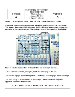
के न्द्रीय विद्यालय संगठन
KENDRIYA VIDYALAYA SANGATHAN
अहमदाबाद संа¤а¤ѕа¤—
AHMEDABAD REGION
अध्ययन-सामग्री
STUDY MATERIAL
CLASS: XI
BIOLOGY
सत्र-2014-15
SESSION-2014-15
CONTENTS
PARTICULARS
Page. No.
S.N.
1.
Chapter 1: The Living World.
1—4
2.
Chapter2: Biological Classification.
5—11
3.
Chapter 3: Plant Kingdom.
12—17
4.
Chapter 4: Animal Kingdom.
18—24
5.
Chapter 5: Morphology of Flowering Plants.
25—34
6.
Chapter 6: Anatomy of Flowering Plants.
35—43
7.
Chapter 7: Structural organisation in Animals.
44—46
8.
Chapter 8: Cell: The Unit of Life.
47—55
9.
Chapter 9: Biomolecules.
56—66
10. Chapter 10: Cell Cycle and Cell Divison
67—74
11. Chapter 11: Transport in Plants.
75—78
12. Chapter 12: Mineral Nutrition.
79—82
13. Chapter 13: Photosynthesis in Higher Plants.
83—90
14. Chapter 14: Respiration in Plants.
91—94
15. Chapter 15: Plant growth and development
95—98
16. Chapter 16: Digestion and Absorption.
99—107
17. Chapter 17: Breathing and exchange of gasses.
108—117
18. Chapter 18: Body fluids and Circulation.
118—129
19. Chapter 19: Excretory products and their elimination.
130—138
20. Chapter 20: Locomotion and Movement.
139—147
21. Chapter 21: Neural control and Coordination
148—154
22. Chapter 22: Chemical Coordination and Intigration.
155—158
Chapter-1
The Living World
TERMS REVISION:
1- Biodiversity: Large variety of organisms.
2- Nomenclature: Scientific naming of organisms .
3- Identification : Correct description of organism prior to nomenclature.
4-Classification: Grouping of organisms in to categories on the basis of similarities & differences.
5-Taxon: Concrete biological object or category of classification.
6-Taxonomy: Study of Identification, nomenclature and classification of organisms.
7-Systematics: Branch of biology dealing with taxonomy along with evolutionary relationship
between organisms.
8-Species :Group of Individual organisms with fundamental similarities (with capacity of interbreeding).
LEARNING TIPS:
1-Concentrate on minute points of the chapter keeping very short answer& short answer type
questions in mind.
2- Emphasise on concepts.
LET US LEARN THE LESSON
What is Living?
-Objects having characteristics of cellular organisation, growth & development, reproduction, ability to
sense environment & give response, metabolism etc.
All organisms grow:
-Increase in mass or number of cells characterise growth.
-Plants grow throughout life but Animals grow to certain age.
-Growth in Non living objects is external and in living beings its internal.
Reproduction:
-Characteristics of living beings to produce progenies possessing features of their own type.
-Reproduction is of sexual& asexual type.
-Fungi produce spores for asexual reproduction.
-Organism viz. Planaria reproduce by regeneration in which a fragment of body forms whole organism.
-Fungi, filamentous algae, protonema of moss reproduce by fragmentation also.
-In unicellular organisms growth & reproduction are synonymous.
- Certain organisms do not reproduce viz. mule,worker bees, infertile human couple.
-Hence reproduction cannot be considered as defining property of living beings. Reproduction is not
essential for survival of an Individual but it is necessary for continuity of species over the time.
Metabolism:
Sum total of all Biochemical reactions in the Body of organisms are called Metabolism. It consists of
Catabolism & Anabolism.
-It is defining property of living beings.
Cellular organisation:
-living organisms consist of cells & their products.
-It is defining property of living beings. Exception: Virus
Consciousness: Ability to sense environment & respond to environmental factors
-It is defining property of organisms.
LIVING ORGANISMS CAN BE CONSIDERED AS SELF REPLICATING, EVOLVING& SELFREGULATING INTERACTIVE SYSTEMS CAPABLE OF RESPONDING TO EXTERNAL STIMULI.
Diversity in the living world:
-Described number of species range 1.7-1.8 million.
-Scientific names are given to organisms after identification, acceptable at global level.
-Nomenclature is done as per criteria given in ICBN (International code for botanical nomenclature) &
ICZN (International code for zoological nomenclature)
-Binomial nomenclature was given by CAROLUS LINNAEUS. Father of Indian Taxonomy=
H.Santapau.
- Scientific names consist of Two words. First word is Generic name & second word is Specific epithet. Names are in Latin or Latinised.
-Names, if hand written , separately underlined & if printed, are italicised.
-First word starts with capital letter & second word with small letter.
Example: Mangifera indica
(Mango)
-Name of author in last as abbreviation.
-For ease of study organisms are classified into groups or categories known as taxa.
- eg. Taxon may be Dogs, Mammals, wheat, Rice etc.
Taxonomic categories:
-Each rank or category of classification is termed as taxonomic category.
-Arrangement of categories in sequence is termed as Taxonomic Hierarchy.
Taxonomical Hierarchy
Species: Group of organisms with fundamental similarities.
eg. Mangifera indica
In this species is indica.
Genus: Group of realated specieswith common characters.
eg. Panthera is a genus which includes lion(Pantheraleo),leopard(Pantherapardus) &
Tiger (Pantheratigris).
Family: Group of realated genera .
eg. Genus Solanum,Petunia&Datura belong to one family Solanaceae.
Order : Group of related families.
eg. FamaliesConvolvulaceae,Solanaceae belong to one order- Polymoniales.
Class : Group of related orders.
eg. Order Primata&Carnivora belong to one class Mammalia.
Phylum: Group of related classes.
eg. Class Mammalia, Pisces,Amphibia,Reptilia belong to one phylum -Chordata.
Kingdom: Group of all related Phyla.
eg. Kingdom Animalia-includes all animals.
KINGDOM
↑
PHYLUM OR DIVISION
↑
CLASS
↑
ORDER
↑
FAMILY
↑
GENUS
↑
SPECIES
Organisms with their Taxonomic Categories
Common Biological Genus
Name
Name
Family
Order
Class
Phylum/Division
Man
Hominidae
Primata
Mammalia
Chordata
Diptera
Insecta
Arthropoda
Housefly
Mango
Wheat
Homo
sapiens
Musca
domestica
Mangifera
indica
Triticuma
estivum
Homo
Musca
Muscidae
domestica
Mangifera Anacardiaceae
Triticum
Poaceae
Sapinda Dicotyledonae
les
Poales
Monocotyledonae
Angiospermae
Angiospermae
Taxonomical Aids
-Articles helpful in correct identification & classification of organisms are called taxonomical aids.
1-Herbarium:
- Store house of collected plant specimens that are dried, pressed & preserved on sheets.
-Sheets are arranged according to universally accepted classification system.
-used as ready reference in scientific studies.
2-Botanical Gardens:
-Collection of various living plant species in form of garden for identification , conservation and study
Example: Royal Botanical Garden at Kew, England, Indian Botanical Garden Howrah,
-National Botanical Research Institute, Lucknow , FRI Dehradun.
3-Museum:
-Collection of preserved plant &animal specimens.
-Specimens may be preserved in preservative solutions viz.Formalin (40%).
-Specimens may also be preserved as dry specimens eg. Insects and stuffed large animals.
-Skeleton of animals may be also as museum specimen.
-Used as actual material for study and identification.
Zoological Parks:
Place where wild animals are kept in protected environment under human care .
eg. Alipur zoo, Kolkata, West Bengal, National Zoological Park of New Delhi
- Used as aid to learn about food habits and behaviour, Life cycle for Incaptive Breeding.
Key:
Taxonomical aid based on contrasting characters called as couplet.
- Couplet has two opposite statements, each called lead.
- Separate keys for separate taxonomic categories needed.
- Used to classify organism.
Flora:
Actual account of habitat & distribution of plant species in an area.
Flora: All the animals species of a particular area.
Manual:
- Have description of species in an area.
- Used for getting information foridentification of names.
Monograph:
- Has information of any one taxon.
- Used for classification purpose.
Some Important Questions:
1. Explain binomial nomenclature by taking an example.
2. Explain taxonomic hierarchy by taking a suitable example from plants.
Chapter – 2
Biological Classification
Terms Revision:
1-Thallus: Plant body without true stem, root & Leaf.
2-Halophiles: Organism living in salty areas.
3-Plankton: Plants floating passively in water current.
4-Chemosynthetic: Using chemical reactions as energy source. Eg Nitrosomonas Bacteria.
5-Heterotrophic: Unable to synthesise own food and dependent on others for food.
6-Pathogenic: Disease causing.
7-Plasmodium: Main body of slime mould.
8-Saprophyte: Organism feeding on dead & decaying organic matter.
9-Parasite: Organism which depend on living host for food.
10-Symbionts
: Two organisms living together benefitting each other.
11- Plasmogamy
: Fusion of cytoplasm.
12- Karyogamy
: Fusion of nuclei.
13- Dikaryon
: A cell with two nuclei.
14- Dikaryophase
: Stage of fungus with dikaryotic cells.
15- Isogamous
: Morphologically & physiologically identical gametes.
16- Anisogamous
: Morphologically non identical Gametes.
17- Oogamous
: Female gamete non motile , large and Male gamete motile,small.
LET US LEARN THE LESSON
Introduction:
- Aristotle classified organisms for the first time.
- Two kingdom system includes – Plantae &Animalia.
Demerits of Two Kingdom system
- No difference in Eukaryotes and Prokaryotes.
- Heterotrophic Fungi kept in Plantae.
- Five kingdom system is given by R.H Whittaker (1969) viz. Monera, Protista, Fungi, Plantae and
Animalia.
Kingdom Monera –
- Prokaryotic unicellular organisms.
- Also live in extreme habitats viz. Hotsprings, Snow etc. as endoparasite etc.
- eg. Bacteria, archaebacteria.
Archaebacteria –
- Cell wall different from other bacteria.
- Live in excess salt habitats eg. Halophile. In high temp. acidic habitats: thermoacidophiles.
- Methanogens are found in the gut of ruminants and produce methane (CH4) gas.
Eubacteria –
- True bacteria.
- Rigid cell wall with or without flagellum.
- Cyanobacteria ( Blue green algae) are also included in this group.
- Cyanobacteria are Photosynthetic autotrophs, unicellular, colonial or filamentous, with gelatinous sheath.
- Have Heterocyst for N2fixation eg. Nostoc, Anabaena, Oscillatoria, Rivularia, Gloeotrichia etc.
- Reproduction occurs by fission. Also by primitive type of sexual reproduction, by transferring DNA piece
from one bacterial cell(+ strain) to other (- strain).
Mycoplasma – PPLO
- Smallest unicellular anaerobic organisms having no cell wall.
- Pathogenic in plants and animals.
Kingdom Protista –
- Unicellular eukaryotes.
- Some have cilia and flagella.
- Reproduction sexual and asexual both.
Chrysophytes –
- Fresh water or marine microscopic Planktons.
- Mostly photosynthetic and chief producer in ocean eg. Diatoms and Golden algae (Desmids).
- Diatoms with cell walls in two halves having Silica (indestructible).
- Diatomaceous earth is formed by cell wall deposits of Diatoms and used in polishing, filtration of oils and
syrups, fire bricks and explosives.
Dinofagellates –
- Marine.
- Photosynthetic yellow , green, blue, brown or red in colour.
- One longitudinal and other transverse two flagella.
- Gonyaulax causes Red tides.
Euglenoids –
- Fresh water forms.
- No cell wall, outer most layer pellicle.
- Two unequal flagella.
- Photosynthetic but also heterotrophic in absence of light ( Myxotroph).
- eg. Euglena.
Slime moulds –
- Saprophytes.
- Body is an aggregation called „Plasmodium‟( multinucleate, without cell wall, irregular in shape and can
spread over several feet ).
- Plasmodium produces fruiting body having spores with walls which are highly resistant and spread
through wind.
Protozoans –
- Fresh water or marine unicellular heterotrophs.
- Primitive relative of animals.
(a) Amoeboid Protozoans –
- Free living or parasites.
- Pseudopodia (false feet) formed eg. Amoeba ,Entamoeba.
(b) Flagellated Protozoans –
- Free living or Parasitic with flagella eg.Trypanosoma( causes sleeping sickness).
(c) Ciliated Protozoans –
- With cilia eg. Paramecium (sleeper animalcule).
(d)Sporozoans –
- Spore like stage in life eg. Plasmodium vivax.( Malarial parasite)
Kingdom Fungi –
- Fungi are a group of achlorophyllous, heterotrophic organisms with cell wall without cellulose.
- Saprophyte or Parasite or Symbiotic.
- Prefer to grow in warm and humid places.
- Unicellular (eg. Yeast) to multicellular filamentous body called mycelium.
- One unit of mycelium called hypha .
- Mycelia maybe aseptate (no septum) or septate.
- Lichens – Symbiotic association of fungus( Mycobiont) and algae( phycobiont). Indicator of pollution
specially for SO2
- Mycorrhiza – Symbiotic association of fungi with root of higher plants ( Mostly with trees)eg. Pinus.
- Reproduction –Vegetative : by fragmentation and by spores.
- Three steps in sexual reproduction
1) Plasmogamy – fusion of protoplasm.
2) Karyogamy – fusion of nuclei.
3) Meiosis of zygote.
Phycomycetes –
- Grow on aquatic places or decaying wood or damp places or obligate parasite.
- Mycelium aseptate, coenocytic.- Reproduction – asexual by zoospores or aplanospores.
Sexual by zygospores.
- eg. Rhizopus(bread mould) and Albugo candida or Cystopus (causing white rust of crucifers).
Ascomycets (sac fungi)- Unicellular (eg. Yeast) or multicellular
- Saprophytic or parasitic.
- Maybe coprophillus (growing on dung) eg. Peziza, pilobolus.
- Mycelium septate and branched.
- Reproduction – asexual by exogenously produced conidia.
-Sexually by Ascospares produced ( endogenously) in asci present in fruiting body called Ascocarp.
- egAspergillus, Claviceps, Neurospora, Saccharomyces (yeast) etc.
Basidiomycetes (club fungi) –
- Grow on soil , logs or parasites ( rusts and smuts).
- Mycelium septate and branched and of two types 1) Uninucleate 2) Dikaryophase.
- Reproduction – vegetative by fragmentation sexual by two somatic cells giving rise to Dikaryophase.
- Dikaryophase makes fruiting body Basidiocarp having Basidia.
-Inside basidia (singular basidium) – Karyogamy and meiosis occours.
-Meiosis results in formation of four basidiopores.
- eg. Agaricus (mushroom), Ustilago (smut fungi), Puccinia (rust fungus).
Deuteromycetes (Fungi- imperfectii) –
- It is formed class – Group of Fungi whose complete life cycle is not known.-Saprophyte/parasite , mostly
decomposers.- eg. Alternaria, Colletotrichum, Trichoderma.
Kingdom Plantae –
- Eukaryotic, chlorophyll bearing autotrophic organisms.
- Only few members partial heterotrophs eg. Insectivorus plants (Bladder wort and Venus flytrap).
- Few parasites eg. Cuscuta
- Reproduction – vegetative, asexual and sexual.
- Life cycle shows alternation of generation.
- eg. Algae, Bryophytes, Pteridophyte, Gymnosperms and Angiosperms.
Kingdom Animalia –
- Eukaryotic, Heterotrophic organisms.
- No chloroplast and no cell wall.
-Holozoic mode of Nutrition .
- Definite shape and size and capable of locomotion.
- Reproduction – Sexual in general
- eg. frog, cockroach, cow, man etc.
Viruses, Viroids and Lichens –
Viruses – Connecting link between living and non living.
- Non cellular structure consisting of protein coat and Nucleic acid
- Can reproduce within a host cell.
- Viruses which infect bacteria are called Bacteriophage.
- Tobacco Mosaic Virus (TMV)- Protein coat: - capsid consists of 2130 capsomers. It is an RNA Virus.
- Viruses can cause diseases viz. Mumps, Small pox, Herpes, Influenza, AIDS etc.
Viroids –- Free RNA without protein coat.
RNA with Low molecular weight.
SOME IMPORTANT QUESTIONS:
1. Write the criteria used by R.H.Whittaker for V kingdom classification.
2. Distinguish between virus and viroids.
3. Give a comparative account of various classes of fungi.
4. Write short note- Lichen
************************************************************************************
Chapter -3
Plant Kingdom
Terms Revision –
1- Phylogeny - Evolutionary history of organism .
2- Zoospores - Motile spores with flagella .
3- Gametophyte - Haploid stage of plant, producing gametes.
4- Sporophyte - Diploid stage of plants producing spores.
5- Archegonium - Female reproductive structure.
6- Antheridium - Male reproductive structure.
7- Megasporangium – The structure which bears megaspores.
8-Sporophyll- Leaf bearing sporangia producing spores.
Linnaeus
- Numerical taxonomy - based on several features compared collectively by computer.
- Cytotaxonomy- based on cytological features.
- Chemotaxonomy- based on chemical constituent.
Algae –
- Group of chlorophyllous, simple, thalloid plants.
- Largely aquatic, grow on soil, stone, wood etc or symbiotic.
- Unicellular to large filamentous.
- Economically useful asa) Large photo synthesiser, release 02 .
b) Food for aquatic animals, humans.
c) Produce Algin (Brown algae), carrageen (red algae), agar (Gelidium, Gracilaria).
- Chlorella- in space travel as protein rich food.
Chlorophyceae
Phaeophyceae
Rhodophyceae
- Green algae chlorophyll a&b
- Brown algae.
- Red algae.
dominant.
- Xanthophyl, Fucoxanthin dominant
- r- phycoerythrin (dominant) and
- Unicellular to filamentous.
others are chl. a, c cartenoid.
others chlorophyll a and d.
- Chloroplast of different shape( cup,
- Simple branched filamentous to
- Marine on surface or in great
spiral, ribbon) with pyrenoids .
profusely branched large body.
depths, multi cellular.
- Stored food starch.
- Gelatinous coating on cell wall.
- Stored food – Floridean starch.
- Reproduction –
-
vegetative-fragmentation
Laminarin.
Vegetative by fragmentation
Asexual- by zoospores
- Reproduction –
Asexual by non motile spores
Sexual- by gametes(iso, aniso and
Vegetative by fragmentation
Sexual by oogamy.
oogamus).
Asexual
- egVolvox,
Chara etc.
Ulothrix, Spirogyra,
Stored
By
food
Mannitol
biflagellate
and
(lateral)
- Reproduction –
- eg. Gracilaria, Gelidium.
zoospores.
Sexual by gametes(Iso, Aniso and
Oogamy).
- eg. Laminaria,Sargassum.
Bryophytes ( Amphibians of plant Kingdom) –
- Group of autotrophic plants with thallus having rhizoids in place of roots.
- Occurs on damp, humid and shaded soil.
- Main plant body gametophyte bears Antheridia and Archegonia.
- Biflagellate antherozoids produced from Antheridium and reach through water to egg in Archegonium.
- Zygote forms sporophyte which produces haploid spores to give rise to new plants.
Types of Bryophytes
Liverworts
Mosses
- Thallus dorsoventrally flattened (Liver
shaped),
leafy members with leaf like appendages.
- Asexual reproduction by fragmentation ,
gemmae formation.
- Sexual reproduction - antheridia and
archegonia produced.
- Antherozoids fuse with egg to form zygote
which give rise to Sporophyte.
- Sporophyte - with foot, seta & capsule.
- Spores give rise to new plant
(gametophyte).
eg. Riccia, Marchantia etc.
Marchantia
- Thallus : Two stages (gametophyte) –
(a) Thread like Protonema(b)erect Leafy
stage.
- Reproduction :
Vegetative
by
Fragmentation
of
protonema& Sexual by antherozoids &egg.
- Zygote forms Sporophyte with foot, seta
&capsule.
-Sporophyte forms spores which germinate
to form protonema.
eg. Funaria, Polytrichum etc.
Funaria
Pteridophyte:
- Group of first terrestrial plants having vascular tissue viz. Xylem & Phloem.
- True stem, root & leaf.
- Found on damp, shady places.
- Sporophyte makes main plant body.
-Sporophylls of Sporophyte bear sporangia (sori) on ventral side producing haploid spores.
-spores give rise to Prothallus which is leafy & autotrophic.
- Prothallus bears sex organs – male – Antheridium and female- Archegonium.
- Fertilisation leads into zygote formation which produces diploid Sporophyte.
Heterospory and Seed habit:
- Two types of spores Microspore and Megaspore are produced in some members viz. Selaginella, Salvinia.
- called Heterospory.
- Heterospory is considered as beginning of seed habit in terrestrial plants.
- eg. Pteris, Dryopteris etc.
GYMNOSPERMS
- Medium sized trees and shrubs.
- Main plant body Sporophyte.
- In some members roots may have fungal association called Mycorrhiza.
- In some (Cycas) coralloid roots present having algal zone with N2-fixing symbiotic algae.
Root – taproot and leaves of two types - 1)Foliage 2)Sporophylls.
- Microsporophyll bears sporangia where microspores are formed.
- Megasporophyll bears ovules. Ovules are naked.
- Compact arrangement of Sporophylls is called Cone and loose one is called Strobilus.
- Microspore i.e. pollens reaches to ovules. Wind Pollination.
- Pollen tubes help to transfer male gametes up to egg of archegonia present in female gametophyte of
ovule. (Siphonogamy)
- Zygote develops in an embryo inside seed
-e.g. Cycas, Pinus ,Cedrusetc.
ANGIOSPERMS
- Group of plants having covered seeds in fruits.
- Produce flowers having reproductive organs.
- Most evolved plants.
- Large no. of plants in varied habitats, small microscopic plants (Wolfia) to large trees( Eucalyptus).
- Androecium is male part and one unit is
stamen.
- Gynoecium is female part and one unit is
carpel and has ovules.
- Ovule bears embryo sac.
- Embryo sac is seven celled and has 8 nucleus
- Reproduction by vegetative and sexual
methods.
- In sexual reproduction pollens shed off and reach to stigma of Gynoecium by pollination.
- Pollen germinates to form pollen tube with two male gametes and one tube nucleus.
- One gamete fuses with egg (Syngamy) and other with secondary nucleus to form PEN (primary
endosperm nucleus). The whole process is called Double fertilization.
- Zygote forms embryo and PEN forms Endosperm in ovule which changes into seed inside fruit.
- Ovary wall changes into Pericarp (fruit wall).
- Alternation of generation occurs.
Plant life cycle and alternation of Generation –
- Alternate stages of haploid (n) and Diploid (2n) phase in life cycle of plants.
- Three Patterns Haplontic
- Dominating phase haploid
(n).
-only zygote diploid (2n).
-Haploid spores form the
main plant body
eg. Algae viz.
Spirogyra etc.
Diplontic
-Dominating phase diploid
(2n).
-Haploid phase only in single
cell
or
few
celled
gametophyte.
-Zygote forms embryo which
forms Sporophyte (main
Ulothrix, plant body).
eg.
Gymnosperms&
Angiosperms
Haplodiplontic
-Intermediate i.e.haploid&
diploid stages equal.
-Gametophyte & Sporophyte
stages both may be free
living.
eg.
&Pteridophyte.
Bryophytes
SOME IMPORTANT QUESTIONS:
1. Explain haplo-diplontic life cycle pattern.
2. Both Gymnosperms and Angiosperms produces seeds then why are they classified
separately?
Chapter – 4
Animal Kingdom
1- Symmetry: Distribution of body parts around a hypothetical axis.
2- Ostia: Minute pores on body of sponge.
3- Osculum: Large outlet in body of sponge.
4- Hermaphrodite: Bisexual.
5- Polyp: Sessile cylindrical form of coelenterate (Asexual).
5- Medusa: Umbrella shaped free swimming sexual stage of coelenterate.
7- Acoelomate: No coelom.
8- Pseudocoelom: With false coelom (cavity not underlined by mesoderm). Eg Ascaris.
9- Dioecious: Unisexual.
10- Operculum: Cover over gills in fish.
11- Notochord: Dorsal rod like bone
12- Homoiotherms: Warm blooded.
13- Bioluminescence- Emit light. Eg Ctenophora.
- Levels of organisation
i) Cellular level- loose cell aggregates, small division of labour eg. Sponges.
ii) Tissue level- Groups of cells performing same functions. eg.
Coelenterate.
iii) Organ level- Tissues grouped into organs eg. Higher animals.
- Circulatory system - a) Open type- No blood vessels, blood flows in sinuses.
b) Close type- Blood flows in closed vessels.
Symmetry
- Asymmetrical – No symmetry eg. Sponges.
- Radial Symmetry – Any plane passing through central axis divides body in two
equal halves.Coelenterate.
- Bilateral Symmetry – Body can be divided into two equal halves through one
plane only. Eg Annelids.
Diploblastic and Triploblastic organisation –
- Two embryonic layers – Ectoderm and Endoderm – Diploblastic.
- Three embryonic layers- Ectoderm, Mesoderm and endoderm- Triploblastic.
Coelom –
- Body cavity lined by mesoderm- True Coelom.
- Body cavity not lined by mesoderm – Pseudo Coelom.
- No body cavity – Acoelomate.
Segmentation –
- True segments- Metameres (Body divided internally and externally).Eg Annelids.
Notochord –
- With notochord – Chordates.
- Without notochord – Non-Chordates.
Classification of Animals –
Phylum Porifera –
- Marine , few species fresh water.
- Multicellular, cellular grade body.
- Asymmetrical.
- Water canal system for food, respiration and excretion.
- Body wall with many pores – Ostia.
- Diploblastic.
- Water enters through Ostia and goes out through Osculum.
- Skeleton of spicules or spongin fibres.
- Hermaphrodite.
- Reproduction asexual by fragmentation and sexual by gametes.
- Fertilisation is internal, development indirect.
- eg. Sycon, Spongilla, Euspongiaetc.
Phylum Coelenterata (Cnidaria) –
- Aquatic (marine), Sessile or free living.
- Presence of Cnidoblasts or Cnidocytes – Stinging cells.
- Cnidoblasts are for defence, anchorage or predation.
- Tissue level body organisation.
- Diploblastic.
- Central gastro vascular cavity, single opening mouth.
- Two body forms – Polyp (Asexual), Medusa (Sexual) stage.
-eg Hydra, Physalia, Obelia, Aurelia etc.
Phylum Ctenophora (sea walnuts or comb jellies) –
- Marine, radial symmetry, Diploblastic, tissue grade.
- Eight external rows of Comb Plates.
- Bioluminescence.
- Reproduction sexual.
- eg. Ctenoplana, Pleurobrachia etc.
Phylum Platehelminthes (Flat worms) –
- Body dorso ventrally flattened.
- Endoparasite.
- Triploblastic, bilateral symmetry.
- Acoelomate.
- Organ level organisation.
-Flame cells- for excretion & osmoregulation.
- Hermaphrodite.
- Reproduction – Sexual - Fertilisation internal.
- eg. Taeniasolium(Tape worm), Fasciola hepatica (liver fluke).
Phylum Aschelminthes (Round Worm) –
- Free living or parasitic, aquatic and terrestrial.
- Bilateral symmetry and Triploblastic.
- Pseudocoelomate.
- Muscular pharynx.
- Male smaller and thinner than female.
- Fertilisation internal, development direct or indirect.
- eg. Ascarislumbricoides, Wucherariabancroftiietc.
Phylum Annelida –
- Aquatic or terrestrial.
- Free living or parasitic.
- Organ system level body bilateral symmetry and Triploblastic, coelomate.
- Metameric segmentation.
- Nephridia for excretion.
- Ventral double Nerve cord.
- Monoecious or Dioecious.
- Reproduction – Sexual.
- eg. Earthworm(Pheretima),Nereis etc.
Phylum Arthropoda( Jointed Legs) –
- Largest phylum.
- Bilateral symmetry, Triploblastic, segmented coelomate.
- Body - Head, Thorax and Abdomen(three parts).
- Respiration by gills, book lungs and trachea.
- Blood without haemoglobin and circulatory system open.
- Excretion by malpighian tubules.
- Fertilisation internal – development direct or indirect.
- eg. Cockroach, Apis, Anopheles etc.
Phylum Mollusca –
- Soft body animals.
- Second largest phylum.
- Aquatic, bilateral symmetry, triploblastic, coelomate.
- Body unsegmented divided into head, muscular foot and visceral hump.
- Soft mantle over visceral hump.
- Respiration and excretion through gills.
- Unisexual.
- Sensory tentacles on head and Radula in mouth as rasping organs
- Oviparous.
-eg.Pila, Octopus etc.
PHYLUM ECHINODERMATA
-Body surface spiny, (due to calcareous ossicles)
-Marine , organ system level, adult radially symmetrical, triploblastic coelomate.
-Mouth ventral
1. Water vascular system present for locomotion, capture and transport of food and respiration.
2. Sexes separate fertilization external, development indirect
e.g. Asterias (Starfish), Sea urchin (Echinus), etc.
PHYLUM HEMICHORDATA
1. Marine
2. Bilateral symmetry, triploblastic, coelomate
3. Body--- i) Proboscis
ii)Collar
iii) Trunk
4.
5.
6.
7.
Circulatory system open
Gills for respiration
Proboscis gland for excretion
Sexes separate fertilization external, development indirect, e.g. Balanoglossus.
PHYLUM- CHORDATA
Distinguishing features---1.
2.
3.
4.
5.
Presence of Notochord
Dorsal hollow nerve cord
Paired pharyngeal gills slits
Post anal tail present
Heart is ventral
SUB PHYLA –
1. Urochordata or Tunicata, Notochord only in larval tail e.g. Ascidia
2. Cephalochordata notochord head to tail in all stage e.g. Branchiostoma
3.Vertebrata: Notochord replaced by a vertebral column.
SUB PHYLUM- VERTEBRATA
AGNATHA-without jaw
CLASS- Cyclostomata- Ectoparasite on fish
- Circular mouth
-No scales and paired fins
-Marine but go in fresh water for spawning and die. Larva returns to ocean.
-Eg. Petromyzon, Myxine.
Gnathostomata – with jaws
Class – Chondrichthyes: Cartilagenous Endoskeleton
Class- Osteichthyes: Bony Endoskeleton
Class Amphibia –
- Aquatic and terrestrial both.
- Two pairs of limbs.
- No neck.
- Body has head and trunk only.
- No external ear, tympanum on surface.
- Heart three chambered.
- Cloaca present as common opening for digestive, reproductive and urinary system.
- Respiration by gills, skin and lungs.
- Sexes separate.
- Fertilisation external, development direct/
- eg. Ranatigrina, Bufo, Hyla etc.
Class Reptilia –
- Creeping or crawling mode of locomotion.
- Skin with scales/scutes.
- Tympanum on surface.
- Heart three chambered (Four chambered in crocodile).
- Fertilisation internal, development direct.
- eg. Chelone, Testudo, Naja, Hemidactylus etc.
Class Aves –
- presence of feather, beak and forelimb modified into wing.
- Hind limb adapted to clasping, walking and swimming.
- No glands on skin (only oil gland at tail base).
- Hollow bones (pneumatic).
- Air sacs connected to lungs to supplement respiration.
- Crop and gizzard are additional chambers in digestive system.
- Warm blooded.
- Heart four chambered.
- Sexes separate.
- Fertilisation internal and development direct.
- eg. Columba, Psittacula etc.
Class Mammalia –
- Aquatic, terrestrial and aerial.
- Mammary glands present for milk production.
- Two pairs of limbs.
- Skin with hair.
- Ear with pinna.
- Homoiothermic.
- Heart four chambered.
- Excretion by kidneys.
- Respiration by lungs.
- Sexes separate.
- Internal fertilisation, vivipary (exception Platypus).
- eg. Whale, Rat , Man, Tiger etc.
SOME IMPORTANT QUESTIONS:
1. What are the reasons that one can think of for the Arthropods to constitute the largest group of
animal kingdom?
2. Distinguish between chordates and non chordates.
3. Mention the significance of presence of Air Bladder in fishes.
4. Write short note on: water vascular system, nephridia.
Chapter-5
Morphology of Flowering Plants
Morphology:The study of various external features of the organism is knownas morphology.
The angiosperms are characterized by presence of roots, stems, leaves, flowers and fruits.
Parts of a flowering plant
The Root:The root is underground part of the plant and develops from elongationof radicle of the embryo.
Various types of root
1. Tap root: Originates from radicle. Dicotyledonous plants e.g., mustard,gram, mango.
2. Fibrous root: Originates from base of the stem. Monocotyledonous plants e.g., wheat, paddy.
3. Adventitious root: Originates from parts of the plant other than radicle. Banyan tree (Prop roots)Maize
(Stilt roots)
Root Cap:The root is covered at the apex by the thimble-like structure which protects the tender apical part of
the root. It is positively Geotropic, hydrotropic and negatively phototropic.
Regions of the root:
1. Region of meristematicactivity:Cells of this region have the capabilityto divide.
2. Region of elongation:Cells of this region are elongated and enlarged, responsible for root growth.
3. Region of Maturation:This region has differentiated and matured cells. Some of the epidermal cells of
this region form thread-like root hairs for absorption of water and minerals.
Modifications of Root:
Roots are modified for support, storage of food, respiration.
Modifications of Root
• For support:Prop roots in Banyan tree, stilt roots in maize andsugarcane.
• For respiration:Pneumatophores in Rhizophora(Mangrove).
• For storage of food:Fusiform (radish), Napiform (turnip), Conical (carrot).
The Stem:Stem is the aerial part of the plant and develops from plumule of theembryo.It bears nodes and
internodes.
Modifications of Stem:
In some plants the stems are modified to perform the function of storage of food, support, protection
and vegetative propagation.
Modifications of Stem
• For food storage:Rhizome (ginger), Tuber (potato), Bulb (onion), and Corm(colocasia).
• For support:Stem tendrils of watermelon, pumpkin, cucumber.
• For protection:Axillary buds of stem of Citrus, Bougainvillea get modified into pointed thorns.
• For vegetative propagation:Underground stems of grass, strawberry, lateral branches of mint and
jasmine.
• For assimilation of food:Flattened stem of Opuntia contains chlorophyll and performs
photosynthesis.
The Leaf: Develops from shoot apical meristem, flattened, green structure, manufacture the
food by photosynthesis. It has bud in axil. A typical leaf has leaf base, petiole and lamina.
Venation:The arrangement of veins and veinlets in the lamina of leaf.
Parts of a leaf
Reticulate Venation
Parallel Venation
Types of Venation:
1. Reticulate:Veinlets form a network as in leaves of dicotyledonous plants(China rose, Peepal).
2. Parallel:Veins run parallel to each other as in leaves of monocotyledonous plants (grass, maize).
Types of Leaves
Simple
Compound
(Single leaf blade)(Leaf has number of leaflets)
e.g., mango, peepal
Pinnately Compound
(Neem, rose)
Palmately Compound
(Silk cotton)
Phyllotaxy: The pattern of arrangement of leaves on the stem or branch.
Types of phyllotaxy
Alternate
(Single leaf at a node)
e.g., China rose, Mustard
Opposite
Whorled
(Two leaves at a node) (More than two leaves in a whorl at a node)
e.g., Calotropis, guava
e.g., Nerium,Alstonia
Opposite
phyllotexy
Alternate
Phyllotaxy
Pinnately
Compound
leaf
Palmately
Compound
leaf
Whorled
Phyllotaxy
Modifications of Leaves:
• Tendrils: (Climbing) −Sweet wild pea
• Spines (Protection) −Aloe, Opuntia, Argemone
• Pitcher: (Nitrogen Nutrition) −Nepenthes
• Fleshy: (Storage) −Onion
The Inflorescence:The arrangement of flowers on the floral axis.
Main types of Inflorescence:
1. Racemose:Main axis is unlimited in growth-Radish, Mustard, Amaranthus. Flowers in Acropetal order.
2. Cymose:Main axis is limited in growth-Cotton, Jasmine, Calotropis. Flowers in Basipetal order.
3. Special type:Ficus, Salvia, Euphorbia.
The Flower:A flower is modified shoot and reproductive unit in angiosperms.
Flowers may be unisexual or bisexual, bracteate or ebracteate. Some features of flower are:
Hypogynous Flower
Parigynous flowers
Symmetry of flower
Actinomorphic (radial
symmetry)
Zygomorphic (bilateral
symmetry)
Asymmetric (irregular)
Epigynous flower
On the basis of no. of
floral appendages
Trimerous
On the basis of position of calyx,corolla,
androecium with respect to ovary
Hypogynous (superior ovary)
Tetramerous
Perigynous (half inferior ovary)
Pentamerous
Epigynous (inferior ovary)
Parts of aflower:
Androecium
Gynoecium
Corolla
Calyx
Pedicel
1. Calyx:Sepals, green in colour, leaf like.Gamosepalousв€’ (Sepals united)Polyseppalousв€’ (Sepals free)
2. Corolla:Petals, usually brightly coloured to attract insects forpollinationGamopetalousв€’ (Petals
united)Polypetalous в€’ (Petals free)
Aestivation:The mode of arrangement of sepals or petals in floral bud with respect to other members of the
same whore.
Types of aestivation:
Valvate
Twisted
Imbricate
Vexillary
1. Valvate:Sepals or petals do not overlap the sepal or petal at margins as in Calotropis.
2. Twisted:Sepals or petals overlap the next sepal or petal as in China rose.
3. Imbricate:The margins of sepals or petals overlap one another but not in any definite direction as in
Gulmohar.
4. Vexillary:The largest petal overlaps the two lateral petals which in turn overlap two smallest anterior
petals as in Pea. (Papilionaceous)
Perianth:If calyx and corolla are not distinguishable (tepals), they are called perianth
3. Androecium:Stamens (filament, anther), male reproductive organ and produce pollengrains. Stamens
may be epipetalous (attach to petals) or epiphyllous (attachto perianth). Stamens may be monoadelphous
(united into one bundle-china rose), diadelphous(two bundles-pea) or polyadelphous
(more than two bundles-citrus).
4. Gynoecium:Made up of one or more carpels, female reproductive part, consists of
stigma, style and ovary, ovary bears one or more ovules. Carpels maybe apocarpous
(free) or syncarpous (united). After fertilisation, ovules developinto seeds and ovary into
fruit.
Placentation:The arrangement of ovules within the ovary.
Marginal
Types of Placentation:
1. Marginal:Placenta forms a ridge along the ventral suture of ovary as in pea.
2. Axile:Margins of carpels fuse to form central axis as in China rose.
3. Parietal:Ovules develop on inner wall of ovary as in mustard.
4. Free central:Ovules borne on central axis, lacking septa as in Dianthus.
5. Basal:Placenta develops at the base of ovary as in sunflower.
Axile
The fruit:
After fertilization, the mature ovary develops into fruit. Theparthenocarpic
fruits are formed from ovary without fertilization.
Parietal
ree Central
Basal
Drupe of Mango
Drupe of Coconut
The Fruit
Pericarp
Epicarp
Mesocarp
Seed
Endocarp
Seed coat
(Testa&tegmen)
Embryonal axis
Embryo
Cotyledons
(Plumule + Radicle)
Structure of a Dicotyledonous Seed:
Structure of a Monocotyledonous Seed:
(Store food)
Description of Some Important Families:
1. Fabaceae (Pulse Family)
Floral formula: %
K(5) C1
+ 2 + (2)
2. Solanaceae (Potato Family)
A(9)
+1
G1
Floral formula:
K(5) C5
A5 G(2)
2. Liliaceae (Lily Family)
Floral formula: Br
P3+3 A3+3 G(3)
SOME IMPORTANT QUESTIONS:
1. Define placentation. Describe various types of placentation.
2. Draw diagram to show arrangement of floral members in bud stage in
relation to other members of their whorl.
Chapter-6
Anatomy of Flowering Plants
Anatomy:Anatomy is the study of internal structure of organisms. Plant anatomy includes organization
and structure of tissues.
Tissue:A group of cells having a common origin and function.
Meristematic tissues: The meristematic tissue is made up of the cells which have the capability to
divide. Meristems in plants are restricted to specialized regions and responsible for the growth of plants.
Thin cell wall, Prominent nucleus, dense cytoplasm are the characteristics of meristems.
Apical meristem
• Occurs at the tips of roots
and shoots
• Primary meristem
• Increase the length of plant
Intercalary meristem
• Occurs between mature
tissues.
• Primary meristem
• Capable of forming branch
and flower
Lateral meristem
•Occurs in the mature regions
of roots and shoots
• Secondary meristem
•Appears later than primary
meristem and responsible
for secondary growth
Axillary bud:The buds which are present in the axils of leaves and are responsible for forming branches
or flowers.
Permanent tissues:The permanent tissues are derived from meristematic tissue and are composed of
cells, which have lost the ability to divide.
Types of Permanent Tissue
Simple
Parenchyma Collenchyma Sclerenchyma
Complex
Xylem
Phloem
1. Parenchyma:Thin walled cells, with intercellular spaces, cell wall is made up of cellulose. It
performs the function like photosynthesis, storage, secretion.
2. Collenchyma:It is formed of living cellswithout intercellular spaces, closely packed isodiametric
cells which are thickened at the corners due to deposition of cellulose, hemicelluloses and pectin. It provides
elasticity to plant parts.
3. Sclerenchyma:It is formed of dead cells with thick and lignified cell walls with pits.They have two
types of cells:fibres and sclereids.They provide mechanical support to organs.
Xylem:Xylem consists of tracheids, vessels, xylem fibres and xylem parenchyma.
It conducts water and minerals from roots to other parts of plant.
Protoxylem:The first formed primary xylem elements.
Metaxylem:The later formed primary xylem.
Endarch:Protoxylem lies towards the centre and metaxylem towards the periphery of the stems.
Exarch:In roots,theprotoxylem lies towardsperiphery and metaxylem lies towardsthe centre.
Phloem:Phloem consists of sieve tube elements, companion cells, phloem fibres and phloem
parenchyma. Phloem transports the food material from leaves to various parts of the plant.
Protophloem:First formed phloem with narrow sieve tubes.
Metaphloem:Later formed phloem with bigger sieve tubes.
.
The Tissue System:
1. Epidermal tissue system:It includes cuticle, epidermis, stomata, epidermal appendages-root
hairs and trichomes.
2. The ground tissue system:It is made up of parenchyma, collenchyma, sclerenchyma. In dicot
stems and roots the ground tissue is divided into: hypodermis, cortex, endodermis, pericycle, medullary rays
and pith. In leaves it is made up of mesophyll cells.
3. The vascular tissue system:It includes vascular bundles which are made up of xylem and
phloem.
Vascular Bundles
Radial bundles
(Xylem and phloem occur
on different radii-root)
Conjoint bundles
(Xylem and phloem are situated at
the same radius of vascular bundle-stem)
Collateral bundles
Bicollateral bundles
Concentric bundles
Open
Closed
(With cambium-dicots) (Without cambium-monocots)
Anatomy of Root
Dicot Root
1. Cortex is comparatively narrow.
2. Endodermis is less thickened. Casparian
stripes are more prominent.
3. The xylem and phloem bundles vary from2
to 5.
4. Pith is absent or very small.
5. Secondary growth takes place with the help
of vascular cambium and cork cambium.
Monocot Root
1. Cortex is very wide.
2. Endodermal cells are highly thickened.
Casparian strips are visible only in young
roots.
3. Xylem and phloem are more than 6
(polyarch).
4. Well developed pith is present.
5. Secondary growth is absent.
Anatomy of Stem
Dicot Stem
1. The ground tissue is differentiated into
Monocot Stem
1. The ground tissue is made up of similar
cortex, endodermis, pericycle and pith.
2. The vascular bundles are arranged in a ring.
3. Vascular bundles are open, without bundle
sheath and wedge-shaped outline.
4. The stem shows secondary growth due to
presence of cambium between xylem and
phloem.
5. Stomata have kidney-shaped guard cells.
cells.
2. The vascular bundles are scattered
throughout the ground tissue.
3. Vascular bundles are closed,
surrounded by sclerenchymatous bundle
sheath, oval or rounded in shape.
4. Secondary growth is absent
5. Stomata have dumb bell-shaped guard cells.
Secondary growth in dicot stem:An increase in the girth (diameter) in plants. Vascular
cambium and cork cambium (lateral meristems) are involved in secondary growth.
1. Formation of cambial ring:Intrafascicular cambium + interfascicularcambium.
2. Formation of secondary xylem (inner side) and secondary phloem (outer side) from cambial ring.
3. Formation of spring (early) wood and autumn (late) wood in the form of annual rings.
4. Development of cork cambium (phellogen).
Cork (phellem) - From outer cells
Cork Cambium
Secondarycortex (phelloderm) - From inner cells
(Phellogen + Phellem + Phelloderm) = Periderm (Bark)
Dendrochronology: The branch of Botany in which age of tree is calculated by counting the annual
rings.
Secondary growth in dicot roots: Secondary growth in dicot root occurs with the activity of
secondary meristems (vascular cambium). This cambium is produced in the stele and cortex, and results in
increasing the girth of dicot roots.
Anatomy of Leaf
Dorsiventral (Dicot) Leaf
1. Stomata are absent or less abundant on the upper side.
2. Mesophyll is differentiated into two parts upper palisade
parenchyma and lower spongy parenchyma.
3. Bundle sheath is single layered and formed of
colourless cells.
4. Hypodermis of the mid-rib region is collenchymatous
Isobilateral (monocot) Leaf
1. The stomata are equally distributed on both sides.
2. Mesophyll is undifferentitated
3. Bundle sheath may be single or double layered.
4. Hypodermis of the mid-rib region
sclerenchymatous.
SOME IMPORTANT QUESTIONS:
1. Distinguish between the anatomy of: a) monocot root with dicot root
b) monocot stem with dicot stem
2. Define periderm. Write the various components of periderm.
Chapter-7
Structural Organization in Animals
POINTS TO REMEMBER
Tissue:A group of similar cells along with intercellular substances which perform a specific function.
Animal Tissues
Epithelial
Connective
Muscular
Neural
Epithelial Tissue
1. Simple epithelium: is composed of a single layer of cells resting on abasement membrane.
2. Compound epithelium: consists of two or more cell layers and has protective function.
Simple: • Composed of single layer of cells.
• Functions as lining for body cavities, ducts and tubes.
1. Squamous• single thin layer of flattened cells.
• found in walls of blood vessels, air sacs of lungs for diffusion.
2. Cuboidal • single layer of cube like cells.
• found in ducts of glands and tubular parts of nephron for secretion and absorption.
3. Columnar• single layer of tall and slender cells.
• free surface may have microvilli.
• found in lining of stomach and intestine for secretion and absorption.
4. Ciliated• columnar or cuboidal cells with cilia.
• move particles or mucus in specific direction, in bronchioles, fallopian tubes.
Glandular epithelium(for secretion)
Exocrine glands
• secrete mucus, saliva, oil, milk, digestive enzymes.
• products released through ducts/tubes.
Endocrine glands
• secrete hormones.
• secrete directly into the fluid bathing
the gland
Compound• Made of more than one layer (multi-layered) of cells.
• Provide protection against chemical and mechanical stresses.
• Cover dry surface of skin, moist buccal cavity, pharynx, inner lining of ducts of salivary
Glands and pancreatic ducts.
Tight junctions: Plasma membranes of adjacent cells are fused at intervals. They help to stop
substances from leaking across a tissue.
Adhering junctions: Perform cementing function to keep neighbouring cells together.
Gap junction: Facilitate the cells to communicate with each other by connecting the cytoplasm
of adjoining cells for rapid transfer of ions, and molecules.
Connective tissue: Link and support other tissues / organs of the body.
1. Loose Connective Tissue: Cells and fibres loosely arranged in semi-fluid ground substance
(i) Areolar Tissue:• present beneath the skin.
п‚· contains fibroblasts, macrophages and mast cells.
п‚· serves as a support framework for epithelium.
(ii) Adipose Tissue:• located beneath the skin.
• cells are specialized to store fats.
2. Dense Connective Tissue: Fibres and fibroblasts are compactly packed.
(i) Dense Regular• Collagen fibres present in parallel rows.
• Tendons attach skeletal muscle to bone.
• Ligaments attach bone to bone.
(ii) Dense Irregular• has collagen fibres and fibroblasts oriented differently.
• This tissue is present in the skin.
3. Specialised Connective Tissue:
(i) Cartilage- made up of chondrocytes and collagen fibres.
(ii) Bones- ground substance is rich in calcium salts and collagen fibres. Osteocytes are present
In lacunae.
(iii) Blood- fluid connective tissue, consists of plasma and blood cells-RBC, WBC and platelets
Muscle Tissue
Consists of long, cylindrical, contractile cells called fibres; bring about movement and locomotion.
(i) Skeletal Muscle• Consists of long cylindrical, multinucleated fibres.
• Closely attached to skeletal bones.
• Striated and voluntary.
(ii) Smooth Muscles• Consists of spindle like, uni nucleated fibres.
• Do not show striations and are involuntary.
• Wall of internal organs such as blood vessels, stomach and intestine.
(iii) Cardiac Muscles• Short, cylindrical, uni nucleated fibres, branched fibres
• Occur in the heart wall and are involuntary.
• Intercalated discs for communication.
Neural Tissue
• Neurons are the functional unit and are excitable cells.
• Neuroglia cells make up more than half the volume of neural tissue. They protect and support
neurons
Cockroach –Periplaneta americanais a terrestrial, nocturnal, omnivorous, unisexual, oviparous insect.
Body covered by a chitinous, hard exoskeleton of hard plates called sclerites.
1. Head:Triangular, formed by fusion of 6 segments. Bears a pair of antennae, compound eyes.
Biting and chewing type mouth parts consists of labrum (upper lip), a pair of mandibles,a pair of
maxillae, labium (lower lip), hypopharynx(acts as tongue).
2. Thorax:3 segments-prothorax, mesothorax and metathorax. Bears 2 pairs of wings:
Forewings:tegmina (mesothoracic).Hindwings: transparent, membranous (metathoracic)
and 3 pairs of walking legs in thoracic segments.
3. Abdomen:10 segments. Bears a pair of long, segmented anal cerci in both sexes and a pair of short,
Unjoined anal styles in males only. Also have anus and genital aperture at the hind end. Genital aperture
surroundedby external genitalia called gonapophysis or phallomere.
Anatomy:Study of the morphology of internal organs.
Alimentary canal:Divided into foregut, midgut and hindgut.
Mouth →Pharynx →Oesophagus→Crop (stores food)→Gizzard (6-teeth for grinding of food)→6-8Hepatic
caeca(at junction of fore and midgut-secretes digestive juice) в†’Hindgut (ileum, colon, rectum)в†’Anus.
Malpighian tubules: Yellowcoloured thin, filamentous tubules present atthe junction of midgut and hindgut in
cockroach; helps in excretion. Cockroach is uricotelic.
Uricotelic: Animals which excrete nitrogenous waste in the form of uric acid.
Blood vascular system:Open types, visceral organs located in haemocoel are bathed in haemolymph (colourle
plasma and haemocytes).
Heart consists of enlongated muscular tube and differentiated into 13 funnel shaped chambers with ostia on ei
side. Blood from sinuses enters heart through ostia and is pumped anteriorly to sinuses again.
Repiratory system:Network of trachea which open through 10 pairs of spiracles on lateral side of the
body.Spiracles regulated by sphincters. Oxygen delivered directly to cells by tracheal tubes and trachioles.
Excretion and osmoregulation by Malpighian tubules; uricotelic(Uric acid as excretory product).
Nervous system:Consists of series of fused segmentally arranged ganglia joined by paired longitudinally
connectives on the ventral side. Three ganglia inthorax, six in abdomen. Brain represented by supraoesophageal ganglion in head. Sense organs are a pair of antennae, compound eyes( with 2000 hexagonal
ommatidia for mosaic vision),maxillary palps,labialpalps and anal cerci.
Reproductive system:Dioecious
Male −Pair of testes (4th-6th abdominal segments) →vas deferens →ejaculatory duct→male gonopore.
Glands в€’Seminal vesicle (stores sperms-spermatophores), mushroom shaped gland (6th-7th segment).
Female reproductive system:
A pair of ovaries in 2nd-6thabdominal segments (with 8 ovarian tubules) →Oviduct →Vagina→Genital chamber
pair of spermathecain 6th segment and opens in to genital chamber.
Sperms transferred through spermatophores. Fertilised eggs encased in capsules called oothecae
(9-10oothecae each with 14-16 eggs); development of P.americana is paurometabolous
(Incomplete metamorphosis). Nymph grows by moulting 13 times to reach adult form.
Interaction with man
• Pests as they destroy food and contaminate it.
• Can transmit a variety of bacterial diseases by contaminating food (Vector).
SOME IMPORTANT QUESTIONS:
1. Write a short note on mouth parts of Cockroach.
2. Mention the function of following: Malpighian tubules, ommatidia in Cockroach.
3. Write any two features of sexual dimorphism in Cockroach.
Chapter-08
Cell – The unit of Life
KEY TERMS
Cell: The structural and functional unit of life.
Cell theory: States that (i) all living organisms are composed of cells. (ii) all cells arise from pre-existing
cells.
Cell Organelles: The membrane bound structures in the cells that perform specific functions.
Endocytosis: Transport of material into the cell by an in folding of the cell membrane forming a vesicle.
Active transport: Movement of molecules across membrane by expending energy from ATP.
Passive Transport: Movement of molecules across membrane depending upon concentration gradient of
molecules without any requirement of energy.
Osmosis: Movement of water molecule across semi permeable membrane from a region of their higher
concentration to a region of their lower concentration.
Facilitated Diffusion: Diffusion of some ions and polar molecules across membranes through special
transport proteins.
Vesicles: Round, spherical sac like structures.
Cisternae: Elongated, flattened irregular structures.
Tubules: Branched, tubular irregular structures.
Chloroplasts: Plastids that contain chlorophyll.
Amyloplasts: Leucoplasts (a type of plastid) that store carbohydrates
Elaioplasts: Leucoplasts (a type of plastid) that store oils and fats.
Aleuroplasts: Leucoplasts (a type of plastid) that store proteins.
Histones: Packaging proteins associated with chromosomes.
Microbodies: Enzyme bearing membrane bound minute vesicles.
Polyribosome/Polysome: A chain of ribosomes.
Cytoskeleton: Network of protein filament in the cell that gives support to the cell.
Chromatin: Coiled nucleo-protein fibres present in the nucleus of cell.
Chromosomes: The network of nucleoprotein condenses into small rod like
structures called
chromosomes during cell division.
Chromatids: Two parts of a chromosome.
Centromere: The primary constriction in a chromosome that holds two chromatids together.
Metacentric chromosome: Centromere at the centre.
Sub-metacentric chromosome: Centromere near the centre.
Acro centric chromosome: Centromere sub terminal.
Telocentric chromosom : Centromere terminal.
Satellite: Chromosomes with a secondary constriction, show a smaller part of chromosome called satellite.
Prokaryotic cells: A cell with naked genetic material and lacks all membrane bound organelles.
Eukaryotic cell: A cell with well organized membrane bound nucleus and number of membrane bound
organelles.
1.
2.
3.
4.
5.
Do you Know:
Power house of the cell: Mitochondria
Suicidal bags of cell: Lysosomes
Energy currency of the cell: ATP
Protein factory of a cell: Ribosomes
Kitchen of a plant cell: Chloroplast
Cells
п‚· Prokaryotic Cell
п‚· Eukaryotic Cell
Bacteria
Plant
Blue green Algae
Animal
PPLO
Fungi
п‚· Nucleus without
nuclear membrane
п‚· Nucleus enclosed
within
nuclear
membrane
1. Capsule
1. Cell wall (Plant Cell)
2. Cell wall
2. Plasma Membrane
3. Plasma Membrane
3. Cytoplasm
4. Cytoplasm
4. Mitochondria
5. Genetic Material (DNA)
5. Endoplasmic Reticulum
6. Ribosomes
6. Golgibodies
7. Plastids (Plant Cell)
8. Lysosome
9. Ribosomes
10.Vacuoles
11. Microbodies
12. Centrosome (Animal Cell)
13. Cilia
Prokaryotic Cell Structure:
S.N.
Struc`ture
Description
Function
1.
Cell envelope
Cell envelope of different bacteria are Act together
of different types.
protective unit
as
a
Capsule : Tough envelope
Slime Layer : Loose Layer
2.
Cell wall
It is present beneath the slime layer
Gives shape to the cell,
Protection
against
mechanical
and
chemical injury.
3.
Plasma
membrane
It is a thin membrane beneath the cell Controls entry and exit
wall
of molecules
It is semi - permeable
4.
Mesosome
Extension of plasma membrane in to Increase surface area
the cell.
Secretion
Respiration
DNA replication
5.
Ribosome
Several ribosome form a chain called Protein synthesis
polyribosome
70 S type with two sub units
50S + 30S
6.
Cytoplasm
Appears granular due to presence of Store house for food,
Ribosomes
lipids,
glycogen
granules
7.
Genetic
Material
(Nucleoid)
It is composed of DNA, (not enclosed Hereditary Material
by membrane).
8.
Plasmid
Non chromosomal, Circular DNA
9.
Flagella
Thin, filamentous extension from the Helps in locomotion
cell wall
Composed of three parts.
Filament, Hook, Basal body
10.
Pilli or
Fimbrae
Elongated tubular structures made of Helps in attachment of
special protein are called pilli
the bacteria to the host
Small bristle like structures are called structure.
fimbrae
11.
Inclusion
body
Eukaryotic Cell Structure
Reserve material like phosphate Stores material
granules, glycogen granules
Cell or plasma Membrane
(Fluid Mosaic Model) Fig. 8.4
Page 131 NCERT Biologic
Textbook for Class-XI
Outermost
covering
of (i)
Provides
and
animal cells that provides maintains shape of the
and maintains shape of the cell.
cell. It is composed of lipids
that are arranged in a bi
layer.
Cell
possess
(ii)
Regulates
transportation
the
of
materials in a out of
membrane
proteins
carbohydrates
also the cell because it is
and semi-permeable
Different types of cells
Chapter-09
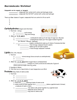
Biomolecules
KEY TERMS
Biomolecules: all the carbon compounds that are found in living tissues.
Biomacromolecules: Biomolecules with mass more than 800 Daltons. These are polymers example proteins,
polysaccharides, Nucleic acids.
Biomicromolecules: The Biomolecules with mass upto 800 Daltons. These are monomers. example Amino acid,
Sugar (Monosaccharide), Nucleotides.
Metabolism: The sum total of all the biochemical reaction taking place in a living body.
Metabolites: The essential organic compounds present in living tissue.
Primary Metabolites: Biochemicals formed as product of vital metabolic pathways of organism example sugars,
Amino Acids.
Secondary Metabolites: Specialized products formed by alteration of normal metabolic pathway example
alkaloids, rubbers. Scents, gums.
Anabolic Pathway: Formation of a complex structure from simple structure.
Catabolic Pathway: Formation of simpler substances from a complex structure.
Enzyme: The catalyst that hasten the rate of metabolic reactions.
Glycosidic Bond: The chemical bond formed between the Aldehyde or Ketone group of one monosaccharide and
the Alcohol group of another.
Peptide Bond: The chemical bond formed between the Carboxyl group (COOH) of one Amino acid and the Amino
group (NH2) of another is called Peptide bond (пЂCO пЂ NH )
Ester bond: The chemical bond formed between the Phosphate and the Hydroxyl group of sugar.
Activation energy: The amount of energy required to initiate a reaction.
GIST OF THE LESSON:
п‚· All cells, tissue are composed of chemical substances.
п‚· The molecules present in living tissue may be organic or inorganic.
п‚· Chemical analysis of living tissue reveals the type of organic & inorganic compounds
present in living organisms.
п‚· Organic compounds are carbohydrates, fats, protein, nucleic acid and inorganic
compound are salts, minerals, water.
п‚· The organic molecules vary in size from simple molecular such as amino acids to
giant molecules proteins.
п‚· Smaller molecule (Biomicromolecules) have molecules mass upto 800 Dalton and are
monomers Ex. monosaccharide, Amino acids, Nucleotides.
п‚· Larger molecules (Biomacromolecules) have molecular mass more than 800 Dalton
and are polymers Ex : Polysaccharide, Proteins, Nucleic Acid.
п‚· The monomers are linked by bonds to form polymers.
п‚· One type of biomolecules changes into some other biomolecules i.e. all biomolecule
have a turn over.
п‚· Living processes can take place only in a non equilibrium steady state. This is termed
as living state.
п‚· All metabolic conversions are Catalysed by Enzymes.
п‚· Enzymes are Proteins that can hasten the rate of Metabolic conversion.
Mind Map
Calcium
Potassium
Sodium
(K+)
(Na+)
NaCl
(Ca+)
Magnesium
CaCO3
(Mg++)
PO43-
CHEMICAL ANALYSIS OF ORGANIC COMPOUNDS
Grind living tissue in Trichloroacetic Acid
Filter through cheese cloth
Filtrate
Retentate
(Acid Soluble Pool)
(Acid insoluble
fraction)
Biomicromolecules
Biomacromolecules
Monosaccharides
Polysaccharides
Amino Acids
Proteins
Nucleotides
Nucleic Acids
Lipids
Chemical Analysis of inorganic compounds
Wash small amount of living tissue.
Dry the tissue and note down its dry weight.
Burn the dried tissue to form ash.
Chemical Analysis of ash.
Different Biomolecules
1.
Carbohydrates
1. Compounds of C, H & O
2. General formula CnH2nOn
3. Principal source of Energy
4. Produced during photosynthesis
Sugars
Polysaccharides
1. Small molecules
1. Macromolecule
2. Low molecular weight
2. High molecular weight
3. Sweet
3. Not sweet
4. Readily soluble in water
4. Insoluble or slightly soluble in
water
5. Crystalline
5. Non-crystalline
Monosaccharide
Disaccharide
Made by joining many
(simple sugar)
made by joining two
molecule of
monosaccharide by
glycosidic bond
monosaccharides
Ex: Glucose
Ex : Maltose
Ex :
Fructose
Sucrose
Cellulose
Galactose
Lactose
Starch
Homopolymers
(formed by repeated
polymerisation of one
type of
monosaccharides)
Inulin
Glycogen
Heteropolymer
(formed
by
polymerisation
of
more than one type of
monosaccharides)
Chitin
Glycosidic bond
2.
Glycosidic bonds
Lipids
п‚· Heterogenous group of organic compound made up of C, H & and few atoms of
oxygen.
п‚· They are insoluble in water & soluble in non-polar organic solvents.
п‚· Lipids are esters of Fatty acids & an alcohol.
O
||
R | пЂ C пЂ OH пЂ« R OH
Fatty Acid
Alcohol
O
||
H2O
R пЂ O пЂ C пЂ R|
Lipids
п‚· Fatty acids are large molecules containing an acidic group.
General Formula R.COOH
R = Alkyl group
COOH = Acidic Group
п‚· True fats are esters of fatty acids and glycerol and are also called triglycerides.
(Glycerol is trihydroxypropane)
п‚· Oils are rich in unsaturated fatty acids that have low melting point.
п‚· Phospholipids are lipids with a phosphate group. Ex : Lecithin
Head : Phosphate group - polar - water attracting (Hydrophilic)
Phospholipids
Tail: Hydrocarbons of fatty acid- nonpolar- water repellant (hydrophobic)
They play important role in the formation of cell-membran.
п‚· Glycolipids are lipids with a carbohydrate.
п‚· Lipoprotein are lipids with a protein molecule.
п‚· Cholesterol is composed of fused hydrocarbon rings and a long hydrocarbon chain.
Difference between unsaturated and saturated Fatty Acids :
Unsaturated fatty acids
1. Contain one or more
double or triple bonds
between carbon atoms.
Saturated fatty acids
1. Do not have any double bond
or triple between carbon
atoms.
2. Melt at lower temperature.
Ex. Oleic Acid
2. Melt at higher temperature.
Ex. Palmitic Acid
3.
Proteins
п‚· Proteins are Heteropolymers containing strings of small units called Amino Acids.
п‚· A peptide bond is formed between Carboxyl group of one Amino Acids and Amino
group of the successive Amino Acids.
п‚· Enormous types of Protein result from 20 Amino Acids.
п‚· Amino Acids are organic compound containing one Amino group and one carboxylic
group as substituents on the same carbon i.e. пЃЎ carbon.
п‚· General Formula
H
H2N
C
COOH
(R= Alkyl group)
R
п‚·
Due to ionizable nature of -NH2 & -COOH group, the structure of Amino Acids
change in different pHs.
п‚· As the Amino Acids carry both positive & negative charges simultaneously, such
substances are called Zwitterions.
п‚· Depending on the availability of Amino Acids, these can be categorized into
Essential Amino Acids
Non Essential Amino Acids
Cannot be synthesised in the Can be synthesised in the body
body of animals
of animal.
They must be obtained from Not essential in our diet.
diet.
Ex. : Valine
Ex. : Glutamic Acid, Alanine
п‚· Based on the number of Amino and Carboxyl group, Amino Acids are categorised as
acidic, basic & neutral.
Acidic
Neutral
Basic
Have more than one Have only one acidic Have more than one
carboxylic group
& one basic group
basic group
Ex. : Glutamic Acid
Ex. : Alanine
Ex. : Lysine
п‚· Depending on the structure of the protein they can be categorised into four types.
Structure
Properties
п‚· Linear arrangement of
amino acids
п‚· Amino acids are held by
peptide bonds
2. Secondary Structure п‚· Protein threads get helical
shape (Pleated Sheet
Structure)
п‚· Amino acids are held by
peptide bonds and inter
molecular
hydrogen
bonds.
3. Tertiary Structure
п‚· Polypeptide chains folded
into three dimensional
globular structure.
п‚· Stabilized by ionic bond,
hydrogen bonds,
disulphide bonds
4.
Quaternary п‚· Relative folding of two are
Structure
more similar or dissimilar
polypeptides upon one
other in the form of a
cube.
п‚· Stabilised by hydrogen
bonds and electrostatic
linkage.
1. Primary Structure
Example
Insulin
Silk Fibre
Enzymatic
protein
Haemoglobin
formed of
two sub
units.
пЃЎ- type and
пЃў- typeпЂ Nucleic Acids
These are the most essential molecules of life. They form the genetic material of all
organisms including virus.
Nucleic acids are made up of large number of nucelotides.
Nucleotide
1.
Structure: Each nucleotide contains three components.
(a) Pentose Sugar
(b) Nitrogen base
(c) Phosphoric Acid
(a) Sugar : There are two kinds of nucleic acids, containing two types of pentose sugars.
Ribonucleic acid (RNA) contains ribose sugar, Deoxyribonucleic acid (DNA) contains deoxyribose
sugar.
(b) Nitrogen base : There are two categories of base purines and pyrimidines.
(i) Purines : Have 2 rings in their structure, example Adenine, Guanine.
(ii) Pyrimidines : Have one ring in their structure. Cytosine, Thymine and Uracil (Uracil is present in
RNA only in place of thymine)
(c) Phosphoric Acids : It contains a phosphate group. It combines two nucleotides together
by formation of phosphodiester bond.
Enzymes
All enzymes are proteinaceous in nature. Some enzymes need a nonprotein part as well.
An example of a Metabolic pathway without/ with Enzyme.
Properties of enzymes
1.
Always proteinaceous in nature.
2.
Lower the activation energy and thereby increase the speed of reaction.
3.
Remain unchanged at the end of the reaction and can be used again.
4.
Work best at optimum temperature which is generally the normal body
temperature.
Inactivated by very low temperatures.
5.
Extremely sensitive to pH.
6.
Substrate-specific : A given enzyme will catalyse only one reaction or a type of
reaction.
7.
The activity of an enzyme is also sensitive to the presence of specific chemicals
that bind to the enzyme. When the binding of the chemical shuts off enzyme
activity, the process is called inhibition and the chemical is called an inhibitor.
Classification of Enzymes:
Oxidoreductases/dehydrogenases:
S reduced + S' oxidised п‚® S oxidised + S' reduced.
Transferases:
S - G + S' п‚® S + S' - G
Hydrolases:
Enzymes catalysing hydrolysis reactions.
Lyases :
X Y
|
|
CпЂCп‚®X-Y+C=C
Isomerases: Includes all enzymes catalysing inter-conversion of optical,
geometric or positional isomers.
Ligases: Enzymes catalysing the linking together of 2 compounds.
SOME IMPORTANT QUESTIONS:
1. Explain primary,secondary and tertiary structure of protein.
2. Write any four properties of a Biocatalyst.
3. Distinguish between DNA & RNA.
*********************************************************
Chapter-10
Cell Division
KEY TERMS
Cell Division: It is the process by which new cells are formed from pre existing cells
Cell Cycle: The sequence of events by which a cell duplicates its genome and eventually divides into
daughter cells.
Karyokinesis: Division of the nucleus.
Cytokinesis: Division of cytoplasm.
Quiscent stage: It represents an inactive stage where cell are metabolically active but do not undergo
division.
Kinetochores: The small dice shaped structure at the surface of centromere, serve as the site of
attachment of the spindle fibres to the centromere of the chromosome.
Bivalent: Pair of homologous chromosomes.
Tetrad: It refers to the four chromatid stage formed during meiosis.
Chiasmata: X shaped structures formed at the site of crossing over during prophase of meiosis.
Crossing over:It is the phenomenon of exchange of equivalent segment between Non- Sister chromatids
of homologous chromosomes (prophase of meiosis)
Homologous Chromosomes: Two similar chromosomes, one contributed by the male parent and other
by the female parent.
Synapsis: A close association & pairing of homologous chromosomes during prophase of meiosis.
GIST OF THE LESSON
1.
2.
3.
4.
All cell reproduce by dividing into two cells.
Each parent cell gives rise to two daughter cells each time they divide.
Cycles of growth and division allow a single cell to form structures consisting of millions of cells.
The sequence of events by which a cell duplicates its genome, synthesizes the constituents of the cell
and eventually divides into two daughter cells is termed as cell cycle.
5. DNA synthesis occurs only during one specific stage in the cell cycle.
6. The replicated DNA (Chromosomes) are distributed to the daughter nuclei by a complex series of
events during cell division.
CELL CYCLE (INTERPHASE + M-PHASE)
Cell Division
Cycle Begins
Mitosis
M
Sister Cells
T
A
Cell Differentiation
P
Cell Growth
M
Cell Growth
G1
G2
S
P- Prophase
DNA Synthesis
M- Metaphase
A- Anaphase
T- Telophase
Interphase
MITOSIS
Diagrammatic representation of different stages of mitosis
MEIOSIS
1.
2.
3.
4.
5.
It takes place in gametes (Gametes are formed form specialized Diploid cells)
The no. of chromosomes in the daughter cells reduces by half (Haploid).
Four haploid daughter cells are formed ( Reduction division)
Exchange of genetic material takes place resulting in variations in the daughter cells.
Involves two cycles of nuclear and cell division called Meiosis I and Meiosis II
Mei
osi
sI
Pr
op
has
eI
Lep
tote
ne
1. C
o
m
p
a
c
t
i
o
n
o
f
c
h
r
o
m
o
s
o
mes.
2. Beaded appearance of chromosomes.
Zygotene
1. Homologous chromosomes paired by synapsis (synaptonemal complex)
Pachytene
1. Appearance of tertad
2. Crossing over between non sister chromatids of homologous chromosomes at chiasmata.
Diplotene
1. Dissolution of the synaptonemal complex.
2. X shape structures called chiasmata remains.
Diakinesis
1. Terminalisation of chiasmata
2. Spindles assembled to prepare the homologous chromosomes for separation.
3. Nuclear envelop and nucleolus disappears.
Metaphase I
1. Homologus Chromosomes line up on the spindle fibre attached by centromeres.
Anaaphase I
1. A Homologus pair separates.
2. Each chromosome still has 2 chromatids and moves to opposite direction.
Telophase I& after Cytokinesis
1. Each cell has haploid no. of chromosomes but each chromosome still has two chromatids.
Meiosis II
Prophase II
1. Nuclear membrane and nucleolus disappear.
2. Chromosomes become compact.
In some cells prophase II may be absent.
Metaphase II
1. Chromosomes line at equator.
Anaphase II
1. Spliting of centromere and hence chromatids separate.
Telophase II & after Cytokinesis
1. Each cell has half no. of chromosomes.
2. Each chromosome is a single unit and may be different in each cell due to crossing over.
After meiosis II four haploid daughter cells are formed.
Diagrammatic representation of different stages of meiosis
DIFFERENCE BETWEEN MITOSIS & MEIOSIS
Mitosis
Meiosis
1. Diagrammatic
2n
nn
n
representation
2n
2n
nn
n
n
2n
n
n
n
n
n
(for a diploid cell)
2. Occurrence
Occurs in diploid cells, may occur in
haploid cells also
Occurs in Diplois cells
3. Kind of Cell
Somatic Cells
Reproductive cells ( Gametes)
4. No. of cells
produced
Two daughter cells
Four daughter cells
5. Significance
п‚· Chromosome no. remains same as
that of the parent cell (equational
division)
п‚· Daughter cells produced have
identical genetic constitution as that
of the parent cell.
п‚· Results in growth and repair,
replacement of cells.
п‚· Chromosome no. reduces to
half the no. of chromosomes
present in the parent
cell.(reduction division)
п‚· Daughter cells produceds
have variation in the genetic
constitution.
п‚· Results in the formation of
gametes.
п‚· Helps in the consevation of
chromosome no. is sexually
reproducing organisms.
SOME IMPORTANT QUESTIONS:
1. Why mitosis is called an equational division?
2. Describe various events during prophase I of Meiosis I.
3. Which type of cell division takes place at the time of gametogenesis in Haploid organisms.
********************************************************************************
*
Chapter-11
Transport in Plants
Means of Transport:
п‚·
There are three means of transport in
plantsi. Diffusionпѓ� Passive & random movement of
ions or molecules.
пѓ� Do not involve expenditure of
energy.
пѓ� Occurs along Concn.gradient.
пѓ� Diffusion rate depends upon Concn.gradient, Permeability of membrane,temp. & Pressure.
ii. Facilitated diffusionпѓ� Movement
of
substances
across
the
membranewith the help of membrane proteins,
do not involve expenditure of energy.
пѓ� Occurs along Concn.gradient.
пѓ� It may be Symport (Two type of molecules
transported across the membrane in same
direction) or Antiport (two type of molecules
transported in the opposite direction across the
membrane) or Uniport (Transport of single type
of molecule across the membrane independent of other molecules).
iii. Active Transportпѓ� Movement of substances across the membrane through membrane proteins.
пѓ� Involves expenditure of energy.
пѓ� Occurs against the Concn.gradient.
Water Potential:
п‚·
п‚·
п‚·
п‚·
The free energy of water is called as water potential. It is denoted by the greek letter П€ (psi) & its
unit is Pascal.
Water potential of pure water is taken as zero and addition of solute causes lowering of water
potential.Therefore water potential of a solution is always considered negative.
Two major components that determine water potential are solute potential(П€s) & pressure potential
(П€p ).
Water potential is affected by solute Concn.(addition of solute causes lowering of water potential)
&pressure(causes increase in water potential).
Osmosis:
п‚·
п‚·
п‚·
Movement of water molecules across a semipermeable membrane from the region of its higher
Concn.to lower Conc. is called as osmosis.
Net direction & rate of osmosis depends upon pressure gradient and Concn.gradient.
The pressure required to stop the entry of water into the solution is called as osmotic pressure.It is a
positive pressure and numerically equal to osmotic potential (solute potential) which is negative.
Plasmolysis:
п‚·
п‚·
п‚·
п‚·
п‚·
Shrinking of the protoplast and its movement away from the cell
wall due to exosmosis(outward movement of water) is called as plasmolysis.
The external solution is said to be isotonic
if it has the same solute Concn.as
cytoplasm. Cells placed in such solutions
shows no net movement of water.
The external solution is called as hypertonic
if it has more solute Concn than the
cytoplasm. Cells placed in such solution
show exosmosis.
The external solution is called as hypotonic if it has less solute
Concn than the cytoplasm. Cells placed in such solution show endosmosis and becomes turgid.
Imbibition:
Movement or diffusion of water along a gradient into an adsorbent is called as imbibition.
п‚·
п‚·
It is responsible for seedlings to come out of the soil.
Two conditions necessary for imbibition are- gradient between the surface of adsorbent & the
liquid/water imbibed, and affinity between the adsorbent & the imbibed liquid.
Absorption of water in Plants:
п‚·
п‚·
п‚·
п‚·
п‚·
Absorption of water
is carried out by the
root hairs through
diffusion.
Absorbed
water
moves
by
two
pathways from root
hairs- Apoplast and
Symplast pathway.
Movement of water
extensively through the non living
components i.e.cell wall & intercellular
spaces without crossing any membrane and
along gradient is called as apoplast pathway.
Movement of water through the cytoplasm of
cells and plasmodesmata involving crossing
of membrane and aided by cytoplasmic
streaming is called as symplast pathway.
Most of water flow in roots through apoplast till endodermis, after that it takes symplast pathway to
reach upto the xylem vessels.
Upward movement of water in plants:
п‚·
Root pressureпѓ� Active absorption & accumulation of various ions in the root cells causes decrease in water
potential and drives entry of water from soil solution into the root cells.
пѓ� It causes development of positive root pressure which pushes water upto a certain height.
пѓ� Root pressure is responsible for coming out of liquid droplets in herbaceeous plants through
a special opening (hydathodes) called as guttation.
п‚·
Transpiration Pullпѓ� Also called as cohesion tension theory and was given by Dixon & Jolly.
пѓ� Most of the water transport in the plants occurs by this method.
пѓ� A transpiration pull (tension or negative pressure) is created at the leaf surface causing
lowering of water potential.
пѓ� Thus a negative water potential is exerted from the leaf surface through stem to the root tips.
пѓ� Since water potential of the soil solution is higher than that of the root cells, water enters the
root hair and moves radially across to reach the xylem.
пѓ� Water is pulled up as continuous column due to the cohesive force among the water
molecules and adhesive force between the water molecules and the lignocellulosic cell wall
of xylem vessels.
Transpiration:
п‚·
п‚·
п‚·
п‚·
п‚·
Loss of water in the form of water vapour through stomata is called as transpiration.
Stomata are the pores found in the leaves guarded by two guard cells.These guard cells are bean
shaped in dicot leaves and dumb-bell shaped in
grasses. The immediate cause of opening and
closing of stomata is change in turgidity of
guard cells.
Dicot leaves have more stomata on lower
surface than on upper surface, while monocot
leaves have almost equal number of stomata on
both the surfaces.
Factors that affect rate of transpiration are –
temp., light, humidity, wind speed, no. &
distribution of stomata etc.
Acc to Curtis: “Transpiration is an essential evil of plants”
Uptake and transport of mineral ions:
п‚· Mineral ions enter root both passively as well as actively,
but mostly they are taken up by active transport.
п‚· After reaching to the xylem through active or passive
uptake, they are further transported up the stem to all parts
of the plant through the transpiration stream by method of
mass or bulk flow system.
Phloem transport:
п‚·
п‚·
п‚·
п‚·
Phloem transport is bidirectional, because source –sink
relationship can vary.
The source for phloem transport is leaf and the sink may be
any part that needs food or storage organ.
Phloem sap mainly contains water, sucrose, amino acids,
hormones etc.
The long distance movement of substances through the
vascular tissues (xylem & phloem) in plants is called as
translocation.
Mass flow hypothesis:
п‚· Also called as pressure flow hypothesis.
п‚· It was proposed by Munch.
п‚· Loading of sucrose (active transport of
sucrose from leaf mesophyll tissues to
companion cells and then into sieve
tubes) at the source(leaf) causes
decrease of water potential in the
phloem, resulting in entry of water into
sieve tubes from adjacent cells by
osmosis.
п‚· Hydrostatic pressures build up in the
phloem sap that moves to regions of
lower pressure (sink).
п‚· Unloading of sucrose and other sugars(
sucrose and other sugars move actively
out of phloem) into the cells of sink
causes increase in water potential of
phloem and water moves out of the
phloem.
SOME IMPORTANT QUESTIONS:
1. What will happen when a cell is placed in Hypertonic solution?
2. Write a short note on water potential.
3. Distinguish between guttation and transpiration.
4. Explain most widely accepted theory for upward movement of water.
Chapter-12
Mineral Nutrition
п‚·
п‚·
п‚·
п‚·
Technique of growing plants in a nutrient solution (complete absence of soil) is called as
hydroponics. It was discovered by Von Sachs (1860).
Hydroponic/Soil less Culture technique is employed to find out the elements essential for plant
growth, ascertain deficiency symptom and nowadays for commercial production of crops like
tomato, seedless cucumber etc.
Depending upon the quantity in which an essential element is required by the plants, nutrients may
beпѓ� Macronutrient(required in amount more than 10 m mole/kg of dry matter).
пѓ� Micronutrient or trace element(required in amount less than 10 m mole/kg of dry matter).
On the basis of function, essential elements may beпѓ� Structural element (as components of biomolecules) like-C,H,O,N
пѓ� Components of energy related compounds like Mg in chlorophyll and phosphorus in ATP
пѓ� Activator or inhibitor of enzymes like Mo activator of nitrogenase, Zn+2 of alcohol
dehydrogenase.
пѓ� Alter the osmotic potential of a cell like Potassium .
Category
of
Nutrients
Name of
element
1-Nitrogen
Macro
Nutrients
Micro
Nutrients
2-Phosphorus
Form in
which
absorbed by
plants
NO3-, NO2- or
NH4+
H2PO4or
HPO42-
3-Potassium
K+
4-Calcium
Ca2+
5-Magnesium
Mg2+
6-Sulphur
SO42-
3+
1-Iron
Fe
2-Manganese
Mn2+
3-Zinc
Zn2+
4-Copper
Cu2+
5-Boron
Bo33- B4O72-
6-Molybdenum
MoO22-
7-Chlorine
Cl-
Functions/ Role played
Major constituent of Proteins, nucleic acids, vitamins
and hormones.
Constituent of cell membranes, nucleic acids,
nucleotides & required for all phosphorylation
reactions.
Maintains anion-cation balance in cells, involved in
protein synthesis, opening and closing of stomata,
turgidity of cells.
Required for permeability of cell membrane,
formation of mitotic spindle, formation of middle
lamella.
Constituent of chlorophyll, maintains structure of
ribosome, activates enzymes of respiration &
photosynthesis, involved in DNA & RNA synthesis .
Constituent of amino acid (Methionine &Cysteine),
several Co-enzymes, ferredoxin &Vitamins (Biotin,
Thiamine & CoA).
Constituent of proteins involved in electron transfer
(ferredoxin, cytochromes), activates catalase enzyme
and essential for formation of chlorophyll.
Necessary for photolysis of water in photosynthesis,
activates enzymes involved in photosynthesis,
respiration & nitrogen metabolism.
Required for synthesis of auxin, activates carboxylases
Required for over all metabolism, associated with
enzymes involved in redox reactions.
Required for uptake& utilization of Ca+2,membrane
functioning, pollen germination, cell elongation, cell
differentiation and carbohydrate translocation.
Component of nitrogenase and nitrate reductase
enzymes.
Anion-Cation balance in cell, necessary for photolysis
of water in photosynthesis.
Deficiency Symptomsп‚·
Common deficiency symptoms areп‚· Chlorosis (loss of chlorophyll)- yellowing of leaves caused due to deficiency of N,K, Mg, S, Fe, Mn,
Zn & Mo.
п‚· Necrosis (Death of tissues)-caused due to deficiency of Ca, Mg, Cu, K.
п‚· Inhibition of cell division- caused due to deficiency of K, N, & Mo.
п‚·
Delay in flowering-caused due to deficiency in N, S, and Mo.
Micronutrients can be toxic
п‚· Mineral ion Conc. in tissues that reduces the
dry weight of tissues by about 10% is called as
toxic.
п‚· Moderate decrease in micronutrients causes
deficiency symptoms and moderate increase
causes toxicity.
п‚· It may be that excess of an element (toxicity)
may inhibit the uptake of another element.
Nitrogen Cycle
Absorption and translocation of Nutrients-
NH3
п‚· Soil acts as reservoir for many nutrients and plants
absorb them through roots.
п‚· Absorption of elements occur in two parts-in first part
rapid uptake of ions in free space or outer space of cell
through passive process and in second part ions are
taken slowly into inner space of cell.
п‚· Movement of ions is called as flux, which may be influx
(inward movement) or efflux (outward movement).
п‚· Absorbed mineral salts are translocated through xylem
with ascending sap by means of transpiration pull.
Electrical N2fixation
Industrial N2Fixation
Atmospheric N2
Biological N2-fixation
п‚· The Conc. of the essential element below which plant
growth is retarded is called as Critical Conc.
п‚· Morphological changes that are observed due to
deficiency (below Critical Concns.) of a particular
element are called as deficiency symptoms.
п‚· Deficiency symptoms of some elements like S, Ca, etc.
first appear in young tissues, while other elements like N,
K, Mg. in older tissues. Because those elements which
are actively mobile can be transported from senescing
organs to younger parts and show deficiency symptoms
first in older parts, while those which are immobile
shows symptoms first in younger parts.
NO2-
NO3-
Soil Nitrogen pool.
Decaying
biomass
Plant
biomass
Animal biomass
п‚·
Process of conversion of molecular nitrogen (N2) into compounds of nitrogen is called as nitrogen fixation.
Nitrogen fixation may be
Industrial N2-fixation like forest fires,
Electrical N2-fixation
Biological N2-fixation
Automobile, Exhaust, Haber’s process Conversion of N2into No, NO2, N2O
of NH3 synthesis etc.
by lightning and UV- radiations.
п‚·
п‚·
Carried out by living beings
Like-bacteria, BGA (Blue,
Green Algae) etc.
Symbiotic N2 fixationMost common symbiotic association observed is between Rhizobium bacteria and roots of
leguminous plants (Gram,Pea etc.)
Small outgrowths called as nodules are formed on roots which, act as site of N2- fixation.
Steps of Nodule Formation
Rhizobium in soil interacts with roots of leguminous plants.
Rhizobia multiply, colonise and get attached to epidermal
& root hair cells.
Curling of root hair & invasion of bacteria into root hair.
Produce infection thread & carry bacteria into root cortex.
п‚· Ammonification is conversion of organic
nitrogen (dead plants & animals)into
ammonia.
п‚· Nitrification is conversion of ammonia into
Nitrate (oxidation of NH3 into NO3-). It is
carried out in two steps by Nitrifying bacteria
(chemoautotrophs).
п‚· NH3
NO2—(by Nitrosomonas,Nitrosococcus
Nitrite
bacteria)
п‚· NO2-
NO3—(by Nitrobacter)
Nitrite Nitrate
Initiate nodule formation in the cortex.
Mechanism of Nitrogen fixation
п‚·
An enzyme Nitrogenase (Mo-Fe protein complex)catalyses the conversion of N2 into
NH3 in root nodules.
п‚·
Enzyme Nitrogenase is highly sensitive to molecular oxygen and requires anaerobic
condition to function.
п‚·
Anaerobic condition in root nodules is provided by a pink coloured pigment called as
leg haemoglobin which acts as oxygen scavenger by binding with O2. Leg-haemoglobin is product
of interaction between Rhizobium & leguminous plant. Either of two cannot have it alone.
Fate of AmmoniaNH3soon get
converted into NH4+, as NH3 is
toxic to plants.
п‚· NH4+ undergoes reductive
amination, in which it reacts
with О±-ketoglutaric acid to
from glutamic acid(amino
acid).
п‚· Then by transfer of amino
group form one amino acid
(glutamic acid) to the keto
2H+
group of a keto acid, other
amino acids areBinding
produced
of N2 and
this process is called as
transamination catalysed by
an enzyme transaminase.
2H+
2H+
SOME IMPORTANT QUESTIONS:
1.
Describe the various events in nodule formation.
2.
Write the significance of Leghaemoglobin in Symbiotic Biological Nitrogen fixation.
3.
Write any three deficiency symptoms in plants.
Chapter-13
Photosynthesis in Higher Plants
п‚·
A physico-chemical process by which green plants use light energy to synthesise organic compounds
(glucose) is called as photosynthesis.
п‚·
It is important for all living beings for two reasons:
i.
ii.
Primary source of all food on earth.
Releases oxygen into atmosphere.
Historical Background
Joseph Priestely (1770) revealed the essential role of air in the growth of plants.
Jan Ingenhousz showed that only green parts of the plants could release O2.
Engelmann described the action spectrum of photosynthesis.
Julius Von Sachs (1854) proved that Glucose is produced when plant grows and glucose is stored in
the form of starch.
Van Niel demonstrated that photosynthesis is essentially light dependent and inferred that oxygen
evolved during process comes from H2O not from CO2.
п‚·
The present day equation of photosynthesis was given by Van Niel as follows-
6CO2+12H2O Light
C6H12O6+6H2O+6O2
Why 12H2O shown in equation not 6H2O?
п‚· Because it has been already proved that oxygen
evolved during photosynthesis comes from H2O
not from CO2.
п‚· In case if 6H2O is used then 6O2 will not be
released (only 3O2 is produced).
п‚· So, 12H2O produces 6O2 and out of 12H2
molecules 6H2 is used in making of glucose and
rest 6H2 with oxygen of CO2 form 6H2o as
product.
Chlorophyll
Site of Photosynthesis
п‚· Occurs in Green parts of the plant (mainly
in leaves but also in green stems & sepals of
flower).
п‚· Cells of green part contain chloroplast, a
cell organelle which is actually involved in
carrying out photosynthesis. (site of
photosynthesis in cell).
Outer membrane
1-Envelope
Inner membrane
Granum
Stromal Lamella
2-Membrane System
Responsible for synthesis of ATP & NADPH
3-Stroma
(Matrix)
(Light reaction)
Structure
of Chloroplast
Photosynthetic
PigmentsResponsible for CO2 reduction to form glucose (Dark reaction)
Phtosynthetic Pigmentsп‚· Pigments involved in photosynthesis areSl.
Name of
Colour of Pigment
No.
Pigment
1.
Chlorophyll a
2. Chlorophyll b
3. Xanthophylls
4.
Carotene
Bright Green or bluish green
Yellowish green
Yellow
Yellow to yellow orange
Function/Role
Actually involved in photosynthetic process (Reaction
centre)
Called as accessory pigments, because not directly involved
in photosynthetic process but help in photosynthesis by1. Absorbing light at different wavelengths and
passing it to reaction centre.
2. Protect chlorophyll from photo oxidation.
ption of light by
pigments
п‚· Process of destruction of chlorophyll at high light intensity and oxygen is called as
photooxidation.
п‚· Only visible spectrum of light (400 nm-700 nm) is utilized by Plants in photosynthesis. Therefore
it is called as PAR (Photosynthetically active radiation).
п‚· Pigments are organized in thylakoid membrane in the form of two photosystems-photosystem I
&II.They are named so according to the order in which they have been discovered.
п‚· Each photo system contains, a reaction centre (Chl a molecule only) and light harvesting
complex(other pigment molecules).
п‚· Reaction centre of PSI is called as P700 as it absorbs light at 700nm and PSII as P680 which
Chl b
absorbs light at 680nm.
п‚· Graph plotted between amount of light absorbed by
Chl a
photosynthetic pigments and different wavelengths of
visible spectrum (white light) is called as absorption spectrum..
п‚· Graph plotted between rate of photosynthesis (measured by O2
released) and different wavelength of visible spectrum is called
as action spectrum.
Rate of Photosynthesis
Wavelength of light (nm)
Mechanism of PhotosynthesisPhotosynthesis occurs in two stepsi.
Light reaction or photochemical phase
Wavelength of light (nm)
ii.
Dark reaction or biosynthetic phase.
Light Reactionп‚· Occurs in grana
п‚· Directly depends on light, so also called as light dependent phase.
п‚· Products of this step area. O2
released by photolysis of water (light induced splitting of water)
b. ATP
synthesized by photophosphorylation.
c. NADPH (Reduced Coenzyme, Nicotinamide dinucleotide phosphate)
п‚· Synthesis of ATP molecules in chloroplast from ADP & inorganic phosphate (Pi) using light energy
is called as Photophosphorylation. It is of two types-
Light
a. Non-cyclic
п‚· Occurs due to unidirectional/non-cyclic electron transport.
п‚· Both PS II & PS I are involved.
п‚· Products are ATP, NADPH & O2.
b. Cyclic
п‚· Occurs due to cyclic electron transport (same electron returns back to reaction centre
of PS I).
п‚· Only PS I is involved.
NADP++2H+
NADPH
п‚· Only ATP is produced.
2eStroma
Primary electron acceptor
Primary electron acceptor
2e-
Light
-
2e
ADP+PiATP
Cyt b6/f
complex
PS II P680
2e-
2ePS I
P700
H2o
2e-+2H++ O2
Photolysis of water
Lumen
Thylakoid membrane
Z-Scheme of electron transport/ non cyclic
Unidirectional- i. e.
п‚· Both PSII & PSI illuminated to emit electron, accepted by their corresponding PA(primary
acceptor).
п‚· Loss of electron at P680 is replaced by electrons generated by splitting of H2O & at P700 , by
electron emitted from P680 (through PA, cyt b6 /f complex).
п‚· Electron emitted from PS I is ultimately accepted by NADP+ (Oxidized) and form NADPH (reduced)
after taking H+ released from photolysis of water.
PS I
Not involved
PS II
Stroma
PA
2eCyt b6/f
P680
2e-
Light
PA
2eP700
ADP
ATP
H2o
Lumen
п‚· Only PS I is functional,
PS II is not activated.
п‚· Same electron
emitted from P700
returns back to P700
after passing through
PA (Primary Acceptor)
and cyt b6/f complex.
Thylakoid membrane
Cyclic electron transport
Chemiosmotic hypothesis:
(An explanation for mechanism of ATP synthesis due to electron transport during light reaction)
п‚·
Due to illumination of PS II & PSI simultaneously an electron is emitted both from P 680& P700 which
is accepted by corresponding primary acceptors.
п‚·
Primary acceptor of P680transfers their electrons to a hydrogen carrier which removes a proton from
stroma and releases it into grana lumen with transfer of electron to P700.
п‚·
Primary acceptor of P700 transfers electron to NADP+ which takes proton from stroma (proton
obtained from photolysis of water) and reduced to NADPH.
п‚·
Splitting of water molecule produces 2H+ +2e-& O2 in lumen of thylakoid membrane, of which
electron is transferred to P680 and proton accumulates in lumen.
п‚·
As a result proton in stroma decrease in number and in thylakoid lumen increase in number creating
a proton gradient.
п‚·
When this gradient is broken down due to release of protons from thylakoid lumen to stroma through
ATP synthase (F0-F1 particle), huge amount of energy is released.
п‚·
This energy is utilized in ATP synthesis from ADP & Pi.
2H+
2e--
Cytb6/
f
2e
2e-
2e-
PS II
-
P680
PS I
2e-
(P700)
+
2H
-
+
H2o 2e +2H +1/2O2
Lumen
H+
Thylakoid membrane
F0
ATP Synthase
F1
ADP+ Pi
ATP
Schematic representation of chemiosmotic hypothesis
Dark Reaction
п‚·
Occurs in stroma.
NADP+
2H+
NADPH
п‚·
Does not depend on light, hence called light independent phase.
п‚·
In this phase CO2 fixation or reduction occurs to synthesize glucose, hence called biosynthetic
phase.
п‚·
Products of light reaction (only ATP & NADPH) are utilized in this phase.
п‚·
There are two different pathways of CO2 fixation occurring in different plants.
1. Calvin Cycle or C3 Cycle
п‚·
Discovered by Melvin Calvin, hence called calvin cycle.
п‚·
First stable product is 3-carbon compound hence called C3 cycle and plants having this pathway of
CO2 fixation are called as C3 plants.
п‚·
It occurs in following three stepsa. Carboxylation, in which CO2 combines with primary acceptor called RUBP (ribulose 1,5
bisphosphate) in the presence of enzyme RUBISCO (Ribulose, 1.5 bisphosphate carboxylase
oxygenase) to produce two molecules of PGA (Phosphoglyceric acid).
b. Reduction, in which 2 molecules of PGA get reduced to 2 molecules of PGAL
(Phosphoglyceraldehyde, triose sugar) using 2ATP and 2 NADPH.
c. Regeneration, in which again primary acceptor (RUBP) is synthesized from PGAL & ATP.
п‚· This cycle goes six turns to
produce one molecule of glucose.
Hence
6CO2+6RUBP-------------(6C)
In synthesis of one
molecule of glucose
through C3 cycle-
ADP
3-PGA X 2 Molecules
3C- Compound
(6x5C=30C)
12 Molecules of PGA
(12x3C)
CO2+H2O {5C+1C=6C=2X3C}
RUBP
5 CSugar
No.
ATP
2ATP
12 Molecules of PGAL
2 Molecules used in
Glucose synthesis
(6 C sugar= 2x 3 C)
PGAL x 2 Molecules
(Triose phosphate)
3C- Compound
Glucose
2. C4 Cycle or Hatch & Slack Pathwayп‚· Discovered by Hatch & Slack, so called as Hatch &Slack
Pathway.
п‚·
ATP
used=18
2ADP+Pi No. of NADPH=12Molecule
2NADPH
10 Molecules used in
regeneration of RUBP
[10x 3C=6 molecules
of RUBP (6x5 C)]
of
molecule
First stable product is 4- carbon compound
(OxaloaceticAcid) hence called C4-cycle and plants having
this cycle are called as C4 plants.
 C4 plants have special kind of leaf anatomy called as “Kranz
anatomy”
2 NADP
+
No. of CO2 = 6 molecules.
No. of H2O = 12 molecules
Why this pathway?
-In C3 plants RUBISCO, an enzyme
used in CO2 fixation is very much
sensitive to relativeConcn.Of
CO2& O2 in the chloroplast.
-At higher CO2Concn. behaves as
carboxylase and at higher
O2Concn.behaves as oxygenase.
-Since light reaction releases O2,
hence itsConcn.risesup.Thus
RUBISCO starts behaving as
oxygenase and binds RUBPwith
O2 instead of CO2 resulting in no
synthesis of sugars.
-To avoid this some plants
evolved a new mechanism i. e. C4
cycle.
п‚· (Presence of two types of photosynthetic cells, i.e. mesophyll cells and bundle sheaths cells).
п‚· In C4 plants (Maize, sugar cane etc.) both C3 cycle (In bundle sheath cell chloroplasts) & C4 cycle (In
mesophyll cell chloroplasts) occurs.
CO2 enters
CO2 +PEP
PEPcorboxylase
Mesophyll cell chloroplasts
п‚· Are granal (have grana), light reaction
takes place, O2 evolution occurs.
п‚· Have phosphoenol pyruvate (PEP) as
primary CO2 acceptor, and PEP
carboxylase enzyme for CO2fixation
(not sensitive to O2Concn.).
oxaloacetic acid (4c)
(3C)
NADPH
AMP
NADP+
ATP
Malic Acid (4C)
Mesophyll Cell
Transported
Transported
Pyruvic Acid
Bundle Sheath
Cell
Bundle sheath cell chloroplasts
п‚· Are agranal (lacks grana), hence no light
reaction, no O2 evolution.
п‚· Receives only CO2due to decarboxylation of
malic acid, hence high CO2Concn.is
maintained to carry out CO2 fixation by
RUBISCO.
п‚· Have, RUBP as primary acceptor and
RUBISCO as enzyme for CO2 fixation (C3cycle).
Malic Acid (4C)
NADP+
NADPH
PGA
CO2+ Pyruvic
Acid
C3Cycle
RUBP
Sugar synthesis
Photorespiration
п‚·
Is a wasteful process that occurs in C3 plants, as no synthesis of sugar ATP, NADPH takes place.
п‚·
At high temp. and high O2 Concn., RUBISCO functions as oxygenase and catalyses the combination
of O2 and formation of one PGA & one phosphoglycolate molecules.
п‚·
In this process three cell organelles- chloroplast, peroxisomes and mitochondria are involved.
PGA
RUBP+O2
Transported
PGA
Glycerate
Hydroxy
Pyruvate
Serine
Transported
Phosphoglycolate
Serine
Glycolate
Transported
Transported
Glycolate
Glyoxylate
Glycine
Glycine
CO2
Chloroplast
Peroxisome
Mitochondria
Factors Affecting photosynthesis
п‚·
According to Blackman‟s law of limiting factors “if a chemical process is affected by more
than one factors, then the rate of the process will directly depend on the factor nearest to its
minimal value”.
п‚·
As photosynthesis is affected by number of internal (No., Size, and orientation of leaves,
merophyll cells & chloroplasts etc.) and external factors (light, CO2 Conc, temp., soil water
etc.), it‟s rate will also directly depend on the factor nearest to its minimal value.
п‚·
Light intensity and quality affect rate of photosynthesis. Light of wavelengths 400nm-700nm
is effective for photosynthesis. With increase in light intensity, rate of photosynthesis
increases, but at higher light intensity rate decreases either due to the destruction of
chlorophyll or due to other factors that becomes limiting.
п‚·
Only dark reaction is affected by temperature as it is enzyme catalysed reaction and enzymes
remain inactive at low temp. while become denatured at high temperatures.
п‚·
Rate of photosynthesis increases with increase in CO2Concn. upto a certain limit (upto 500
Вµl/L in C3& 360 Вµl/L in C4 plants).
п‚·
Lower availability of soil water causes stomatal closure causing decreased supply of CO2,
hence decrease in photosynthesis.
SOME IMPORTANT QUESTIONS:
1. Explain Chemiosmotic hypothesis.
2. Distinguish between cyclic and non cyclic photo phosphorylation.
3. Why photo respiration is considered as a wasteful process?
4. Write a short note on Kranz anatomy.
Chapter-14
Respiration in Plants
п‚·
Respiration is a biochemical process involving breakdown of C-C bonds of complex organic
molecules by oxidation leading to step wise release of energy in the cells. It is called as cellular
respiration, because the site of respiration is cell.
п‚·
Steps of respiration is common in all eucakaryotic cell, whether plants or animals.
п‚·
The substrate that undergoes oxidation in the process of respiration is called as respiratory substrate.
It may be carbohydrate, protein, fat or organic acid.
Difference between Respiration in plants and animals
п‚· Only the difference lies in mechanism of gaseous
exchange, which is called as breathing (in animals)
because of involvement of some specialized
organs/cells.
п‚· Plants do not have any specialized organ for gaseous
exchange, rather this function is performed in them by
stomata & lenticels.
Why plants can get along without respiratory
organs?
п‚· Because, each part of plants can fulfill their need
of gaseous exchange & very little transfer of gases
occur.
п‚· Rate of respiration in root, stem, leaves is very
low, hence no great demands for gas exchange.
п‚· Plant cells are located quite close to surface, so no
much distance for diffusion of gases.
Types of respirationп‚· May be aerobic (molecular oxygen is required) or anaerobic (molecular oxygen is not required).
Mechanism of respirationп‚·
Both anaerobic and aerobic respiration starts with Glycolysis.
п‚·
In anaerobic (In cytoplasm of cell)
Glucose
In aerobic
Glucose
Glycolysis
Glycolysis
Cytoplasm
Pyruvic Acid
Transported
Pyruvic Acid
Pyruvic Acid
or Ethyl alcohal
Lactic Acid
п‚·
CO2
It is incomplete oxidation and end products are
lactic acid or ethyl alcohol & CO2, and 2 ATP
molecules from one molecule of glucose.
EquationC6H12O6
Ethyl Alcohal+CO2+Energy
OR
C6H12O6
Lactic acid + Energy
Mitochondrial
Matrix
Acetyl CoA
Krebs
cycle
NADH
(Reduced)
ETS
NAD+
(Oxidised)
Mitochondrial
Inner membrane
ATP
O2
H2O
It is complete oxidation and end products are
CO2,H2O and 36 ATP molecules from one glucose
molecule.
Equation- C6H12O6+6O2
6CO2+6H2O+Energy
GlycolysisGlucose
(6C)
ATP
ADP
G-6-PO4
(6C)
Fructose-6-PO4
ATP
ADP
Fructose 1,6- bisphoshate
(6C)
Dihydroxyacetone
Phosphate (3C)
3-phosphoglyceraldehyde
+
NAD
(3C)
NADH
NADH+H
+
1,3 bisphosphoglycerate
ADP
ADP
ATP
п‚· Occurs in Cytoplasm.
п‚· 1 molecule of glucose at the end of this step
producesпѓ� 2 molecules of Pyruvic Acid (3C)
пѓ� 2 molecules of NADH+H+
пѓ� 4 molecules of ATP, out of which 2
molecules are consumed during the
process, so net gain is only 2 molecules of
ATP.
пѓ� In anaerobic respiration pyruvic acid
receives hydrogen from NADH and forms
lactic acid (in human striated muscles,
bacteria) or ethyl alcohol & CO2 (called as
fermentation. Ex. in yeast).
пѓ� In aerobic respiration, pyruvic acid is
transported into mitochondria to enter into
Krebs cycle.
ATP
3-Phosphosphoglycerate
2-Phosphoglycerate
Phosphoenol pyruvate
ADP
ADP
ATP
ATP
Pyruvic Acid
(3C)
Krebs cycleCo A
Pyruvate (3C)
NAD+
NADH+H
+
п‚· Occurs in mitochondrial matrix.
п‚· Called as Krebs cycle, because it was discovered
CO2
Acetyl Co A (2C)
NADH+H
+
Oxaloacetic acid
(4C)
Citric acid (6C)
CO2
NAD+
NAD+
Krebs cycle
NADH+H+
О±-Ketoglutaric acid
(5C)
CO2
NAD+
Malic acid (4C)
FADH2
FAD+
From 1 molecule
of Pyruvic acid
NADH+H+
Succinic acid
(4C)
GDP
GTP
Electron Transport System (ETS)Inter membrane Inner membrane of Mitochondrial
Space
mitochondria
matrix
(Space between
Outer and inner
Membrane)
п‚· In ETS, , Molecular Oxygen (O2)acts as terminal electron
acceptor.
+
NADH+H
+
п‚· Electrons from NADH+H is transported through FMN, UQ
(Hydrogen Carrier, Cytochromes(b,C1,C,a-a3) and finally to
+
O2 which takes 2H from matrix and form H2O.
NAD+
2H
FMN
+
п‚· During transport of electron, proton (H ) is pumped by UQ
to inter membrane space from matrix causing formation of
proton gradient (due to increase in protons in inter
membrane space and decrease in matrix).
2H
2e
2H+
-
2eFeS
2H
+
+
п‚· All NADH+H Produced during Glycolysis & Krebs cycle is
oxidized by NADH-dehydrogenase and electrons are
transported to Ubiquinone(UQ)via FMN.
2eFeS
UQ H2
2e-
2eUQ
п‚· Breaking of this gradient and return of these
+
protons(H )through ATP synthase/ Fo-F1 particle/ oxysomes
located in inner membrane cause synthesis of ATP.
2H+
2H+
+
п‚· Since oxidation of NADH+H is the cause of formation of
proton gradient and ATP synthesis thereupon, so this
method of ATP synthesis is called as oxidative
phosphorylation.
2e-
Cytb
2e-
+
п‚· Return of 2H from inter membrane space to matrix through
ATP synthase release sufficient energy to synthesise one ATP
molecule.
FeS
2e2H+
-
2e
UQH2
2H+
2H+
UQ
+
Cyt C1
2e-
2eCyt C
2e-
+
п‚· On oxidation of 1 molecule of NADH+H pumps 6H ,
therefore it produces 3 ATP molecules while oxidation of
+
FADH2 pumps only 4H , so it produces 2 ATP molecules.
2e-
O2
2H+
Cyt a-a3
H2O
Net Gain of ATP
п‚·
After complete oxidation of 1 molecule of glucose1.Glycolysis2ATP = 2ATP
+
2NADH+H (2x3 ATP) = 6ATP
2.Pyruvic Acid to Acetyl CoA3.Krebs Cycle-
2 NADH+H+ (2x 3ATP)= 6 ATP
2GTP= 2ATP
+
6NADH+H (6x3ATP) = 18 ATP
2FADH2 (2 x 2 ATP)= 4ATP
Total=38 ATP
п‚· So, Complete Oxidation of 1 molecule of glucose produces molecules 38ATP molecules.
п‚· In eucaryotes 2ATP is consumed in transportation of NADH+H+ from cytoplasm (Produced in
Glycolysis) to mitochondria for oxidation. Hence the net gain is 36ATP molecules.
Break Down of different respiratory substratesFat
Carbohydrates
fatty acid & Glycerol
Proteins
Monosaccharides Amino Acids
(Glucose)
Glycolysis
DHAP
PGAL
п‚· Respiratory pathway is called as amphibolic
pathway because it involves break down of
respiratory substrate (catabolic) to release
energy as well as also contribute to
synthesis of other compounds (anabolic) by
providing carbon skeletons.
п‚· Respiratory quotient(RQ) is ratio of the
volume of CO2 evolved to the volume of O2
consumed in respiration.
RQ=
п‚· RQ of different respiratory substrates areCarbohydrate=1
Fat= less than 1
Protein=less than 1
Organic acids=more than 1
п‚· RQ in case of anaerobic respiration is infinite
as no oxygen is consumed.
Pyruvic Acid
Acetyl CoA
Krebs
cycle
H2O
Volume of CO2 evolved
Volume of O2 consumed
.
CO2
SOME IMPORTANT QUESTIONS:
1.Define Glycolysis. Where does it takes place? Write various steps of Glycolysis.
2. Give schematic representation of Kreb‟s Cycle.
3. Define RQ. Calculate the Value of RQ when Respiratory substrate is: Carbohydrate, organic
acid.
Chapter-15
Plant Growth and Development
п‚·
п‚·
п‚·
п‚·
An irreversible permanent increase in size of an organ/parts/cell is called as growth.
Plant growth is indeterminate i. e. capable to unlimited growth throughout life, because of presence
of meristems at certain locations in their body.
Form of growth where new cells are always being added to the plant body by the activity of
meristem is called as open form of growth.
In plants apical meristem is responsible for primary growth (increase in height) and lateral
meristems for secondary growth (increase in thickness).
Phases of GrowthThree phases of growth
i.
Meristematic
ii.
Elongation
iii. Maturation
Growth is measured asп‚· Increase in
пѓ� Fresh weight
пѓ� Dry weight
пѓ� Length or area
пѓ� Volume/Size
пѓ� Cell number
п‚· At cellular level, growth is mainly due to increase in
protoplasm.
Cells attain their
maximum size
Region of maturation
Cells show increased vacuolation
Cell enlargement
Region of elongation
New cell wall deposition
Cells rich in protoplasm
Region of meristematic
activity
Prominent nuclei
Thin cell wall
Root Cap
Growth Rate-
Conditions for growthFollowing are essential
elements for growthп‚· Waterfor cell
enlargement(causes
increase in size) and as
medium for enzymatic
activities.
п‚· Oxygenfor release of
metabolic energy.
п‚· Nutrientsfor synthesis of
protoplasm & source of
energy.
п‚· Optimum temperature
Height of the plant
п‚· Increase in growth per unit time is called as growth rate.
п‚· It may bei.
Arithmatic growth (2,4,6,8,10 etc.)
пѓ� If one of the daughter cells formed by mitosis continues to divide while other undergoes
differentiation and maturation.
пѓ� Growth curve plotted is linear.
Time
ii. Geometric Growth (4, 8, 16, 32, etc.)
пѓ� Also called as exponential growth and have three phases- initial
slow (lag phase), increases rapidly (exponential/log phase) and
then slows down (stationary phase).
пѓ� Growth curve plotted is S-shaped called sigmoid curve.
This curve is characteristics of living organisms growing in
natural environment.
Developmentп‚·
п‚·
Sum of growth and differentiation is called as development.
Includes all changes that an organism undergoes throughout life from seed germination to
senescence.
Development in plants is under control of intrinsic and extrinsic factors. Intrinsic factors include
both intracellular (genetic) or intercellular factors (plant growth regulators). Extrinsic factors include
light, temperature, water, oxygen, nutrition etc.
п‚·
Plant Growth Regulators (PGR)
п‚·
Small, simple molecules secreted in very small quantities which influence physiological functions in
plants are called as plant growth regulators or plant hormones or phytohormones.
Type of Plant Growth Regulators
PGR
Growth inhibitors
Growth Promoters
Involved in growth promoting activities
like-cell division, cell enlargement etc.
Auxins
Gibberellins
Involved in growth inhibiting activities
like-dormancy, abscission etc.
Cytokinins
Ethylene
AbscissicAcid (ABA)
(ABA)
Growth PromotersName of Hormone
Physiological Effects
Application/Uses
1. Auxinп‚· first isolated from human urine.
п‚· IAA, IBA are natural auxins
isolated from plants.
п‚· NAA, 2, 4-D are synthetic auxins.
2. GibberellinsAll are acidic, hence also called
Gibberellic acid.
3. Cytokinins
п‚· Cause apical dominance.
п‚· Prevent premature fall of leaves,
flowers, fruits etc.
п‚·
п‚·
п‚·
п‚·
п‚· Bolting and flowering in rosette
plants.
п‚· Cause elongation of internodes.
п‚· Break seed dormancy.
п‚· Can elongate genetically dwarf
plants.
п‚· Cause growth of lateral buds and
overcome apical dominance.
п‚· Promote production of new
leaves, and chloroplasts in leaves.
п‚· Promote adventitious shoot
formation
п‚· Increase the length of grape stalks.
п‚· Delay senescence, hence fruit can
be left on trees for longer period.
п‚· Increases lengths of internodes in
sugarcane, thereby increases yield.
Initiate rooting in stem cuttings.
Promote flowering in pineapple.
Induce parthenocarpy in tomatoes.
2, 4-D used as weedicide.
п‚· Used to delay leaf senescence.
Growth Inhibitors –
Name of Hormone
1. Abscissic Acid (ABA)
п‚· Also called as stress hormone,
because increases tolerance of
plants to various stresses.
2. Ethylene
п‚· Only gaseous hormone.
п‚· Also called as fruit ripening
hormone, because hastens
ripening of fruits.
Physiological Effects
п‚·
п‚·
Reduces seed dormancy
Stimulates closure of
stomata
under water stress conditions.
п‚·
Stimulates formation of
abscission
layer and abscission of leaves,
flowers and fruits.
п‚·
Promotes horizontal
growth of
seedlings.
п‚·
Induces apical hook
formation in dicot seedlings.
п‚·
Promotes senescence
and
abscission of leaves and flowers.
п‚·
Promotes root growth
and root
hair formation.
Application/Uses
п‚· Prolongs dormancy of seed for
storage.
п‚· It hastens fruits ripening in
tomato and apple.
п‚· Promotes production of female
flowers on a monoecious plant.
Photoperiodism –
п‚· Phenomenon in which the plants respond to relative length of day and night to which they are exposed for
flowering is called as photoperiodism.
п‚· Site of perception of light for photoperiodism is the leaf where a pigment called phytochrome perceives the
light stimulus.
п‚· It has been hypothesized that after exposure of plants to inductive conditions, florigen (a hormonal substance) is
synthesized which migrates from leaves to shoot apices for inducing flowering.
п‚· Based upon the duration of light required for photoperiodic responses plants can be categorized as--
Plants
Long day Plants (LDPs)
Requires exposure of light more
than critical duration
Ex. Radish, sugar beet.
Short Day Plants(SDPs)
Requires exposure of light less
than critical duration.
Ex- Tobacco, Chrysanthemum
Day Neutral Plants (DNPs)
Flowering not influenced by
duration of light.
Ex. Tomato, Cotton etc.
п‚· Phenomena in which the apical bud
suppresses the growth of lateral buds
is called as apical dominance.
п‚· Sudden elongation of the internodes
just prior to flowering is called as
bolting.
п‚· Low temperature treatment given to
the seeds/seedlings to induce early
flowering is called as vernalisation.
SOME IMPORTANT QUESTIONS:
1.
Distinguish between arithmetic and geometric growth.
2.
Write any four physiological effects of Auxins.
3.
Which plant hormone is known as stress hormone and why?
Chapter-16
Digestion and Absorption
Holozoic nutrition – When whole plants/ their parts and whole animals/their parts or both are consumed
either in solid or liquid form through mouth- eg. most animals.
Digestion – Break down of complex, non-absorbable food substances into simple, absorbable form by
hydrolysis of bonds by digestive enzymes (hydrolases).
Alimentary canal – Tubular structure from mouth to anus (6-9 meters long).
Bucco - Pharyngeal Cavity – Space bounded dorsally by skull and ventrally by throat. Has three parts –
п‚· Vestibule - space bounded by lips and cheeks and gums and teeth.
 Buccal / oral cavity – surrounded by upper jaw (fixed) and lower jaw (movable).
пѓ�
Tongue – Musculo-sensory, protrusible organ, an organ of taste, helps in chewing,
swallowing and speech.
пѓ�
Teeth – masticating organ, used for cutting, chewing and grinding.
o Thecodont – Teeth embedded in sockets of jaw bones (of
mandible – lower jaw and maxilla- upper jaw).
o Diphyodont - two sets of teeth that develop during life time - milk
/temporary/ deciduous teeth (start erupting after six months of birth and
appear between six to twenty four months). Get replaced by a set of
permanent or adult teeth, (28 teeth erupt between 6 to 14 years while
dentition is complete by 24 years of age).
o Heterodont – Presence of different types (four) of teeth in
permanent set i.e. – incisors , canines , premolars and molars.
Formula showing the number and arrangement of teeth in one
half of each jaw in the order I, C,PM, M, is called dental
formula (I,C,PM and M denote incisors, canines, premolars
and molars, respectively).
HETERODONT DENTITION
Dental formula of milk teeth – I 2/2, C 1/1, PM 0/0, M 2/2 or
2102/2102 =20. Premolars and last molar absent. Grow twice
during life time-Diphyodont.
Dental formula of adult human is : I 2/2, C 1/1, PM 2/2, M 3/3
or 2123/2123= 32 (12 teeth appear only once – Monophyodont
– 0021/0021)
п‚· Pharynx - Serves as a common passage for food and air.
Nasopharynx – Lies behind the nasal chambers.
Oropharynx – lies behind the buccal cavity and is the passage for food bolus.
Laryngopharynx – lowest part of
phary
nx.
Laryngopharynx - has two
apertures – anterior slit – like
glottis (leads into trachea/
wind
pipe,
bears
a
cartilaginous
flap
–
epiglottis, which covers the
glottis to prevent the entry of
food
into
it,
during
swallowing) and posterior
gullet (leads into oesophagus
/ food pipe).
Funct
ionserves
as
passa
ge for
the
food
from
the
oral
cavity
to the
oesophagus.
п‚· Oesophagus - 22-25 cm
narrow, muscular tube, extends
posteriorly and carries food into
stomach by peristalsis.
 Stomach –is a wide, „J‟ shaped,
bag like, distensible, muscular
sac.
Function – food reservoir and
mechanical churning and subsequent
digestion of food.
 Small intestine – narrow tube,
longest part (6 mts in an living
adult), has 3 parts –
Duodenum – „U‟ shaped, widest, shortest part (25 cm), receives hepato - pancreatic duct{joint duct from
liver (hepato) and pancreas}.
Jejunum – Long , coiled, middle part.
Ileum - Longest part , highly coiled, opens into large intestine.
Functions – Completion of digestion, absorption of digested food.
 Large intestine – shorter (1.5 meters) and wider than small intestine, has 3 parts –
Caecum –A small blind sac coming off the colon at the latter‟s junction with the ileum.
Vermiform appendix –A short (8 cm), slender, worm – like projection in caecum (vestigial organ in
humans).
Colon – Long, sacculated, has 4 regions – ascending colon, transverse colon, descending colon, sigmoid
colon.
Rectum – Leads into an anal canal which opens out at the anus guarded by two anal sphincters.
Digestion of food
- Starts in the oral cavity in humans, continues in the stomach and is completed in small intestine.
Buccal cavity
- Salivary glands
– secrete salivary juice/ saliva into the buccal cavity and
salivary amylase or ptyalin (pH= 6.8) and lysozyme (kills
bacteria).
- Mucus in saliva lubricates and adheres masticated food particles
into a bolus which is swallowed and conveyed through pharynx and
oesophagus by successive waves of muscular contractions
(peristalsis) into the stomach.
Mucus and bicarbonates in
gastric juice – lubricate the
food and protect mucosal
epithelium from excoriation
by highly concentrated HCl.
Functions of HCl – to convert
inactive enzymes into active
enzymes, destroys bacteria in
food, stops action of saliva on
food.
Stomach
– Gastric glands –secrete acidic gastric juice (pH= 1.8) from 3 types of gland cells:
1) Oxyntic cells / parietal cells – secrete hydrochloric acid which kills microbes and provide acidic
medium for activation of pepsin enzyme.
2) Chief cells/ peptic cells / zymogen cells – secrete gastric enzymes as proenzymes – pepsinogen,
prorennin.
3) Mucous cells / Goblet cells – secrete mucus.
- Stomach stores food for 4-5 hours and mixes the food thoroughly with acidic gastric juice by churning
movements of its muscular wall and is called chyme.
Small intestine
– Chyme gets mixed with three digestive juices which function in alkaline medium. – 1) Bile (from liver),
2) Pancreatic juice (from pancreas), 3) Intestinal juice (from intestine).
п‚·
Liver – largest gland in our body, weighing 1.2-1.5 kg in an adult human, located in upper right side
of abdominal cavity just below the diaphragm, dark red and spongy.
Gall bladder- A thin walled muscular, situated below right lobe of liver. Bile juice is temporarily stored in
Gall Bladder.
Bile – yellowish green, alkaline fluid containing bile salts (help in emulsification of fat), sodium
bicarbonate, glycocholate, taurocholate, bile pigments (bilirubin and biliverdin), cholesterol, phospholipids
etc. Bile juice does not contain any enzyme, yet essential for digestion because of its ability to emulsify
fats.

Pancreas - is an elongated, compound (having both endocrine and exocrine parts) gland situated
between the limbs of the „U‟ shaped duodenum.
Acini - Exocrine part. Secrete an alkaline pancreatic juice containing sodium bicarbonate,
proenzymes: trypsinogen, chymotrypsinogen and procarboxypeptidases, and other enzymes like –
pancreatic amylase, pancreatic lipase and nucleases.
Islets of Langerhans - Endocrine part. Secretes hormones – insulin and glucagon.

Intestinal glands – numerous, microscopic glands present in the mucosa of small intestine. Two
types – crypts of Lieberkuhn and Brunner’s glands.


Intestinal juice/succus entericus (pH = 8.3) - Mixture of secretions of crypts of Lieberkuhn –
secrete enzymes (aminopeptidases, dipeptidases, intestinal amylase, maltase, sucrase, lactase,
intestinal lipase, nucleotidases, nucleosidases and enteropeptidases/ enterokinase etc.) and mucus.
Brunner‟s glands – secrete alkaline watery fluid and mucus.
Mucus glands – Mucosal epithelium of entire digestive tract has goblet cells which
produce mucus (lubricates food and digestive tract, protects underlying cells from digestive
enzymes).
Duodenum – receives bile juice and pancreatic juice, creates alkaline medium for the activity of pancreatic
enzymes.
Large intestine – Though no digestion takes place in large intestine but absorption of water & formation
of faeces occurs here.
Digestion of fat –
Action of bile –
Emulsification of fats- Breaking of large fat droplets into many small droplets which provides larger
surface area for lipase activity.
Emulsification
Bile juice
Fat Droplet
Emulsified fat
Action of Pancreatic lipase- principal fat digesting enzyme and digests emulsified fats in stages.
Pancreatic lipase
Diglycerides
Monoglycerides + Fatty acids + Glycerol
Emulsified fat
SUMMARY OF DIGESTION
Part of
alimentary
tract
Buccal
cavity
Name of glands
Enzyme
Salivary glands
Salivary amylase
(Ptyalin)
Oesophagus
Stomach
-
No Enzyme
Gastric glands
1) Pepsin
Liver
Hepatocytes
Pancreas
Acini
Optimum
pH
6.8
Substrate acted
upon
Starch
1.8 – 3.2
End products
Maltose
-
-
1) Proteins
1) Peptones
2) Rennin
(only
in
calves of ruminants) and
some amount in human
infants.
2) Casein (milk
protein)
2) Caparacaseinate
3) Gastric lipase
3) Fats
3) Glycerol +
Fatty acids
7.1-8.2
Fats
Emulsify fats
7.1- 8.3
1) Proteins
1) Peptones +
Polypeptide
Bile (No Enzyme)
1) Trypsin
2) Chymotrypsin
Maximum absorption of food - in Jejunum.
2) Milk proteins
2) Coagulation
3) Peptides
3) Dipeptides +
Amino acids
3) Carboxypeptidase
4) Amylase
4) Starch
4) Maltose
5) Fats
5) Lipase
5) Glycerol+
Fatty Acids
6) Nucleases
6) RNA, DNA
6) Nucleotides
Small
Intestine
Crypts of
Lieberkuhn
1) Enterokinase
7.6
1) Trypsinogen
1) Trypsin
2) Erepsin
2) Peptides
2) Dipeptide +
amino acids
3) Dipeptidase
3) Dipeptide
3) Amino acids
4) Lipase
4) Fats
4) glycerol +
Fatty Acids
5) Maltose
5) Glucose
6) Sucrose
6) Glucose +
Fructose
5) Maltase
6) Sucrose
7) Lactase
7) Lactose
8) Nucleotidase
8) Nucleotides
9) Nucleosidase
9) Nucleosides
7) Glucose +
Galactose
8) Nucleosides
9) Free bases
Large
Intestine
No digestive enzyme, only
mucus
-
Lubricates faeces
-
CONTROL OF ENZYME SECRETION
Part of Alimentary
Canal
1) Stomach epithelium
Hormone
Secreted
Gastrin
2) Duodenal epithelium
1) Enterogasterone
Slows gastric contraction & stops the secretion of
gastric juice.
2) Secretin
Pancreatic juice
3) Duocrinin
release of mucus in Duodenum
4) Pancreozymin
Digestive enzymes in Pancreatic juice
5) Cholecystokinin
Contraction of Gall Bladder
Enterocrinin
Succus entericus
3) Epithelium of Duodenum and
Ileum
Activity
Promotes secretion of HCl & Pepsin
4) Intestinal Villi
Villikinin
Movement of villi to increase absorption
Absorption of digested food
– process through which digested food diffuses through the intestinal mucous membrane and reaches the
blood or lymph.
Different processes facilitating absorption of digested food
Simple diffusion – Movement of molecules along the concentration gradient of molecules without any
requirement of energy. (some glucose, amino acids, chloride ions).
Active transport – Movement of molecules across semipermeable membrane against the concentration
gradient requiring energy from ATP (amino acids, glucose, sodium).
Facilitated Diffusion: Diffusion of some ions and polar molecules across membranes through special
transport proteins (fructose and some amino acids).
Osmosis: Movement of water molecules across semi permeable membrane from a region of their higher
concentration to a region of their lower concentration (transport of water).
Fats
Micelles –water soluble droplets having a combination of fatty acids, monoglycerides and bile salts.
Chylomicrons – Protein- coated water soluble fat droplets.
пѓ� Fatty acids, monoglycerides and glycerol (insoluble) cannot be absorbed into the blood.
пѓ� They first form micelles into the intestinal mucosa.
пѓ� Then are reconstructed to triglycerides in absorptive cells and are released into lymph vessels as
chylomicrons.
Lymph vessels ultimately release absorbed substances into the blood stream.
Assimilation of food
пѓ� Blood transports absorbed food materials to different body cells where food is utilized for energy,
growth and repair.
Egestion / Defaecation
пѓ� Faeces (Undigested and unabsorbed wastes, solidified into coherent structures) in small intestine
are passed on to the large intestine.
пѓ� No digestion in large intestine.
пѓ� Water, minerals and drugs get absorbed.
� Egestion – Faeces are eliminated through the anus by a voluntary process carried out by mass
peristaltic movement.
Disorders of digestive system
� Jaundice – Liver affected. Skin and eyes turn yellow due to accumulation of bile and its absorption
in blood.
� Vomiting – Ejection of stomach contents through mouth (reflex action).Nausea precedes vomiting.
� Diarrhoea – Abnormal frequency of bowel movement and increased liquidity of faecal discharge.
Reduces absorption of food.
� Constipation – Faeces retained in rectum due to irregular bowel movements.
� Indigestion – Food not properly digested leading to feeling of fullness.
� Kwashiorkor – protein deficiency disease commonly affecting children between1 to 3 years of age.
� Marasmus – is protein – energy malnutrition (PEM). Mainly occurs in age group of one year in
newly born baby.
SOME IMPORTANT QUESTIONS:
1.
Explain the role of gastric juice in digestion.
2.
Explain the process of digestion and absorption of fats in human digestive system.
3.
Name any three pancreatic enzyme.
Chapter-17
Breathing and Exchange of Gases
Breathing / Ventilation – First step of respiration, refers to the movements that send fresh air or
with dissolved in water to the respiratory organs (inspiration) and removes foul air or water from
them (expiration).
External respiration – intake of oxygen by the blood from water or air in the respiratory organs
and elimination of carbon dioxide.
Internal respiration – involves uptake of oxygen by tissue cells via tissue fluid, oxidation of food
in tissue cells leading to production of carbon dioxide, water and energy, storage of energy in the
form of ATP and release of CO2 from tissue cells into blood via tissue fluid.
Respiration – a physico-chemical catabolic process which involves exchange of environmental
oxygen with the carbon dioxide produced in the cells during oxidation, at a moist surface to utilize
the oxygen for the oxidation of glucose in the mitochondria (power house of cell) to produce the
energy, some of which is stored in ATP molecules as biological useful energy.
C6 H12 O6 + 6O2
6CO2 + 6H2O + Energy
(Glucose)
(Oxygen)
ATP
Hydrolysis
(Carbon dioxide)
(Released)
(Water)
( Reused)
(Stored as ATP)
ADP + Pi + Energy used in life activities
Aerobic respiration – involves use of molecular oxygen for break down of respiratory substrate
and release of carbon dioxide simultaneously. Occurs in most animals and plants.
Anaerobic respiration –does not utilize molecular oxygen, for the breakdown of organic
substrate and CO2 may or may not be released.
In Yeast C6 H12 O6
(Glucose)
2 C2H5OH + 2CO2
+ Energy
(Ethanol)
In Certain Bacteria and Parasitic Worms (Ascaris, Tapeworm)
C6 H12 O6
2CH3CHOHCOOH + Energy
(Glucose)
(Lactic acid)
Types of respiration –
Cutaneous respiration – Exchange of respiratory gases through the thin, moist, permeable and highly vascularised
skin.
Buccopharyngeal respiration – Exchange of respiratory gases through thin, vascular lining of buccopharyngeal
cavity. In frog
Branchial respiration – Exchange of respiratory gases in gills.
Pulmonary respiration – Exchange of gases through lungs.
Respiratory surface – an efficient respiration requires a thin, permeable, moist, large and highly
vascular surface in direct or indirect contact with source of oxygen (air/ water) and presence of a
respiratory pigment.
Respiratory pigments – Greatly increase the capacity of blood to carry oxygen and carbon
dioxide.
Haemocyanin – respiratory pigment in plasma in crustaceans and mollusks contains Cu
Haemoglobin - respiratory pigment in the red blood cells in vertebrates and in annelids in the
plasma contains Fe.
BREATHING Vs RESPIRATION
CO2
O2
AIR
BREATHING / VENTILATION
LUNG
ALVEOLI
CO2
O2
-
EXTERNAL RESPIRATION
CO2
O2
GASEOUS
O2
TRANSPORT
CO2
O2
CO2
BLOOD CIRCULATION
C6H12O6 +O2
CO2+H2O+ATP
INTERNAL
RESPIRATION
TISSUE CELL
TISSUE CELL
Mammals
Lungs
Birds
Vertebrata
Cephalochordata
Reptiles
Pharyngeal
Wall
Pharyngeal
Wall
Chordata
Urochordata
Respiratory organs in
different animal
Amphibians
Cyclostomes, Fishes
Hemichordata
1)Skin 2)
Buccopharyngeal
lining 3)
Lungs
Gills
Pharyngeal Wall
Echinoderms
Dermal Branchae
and Tube Feet
Nemathelminthes
Molluscs
Platyhelminthes
Body
surface
Cnidarians
Sponges
Protozoa
Annelida
Ctenidia (Gills) and
Pulmonary Sac
Arthropoda
1) Prawn – Gills
s
(Branchial respiration)
Moist Skin
2)Insects – Tracheae
(Tracheal respiration)
3)Scorpion –Book Lungs
4)King Crab – Book Gills
PASSAGE OF AIR
HUMAN RESPIRATORY SYSTEM
NOSTRILS
NASOPHARYNX
OROPHARYNX
LARYNGOPHARYNX
LARYNX
TRACHEA
BRONCHI
BRONCHIOLES
ALVEOLAR DUCTS
ALVEOLAR SACS
ALVEOLI
Larynx – “Adam‟s apple”
Glottis – Aperture in laryngopharynx which opens into trachea.
Epiglottis – A cartilaginous flap present at the anterior margin of glottis. It projects into the pharynx
opposite the uvula. During swallowing, larynx moves upward to close the glottis to check the entry of food
into it.
Trachea/ Windpipe – thin walled tube extends downward through the neck.
Bronchi – Trachea divides into two tubes called bronchi in the middle of the thorax.
Bronchioles – Bronchi divide and re-divide into tertiary bronchi which divide into alveolar ducts which
enter into alveolar sacs.
Lungs – Human respiratory organ, located in the thoracic cavity.
Alveolar sac – In the lung, each alveolar duct opens into a
blind chamber, the alveolar sac which appears like a small bunch
of grapes.
Alveoli / Air sacs – The central passage of each alveolar sac
gives off several small
pouches on all sides, the
alveoli or air sacs.
Alveolar wall – is very
thin (0.0001 mm) wall
composed of simple
moist,
non-ciliated,
squamous
epithelium
Advantages of nasal
breathing over mouth
breathing
Air passing through nasal
chambers is subjected to
warming,
moistening,
sterilization and cleaning
specially by virtue of the
presence of hair and mucus
which holds the dust
particles and bacteria of the
passing air, which are absent
in mouth.
which easily recoil and
expand during breathing.
Respiratory membrane – consists of alveolar epithelium, epithelial basement membrane, a thin
interstitial space, capillary basement membrane and capillary endothelial membrane (total thickness=
0.3Вµm). Hence, diffusion of gases between the blood and alveolar air occurs easily and quickly.
BREATHING MECHANISM –
Breathing is brought about by alternate expansion and contraction of the thoracic cavity wherein the lungs
lie.
Inspiration/ Inhalation/ Breathing in – Intake of fresh air.
Expiration/ Exhalation/ Breathing out – elimination of foul air.
Breathing in:
Breathing out:
Sternum is lifted upwards and outwards as
intercostals contract. Diaphragm descends, thoracic
volume increases, pressure decreases, air is inhaled.
Sternum is pulled downwards and inwards as intercoastals
relax. Diaphragm ascends, decreasing thoracic volume,
increasing pressure thereby causing exhalation.
Partial pressure – of a gas is the pressure it exerts in a mixture of gases.
Gaseous exchange – In alveoli
п‚· is due to higher partial pressure of oxygen in alveoli than in blood, hence oxygen diffuses from
alveoli into the blood through respiratory membrane.
п‚· Oxygen combines with haemoglobin in red blood cells to form oxyhaemoglobin.
п‚· Carbon dioxide in lung capillaries has higher partial pressure than that in the alveoli, hence it
diffuses from blood into alveoli.
п‚· Alveolar air thus becomes foul and is renewed
п‚·
– In tissues
п‚·
п‚·
п‚·
Exchange occurs between blood and tissue cells via tissue
fluid. Blood in tissue capillaries have partial pressure of
oxygen higher than that in the tissue cells.
Tissue cells constantly use oxygen in oxidation that produces
carbon dioxide, hence, here partial pressure of O2 is lower
and partial pressure of CO2 is higher than the blood coming to
them.
Due to these differences in the partial pressures of CO2 and
O2 between blood and tissue cell, O2 separates from
oxyhaemoglobin and diffuses from the blood into the tissue
Vital capacity is higher
in athletes and
sportsmen.
In young man than in
old man.
Smoking reduces the
vital capacity of the
lungs and decreases the
capacity for strenuous
muscular activity.
fluid and then into the tissue cells and CO2 diffuses from the
tissue cells into the tissue fluid and thence into the blood.
GASEOUS TRANSPORT IN BLOOD
п‚·
п‚·
Oxygen transport –
As solution – 3% of O2 is transported in dissolved state in plasma.
As oxyhaemoglobin – 97% of oxygen diffuses from plasma into the R.B.Cs. An
oxyhaemoglobin molecule may carry 1 to 4 oxygen molecules of O2.
пѓ� Oxygenation - Hb loosely joins with Fe++ ions of Hb to form bright red oxyhaemoglobin.
In lungs
Hb4 + 4O2
(Haemoglobin)
Hb4 O8
In tissues
(Oxyhaemoglobin)
A fully saturated oxyhaemoglobin molecule carries 4 oxygen molecules.
п‚·
п‚·
Haemoglobin – A fall in the p CO2. of blood due to its diffusion in the alveoli and when exposed
to high p O2 in the respiratory organs haemoglobin readily combines with oxygen and пѓ� Releases oxygen equally readily when exposed to low p O2 in the tissues and high p CO2
favour dissociation of oxyhaemoglobin to purplish red reduced haemoglobin and molecular
oxygen. Haemoglobin is returned to lungs for reuse in oxygen transport.
Oxygen dissociation curve of haemoglobin – The sigmoid curve showing relationship
between the percentage saturation of haemoglobin in blood and the pO2 of the blood.
пѓ� When fully saturated, each gram of haemoglobin combines with nearly1.34ml of oxygen.
пѓ� H+ concentration, CO2 tension, temperature affect the curve. Increase in their concentration
decreases the affinity of haemoglobin for oxygen.
Carbon Dioxide Transport
п‚· As Solution- 7% of the CO2 dissolves and is carried in the plasma.
п‚·
п‚·
п‚·
As bicarbonate ions – 70% of CO2 into the R.B.Cs. Here it combines with water to form carbonic
acid in the presence of enzyme carbonic anhydrase.
Carbonic acid dissociates into bicarbonate and H+.
As carbaminohaemoglobin – 23% of CO2 entering the R.B.Cs. loosely combines with the amino
group (-NH2) of the reduced haemoglobin (Hb) to form carbaminohaemoglobin. The reaction
releases oxygen from oxyhaemoglobin.
Every 100 ml of deoxygenated blood delivers approximately 4ml of CO2 to the alveoli.
Pulmonary air volumes and capacities
Pulmonary / Lung volumes – The quantities of air the lungs can receive, hold or expel under different
conditions.
Pulmonary capacity – refers to a combination of two or more pulmonary volumes.
Tidal volume – Volume of air normally inspired or expired in one breath without any effort (500ml for an
average adult human male).
Inspiratory reserve volume (IRV) –extra amount of air which can be inhaled forcibly after a normal
inspiration (2000-2500 ml).
Expiratory reserve volume (ERV) – the extra amount of air which can be exhaled forcibly after a
normal expiration (1000 -1500 ml).
Vital capacity (VC) – Amount of air which one can inhale with maximum effort and also exhale with
maximum effort (3.5 – 4.5 litres in normal adult
).
TV+ IRV+ ERV = VC
Residual volume (RV) – the air that always
remains in the lungs even after forcible
expiration. It enables lungs to continue exchange
of gases even after maximum exhalation or on
holding the breath.
Inspiratory capacity (IC) – Total volume of
air which can be inhaled after a normal
expiration(IC = TV + IRV = 2500 – 3000 ML).
Functional residual capacity (FRC) –
FRC=RV + ERV = 2500 to 3000 ml.
Total lung capacity (TLC) – TLC =VC+ RV (5000 -6000ml).
Regulation of respiration
п‚·
п‚·
п‚·
Respiratory rhythm centre in the medulla of brain - mainly responsible for this regulation.
Pneumotaxic centre in pons region of the brain moderates functions of respiratory rhythm centre.
A chemosensitive area, adjacent to rhythm centre is highly sensitive to CO2 and H+ ions. Increase in
them activates this center, which in turn signal the rhythm centre to make necessary adjustments in
the respiratory process by which these substances can be eliminated.
п‚·
Receptors associated with aortic arch and carotid artery- also sensitive to CO2 and H+ ion
concentrations and send signals to rhythm centre for remedial action.
A
****************************************************************************************************
SOME IMPORTANT QUESTIONS:
1.Explain oxygen dissociation curve.
2. Describe various methods of CO2 Transportation.
3. Define Vital Capacity and Total lung capacity.
Chapter-18
Body Fluids and Circulation
Need – Body fluids need to be circulated constantly to supply nutrients, oxygen and other essential
substances and for simultaneous removal of the wastes, for the healthy functioning of a
living organisms.
Circulatory system – Blood vascular system and lymphatic system are together referred to as
Circulatory system.
BLOOD VASCULAR SYSTEM –
Heart- Special contractile blood pumping organ.
Blood vessels – Tubes associated with heart into which the blood is pumped and circulated.
Artery, Veins and Blood capillaries.
Blood - Fluid connective tissue, consists of a fluid matrix, plasma and formed elements.
Blood is a safer circulating fluid than
environmental water because it is not
affected by external changes like
temperature variation, pollution etc.
Blood contains carrier molecules
(haemoglobin, haemocyanin, plasma
proteins) that can transport much
larger amounts of nutrients and gases
than water.
BLOOD
Formed Elements
( Erythrocytes, leucocytes and platelets
-They constitute about 45 per cent of
the blood.)
Plasma
• Plasma is a straw coloured, viscous fluid
constituting nearly 55 per cent of the blood.
• 90-92 per cent of plasma is water and
proteins contribute 6-8per cent of it.
• Fibrinogen, globulins and albumins are the
major proteins.
• Plasma without the clotting factors is called
serum.
• Fibrinogens are needed for clotting or
coagulation of blood.
• Globulins -primarly are involved in defense
mechanisms of the body
• Albumins - help in osmotic balance.
• Glucose, amino acids, lipids, etc., are also
present in the plasma as they are always in
transit in the body.
• Plasma also contains small amounts of
minerals like Na+, Ca++, Mg++, HCO3 etc.
• Factors for coagulation or clotting of blood
are also present in the plasma in an inactive
form.
Erythrocytes or red blood cells (RBC) -most
abundant of all
• The cells in blood. Erythrocytes or red blood
cells (RBC) -most abundant of all the cells in
blood.
• A healthy adult man has, on an average, 5
millions to 55 millions of RBCs mm-3 of blood.
RBCs are formed in the red bone marrow in
the adults. RBCs are devoid of nucleus in most
of the mammals and are biconcave in
shape.They have a red coloured, iron
containing complex protein called
haemoglobin, hence the colour and name o f
these cells. A healthy individual has 12-16 gms
of haemoglobin in every 100 ml of blood.
These molecules play a significant role in
transport of respiratory gases. RBCs have an
average life span of 120 days after which
they are destroyed in the spleen (graveyard of
RBCs).
Leucocytes are also known as white blood
cells (WBC) as they are colourless due to
the lack of haemoglobin.
• They are nucleated and are relatively
lesser in number which averages 60008000 mm-3 of blood.
• Leucocytes are generally short lived.
• We have two main categories of WBCs
• Agranulocytes and granulocytes.
Granulocytes
Agranulocytes
Monocytes (6-8 per cent) are
phagocytic cells which destroy foreign
organisms entering the body.
Lymphocytes (20-25 per cent) are of two
major types Band T forms.
• Both B and T lymphocytes are
responsible for immune responses of the
body.
Blood Platelets also called
thrombocytes, are cell
fragments produced from
megakaryocytes (special cells
in the bone marrow). Blood
normally contains 1,500,003,500,00 platelets mmГ±3.
Platelets can release a variety
of substances most of which
are involved in the coagulation
or clotting of blood. A
reduction in their number can
lead to clotting disorders which
will lead to excessive loss of
blood from the body.
Neutrophils are the
most abundant cells
(60-65 per cent) of the
total WBCs are
phagocytic.
Eosinophils (2-3 per cent)
resist infections and are
also associated with
allergic reactions.
Basophils are the least (0.5-1 per cent)
among them.
• Basophils secrete histamine,
serotonin, heparin, etc., and are
involved in inflammatory reactions.
Blood Groups
п‚· Two groupings are ABO and Rh.
ABO grouping: Father of blood Grouping Carl landsteiner
п‚· ABO grouping is based on the presence or absence of two surface antigens (chemicals that
can induce immune response) on the RBCs namely A and B.
п‚· Plasma of different individuals contains natural antibodies (proteins produced in response
to antigens).
п‚· Blood of a donor has to be carefully matched with the blood of a recipient before any blood
transfusion to avoid severe problems of clumping (destruction of RBC).
Combinations of antigens and antibodies in different blood groups
Blood
group
Antigen (on the surface of
R.B.Cs)
Antibody (in plasma)
Alleles
A
A
anti B
IAIA, IAi
B
B
anti A
IBIB, IBi
AB
A and B
None
IAIB
O
None
anti A and anti B
ii
Blood Groups and Donor Compatibility
Donor
Recipients
agglutinating
blood of O A B
AB
Possible
recipients
having blood
group
Prospective
donors having
blood group
Remarks
O
в€’ в€’ в€’ в€’
O, A, B, AB
O
Donor
A
+ в€’ + в€’
A, AB
O, A
в€’
B
+ + в€’ в€’
B, AB
O, B
в€’
AB
+ + + в€’
AB
O, A, B, AB
Universal
recipient
п‚· Blood group O can be donated to persons with any other blood group - hence
called universal donors.
п‚· Persons with AB group can accept blood from persons with AB as well as from
people with other groups of blood- hence called universal recipients.
Rh grouping
п‚· Rh positive (Rh+ve) - Rh antigen similar to one present in Rhesus monkeys (hence Rh),
observed on the surface of RBCs of majority (nearly 80 per cent) of humans.
п‚· Rh negative (Rh-ve) - those in whom this antigen is absent .
п‚· An Rh-ve person, if exposed to Rh+ve blood, will form specific antibodies against the Rh
antigens.
п‚· Blood transfusion - Rh group should also be matched before transfusions.
п‚· Erythroblastosis foetalis - A special case of Rh incompatibility (mismatching) observed
between the Rh-ve blood of a pregnant mother with Rh+ve blood of the foetus.
п‚· First pregnancy - Rh antigens of the foetus do not get exposed to the Rh-ve blood of the
mother as the two bloods are well separated by placenta.
п‚· During delivery of first child, there is a possibility of exposure of the maternal blood to
small amounts of Rh+ve blood from the foetus.
п‚· The same mother starts preparing antibodies against Rh antigen in her blood.
п‚· Subsequent pregnancies - the Rh antibodies from the mother (Rh-ve) can leak into the
blood of the foetus (Rh+ve) and destroy the foetal RBCs.
п‚· Could be fatal to the foetus or could cause severe anaemia and jaundice to the babyerythroblastosis foetalis.
п‚· Can be avoided by administering anti-Rh antibodies to the mother immediately after the
delivery of the first child.
Coagulation of Blood
п‚· Usually the blood stops flowing after sometime after an injury or trauma due to
coagulation or clotting.
п‚· Mechanism to prevent excessive loss of blood from the body.
п‚· A dark reddish brown scum formed at the site of a cut or an injury over a period of time clot or coagulam .
п‚· Calcium ions play a very important role in clotting.
LYMPH (TISSUE FLUID)
п‚·
п‚·
п‚·
п‚·
п‚·
TISSUE FLUID - When
blood passes through capillaries in tissues, some water along with
many small water soluble substances move out into spaces between the cells of tissues
leaving larger proteins and most of the formed elements in blood vessels.
It has same mineral distribution as that in plasma.
Exchange of nutrients, gases, etc., between the blood and cells always occur through this
fluid.
An elaborate network of
vessels called lymphatic
Lymph
system collects this fluid
п‚· is a colourless fluid containing specialised lymphocytes .
п‚· responsible for the immune responses of the body.
and drains it back to major
п‚· Lymph is also an important carrier for nutrients, hormones, etc.
veins.
п‚· Fats are absorbed through lymph in the lacteals present in the
Lymph –Fluid present in
intestinal villi.
the lymphatic system.
CIRCULATORY PATHWAYS
Types of blood vascular system
Open circulatory system – The main blood vessels arising from the heart pour the blood into
tissue spaces called sinuses.
-The blood comes in direct contact with the tissue cells thus exchanging respiratory gases, food
materials and waste products directly between blood and tissue cells.
-Blood flows slowly through the open sinuses because of lack of enough blood pressure.
-Respiratory pigment, if any, is dissolved in blood plasma and red corpuscles are absent.
-Found in some arthropods and most molluscs.
Haemolymph – No distinction between blood and tissue fluid in animals with open circulatory
system. Hence, blood / body fluid is appropriately called haemolymph. .
Closed circulatory system –blood remains in blood vessels while circulating through the body
and never comes in direct contact with the tissue cells.
-Blood is distinct from the body fluid.
Course of blood circulation –
Heart
Aortic arch
Arteries
Arterioles
Capillaries
-Tissue cells pass the wastes into the tissue fluid and thence into the capillaries which
unite to form venules
vein
vena cavae
open into heart.
Human Circulatory System
п‚·
п‚·
Human circulatory system/ blood vascular system comprises Heart (a muscular
chambered organ), blood vessels (a network of closed branching vessels) and blood( the
fluid which is circulated).
Heart- situated in the thoracic cavity, in between two lungs, slightly tilted to left, size of
a clenched fist.
-pericardium - double walled membranous bag, protects heart, encloses pericardial
fluid.
-has four chambers- atria - small upper chambers, ventricles - two larger lower
chambers
- interatrial septum - A thin, muscular wall, separates the right and the left atria.
- inter-ventricular septum - thick-walled, separates left and right ventricles.
-atrio-ventricular septum- a thick fibrous tissue that separates atrium and ventricle of
same side, each of these septa are provided with an opening through which the two
chambers of the same side remain connected.
INTERNAL STRUCTURE OF HUMAN HEART
-Tricuspid valve - opening between right atrium and right ventricle is guarded by this valve
formed of three muscular flaps or cusps,
-Bicuspid or mitral valve - guards opening between left atrium and left ventricle, allows flow of
blood only in one direction, i.e., atria
ventricles, valves prevent any backward flow.
Cardiac Cycle / Functioning of heart
п‚·
Joint diastole - all the four chambers of heart are in a relaxed state.
- the tricuspid and bicuspid valves are open,
- blood from pulmonary veins and vena cava flows into the left and right ventricle,
respectively through the left and right atria.
- semilunar valves are closed at this stage.
п‚·
Atrial systole -The SAN now generates an action potential which stimulates both atria
to
п‚·
undergo a simultaneous contraction.
-This increases the flow of blood into the ventricles by about 30 per cent.
п‚· Ventricular systole - action potential is conducted to the ventricular side by the AVN
and AV bundle from where the bundle of His transmits it through the entire ventricular
musculature.
п‚·
п‚·
CAR
DIA
C
CYC
LE
-This causes the ventricular muscles to contract - ventricular systole.
Atrial diastole - relaxation (diastole) of the atria.
- coincides with the ventricular systole.
Closure of tricuspid and bicuspid valves - Ventricular systole increases the
ventricular pressure causing the closure of tricuspid and bicuspid valves due to
attempted backflow of
blood into the atria.
Pacemaker (SAN)
autoexcitable - The nodal musculature has the ability to generate action
potentials without any external stimuli.
the number of action potentials that could be generated in a minute vary at
different parts of the nodal system.
SAN can generate the maximum number of action potentials, i.e., 70-75 min-1,
and is responsible for initiating and maintaining the rhythmic contractile activity
of the heart.
Therefore, SAN is called the pacemaker.
Our heart normally beats 70-75 times in a minute (average 72 beats min-1).
Opening of semilunar valves -As the ventricular pressure increases further, the Semi Lunar
valves guarding the pulmonary artery (right side) and aorta (left side) are forced open, allowing
the blood in the ventricles to flow through these vessels into the circulatory pathways.
 ventricular diastole –Now the ventricles relax ( diastole)
п‚· Closure of semilunar valves - ventricles relax
- ventricular pressure falls
- causing the closure of semilunar valves which prevents the backflow of blood into the
ventricles.
п‚· Opening of tricuspid and bicuspid valves - As the ventricular pressure declines
further,
the tricuspid and bicuspid valves are pushed open by the pressure in the atria exerted by the
blood which was being emptied into them by the veins.
The blood now once again moves freely to the ventricles.
п‚· Joint diastole -The ventricles and atria are now again in a relaxed (joint diastole) state, as
earlier.
This sequential event in the heart which is cyclically repeated is called the cardiac cycle.
-the heart beats 72 times per minute, i.e., that many cardiac cycles are performed per minute.
-From this it could be deduced that the duration of a cardiac cycle is 0.8 seconds.
Stroke volume - During a cardiac cycle, each ventricle pumps out approximately 70 ml of
blood.
п‚· Cardiac output- stroke volume multiplied by the heart rate (no. of beats per min.) gives the
cardiac output.
-volume of blood pumped out by each ventricle per minute and averages 5000 mL or 5 litres
in a healthy individual.
- Heart sounds - During each cardiac cycle two prominent sounds are produced which
can be easily heard through a stethoscope. These sounds are of clinical diagnostic
significance.
п‚· First heart sound (lub) is associated with the closure of the tricuspid and bicuspid valves
п‚· Second heart sound (dub) is associated with the closure of the semilunar valves.
Electrocardiogram (ECG)
п‚·
п‚·
Graphical representation of the electrical activity of the heart during a cardiac cycle is
called as ECG & machine used to obtain an ECG is called as electrocardiograph.
To obtain a standard ECG (a patient is connected to the machine with three electrical
leads (one to each wrist and to the left ankle) that continuously monitor heart activity.
Electrocardiogram
A standard ECG- Each peak in the ECG is identified with a letter from P to T that corresponds to a
specific
electrical activity of the heart.
- P-wave - represents electrical excitation (or depolarisation) of the atria,which leads
to the contraction of both the atria.
-The QRS complex represents the depolarisation of the ventricles, which initiates the
ventricular contraction.
- The contraction starts shortly after Q and marks the beginning of the systole.
-The T-wave represents the return of the ventricles from excited to normal state
(repolarisation).
- The end of the T-wave marks the end of systole.
- Since the ECGs obtained from different individuals have roughly the same shape for a
given lead configuration, any deviation from this shape indicates a possible abnormality
or disease.
DOUBLE CIRCULATION
п‚· Blood passes through the heart twice in each complete circuit round the body.
п‚· Blood pumped by the right ventricle enters the pulmonary artery, whereas the left
ventricle pumps blood into the aorta.
п‚· Pulmonary circulation - deoxygenated blood pumped into the pulmonary artery is
passed on to the lungs from where oxygenated blood is carried by the pulmonary veins
into left atrium.
п‚· Systemic circulation - oxygenated blood entering the aorta is carried by a network of
arteries, arterioles and capillaries to tissues from where the deoxygenated blood is
collected by a system of venules, veins and vena cava and emptied into right atrium.
- The systemic circulation provides nutrients, O2 and other essential substances to the
tissues and takes CO2 and other harmful substances away for elimination.
-The deoxygenated and oxygenated blood remain fully separate and there is a complete
double
circulation..
Right
Ventricle
Deoxygenated
Oxygenated
Lungs
Blood
Left
Auricle
Blood
Pulmonary Circulation
Systemic Circulation
Deoxygenated
Oxygenated
Body
(Except Lungs)
Right
Auricle
Blood
Blood
Double circulation of blood
-
Left
Ventricle
Hepatic portal system- A unique vascular connection exists between the digestive tract and
liver.The hepatic portal vein carries blood from intestine to the liver before it is delivered to the
systemic circulation.
п‚· Coronary circulation - special coronary system of blood vessels is present in our body
exclusively for the circulation of blood to and from the cardiac musculature.
REGULATION OF CARDIAC ACTIVITY
п‚·
Myogenic heart - Normal activities of the heart are regulated intrinsically, i.e., auto
regulated by specialised muscles (nodal tissue).
п‚· A special neural centre in the medulla oblangata can moderate the cardiac function through
autonomic nervous system (ANS).
п‚· Neural signals through the sympathetic nerves (part of ANS) can increase the rate of heart
beat,the strength of ventricular contraction and thereby the cardiac output.
п‚· Parasympathetic neural signals (another component of ANS) decrease the rate of heart
beat, speed of conduction of action potential and thereby the cardiac output.
п‚· Adrenal medullary hormones can also increase the cardiac output.
DISORDERS OF CIRCULATORY SYSTEM
п‚·
п‚·
п‚·
п‚·
High Blood Pressure (Hypertension): blood pressure higher than normal i.e. (120/80
where, 120 mm Hg (millimetres of mercury pressure) is the systolic, or pumping pressure
and 80 mm Hg is the diastolic, or resting pressure.
-If repeated checks of blood pressure of an individual is 140/90 (140 over 90) or higher, it
shows hypertension.
-High blood pressure leads to heart diseases and affects vital organs like brain and
kidney.
Coronary Artery Disease (CAD)/ Atherosclerosis: affects the vessels that supply blood
to the heart muscle.
-caused by deposits of calcium, fat, cholesterol and fibrous tissues, which makes the
lumen of arteries narrower.
Angina / Angina pectoris- acute chest pain appears when no enough oxygen is reaching
the heart muscle.
-occur in men and women of any age, but is more common among the middle-aged and
elderly.
- occurs due to conditions that affect the blood flow.
Heart Failure / congestive heart failure: state of heart when it is not pumping blood
effectively enough to meet the needs of body.
-main symptom - congestion of the lungs.
-Heart failure is not same as cardiac arrest (when the heart stops beating) or heart attack
(when the heart muscle is suddenly damaged by an inadequate blood supply).
SOME IMPORTANT QUESTIONS:
1.Which part of human heart is known as natural pace maker and why?
2. What does P,QRS, and T represent in an ECG?
3. What are heart sounds and when does these are generated?
4. Explain cardiac cycle.
Chapter-19
Excretory Products and Their Elimination
Excretory products:
Products (like- ammonia, urea, uric acid, carbon dioxide, water and ions like Na+, K+, Cl–,
phosphate, sulphate, etc.) get accumulated in animals either due to metabolic reactions or by
other means and need to be eliminated totally or partially from the body, as they may prove
harmful for the body.
Nitrogenous excretory wastes:
Ammonia, urea and uric acid are the major forms of nitrogenous wastes excreted by the
animals.
Ammonia - most toxic form and animal requires large amount of water for its elimination.
Urea – is less toxic than ammonia but more toxic than uric acid and animal requires less amount
of water for its elimination.
Uric acid- least toxic, can be removed with a minimum loss of water by the animal.
Excretion:The process of removal of waste products from the body is called as excretion.
Excretion
On the basis of type of excretory products
Ammonotelism
п‚· Excretion of
ammonia.
п‚· Bony
fishes,aquatic
amphibians and
aquatic insects.
Ureotelism
п‚· Excretion of
urea.
п‚· Mammals, many
terrestrial
amphibians and
marine fishes
mainly excrete
Uricotelism
п‚· Excretion of Uric acid.
п‚· Reptiles, birds, land
snails and insects
excrete nitrogenous
wastes as uric acid in
the form of pellet or
paste with a minimum
loss of water.
Nephridia
п‚·
Protonephridia/
flame cells
п‚· are the excretory
structures in
Platyhelminthes
(Flatworms, e.g.,
Planaria),rotifers, some
annelids and the
cephalochordate –
Amphioxus.
п‚· They are primarily
concerned with ionic and
fluid volume regulation,
i.e., osmoregulation.
are the tubular excretory structures of earthworms
and other annelids.
п‚· They help to remove
nitrogenous wastes and maintain a fluid and ionic
balance.
Excretory
organs in
invertebrates
Antennal glands
or green glands
п‚· Perform the
excretory function
in crustaceans like
prawns.
Malpighian tubules
п‚· are the excretory structures of most of the insects
including
cockroaches.
п‚· They help in the removal of nitrogenous wastes and
osmoregulation.
Excretory organs in
vertebrates
Vertebrates have complex tubular organs
called kidneys.
Human
Excretory
System
п‚·
In
humans,
the
excretory
system
consists
of - kidneys(a pair)
- ureters( one pair)
- a urinary bladder
- a urethra
Kidneys
п‚· are reddish brown, bean shaped structures situated between the levels of last thoracic and
third lumbar vertebra close to dorsal inner wall of abdominal cavity.
п‚· Each kidney of an adult human measures 10-12 cm in length, 5-7 cm in width, 2-3 cm in
thickness with an average weight of 120- 170 g.
Internal Structure of Kidney
п‚·
п‚·
п‚·
п‚·
п‚·
п‚·
п‚·
п‚·
п‚·
п‚·
п‚·
п‚·
Hilum - a notch towards the centre of inner concave surface of kidney. Through it ureter,
blood vessels and nerves enter.
Renal pelvis- a broad funnel shaped space inner to hilum.
Calyces- are projections in the renal pelvis called calyces.
Capsule - is a tough outer layer of kidney.
Cortex-There are two zones inside the kidney. The outer zone is called cortex .
Medulla - The inner zone inside the kidney is called medulla.
Medullary pyramids- are conical masses in medulla which project into the calyces
(sing.:
calyx).
Columns of Bertini - The cortex extends in between the medullary pyramids as renal
columns called Columns of Bertini.
Nephrons - complex
tubular structures, which
form the structural and
functional
units
of
A nephron
kidney.
Each kidney has nearly
one million nephrons.
Each nephron has two
parts – the glomerulus and
the renal tubule.
п‚·
п‚·
п‚·
п‚·
п‚·
п‚·
п‚·
п‚·
п‚·
п‚·
п‚·
п‚·
п‚·
п‚·
п‚·
Afferent arteriole - a fine branch of renal artery entering Bowmen‟s capsule carrying
blood to the glomerulus.
Glomerulus - a tuft of capillaries formed by the afferent.
Efferent arteriole- Blood from the glomerulus is carried away by an efferent arteriole.
Bowman’s capsule - renal tubule begins with a double walled cup-like structure called
Bowman’s capsule, which encloses the glomerulus.
Malpighian body or Renal corpuscle- Glomerulus alongwith Bowman‟s capsule.
Proximal convoluted tubule (PCT) - The renal tubule continues further to form a highly
coiled network – proximal convoluted tubule (PCT).
Henle’s loop - A hairpin shaped, next part of the tubule which has a descending and an
ascending limb.
Distal convoluted tubule (DCT) – The ascending limb continues as another highly
coiled tubular region called distal
convoluted tubule (DCT).
Collecting duct- a long, straight tube
which extends from cortex of the
kidney to the inner parts of medulla
and DCTs of many nephrons open into
it.
Renal pelvis - many collecting ducts
converge and open into the renal pelvis
through medullary pyramids in the
calyces.
Malpighian corpuscle, PCT and DCT
of the nephron are situated in the
cortical region of the kidney whereas
the loop of Henle dips into the MalpighianBody
medulla.
Cortical nephrons - the nephrons (majority) in which, the loop of Henle is too short and
extends only very little into the medulla.
Juxta medullary nephrons- the nephrons (some) in which the loop of Henle is very long
and runs deep into the medulla.
Peritubular capillaries- The efferent arteriole emerging from the glomerulus forms a
fine capillary network around the renal tubule called the peritubular capillaries.
Vasa recta - A minute vessel of peritubular capillary network runs parallel to the
Henle‟s loop forming a „U‟ shaped vasa recta.
- Vasa recta is absent or highly reduced in cortical nephrons.
Urine formation
п‚·
It involves three main processes which takes place in different parts of the nephron 1) Glomerular filtration
2) Reabsorption and
3) Secretion
1) Glomerular filtration / ultra filtration
п‚·
п‚·
п‚·
п‚·
п‚·
п‚·
п‚·
п‚·
п‚·
First step in urine formation, is the filtration of blood, which is carried out by the
glomerulus and is called glomerular filtration.
On an average,1100-1200 ml of blood is filtered by kidneys per minute which constitute
roughly 1/5th of the blood pumped out by each ventricle of the heart per minute.
The glomerular capillary blood pressure causes filtration of blood through 3 layers, i.e.,
the endothelium of glomerular blood vessels, the
epithelium of Bowman‟s capsule and a basement membrane between these two layers.
Podocytes– are epithelial cells of Bowman‟s capsule which are arranged in an intricate
manner so as to leave some minute spaces called filtration slits or slit pores.
Ultra filtration - Blood is filtered so finely through these membranes, that almost all the
constituents of the plasma except the proteins pass onto the lumen of the Bowman‟s
capsule. Hence, considered as a process of ultra filtration.
Glomerular filtration rate (GFR) -The amount of the filtrate formed by the kidneys per
minute.
GFR in a healthy individual is approximately 125 ml/minute, i.e., 180 litres per day.
Regulation of glomerular filtration rate - The kidneys have built-in mechanisms for
regulation of glomerular filtration rate.
By juxta glomerular apparatus (JGA) –
пѓ� is a special sensitive region formed by cellular modifications in the distal convoluted
tubule and the afferent arteriole at the location of their contact.
пѓ� efficient mechanism of regulation of GFR.
пѓ� A fall in GFR activates the JG cells to release renin which can stimulate the
glomerular blood flow and thereby the GFR back to normal.
2) Reabsorption
п‚·
п‚·
п‚·
п‚·
п‚·
п‚·
п‚·
Reabsorption-is the process in which nearly 99 per cent of the filtrate has to be
reabsorbed by the renal tubules.
Tubular epithelial cells- present in different segments of nephron perform this
either by active or passive mechanisms.
Active mechanism – When the substances are absorbed against their concentration
gradient by the expenditure of energy.
Substances like glucose, amino acids, Na+, etc., in the filtrate are reabsorbed actively.
Passive mechanism - When the substances are absorbed along their concentration
gradient.Their is no expenditure of energy.
The nitrogenous wastes are absorbed by passive transport. Reabsorption of water also occurs passively in the initial segments of the nephron .
3) Tubular secretion
п‚·
During urine formation, the tubular cells secrete substances like H+, K+ and ammonia
into the filtrate.
п‚·
an important step in urine formation as it helps in the maintenance of ionic and acid
base balance of body fluids.
FUNCTION OF THE TUBULES
Proximal Convoluted Tubule (PCT):
п‚· Reabsorption- PCT is lined by simple cuboidal brush border epithelium which
increases the surface area for reabsorption.
п‚· Nearly all essential nutrients, and 70-80 per cent of electrolytes and water are
reabsorbed by this segment.
п‚· Maintenance ofpH and ionic balance of body fluids- by selective secretion of
hydrogen ions, ammonia and potassium ions into the filtrate and by absorption of
HCO3 – from it.
Henle’s Loop:
п‚·
п‚·
п‚·
п‚·
п‚·
Maintenance of high osmolarity of medullary interstitial fluid this region plays a
significant role in it.
The descending limb of loop of Henle is permeable to water but almost
impermeable to electrolytes.
This concentrates the filtrate as it moves down.
The ascending limb is impermeable to water but allows transport of electrolytes
actively or passively.
Therefore, as the concentrated filtrate pass upward, it gets diluted due to the passage
of electrolytes to the medullary fluid.
Distal Convoluted Tubule (DCT):
п‚·
п‚·
п‚·
Conditional reabsorption of Na+ and water takes place in this segment.
Reabsorption of HCO3–.
Maintenance the pH and sodium-potassium balance in blood by selective
secretion of hydrogen and potassium ions and NH3.
Collecting Duct:
п‚·
Concentration of urine - Large amounts of water is reabsorbed from this region to
produce a concentrated urine.
п‚·
п‚·
п‚·
п‚·
п‚·
This mechanism helps to maintain a concentration gradient in the
medullary interstitium.
Presence of such interstitial gradient helps in an easy passage of
water from the collecting tubule thereby concentrating the filtrate
(urine).
Human kidneys can produce urine nearly four times concentrated
than the initial filtrate formed.
Maintenance of osmolarity- It allows passage of small amounts of urea into the
medullary interstitium to keep up the osmolarity.
Maintenance of pH and ionic balance of blood - by the selective secretion of H+ and
K+ ions.
Mechanism of concentration of the filtrate /Counter current mechanism:
п‚·
п‚·
п‚·
п‚·
п‚·
The Henle’s loop and vasa rectaplay a significant role in this.
Counter current - Flow of filtrate in the two limbs of Henle‟s loop is in opposite
directions and thus forms a counter current.
The flow of blood through the two limbs of vasa recta is also in a counter current pattern.
The proximity between the Henle‟s loop and vasa recta, as well as the counter current in
them help in maintaining an increasing osmolarity towards the inner medullary
interstitium, i.e., from 300 mOsmolL–1 in the cortex to about 1200 mOsmolL–1 in the
inner medulla.
This gradient is mainly caused by NaCl and urea.
counter current mechanism
- NaCl is transported by the ascending limb of Henle‟s loop which is exchanged with
the descending limb of vasa recta.
- NaCl is returned to the interstitium by the ascending portion of vasa recta. Similarly,
small amounts of urea enter the thin segment of the ascending limb of Henle‟s loop
which is transported back to the interstitium by the collecting tubule.
- This transport of substances facilitated by the special arrangement of Henle‟s loop
and vasa recta is called the counter current mechanism.
Counter current mechanism
Regulation of kidney function
п‚·
п‚·
The hypothalamus, JGA and the heart efficiently regulate the functioning of kidneys
Osmoreceptors in the body are activated by changes in blood volume,
body fluid volume and ionic concentration.
п‚· Hypothalamus
- Excessive loss of fluid from the body activates these receptors which stimulate
hypothalamus to release antidiuretic hormone (ADH) or vasopressin from
neurohypophysis.
- ADH facilitates water reabsorption from DCT thereby preventing diuresis
(excessive loss of water from the body).
- Increase in body fluid volume can switch off the osmoreceptors and suppress the
ADH release thus retaining the required water in the body.
- ADH constricts blood vessels causing an increase in blood pressure which
increases the glomerular blood flow and thereby the GFR.
п‚·
JGA (Juxtaglomerular apparatus) and Renin-Angiotensin mechanism
- A fall in glomerular blood flow / glomerular blood pressure/GFR - activates JG
cells to release rennin.
- Angiotensinogen (blood)Angiotensin I
AngiotensinII
(powerful vasoconstrictor)
increases
Glomerular blood pressure and GFR
Adrenal
(released)
cortex
Aldosterone
reabsorption of Na+ and water from
distal part of the tubule
Increase in blood pressure and GFR
п‚·
Heart
-
Increase in blood flow to atria of heart causes release of Atrial Natriuretic
Factor (ANF).
ANF causes vasodilation (dilation of blood vessels) and thereby decreases blood
pressure.
ANF mechanism acts as a check on the renin-angiotensin mechanism.
-
MICTURITION
Urine formed gets stored in urinary bladder till a voluntary signal is given by the central
nervous system (CNS) for contraction of smooth muscles of bladder and simultaneous
relaxation of the urethral sphincter causing the release of urine character.
Role of other organs in excretion:
п‚·
п‚·
Lungs - remove large amounts of CO2 (18 litres/day) and also significant quantities of
water every day.
Liver- secretes bile-containing substances like bilirubin, biliverdin,
cholesterol,
degraded steroid hormones, vitamins and drugs. Most of these substances ultimately pass
п‚·
п‚·
п‚·
п‚·
An adult human excretes, on an average, 1 to 1.5 litres of urine per day.
The urine formed is a light yellow coloured watery fluid which is slightly
acidic (pH-6.0) and has a characteristic odour. On an average, 25-30 gm of
urea is excreted out per day. Various conditions can affect the characteristics
of urine.
Analysis of urine helps in clinical diagnosis of many metabolic disorders as
well as malfunctioning of the kidney.
For example, presence of glucose (Glycosuria) and ketone bodies (Ketonuria)
in urine are indicative of diabetes mellitus (due to lack of insulin secretion
from Pancreas).
out alongwith digestive wastes.
Disorders of the excretory system
п‚·
п‚·
п‚·
п‚·
п‚·
п‚·
Uremia - Malfunctioning of kidneys leading to accumulation of urea in blood and may
lead to kidney failure.
In such patients, urea can be removed by a process called hemodialysis.
- Blood drained from a convenient artery is pumped into a dialysing unit after adding an
anticoagulant (heparin).
- The unit contains a coiled cellophane tube surrounded
by a fluid
(dialysing fluid) having the same composition as that of plasma
except the
nitrogenous wastes.
- The porous cellophane membrance of the tube allows
the passage of molecules based on concentration
gradient.
- As nitrogenous wastes are absent in the dialysing fluid,
these substances freely move out, thereby clearing the
blood.
- The cleared blood is pumped back to the body through
a vein after adding anti-heparin to it.
Kidney transplantation - ultimate method in the correction of acute renal failures
(kidney failure).
- A functioning kidney is used in transplantation from a
donor, preferably a close relative, to minimise its
chances of rejection by the immune system of the host.
Renal calculi: Stone or insoluble mass of crystallised salts (oxalates,
etc.) formed within the kidney.
Glomerulonephritis: Inflammation of glomeruli of kidney.
SOME IMPORTANT QUESTIONS:
1.Explain counter current mechanism.
2. Write the role of Henle’s loop in Formation of Urine. In which group of
organisms Henle’s loop should be longer?
3. Explain the Role of Vasopressin in regulation of Kidney function.
******************************************************************************
****
Chapter-20
Locomotion and Movement
п‚· Movement - one of the significant features of living beings.
Amoeba
Streaming of
protoplasm
Movement
Examples in different
group of animals
Paramoecium
Cilia- in movement
of food through
cytopharynx
Hydra
Human beings
can move limbs,
jaws, eyelids, tongue,
etc.
tentacles for
capturing its prey
п‚· Locomotion - Some voluntary movements result in a change of place or location.
Walking, running, climbing, flying, swimming are some forms of locomotory
movements.
Paramoecium
Cilia helps in
locomotion
Locomotion
Human beings
Use limbs for
locomotion
Examples in different
groups of animals
Hydra
tentacles for
locomotion
-
Hence,all locomotions are movements but all movements are not locomotions.
-
Locomotion is generally for search of food, shelter, mate, suitable breeding grounds,
favourable climatic conditions or to escape from enemies/predators.
Locomotion requires a perfect coordinated activity of muscular, skeletal and neural
systems.
Amoeboid Movement
macrophages leucocytes in blood.
is effected by pseudopodia
formed by the streaming of
protoplasm (as in Amoeba).
Ciliary Movement
Occurs in most of our
internal tubular organs
which are lined by ciliated
epithelium.
Coordinated movements
of cilia in the trachea
help us in removing dust
particles and some of the
foreign substances
inhaled alongwith the
atmospheric air.
Passage of ova through
the female reproductive
tract is also facilitated by
the ciliary movement.
Muscular Movement
TYPES OF
MOVEMENT IN
HUMAN BODY
Eg.- Movement of our
limbs, jaws, tongue, etc.
Contractile property of
muscles are effectively
used for locomotion and
other movements by
human
beings
and
majority of multicellular
organisms.
Muscle
-
Muscle - specialised tissue of mesodermal origin.
They have special properties like excitability, contractility, extensibility and elasticity.
Based on their location, three types of muscles are identified :
(i) Skeletal (ii) Visceral (iii) Cardiac.
п‚· Skeletal muscles
- associated with the skeletal components of body.
- Striated muscles -They have a striped appearance under the microscope
- Voluntary muscles - As their activities are under the voluntary control of the nervous
system.
-They are primarily involved in locomotory actions and changes of body postures.
п‚· Visceral muscles
- located in the inner walls of hollow visceral organs of the body like the alimentary canal,
reproductive tract, etc.
- Non-striated muscle - do not exhibit any striation.
- Smooth muscles - are smooth in appearance.
- Involuntary muscles- Their activities are not under the voluntary control of the nervous
system.
-They assist in transportation of food through the digestive tract and gametes through the
genital tract.
п‚· Cardiac muscles
-are muscles of heart.
- Many cardiac muscle cells assemble in a branching pattern to form a cardiac muscle.
-Appearance - striated. Are involuntary in nature as the nervous system does not control their
activities directly.
Structure of a skeletal muscle:
п‚· Muscle bundles or fascicles- Many such fascicles are held together by a common
collagenous connective tissue layer called fascia to form a skeletal muscle in our body.
 Sarcolemma – Plasma membrane lining each muscle fibre which encloses sarcoplasm.
 Sarcoplasm – Cytoplasm of a muscle cell. Muscle fibre is a syncitium as the sarcoplasm
contains many nuclei.
п‚· Sarcoplasmic reticulum -Endoplasmic reticulum of the muscle fibres which is the store
house of calcium ions.
п‚· Myofilaments or Myofibrils - a large number of parallel arranged filaments in the
sarcoplasm.
-Each myofibril has alternate dark and light bands on it.
-Striated appearance of myofibril is due to the distribution pattern of two important
proteins – Actin and Myosin.
-Light band contains actin and is called I-band or Isotropic band.
-Dark band called �A’ or Anisotropic band contains myosin.
-Both proteins are arranged as rod-like structures, parallel to each other and
also to
longitudinal axis of the myofibrils.
-Actin filaments are thinner as compared to myosin filaments, hence are commonly
called thin filaments.
-Myosin filaments are thick filaments.
-„Z’ line - an elastic fibre called „Z‟ line which bisects „I‟ band.
-Thin filaments are firmly attached to the „Z‟ line.
- �M’ line- A thin fibrous membrane that holds together the thick filaments in the middle
of „A‟ band.
-The „A‟ and „I‟ bands are arranged alternately throughout the length of the myofibrils.
- Sarcomere -Portion of myofibril
between two successive „Z‟ lines.
- is the functional unit of contraction.
-�H’ zone - In resting state, central part of thick filament, not overlapped by thin
filaments.
Structure of contractile proteins
п‚·
п‚·
Actin (thin) filament- each made of two „F‟ (filamentous) actins helically wound to each
other.
-�F’ actin – each is a polymer of monomeric „G‟ (Globular) actins.
-Tropomyosin -Two filaments of this protein, also run close to the „F‟ actins throughout
the length of actin filament.
-Troponin - A complex protein which is distributed at regular intervals on the
tropomyosin.
-In resting state a subunit of troponin masks the active binding sites for myosin on the
actin filaments.
Myosin (thick) filament- Each myosin filament is also a polymerised protein. –
Meromyosins- monomeric proteins.
- Many such proteins constitute one thick
nt
filament.
- Each meromyosin has two
important parts, a globular head with a short arm (heavy meromyosin -HMM)and a tail
(light meromyosin -LMM).
- Cross arm - HMM component, i.e. the head and short arm projects outwards at regular
distance and angle from each other from the surface of a polymerised myosin filament
and is known as cross arm.
-Globular head is an active ATPase enzyme and has binding sites for ATP and active
sites for actin.
Mechanism of muscle contraction
п‚·
Sliding filament theory - states that contraction of a muscle fibre takes place by the
sliding of the thin filaments over the thick filaments.
- A motor unit - A motor neuron alongwith the muscle fibres.
- Neuromuscular junction or motor-end plate - The junction between a motor neuron
and sarcolemma of muscle fibre.
- Muscle contraction is initiated by a signal sent by the central nervous system (CNS) via
a motor neuron.
- A neural signal reaching this junction releases a neurotransmitter (Acetyl
choline)
which generates an action potential in the sarcolemma.
-This spreads through the muscle fibre and causes the release of calcium ions into the
sarcoplasm.
- Increase in Ca++ level causes binding of calcium with a subunit of troponin on
actin filaments thus removing the masking of active sites for myosin.
Sliding Filament Theory Of Muscle Contraction
Formation and Breaking of Cross Bridge
Cross bridge - Utilising the energy from ATP hydrolysis, the myosin head now binds to
the exposed active sites on actin and forms a cross bridge.
-This pulls the attached actin filaments towards the centre of „A‟ band.
-The „Z‟ line attached to these actins are also pulled inwards thereby causing a
shortening of the sarcomere ( contraction ).
-Hence, during shortening of muscle (contraction), the �I’ bands get reduced, whereas
the �A’ bands retain the length.
-Myosin, releases ADP + Pi and goes back to its relaxed state.
-A new ATP binds breaking the cross-bridge .
-ATP is again hydrolysed by myosin head and the cycle of cross bridge formation and
breakage is repeated causing further sliding.
-The process continues till the Ca++ ions are pumped back to the sarcoplasmic cisternae,
resulting in masking of actin filaments.
-Causes the return of „Z‟ lines back to their original position(relaxation).
- Fatigue - Repeated activation of the muscles can lead to the accumulation of lactic
acid due to
anaerobic breakdown of glycogen in them, causing fatigue.
- Myoglobin - A Red coloured oxygen storing pigment present in muscle, which gives a
reddish appearance to them.
- Red fibres/ Aerobic muscles - Myoglobin content is high in some muscles which gives
them a reddish appearance. Such muscles are called the Red fibres.
-They also contain plenty of mitochondria which can utilise large amount of oxygen
stored in them for ATP production. Hence, also called aerobic muscles.
-White fibres- Some of the muscles possess very less quantity of myoglobin and
therefore, appear pale or whitish.
-Number of mitochondria - also few in them, but the amount of sarcoplasmic reticulum is
high and depend on anaerobic process for energy.
Skeletal System
- Has a significant role in movement shown by the body.
- Consists of a framework of bones (206 bones) and a few cartilages
connective tissues).
- Bone - has a very hard matrix due to calcium salts in it.
- Cartilage - has slightly pliable matrix due to chondroitin salts.
is grouped into two principal divisions –
- Axial skeleton and Appendicular skeleton.
(specialised
Axial skeleton
-
comprises 80 bones distributed along the main axis of the body.
Constitutes,the skull, vertebral column, sternum and ribs.
Skull (22 bones)- composed of two sets of bones :
8 in number- [ Frontal (1), Parietal (2),Temporal
(2),Occipital (1),Sphenoid (1), Ethmoid (1) ].
form hard protective outer covering- cranium for the
brain.
Skull
made up of 14 skeletal elements- [ Nasals (2),
maxillae (2), Palatines (2), Zygomatic (2), Lacrimals
(2),, inferior nasal conchae (2), Vomer (1), Mandible
(1)].
form the front part of the skull.
-
Skull articulates wtth vertebral column with the help of two occipitalcondyles (dicondylic
skull).
Vertebral column - is dorsally placed.
- Formed by 26 serially arranged units
called vertebrae.
- Extends from the base of the skull and
constitutes the main framework of the
trunk.
- Each vertebra has a central hollow
portion (neural canal) through which
the spinal cord passes.
-
-
-
First vertebra is atlas and it articulates
with the occipital condyles.
- Vertebral column is differentiated into
cervical (7), thoracic (12), lumbar (5), sacral (1-fused) and coccygeal (1-fused) regions
Skull
starting from the skull.
Vertebral column protects the spinal cord, supports the head and serves as the point of
attachment for the ribs and musculature of the back.
Sternum - is a flat bone on the ventral midline of thorax.
Ribs -12 pairs of ribs.
Each rib - a thin flat bone connected dorsally to the vertebral column and ventrally to the
sternum.
Has two articulation surfaces on its dorsal end and is hence called bicephalic.
True ribs- First seven pairs of ribs.
Dorsally, they are attached to the thoracic vertebrae and ventrally connected to the
sternum with the help of hyaline cartilage.
RIB CAGE
VERTEBRAL COLUMN
-
Vertebrochondral (false) ribs-The 8th, 9th and 10th pairs of ribs do not articulate
directly with the sternum but join the seventh rib with the help of hyaline cartilage.
Floating ribs - Last 2 pairs (11th and 12th) of ribs which are not connected ventrally .
Rib cage - Thoracic vertebrae, ribs and sternum together form the rib cage.
Appendicular skeleton
п‚·
п‚·
-
Bones of limbs alongwith their girdles constitute the appendicular skeleton.
Bones of limbs –
Each limb is made of 30 bones.
-
Bones of hand (fore limb) - humerus,
radius and ulna, carpals (wrist bones – 8
in number), metacarpals (palm bones – 5
in number) and phalanges (digits – 14 in
number).
-
Bones of the legs (hind limb) - Femur
(thigh bone – the longest bone), tibia and
fibula, tarsals (ankle bones – 7 in
number), metatarsals (5 in number)
phalanges (digits – 14 in number) and a
cup shaped bone called patella cover the
knee ventrally (knee cap).
Pectoral and Pelvic girdle bones help in the
articulation of upper and lower limbs,
respectively with axial skeleton.
п‚·
-
Pectoral girdle - consists of a clavicle and a scapula.
Clavicle / collar bone – Each clavicle is a long slender bone with two curvatures.
Scapula is a large triangular flat bone situated in dorsal part of thorax between the
second and seventh ribs.
Dorsal, flat, triangular body of scapula has a slightly elevated ridge called - spine which
projects as a flat, expanded acromion process.
Clavicle articulates with this.
Fore Limb and Pectoral girdle
п‚·
Shoulder joint – Joint at which the glenoid cavity (depression below the acromion)
articulates with the head of the humerus. Pelvic girdle - consists of two coxal bones.
Coxal bones - Each is formed by the fusion of three bones – ilium, ischium and pubis.
Acetabulum - cavity formed at the point of fusion of above bones, to which the thigh
bone articulates.
- Pubic symphysis -The two halves of pelvic girdle meet ventrally to form pubic
symphysis containing fibrous cartilage.
- Pelvic joint - Joint at which head of femur articulates with the acetabulum.
Joints
- Joints are points of contact between bones, or between bones and cartilages.
- Joints are essential for all types of movements involving the bony parts of the body.
- Force generated by muscles is used to carry out movement through joints, where the
joint acts as a fulcrum.
- Joints have been classified into three major structural forms - fibrous, cartilaginous and
synovial.
п‚· Fibrous joints - do not allow any movement.
- Eg.- flat skull bones which fuse end-to-end with the help of dense fibrous
connective tissues in the form of sutures, to form the cranium.
п‚· Cartilaginous joints - the bones involved are joined together with the help of
cartilages.
- Eg.- joint between the adjacent vertebrae in vertebral column is of this pattern and it
permits limited movements.
-
п‚·
-
Synovial joints – have a fluid filled synovial cavity between the articulating surfaces
of two bones.
Such an arrangement allows considerable movement.
These joints help in locomotion and many other movements.
Eg.- Ball and socket joint (between humerus and pectoral girdle), hinge joint (knee joint),
pivot joint (between atlas and axis), gliding joint
(between the carpals) and saddle joint (between
carpal and metacarpal of thumb).
Osteoporosis:
Age-related
disorder
characterised
by decreased
bone mass
and increased
chances of
fractures.
Common
cause decreased
levels of
estrogen.
.
Tetany:
Rapid spasms
(wild
contractions) in
Arthritis :
Inflammation
of joints.
Muscular dystrophy:
Progressive
degeneration of
skeletal muscle
mostly due to genetic
disorder.
Disorders of
muscular
and skeletal
system
Myasthenia gravis:
Auto immune
disorder.
Affects
neuromuscular
Junction.
Causes fatigue,
Q. Explain the sliding Filament theory of Muscle Contraction.
Q.2 Name the various bones of Fore limb along with their number.
Q.3 Which type of Girdle is present between femur and axial skeleton?
Q.4 Which pair of ribs is known as floating ribs and why?
Chapter-21
Neural Control and Coordination
Action potential: A sudden change in the electrical charges in the plasma membrane of a nerve fibre.
Aqueous humour : The thin watery fluid that occupy space between lens and cornea in eye.
Blind spot: A spot on retina which is free from rods and cones and lack the ability for vision.
Cerebrospinal fluid: An alkaline fluid present in between inner two layers of meninges.
Cerebellum: A part of hind brain that controls the balance and posture of the body.
Cochlea: A spirally coiled part of internal ear which is responsible for hearing.
Corpus callosum: A curved thick bundle of nerve fibres that joins two cerebral hemisphere.
Depolarisation: A condition when polarity of the plasma membrane of nerve fibre is reversed.
Endolymph: The fluid filled within membranous labyrinth.
Eustachian tube: A tube which connects ear cavity with the pharynx.
Fovea: An area of highest vision on the retina which contain only cones.
Meninges: Three sheets of covering of connective tissue wrapping the brain.
Grey Matter: This shows many convolutions which increase the amount of vital nerve tissue.
Medulla oblongata: Posterior most part of the brain which is continuous with spinal cord and
control respiration, heart rate, swallowing, vomiting.
Pons: Thick bundles of fibres on the ventral side of brain below cerebellum.
Foramen magnum: A big aperture in the skull posteriorly through which spinal cord emerges out.
Spinal cord: A tubular structure connected with medulla oblongata of brain and situated in the
neural canal of the vertebral column, covered by meninges.
Synaptic cleft: A narrow fluid filled space which separates two membranes of the two neurons at
the synapse.
Synaptic vesicles: These are membrane bound vesicles in the axoplasm of the axon terminal and
these store neurotransmitter.
Neurotransmitter: These are chemicals stored in synaptic vesicles, diffuse to reach the membrane
of next neuron for its stimulation. Eg Dopamine, acetyl choline.
Synapse: A physiological junction between axon of one neuron and dendrite of next neuron.
CNS в€’ Central neural system
PNS в€’ Peripheral neural system
ANS в€’ Autonomic neural system
Neural System
Central neural System
Brain
Peripheral neural System
Spinal cord
Cranial nerve
12 pairs
Spinal nerve
31 pairs
Nerve fibres of PNS
Afferent
Transmit impulse from
Tissue/organ to CNS
Efferent fibres
Transmit impulse from CNS
to Peripheral tissue/organ
Division of PNS
Somatic neural System
Relays impulse from CNS
to Skeletal muscle
Autonomic neural system
Transmit impulse from CNS
to involuntary Organ and
smooth muscles
PARTS OF NEURON:
1. Cell body = Cytoplasm with nucleus, cell organelles and Nissl's granules
2. Dendrites = Short fibres which branch repeatedly and project out of cell.
3.Axon = Single, long fibre, branched at distal end.
4. Multipolar Axon = One axon and two or more dendrites. Found in cerebral Cortex.
5. Bipolar Axon = One axon and one dendrite. Found in ratina of eye.
6. Unipolar Axon = Cell body with axon only. Found usually in the embryonic stage.
A
of
Conduction
nerve
impulse
along axon:
Polarised membrane/Resting Potential-
In resting phase when neuron is not conducting an impulse, the axonal membrane is called polarised.
This is due to difference in concentration of ions across the axonal membrane.
At Rest:
• Axoplasm inside the axon contains high conc. of K and low conc. of Na .
• The fluid outside the axon contains low conc. of K and high conc. of Na .
+
+
+
+
CONDUCTION OF NERVE IMPULSE
As a result the outer surface of axonal membrane is positively charged and inner surface is negatively
charged. The electric potential difference across the resting plasma membrane is called resting potential.
Action Potential: When a nerve fibre is stimulated, the permeability of membrane to Na is greatly
increased at the point of stimulus (rapid influx of Na ) and hence polarity of membrane is reversed
and now membrane is said to be depolarised. The electric potential difference across the plasma
membrane at that site is called action potential, which infact termed as nerve impulse. Depolarisation
is very rapid, so that conduction of nerve impulse along the entire length of axon occurs in fractions
of second.
+
+
Transmission of Impulses at Synapse:
(i) At electrical synapses: Here the membrane of pre and post-syneptic neuron are in very close
proximity. Electric current can flow directly from one neuron into other across these synapses,
like impulse conduction along a single axon.
(ii) At chemical synapses: Here the membrane of pre and post-synaptic neuron are separated by fluid
filled space called synaptic cleft. Neurotransmitter is involved here. When an impulse arrives at the
axon terminal, it stimulates the movement of the synaptic vesicles towards membrane and they fuse
with the plasma membrane and release their neurotransmitter in the synaptic cleft. These chemicals
bind to specific receptors, present on the post-synaptic membrane. Their bindings open ions channels
and allow the entry of ion which generate new potential in post synaptic neuron.
A SYNAPSE
Parts of Brain
Fore brain
(a) Cerebrum
(b) Thalamus
(c) Hypothalamus
Mid brain
Hind brain
(a) Cerebellum
(b) Pons
(c) Medulla oblongata
Sagittal Section of Human Brain
Functions of parts of brain:
Cerebrum: Centre of intelligence, memory and imagination, reasoning, judgement, expression
of will power.
Thalamus: Acts as relay centre to receive and transmit general sensation of pain, touch and
temperature.
Hypothalamus: Centre for regulation of body temperature, urge for eating and drinking.
Mid brain: Responsible to coordinate visual reflexes and auditory reflexes.
Cerebellum: Maintains posture and equilibrium of the body as well as coordinates and regulates
voluntary movement.
Pons varoli: Relays impulses between medulla oblongata and cerebral hemisphere and between the
hemisphere of cerebrum and cerebellum.
Medulla oblongata: Centre that control heart beat, breathing, swallowing, salivation, sneezing,
vomiting and coughing.
Reflex Action: as a spontaneous, automatic and mechanical response to a stimulus
acting on a specific receptor without the will of an animal.
Eg.- movement of diaphragm during respiration, blinking of eyes, coughing, yawning,
sneezing etc.
Reflex arc – The path travelled by an impulse in a reflex action is called a reflex arc.
It comprises - i) A specific receptor, ii) an afferent nerve iii) a portion of central
nervous system, iv) an efferent nerve, v) an effector.
Reflex action and Reflex arc
Organ of Sight – Eye:
PARTS OF AN EYE
Layer
Component
1. External Sclera
Function
Protects and maintain shape of the eye ball.
layer
2. Middle
layer
3. Inner
layer
Cornea
Choroid
Ciliary body
Iris
Retina
Helps to focus light rays.
Absorb light and prevent light from being reflected within the eye ball.
Holds lens, regulate shape of the lens.
Control amount of light entering.
Vision in dim light, colour vision, vision in bright light.
Organ of Hearing – Ear
Diagrammatic View of Ear
Portion of
the Ear
1. External
ear
2. Middle
ear
3. Internal
ear
1.
2.
3.
4.
Component
Function
Pinna
Collect sound waves.
External auditory canal Direct sound waves toward ear drum,ear wax prevents
the entry of foreign bodies.
Tympanic membrane
Acts as resonator that reproduces the vibration of sound.
Ear ossicles
Transmit sound waves to internal ear.
Helps in equalising the pressure of either side of ear drum.
Eustachian tube
Cochlea
Hearing.
Vestibular apparatus
Balancing of body.
SOME IMPORTANT QUESTIONS:
Explain conduction of nerve impulse.
Which part of brain maintain body posture?
Explain the mechanism of Hearing.
Draw a labeled diagram of reflex arch.
Chapter-22
Chemical Coordination & Integration
1.
2.
3.
4.
5.
6.
7.
Coordination - Integration of function between different organs of body.
Endocrine gland- Ductless gland secreting hormone.
Hormone- intercellular messengers in trace amounts secreted by Endocrine glands.
Hypoglycemia- low blood glucose level.
Hyper glycemia- high blood glucose level.
Diabetes mellitus- loss of glucose through urine.
Glycogenesis- conversion of glucose into glycogen.
Introduction:
п‚·
п‚·
п‚·
Chemical coordination is through hormones.
Hormones act on target tissue.
Hormones & neural system together control & coordinate the body physiology.
Endocrine Glands & hormone:
п‚·
п‚·
п‚·
Ductless glands.
Secretions are hormones which are directly poured into blood.
Hormones are non- nutrient chemicals acting as inter-cellular messenger& are produced
in trace amounts.
Human Endocrine System:
п‚·
п‚·
Consist of-Endocrine Glands & diffused tissues or cells.
Main glands are-Pituitary, Pineal, Thyroid, Adrenal,
Pancreas, Parathyroid, Thymus & Gonad others are organs:
Hypothalamus, Gastro-intestinal tract, Liver, Kidney, and
Heart.
The Hypothalamus:
п‚·
п‚·
Basal part of fore brain.
Cell groups- called nuclei release hormones of two types-
Location of endocrine glands
1-Releasing Hormones- stimulate secretion of pituitary hormones e.g.GnRH.
2-Inhibiting Hormones- inhibit the release of Pituitary
hormones.
e.g. Somatostatin.
п‚· These hormones act on anterior pituitary.
Pituitary (with Hypothythalamus)
The Pituitary Gland:
п‚·
п‚·
п‚·
Placed in Sella Tursica
Two parts- (1) Adenohypophysis having two regions –pars distalis & pars intermedia
(2) Neurohypophysis
Hormones of Pars distalis-
Hormones
Growth Hormone (GH)
Function
Controls body growth. Low secretion –
dwarfism High secretion –Gigantism.
Controls growth of mammary gland & milk
formation
Stimulates Synthesis & secretion of thyroid
hormone
Stimulates synthesis & secretion of
glucocorticoids of Adrenal cortex
In males synthesis & secretion of androgens.
In females induces ovulation.
Growth & development of ovarian follicles
Prolactin (PR)
Thyroid Stimulating Hormone(TSH)
Adrenocorticotropic Hormone(ACTH)
Luteinising Hormone(LH)
Follicle Stimulating Hormone(FSH)
п‚·
п‚·
п‚·
Pars-intermedia- produces only one hormone- Melanocyte Stimulating Hormone (MSH).
MSH regulates Melanin pigmentation of skin.
Hormones of NeurohypophysisHormones
Function
Oxytocin(birth hormone)
Vasopressin(Antidiuretic Hormone ADH)
Acts on smooth muscles, uterus wall
contraction during child birth, milk ejection.
Stimulates resorption of water & electrolytes
by kidney.
The Pineal Gland: On dorsal side of brain.
Hormone
Function
Melatonin
Regulates 24-hour rhythm of body &
influences temperature, metabolism,
pigmentation, menstrual cycle, body
defence.
Thyroid Gland
Thyroid Gland:
п‚·
п‚·
Two lobes in gland joined with isthmus.
Present on either side of trachea. Deficiency disease: Goitre. Hyper secretion-Exoopthalamia.
Hormone
Tetraiodothyronine or Thyroxin(T4) &
Triiodothyronine(T3)
Thyrocalcitonin
Function
Regulate basic metabolic rate, support RBC
formation
Regulate blood calcium level.
Parathyroid Gland:
п‚·
п‚·
Present on back side of Thyroid gland two in each lobe.
Secrete one hormone - Parathyroid hormone which
increases blood Ca++ level.
Thymus gland:
п‚·
п‚·
п‚·
п‚·
Parathyroid Gland
Present on dorsal side of Heart & aorta.
Secrete Thymosins hormone.
Thymosins regulate T- Lymphocyte differentiation (Cell mediated immunity) & promote
antibody formation (Humoral immunity).
Thymus degenerates in old people.
Adrenal Gland:
п‚·
п‚·
Present at anterior part of each kidney.
Has two parts- outer cortex & central- medulla.
Hormone
Adrenaline or epinephrine
& Noradrenalin or Norepinephrine (Emergency
Hormone)
Function
Increase alertness, pupilary dilation,
piloerection sweating etc, increase
heart beat, rate of respiration,
breakdown of glycogen into glucose.
Aldosterone
Balance of water & Electrolytes in
body ,blood pressure,
Metabolism of carbohydrates, anti
inflamatory reactions & suppress
immune response.
Growth of axial, pubic & facial hair.
Cortisol
Androgenic steroids
п‚·
Pancreas:
п‚·
п‚·
Composite or mixed gland.
Islet of Langerhans part is
endocrine.
Lack of insulin leads in to Diabetes Mellitus.
Hormone
Function
Glucagon(from О± cells)
Insulin
Glycolysis, increase blood glucose level
Glycogenesis,Cellular uptake & utilisation of
glucose
Testis:
п‚·
п‚·
п‚·
Present in scrotal sac.
Leydig cells of testis produce androgens mainly Testosterone.
Androgens regulate maturation & functioning of male sex organs, muscular growth,
growth of facial & axial hair, aggressiveness, low pitch voice, spermatogenesis & libido.
Ovary:
п‚·
п‚·
п‚·
Produce two groups of hormones-Estrogens & Progesterone.
Estrogens regulate many actions viz. growth & function of female secondary sex organs,
growth of ovarian follicles, high pitch voice, growth of mammary glands & sexual
behaviour in females.
Progesterone supports pregnancy & stimulates milk secretion.
Hormones of Heart, Kidney & Gastrointestinal Tract:
Organ
Heart
Kidney
Gastrointestinal Tract
Hormone
Atrial Natriuretic Factor
Erythropoetin
Gastrin
,,
Secretin
,,
Cholecystokinin(CCK)
,,
Gastric Inhibitory
Peptide(GIP)
Growth Factor
Several other non endocrine
tissues
Mechanism of Hormone Action:
Function
Reduces blood pressure.
Formation of RBC.
Stimulates secretion of HCl
& Pepsinogen in stomach.
Secretion of water &
bicarbonate ions from
pancreas.
Secretion of pancreatic juice
& bile juice.
Inhibits gastric secretion &
motility.
Growth, repair& regeneration
п‚·
п‚·
п‚·
п‚·
Hormones bind to hormone receptors of target tissue cells.
Protein hormones bind to membrane bound receptors & steroid & thyroid hormones bind
to intracellular receptors & make hormone- receptor complex.
Former generates second messengers viz.cAMP, IP3, Ca++ & later interact with genome.
Above messengers or genome affects the cellular physiology giving required result.
Q.1 Explain the mechanism of Hormone action.
Q.2 A persons with swollen throat has been suggested to take sea foods or iodized salt.
Comment on possible disorder he is suffering from.\
Q.3 which hormone is considered as Emergency hormone and why?
******************************************************************
© Copyright 2026 Paperzz