ABSTRACTS
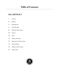
Table of Contents
Adrenal Disorders . . . . . . . . . . . . . . . . . . . . . . . . . . . . . . . . . . . . . . . . . . . . . . . . . . . . . . . . . . . . . . . A1
Diabetes Mellitus . . . . . . . . . . . . . . . . . . . . . . . . . . . . . . . . . . . . . . . . . . . . . . . . . . . . . . . . . . . . . . A23
Hypoglycemia . . . . . . . . . . . . . . . . . . . . . . . . . . . . . . . . . . . . . . . . . . . . . . . . . . . . . . . . . . . . . . . . . A81
Lipid Disorders . . . . . . . . . . . . . . . . . . . . . . . . . . . . . . . . . . . . . . . . . . . . . . . . . . . . . . . . . . . . . . . . A87
Metabolic Bone Disease . . . . . . . . . . . . . . . . . . . . . . . . . . . . . . . . . . . . . . . . . . . . . . . . . . . . . . . . A93
Obesity . . . . . . . . . . . . . . . . . . . . . . . . . . . . . . . . . . . . . . . . . . . . . . . . . . . . . . . . . . . . . . . . . . . . . A121
Other . . . . . . . . . . . . . . . . . . . . . . . . . . . . . . . . . . . . . . . . . . . . . . . . . . . . . . . . . . . . . . . . . . . . . . . A127
Pituitary Disorders . . . . . . . . . . . . . . . . . . . . . . . . . . . . . . . . . . . . . . . . . . . . . . . . . . . . . . . . . . . . A151
Reproductive Endocrinology . . . . . . . . . . . . . . . . . . . . . . . . . . . . . . . . . . . . . . . . . . . . . . . . . . . . A177
Thyroid Disease . . . . . . . . . . . . . . . . . . . . . . . . . . . . . . . . . . . . . . . . . . . . . . . . . . . . . . . . . . . . . . A187
Late Breaking . . . . . . . . . . . . . . . . . . . . . . . . . . . . . . . . . . . . . . . . . . . . . . . . . . . . . . . . . . . . . . . . A251
Author Index . . . . . . . . . . . . . . . . . . . . . . . . . . . . . . . . . . . . . . . . . . . . . . . . . . . . . . . . . . . . . . . . . A271
ABSTRACTS
ADRENAL Disorders
Abstract #100
PHEOCHROMOCYTOMA COEXISTING WITH
VASCULAR LESIONS
Sunil Kota, MD, Siva Kota, Svs Krishna, Lalit Meher,
Kirtikumar Modi
Objective: To report associated vascular lesions in
pheochromocytoma and discuss possible mechanisms.
Methods: From 1990 to 2010, 50 patients were diagnosed
with pheochromocytoma/ paragangliomas. Hospital charts
were reviewed for coexistent vascular lesions.
Results: 50 patients (M: F= 35: 15) with mean age of
45.5В±23.3 years, were diagnosed to have pheochromocytoma
(42 adrenal and 8 extra adrenal). 7 patients (14%) had
coexisting vascular lesions including renal artery stenosis
(RAS) in 4, aortoarteritis in 1, aortic aneurysm in 1 and
inferior vena cava thrombosis in 1. All of them harbored
pheochromocytoma [adrenal in 6 patients (4- left, 1- right)
and ectopic in one (at left renal hilum)]. RAS was suspected
because of small ipsilateral kidneys in 2, delayed nephrogram
in 1 and impingement of renal artery in 1 patient. A patient
with RAS due to intimal fibrosis was offered percutaneous
baloon angioplasty, other 3 improved after adrenalectomy and
lysis of fibrous adhesive bands. Aortoarterits was treated with
oral steroids. Inferior vena cava thrombosis was reversed with
anticoagulants. The patient with abdominal aortic aneurysm
was advised for annual follow up on account of its size of 4.5
cm and asymptomatic presentation.
Discussion: Pheochromocytoma has been described
previously in coexistence with RAS, renal artery aneurysm,
inferior vena cava thrombosis. Though the coexistence of
the vascular abnormalities can reflect chance association,
we propose certain causative factors. Mechanism for RAS
in pheochromocytoma include 1) tumor compression,
2) catecholamine induced vasospasm, 3) periarterial
adhesion, 4) associated atherosclerosis and fibromuscular
dysplasia. Mechanism for associated aortoarteritis include
1) catecholamine induced endothelial damage and intimal
fibrosis, 2) association with autoimmune conditions like SLE,
Behcet’s disease. Mechanisms for associated IVC thrombosis
include 1) local compression leading to alteration in blood
flow and stasis 2) sustained hypertension leading to vascular
endothelial injury and hypercoagulbility, 3) association with
autoimmune conditions like SLE, Behcet’s disease and 4) An
underlying anatomic abnormality or coagulation disorder.
Mechanism for associated abdominal aortic aneurysm are
1) persistent exposure to high catecholamines induced
vascular wall damage and weakening 2) Associated cigarette
smoking, increasing age, hypertension and atherosclerosis 3)
Coexistence of vasculitis like takayasu’s disease, Giant cell
arteritis and cystic medial necrosis due to marfan and ehlers
danlos syndrome.
Conclusion: The state of catecholamine excess and various
other coexisting factors can lead to simultaneous occurrence
of uncommon vascular abnormalities.
Abstract #101
CASE OF DISSEMINATED HISTOPLASMOSIS IN
IMMUNOCOMPITENT PATIENT WHO DEVELOPED
ADRENAL INSUFFICIENCY WHEN TREATED
WITH ITRACONAZOLE
Issac Sachmechi, MD, FACP, FACE, Kelash Kumar, MD,
Chinmay Patel, Uday Shankar, Victoria Bellot
Objective: Histoplasmosis is the most prevalent endemic
mycosis (Fungal infection) in the United States. Usually,
the infection follows an asymptomatic and self limited. We
describe disseminated Histoplasmosis in an immunocompetent
patient from a non endemic area without lung involvement
but involving the adrenal gland.
Case Presentation: We report a case of Disseminated
Histoplasmosis with iatrogenic adrenal insufficiency in a 41
year old male from Bangladesh with an extensive travel history
to western countries, presented with weight loss, intermittent
loose watery stools, generalized weakness and anorexia for
4 months. On physical exam the patient was noted to have
generalized wasting, had few mucosal papular lesions on the
tongue. His labs were notable for hyponatremia: Low Na 126
meq/l (135-145meq/l), hypoalbuminemia: 1.7g/dl (3.4-5.4 g/
dl) and prealbumin: 3.3mg/dl (normal 16-35mg/dl), AST 115
U/l, ALT 53 U/l, GGT 102, Alkaline phos: 489(30-115 U/l)
and PT/INR: 12.8/1.15 and HIV negative. Chest x-ray was
normal. His stools studies including C.difficile were negative.
The Contrast computed tomography imaging of chest,
abdomen and pelvis revealed bilateral adrenal enlargement.
Blood cultures and fungal cultures were negative. The serum
IgG for Histoplasma was positive. Biopsies of the skin,
liver and colon revealed Histoplasma organisms and were
consistent with disseminated Histoplasmosis. The patient was
started on the intravenous Amphotericin B for two weeks,
– A1 –
ABSTRACTS – Adrenal Disorders
with improvement in appetite and resolution of diarrhea.
The patient was discharged home on Itraconazole 200mg
PO daily. Two months after the discharge the patient
readmitted from medical clinic on his follow up, with the
hyponatremia, hyperkalemia, acidosis and generalized
weakness. ACTH stimulation test revealed cortisol levels:
0.3Ug/dl at baseline & 0.6 Ug/dl at 30 minutes, consistent
with primary adrenal insufficiency. The patient was started
on hydrocortisone and fludrocortisones with resolution of
weakness, weight gain and normalization of electrolytes.
Discussion: Itraconazole can cause adrenal insufficiency
by inhibiting CYP3A in less than 2% of patients. This
medication adverse effect may be due to subclinical
adrenal insufficiency caused by Histoplasma infestation
of adrenals with addition of Itraconazole inducing full
blown adrenal insufficiency. The presence of bilateral
adrenal enlargement raised the possibility of Disseminated
Histoplasmosis while biopsies of the skin, colon and liver
confirmed this diagnosis.
Conclusion: Patients with disseminated Histoplasmosis,
treated with Itraconazole, should be closely monitored for
adrenal insufficiency.
Abstract #102
ADRENOCORTICOTROPHIC INDEPENDENT
CUSHING’S SYNDROME: BEWARE OF
MALIGNANCY IN EVERY LARGE ADRENAL
TUMOR
histopathological findings although suggested incomplete
resection, didn’t meet the criteria for adrenocortical
carcinoma (ACC). Her post operative random cortisol
was 31 nmol/L consistent with adrenal insufficiency and a
remission. Although initial CT thorax, abdomen and pelvis
did not show any evidence of metastasis, a CT scan just
6 months later showed recurrence of left adrenal tumor
measuring 2.3x3.1 cm with mesenteric, retroperitoneal,
peritoneal and lung metastasis. CT guided biopsy of
left para-aortic lymph node confirmed metastasis.
Biochemically, patient had hypercortisolism with 24 hours
UFC of 1371 nmol/day indicative of a recurrence. She has
been started on ketoconazole and palliative chemotherapy
with mitotane.
Discussion: Although the histology in this case is
more suggestive of benign tumor and the initial tumor
surveillance studies were negative, she developed
metastatic disease within 6 months. Despite complete
resection in Stage I-III disease, approximately 40% of
patients develop metastasis within 2 years. Adjuvant
mitotane therapy may prolong recurrence-free survival in
patients with radically resected ACC up to 42 months.
Conclusion: It is important to consider a possibility of
ACC in every large adrenal adenoma associated with
rapidly progressive CS.
Abstract #103
PHEOCHROMOCYTOMA PRESENTING WITH
HEMOPTYSIS: CASE REPORT AND REVIEW
OF OTHER UNUSUAL MANIFESTATIONS OF
PHEOCHROMOCYTOMA
Chee Kian Chew, MD,
Rinkoo Dalan, MBBS, MRCP, FRCP (Edin), FAMS
(Endocrinology)
Case Presentation: A 60 years old lady, with longstanding
diabetes mellitus, hypertension and hyperlipidemia
presented with clinical CS without virilisation. She had
developed features of CS very rapidly in the preceding
one month. Biochemically, 8am cortisol: 795 nmol/L
(RI: 240-618), ACTH <2.0 pmol/L, 24 hours urinary
free cortisol (UFC): 3015 nmol/day (RI: 59-413), 1mg
overnight dexamethasone suppression test showed
non-suppressible cortisol : 746 nmol/L and low dose
dexamethasone suppression test also showed nonsuppressible cortisol: 718 nmol/L. The CT scan showed a
left adrenal, heterogenous 4.6x4.6x4.1 cm mass, with pre
contrast attenuation of 26 HU, delayed attenuation of 52
HU and absolute washout of 64%. This confirmed ACTH
independent CS secondary to left adrenal adenoma. She
underwent a laproscopic adrenalectomy and the histology
showed an unencapsulated adrenal cortical neoplasm
with less than 25% scattered foci of clear cells , Fuhrman
nuclear grade 3, 5 mitotic figures per 50 hpf, Weiss score
2/9 with evidence of tumor cells in the peripheries. These
Sheryl Tugna, MD, Thelma Crisostomo
Objective: To report a case of pheochromocytoma
presenting with hemoptysis and other unusual
manifestations of pheochromocytoma.
Case Presentation: A 45 yr. old male was brought
to ER with hemoptysis. There was no other bleeding
manifestation. He is hypertensive for 10 years with systolic
BP of 130-180 mmHg. 6 months ago, he started to have
palpitations and tremors associated with headache and
diaphoresis. At the ER, he was anxious and in respiratory
distress. HR was 115 and BP was 200/100 mmHg.
Nicardipine drip was started. Coagulation studies were
normal. Serum Amylase was elevated at 418 U/L (N.V 28100). CPK was high at 809 U/L (N.V 39-308) No lesion
and mass noted on Bronchoscopy. Chest CT scan showed
pulmonary hemorrhage and edema with a finding of left
adrenal mass measuring 5.6 X 5.8 X 5.0 cm (APxWxH)
exhibiting central hypodensity. Impression was Adrenal
Incidentaloma to consider Pheochromocytoma. Urinary
VMA and Metanephrine were elevated at 390 umol/24 hr.
– A2 –
ABSTRACTS – Adrenal Disorders
(N.V 0-68) and 166 umol/24 hr. (N.V 0-5.5) respectively.
By 3rd day, blood pressure stabilized to normal levels
with resolution of hemoptysis.Chest Xray showed
clearing of pulmonary edema. He was given oral antihypertensives. Systolic BP maintained at 110-130 mmHg.
Left Adrenalectomy was done. Histopathologic result
confirmed the diagnosis of Pheochromocytoma. Post-op,
blood pressure was maintained at SBP 120-140 mmHg. 4
days after surgery, he was discharged.
Discussion: Pheochromocytoma usually presents with
spell-signs and symptoms like headache, tremors,
hypertension and diaphoresis. The unusual manifestations
in our patient were hemoptysis, pulmonary edema,
elevated serum Amylase and CPK.Bronchoscopy and CT
scan of the Chest result were not able to identify the cause
of hemoptysis in our patient. Frymoyer et.al proposed
that the sudden catecholamine release could cause severe
hypertension in pheochromocytoma. This may cause
pulmonary venous hypertension, pulmonary edema and
hemoptysis.Patient’s hemoptysis and pulmonary edema
resolved concurrently with normalization of blood
pressure. The CPK level was elevated in the absence of
myocardial infarction and muscle trauma. Bahtnagar et al
suggested that catecholamine may cause vasoconstriction
leading to ischemia of the muscle. Patient has elevated
serum amylase in the absence of abdominal pain and
radiographic evidence of pancreatitis. Review of literature
has shown that the source of amylase was pulmonary
endothelial cells under ischemic damage caused by
vasoconstrictive effects of catecholamine.
Conclusion: Clinicians should be aware of various
clinical presentations of Pheochromocytoma so as to have
an early diagnosis and treatment before life-threatening
complications develops.
Abstract #104
QUALITATIVE MOLECULAR PROFILING
OF CLINICALLY FAVORABLE RESECTED
ADRENAL METASTASES
and thyroid) were collected. A commercially available
92-gene assay was used to determine a metastatic gene
expression profile for isolated adrenal metastasis. Pooled
RNA/cDNA of adrenal metastases was compared to
benign, non-functioning adrenal adenomas. Significance
analysis of microarrays (SAM) determined differences
in gene expression between metastatic and benign nonfunctioning adrenal tumors.
Results: A total of 18 genes were over-expressed in the
adrenal metastases group as compared to benign adrenal
controls (False Discovery Rate = 5%). Further analysis
revealed overexpression of 9 genes (4 “pro-metastatic”, 3
“anti-metastatic” and 2 chemokine genes) in the metastatic
group with a greater than 2-fold expression difference. Four
genes (FN1, MTA2, MMP2 and SET) were significantly
under-expressed in the metastatic group, with a greater than
2-fold expression difference. All matrix metalloproteinase
(MMP) genes were under-expressed while tissue inhibitors
of MMPs were overexpressed in the adrenal metastases
group.
Discussion: The adrenal glands are common sites of metastases from a variety of skin and solid organ cancers.
Laparoscopic adrenalectomy for isolated adrenal metastases, once considered controversial, is now increasingly being performed for cure or palliation. We have previously
demonstrated that patients with a disease-free interval of
more than 12 months between initial diagnosis of primary cancer and detection of adrenal metastasis had slowergrowing tumors and an improved overall survival. This
study attempts to identify clinically favorable adrenal metastases at the molecular level.
Conclusion: Resected adrenal metastases from patients
with a disease-free interval of more than 12 months have a
favorable gene profile. We found that genes related to tumor
proliferation were over-expressed in the metastatic group,
while genes related to tumor invasion were generally underexpressed. Further understanding of the molecular profiles
of adrenal metastasis could potentially provide prognostic
information and allow for improved selection of patients
with isolated adrenal metastases for adrenalectomy.
Abstract #105
Elliot Mitmaker, MD, Raymon Grogan, MD,
Joe Kansopon, Avital Harari, Jimmy Hwang,
Jessica Gosnell, Orlo Clark, MD, Quan-Yang Duh,
Wen Shen
Objective: To determine the molecular profiles of
resected adrenal metastases in order to explain the
favorable clinical characteristics and outcomes in patients
undergoing adrenalectomy for isolated metastatic disease
with a 12-month disease-free interval.
Methods: Paraffin-embedded tissue blocks of metastatic
adrenal tumors from 7 different primary sites (lung,
breast, colorectal, renal cell, adrenocortical, melanoma
SUPPRESSION AND RECOVERY OF HPA
FUNCTION AFTER A SINGLE EPIDURAL
GLUCOCORTICOID INJECTION:
DECONVOLUTION ESTIMATION OF ACTH AND
CORTISOL SECRETORY DYNAMICS
Donna Lawson, DO, Dakshin Gullapalli, Johannes
Veldhuis, MD, Ali Iranmanesh, MD
Objective: This study was designed to assess the magnitude
and duration of disrupted ACTH and cortisol secretory
– A3 –
ABSTRACTS – Adrenal Disorders
patterns and time to recovery after epidural glucocorticoid
injection.
Methods: Nine men (25-63 years) on 4 separate days
(baseline, 1, 4, and 12 weeks). Triamcinolone (80 mg) was
injected epidurally right after the 1st study session. During
each visit, blood was collected in a fasting state at 10-min
intervals for a period of 4 hours, with ovine CRH (1Вµg/
Kg) injected after the 6th blood draw (min 60). ACTH (pg/
mL) and cortisol (Вµg/dL) concentrations were measured
in each blood sample, and their respective secretory
properties were assessed by deconvolution analysis
Results: Mean (В±SEM) pre-CRH baseline unstimulated
plasma ACTH (6.1 В± 0.3 v 25.1 В± 1.2: P <0.0001) and
cortisol (1.7 В± 0.1 v 12.9 В± 0.1: P < 0.0001) concentrations
were significantly decreased at week 1. Although full
recovery of ACTH occurred at week 4 (25.4 В± 2.6 v 25.1 В±
1.2: P = NS), respective mean cortisol levels of 10.9 В± 0.3
and 11.1 В± 0.1 at weeks 4 and 12, continued to be lower than
the pre-treatment values (P=0.01). Corticotropic response
to CRH stimulation was similarly blunted at week 1 with
significant decreases in the 3-hr mean (В±SEM) ACTH
(11.1 В± 0.2 v 40.4 В± 3.4: P <0.0001) and cortisol (3.6 В±
0.2 v 21.3 В± 0.8: P < 0.0001) concentrations. While ACTH
response to CRH normalized at week 4 (37.5 В± 2.4 v 40.4
В± 3.4: P=NS), cortisol response did not fully reverse by
week 12 (19.6 В± 0.9 v 21.3 В± 0.8: P <0.01). Twenty-four
hr urinary free cortisol (Вµg) was diminished at week 1 (8.7
В± 1.2 v 62 В± 6.1: P <0.0001), and did not normalize until
week 12. Deconvolution analysis of post-CRH time series
revealed altered pulsatile and basal secretory modes of
ACTH and cortisol release with significant suppression
at week 1, and recovery at week 4. Changes in pulsatile
ACTH and cortisol secretion were primarily due to
changes in the mass of hormone secreted per burst.
Discussion: Corticotropic function was uniformly
suppressed within 7 days of epidural steroid injection.
Pre- and post-CRH concentrations of ACTH and cortisol,
and their respective secretory properties allowed a better
understanding of underlying mechanisms, and the required
time for the axis to recover.
Conclusion: Epidural glucocorticoid administration
markedly represses HPA output via suppression of both
basal and pulsatile modes of ACTH and cortisol release.
The average recovery time appears to be 4 weeks for
ACTH, and potentially 12 weeks or more for cortisol.
These inferences warrant future confirmation in larger
cohorts and over a longer time period.
Abstract #106
PF4 ANTIBODY POSITIVITY IN THE SETTING
OF BILATERAL ADRENAL HEMORRHAGE
Mini Mathew, Pharm.D., D.O., Kamalpreet Singh, MD,
Deepika Reddy
Case Presentation: 56 year old female underwent screening
colonoscopy which led to diagnosis of Stage 4 Carcinoid
tumor. Treatment consisted of right hemicolectomy with
ileal resection. Postoperatively she was found to have
bilateral pulmonary emboli and was started on heparin,
which resulted in her developing heparin induced
thrombocytopenia. She was placed on Coumadin, with her
INR goal being closer to 2.0. CT of the abdomen showed
possible adrenal hemorrhage, but she had no hypotension
or abnormalities in sodium or potassium levels. Three
and a half weeks later she presented to the hospital with
complaints of nausea, vomiting and dizziness. Na was
126 and K was 5.7. Medical staff was unable to place IV
access and so she was given IM Decadron. She reported
improvement of symptoms within 2 hours. She underwent
a cosynotropin stimulation test in the morning which
showed a baseline ACTH of 1232 pg/mL and initial cortisol
of 6.2 mcg/dL, at 30 minutes cortisol was 6.0mcg/dL and
at 60 minutes was 5.5 mcg/dL. CT of the abdomen/pelvis
showed bilateral adrenal hemorrhages. She was started
on hydrocortisone and florinef and reported symptomatic
improvement, repeat Na and K levels normalized.
Discussion: Bilateral adrenal hemorrhages are present
in about 1% of routine autopsies. Major risk factors
for adrenal hemorrhage include hypercoaguable states,
sepsis, severe stress, and anticoagulant therapy. Heparin
is the most common anticoagulant used in hospitalized
patients and heparin induced thrombocytopenia (HIT) is
a rare complication. HIT is caused by platelet-activating
antibodies that recognize complexes of platelet factor four
and heparin. This typically results in thrombocytopenia
but bleeding is seldom an issue.
Conclusion: Bilateral adrenal hemorrhage should be
suspected in patients with complaints of abdominal
pain and hypotension with recent heparin exposure. The
role of HIT in bilateral adrenal hemorrhage is likely
underestimated as use of heparin is not always documented
or PF4 antibodies are not ordered. The predominant
thrombotic event is usually arterial but in this case it
was venous. Adrenal insufficiency usually presents after
a latency period weeks after initial episode of bilateral
adrenal hemorrhage.
– A4 –
ABSTRACTS – Adrenal Disorders
Abstract #107
Abstract #108
EAT MORE YET FEEL WEAK
FAMILIAL CUSHING’S SYNDROME DUE
TO A BILATERAL ACTH-INDEPENDENT
MACRONODULAR ADRENAL HYPERPLASIA
(AIMAH) RELATED TO THE ECTOPIC
EXPRESSION OF BETA ADRENERGIC
RECEPTORS
Radha Devi, MD, Parakkal Deepak
Objective: Megesterol acetate (Megace) is a synthetic progesterone derivative used in the treatment of cachexia and
anorexia in metastatic cancers or AIDS. It is associated with
side effects like thromboembolism, hypertension, gynecomastia and adrenal insufficiency. Patients with adrenal insufficiency may present with non-specific symptoms of fatigue,
anorexia and decreased libido or hypotension and cardio vascular instability in times of stress. This case highlights the serious side effect of adrenal insufficiency caused by Megace.
Case Presentation: A 65 year old man with past medical
history of coronary artery disease, hypertension, abdominal
aortic aneurysm, and failed renal transplant on hemodialysis, was admitted to the hospital with complaints of altered
mental status. He was placed on Megace 800 mg per day
to treat decreased appetite and weight loss of 60 lb over
the last 2 months. Physical exam revealed no mucosal or
flexural hyper pigmentation with a normal blood pressure
and neurological exam. Basic labs, thyroid function test and
CT scan of the head were normal. An 8 am serum cortisol
level was less than 6.2 microgram per deciliter(mcg/dl) and
a low dose cosyntropin test was performed revealing cortisol level of < 1mcg/dl and ACTH level of 11 pg/ml indicating secondary adrenal insufficiency. A CT of the abdomen
(done to rule out malignancy) revealed enlargement of his
abdominal aortic aneurysm. Megace was discontinued and
he was started on stress dose of steroids with hydrocortisone
at 100mg intravenously every 8 hours, prior to undergoing
endovascular repair of his aneurysm. He was discharged on
a gradual taper of oral hydrocortisone with instructions not
to resume megace on discharge.
Discussion: Megace is postulated to lead to a suppression
of the hypothalamic pituitary axis by its glucocorticoid like
action especially when used in doses more than 300mg per
day. Patients may present with symptoms of acute adrenal
insufficiency in times of stress or non specific symptoms
of chronic adrenal insufficiency like fatigue. It is essential
for physicians to be aware of this potentially life threatening side effect prior to prescribing this medication. Hence,
patients receiving chronic high dose Megace therapy may
need stress doses of glucocorticoids in times of stress. It
has also been suggested that patients should be prescribed
Megace as an appetite stimulant only for shorter periods
and should be tapered off to avoid precipitating an adrenal
crisis.
Conclusion: It is important to recognize the early symptoms and signs of adrenal insufficiency secondary to this
under recognized etiology, in order to prevent morbidity
and mortality.
Duarte Pignatelli, MD, PhD, Jorge Lima
Objective: ACTH-independent bilateral macronodular
hyperplasia (AIMAH) is a rare cause of Cushing’s
syndrome(CS). Recent studies demonstrated adrenal
cortisol secretion to be regulated by ectopic membrane
hormone receptors(HR) , but few reports described
familial aggregation in these cases. We report a familial
case in which three members of a family were confirmed
as having this syndrome.
Methods: The clinical screening for potentially
illegitimate HR was done according to A Lacroix
protocol. The first case was diagnosed by postural
testing and propranolol (Prop) suppression testing. His
daughter had a clear response to isoproterenol and the
affected son had AIMAH, but not Cushing’s syndrome.
Histological analysis confirmed the diagnosis of AIMAH
in the father and his daughter that were the two only cases
that were surgically intervened. Real-time PCR was also
performed in samples obtained at surgery.
Case Presentation: The father was diagnosed as
having a posture-sensitive, Prop-responsive, CS due
to an asymmetric and bilateral adrenal hyperplasia.
Many years after this case was diagnosed, his daughter
also appeared with CS and an MRI also revealed the
presence of bilateral AIMAH. One of her brothers
was tested for the same disease and in spite of not
having any CS characteristics or hypercortisolism he
had bilateral AIMAH! A last brother was also studied
but had neither CS nor AIMAH as detected by MRI.
The father was initially submitted to unilateral
adrenalectomy but later CS relapsed and he had to be
treated with Prop. Remission then lasted for many years.
Prop remarkably normalized the bl.pressure, the cortisol
levels and its circadian rhythm. The response to Prop was
much less efficient in the case of his daughter, She was
also submitted to unilateral adrenalectomy with success
(normalization of the signs and symptoms of CS as well
as the cortisol levels
Discussion: This is one of the first cases of AIMAH to
reveal a clear hereditary transmission. In the present
case AIMAH was dependent on the ectopic expression
of beta-adrenergic receptors. This was confirmed by the
performance of Real-time PCR. The differences in the
clinical expression in the different members of the family
– A5 –
ABSTRACTS – Adrenal Disorders
namely the different response to the use of Propranolol
in spite of both cases having an hyper-expression of
ОІ-adrenergic receptors, deserves consideration.
Conclusion: We conclude that heritability may be an
important pathogenic cause of ectopic receptor expression
in AIMAH cases and screening of family members of
affected patients may reveal a much higher frequency of
such cases and allow the design of appropriate genetic
studies to be dome in muticenter studies.
Abstract #109
THE PREVALENCE OF POSTOPERATIVE
HYPOGLYCEMIA FOLLOWING
ADRENALECTOMY FOR
PHEOCHROMOCYTOMA: A RARE AND OFTEN
FORGOTTEN COMPLICATION
Elliot Mitmaker, MD, Raymon Grogan, MD,
Robin Cisco, MD, Daniele Rottkamp, Jessica Gosnell,
Orlo Clark, MD, Wen Shen, J. Blake Tyrrell,
Quan-Yang Duh
Discussion: Pheochromocytomas are rare catecholaminesecreting tumors of the adrenal medulla. Excessive
catecholamine release has multiple physiologic effects,
including abnormal glucose metabolism. Hyperglycemia
and new onset diabetes are well described, but the problem
of severe postoperative hypoglycemia following resection
is under-recognized. Postoperative hypoglycemia
following adrenalectomy for pheochromocytoma is rare
but potentially serious. It is likely caused by the acute
withdrawal of excessive catecholamines, coupled with
preoperative alpha and beta-adrenergic blockade, blunting
the normal mechanism of glucose regulation.
Conclusion: Postoperative blood glucose monitoring is
recommended to identify this potentially fatal complication. Implementing a standardized hospital-based protocol
for postoperative continuous blood glucose monitoring
will lead to early recognition of this rare and easily reversible metabolic event.
Abstract #114
Objective: To determine the prevalence of postoperative
hypoglycemia in pheochromocytoma patients following
adrenalectomy and to investigate the associated risk factors
that predispose these patients to develop postoperative
hypoglycemia.
Methods: We retrospectively reviewed all adrenalectomies
performed between 1993-2011 at a single institution and
identified 124 patients who underwent laparoscopic or
open adrenal resections for pheochromocytoma.
Case Presentation: 81 of the 124 patients had serial glucose
levels measured during the immediate postoperative period.
Thirteen patients (13/81=16%) were diagnosed with noninsulin dependent diabetes mellitus preoperatively, while
3 patients had long-standing insulin dependent diabetes
mellitus. Four patients (4.9%) developed hypoglycemia
within 4 hours after adrenalectomy. These four patients
were women and had an average body mass index
(BMI) of 21, compared to a higher average BMI (27.4;
p=0.055) among those who remained normoglycemic
(range=70-199 mg/dL). None of the four patients with
postoperative hypoglycemia had preoperative diabetes,
although one patient had a preoperative HgA1C=6.9.
These four patients were treated with intravenous 50%
dextrose solution and became normoglycemic after 3
hours. All patients received preoperative alpha-blocking
agents, as per our routine for preoperative preparation
for pheochromocytoma. One patient who had received
both alpha and beta adrenergic-blockade had severe
postoperative hypoglycemia (glucose=14 mg/dL) and was
unresponsive in the recovery room until after treatment
with dextrose solution.
LATE ONSET CONGENITAL ADRENAL
HYPERPLASIA (CAH) PRESENTING AS
ISOLATED NOXIOUS BODY ODOR IN AN ADULT
MALE - A SMELLY DIAGNOSIS!
Nisha Acharya, MD, Melissa Li-Ng
Case Presentation: A 43-year old male presented with
intermittent noxious body for the past year. He noted
some improvement on gluten-free diet but continued to
have recurrence of this symptom despite daily showering
and deodorant use. He noted hair thinning at the top of
his scalp for the past 7 years. He otherwise felt well and
denied erectile dysfunction, acne, and mood changes. He
fathered a 4-year old daughter. He denied family history
of endocrinopathies and infertility. Physical examination
revealed a well-appearing male, blood pressure 118/81
mmHg, height 1.85 m, BMI 22.30 kg/m2. Testes
were normal size & consistency, no palpable masses.
Laboratory investigations including complete blood
count, thyroid-stimulating hormone, hemoglobin A1C
and comprehensive metabolic panel were unremarkable.
Total and free testosterone as well as DHEA-S levels
were within normal range. 17-hydroxyprogesterone (17OHP), however, was elevated at 5.4 ng/ml (normal 0.41.8 ng/ml). Subsequently, ACTH stimulation showed an
increase in 17-OHP levels from 7.4 ng/ml at baseline to
115.8 ng/ml at 60 minutes. The cortisol level increased
from 9.4 ug/dl to 17.5 ug/dl at 60 minutes. These
results were consistent with late onset CAH. Patient
was offered dexamethasone therapy to decrease his
body odor but he opted to forego any medical treatment
at this time and was referred for genetic counseling.
– A6 –
ABSTRACTS – Adrenal Disorders
Discussion: Late onset CAH due to CYP21A2
(21-hydroxylase) deficiency is characterized by signs of
androgen excess later in life. The defective conversion
of 17-hydroxyprogesterone to 11-deoxycortisol in
patients with CYP21A2 deficiency causes decreased
cortisol synthesis, which results in increased ACTH
secretion. This in turn causes adrenal stimulation leading
to increased production of androgens. Children with late
onset CAH can present with premature adrenarche, which
includes pubic hair, axillary hair and adult body odor. Men
with late onset CAH can present with acne, infertility or
testicular adrenal rest tumors. In the skin, androgen excess
stimulates hyperplasia of sebaceous glands and apocrine
glands. Sebaceous glands produce sebum while apocrine
glands secrete a fatty, viscous sweat. Bacterial breakdown
of sebum and apocrine sweat produces body odor.
Conclusion: Our case is unique because of the isolated
finding of abnormal body odor as a presenting symptom
of late-onset CAH. Thus it is important to think of late
onset CAH in the differential diagnosis when evaluating a
patient for body odor.
Abstract #111
HYPERCORTISOLISM IN YOUNG PATIENT
WITH OBESITY
UfE compared with the control group and normal indices
of F/E and UFF/UFE. However, 7 patients (17.5%) had
increased F/E and UfF/UfE indices and 5 showed lack of
UfF suppression after 2 mg dexamethasone. Significant
positive association was found between concentration of
plasma cortisol by RIA, plasma cortisol by HPLC and fat
mass (P < 0.001).
Discussion: It is known that young obese patients
may show hyperactivity of hypothalamic-pituitaryadrenal (HPA) axis, which leads to a state of functional
hypercortisolism. HPA axis may be dysregulated due
to such reasons as puberty, stress, early life events and
others. Also, over-expression of 11-beta-hydroxysteroid
dehydrogenase type 1 in obese patients can result in
increased conversion of cortisone (E) to cortisol (F) thus
increasing glucocorticoid activity.
Conclusion: Our data confirms that young people with
obesity often reveal functional hypercortisolism. Some
patients have biochemical abnormalities indicating
subclinical corticosteroid excess. These endocrine
abnormalities in the young obese may place them at risk
for glucose intolerance, diabetes, bone loss and other
conditions.
Abstract #112
ADRENOMYELONEUROPATHY AND PRIMARY
ADRENAL INSUFFICIENCY
Zulfiya Shafigullina, MD, Ludmila Velikanova,
Natalya Vorokchobina
Objective: The aim of this study was to investigate the
features of adrenal steroid synthesis in young people with
obesity.
Methods: We examined 40 patients (15-20 years old)
with obesity (BMI 32.3 В±0.8). Twenty three subjects
(57.5%) had arterial hypertension. Control group included
20 healthy subjects (15-20 years old, BMI 23.4 В±0.6)
without obesity and hypertension. All patients underwent
hormonal evaluation for circadian rhythm of plasma
cortisol and ACTH secretion and also low dose (2 mg)
dexamethasone suppression test (DST). Intermediates
of steroido-genesis were assessed by means of highperformance liquid chromatography (HPLC) including
measurement of plasma levels of cortisol (F), cortisone
(E), corticosterone (B), 11-deoxycorticosterone (DOC),
11-deoxycortisol (S) and urinary excretion of free cortisol
(UfF) and free cortisone (UfE).
Results: Among 40 patients with obesity, hormonal workup showed high baseline cortisol in 30%, high ACTH in
27.5% and disturbed circadian cortisol rhythm in 22.5%.
DST was normal in the majority of subjects although 6
patients (15%) had plasma cortisol concentrations above
60 nmol/L after this test. These patients were found to
have significantly higher concentrations of F, B, UfF and
Shilpa Swamy, MD,
Donald Richardson, MD, FACE, FACP
Objective: To describe an unusual cause of primary
adrenal insufficiency.
Case Presentation: A 45 year old Japanese male was
evaluated for adrenal insufficiency. The patient was
diagnosed with adrenomyeloneuropathy at age 35 during
further investigation of lower extremity weakness. Recent
serum cortisol, drawn at 1pm, was 14mcg/dL with an
ACTH of 69.2 pg/mL. No clinical signs of adrenal
insufficiency were found but increased ACTH suggests
impaired adrenal reserve.
Discussion: Adrenomyeloneuropathy (AMN) is a sub-set
of X-linked Adrenoleukodystrophy (X-ALD), a group of
disorders with abnormal accumulation of very long chain
fatty acids (VLCFA) in the brain, adrenal cortex and Leydig
cells of the testes. Responsible mutations affect the ABCD1
gene on chromosome Xp28, which encodes the protein
involved in the import of VLCFA into the peroxisome.
Adrenal pathology may be attributed to the combination
of effects of VLCFA on membrane structure (increased
micro-viscosity) and accumulation of cholesterol
esterified by VLCFA (which are poor substrates
for cholesterol hydrolases), impairing the response
– A7 –
ABSTRACTS – Adrenal Disorders
of adrenal cortical cells to ACTH stimulation.
The AMN phenotype represents ~45% of X-ALD.
It presents in adulthood with spastic paraparesis and
peripheral neuropathy as a consequence of spinal cord
and peripheral nerve demyelination, as opposed to the
childhood cerebral form which presents between 4-8yrs
with cognitive dysfunction and progressive neurological
deterioration. 8% of males present with adrenal
insufficiency at any age and are at risk for X-ALD/
AMN for life. Up to 50% of female heterozygotes
may manifest an AMN-like syndrome. Adrenal
insufficiency affects 80% of the childhood cerebral
form, 50% of AMN and 1% of heterozygous women.
Diagnosis is by plasma VLCFA assay, especially C26:0 and
C24:0. Lorenzo’s oil (a combination of mono-unsaturated
fatty acids erucic acid and oleic acid, which blocks the
endogenous synthesis of VLCFA) in asymptomatic cases
may reduce the risk for neurological manifestations in
cerebral X-ALD and slow the progression of AMN,
but offers little once neurological impairment has set in
(suspected to be due to failure of active ingredient to enter
nervous system in significant quantity). Hematopoietic
cell transplantation may be offered to boys with early
cerebral involvement, and gene therapy is being studied.
Conclusion: Adrenoleukodystrophy, while only affecting
between 1 in 17 to 21,000 males, is responsible for up to
50% of cases of Addison’s disease in boys and young men.
Once X-ALD is diagnosed, patients should be evaluated
for adrenal insufficiency on an annual basis with serum
ACTH and ACTH stimulation test.
Abstract #113
PHEOCHROMOCYTOMA PRESENTING AS
ACUTE DECOMPENSATED HEART FAILURE
Christopher Mulla, MD, Paul Marik
Objective: Pheochromocytomas can have varied
clinical manifestations, ranging from asymptomatic to
hypertension with headaches and palpitations and very
rarely to acute decompensated heart failure (ADHF) with
multisystem organ failure.
Case Presentation: A 26-year-old woman was seen in
the ER for chest pain. She was found to be hypertensive
179/121 mmHg and was diagnosed with a urinary tract
infection. She was discharged with an antibiotic and
thiazide diuretic. She returned 2 days later with epigastric
pain, nausea, emesis and myalgias. On examination she
remained hypertensive to 169/105 mmHg and tachycardic
125 bpm. She was given 1.5 L of fluid for presumed prerenal azotemia and soon developed respiratory distress
requiring intubation. A chest film demonstrated pulmonary
edema. She was transferred to the ICU and she progressed
to multisystem-organ failure. An echocardiogram
established global left ventricular hypokinesis with a leftventricular ejection fraction <10%. She was cautiously
rehydrated with IV fluids and transiently treated with
pressors followed by a gradual recovery. Her workup
included right heart catheterization and biopsy for
possible acute viral myocarditis that was negative. Her
ejection fraction improved to 15%, she regained kidney
function, was extubated, and eventually discharged home
on a non-selective beta blockade with planned follow up
at the CHF clinic. She returned to the ER 4 days later with
an elevated BP, nausea and abdominal pain. A contrast CT
scan of her abdomen revealed pancreatitis and an adrenal
mass. During her treatment for pancreatitis she began
to have paroxysms of headaches, nausea, emesis and
abdominal pain with corresponding severe hypertension
and tachycardia. Her urinary and serum catecholamine
levels were elevated. She was rehydrated, treated with
an alpha and beta adrenergic blocker followed by an
adrenalectomy. Medical management prior to surgery was
associated with improvement in left-ventricular ejection
fraction to 55%.
Discussion: Pheochromocytomas are a rare growth of
chromaffin cells which produce excess catecholamines.
Failure to diagnose and treat this condition can lead to
ADHF either due to stress induced cardiomyopathy or
due to abrupt withdrawal of catecholamine signaling in
a patient with desensitized adrenoreceptors and a reduced
circulatory volume. Chronic catecholamine exposure and
hypertension leads to excessive glomerular filtration and
dehydration; therefore treatment includes fluid hydration.
While surgical removal of offending tumor is definitive
therapy, patients must first be stabilized with alpha and
beta adrenergic blockade.
Conclusion:
This
case
demonstrates
that
pheochromocytoma-induced ADHF can be reversed
with medical therapy.
Abstract #110
AN ELUSIVE NEUROENDOCRINE TUMOR;
A CHALLENGING DIAGNOSIS IN A PATIENT
WITH CUSHING’S SYNDROME
Kwame Ntim, MBCHb, Daniel Wong, MD
Objective: Case report illustrating some of the challenges
associated with localizing an ectopic ACTH dependent
Cushing’s syndrome.
Case Presentation: 40 y/o male was referred to our hospital
for management of a perforated duodenal ulcer previously
managed with an omental patch. He had a past history of
Cushing’s syndrome suspected to be secondary to an un-localized ectopic ACTH lesion. Past records indicated he had
– A8 –
ABSTRACTS – Adrenal Disorders
been relatively well until a year ago when he was treated
with fluticasone for allergic rhinitis. Two months later, he
had cellulitis treated with antibiotics and subsequently with
IM and oral steroids for metacarpophalangeal joint nodules. A couple of months after this, he is reported to have
developed Cushingoid symptoms. His labs also showed
hyperglycemia and hypokalemia. Endocrine work up demonstrated elevated ACTH 261pg/ml (7-50 pg/ml), 24 hr cortisol 1141mcg/24 (4-50 mcg/24) and 1 mg dexamethasone
suppression test of 32.3mcg/dl (4-22 mcg/dl) with a normal
pituitary MRI. Further work up at a tertiary center included
a normal CT chest, abdomen and pelvis and CRH stimulation test. He was started on ketoconazole, spironolactone
and insulin for diabetes. He then subsequently developed a
perforated duodenal ulcer. Physical exam on admission was
significant for moon facies, supraclavicular fat pads, buffalo hump, violaceous striae, central obesity and increased
abdominal girth. Labs confirmed in our hospital were; 24
hour urine free cortisol of 1692mcg/24 (4-50 mcg/24),
ACTH 200pg/ml (7.2-63 pg/ml). Repeat pituitary MRI and
CT abdomen showed a 3mm suspicious pituitary mass and
an ill defined mass in the uncinate process of the pancreas
respectively. Patient underwent surgical removal of the
mass. Pathology confirmed a neuroendocrine tumor. Post
surgery ACTH level was 10pg/ml (7.2-63 pg/ml) though
the initial pathology did not stain for ACTH.
Discussion: The symptoms and signs of hypercortisolism
are non-specific making the diagnosis challenging. If established that the cause is endogenous, the next step involves
identifying the underlying pathophysiology. Endogenous
causes can be divided into ACTH-dependent (namely
Cushing’s disease, ectopic ACTH or ectopic CRH) or an
ACTH-independent hypercortisolism secondary to hyper
functioning adrenals. Inferior petrosal sinus sampling is
useful in differentiating pituitary ACTH production from
ectopic ACTH. The alternative is a CRH stimulation test.
CT scans, MRIs, Octreotide scintigraphy are helpful in localizing ectopic sites. Surgical removal is often the first line
of treatment.
Conclusion: Knowledge of the physiology of the
Hypothalamus-pituitary-adrenal axis is essential in the
evaluation, diagnosis and management of hypercortisolism.
Abstract #115
ADRENAL VENOUS SAMPLING: AN UNUSUAL
METHOD FOR INVESTIGATING BILATERAL
ADRENAL MASSES
incidentaloma. Bilateral adrenal masses account for 10
to 15 % of adrenal incidentalomas. We are presenting a
case of bilateral adrenal masses with SCCS secondary to
ACTH-Independent Macronodular Adrenal Hyperplasia
(AIMAH). We are also describing Adrenal venous
sampling (AVS), a new emerging technique, which helps
identify the source of cortisol secretion in this setting.
Case Presentation: 51 year old lady, was evaluated for
bilateral adrenal masses found incidentally on an abdominal
MRI, with loss of signal on chemical shift method
indicating lipid content of the masses. Laboratory data
showed lack of suppression of cortisol in response to both
low and high dose dexamethasone with undetectable base
line ACTH, normal 24 hour urine free cortisol and normal
mid night salivary cortisol, suggestive of SCCS. Both
MRI and CT were unable to differentiate the hyperplastic
versus adenomatous nature of the masses. AVS was
performed and blood cortisol and epinephrine levels were
obtained from both adrenal veins (AV) and peripheral vein
(PV). Results showed an AV: PV cortisol ratio of 5.85 and
5.04 on right and left side respectively, without significant
lateralization. AVS results, coupled with the MRI and CT
scan findings, favor the diagnosis of bilateral AIMAH.
Robotic left adrenalectomy was performed since the left
adrenal mass was larger. Pathology favored the diagnosis
of AIMAH. ACTH and cortisol levels will be monitored to
ensure cure and surveillance for recurrence.
Discussion: In this case, anatomical configuration of the
bilateral adrenal masses on MRI and CT were not typical
of hyperplasia or bilateral adenomas. The dilemma was:
which gland is hypersecreting cortisol. AVS proved to be a
useful tool. Adequate catheterization of the AV is ensured
if the epinephrine level difference between AV and PV
is more than 100. An AV: PV cortisol ratio of >4.1 may
mean autonomous cortisol secretion, >6.5 points towards
an adrenal adenoma, and between 4.1 and 6.5 (as seen in
our case) may indicate hyperplasia. The combination of
functional (AVS) and anatomical (CT) picture suggested
AIMAH as the cause of the SCCS, which is a rare entity
associated with either aberrant hormone receptors or
genetic mutations.
Conclusion: AVS can be a useful tool to localize the source
of the cortisol hypersecretion in ACTH-independent
Cushing syndrome with bilateral adrenal masses.
Furthermore, AVS can also help distinguish bilateral
adrenal adenomas from AIMAH if the radiological
findings are not clear.
Adam Maghrabi, MD, Saba Faiz, MD,
Tipu Faiz Saleem, MD
Objective: Subclinical Cushing syndrome (SCCS) is
the most frequent hormonal abnormality in adrenal
– A9 –
ABSTRACTS – Adrenal Disorders
Abstract #116
Abstract #117
SEQUENTIAL ADRENAL GLAND
HEMORRHAGE AND ACUTE MYELOID
LEUKEMIA в€’ IS THERE AN ASSOCIATION?
SUPRAPHYSIOLOGIC RESPONSE TO ACTH
STIMULATION TEST IN A 72 YEAR OLD MAN,
UNMASKING SUBCLINICAL CUSHING’S
SYNDROME
Preethi Sridhar, MD, Boby Theckedath,
Janice Gilden, MD
Marianna Antonopoulou, MD, Asya Perelstein, MD
Objective: To describe a patient on anticoagulation
presenting with sequential adrenal gland hemorrhage,
thrombocytopenia and anemia who was later diagnosed
with Acute Myeloid Leukemia.
Case Presentation: A 63 year old gentleman with new
onset atrial fibrillation was treated with enoxaparin and
warfarin. Ten days later, he was admitted with left flank
pain. His INR was 1.9 and platelet count 122,000/cubic
mm. A CAT scan of the abdomen revealed an acute left
adrenal hemorrhage. All anticoagulants were discontinued.
He was discharged after 2 days of observation, since he
was clinically and radiologically stable. One week later
he was readmitted for fatigue, dizziness, hypotension,
bradycardia, and orthostatic hypotension. A repeat
abdominal CAT scan revealed a new right adrenal
hemorrhage. Laboratory investigation showed a low serum
AM cortisol of 0.93 mcg/dl, elevated ACTH at 57 pg/ml
and a positive ACTH stimulation test (baseline cortisol
= 5.62 mcg/dl, 30 minutes = 6.95 mcg/dl, 60 minutes =
8.07 mcg/dl). The patient was subsequently treated with
hydrocortisone for adrenal insufficiency. The initial work
up for persistent anemia and thrombocytopenia was
negative, including repeatedly negative HIT antibodies.
Heparin-induced thrombocytopenia was hence ruled out.
Further work up with a bone marrow biopsy revealed
Acute Myeloid Leukemia.
Discussion: Literature reports of the association between
bilateral adrenal hemorrhage and acute leukemia are
very rare. Bilateral adrenal hemorrhage has been
increasingly reported as a complication of heparininduced thrombocytopenia and anticoagulation therapy.
In our patient, thrombocytopenia (not related to HeparinInduced Thrombocytopenia and Thrombosis) and a subtherapeutic INR are unlikely to have caused the bilateral
adrenal hemorrhage.
Conclusion: Bilateral adrenal hemorrhage and adrenal
insufficiency might occur in Acute Myeloid Leukemia and
this association needs further investigation.
Objective: To document a case of subclinical Cushing’s
syndrome in a 72 year old man with adrenal incidentalomas.
Methods: We present the diagnostic approach of a male
patient with adrenal incidentalomas
Case Presentation: A 72 year old African American
male with past medical history of hypertension, coronary
artery disease (CAD), hyperlipidemia, spinal stenosis,
and monoclonal gammopathy of uncertain significance
(MGUS) had a CT scan of abdomen in 2009, showing
right and left adrenal masses measuring 5x3.5 cm and
3.7x2.9 cm respectively. Patient underwent hormonal
work up to rule out functioning adrenal tumors 3 times,
including 24 hour urine cortisol and metanephrines, serum
aldosterone, all of which were normal. The radiologist
insisted that the CT findings are consistent with adrenal
hyperplasia and since the patient was hypertensive, he
underwent 250mcg ACTH stimulation test to rule out
late onset congenital adrenal hyperplasia (CAH). The
stimulation test revealed that 17-hydroxyprogesterone,
11-deoxycortisol increased to levels high enough to
confirm CAH, but cortisol had exaggerated response too,
making the diagnosis of CAH unlikely, where metabolism
is shifted to precursors. Other causes of abnormal response
to ACTH stimulation, including depression, medications,
alcohol and obstructive sleep apnea were excluded.
Subsequently patient underwent screening for Cushing’s
syndrome (CS) with overnight 1 mg dexamethasone and
low dose (4 mg) suppression test. He did not suppress,
making the diagnosis of subclinical CS (SCS) due to
aberrant receptors likely. The full aberrant receptor work
up could not be completed, because TRH and GnRH
are not available. Also patient developed chest pain and
underwent cardiac stent placement, so adrenalectomy was
deferred. Patient is closely monitored for progression to
overt CS.
Discussion: Our patient had been diagnosed in 2009
with MGUS; so far there are only 3 case reports of
extramedullary plasmatocytoma arising from the adrenals.
One was bilateral and one had functional abnormalities.
Our differential diagnosis includes subclinical CS with
aberrant receptors versus a functioning extramedullary
plasmatocytoma. Unfortunately diagnosis remains
uncertain without histologic examination.
Conclusion: Adrenal incidentalomas are often seen, as
healthcare advances, more imaging studies are available
– A10 –
ABSTRACTS – Adrenal Disorders
and the clinician is called to evaluate. As in our patient,
there can still be a possibility of subclinical CS when
using the screening tool of 24 urine cortisol. It is advisable
to screen patients with the addition of overnight 1 mg
dexamethasone suppression test, since even SCS can be a
cause of increased morbidity and mortality.
Abstract #118
ADRENAL INSUFFICIENCY (AI) FOLLOWING
INTRA-ARTICULAR STEROID INJECTION
injection has been described in the literature as a transient
phenomenon, with onset as early as 1 day post injection
and typically resolving within 7-14 days, however,
alterations in pituitary-adrenal axis function have been
reported several months following injection.
Conclusion: This case highlights a rare but potentially
life threatening complication of a widely used therapy,
and illustrates the importance of considering a diagnosis
of secondary adrenal insufficiency in a patient receiving
intra-articular (non-systemic) glucocorticoid therapy.
Abstract #119
Ava Port, MD, Stephanie Lee, MD, PhD
Case Presentation: An 83 year-old African American
Female with a medical history significant for coronary
artery disease, myocardial infarction, hypertension,
dyslipidemia and osteoarthritis presented to the hospital
with 3 weeks of progressive dizziness upon standing,
fatigue, weakness, diminished appetite, and significant
weight loss. Physical examination was remarkable for
a fatigued appearance, temporal/quadriceps muscle
wasting, and a marked drop in systolic blood pressure
with postural change. Laboratory studies revealed mild
hypokalemia and hypoalbuminemia, but were otherwise
normal. She was admitted with a diagnosis of dehydration
and given several liters of normal saline, with only modest
improvement in orthostatic blood pressure. Further testing
on hospital day 3 revealed a random morning Cortisol
of 0.6 mcg/dL. A cosyntropin (ACTH) stimulation test
was performed the next day at 8am, which showed low
baseline Cortisol 0.7 mcg/dL and relatively low ACTH
of 6 pg/dL, with blunted peak Cortisol response of 10
mcg/dL after 30 minutes, and 12.8 mcg/dL at 60 minutes.
Additional labs included normal aldosterone/renin, TSH,
prolactin, LH and FSH. Computed tomography imaging
revealed normal pituitary and adrenal glands. An extensive
medication review ruled out glucocorticoid exposure, with
the exception of two steroid injections in the year prior
to admission, including 20 mg Kenalog (triamcinolone
acetate) into her knee joint about 10 months prior, and 80
mg Kenalog in her lumbar spine approximately 1 week
before symptom onset. Her normal electrolytes, pituitary
evaluation, imaging and ACTH suggested secondary AI,
and the intra-articular steroid injection was felt to be the
causative agent given lack of alternative explanation.
She was started on Hydrocortisone 10 mg qAM + 5 mg
qPM and discharged home with plan to slowly taper
glucocorticoid dose. Symptoms resolved within 2 weeks
of initiating hydrocortisone and weight returned to
baseline at 2 month follow-up.
Discussion: Secondary AI is a well established side
effect of systemic glucocorticoid therapy, but is rarely
reported with intra-articular steroid use. AI following joint
PARADOXICAL USE OF OF PRESSORS IN A
PATIENT WITH EPINEPHRINE SECRETING
PHEOCHROMOCYTOMA-DIAGNOSED ON
ECHOCARDIOGRAM
Divyashree Varma, MBBS, Pratik Dalal
Objective: Pheochromocytomas are rare tumors of
chromaffin cells, most commonly seen arising from the
adrenal glands. Commonly, they secrete epinephrine,
norepinephrine, or IL-6, which are responsible for certain
features sometimes seen with pheochromocytomas.
Pathognomonic symptoms include episodic hypertension,
palpitations and diaphoresis; but pheochromocytomas
can be notorious for presenting with subtle, atypical
symptoms. We present a case that presented with typical
features but also had some lesser known findings. We will
also talk about what to avoid in suspected cases.
Case Presentation: A 59 years old gentleman with no past
medical history presented with progressively worsening
and more frequent “attacks” of shaking, palpitations, back
pain, flushing and diaphoresis, followed by prostration and
eventual resolution of the episode. Vitals signs showed a
Blood pressure of 180/90mmhg & pulse of 150bpm. EKG
showed Sinus Tachycardia, while an echo showed severe
global hypokinesis with LVEF of 20%. Echo also showed
a huge mass on the right adrenal. Labs showed creatinine
of 1.9 and hemoglobin of 18.5 suggestive of severe
volume depletion and a WBC count of 27000, mimicking
infection, along with a troponin level of 0.34. The patient
later became hypotensive with systolic blood pressure in
the 70s and requiring pressor support. He also became
confused, agitated and had to be intubated for airway
protection. He became severely acidotic, and was later
found to have multiple cerebral infarcts, some of which
were assessed to be watershed. Epinephrine levels came
out at 34649 with norepinephrine levels of 13028. BP
gradually stabilized, patient was successfully extubated.
Repeat echo showed normalization of EF. The patient was
started on Doxazosin for alpha blockade prior to betablocker initiation and finally was successfully operated
– A11 –
ABSTRACTS – Adrenal Disorders
with removal of the tumor. On admission, an abdominal
examination, with manipulation of the mass seemed to
have exacerbated an impending pheochromocytoma crisis.
Discussion: Fluctuations on blood pressure should raise
suspicion for a pheochromocytoma. Alpha receptor
stimulation at lower doses is thought to cause the hypertensive episodes, whereas beta stimulation at higher
doses is thought to cause vasodilatation and hypotension,
compounded by catecholamine induced cardiomyopathy.
Hypotension resulted in watershed infarcts and neuropsychiatric symptoms.
Conclusion: We recommend avoiding repeated deep
abdominal exams for fear of tumor manipulation and
catecholamine surge.
Abstract #120
clinical data with MIBG. I-131 MIBG ablation therapy is
not currently approved by FDA for treatment of malignant
metastatic pheochromocytoma. However several small
case studies have shown improved survival with it (4.7 vs.
2.8 years in one study with 500 mCi). Dose ranges have
been between 100 to 1690 mCi with more response seen
at higher doses. Risk of hematological complications was
26% in one study with a dose of 600 mCi. High doses need
stem cell harvest to be performed before ablation.
Conclusion: I-131 MIBG ablation therapy can be
considered in a patient with metastatic malignant
pheochromocytoma which is not amenable to surgery.
However more data and clinical trials are needed.
Abstract #121
RAPID GROWING ANDROGEN SECRETING
ADRENOCORTICAL MASS IN A PUERTO RICAN
FEMALE
CASE REPORT OF MALIGNANT METASTATIC
PHEOCHROMOCYTOMA
Kamran Rasul, MD, Robert Dubin,
Robert Richards, MD, Gabriel Uwaifo, MD
Objective: To report a case of malignant metastatic
pheochromocytoma.
Case Presentation: A 22 year old male was admitted
with uncontrolled hypertension. He had a history of
right adrenalectomy at age 4 due to pheochromocytoma.
He remained stable until age 12 when he developed
hypertension again. Due to poor compliance with
medications, his hypertension had remained uncontrolled.
His mother and his maternal aunt also had history of
surgery for pheochromocytoma. Plasma normetanephrine
levels were elevated at 827 pg/mL. His calcitonin, intact
PTH and calcium were normal. A CT scan of his abdomen
showed two nodules in left adrenal measuring 1.7 X 1.5
cm with two nodules in periaortic chain, right external
iliac lymphadenopathy, right sided bladder mass, and
multiple nodules in seminal vesicles. An I-123 MIBG
scan was performed and it showed intense localization in
left adrenal, right side of urinary bladder and right iliac
lymphadenopathy, consistent with pheochromocytoma.
Patient was deemed not to be a candidate for surgical
resection and FDA approval was obtained to treat patient
with I-131 MIBG ablation therapy.
Discussion: Pheochromocytomas are chromaffin tumors
arising in adrenal medulla. They are unilateral in 90%
of cases. Bilateral pheochromocytomas are common
in familial pheochromocytoma syndromes. Treatment
is surgical resection. Chemotherapy has been used with
a median survival of 3.3 years in a small study of 14
patients. Sunitinab was used in anecdotal case reports.
Our patient has malignant recurrent metatastatic likely
familial pheochromocytoma and I-131 MIBG ablation
therapy was preferred over chemotherapy because of more
Nixzaliz Rodriguez, MD, Margarita Ramirez,
Myriam Allende, Marielba Agosto, Meliza Martinez,
William Mendez, Carlos Alvarez
Objective: To describe a case of rapid growing androgen
secreting adrenal tumor at unusual age in an adolescent
female patient in Puerto Rico.
Case Presentation: Case of 20 years old female without
past medical history evaluated in endocrinology clinics
due to right adrenal incidental mass discovered in
evaluation of right flank pain. Her menarche was at 12
years old and refers irregular menses, excessive body hair
and acne. Physical examination showed normotensive
normal weight female with body mass index of 22.8 kg/
m2 and current positive findings: hirsutism at lip, chin,
chest, back, abdomen, upper and lower extremities and
mild deepening of the voice. No acanthosis nigricans,
abdominal or axillary striaes, thin skin or skin bruising,
clitoromegaly, frontal balding or galactorrhea. Abdominal
ultrasound revealed right adrenal mass 4.9 x 4.4 x 4.8 cm.
Total testosterone 171 ng/dL; Free testosterone 2.70 PG/
ML; DHEA-S 1,000 UG/DL Prolactin 17.47 NG/ML;
Cortisol 11.50 ug/dL.and negative urine collection for
cathecolamines, metanephrines and vanillymandelic acid.
Adrenal CTScan four months later reported large right
suprarenal well-defined solid lipid poor mass measuring
8.0 x 7.8 x 7.9 cm that displaces the right kidney and
causes mass effect on the overlying liver. On September
2011 she underwent right total adrenalectomy. Pathology
report: Adrenal cortical neoplasm; tumor weight: 320 g;
tumor size: 11 x 7 x 5 cm; negative for lymphovascular
or sinusoidal invasion; negative for perineural invasion;
absent necrosis, but extensive hemorrhage; the tumor
capsule is rupture. Staging (T3NxMx).
Discussion: Adrenocortical carcinomas (ACCs) are rare,
– A12 –
ABSTRACTS – Adrenal Disorders
aggressive tumors that may be functional or nonfunctional,
and present as an abdominal mass or an incidental finding.
The incidence is approximately one to two per million
population per year. Can develop at any age, there is a
bimodal age distribution, with disease peaks before
the age of five and in the fourth to fifth decade of life.
Androgen-secreting adrenal tumors are usually malignant.
Less than 10 percent present with virilization alone, but
the presence of virilization in a patient with an adrenal
neoplasm suggests an ACC rather than an adenoma. In
general, the level of aggressiveness and pace of disease
progression are more rapid in adults than in children.
Conclusion: In spite nonfunctioning ACCs tended to
progress more rapidly than functioning tumors and that
the majority of adult patients with ACC have relatively
advanced disease stage at initial presentation unusual
clinical presentations and age onset of should be consider.
The impact of clinical characteristics on outcome of ACC
is controversial.
Abstract #122
MASSIVE ELEVATION OF LOW DENSITY
LIPOPROTEIN IN A PATIENT WITH
ADRENOCORTICAL CARCINOMA ON
MITOTANE
with increases in HDL cholesterol to 72 mg/dL and
triglycerides to 253 mg/dL. Her thyroid, liver and kidney
function were normal. Adrenal androgens remained
mildly elevated, but her estrogen level was appropriate
for the post-menopausal state. A diagnosis of mitotaneinduced hypercholesterolemia was made. Simvastatin was
discontinued and rosuvastatin 10 mg daily was begun. At
follow-up, her LDL had improved to 250mg/dl.
Discussion: Moderate elevations in LDL and HDL are
not infrequent in patients taking mitotane, though massive
elevations in LDL have been only rarely reported. A
proposed mechanism is increased HMG-CoA reductase
activity, with mitotane-induced inhibition of P450
enzymes responsible for oxysterol formation in the liver
leading to decreased negative feedback on HMG-CoA
reductase. HDL cholesterol level may be elevated due
to the estrogen-like activity of mitotane itself. Almond
oil has been shown to decrease LDL, and, therefore, the
elevation in cholesterol is due to mitotane itself, not the oil
it was compounded in.
Conclusion: Mitotane can cause massive elevation in
LDL. Because Mitotane strongly induces CYP3A4,
statins metabolized by other pathways, such as pravastatin
or rosuvastatin, should be used to lower lipids.
Abstract #123
Ha Nguyen, MD, Jane Mayrin, MD,
Marc Laufgraben, MD, MBA, FACE, FACP
A CASE OF CUSHING’S SYNDROME
PRESENTING WITH AORTIC DISSECTION
Objective: Mitotane is commonly used to treat
adrenocortical carcinoma (ACC), and has been associated
with moderate changes in lipid levels. We present the
case of a 62 year-old woman with dyslipidemia and ACC
who was treated with mitotane and experienced a massive
elevation in low density lipoprotein (LDL) cholesterol as
well as significant elevations of high density lipoprotein
(HDL) cholesterol and triglycerides.
Case Presentation: A 62 year-old post-menopausal
female was found to have an 8.6 x 6.1 x 6.4 cm left
adrenal mass. Her evaluation revealed elevated levels of
androgens as well as estrogen. The patient underwent a
left adrenalectomy with pathology demonstrating ACC.
After surgery, she was started on Mitotane 1 gram twice
a day (compounded in almond oil due to dysphagia to
pills) and Hydrocortisone 15 mg AM, 5 mg PM. Prior to
initiation of mitotane, her lipid profile while on simvastatin
20 mg daily demonstrated a total cholesterol 170 mg/
dL (normal limit [nl] 125-200), LDL cholesterol 99 mg/
dL (nl <130), HDL cholesterol 47 mg/dL (nl >46), and
triglycerides 122 mg/dL (nl < 150). Following initiation
of mitotane, her lipid profile (still on simvastatin 20 mg)
demonstrated a massive increase of total cholesterol to
521 mg/dL and LDL cholesterol to >350 mg/dL, along
John Reyes-Castano, MD, Jennifer Swaner,
Shannon Sullivan, MD, PHD
Case Presentation: A 31yo African American man with
a 5 year history of resistant hypertension was emergently
transferred to our institution for treatment of a type A aortic
dissection (TAAD). CT angiogram confirmed the TAAD
and also demonstrated a 2cm right adrenal mass. After
aortic dissection repair, the patient underwent complete
biochemical evaluation of the adrenal incidentaloma.
Biochemical testing was negative for pheochromocytoma
and aldosteronism, however, 1mg dexamethasone
suppression test and 24hr urine free cortisol were both
consistent with CS. In fact, he had several phenotypic
features consistent with CS, including central obesity,
muscular atrophy of lower and upper extremities, moon
facies, and prominent supraclavicular and dorsocervical fat
pads. The patient was medically treated for hypertension
and hyperglycemia. He received prophylactic treatment for
opportunistic infections and for venous thromboembolism
due to the increased risk in CS. Four weeks after aortic
aneurysm repair, the patient underwent laparoscopic right
adrenalectomy. Histopathology confirmed adrenal cortical
adenoma with myelolipomatous change. He was treated
– A13 –
ABSTRACTS – Adrenal Disorders
with stress dose hydrocortisone in the perioperative
period, then quickly tapered to physiologic replacement
dose HC.
Discussion: Cushing’s Syndrome (CS) has been identified
as a risk factor for aortic aneurysm dissection; however,
historically, the association of these two conditions is rare.
To our knowledge, there have been 9 reported cases of
artery aneurysms associated with CS in the literature,
8 of which were dissecting aortic aneurysms. Our
patient presented with TAAD requiring emergent
surgical repair and a prolonged hospital stay that
included cardiac rehab followed by laparoscopic right
adrenalectomy. The mechanisms that lead to dissecting
aneurysm in patients with CS are not well understood.
Chronic hypercortisolemia has been demonstrated
to cause atherosclerosis, hypertension and dissecting
aneurysm in experimental models. One hypothesis is that
hypercortisolemia disrupts aortic smooth muscle cells,
ultimately leading to aneurysm formation. In hamsters,
cellular metaplastic transformation of smooth muscle cells
into fibroblast-like cells has been shown in the media of the
aorta adjacent to cortisone-induced dissecting aneurysms.
Conclusion: Given the rare yet important association
between CS and aortic aneurysm formation, CS should
be considered in patients presenting with aortic aneurysm,
with a biochemical evaluation for hypercortisolism in
those with suspicious phenotypic features.
Abstract #124
enzymes. Prior to discharge, metyrapone was initiated at
250 mg four times daily. One month later random cortisol
level had decreased to 40 mcg/dl. Shortly thereafter, she
entered hospice care.
Discussion: Approximately 15% of cases of Cushings
Syndrome are caused by non-pituitary tumors secreting
ACTH, known as Ectopic ACTH Syndrome. Half of these
cases are caused by small cell lung carcinoma. Serum levels
of ACTH and cortisol can be very high, and time from
symptom onset to presentation is usually less than 3 months.
Primary treatment for ectopic ACTH production is
surgical removal of the tumor. If the tumor is unresectable,
chemotherapy and radiation may be of some benefit. The
second step in treatment is the use of adrenal enzyme
inhibitors such as ketoconazole and metyrapone. They
inhibit the conversion of 11-deoxycortisol to cortisol,
with ketoconazole also inhibiting the first step in cortisol
synthesis. The last treatment option includes either surgical
or medical adrenalectomy with mitotane. Our patient had
end stage lung cancer with limited options. Metyrapone
seemed to be effective but her overall prognosis was poor.
Conclusion: A 40 year old woman with history of small
cell carcinoma presents with symptoms of excess cortisol
due to ectopic ACTH production. Her tumors were
unresectable and therefore therapy with adrenal enzyme
inhibitors was initiated.
Abstract #125
INHALED CORTICOSTEROIDS AND
CLINICALLY SIGNIFICANT HYPOTHALAMICPITUITARY-ADRENAL AXIS SUPPRESSION
ECTOPIC ACTH SYNDROME
Candice Rose, MD, Aimee Eidson, Rajib Bhattacharya
Case Presentation: A 38 year old woman presented to
the ER with the complaint of shortness of breath. A CT
scan showed a large mass in the lung with evidence of
metastases. She underwent bronchoscopy with biopsy,
which revealed small cell carcinoma. Two years later after
multiple chemotherapeutic regimens, she developed rapid
onset 50 pound weight gain, edema, and muscle cramping.
Labs revealed random cortisol level of 82.9 mcg/dl (5-20)
ACTH 318 pg/ml (10-60), potassium 2.6 mmol/L (3.55.1), CO2 36 mmol/L (21-30), and alkaline phosphatase of
388 U/L (25-110). She was later admitted to the hospital
for hypokalemia and endocrinology was consulted for
hypercortisolism. On physical exam she was noted to have
moon facies, facial acneiform rash, increased abdominal
girth, purple striae, muscle strength 3/5 in shoulders
and thighs, and anasarca. An EKG revealed mild ST
depression and low amplitude T-waves. A previous CT
scan showed unremarkable adrenals. Spironolactone
and potassium supplementation were increased. Increase
in dose of spironolactone was limited by elevated liver
Deepika Nallala, MBBS, MD, Chaitanya Mamillapalli,
Michael Jakoby, IV, MD
Objective: Inhaled corticosteroids (ICS) are commonly
used for management of chronic obstructive pulmonary
disease (COPD). CS are less likely to cause systemic side
effects than oral corticosteroids because 80 to 90 percent
of a dose is absorbed through the upper gastrointestinal
tract and inactivated by first pass hepatic metabolism.
Although there is variability between ICS preparations
and different inter-individual susceptibilities, high doses
of ICS may lead to biochemical evidence of hypothalamicpituitary-adrenal (HPA) axis suppression. Reports of
clinical secondary adrenal insufficiency, however, are
rare. We present a case of clinically apparent secondary
adrenal insufficiency in a patient receiving high dose ICS
for management of COPD.
Case Presentation: An 86-year-old woman was admitted
to hospital for near syncope. Several weeks prior to
admission, she experienced dizziness and weakness after
standing that resolved in a seated or supine position. The
– A14 –
ABSTRACTS – Adrenal Disorders
patient denied falls, chest pain, dyspnea, palpitations,
or weight loss. She reported full compliance with her
medical regimen which included olmesartan, nifedipine,
furosemide, and isosorbide mononitrate. Inhaled fluticasone
(1,000 mcg daily) had been prescribed for two years to
manage COPD. The patient denied treatment with oral or
articular corticosteroids. Examination was unremarkable
with the exception of orthostatic blood pressure and heart
rate changes. Orthostasis resolved after blood pressure
medications were held and isotonic fluid was administered.
Two consecutive 8:00 AM cortisol levels were
unequivocally low (0.9 mcg/dL), and simultaneous ACTH
measured on the second morning was 10 pg/mL (ref range
0-64). Thirty minute stimulated cortisol during a 250 mcg
Cosyntropin stimulation test was 7 mcg/dL. FSH was
unequivocally elevated, and simultaneous measurements
of TSH and free T4 were unremarkable. Secondary adrenal
insufficiency due to high dose fluticasone was diagnosed.
The patient remained normotensive off anti-hypertensive
agents, and she was given a prescription for prednisone to
take during acute illnesses.
Discussion: Inhaled fluticasone has minimal effects on
the HPA axis when prescribed at doses less than 500 mcg
daily. Though data are limited, there is an apparent 5-10%
risk of measurable HPA axis suppression at cumulative
daily doses exceeding 500 mcg. Most cases appear to
be subclinical, and the rate of clinical secondary adrenal
insufficiency as unmasked by anti-hypertensive therapy in
our patient is unknown.
Conclusion: The HPA axis should be evaluated promptly in
patients receiving high dose ICS and a clinical presentation
suspicious for secondary adrenal insufficiency.
Abstract #126
FALSELY ELEVATED URINARY DOPAMINE
LEVELS DURING WORK-UP FOR
PHEOCHROMOCYTOMA IN A PATIENT
TREATED WITH LEVODOPA-CARBIDOPA
showed plasma free metanephrines of 539 pg/ml (0-205pg/
ml) and normetanephrines of 449 pg/ml (0-148 pg/ml), 24
hour urine epinephrine 6mcg/24hr (0-20), norepinephrine
18 mcg/24 hrs (15-100). An abdominal MRI showed
a 2.6 x 1.8cm nodule that was bright on T2 images,
suspicious for pheochromocytoma. A laparoscopic right
adrenalectomy was performed and pathology confirmed
a benign pheochromocytoma. Six months after surgery,
he was readmitted with abdominal pain and abdominal
CT and MRI scan showed multiple small bowel masses.
Plasma and urine metanephrines were normal but 24 hr
urine DA was markedly elevated, 1994 and 1361pg/ml
(normal 0 - 30pg/ml) on two separate collections. He had
recently been diagnosed with Parkinson’s disease, for
which he was started on L-dopa, 25/100mg three times
a day. Because of concern for a malignant DA secreting
pheochromocytoma, an octreotide scan was done, which
was negative. His L-dopa was stopped for a week and the
repeat 24 hour urine DA was undetectable.
Discussion: L-dopa use for Parkinson’s disease in our
case resulted in markedly elevated urine DA levels,
raising suspicion for persistent pheochromocytoma.
Recent studies in the Parkinson’s disease literature
have shown significant increases in urinary DA levels
up to 100 times normal in patients treated with L-dopa,
with severity of elevation related to drug dose. L-dopa
treatment can also falsely elevate urinary free adrenaline
and metaadrenalines. Endocrinologists should be aware
of the potentially dramatic effect of L-dopa on urinary
catecholamines as a confounder in the evaluation of
pheochromocytoma.
Conclusion: Our case illustrates the importance assessing
urinary catecholamines off L-dopa in the evaluation of
pheochromocytoma.
Abstract #127
AN UNUSUAL CASE OF AUTOIMMUNE
POLYGLANDULAR SYNDROME (APS) II
PRESENTING WITH NEUROLOGICAL
SYMPTOMS AND HYPOGLYCEMIA DUE TO
ADRENAL INSUFFICIENCY
Nicoleta Ionica, MD, Sruti Chandrasekaran, MBBS,
Elizabeth Streeten
Objective: Levodopa-carbidopa (L-dopa) can result in
falsely positive testing for pheochromocytoma. L-dopa
is converted by peripheral tissues to noradrenaline,
adrenaline and their metabolites. We present a patient with
Parkinson’s disease treated with L-dopa who had falsely
elevated urinary dopamine (DA) levels during evaluation
for pheochromocytoma.
Case Presentation: A 69 year old African American man
with a history of neurofibromatosis type 1 (NF-1) was
found to have a 3 cm right adrenal nodule found on CT
scan done to evaluate abdominal pain. Laboratory studies
Carla Romero MD, Nina Needleman, Agustin Busta
Case Presentation: A 24yo man, with alopecia at age
8, and DM1 at age 12, treated with an insulin pump,
reported right hemiparesis and hemiparesthesia occurring
upon waking up which resolved after eating. After the
third episode, he turned off his insulin pump and went
to the emergency department. On exam, he was found
to have new skin hyperpigmentation. He was started on
subcutaneous insulin. A 0.25Вµg cosyntropin test was
– A15 –
ABSTRACTS – Adrenal Disorders
performed; cortisol was 4 Вµg/dl at 0, 30 and 60 minutes;
baseline ACTH was 424 pg/ml. The test was repeated in
the morning: cortisol was 6, 5, and 5 Вµg/dl at 0, 30 and 60
minutes respectively; and baseline ACTH of 2110 pg/ml.
TSH and free thyroxine were normal. However, anti-TPO
antibody was 115 IU/ml (nl 0 - 34.9 IU/ml). He was started
on corticosteroid and mineralocorticoid replacement
therapy, and was discharged with multiple subcutaneous
insulin injections.
Discussion: Thomas Addison first described a syndrome
including weakness, fatigue, anorexia, abdominal
pain, orthostatic hypotension, salt craving, and
hyperpigmentation. It has been reportedly caused by
hereditary disorders, drugs, meningitis, TB adrenalitis,
critical illness, and liver disease. In the Western
world 80% of cases are autoimmune. APS has been
rarely described, and most case reports show unusual
presentations. There are three types; type I consists
of 2 out of 3 of candidiasis, hypoparathyroidism, and
autoimmune adrenal insufficiency; type II consists of
adrenal insufficiency and either thyroid disease or DM1
associated with other disorders such as vitiligo and
alopecia; and type III consists of autoimmune thyroiditis
and other autoimmune disorders other than Addison’s.
Our patient was diagnosed with Addison’s disease, and
had previously been diagnosed with alopecia and DM1.
He also had elevated anti-TPO antibodies, indicating
Hashimoto’s thyroiditis. Although currently euthyroid,
he should be monitored regularly for the development of
hypothyroidism. Our patient’s presentation was unusual
in that he presented with hemiparesis in the setting of
hypoglycemia as a manifestation of hypocortisolism.
Conclusion: Neurologic deficits, such as hemiparesis, can
be a presenting manifestation of low levels of glucose.
In patients with autoimmune DM1 at a stable insulin
dose and no recent lifestyle changes, other etiologies for
hypoglycemia should be investigated, including adrenal
insufficiency (Addison’s disease).
Abstract #128
TWO CASES OF ADRENOCORTICAL
CARCINOMA: DIAGNOSTIC AND TREATMENT
CHALLENGES.
Anna Marino, MD, Philip Kern
Objective: Adrenocortical carcinoma is a rare tumor,
accounting for 0.05 to 2% of all cancers. The prognosis is
poor with 5-year survival rate in adults of 16-47%. Female
patients are more likely to have an associated endocrine
syndrome. We present 2 cases of adrenal carcinoma
with Cushing’s syndrome and discuss the challenges of
evaluation and approach to the management depending on
the stage.
Case Presentation: 1st case: 20-year-old female with
quickly developing Cushing’s syndrome during the previous year. She was found to have both clinical and biochemical findings of hypercortisolism, hyperandrogenism and unsuppressed cortisol and ACTH levels, even
after high dose 48 hours dexamethasone suppression
test. CRH level, chromogranin A and urine fractionated
metanephrines were normal. CT scan revealed a left 7.5
x 4.6 cm adrenal mass with no additional findings to
suggest metastatic disease. The patient underwent adrenalectomy. The pathology revealed adrenocortical carcinoma with invasion into the surrounding adipose tissue
but negative staining for ACTH. Postop Cushing’s symptoms have improved. The patient underwent adjuvant radiation therapy, and therapy with mitotane is considered.
2nd case: 48-year-old female diagnosed with adrenal
carcinoma on biopsy of a liver metastasis. The patient
had the stigmata of Cushing syndrome and hypercortisolism with suppressed ACTH on biochemical evaluation.
Chromogranin A level was elevated. Abdominal and chest
CT revealed 11.5 x 6 cm tumor in right adrenal gland with
lung and liver metastases. The patient was not a candidate
for surgery and started mitotane, which resulted in significant improvement in Cushing’s symptoms. Unfortunately
the imaging showed progression of the disease, and she
plans to enroll in a phase II clinical trial.
Discussion: Both cases have features representing the
challenges of diagnosis and treatment of adrenal cancer.
The first case had a classical presentation of rapidly
progressive Cushing’s, but her biochemical profile was
consistent with ACTH-dependent disease. Our hypothesis
to explain this phenomenon is the loss of negative feedback
from the tumor or the partial co-secretion of ACTH by the
adrenal mass. The second case has classical biochemical
features of adrenocortical cancer, and the main challenge is
the management of non-operable disease. The application
of novel strategies and participation in a clinical trial is the
approach we chose in the second case.
Conclusion: Management of patients with adrenocortical
cancer requires a multidisciplinary approach, and
should involve surgeons, endocrinologists, oncologists
and radiation oncologists. The prospective randomized
trials are needed to provide future evidence-based
recommendations.
– A16 –
ABSTRACTS – Adrenal Disorders
Abstract #129
be added when adrenal insufficiency is present.
Conclusion: Disseminated histoplasmosis is rare in
an immunocompetent host. Adrenal involvement in
disseminated infection is seen in upto 80% and is usually
unilateral. Bilateral adrenal gland involvement and
hypoadrenalism are rare.
DISSEMINATED HISTOPLASMOSIS WITH
BILATERAL ADRENAL ENLARGEMENT IN AN
IMMUNOCOMPETENT HOST
Saraswathi Saiprasad, MBBS, MRCP (UK),
Narayana Swamy, Swapna Kolukula, MD,
Tracey Harbert, Robert Anderson, MD, FACP
Abstract #130
Objective: Adrenal Histoplasmosis with adrenal
insufficiency can be fatal if undiagnosed and untreated.
Case Presentation: 76 year old male from Iowa with
a medical history of amiodarone-induced pulmonary
fibrosis, atrial fibrillation, heart failure and chronic kidney
disease was admitted with complaints of black tarry stools,
30 pound weight loss and breathlessness. Examination
was unremarkable except for bibasilar crackles. Pertinent
labs: Hemoglobin 7 g/dl, white cell count 36,400/
mm3, BUN 87 mg/dl, creatinine 2.5 mg/dl. Iron studies
revealed iron deficiency anemia. Endoscopy showed
esophagitis and gastritis. Patient refused colonoscopy.
Hemoglobin and platelets started to decline. He required
multiple blood transfusions. Dexamethasone was started
for possible idiopathic thromocytopenic purpura versus
drug induced (amiodarone) thrombocytopenia. He
deteriorated clinically with anion gap metabolic acidosis
(high lactate). Blood and urine cultures were obtained. CT
scan showed calcified mediastinal lymph nodes, colonic
diverticulae and bilateral adrenal gland enlargement with
hemorrhage (hormonal work up was negative for what
were thought to be bilateral adrenal incidentalomas a few
months previously). Adrenal insufficiency was suspected
during this admission and fludrocortisone was added. He
sustained a fatal cardiac arrest. On autopsy, right and left
adrenal glands weighed 56 and 66 grams respectively
and were hemorrhagic. (Normal weight 4 to 6 grams).
Histological staining of the adrenal glands, lungs, spleen
and kidneys revealed histoplasmosis. Blood cultures grew
histoplasma establishing the diagnosis of disseminated
histoplamosis.
Discussion: Differential diagnosis of bilateral adrenal
enlargement includes primary malignancies, metastasis,
lymphoma, adrenal hemorrhage, sarcoidosis and infections.
The initial work up to look for hypo- or hyperfunctioning
glands should be performed even in an asymptomatic
individual. CT findings of adrenal histoplasmosis are
non-specific and include preservation of normal outlines,
peripheral enhancement, central hypodense areas and
calcifications. Percutaneous imaging-guided biopsy is
considered as the most sensitive and highly specific
test in evaluation of adrenal masses after biochemical
evaluation and exclusion of pheochromocytoma. Adrenal
histoplasmosis is treatable if antifungals are initiated in a
timely fashion. Glucocorticoids and fludrocortsone must
ADRENAL MASS INCIDENTALLY DISCOVERED
DURING ENDOSCOPIC ULTRASOUND (EUS)
Nirali Shah, M.D, Harmeet Narula
Objective: Adrenal nodules are often incidentally
discovered during CT, PET scans and other abdominal
imaging. We describe a case where an incidental adrenal
mass was seen on Endoscopic Ultrasound (EUS) and
discuss the appropriate use of this new modality for
evaluation of adrenal masses.
Case Presentation: A 70-year-old man with no known
malignancy was found to have an incidental adrenal nodule
during EUS for evaluation of gallbladder polyps. A CT scan
of the abdomen with contrast demonstrated a 3.5 x 3.3 cm
left adrenal heterogeneous solid mass with �no discernible
or macroscopic fat content’. The gastroenterologist
performed an EUS guided FNA revealing �adrenocortical
neoplasm’, ruling out metastasis. He was then referred to
the endocrinology clinic for further evaluation. Functional
evaluation revealed it to be a non-functioning mass.
Repeat noncontrast dedicated CT scan of the adrenals at
6 and 12 months, revealed stable left sided adrenal nodule
with a benign imaging phenotype, compatible with an
adrenal adenoma.
Discussion: Adrenal masses are common and
often discovered during CT, MRI, PET or other
abdominalimaging. EUS is a new imaging modality,
increasingly being used by gastroenterologists for
evaluation of various abdominal masses, including
pancreatic neoplasms. EUS guided FNA has been used
for evaluation of suspicious left adrenal masses, seen
in patients with known primary malignancies (right
adrenal is technically very difficult to approach by
EUS). It is important that endocrinologists educate their
gastroenterology colleagues to get a functional evaluation
(to rule out a pheochromocytoma) before a biopsy of the
adrenal mass to prevent a life-threatening crisis during the
procedure. Also, a dedicated noncontrast CT or chemical
shift MRI should be performed before doing invasive
procedures like FNA of a mass.
Conclusion: Endocrinologists should be aware of this
new modality, so they may educate their gastroenterology,
oncology and hospitalist colleagues, for appropriate use of
this new technology.
– A17 –
ABSTRACTS – Adrenal Disorders
Abstract #131
Conclusion: Cortisol resistance syndrome should be
considered in patients with biochemical evidence of
Cushing’s syndrome but lack of clinical stigmata.
ELEVATED CORTISOL LEVEL WITHOUT
STIGMATA OF CUSHING’S SYNDROME
Abstract #132
Bhavini Bhavsar, MBBS, M.D, Ved Gossain, MD,
Dana Fletcher, DO
Objective: To report a case of markedly elevated cortisol
levels in a patient without clinical stigmata of Cushing’s
syndrome.
Case Presentation: A 41-year-old Caucasian woman with
history of hypothyroidism, hyperlipidemia, Polycystic
Ovarian Syndrome (PCOS) and depression, was referred
to us in December, 2008 for elevated 24-hour urine free
cortisol level of 317 mg/24 hour (reference range: 3.5-45).
She reported decreased libido, hair loss, irregular periods,
anxiety and excessive facial hair. History was otherwise
unremarkable except that she consumed a glass of wine
everyday. Physical examination revealed BP122/80
mmHg, pulse rate 60/minute and BMI 21. A pea-sized
thyroid nodule and increased hair on chin were noted.
The remainder of the physical examination was normal.
There were no clinical stigmata of Cushing’s syndrome. A
repeat 24-hour urinary cortisol was elevated at 320 mg/24
hour. After1 mg overnight dexamethasone, and after 2 mg
dexamethasone for 2 days, serum cortisol levels were 20.6
and 12.1 mg/dL respectively. A year later she was seen by
another endocrinologist and underwent inferior petrosal
sinus (IPS) sampling. Baseline ACTH levels in left IPS,
right IPS and peripheral vein were 285 pg/mL, 45 pg/mL
and 18 pg/mL respectively. Five minutes after intravenous
CRH, these levels were 865 pg/mL, 279 pg/mL and 27 pg/
mL respectively. MRI of pituitary gland and CT scan of
abdomen were normal. As recently as January 2012, no
clinical stigmata of Cushing’s syndrome were present.
Discussion: Major signs of Cushing’s syndrome include
centripetal obesity, supraclavicular fat pads, facial plethora,
purplish striae and proximal myopathy. Although it is well
known that Cushing’s syndrome manifestations are slow
to develop, our patient did not have any of these features
at time of initial consultation and at 3 years of followup. She did have chronic facial hair and irregular periods,
which can be attributed to history of PCOS. No causes for
Pseudo Cushing’s were present. Elevated cortisol levels,
lack of suppression with dexamethasone and elevated
ACTH levels in IPS compared to periphery in the absence
of clinical features suggest Cortisol resistance syndrome,
which results from glucocorticoid receptor gene mutations
resulting in impaired glucocorticoid signal transmission
and altered tissue sensitivity. It can be sporadic or familial.
In familial cases, it is transmitted in autosomal dominant
or recessive patterns depending on the type of gene
mutations.
CUSHING’S SYNDROME SECONDARY TO
ADRENAL CARCINOMA IN 13 YEAR-OLD GIRL
Adediran Aramide, MBBS, A. Ogbera, A. Ale,
O. Adeleye, E. Ezeobi, O. Dada
Objective: Background: Cushing’s carcinoma is a rare
disorder that has not been previously documented in a
Nigerian. We report a case of adrenal carcinoma in a 13
year old girl - a rare occurrence in this age group.
Case Presentation: Case report: A 13 year old girl
presented at the medical emergency because of worsening
of her ill-health. She complained of 3month history of
easy bruising, generalized hypo-pigmentation, cough,
productive of yellowish sputum, chest pains and back pains.
The patient had previously been seen by the gynecologist
on account of congenital adrenal hyperplasia until
she developed anasarca and proximal myopathy.
On examination, patient is plethoric, dyspneoic, had
Cushingoid phenotype, and generalized hypo-pigmentation
and pitting pedal oedema. Pulse rate 124 beats/min, B.P
180/110mmHg ,R.R=40 c/min. Detailed examination
revealed that she had tender hepato-splenomegaly and
coarse crepitations in the right middle and lower zones
of the lungs. Investigation showed PCV 52% electrolytes
showed hypokalaemia and a RBS of 340mg. Screening
test for Cushing syndrome showed elevated 24hour
urinary cortisol >75.0 microgram/dl (20-70 microgram/
dl). Further investigations done revealed Normal ACTH,
elevated DHEAS, abdominal CT scan showed adrenal
tumour with distant metastasis. Histopathology report
confirmed an invasive adrenal carcinoma. She developed
diabetes and hypertension and was on insulin and blood
pressure lowering agents. She however did poorly
whilst on admission and she deteriorated rapidly and
subsequently died after 6 weeks on admission.
Discussion: Discussion: Adrenal Carcinoma is an
uncommon cause of Cushing’s syndrome. The diagnosis
of cortisol - producing adrenal tumour in a female
is suggested by clinical features of virilization and
defeminization and biochemical parameters of elevated
free urinary cortisol level, urinary 17-keto steroids DHEA
sulfate.
Conclusion: Conclusion: All forms of Cushing ’s
syndrome should be properly investigated bearing in mind
malignant causes to allow for early intervention.
– A18 –
ABSTRACTS – Adrenal Disorders
Abstract #133
to spironolactone lead us to a provisional diagnosis of
Liddle’s syndrome. Response to amiloride with resultant
normalization of blood pressure confirmed the diagnosis.
Conclusion: Liddle’s syndrome presenting with
hypertensive encephalopathy is a rare occurrence.
Early diagnosis followed by appropriate treatment with
amiloride is need of the hour.
LIDDLE’S SYNDROME PRESENTING WITH
HYPERTENSIVE ENCEPHALOPATHY
Sunil Kota, MD, Siva Kota, Svs Krishna, Lalit Meher,
Kirtikumar Modi
Objective: Liddle’s syndrome is a rare cause of secondary
hypertension. We hereby wish to report a rare presentation
with hypertensive encephalopathy in Liddle’s syndrome.
Methods: Clinical and laboratory data are reported on
a 35 year old lady, a known diabetic and hypertensive
presenting with hypertensive encephalopathy.
Case Presentation: A 35 years old lady presented with
headache, nausea, vomiting and 1 bout of generalized
tonic clonic convulsion lasting for 2 minutes, without
any accompanying focal neurologic deficits. She denied
having any episodes of paroxysmal headache, palpitation
and profuse sweating or any weakness of muscles,
polyuria, swelling of body. For last 4 years despite
receiving various combinations of calcium channel
blocker, beta blocker, ACE inhibitor, angiotensin receptor
blocker, alpha blocker and spironolactone and centrally
acting sympathomimetics, her blood pressure was not
controlled. There was no family history of hypertension.
On examination her BP was 210/ 130 mm hg position
without significant asymmetry or postural variation with
palpable pulses in all extremities. There was no edema.
Patient was confused without any focal neurologic signs,
but had evidence of hypertensive retinopathy (retinal
hemorrhages, exudates and papilledema). Rest of the
systematic examination including CVS was normal.
On investigation, she had hypokalemia (2.2 mmol/L) with
normal serum sodium (137 mmol/L), high 24hr urine
potassium (40 meq/L) and metabolic alkalosis (pH-7.47,
HCO3-28 meq/L). Her urine examination, renal profile,
serum cortisol and ACTH, 24 hour urine fractionated
metanephrines, vanillyl mandelic acid and plasma
metanephrines were normal. Her serum aldosterone
level was low (30 pg/mL, normal 40-480 pg/ml) with
low plasma renin activity (0.04 ng/ ml/ hr). CT scan of
the brain was normal. Hypertensive emergency was
treated with sodium nitroprusside infusion, resulting in
gradual improvement of BP and patient’s neurological
status over 2 days. Patient was prescribed amiloride with
oral potassium supplementation. 3 months later BP was
stabilized at 130/ 80 mm hg with serum potassium at 3.6
mmol/L
Discussion: The combined picture of hypokalemic
alkalosis with hyperkaliuria pointed to a state of
mineralocorticoid excess, which could be excluded
by virtue of low aldosterone levels. Hyporeninemic
hypoaldosteronism in the backdrop of nonresponsiveness
Abstract #134
ATYPICAL PRESENTATION OF RECURRENT
ADRENAL CELL CARCINOMA
Richard Hilliard, DO, Kimberly Rieniets, DO,
Tyler Jenkins
Case Presentation: Aldosterone producing adrenocortical
carcinoma (APAC) is a very rare malignancy, usually
presenting in a clandestine manner with hypertension
and potassium wasting.
Our patient with APAC
presented in June 2011 for chest pain. He was found to
have mild hypertension, severe potassium wasting and
metabolic alkalosis. The patient eventually revealed
a long history of adrenal disease to include partial
left adrenalectomy in 2004. During work up, Conn’s
syndrome with severe potassium wasting was diagnosed;
Progressive hyperaldosteronism was noted with serum
aldosterone levels ranging from 37 to 172 ng/dL. Adrenal
CT imaging revealed a 2.2 x 2.4 x 2.4cm hyperdense
nodule of his left adrenal gland and multiple other noncalcified and calcified nodules near the adrenal mass.
Exploratory laparotomy with excisional biopsy of a
mass and several diaphragmatic nodules was performed.
Histology of a mass excised from the omentum revealed 6
mitotic figures per HPF and a paucity of vacuolated cells.
Immunohistochemical stains had strong positivity for
Calretinin, Vimentin and MART-1/melan A. Inhibin and
Synaptophysin were focally blush positive. Pancytokeratin
and Chromogranin were negative. Ki-67 proliferative
index approached 15-20%. Modified Weiss criteria was
>3, consistent with ACC. Clinical, biochemical, and
histologic findings support APAC diagnosis.
Discussion: Discovery of adrenal tumors is increasing as
imaging technologies advance. Adrenal masses may be
benign or malignant adrenal tumors, metastatic tumors,
infection, adrenal hemorrhage or adrenal hyperplasia.
Roughly 50% of adrenal masses are benign adenomas.
Adrenocortical Carcinoma (ACC) is a rare aggressive
disease with an estimated incidence of one case
per two million people annually with a female
preponderance of 2.5:1. ACC’s can present as nonfunctioning tumors or hyperfunctioning tumors.
Cushing’s syndrome with virilization is the most
common presentation of hyperfunctioning ACC. APAC
– A19 –
ABSTRACTS – Adrenal Disorders
account for 6% and Estrogen-producing tumors account
for 2% of hyperfunctioning tumors, respectively.
10% of APAC are metastatic at diagnosis and 50% are
metastatic by follow up. Therefore, it is essential to
diagnose and resect this tumor early and to implement
continuous surveillance since this malignancy has a high
rate of recurrence and death.
Conclusion: We present a 62 YO male patient with a rare
metastatic Aldosterone Producing Adrenal Carcinoma
(APAC).
Abstract #135
RETROCARDIAC PARAGANGLIOMA:
A CASE REPORT
Angela Boldo, MD, Sushela Chaidarun,
Laura Trask, MD, Kathleen Belbruno
Case Presentation: We present a 24 year old female with 5
month history of hot flashes, sweating, episodic headache
associated with nausea and new onset of hypertension.
Her blood pressure was as high as 200/110mmHg
and associated with pre-syncope symptoms but no
documented hypotension or orthostasis. The patient has
a history asthma and was on cyclobenzaprine and oral
contraceptive pills at the time of presentation. She has no
contributory family. Initial investigation showed a high 24
hour urinary normetanephrines 2837 mcg (40-412mcg)
with normal epinephrine 7 mcg (2-24mcg) and high
norepinephrine 779 mcg (15-100mcg). Cyclobenzaprine
was stopped and the 24 hour urine showed similar
results. The patient underwent a 123 I-MIBG scan with
SPECT and abdominal/pelvis CT scan that were negative.
For further investigation, patient underwent a Octreoscan
that showed a focally intense activity projecting over
the left posterior mediastinal region consistent with
paraganglioma. This was confirmed with a cardiac
MRI with gadolinium that showed a 3.1 x 2.1 x 2.1 cm
homogenous lesion hyperintense on T2-weighted imaging.
The lesion was retrocardiac with a course along the lateral
wall of the aorta. She was started on phenoxybenzamine
and subsequently metoprolol with good blood pressure
control. Patient underwent surgery and the retrocardiac
paraglanglioma was removed with complete resolution of
her symptoms and hypertension.
Discussion: This case was unique for the rarity of a
retrocardiac paraganglioma in a young patient with
apparent sporadic disease. Genetic testing was negative
for succinate dehydrogenase mutation/deletion and von
Hippel-Lindau Disease. Even though her tumor was
secreting high levels of norepinephrine, her MIBG scan
was negative. Sensitivity is reported to be around 86%
for 131I-MIBG for pheochromocytoma (higher with use
of 123I-MIBG and SPECT), but only around 70% for
paraganglioma and metastatic disease. False negatives
may be seen with medications that interfere with
MIBG uptake, small tumors, or large highly necrotic or
dedifferentiated masses. On the other hand, octreotide scan
has a complimentary role if the MIBG-scan is negative and
sensitivity is around 70-80% for paraganglioma. FDGPET has a sensitivity for pheo/paraganglioma of around
70% but as well as Octreoscan it has a role in MIBG-scan
negative tumors.
Conclusion: In summary, this is a rare case of retrocardiac
paraganglioma that was found to have a negative MIBGscan and the patient’s tumor was localized after an
Octreotide scan. Therefore, Octreoscan, in this case, or
possibly also FDG-PET can be very helpful to localize
paragangliomas when abdominal morphological imaging
(CT scan and MRI) and MIBG scan are negative.
Abstract #136
ROLE OF BILATERAL ADRENALECTOMY
IN THE MANAGEMENT OF CUSHING’S
SYNDROME DUE TO ECTOPIC ACTH
Kamalpreet Singh, MD, Deepika Reddy
Case Presentation: Cushing’s disease and Cushing’s
syndrome due to ectopic ACTH productions are rare
entities. Distinguishing between them can be challenging.
MRI using pituitary protocol reveals a mass in roughly
60% of patients with Cushing’s disease. The source of
ectopic ACTH remains unidentified in approximately 10%
of patients with Cushing’s.
Discussion: 37 yr old male presented with uncontrolled
Diabetes, hypertension, weight gain, and generalized
fatigue. At presentation, physical findings showed a
cushingoid appearance with moon facies, buffalo hump,
supraclavicular fat pads, temporal fat, and violaceous
stria. 24 hour urine cortisol was 1353.0 and plasma
ACTH was184. Dexamethasone suppression test was
consistent with non-suppressible cortisol levels. MRI of
pituitary showed no discrete lesion. CT of chest/ thorax/
abd revealed a 6mm pulmonary nodule in left lobe and
adrenal glands showed bilateral fullness without discrete
nodule. CT of his chest at outside facility 5 months prior
had reported the 6mm pulmonary nodule and he reported
a positive smoking history. There was no interval increase
in size of pulmonary nodule which would have been
expected in setting of malignancy. He underwent an
IPSS which did not lateralize at 10 minutes. His hospital
course was complicated by development of DVT, fracture
of humerus, bacteremia, and empyema which required
chest tube placement. He was started on high dose
Ketoconazale and cortisol initially trended downwards.
– A20 –
ABSTRACTS – Adrenal Disorders
He was readmitted to the hospital with worsening
abdominal pain, muscle weakness, and hypertensive
emergency. It was still unclear whether this was an ectopic
versus pituitary source. Biopsy of 6mm pulmonary nodule
was non-diagnostic due to hypocellularity. He was
considered to be a poor surgical canidate for open lung
biopsy. Octreotide scan was ordered to help localize the
ACTH source. Patient was clinically deteriorating from
his hypercortisolism. Endocrine surgery was consulted
and he undwerwent bilateral adrenalectomy. Post-op blood
pressure remained elevated initially. Diabetes improved,
no longer requiring insulin. His face appeared less round
and plethoric, and his abdomen was less distended.
Conclusion: Bilateral adrenalectomy is useful in
controlling hypercortisolism in some severely ill patients
who have a presumed occult, ectopic source of ACTH. In
this patient, bilateral adrenalectomy led to rapid control of
hypercortisolism, that high dose ketoconazole was unable
to achieve. This case illustrates the difficulty in locating
the source of ACTH, the significant morbidity associated
with hypercortisolism, and the need to aggressively reduce
cortisol production using surgical means if necessary.
– A21 –
ABSTRACTS – Diabetes Mellitus
DIABETES MELLITUS
Hypertension had the highest prevalence among the
modifiable cardiovascular risk factors. The mean AAA
size of DS was not statistically different from that of NDS.
Since this was a retrospective study, the rate of growth of
AAA could not be ascertained. Further prospective studies
are needed to compare the rate of growth of AAA in DS
vs. NDS.
Abstract #200
DIABETES, ABDOMINAL AORTIC ANEURYSM
AND THE EFFECT OF SMOKING
Remya Tharackal Ravindran, MBBS,
Sowjanya Bhagavatula
Abstract #201
Objective: The main objectives of this study were to assess
the prevalence of diabetes in patients with abdominal
aortic aneurysm (AAA) and to compare the mean AAA
size in diabetic vs. non-diabetic patients who ever smoked.
Methods: A retrospective, comparative observational study
of patients with a history of AAA seen in a community
hospital setting during a one year period was conducted.
The prevalences of some of the major cardiovascular risk
factors (old age, diabetes, hypertension, hyperlipidemia,
and smoking) and AAA size were analyzed. Only the
largest available AAA size was taken into account. The
mean size of AAA in diabetic smokers (DS) vs. nondiabetic smokers (NDS) was compared using the twosample t-test with unequal variances. The one proportion
z-test was used to compare the percentages of DS vs. NDS.
Results: 200 charts were reviewed. All patients whose
health records had data available on the size of AAA
were included (n=130), (mean age=79.4В±8.9 years; 76%
male, 23% female). Patients with no record of their
AAA size were excluded (n=70). Prevalences of various
cardiovascular risk factors in the total study population
were as follows: diabetes: 25.3% (33/130); smoking: 60%
(78/130); hypertension: 84.6% (110/130); hyperlipidemia:
27.6% (70/130); age over 65: 96.9% (126/130). Patients
with an active or a former smoking history (n=78) was
further subdivided into two groups: DS vs. NDS. The
proportion of DS was significantly lesser than that of NDS
{30.7% (24/78) vs. 71.7% (56/78); p<0.0001}. The mean
age of patients in both groups did not differ significantly
(DS vs. NDS: 77.7В±7.7 vs. 77.6В±9.2 years; p=0.9). Though
the mean AAA size of DS was observed to be greater than
that of NDS, this difference was not statistically significant
(DS vs. NDS: 4.7В±1.1 vs. 4.3В±1.2 cms; p=0.15).
Discussion: Studies have also shown that hyperglycemia
is associated with reduced AAA size. There have been very
few studies that examined the prevalence of diabetes in
patients with AAA. Smoking is a well know risk factor for
the development of AAA. To our knowledge, no studies
have compared the AAA size between DS vs. NDS and
thereby assess if the adverse effect of smoking is offset by
the protective effect of diabetes.
Conclusion: This study shows that diabetes is negatively
associated with AAA since it had the lowest prevalence
among the cardiovascular risk factors analyzed.
ASSOCIATION OF BODY MASS INDEX AND
ABDOMINAL ADIPOSITY WITH ATHEROGENIC
LIPID PROFILE IN NIGERIANS WITH TYPE 2
DIABETES AND/OR HYPERTENSION.
Ifedayo Odeniyi, MBBS, MWACP, FMCP,
Olufemi Fasanmade, MBBS, FWACP,
Christiana Amira, Njideka Okubadejo
Objective: To determine if either anthropometric index
(BMI or WC) could be used to identify persons with preexisting major cardiovascular risk factors (T2DM, HBP,
or concomitant disease) who have lipid abnormalities
associated with atherogenicity.
Methods: Using a prospective design, patients with
T2DM, HBP, or concomitant disease, attending outpatient diabetes and hypertension clinics at a tertiary
institution in Nigeria were evaluated. All patients were
cholesterol-lowering oral medication naГЇve. Demographic
and clinical data, and anthropometric measurements
were documented. Fasting lipid profiles were measured
in all cases. The cut-off points for defining dyslipidemia
were: elevated TC (mg/dL) ≥200, elevated LDL-C (mg/
dL) ≥100, low HDL-C (mg/dL) <40 for men and <50 for
women, and high TG (mg/dL) ≥150mg/dL.
Results: We found a significantly higher mean BMI (kg/
m2) in the HBP group (30.5В±6.0) compared to T2DM
(28.1В±5.9) and concomitant HBP and T2DM groups
(29.4В±5.2) (ANOVA; P=0.02). The most frequent
dyslipidemia was elevated LDL-C in 92 (96.8%) HBP, 73
(85.9%) T2DM and 79 (80.6%) concomitant disease. The
frequency of low HDL-C was highest in T2DM (68.2%)
compared to the other 2 groups (P=0.03).
Discussion: The major findings of this study are that
although dyslipidaemia, abdominal adiposity, and obesity
are all prevalent in our patients with HBP and T2DM,
the only association of any lipid parameter with an
anthropometric index was between TG and WC, and that
occurred only in combined analyses of the three disease
states. The possibility of a genetic basis for certain lipid
traits (despite increased risk of insulin resistance) in
persons of African ancestry exists. Sumner et al explored
the biochemical basis of normal triglyceride levels despite
the presence of insulin resistance in African Americans,
– A23 –
ABSTRACTS – Diabetes Mellitus
and showed that lipoprotein lipase activity remains high
in the presence of insulin resistance. This enzymatic
activity allows for the clearance of TG, a plausible
explanation for the coexistence of insulin resistance and
normotriglyceridaemia in African Americans.
Conclusion: Only TG levels were found to relate with any
anthropometric index (WC in this case) in Nigerians with
major cardiovascular risk factors in this study. Routine
anthropometric indices do not appear to be reliable
surrogates for atherogenicity measured by abnormalities
in TC, LDL-C, and HDL-C.
Abstract #202
THE EFFECTS OF
DEHYDROEPIANDROSTENDIONE ON INSULIN
RESISTANCE IN PATIENTS WITH IMPAIRED
GLUCOSE TOLERANCE
Afsaneh Talaei, PHD, Massoud Amini, MD,
Mansour Siavash, MD, Maryam Zare
Objective:
Dehydroepiandrosterone(DHEA)
and
Dehydroepiandrosterone-sulfate (DHEAS) are the most
abundant steroid hormones in the body.Recently,DHEA-S
has gained interest as anantidepressant ,with positive effects
on autoimmune diseas,obesity,cancer, cardiovascular
disease and diabetes.Its effect on insulin resistance is also
assumed to be positive,but has not as yet been confirmed.
Methods: Participants were selected among relatives
of diabetic patients who were reffered to the Isfahan
Endocrine Research Center because of IGT test.Thirty
IGT patients were treated randomly with DHEA (50 mg/
day)or placebo by cross-over clinical trial for six months
and insulin resistance between the beginning and the end
of each three months treatment period was assessed.
Results: At the end of the first trimester,the mean
changes from base in the drug group were:DHEA-S
5Ојmol/l(p=0.008); HOMA-IR,0.6(p=0.6); insulin, 7.1
pmol/l(p=0.3), FPG,0.5mmol/l(p=0.1). The changes in
the placebo group were: DHEAS,0.08 Ојmol/l(p=0.6);
HOMA-IR, 0.9(p=0.03)FPG, 0.8mmol/l(p=0.1); insulin,
25.1pmol/L(p=0.05). In the second three months,
the mean changes in the drug group were: DHEAs,
4.5Ојmol/l(p=0.003): FPG,0.1mmol/l(p = 0.4); insulin,
4.3 pmol/l(p=0.2)HOMA-IR,0.3(p=0.1) and the changes
in placebo group were: DHEAS,0.7Ојmol/l(p=0.5); FPG,
0.3mmol/l(p=0.3); insulin, 10.7pmol/l(p=0.1); HOMAIR,0.6(p=0.03).
Discussion: patients who took DHEA in the first trimester
decreased insulin resistance in the second trimester when
they were on placebo.This observation might indicate that
the wash-out period was not sufficient or that the effects
of DHEA lasted longer even after discontinuation of the
drug. The first trimester results indicating that daily DHEA
to women with IGT favorably affected insulin sensitivity.
Metabolism and secretion of many steroid hormones are
altered in diabetes.Thus,in cases of poor glycemic control
in both type 1 diabetes and also in type 2 diabetes,DHEA
and DHEA-S are decreased.Furthermore DHEA has
been shown to increase the number of pancreatic betacells and improve glycemic control by increasing insulin
release in animals. In a study by Kawano DHEA improved
endothelial function and insulin resistance while in another
study,a strong inverse relation between insulin levels and
atherosclerosis was observed. In another study, it was
shown that taking DHEA for one year could improve
insulin resistance and potentially prevent the development
of diabetes.In another study,a two-week intake of DHEA
by type 2 diabetic men improved glucose control and
HOMA-IR.Other results however have shown that a two
year intake of DHEA had no effect on insulin sensitivity.
Conclusion: The data indicate a possible but not clearly
favorable effect of DHEA on insulin resistance.
Abstract #203
EVALUATION OF ANEMIA IN DIABETIC
NEPHROPATHY AND CORRELATION OF IRON
STATUS WITH DIFFERENT STAGES OF CKD
S.M. Ashrafuzzaman, MBBS, MD, Zafar Latif, FCPS
Objective: Anemia is extremely common in CKD affecting
upto 95 % of the patients. Severity of anemia may vary at
different stages of CKD in different population. Reduced
Erythropoietin Synthesis by the kidneys plays the major
role for anemia in CKD, though other factors also
contributes for the development of Anemia in CKD.
Methods: A cross sectional study was done on 200 subjects
of Diabetic Nephropathy. All the subjects has stage 3 (
CCR < 60 ml/min/1.73 m2 according to Cock-Croft and
Gault Equation from S. Creatinine) to Stage 5 CKD( CCR
< 15 ml/min/1.73m2). Anemia is defined according to
WHO criteria ( Hb < 13 gm/dl in Men and < 12 gm/dl in
women).
Results: Among 200 study subjects 43 % Male and 57 %
Female. Age matched (Mean Age 55.37В±11.07. Hb % is
corrected for Sex . Among the CKD subjects anemia was
found in 89%. Mean concentration of Haemoglobin was
10.24В±1.60, 9.74 В±1.36, 9.04 В±1.37 in Stage 3, stage 4 and
Stage 5 CKD respectively, which is statistically significant
(p< 0.001). Mean S.Iron concentration was 13.55В± 6.53,
11.08 В±5.75 and 10.84 В±7.75 respectively. Mean Serum
Ferritin was 292.45 В±342.48 in Stage 5 CKD, which is
higher than Stage 3 and Stage 4 CKD. TIBC was 39.37
В±15.96, 34.29 В±11.53, and 39.84 В±11.98 respectively. All
are statistically Significant(p<0.04). According to PBF
– A24 –
ABSTRACTS – Diabetes Mellitus
63.7% are having Normocytic Normochromic anemia
and 30.2 % with Non specific morphology. Rest( 6.1%)
are Microcytic hypochromic. Among the study subjects
60.5% are mildly anemic( Hb > 10 gm/dl), 29.5%
Moderately anemic and 10.0% severely anemic(Hb< 7
gm/dl). Seventy four percent(74%) showed T-Sat ≥ 20 %
and rest(26%) only constitutes T-Sat < 20 %.
Discussion: Many of the patients with DM and CKD are
anemic. But some of them may EPO defient or inefficient
As Iron is the essential raw material for hemoglobin
synthesis, T-Sat is the best indicators for Iron available for
Erythropoisis and stored Iron.
Conclusion: Anemia was found in 89 % at different stages
of CKD patents. Serum Iron level gradually decreases from
stage 3 CKD onwards reaching maximum at stage 5. T-Sat
level also decreases accordingly. Thus in a developing
country like Bangladesh, to make the treatment costeffective, all patient with CKD should be screened for Iron
profile and should be replaced if necessary, before giving
Erythropoietin.
Abstract #204
PREDIABETES IN A HYPERTENSIVE
POPULATION
Shashi Agarwal, MD
Objective: Diabetes mellitus is a common disease. It is
estimated that it affects about 20 million Americans. It
is also common in hypertensives, and increases their
morbidity and mortality. It is estimated that 57 million
people in the United States are prediabetic. These people
have an increased propensity to develop premature adverse
cardiovascular events. The prevalence of prediabetes in a
hypertensive population has not been well studied.
Methods: We reviewed the charts of 277 consecutive
hypertensive patients who had their HbA1c recorded.
HbA1c was categorized as follows: Normal: <5.7 (less
than 39 mmol/mol); pre-diabetes: 5.7% - 6.4% (39 - 46
mmol/mol); diabetes: 6.5% (47 mmol/mol) or higher.
Results: Of the 277 patients (males: 158; females: 119)
(ages 16-92 years), 76 (27%) (M:52; F:24) had HbA1c
<5.6; 127 (46%) (M:73; F:54) had HbA1c between 5.76.4; and 74 (27%) (M:33; F:41) had HbA1c >6.5.
Discussion: Most hypertensive patients with prediabetes
have similar cardiovascular risk factors as patients with
type 2 diabetes, such as dysglycemia, dyslipidemia, obesity,
physical inactivity, insulin resistance, procoagulant state,
endothelial dysfunction and inflammation. This places
these patients at a high risk for premature cardiovascular
morbidity and mortality.
Conclusion: We found an extremely high prevalence of
prediabetes in our hypertensive population. Although
established diabetes affected 27%, prediabetes was
present in 46% of these patients. Hypertensive patients
with prediabetes should be treated aggressively with
lifestyle modifications. Further studies are needed to
evaluate the protective role of specific anti-diabetic and
anti-hypertensive agents in this population.
Abstract #205
DAPAGLIFLOZIN IS ASSOCIATED WITH
WEIGHT REDUCTION AS A SECONDARY
BENEFIT IN PATIENTS WITH TYPE 2 DIABETES
MELLITUS: POOLED SUBGROUP ANALYSIS OF
9 CLINICAL TRIALS
Afshin Salsali, MD, Li Wei, Traci Mansfield,
Catrin Wessman, Elise Hardy, Tjerk de Bruin,
Shamik Parikh
Objective: Reduction of body weight is a fundamental
goal in the overall management of patients with type 2
diabetes mellitus (T2DM) because of the association
between excess body weight and insulin resistance.
Dapagliflozin (DAPA), a selective inhibitor of sodium
glucose cotransporter 2 (SGLT2), reduces plasma glucose
independently of insulin secretion or action by increasing
the excretion of excess glucose. DAPA also reduces body
weight in patients with T2DM through the urinary loss of
calories. This report analyzed body weight data pooled
from 9 double-blind, randomized clinical trials of DAPA
in patients with T2DM to determine if baseline parameters
affect weight reduction.
Methods: Patients with T2DM (N=4047) received DAPA
2.5, 5, or 10 mg/d or placebo for 24 wk as monotherapy
(NCT00528372, NCT00736879) or as add-on to
metformin (NCT00528879, NCT00855166), glimepiride
(NCT00680745), pioglitazone (NCT00683878), or
insulin (NCT00673231), or as an initial combination with
metformin (NCT00859898, NCT00643851). Adjusted
mean change from baseline in total body weight, excluding
data after rescue (LOCF), was analyzed by an ANCOVA
model with treatment group, subgroup, and study as
categorical factors, interaction between treatment group
and subgroup, and baseline weight and study-by-baseline
weight interaction as continuous covariates. Treatment by
subgroup interactions were analyzed for baseline HbA1c,
estimated glomerular filtration rate (eGFR), age, gender,
race, ethnicity (US only), region, body mass index (BMI),
and T2DM duration. P values are reported for treatment
by subgroup interaction based on average treatment effect
relative to placebo. A P value <0.1 indicated a potential
interaction.
Results: Within individual studies, placebo-corrected
change from baseline in body weight ranged from -0.46
– A25 –
ABSTRACTS – Diabetes Mellitus
to -2.16 kg in the overall population. No interaction of
weight reduction with age, gender, baseline HbA1c, BMI,
eGFR, or duration of T2DM was detected. Treatment by
subgroup interactions were detected for geographic region
(P=0.03), race (P=0.06), and ethnicity (P=0.09).
Discussion: North Americans had numerically greater
weight loss compared with patients from Latin America,
Europe, and the Asia/Pacific region. Asians, blacks, and
whites tended to have greater weight loss with DAPA
than those classified as other; however, there were
small numbers of blacks and other patients within each
treatment group. Non-Hispanic patients tended to have
greater weight loss than Hispanic patients.
Conclusion: Overall, DAPA treatment reduces body
weight in patients with T2DM, and this effect appears
to be independent of age, gender, baseline HbA1c, BMI,
eGFR, and duration of diabetes.
Abstract #206
RANDOMIZED, PLACEBO-CONTROLLED,
DOUBLE-BLIND, 24-WEEK STUDY OF
LINAGLIPTIN 5 MG/DAY IN BLACK/AFRICAN
AMERICAN PATIENTS WITH TYPE 2 DIABETES.
James Thrasher, MD, Azazuddin Ahmed, MD,
Kristen Daniels, Sanjay Patel, Jacqueline Whetteckey
P=0.0005) and more patients in the linagliptin group
achieved HbA1c <7.0% (26.0% vs 9.0%, OR 4.0, P=0.001)
or an HbA1c reduction ≥0.5% (53.0% vs 29.7%, OR
2.8, P=0.0004). Both groups showed weight loss: mean
(SD) with linagliptin -1.1 (3.8) kg, placebo -1.1 (7.6) kg.
During the 24-week treatment period, 8/100 patients in the
linagliptin group and 17/111 in the placebo group required
rescue therapy (OR 0.5, P=0.13); AEs were reported in
62/106 (58.5%) and 74/120 (61.7%) of the 2 groups,
respectively, most were mild or moderate and considered
unrelated to study drug. Overall, the most common AEs
were hyperglycemia (linagliptin 2.8%; placebo 9.2%) and
nasopharyngitis (linagliptin 3.8%; placebo 5.0%). Serious
AEs were reported in 1 patient in the linagliptin group and
2 in the placebo group. Investigator-defined hypoglycemia
was rare (3 patients in the linagliptin group and 1 in the
placebo group) and no event required external assistance.
Discussion: In this group of black/African Americans
with T2DM, linagliptin 5 mg/day was associated with
significant improvements in measures of hyperglycemia,
and was well tolerated with an AE profile similar to
placebo.
Conclusion: This study confirms linagliptin is an
efficacious treatment option in black/African American
patients with T2DM.
Abstract #207
Objective: Black/African Americans have high rates
of type 2 diabetes mellitus (T2DM) yet are underrepresented in clinical trials of oral antidiabetic drugs
(OADs). Therefore, a trial of the recently developed OAD,
linagliptin, was undertaken to specifically recruit African
American or black patients with T2DM.
Methods: In this US, multicenter, randomized, placebocontrolled, double-blind trial (NCT01194830), T2DM
patients who were treatment-naïve or on ≤1 OAD, and
who reported their race as black or African American were
randomized to 24 weeks linagliptin 5 mg/day or placebo.
Results: Of 592 patients screened and enrolled, 226 were
randomized and received ≥1 dose of study drug (safety
set: 106 linagliptin; 120 placebo) and 211 had a baseline
and ≥1 on-treatment measurement (efficacy set: 100
linagliptin; 111 placebo). Linagliptin and placebo groups
were well balanced for baseline characteristics: overall,
54% were men, mean age was 54 (SD 9.9) years, mean
BMI 32.7 (SD 5.7) kg/m2, and 72% had hypertension. In
the efficacy analysis set, mean (SE) baseline HbA1c was
8.60 (0.1)% and 8.68 (0.1)% for the linagliptin and placebo
groups. Across groups, most patients were on metformin
or a sulfonylurea, which was continued unchanged; 12%
were treatment-naive. By 24 weeks, mean (SE) HbA1c
changes were в€’0.84 (0.2)% with linagliptin and в€’0.25
(0.2)% with placebo (placebo-adjusted mean change,
200 U/ML INSULIN DEGLUDEC IMPROVES
GLYCEMIC CONTROL SIMILAR TO
INSULIN GLARGINE WITH A LOW RISK OF
HYPOGLYCEMIA IN INSULIN-NAГЏVE PEOPLE
WITH TYPE 2 DIABETES
Richard Bergenstal, MD, Anuj Bhargava,
Rajeev Jain, MD, Jeff Unger, MD, SГёren Rasmussen,
Henriette Mersebach, Stephen Gough
Objective: Insulin degludec (IDeg) is a new basal insulin
that forms soluble multihexamers that dissociate slowly
and steadily upon subcutaneous injection to produce an
ultra-long and stable profile with a half-life >24 hours.
The 200 U/mL formulation of IDeg (IDeg U200) contains
equal units of insulin in half the volume compared to the
100 U/mL formulation, and thus allows larger insulin
doses to be administered in a single injection (up to 160
U) with a prefilled pen device. This 26-week, open-label,
treat-to-target trial compared the efficacy and safety of
once-daily IDeg U200 with 100 U/mL of insulin glargine
(IGlar), both in combination with oral antidiabetic drugs.
Methods: Insulin-naïve patients (≥18 years old) with type
2 diabetes (T2D) and HbA1c 7-10% ≥6 months (n=457;
mean: 57.5 yrs old, diabetes duration 8.2 yrs, BMI 32.4
kg/m2, HbA1c 8.3%, and fasting plasma glucose (FPG)
– A26 –
ABSTRACTS – Diabetes Mellitus
173.2 mg/dL) who qualified for intensified treatment
were randomized to IDeg U200 or IGlar, both given OD
in combination with metformin В± DPP-4 inhibitor in
prefilled pen devices. Basal insulin was initiated at 10
U/day and titrated weekly to a FPG target of ≤90 mg/dL
based on mean pre-breakfast plasma glucose values from
the preceding 3 days.
Results: An equal proportion of subjects completed the
trial (87% for both IDeg and IGlar). By 26 weeks, IDeg
U200 effectively reduced HbA1c by 1.30%-points and
was noninferior to IGlar (estimated treatment difference
(ETD) IDeg-IGlar: 0.04%-points [95% CI: -0.11; 0.19]).
Mean observed FPG reductions were significantly greater
with IDeg U200 than with IGlar (-67 vs. -61 mg/dL; ETD:
-7.59 [-14.09; -1.09], p=0.02). Rates of overall confirmed
hypoglycemia (PG <56 mg/dL or requiring assistance)
were numerically lower with IDeg U200 vs IGlar (1.22
and 1.42 episodes/patient-year, respectively; estimated
rate ratio (ERR) IDeg/IGlar: 0.86 [95% CI: 0.58; 1.28],
p=0.46). Rates of nocturnal confirmed hypoglycemia
(occurring between 00:01-05:59) also were numerically
lower with IDeg (0.18 vs. 0.28 episodes/patient-year,
respectively; ERR: 0.64 [95% CI: 0.30; 1.37], p=0.25).
Mean daily basal insulin dose was similar after 26 weeks
(IDeg U200, 0.62 U/kg; IGlar, 0.66 U/kg). IDeg U200 was
well tolerated and the rate of treatment-emergent adverse
events, including injection site reactions, was similar
across groups.
Discussion: Insulin degludec 200 U/mL allows patients
who require larger daily doses of basal insulin and use
prefilled pen devices to administer up to 160 U in a single
injection.
Conclusion: In this trial in insulin-naГЇve patients with
T2D, insulin degludec 200 U/mL improved glycemic
control similar to IGlar with a low risk of hypoglycemia.
Abstract #208
GLYCEMIC CONTROL AT INITIATION OF
HYPERBARIC OXYGEN THERAPY DOES NOT
AFFECT DIABETIC LOWER EXTREMITY
WOUND HEALING
therapy measured by hemoglobin A1c (HbA1c) has a
significant impact on diabetic wound healing.
Methods:Amulti-center, prospective cohort study assessing
lower extremity wound healing rates among adult patients
with diabetes mellitus treated with HBO was conducted
at the Regional Wound Care Center in Springfield, IL and
University Medical Center Hyperbaric Oxygen Center and
Burn Care Unit in Las Vegas, NV. Patients underwent 20
sessions of HBO over the course of one month, and ulcer
size (surface area and depth) and location were recorded
at each visit. Transcutaneous oxygen pressures (TcPO2)
were measured during each session. HbA1c was measured
at first and last HBO treatments from capillary blood
specimens using a Siemens DCA Vantage A1c Analyzer.
Patient characteristics including age, gender, weight,
type and duration of diabetes mellitus, current diabetes
treatment regimen, hypertension, peripheral artery disease
(PAD), tobacco use, ulcer duration, and additional wound
care therapies (e.g. debridement, platelet derived growth
factor) were determined.
Results: Complete data were collected for 22 patients
who were included in the study analysis and divided
into two groups based on pre-HBO HbA1c. Patients in
the “controlled diabetes” group had HbA1c < 7.5%, and
patients in the “uncontrolled diabetes” group had HbA1c
≥ 7.5%. Mean HbA1c in the “controlled diabetes” group
(6.5 В± 0.8%, N=12) was significantly lower (P < 0.001)
than in the “uncontrolled diabetes” group (8.8 ± 1.4%,
N=10). Both groups were well matched across all other
recorded characteristics. Wound volume was reduced by
65 ± 29% in the “controlled diabetes” group and 71 ± 30%
in the “uncontrolled diabetes” group (P = 0.60). Wound
healing was also unaffected by presence or absence of
PAD, hypertension, tobacco use, weight, duration of
diabetes, or ulcer duration.
Discussion: This study demonstrates that diabetic lower
extremity wound response to HBO treatment is unaffected
by pre-treatment glycemic control and several other clinical
factors that may adversely impact wound healing.
Conclusion: HBO treatment should not be delayed if
glycemic control is suboptimal at time that therapy is
prescribed.
Owaise Mansuri, MD, Parkash Bakhtiani, MBBS,
Abhijeet Yadav, Chima Osuoha, Patricia Knight,
Robert McLafferty, Michael Jakoby, IV, MD
Objective: Diabetic lower extremity ulcers are a
major cause of disability and mortality, accounting for
approximately two-thirds of all non-traumatic amputations
performed in the United States. Hyperbaric oxygen
(HBO) is increasingly used as an adjunct to antibiotics,
debridement, and revascularization for therapy of chronic,
non-healing wounds associated with diabetes mellitus. We
investigated whether glycemic control at time of HBO
– A27 –
ABSTRACTS – Diabetes Mellitus
Abstract #209
onset diabetes, no family history and poor response to oral
insulin secretagogues should be screened for Autoimmune
Diabetes.
PREVALENCE OF LATENT AUTOIMMUNE
DIABETES IN YOUNG, NON OBESE ,
ADULT ONSET DIABETES PATIENTS WITH
POOR RESPONSE TO ORAL INSULIN
SECRETAGOGUES
Abstract #210
SCREENING MODELS FOR UNDETECTED
DIABETES AND HIGH RISK FOR DIABETES IN
THE UNITED ARAB EMIRATES
Ankit Shrivastav, MD, Satinath Mukhopadhyay,
Indira Maisnam, Deep Dutta, Sujoy Ghosh,
Subhankar Chowdhury
Objective: Latent autoimmune diabetes in adults (LADA)
is defined as adult-onset diabetes with circulating islet
antibodies but not requiring insulin initially. Diagnosing
LADA has treatment implications because of the high
risk of progression to insulin dependency. This study was
done to observe the prevalence of LADA in young (25-40
years), adult onset ( >20 years) and non obese (BMI < 25)
diabetic patients having poor glycemic control with Oral
insulin secretagogues.
Methods: One hundred young (25-40 years), adult
onset (> 20 years) and non obese (BMI < 25) diabetic
patients having poor glycemic control (HbA1c > 8.5
%) with optimal dosage of Oral insulin secretagogues
(Sulphonylureas) were included in the study. None of the
patient had e/o Ketoacidosis. Detailed clinical History,
family history and Anthropometric measurements were
taken. Ultrasound screening of pancreas was done for
every patient. HbA1c, fasting C peptide, 1 hour post meal
C peptide, Anti Glutamic acid decarboxylase antibodies
(GADA) and Islet Cell Autoantigen 512 Antibodies (AntiIA2) were estimated for every patient.
Results: The mean age of patients was 33 years and they
had diabetes for an average of 6 years. On Autoantibody
screening, 48 patients came out to be Autoantibody
positive. 45 patients (45 %) were positive for GADA and
7 patients were positive for Anti IA2 antibodies. 4 of the
7 Anti IA2 positive patients were also having Anti GAD
Antibodies. Patients who were Antibody positive had
lower BMI , more complications and worse glycemic
control. Family history of Diabetes was most predictive of
absence of Antibodies.
Discussion: The high prevalence of LADA(48 %) in our study
is among a selected subclass of diabetic with higher pretest
probability for LADA. The prevalence in general diabetics
will be much lower. However we can say that Antibody
screening in this select group of diabetic (young, non obese,
no family history) is warranted and will lead to better
standards of care. The low positivity of Anti IA2 Antibody as
compared to GADA has also been observed in other Indian
studies and may be due to lower prevalence of HLA DR4.
This study is ongoing and more patients are being recruited.
Conclusion: Young, non obese diabetes patients with adult
Salah El Badawi, MD, MSc, Line Handlos,
Pavika Jain, Ole Henriksen, Dorte Vistisen, Daniel Witte
Objective: The prevalence of diabetes in the United Arab
Emirates (UAE) is among the world’s highest. We aimed
to develop a simple non-invasive screening model for
identifying adults with or at high risk of having undetected
diabetes or glycaemia in the UAE.
Methods: Between November 2010 and January
2011 the Changing DiabetesВ® World Tour based on
opportunity sampling screened 2466 individuals in
the UAE aged between 30 and 75 years. Screening
consisted of a self-administered questionnaire and
measurement of anthropometry and HbA1c. After
exclusion of those with known diabetes and missing
data, the analyses included 1982 individuals. A risk
score was created for the country on the basis of a
backwards logistic regression of all relevant risk factors.
Risk factors significant at a 10% level in a univariate
analyses with HbA1c ≥6.0% as outcome were included
in a multiple logistic regression using stepwise backward
elimination and HbA1c ≥6.0% as outcome. A p value of
<5% was considered significant in selection of factors
to be included in the risk score. The risk factors in the
final model were each assigned a score by multiplying
the regression coefficients by 10 and rounded off to the
nearest integer. Each participant was assigned a summed
risk score calculated by adding the individual scores of the
risk factors in the model.
Case Presentation: We found 171 cases of undiagnosed
diabetes (HbA1c ≥ 6.5) and 230 cases of high risk of
diabetes (HbA1c ≥ 6.0 and < 6.5). The final risk score
included age in 5-year age groups, BMI in 5 kg/m2 groups,
mother’s diabetes status, gender and ethnic background.
The area under the receiver operating characteristics curve
(AUC) was 0.71 (95% CI: 0.68-0.73) and a sensitivity of
73% corresponded to a specificity of 56%. If applied for
screening purposes the model would select 50% of UAE’s
population aged 30 to 75 as needing a diagnostic test for
diabetes.
Discussion: If only the three most important risk factors
were included in the risk score (age, BMI and mother’s
diabetes status), the AUC of the model was 0.69 (95%
CI: 0.67-0.71), and a sensitivity of 78% corresponded
– A28 –
ABSTRACTS – Diabetes Mellitus
to a specificity of 49%. If this model was chosen, the
proportion of people who would be referred to a diagnostic
test would be 14% higher than if the more comprehensive
model was used.
Conclusion: We developed a simple and non-invasive
screening model which can be used in a stepwise screening
strategy for diabetes and impaired glycaemia in the UAE.
The diabetes risk score, based on information easily
obtained through a simple questionnaire or interview, can
be a good first line selection tool to find those most likely
to benefit from a diagnostic HbA1c measurement.
Abstract #211
FAILURE OF SHORT TERM IPROв„ў CGM TO
IMPROVE GLYCOHEMOGLOBIN A1C LEVELS
IN CLINICAL PRACTICE
Gary Pepper, MD, Kathryn Reynolds, Jaimie Steinsipar
Objective: To determine if short term (professional) CGM
improves glycohemoglobin A1c levels in a mixed group
of patients with type 1 and type 2 diabetes in the clinical
setting of an office practice.
Methods: A retrospective analysis of 102 consecutive
diabetics over the course of 10 months in a sub-specialty
practice undergoing 3 day CGM utilizing the iPRO device.
Glycohemoglobin A1c levels were measure prior to and
up to 7 months post CGM procedure.
Results: Before CGM the average glycohemoglobin
A1c levels was 7.7 В± 1.0 %, and after 7.8 В± 1.1 %. These
values are not statistical different. A subgroup analysis of
subjects using portable insulin pump devices also failed
to show a significant glycohemoglobin A1c difference pre
and post CGM.
Discussion: It is tempting to use new technologies to assist
in management of diabetes, but the complexities involved
in regulating glycemic control in a real life setting, appears
to require more than is offered by this popular device.
Short term CGM with the iPRO may be useful however,
for other purposes including detecting asymptomatic or
nocturnal hypoglycemia.
Conclusion: We are in agreement with the most recent
recommendations by the American Association of
Clinical Endocrinologist advising research to pinpoint
patient groups which are the best candidates for CGM
technology. As a first step, restricting short term CGM
procedures to the groups most likely to benefit such as
those with frequent nocturnal hypoglycemia or as a lead
in to personal CGM use, seems advisable.
Abstract #212
LONG TERM TYPE 1 DIABETES IN A PRIVATE
PRACTICE SETTING
Thomas Flood, M.D., F.A.C.E.
Objective: Present data on patients with Type 1 diabetes of
40-50 years duration.
Methods: Chart review
Case Presentation: Survival after 50 years of insulin
requiring diabetes has been recognized as a benchmark of
successful treatment and rewarded with medal recognition
(Eli Lilly and Joslin Diabetes Center). As a group, these
medalists are being increasingly recognized and many
are evidencing exceptionally good health despite their
many years of disease prompting calls for investigation
of factors responsible for such quality survival.
Data relevant to the 50 years patients (#11) is presented
and expanded to include a much larger (#26) cohort
of patients with Type 1 diabetes of 40 to 50 years
duration under active follow up in a private practice.
Full demographics to include age, duration of diabetes,
control (A1C), treatment regimen, specific complications
and years of follow up is presented in tabloid form for
individual members of both cohorts. As a group, the
40 year cohort evidence an extremely high quality of
complication free survival. There has been no significant
visual loss, renal insufficiency, peripheral neuropathy or
peripheral vascular disease in 25 of the 26 patients.
Discussion: Overall statistics of the 40-50 years cohort
include: a) Mean A1C 7.3%. Range 5.5-9.3% with 10<7.0%; 9-7.0-7.5%; 3- 7.6-8.0%; 4- > 8.0%. b) Mean age
62.4 years. Range 49-76 years old. c) Treatment: CSII-14;
MDI-11; Split NPH-1. d) Retinopathy: Normal-11; NPDR6; Laser Rx-9. e) Nephropathy: Normal Creatinine 24;
Slightly increased creatinine 1; Dialysis 1. f) Neuropathy:
25/26 intact sensation to tuning fork and/or monofilament.
g) Coronary Artery Disease: 23-asymptomatic; 2CABG; 1- Stent. h) Peripheral Vascular Disease: 23/26
asymptomatic with intact pedal pulses. Other Treatment
modalities: Statins: 16/26; Antihypertensives: 12/26 (10
with ACE or ARB alone; 3 with ACE or ABR plus other).
Conclusion: The high quality survival in this group
of Type 1 patients of over 40 and less than 50 years
duration suggests continued success once past the iconic
50 years, bench mark and potentially expands the pool
of individuals eligible for investigation seeking factors
above and beyond simple glycemic control which may be
contributing to the good outcome.
– A29 –
ABSTRACTS – Diabetes Mellitus
Abstract #213
other systems. Although our reported incidence rate of
DM in TB appears relatively low, it however translates
into a significant number .
Conclusion: Age is an independent risk factor in the
occurrence of DM in persons with TB and persons older
than 20 years of age with TB should be screened routinely
for DM.
UNMASKING A LETHAL COMBINATION:
DIABETES MELLITUS IN TUBERCULOSIS
Anthonia Ogbera, MBBS, FMCP, Sonny Chinenye,
Adeleye Olufunmilayo, Olufemi Adewusi, Ayotunde Ale,
Emmanuel Ezeobi, Olufemi Fasanmade, MBBS, FWACP
Objective: The global burden if diabetes mellitus (DM)
and tuberculosis (TB) is immense and the burden of
these disorders in Nigeria, a country with emerging
high incidence rates of DM and TB is expected to be
unacceptably high. The possible association between
diabetes mellitus and tuberculosis has been shown in
studies carried out in countries other than those from
Africa. The objectives of this Report include documenting
prevalence and incidence rates of DM in TB and also
exploring for possible associations between these two
disorders.
Methods: This was a cross sectional study carried out in
persons with TB who were already on treatment with antiTB drugs. The study subjects were recruited consecutively
from 30 DOT centres in Lagos State. All Study subjects
were screened for DM and suspicious blood glucose
results were repeated.Clinical characteristics and TB
relapse rates were compared between persons with DM
and those without DM. Statistical tests used were Students
t test and chi square.
Results: The study subjects were 1,687 in number
and there were 1000 males thus making up 59% of the
study population. The mean age and age range of the
study subjects were 36.17 (13.4)years and 14-83years
respectively. The incidence of DM in TB was found to be
4.5% of which the Male: Female ratio was 1.8:1 and the
age range was 25 to 57 years. The mean age of persons
found to have new onset TB was comparatively higher
than those without TB and the difference was statistically
significant (40.4 Vs 36.1 years, p=0.018). Positive family
histories of DM in persons with new onset DM and persons
without DM were comparable(1.4% Vs 4.3%, p=0.8). The
prevalence of relapsed cases of TB was 5.7% and subjects
with new onset DM were found to have higher numbers
of relapsed TB cases compared to those without DM but
this difference was not statistically significant.Sputum
smear positivity was comparable in new onset DM and
those without DM. A total of 75 persons with TB had a
history of DM diagnosed prior to diagnosis and treatment
of DM thus giving a total prevalence rate of DM in TB
to be 8.7%. 70% of persons with DM were noted to have
poor short term glycaemic control and hospitalisation was
indicated for about a fourth of these patients.
Discussion: The presence of TB in DM may be responsible
for poor glycaemic control which may negatively impact
Abstract #214
FLEXTOUCHВ®, A NEW PREFILLED INSULIN
PEN: USABILITY STUDY VERSUS VIAL AND
SYRINGE INVOLVING PHYSICIANS, NURSES
AND PEOPLE WITH DIABETES
Rosemarie Lajara, MD, Jerome Thurman, MD, German
Guerrero, Davida Kruger
Objective: FlexTouchВ® (FT; Novo Nordisk A/S, Bagsvaerd,
Denmark) is a new prefilled insulin pen with no pushbutton extension through all doses, a low injection force
and accurate and consistent dosing. It has been approved
by the European Medicines Agency with currently
marketed NovoRapidВ® (Novo Nordisk A/S, Bagsvaerd,
Denmark) and will be available, upon approval, with
insulin degludec: a new ultra-long-acting basal insulin
with a flat and stable action profile, a half-life of greater
than 24h and low day-to-day variability in glucoselowering effect. Use of insulin pens as an alternative to
vial and syringe (V&S) is increasing, as is the interest to
assess how they may improve insulin delivery.
Methods: This subgroup analysis of a multicenter study
assessed perceptions of FT vs V&S among 30 physicians,
30 nurses, 30 V&S-experienced people with diabetes
(PwD) and 30 needle-naГЇve PwD for ease of use, confidence
in the device, ease of learning and teaching to use, and
overall preference. After performing test injections with
both devices, participants rated them on a 5-point scale.
For ease of teaching and learning to use, the results for
each subgroup and for the total population are presented.
For all other results only subgroups are presented here.
Results: In the total population, FT was rated higher than
V&S for ease of teaching or learning to use (both p<0.001),
and was preferred to V&S for teaching or learning to use
(both p<0.001). Most physicians (87%) and all nurses
(100%) preferred FT to V&S for ease of teaching. Most
V&S-experienced (73%) and needle-naГЇve (83%) PwD
preferred FT to V&S for ease of learning. More participants
in each group rated FT �very/fairly easy’ than V&S for
ease of use questions, including ease of depressing the
push-button/plunger (FT vs V&S; physicians, 93% vs
80%; nurses, 97% vs 80%; V&S-experienced PwD, 93%
vs 90%; needle-naГЇve PwD, 100% vs 77%), and ease of
injecting three doses including the maximum dose of 80
– A30 –
ABSTRACTS – Diabetes Mellitus
units (FT vs V&S; physicians, 100% vs 40%; nurses, 87%
vs 57%; V&S-experienced PwD, 90% vs 57%; needlenaïve PwD, 87% vs 43%). More participants were �very/
rather confident’ in managing daily injections using FT
than V&S (FT vs V&S; physicians, 100% vs 60%; nurses,
100% vs 70%; V&S-experienced PwD, 93% vs 90%;
needle-naГЇve PwD, 90% vs 40%).
Discussion: These results suggest that physicians, nurses
and patients, both needle-naГЇve and V&S-experienced,
find FT easier to use and prefer it to V&S.
Conclusion: From the data we also infer that FT requires
less time to train and that there is minimal resistance to
transfer from V&S to FT.
USE OF REAL-TIME CONTINUOUS GLUCOSE
MONITORING FOR ENDURANCE ATHLETES
WITH TYPE 1 DIABETES
optimal performance in athletes with type 1 diabetes.
Failure to reduce insulin levels, consume adequate
carbohydrates, and recognize counterregulatory response
signals can lead to dangerous hypoglycemia. Unguided
carbohydrate loading, adrenaline responses, and excessive
insulin reductions can lead to hyperglycemia, dehydration,
and poor performance. Lack of a convenient, reliable
method of monitoring blood glucose levels during exercise
has historically been a barrier to exercise for individuals
with type 1 diabetes. Rt-CGM, however, offers the ability
to exercise safely with optimal performance.
Conclusion: Use of rt-CGM allows for improved
confidence and performance during endurance exercise
for athletes with type 1 diabetes. Use of rt-CGM during
training allows an understanding of the glycemic response
to exercise, adaptation of carbohydrate intake, and
adjustment of insulin dosing. Addressing sensor adherence
and insulin-on-board are important to maintaining sensor
accuracy.
Joseph Henske, MD
Abstract #216
Objective: Demonstrate the utility of real-time continuous
glucose monitoring (rt-CGM) during exercise for
endurance athletes with type 1 diabetes.
Methods: Use of a rt-CGM device (DEXCOM) in a 34 year
old male with a 19 year history of type 1 diabetes while
training for and running the 2011 Chicago Marathon. Data
was collected during training runs and used to optimize
pump settings and race-day strategy. Data collected
included start and finish blood glucose meter and CGM
readings, minimum and maximum glucose levels, total
carbohydrates consumed, basal rate percentage changes,
and duration of exercise.
Case Presentation: Use of rt-CGM consistently during
the training phase allowed: 1. Optimization of preexercise blood glucose. A blood glucose level of 160180mg/dL before exercise was adequate to accommodate
a fall in blood glucose during the first hour of exercise
while avoiding hypoglycemia. 2. Optimization of basal
rate reduction during exercise. A basal rate reduction
of 50% from baseline reduced hypoglycemia with
exercise and increased time in target glucose range.
3. Determination of optimal carbohydrate intake amounts
and frequency to sustain energy and maintain target
blood glucose. These can vary significantly depending
on dynamic variables such as intensity and duration of
exercise and “insulin-on-board.” It was additionally
observed that sensor adherence is important to address
when exercise is performed in hot or humid conditions
expected to increase perspiration in the sensor location.
Sensor accuracy is optimized when the “insulin-on-board”
is reduced by exercising in the fasting state.
Discussion: Glycemic variability is a major barrier to
ETHNIC VARIATION IN THE CORRELATION
BETWEEN FASTING SERUM GLUCOSE
CONCENTRATION AND GLYCATED
HEMOGLOBIN (HBA1C)
Abstract #215
Rinkoo Dalan, MBBS, MRCP, FRCP (Edin),
FAMS (Endocrinology), Melvin Leow, Arul Earnest
Objective: A prospective cohort study in Singapore found
that Malays have significantly higher HbA1c when
compared to Chinese and Indians both at baseline and at
subsequent follow up. It is possible that socioeconomic
and cultural differences can influence the fasting serum
glucose (FSG) and postprandial glucose (PPG) and
alter their relative contributions to HbA1c. We aim to
determine the relationship between fasting serum glucose
(FSG) concentration and glycated hemoglobin (HbA1c)
in the three ethnicities in Singapore after adjustment for
demographic and therapeutic variables.
Methods: FSG, HbA1c and serum creatinine levels
were simultaneously sampled from 479 patients with
diabetes (315 Chinese, 90 Indians, 74 Malays) in this
cross-sectional study between January to May 2008. We
performed multivariate linear regression analysis using
Stata V11.2.
Results: We found that there was a significant interaction
effect between FSG and ethnicity on HbA1c. In other
words, the correlation between FSG and HbA1c among the
Chinese was 0.25 (95% CI:0.2-0.3), much lower than that
among the Malays 0.38 (95% CI: 0.30-0.45 after adjustment
for age, gender, serum creatinine concentrations, BMI,
duration of diabetes, use of sulfonylureas, metformin and
– A31 –
ABSTRACTS – Diabetes Mellitus
insulin, the hemoglobin concentrations, MCV, MCHC
and MCH. (P=0.005). Hence, for a given fasting glucose
concentration, the predicted HbA1c will be higher in
the Malays when compared to the Chinese. For eg, for
a fasting glucose concentration of 7.0 mmol/L in a 50
years old male, with diabetes mellitus for 5 yrs, BMI=22,
Hb=13.5g/dl, Creatinine 90 umol/L using metformin and
a sulfonylurea, the predicted HbA1c will be 7.2% in the
Chinese when compared to 8.4% in the malays.
Discussion: We showed that Malays had higher correlation
between HbA1c and FSG relative to the Chinese amongst
our patients. It is possible that Malays have higher PPG
contributing to higher HbA1c due to metabolic and dietary
factors. Other relevant factors including educational,
socioeconomic, psycho-behavioural attributes, personal
beliefs, attitudes and adjustment issues deserve further
investigation. Future studies using continuous glucose
monitoring system (CGMS) to elucidate the relative
contributions by FSG and PPG to the daily blood glucose
profile and the overall HbA1c by ethnicity are required.
Conclusion: The ethnic variation in HbA1c:FSG
relationship may be related to differences in percentage
contribution by the FSG to overall HbA1c between ethnic
groups. The possibility of race exerting an independent
effect will have to be addressed by future studies.
Abstract #217
THYROID DYSFUNCTION IN IRAQI DIABETIC
PATIENTS
association between DM and hypothyroidism, and
the prevalence of thyroid dysfunction in our study
was higher than general population, and the result is
consistent with those of previous studies (9, 10, 11,
12, 17), and the prevalence in our study was higher
than most studies. The prevalence of hypothyroidism
in our study was higher in diabetic females.
And this result is consistent with previous studies (6,
8, 10, 17), but the relation of no statistical significance
(p=0382). The prevalence of hypothyroidism was higher
in type 1DM in compression with type 2DM, and this
result is consistent with previous studies (6, 10, 17)
Overt hypothyroidism prevalence in our study was more than
subclinical one, and this result is in contrary with previous
studies (10, 17), this result may explain by the age of study
group (45.16 year, SDВ±10) and by the long time after the
onset of diagnosis (mean 14.3 year SDВ±2.2) during which
the progression to overt hypothyroidism might happened.
In our study the vast majority of affected diabetic patients
were with unhealthy weight (90%) and the mean BMI was
(29.04), SD В±2.86, this finding is consistent with previous
study (10). The prevalence of hyperthyroidism was (7.1%)
compared with (2.8%) in control group (p<0.05), and this
result is consistent with previous studies (6, 10, 15, 17).
Conclusion: Baseline serum TSH need to be check at
time of diagnosis of DM, and annual thyroid screening
for patients with levels more than 2.2mU per L is
recommended.
Abstract #218
Fuad Alkurdi, MD
Methods: prospective study include type1 and type 2
DM,control group was healthy persons with age and sex
match with patients. normal range for lab used was (0.955mU/l)for TSH, and (0.39-60mU/l) for patients
Results: The overall prevalence of hypothyroidism in
study group was (19.2%) compared with (4.3 %) in
control group (p<0.05), the prevalence of hypothyroidism
in type 2DM was (17.1%), and it was (25%) in type 1 DM.
The subclinical hypothyroidism composed (39.2%)
of total number of affected patients, while overt
hypothyroidism composed (60.8%). The diabetic females
with hypothyroidism composed (59.2%) of total number,
and males composed (40.8%).p=0.382 The prevalence of
hypothyroidism is higher in age group (45-64 year) than age
group (15-44 year). The prevalence of hyperthyroidism in
study group was (7.1%) compared with (2.8%) in control.
(p<0.05), and (55%) of them were subclinical. The vast
majority of affected diabetic patients were overweight
(46.65) or obese (43.4%). The mean BMI of diabetic
patients with hypothyroidism was (29.04%), SD В±2.86.
Discussion: The present study confirmed the strong
HEMIBALLISM-HEMICHOREA: A RARE
MANIFESTATION OF DIABETIC KETOACIDOSIS
Adam Maghrabi, MD, Omar Akhtar, MBBS,
Tipu Faiz Saleem, MD
Objective: Diabetes Mellitus is a common disorder with
multiple complications and varied manifestations including
CNS. Acute presentation with Hemiballism-Hemichorea
has been described infrequently in the literature usually
related to non-ketotic hyperglycemia. We describe here a
case of new onset Hemiballism-Hemichorea associated
with Diabetic ketoacidosis (DKA).
Case Presentation: A 72 year old male, with history of
uncontrolled Diabetes Mellitus Type 2, and multiple
past admissions for DKA, presented with uncontrollable
left upper and lower extremity jerking movements for
2 days. Blood sugar was 871mg/dl, Anion gap was 25,
Bicarbonate was 14, and HgbA1C 14.9. The patient was
admitted to the MICU for management of DKA. Exam was
unremarkable for any focal deficit, but showed left upper
and lower extremity choreiform movements. CT scan of
the head revealed an increased signal in the right basal
– A32 –
ABSTRACTS – Diabetes Mellitus
ganglia. There was no evidence of CVA, even on repeat
imaging. MRI was not performed due to the presence of
his pacemaker. EEG was negative. Aggressively treated
with intravenous insulin and rehydration and Clonazepam.
His symptoms significantly improved and then resolved
with normalization of blood glucose levels. A diagnosis
of Hemiballism-Hemichorea secondary to hyperglycemia
was made after ruling out other secondary causes.
Discussion: Hyperglycemia, mostly non-ketotic, has been
associated with Hemiballism-Hemichorea, which in rare
instances, has been described with DKA as well. The
pathophysiology is not well understood, many theories have
been suggested, including ischemia, calcification, osmotic
demyelination syndrome, and genetic predisposition.
Blood glucose control has been associated with rapid
resolution of the Hemiballism-Hemichorea in most
reported cases, but rarely, it could persist for several months.
Conclusion: Hemiballism-Hemichorea is a rare
manifestation
of
hyperglycemia.
HemiballismHemichorea in this setting can be usually reversed by
optimizing glycemic control. Further research is needed
to identify the underlying pathophysiological mechanism.
Abstract #219
DIABETIC FOOT CARE: AWARENESS AND
PRACTICE AMONG PATIENTS ATTENDING A
TERTIARY HOSPITAL IN LAGOS, NIGERIA
Oluwatosin Kayode, MBBS, Oluwakemi Odukoya,
Ifedayo Odeniyi, MBBS, MWACP, FMCP, Olufemi
Fasanmade, MBBS, FWACP, Augustine Ohwovoriole
Objective: To assess the awareness and practice of diabetic
foot care among patients attending the diabetic clinic of
Lagos University Teaching Hospital
Methods: A cross-sectional descriptive survey was carried
among patients attending the diabetic clinic from January to
March 2011 using a systematic random sampling method.
Four hundred and nineteen patients were selected and
interviewed using a pre tested interviewer administered
questionnaire after written informed consent.
Results: The age of the patients ranged from 25-84 years
with a mean of 55.2В±12.5 years. There were more females
(62%) than males 38%. Most of them were married (65%)
with a mean year of diagnosis of 8.4В± 6.5 years. Almost
all the patients (97.1%) were aware that they needed to
examine their feet regularly. However, 28.5% of them said
that they never checked their feet, another 26.8% did so
only occasionally. Only 32% said that they did so always.
Similarly, almost all (96.4%) of them were aware that it
is important for people with DM to wear the correct type
of foot wear. However 8.1% said that they never wear
the correct type of footwear while 46.8% did so only
occasionally.
Discussion: Diabetic foot disease is a common
complication of diabetes mellitus and is associated with
significant morbidity and mortality among DM patients in
Nigeria. Unfortunately podiatry services are almost absent
in Nigeria. Increased awareness and practice of diabetic
foot care is important in the prevention of DM foot disease
for the promotion of optimal diabetic outcome.
Conclusion: There is a huge gap between awareness and
practice of DM foot care. Possible barriers to adherence
to optimal DM foot care should be explored in order to
reduce the prevalence of DM foot as a complication of
diabetes Mellitus.
Abstract #220
U-500 REGULAR INSULIN USE IN TYPE 2
DIABETIC PATIENTS: A RETROSPECTIVE
STUDY
Allison Galloway, DO, Madona Azar
Objective: To evaluate the impact of U-500 insulin use on
glycemic control, weight, and total daily insulin dose in
patients with type 2 diabetes and severe insulin resistance.
Methods: A retrospective chart review of all patients
followed at the Harold Hamm Diabetes Center in
Oklahoma City who were transitioned to U-500 insulin
due to severe insulin resistance (total daily dose>200 units)
from May 2010 until present was performed. Patients for
whom we had data before and three months after initiation
of U-500 were included. The total daily dose of insulin,
weight, and HbA1c were obtained by chart review and
the average difference in these data before the initiation
of U-500 and three months after the initiation of U-500
insulin were calculated and compared.
Case Presentation: Fifteen patients met inclusion criteria.
After transition to U-500 insulin from other forms of
insulin, mean reduction in HbA1c was 1.2% (В± 2.02%)
after three months of therapy. Thirteen of the fifteen
patients gained weight and the average weight gain was
10.2 pounds (В± 5.6 pounds). The total daily dose of insulin
increased in 9 patients by an average 181 units per day
and decreased in 5 patients by an average of 82 units per
day. There was no report of severe hypoglycemia leading
to an emergency room visit in any patient and no noted
increased frequency of hypoglycemia reported by patients
after switching to U-500 insulin.
Discussion: The number of patients in the United States
with severe insulin resistance is growing as the population
becomes increasingly obese. As insulin doses increase,
adherence, injection site pain/leakage, and cost all
become concerns. The use of more concentrated insulin
formulations could help overcome some of those issues.
– A33 –
ABSTRACTS – Diabetes Mellitus
The use of U-500 insulin increased by 137% from
2007 to 2009 as clinicians, particularly diabetologists,
became more familiar with its use. Previous algorithms
have suggested starting U-500 insulin when a patient
requires >200 units of insulin/day and his/her HbA1c
is >8.5%. Despite these recommendations, physicians
are often hesitant to initiate U-500 insulin because of
lack of familiarity with this specific insulin, as well
as concern for increased frequency of hypoglycemia.
Although no large randomized clinical trials have been
published, several case series and reports have shown
improvement in HbA1c and lipids with the use of U-500,
along with proving its safety when adequate instruction is
given to patients. Our results support these findings.
Conclusion: In severe insulin resistance, U-500 insulin
is a safe and effective option. Despite a significant
improvement in glycemic control, it may promote weight
gain and patients need to be educated on adequate dietary
intake.
Abstract #221
ACHIEVING TIGHT GLYCEMIC CONTROL
SAFELY IN PATIENTS WITH CHRONIC
CRITICAL ILLNESS (CCI) USING A
SUBCUTANEOUS INSULIN PROTOCOL
Rifka Schulman, MD, Chenbo Zhu, James Godbold,
Jeffrey Mechanick, MD, FACP
0.0018). Although individual standard deviation (SD) of
blood glucose values, a measure of glycemic variability,
did not change significantly, group SD values decreased
from 35.7 in the first 72 hours to 21.0 thereafter. The rate
of severe hypoglycemia (BG < 40 mg/dL) was zero in this
group for all time intervals. The ratio of incident blood
glucose 40-59 mg/dL was 0.0053 in the first 72 hours and
zero thereafter. A positive non-significant association was
noted between glucose SD and length of stay in the RCU
(Spearman correlation coefficient 0.166, P=0.19). A higher
but non-significant mean Wilcoxon score for glucose SD
was noted in patients expired in the RCU compared to
those discharged alive, on or off the ventilator (40.67,
29.00, 30.53 respectively, P=0.18).
Discussion: A sq insulin protocol can be used to safely
achieve tight glycemic control in CCI. The RCU CCI
protocol involves less invasive and costly glucose
monitoring, with only four daily point-of-care glucose
values. Decreased BG SD reflects improved glycemic
variability. While associations of BG means and SDs with
RCU outcomes did not reach statistical significance in
this ongoing study, a power analysis has demonstrated a
minimum N of only 162 to reduce type-II error.
Conclusion: A sq insulin protocol utilizing 4 point-ofcare glucose values daily can successfully achieve tight
glycemic control while minimizing rates of hypoglycemia
in CCI.
Abstract #222
Objective: Tight glycemic control (blood glucose [BG]
80-110 mg/dL) in the ICU using IV insulin has been
associated with reduced morbidity and mortality in
critically ill patients when rates of severe hypoglycemia
(BG < 40 mg/dL) are low. The purpose of this study is to
determine if tight glycemic control can be safely achieved
using a sq insulin protocol in patients with Chronic Critical
Illness (CCI; tracheostomy placed).
Methods: This is a retrospective case series of all patients
admitted to The Mount Sinai Hospital Respiratory Care
Unit (RCU) from June-December, 2010. The RCU uses
point-of-care BG testing every 6 hours and multiple
daily sq insulin injections with combinations of rapid,
intermediate, and long acting insulin based on the total
daily dose of insulin. Daily titrations of insulin are
guided by an endocrinologist to target BG 80-110 mg/
dL. BG levels were analyzed with respect to mean В± SD,
hypoglycemia, and glycemic variability.
Results: Mean blood glucose (n=58) decreased from
139.1 mg/dL in the first 72 hours of the RCU stay to 128.4
mg/dL during the remainder of the RCU stay (P=0.001).
Median incident rate of BG >180 mg/dL decreased from
0.129 in the first 72 hours to 0.065 thereafter (P=0.0114),
and showed a decreasing trend over time in the RCU (P=
PATTERN OF CRITICAL CORONARY ARTERY
STENOSIS IN DIABETIC VERSUS NONDIABETIC PATIENTS
Remya Tharackal Ravindran, MBBS,
Sowjanya Bhagavatula
Objective: To analyze the pattern of critical coronary
artery stenosis in diabetic vs. non-diabetic patients.
Methods: A retrospective, comparative observational
study of patients who underwent PCI in a community
hospital setting between years 2000 and 2010 was
conducted. Data was collected from patients’ initial PCI
procedure. Patient demographics, critically stenosed
coronary arteries (>70%), maximally stenosed coronary
artery and the degree of maximal stenosis were noted.
Percentages were compared using the z-test and means;
with the t-test.
Results: PCI records of 250 random patients were
analyzed. Patients with noncritical stenosis (n=23) and
those with no record of their initial PCI (n=31) were
excluded. Of the 196 included patients, 57% (112/196)
were diabetic (DM) and 42% (84/196) were non-diabetic
(NDM) {Mean age: 67.4В±11.2 vs. 70.8В±11.7 years;
– A34 –
ABSTRACTS – Diabetes Mellitus
p=0.08}. Diabetic females underwent first-time PCI at
a younger age than non-diabetic females (mean age:
67.2В±12.4 vs. 73.5В±11.7 years; p=0.01). The left anterior
descending artery (LAD) was the maximally stenosed one
in both groups {59% (67/112) vs. 42% (36/84); p=0.01}.
The degree of LAD stenosis was higher in the DM group
(90.6В±8% vs. 86.5В±8%; p=0.0006). Right coronary artery
(RC) was the next maximally stenosed one in both groups.
However, greater proportion of non-diabetics had RC as
the maximally stenosed artery {39% (33/84) vs. 23%
(23/112); p=0.01}. The degrees of RC stenosis were not
significantly different (90.3В±9.1% vs. 88В±9.6%; p=0.09).
Similar proportion of diabetics and non-diabetics had
circumflex artery (Cx) as the maximally stenosed one
{15% (17/112) vs. 17% (15/84); p=0.7}. The degrees of
Cx stenosis were not statistically different {90.1В±8.9%
vs. 89.7В±6.7%; p=0.72}. Left main coronary artery was
the maximally stenosed one in only two diabetic patients
(2/196). There was no statistical difference between the
proportion of non-diabetics with LAD vs. RC involvement
{42% (36/84) vs. 39% (33/84); p=0.5}. However, the
proportion of diabetics with LAD vs. RC involvement
differed significantly {59% (67/112) vs. 23% (23/112);
p<0.0001}. Frequency of multi-vessel critical stenosis did
not differ significantly between the groups {54% (62/112)
vs. 45% (39/84); p=0.2}.
Discussion: Over 228,000 diabetic patients undergo
percutaneous coronary intervention (PCI) per year in the
U.S. A review of medical literature revealed few studies
that assessed the pattern of coronary artery involvement in
diabetics undergoing PCI.
Conclusion: LAD was the maximally stenosed artery in
diabetics as well as non-diabetics, followed by RC and
Cx. Non-diabetics had near equal involvement of both
LAD and RC; however diabetics had a greater propensity
for LAD involvement.
Abstract #223
CAROTID INTIMA MEDIA THICKNESS IN
TYPE-2 DIABETES MELLITUS WITH ISCHEMIC
STROKE
Sunil Kota, MD, Siva Kota, Svs Krishna, Lalit Meher,
Kirtikumar Modi
Objective: To find cut off point for carotid intima media
thickness (CIMT) for ischemic stroke in type 2 diabetes
mellitus(T2DM) patients.
Methods: 80 subjects in the age group of 30-75 years
(M: F= 57: 23) were selected and divided into 3 groups
A) subjects with T2DM and ischemic stroke, B) subjects
with only T2DM and C) healthy subjects. Patients with
cardio-embolic stroke, hemorrhagic stroke and stroke
secondary to trauma, impaired coagulation or tumor
were excluded. All the participants were subjected to
B-mode ultrasonography of both common carotid arteries
to determine CIMT along with history taking, physical
examination and routine laboratory investigations
including included FBS and PPBS, HbA1C, renal profile,
lipid profile and microalbuminuria.
Results: 40 patients (M: F= 30: 10) were selected in
group A with mean age 60.4 В± 10.2 years. Group B had
20 patients (M: F=12: 8) with mean age 56.8 В± 11.7 years
and group C had 20 subjects (M: F= 15: 5) with mean
age 51.3 В± 16.7 years. CIMT value greater than 0.8 mm
was found to be associated with occurrence of stroke in
group A subjects. Patients with T2DM with or without
ischemic stroke were found to have significantly higher
prevalence of increased CIMT (92.5 % in group A, 80%
in group B and 20% in group C). The mean CIMT of study
population was 0.84 В± 0.2 mm (Group A- 1.06В± 0.2mm,
Group B- 0.97В± 0.26 mm and Group C- 0.73В± 0.08 mm).
The mean CIMT was not significantly different between
T2DM patients with or without ischemic stroke (p-0.08).
Type I variety (plaque with thin rim over the surface, but
predominantly anechoic) was the most common among
subjects with T2DM and ischemic stroke. However, the
mean CIMT was significantly higher in diabetic subjects
with and without stroke compared to healthy subjects
(group A versus group C, p-0.003, group B versus group
C, p- 0.03, combined group A & B versus group C,
p- 0.006). Other parameters like higher age, smoking,
hypertension, hyperlipidemia, low HDL cholesterol, the
glycemic parameters, microalbuminuria and the duration
of diabetes were independently and significantly related
to CIMT.
Discussion: CIMT is used as a noninvasive tool for
assessment of atherosclerosis. Rotterdam study has
demonstrated CIMT > 1.2 mm as a risk for stroke in middle
aged adults. CIMT is demonstrated as a surrogate marker
for subclinical coronary artery disease (CAD) in diabetic
patients. Our study highlights CIMT > 0.8 mm as a risk
factor for occurrence of stroke in T2DM. T2DM patients
with or without stroke are at equal risk for development
of stroke, as evidenced by presence of CIMT > 0.8 mm.
Conclusion: A high CIMT (>0.8 mm) is a surrogate and
reliable marker of higher risk of ischemic stroke amongst
type 2 diabetic patients.
Abstract #224
– A35 –
ABSTRACTS – Diabetes Mellitus
ETIOPATHOGENETIC ASSOCIATION OF
COEXISTING DISEASES IN TYPE 1 DIABETES
MELLITUS
ILEAL INTERPOSITION WITH SLEEVE
GASTRECTOMY/ DIVERTED SLEEVE
GASTRECTOMY FOR TREATMENT OF TYPE 2
DIABETES
Sunil Kota, MD, Siva Kota, Svs Krishna, Lalit Meher,
Kirtikumar Modi
Objective: Indian data citing association of type 1 diabetes
mellitus (T1DM) with other diseases is scarce. We hereby
profile the clinical association of such diseases among patients from our centre.
Methods: Consecutive patients of T1DM presenting to department of Endocrinology from May 1997 to December
2011 were retrospectively analyzed in context of associated
clinical profile with reference to etiopathogenetic correlation.
Results: Among 260 patients diagnosed as T1DM, 21 (8%)
had hypothyroidism, 4 (1.5%) had hyperthyroidism and 2
(0.7%) had primary adrenal insufficiency. Eighteen patients
(7%) had celiac disease, 9 (3.5%) had Turner’s syndrome, 5
patients (1.9%) had Klinefelter’s syndrome, whereas Down’s
syndrome and Noonan’s syndrome was present in 2 and 1
patients (0.7%) respectively. One patient each had Wolframs’
syndrome and myasthenia gravis. Systemic lupus erythematosus and Rheumatoid arthritis were present in 3 and 1
patients respectively. Alopecia and vitiligo were present in 2
and 3 patients respectively. Total of 5 patients with cerebral
palsy, 4 cases with deaf mutism, 4 cases with acute psychosis and 16 patients with depression were noted. Mean age of
study patients was 20.8 В± 9.8 years (range, 3 to 23 years).
Discussion: Genetic predisposition, autoimmunity and viral
infections are the main etiopathological factors implicated
in the pathogenesis of T1DM. These coexisting diseases are
attributed to organ specific antibodies like thyroid peroxidase, thyroglobulin with autoimmune thyroid disorders, endomysial antibody and tissue transglutaminase with celiac
disease, muscarinic receptor with myasthenia gravis and
21- hydroxylase for addison’s disease. Similarly patients
with genetic disorders like Turner’s syndrome, Down’s syndrome, Klinefelter’s syndrome etc are more prone for development of T1DM. Coexistence of T1DM with Noonan’s
syndrome is not reported yet. Since viral infections like
coxsackie B, hepatitis C and HTLV-1 have been implicated
in the etiopathogenesis of T1DM, there could be associated
neurological disorders of probable or confirmed viral etiology with T1DM. Some such slow virus diseases can give
rise to cerebral palsy or deaf-mutism in T1DM.
Conclusion: Various genetic, autoimmune and central nervous system diseases were the associated diseases encountered in our patients. These coexisting conditions render
the care of diabetic patients more difficult and challenging.
Routine screening is required for early diagnosis and treatment of associated co morbidities.
Abstract #225
Sunil Kota, MD, Surendra Ugale, Neeraj Gupta,
Siva Kota, Kvs Kumar, Kirtikumar Modi, Viswas Naik
Objective: We evaluated the efficacy of ileal interposition
(II) + sleeve gastrectomy (SG) / diverted SG (DSG) for
control of type 2 diabetes (T2DM) and related metabolic
abnormalities.
Methods: II+SG was performed on 43 patients (M:
F=25:18). The inclusion criteria were T2DM > 1 year
duration, age 25- 70 years, stable weight for 3 months
and stimulated C-peptide level >1.5 ng/ml. 17 patients
(M:F= 12: 5) were subjected to II+DSG, based on adverse
clinical profile like longer duration of diabetes, lower
BMI, poorer C-peptide response, requirement of ≥ 3 oral
hypoglycemic agents (OHA) В± Insulin (> 100 IU/day), end
organ impairment. The primary outcome was remission of
diabetes (HbA1C < 6.5 % without OHAs/ Insulin) and
secondary outcomes were reduction in OHA requirement
and improvement in metabolic profile. Patients were
followed up at 3 monthly intervals.
Results: Patients subjected to II+SG had mean age
47.2В± 8.2years, DM duration 10.1В± 9.2 years and BMI
33.2В± 7.8 kg/m2. All patients had poorly controlled
T2DM (HbA1C- 9.6 В± 2.1%). 30 (70%) patients had
hypertension, 20 (46%) had dyslipidemia and 18 (42%)
had microalbuminuria. Mean follow up was 20.2 В± 8.6
months (range: 4-40 months). Postoperatively glycemic
parameters improved at all intervals (p<0.05). 20 patients
(47%) had remission in diabetes and the remaining patients
showed significantly decreased OHA requirement. 27
patients (90%) had remission in hypertension. There was
a declining trend in lipids and microalbuminuria. Patients
with duration of T2DM < 10 years, stimulated C-peptide
> 4 ng/ ml and BMI > 27 kg/ m2 performed better.
Patients subjected to II+DSG, had mean age 50.7В± 8.1
years, DM duration 15.1 В± 5.8 years and BMI 29.2 В± 7.5
kg/ m2. 8 (45%) patients had hypertension, 7 (39%) had
dyslipidemia and 7 (39%) had microalbuminuria. Mean
follow-up data was 9.1 В± 5.3 months (range: 3-21 months).
12 patients had diabetes remission, and the remaining
five showed significantly decreased OHA requirement. 7
patients (87.5%) had remission in hypertension. Significant
decline was observed in the glycemic, lipid parameters
and microalbuminuria at all intervals (p< 0.05). HbA1C
reduction was higher than decline in BMI, justifying their
weight loss independent glycemic benefits.
Discussion: The surgery addresses the foregut and
hindgut mechanisms for DM control. The SG component
– A36 –
ABSTRACTS – Diabetes Mellitus
restricts calorie intake and induces ghrelin (orexin) loss.
II leads to rapid stimulation of interposed ileal segment
by ingested food resulting in augmented GLP-1 secretion.
Accompanying improvement in hypertension, lipid profile
and microalbuminuria justify its metabolic beneficial
effects.
Conclusion: II+SG/ DSG can control Type 2 DM and
associated metabolic abnormalities.
Abstract #226
MORE PATIENTS WITH DIABETES RESORT
TO MEDICAL ALERT TATTOOS AS AN
ALTERNATIVE TO METAL MEDICAL ALERTS:
A CALL UPON HEALTH ORGANIZATIONS TO
DEVELOP REGULATIONS AND PRACTICE
GUIDELINES FOR MEDICAL TATTOOING
piece in diabetes management.
Conclusion: Given the observed trend of patients with
diabetes resorting to medical alert tattooing, we suggest
that health organizations address this practice.
Abstract #227
INPATIENT MANAGEMENT OF DIABETES
MELLITUS AMONG NONCRITICALLY ILL
GENERAL MEDICINE PATIENTS AT THE
PUERTO RICO UNIVERSITY HOSPITAL.
Rafael Gonzalez-Rosario, MD, Loida Gonzalez,
Monica Vega, MD, Viviana Sanchez, MD, Milliette
Alvarado Santiago, MD,
Myriam Allende-Vigo, MD, MBA, FACP
Saleh Aldasouqi, MD, Crystal Glassy, Matthew Glassy,
Nicolas Kluger, Mamata Ojha, Sameer Ansar, MD,
Bhavini Bhavsar, MBBS, M.D
Objective: We recently reported a patient with diabetes who
elected to replace metal medical alerts with a permanent
tattoo on his wrist, for the purpose of identification in
emergency situations. We have encountered more patients
with diabetes resorting to such technique recently, citing
easy breakability of metal medical alerts, and persistently
accruing expenses. No clinical practice guidelines are
available in regards to this practice, given the potential
risks and complications of tattooing, such as risk of
transmission of communicable diseases, and the risk of
poor healing and local infection in patients with poorly
controlled diabetes. We present such a case we encountered
recently, to draw the attention of diabetes specialists to
this surfacing trend.
Methods: A case presentation.
Case Presentation: A 32 year old woman with type
1 diabetes since age 2, followed in our diabetes center,
decided on her own to have a permanent tattoo on her wrist,
depicting a traditional medical alert with the six-pointed
“Star of Life”, “Snake-Staff” and medical identification
info. When asked about her rationale, she cited frustration
having broken numerous metal necklaces and bracelets,
with the accruing cost of the metal alerts, especially the
jewelry types.
Discussion: This is the second case we report on patients
with diabetes resorting to medical alert tattoos, in
addition to few other unpublished cases we encountered.
Furthermore, the internet is full of discussions about
medical alert tattooing amongst patients with diabetes and
their families. However, search of the medical literature
retrieved no clinical practice guidelines addressing this
Objective: To evaluate the adherence to current
recommendations presented by the American Association
of Clinical Endocrinologists and the American Diabetes
Association for the treatment of Diabetes Mellitus (DM)
in non-critically ill patients admitted to the Puerto Rico
University Hospital.
Methods: In this retrospective observational study
conducted at the PR University Hospital, 147 noncritically ill patients admitted to a General Medicine
ward from Sep 1st 2010 to Aug 31st, 2011, with DM
as a secondary diagnosis, were identified. Clinical data,
including bedside glucose measurements and orders
related to glucose management, was abstracted for the
first 5 days of admission and for the last 24 hrs before
discharge.
Results: The average age of patients was 58 В± 12 yrs.
Sixty percent of the patients were men and 90% had type
2 DM. The mean glycated hemoglobin on admission
was 8.19% with mean bedside glucose level of 226.5
В± 97.7 mg/dL. The mean percent of glucose readings
>180 mg/dL per patient persisted above 50% for the
first 5 days of admission. Nearly 60% of patients were
hyperglycemic (mean bedside glucose > 180 mg/dL)
during the first 24 hrs of admission, 64.6% had persistent
hyperglycemia throughout the first 5 days, and 54.2%
were hyperglycemic during the last 24 hrs. The mean last
glucose value before discharge was 189.6 В± 73 mg/dL.
The rate of hypoglycemia was low, 2.8%, as compared
to the rate of hyperglycemia (56.7%). Regarding DM
management, the mean total daily insulin per patient
increased significantly in the first 5 days. The percent of
patients prescribed basal/bolus therapy in the uncontrolled
group (mean glucose >180 mg/dL) increased from 45% on
admission to 63% in the last 24 hrs. However, there were
still 37% of uncontrolled patients who remained with only
basal therapy. In this same group, 64.2% of patients had an
– A37 –
ABSTRACTS – Diabetes Mellitus
increase in the insulin dose in the first 5 days of admission
as compared to 15.4% in the controlled group. However,
28.4% of patients with uncontrolled glucose levels had no
change in insulin dose and 7.4% actually had a decrease
in the dose of insulin administered. Only 2.1% of patients
in the uncontrolled group had an Endocrinology consult
requested.
Discussion: The majority of patients presented uncontrolled
DM upon admission and persistent hyperglycemia during
hospitalization. Although most of the uncontrolled patients
had their insulin regimen optimized, a significant portion
had no change or even a decrease in insulin dose.
Conclusion: These findings are consistent with the
phenomena of clinical inertia and negative therapeutic
momentum previously reported in the inpatient
management of DM and provide possible targets to
improve inpatient glycemic control.
Abstract #228
INPATIENT BLOOD GLUCOSE CONTROL
BEFORE AND AFTER IMPLEMENTATION OF
AN INSULIN ORDER SET TO THE ELECTRONIC
MEDICAL RECORD
Karla Arce, MD, Arnaldo Villafranca,
Marlow Hernandez, Carmen Villabona
Objective: Poor glycemic control has been associated
with adverse outcomes in hospitalized patients. We
aimed to determine if implementation of a subcutaneous
insulin order-set in the electronic medical records (EMR)
will improve glycemic control and reduce incidences of
hypoglycemia in the inpatient setting.
Methods: We conducted a retrospective study in our
institution evaluating glucose readings in 256 randomly
selected diabetic patients in the general wards from
December 2007 to March 2011. The pre-intervention
group was composed of 34 patients, 730 blood glucose
values from December 2007 to March 2008 and the
post-intervention group was composed of 222 patients,
2967 blood glucose values from Dec 2010 to March
2011. The glucose readings were subdivided as follows:
hypoglycemic readings (<80), euglycemic readings
(between 80 and 180), hyperglycemic readings (>180);
and frequency of HbA1C testing. The results were
analyzed using an independent sample t-test and chi
square analysis.
Results: The pre-intervention group had a hypoglycemic
rate of 9.6% compared to the post-intervention group with
3.8% (p<0.05) (95% CI: 0.27-0.51). Euglycemia in the
pre-intervention group occurred 60% of the time vs. 65%
(p<0.05) (95% CI: 1.01-1.41) in the post-intervention
group. There was no statistical difference between the
pre- and post-intervention groups in relationship to
hyperglycemia and frequency of HbA1c measurement.
The number needed to treat in order to prevent one
hypoglycemic episode was 17, and the number needed to
treat in order to increase euglycemic episodes by 1 was 24.
Discussion: Hypoglycemia has been associated with poor
patient outcomes in both general wards and intensive
care units. Patients with hypoglycemia have longer
hospital stays and increased mortality both during and
after admission. In addition, hypoglycemia has been
strongly associated with mortality in patients with sepsis,
bacteremia, liver disease, renal insufficiency and in elderly
patients hospitalized with pneumonia. Studies have
suggested that the benefits of tight glycemic control may
be partially offset by the increased risk of hypoglycemia,
therefore identifying at risk groups is crucial so that they
can be monitored closely. The development of insulin order
sets and documentation tools has had a dramatic effect
on decreasing the incidence of hypoglycemia, improve
glycemic control, increase adherence to evidence-based
practices, and improve clinical outcomes in the hospital.
Conclusion: Implementation of the new insulin order-set
within the EMR has led to significantly less hypoglycemic
episodes (about 6%) and significantly more euglycemic
episodes (about 4%) in the hospitalized patients on the
general wards.
Abstract #229
PREVALANCE OF UNDIAGNOSED DIABETES IN
HOSPITALIZED PATIENTS.
Karla Arce, MD, Divya Yogi-morren, M.D., Marlow
Hernandez, Carmen Villabona
Objective: To determine the prevalence of undiagnosed
diabetes and prediabetes in hospitalized patients
determined by elevated random blood glucose levels of
more than 140 mg/dl and an HbA1c > 6.5% for diabetes
and HbA1c 5.7-6.4 for prediabetes.
Methods: Inpatient records from our institution, for
the period of January 2009 to September 2011, were
retrospectively reviewed. The study sample was composed
of a random sample of inpatients without a diagnosis DM.
We included male and female patients, age > 18 admitted
from June 2009- June 2011. Patients with a diagnosis
of DM, cancer, pregnancy, and glucocorticoid treatment
were excluded. The collected variables included minimum
blood glucose reading, maximum blood glucose reading,
HbA1c (if obtained), and whether they were subsequently
diagnosed with DM. Descriptive statistics were used to
describe the study population with regards to HbA1C
testing and DM diagnosis. A t-test was used to differentiate
diabetics from non-diabetics with regards to minimum
– A38 –
ABSTRACTS – Diabetes Mellitus
and maximum blood sugars.
Results: A total of 3,321 randomized patients without
a pre-hospitalization diagnosis of DM were analyzed.
HbA1C was checked in 11% (N = 30) of patients with
blood glucose > 140 (N = 286), compared to 0.3% (N = 8)
in those with blood glucose less than 140 (N = 3036). 53%
of patients (N = 16) who had an HbA1C checked had DM
and 20% of patients (N= 6) had prediabetes.
Discussion: It is well known that patients with acute
illness without a diagnosis of diabetes mellitus (DM)
may develop stress hyperglycemia while hospitalized.
Additionally, patients with newly diagnosed DM have
a significantly higher mortality and a lower functional
outcome than patients with a known history of DM or
normoglycemia. Some studies have found that up to 18%
of hospitalized patients had an elevated HbA1c of more
than (>) 6.1 % without a prior diagnosis of DM. Our
study was designed to identify this population of patients
whose hyperglycemia may represent DM and to determine
the percentage of these patients that have their HbA1c
checked, thus enabling a diagnosis of DM to be made
prior to discharge.
Conclusion: This study shows that 11 % of hospitalized
patients with hyperglycemia had an HbA1c checked. Of the
small proportion of patients tested, more that half received
a diagnosis of diabetes and 20% had prediabetes. We
understand that HbA1c testing in hospitalized patients has
limitations. However, we must recognize the importance
of ordering it in selected patients with hyperglycemia
to diagnose DM early and to be able to institute proper
follow up and treatment to prevent complications.
for females defined Truncal adiposity. Body mass index
(BMI) ≥ 30kg/m2 was regarded as generalised adiposity.
Results: The risk factors that were significantly associated
with glucose intolerance were advancing age, WC, BMI
and smoking. Others were physical inactivity, family
history of diabetes mellitus and hypertension. The
prevalence of hypertension was 33.3% (P<0.01), smoking
14.6% (P<0.01), Truncal obesity 38.8% (P<0.01),
generalized adiposity 27.4% (P<0.01). Males had a higher
mean waist circumference than females. Males had a
higher prevalence of hypertension (37.2%) compared
to (28%) in females. About 17.9% of all subjects with
glucose intolerance were hypertensive. The mean BMI of
the subjects was 27.6 В± 4.9. Females had a higher BMI
than males.
Discussion: High prevalence of the risk factors for
glucose intolerance has been reported in other studies and
the odds for developing glucose intolerance increases with
most of the modifiable risk factors for glucose intolerance
including cigarette smoking, obesity and hypertension.
The odds ratio for developing glucose intolerance was
1.02 times more for every year increase in age. The odds
for developing glucose intolerance when physically
active was halved.The odd ratio for developing glucose
intolerance with cigarette smoking in this study was 1.5
times higher.
Conclusion: The prevalence of risk factors for glucose
intolerance among residents of Calabar, South East Nigeria
appears higher than the previous national prevalence.
Abstract #230
PREVALENCE OF ANTI-GAD ANTIBODIES
IN PATIENTS WITH PANCREATIC DIABETES
MELLITUS
PREVALENCE OF RISK FACTORS FOR
GLUCOSE INTOLERANCE IN SOUTH EAST
NIGERIA
Abstract #231
Soni Srivastav, MD, Fernando Ovalle, MD
Ofem Enang, MBBCh, MPH, O. Essien, Olufemi
Fasanmade, MBBS, FWACP, A. Ohwovoriole
Objective: To determine the prevalence of risk factors for
glucose intolerance among the inhabitants of a coastal
Nigerian city.
Methods: In a cross sectional survey, a sample comprising
1134 subjects (645 males and 489 females) representative
of the entire population of Calabar aged 15-79 was studied.
Using the WHO STEPS instrument, information obtained
included anthropometric indices (height in meters, weight
in kilogram, waist circumference in centimetre, Hip
circumference in centimetre), Blood Pressure in mmHg
and Plasma glucose in mg/dl. An oral glucose tolerance
test (OGTT) was performed on all participants. Waist
circumference (WC) ≥ 102cm for males and ≥ 88cm
Objective: Pancreatic Diabetes (DM Type 3c ) is an
increasingly recognized phenomenon of DM due to
inherited or acquired pancreatic disease (eg cystic fibrosis,
acute or chronic pancreatitis) or pancreatic resection (total
or partial). Anti-GAD antibodies (ab) are thought to be
highly specific for DM Type1a. We set out to determine
the prevalence of anti-GAD ab in patients with pancreatic
form of DM.
Methods: In this study 1257 consecutive new patients
referred for DM evaluation to our outpatient DM specialty
clinic were evaluated prospectively and systematically.
Clinical evaluation and biochemical testing was used to
categorize specific subtypes of DM. These included 32
patients with DM secondary to a variety of pancreatic
pathology, who were determined to have pancreatic DM.
Patients with DMT1a and DMT2 were considered as
– A39 –
ABSTRACTS – Diabetes Mellitus
controls.
Results: In our study population 32 patients were
determined to have pancreatic DM: 10 patients with
cystic fibrosis (CF), 10 patients with chronic pancreatitis,
7 patients with pancreatectomy (total or partial), and
5 patients were labeled with �mixed’ diabetes (chronic
pancreatitis and preexisting diabetes type 2). We also
studied 222 patients with DM Type 1 and 614 patients
with DM Type 2. The prevalence of Anti-GAD ab in these
patients was as follows: in Type 1 DM 50% (110/222
patients), Chronic pancreatitis 30% (3/10 patients), status
post pancreatectomy 14% (1/7), CF 10% (1/10), mixed
(0%, 0/5) and Type 2 DM 4.2% (26/614).
Discussion: Antibodies to GAD are found in about
70% of type 1 diabetics at the time of diagnosis with
a tendency to decrease with time after the onset of
autoimmune destruction. The prevalence of AntiGAD in the general population has been reported to be
from 0.4-2.97%. Prevalence in first degree relatives of
type 1 diabetics has been reported as 5-13%. Hardt et
al also found that patients with non-alcoholic chronic
pancreatitis had an increased prevalence of antiGAD ab (8.3%) compared to normal subjects (2.5%).
In our study we found that patients with pancreatic DM
due to a variety of pathology had a higher prevalence
rate of positive Anti-GAD ab. We speculate that various
degrees of pancreatic cell damage expose cytoplasmic
elements such as GAD to the immune system during nonautoimmune destruction of the islet cells in these forms of
pancreatic injury. The presence of anti-GAD ab is likely
only an epiphenomenon and unlikely to be playing a role
in etiopathogenesis of pancreatic DM.
Conclusion: Our data suggests that anti-GAD ab may
be positive in greater numbers than previously thought
in pancreatic disorders other than DM Type1. This is
important to keep in mind to avoid misinterpreting a
positive anti-GAD ab in this patient population.
Abstract #232
these procedures among diabetic patients.
Methods: Using extant data on 29,589 patients with
DM and 793,827 patients without DM from the 2008
Healthcare Cost and Utilization Project (HCUP),
Nationwide Inpatient Sample; thirteen elective operations
were evaluated including thyroidectomy, hysterectomy,
mastectomy, colectomy, cholecystectomy, amputations,
fracture repairs and total joint replacements.
Results: Patients with DM were more likely to have HTN
prior to surgery (72.7% vs. 46.1%) and had more chronic
conditions on admission (mean of 7.6 vs. 3.6 diagnoses).
There were significant differences between those with
DM and those without in terms of mortality rate (1.42%
vs. 0.93%); MI (1.6% vs. 0.59%), pneumonia (2.88% vs.
0.32%) or acute kidney injury (11.2 vs. 2.53%) respectively
(p > 0.0001). There were significant increases in length
of hospital stay (mean, 9.08 vs. 4.76 days; p < 0.0001)
and hospital costs per patient ($19,547 vs. $15,873; p <
0.0001) for DM vs. non-DM and associated older age.
Overall, DM costs were higher except for mastectomy
($9,210 vs. $9,722, p=0.41) and amputations (19,812 vs.
25,045 [non-DM], p<0.0001).
Discussion: Diabetes remains a morbid condition
for those undergoing many elective procedures and
is associated with increased length of stay and costs.
For both common (joint replacement) and uncommon
procedures (thyroidectomy), diabetic patients incurred
greater rates of complications, had longer LOS and
associated hospital costs. National policy initiatives will
soon bundle payments for many elective procedures.
This will likely compel healthcare systems to reevaluate
their protocols and improve preoperative control of
diabetes in order to lower the costs of elective surgery.
Conclusion: Patients with diabetes mellitus experienced
significantly higher rates of surgical and systemic
complications, higher mortality, and increased length of
stay during hospitalization following 11 major elective
procedures.
Abstract #233
THE IMPACT OF DIABETES ON LENGTH OF
STAY AND HOSPITAL COSTS AFTER ELECTIVE
SURGICAL PROCEDURES
DIABETIC KETOACIDOSIS -A
PROTHROMBOTIC STATE-A LESSER KNOWN
FACT
M. Figaro, MD, Kyoungrae Jung, Dooyoung Lim,
Rhonda BeLue
Pratik Dalal, MBBS, Divyashree Varma, MBBS
Objective: Longer length of stay (LOS) after elective
surgery is associated with an increased use of health care
resources and higher costs. The objectives of this study
were to compare LOS after 13 major elective procedures
between those with diabetes (DM) and those without and
to test the hypothesis that comorbidities and perioperative
complications predict greater costs and longer LOS for
Objective: Diabetic ketoacidosis, a commonly
encountered complication of insulin-dependent diabetes
mellitus, has been found to be a prothrombotic state. This
complication of thrombosis is more commonly seen in
children and adolescents, but can sometimes overflow
into the adult population. Though venous thrombosis is
more common, cases of arterial thrombosis are known.
– A40 –
ABSTRACTS – Diabetes Mellitus
We present a case of an adult type 1 diabetic patient who,
after presenting with DKA, went on to have thrombosis
of his celiac and renal vasculature along with multiple
pulmonary emboli.
Case Presentation: A 21 year old caucasian insulin
dependent diabetic male, was admitted for DKA.
Patient had historically been known to be complacent
with his insulin regime with resultant caustic effects of
hyperglycemia obvious in the form of neuropathy and
gastroparesis. The DKA subsequently, resolved; however,
patient remained nauseous and tachycardic , vomiting
feculent material. Decompression of the GI tract with NG
aspiration was urgently done. Abdominal X-ray showed
gastric pneumatosis which needed to be differentiated
from free air under the diaphragmn. Emergent surgery
consult was placed for fear of gastric wall necrosis and
possible perforation. CT scan of the abdomen showed
thrombosis of common iliac vein, external iliac veins, left
renal vein and the left gastric artery. CT scan of the thorax
showed right segmental pulmonary emboli.
Discussion: The unfortunate patient above not only had
DKA, but he also presented with gastric pneumatosis, and
multiple venous and arterial thrombi. We urge physicians
to keep in minds that DKA is prothrombotic state which
can lead to dire circumstances if ignored. There are several
reasons why DKA can lead to systemic thromboses. First,
DKA is a hyperosmolar state. Both an elevated serum
glucose and serum sodium increase the serum osmolarity
causing hyperviscosity and slugglish blood flow leading
to thrombosis formation. Second, a recent study showed
that protein C activity was significantly decreased in
DKA but normalized after treatment. However, protein
S activity remained diminished despite treatment. vWF
activity was increased in the first 120 hours of treatment.
Among 2859 patients with diabetes and hyperosmolarity,
34 (1.2%) developed VTE during the hospitalization and
14 (0.5%) developed VTE within 91 days after discharge.
Conclusion: The risks are higher when central lines are
used, which may be the case in an ICU setting. Therefore,
shortness of breath and abdominal pain may be ominous
signs in a patient presenting with DKA.
Abstract #234
RECURRENT DIABETIC MYONECROSIS
PRESENTING AS FOOT PAIN: ROLE OF
IMAGING IN DIAGNOSIS.
bearing. Examination revealed erythema along the lateral
aspect of her left foot and tenderness to palpation over this
area and the anterior edge of calcaneus. Laboratory studies
showed leukocytosis of 11.8 x 10(9)/L. HBA1c was 9.9%.
X-ray was negative. MRI demonstrated abnormal signal
and loss of normal muscle anatomy involving the abductor
digiti minimi muscle. The myotendinous complex was
intact and the muscle itself not disrupted. Findings were
consistent with diabetic myonecrosis (DM). The patient
had presented to medical care four years previously
with right foot pain and erythema, underwent a foot
x-ray which was unremarkable and was treated with
oral and then IV antibiotics for presumed cellulitis.
She represented to a physician with persistence of her
symptoms. She underwent MRI which showed abnormal
signal throughout the adductor hallucis muscle extending
from the calcaneus to the base of the 1st metatarsal on the
right. On both occasions the diagnosis of DM was made
based on MRI without muscle biopsy and was managed
conservatively.
Discussion: DM was first described in 1965 in two diabetic
patients who had excision of thigh masses believed to be
soft-tissue sarcoma. It is a rare manifestation of poorlycontrolled diabetes mellitus which usually affects the
thigh or less commonly the calf. This is the first reported
case involving the feet. MRI is the principal imaging
modality. The findings include isointense enlargement
of the involved muscle on T1 imaging with obscuration
of the fascial planes. On T2 images, affected muscle
demonstrates increased signal intensity as well as
surrounding edema. The major differential is infection
such as pyomyositis. Findings suggestive of infection
include prominent fluid collections demonstrating low
signal intensity on T1 sequences and high-intensity on T2
or cellulitis involving the overlying hypodermis. Infection
is often associated with reactive inflammatory changes in
adjacent muscle groups while in DM abnormalities are
generally limited to discrete groups.
Conclusion: DM is an uncommon condition generally
affecting poorly-controlled diabetic patients. It is likely
underdiagnosed or, as in our case, misdiagnosed as
infection due to pain, erythema and leukocytosis. MRI is
the principal imaging modality currently utilized for the
diagnosis of DM. Clinicians should be mindful of this
condition in consideration of diabetic patients who present
with localized soft tissue pain or swelling.
Richard Elias, MBBS, Norman Egger,
Kimberly Amrami, Diana Dean, MD
Case Presentation: A 23-year-old woman with Type I
diabetes mellitus presented to the emergency department
with four days of left foot pain worsened by weight– A41 –
ABSTRACTS – Diabetes Mellitus
Abstract #235
DAPAGLIFLOZIN CONSISTENTLY REDUCES
HBA1C IN PATIENTS WITH TYPE 2 DIABETES
MELLITUS: POOLED SUBGROUP ANALYSIS
OF INTERACTION BETWEEN BASELINE
PARAMETERS AND HBA1C ACROSS 9 CLINICAL
TRIALS
Elise Hardy, Afshin Salsali, Li Wei, Traci Mansfield,
Catrin Wessman, Tjerk de Bruin, Shamik Parikh
Objective: Dapagliflozin (DAPA), a selective inhibitor
of sodium glucose cotransporter 2 (SGLT2), reduces
plasma glucose independently of insulin secretion or
action by increasing the excretion of excess glucose.
This report analyzed glycated hemoglobin (HbA1c) data
pooled from 9 double-blind, randomized clinical trials of
DAPA in patients with type 2 diabetes mellitus (T2DM) to
determine if reduction of HbA1c is dependent on baseline
parameters.
Methods: Patients with T2DM (N=4047) received DAPA
2.5, 5, or 10 mg/d or placebo for 24 wk as monotherapy
(NCT00528372, NCT00736879) or as add-on to
metformin (NCT00528879, NCT00855166), glimepiride
(NCT00680745), pioglitazone (NCT00683878), insulin
(NCT00673231), or as an initial combination with
metformin (NCT00859898, NCT00643851). Adjusted
mean change from baseline in HbA1c, excluding data
after rescue (LOCF), was analyzed by an ANCOVA model
with treatment group, subgroup, and study as categorical
factors, and interaction between treatment group and
subgroup, baseline HbA1c, and study-by-baseline HbA1c
interaction as continuous covariates. Treatment by
subgroup interactions were analyzed for baseline HbA1c,
estimated glomerular filtration rate (eGFR), age, gender,
race, ethnicity (US only), geographic region, body mass
index (BMI), T2DM duration, and combined eGFR and
age. P values are reported for subgroup interaction based
on average treatment effect relative to placebo; a P value
<0.1 indicated a potential interaction.
Results: Control-corrected mean changes from baseline in
the individual studies for HbA1c in the DAPA treatment
groups (range, %) were: monotherapy, -0.35 to -0.84;
+metformin, -0.28 to -0.54; +glimepiride, -0.44 to -0.68;
+pioglitazone -0.40 to -0.55; +insulin, -0.45 to -0.60, and
initial combination with metformin, -0.54 to -0.70. No
interaction of change from baseline in HbA1c was detected
for gender, race, ethnicity, region, BMI, or T2DM duration.
Interactions were detected for baseline HbA1c (P<0.0001),
eGFR category (P=0.015) and age (P=0.054).
Discussion: To determine whether the interaction
observed for the age subgroup analysis was still present
after controlling for baseline eGFR, an analysis was
performed to evaluate the relationship between HbA1c
and the joint effects of age and degree of renal impairment.
The interaction P value was -0.29, suggesting that such a
systematic difference in efficacy between age groups after
controlling for eGFR, could not be established.
Conclusion: In conclusion, DAPA provides consistent
efficacy across a range of subgroups of patients with
T2DM. The magnitude of the reduction in HbA1c with
DAPA is influenced by baseline HbA1c and eGFR, a
determinant of the filtration of glucose.
Abstract #236
PERFORMANCE OF ESTIMATED
GLOMERULAR FILTRATION RATE IN
DIAGNOSING CHRONIC KIDNEY DISEASE IN
PATIENTS WITH TYPE 2 DIABETES MELLITUS
Olusegun Sheyin, MD,
Olufemi Fasanmade, MBBS, FWACP,
Augustine Ohwovoriole
Objective: 1).To determine the accuracy of estimated
Glomerular Filtration Rate(eGFR) in assessing kidney
function in persons with diabetes. (2) To test the hypothesis
that the use of lean body mass(LBM) in the calculation of
eGFR produces more accurate results than using the actual
weight of the subject.
Methods: The records of patients attending the Diabetes
clinic of a tertiary hospital in Lagos who had creatinine
clearance performed as part of annual review were studied.
Demographic data, anthropometric measurements and comorbidities were noted. LBM was determined using a
validated equation. Creatinine clearance was determined
in the standard way, and eGFR was obtained by using the
Cockcroft and Gault equation. CKD was defined as GFR<
60ml/min. Data was analyzed with Epi-Info software.
Results: 35 patients, equally distributed gender-wise
with an age range of 35-82 years were studied. There
was agreement in the diagnosis of chronic kidney disease
by the two methods at low and normal GFR ranges with
sensitivity, specificity, positive predictive and negative
predictive values of 92.3%,72.7%,66.7% and 94.1%
respectively. There was however a discordance at higher
than normal creatinine clearance values(hyper filtration
stage of diabetic nephropathy). The use of LBM improved
sensitivity and negative predictive value to 100%, however
the specificty was very low (23.7%) with a tendency to
underestimate GFR and over diagnose CKD.
Discussion: Screening for Chronic Kidney Disease(CKD)
by determination of GFR is traditionally by creatinine
clearance. This method which requires collection of
a timed urine sample is inconvenient. Simpler but
reliable methods are needed for the routine screening
– A42 –
ABSTRACTS – Diabetes Mellitus
for diabetic nephropathy. Estimated GFR obtained
using the Cockcroft and Gault equation has been found
to correlate well with creatinine clearance in various
reports; however, no report was found on its diagnostic
performance in persons with type 2 diabetes in Nigeria.
Limitations in the use of the Cockcroft and Gault equation
include its unreliability in the very obese and in edematous
patients. We sought to circumvent these limitations
by testing the hypothesis that the use of LBM in the
Cockcroft and Gault equation will produce more accurate
results than using the weight of the subjects. The use of
LBM improved sensitivity to 100%, however specificity
was very low (23.7%).
Conclusion: There should be caution in the use of eGFR
as a surrogate for creatinine clearance using the Cockcroft
and Gault equation in patients with DM as the method
is unreliable at higher than normal creatinine clearance
values. The use of LBM in doing the calculation of
eGFR appears to underestimate renal function and thus
overdiagnose CKD.
Abstract #237
INTERNAL MEDICINE RESIDENTS
CONFIDENCE, KNOWLEDGE, AND COMFORT
LEVEL IN MANAGING DIABETES MELLITUS
Renee Amori, MD, David Bernstein, Barbara Simon, MD
Objective: The high prevalence of diabetes mellitus requires
a sufficiently prepared and trained physician workforce.
With a nationwide shortage of endocrinologists, many
patients must rely on non-endocrinologists to manage and
treat their disease. An assessment of internal medicine
trainee knowledge and confidence in managing diabetes
can help design an educational curriculum targeting
diabetes knowledge gaps.
Methods: A self selected subset of Internal Medicine
residents at a single, three year training program
participated in a voluntary 35 question online survey
created by faculty to evaluate their experience in
evaluating and managing diabetes mellitus. The survey
included qualitative questions assessed with a modified
Likert scale to appraise confidence levels, and quantitative
questions to assess knowledge, which included diagnostic
criteria, glycemic goals, and preventative screening.
Results: Data were analyzed by training year. The majority
of respondents had completed an Endocrinology rotation,
and did report more confidence in their management
compared to those residents who had not. All residents had
managed diabetes in the inpatient setting, and most had
managed acute hyperglycemic emergencies. More than
85% of respondents had managed diabetes in the outpatient
setting. Residents felt more confident in managing insulin
in the inpatient setting versus outpatient. Residents also
felt more confident in diagnosing type 2 versus type 1
diabetes. However, more than 50% of respondents did
not use currently recommended terminology to classify
types of diabetes. Third year residents reported the most
confidence in outpatient management of type 2 diabetes.
When asked to select the most cost effective insulin
regimen for an uninsured patient, one in four respondents
chose a basal-bolus analogue insulin regimen over a premixed human insulin regimen. The majority identified
an A1c of either 6.5% or 7% as a reasonable goal, but
approximately 15% identified an A1c of 6% or less as a
glycemic goal.
Discussion: Residents did improve their diabetes
knowledge over the course of training. Limitations of these
data include resident self-selection as the respondents were
more likely to have completed an Endocrinology rotation.
Conclusion: Based on these results, education strategies
should focus on reinforcing basic diabetes knowledge and
treatment earlier during residency training. Educational
focus areas could include proper medication selection,
patient safety, and awareness of medical costs related to
diabetes care and treatment. More ambulatory experience
in insulin management may improve residents comfort
levels with insulin management in the outpatient setting.
Abstract #238
DIABETIC KETOACIDOSIS IN A SICKLE CELL
ANEMIA PATIENT ALSO PRESENTING IN VASOOCCLUSIVE CRISIS
Olusegun Sheyin, MD
Objective: To report and discuss the management
challenges of a case of diabetic ketoacidosis(DKA) in a
sickle cell anemia(HbSS) patient who also presented in
vaso-occlusive crisis(VOC) to the emergency unit of a
tertiary hospital in Lagos.
Case Presentation: A 20 year-old female, known HbSS
and type I diabetes mellitus patient previously well
controlled on subcutaneous split mixed insulin, presented
with a 1 week-history of cough, bone pains, polydipsia
and polyuria. Examination findings included painful
distress, dyspnea, pallor, icterus, dehydration, a sickle cell
habitus and pyrexia of 38 degrees Celsius. Respiratory
rate was 48 cycles per minute with bronchial breath
sounds and crackles in the left lower lung zone. Pulse was
104 per minute with a blood pressure of 140/80mmHg.
The remainder of the examination was unremarkable.
Random Blood Sugar(RBS) was 388mg/dL. Urinalysis
revealed ++ glucose and ++ ketones. Urine and sputum
culture yielded no growth. Her chest radiograph showed
consolidation in the left lower lung zone consistent
– A43 –
ABSTRACTS – Diabetes Mellitus
with a left lobar pneumonia. Serum chemistry revealed
hypokalemia and a high anion gap metabolic acidosis.
Anemia (hematocrit of 15.6%) and leukocytosis
(31 x 109/L) were found on complete blood count.
A diagnosis of DKA and VOC precipitated by chest
infection was made. Treatment was with intravenous fluids
0.9% saline 500mls 6hrly initially and then 4.3% Dextrose
in 0.18% saline 500mls 8hrly when her RBS became less
than 250mg/dL. Hourly intramuscular soluble insulin was
instituted with a change to dextrose-potassium-insulin
infusion when her RBS fell below 250mg/dL. Intravenous
ceftriaxone 1g daily and a 5 day-course of azithromycin
were commenced. Pain was managed with pentazocine,
diclofenac potassium, ketoprofen and Tramadol.
Transfusion with 2 units of packed cells was also given.
She was discharged on split mixed insulin 20 IU a.m. and
10 IU p.m. to the Sickle Cell and Diabetes clinics.
Discussion: To the best of my knowledge, no case of
coexistent VOC and DKA has been reported in the literature.
This co-existence may have a common precipitant such as
an infection as is seen in this case. In addition, the presence
of one of the conditions may precipitate or worsen the
other, setting up a vicious cycle. Antibiotic use prior to
presentation may explain the negative sputum culture.
Furthermore, the presence of severe anemia limits
aggressive fluid resuscitation in order to avoid precipitation
of anemic heart failure. Therefore, red cell transfusion to
near steady state hematocrit may be necessary.
Conclusion: With co-existent VOC and DKA, prompt
and adequate management of each of the conditions is
essential to reduce morbidity and mortality.
Abstract #239
PIOGLITAZONE INDUCED REVERSIBLE
VALVULAR REGURGITATION
Sunil Kota, MD, Siva Kota, Svs Krishna, Lalit Meher,
Kirtikumar Modi
reduction of hemoglobin by 2 gm/dl and an increase in
BNP from 5 to 22 pmol/l. Transthoracic echocardiography
revealed moderate mitral and aortic regurgitation, with
a predominantly central jet. Valves were not deformed.
Furthermore, ejection fraction was increased and left
ventricular and atrial dimensions were also increased,
compared to baseline values. Left ventricular filling
pressure was slightly increased. There was no evidence
of diastolic failure. Pioglitazone was substituted with
sitagliptin 100 mg daily. All the laboratory indications
of fluid retention (BNP and haemoglobin) were back
to baseline values 5 months after discontinuation of
pioglitazone. Echocardiography at 5 months was normal
without any residual regurgitant lesions.
Discussion: The increase in plasma volume related to
TZDs result from a reduction in renal excretion of sodium
and an increase in sodium and free water retention. TZDs
interact synergistically with insulin to cause arterial
vasodilatation leading to sodium reabsorption and
increase in extracellular volume and subsequent pedal
edema. Increased sympathetic nervous system activity,
altered interstitial ion transport, alterations in endothelial
permeability and peroxisome proliferator activated receptor
mediated expression of vascular permeability growth
factor represent other possible mechanisms for edema
with these agents. In our case, changes in haemoglobin
and BNP with bipedal edema indicated fluid retention in
the absence of pulmonary edema. Mitral regurgitation due
to left ventricular dilatation tends to have central jet, as in
our patient. Similarly aortic regurgitation can be explained
by hypervolemic state. Dorkhan M, et al have reported
occurrence of isolated mitral valvular regurgitation
after 6 months of usage of pioglitazone 45 mg/day.
Conclusion: Five months treatment with pioglitazone
could induce significant but reversible valve dysfunction.
Awareness regarding safety profile of TZDs and carefully
monitoring of patients receiving TZDs is needed.
Abstract #240
Objective: Thiazolinediones (TZD) have beneficial effect
on glycemia and cardiovascular risk factors. However
edema with or without heart failure can occur in patients
treated with TZD. Here we report the occurrence of
pioglitazone induced reversible valvular regurgitant
lesions.
Methods: Clinical and laboratory data are reported on a
known case of Type 2 diabetes treated with pioglitazone.
Case Presentation: A 50 year-old lady, known diabetic
for 5 years was treated with pioglitazone 30 mg/ daily.
5 months later she presented with occasional exertional
dyspnea and gradually progressive swelling of feet and
weight gain of 3 kg. There were no signs of left ventricular
failure. HbA1c decreased to 6.8% in parallel with a
DO PREDIABETICS NEED INSULIN THERAPY?
Shashi Agarwal, MD
Objective: Prediabetes is three times as common as
diabetes. Most patients with prediabetes not only progress
to overt diabetes but experience higher cardiovascular
events compared to those with normal glycemia.
Although lifestyle changes have a positive impact and
insulin sensitizing agents may be beneficial, the need for
early initiation of insulin therapy in this population is
not known. An understanding of the underlying pathophysiology may help in establishing a new treatment
paradigm in these patients.
– A44 –
ABSTRACTS – Diabetes Mellitus
Methods: HbA1c and fasting insulin levels were collected
in 50 consecutive patients being treated for hypertension.
HbAic levels were regarded as being prediabetic if they
measured 5.7% to 6.4% (39 - 46 mmol/mol). Fasting
insulin level considered elevated if they measured 25
micro IU/mL or higher.
Results: Of the 50 patients (ages 29 to 80 years) there
were 26 males and 24 females. Of these, 19(38%) had
HbA1c levels below 5.7% and 31 (62%) had HbA1c
levels between 5.7% and 6.4%. Of the 19 with normal
HbA1c levels, 14 (74%) had normal insulin levels and 5
(26%) had elevated insulin levels. Of the 31 (62%) with
prediabetes, 16 (52%) had normal insulin levels and 15
(48%) had elevated insulin levels.
Discussion: Diabetes mellitus is a common disease.
According to the American Diabetic Association, it affects
about 26 million Americans. It is estimated that 79 million
people in the United States also suffer from prediabetes.
Prediabetics have an increased risk of progressing to type
2 diabetes. They are also at a higher risk of developing
cardiovascular and diabetes-related complications.
Prediabetes is also associated with a significantly
increased all-cause mortality. Prediabetes is common
in hypertensive patients, and further increases their
cardiovascular morbidity and mortality. The therapeutic
approach to these patients is not very clear.
Conclusion: Prediabetes is common in patients with
hypertension. Approximately one half of our prediabetic
hypertensive patients had normal insulin levels, indicating
probable associated insulin deficiency. Early insulin
therapy in these patients, in addition to insulin sensitizing
agents, may play a therapeutic role in restoring normal
glucose tolerance. Further studies are needed to evaluate
this suggested treatment paradigm.
Abstract #241
REDUCED RISK OF HYPOGLYCEMIA WITH
INSULIN DEGLUDEC VS INSULIN GLARGINE IN
PATIENTS WITH TYPE 2 DIABETES REQUIRING
HIGH DOSES OF BASAL INSULIN: METAANALYSIS OF FIVE RANDOMIZED TRIALS
Helena Rodbard, MD, FACP,
Yehuda Handelsman, MD, FACP, Stephen Gough,
Patrik Dykiel, Nathan Lassota, Wendy Lane, MD
available on hypoglycemia risk in the subset of subjects
using high basal insulin doses (>60 U/day). We have
compared IDeg to IGlar in patients with type 2 diabetes
(T2DM) who require >60 U/day of basal insulin.
Methods: The meta-analysis reported here compared
glycosylated hemoglobin (HbA1c), fasting plasma glucose
(FPG), and rates of overall confirmed and nocturnal
confirmed hypoglycemia in a pooled population of T2DM
subjects using >60 U of basal insulin at trial completion.
Five phase 3A, open-label, randomized, treat-to-target,
confirmatory 26- or 52-week trials with IDeg (N=2,262)
vs IGlar (N=1,110) administered once daily in T2DM were
included. Confirmed hypoglycemia was defined as selfmeasured blood glucose <56 mg/dl (plasma calibrated)
or any episode requiring assistance (ADA �severe’
definition); nocturnal confirmed hypoglycemia was any
confirmed episode with onset between 00:01-05:59,
inclusive. Analysis of HbA1c and FPG was based on an
ANCOVA model and analysis of hypoglycemic episodes
was based on a negative binomial regression model.
Results: At end-of-trial, a substantial and similar observed
percentage of IDeg- and IGlar-treated T2DM subjects
required >60 U of basal insulin daily [IDeg, 35.1%
(795/2262); IGlar, 33.7% (374/1110)]. Patients achieved
similar mean HbA1c values at end-of-trial (estimated
treatment difference (ETD) IDegв€’IGlar: 0.05%, p=0.44,
NS). End-of-trial mean FPG values were lower with
IDeg than IGlar (ETD: в€’5.9 mg/dL, p<0.04). Overall
confirmed and nocturnal hypoglycemia rates were lower
in individuals requiring >60U of insulin on IDeg than on
IGlar. There was a 21% lower rate of overall confirmed
hypoglycemic episodes for IDeg (rate ratio (RR) IDeg/
IGlar: 0.79, p=0.02) and a 52% lower rate of nocturnal
confirmed hypoglycemic episodes for IDeg (RR: 0.48,
p<0.0001).
Discussion: In this post-hoc meta-analysis, >30% of
subjects with T2DM required >60 U/day of basal insulin.
In these individuals, IDeg achieves similar HbA1c
reduction with significantly less overall and nocturnal
hypoglycemia compared to IGlar.
Conclusion: These findings are consistent with results
for the overall trial population, confirming that IDeg is a
safe and effective basal insulin choice for T2DM patients
across the spectrum of insulin requirements.
Objective: Insulin Degludec (IDeg), a new basal insulin
that forms soluble multi-hexamers after sc injection, has
an ultra-long, stable action profile with low hour-to-hour
and day-to-day variability which may explain its low risk
for hypoglycemia. A key finding in IDeg phase 3 clinical
trials has been a consistent reduction in hypoglycemia risk
compared to insulin glargine (IGlar). Little information is
– A45 –
ABSTRACTS – Diabetes Mellitus
Abstract #242
Conclusion: Mixed DKA and HHS occurs in up to 30% of
patients with hyperglycemic crisis and has a high level of
mortality. New onset diabetes appears to be a predictor of
mixed states, but further work needs to be done to identify
other risk factors. Additionally, further work should also
be done to identify effective approaches in management
of patients with mixed DKA and HHS, as this is not
addressed in the current ADA consensus statement.
NEW ONSET DIABETES MELLITUS
PRESENTING AS MIXED HYPEROSMOLAR
HYPERGLYCEMIC SYNDROME AND DIABETIC
KETOACIDOSIS
Amanda Long, DO, Aidar Gosmanov, MD, PhD,
Samuel Dagogo-Jack, MD
Objective:
Diabetic
ketoacidosis
(DKA)
and
hyperglycemia hyperosmolar syndrome (HHS) are
often thought of as two distinct entities, but can be also
characterized as points along a spectrum of disease. The
overlap of DKA and HHS is reported to occur in up to
30% of patients with hyperglycemic crisis.
Case Presentation: A 73 year old African American
woman presented to the hospital with complaints of
coffee ground emesis, weakness, polyuria, and polydipsia
for 2 weeks. She reported no known history of diabetes
mellitus or chronic kidney disease. Initial labs showed
a serum glucose of 1,137 (65-99mg/dL), bicarbonate of
12 (22-32mmol/L), , creatinine of 5.0 (0.6-1.3mg/dL),
phosphorous of 8.4 mg/dL (2.5-4.9), anion gap of 36 (520), venous pH of 7.08 (7.35-7.45), positive serum and
urine ketones, hemoglobin A1c of 10.9 % (4-6), and
measured serum osmolality of 409 (270-290mOsm/kg).
The presentation was consistent with mixed DKA and
HHS. Treatment with IV fluids and insulin resulted in
resolution of DKA and acute renal failure. Patient was
discharged from the hospital on split mix insulin regimen
with outpatient follow up.
Discussion: Diagnostic criteria for DKA includes
a glucose greater than 250mg/dl, pH less than 7.3,
bicarbonate less than 18mEq/L, and a ketonemia. DKA
is often characterized by an anion gap metabolic acidosis.
HHS criteria includes a serum osmolality greater than
320mOsm/kg, glucose greater than 600mg/dL, pH greater
than 7.3, bicarbonate greater than 18-20mEq/L, and
absence of ketonemia. The degree of beta-cell reserve and
variable levels of counter-regulatory hormones determine
whether patients present with DKA or HHS. However,
the pathogenesis of mixed states remains unclear. In our
patient, the presence of acute kidney injury, which is rare
in DKA, suggests that perhaps prolonged HHS triggered
beta-cell decompensation which resulted in development
of DKA. As with HHS alone, the level of mortality is
higher in mixed states when compared to DKA alone.
New onset diabetes is a risk factor for the development
of mixed DKA and HHS. The treatment of both DKA
and HHS are similar, but the current American Diabetes
Association (ADA) consensus statement does not address
management of mixed states.
Abstract #243
FOURNIER’S GANGRENE IN A PATIENT WITH
TYPE 1 DIABETES (T1D) AND DRUG- INDUCED
AGRANULOCYTOSIS
Ayse Bag Ozbek, MD, Anand Viswanathan,
Harmeet Narula, MD, FACP
Objective: To report a case of Fournier’s Gangrene with
Type 1 DM and drug-induced agranulocytosis.
Case Presentation: A 56 year old gentleman with
longstanding T1D on insulin pump and long term antibiotic
use (for infected surgical wound of shoulder) was admitted
to the hospital with a skin rash & agranulocytosis (both
likely secondary to Vancomycin and Cefepime). The
patient was started on prednisone for his drug-induced
agranulocytosis and skin rash, but soon thereafter
developed a fever to 103 F and complained of severe
testicular pain. He had diffuse scrotal edema, without
any focal findings. A stat testicular sonogram showed
no torsion or abscess. Due to his immunosuppressed
state, aggressive antibiotics were initiated, and he was
monitored closely with Urology. Within hours he became
hypotensive (septic shock) and developed progressive
scrotal wall swelling and discoloration of the scrotum,
consistent with impending infarction of the scrotal skin.
Patient underwent urgent surgical debridement, and
received aggressive antibiotics and vasopressors in the
ICU. Pathology results confirmed Fournier’s Gangrene
and the tissue cultures were positive for Pseudomonas
Aeruginosa.
Discussion: Fournier’s Gangrene is a rapidly progressing
necrotizing fasciitis with a high mortality (without urgent
surgical debridement, the mortality rate with antibiotics
alone is 100 %. Hence the importance of early diagnosis
and treatment. Endocrinologists should be aware of this
rapidly progressive, life-threatening infection which may
present with nonspecific signs and symptoms, like in our
patient.
Conclusion: In a patient with T1D and fever, sudden
onset, severe testicular pain should raise high suspicion of
Fournier’s Gangrene and emergent Urology consultation
should be obtained for prompt and aggressive debridement.
– A46 –
ABSTRACTS – Diabetes Mellitus
Abstract #244
expected, individuals achieving HbA1c ≤6.5% or <7% are
characterized by a lower baseline HbA1c.
Conclusion: Based on the above studies, the likelihood of
a patient achieving HbA1c targets with once-daily IDeg
is not influenced by diabetes duration or BMI at baseline,
but by baseline HbA1c.
INSULIN DEGLUDEC IS HIGHLY EFFICACIOUS
REGARDLESS OF DIABETES DURATION
OR BODY MASS INDEX: A CROSS-TRIAL
EVALUATION
Lawrence Blonde, MD, FACP, FACE, Lars Endahl,
Nathan Lassota, Jeff Unger
Abstract #245
Objective: A widely accepted treatment goal with any
diabetes therapy is to lower HbA1c to levels of ≤6.5%
or <7%. Not all patients are able to achieve these targets,
for diverse reasons. We conducted a cross-trial evaluation
to determine if certain baseline characteristics influence
whether a patient achieves treatment goals with the new
ultra-long-acting basal insulin, insulin degludec (IDeg).
Methods: Three phase 3a trials, including previously
insulin-naГЇve T2DM patients as well as T1DM and
T2DM patients on basal-bolus treatment, were utilized to
assess whether certain baseline characteristics (HbA1c,
diabetes duration, and body mass index (BMI)) differed
between the entire IDeg-treated trial population and those
individuals achieving HbA1c ≤6.5% or <7% with IDeg
treatment. In trial A (trial 3668), a total of 228 T2DM
patients previously insulin-naГЇve or receiving basal insulin
treatment were given once-daily (OD) IDeg for 26 weeks.
In trial B (trial 3582) and trial C (trial 3583), 744 and 472
previously insulin-treated patients with T2DM and T1DM,
respectively, received IDeg OD for 52 weeks, with insulin
aspart at mealtimes.
Results: The proportion of IDeg-treated patients achieving
HbA1c ≤6.5% or <7% was 23% and 39%, respectively
(trial A), 31% and 49%, respectively (trial B), and 24%
and 40%, respectively (trial C). Mean baseline values
(HbA1c, diabetes duration, and BMI) varied across trials,
reflecting the different patient populations being studied.
In all three trials, patients achieving HbA1c ≤6.5% or
<7% had a lower HbA1c at baseline compared with the
entire population treated with IDeg (values for ≤6.5% vs.
<7% vs. entire population; trial A: 7.8 vs. 7.9 vs. 8.4%,
trial B: 8.0 vs. 8.0 vs. 8.3%, trial C: 6.8 vs. 7.0 vs. 7.7%).
Neither duration of diabetes nor BMI were systematically
different for patients achieving HbA1c ≤6.5% or<7%
compared to the entire population; diabetes duration (trial
A: 9.0 vs. 9.9 vs. 10.3 years, trial B: 14.0 vs. 13.9 vs. 13.6
years, trial C: 19.1 vs. 19.9 vs. 19.1 years); BMI (trial A:
28.4 vs. 29.0 vs. 29.4 kg/m2, trial B: 32.9 vs. 32.8 vs. 32.2
kg/m2, trial C: 26.7 vs. 26.5 vs. 26.3 kg/m2).
Discussion: This evaluation of T2DM and T1DM patients
suggests that neither diabetes duration nor BMI are factors
that influence whether patients will achieve HbA1c ≤6.5%
or <7.0% when initiating treatment with once-daily IDeg
or switching to IDeg from another basal insulin. As
A NOVEL ENDOCRINOLOGY-BASED WELLNESS
PROGRAM TO REDUCE MEDICATION
EXPENDITURES AND IMPROVE GLYCEMIC
OUTCOMES
John Evans, MD, David Sutton, MD,
Lorraine Dajani, MD, James Magee, MD,
Ricardo Silva, MD, Miguel Roura, Khurram Wadud, MD,
John Purcell, Stefano Travaglini, MD, Scott Segel, MD,
Senan Sultan, Melissa Roffman, Salwa Ayad, MD
Objective: The aim of this study was to calculate diabetesrelated medication expenditures before and after enrolling
into a 16-week lifestyle intervention program.
Methods: The Wellness Life Program includes nutrition,
fitness, and behavioral therapy. All educational sessions
are provided by specialists: registered dieticians, personal
fitness trainers, and a behavioral psychologist. Laboratory
assessments are completed within 3 weeks prior to
program initiation and within 1 week before program
discontinuation. Patients are seen by their endocrinologist
at least one time during the 16-week interventional period.
Patients pay out of pocket to participate. Medication
costs were calculated for a 30 day supply using average
wholesale prices from Thomson Reuters 2010 Red Book.
Results: A total of 36 patients enrolled in the Wellness
Life program, and 27 patients have Type 2 diabetes (Type
2 DM enrollees: mean age 63В±7 years, body mass index
(BMI) 40.3В±9.4, hemoglobin A1c (HbA1C) 8.0В±1.7).
Prior to enrollment, 22% were on 1 antidiabetic agent,
45% were on 2 agents, 33% were on ≥3 agents, with a
mean of 2 antidiabetic agents (19 patients using insulin
therapy). Mean 30-day prescriptions costs were $412.54.
By the end of the program, 30% of enrollees were on 1
antidiabetic agent, 48% were on 2 agents, and 22% were
on ≥3 medications. Mean 30-day prescription costs were
$269.62 following the interventional period, yielding an
average reduction of $142.92 per patient per month. Insulin
and oral medication doses were decreased on average by
46% and 16%, respectively. Average reductions in BMI
and HbA1c were 3.07 and 0.7%, respectively. Patients
with a baseline HbA1c >8 (n=12) experienced an average
decrease of 1.3%.
Discussion: Type 2 diabetes typically requires
lifestyle changes and multiple medications to improve
– A47 –
ABSTRACTS – Diabetes Mellitus
blood glucose, reduce the risk of microvascular and
macrovascular complications, and produce a favorable
impact on comorbidities. Medical expenditures for patients
with diabetes are two times higher than patients without
this medical condition. Normal physician office visits do
not allow time to provide thorough patient education to
improve fitness, eating, and life behaviors.
Conclusion: Employing a wellness program within
an endocrinology practice can reduce anti-diabetic
medication expenses. Recently, the Centers for Medicare
and Medicaid Services (CMS) ruled that Medicare will
now cover obesity counseling services. However, at this
time only primary care providers will be covered. Wellness
Life illustrates the successful use of a multidisciplinary
team which includes endocrinologists and nutritionists to
reduce body mass index and medication expenditures.
Abstract #246
THE LIPID ACCUMULATION PRODUCT IS
NOT BETTER THAN ANTHROPOMETRIC
MEASUREMENTS IN IDENTIFYING INSULIN
RESISTANT BLACK YOUTH
identified insulin resistant men (ROC area under the curve
[95%CI]=0.72[0.65, 0.79]) and women (0.73[0.67,0.79])
it was not superior to BMI, WC or WTHtR. LAP had
moderate agreement with WC (Kappa = 0.61 M, 0.55 F)
and WTHtR (Kappa = 0.46M, 0.47F) but poor agreement
with BMI (kappa = 0.38M, 0.36F).
Discussion: The LAP combines waist circumference, a
measure of central adiposity, with serum triglycerides, a
simple index of circulating free fatty acids and may be
indicative of an accumulation of metabolically active fat
and not just central adiposity. Blacks are less likely to
have elevated triglycerides compared to Caucasians and
this may account for our not finding LAP superior to WC
or WTHtR in identifying insulin resistant youth. There
was poor agreement between LAP and BMI, a measure
of general adiposity, despite both measurements having a
similar ability to identify insulin resistant youth. Studies to
identify and assess other markers of metabolically active
fat that increase the risk of cardiovascular disease and type
2 diabetes in black populations are needed.
Conclusion: LAP was able to identify black youth with
relative insulin resistance but did not perform better than
traditional anthropometric measurements.
Abstract #247
Marshall Tulloch-Reid, MBBS, DSc, Trevor Ferguson,
Rainford Wilks
Objective: The lipid accumulation product ([LAP = (Waist
Circumference-65) Г— Triglycerides for men and (Waist
Circumference -58) Г— Triglycerides for women]) has been
proposed as a better marker of cardiovascular risk than
simple measures of obesity. In this study we investigate
the ability of the LAP to identify black youth with relative
insulin resistance (a precursor of cardiovascular disease
and type 2 diabetes) and compare it against traditionally
used anthropometric measurements.
Methods: Trained nurses performed anthropometric
measurements and collected fasting blood samples for
glucose, insulin and lipid measurement in 18-20 year old
youth. Insulin resistance was determined using HOMA,
with participants in the highest sex-specific quartile
considered insulin resistant. Receiver Operating Curve
(ROC) analysis was used to assess the ability of the LAP
to identify insulin resistant youth. The ROC area under
the curve was used to compare the ability of the LAP to
identify insulin resistant youth with that of the body mass
index (BMI), waist circumference (WC) and waist-height
ratio (WTHtR). The agreement, between sex specific
quartiles for LAP and anthropometric measurements, was
assessed using the kappa statistic.
Results: Data from 708 participants (316M; 392F,
Age [MeanВ±SD]-19В±0.5years, BMI - 23.1В±5.2kg/
m2, WC-74В±12cm, WTHtR- 0.42 В±0.7, triglycerides
- 0.58В±0.26mmol/L) were analyzed. While the LAP
PRE-DIABETES AND CARDIOVASCULAR RISK
FACTORS IN URBAN AND RURAL WOMEN IN
SOUTH-EASTERN NIGERIA
Ekenechukwu Young, MBBS, FWACP, Chinyere Okafor,
Chidi Ubani-Ukoma
Objective: The prevalence of diabetes mellitus is on the
rise in Sub-Saharan Africa with its consequent burden
on the health care systems of these emerging economies.
Pre-diabetes predates clinical diabetes by many years
and is known to increase cardiovascular risk in affected
individuals and even more so in women, yet goes
unrecognised without screening. This cross-sectional
study was carried out to estimate the prevalence of prediabetes and other cardiovascular risk factors in Nigerian
women and its association with residence in either a rural
or urban area.
Methods: A total of 130 apparently healthy women; 65
living in an urban area in Enugu, SE Nigeria and 65 living
in a rural area in the same state were sampled. Interviewer
administered questionnaires were used to obtain
demographic data including exercise habits or an active
lifestyle, alcohol and tobacco intake. Blood pressure,
BMI, waist circumference and biochemical parameters
including FPG and total serum cholesterol were measured.
Level of significance set at p<0.05.
Results: The mean ages of the urban and rural women
– A48 –
ABSTRACTS – Diabetes Mellitus
were 50.1В± 9.6years and 50.9 В± 14.2 years respectively (p
=0.73). The frequency of alcohol and tobacco use was low
in both groups of women and they both had high levels
of physical activity. The prevalence of pre-diabetes was
6.2% in the urban women and 1.5% for rural, while for
diabetes was 15.4% and 7.7% with newly-diagnosed DM
accounting for 7.7% and 1.5% respectively. Obesity was
more prevalent in the urban women (mean BMI of 30.3
В± 5.4kg/m2 and 25.8 В± 5.6 kg/m2, waist circumference
97.0 В± 11.5cm and 86.9 В± 11.6cm, p <0.01). Mean total
cholesterol was urban vs rural; 196.1 В± 45.9mg/dl and
190.5 В± 43.3mg/dl (p=0.84) while hypertension was
present in 40% and 43.1% of urban and rural women
respectively.
Discussion: The prevalence of pre-diabetes and diabetes
was higher in the urban women and also notably higher
than previous studies in the country. Urban women also
had a higher prevalence of frank diabetes and newlydiagnosed diabetes. This rising trend in diabetes burden
has been similarly reported in other developing countries
especially in the urban areas. The higher prevalence of
obesity in the urban dwellers appears to be the major risk
factor for their developing both diabetes and pre-diabetes.
Diet, rather than physical activity may have played a larger
role in the development of obesity. Other cardiovascular
risk factors were similar in both populations, suggesting
that obesity and diabetes burden are the major contributors
to the development of cardiovascular disease in our
population.
Conclusion: The prevalence of obesity, pre-diabetes and diabetes is higher in our urban women than the rural dwellers.
Abstract #248
ROLE OF ACOUSTIC RADIATION FORCE
IMAGING SONOELASTOGRAPHY IN
DETECTION OF LIVER FIBROSIS AMONG
PATIENTS WITH TYPE 2 DIABETES MELLITUS
WITH COEXISTENT NONALCOHOLIC FATTY
LIVER DISEASE
Kiran Singh, MD, DM, Mohinish Chhabra,
Abhishek Prasad
drugs, alcohol & hepatitis, BMI, systemic examination
including cardiovascular examination were recorded.
Lab tests including LFTs, coagulogram, lipids, Hepatitis
B & C markers, duration & status of diabetes - fasting
& post prandial blood sugar & HbA1c were carried out.
Upper GI endoscopy to detect oesophageal varices in
patients with moderate to severe fibrosis was also done.
Stages of Liver Fibrosis: F1 - 1.185 to 1.215; F2 - 1.215 to
1.54; F3 - 1.54 to 1.94; F4 - 1.94 & beyond.
Results: Total 12 patients showed evidence of fibrosis.
Patients in the control group showed evidence of early
fibrosis in 2(F1 & F2 one each) patients. While 10 patients
in the diabetic group had evidence of moderate to severe
fibrosis (3-F3, 7-F4). 6 out of 10 patients in this group had
oesophageal varices.
Discussion: NAFLD is a common occurrence among
patients with diabetes & obesity. It is also associated with
insulin resistance, linking atherosclerosis & progressive
liver damage. NAFLD may lead to chronic necrosis &
inflammation culminating in cirrhosis. Management of
diabetes has to be changed in the presence of liver cirrhosis
as a few anti diabetic drugs are hepatotoxic. Liver biopsy
is the gold standard for diagnosis of liver cirrhosis, but
being invasive it cannot be recommended in every case.
ARFI has shown promising results for detecting liver
fibrosis that too without any harm and/or complications
of liver biopsy. It should be the tool of choice for patients
with contraindications to a biopsy. In our study, 6 out of
10 patients had oesophageal varices indicating advanced
liver damage. Rest of the 4 patients were confirmed to
have biochemical alterations of liver function. Our study
highlights the importance of detecting liver fibrosis at an
early stage in diabetic patients with NAFLD.
Conclusion: 1.Sonoelastography detected liver fibrosis in 10 out of 37 diabetics (27%) while 2 out
of 38 controls (5.2%) had evidence of fibrosis.
2.In the diabetic group fibrosis was in the advanced stage (F3
& F4) while in control group it was in the early stage (F1 & F2).
3.Sonoelastography can detect liver fibrosis in patients
with normal serum transaminase levels.
Abstract #249
Objective: To detect liver fibrosis in patients with diabetes
with the help of a safe, efficient and non invasive method
Methods: The study was conducted on 75 patients with
nonalcoholic fatty liver disease (NAFLD) in the age group
25-60 years, 37 of these had type 2 diabetes mellitus. All
patients were subjected to ultrasound scan for elastography
of the liver. Their liver echotexture and shear velocity
measurements at four different sites were carried out by
acoustic radiation force imaging (AFRI) sonoelastography.
In both the groups clinical characteristics, history of
A PREVENTIVE CARE MODEL IMPROVES
MONITORING AND CONTROL OF DIABETES
AND CARDIOVASCULAR DISEASE VERSUS
TRADITIONAL PRACTICE.
Andrea Klemes, DO, Larry Gassner, Ned Stolzberg,
Nisal Samarasekera, Kimberly Warth, Ralph Seligman
Objective: Many Americans are not receiving
recommended preventive and routine healthcare services
in the current healthcare model. The purpose of the study
– A49 –
ABSTRACTS – Diabetes Mellitus
was to assess the impact of the MDVIP primary care
model that focuses on personalized preventive healthcare
and compare the rates of HEDIS effectiveness of care
measures from the MDVIP model to national health plan
results.
Methods: We performed a retrospective chart review of 15
MDVIP-affiliated Arizona physicians and used descriptive
statistics (percentages) to compare the average rates of
each of the HEDIS measures from our chart review to the
2008 national health maintenance organization (HMO)
and preferred provider organization (PPO) health plans
average rates published by the National Committee for
Quality Assurance (NCQA).
Results: 357 members (41.5% men, 70.9% age > 65 years)
identified as having a diagnosis of uncomplicated diabetes,
hyperlipidemia, hypertension, or atrial fibrillation were
included. A higher percentage of MDVIP members with
Diabetes Mellitus had blood pressures <140/90 (83%)
and LDL-C levels <100 (69%) as compared with national
HMO plan rates (63% and 47%). The majority (>90%)
of diabetic MDVIP members had “good” HbA1c (<8%)
control, which was much higher than the national HMO
(43%) and PPO (<25%) plan reported rates. 89.2% of
all MDVIP members had a retinal exam (higher than the
90th percentile of commercial HMOs average of 73.7%),
92.5% had a foot exam within the past 12 months,
and 70% had a urinary microalbumin test performed.
Ahigher percentage of MDVIPmembers with cardiovascular
conditions had LDL-C screenings (96%) performed
compared with national HMO (89%) and PPO (>75%)
plan rates. Approximately 50% of the MDVIP members
with cardiovascular conditions had LDL-C levels <100,
which is similar to national HMO plan reported rates (58%)
but higher than national PPO plan reported rates (<30%).
A higher percentage of MDVIP members had
mammograms (87%) and DXA scans (90%) (Women
only) and colonoscopies (85%) performed as compared
with national HMO and PPO plan rates.
Discussion: MDVIP members with diabetes and
cardiovascular disease had better hgbA1c, lipid and BP
control than national benchmarks. They also had more
preventive screenings.
Conclusion: The MDVIP primary care model delivers
better preventive care services and better healthcare
outcomes when compared to national health plans for
patients with diabetes and cardiovascular disease. This
improvement in outcomes and screening will ultimately
lower health care costs.
Abstract #250
STUDY OF CLINICAL AND BIOCHEMICAL
PARAMETERS OF PULMONARY
TUBERCULOSIS IN SUBJECTS WITH TYPE 2
DIABETES MELLITUS
Faria Afsana, MBBS, Ashraf Jamil, Zafar Latif
Objective: Type 2DM is a global epidemic and recognized
as a threat to pulmonary tuberculosis (PTB) control
worldwide especially in developing countries. When
tuberculosis is diagnosed in diabetic subjects these can
effect each other in term of clinical presentation and
course of disease. The aim of the study was to evaluate
the demographic, clinical and biochemical parameters of
newly detected PTB patients with type 2 diabetes .
Methods: Seventy two diabetic subjects with newly detected
PTB were studied. Patients with fever, cough, hemoptysis
and or weight loss were referred to pulmonologist from
OPD, BIRDEM. After thorough clinical examination,
blood sugar, complete blood count(CBC),ESR, sputum
for bacteriological C/S and Acid fast bacilli(AFB) (3
sample),X-Ray chest was done. The diagnosis of PTB was
based on a positive sputum AFB test, a suggestive CBC
report or typical radiographic findings with high clinical
probability. All patients are followed up at least at1st,
3rdand 6th month of antitubercular therapy.
Results: Mean age of study subjects was 46(19-75) years.
Mean BMI was 19.9Kg/meter square.Mean ESR (mm in
1st hour) was 94.5 with 60% subjects having ESR >100.
Mean fasting and post prandial blood sugar (mmol/l) was
13.69 & 21.02 respectively. Sputum for bacteriological
C/S revealed no growth in 75% patients, 12.5% had
klebsiella, 2.8% pseudomonus & candida each and rests
were streptococcus. Chest X-Ray revealed cavity in47.2%,
opacity in40.3%, both opacity and cavity in5.6%, pleural
effusion in5.6%and 5.5% had no lesion. All patients were
sputum AFB positive and among them 73.6% (n=53)
had AFB positive in all 3 samples. Most of the patients
(98%) with positive x-ray finding showed radiological
improvement after 2 months of antitubercular treatment.
Discussion: PTB is common in developing countries as
well as in diabetic subjects. Most of the PTB patients have
features typical of tuberculosis. Most of them do not show
any growth of organism in association of mycobacterium
tuberculosis. About 95% of study subjects have Chest
X-ray suggestive of TB. All the study subjects showed
radiological improvement within a period of 2 months.
Conclusion: PTB is a common cause of uncontrolled
diabetes. Most of them had high ESR. Klebsiella is the
commonest associated organism in sputum of PTB
patients. Pulmonary cavity and opacity is the commonest
radiological observation among study subjects. Sputum
AFB is a good diagnostic tool for PTB in diabetic subjects.
– A50 –
ABSTRACTS – Diabetes Mellitus
Abstract #251
Abstract #252
A NEW APPROACH TO THE CARE OF
HOSPITALIZED PATIENTS WITH TYPE 2
DIABETES: USE OF INCRETINS TO CONTROL
HYPERGLYCEMIA WITHOUT UNDUE
HYPOGLYCEMIA
PROFILES OF PARTICIPANTS ATTENDING A
FREE DIABETES EDUCATION CLASS
Renee Amori, MD, Jason Schafer, Barbara Simon, MD
Stanley Schwartz, MD
Objective: The rates of obesity, pre-diabetes, type 2
diabetes are increasing in epidemic proportions. They
are associated with microvascular -macrovascular
(cardiovascular) disease complications, but it is clear
that aggressive control of hyperglycemia can decrease
complication rates. Both ADA/AACE organizations
recommend aiming for lowest sugar possible without
undue hypoglycemia. The same situation obtains for
the hospitalized patient with pre-diabetes, stress DM, or
DM, with clear epidemiological data showing adverse
outcomes are increased in proportion to the degree of
hyperglycemia. Though several studies clearly show
benefit of tight glycemic control in hospitalized patients,
others do not. Most feel hypoglycemia is the main culprit
of these negative studies, though clearly other factors
may obtain. As such, the glycemic goals in hospital are in
debate. Many however believe that tighter control would
be a wonderful goal if it could be achieved without (any)
undue hypoglycemia.
Case Presentation: We present a short case study of a
patient without prior diabetes, s/p MI, resuscitation 4
times, intubated, NPO, whose IV insulin drip (24u/d)
could be stopped by substituting a DPP-4 inhibitor.
Discussion: Our presentation reviews incretin-based
therapies which may ontrol of hyperglycemia without
undue hypoglycemia because of: glucose-dependent
insulin secretion which results in infrequent hypoglycemia
in absence of SU/glinides/insulin, marked benefit in
reducing glycemic elevations from two stress hormonesglucagon and corticosteroids (ie: stress diabetes), reduced
hypoglycemia even with insulin therapy, decreasing
glycemic variability, eliminate need for bolus insulin in
some patients even as may need basal insulin, eliminate
need for insulin in some, and potential benefit on CV
function, even acutely.
Conclusion: We offer a simple, practical guideline for
using Incretins in concert with previous standard care
to improve control without undue hypoglycemia in the
hospital.
Objective: Diabetes Self Management Education (DSME)
is a cornerstone of therapy for those with diabetes mellitus.
Better patient outcomes have been associated with
curriculums that reinforce diabetes education with multiple
class sessions and personalized follow-up. However, many
barriers exist to DSME among underserved communities
including lack of insurance coverage for these services,
lack of finances to pay out of pocket and a lack of time and
resources attend multiple sequential classes, which leads
to a high attrition rate after beginning a class series.
Methods: The Drexel Diabetes Education Programs holds
a single monthly “Basics of Diabetes” class. The class is
free and open to the public. The class content focuses on
type 2 diabetes, but does not exclude participants with type
1 diabetes or glucose intolerance. The class draws 6-14
participants monthly. A survey was designed to assess class
demographics, attendee’s knowledge of diabetes goals,
and self management activities. Recruitment including
informed consent was done prior to each monthly class.
The initial survey was administered prior to the beginning
of each class, and survey respondents were contacted 4-6
weeks later by telephone for a repeat survey.
Results: Results presented here describe the population
attending the class sessions based on the initial survey.
Consent was obtained from 43 subjects representing 43%
of class attendees. Respondents had a mean age of 54
years, and were 65% female. Specifying race was optional,
and 22.6% did not report race. A total of 46.5% identified
race as black or African American. Four respondents
self-identified as having “pre-diabetes,” and two selfidentified as having type 1 diabetes. Over 45% had their
diagnosis less <1 year, and 19% had their diagnosis >10
years. However, 69.8% of respondents identified this as
their first diabetes education class regardless of duration
of diagnosis. 39.5% of respondents live in zip codes with
median household income <$35,000 per year.
Discussion: A free DSME class attracted participants
with prediabetes, Type 1, and 2 diabetes. For many, this
was their initial DSME experience despite having a longstanding diagnosis.
Conclusion: Though this data is limited by sample
size, a single, a free diabetes education class removed a
significant barrier to diabetes self management education,
and attracted participants from underserved communities.
– A51 –
ABSTRACTS – Diabetes Mellitus
Abstract #253
effective control of diabetes with reduced need for faceto-face encounter time seems to be a promising solution
Conclusion: TM proved to be an acceptable means of
providing affordable and accessible care in endocrine
patients. Prospective studies on the application of TM in
endocrine practice are recommended.
TELE-ENDOCRINOLOGY: BRIDGING THE GAP
IN ENDOCRINE CARE VIA TELE-MEDICINE.
Rabia Rehman, Nicole Pant, MD, Jamie Tidwell,
Sydney Ashby, Ebenezer Nyenwe, MD
Objective: The prevalence of diabetes and other endocrine
diseases continues to rise especially in rural areas of USA
where the prevalence of diabetes is ~17% higher than in
urban centers. This disparity is compounded by the lack
of specialists in rural locations. Telemedicine (TM), the
transfer of electronic medical data to distant locations
using telecommunications like internet may be beneficial
in providing care in remote areas. We investigated the
efficacy of a telemedicine-based endocrine consultative
service in improving outcome measures in endocrine
patients in rural communities.
Methods: Patients from 5 rural areas in Tennessee
(Somerville, Parsons, Savannah, Trenton and Dyersburg)
were referred by their primary care providers (PCP) to
the Tele-medicine Unit of the University of Tennessee,
Memphis. Patients were interviewed and examined
(inspection only) from the Tele-medicine studio. The
studio and the remote sites were connected via videoconference using Polycom VSX 7000 video cameras,
television monitors and internet to transmit video and
audio electronic records. Recommendations regarding
management were sent to PCP via fax. Laboratory
evaluations were done by PCP as requested. Patients did
not return for follow-up if PCP was comfortable with
continuing their care.
Case Presentation: Sixty-six patients aged 53.8 В± 15.5
years, 73% of whom were females were seen over 2.5
years. Of the 66 patients, 35 (53%) had type 2 diabetes,
27 (41%) had hypertension, while 30 (45%) had
dyslipidemia; 20 (30%) had thyroid disease, and 8 (12%)
had osteoporosis or hypercalcemia. Other consultations
included hyponatremia and polycystic ovarian syndrome.
Some patients had more than one endocrine disorder.
Data available for 20 diabetic patients showed significant
improvement in 17 (85%) of them. After ~ 6 months of
follow-up, A1c decreased from 9.1 В± 1.3 to 7.5 В± 1.4 %; (P
< 0.002). Follow-up data were available in 4 patients with
thyroid disease, all of whom had become euthyroid; 12
(70%) of 17 subjects with dyslipidemia who had available
follow-up data showed improvement in lipid profile.
Nearly all patients (97%) were comfortable with receiving
care from a remote site through video-conference.
Discussion: TM based endocrine consultation using
video-conference technology was effective in improving
outcome measures in diabetes and other endocrine
disorders. The potential growth of this strategy for
Abstract #254
COMPARISON OF INSULIN INFUSION
PROTOCOLS TARGETING BLOOD
GLUCOSE(BG) 110-140MG/DL IN PATIENTS
AFTER CARDIAC SURGERY
Vasudev Govardhan Magaji, MD,MS, Amy Donihi,
Shriddha Nayak, Srinivas Jampana, Lauren Willard,
Nivedita Parachur, Raymond Eder,
Mary Korytkowski, MD, Jann Johnston, MD
Objective: To compare the efficacy and safety of two
different IV insulin infusion protocols (IVIIP) targeting
BG 110-140mg/dL in cardiac surgery patients utilized in 2
hospitals of same hospital system due to limited data about
IVIIPs targeting these revised glycemic goals published in
2009.
Methods: IVIIP1 consists of 4 algorithms to adjust
insulin infusion rates(IIR) based upon BG and it’s rate
of change. IVIIP2 is a single table adjusting IIR based
on rate of change BG and current IIR. Consecutive
patients receiving IVIIP over 6 months were identified
retrospectively. BG and IIR were recorded for 48
hours postoperatively or till IVIIP discontinuation.
Patients with heart transplantation, on systemic
glucocorticoids, enteral/parental nutrition and sepsis were
excluded.
Results: There were 117 patients in IVIIP1 and 130
patients in IVIIP2.
BASELINE CHARACTERISTICS:IVIIP1 patients were
older (65 vs. 61 years, p=0.006), had more CABG (68
vs. 46%;p<0.001) and fewer valve procedures(15 vs.
37%; p<0.001). There were no differences in baseline
BG (149В±40.6 vs. 151 В±38.1mg/dL, meanВ±SD), BMI
(30В±6.3 vs. 30В±6.4 kg/m2), HCT(28 vs. 28%), %patients
with diabetes(32 vs. 31%)and GFR<30 ml/min(5 vs. 6%)
and median duration of ionotropes (18 (0.25-182) vs
29(0.06-642) hours[range]) between groups. INSULIN
REQUIREMENTS: There was no difference in duration
of insulin infusion (42(9-48) vs 40 (14-48) median
hours[range]), total insulin dose (99 (15-376) vs. 114(12457) median units [range]) or average insulin rates (2.59(021) vs. 2.96(0-25), median[range]) between groups.
GLYCEMIC OUTCOMES: There was no difference in
percent BG in 110-140mg/dL (44.27 vs 43.25), >180mg/
dL (5.92 vs. 7.68), 40-69mg/dL(0.99 vs. 0.84), <40mg/
– A52 –
ABSTRACTS – Diabetes Mellitus
dL (0.03 vs. 0) and in the CV of BG (21В± 6.5 vs. 21В±6.1)
between groups. There was no correlation between time to
goal BG and duration of inotropic agents (r=0.03). IVIIP1
had shorter time to achieve goal (3.32[0.22-19.35] vs
5.03[0.92-19.80] median hours[range], p=0.018), fewer
% BG 141-180mg/dL (21.37 vs. 27.85, p=0.001), lower
mean BG (127+/-12.2 vs. 133+/-12.1 meanВ±SD, p<0.001)
and more % BG 70-109mg/dL (27.42 vs. 20.37, p<0.001).
Discussion: Both IVIIP were safe and effective but with
some differences in glycemic outcomes. It is not clear if
differences were due to patient population or differences
in the IVIIP. Adherence to IVIIP was not evaluated but
may also have been a factor especially since BG targets
had recently changed.
Conclusion: Both IVIIP were found to be safe and
effective. Each hospital decided to continue the use of
their existing IVIIP but identified areas for improvement.
Abstract #255
A RETROSPECTIVE STUDY ON METFORMIN
USE AND NEUROPATHIC PAIN
Amber Taylor, MD, Magdalena Skudlinska, Prathima
Guruguri, Emil Annabi, Theodore Price, Hussein Yassine
Objective: The oral hypoglycemic agent metformin
is long approved and widely used for type 2 diabetes
treatment. Its mechanism has never been fully elucidated;
however, recent research has found that metformin
exhibits anti-oxidative properties and activates adenosine
monophosphate activated protein kinase (AMPK). We
have recently shown that metformin and other AMPK
activators reverse neuropathic pain in preclinical models in
rats and mice. Therefore, we hypothesized that metformin
might decrease lumbar radiculopathy pain in humans.
Methods: We performed a chart review on more than
2000 patients who sought care from a university pain
specialist for lumbar radiculopathy between Jan 2008
and Nov 2011. They were asked to fill out a standardized
questionnaire regarding their pain qualities. Patients
coincidentally taking metformin at the time of the visit
were considered the treatment group and were matched
with subjects having lumbar radiculopathy but not taking
metformin therapy. Patients with diabetic complications
or pre-existing peripheral neuropathy were excluded. We
matched the treatment and control groups on age, sex,
BMI, socioeconomic status, and comorbidities. These
included stroke, depression, diabetes, heart disease,
and pain medications (in particular opioids, TCAs,
and benzodiazepines). The outcomes were individual
qualitative and quantitative pain scores, total pain
experience, pain at the time of visit, and interference with
daily functions. Responses of pain questionnaires were
recorded and analyzed using Pearson chi square and t tests.
Results: We selected a total of 45 patients on metformin
(age 59.3, BMI 33.2) and 58 controls (age 55.1, BMI
31.3), most of whom had diabetes. Mean number of pain
medications taken were 2.5 in treatment group versus
2.7 in controls (p=0.56), with mean onset of pain 9.9
years ago on metformin and 10.9 years ago for those not
on therapy (p=0.64). Individual subtypes of pain were
analyzed, both “hot burning” and “aching” were more
likely to be observed in those not taking metformin vs.
treatment group (p=0.08, and 0.02 respectively). Those on
metformin were less likely to experience interference with
their working routine (p=0.1), interference with relations
with others (p=0.09) and decreased enjoyment of life
(p=0.02).
Discussion: Metformin therapy is associated with
decreased severity of neuropathic pain independent of
diabetes status.
Conclusion: These finding support mechanistic studies
from preclinical models and warrant a prospective study
of metformin on neuropathic pain.
Abstract #256
COEXISTING AUTOIMMUNE DISEASE IN
ADULTS WITH TYPE 1 DIABETES MELLITUS
(T1DM).
Prajesh Joshi, MD, Janet McGill, MD,
Kavita Juneja, MD
Objective: The objective of this study was to evaluate
prevalence of coexisting autoimmune diseases in adults
with T1DM and to determine whether age, gender, race,
duration of diabetes or age of onset of T1DM has an effect
on the likelihood of developing additional autoimmune
diseases.
Methods: We performed a cross-sectional study of
569 adult subjects of age more than ≥18 years with
T1DM from Washington University and community
endocrinology practices. All subjects completed a
structured questionnaire either online, over the phone or
using a paper questionnaire. Chart review was performed
to confirm the diagnoses listed in >50% of questionnaires
to validate the responses.
Results: Out of 569 subjects, 60.1% (342/569) were female.
The mean age at the time of survey of respondents was 45
±15.5 years with 37% (211/569) of them ≥50 years of age.
At least one coexisting autoimmune disease was present
in 34.3% (195/569) of T1DM subjects. Autoimmune
diseases were more common in female (76.4%) than
male (23.6%). Hypothyroidism (20%) was the most
common coexisting autoimmune disease followed by
hyperthyroidism (6%), pernicious anemia (3%), vitiligo
– A53 –
ABSTRACTS – Diabetes Mellitus
(2.8%), rheumatoid arthritis (2.3%), psoriasis (2.1%) and
celiac disease (1.8%). The ratio of hypothyroidism to
hyperthyroidism was higher in Whites (112/29) compared
to African-Americans (1/5) at p < 0.05. The prevalence
of coexisting autoimmune diseases increased with age;
consequently many autoimmune diseases were diagnosed
after the age of 30 in subjects with T1DM. At least one
coexisting autoimmune disease was present in 14% of
index cases with T1DM of ≤30 years incomparison to
52% of T1DM ≥60 years.
Discussion: T1DM is associated with both organ-specific
and systemic autoimmune diseases, however data about
the prevalence of coexisting autoimmune diseases in older
ages, in African-Americans vs. Whites and in persons
diagnosed with T1DM at older ages is sparse. This
study confirmed the increasing prevalence of coexisting
autoimmune diseases with advancing age in subjects with
T1DM. We also demonstrated an ethnic difference in
the type of thyroid disease likely to occur, with AfricanAmericans at higher risk for hyperthyroidism compared to
White patients.
Conclusion: In this cross-sectional study using structured
questionnaire, the prevalence of coexisting autoimmune
diseases in subjects with T1DM was found to increase
with age. African-American patients with T1DM may be
more susceptible to hyperthyroidism than Whites. Adult
endocrinologists should continue to screen for autoimmune
thyroid disease, and others as clinically indicated.
Abstract #257
THE EFFECT OF CLINICAL PHARMACIST
FOLLOW-UP AND DIABETES EDUCATION
ALONG WITH MEDICATION IN ACHIEVING
A1C GOAL: THE HOWARD UNIVERSITY
HOSPITAL DIABETES TREATMENT CENTER
EXPERIENCE
educator or medication adherence intervention with the
clinical pharmacist, or both. Charts were reviewed from
the electronic medical record retrospectively from January
2007 through August 2011. Pre and post-intervention A1c
levels were compared. Statistical analysis using t-test,
ANOVA and Bonferroni were used to analyse the data
Results: There were a total of 273 diabetic patients
included in the study of which 190 patients had diabetes
education alone, 22 patients had only pharmacy
medication adherence intervention, and 61 patients had
both interventions. The mean age was 58.95 +/- 12.53
years with 174 females. The average pre-intervention A1c
was 9.15 +/- 2.62 % and post intervention A1c was 8.23
+/- 2.23% for the entire group.
Discussion: There were no significant differences
between age or sex in the groups. There was no significant
difference in baseline A1c between diabetes education
alone and pharmacy medication adherence alone, but there
was a significant difference in the combination pharmacy
and diabetes education intervention group. However,
when comparing the effectiveness of each intervention,
significant decreases in A1c were found in diabetes
education alone and diabetes education plus pharmacy
intervention, (p=0.019 and p=0.003), respectively. There
was no significant decrease in A1c in the pharmacy
group alone (p=0.224). When using the Bonferroni
multiple comparison test, the impact of A1c reduction
in the combination diabetes education and pharmacy
intervention group was due to the education.
Conclusion: In this retrospective study, diabetes
education appears to be more effective in A1c reduction.
We recommend further randomized controlled trials to
compare the effectiveness of diabetes education and
pharmacy medication adherence interventions in diabetes
management.
Abstract #258
Anteneh Zenebe, MD, Gail Nunlee-Bland, MD,
Yassin Mustafa
Objective: Diabetes Mellitus is costly, affecting 285
million people globally. Diabetes education, involving
nurse educators, nutritionists and physicians in a
multidisciplinary team approach, plays a vital role in
glycemic control. Pharmacists have become more active
in improving medication compliance in diabetic patients.
Methods: This study was designed to examine the effect
of clinical pharmacist intervention and diabetes education
in reducing A1c. The study was conducted on a total of
273 diabetic patients attending the Howard University
Hospital Diabetes Treatment Center. Patients 18 years
or older with T2DM were included in the study. Patients
had either diabetes education with a certified diabetes
UTILITY OF THE HEMOGLOBIN A1C AS
A SCREENING TOOL FOR GESTATIONAL
DIABETES MELLITUS
Brandon Perry, MD, Uma Gunasekaran, MD,
Elaine King, Li Wang, Etoi Garrison, Bennett Spetalnick,
Nancy Lorenzi, Shubhada Jagasia, MD
Objective: Gestational diabetes mellitus (GDM) has been
defined as any degree of glucose intolerance with onset
or first recognition during pregnancy. GDM correlates
with maternal and fetal complications, including
preeclampsia, fetal macrosomia, birth trauma, shoulder
dystocia, and neonatal hypoglycemia. Early treatment
that limits exposure to hyperglycemia mitigates GDMassociated morbidity. GDM diagnosis has traditionally
– A54 –
ABSTRACTS – Diabetes Mellitus
relied on algorithms based on the oral glucose tolerance
test (OGTT). The glycosylated hemoglobin (HbA1c)
level is a simple, low-cost test that has been validated as a
screening tool for diabetes mellitus. We performed a study
to evaluate the utility of the HbA1c as a screening tool for
GDM.
Methods: In a study involving all women who presented
to the outpatient obstetrics clinics of one large academic
center, a HbA1c level was measured at the first prenatal
visit. Per ADA criteria, women with HbA1c ≥ 6.5% were
diagnosed with GDM. Those with HbA1c values between
5.7% and 6.4% underwent a 50 g non-fasting OGTT. GDM
was diagnosed if the plasma glucose was ≥ 200 mg/dl 1
hour after glucose ingestion. Women with 1-hour plasma
glucose levels between 140 and 200 mg/dl underwent a
100 g fasting OGTT. GDM was diagnosed if ≥ 2 plasma
glucose levels were above normal limits (95, 180, 155,
or 140 mg/dl at fasting, 1, 2, or 3 hours, respectively).
Women with normal first trimester screening tests were
rescreened at 24-28 weeks of gestation, per standard
recommendations.
Results: First trimester HbA1c levels were measured in
1372 women. Among the women with no known history
of diabetes, 136 had an abnormal HbA1c. Twenty-nine of
these women were diagnosed with GDM, 17 in the first
trimester and 12 later in gestation. To date, chart review
of women with normal first trimester HbA1c values has
identified 29 women who were diagnosed with GDM later
in gestation.
Discussion: We used an algorithm that incorporates the
HbA1c to screen for GDM among women presenting
for routine prenatal care. Of the women diagnosed with
GDM, 17 were identified early in pregnancy, allowing
early treatment and potentially reducing maternal and
fetal morbidity. An ongoing study focuses on combining
the HbA1c with other factors such as age, family
history of diabetes, ethnicity, and body mass index, to
more accurately identify women at risk for GDM early
in pregnancy. Further study is needed to validate this
screening tool and to examine its cost-effectiveness.
Conclusion: This study demonstrates a potential role of
the HbA1c in the early diagnosis of GDM. The future aim
is to incorporate a more comprehensive risk factor profile
to develop a valid, cost-effective screening tool.
Abstract #259
THE PREVALENCE OF SLEEP
DISORDER,RELATION WITH OTHER
CARDIOVASCULAR RISKS AND IMPACT ON
GLYCAEMIC CONTROL IN TYPE 2 DIABETES
Ayotunde Ale, MD,Bsc,Msc,
Anthonia Ogbera, MBBS, FMCP, O. Dada, O. Adeleye
Objective: Sleep dysfunction (SD) is linked with insulin resistance in type 2 DM.This disorder affects the neuroendocrine regulation of glucose metabolism and may be a potential cardiovascular risk factor.Therefore, this study is set out
assess the qualitative, quantitative aspect of sleep dysfunction, atherogenic correlates and impact on glycaemic control.
Methods: This is a prospective study in which 150 patients
with type 2 DM were randomly selected. Interviewer based
questionnaire were administered. Sleep dysfunction was assessed using Pittsburgh Sleep quality Index and Epworth
Sleepiness scale. Fasting venous blood and urine samples
were analysed for biochemical indices and statistical analysis carried out using SPSS 17.p value < 0.05 is significant.
The atherogenic indices between the study subjects with SD
and those without SD were compared.
Results: The prevalence of sleep disorder was 53.8% with
a male preponderance of 64%.The BMI (kg/m2) and neck
circumference(inches) in the patients with sleep disorder
were higher but statistically insignificant (28.77 + 5.6 vs.
28.38 + 7.93 and 13.53 +2.57 vs. 13.44 + 0.99 p=0.06).
50% of obese DM suffered from SD. Elevated LDL-C and
reduced HDL-C levels(mg/dl) were significantly higher in
subjects with SD compared to those without SD (136.39 +
36.22 vs.135.79 +37.9 p=0.05 and 44.38 + 13.27 vs. 47.71
+ 14.44, p=0.02 respectively).The mean levels of uric acid
(mg/dl) for male (M) and female (F) were high but statistically insignificant. (M=9.13 +1.28 vs. 5.99 +0.83, F=
7.72 + 1.58 vs. 4.96 +0.88, P>0.05). Although short and
long -term glycaemic indices were higher in the SD but
this is statistically insignificant (FBS=195.09 +54.8 vs99.7
+19.02, 2hpp= 234.96+88.73vs 110.66+16.53 and HBAic=
9.92+2.68vs8.22+2.11 p>0.05).Moreover,81% of subjects
with SD had hypertension which was comparable to 68%
without SD,p=0.24.
Discussion: The occurrence of SD in Type 2 DM was high.
Male factor being a significant risk factor.33.8%(26) of
subjects with SD were obese.There was significant association of SD with elevated LDL-C and reduced HDL-C.
However,mean levels of uric acid,FBS,2HPP,HBAic and
prevalence of hypertension were comparable in both groups.
Conclusion: SD is a prominent feature in type 2 DM and
is strongly associated with male gender, elevated LDL-C,
reduced levels of HDL-C and greater risk of cardiovascular
complications.
– A55 –
ABSTRACTS – Diabetes Mellitus
pneumonia within 30 days.
Conclusion: Further study needs to be done in a prospective
fashion to verify these results and better understand
the interaction between in-hospital hyperglycemia and
community acquired pneumonia.
Abstract #260
COMMUNITY ACQUIRED PNEUMONIA IN
ELDERLY DIABETICS (CAPED)
Abstract #261
Rajib Bhattacharya, MD, Shelley Bhattacharya,
Amanjot Lehil, MBBS, Sally Rigler, Jonathan Mahnken
Objective: According to CDC data from 2009, pneumonia
is the 8th most common cause of death in the United States
and ranks 2nd among infectious causes of mortality. Over
5 million people develop pneumonia every year, mostly
adults over age 65. Hyperglycemia has been correlated
with increased morbidity in individuals admitted
for cardiovascular causes but limited information is
available for individuals admitted with hyperglycemia
and community acquired pneumonia. In our study, we
evaluated the association of hyperglycemia with mortality,
length of stay and re-admission rates in patients over 65
admitted with community acquired pneumonia.
Methods: Observational data were extracted from
admissions to The University of Kansas Hospital for
community acquired pneumonia from January 1, 2008 to
December 31, 2010. We examined the effects of both the
first glucose measure upon admission and age on: hospital
mortality, length of stay, and readmission for community
acquired pneumonia within 30 days. Initially, our cohort
had 857 hospital admissions. To analyze the 30-day
readmission outcome, we removed subjects that died or
were discharged to hospice on their initial hospitalization,
leaving 797 subjects to analyze the readmission outcome.
Multivariable results also adjusted for comorbid
conditions.
Results: We found little impact of first glucose measures
on in-hospital mortality (p=0.94), length of stay (p=0.95),
and 30-day readmission (p=0.56). Subjects 65 years and
older trended towards higher in-hospital mortality than
those aged 40-64 (p=0.13). Age was also associated with
increased length of stay, as those ages 40-64 had shorter
hospital durations compared to those ages 65 and older
(p=0.01). Thirty-day readmission rate was higher among
subjects over 65 than for those ages 40-64 (p=0.03). Of
the comorbid conditions, cancer (p<0.01) and heart failure
(p<0.01) were associated with increased in-hospital
mortality; cancer (p<0.01), chronic renal failure (p=0.05),
and cirrhosis (p=0.03) were associated with longer lengths
of stay; and cancer (p=0.02) and cirrhosis (p<0.01) were
associated with higher 30-day readmission rates.
Discussion: Although age was associated with increased
length of stay and 30-day readmission rate, our
retrospective observational data revealed that first glucose
measure upon admission did not predict hospital mortality,
length of stay, and readmission for community acquired
ADULT DIABETIC KETOACIDOSIS ASSOCIATED
CEREBRAL EDEMA
Jeremy Anthony, MD, Edward Chin, MD
Objective: Present a case of diabetic ketoacidosis (DKA)
associated cerebral edema (CE) in an adult, discuss risk
factors and management.
Case Presentation: An 18 year old man with poorly
controlled type 1 diabetes mellitus presented with nausea,
vomiting and hyperglycemia for one day. Evaluation
showed arterial blood gas, pH 7.05 and PC02 of 17. He
was treated with intravenous fluids and insulin for DKA
which resolved within 24 hours. Forty-eight hours after
admission, the patient became confused and vomited.
He progressively lost consciousness and was intubated
for airway protection. Head CT scan showed generalized
cerebral edema. IV hydrocortisone was given and he was
transferred. On arrival, he was unresponsive, areflexic
and had fixed dilated pupils. Repeat CT scan showed
progression of diffuse cerebral edema with interval
development of uncal and tonsillar herniation. Invasive
monitoring demonstrated an intracerebral pressure of 87
mmHg (normal 7-15). The patient received intravenous
mannitol, continued to deteriorate and despite maximal
therapy died within seven hours of arrival.
Discussion: CE is a rare, devastating complication of
DKA with 95% of cases occurring in individuals under 20
years of age. DKA associated CE is associated with a 5090% mortality rate and 30% of survivors suffer permanent
neurologic damage. Clinically significant CE typically
occurs 4 to 12 hours after DKA treatment initiation. Some
DKA patients develop radiographic evidence of CE in
the absence of clinical symptoms. Mental status changes
or severe headache may be present for some time before
clinical deterioration which may vary from progressive
coma to gradual improvement followed by sudden loss
of consciousness to respiratory arrest. Risk factors for
developing clinically significant CE are blood pH less than
7.1, PCO2 less than 20 mmHg, low arterial bicarbonate
concentration, bicarbonate use, prolonged acidosis prior
to treatment, high rate of initial fluid administration,
rapid correction of serum glucose and high serum BUN.
Proposed etiologies include osmotic edema due to rapid
correction of hyperosmolarity and brain cell ischemia
induced edema due to vasogenic and inflammatory
– A56 –
ABSTRACTS – Diabetes Mellitus
mediators. Glucocorticoids and diuretics have not been
universally successful in treatment. IV mannitol, within 5
to 10 minutes of neurologic deterioration, hyperventilation,
and intracranial monitoring may improve outcome.
Conclusion: CE is the main cause of DKA associated
death in children. While CE is rare in adults, mortality
remains high. Risk factor awareness, prompt recognition,
and immediate therapy are critical as patients transition
from pediatric to adult care.
Abstract #262
CASE SERIES ON TROPICAL DIABETIC HAND
SYNDROME (TDHS)
Ignatius Ezeani, MBBS, Aihanuwa Eregie, MBBS, FMCP,
Andrew Edo, MBBS, FMCP
Objective: To report 3 cases of TDHS in DM patients.
Case Presentation: First patient is F/47yr,diabetic of 8yrs
duration that presented with insidious swelling of right
index finger and fever both of 2wks duration.Examination
revealed swelling of the index finger extending up to the
radial side of the palm over the second metatarsal with
differential warmth and tenderness over the index finger
and palm.Wound swab yielded growth of Staph aureus
sensitive to ofloxacin and ceftriaxone,while X-ray of
affected hand showed soft tissue swelling.RBS=292mg/
dl,HbA1c=12%.She had wound debridement.Second
patient is a F/30yr recently diagnosed diabetic who
presented with ulcer which started as a painful swelling on
the right hand of 1wk duration.Examination revealed tender
swelling of thenar aspect of right hand with associated
swelling of the distal third of right forearm with differential
warmth and pus collection in the thenar eminence.Wound
swab yielded growth of Klebsiella species sensitive to
imipenem and ceftazidine. HbA1c=14%, RBS=312mg/
dl.She had wound debridement.Third patient is a
M/53yr,diabetic of 2yrs duration who presented with
4 wks history of insidious ulcer in the middle finger of
the right hand.Examination revealed an ulcer in middle
phalanx of the mid-finger with gangrenous distal phalanx
and necrotic tissue slough. X-ray of the affected hand
showed soft tissue swelling with evidence of subcutaneous
emphysema involving the right mid-finger. Wound swab
yielded growth of Klebsiella sp. sensitive to Ofloxacin
and Ceftriazone. RBS=262mg/dl,HbA1c=8.6%.He had
disarticulation of the right middle finger.
Discussion: TDHS is a term used to describe diabetes
complication of the hand affecting people in the tropics.
It encompasses celluitis of the hand,swelling,fulminant
sepsis and gangrene.It is less well recognized and reported
than foot ulcers.It typically occurs in a female with type 2
DM,50-60years of age,with poor glycaemic control,history
of mild trauma,low socioeconomic status,malnutrition and
delay in treatment.They present with severe necrotizing
infection,swelling gangrene,mostly mixed bacterial
growth(Staph aureus,gram negative organism).Treatment
involves hospitalization,hand elevation,broad spectrum
antibiotics,optimization of glycaemic control,surgical
drainage,amputation and rehabilitation.Outcome is poor
with amputation,disability and death is not uncommon.
Conclusion: Prevention involves patient’s education about
hand care,good nutrition and early presentation.These
cases emphasizes the importance of early recognition and
treatment of TDHS by clinician in developing countries
to prevent complications which have potential for
socioeconomic burden.
Abstract #263
EFFECTS OF THE IMPLEMENTATION OF A
SUBCUTANEOUS GLARGINE PROTOCOL ON
GLUCOSE CONTROL IN POSTOPERATIVE
CARDIOTHORACIC PATIENTS
Ahmet Ergin, MD
Objective: Hyperglycemia is common in critically-ill
patients, especially after cardio-thoracic surgery and can
be associated with bad outcomes. Glycemic control may
improve these outcomes. Intravenous insulin infusion
(IVII) is typically used in the immediate post-operative
period and is subsequently transitioned to a subcutaneous
(SC) insulin protocol. The objective of the study was to
compare outcomes before and after the implementation of
a protocol using SC glargine at transition from IVII.
Methods: This was a retrospective review of postcardiothoracic surgery patients. The setting was
cardiothoracic intensive care units in a single institution.
In August 2006, the authors’ institution started using
glargine and supplemental rapid-acting insulin (SSI) at
transition from IVII (group 1). Before that month, only
supplemental insulin was used (group 2). The Primary
outcome was first glucose (BG1) after discontinuation
(DC) of IVII. Secondary outcomes were absolute
difference between last glucose before DC of IVII (BG0)
and BG1, mean glucose in first 24 hours after DC of IVII
(BG-24), need for SSI, hypoglycemia as well as other
outcomes.
Results: Ninety-three patients before August 2006 and 100
after that month were eligible. Seventy-eight had diabetes
(DM) and 115 did not. Mean BG0, mean BG1, Mean
BG-24, the difference between BG1 and BG0 and the
difference between BG-24 and BG0 were not statistically
different between the 2 groups. Among DM patients, those
who had received glargine had lower mean BG1 and lower
mean BG-24 than those who had not received glargine
– A57 –
ABSTRACTS – Diabetes Mellitus
(14.5 vs. 33.2 mg/dL, p=*** and 163.8 vs. 177.9 mg/
dL, p=*** respectively). A higher proportion of patients
with DM needed SSI compared to those without DM
(81% vs. 35%, p<0.0001), the former group needed less
cumulative doses of SSI than the latter (Mean 9.2 units
vs. 3.8 units, p<0.0001). Among patients without DM who
received glargine, 43% needed SSI compared to 29% of
those who did not receive glargine (p=0.086), whereas in
patients with DM who received glargine, 75.5% needed
SSI compared to 89.7% of those who did not receive
glargine (p=0.106). Only 1 patient had a glucose of 58
mg/dL; otherwise, no glucose <70 mg/dL was recorded.
Four patients (2 in Group 1 and 2 in Group 2) had sternal
wound infections; 2 had received glargine and 2 had not.
Discussion: In this cohort of retrospectively reviewed
post-cardiothoracic surgery patients, glargine administered
at the cessation of IVII did not provide any meaningful
benefit to subsequent glucose control except in patients
with DM
Conclusion: Prospective studies with larger number
of patients will be needed to show clinically significant
benefits of this intervention.
Abstract #264
DIABETIC MYONECROSIS
anaerobes, acid-fast bacilli, or fungi were identified.
Muscle biopsy showed end-stage muscle. The patient was
treated conservatively with rest and analgesics, and his
diabetes was well-controlled in the hospital. The patient
had gradual clinical improvement and was discharged
home.
Discussion: Diabetic myonecrosis occurs in long-term,
poorly controlled, type 1 diabetic patients with multiple
microvascular complications. Microangiopathy and
hypoxia-reperfusion injury causing severe inflammatory
response and edema are the likely underlying mechanisms.
Our patient had a classic presentation with involvement of
the quadriceps and hamstrings. Patients can have normal
or elevated creatine kinase. MRI, the radiological test of
choice, will show increased intensity within the affected
muscle, sub-cutaneous edema, and subfascial fluid. Muscle
biopsy is the confirmatory test for diabetic myonecrosis
and will show muscle necrosis, edema, and occlusion of
arterioles and capillaries by fibrin. Conservative treatment
with rest, analgesics, and aspirin is recommended.
Conclusion: Clinicians should consider diabetic
myonecrosis as an etiology in poorly controlled diabetic
patients with sudden onset muscle pain, initiate workup with MRI, confirm with a muscle biopsy, and treat
conservatively.
Abstract #265
Vijayaratna Chockalingam, M.B.B.S
Objective: Diabetic myonecrosis is a rare microvascular
complication of diabetes mellitus. Clinicians should be
aware of this condition to avoid overzealous management.
Our objective is to describe the pathogenesis; clinical,
radiological features and treatment of diabetic
myonecrosis.
Case Presentation: A 56-year-old African-American
male with a 14-year history of type 1 diabetes mellitus,
end-stage renal disease, status post-kidney transplant,
peripheral neuropathy, and deep venous thrombosis
presented with left thigh pain and swelling. Examination
showed swelling, redness, left thigh tenderness, left knee
joint swelling, and knee joint limitation of movement. The
patient’s left thigh circumference was 10 cm more than
that of the right thigh. Laboratory tests were unremarkable
except for hemoglobin of 10.9 gm/dL (reference range 1216), creatinine of 1.5 mg/dL (0.5-1.4), and aldolase of 14.7
U/L (0-7.6). MRI of the left thigh showed large regions of
increased T1 signal intensity in the vastus intermedialis
and lateralis along with edema involving the entire
anterior compartment of the left thigh. Small collection
within the abductor magnus muscle with peripheral
enhancement represented early myonecrosis. The patient
underwent ultrasound-guided aspiration of the thigh.
The aspirate showed few neutrophils, and no organisms,
INSULIN RESISTANCE: CASE REPORT AND
REVIEW ON MECHANISM, CURRENT CONCEPT
AND MANAGEMENT
Yin Oo, MD, Jocelyne Karam, MD, Christine Resta, MD
Objective: In hospitalized patients, development of
transient insulin resistance related to different medical
conditions like acute myocardial infarct (MI), sepsis
and medications has been reported. However, majority
of us rarely come across with a case of extreme insulin
resistance. Here, we are reporting a case of extreme insulin
resistance in a patient admitted with diabetic ketoacidosis
(DKA) and MI.
Case Presentation: A 60 years-old Hispanic man with type
2 diabetes mellitus for 20 years admitted with DKA, acute
renal failure and non-ST-elevation myocardial infarct. The
initial anion gap was resolved after receiving 657 units of
regular insulin over 24 hours. However, he was noted to
be in DKA again on the 3rd day of hospitalization without
evidence of recurrent ischemia or infection. His blood
glucoses had been in high to 500s with insulin infusion rate
of 76 units/hour from day 3 to day 4 of hospitalization. On
day 5, his anion gap was closed. However, from day 5 to
day 10 his insulin requirements were varied from 120 units/
hour to 1 unit/hour. The work up for possible causes of
– A58 –
ABSTRACTS – Diabetes Mellitus
extreme insulin resistance and autoimmune work up were
non-significant except positive insulin autoantibodies. On
day 25 of hospitalization, he had coronary artery bypass
graft (CABG) as coronary angiogram showed triple
vessels coronary artery disease. Though we expected him
to have increased insulin requirement in perioperative
period of CABG, contradicts to our expectation, the
insulin requirement and fluctuating in blood glucoses
were significantly improved after CABG.
Discussion: The pathophysiological stressful conditions
like DKA and MI cause insulin resistance by increasing
the level of catecholamines and cortisol through activation
of the sympathetic nervous system and hypothalamicpituitary-adrenal axis respectively. In addition, the upregulation of pro-inflammatory cytokines interferes with
insulin signal transduction and creates insulin resistance.
Although the role of low pH in terms of effect on insulin
binding, biologic activity of insulin or rate of insulin
degradation were not seem to explain the development
of insulin resistance in DKA as per Yokoyama H et al,
those areas should be re-examined. As insulin resistance
resolved after CABG, the effect of clinically unapparent
oxidative injury at cellular level on insulin action as
described in the animal models by Ohta Yet al, should
be explored further to understand the pathophysiology of
extreme insulin resistance in MI patients.
Conclusion: In conclusion, the exact mechanism of
development of extreme insulin resistance in DKA and MI
is still unknown. However, we believe it is not solely due
to excess of counter-regulatory hormones or inflammatory
cytokines.
Abstract #266
INSULIN REQUIREMENTS IN DIABETIC
PATIENTS WITH HEART FAILURE BEFORE AND
AFTER LVAD
Sandra Barrow, MD, Dale Hamilton
Objective: Advanced heart failure is associated with
insulin resistance and new onset diabetes mellitus. Left
ventricular assist devices (LVAD) are approved for longterm heart failure therapy. Clinical observations suggested
that insulin resistance improves after LVAD. This has now
been recently reported in a small retrospective study by
Uriel et al. Our study was aimed to confirm the metabolic
effect of heart failure treatment with LVAD on a larger
patient population and to establish preliminary data on
a possible physiologic mechanism by distinguishing
between improvement of insulin resistance in the liver
versus the muscle and fat compartment.
Methods: We conducted a retrospective chart review
and identified 30 patients with diabetes who received a
Heart Mate II LVAD device at the Methodist Hospital in
Houston, TX. Diabetes specific data collection included
hemoglobin A1c before and after LVAD as well as fasting
blood glucose, random blood glucose (average of 3) and
daily insulin requirements at 48 hours before and at 7, 14,
21 and 28 days after LVAD. ALT levels were collected
within 1 week prior and 1-3 months after LVAD and
compared to ALT levels obtained from 31 non-diabetic
patients with heart failure who also received LVAD
therapy.
Results: Hemoglobin A1c was reduced from 8.56 +/- 1.8%
to 6.78 +/- 1.01% (p=0.011) at follow up 7.3 +/- 3.4 months
after LVAD. Fasting blood glucose averaged 122.8 +/- 41.8
mg/dl before LVAD and showed a reduction to 117.6 +/42 mg/dl (p=0.51) at 28 days after LVAD. Random blood
glucose levels were decreased by 27.9 mg/dl averaging
170.9 +/- 54.8 mg/dl 48 before LVAD and 143.0 +/- 32.6
mg/dl after LVAD (p=0.056). Stable or even improved
blood glucose control was achieved despite a reduction
of daily insulin requirements by 44% (43.53 +/- 50.5 units
vs. 24.23 +/- 24.4 units/day; p=0.036). ALT levels were
significantly higher in the diabetic patient population
when compared to the non-diabetic group before LVAD
(92.8 +/- 137 U/l vs. 37.2 +/- 30 U/l; p= 0.03) and showed
impressive improvement after LVAD to 29.8 +/- 32 U/l
(p= 0.008).
Discussion: Our study confirmed that LVAD reduces
insulin requirements and long term glucose control.
Fasting glucoses are maintained despite lower insulin
requirements. Before LVAD, ALT levels serving as an
indirect marker for hepatic insulin sensitivity are higher in
the diabetic heart failure group and improve significantly
after LVAD suggesting reduced hepatic insulin resistance.
Lastly, a downward trend in random blood glucoses was
observed and may reflect improvement of peripheral
insulin resistance as well.
Conclusion: Further studies are warranted to further
characterize the pathomechanism of these effects.
Abstract #267
USER ACCEPTANCE OF THE MYSENTRY
REMOTE MONITORING SYSTEM
John Welsh, MD, Kevin Kaiserman,
Gnanagurudasan Prakasam, Fred Gunville,
Robert Slover, Bruce Buckingham, Xuan Nguyen,
Francine Kaufman, MD, Scott Lee, MD
Objective: Continuous glucose monitoring (CGM) sensors
are typically connected to low-power radio frequency
transmitters. The mySentry system consists of an outpost
device placed near the patient which amplifies and relays
pump and CGM data to a remote monitor so that parents
– A59 –
ABSTRACTS – Diabetes Mellitus
or caregivers elsewhere in the home can be alerted to
abnormal glucose values or trends. We evaluated the
usability and acceptance of the system.
Methods: Each enrolled family included a child (age 7-17)
using a MiniMed Paradigm REAL-Time Revel sensoraugmented pump system (SAP) to treat type 1 diabetes,
and a parent or caregiver. Surveys were conducted before
and after 3 weeks’ use of the mySentry system. Parents or
caregivers rated their agreement with statements related to
acceptability and usability of mySentry on a 7-point Likert
scale. Responses ≥4 were considered affirmatory. Narrative
comments regarding mySentry were also solicited from
participants and from the study’s investigators. No formal
hypothesis testing was performed.
Results: Thirty-six children enrolled and 35 completed the
study. Enrolled children (61.1% female) had a mean age of
11.9 years, mean age at diagnosis of 5.4 years, and mean
age at initiation of pump therapy of 7.1 years. Most parents
reported that their children had nocturnal low or high
blood sugars (91.2% of responses ≥4) and being fearful of
their unawareness of these excursions (80% of responses
≥4). All respondents found the mySentry controls easy to
understand during set-up, found the mySentry alarms easier
to hear than alarms from the pump, and found the display
easy to read. All respondents reported that mySentry was
easy to use, gave them greater confidence in managing
their child’s diabetes at night, and would recommend it to
other parents or caregivers. Investigator surveys showed
that mySentry could be expected to provide parents and
caregivers with increased comfort at night and assist
them in successfully managing their child’s diabetes.
Narrative comments included suggestions to allow for
more extensive adjustment of the screen’s brightness and
to provide more robust inter-device communication. One
severe adverse event occurred, and it was unrelated to the
study device. There were no unanticipated adverse device
effects.
Discussion: The mySentry system met all predefined
criteria for acceptability, was easy to set up and use, and
did not demonstrate any safety issues.
Conclusion: Remote monitoring of children’s sensoraugmented pump systems was associated with an
improved level of comfort and confidence felt by parents
and caregivers at night.
Abstract #268
TREATMENT OF OBSTRUCTIVE CORONARY
ARTERY DISEASE WITH THE RESOLUTE
ZOTAROLIMUS-ELUTING STENT IN PATIENTS
WITH DIABETES MELLITUS
Scott Lee, MD, Francine Kaufman, MD, Jorge Belardi,
Martin Leon, Laura Mauri, Ian Meredith,
Franz-Josef Neumann, Shigeru Saito, Patrick Serruys,
Petr Widimsky, Stephan Windecker, Alan Yeung,
Sigmund Silber
Objective: Current revascularization guidelines support the
use of drug-eluting stents for the treatment of obstructive
coronary artery disease in patients with diabetes, yet no
drug-eluting stent has obtained a specific indication from
the FDA for use in this high risk patient population. The
Resolute zotarolimus-eluting stent comprises a thinstrut cobalt chromium bare-metal stent and a durable,
biostable polymer that allows prolonged drug elution
(up to 180 days) for treatment of patients with complex
lesions and clinical characteristics, such as those with
diabetes. Reports from the RESOLUTE Clinical program
have shown the Resolute stent to be safe and effective in
patients with complex clinical and lesion characteristics.
We evaluated 1-year clinical outcomes in patients with
diabetes treated with the Resolute stent.
Methods: We evaluated 878 patients with Type 2 diabetes
and 1903 patients without diabetes from 5 RESOLUTE
trials, from a prespecified analysis cohort to obtain the
indication for use of the Resolute zotarolimus-eluting
stent in patients with diabetes in the US. Patients were
treated for 1 or 2 lesions in separate vessels.
Results: Insulin-taking patients comprised 28.5% of the
cohort. Patients with diabetes compared to those without
were older (65.2 В±10.2 vs 63.5В±10.8 years, p<0.001);
more likely female (33.6% vs 25.6%, p<0.001), had
more hypertension (87.6% vs 73.1%, p<0.001) and
hyperlipidemia (86.2% vs. 76.0%, p<0.001); more had
a prior percutaneous coronary revascularization (34.6%
vs 29.5%, p<0.001) or a prior coronary artery bypass
grafting (10.5% vs. 7.4%, p=0.006). The existence of
a prior myocardial infarction at baseline was similar
between groups (24.9% vs 25.5%, p=0.72). At 1 year, the
composite of cardiac death, myocardial infarction, and
revascularization of the target vessel occurred in 57 (6.6%)
patients with diabetes compared with 92 (4.9%) patients
without diabetes (p=0.09). There were no differences
in the rates of stent thrombosis between patients with
diabetes and those without (both 0.3%). Adverse events
in patients taking insulin were higher than those not
taking insulin (composite; 10.6% vs. 5.0%, p= 0.006).
There were no statistically significant differences in the
– A60 –
ABSTRACTS – Diabetes Mellitus
composite endpoint between patients not taking insulin,
and patients without diabetes (5.0% vs 4.9%, p=NS).
Discussion: Patients with diabetes and obstructive
coronary artery disease may benefit from the Resolute
stent.
Conclusion: The Resolute drug-eluting stent was shown
to be as safe and effective in non-insulin requiring patients
with diabetes compared to patients without diabetes. Both
groups had similar cardiovascular outcomes at one year.
Abstract #269
SOME METABOLIC PARAMETERS IN SIBLINGS
OF TYPE 2 DIABETES PATIENTS IN NORTHERN
NIGERIA: A FOCUS ON BLOOD LIPIDS AND
BLOOD GLUCOSE
Innocent Okpe, MBBS, FMCP, A. Fasanmade
Objective: This report seeks to identify early
metabolic derangement in the siblings of Type
2 Diabetes patients who are apparently healthy.
Methods: A total of 107 siblings of Type 2 Diabetes
patients aged between 30 and 75 years were compared with
105 apparently healthy matched controls without known
family history of Diabetes. Anthropometric indices, Blood
pressure, serum lipid profile including the atherogenic
index, and fasting and 2hour post glucose load estimates
were measured. Test statistics used include the student- T
test and the chi-squared test for proportions and the level
of statistical significance was set at <0.05
Results: The mean waist circumference in both males and
females and the mean BMI was higher for the siblings
of Type 2 Diabetes patients than in those without a
family history of Diabetes {91.7(13.2),95.4(11.0) VS
85.9(8.2),86.6(9.2) AND 30.3(5.8) VS 28.7(4.8) P=0.001
and 0.035 }respectively. Twenty eight percent (30) of the
siblings of Type 2 DM patients had their Triglycerides
raised above 1.7mmol/L as compared to 12% (13) in those
without family history of Diabetes.( p=0.004). The LDL-C
was found to be abnormal in 40.2% (43) of the siblings
of Type2DM as compared to 25.7%(27) of the controls.
26.1% (28) of the siblings of Type 2DM had combined
dyslipidemia as compared to 11.2%(12) of the controls.
It was observed that the siblings of Type2DM patients
had statistically higher mean FBS and 2HrPP glucose
levels when compared with the controls(6.28[4.28] and
8.21[5.99] vs 4.49[1.14] and 5.44[1.15]). Only 43.9%(47)
of the siblings of Type 2DM patients had their blood glucose
within the normal range as compared to 84.8%(89) of the
controls p=0.00001. A total of 54.2%(58) of the siblings
of T2DM patients had glucose dysregulation compared to
15.2%(16) of the controls. P <0.05. Age, sex, BMI and the
WC were found to be significant determinants of glucose
dysregulation and dyslipidemia in this study.
Discussion: The anthropometric and metabolic
parameters studied in this research reveals a statistically
significant derangement amongst the siblings of Type 2
Diabetes patients when compared to the control group as
corroborated in several other similar studies, but whether
these findings are influenced by environmental factors
or could be solely accounted for by the genetic make-up
of these siblings of Type Diabetes patients remains to be
determined.
Conclusion: Early metabolic derangements in apparently
normal subjects who are at risk of Type 2DM include
dyslipidemia and glucose dysregulation and these are
readily accessible and affordable tools in the global war
against the epidemic of Type2DM.
Abstract #270
CLINICAL PROFILE OF TROPICAL DIABETIC
HAND SYNDROME IN A TERTIARY HEALTH
INSTITUTION IN NORTHERN NIGERIA
Anas Sabir, MBBS
Objective: The objective of this study was to determine
the pattern of clinical presentation, causes and outcome of
patients with tropical diabetic hand syndrome as seen in a
tertiary health center in Nigeria over a three year period.
Methods: All patients admitted at Usmanu Danfodiyo
University Teaching Hospital (UDUTH) Sokoto, Nigeria,
from January 2008 to December 2010 with tropical
diabetes hand syndrome were included. The data retrieved
from the case files of the patients included the age, gender,
blood glucose, type of hand lesion, wound swab m/c/s,
treatment offered and the outcome.
Results: Of the thirty one patients diagnosed with TDHS,
18(58.1%) were females while 13(41.9%) were males.
The mean (SD) blood glucose level at initial presentation
was 18.1(5.9) mmol/l. The mean (SD) age of the study
patients was 44.8 (6.1) years. Nine (29.1%) patients had
hand ulcerations, 8 (25.8%) had cellulitis, 10 (32.2%) had
palmar abscess, while 4 (12.9%) had localized gangrene.
Wound swab cultures of hand lesions obtained yielded
polymicrobial growth that included Staphylococcus
aureus (36.4%), Pseudomonas aeruginosa(18.2%) and
Escherichia coli (45.5%). Thirteen (41.9%) patients were
completely healed after treatment, while18 (58.1%) had
residual deformities.
Discussion: Tropical diabetic hand syndrome (TDHS) is
a complication affecting patients with diabetes mellitus
in the tropics, characterized by localized cellulitis and
ulceration of the hands, to progressive, fulminant hand
sepsis, and gangrene affecting the entire limb. This
study showed that TDHS occurred in patients with Poor
– A61 –
ABSTRACTS – Diabetes Mellitus
glycemic control of diabetes mellitus irrespective of their
age and sex.
Conclusion: Although all the patients survived, permanent
disability was present in 58.1% of patients. There is the
need for TDHS to be recognized early by clinicians in
developing countries and treated aggressively to prevent
occurrence of such complications with their resultant
socioeconomic effects.
Abstract #272
Abstract #271
Objective: The objective of this study was to determine
the pattern of dysglycaemia amongst persons with multidrug resistant tuberculosis (MDR-TB) at the University
College Hospital, Ibadan.
Methods: 17 persons being managed for MDR-TB
participated in the study after informed consent had been
obtained. Anthropometric measurements (weight, height,
and waist circumference) and blood pressure recordings
were taken. Oral glucose tolerance test was performed
on all patients except in persons known to have DM
and analysis for glycated haemoglobin was also done.
The results obtained were analyzed using SPSS package
version 16.
Results: Persons studied consisted of 11(64.7%) males
and 6(35.3%) females with a mean (SD) age of 35.8(8.4)
years. The mean (SD) Fasting plasma glucose (FPG) was
75.1(18.3) mg/dl while the mean (SD) 2hours post glucose
load was 98.2 (22.8) mg/dl. The mean (SD) HbA1c was
4.3(0.4) %. Four persons (23.5%) had different degrees of
dysglycaemia. One person had previously diagnosed DM;
another person was newly diagnosed to have DM, while
2 (11.8%) other persons had impaired glucose tolerance.
Only 1(5.9%) person was found to be obese, 1was
underweight while the rest had normal BMI. In addition,
1 person had truncal obesity while 6(35.3%) persons had
elevated blood pressure.
Discussion: Tuberculosis and diabetes are twin epidemics
in many middle and low income countries. Some studies
also reported that persons with DM and tuberculosis
(TB) are more likely to develop MDR-TB; however the
mechanism is yet to be determined. Approximately a
quarter of persons in this study with MDR TB had varying
degrees of dysglycaemia. Self reported incidence of DM
amongst other ethnic groups with MDR -TB is reported
to vary from 29 to 37%. Although there are conflicting
reports as regards the relationship between DM and
MDR-TB, their co-occurence has serious public health
implications for persons with diabetes mellitus, as well as
the treatment and control of tuberculosis.
Conclusion: The association and effect of diabetes mellitus
on MDR-TB is often under-reported. There is a need for
awareness, screening and education for patients and health
care providers, especially because of the epidemic of both
diseases in many countries.
MAURIAC SYNDROME. A CASE REPORT
Miguel Pinto, MD, FACE, Helard Manrique
Objective: To describe a case of Mauriac syndrome in a
poorly controlled type 1 diabetes patient.
Methods: We abstracted the clinical chart and reviewed
the pertinent medical literature.
Case Presentation: A 17-year-old woman with history
of type 1 diabetes diagnosed since 6 years of age
with poor glycemic control and chronic malnutrition
came to Emergency room because of abdominal pain,
nausea, vomiting, and diarrhea. iN THE PAST, she
was admitted several times due to diabetic ketoacidosis
and her insulin therapy and follow-up was erratic.
She weighed 25 kg (under 3rd percentile) and was 123
cm in length (under 3rd percentile). Physical examination
showed severe growth impairment, cushingoid facies, and
absence of secondary sexual characteristics. Laboratory
results showed a glycated hemoglobin A1c of 11.6%,
IGF-1 of 55 ng/mL (NR: 182-780), IGFBP3 of 2.2 Ојg/
mL (NR: 2.9-7.8), and TSH of 6.68ОјU/mL (NR: 0.3-5).
Creatinine, serum albumin, ALT, AST, ALKP, and total
bilirubin were normal. The abdominal ultrasound revealed
no hepatomegaly or ascites, and bone age was delayed
(9.5 years).
Discussion: Mauriac syndrome may present in poorly
controlled type 1 diabetic children and adolescents. It
is characterized by growth failure, hepatomegaly and
cushingoid features. Optimal glycemic control may
play an important role in preventing its occurrence.
Several plausible nonexclusive mechanisms are suggested
for growth failure in the Mauriac syndrome. They
include: insufficient tissue glucose availability, decreased
circulating IGF-I, and relative growth hormone resistant
state. On the other hand, cushingoid features are related to
secondary hypercortisolism in poorly controlled diabetes
and hepatomegaly is due to deposition of glycogen in the
hepatocytes.
Conclusion: Mauriac syndrome is a rare complication of
type 1 diabetes, it should be considered in type 1 diabetes
children with growth retardation and liver disease.
PATTERN OF DYSGLYCAEMIA AMONGST
PERSONS WITH MULTI-DRUG RESISTANT
TUBERCULOSIS IN IBADAN
Arinola Ipadeola, MBBS, Olusoji Ige, Modupe Kuti,
Jokotade Adeleye, MBBS
– A62 –
ABSTRACTS – Diabetes Mellitus
Abstract #273
justify the hopes of Congress that EHR will improve the
quality and efficiency of health care.
IMPLEMENTING THE GOALS OF THE HITECH
ACT IN CLINICAL PRACTICE.
Abstract #274
J. Prendergast, MD, Evelyn Castillo-Profeta,
Abraham Silvers
Objective: The Health Information Technology for
Economic and Clinical Health (HITECH) Act established
programs to improve health care quality, safety, and
efficiency through promotion of health information
technology including electronic health records (EHR).
Great emphasis is placed on physicians to improve the
quality of health care and to monitor their patients more
efficiently. A typical endocrine practice sees a large
number of elderly patients with several comorbidities;
type 2 diabetic patients are a prime example.
Methods: Using EHR data, three comorbidities were
measured in our diabetic patients: autonomic nervous
system (ANS) disorders, HgA1c, and blood pressure
(BP). Abnormal ANS is associated with coronary disease
and may worsen arterial inflammation. The FDA recently
cleared a medical device, the ANSiscope. It measures
activity of the parasympathetic and sympathetic systems
with every heartbeat. It is more sensitive than conventional
techniques, easy to perform and, importantly, not affected
by subject variability. Within 5-10 min. it 1) captures the
percentage of autonomic dysfunction and 2) classifies the
severity of dysfunction.
Results: 93 patients (64 male) over age 60 with type 2
diabetes were evaluated for ANS status, HgA1c and
systolic and diastolic BP at their first visit and at their
most recent follow up visit (average 6 months). 8 patients
exhibited a decrease of 0.7% in HgA1c from the 5.9%
measured at first visit. 38 patients, also with a low HgA1c
at initial visit (5.7%), demonstrated an increase of 2.1%.
46 patients, with a higher initial HgA1c (7.7%), had
a decrease of 2.2%. A lower HgA1c at initial visit does
not mean physicians should be complacent about glucose
control. Only 2 patients had ANS values at initial visit that
would be considered normal. 38 patients were abnormal
and 53 were very abnormal.
Discussion: Clearly, using HgA1c to predict clinical
success in this patient population may not be optimal.
By using EHR we confirmed the importance of ANS.
Abnormal ANS was correlated with HTN as initial systolic
BP was > 140 mmHg in 1/3 of the patients and more than
half had BP between 120 and 140 mmHg.
Conclusion: ANS identifies patients in whom aggressive
intervention should be undertaken to prevent eventual,
but treatable, cardiovascular complications, despite what
appear to be reasonable HgA1c. BP alone may no longer
be optimal patient care in this era of EHR. The results
ASSESSMENT OF PAIN AND TREATMENT
SATISFACTION IN PATIENTS WITH PAINFUL
DIABETIC PERIPHERAL NEUROPATHY
Latha Dulipsingh, MD, FACE, FACP, Susan Zailskas,
MSN, RN, CDE, Teresa McInnis, RN, CDE, CCRC,
Aniello Marotta, PharmD
Objective: The purpose of this project was to describe pain
experienced from painful diabetic peripheral neuropathy
(pDPN) and evaluate current treatment satisfaction in
patients followed through an endocrinology clinic.
Methods: Patients ≥ 18 years of age with a diagnosis of
diabetes, with or without diabetic peripheral neuropathy,
were offered a voluntary self-administered questionnaire,
prior to their examination by the healthcare provider.
Only patients who had nerve pain were asked to complete
the entire questionnaire. In addition to basic information
related to diabetes, patients were asked if they experienced
nerve pain, the characteristics of the nerve pain, and
questions related to treatment and satisfaction with current
therapy.
Results: A total of 98 questionnaires were collected with
53.1% of the patients being female in gender. The mean
age was 55.1 years, 75.6% had type 2 diabetes and the
mean duration of diabetes was 16.3 years. Thirty-one
patients (31.6%) reported neuropathic pain with 83.3%
having experienced nerve pain for one year or greater and
the majority (67.7%) reporting nerve pain in their feet. The
percentage of patients who reported moderate to severe
nerve pain, score of ≥4 on the 0-10 numeric pain rating
scale, over the past week was 54.8% with 32.3% reporting
nerve pain “All Day “ in the past 24 hours. Overall health
for past 4 weeks was described as being “Good” to “Very
Good” in 45.2% of patients. Impact of nerve pain on
quality of life described as “Somewhat” , “Quite a lot “,
to “Very much” was noted by 61.3% of patients although
64.5% of patients reported that nerve pain did not cause
them to avoid activities. Fifteen (48.4%) patients reported
receiving medication for their nerve pain of which 66.7%
reported being satisfied with their treatment. Of the
satisfied patients, 40% reported severe nerve pain, scoring
≥7 out of 10, over the past week with 30% reporting pain
“All Day”.
Discussion: Painful diabetic peripheral neuropathy is a
common complication of diabetes mellitus and can have
debilitating consequences with a significant impact on
quality of life. Based on our questionnaire we found about
a third of patients with long standing diabetes having
– A63 –
ABSTRACTS – Diabetes Mellitus
pDPN of which about two thirds of them reported that it
affected their quality of life. Surprisingly, almost half of
the patients reported being satisfied with their treatment
despite severe nerve pain.
Conclusion: It is important for healthcare providers to
recognize barriers to pain control in patients with pDPN.
Our findings will serve as a foundation for a pDPN
provider-patient initiative focused at improving education
and communication at our clinic.
when there is a permitted error of > 10% for SMBG. The
clinicians should always consider this possibility in their
approach to patients with hypoglycemia.
Conclusion: Our case again highlights the importance
of multidisciplinary approach (diabetes education,
instrument checks) to minimize errors and optimize
diabetes management.
Abstract #275
BRITTLE DIABETES IN HEPATITIS C VIRUS
INDUCED PANCREATITIS
LIMITATIONS AND PITFALLS OF SELF
MONITORING OF BLOOD GLUCOSE: A CASE
STUDY.
Abstract #278
Ayoola Oladejo, MBBS, FWACP, Michael Olamoyegun,
MBBS, Abimbola Alabi
Nidhi Bansal, MBBS, Liviu Danescu, Harminder Grewal
Objective: Self monitoring of blood glucose (SMBG) is
integral to the management of diabetes mellitus. Blood
glucose meters should be sufficiently accurate to allow
patients and clinicians to monitor and manage diabetes.
Case Presentation: A 59 y/o lady with history of diabetes
mellitus type 2, peripheral neuropathy & hypertension
presented with an episode of unconsciousness. Her
fingerstick glucose was found to be 36 mg% by EMS.
She responded well to glucagon and dextrose with no
permanent neurological sequelae. Detailed interview
revealed that her PCP increased her basal and bolus
insulin doses on multiple occasions in recent months in
accordance with glucometer records to improve glycemic
control. Her insulin supplies and usage also corroborated
to the prescribed doses to allay any suspicion of
malingering. She did not admit to any suicidal ideation
either. Physical examination was unremarkable. Complete
metabolic profile was normal. C- peptide levels were
consistent with exogenous insulin administration. Blood
test for sulfonylureas and anti insulin antibodies were also
negative, thus leaving this medical puzzle unresolved.
In hospital, her fingerstick glucose ranged between 80186 mg% with only half of her home insulin dose. Her
HBA1C level was 6.8% indicating a better glycemic
control than that suggested by glucometer readings. Our
multidisciplinary team for diabetic management checked
her glucometer for accuracy and discovered a discrepancy
of more than 100 points. These erroneous records
resulted in administration of excessive insulin doses thus
precipitating hypoglycemia.
Discussion: Glucometers are the standard of care in
SMBG. The ADA mandates a maximum error of 5 % for
all glucometers. Multiple sample-related, analysis-related,
and data display-related factors can interfere with the
accuracy of the instrument. Computer simulated models
show that there is 10 fold rise in hypoglycemic episodes
Objective: To report a rare case of diabetes mellitus
secondary to Hepatitis C virus induced chronic pancreatitis
and the challenge of achieving an optimal glycemic control
Case Presentation: : Patient is a 33 year old male who
presented at our facility on account of recurrent episodes
of polyuria, polydipsia and diarrhea necessitating hospital
admissions at various times. The random plasma glucose
estimation at admission was 479mg/dl with no detected
ketones in his urine samples and glycated hemoglobin was
11.4%. A possibility of chronic pancreatitis was entertained
because of the unusual presentation and associated
chronic diarrhea with both the plain abdominal X-ray
and Ultrasonography revealing pancreatic calcification.
There was no prior intake of alcohol or abuse of alcohol
and no history of malnutrition in childhood. HIV Elisa
antibody screening and Hepatitis B surface antigen were
both negative. Serologic screening for hepatitis C showed
antibody to hepatitis C virus. The management of diabetes
posed a great challenge with fluctuating episodes of
hyperglycemia and hypoglycemia despite the use of low
dose insulin therapy.
Discussion: Pancreatic disorders are much rarer causes of
diabetes mellitus as compared to conventional types 1 and
2 diabetes mellitus accounting for over 90% of diabetes.
Alcoholism accounts for over 90% of chronic pancreatitis
with few reported cases of hepatitis C virus induced
pancreatitis in available literature. Hepatitis C virus is
known to cause some extra-hepatic manifestations such as
cryoglobinemia and porhyria cutanea tarda.
Conclusion: Hepatitis C virus induced pancreatitis is
a rare extra-hepatic manifestation of hepatitis C virus
infection. The secondary diabetes that results from
chronic pancreatitis poses a great challenge in achieving
an optimal glycemic control
– A64 –
ABSTRACTS – Diabetes Mellitus
Abstract #279
follow-ups which could indeed explain the improvement
in blood glucose. In our case-control analyses we will
examine this issue and control for this factor if it is found
to be a significant confounder. Further investigation is
warranted to determine the most appropriate and effective
use of PharmD co-management within the endocrine
setting.
THE ROLE OF THE CLINICAL PHARMACIST
IN THE CARE OF PATIENTS WITH TYPE 2
DIABETES (T2DM) MANAGED WITHIN AN
ENDOCRINOLOGY SPECIALTY CENTER
Eyob Makonnen, MD, Pharm D, Laura Young
Objective: The complex nature of T2DM and its multiple
treatment goals are difficult to achieve in the limited
time between patients and providers, particularly in
those patients who have poorly controlled T2DM.
Clinical pharmacist (PharmD) based management has
proven to be successful in management of T2DM in the
primary care setting. No studies to date have evaluated
the effect of PharmDs in endocrine specialty clinics.
We performed a retrospective, observational analysis to
determine the impact of PharmD co-management along
with an endocrinologist in patients with poorly controlled
T2DM on glycemic and blood pressure (BP) control.
Methods: A retrospective chart review was completed.
Seventy-four patients seen at the UNC Endocrine clinic
between Jan 2009 and Aug 2011 with an A1 c >9% and
co-managed by a PharmD and endocrinologist were
identified.
Results: On average patients met with the PharmD 2-3
times (range 1-10). The average A1c in the year prior to
PharmD intervention was 10.8+2.0%; the average A1c in
the year following initiation of PharmD co-management
was 9.9+1.7% (paired t-test p<0.001). Sixty percent did
not meet BP goals (<130/80 mmHg) in the year prior
to the working with the PharmD while 70% did not
meet BP goals following PharmD co-management. This
was not statistically significant nor was the change in
average systolic BP or diastolic BP following PharmD comanagement.
Discussion: Similar to previous results, improvements
in glycemic control were observed following PharmD
intervention. No statistically significant difference in the
percent of patients attaining BP goals following PharmD
intervention was observed. Given the lack of a control
group, and our inability to control for potential confounders
in the current design, we are presently collecting similar
outcomes for patients in our practice that were managed
only by an endocrinologist during the same time frame.
This data will be presented along with the above described
finings during the poster session.
Conclusion: Our findings suggest that the impact of comanagement of high risk patients with T2DM with a
PharmD and an endocrinologist may positively impact
glycemic control, while benefits on BP control are not
apparent. In most cases patients followed by the pharmacist
will have had more frequent clinic encounters and phone
Abstract #280
DIABETIC PEOPLE - INCREASINGLY HEAVIER
Zdravko Kamenov, MD, PhD, DMedSc,
Rumyana Parapunova, Rumyana Georgieva
Objective: The aim of this study was to analyze the
phenotype evolution in diabetic patients during an 18
years interval.
Methods: This retrospective cross-sectional study,
conducted at the University clinic of endocrinology of
the Medical University-Sofia, consists of four consecutive
periods of 3 years each, starting in 1990(1st period),
1995(2nd), 2000(3rd) and 2005(4th) years. The manually
written hospital medical records, collected for 18 years,
for 2031 patients >18 years old with DM1 and DM2,
hospitalized for the first time, were analyzed. Data about
gender, age, height, weight, BMI, waist (WC) and hip
(HC) circumferences and W/H ratio, diabetes type,
duration, glucemic control, micro-and macrovascular
complications/co-morbidities was collected.
Results: The prevalence of DM2 was 84% and female
were 55.6% of the patients. The age (meanВ±SD) in DM1
(mean 32,9В±13,4) significantly increased (28,4В±11,6
vs.35,1В±13,1;p<0.001) but in DM2 (mean 60,0В±11,9)
decreased (60,7В±11,7 vs.59,2В±11,8;p<0.05) in the
course of four periods. Women with DM2 (61,8В±11,0)
were considerably older than men (57,7В±12,7 years;
p<0.001). No gender age-difference was observed in
DM1. Significant increase in height (+3.5 cm) and weight
(+9.7 kg), resulting in a BMI increase of 2.2 kg/m2 (all
p<0.001) was observed from 1st to the 4th periods, more
pronounced in DM2 (74,0В±14,6 vs. 84,5В±19,4 kg and 28,2
В±5,6 vs. 30,5В±6,3 kg/m2;p<0.001). WC and HC increased
in the whole population - more demonstratively in DM2,
where W/H increased from 0,92В±0,11 to 0,95В±0,08
(p<0.001). Obese were 26.6% of all patients in the 1st
and 41.9% during the 4th period (p<0.001). Diabetes
duration decreased in DM2 and increased in DM1.
HbA1c was significantly (p<0.001) higher before 2000
year (9,9В±2,4%), compared to the last decade (8,6В±2,0
and 8,8В±2,1%). Although the prevalence of arterial
hypertension increased, those of coronary-, brain- and
peripheral artery disease decreased. Compared to men,
women with DM2 had higher prevalence of obesity
– A65 –
ABSTRACTS – Diabetes Mellitus
(46,3 vs. 32,0%;p<0.001), hypertension (86.7 vs. 77.8%;
p<0.001) and dyslipidemia (61.2 vs. 55.0%;p<0.01).
Discussion: Obesity is a proven risk factor for diabetes
type 2 (DM2). Less is known about the opposite
link - how DM2 and its treatment impact the weight.
Increasing efforts for tight glucemic control often induce
hyperinsulinemia and weight gain, which amplify the
underlying insulin resistance, facilitate the exhaustion
of the beta-cells and accelerate diabetic complications.
Conclusion: A substantial increase in weight, BMI and
prevalence of obesity was observed in diabetic patients in
the course of time, which accompanied the improvement
of glucemic control.
Abstract #281
HYPERGLYCEMIA INDUCED SEIZURESAN ENDOCRINOLOGICAL SOLUTION TO A
NEUROLOGICAL PRESENTATION
discharged on a basal bolus insulin regimen.
Discussion: Seizure(s) can be the presenting symptom
of previously undiagnosed diabetes. It usually occurs in
nonketotic hyperglycemia (NKH) but can occur in the
presence of ketosis also. The majority of these patients are
found to have type 2 diabetes. The most common seizure
type described with this condition is partial seizure or
partial seizure with secondary generalization. Brain
imaging and EEG findings in between the seizure episodes
are usually normal. It is important to recognize this entity
as these seizures are refractory to anticonvulsant therapy
but resolve once the hyperglycemia is controlled.
Conclusion: Seizures can be the presenting symptom of
undiagnosed diabetes in adults. The mainstay of treatment
of hyperglycemia induced seizures is normalization of
blood glucose with insulin and rehydration whereas use
of anticonvulsants is usually ineffective and unnecessary.
Abstract #283
Niharika Singh, MD, Tahira Yasmeen, MD,
Farah Hasan, MD
Objective: To present a case of new onset seizures due to
hyperglycemia and discuss its management.
Case Presentation: A 55 year old female with history of
hypertension was brought to the hospital for lethargy and
an episode of aphasia. It was also noted that she had increased thirst and frequent urination for the last two weeks.
Her blood sugar was found to be 458mg/dl with normal bicarbonate level and no anion gap. She was started on an
insulin drip and her mental status improved. She was subsequently switched to a basal bolus insulin regimen. Within a
few hours of this she developed two episodes of tonic clonic seizures. She was afebrile and her blood pressure was
130/90mm of Hg. Her blood sugar was now found to be
443mg/dl, bicarbonate was 16mmol/L and anion gap was
26mmol/L. Her electrolytes were within normal limits except corrected calcium level was 10.9mg/dl. Her renal and
liver functions were normal except for a mildly elevated alkaline phosphatase of 138unit/L. Her HbA1c was found to
be 14.8 and she did not have islet cell Ab IgG or Glutamic
acid decarboxylase Ab. Brain MRI and EEG were both normal. There was no history of illicit drug use or personal or
family history of seizures. She did not have any signs or
symptoms suggestive of CNS infection. The patient’s only
medication was hydrochlorothiazide. She was given lorazepam and phenytoin for her seizures and started on an insulin drip and intravenous fluids for the treatment of Diabetic
Ketoacidosis (DKA). Her hydrochlorothiazide was discontinued due to hypercalcemia, however, hypercalcemia was
not considered the cause of her seizures. With the normalization of blood sugars the patient’s mental status improved
and she had no more episodes of seizures. She was later
TO EVALUATE AND COMPARE THE OUTCOME
OF PREGNANCIES IN WOMEN WITH TYPE
1 DIABETES TREATED WITH CONTINUOUS
SUBCUTANEOUS INSULIN PUMP OR MULTIPLE
INSULIN INJECTIONS
Banshi Saboo, MD, Shashank Joshi, MD, FACP, FRCP,
Hardik Chandarana, Smita Shah, Asha Shah
Results: The study was aimed to compare the outcome of
pregnancies in women with type 1 diabetes treated with
continuous subcutaneous insulin infusion pump (CSII) or
multiple insulin injections (MDI). Total 14 patients were
treated with insulin pump and 20 patients were treated
with multiple injections; were mainly investigated for
HbA1c, incidence of hypoglycemia, fetal outcome, rates
of pregnancy induced hypertension and cesarean section.
HbA1c with insulin pump was significantly better from
that obtained with multiple injections. Hypoglycemic
events were significantly less in CSII group as compared to
MDI group. Moreover, severe hypoglycemia was not seen
in CSII group, whereas there were numerous episodes of
severe hypoglycemia in MDI group, few of them required
hospitalization. Rate of early or mid pregnancy abortion
was 10% treated with multiple insulin injections, while
no abortion was seen in CSII group. Fetal prognosis was
also better in pump treated patients, macrosomia was seen
in 4 new borns in pump group whereas 12 in MDI group.
The occurrence of pregnancy induced hypertension was
similar in both groups. The rate of cesarean section was not
influenced by therapeutic device, similar in both groups
– A66 –
ABSTRACTS – Diabetes Mellitus
Abstract #284
KNOWLEDGE BASED ACTION FOR DIABETES
AWARENESS MOVEMENT
Banshi Saboo, MD, Shashank Joshi, MD, FACP, FRCP,
Asha Shah, Hardik Chandarana
Results: The prevalence of type 2 diabetes mellitus
(T2DM) in India is among the highest in the world. Rapid
rise in prevalence of T2DM is a major concern of public
health sector in India. Studies have confirmed that T2DM
can be prevented or delayed by modifications in lifestyle.
In India a low-cost, sustainable program is required
which can raise mass awareness for prevention of T2DM.
Raising awareness at a mass level or in whole society can
transform lifestyle of all individuals, and it is indeed a very
low cost, effective and sustainable model for prevention
of such life style disorders. Therefore we propose a
public awareness campaign KADAM (Knowledge
based Action for Diabetes Awareness Movement) in
Ahmedabad - 7th largest city of India, then same model
can be replicated in different part of country. KADAM is
aimed to develop awareness program about prevention of
diabetes and its complications through various tools of
mass communication. This project’s innovations include
training of low-cost lay interventionists, development
and evaluation of culturally appropriate nutrition
education and mass education programs for lay men. In
addition, we will screen high-risk individuals and their
lifestyle patterns, and will provide them appropriate
lifestyle interventions. Through the screening we will be
able to figure out incidence of T2DM, pre-diabetes and
high risk individuals by gender, age, type and etiology.
KADAM, in turn, will be helpful to devise appropriate
therapeutic interventions and preventive strategies for
people having diabetes or are at a high risk of diabetes.
Further that intervention can be used by others in
developing countries of Asia for prevention of diabetes.
Abstract #282
VALIDATION OF DIGITAL BLOOD PRESSURE
DEVICES AGAINST MANUAL
Kamal Naser, MBBS, MD, MRCP
pulse rate (three measurements, averaged) were recorded
in each patient with mercury sphygmomanometer and
one of the study digital sphygmomanometers. Precision
of measurement for mercury sphygmomanometer was
2mm Hg and for digital device was 1mm Hg. Data were
descriptively analyzed by using Kappa measurement of
agreement method and paired t-test.
Results: Median age in each group varied between
37 and 48 and most were females. Compared to
sphygmomanometer readings, SBP readings were
higher in all digital devices and the median pulse
rate readings were also showing the same results.
Kappa measure of agreement between digital devices
and mercury device readings were calculated. In device
A both SBP and DBP showed Kappa<0.3 but findings
are not significant. The pulse rate showed Kappa >0.3
with a significant fair agreement with manual counting.
In device B pulse rate showed very good agreement
(Kappa-0.8, P<0.01), while Blood Pressure showed poor
agreement between devices (P 0.085). Device C and E
showed considerable agreement between the devices
(P<0.01). The device D showed moderate to good
agreement between the devices (P<0.01)) except for SBP.
A paired-samples t-test was conducted to evaluate
the mean differences between each of digital measure
of blood pressure and pulse rates with a mercury
sphygmomanometer. Significant mean differences were
found in BP and pulse between electronic devices and
manual readings in all five groups (device A to device E)
except pulse rate on device B (p=0.053), SBP on device D
(p=0.45) and DBP in device E (p=0.89). The mean BP and
pulse rates were higher in digital devices except diastolic
BP in device D and device D.
Discussion: Blood pressure is an important determinant of
cardiovascular outcome in diabetes. In order to ensure that
these new devices provide accurate data, they should be
calibrated and validated periodically. Calibration ensures
that measurements start from zero on all occasions.
Conclusion: Our finding revealed that “device A” not
agreeing with sphygmomanometer reading (Kappa <0.3),
“device E” showing good agreement and the rest of the
devices showing agreement to some extent. There were
significant differences in mean blood pressure and pulse
rate between the readings of digital devices and manual
sphygmomanometer.
Objective: To determine the accuracy of different types of
automated blood pressure devices by comparing with that
of mercury sphygmomanometer readings.
Methods: Two hundred patients were studied at a Diabetes
clinic in Sri Lanka. Five digital devices were tested
comparing with that of mercury sphygmomanometer
(40 patients for each digital device). Blood pressure and
– A67 –
ABSTRACTS – Diabetes Mellitus
Abstract #285
Abstract #286
SWEET RASH: AN UNCOMMON PRESENTATION
OF A COMMON DISEASE
NEW-ONSET DIABETES MELLITUS IN A
NIGERIAN MALE ON SALVAGE THERAPY FOR
HIV INFECTION - A CASE REPORT
Gavin Jackson, MD, Michael Lee, MD
Case Presentation: A fifteen year-old girl presented
with necrobiosis lipoidica diabeticorum as her
initial symptom of type one diabetes mellitus.
She presented to her primary care physician with a
six month history of bilateral anterior shin bruising.
Routine labs revealed a non-fasting blood glucose of 385.
She was subsequently referred to endocrine clinic where
additional history revealed polydipsia, polyuria and
nocturia, as well as a 30 pound weight loss over the
previous twelve months. Further workup revealed positive
glutamic acid decarboxylase-65 antibodies and a negative
workup for type 3 Maturity Onset Diabetes of the Young.
She was started on multiple daily injection treatment
with long-acting and short-acting insulins. Despite
improvement of her dysglycemia, the necrobiosis lipoidica
lesions on the anterior shins persisted.
Discussion: Necrobiosis lipoidica diabeticorum is an
unusual dermatologic manifestation associated, most
commonly, with diabetes mellitus. It is seen in less
than one percent of diabetics, but over 75 percent of
cases occur in patients with diabetes, impaired glucose
tolerance, or a family history of diabetes. The lesions tend
to occur in patients with more substantial hyperglycemia,
and are associated with an increased risk of retinopathy
and nephropathy. Treatment involves correction of
hyperglycemia, however specific treatment for the skin
lesions have limited effecacy. Some of the treatments that
have been used include topical and intra-lesional steroids,
topical tacrolimus and anti-malarial drugs.
Conclusion: Our patient presented with necrobiosis
lipoidica- which is particularly unusual as the presenting
symptom of diabetes. In the case of our patient, the skin
lesions led her to seek medical attention and hastened her
diagnosis. This clearly decreases her risk of developing both
short-term and long-term complications of uncontrolled
diabetes. Recognition of these characteristic lesions and
their association with diabetes mellitus is important for
endocrinologists and primary care physicians alike.
Andrew Uloko, MD, Musa Babashani, Ibrahim Gezawa,
Fabian Puepet, MBBCh, FMCP, Rifkatu Mshelia, MD,
Kabiru Sada
Objective: We report a case of diabetes mellitus in a
Nigerian HIV-infected male on salvage therapy with a
view to drawing the attention of clinicians to metabolic
problems in HIV patients on Highly Active Anti-retroviral
Therapy (HAART).
Methods: The case records of an HIV-infected 39-year
old Nigerian male on HAART who developed diabetes
mellitus was reviewed. Review of relevant literature was
undertaken.
Case Presentation: A 39 year old Nigerian male with
HIV-1 infection since 2006 was recently commenced on
salvage therapy after clinical and virological failure on
first and second line antiretrovirals (ARTs). Genotypic
and phenotypic resistance testings by Polymerase Chain
Reaction and direct bidirectional DNA sequencing
confirmed HIV-1 group A, subtype G with Reverse
Transcriptase gene resistance-associated mutations.
He was subsequently commenced on salvage HAART
consisting of DVR/r, Raltegravir and Tenofovir/
Emtricitabine (Truvada) with a good virology response
(undetectable Viral Load after 3months of salvage
HAART) and steady clinical improvement. However,
by the 4th month he developed symptoms of diabetes
mellitus (DM) with fasting plasma glucose (FPG) of 14
mmol/L and glycated haemoglobin (HbA1c) 7.6%; was
commenced on lifestyle measures and metformin. His
lipid profile, liver and renal function tests were normal.
The glycaemic control became optimal three months later
(FPG 5.6mmol/L, HbA1c 6.2%).
Discussion: The introduction of HAART has remarkably
improved the morbidity and mortality of HIV-infected
patients at the cost of increased risk of abnormalities of
lipid and glucose metabolism. These complications may
increase the patients’ risk of cardiovascular disease, a rare
occurrence prior to the era of HAART. Insulin resistance
is implicated in the pathogenesis of lipodystrophy,
dyslipidemia, hyperinsulinemia, and hyperglycemia.
In a recent study from Jos, North-Central Nigeria, the
prevalence rates of Impaired Fasting Glucose (IFG)
(27.1%) and Diabetes Mellitus (11.2%) in HAART-treated
patients were significantly higher than those in HAARTnaГЇve patients (IFG 5.0%, DM 6.0%), p<0.005. Salvage
therapy in HIV involves the use of ARTs after failure of
first and second line therapy. The repeated use of ARTs
– A68 –
ABSTRACTS – Diabetes Mellitus
and later introduction of salvage therapy in this patient
may account for the development of DM.
Conclusion: New onset DM can occur in HIV-infected
persons on HAARTs (especially 2nd line ARVs). High
index of suspicion and screening for diabetes is advised in
HIV-infected persons receiving second line antiretroviral
agents.
Abstract #287
IMPROVED PATIENT-REPORTED OUTCOMES
WITH INSULIN DEGLUDEC 200 U/ML (IDEG
U200) VERSUS INSULIN GLARGINE IN INSULINNAГЏVE PEOPLE WITH TYPE 2 DIABETES
Richard Bergenstal, MD, Anuj Bhargava,
Rajeev Jain, MD, Jeff Unger, MD, Torsten Christensen,
Henriette Mersebach, Stephen Gough
CI: 0.1; 3.0], p=0.04). Five of the 6 remaining domains
were numerically in favor of IDeg U200, albeit not
significantly. IDeg effectively improved glycemic control
(HbA1c and FPG) and the rates of overall confirmed
hypoglycemia (PG <56 mg/dL or requiring assistance)
and nocturnal confirmed (occurring between 00:01 and
05:59) were numerically lower with IDeg U200 (14% and
36% lower, respectively) compared to IGlar.
Discussion: Using the SF-36 questionnaire, IDeg U200
demonstrates significantly greater improvements in bodily
pain and overall vitality vs IGlar, both important measures
in determining the health status and treatment efficacy of
patients with diabetes.
Conclusion: The lower injection volume (200 U/mL) may
explain the observed improved outcomes with IDeg U200.
Abstract #288
Objective: Health status is a vital measure of overall
treatment efficacy, satisfaction and adherence in people
with diabetes. Insulin degludec, a new-generation basal
insulin with an ultra-long action profile >24 hours,
has the potential to improve patient outcomes. IDeg
U200 contains equal units of insulin in half the volume
compared with the 100U/mL formulation, and permits the
delivery of larger insulin doses (up to 160U) in a smaller
volume administered in a single injection with a prefilled
pen device. This 26-week, open-label, randomized trial
investigated the health status of insulin-naГЇve participants
with type 2 diabetes (T2D) given IDeg U200 or 100U/mL
insulin glargine (IGlar), both once daily.
Methods: Patient health status was assessed using the
validated Short-Form 36 Health Survey, version 2 (SF36,v2) questionnaire which has two summary measures:
mental and physical well-being, each with 4 scales
totalling 8 domains. Change in score from baseline was
analyzed using ANOVA, with treatment, antidiabetes
therapy at screening, sex and region as fixed factors, and
age and relevant baseline values as covariates. Data were
compiled from the intent-to-treat population, and missing
data were replaced by last observation carried forward.
While open-label designs can impose bias in patient
reported outcomes, the impact here is likely negligible in
an insulin-naive study population.
Results: At baseline, patients (IDeg, n=228; IGlar, n=229)
had a mean age of 57.5 yrs; HbA1c 8.3%; FPG 173.2 mg/
dL; and BMI 32.4 kg/m2. Mean observed physical SF-36
scores improved by 1.3 with IDeg and by 1.2 with IGlar,
while mean observed mental scores improved by 1.7
with IDeg and by 0.3 with IGlar. After 26 weeks, 2 of 8
domains in the SF-36 questionnaire significantly favored
IDeg U200, including less bodily pain (ETD: 1.6[95% CI:
0.1; 3.2], p=0.04) and improved vitality (ETD: 1.5[95%
RESULTS OF PERCUTANEOUS CORONARY
ANGIOPLASTLY WITH STENT PLACEMENT
IN DIABETIC PATIENTS WITH CORONARY
ARTERY DISEASE
Jose Jimenez-Montero, MD, Patricia Monge-Ortega,
Jorge Arauz-Chavarria
Objective: This is a descriptive, retrospective, observational
study aimed to evaluate the clinical characteristics and
outcomes of diabetic patients who had re-vascularization
procedures.
Methods: We reviewed medical records of 268 patients
with coronary heart disease admitted at Hospital San Juan
de Dios from January to December 2009. Eligible patients
were those who had diabetes, coronary artery disease, and
a complete medical record and had percutaneous coronary
angioplasty (PTCA).
Case Presentation: Sixty-two males and 26 females,
(age 62.8В±9.4 years old), with coronary artery disease
(CAD) who underwent PTCA with stent placement (89%
medicated) were included. Major coronary risk factors
were: smoking (49,8%). hypertension (87,5%), and
dyslipidemia (81.7%); LDL cholesterol ≤ 70 mg/dl was
seen in 18.3% of the population, low HDL cholesterol (≤
50 mg/dl) in 82 % of females and low HDL cholesterol (≤
40 mg/dl) in 80 % of males. Fasting glucose > 130 mg/dl
and glycosylated haemoglobin A1c (HbA1c), ≤ 7.0% were
observed in 62% and 32% of the patients, respectively. On
average there were 1,4 В± 0,6 vessels treated, and 1,7 В±
1,0 stents per patient. PTCA was successful in 96.6% of
the patients. Complications related to PTCA occurred in
6.8%, representing a 26 % major cardiovascular adverse
event after the PTCA and 39.1% required a new revascularization. Restenosis was observed in 4.5% of the
patients. Two patients died during the first month due
– A69 –
ABSTRACTS – Diabetes Mellitus
to cardiogenic shock and a third patient died 12 months
latter. Before PTCA aspirin and clopidogrel were used
in 71.8% of the patients, 45 % were on statins and 42%
received beta-blockers. Insulin and oral agents were used
in 37% and 36 % of the patients before PTCA.
Discussion: Poor diabetic control and dyslipidemia were
present our CAD patients. Previous studies showed HbA1c
levels ≤ 7.0 % in 50 % in diabetic patients attended in
primary clinical settings in Costa Rica; our patients had a
more complicated clinical condition with hyperglycaemia
and dyslipidemia and only 45 % were on statins before
PTCA. After PTCA, thrombotic complications were
similar to other series, but mortality and new revascularization procedures were slightly higher compared
to other studies.
Conclusion: This is an observational study, which
illustrates the clinical and re-vascularization management
of diabetic patients with CAD in a general hospital in Costa
Rica. Despite the limitations of this report, the results of
the PTCA were similar to other studies and allowed us to
improve the clinical management of these patients.
Abstract #289
PATTERNS OF DIABETES THERAPY AND RATES
OF GLUCOSE CONTROL IN SAUDI ARABIA
Mohsen Eledrisi, MD, Buthina Alhaj, Mahmoud Mustafa,
Daad Akbar, Shadia Matboli
Objective: Diabetes Mellitus is a common disease in
Saudi Arabia with a prevalence of about 24 %. Data on
the patterns of diabetes therapy and their relation to rates
of glucose control among patients with type 2 diabetes
in Saudi Arabia are limited. We aimed at examining the
patterns of use of different medications for diabetes and
their associated rates of glucose control.
Methods: We conducted a cross-sectional study at
multicenter sites in the Eastern and Western provinces of
Saudi Arabia. Patients aged 18 years and older with type
2 diabetes attending different outpatient clinics with a
minimum follow up of 6 months were evaluated. Types of
diabetes therapy-oral hypoglycemic drugs or insulin- and
associated glycemic control assessed by levels of HbA1c
were examined.
Results: A total of 1,107 patients were evaluated with a
mean age of 52.6 В± 11 years, mean duration of diabetes
since diagnosis 8.5 В± 7 years and mean HbA1c 8.2 В± 1.8 %.
Therapy consisted of diet alone in 3 %, oral hypoglycemic
drugs in 55.6 %, and insulin in 41.4 % (including 26 %
on both insulin and oral agents) of patients. Acceptable
glucose control (defined as HbA1c < 7 %) was observed
in 33.3 % of patients on oral therapy and 10.2 % of
patients on insulin, while poor glucose control (defined as
HbA1c > 9.5 %) was found in 19.6 % of patients on oral
therapy and 40.5 % of patients on insulin. Of all patients,
adequate glucose control was observed in only 21 % (95
% confidence interval 21.1-27.8) of patients, while 24.5 %
(95% CI 25.2-31.9) had poor glycemic control.
Discussion: We found that about two-third of patients
on oral hypoglycemic agents had uncontrolled glucose
(HbA1c levels > 7 %) and about one fifth (19.6 %) had poor
glycemic control (HbA1c levels > 9.5 %).This indicates that
a significant number of these patients require insulin; this
delay in starting insulin deserves further study and may be
related to physician and/or patient factors. For patients on
insulin therapy only 10.2 % achieved acceptable glucose
control; this may be related to both decreased patient’s
compliance with insulin as well as inadequate insulin doses.
Conclusion: A large percentage of Saudi patients with
type 2 diabetes have uncontrolled glucose; this is marked
by failure to prescribe insulin for many patients on oral
drugs and the use of suboptimal doses for those on insulin.
Public health, clinical and research efforts are needed to
improve diabetes care at the patient, physician and health
care system levels.
Abstract #290
TYPE2 DIABETES PRESENTING AS
RECURRENT SEIZURES IN A NIGERIAN MALE
Ekenechukwu Young, MBBS, FWACP, Christian Okafor,
Esther Ofoegbu
Objective: To report a case of previously-undiagnosed
type2 diabetes mellitus presenting as recurrent seizures in
a Nigerian male.
Case Presentation: : A 50 year old Nigerian male was
admitted in the emergency with a history of recurrent
generalised tonic-clonic seizures with focal onset from the
right arm for two days. This was preceded by numbness
and paraesthesiae of the right fingers. There was residual
weakness of the right upper and lower limbs. The seizures
gradually increased in frequency from about three times
on the first day till he lapsed into status by the second day
with loss of consciousness. There was no fever, headache,
neck stiffness, vomiting, photophobia, drug or alcohol
ingestion. He had no prior history of hypertension or
DM or epiliepsy, no history of polyuria or polydipsia.
Examination revealed an obese middle-aged man, (BMI
31.5kg/m2, WC 112cm), dehydrated, had tachycardia of
118/min, BP 140/80mmHg with a slightly displaced apex
beat. No cranial nerve deficits were observed and he had
Grade 4 power in the right upper and lower limbs with
a slight reduction in tone. RBS was 468mg/dl. There
was no ketonuria. Na 140meq/l, K 3.4, HCO3 26, Urea
4.2mmol/l, Creatinine 76Вµmol/l, Ca2 2.8mmol/l, PO4
– A70 –
ABSTRACTS – Diabetes Mellitus
1.1mmol/l. Serum osmolarity was 310.2mosm/kg, anion
gap 10. Brain CT scan did not reveal any abnormalities.
CBC was normal with Hb 14.7g/dl. Lipid profile showed
dyslipidaemia with elevated total cholesterol 6.1mmol/l,
low HDL 0.7mmol/l and high LDL 4.3 mmol/l. A
diagnosis of HHS with status epilepticus was made and he
was managed in the intensive care unit with IV Phenytoin
, Oxygen and HHS protocol with adequate rehydration,
insulin and potassium. Sodium valproate was added for
further seizure control. By the fifth day, blood glucose
control was stable, he had regained consciousness and
seizures had abated. He was ambulant with only very
slight residual weakness in the right lower limb. He was
subsequently counselled by the diabetes educator and the
dietician before discharge on pre-mixed (70/30) insulin.
Discussion: This is a rare case of first time presentation
of diabetes with status epilepticus as a major feature
masking a hyperglycaemic emergency. Similar reported
cases include cases of recurrent partial seizures (epilepsia
partialis continua). Hyperglycaemia and electrolyte
imbalance are risk factors for seizures and should be
immediately sought for in a patient presenting with
seizures. Correction of the blood glucose will lead to
seizure cessation, and the seizures are usually refractory
to anti-epileptic drugs. Stroke and other focal neurological
problems need to be excluded as in this case.
Conclusion: Severe hyperglycaemia can result in
metabolic derangements leading to seizures.
Abstract #291
PROFILE OF NEW-ONSET TYPE2 DIABETES
MELLITUS IN THE ELDERLY
Ekenechukwu Young, MBBS, FWACP,
Arthur Onwuchekwa, Sunday Chinenye, MBBS, FWACP
of DM (27%), visual symptoms (9.6%), and symptoms
of complications such as paraesthesiae, leg oedema and
stroke (17.2%). Only 15.4% of the patients carried out
regular exercise at least two times a week. History of
DM in a first degree relative was present in 57.7%. The
mean BMI was 26.7+3.8kg/m2, with 42.3% overweight
and 21.2% obese. Hypertension was present in 51.9% of
the patients. The mean FPG was 8.3 В±3.9mmol/l. Good
glycaemic control (FPG < 7.2mmol/l) was present in
42.3%.The mean total cholesterol was 190В±34.9mg/
dl. A total cholesterol level greater than 200mg/dl was
present in 38.5% of the patients. The mean triglyceride
was 106.8±35.6mg/dl and 14.9% had levels ≥ 150mg/dl,
while the mean LDL was 128.1+34.5mg/dl with 55.8%
having LDL ≥ 115mg/dl. Only 29.8% had HDL ≥ 45mg/
dl. Complications were present in 30.8% of the patients of
which 17.3% had neuropathy, 5.8% had eye complications
and abnormal GFR was present in 77.5%.
Discussion: The high percentage of patients who were
diagnosed in the course of investigation for other illnesses
underscores the need for screening for diabetes in the
elderly. This is because old age is a risk factor both for
diabetes and cardiovascular disease. Hypertension was
also common as expected as BP also rises with age with it
being an additional risk factor for cardiovascular disease
in them. Their overall poor glycaemic control may be due
to the fact that their treatment was still being optimised,
though the need to avoid hypoglycaemia may have led
to the reluctance of their physicians to adopt aggressive
measures to lower their blood glucose levels.
Conclusion: : Diabetes in the elderly is associated with
a high prevalence of cardiovascular risk factors and
complications and is frequently detected in the course of
other illnesses.
Abstract #292
Objective: :This was a cross-sectional study carried
out in elderly patients developing diabetes for the first
time. The aim of the study was to assess their common
pattern of presentation and their clinical and biochemical
characteristics.
Methods: All newly-diagnosed elderly (≥ 65 years) patients
with type2 diabetes presenting at a tertiary facility in Port
Harcourt Nigeria over a period of 3 months were assessed
with interviewer-administered questionnaires. Their BP,
BMI, and biochemical parameters including FPG, lipid
profile and creatinine was measured. Nephropathy was
assessed with the MDRD equation. Monofilament testing
for neuropathy and a fundoscopy was also done.
Results: There were 19 males and 33 females with a
mean age of 70.5В±4.9years. Their presenting symptoms
ranged from asymptomatic; being diagnosed while
being investigated for other illness (46.2%),symptoms
A NURSE-DIRECTED COMPUTER PROGRAM,
WHICH RE-ADJUSTS SUBCUTANEOUS
MULTIPLE DAILY INJECTIONS (MDI) OF
INSULIN, LOWERS THE MEAN BG IN HOSPITAL
PATIENTS BY 93 (MG/DL)
Paul Davidson, MD, Bruce Bode, MD, John Clarke,
Harry Hebblewhite
Objective: To examine the performance of a computerized
algorithm for adjusting subcutaneous MDI insulin
regimens in hospital patients.
Methods: This algorithm determines a meal bolus by
adjusting the dose given for the same interval of the
previous day. Example: Breakfast Bolus = [Yesterday’s
Breakfast Bolus]*AF, where AF is an Adjustment Factor,
that is governed by [Yesterday’s pre-Lunch BG]. The
– A71 –
ABSTRACTS – Diabetes Mellitus
Basal dose is determined by an adjustment to yesterday’s
bedtime Basal dose, and is governed by the BG’s from
pre-Breakfast and MidSleep (3:00AM) earlier today.
Adjustment for insulin dosing was made possible by the
interfacing of the nurse (program-user) and the lab data
system. The ADA-published correlation between A1c and
BG was used to convert the numbers in both directions, as
shown below.
Results: There were 30 patients in the Treatment Group.
The average pre-study A1c was 9.7%, and its calculated
equivalent BG (eAG) was 232mg/dl; these decreased to
the final day’s average BG of 139mg/dl and its calculated
equivalent A1c of 6.5%. The change in the Treatment
Group’s Mean BG (final BG - initial eAG) was -93mg/
dl; P<0.007. The equivalent change in A1c% was -3.2
percentage points; P<0.007. There were 31 patients in
the Control Group. The average pre-study A1c was 8.5%
and its calculated equivalent BG (eAG) was 198mg/
dl; these decreased to the final day’s average BG of
158mg/dl and its calculated equivalent A1c of 7.1%. The
change in the Control Group’s Mean BG was -40mg/dl
The percent of all BG’s < 50 was 0.3% for the Treatment
Group and 0.5% for the Control Group. The percent of all
BG’s < 40 was 0.0% for the Treatment Group and 0.3%
for the Control Group.
Discussion: The drop in the Mean BG of the Treatment
Group was significantly greater than that of the Control
Group; P<0.03
Conclusion: We suggest that this algorithm may be a
valuable tool for adjusting subcutaneous MDI regimens.
The algorithm is currently being incorporated into a
server-based enterprise platform for continuous on-going
dosing guidance.
Abstract #293
ANTI GAD ANTIBODY IS ASSOCIATED WITH
IMPAIRED BRACHIAL ARTERY FLOWMEDIATED VASODILATION IN YOUNG
NONOBESE PATIENTS WITH ADULT ONSET
DIABETES
Methods: A total of 100 young (25-40 years) , non obese
(BMI < 25) , adult onset diabetic patient were recruited
for the study. Patients were rested in a temperature
controlled quiet room for 30 min before scan. Scans
were done using B-mode ultrasound imaging with a 10
-MHz linear array transducer. Brachial artery diameter
was measured before inflating the cuff. Reactive
hyperaemia was induced by inflating the cuff 50 mm of
hg above the systolic blood pressure of the patient and
maintained at that level for 5 minutes. Scans measuring
post hyperaemia diameter were started 30 seconds before
deflation and continued 60 seconds after inflation. The
maximum diameter during this time was taken as the
post hyperaemia diameter of the brachial artery. FMD%
was expressed as the percentage change in the internal
diameter of the brachial artery from the baseline diameter.
HbA1c, fasting C peptide, 1 hour post meal C peptide,
Anti Glutamic acid decarboxylase antibodies (GADA)
and Islet Cell Autoantigen 512 Antibodies (Anti-IA2)
were estimated for every patient.
Results: Flow-mediated dilatation was significantly lower
in patients with positive Anti GAD antibody (11.7% В±
4.9%) than in those with negative Anti GAD antibody
(14.57% В± 6.2%) (P < 0.001). However there was no
significant difference in Flow-mediated dilatation in
patients with positive Anti IA2 antibody (13.0% В± 4.6%
vs 13.2% В± 5.8%).
Discussion: Flow-mediated dilatation of Brachial Artery
is a reliable marker of endothelial dysfunction. In young
non obese adult onset diabetics, presence of Anti GAD
antibody was associated with decreased FMD ie more
severe endothelial dysfunction in this study. This may
be due to poorer glycemic control due to more severe
insulinopenia in these patients. However the difference in
HbA1c was not significant in the two groups. This is a
preliminary report and the study is still ongoing.
Conclusion: In young, non obese patients with adult
onset diabetes mellitus, presence of Anti GAD antibody
is associated with increased endothelial dysfunction
as shown by impaired brachial artery flow-mediated
vasodilation.
Abstract #294
Ankit Shrivastav, MD, Manojit Lodha, Jyotirmoy Pal,
Satinath Mukhopadhyay, Subhankar Chowdhury
Objective: Flow-mediated dilatation (FMD), induced by
occlusion of the brachial artery, is an index of endothelialdependent vasodilatation (nitric oxide- dependent
vasodilatation). It is a frequently used and relatively
reliable method for detecting endothelial dysfunction.
It is usually impaired early in patients with diabetes.
This study was done to find if FMD was affected by the
presence of Autoantibodies ( Anti GAD and Anti IA2) in
diabetic patient.
COMBINATION THERAPY DURING THE HOLY
MONTH OF RAMADAN
MD Uddin, DEM, MD, Hajera Mahtab, MBBS, FCPS,
Faruque Pathan, MBBS, MD, Zafar Latif, FCPS,
Fazlarabbi Khan, MBBS
Objective: To evaluate the changes in bodyweight,
glycemic control and hypoglycemic events in Muslim
type 2 diabetes patients treated with Insulin Glargine
– A72 –
ABSTRACTS – Diabetes Mellitus
and Glimepride during fasting on Ramadan in type 2
Bangladeshi diabetes patients.
Methods: In these observational study 102 Muslim
patients with type 2 diabetes who were on Insulin
Glargine and Glimepride for 3.0 to 5.0 years and
intended to fast during Ramadan were included with
pre-designed screening process and counseling. Weight,
BMI, glycemic status and hypoglycemic events were
assessed before, during and after the period of Ramadan.
Results: The patient’s mean age was 54.8 ± 9.4 years
with average duration of diabetes 13.8 years. The mean
weight and BMI were similar before and after Ramadan
which was less during the month of Ramadan (68 В±10
vs. 67.3 В± 9.4 kg/m2, 26.1В± 4.1 vs. 25.6 В± 4 kg/m2).
Average daily dose of Glargine and Glimepride were
21.9 В± 10.3 IU and 3.1В±1.5 mg which was reduced to
19.5 В±10.4 IU and 2.6В±1.6 mg during Ramadan. Fasting
blood glucose improved from 7.2В±1.9 to 6.1В±1.2 mmol/L
during Ramadan. HbA1c level was also improved from
7.9В±1.2 to 7В±1% compared with before and after fasting in
Ramadan. There was no severe hypoglycemia.
Discussion: Most of the T2 Diabetic Patients can fast
during the holy month of Ramadan safely. But they need
pre-Ramadan assessment and counseling for life style
modification and drug adjustment to avoid complications
during fasting.
Conclusion: Combination (Insulin Glargine and
Glimepride) therapy is safe and effective before, during
and after Ramadan.
were measured and GFRs were calculated at baseline and
after the end of the study.
Results: The ACR was decreased significantly in the
women on raloxifen(505 В± 115 Вµg/mg at the end of study
Vs. 844В±162 Вµg/mg at baseline,p: 0.007).The reduction
in microalbuminuria with raloxifen was independent of
other confounding factors like age, duration of diabetes or
menopause, blood pressure and body mass index (BMI).
No significant changes in ACR were seen in control
group (607В±109 Вµg/mg Vs. 347В± 584 Вµg/mg, p: 0.4).
Furthermore, raloxifen resulted in no significant changes
in HbA1c, lipid profiles, GFR, BMI and systolic blood
pressure.
Discussion: This study revealed a significant reduction
in albuminuria and no change in GFR with Raloxifen
in post-menopausal women with diabetic nephropathy.
A reduction in albuminuria and glomerolosclerosis in
diabetic rats with estradiol and also with raloxifen has
been reported previously. These effects may be related to
the inhibitory effect of these agents on synthesis of type
I& IV of collagen and also TGF-ОІ, angiotensin II receptor
and endotelin I. Increased activation of nitric oxide
synthetase and vascular endothelial growth factors may
be other mechanisms of action for SERMs to improve
glomerol functions.
Conclusion: Raloxifen may limit the progression of
albuminuria in post-menopausal women with diabetes.
Abstract #295
ELECTRONIC DATA BASE (EDB) IMPROVES
CARE OF PATIENTS WITH DIABETES BEFORE
OVERT HYPERTENSION
THE EFFECT OF RALOXIFEN ON RENAL
FUNCTION IN POST-MENOPAUSAL WOMEN
WITH DIABETIC NEPHROPATHY
Abstract #296
J. Prendergast, MD, Evelyn Castillo-Profeta,
Abraham Silvers
Faranak Sharifi, MD, Seddigheh Abazari,
Mahnaz Rahimi, Saeideh Mazloomzadeh
Objective: Experimental data suggest that activation of
estrogen receptor pathway limits the progression of diabetic
nephropathy. Selective estrogen receptor modulators (
SERMs) may possess the optimal characteristics desirable
in a drug designed for use in post-menopausal women. The
aim of this study was to evaluate the effects of SERMs,
raloxifen, on renal function in women with diabetic
nephropathy after menopause.
Methods: Thirty seven post-menopausal women with type
2 diabetes mellitus and albuminuria included in a fourmonth double blind randomized placebo-controlled trial.
The first group including 18 subjects received raloxifen
tablets (60mg/d) and the second group (19 subjects)
received placebo. Fasting plasma glucose,HbA1c, lipid
profile, creatinin and urine albumin/creatinin ratio(ACR)
Objective: Slow insidious deterioration of small and large
vessel arterial compliance (AC) occurs in patients with
type 2 diabetes. This decrease is thought to be related to
declining endothelial function. Being able to identify, and
treat, this change before the subsequent increase in blood
pressure would improve morbidity and mortality that
otherwise accompanies hypertension (HTN).
Methods: We entered arterial compliance data, obtained
non-invasively, for 768 sequential patients from our
practice with, and without, HTN into our EDB to determine
if this would improve the quality of healthcare delivery.
284 patients were normotensive controls, i.e., they did
not have diabetes, 211 were normotensive type 2 patients,
129 were hypertensive and did not have diabetes and 144
here both hypertensive and had diabetes. Additional data
for lipids, HgA1c, BMI, age, gender and ethnicity were
noted. The majority was Caucasian, over weight and over
– A73 –
ABSTRACTS – Diabetes Mellitus
60 years old.
Results: Large and small vessel AC is clearly compromised
in HTN patients with or without diabetes. However, small
vessel AC is already reduced in diabetic patients who are
not yet hypertensive.
Discussion: EDB information such as these dictates that
we should be addressing AC in diabetic patients much
earlier in their disease than previously thought. These
data are an example of how our clinic has embraced the
use of EDB to further the goals of the Hitech Act that
was established to improve healthcare quality, safety and
efficiency.
Conclusion: Clearly more intense follow up is needed to
prevent the development of HTN in patients with diabetes
and to aggressively treat HTN in those who have already
developed it. AC, captured non-invasively, provide an
additional tool for maximizing clinical care of patients
with type 2 diabetes.
Abstract #297
EVIDENCE THAT USE OF
TELECOMMUNICATION TECHNOLOGY DOES
IMPROVE CARE OF DIABETIC PATIENTS
Results: No patient experienced a diabetes related ER
or hospital visit during this time. There were no vision
or kidney problems. There was no deterioration in blood
pressure and only a 5 pound weight increase (p=NS).
The most important sign of improvement in the patient’s
medical condition was the significant and sustained
decrease in HgA1c from 9.6% at baseline to 7.2%, a
decrease of 21% (p=0.001).
Discussion: The cost savings and health benefits reflected
in these outcomes should be evident. The success of
the patient management style presented here relied on
a social framework that has been part of the Hispanic
community since early in the last century. Other at risk
populations should be able to adopt this mixture of expert
medical opinion, healthcare professional monitoring and
local “volunteers”, already invested in the well being of
their community, to delay or prevent many health care
problems.
Conclusion: Clinical medicine can and must adopt these
and other social media tools to improve the care of patients
and slow the escalating cost of healthcare.
Abstract #298
COMPARISON BETWEEN EXENATIDE AND
SITAGLIPTIN IN NEW ONSET TYPE 1 DIABETES
J. Prendergast, MD, Penelope Mayes, Abraham Silvers
Objective: Severely restricted availability of specialized
diabetes care eventually leads to enormous strains on
emergency rooms and otherwise avoidable medical
costs. We adapted the Mayo Clinic Stroke Model for our
practice. It is called the Promatoras-Telemedicine Care
Provider interaction model and was developed to serve our
expanding Hispanic diabetic patients who have more than
twice the incidence of diabetes (18%) as do Caucasians
(8.3%).
Methods: We gathered together a group of
endocrinologists, nurses, primary care physicians and
Promatoras (health promoters) to serve our Hispanic
diabetes patients. The Promatoras have a long-standing
history of serving this group with language translation,
social customs, and financial transactions and is now
helping to obtain meaningful medical care. In our model,
a Promatoras becomes the primary educator and point of
communication to patient or medical personnel overseeing
each patient’s home glucose monitoring, medical records,
and medication. Telephone and email were used to
communicate with patients and diabetes specialists.
Between clinic visits, routine care, i.e., body weight, blood
glucose and blood pressure were obtained or monitored by
the Promatoras, shared over the Internet with the medical
personnel and each patient was interviewed by audio and
camera. 19 high-risk patients with high HgA1c entered
our pilot study and 16 completed this 3.5 year evaluation.
Hari Kumar KVS, MD, DNB, P. Prusty
Objective: To study the effect of addition of incretin
modulators to insulin requirement in new onset type 1
diabetes mellitus (T1DM)
Methods: Fifteen newly detected T1DM adult patients
participated in this open label, randomized study for a
period of one year. The patients were divided into 3 groups:
Group1 (Insulin alone), Group 2 (Insulin and Exenatide),
Group 3 (Insulin and Sitagliptin). The primary outcome
was change in insulin requirement from onset of T1DM
and secondary outcomes were preservation of c peptide
secretion and risk of hypoglycemia at the end of one year
observation (NCT01235819).
Results: The study participants consist of 12 males and 3
females with a mean age of 26.8 В± 3.5 yr. The decrease in
insulin requirement was 16.7 В± 12.5, 39.8 В± 17.2, 21.2 В±
9.6 units in groups 1, 2 and 3 respectively at the end of one
year (P=0.0431). The mean stimulated c peptide secretion
was 0.34 В± 0.12, 0.45 В± 0.34, 0.44 В± 0.5 ng/mL at the end
of the study period in 3 groups respectively (P=0.8656).
Group 2 had the maximum percentage preservation in c
peptide when compared with other groups. Two patients
in group 2 and one patient in group 3 did not require
insulin during last 9 months. The incidence of severe
hypoglycemia was same in all the groups and none of the
study participants had ketoacidosis during follow up.
– A74 –
ABSTRACTS – Diabetes Mellitus
Discussion: Incretin based therapies are known to
have pleotropic benefits in diabetes mellitus including
preservation of ОІ-cell mass in animal studies. They have
no demonstrable role in preventing the autoimmune
destruction of ОІ-cells, but may preserve ОІ-cell integrity,
longevity, proliferation and function. The data about the
use of incretin modulators is plenty in type 2 diabetes
but scanty in T1DM. We studied the use of exenatide and
sitagliptin along with insulin in newly detected T1DM
(less than 3 months from onset of the disease). Our data
showed that use of the incretin modulators decreased the
total insulin requirement with similar glycemic control
between three groups.
Conclusion: Our preliminary data showed that the addition
of exenatide at onset in patients of T1DM decreases
the insulin requirement. Use of incretin modulators had
no effect on endogenous insulin production and risk of
hypoglycemia.
AN UNUSUAL CASE OF DIABETIC AMYOTROPHY
and symptoms. He was discharged to acute rehab. One
year later, he was confined to a wheelchair secondary to
residual weakness, and autonomic dysfunction causing
severe dizziness and orthostatic hypotension.
Discussion: Traditional features of diabetic amyotrophy
include acute onset of pain followed by weakness
involving the proximal leg, although distal leg weakness
is not uncommon. This is associated with autonomic
dysfunction and weight loss. Diagnosis is made on
the basis of clinical signs and symptoms. There are no
effective treatments. Some clinical improvement occurs,
but complete recovery is uncommon. Although it typically
occurs in older patients with a newly diagnosed type 2
DM that is under fairly good control, our patient had long
standing Type 1 DM that was not well-controlled.
Conclusion: Although diabetic amyotrophy classically
occurs in older patients with newly diagnosed type
2 DM that had been under fairly good control, it can
occur in anyone with diabetes. This report highlights the
importance of always including diabetic amyotrophy in
the differential diagnosis of any diabetic patient presenting
with weakness, even if they do not fit the typical profile.
Esti Charlap, MD, Patricia Dharapak
Abstract #300
Objective: To report the occurrence of diabetic amyotrophy
in a middle-aged man with long-standing uncontrolled
type 1 DM.
Case Presentation: A 45 year old man with type 1 DM
presented to the hospital with complaint of lower extremity
weakness. He noted for the past year he had progressive
weakness of his lower extremities to the point where he
needed to hold on to walls or furniture to stand up. This
was accompanied by a burning, shooting pain in his feet.
He also complained of feeling dizzy and lightheaded upon
standing. Review of systems was positive for erectile
dysfunction and an unintentional 20-pound weight loss
over a few months period. He had type 1 DM for ten
years ago and was on an insulin pump. HbA1C was 19
six months ago. Finger sticks were frequently in the 400’s.
Blood pressure on admission was 125/76 sitting and
100/69 standing. Physical exam revealed 4/5 strength in
his lower extremities, with positive leg drift bilaterally.
He had decreased sensation to pin prick below his mid
calves. HbA1c on admission was 12.1. CBC, electrolytes,
and liver function tests were normal. ESR was mildly
elevated at 15 mm/hr (normal 0-13 mm/hr). TSH, SPEP,
and Vitamin B12 were normal. HIV test was negative.
Rheumatologic work-up including ANA, rheumatoid
factor, SSA, and SSB were negative. Tests for Lyme
disease and syphilis were negative. Pelvic CT, MRI
lumbar spine, and EMG were unremarkable. A trial of
steroids did not improve his symptoms. A diagnosis of
diabetic amyotrophy was made based on clinical signs
FIRST CASE OF SEVERE CUTANENOUS
HYPERSENSITIVITY REACTION ASSOCIATED
WITH LINAGLIPTIN (TRADJENTA)
Abstract #299
Margie Banzuelo-Rio, MD, Sunil Asnani, MD, FACE
Objective: To report the first case of severe cutaneous
hypersensitivity reaction associated with linagliptin
(Tradjenta).
Methods: Clinical and laboratory findings of a case are
presented, and the relevant literature is reviewed.
Case Presentation: A 70-year-old Caucasian male with
history of type 2 diabetes for 35 years was recently started
on linagliptin (Tradjenta) for uncontrolled type 2 DM.
Six days after use, he developed a diffuse erythematous
rash over his trunk associated with itching and myalgia.
He presented to the ER 2 days later with additional
complaints of high grade fever, fatigue and difficulty
swallowing. Vital signs revealed fever of 101*F. Physical
exam revealed diffuse painful blanching erythematous
maculopapular rash that was worse on his trunk than
his extremities. Ulcers were noted on lips and the oral
cavity as well. Linaglpitin was discontinued and patient
was started on intravenous glucocoticoids. Fever came
down and rash improved with sloughing and occasional
vesiculation over the next few days.
Discussion: Linagliptin (Tradjenta) is the newest member
of the group of pharmaceutical agents which inhibit
dipeptidyl peptidase IV (DPP-IV) enzyme resulting in
– A75 –
ABSTRACTS – Diabetes Mellitus
prolonged active incretin levels. We present here probably
the first known case of severe cutaneous hypersensitivity
reaction and possibly even Stevens- Johnson syndrome
(SJS) associated with linagliptin (Tradjenta). Both
sitaglipitin (Januvia) and saxaglipitin (Onglyza) have post
marketing signals of serious cutaneous hypersensitivity
reactions, including SJS. Because these reactions are
reported voluntarily from a population of uncertain size, it
is generally not possible to reliably estimate their frequency
or establish a causal relationship to drug exposure. Onset
of these reactions occurred within the first 3 months after
initiation of the treatment, with some reports occurring
after the first dose.
Conclusion: All DPP-IV inhibitors have the potential
to cause severe cutaneous hypersensitivity reactions,
including SJS. Clinicians and patients need to be aware of
these adverse reactions to ensure timely intervention and
discontinuation of the offending agent.
Abstract #301
6-12 month prospective controlled trials evaluated its
renoprotection for diabetes patients with hypertension.
This retrospective study evaluates real world data on 47
diabetic (type 1 and type 2) hypertensive patients at 4
US endocrinology/family practice centers, treated up to 2
years with addition of aliskiren to other antihypertensives.
All patients took 2 or more antihypertensives prior to
Aliskiren initiation with at least 6 months pretreatment
data available.
Conclusion: In real world practice, Aliskiren demonstrated
significant antihypertensive effects and renal benefits in
diabetic patients treated up to 2 years.
Abstract #302
THE IMPACT OF A BASAL-BOLUS INSULIN
REGIMEN ON THE MANAGEMENT OF
HYPERGLYCEMIA IN A HOSPITALIZED
POPULATION
Michael Tsoukas, MD, Kavya Chitra Mekala, M.D,
Jason Lancaster, MaryBeth Hodge, MD
RENAL AND BLOOD PRESSURE EFFECTS OF
ALISKIREN IN DIABETIC HYPERTENSIVE
PATIENTS: A REAL WORLD RETROSPECTIVE
PILOT STUDY
Philip Levin, MD, James Mersey, MD, FACP,
Lee Bromberger, Zachariah Koshy, Christopher Zacker
Objective: Assess real-world renal benefits of aliskiren in
diabetic hypertensive patients at multiple sites.
Methods: Retrospective real-world chart view of 47
diabetic hypertensive patients treated for up to 2 years
with aliskiren. Statistical analysis used was fixed effects
estimates from mixed model.
Results: Measure: Time 0 (Baseline) to 2 year tx, Systolic
BP: 152 baseline, 138 year 2 tx, P<.001 Diastolic BP -80
baseline, 73 year 2 tx, P<.008 Potassium: 4.3.Baseline,
4.3 year 2 tx, P<NS Creatinine: 1.2 Baseline, 1.26 year
2 tx, P<NS Log (ACR) Mc/CR: 3.9 Baseline, 3.3 year 2
tx, P<.045 Aliskiren significantly lowered SBP and DBP,
Log ACR, and Log ACR micro/cre over a 2 year period.
Potassium had an increase after 1 year but returned to
baseline by year 2. Creatinine was not elevated significantly
at year 2. Study population was 53% female, 55% white,
15% African American, median age of 62 years old, 83%
type 2 and 17% type 1 diabetes. Duration of diabetes was
8 years and of hypertension 4 years. At baseline, 34%
of patients were on any ACEI or combination and 78%
on any ARB or combination, 64% were taking insulin
with 66% taking oral anti-diabetic agents with a baseline
HbA1c of 6.6.
Discussion: Aliskiren is a direct renin inhibitor affecting
the RAAS system in regulation of blood pressure. Recent
Objective: To analyze glycemic control in non-critically ill
inpatients before and after implementation of a new basalbolus insulin regimen order set from a correctional sliding
scale order set.
Methods: An IRB approved, retrospective study was
performed at our 317-bed academic medical center to
compare glycemic control between 2 cohorts of patients
after the introduction of a new basal-bolus insulin regimen
order set. Patients with fasting blood glucose of >126
mg/dL (7.0 mmol/L) or random blood glucose level of
>180 mg/dL (10.0 mmol/L) were considered eligible for
inclusion. Patients were excluded if < 18 years of age,
had less than 4 capillary blood glucose measurements
(CBGM), admitted for < 24 hours, admitted to an ICU,
found to be in DKA, administered IV insulin or parenteral
nutrition. Admission type, admitting service, diabetic
history, age, gender, creatinine, glycated hemoglobin,
and outpatient glycemic regimens were recorded. A total
of 162 patients were assessed for glycemic control over
an 8-month period, (63 correctional insulin, 99 basalbolus insulin). Primary endpoint was percentage of serial
CBGM at or below goal, defined as CBGM <140 mg/dL
(7.8 mmol/L).
Results: 1620 CBGM’s were obtained, (1012 basal-bolus
insulin, 608 correctional insulin). The percentage of
patients in the basal-bolus insulin cohort achieving goal
CBGM was 35.5% versus 40.7% for the correctional
insulin cohort, p = 0.23. Additionally, the mean blood
glucose values did not differ between cohorts, 172.9 mg/
dL (basal-bolus) versus 178.9 mg/dL (correctional), Pr<f
= 0.41. These findings held true even after adjusting
– A76 –
ABSTRACTS – Diabetes Mellitus
for age, sex, medication and renal function, where the
mean CBGM was 152.1 (basal-bolus) versus 163.7
(correctional), Pr<f = 0.259.
Discussion: The basal-bolus insulin regimen is more
physiological in comparison to using a correctional insulin
scale alone. Efforts were taken to educate providers in the
appropriate utilization of the new regimen. Despite this,
results of our study suggest that the mean blood glucose
control in inpatients has not significantly improved after
implementing the new protocol. We suspect under-dosing
of insulin and provider inertia toward titration of insulin
doses as possible culprits.
Conclusion: The implementation of a basal-bolus insulin
regimen in an inpatient setting did not improve patients’
mean blood glucose control, as compared to correctional
insulin, both in terms of mean serial CBGM and percentage
of blood glucose measurements less than 140 mg/dL.
More efforts need to be made in regards to management
practices and physician compliance with new insulin order
sets to improve overall utilization and patient outcomes.
NEW DIABETIC EMERGENCY: ACUTE
RHABDOMYOLYSIS COMPLICATING
HYPERGLYCEMIC HYPEROSMOLAR COMA:
SUCCESSFUL MANAGEMENT OF A CASE AND
INSIGHT INTO PATHOGENESIS.
in 28 hours. Sodium peaked the day after he presented to
166mg/dl and gradually corrected over the next 3 days. P
and K remained normal and CPK trended down to 881u/l
on day 7. Mental status returned to baseline by day 3.
Discussion: Hyperglycemic hyperosmolar state precipitates
rhabdomyolysis which can aggravate acute renal failure.
The pathogenesis of this nontraumatic rhabdomyolysis
is multifactorial and includes inhibition of the Na pump
by hyperosmolar state, acidosis, hypernatremia, and K
deficiency, decrease in intramuscular energy supply due to
insulin deficiency(1). The resultant fall in transmembrane
potential and elevated intracellular calcium, activates
proteases with subsequent leakage of muscle enzymes
contributing to rhabdomyolysis(2). We also hypothesize
that the prothrombotic state that is induced by severe
hyperglycemia results in muscle tissue infarction with
subsequent elevated CPK, further confirmed in our patient
as 100% MM.
Conclusion: CPK is not routinely measured in
hyperosmolar hyperglycemic states. We recommend
routine monitoring of CPK in these patients with particular
attention to cases with very high serum Na as without
early recognition and treatment of severe rhabdomyolysis,
patients could have potentially fatal outcomes. We
also suggest that the CPK be re-measured a day after
the peak blood glucose and serum osmolality given the
temporal association that was demonstrated by our case.
1.Am J Nephrol 11: 447-450 2.Nephron 47: 202-204
Cherie Lisa Vaz, MD, Ajay Chaudhuri
Abstract #304
Objective: We describe a case of severe rhabdomyolysis
(peak CK 48897u/ml & massive myoglobinuria)
complicating hyperglycemic hyperosmolar coma.
Case Presentation: A 57-year-old male with type 2
diabetes mellitus, on metformin, presented with polyuria,
polydipsia, generalized weakness and confusion. On
admission he had GCS E2M3V1 and no signs of trauma
or infection. Na 137meq/l, K 4.8meq/l, HCO3 22meq/l, Cl
93meq/l, BUN 116mg/dl, Cr 3.2mg/dl, glucose 1710mg/
dl, pH 7.22, ketones +small, Mg 5.2mg/dl, P 3.3mg/dl, Ca
9.6mg/dl, TSH 1.47mcU/ml, Ethanol <5mg/dl, toxicology
screen negative, osmolality 364mosm/kg. Initial Troponin
0.03ng/ml peaked at 0.12ng/ml 28 hours later. CPK
initially 2379u/l, 21767u/l at 24 hours and peaked at
48897 u/l 28 hours later. CK isoenzyme 100% MM and
0% MB and BB. AST and ALT were initially normal,
elevated on day 3 to 584u/ml and 160unit/ml respectively.
Urine myoglobin was >10000ng/ml (Nl<1). TSH was
1.47mcU/ml(0.4-5). As he was on flexeril, serum & urine
cyclobenzaprine level were checked & were negative. He
received aggressive i.v. hydration with NS and Bicarb
drip along with the insulin drip. Blood glucose control
was achieved over 48 hours. His creatinine normalized
TYPE 2 DIABETES IN CHILDREN. A CASE
SERIES FROM LIMA, PERU
Abstract #303
Miguel Pinto, MD, FACE, Helard Manrique
Objective: To describe a case series of type 2 diabetes in
children from two general hospitals from Lima, Peru.
Methods: We abstracted the clinical charts and describe
the clinical and laboratory details at presentation.
Case Presentation: We report 27 cases of children (15
girls, mean age 14.5 years) which developed type 2
diabetes. At presentation, all patients were obese (mean
BMI of 31.9), with acanthosis nigricans, 45% had family
history of type 2 diabetes, and 18.5% presenting with
diabetic ketoacidosis. The initial mean glucose level
was 376 mg/dl (SD 159), HbA1c was 11.5% (SD 3),
and C-peptide was 2.7 ng/dL (SD 1.8). Further work-up,
showed that anti-GAD antibodies were negative (85%
of the children) or inconclusive (15% of the children).
The initial treatment included metformin (22% as
monotherapy, 18.5% in combination with sulphonylurea,
and 29.5% in combination with insulin), insulin alone
(14.8%), and diet alone (11.1%). Intensive life style change
– A77 –
ABSTRACTS – Diabetes Mellitus
was started at the Diabetes Clinics of both hospitals. In
five cases, a follow-up of 3-12 months showed that the
mean HbA1c was 6.2% (SD 1.28), and 60% were treated
with metformin, one child with glibenclamide, and one
child with metformin plus NPH insulin.
Discussion: Type 2 diabetes has traditionally been viewed
as a disorder of adults. However, as the prevalence of
obesity in youth is increasing, type 2 diabetes is now
occurring in children and adolescents. Currently, Peru
is passing through its epidemiological transition, were
infectious disease are coexisting with chronic diseases
like obesity, diabetes, and cardiovascular diseases. In the
long-term, these children have higher risk of developing
complications at early ages.
Conclusion: The growing number of obese children and
adolescents is related with early glucose homeostasis
dysregulation and type 2 diabetes.
Abstract #305
(HOMA) and Apo-A1 improved in the exercise group,
though fasting glucose levels, HbA1C, HDL levels did
not change.
Discussion: Prediabetic patients have poor vascular
reactivity, similar to patients with diabetes. In Prediabetes
patients, FMD measurements post exercise indicate
reversal of poor vascular reactivity and along with
biochemical inflammatory markers can serve as robust
method for monitoring vascular reactivity and endothelial
cell dysfunction in a “at risk of developing diabetes”
population.
Conclusion: Pre-diabetic state is associated with poor
vascular reactivity but it also may be the period when
intervention such as regular aerobic exercise improves it
almost close to that of a non-diabetic patient, inspite of no
statistically significant weight loss, post exercise phase.
Abstract #306
VASCULAR REACTIVITY IMPROVES IN
PRE-DIABETES PATIENTS, POST AEROBIC
EXERCISE
AMBULATORY BLOOD PRESSURE
MONITORING (ABPM) AND VASCULAR
STIFFNESS IN LEAN, OBESE, AND DIABETIC
MINORITY YOUTH
Sabyasachi Sen, MD, MRCP, FACP, Anne Lagoy,
Ashequl Islam, Sarah Witkowski
Natia Potter, MD, Rachna Walia, Arlene Mercado, MD,
Nathaniel Winer
Objective: Literature confirms presence of endothelial
cell dysfunction and poor vascular reactivity in diabetes
compared to non-diabetes state. However there is lack of
data on endothelial function in Pre-diabetes (defined as per
Diabetes Care, Clinical Guideline, Jan’11). We decided to
investigate degree of vascular reactivity in Prediabetes
patient at baseline and whether weight neutral aerobic
exercise impacts the reactivity.
Methods: We selected exercise-naive patients, 45-65
yrs, BMI 25-34.9, n=10 in each group and studied their
endothelial function after 150min/week of aerobic
exercise (similar to Diabetes Prevention Program) and
after non exercise phase (6wks phase each), in a cross
over design with a 4 week wash-out period between the
2 groups. Exercise was monitored using frequent phone
calls and recorded by Accelerometers. For endothelial
function assessment we looked at vascular reactivity,
ie. flow mediated dilatation (FMD of brachial artery)
and tried to co-relate with endothelial bio-inflammatory
markers and BP, fasting lipids, insulin, glucose.
Results: FMD studies (undertaken by 3 observers,
blinded to glycemic status) showed, mean FMD in nonexercise group of 5.7+0.6% and post exercise it improved
to 11.2+0.9%. There was no weight loss noted between
2 groups, however, there was statistically significant
reduction in leptin, IL-6, hs-CRP, TNFО±, fasting
Triglyceride, LDL and ApoB levels. Insulin sensitivity
Objective: Our aim was to determine if children and
adolescents who have obesity (OB) or type 2 diabetes
(DM) of relatively short duration (mean 23 months) have
impaired cardiovascular function compared with lean
subjects (LN), using surrogate measures of evaluation.
Methods: We enrolled 100 African-Caribbean subjects
(45 male/55 female), mean ages 14.6 to 15 years (range
11.8-18.5 years) and Tanner stage 4.2-4.9. Mean BMI for
DM (n=39), OB (n=40), and LN (n=21) groups was 34.2,
40.3, and 20.8 m/kg2, respectively (P<.0001, DM and OB
vs. LN). Mean hemoglobin A1c in LN and OB was 5.2%
and 5.3%, compared to 8.3% in DM (P<.0001, DM vs.
LN and OB).
Results: Blood pressure (BP) was recorded at 20-minute
intervals over 24-hours (Spacelabs 70207). Mean 24 hour,
daytime, and nighttime systolic BP was significantly
higher in DM and OB compared to LN subjects (Mean:
120 and 117 vs.109 mm Hg; Daytime: 123 and 121 vs.
113 mm Hg; and Nighttime: 115 and 109 vs.101 mm
Hg- P<.01 for all time periods. The nocturnal systolic dip
in DM and OB did not differ from that of LN, whereas
nocturnal diastolic BP decreased significantly in DM and
OB, compared to LN (10.4 and 11.5 vs. 20.6 mm HgP<.01). Mean pulse pressure was significantly increased in
DM and OB groups compared to LN subjects (54 and 51
vs. 45 mm Hg P<.01). Large artery stiffness did not differ
significantly between groups (HDI Pulse wave CR-2000).
– A78 –
ABSTRACTS – Diabetes Mellitus
Small vessel elasticity was significantly greater in DM and
OB than in LN subjects (10.4 and 10.9 vs. 7.8 mL/mm Hg
x100 (P<.05, DM and OB vs. LN). Weight and BMI were
strongly correlated with small artery elasticity (P<.0001
for both).
Discussion: Young persons with early type 2 DM and
non-DM OB have increased 24 hour mean systolic BP,
absence of the normal nocturnal systolic BP dip, widened
pulse pressure, and greater small vessel elasticity,
compared with LN controls matched for age and sexual
maturation. The strong correlation of body size and small
vessel elasticity in DM and OB youth may reflect higher
circulating insulin and/or leptin levels leading to smooth
muscle vasodilatation, which may later be reversed by
factors which promote atherosclerosis, such as aging,
hypertension, and dyslipidemia.
Conclusion: Adolescent DM and OB groups share adverse
cardiovascular risk factors, which may be harbingers of
adult cardiovascular events.
Abstract #307
A MULTIDISCIPLINARY LIFESTYLE
INTERVENTION PRODUCES MARKED WEIGHT
LOSS IN OBESE PATIENTS WITH TYPE 2
DIABETES MELLITUS
Jaime Almandoz, MB BCh, Lisa Howell, Karen Grothe,
Brian Irving, Ekta Singh, MD, Robert Nelson,
John Miles, MD
months with dual energy X-ray absorptiometry and singleslice (L2-L3) CT scans.
Results: This intervention resulted in WL of 13В±2% (range
6-26%), and reductions in total fat mass and visceral fat
area of 28В±4% and 37В±5%, respectively (both P<0.001).
HbA1c decreased to 6.3В±0.2% (P<0.001) and insulin
sensitivity index improved from 2.0В±0.3 to 5.7В±0.8
(P<0.001). Sulfonylurea agents were discontinued in 11 of
the 12 subjects taking them. Fasting triglycerides and total
cholesterol decreased (167В±21 to 96В±20 mg/dL (P<0.001)
and 185В±10 to 165В±7 mg/dL (P=0.03), respectively), and
HDL-cholesterol increased from 43В±4 to 50В±5 mg/dL,
P=0.027. There was a non-significant decrease in LDLcholesterol (100В±9 to 90В±6 mg/dL, P=0.18) in the subjects,
all of whom were already on statin therapy. Systolic blood
pressure improved significantly (126В±3 to 115В±3 mmHg,
P=0.005) as did VO2 max (44В±2 to 49В±2 mLв—Џmin-1в—Џkg
FFM-1, P<0.001).
Discussion: This aggressive integrated lifestyle
intervention produced WL greater than is ordinarily
achieved in research studies or clinical practice. There was
a significant reduction in the need for diabetes medications
and improvement in metabolic parameters.
Conclusion: Our results demonstrate that an intensive
but practical program, consisting of real-time monitoring
of calorie balance with regular follow-up and behavioral
modification support, can provide significant weight loss
in obese subjects with T2DM.
Abstract #308
Objective: Overweight and obesity affect the majority of
American adults and greatly increase the risk of type 2
diabetes mellitus (T2DM). Moreover, pharmacological
therapy for T2DM often has the undesirable consequence
of promoting weight gain. Weight loss (WL) in people
with T2D tends to be more difficult to achieve and of lesser
magnitude than in non-diabetic individuals. However,
successful WL in T2DM improves glycemic control and
is associated with reduced mortality.
Methods: We enrolled 13 obese subjects with T2DM on
oral agents (9 men, 4 women, age 52В±2 y, BMI 33В±1 kg/
m2, HbA1c 7.9В±0.3%) in a 5 month multidisciplinary
WL intervention. BodyMedia FIT activity monitors were
worn continuously to provide data on energy expenditure,
which combined with online calorie intake tracking
helped subjects aim for and achieve a negative energy
balance of 500-1000 kCal/d. Calorie deficit targets were
encouraged more than specific expenditure or intake goals
at fortnightly meetings with a study physician. Education
regarding food choices was reinforced in 8 meetings with a
study dietician. Behavioral modification and group support
dynamic was provided weekly via a 3 month cognitive
behavioral therapy program led by study psychologists.
Body composition was determined at baseline and at 5
MORTALITY RATES DUE TO DIABETES AND
DIABETIC COMPLICATIONS IN COSTA RICA
2005 - 2010.
Jose Jimenez-Montero, MD
Objective: In Costa Rica diabetes prevalence is 10.8%.
Mortality due to diabetes increased (1990 to 2004) from
8 to 20 /100.000 habitants and length of hospitalization
average 7 days. Along with the health reform, a national
diabetes management program was lunched in 2007. This
study analyzes mortality and morbidity due to diabetes and
diabetic complications from 2005 to 2010 in this country.
Methods: The database from the Centro Centroamericano
de PoblaciГіn and the International Classification of
Diseases (CIE-10) were used to calculate death rates
due to diabetes, specifically, death associated with
diabetic nephropathy and peripheral vascular disease.
Data of hospital discharges provided by the Department
of Statistics of the Caja Costarricense de Seguro Social
was employed to determine changes in morbidity from
diabetes during the same timeframe.
– A79 –
ABSTRACTS – Diabetes Mellitus
Results: Compared to previous reports, mortality rate due
to diabetes was reduced and remained stable at 9/100.000
habitants. Mortality rose after 60 years of age in both male
and female populations. Death due to renal complications
was lower in elderly diabetics in 2010 compared to
2005, on the other had no changes in peripheral vascular
mortality was observed. Country based hospital discharges
due to renal complications were 954 and 552 in 2005 and
2010, respectively and hospital discharges due to coronary
artery disease showed a modest increase from 1123 to
1417, in same period. Deaths due to acute complications
such as diabetic ketoacidosis remain low and tend to
diminish over time.
Discussion: Fewer deaths due to renal complications
occurred, specifically, in the elderly diabetic population
and an approximately 40% reduction in hospital discharges
due to renal complications was also noted. In this study
it was not possible to calculate coronary heart disease
mortality in diabetics, but hospitalizations due to coronary
heart disease in diabetics increased.
Conclusion: Improvements in glucose control seen in
Costa Rica in the last decades may have played a positive
impact on diabetic nephropathy. Alternatively, since
cardiovascular disease remains as the first cause of death
in diabetics, one may speculate if the results of this study
could be explained by an increased premature death due to
cardiovascular mortality.
– A80 –
ABSTRACTS – Hypoglycemia
HYPOGLYCEMIA
mTOR pathway inhibitors and radiolabelled somatostatin
analogues may be an effective option in situations where
hypoglycemia is difficult to control with other available
agents and have been shown to decrease tumor burden in
cases of malignant insulinoma.
Abstract #400
MEDICAL OPTIONS FOR TREATMENT OF
INSULINOMA
Abstract #401
Anda Gonciulea, MD
Objective: We describe a case of endogenous
hyperinsulinemic hypoglycemia and review medical
treatment options, including newer agents.
Methods: This is a review of literature looking at the
medical treatment options for managing hypoglycemia in
patients with insulinoma.
Case Presentation: An 88 year old male with a history
of “borderline” diabetes (no medications) experienced
multiple episodes of fasting hypoglycemia (range 46-65
mg/dl) over a two week period while hospitalized for a
traumatic fall. He denied symptoms of hypoglycemia but
of note, the patient finished a 5 lb bag of sugar every 2
weeks. CT and MRI of the abdomen (done on admission
for trauma) suggested a possible 1.1 cm lesion in the neck
of the pancreas, but this was not confirmed. Diagnostics:
After a 9 hour fast, he became hypoglycemic at 46mg/
dl with endogenous hyperinsulinism confirmed with:
insulin level 13 ОјU/ml (n 2.6 - 24.9) , C-peptide 4.8
ng/ml (n 1.1-4.4), pro-insulin 180pmol/l (n 3-20) and
betahydroxybutyrate 0.2 mmol/l (n <0.4). Prolactin,
calcium, PTH and gastrin levels were all normal. The
patient and family refused further evaluation and did not
want surgical intervention. He was started on diazoxide
and dietary therapy with good response to date.
Discussion: Insulinomas, though rare, are the most
common functioning pancreatic endocrine neoplasms. 9095% are benign. Surgical management with laparoscopic
approach is often the treatment of choice for benign
cases. Medical management is indicated in malignant
disease, when surgery is refused or when surgical risks
outweigh benefits. Diazoxide and octreotide analogues
are first line treatment choices for hypoglycemia control,
with steroids, phenytoin and calcium channel blockers as
other possible options. Refractory hypoglycemic cases
have responded to mTOR pathway inhibitors (sirolimus
and everolimus) and radiolabelled somatostatin analogues
(lutetium-177 octreotide with or without capecitabine)
Our case of endogenous hyperinsulinemic hypoglycemia,
in an elderly patient illustrates the need to fully understand
medical treatment options. Further information on the
newer agents will be discussed in detail.
Conclusion: Control of insulin hypersecretion and
hypoglycemia may be difficult in patients with insulinomas.
Diazoxide, octreotide analogues, steroids, phenytoin and
calcium channel blockers are some of the treatment choices
available for hypoglycemia control. Newer agents like
CAUSES OF FASTING-EVOKED EN ROUTE
HYPOGLYCEMIA IN DIABETES (FEEHD): A
CASE SERIES AND RECOMMENDATIONS FOR
PREVENTIVE MEASURES
Saleh Aldasouqi, MD, Archana Reddy, MD,
Ved Gossain, MD, George Hebdon, M.D.,
Wendy Kushion, Bhavini Bhavsar, MBBS, M.D,
Sameer Ansar, MD
Objective: In a recent observational study, patients
with diabetes who fast overnight for laboratory tests
were found to be at risk of developing hypoglycemia
en route to and from laboratory facilities. In patients
with hypoglycemia unawareness, severe hypoglycemia
may progress without warning symptoms, and because
these hypoglycemic events are not captured at the
time of occurrence, the study findings raised concerns
about patients’ safety, including potential risk of traffic
accidents while driving if hypoglycemia is severe
enough to impair cognition. Due to design limitations in
the aforementioned study, the causes or circumstances
contributing to hypoglycemia could not be fully
elucidated. This study was undertaken to better understand
this overlooked safety problem in diabetes management.
Methods: A retrospective case series of laboratoryconfirmed fasting hypoglycemia in patients with
diabetes taking various anti-diabetic medications
who fasted overnight for lipid profile tests.
Case Presentation: In a 4-month period, 3 patients with
either type 1 or type 2 diabetes, developed hypoglycemia
while fasting for laboratory tests, ranging in severity from
mild (65 mg/dl) to critical (31 mg/dl). While fasting was
the main denominator, specific causes of hypoglycemia
in these cases included excessive basal insulin dose
while fasting; taking regular and intermediate insulin
without eating; and taking long-acting sulphonyluria in
the evening before fasting. Hypoglycemic symptoms
ranged from tremors and anxiety to asymptomatic.
Discussion: Patients with diabetes who fast for
laboratory tests are at risk of hypoglycemia, that
could be potentially hazardous to patients and to
society if hypoglycemia results in traffic accidents.
Conclusion: This overlooked, underreported and
potentially serious problem can be preventable by
appropriate education and preparation of patients who are
requested to fast overnight for laboratory tests.
– A81 –
ABSTRACTS – Hypoglycemia
Abstract #402
Abstract #403
FASTING-EVOKED HYPOGLYCEMIA IN
INSTITUTIONALIZED PATIENTS WITH
DIABETES DUE TO LACK OF BASIC DIABETES
KNOWLEDGE IN A MEMBER OF THE NURSING
STAFF
HYPOGLYCEMIA WHILE FASTING FOR
MORNING BLOOD TESTS: A FOLLOW UP OF
THE CAPE GIRARDEAU HYPOGLYCEMIA EN
ROUTE PREVENTION PROGRAM
Saleh Aldasouqi, MD, Ahmad Sheikh, MD,
Pamela Klosterman, Sheila Kniestedt,
Lisa Schubert, Rosie Danker,
Martin Grajower, MD, FACP
Saleh Aldasouqi, MD, George Hebdon, M.D.,
Ved Gossain, MD, Sameer Ansar, MD,
Bhavini Bhavsar, MBBS, M.D, Mamata Ojha,
Cynthia Monroe
Objective: A recent study reported that ambulatory
patients on antidiabetic medications who fast for
laboratory tests are at risk for hypoglycemia, occurring
en route, to and from laboratory facilities. The
aforemnetioned study underscored a deficit in diabetes
management knowledge amongst care providers. In
this case report, we reoprt a case of diabetes-related
hypoglycemia in a different context—institutionalized
patients, where nursing staff handle diabetes management.
Case Presentation: An 88-year-old woman with diabetes,
who resides in an assisted living facility, was scheduled
for a fasting laboratory test, to be drawn by a visiting
phlebotomist. She took her daily dose of 40 units of 70/30
mixed insulin at 8:00 AM, but the phlebotomist from the
hospital laboratory did not arrive until 9:08 AM. Within
30 minutes of insulin administration, she began feeling
shaky and anxious, and she asked the nurse to forgo the
laboratory test, and to allow her to go to the cafeteria of
the facility to eat breakfast, for fear of hypoglycemia. The
nurse refused, despite the patient’s insistence that she
should eat following insulin administration. Following
the blood draw, the patient finally took her breakfast and
felt better. In retrospect, the glucose result was 26 mg/dl,
while the fingerstick glucose was 184 mg/dl before taking
insulin.
Discussion: This case illustrates a serious deficit of medical
knowledge about insulin pharmacokinetics by a nursing
staff member of a nursing home. Whether this knowledge
deficit is isolated or more common is unknown. Diabetes
education curricula are usually targeted towards patient
education, but this case suggests the need for diabetes
education amongst nursing staff. Hypoglycemia in
residents of nursing homes can cause serious harm in view
of advanced age, frailty and associated comorbidities.
Conclusion: Nursing staff in nursing homes, assistedliving facilities and other settings, such as prisons, should
be adequately educated on the use of insulin and other
anti-diabetic medications, in relation to hypoglycemia,
especially that caused by fasting for laboratory tests.
Objective: Periodically, laboratories call physicians’
practices with critical fasting glucose results of patients
who had already left the lab facility, creating nervousness
about patient safety. Diabetes education programs
lack guidelines to prepare patients fasting for lab tests.
We undertook this prevention project to evaluate this
overlooked problem, and to evaluate the effectiveness
of a prevention program. Results of the pilot study
(completed in September, 2009) which consisted of cohort
A (described below) were previously presented at AACE
2011 annual meeting and were published in May, 2011
in the Diabetes Care journal. This follow up study was
completed in September, 2011. This abstract is a combined
report, including a comparison between the original cohort
(A) and the follow up cohort (B), as described below.
Methods: A retrospective study consisting of chart reviews
and telephone interviews of consecutive hypoglycemic
events (glucose below 70 mg/dl). Cohort A consisted
of patients prior to—and cohort B subsequent to—the
implementation of a prevention program involving glucose
monitoring and adjustment of anti-diabetic medications.
Duration of each cohort was 21 months.
Results: In cohort A (n=55), of 35 patients on anti-diabetic
medications who recalled fasting or probably fasting, there
were 39 hypoglycemic events, as compared to 18 events
in 17 patients in cohort B (n=22), indicating a 54% risk
reduction. Frequency of critical hypoglycemia (< 50 mg/
dL) was significantly reduced from 11 to 2 events (82%).
Discussion: This is the first study to evaluate iatrogenic,
fasting-evoked hypoglycemia, an overlooked safety
problem in diabetes management, and the effectiveness of
a prevention program. This form of hypoglycemia can put
patients with diabetes at risk of traffic accidents. In view
of this concept, occurring en route, we refer to this form
of hypoglycemia as FEEHD (Fasting-Evoked, En-route
Hypoglycemia in Diabetes). We developed this acronym
to refer to the need to revisit the actual need for fasting in
ordering routine, regular lab tests, and if so, the need to
carefully prepare patients. For example, resorting to NonHDL lipid profiles, when appropriate. Study limitations
include retrospective design and reliance on patients’
recollection.
– A82 –
ABSTRACTS – Hypoglycemia
Conclusion: Diabetes clinicians and educators should
properly instruct their patients on adequate glucose
monitoring and adjustment of anti-diabetic medications,
when fasting for lab tests, to avoid FEEHD, especially
traffic accidents. We present guidelines which proved
effective in our program, to help patients with diabetes and
their clinicians avert this potentially harmful complication.
Abstract #404
should include drug side effects, food poisoning (such as
ackee fruit), alcohol use, critical illness, hormonal and
enzyme deficiencies, and pancreas ОІ cell tumors (such as
insulinoma).
Conclusion: In a young individual with neuropsychiatric
complaints, the clinical workup must include glucose
metabolism disorders. Insulinoma should be included in
the differential, once other more common etiologies have
been excluded.
TRANSIENT RECURRENT HEMIPARESIS AS
A PRESENTATION OF HYPOGLYCEMIA IN A
PATIENT WITH INSULINOMA
Abstract #405
Carla Romero MD, Nina Needleman, Agustin Busta
Miguel Pinto, MD, FACE, Rosa Ramirez-Vela
Objective: To report hemiparesis as an early symptom in a
patient with insulinoma.
Case Presentation: 32yo woman with no history of
diabetes (DM), presented with perceptions of detachment
from reality, left hemiparesis, and numbness for 6
months, which usually resolved in 1 or 2 hrs. She is
being treated for depression and muscle weakness. Initial
visit labs, blood glucose (BG) 48mg/dl (nl 74-106mg/
dl). Creatinine, WBC, TSH, and liver panel normal.
She is admitted after 12 hrs of fasting for hypoglycemia
tests. Asymptomatic fingerstick (FS) 46mg/dl, labs: BG
53mg/dl, insulin <2µIU/ml (nl ≤29.1µIU/ml), C-peptide
0.73ng/ml (nl 0.8-3.1ng/ml), proinsulin 13.7pmol/l (nl
≤18.8pmol/l), and cortisol 8µg/dl (nl 4-22µg/dl); 2 hours
later she became symptomatic with FS 46mg/dl, insulin
<2 ВµIU/ml, C-peptide 0.98ng/ml, proinsulin 12.9pmol/l.
She received an infusion of D5ВЅNS overnight. The next
day, D5ВЅNS was stopped; and blood work re-sent when
FS 50mg/dl. Insulin 23.5ВµIU/ml, C-peptide 5.1ng/ml,
proinsulin 68.6pmol/l; sulfonylureas and metiglinides
were negative. A CT scan showed an 8mm hypervascular
round lesion in the pancreatic head, and she underwent a
laparotomy. Pathology reported a 6mm well differentiated
neuroendocrine tumor. After surgery, her hemiparesis
and numbness completely resolved. She is no longer on
antidepressants, and she is back to work. Her glucose
levels have normalized.
Discussion: Hypoglycemic symptoms can occur when BG
falls <70 mg/dl. One can experience anxiety, sweating,
hunger, palpitations and other symptoms (Adrenergic
symptoms). If BG <55mg/dl, one may experience
confusion, fatigue, seizures, loss of consciousness, and
focal neurologic deficits (Neuroglycopenic symptoms).
Patients with hypoglycemia unawareness, or frequent
hypoglycemic episodes, have a lower threshold for
counterregulatory hormone release and sympathetic
activation, usually when glucose <50mg/dl. Most
hypoglycemic episodes occur in patients under treatment
for diabetes. In non-diabetics, the hypoglycemia work-up
Objective: To describe a case of Doege-Potter syndrome
and Pierre-Marie-Banberg syndrome due to intrathoracic
tumour.
Methods: We abstracted the clinical chart and reviewed
the pertinent medical literature.
Case Presentation: A 55-year-old woman with history of
progressive dyspnea, weight loss, and episodes of palpitations, anxiety and diaphoresis came to Emergency room
because of confusion. At presentation, plasma glucose was
30 mg/dL. Physical examination showed marked nail clubbing and decreased breath sounds in the right lower chest.
Serum insulin and C-peptide were depressed, and chest
x rays and chest CT scan showed a lobulated mass in the
right chest suggestive of benign pleural tumour. She was
treated with a continuous glucose infusion, and patient underwent thoracotomy with excision of the complete tumour.
Microscopic examination of the tumour, revealed spindle
cell proliferation arranged in short intersecting fascicles
with areas separated by dense collagenous fibrous stroma.
Inmmunohistochemistry was consistent with a solitary fibrous tumour. The episodes of hypoglycemia abated postoperatively. The dyspnea resolved within three months.
Discussion: Pleural solitary fibrous tumour (SFT) is an
uncommon tumour. A recent review identified 48 cases
presenting with hypoglycemia that have been published
since 1981 in the English literature. Pleural SFT was considered to be of mesothelial origin, but recent evidence revealed it to be of mesenchymal histogenesis. At least, 80%
of all pleural SFTs are benign, while the rest may show
local recurrence and metastases. Hypoglycemia is rare
in pleural SFT, occurring in approximately 5% of cases.
Paraneoplastic hypoglycemia results from secretion of an
unprocessed or incomplete high molecular weight form
of insulin-like growth factor type II. Surgery is the treatment of choice. Hypoglycemia and finger-clubbing almost
always completely resolve following surgical excision.
Conclusion: Doege-Potter syndrome is a rare complication
of a pleural SFT. Complete surgical resection is associated
with rapid resolution of episodic hypoglycemia and dyspnea.
DOEGE-POTTER SYNDROME. A CASE REPORT
– A83 –
ABSTRACTS – Hypoglycemia
Abstract #406
Abstract #407
HYPOGLYCEMIA DUE TO ACUTE HEPATITIS. A
CASE REPORT
ASSOCIATION OF ORAL ANTI-DIABETIC
MEDICATIONS VERSUS BASAL-BOLUS INSULIN
AND THE OCCURRENCE OF HYPOGLYCEMIA
IN HOSPITALIZED, NON-CRITICALLY ILL TYPE
2 DIABETIC PATIENTS
Miguel Pinto, MD, FACE, Jose Pinto, Rosa Ramirez-Vela
Objective: To describe a case of hypoglycemia due to
acute hepatitis in a young adult without diabetes.
Methods: We abstracted the clinical chart and reviewed
the pertinent medical literature.
Case Presentation: A 25-year-old man with history of
alcohol and marijuana abuse was admitted because of
symptomatic hypoglycemia (43 mg/dL). In the previous
nine days, he was complaining of malaise, fever, nausea
and vomiting. Three days before admission, diarrhea
and abdominal pain was added. He was medicated with
acetaminophen, ciprofloxacin and dimenhydrinate without
improvement. He stopped alcohol use in the previous two
weeks. Physical examination was normal and laboratory
results were ALT 998 U/L (NR: 21-72), AST 1234 U/L
(NR: 17-59), LDH 4436 U/L (NR: 313-618), ALKP
868 U/L (NR: 38-126), GGT 446 U/L (NR: 15-73),
albumin 2.9 g/dL, conjugated bilirrubin 0.7 mg/dL, and
unconjugated bilirrubin 0.6 mg/dL. TSH, free T4, cortisol,
insulin, and C-peptide were normal. Abdominal ultrasound
showed acute diffuse hepatopathy with diminished
echogenicity and normal intra/extrahepatic biliary ducts.
He was treated with IV glucose and vitamin
supplementation. HBsAg, anti-HAV (IgM), and anti-HCV
were negative. He was discharged without complaints and
no new episode of hypoglycemia was documented.
Discussion: Clinical hypoglycemia is a plasma glucose
concentration low enough to cause symptoms and/or
signs, including impairment of brain function. In healthy
individuals, symptoms of hypoglycemia develop at a mean
plasma glucose concentration of approximately 55 mg/dL.
Hypoglycemia is an uncommon clinical event except in
persons who use drugs that lower plasma glucose levels,
particularly insulin or an insulin secretagogue, to treat
diabetes mellitus. Drugs are the most common cause
of hypoglycemia. Offending drugs include alcohol and
fluoroquinolones. However, critical illnesses like hepatic,
renal or cardiac failure also are related to hypoglycemia. In
the case of acute hepatitis, hypoglycemia due to depleted
hepatic glycogen storage can be present.
Conclusion: Hypoglycemia is a rare complication in
persons without diabetes. Drugs are the most common
cause of hypoglycemia. In this case, multiple factors were
present (acute hepatitis, alcohol abuse, anorexia, and use
of ciprofloxacin).
Ha Nguyen, MD, Arthur Chernoff, Gentry King,
Sherry Pomerantz
Objective: Hypoglycemia is associated with worse patient
outcomes. The use of oral anti-diabetic medications
(OADM) and hypoglycemia in the inpatient setting has not
been well studied. The purpose of this study is to examine
the occurrence of hypoglycemia with 2 commonly used
inpatient diabetic regimens: OADM + sliding scale insulin
(SSI) versus basal-bolus insulin (BBI) in non-critically ill
type 2 diabetic patients.
Methods: This is a retrospective, case-controlled study
conducted at a tertiary care hospital. All patients who had
at least one hypoglycemic episode while admitted to the
medicine service from October 2009 to July 2010 were
identified. Demographic features, risk factors and the
regimen used at the time of hypoglycemia were recorded.
Patients with medical conditions that could directly cause
hypoglycemia were excluded. A group of sex and age
matched patients during the same hospitalization but who
did not have hypoglycemia served as a control group.
Results: The study and the control group each included
240 patients. Demographic features between the study and
control group had no statistically significant difference:
mean age (67, 67.1), sex (female 60%, 62%), and race
(black 81%, 78%). OADM + SSI was used in 21% of
the study group and 36% of the control group. Bivariate
regression analysis suggests the association of BBI use
and hypoglycemia (odds ratio [OR] 2.12, 95% confidence
interval [CI] 1.36-3.26, p<0.001). Other risk factors for
hypoglycemia were identified: congestive heart failure
(OR 1.77, 95%CI 1.20-2.63), diabetes > 10 years (OR
6.48, 95%CI 3.34-12.79), and serum albumin < 2.5 g/dl
(OR 1.89, 95%CI 1.26-2.83). When a logistic regression
analysis, adjusted for these additional factors, was
performed, the higher odds of hypoglycemia with BBI use
was no longer statistically significant (OR 1.52, 95%CI
0.91-2.53, p=0.107). However, diabetes > 10 years and
serum albumin < 2.5 g/dl were significant risk factors for
hypoglycemia (OR 6.05, 95%CI 3.01-12.13, p<0.001 and
OR 1.69, 95%CI 1.04-2.74, p=0.03, respectively).
Discussion: This study suggests that a duration of
diabetes for more than 10 years and albumin less than
2.5g/dl are significant risk factors for hypoglycemia in
non-critically ill, hospitalized type 2 diabetic patients.
Conclusion: In this study, there was no statistically
– A84 –
ABSTRACTS – Hypoglycemia
significant difference between the use of oral anti-diabetic
medications + SSI versus basal-bolus insulin and the
occurrence of hypoglycemia. A prospective randomized
study to confirm this finding would be helpful.
Abstract #408
SEVERE REACTIVE HYPOGLYCEMIA AS AN
UNUSUAL SYMPTOM OF AMYLOIDOSIS
and dysregulated pancreatic insulin release were likely
responsible for the postprandial hypoglycemia in our patient.
Conclusion: Patients with amyloidosis may develop lifethreatening hypoglycemia. Endocrinologists should be
aware of this rare complication, to appropriately manage
patients with this rare metabolic disorder.
Abstract #409
PATIENT CASE REPORT OF DAPTOMYCIN
INDUCED HYPOGLYCEMIA
Faustino Macuha, MD, Harmeet Narula, MD
Objective: Amyloidosis is an uncommon infiltrative
disorder which may rarely cause hyperglycemia &
secondary diabetes due a reduced mass of insulin
producing beta cells. However, hypoglycemia is rare in
patients with amyloidosis. We present an unusual case of
reactive hypoglycemia in a patient with amyloidosis.
Case Presentation: 70 year old non-diabetic male with
Primary Amyloidosis with severe autonomic neuropathy,
HTN, CAD, CHF (EF= 25%) was admitted with a CHF
exacerbation. On the 5th hospital day, during discharge
planning, patient had an episode of post-prandial
hypoglycemia (<1 hour after eating.) Patient’s mental
status was altered with involuntary movements and
diaphoresis. A bedside capillary glucose was noted to be
26mg/dl (Serum glucose was 19mg/dl). Patient was given
intravenous D50 which alleviated his symptoms. He
denied any surreptious insulin use or oral hypoglycemic
intake. Workup revealed: elevated serum C-peptide at
17.5 ng/ml (normal 0.8-3.5); elevated serum insulin
level at 204 (fasting 3-19 uIU/ml); Serum ОІ-OH Butyric
acid was low at 0.4mg/dl (normal 0.0-3.0); proinsulin
23.6 pmol/L (normal 2.1-26.8). Sulfonylurea screen
was negative. ACTH stimulation test was negative for
adrenal insufficiency. A 72-hour fast failed to reproduce
hypoglycemia. However, the patient had multiple similar
episodes of exclusively postprandial hypoglycemia. A
diagnosis of reactive hypoglycemia related to amyloidosis
was made. Patient was discharged on the 9th hospital
day. On outpatient follow-up, dietary modification with
frequent feedings provided adequate control of mild
postprandial hypoglycemic symptoms.
Discussion: Amyloidosis refers to the extracellular
deposition of fibrils of a variety of normal serum proteins
. It may be primary (AL amyloidosis) or secondary (AA
amyloidosis). Systemic effects depend on the type of
precursor protein, tissue distribution, and amount of
amyloid distribution. Hypoglycemia in a patient with
amyloidosis may be related to hepatic involvement with
amyloid, with reduced glycogen stores, leading to increased
risk of fasting hypoglycemia. Our patient never had any
fasting hypoglycemia, and had exclusively postprandial
symptoms. Dysmotility from autonomic neuropathy
Maher Ghawji, MD, Maher Ghawji
Objective: We aim to recommend a close glucose
observation of patient’s under Daptomycin to avert a
potentially life threatening hypoglycemia.Further in vitro
studies are needed to examine the exact mechanisms of
the B-cell activation and potential therapeutic application
in finding a new line or class of drug in diabetes treatment.
Case Presentation: A patient’s case report indicated that
daptomycin is the main cause of this patient’s induced
hypoglycemia. Workup of the hypoglycemia revealed that
the cause was due to pancreatic beta cell activation and
excessive release of insulin.
Discussion: Daptomycin is a novel cyclic lipopeptide
coumpound in cubicin used to treat skin and soft tissue
infections. It fights against gram positive bacteria by
acting on the cytoplasmic membrane and inhibiting
RNA, DNA, protein synthesis thus causing cell death.
We observed a patient’s case who was given daptomycin
for a duration of 5 days and displayed an unusual adverse
effect not previosusly reported in literature. During this
time of examination, the patient exhibited a dramatic drop
in blood sugar. Once the daptomycin was withdrawn, his
blood sugar returned to normal.
Conclusion: We concluded from blood tests taken from
the patient that daptomycin was the cause of this unusual
adverse symptom. The patient’s blood sugar dramtically
dropped to a critical point and his insulin levels remained
unusually high for the 5 days he was on the drug. The
day after the patient was taken off daptomycin, his blood
glucose levels returned to a normal range.
– A85 –
ABSTRACTS – Lipid Disorders
LIPID DISORDERS
treatment regimens should be enforced early on to prevent
adverse events in all patients with dyslipidemia.
Abstract #500
Abstract #501
DO AGE AND GENDER CORRELATE
SIGNIFICANTLY WITH DYSLIPIDEMIA?
ELEVATED APOLIPOPROTEIN A-I GENE
EXPRESSION IN VITAMIN D RECEPTOR
KNOCKOUT MICE.
Shamsuddin Shaik, MD, Haseeb Kazi, MD, David Leh
Objective: Observationally, at our institution, younger individuals paradoxically appeared to have disproportionately higher abnormal lipid profiles, thus we hypothesize
that advancing age does not directly correlate with dyslipidemia and subsequent coronary heart disease risk.
Methods: Data was obtained by retrospective review
of 150 randomly selected admissions with the primary
diagnosis of chest pain. Lack of lipid profile and chest
pain as a secondary diagnosis was exclusion criteria. Data,
obtained from the Internal Medicine residency patient
lists at our institution from 2006 to 2008, was recorded
and compared with ATP III Guidelines to classify risk
factors and determine the degree of dyslipidemia. Selected
demographic details (age, gender, etc.) were also obtained.
Correlation of age and gender with dyslipidemia was
examined by applying a binary logistic regression model.
Results: Of the patients selected for review, 66% (99/150)
fulfilled the selection criteria. The mean age of the cohort
was 59 years (range 29-97) and 51% of the subjects
were females. The quartiles were evenly distributed
around the age ranges, with Q1<46, Q2=46-56, Q3=5672 and Q4>72. Based on the quartile split, prevalence
of dyslipidemia in the four age groups was 73%, 67%,
71% and 53% respectively. Notably, 74% of men and
58% of women had dyslipidemia. Thus it appears that
dyslipidemia may be better correlated with gender than
age. A logistic regression model demonstrated a one-sided
p-value of 0.095 for age and 0.035 for gender, with the
usual significance threshold of 0.05. The odds ratios of
0.52 (gender) and 0.99 (age) indicate that women had a
48% reduced risk of dyslipidemia as compared to men,
and that the risk goes down by 1% with each additional
one year in age.
Discussion: Dyslipidemia increases morbidity and
mortality related to coronary heart disease (CHD). ATP
III Guidelines and Framingham scores determine the
risk of CHD and risk factors are presumed to be related
to advanced age. Given our study, gender was a greater
correlate with dyslipidemia, with men at a significantly
higher risk. Interestingly, the eldest patients (age > 72)
had the lowest prevalence of dyslipidemia. This may be
indicative of better personal care or optimal pharmacologic
treatment for these patients.
Conclusion: Advanced age fails to demonstrate increased
prevalence of dyslipidemia. Patient education and strict
Sandra Mesliniene, MD, Margaret Gladysz, MD,
Emad Naem, MD, Kent Wehmeier, Arshag Mooradian,
Michael Haas
Objective: Apolipoprotein A-I (apo A-I) is the primary
protein component of high-density lipoprotein (HDL).
Though most epidemiological studies suggest that vitamin
D levels are positively associated with plasma HDL
levels, it is unclear how vitamin D regulates cholesterol
metabolism and reverse-cholesterol transport (RCT).
Methods: Apo A-I and albumin levels were measured by
Western blotting. Apo A-I, ATP-binding cassette protein
A1 (ABCA1), scavenger receptor B type 1 (SR-B1),
albumin, and glyceraldehyde-3-phosphate dehydrogenase
(GAPDH) mRNA levels were measured by real-time
quantitative PCR.
Results: To determine if the vitamin D receptor (VDR)
regulates apo A-I gene expression and HDL synthesis in vivo, apo A-I protein levels were measured in two
separate lines of VDR knockout mice. In both cases apo
A-I protein levels were high in the homozygous knockout animals than in the wild type. Albumin levels were
similar for knockout and wild type mice. Likewise, apo
A-I mRNA levels were elevated in livers of VDR knockout mice. To determine if other hepatic genes involved in
HDL synthesis and RCT are also regulated by the VDR,
we measured ABCA1 and scavenger receptor B type 1
SR-B1 mRNA levels. Albumin and GAPDH mRNA levels
were measured as controls. In homozygous VDR knockout mice, apo A-I and SR-B1 mRNA levels were significantly elevated relative to WT mice, while GAPDH, albumin and ABCA1 mRNA levels were similar in each group.
Discussion: Early studies in our laboratory demonstrated
that 1,25-dihydroxy vitamin D represses hepatic apo A-I
gene expression. Based on this observation, we used a genetic approach to determine if the VDR is involved in regulating apo A-I expression in vivo by ascertaining whether or
not apo A-I levels are elevated in the plasma of VDR-null
mice. This was observed in both lines of mice. The increase
in hepatic SR-B1 mRNA levels in the VDR-null mice suggests that other genes involved in reverse-cholesterol transport and atherosclerosis may be regulated by vitamin D.
These studies suggest that the relationship between VDR
activation and dyslipidemia is complex and that there are
genetic modifiers that dramatically alter the phenotype.
– A87 –
ABSTRACTS – Lipid Disorders
Conclusion: These results suggest that the VDR, either
directly or indirectly, regulates expression of several genes
involved in hepatic HDL synthesis and RCT.
not only at the beginning of PGY-1, but needs to be
reemphasized and monitored throughout training to ensure
optimal patient care.
Abstract #502
Abstract #503
TIME FOR CHANGE: DYSLIPIDEMIA
MANAGEMENT BY INTERNAL MEDICINE
HOUSESTAFF
DYSLIPIDEMIA IN NIGERIANS WITH
NEWLY DIAGNOSED SYSTEMIC ARTERIAL
HYPERTENSION
Haseeb Kazi, MD, Shamsuddin Shaik, MD, David Leh
Doris Uchenna, MD, Jane Anyalechi
Objective: To demonstrate that management of
dyslipidemia does not improve with post-graduate level
advancement.
Methods: Data was extracted by retrospective review
of admissions for the primary diagnosis of chest pain,
regardless of final diagnosis. Exclusion criteria included
the lack of an ordered lipid profile or identification of
chest pain as a secondary diagnosis. Data from 150
patients, from the Internal Medicine residency patient
lists at our institution from 2006 to 2008, was recorded
and compared with the ATP III Guidelines to classify risk
factors and lipid profiles. For each patient, we identified
whether appropriate treatment measures were followed
and the post-graduate level of the house officer involved
with the care. Appropriate treatment is defined as lifestyle
modification or pharmacologic treatment.
Results: Among the 150 randomly selected patients, there
were 99 cases (66%, 95%-confidence interval 58%-74%)
where lipid profiles were abnormal. Appropriate treatment
based on guidelines was not followed in 63 cases (42%,
with a 95%-confidence interval 34%-50%). Of these
cases where appropriate treatment was not followed, 94
cases were handled by interns (PGY-1) and 56 cases were
handled by senior residents (PGY-2 and PGY-3). The error
rate among interns was 38/94 (40.4%) and the error rate
among senior residents was 25/56 (44.6%). A two sample
comparison of binomial proportions resulted in a p-value
of 0.614, indicating that the error rates among the two
groups were statistically similar.
Discussion: Dyslipidemia is associated with increased
morbidity and mortality in coronary heart disease (CHD)
and its equivalents. ATP III Guidelines recommend
treatments depending on major and minor risk factors. As a
low Framingham score correlates with a lower 10-year risk
of CHD, appropriate lipid management leads to improved
outcome and fewer adverse/fatal events. Inadequate
appreciation of cardiovascular risk stratification results
in suboptimal patient care. Given our study, advancement
in Internal Medicine post-graduate level fails to improve
dyslipidemia management.
Conclusion: Lipid management should be promoted
Objective: Hypertension is a formidable cardiovascular
problem in Nigeria. It often coexists with dyslipidemia,
thereby worsening the overall cardiovascular risk
immensely
Methods: This study was conducted at the University
of Port Harcourt Teaching Hospital. One hundred and
fifty newly diagnosed hypertensives aged 18 - 60years
were randomly selected. The same number of healthy
non hypertensive age, sex and body mass index matched
controls was also randomly selected. The fasting lipid
profile in all study participants was determined.
Results: Total cholesterol > 5.2 mmol/l occurred in 73
(48.7%) and 34 (22.7%) of cases and controls respectively.
Low density lipoprotein (LDL) cholesterol > 3.4mmol/l
occurred in 67 (44.7%) and 24 (16%) of cases and
controls respectively. Sixty five (43.3%) and 21 (14%)
cases and controls respectively, had triglyceride levels >
1.7mm0l/l. Sixty one (40.7%) and 57 (38.0%) cases and
controls respectively had high density lipoprotein (HDL)
cholesterol levels < 1.0mmol/l. All the differences were
statistically significant except for HDL-C.
Discussion: Co-morbid hypertension and dyslipidemia is
quite prevalent as demonstrated by this study. Hypertension
and dyslipidemia are related to atherosclerosis both in
aetiology and prognosis. With co-morbid hypertension
and dyslipidemia, the absolute cardiovascular risk is
multiplicative rather than additive. The singular discovery
of association of cholesterol with cardiovascular disease
in the Framingham study has led to dietary changes and
pharmacotherapy for dylipidemia, with reduction in the
incidence of cardiovascular disease
Conclusion: The prevalence of dyslipidemia in Nigerians
with newly diagnosed systemic hypertension is remarkable.
It should therefore be sought in all hypertensives and
tackled aggressively. Education about healthy life style
modifications should be intensely encouraged.
– A88 –
ABSTRACTS – Lipid Disorders
Abstract #504
Abstract #505
SEVERE HYPOTHYROISIM REVEALED BY
EZETIMIBE INDUCED MYOPATHY
PROFOUND FASTING CHYLOMICRONEMIA
DURING PREGNANCY: COMPLEXITIES IN
MANAGEMENT
Emad Naem, MD
Objective: To report a case of severe hypothyroidism
presenting with hand swelling and worsening myopathy
after Ezetimibe therapy.
Methods: We present a case report, including clinical and
laboratory data in a man with myopathy and concomitant
severe hypothyroidism.
Case Presentation: A 40 year old male presented to
rheumatology clinic for evaluation of elevated CK,
rashes, fatigue, myalagia and upper and lower extremities
edema. His condition got rapidly worse after he was
started on Ezetimibe several weeks prior to his visit.
He was found to have a significantly high CK and renal
insufficiency. When Ezetimibe was discontinued, CK
level improved. However, it remained elevated at a
level similar to his CK level prior to starting Ezetimibe.
His physical examination revealed a delayed relaxation
phase suggestive of hypothyroidism. Diagnosis of
hypothyroidism was confirmed with the thyroid
function tests. Treatment with Levothryoxine resulted in
significant improvement of patient’s symptoms as well
as normalization of the elevated CPK level and renal
function.
Discussion: Treating this patient’s hyperlipidemia with
Ezetimibe resulted in worsening of his symptoms and
further elevation in the CK level. When Ezetimibe was
discontinued, CK level improved. However, it remained
elevated at a level similar to his CK level prior to starting
Ezetimibe. Only after starting Levothyroxine did the raised
CK, and Cr fall towards normal values. This suggests that
Ezetimibe can worsen myopathy and cause significant
CK elevation in the setting of untreated hypothyroidism.
Hypothyroidism is a well-recognized but often
overlooked cause of dyslipidemia and myositis. There
are several case reports of hypothyroidism induced
myopathy that have been precipitated by statin therapy.
To our knowledge, no case of myopathy with significant
CK elevation associated with Ezetimibe in the setting of
hypothyroidism has been previously reported.
Conclusion: Ezetimibe , like statin, may cause worsening
of the hypothyroidism related myopathy. Hypothyroidism
should be considered in patients with hyperlipidemia
before starting hypolipidemic drugs. Treatment with
levothyroxine will likely resolve the problem and prevent
the myopathy that could be induced by these drugs.
Daniel Okorodudu, MD, Matthew Crowley, MD
Objective: To present a pregnancy complicated by fasting
chylomicronemia, eruptive xanthomas, and epigastiric
pain, managed with very low fat diet, medications,
plasmapheresis, and insulin infusion.
Case Presentation: We report a 40-year-old G4P1A2
woman at 25 weeks gestation who had 2 days of
progressive mid-epigastric pain radiating to her back
associated with nausea. Her past history included elevated
triglycerides (TG average 450mg/dl) and type 2 diabetes
mellitus (hemoglobin A1C 7.4% on presentation). She
was taking gemfibrozil 600mg b.i.d. and rosuvastatin
10mg daily. Exam showed lipemia retinalis, scattered
eruptive xanthomas , and diffuse epigastric tenderness.
TG was 24,390 mg/dl with dilution to assess within the
linear range. Amylase, lipase, and hepatic panel were
normal. Abdominal ultrasound showed normal gall
bladder; pancreas was not visualized due to bowel gas.
Patient was presumptively managed for acute pancreatitis.
Insulin infusion protocol was initiated. She received 3
daily plasma exchanges, lowering TG to 2,982 mg/dL.
Rosuvastatin was stopped while omega-3 fatty acid ethyl
esters 4 g daily was added to her gemfibrozil. Patient was
educated on a very low fat diet. At discharge, her abdominal
pain had resolved. Eruptive xanthomas improved over
2 weeks, and no new xanthomas have appeared. The
fetus has been monitored biweekly and remains without
abnormality except for borderline fetal growth restriction.
The patient receives twice weekly plasmapheresis with
TG range between 948 and 8,514mg/dl. She is on a 4-shot
insulin regimen using Lantus and Novolog with mean premeal glucose of 100 and 1 hour postprandial 120.
Discussion: Fasting chylomicronemia in pregnancy
may result in life threatening complications such as
pancreatitis. Prior to pregnancy, our patient had only
moderate hypertriglyceridemia, which makes recessive
disorders involving lipoprotein lipase, apolipoproteins
C-II or A-V, or GPI-anchored HDL-binding protein 1
highly unlikely. High estrogen levels of pregnancy likely
exacerbated her hypertriglyceridemia. Strict avoidance
of fat minimizes formation of new chylomicrons, while
intensive insulin replacement reduces fatty acid transport
to the liver and supports lipoprotein lipase. Endogenous
plasma TG (in VLDL) might be less injurious to the
pancreas than chylomicrons derived from exogenous fat.
Nevertheless, continuing TG over 4000 mg/dl warrants
repetitive plasmapheresis.
– A89 –
ABSTRACTS – Lipid Disorders
Conclusion: Management of severe hypertriglyceridemia
in pregnancy is complex and not without risks. These
include potential teratogenicity of lipid-modifying
drugs, hemodynamic and metabolic instability during
plasmapharesis, and impact of diet on the developing
fetus.
Abstract #506
A CASE OF PEG-L-ASPARAGINASE-INDUCED
HYPERTRIGLYCERIDEMIA TREATED WITH
INTRAVENOUS HEPARIN BOLUSES
to fenofibrate and fish oil, heparin IV boluses were
administered instead of continuous infusion due to ease of
administration and monitoring. Heparin reduces TGs via
release of LPL and hepatic lipase from the endothelium.
Conclusion: Hypertriglyceridemia is a potential
complication of PLA therapy and can be successfully
treated with a combination of fenofibrate, fish oil and IV
heparin boluses. Using IV heparin boluses to treat PLAinduced hypertriglyceridemia should be considered as an
alternative to continuous heparin infusion.
Abstract #507
Patricia Sareh, MD, Kashif Munir, MD, Rana Malek
Objective: Peg-L-asparaginase (PLA) is commonly
used for treatment of acute lymphoblastic leukemia
(ALL). We describe a case of severe PLA-induced
hypertriglyceridemia treated with intravenous (IV)
heparin boluses.
Case Presentation: A 36 year-old Asian man with pre-Bcell ALL presented for maintenance chemotherapy. He
was noted to have grossly lipemic blood. His triglyceride
(TG) level was >2000 mg/dL. The patient presented
with nausea, without abdominal pain or change in bowel
movements. He had no other significant medical history,
including any prior lipid abnormalities. He was on phase
3 of CALGB protocol, which included methotrexate and
PLA (last given 3 weeks prior). His other medications
were: acyclovir, pantoprazole, loratadine, monthly
pentamidine, and ondansetron, lorazepam and oxycodone
as needed. His alcohol intake was 1-2 servings per month.
His brother had a history of hyperlipidemia. There was
no family history of premature coronary artery disease.
On physical exam, vital signs were normal and he had no
abdominal tenderness or eruptive xanthomas. Laboratory
studies showed: total cholesterol 885 mg/dL, TG > 2000
mg/dL (not further quantified due to the upper limit of
assay), total bilirubin 1.6mg/dL, amylase 70 units/L and
lipase 76 units/L. Hypertriglyceridemia was treated with
fenofibrate 145mg daily, fish oil 2 g tid, and heparin 5000
units IV q12h. After 48 hours, TG level improved to 411
mg/dL. Long-term maintenance therapy with fenofibrate
and fish oil was continued.
Discussion: PLA is known to cause elevation in
TGs. Hypertriglyceridemia is likely due to increased
endogenous VLDL synthesis and decreased lipoprotein
lipase (LPL) activity, which is a key step in the removal
of TG from TG-rich lipoproteins. The complications
of severe hypertriglyceridemia include pancreatitis and
hyperviscosity syndrome. There are no guidelines for
treatment of PLA-induced hypertriglyceridemia, but case
reports in the literature have described treatment with
plasmapheresis, insulin and heparin infusions. In addition
PREVALENCE OF LOW HDL IN MARKEDLY
OBESE PATIENTS
Kamran Rasul, MD, Robert Dubin, William Cefalu,
Timothy Allerton, Gabriel Uwaifo, MD
Objective: To identify the prevalence of low HDL in a cohort of markedly obese, predominantly African American
subjects enrolled in a multidisciplinary weight management program at LSUHSC, New Orleans , Louisiana.
Methods: A retrospective analysis of serum HDL was performed in 50 patients with BMI of more than 35, enrolled
in the weight management program, who were seen from
November 2010 to September 2011.
Results: Low HDL cholesterol (<40 mg/dL) was observed
in 66% of patients with BMI of more than 35 kg/m2. All
patients had Edmonton Staging System score 2 or greater,
with class 3 obesity. Mean BMI was 56.3 kg/m2. Mean
HDL was 38.8 mg/dL. 44% of patients were diabetics and
the prevalence of low HDL in this population was 81%.
Among non-diabetics, the prevalence was 53.5%. 76% of
patients were females and the prevalence of low HDL in
that group was 57.8%. Males were 24% and prevalence
of low HDL in them was 91.6%. African Americans were
58% and prevalence of low HDL was 62% in this group.
Caucasians were 38% and they had a prevalence of low
HDL of 68%.
Discussion: Obese patients because of high prevalence of
comorbidities such as diabetes, hypertension and dyslipidemia are at high risk of cardiovascular disease. Low HDL
cholesterol levels are one of such cardiovascular risk surrogates. Severely obese patients have a high prevalence of
low HDL. This is true even among African American subjects despite their historically higher reported HDL levels
and this adds to the already high cardiovascular metabolic
risk burden of these patients.
Conclusion: Markedly obese patients have high prevalence of Low HDL, which may be an important target of
therapy beyond LDL and triglyceride lowering among
markedly obese subjects so as to optimize their cardiovascular risk profile reduction.
– A90 –
ABSTRACTS – Lipid Disorders
Abstract #508
AGGRESSIVE CHOLESTEROL LOWERING
WITH HIGH DOSE STATIN THERAPY IN
GERIATRIC POPULATION: HOW SAFE IS THIS
PRACTICE?
Nidhi Bansal, MBBS, Divey Manocha, Sharon Brangman
Objective: Use of high dose statin therapy (HDST) in
patients with stroke has become the standard clinical
practice after the SPARCL study in 2006. Although the
mean age of population in that study was around 63
years, scientific evidence derived from the same has been
extrapolated to much older patients in clinical practice.
The goal of our study was to define the magnitude of side
effects of HDST in the selected geriatric study population.
Methods: This single-center retrospective study was
conducted at Upstate Medical University, Syracuse, NY.
We reviewed electronic medical records of 120 patients
between the ages of 65- 89 years to collect demographic,
clinical, laboratory & adverse drug reaction data. Data
were compared between patients on HDST(cases) vs.
those on regular doses (controls) using Chi square, Fisher
exact test & Student T test. P value <.05 was considered
significant. HDST was defined as daily intake of ≥80
mg of simvastatin/ ≥40 mg of atorvastatin/ ≥10 mg of
rosuvastatin.
Results: 60 cases were compared with age & sex matched
60 controls.67% cases were on simvastatin, 20% on
atorvastatin & 13% were on rosuvastatin. Of these, 79%
were initiated on statins for the first time upon hospital
admission.Prevalence of elevated ALT (13.4%) and
creatine kinase (16.6%) was significantly higher in cases.
14% cases reported myalgias, 9% had nausea & 6% had
diarrhea. 27% cases had LDL <100 mg%. 40% cases had
total cholesterol <160 mg%, 23% had serum albumin
<3.5 g% and 6/8 had low pre albumin levels, all of which
indicate poor nutritional status at baseline. Mean HbA1C
≥6.5% was noted in a significantly higher number of cases
(41%) than controls (13%).
Discussion: Studies in adults on HDST show association
with liver enzyme elevation(1.5%), myopathy(5- 7%)and
rhabdomyolysis. Recent data also proposes an increased
risk of neuropathy, memory loss,diabetes (upto 12%)and
cancer.But evidence based literature on the magnitude of
adverse effects of such regimens in geriatric patients is
scanty.
Conclusion: When compared to other age groups, use of
high dose statins in geriatric stroke patients is associated
with significantly higher prevalance of liver dysfunction,
elevated muscle enzymes, myalgias and diabetes.
Thus dosing of statins in geriatric patients should be
individualized.The increased risk may be related to age
related changes in liver function, muscle mass, pancreatic
islet reserve and poor nutritional status at baseline. There
is also a potential risk of worsening diabetes control in post
stroke period with continued use of statins and consequent
need for more aggressive diabetic management in this
large subgroup. This needs to be examined in larger
prospective studies.
Abstract #509
RELATIONSHIP AMONG LIPID PROFILE,
CALCIUM METABOLISM, AND OTHER
CARDIOVASCULAR RISK FACTORS WITH
CAROTID-WALL INTIMA-MEDIA THICKNESS
IN PATIENTS WITH END-STAGE RENAL
DISEASE IN HEMODIALYSIS
Miguel Pinto, MD, FACE, Rosa Ramirez-Vela,
Javier Cieza, Felix Medina
Objective: To determinate the relationship among
biochemical and clinical factors with carotid-wall intimamedia thickness in patients with end-stage renal disease in
hemodialysis.
Methods: Fifty adult subjects with end-stage renal disease
(ESRD) and regular hemodialysis (>6 months, thrice a
week) at the Cayetano Heredia University, Hemodialysis
Center were included. Subjects with history of
hypothyroidism, chronic liver disease or previous use of
statins were excluded. Doppler carotid wall intima-media
thickness (IMT) was used as marker of atherosclerosis.
Baseline fasting plasma levels for total cholesterol,
HDL cholesterol, triglycerides, calcium, phosphorus,
PTH, and albumin were measured. Data were analyzed
using Pearson’s correlation coefficient and multiple
linear regression for modeling the relationship among
biochemical a clinical variables with IMT (dependentvariable). Subjects signed an informed consent and
protocol was approved by the Ethics Committee of
Cayetano Heredia University.
Results: The mean age (В±SD) was 60 years-old (В±19.6)
and 42% were female. Hypertension was present in 90%
of patients and diabetes was the cause of ESR in 38%.
In the bivariate analysis PTH and total cholesterol were
related with wider IMT. By multiple regression analysis,
older age, smoking, and lower HDL cholesterol were
related with IMT (p= 0.002).
Discussion: Cardiovascular disease and other
complications of atherosclerosis are the most common
cause of death in patients with ESR in maintenance
hemodialysis. When compared to the general population,
dialysis patients have approximately 10 times higher risk
of dying from heart disease. On the other hand, carotid
atherosclerosis and stiffness are independent prognostic
– A91 –
ABSTRACTS – Lipid Disorders
factors of cardiovascular morbidity and mortality in the
general population and in ESRD patients. The determinants
of atherosclerosis and cardiovascular disease in
hemodialysis are multiple and consist of conventional risk
factors, like hyperlipidemia, hypertension, diabetes and
obesity; and specific factors attributed to uremic process
or dialysis treatment such as the altered lipid profile, the
existence of hyperparathyroidism, the increased levels
of acute phase proteins and inflammation markers, all of
which seem to play important role in the atherosclerotic
process.
Conclusion: In patients with ESRD and hemodialysis,
smoking, older age, and lower HDL cholesterol are related
with wider IMT.
Abstract #510
UNDIAGNOSED PANHYPOPITUITARISM
PRESENTING AS SEVERE DYSLIPIDEMIA IN AN
ADOLESCENT
Anjana Harnoor, MD, Sandra Hardee, Fiona Cook, MD,
Robert Tanenberg, MD, FACP
at 0.4 mg/d. Two months later his lipid panel showed
marked improvement: TC 221, HDL 38, LDL 120, and
TG 315.Upon further follow up his lipid panel further
improved:TC 138, HDL 34, LDL 55, TG 245.
Discussion: In the setting of TBI, lipid abnormalities
can
suggest
multiple
hormone
deficiencies.
Panhypopituitarism, a common complication of TBI, may
have an insidious presentation depending on the degree
of each hormonal abnormality. In our case the patient
presented with a severe lipid abnormality that improved
dramatically with the treatment of the underlying hormone
deficiencies. It is well established that a normal serum TSH
rules out primary hypothyroidism. However in secondary
hypothyroidism the TSH is usually in the normal range
and can be misleading. A low free T4 and a normal TSH
should prompt evaluation for secondary hypothyroidism.
Conclusion: In our case the patient’s TBI led to
undiagnosed panhypopituitarism which contributed to
his dyslipidemia. Hormone replacement (cortisol/thyroid/
GH) improved the patient’s lipid profile and overall
quality of life.
Objective: 1) In addition to thyroid hormone deficiency,
multiple hormone deficiencies from panhypopituitarism
may contribute to severe mixed dyslipidemia. 2) A normal
TSH does not rule out hypothyroidism.
Case Presentation: We describe the case of a 17 year old
Caucasian male who was referred to Endocrinology for
evaluation and management of significant dyslipidemia.
The patient had a traumatic brain injury (TBI) following
an accident two years prior to presentation. His initial lipid
profile at the time of screening prior to medications was TC
480, HDL 33, and TG 968. At the time of his initial consult
he was taking fish oil and niacin. His lipid profile was
TC 626, TG 1463.His physical exam revealed somewhat
less than expected secondary sexual characteristics for
his age. Height 5’7” Wt 164 lb BP 107/66 P 87.Further
work up revealed TSH 2.38,FT4 0.40,Baseline cortisol
4.8,stimulated cortisol at 30 mins was 12.9mcg/dl and at 60
mins was 16.0mcg/dl,FSH 0.6,LH 2.4,Total testosterone
143 ng/dl,ACTH 31.6,IGF 1 64.These labs indicated that
he had secondary hypothyroidism, adrenal insufficiency,
mild hypogonadism and growth hormone deficiency,
all attributable to panhypopituitarism secondary to TBI.
Treatment with hydrocortisone 15 mg/d(10 mg qam and
5 mg qpm) followed one week later by levothyroxine
100 mcg a day was begun. One month after starting
hydrocortisone and levothyroxine, his BP had improved
to 115/74 .His Free T4 was now normal at 1.4 and his lipid
profile improved .TC 369, TG 650. His mother noticed
that his school performance had improved significantly.
At this time he was started on growth hormone treatment
– A92 –
ABSTRACTS – Metabolic Bone Disease
METABOLIC BONE DISORDERS
Abstract #600
HYPERCALCEMIC CRISIS FROM
PARATHYROID CANCER
Erica Kretchman, DO, Dalal Alromaihi, MD, D. Rao,
MBBS, Eric Langer, DO, FACOI, Courtney Tabaka
Case Presentation: 34 year old male presented to a community hospital with complaints of rapid onset of abdominal pain and was diagnosed with pancreatitis. He had prior
history of pancreatitis with gallbladder disease 3 years ago
but was unable to complete work-up or surgery due to lack
of insurance. After presentation he rapidly declined and required ventilator and pressor support. An emergent endocrine consultation was obtained due to calcium of 22 mg/
dL and parathyroid hormone of greater then 1300 ng/dL.
He had no personal or family history of hypercalcemia or
hyperparathyroidism. On exam he had a significant goiter
approximating 200 gms. He also presented in acute renal
failure and note of osteitis fibrosa cystica. Hypercalcemia
was managed with hemodialysis and Sensipar. When pancreatitis became necrotic he was transferred to a large referral hospital and managed with a new endocrinology team.
His parathyroid hormone continued to trend down, but was
still above 500. With the high suspicion of parathyroid cancer blind biopsies of his goiter were attempted, but revealed
only thyroid tissue. His pancreatitis, sepsis, and multi-organ
failure eventually caused his demise. On autopsy tissue diagnosis was consistant with parathyroid cancer.
Discussion: Parathyroid carcinoma is a rare cause of PTH
dependent hypercalcemia. Our suspicion was high given the
significant elevation in his parathyroid hormone and level
of hypercalcemia. Even though he presented in acute renal
failure, prior lab results had a normal serum creatinine. The
bony lesions brought more certainty toward the diagnosis.
The diagnosis of parathyroid cancer was made at autopsy.
Conclusion: This is a challenging yet textbook case of
parathyroid carcinoma. We are anticipating reviewing
interesting radiographic and pathologic images with the
presentation.
Abstract #601
VITAMIN D DEFFICIENCY OBFUSCATES THE
WORK-UP OF HYPERCALCEMIA
of 1.45 mmol/L, chloride of 110 mmol/L, and creatinine
of 1.1 mg/dl, suggesting volume contraction. History was
further confounded by the use of hydrochlorothiazide.
Following IV fluids, her ionized calcium normalized (1.26
mmol/L). Concomitant labs included intact PTH 57pg/ml,
phosphorus 2.1 mg/dL and normal TSH. Repeat corrected
calcium was 10.8 mg/dL. Hydrochlorothiazide was
discontinued and the patient was referred to Endocrinology.
She has no history of nephrolithiasis or bone fracture.
Due to GI intolerance, her diet is sparse in dairy
products (≤ one serving / week). She has no family
history of hypercalcemia, osteoporosis, fractures
or nephrolithiasis. She neither smokes nor drinks
alcohol. Review of systems included ongoing fatigue
as well as bony, knee, and back pains, constipation,
depression, and occasional abdominal pain and loss of
appetite. Her physical examination was unremarkable.
Off of hydrochlorothiazide for 3 months, repeat blood
work demonstrated a persistent corrected calcium
elevation of 11.0 mg/dL, Ionized calcium 1.39 mmol/L,
intact PTH 203 {ref. 14-72}, and severe 25,OH Vitamin
D deficiency at 7 ng/ml. A 24 hour urine collection for
calcium and creatinine was obtained.
Discussion: PTH-mediated hypercalcemia suggests
either hyperparathyroidism (PHPT), primary or tertiary,
or familial hypocalciuria hypercalcemia (FHH). In
FHH, hypercalcemia occurs due to a mutation in the
calcium sensing receptor that resets the set point for
PTH secretion. Given the somatic nature and therefore
renal effect of the mutation, patients with FHH have
very low renal calcium clearance as measured by the
fractional excretion of calcium (FeCa: {Ca x serum Cr}
/ {Serum Ca x Urine Cr}). Although her 24-hour urine
calcium was normal (193 mg), her FeCa was very low
and more consistent with FHH (0.00813) than PHPT.
Generally, patients with FHH will have urine
calcium excretion of < 0.01 and those with primary
hyperparathyroidism (PHP) of > 0.02. Recent studies
suggest that the presence of vitamin D deficiency is
associated with worsened PTH hypersecretion, impairment
of urinary calcium excretion and reduced sensitivity of
FeCa measurement with respect to the detection of PHP.
Indeed, after repletion of vitamin D to 19 ng/mL repeat 24
hour urine resulted in a higher FeCa of 0.0196.
Conclusion:
Primary
hyperparathyroidism
may
masquerade as FHH in the setting of profound vitamin
D deficiency, necessitating judicious replacement and
correction of D deficiency to properly diagnose and
manage hypercalcemia in these patients.
Stephanie Mayer, MD, Thomas Weber
Case Presentation: Patient is a 41 y.o. AA female who
presents with total body pain and 4 days of left-sided
headache. Labwork revealed an elevated ionized calcium
– A93 –
ABSTRACTS – Metabolic Bone Disease
Abstract #602
cervical, thoracic or sacral spine. Our case is unique in that
the FGF-23 secreting mesenchymal tumor was located in
a lumbar vertebra and the diagnosis was confirmed by
FGF-23 mRNA expression by RT-PCR . The patient has
elected to undergo curative resection of the tumor with
spondylectomy and front to back reconstruction with
instrumentation.
TUMOR-INDUCED OSTEOMALACIA (TIO)
CAUSED BY PRIMARY FIBROBLAST GROWTH
FACTOR-23 (FGF-23) SECRETING NEOPLASM
IN AXIAL SKELETON
Aashish Shah, MD, Vivek Gupta, Kevin Wu,
Gunjan Gandhi
Abstract #603
Case Presentation: A 66-year-old female presented
with non-traumatic, chronic dull-aching low back pain
with radiation to the right leg. She denied history of
nephrolithiasis or fractures. She was being treated for
diabetes and hypertension. Physical exam revealed a
waddling gait. MRI of the lumbar spine showed a focal
lesion in L4 vertebra which was hypermetabolic on PET
scan. CT guided biopsy of the lesion was nondiagnostic. A
bone scan to evaluate possibility of metastatic disease only
showed features of metabolic bone disease. Laboratory
testing was remarkable for low serum phosphorous of
1.5 mg/dL (2.5-4.5 mg/dL). Additional abnormalities
included elevated serum calcium of 10.6 mg/dL (8.910.1 mg/dL), inappropriately high PTH of 120 pg/mL
(15-65 pg/mL), low 1,25 dihydroxy vitamin D of <8 pg/
mL (18-78 pg/mL) and mildly elevated serum alkaline
phosphatase of 162 u/L (45-115 u/L). She had frank
phosphaturia, especially for level of serum phosphorous
(772 mg, normal 0-1,099 mg). Parathyroid scan was
normal. Plasma FGF-23 level was significantly elevated
at 3,500 RU/mL (<180 RU/mL). Patient later sustained
spontaneous right and left femur fractures. Repeat CT
guided needle biopsy of L4 showed a low grade spindle
cell neoplasm with positive FGF-23 mRNA expression
by RT-PCR in paraffin embedded tissues, confirming the
diagnosis of phosphaturic mesenchymal tumor.
Discussion: TIO causes increased excretion of renal
phosphate, hypophosphatemia and osteomalacia.
Although referred to as a paraneoplastic phenomenon,
the tumors are usually benign. Localizing the tumor can
be challenging: skeletal survey, MRI and recently, FDGPET have been used. In addition, somatostatin receptor
imaging, phosphate uptake bioassay and indium-111
octreotide scintigraphy have been utilized. Tumors that
secrete FGF-23 are typically found in the appendicular
skeleton, unlike in our patient. Most osteomalaciaassociated phosphaturic mesenchymal tumors are a
single histopathologic entity. FGF-23 inhibits phosphate
transport in renal tubule and reduces calcitriol production.
Tumors producing FGF-23 by frizzled-related protein
4 and matrix extracellular phosphoglycoprotein have
been implicated as inducers of hypophosphatemia.
Conclusion: Only 13 cases of TIO with primary lesions in
spine have been reported. These lesions were localized to
LOW SERUM VITAMIN D IS ASSOCIATED
WITH METABOLIC SYNDROME IN AFRICAN
AMERICAN AND CAUCASIAN AMERICAN
MALE VETERANS.
Buvana Manickam, MD, Victoria Ryvkin, MD,
Valeriu Neagu, MD, Subhash Kukreja, MD,
Elena Barengolts, MD
Objective: Vitamin D and metabolic syndrome interaction
has not been well studied; yet both define risk for
developing cardiovascular disease (CVD). We assessed
whether serum 25-hydroxyvitamin D (s25D) is associated
with prevalent metabolic syndrome (MetS) and MetS
components in a group of predominantly African American
male (AAM) veterans at an urban veteran administration
medical center (VAMC).
Methods: Male veterans were recruited, s25D was
measured, and health surveys and medical chart reviews
were completed. Vitamin D deficiency and insufficiency
were defined as s25D < 20 and < 30 ng/ml, respectively.
Results: Among 923 men prevalence of MetS was 35%
(n = 323). Race distribution was 65.5, 28.1, 5.5, and 0.9%
for AAs, Caucasian Americans (CA), Hispanics, and
other, respectively, reflecting diversity of our population.
Majority of men were younger than 70 years (76%),
retired (61%), considered themselves to be in good health
(54.8%), and had low (< 30 ng/ml) s25D level (79%).
Overall, 76% of males had a higher than normal BMI of 25
kg/m2, with 35% being overweight and 41% being obese.
We compared men without (MetS-) and with (MetS+)
metabolic syndrome. Some of significantly different (p
< 0.01) comparisons (MetS- vs MetS+) included current
alcohol use (28.7 vs 15.2%), been married (32.4 vs
41.9%), retired (57.3 vs 67.3%), and health self-perception
been fair/poor (40.7 vs 53.1%). Mean (standard deviation,
SD) level of s25D in MetS- compared to MetS+ was 22.3
(9.3) and 18.5 (7.6) ng/mL, respectively (p < 0.001). The
serum 25D decreased (p for trend <0.05), as the number of
components of metabolic syndrome increased. Prevalence
of metabolic syndrome in AA (36.1%) and CA (30.6%)
was similar while s25D level was lower in AA (mean
[SD] 18.8 [11.8] ng/ml) vs CA (25.7 [13.3] ng/ml, p<0.01)
men. In multivariate regression analysis of the entire
– A94 –
ABSTRACTS – Metabolic Bone Disease
group, after controlling for other variables, independent
determinants of metabolic syndrome included s25D level,
current alcohol use, marital and working status, and selfperception of health. In the model, an increase in s25D
of 1 ng/ml was associated with 4% decrease in odds of
having metabolic syndrome.
Discussion: Low vitamin D is a significant predictor of
metabolic syndrome in African American and Caucasian
American male veterans. There is racial variability in
vitamin D/metabolic syndrome interaction as well as
important contribution of psychosocial risks to metabolic
syndrome.
Conclusion: Complexities of interracial interactions and
plausible bi- or multi-directional vitamin D/metabolic
syndrome relations suggest that further research engaging
approach defined as systems biology is warranted.
Abstract #604
ATYPICAL FEMORAL FRACTURES:
RADIOGRAPHIC AND HISTOMORPHOMETRIC
FEATURES IN 9 PATIENTS
the first AFF. Renal function was well preserved with
estimated glomerular filtration (eGFR) rates > 60 ml/min
in all cases. 25 hydroxy Vitamin D levels were >75 nmol/L
in 5 cases. 2 cases had mild Vitamin D insufficiency (>50
nmol/L).
Discussion: A large number of the AFF occurred in
women of Asian descent (4 of 9). 6 of the 9 AFF occurred
in the absence of a fall. Prodromal pain was commonly
seen. Proton pump inhibitors were used in only 1
patient. Histomorphometric features included evidence
of mineralization abnormalities and decreased bone
formation.
Conclusion: AFF in association with long term BP use are
being seen in a disproportionately large number of Asian
women. Further evaluation of all AFF with identification
of predisposing key clinical risk factors is needed.
Improved understanding of the pathophysiology may
be gained with further histomorphometric data in larger
numbers of patients.
Abstract #605
Aliya Khan, MD, Adil Zaidi, Nazir Khan
Objective: This study describes characteristics and
histomorphometric and radiographic features of atypical
femoral fractures (AFF) as seen in 9 cases referred for
evaluation.
Methods: All patients referred for evaluation of AFF were
reviewed. Patients meeting the ASBMR criteria for AFF
were further evaluated and tetracycline labelled bone
biopsies were completed. Radiographs were reviewed by
a musculoskeletal radiologist.
Case Presentation: All fracture lines were transverse or
short oblique with thickened cortices. We report 9 cases
of AFF in patients on long term bisphosphonate (BP)
therapy. 6 of 7 fractures occurred without a fall or direct
trauma to the femur with 1 case occurring after a fall
from standing height. All patients were female; average
age was 67 years (range 54-80 years). 2 of the 9 cases
were of Chinese descent, 2 were East Indian with 3 being
Caucasian. Average BP durations of us was 8.5 years
(range 7-14 years). 5 of 9 patients were on alendronate
alone, 2 patients were on risedronate. 1 patient had received
18 months of teriparatide, 3 years prior to AFF and had
received a total of 10 years of BP use prior to teriparatide.
Prodromal thigh or groin pain was seen in 5 of the 9
patients for 3 to 12 months prior to fracture. Proton pump
inhibitor use was present in 1 patient for the previous 2
years. 1 patient was on prednisone for rheumatoid arthritis.
Rheumatoid arthritis was present in 2 cases. Diabetes was
not present in any of the 9 cases. Bilaterality occurred in
1 patient with the second AFF occurring 1 month after
CALCIMIMETIC THERAPY FOR SEVERE
SECONDARY HYPERPARATHYROIDISM
REFRACTORY TO VITAMIN D REPLETION
AFTER DUODENAL SWITCH SURGERY
Deepika Nallala, MBBS, MD, Chaitanya Mamillapalli,
Michael Jakoby, IV, MD
Objective: Duodenal switch surgery achieves weight loss
through both restrictive and malabsorptive changes to
the alimentary tract. Malabsorption is achieved through
biliopancreatic diversion that bypasses the duodenum and
proximal jejunum. Hypovitaminosis D and hypocalcemia
may result and lead to secondary hyperparathyroidism
(SHPT). Little is published regarding refractory SHPT in
obese patients managed by bariatric surgery. We present
a case of severe SHPT that persisted after vitamin D
repletion but responded well to calcimimetic therapy.
Case Presentation: A 49-year-old woman underwent
duodenal switch surgery six years before referral
for vitamin D deficiency and osteoporosis. The
patient reported feeling well and denied weakness or
musculoskeletal discomfort. Calcium carbonate 2,000 mg
daily and calcitriol 0.25 mcg twice daily were prescribed
for treatment. Laboratory data at presentation were notable
for 25-hydroxy-vitamin D (25-OH D) 4 ng/mL, intact
parathyroid hormone (PTH) 826 pg/mL (ref range 10-65),
serum calcium 8.4 mg/dL (ref range 8.6-10.2), albumin 3.7
g/dL (ref range 3.4-5.0), phosphorus 3.5 mg/dL (ref range
2.5-4.9), and creatinine 0.9 mg/dL (ref range 0.6-1.0).
Hypovitaminosis D was treated with high dose
ergocalciferol (initially 50,000 IU twice weekly, titrated
– A95 –
ABSTRACTS – Metabolic Bone Disease
to 50,000 IU three times daily), and 25-OH D recovered
to 41 ng/mL. Serum calcium consistently fell in the range
of 8.5-9.0 mg/dL, and phosphorus ranged from 3.5-4.0
mg/dL. However, after nine months of therapy, intact
PTH remained markedly elevated (1,337 pg/mL). The
calcimimetic agent cinacalcet was started at 30 mg daily,
and the drug was increased at two to three week intervals
to a maximum dose of 120 mg daily. Calcium carbonate
was advanced to 3,000 mg three times daily. Intact PTH
fell over two-fold to 546 pg/mL at 90 mg cinacalcet, with
no improvement in PTH on advancing to 120 mg. Serum
calcium remained in the range of 7.8-8.7 mg/dL.
Discussion: Risk of hypovitaminosis D, hypocalcemia,
and SHPT is significant in patients who undergo
malabsorptive bariatric surgery. SHPT may occur in
the absence of subnormal 25-OH D levels and may be
refractory to vitamin D repletion. Though cinacalcet is
used to manage SHPT due to chronic kidney disease, we
are unaware of any reports documenting cinacalcet as
therapy for SHPT caused by bariatric surgery.
Conclusion: This case demonstrates that calcimimetic
agents can be used to safely and effectively lower PTH
levels in vitamin D replete patients with refractory SHPT
after biliopancreatic diversion with duodenal switch.
Abstract #606
TUMOR-INDUCED OSTEOMALACIA
ASSOCIATED WITH UNDIFFERENTIATED
CARCINOMA OF THE PANCREAS WITH
OSTEOCLAST-LIKE GIANT CELLS
Evelina Svrdlan, MD, Ivance Albert Pugoy, MD,
Marianne Ghobrial, MD, Katayoun Edalat Parsi, DO,
Jagrati Mathur, Soe Naing, MD, MRCP, FACE
chest were negative. However, CT abdomen revealed an
ill-defined heterogeneous soft tissue mass of 4.1cm at the
body of pancreas. He underwent pancreatectomy followed
by radiotherapy. Pathology report showed undifferentiated
carcinoma of the pancreas with osteoclast-like giant cells.
His severe knee pain drastically improved and he was able
to walk without crutches within a week after removal of
the tumor. There was also rapid normalization of abnormal
laboratory results within 2 weeks after surgery. However
he had cancer recurrence 4 months later and succumbed to
pancreatic cancer approximately 7 months after surgery.
Discussion: TIO, or oncogenic osteomalacia, is a
rare paraneoplastic syndrome and is characterized by
osteomalacia due to hypophosphatemia, renal phosphate
wasting and low serum concentration of 1,25-dihydroxy
vitamin D occurring in the presence of a tumor. These
tumors are usually small, slow-growing and benign and
secrete the phosphaturic hormone, fibroblast growth
factor 23 (FGF-23). Our patient had the clinical and
biochemical hallmarks of TIO. He was also found to
have undifferentiated carcinoma of the pancreas with
osteoclast-like giant cells, which is the rare tumor with
aggressive biologic behavior. Tumor resection resulted in
rapid and drastic resolution of symptoms and laboratory
abnormalities.
Conclusion: We describe a patient with undifferentiated
carcinoma of the pancreas with osteoclast-like giant
cells, who presented with typical clinical features and
biochemical abnormalities of TIO. After the tumor
resection, all these abnormalities resolved drastically. To
our knowledge, this is the first case of TIO associated with
an aggressive pancreatic cancer in the literature.
Abstract #607
Objective: To describe a rare cause of tumor-induced
osteomalacia (TIO)
Case Presentation: A 53 years old male presented with
one year history of disabling bilateral knee pain. Initial
evaluation revealed hypophosphatemia (1.5, NR: 2.54.5 mg/dl), normal serum calcium (9.3, NR 8.4-10.2 mg/
dl), low 1,25-dihydroxyvitamin D (20.1, NR 25.1-66.1),
normal 25-hydroxyvitamin D (56.1, NR 20- 100 ng/ml),
normal PTH (64.7, NR 11.1 to 79.5 pg/ml) and high bonespecific alkaline phosphatase (62.3, NR 15-41.3 u/l). He
also had low threshold for renal tubular reabsorption of
phosphate (TmP/GFR) (0.389, NR 0.8-1.6 nmol/l) and
high fractional excretion of phosphate (35.07%, NR 1015%), indicating renal phosphate wasting. DXA bone scan
showed low bone density with T score of -2.6 at L1-L4
spine and -2.0 at left femoral neck. These results were
highly suggestive of TIO and therefore he had imaging
studies in search of the tumor. Octreotide scan and CT
A CASE OF EUGONADAL OSTEOPOROSIS
IN A MALE WITH HEREDITARY
HEMOCHROMATOSIS
Andrew Brackbill, MD, Patrick Clyde, MD,
K. M. Shakir, MD, FRCP, MACP,MACE,FACN
Case Presentation: Hereditary hemochromatosis(HH)
decreases bone mineral density(BMD) in several ways,
typically via hypogonadism. Roughly one quarter
of phenotypically expressed HH patients will have
osteoporosis. However, as in this case, some patients
with hemochromatosis develop significant low bone
mass despite normal serum Testosterone levels. This
patient was discovered to have low BMD despite normal
serum concentrations of testosterone, follicle-stimulating
hormone (FSH) and Luteinizing hormone (LH).
A 57-year-old Puerto Rican male with a several year history
of hyperlipidemia, erectile dysfunction, and abnormal
– A96 –
ABSTRACTS – Metabolic Bone Disease
liver associated enzymes was found to have worsening
liver associated enzymes when his hypolipidemic drug
was increased. Tests revealed elevated serum ferritin and
iron saturation, consistent with Hemochromatosis. Liver
biopsy demonstrated bridging fibrosis; genetic testing
confirmed the patient was homozygous for the C282Y
mutation, which causes HH. A BMD measurement
performed one year prior to diagnosis of hemochromatosis
showed osteoporosis with a lowest BMD of 0.631gm/cm2,
T-score of -3.4 and a Z-score of -2.7 at the forearm. After
diagnosis of HH, repeat BMD measurements revealed a
significant decrease in Total Hip BMD of -5.2%. Lowest
BMD remained roughly stable, however, at the forearm,
with BMD of 0.635gm/cm2, T-score of -3.3 and Z-score
of -2.6. The patient’s FSH and LH levels were normal
(5.6IU/L [nl. 1.4-18.1] and 5.2IU/L [1.5-9.3] respectively).
Neither total nor free testosterone levels were low
(1106ng/dL [nl. 250-1100] and 81pg/mL [35-155],
respectively), consistent with a normal hypothalamicpituitary-gonadal axis. Sex-hormone binding globulin
was increased (94nmol/L [22-77]), accounting for slightly
elevated total testosterone. Serum calcium (10.1mg/dL
[8.7-10.4]), 25-hydroxy Vitamin D (32ng/mL [>30]),
intact PTH (31.6pg/mL [13-75]), estradiol (32pg/mL [052]), cortisol (14mcg/dL [>10]), and prolactin levels were
normal (4.4ng/mL [2.1-17.7]).
Discussion: HH typically causes low bone mass and
osteoporosis via hypogonadism. However, other
mechanisms clearly contribute as well. Iron overload
itself appears to directly interfere with hydroxyappetite
mineralization and with osteoblast function selectively. The
majority of HH patients with iron overload, as reflected by
elevated ferritin and iron saturation, will have low bone mass.
Conclusion: This case illustrates the importance of
obtaining screening ferritin and iron saturation levels even
in the absence of hypogonadism.
levels were 10 and 9 mg/dL respectively . The patient was
asymptomatic from a parathyroid standpoint. Throughout
one year her parathyroid hormone has remained elevated
until resolution 15 months after ablative therapy.
Discussion: Trends of intact parathyroid hormone (pg/
mL): 1/12/10 (pre-therapy) - 51.8, 3/3/10 (post-therapy)
- 1369, 3/11/10 - 613, 4/13/10 - 289,11/30/10 - 127,
4/22/11 -105.8, 7/29/11 -48.2. Autoimmune reactions to
parathyroid cells have been observed in human autoimmune
polyendocrinopathy. Most cases are associated with
Hashimoto’s thyroiditis or other autoimmune conditions
(Addison’s). Focal lymphocytic infiltration of parathyroid
glands has also been found at necropsy in up to 10% of
patients assumed to be euparathyroid. Although evidence
of parathyroid infiltration has been reported, there were
no studies that mentioned parathyroid hormone elevation.
When evaluating hyperparathyroidism after radioiodine
treatment for Graves’ disease, multinodular goiter or
papillary thyroid carcinoma, the average latency time to
the development of hyperparathyroidism after radioiodine
treatment was 13.5 +/- 9.1 years and was found to be
inversely correlated with age at radioiodine exposure. A
separate study evaluated the parathyroid hormone levels
were evaluated during the year post ablation they found
serum parathyroid levels were decreased significantly at
the 6th month and reached basal levels at the 12th month,
and Serum Ca levels decreased below 2.1 mmol/l in
several patients without clinical symptoms.
Conclusion: Given the evidence of a normal pre-therapy
intact parathyroid hormone and significant elevation after
therapy followed by resolution without development of
hypercalcemia we have concluded that this unique patients
hyperparathyroidism was caused by parathyroidits from
exposure to radioactive iodine.
Abstract #608
A CASE OF CARCINOID SYNDROME
ASSOCIATED WITH HYPERCALCEMIA.
PARATHYROIDITIS INDUCED BY
THERAPEUTIC RADIOACTIVE IODINE
Abstract #609
Fadi Siyam, MBBS, Obai Abdullah, Stephen Brietzke,
James Sowers
Erica Kretchman, DO, Courtney Soubliere, DO
Case Presentation: 38 year old female with history of
MNG, hashimoto’s thyroiditis since age 10, and celiac
disease. The patient underwent total thyroidectomy and
was diagnosed with stage one papillary thyroid cancer
with postive surgical margins. Post-operative course
was complicated with temporary hypocalcemia. Prior
to her I-131 ablation her iPTH returned to normal. The
patient was ablated with 75 mCi, during routine follow-up
iPTH was evaluated elevated at 1369 pg/mL, this value
was repeated one week later at 613 pg/mL, her calcium
Case Presentation: A 55 year-old white male known with
smoking and GERD was referred for management of
severe diarrhea of 1 to 2 years duration interfering with
daily activities and sleep along with weight loss of 30
pounds. Previous evaluation included an abdominal CT
scan which demonstrated multiple nodular liver densities
suggestive of metastases and a colonoscopy which
yielded benign hyperplastic polyps. Upon examination, he
appeared thin but was afebrile. Blood pressure was 96/66
mmHg with orthostatic tachycardia and mucus membranes
were dry. The remainder of the physical examination was
– A97 –
ABSTRACTS – Metabolic Bone Disease
normal. Serum chemistry profile revealed hyponatremia
(130 mmol/L), hypokalemia (2.0 mmol/L), pre-renal
azotemia (BUN 43 mg/dl, creatinine 1.67 mg/dl),
hypophosphatemia (2.1 mg/dl ) and calcium elevated at
11.2 mg/dl , with normal albumin and total protein. Initial
management consisted of aggressive intravenous hydration
with improvement in azotemia and blood pressure, but
none in serum calcium. Profuse diarrhea persisted and was
evaluated extensively with negative test results for fecal
leukocytes, ova and parasites, Clostridium difficile toxins,
celiac antibody panel and urinary 5-hydroxyindoleacetic
acid. However, a Serum chromogranin A level was
elevated (620 ng/ml). Biopsy of liver lesions demonstrated
carcinoid tumor staining positively for chromogranin
A and synaptophysin. Subsequently, a whole body
octreotide scan showed uptake consistent with carcinoid
tumor primarily involving the small bowel, with
multifocal hepatic metastases. Workup of the persistent
hypercalcemia was unrevealing: parathyroid hormone
(PTH) was appropriately suppressed (11.1 pg/ml), PTHrelated peptide < 2 pmol/L, 25- hydroxy Vitamin D normal
at 31 ng/ml and 1,25-dihydroxy Vitamin D normal at 38 pg/
ml.Serum and urine protein electrophoreses demonstrated
no M-spike. However, the fibroblast growth factor-23
(FGF-23) was borderline elevated at 172 rU/mL (normal
<180) .Following diagnosis of metastatic carcinoid tumor,
therapeutic trial of octreotide resulted in improvement of
diarrhea and normalization of serum calcium.
Discussion: Hypercalcemia has been reported in
association with carcinoid tumors in a total of 16 cases
since 1966. Putative explanations for the hypercalcemia
included production of PTHrp; direct osteolysis by bone
metastases; primary hyperparathyroidism as part of a
multiple endocrine neoplasia syndrome and elevated
calcitonin. None of these explained the hypercalcemia in
the currently reported case. However, the FGF-23 was in
the high normal range despite a low serum phosphate.
Conclusion: Thus, the most likely explanation of the
elevated calcium was the inappropriately high FGF-23.
Abstract #610
PRIMARY HYPERPARATHYROIDISM
MASQUERADING AS AMYOTROPHIC LATERAL
SCLEROSIS
Petpring Prajuabpansri, MD, Milana Tedford,
Robert Weinstein
such as amyotrophic lateral sclerosis. We report a case
of hypercalcemia with elevated parathyroid hormone in a
70-year-old man who presented with neuromuscular signs
and symptoms mimicking primary motor neuron disease.
Following parathyroid surgery, there was remarkable
recovery of the neuromuscular symptoms.
Methods: A 70-year-old Caucasian man with a history of
a kidney stone 40 years ago presented with symmetrical
proximal muscle weakness, atrophy, and foot drop
resulting in frequent falls. He reported difficulty in
speaking, polyuria, fatigue, anorexia, depression, and a
40-pound weight loss over the past few years. Examination
revealed significant upper and lower limb proximal
muscle atrophy. Muscle power was grade 3 in his proximal
limbs and he failed to rise from a squat. His speech was
slurred. The sensory examination was unremarkable.
Reflexes returned normally in the upper and lower
extremities but the Babinski sign was positive bilaterally.
Magnetic resonance imaging of the brain and cervical
spine showed changes appropriate for his age and mild
cervical degenerative disease. The electromyogram and
nerve conduction velocity were normal. Muscle biopsy
showed neurogenic changes. Bone density at the radial
diaphysis, distal radius, and total proximal femur was far
below the normal peak adult range but at the lumbar spine,
the density was normal. The serum calcium level was 11.1
mg/dL (normal is 8.6-10.2), parathyroid hormone was 344
pg/mL (normal is 12-88), and alkaline phosphatase was
118 IU/L (normal is 32-91). Neck ultrasound showed a
right inferior neck mass measuring 2.3x2.5x2.7 cm and
scintigraphic findings were consistent with a right inferior
parathyroid adenoma.
Case Presentation: At neck exploration, the right
inferior parathyroid gland was 4x3x2 cm and weighed
12.08 grams. Pathology revealed a chief cell adenoma.
Shortly after recovery on the day of surgery, there were
improvements in the neuromuscular symptoms and his
speech was no longer slurred.
Discussion: Hyperparathyroidism can present with severe
neuromuscular disease similar to that seen in patients with
amyotrophic lateral sclerosis and recovery may be swift
after parathyroid surgery.
Conclusion: It is important to rule out hyperparathyroidism
as a cause of motor neuron disease.
Abstract #611
FRAX AS A PREDICTOR FOR OSTEOPOROSIS
Objective:
The
neuromuscular
symptoms
of
hyperparathyroidism usually include non-specific
weakness, fatigue, listlessness, and trouble concentrating.
However, in some instances, the symptoms may be
profound and suggest a primary neurological disorder
Ramesh Gadam, MD, Kenneth Izuora, MD
Objective: To compare the prediction of 10 year fracture
risk using the FRAX tool with and without the bone
mineral density (BMD).
– A98 –
ABSTRACTS – Metabolic Bone Disease
Methods: This is an ongoing cross-sectional study of
post-menopausal women and men>50 years of age
getting DXA scans as a part of their routine medical
care. Included subjects have never been diagnosed or
treated for osteoporosis. Following informed consent, a
questionnaire on epidemiologic risk factors for fracture i.e.
previous fracture, hip fracture in parents, current smoking,
corticosteroid use, rheumatoid arthritis, secondary risks
for osteoporosis, alcohol consumption is administered.
Height and weight is measured by the investigator using
medical scales. The FRAX score with and without BMD
is calculated using the FRAX online tool and compared.
Results: Based on our power calculations, we plan to
recruit 300 subjects for this study. Preliminary data from
67 female participants recruited to date, shows that 58
subjects (86.6%) had similar FRAX recommendations
with or without the BMD. Eight subjects who did not meet
treatment criteria with inclusion of BMD into the FRAX
calculation were categorized as requiring treatment by the
FRAX tool without their BMD and one subject that met
treatment criteria with inclusion of their BMD in FRAX
was excluded without the BMD.
Discussion: The FRAX tool uses epidemiologic risk
factors with or without femoral neck BMD to estimate the
10-year probability of hip and other major osteoporotic
fractures for patients with osteopenia. Our study aims to
further explore the concept that the FRAX tool without
the BMD is an accurate predictor of fracture risk in
patients being evaluated for osteoporosis. Our findings so
far suggest that for most subjects, the FRAX predictions
with and without the BMD were similar. However, it is too
early to draw conclusions at this point in the study.
Conclusion: 1. In most cases FRAX scores have
similar prediction with and without the BMD.
2. For certain populations the FRAX tool alone may
predict the ten year fracture risk accurately and may be
more cost effective by reducing the frequency of BMD
measurement with DXA scans.
Abstract #612
PARATHYROID CARCINOMA PRESENTING AS
A GIANT CELL TUMOR
Three months later, the patient’s dentist noticed bony lesions
suspicious of malignancy on X-ray. The patient had PET
scan that showed two thyroid masses, confirmed on thyroid
ultrasound (US), 4 x 2.4 x 2.3 cm on left and the 1.6 x 1.1 x
1.1 cm on right. Fine needle aspiration (FNA) biopsies were
suspicious for follicular neoplasms, hurthle cell type on
the left and neoplasm of undetermined significance on the
right. Testing showed a serum calcium level of > 15 mg/dl,
24 hours urine calcium of 1265 mg/24 hours, phosphorus
level of 2.3 mg/dL, and parathyroid hormone (PTH) level
of 1139 pg/ml. The patient’s otherwise benign physical
exam was remarkable for nodular thyroid. A sestamibi scan
clarified that the thyroid mass seen on the left by thyroid
US was actually consistent with parathyroid adenoma.
Hand X-ray showed sub-periosteal resorption. The patient
had resections of the left parathyroid mass and right thyroid
lobe. PTH dropped from 1318 to 88 pg/ml and calcium
dropped from 12.2 to 10 mg/dl in less than 2 hours after
surgery. Pathology result showed a well-circumscribed, 11
gm, 3.5 x 3 x 1.6 cm parathyroid carcinoma with foci of
lymphovascular invasion and possible capsular invasion.
Right thyroid lobectomy revealed a nodular goiter.
Discussion: Brown tumors and other signs of parathyroid
bone disorder are now uncommonly found because of
earlier diagnosis of hyperparathyroidism in generally
asymptomatic patients. Brown tumors are seen in cortical
bones. Patients with hyperparathyroidism may have giant
cell granulomas, loss of lamina dura, and demineralization
of the jaws. It is difficult to distinguish brown tumor from
other giant cell lesions, histopathologically. Therefore,
a clinical diagnosis of brown tumor is made based on
the association with hyperparathyroidism. Furthermore,
excising a brown tumor may not be necessary as the lesion
may improve as hyperparathyroidism resolves.
Conclusion: This case represents classical findings
consistent with parathyroid carcinoma: sub-periosteal
resorption, brown tumor likely presenting as a giant cell
tumor, markedly elevated levels of calcium and PTH,
and cytology misdiagnosed as thyroid neoplasm. Finally,
calcium level should be checked in all cases of giant cell
tumor to rule out brown tumor.
Abstract #613
Taral Shah, MD, Sara Lubitz, MD, Stephen Schneider,
Xiangbing Wang, MD, PhD, Louis Amorosa
Objective: Recognizing that bone giant cell tumors
may be a sign of severe hyperparathyroidism, including
carcinoma, may preclude bone surgery.
Case Presentation: A 36 year old white male noticed a right
elbow mass. He was operated on at a referral center and was
thought to have a giant cell tumor. A serum calcium value
of 13.9 mg/dL on pre-admission testing was overlooked.
CHANGING SESTAMIBI AGENTS AND
CONVERSION OF PARATHYROID SCAN OVER
TIME IN DUAL AGENT PROTOCOL - ONE OF
FEW CASE REPORTS IN LITERATURE
Subramanian Kannan, MD, Donald Neumann,
Kresimira Milas, Angelo Licata, MD, PhD, FACP
Case Presentation: Technetium Parathyroid Scintigraphy
(TPS) is the most popular noninvasive localization
– A99 –
ABSTRACTS – Metabolic Bone Disease
procedure in patients with primary hyperparathyroidism
(PHPT) because it can accurately localize 80% to 90%
of the single adenomas. False negative scans are found
in about 20-30% of cases. Reports of conversion of a
negative scan to a positive are sparse and have occurred
in dual phase TPS. This case highlights the conversion
of the TPS from negative to positive in a dual agent
subtraction TPS. Case: A 56 year postmenopausal (for 4y)
Caucasian female was evaluated for elevated PTH [72 pg/
ml (10-60)] and osteopenia (BMD L-spine T-score -1.5;
Hip -1.3). She had a medical history of migraine and acid
reflux but no renal stones, hypertension, or constipation.
She used hormone replacement therapy for about 3 years.
There was no family history of osteoporosis. Her daily
calcium intake included 3-4 servings of dairy products
a day, about 1000 mg of elemental calcium (Calcium
Carbonate) and vitamin D 400-800 IU/day from MVI. Her
serum calcium was 10.4 mg/dl (8.5-10.5), Phosphorus 3.6
mg/dl (2.5-4.5), 25-hydroxy vitamin D 37.6 ng/ml(3180),1,25 vitamin D 68.7 pg/ml (25.1-66.1), urine N-telopeptide 70.1 nmolBCE/mmol Cr (14.4-75) and 24 hour
urine calcium 263 mg. A TPS using Tc99m-Tetrofosmin
(TETRO) showed no definite areas of abnormal parathyroid
tissue. Patient refused surgical exploration, was started on
Risedronate 35mg/week and subsequently monitored.
In 2010, she suffered a Colles fracture of right wrist and
then agreed to have surgery. A repeat TPS using Tc-99m
Sestamibi (SES) revealed hypervascular parathyroid lesion
in the right lower neck. She underwent successful removal
of a right lower parathyroid adenoma which revealed
predominantly clear cells and pockets of Oxyphil cells.
Discussion: Initial false-negative result scans may be
related to technical differences in Technetium agents
used or biological factors such as adenoma size, content
of Oxyphilic cells, vascular perfusion, cell cycle phases,
serum calcium levels, P-glycoprotein or multidrug
resistance associated protein expression. We presume
the conversion of TPS in our case may be related to
difference in the Technetium agent used. Previous case
reports of such conversion used TETRO in dual phase
protocol. Conversion of TPS in dual agent subtraction
protocol as in our case has not been reported before.
Conclusion: Multiple technical and biological factors may
be involved in a conversion of negative TPS to positive.
The need to repeat an initially negative TPS scan in patient
followed up for PHPT has to be evaluated prospectively.
Abstract #614
CHRONIC DISABILITIES MARKEDLY
INCREASE BONE LOSS AND FRACTURES
Sunil Wimalawansa, MD, PhD, MBA,FRCP,FACP,
Patricia Graham
Objective: Adults and children with disabilities are at high
risk for falls & fractures in comparison to non-disabled
peers. In 2006, 16% of Americans reported disabilities and
the rate is increasing. We hypothesize that Persons with
Disability (PWD) are under-diagnosed & under-treated
for osteoporosis, and not included in clinical research.
Methods: Informational literature review on pediatric
and adult PWD with reference to BMD, falls, fractures &
utilization of EMR.
Case Presentation: PWDs are grossly under-represented
in bone research, frequently not evaluated for low BMD,
and have low serum vitamin D levels, and increase fractures
relative to non non-disabled peers. Disability & immobility
are strong independent predictors of low BMD and fractures.
Those with spina bifida, SCI, CP, MS, CVA, amputations,
COPD & RA, and all patients with developmental
disabilities have high incidence of low BMD. In addition,
patients with childhood onset brain disorders & adults
with developmental disabilities have lower BMD & low
serum vitamin D levels (prevalence of 70 to 80%) and high
fracture rates (2.7%) (local and NJ-DHSS/NJCHS data).
Discussion: While falls and injuries are common, fractures
are a common cause of hospitalization in PWD, and occurs
at a younger age. Even though risk factors for falls are
common in PWD, they are rarely included in preventive
care models. Unlike the FRAX, Garvan nomogram includes
fall risk with 10-year hip fracture risk assessment and
medication guidance. It is likely that, correction of calcium
and vitamin D deficiency and perhaps, early medication use
may reduce BMD loss and fractures in PWD. Optimized
EMR can capture patients with low BMD and with high falls
risks. Identification of such a sub-group would allow health
care professionals to give special attention, screening, and
treat PDWs promptly, leading to improve health, decrease
fractures, and decrease health care cost.
Conclusion: More research is needed to define the etiology
of low BMD and fracture risks in PWD. With FRAX or
Garvan tool, one could establish uniform best practice
parameters including safe exercise regimens, cost-effective
screening, & early initiation of therapy. Such should be
incorporated into rehabilitation strategies. Focus is to
minimize injuries, eliminate disparity in health care, early
screening for falls & low BMD, manage pain and disability,
provide safe weight-bearing exercise strategies, home
safety and psychosocial support for PWDs. The use of EHR
system facilitates targeting PWD for preventative care.
– A100 –
ABSTRACTS – Metabolic Bone Disease
Abstract #615
management of denosumab induced severe hypocalcemia,
and other life-threatening complications, such as
infections, as use of this drug will likely increase for both
treatment of osteoporosis and bone metastases/fracture
prevention.
DENOSUMAB INDUCED SEVERE
HYPOCALCEMIA IN A CANCER PATIENT:
MANAGEMENT OPTIONS
Simona Ioja, MD, Victor Ciofoaia, MD,
Rahim Rahimyar, Nancy Rennert, MD
Abstract #616
Objective: To describe the mechanism and management
of severe hypocalcemia associated with the RANK ligand
inhibitor denosumab (XgevaВ®/ ProliaВ®).
Case Presentation: A 75 year old male with metastatic
prostate cancer developed generalized weakness and facial
cellulitis three weeks after the first dose of denosumab.
Comorbidities included stage IV chronic kidney disease,
type 2 diabetes mellitus and seizure disorder controlled
with phenytoin and carbamazepine. Prior to treatment
with denosumab, serum calcium (Ca) was 8.8mg/dl,
albumin 3.6 mg/dl and vitamin (Vit) D was not measured.
Three weeks later, Ca level was 3.9mg/dl - corrected 4.4
(nl 8.4-10.7mg/dl), magnesium 1.7 mg/dl (nl 1.8 -2 .4,
phosphorus 2.3 mg/dl (nl 2.7-4.5mg/dl), PTH 764pg/ml
(nl 15-65 pg/ml) and QTc was prolonged on EKG (570ms).
Ionized Ca was sent and 25-HO and 1.25 HO- Vit D levels
were very low at 7.5 ng/ml and <8pg/ml respectively.
Exam was remarkable for the absence of tetany or positive
Chvostek �s/Trousseau’s signs. Treatment with Ca and Vit
D (including iv Ca drip) and the patient’s gradual response
will be detailed.
Discussion: Denosumab is a fully human monoclonal
antibody against the osteoblast receptor activator
of nuclear factor KB (RANK) ligand. It inhibits the
interaction between RANK (osteoclast marker) and RANK
ligand, with subsequent loss of osteoclast differentiation,
activation and survival and promotion of bone formation.
In different concentrations, Denosumab is FDA approved
for treatment of osteoporosis (Prolia) in women with
high risk of fracture and for prevention of skeletal-related
events in patients with bone metastasis from prostate and
breast cancer (Xgeva) as well as to increase bone mass in
patients on androgen deprivation therapy in non-metastatic
prostate cancer or adjuvant aromatase inhibitor therapy for
breast cancer. Severe hypocalcemia (< 7mg/dl) has been
reported in 3 - 5.5% of patients on denosumab and is more
frequent in those with risk factors (Cr Cl < 30 ml/min, low
Vit D and/or Ca levels prior to treatment). Vit D and Ca
supplementation and monitoring are recommended prior
to and during denosumab use. There is however, little
guidance regarding management of denosumab induced
severe hypocalcemia. Interestingly, our patient presented
with very few clinical manifestations of hypocalcemia,
possibly due to a protective effect of antiepileptics.
Conclusion: It is important to understand the risks and
A NOVEL ROBOTIC TECHNIQUE
OF TRANANSAXILLARY GASLESS
PARATHYROIDECTOMY FOR THE
SURGICAL MANAGEMENT OF PRIMARY
HYPERPARATHYROIDISM (PHPT) DUE TO
PARATHYROID ADENOMA
Shamsa Ali, MBBS, Salem Noureldine, Nicholas Lewing,
Nicholas Avitabile, MD, Emad Kandil, MD
Case Presentation: Nine patients with confirmed PHPT
underwent robotic parathyroidectomy for a parathyroid
adenoma. Mean (В±SD) age was 37.5 В±8.1 years. Curative
resection was established in all nine patients with the aid
of intraoperative monitoring of serum intact PTH levels.
One patient required conversion to the cervical approach
for bilateral exploration of multi-glandular disease. The
mean (SD) total operative time was 119 (15.6) minutes;
and mean console operative time was 33 (11.6) minutes.
Mean blood loss (В±SD) was 21 В±19.9 mL. There were no
perioperative or postoperative complications. In addition
there was no evidence of vocal cord palsy on postoperative
laryngoscopy. Two patients were discharged within four
hours after the procedure and the remaining patients were
observed under a 23-hour status
Discussion: The cosmetic results were considered excellent
due to the hidden anatomic location of the incision site.
All patients were followed up for a period exceeding 6
months without any evidence of persistent or recurrent
hyperparathyroidism. Robotic parathyroidectomy is
feasible, safe and effective for the treatment of PHPT .
Conclusion: We believe that the use of robotic technology
for endoscopic parathyroid surgeries could overcome the
limitations of conventional endoscopic techniques in the
surgical management of parathyroid lesions
Abstract #617
USE OF RISEDRONATE FOR PREVENTION OF
BONE LOSS AFTER LUNG TRANSPLANTATION
Adrienne Barnosky, DO, Charles Alex, Barbara Sexton,
Pauline Camacho, MD
Objective: Osteoporosis is a known sequela of lung transplantation. Immunosuppressive therapy and preexisting
– A101 –
ABSTRACTS – Metabolic Bone Disease
low bone mass are factors that lead to decline in bone
mass after transplantation. One study of osteoporosis after lung transplantation found bone loss to be most significant within the first 6 months post-transplant. Only
one randomized, placebo-controlled, prospective study
has shown efficacy in the prevention of transplant-induced
bone loss after lung transplantation using bisphosphonates. Risedronate is a potent anti-resorptive agent proven
to be effective in the prevention and treatment of glucocorticoid-induced osteoporosis. The aim of this study was
to determine whether risedronate with calcium and vitamin D would lead to a reduction in bone loss compared
to calcium and vitamin D alone in individuals after lung
transplantation.
Methods: We conducted a small randomized, double-blind,
placebo-controlled trial from 2002 to 2011 comparing
bone mineral density (BMD) changes one year after lung
transplantation. Sixteen patients who underwent lung
transplantation (9 men, 7 women) at Loyola University
Medical Center were evaluated at months 0, 1, 3, 6, 9,
12 after lung transplantation. All patients were given
calcium, vitamin D, and either risedronate or placebo posttransplant. Bone densitometry was performed at baseline,
6, and 12 months after transplant and BMD between the
risedronate group and the placebo group were analyzed.
Results: Out of 16 lung transplant recipients given
calcium, vitamin D, and either risedronate or placebo,
the lumbar spine BMD was not statistically significant at
baseline (p = 0.19), 6 months, (p = 0.28), or 12 months
(p = 0.28) between the 2 groups. The total hip BMD was
also not statistically significant at baseline (p = 0.21), 6
months (p = 0.29), or 12 months (p = 0.50). Bone turnover
markers (osteocalcin and urinary N-telopeptide) were
obtained and showed dramatic lowering in bone formation
after transplant.
Discussion: Our findings demonstrate that despite early
intervention with calcium, vitamin D, and risedronate,
a difference in BMD was not observed within the first
12 months after lung transplant when compared with
controls. The lack of benefit observed in those patients
treated with a bisphosphonate may have been secondary
to small sample size, a follow-up duration of only 1 year,
or the possibility that one year post-transplant, aggressive
calcium and vitamin D supplementation provides
enough protection that a loss in BMD is not observed.
Conclusion: Early intervention with calcium, vitamin
D, and either bisphosphonate therapy or placebo during
the first year after lung transplantation failed to show a
difference in BMD when compared with controls.
Abstract #618
WHAT IS THE BEST VITAMIN D
FORMULATION FOR THE TREATMENT
OF HYPOPARATHYROIDISM: VITAMIN
D PRECURSORS OR ACTIVE VITAMIN D
PREPARATIONS?
Saleh Aldasouqi, MD, Bhavini Bhavsar, MBBS, M.D,
Ala Elayyan, Sameer Ansar, MD, Deepthi Rao, MD,
Shaza Khan, MD
Objective: Clinicians have different protocols for treating
surgically-induced hypoparathyroidism (HPOPTH).
Some use high doses of vitamin D2 or D3 while others
use active metabolites, e.g., calcitriol, along with calcium
supplementation. We discuss a case of HPOPTH treated
with Vitamin D precursors with refractory hypocalcemia
and large calcium supplementation that was conveniently
managed with calcitriol.
Case Presentation: A 40-year old woman underwent
thyroidectomy for a large obstructive goiter, and sustained
permanent HPOPTH, with refractory hypocalcemia,
with several visits to the ER. Symptomatic and severe
hypocalcemia, as low as 5.5 mg/dl, required multiple
intravenous calcium treatments. She was on an average
total of 32 tablets of calcium daily (in the form of standard
over the counter Tums and Calcium Citrate with D). In
addition she was on vitamin D3, averaging 7000-9000
units daily. She had perioral numbness and positive
Chvostek’s sign. She reported 100 % compliance, but
acknowledged the difficulty of swallowing the calcium
tablets. Her calcium has rarely reached or exceeded 7.0
mg/dl over the prior 3 years. She was started on calcitriol,
and within 6 weeks, her calcium was normalized, using
0.25 mcg of calcitriol. She was able to reduce her calcium
requirements from 32 to only 2 tablets of calcium with
vitamin D daily.
Discussion: Management guidelines are not clear about
which vitamin D preparation is preferable in the treatment
of HPOPTH. Our own observation has been that for many
patients, achieving normocalcemia with D2 or D3 is more
difficult, than with calcitriol, and is only achievable, with
high doses of D2 or D3, and in some patients, at the expense
of ingesting a large number of calcium tablets, which
are difficult to swallow, posing a compliance challenge.
Unlike vitamin D deficiency, the absence of parathyroid
hormone (PTH) in HPOPTH results in ineffective renal
activation of vitamin D, ineffective phosphorus excretion,
as well as ineffective calcium mobilization from bones. If
vitamin D precursors are used (D2, or D3), this requires
very high doses, as high as 50,000 units daily. This high
dose, and the long-duration of action of stored vitamin
D would pose the risk of hypercalcemia, and calcinosis,
– A102 –
ABSTRACTS – Metabolic Bone Disease
especially during illness. Our patient had difficulty in
complying with the high calcium supplementation. She
was able to reduce her requirement from over 30 tablets
to 2 tablets of calcium, with just 0.25 mcg of calcitriol.
We continued small dose of vitamin D to keep her serum
levels in the physiologic range.
Conclusion: This case illustrates the inconvenience and
difficulty of achieving normocalcemia with vitamin D2 or
D3. We recommend active vitamin D preparations instead.
Abstract #619
HYPOCALCEMIA SECONDARY TO
ZOLEDRONATE THERAPY IN A PATIENT WITH
VITAMIN D DEFICIENCY
Narendranath Epperla, MD, Ram Pathak, MD
Objective: To present a case showing that hypocalcemia
is common with Zoledronate (ZDA) therapy especially in
the presence of inadequate vitamin D.
Case Presentation: A 54-year-old male undergoing
chemotherapy for metastatic small cell lung cancer was
started on ZDA for bony skeletal metastasis. Six days later,
he presented with weakness, vomiting, and hand and foot
numbness. He had not taken the recommended calcium
and vitamin D supplements. His total serum calcium
(Ca2+; 10.3mg/dl before ZDA therapy) had decreased to
7.7mg/dl. Ionized Ca2+ was 3.9mg/dl, creatinine 1.1mg/
dl, 25-OH vitamin D 24 ng/ml and PTH was elevated at
104pg/dl. He received calcitriol (0.25mcg) and IV calcium
gluconate. He was advised to continue the calcitriol and
also take calcium (1200mg) and vitamin D (800U) twice
daily. Seven days later, his symptoms had improved and
his PTH levels normalized to 36pg/ml. The calcitriol
was discontinued, but the calcium and vitamin D were
continued. Two weeks later, he was asymptomatic and his
total serum Ca2+ was normal (9.4mg/dl).
Discussion: In healthy individuals, osteoclast-mediated
bone resorption constitutes a major defense against
hypocalcemia. However, in medically-compromised
patients, this mechanism may be disrupted, leading to
hypocalcemia. Hypoparathyroidism from radiation or
metastases to parathyroid glands have been reported to
cause hypocalcemia. Potent bisphosphonates such as
ZDA can also cause hypocalcemia. Zoledronate inhibits
farnesyl pyrophosphate synthase, a key enzyme in the
mevalonate pathway, leading to decreased production
of isoprenoid lipids. This disruption leads to decreased
osteoclast activity and increased cell death, which results
in decreased elution of Ca2+ in the blood. Regardless of
Ca2+ levels before administration, ZDA can decrease blood
Ca2+. However, normal Ca2+ levels are rapidly restored
provided factors involved in Ca2+ homeostasis are intact.
Hypocalcemia during bisphosphonate therapy is
mostly asymptomatic, mild and transient, and has been
attributed to preexisting vitamin D deficiency, renal
failure, hypoparathyroidism, or concurrent therapy with
aminoglycosides. In studies involving the use of ZDA in
patients with metastatic cancers, the incidence of severe
hypocalcemia ranged from 0-2%. Our patient was not
on aminoglycosides, his serum creatinine was normal,
and PTH was elevated. Therefore, the hypocalcemia was
likely contributed to by vitamin D insufficiency.
Conclusion: Vitamin D insuffiency/deficiency afflicts a
large proportion of the elderly population. Its existence
needs to be recognized before the start of bisphosphonate
therapy, so that adequate calcium and vitamin D
supplementation can be given to reduce the occurrence of
hypocalcemia.
Abstract #620
VERY HIGH PREVALENCE OF VITAMIN
D DEFICIENCY AMONG SAUDI FEMALE
ADOLESCENTS
Riyad Sulimani, MBBS, Ashry Gad, Assim Alfadda,
Suliman Alshehri, Abdulaziz Alothaman
Objective: To study the prevalence of vitamin D
deficiency/insufficiency among Saudi adolescent females
in intermediate and secondary (high) schools in Riyadh,
Saudi Arabia.
Methods: This is a prospective cross sectional study in
which adolescent Saudi females living in Riyadh, Saudi
Arabia -latitude 24 39 North- were tested for the prevalence
of vitamin D deficiency.Basic demographic data of the
girls including their exposure to direct sunlight, drug
history and life style was obtained. Fasting serum 25 (OH)
vitamin D , calcium, alkaline phosphatase, phosphorous
and parathyroid hormone levels were obtained during
the winter months (December, January and February)
and in summer (May and June). Vitamin D deficiency
was defined as levels below 50 nmol/l while vitamin D
insufficiency was defined as levels between 50- 75 nmol/l.
Results: A total of 1199 girls were studied. Their mean age
was 16.3В± 1.7 years.The majority were minimally exposed
to sun (32.2% with no exposure and 35% with exposure
for 1-10 minutes daily). Consumption of milk and dairy
products was inadequate with mean calcium intake of
283.0В±212.3 mg /day. 64.3% of the girls had sedentary
life style. Vitamin D deficiency was present in 98.9% of
the girls during winter increasing to 99.2% in summer.
Vitamin D insufficiency was present in 1.1% of girls
in winter and 0.8% in summer (p=1.00). Osteomalacia
(defined biochemically by elevated alkaline phosphatase,
raised parathyroid hormone levels with low 25 vitamin D
– A103 –
ABSTRACTS – Metabolic Bone Disease
levels) was present in 14.5 % of the students.
Discussion: The results of the study document a very
high (endemic) prevalence of vitamin D deficiency/
insufficiency among Saudi female adolescent students.
The main risk factors appear to be related to minimal direct
sun exposure and a low consumption of vitamin D.These
results confirm previous reports also obtained from this part
of the world. They also support global observations about
the emerging high prevalence of vitamin D deficiency in
many parts of the world including sun rich countries. The
high prevalence of vitamin D deficiency was responsible
for the relatively high frequency of metabolic bone
disease in the form of osteomalacia among young females.
Suggested causes include a lack of sun exposure due to
hot climate and poor vitamin D intake.
Conclusion: Severe vitamin D deficiency is highly
prevalent among Saudi adolescent females. Measures to
overcome this problem are urgently needed. Fortification of
dairy products and other food items should be encouraged.
Adequate exposure to direct sunlight at homes and schools
is also important.
Abstract #621
NORMAL INTACT PARATHYROID
HORMONE LEVEL AS A CLUE TO
CONCURRENT HYPERCALCEMIA OF
MALIGNANCY IN A PATIENT WITH PRIMARY
HYPERPARATHYROIDISM
improvement in serum calcium to 10.2. The patient was
found to have metastatic lesions of an unknown primary
malignancy in the lungs, liver, and pancreas. She was
discharged to hospice care.
Discussion: Although patients with hypercalcemia
due to PHPT classically present with elevated iPTH,
“inappropriately normal” iPTH levels are seen in onethird of patients with PHPT in some series. Patients with
parathyroid neoplasms or hyperplasia are felt to exhibit
impaired suppression of iPTH secretion by calcium; the
“set point” for suppression has been shifted to the right
and higher levels of calcium are needed to decrease
PTH secretion. This phenomenon may also result in
incomplete suppression of iPTH when a concurrent cause
of hypercalcemia supervenes in a patient with PHPT.
In rare cases, PHPT has been described concurrently
with sarcoidosis, with multiple myeloma, with chronic
lymphocytic leukemia, and with metastatic carcinoma.
Conclusion: Although hypercalcemia with normal iPTH
may be due solely to PHPT, we recommend further
evaluation in such patients to exclude multiple concurrent
causes of hypercalcemia. (As well, familial hypocalciuric
hypercalcemia should be excluded in all patients with
suspected PHPT.) This case also reinforces the importance
of ongoing reevaluation of hypercalcemic patients who
follow an unexpected course.
Abstract #622
Marc Laufgraben, MD, MBA, FACE, FACP, Batool Razvi
Objective: Although most patients with hypercalcemia
will have a single etiology—most commonly primary
hyperparathyroidism (PHPT) or hypercalcemia of
malignancy--rare patients may have several concurrent
causes. We present the case of an elderly woman with
PHPT who developed hypercalcemia of malignancy.
Case Presentation: An 81 year-old female presented
with lethargy and poor oral intake. Six months earlier,
she presented with calcium (Ca) 12.2 (nl 8.6-10.2 mg/
dL), phosphorous 2.3 (2.7-4.6 mg/dl) and creatinine 1.15
(nl 0.60-1.20 mg/dl). She was treated with pamidronate.
Further evaluation showed intact parathyroid hormone
(iPTH) 55 (nl 18-65 pg/mL), with Ca 10.5, PTH-related
peptide (PTH-rp) 12 (nl 14-27 pg/mL) and 25-OH
Vitamin D 11 (nl 30-100 ng/ml). Three months later, she
was readmitted with Ca 12.6. Repeat iPTH was normal
at 53 (with Ca 11.5); 25-OH Vitamin D was now normal.
A thiazide diuretic was stopped. She was not a candidate
for parathyroid surgery, and cinacalcet was initiated. On
the present admission, Ca was 14.0 and iPTH 46. PTHrp was rechecked and elevated to 34. She was treated
with intravenous fluids, calcitonin, and pamidronate with
SEVERE CARBOPLATIN-INDUCED
HYPOMAGNESEMIC HYPOCALCEMIA
IN A PATIENT WITH ACTH-SECRETING
NEUROENDOCRINE TUMOR
Marc Laufgraben, MD, MBA, FACE, FACP, Hilary
Whitlatch
Objective: Although hypocalcemia is a well-described
side effect of cisplatin, there are few reports of severe
hypocalcemia with carboplatin. We present the case of
a 52-year-old woman with metastatic ACTH-secreting
neuroendocrine tumor (NET) who developed severe
hypocalcemia following carboplatin infusion.
Case Presentation: A 52-year-old woman with Cushing’s
syndrome due to metastatic ACTH-secreting NET
presented with hypocalcemia. She was receiving treatment
with metyrapone, dexamethasone, ketoconazole, and
Octreotide LAR, and had begun chemotherapy with
carboplatin and etoposide. Prior to Cycle 1, she had
calcium (Ca) 8.6 (nl 8.5-10.5 mg/dl) and phosphorous
(Phos) 5.6 (nl 2.7-4.8 mg/dl). Prior to administration of
Cycle 2, her Ca was noted to be 6.5 with albumin 3.3. She
was continued on Ca carbonate 600 mg/Vitamin D 400
IU three times a day and received Cycle 2 per protocol.
– A104 –
ABSTRACTS – Metabolic Bone Disease
Lab tests nine days later revealed Ca 6.7 and albumin 3.5.
She had minor abdominal cramping and was referred to
the emergency department (ED). Her EKG was normal.
She received two grams of IV Ca gluconate and was
discharged. Eight days later, she was noted to have Ca 6.2
and albumin 3.6. She was referred back to the ED where
her Ca was 6.3 with a magnesium (Mg) 0.6 (normal 1.3
-1.9mEq/l), Phos 7.0, and creatinine 1.1. She received IV
Mg sulfate and IV Ca gluconate and was transferred to a
tertiary care hospital. There, she denied muscle cramping
or perioral paresthesias. She had chronic distal extremity
paresthesias attributed to prior chemotherapy. Chvostek’s
and Trousseau’s signs were negative. She had intact
PTH 81 (nl 10-65 pg/ml) and 25-hydroxy Vitamin D 29
(nl 30-100 ng/ml). She received periodic IV boluses of
Ca gluconate and Mg sulfate. By the time of discharge,
Ca was 9.2, albumin 3.5, Phos 4.3 and Mg 1.5. She was
discharged on Ca carbonate 1000 mg, vitamin D 400,
and Mg oxide 800 mg —all PO TID. Her subsequent
chemotherapy cycles included IV Mg and Ca repletion.
Discussion: Hypomagnesemic hypocalcemia, described
frequently with cisplatin, is an unusual side effect of
carboplatin. Cisplatin-treated patients are believed to
develop hypomagnesemia due to renal Mg wasting from
tubular dysfunction.. We hypothesize that the reduced
nephrotoxicity of carboplatin results in less renal tubular
damage and less Mg wasting. Hypomagnesemia causes
hypocalcemia through both impairment of PTH secretion
and induction of PTH resistance. Repletion of Mg restores
normal PTH secretion and action.
Conclusion: Practitioners should be aware of the potential
for severe hypomagnesemic hypocalcemia in patients
treated with carboplatin.
Abstract #623
LIGHT CHAINS THAT BREAK BONES
Paulina Cruz, MD, Tahira Yasmeen, MD,
Niharika Singh, MD, Farah Hasan, MD
the latter was discontinued after 6 weeks due to dyspepsia.
She had menopause at age 50 and a 50 pack-year history
of smoking. Her medications included calcium, vitamin
D and phenytoin. On physical examination, she was in
no acute distress, vitals signs were stable, respirations
were 20. An incidental 3-cm thyroid nodule was found
in the right lobe. Strength in the lower extremities was
4/5, sensation was intact. Back exam did not reveal focal
tenderness but range of motion was extremely limited due
to pain. Rest of examination was unremarkable. Relevant
laboratory and imaging findings were: normal CBC, TSH
6.5, T4 1 ng/dl, calcium 9.8 mg/dl, globulin 3.2 g/dl,
25(OH)D 36.5 ng/ml, PTH 5 pg/ml, urine electrophoresis
with immunofixation detected free monoclonal kappa
light chains. CT of chest revealed multiple pulmonary
emboli in the right middle and lower lobes. CT of lumbar
spine showed progression of compression fractures at L1,
L2 and L4. US Duplex of lower extremities was negative
for DVT. DEXA scan showed a T-score of -2.9. Patient
underwent kyphoplasty with vertebral body biopsies that
confirmed the diagnosis of multiple myeloma.
Discussion: Many primary care physicians do not evaluate
patients for secondary causes of osteoporosis. At the very
least a 25(OH)D level, PTH, chemistries, CBC and TSH
should be done on patients with low T scores. Unexplained
or refractory vitamin D deficiency should prompt the
evaluation for celiac disease even in the absence of GI
symptoms. Attention should be given to the patients
pharmacological history as there are well recognized
and commonly used drugs that can cause secondary
osteoporosis such as glucocorticoids, anticonvulsants and
long-term heparin. Our patient had been started on therapy
for osteoporosis without any workup for secondary causes
prior to admission.
Conclusion: It is imperative to recognize risk factors for
osteoporosis along with high-risk individuals to avoid
delays in diagnosis and possibly prevent associated
morbidity.
Abstract #624
Objective: Osteoporosis is a disease characterized by
low bone mass predisposing the individual to fractures.
In the US, it is estimated that 9.1 million women and
2.8 million men have osteoporosis, which translates to
about $17 billion in direct medical costs. Significant gaps
exist between what is known and its application in the
community.
Case Presentation: A 63 year-old white female presented
to the Emergency Department with a two-month history
of back pain and a 1-day history of right side chest pain.
Chest pain was pleuritic, associated with dyspnea. A
vertebral compression fracture was diagnosed 2 months
prior for which she received analgesics and ibandronate;
RELATION OF VITAMIN D LEVELS WITH
BONE MINERAL DENSITY AND PARATHYROID
HORMONE IN ADULTS WITH LOW BONE
DENSITY
Sunil Kota, MD, Siva Kota, Svs Krishna,
Kirtikumar Modi, Lalit Meher
Objective: To investigate the relationship among serum
25-hydroxyvi¬tamin D [25(OH)D] levels, parathyroid
hormone (iPTH) levels, and bone mineral density (BMD).
Methods: Adults with or without prevalent fragility
fractures and with low BMD at the total hip or lumbar
– A105 –
ABSTRACTS – Metabolic Bone Disease
spine underwent assessment. Multivariate regression
models were used to investigate the relationships among
serum 25(OH)D, iPTH, and BMD.
Results: 102 patients (M: F= 38: 64) with mean age of 62.5
В± 6.4 years were inducted into the study. 44 patients had
osteopenia and osteoporosis was present in 58 patients.
The mean values for serum 25(OH)D and IPTH levels
were 21.3 В± 0.5 ng/ ml and 53.1 В± 22.3 pg/ ml respectively.
In 84.3% of patients, serum 25(OH)D levels were below
30 ng/mL. There was no association between 25(OH)
D levels and BMD at the total hip, or lumbar spine (P =
.473, and .353, respectively). Serum iPTH levels were
negatively associated with BMD at the total hip (P = .019)
and the lumbar spine (P = .02). Both at the total hip and
lumbar spine, iPTH levels, male sex, BMI, and age were
found to be significant predictors of BMD. Patients with
higher BMI had significantly lower BMD and T score. At
levels <30 ng/mL, 25(OH)D was negatively associated
with iPTH (P = .041).
Discussion: Our study has reaffirmed the previous studies
citing high prevalence of vitamin D deficiency. Vitamin D
deficiency is a risk factor for osteoporosis. Some studies
have reported a positive correlation between 25(OH)
D levels and BMD at all sites measured, others finding
a correlation at the femoral neck but not at other sites.
When patients with osteoporosis were included, a positive
correlation between 25(OH)D levels and BMD at the
femoral neck but not at the lumbar spine was found in one
study; another study found an association between serum
25(OH)D and BMD at the trochanter only—and only
in patients with a serum 25(OH)D concentration below
10 ng/mL. Similar study from south east Asia including
patients with low BMD revealed no association between
25(OH)D levels and BMD at the femoral neck, total hip,
or lumbar spine.
Conclusion: Among our cohort of patients with low BMD,
no direct relationship between serum 25(OH)D levels
and BMD at total hip and lumbar spine was observed. A
negative correlation existed between iPTH and 25(OH)D
at serum 25(OH)D concentrations <30 ng/mL, and serum
iPTH levels showed a significant negative association
with BMD at the total hip and lumbar spine. These
significant negative associations between iPTH levels
and BMD at the total hip and lumbar spine underscore
the critical role of this hormone in bone metabolism and
health. Advancing age, male sex, BMI are other signifcnat
predictors for BMD both at total hip and lumbar spine.
Abstract #625
THE EFFICIENCY OF IBANDRONATE AFTER
6 MONTHS OF ORAL / I.V. ADMINISTRATION
FOR OSTEOPOROSIS IN POSTMENOPAUSAL
WOMEN
Mara Carsote, MD, Diana Loreta Paun, MD, Nicoleta
Totolici, Rodica Petris, Catalina Poiana, MD, PhD,
Constantin Dumitrache, MD
Objective: Ibandronic acid is a bisphosphonate used in
the treatment of osteoporosis. It is available as 150 mg
film-coated tablet for once monthly oral administration or
3 mg intravenously every 3 months. This study evaluates
the efficiency of ibandronate after 6 months of oral / i.v.
administration in women with osteoporosis.
Methods: This is a retrospective study including
postmenopausal women that were registered in The
National Program of Osteoporosis. Two groups were
formed: one of 100 women with oral administration and
another group of 50 women who received intravenously
ibandronate. All patients received vitamin D. The
average age was 64.51+12.1 vs. 65.77+8.23 yrs. The
postmenopause yrs were 13 vs. 18 yrs. The av. T score
was -3.43 + 0.66 SD vs. -3.5+0.7SD. We investigated:,
calcemia, phosphatemia, alkaline phosphatase, vitamin
D, markers of bone formation (osteocalcin) and bone
resorption (cross-laps) and the presence of fragility
fracture.
Results: 30% of the women from first group were not
previously treated with other antiresorbtives . The
number of patients with hip or wrist fracture was 20
(20%), respective 8 (13%). The biological parameters
improved with treatment: calcemia- 9.12+ 0.47mg/dl
в†’9.30+ 0.40mg/dl (p=0,02 ), cross-laps-0.501+0.34ng/
ml→0.250+0.159ng/ml (group 1) // 0.364+0.33ng/ml→
0.283+0.2ng/ml ( group 2) (p=0.005).
Discussion: Both groups of women improved their
biological parameters but it is difficult to establish if the
administration of ibandronic acid is more important than
the patients’ profile.
Conclusion: Ibandronate is an efficient drug in the
treatment of postmenopausal osteoporosis but a period of
6 months is not enough to appreciate the superiority of
oral or intravenous administration.
– A106 –
ABSTRACTS – Metabolic Bone Disease
Abstract #626
RECOMBINANT HUMAN PARATHYROID
HORMONE THERAP IN AN OLDER PATIENT
WITH A GAIN OF FUNCTION MUTATION OF
THE CALCIUM SENSING RECEPTOR-A CASE
REPORT
Michael Gonzales, MD, David Lieb, MD,
Joseph Aloi, MD, Donald Richardson, MD, FACE, FACP,
John O’Brian, MD, Romesh Khardori,
Christopher Mulla, MD
Objective: To describe a case of hypocalcemia in a
patient with a gain-of-function mutation in the calciumsensing receptor that was undetected until adulthood
and successfully treated with recombinant parathyroid
hormone
Methods: Clinical findings and laboratory data are
presented followed by a review of pertinent literature.
Case Presentation: A 55 year-old hospitalized woman
was seen for hypocalcemia (7.1 mg/dl, albumin 4.0 g/
dl) that could not be corrected despite repeated doses of
intravenous calcium gluconate. She complained of chronic
muscle cramping and paresthesias and had no prior history
of neck surgery, neck irradiation, or autoimmune disease.
She was a well-appearing female with no dysmorphic
features or skin changes. Laboratory testing revealed
hyperphosphatemia (6.5 mg/dl), hypomagnesemia,
and hypovitaminosis D (21.8 ng/ml). Her parathyroid
hormone concentration was inappropriately low at 14.2
pg/ml. Her PTH and calcium concentrations remained
low despite repletion of magnesium and treatment with
calcitriol and oral calcium replacement. A 24-hour
collection for urinary calcium showed inappropriate
hypercalciuria (240 mg/24hr). Previous records showed
her hypocalcemia to be chronic. Several family members
had also complained of muscle cramping. A congenital
cause of her hypoparathyroidism was considered.
Genetic testing confirmed heterozygosity for a gain-offunction mutation in the calcium-sensing receptor gene
associated with autosomal dominant familial isolated
hypoparathyroidism (ADH). Recombinant parathyroid
hormone (teriparatide) 20 micrograms subcutaneously
twice daily resulted in normalization of her calcium and
phosphorus concentrations.
Discussion: The CaR detects changes in the serum
calcium level and modulates key systems for homeostasis.
Activation of the CaR lowers PTH secretion and PTH
mRNA production in parathyroid cells. Activating
mutations that cause ADH, increase receptor sensitivity to
ligands causing a left shift in the PTH-dose response curve
to both Ca2+ and Mg2+. Primary hypoparathyroidism is
not typically treated by replacing the missing hormone.
Conventional therapy with calcium, calcitriol, or other
vitamin D analogs normalizes serum calcium with the risk
of nephrocalcinosis. Recombinant PTH is a promising
treatment for hypoparathyroidism, particularly for patients
withADH as it may help normalize urinary calcium excretion.
Conclusion: Teriparatide is an effective treatment for
patients with hypoparathyroidism due to gain-of-function
mutations in the calcium-sensing receptor. ADH can be
insidious in presentation and diagnosis missed unless the
index of suspicion is high.
Abstract #627
CLINICAL FEATURES OF SAPHO SYNDROME
Matheni Sathananthan, MD, Robert Wermers, MD
Objective: To evaluate clinical, radiographic, and
laboratory features of patients with a diagnosis of
synovitis, acne, pustulosis, hyperostosis and osteitis
(SAPHO) syndrome.
Methods: Medical records from 1/1/1996-11/26/2010
were searched for a diagnosis or inclusion in the clinical
notes of SAPHO syndrome. Inclusion criteria (Benhamou)
included at least one of the following: skin manifestations
of severe acne, palmoplantar pustulosis, hyperostosis, or
chronic recurrent multifocal osteomyelitis involving the
axial or peripheral skeleton. Exclusion criteria included
septic osteomyelitis, infectious palmoplantar pustulosis,
infectious chest wall arthritis, palmoplantar keratodermia,
diffuse idiopathic skeletal hyperostosis or osteoarticular
manifestations of retinoid therapy.
Case Presentation: Sixteen patients with clinical and
radiographic features of SAPHO syndrome met inclusion
and exclusion criteria from the 99 patients initially
identified. Eleven patients (68.8%) were female and 5
(31.2%) were male ranging in age from 14-70 years at
time of presentation (mean 43.4 years). The majority were
Caucasian (n = 13, 81.3%). The duration of symptoms
ranged from 2 days to 23 years at time of presentation
(mean 6 years). The majority of patients presented
with either chest or sternoclavicular joint pain (n = 10,
62.5%). Eight patients (50%) had manifestations of severe
acne, 12 (75%) had palmoplantar pustulosis, 1 (6.3%)
had chronic recurrent multifocal osteomyelitis and 13
(81.3%) had hyperostosis. Hyperostosis was present at the
following sites; sternocostoclavicular - 10 (76.9%) and
spine - 3 (23.1%). Fourteen patients were smokers or had
a history of smoking (87.5%). Erythrocyte sedimentation
rate was elevated in 8/14 (57.1%) patients in whom it was
measured. HLA-B27 antigen was positive in 2/7 patients
(28.6%) where it was performed.
Discussion: The majority of our patients presented with
chest or sternoclavicular joint pain. Various imaging
– A107 –
ABSTRACTS – Metabolic Bone Disease
modalities were used in evaluation of our cohort, and
multiple treatment regimens were prescribed with varying
results.
Conclusion: Hyperostosis and palmoplantar pustulosis,
the most common clinical manifestations of SAPHO
syndrome, often remain unrecognized based on the
long duration of symptoms prior to diagnosis. Although
not previously reported, smoking may be an important
etiologic factor for this condition, but further research is
needed to confirm this observation.
Abstract #628
PLACENTAL CALCIFICATION: A
COMPLICATION OF HYPERPARATHYROIDISM
IN PREGNANCY
Mini Mathew, Pharm.D., D.O., Veronica Piziak, Deepika
Reddy, Kamalpreet Singh, MD
Case Presentation: Primary hyperparathyroidism is rare
in pregnancy but morbidity due to its complications is
reported in 67% of affected mothers and 80% of affected
fetuses. Pregnancy itself alters levels of calcium and
calcitropic hormones like PTH. Serum calcium is usually
lower in pregnancy. In contrast, our patient presented with
hypercalcemia.
Discussion: Patient is a 40yr old pregnant female G6P20-3-2, who was transferred to our hospital for severe
hypercalcemia with a serum calcium of 14.5mg/dL.
She had been complaining of hyperemesis throughout
pregnancy. At 17 weeks gestation, an amniocentesis and
ultrasound showed a small for gestational age female
fetus. At about 25 weeks gestation, routine labs showed
a Calcium of 11.7 mg/dL. A repeat Calcium done a week
later showed a Ca 12mg/ dL with an albumin of 3.1gm/
dL. At 28 weeks, the patient complained of losing muscle
control of 50% of the left side of her face and was started
on a ten day prednisone taper for possible Bells Palsy. A
repeat fetal ultrasound at 30 weeks gestations, showed
a thickened placenta with surface calcifications, small
fetal thorax circumference and short femur and humerus.
Serum calcium level was 14.5 mg/dL. The patient was
admitted to our hospital for therapy of hypercalcemia.
Physical examination was unremarkable except for
Blood pressure of 155/77. Upon admission to the hospital
calcium was 15.3 and PTH was 245 pg/mL. Aggressive
IV hydratiion with NS IV at 200cc/hr was started.
Despite good urine output, the Calcium did not improve.
Calcitonin therapy then lowered the calcium to 11.8mg/
dL. Calcitonin was continued as needed. At 32 weeks
gestation after steroid prep, induction resulted in the
delivery of a female infant. The patient did not breast feed
and was given cinacalcet after delivery. A sestamibi scan
showed a parathyroid adenoma in the region of the left
thyroid lobe and a parathyroidectomy was performed. On
the following day her Calcium was 8.9 and cinnacalcet
was dicontinued with no recurrence of hypercalcemia.
Conclusion: Calcium is not routinely checked during
pregnancy. Patient’s hyperemesis and muscle weakness
may have been due to the hypercalcemia. In cases of
maternal hyperparathyroididsm, the fetus should be
followed closely because they can develop hypocalcemia
from suppressed PTH. Even though this patient
presented with severe hypercalcemia, an excellent
outcome was achieved for both the mother and the
fetus by a team approach using high dose calcitonin and
parathyroidectomy.
Abstract #629
RECURRENT PLEURAL EFFUSION AFTER
ZOLEDRONIC ACID IN A PATIENT WITH
FIBROUS DYSPLASIA
Tulsi Sharma, MBBS, Jennifer Kelly
Objective: Multiple serious adverse effects have been
attributed to bisphosphonate use; some resulting from offlabel uses, but others seen at the recommended dosage and
infusion time.
Case Presentation: We present the case of a 41 yearold male with a history of polyostotic fibrous dysplasia
since 1986. He had increasing bone pain for which he
received IV zoledronic acid 5mg infusion in April 2011.
He complained of progressive dyspnea over the next few
weeks. Workup revealed large bilateral exudative pleural
effusions. He underwent a therapeutic thoracentesis and left
drain placement which was removed over the next 2 days.
His symptoms recurred the following month, he was
again found to have massive pleural effusions. He had a
repeat thoracentesis and more than 1800ml of exudative
fluid was removed. Since that time, his effusions have
been recurrent requiring repeated thoracentesis. His
course over the last few months has been complicated
by development of pleural fibrosis and trapped lung as a
result of his effusions. He has recently undergone a right
sided VATS (video-assisted thoracic surgery) with parietal
pleurectomy and pulmonary decortication in July 2011 for
the pleural thickening. Pleural biopsy showed evidence
of fibrotic adhesions. Rib lesion biopsy showed fibrous
dysplasia without any evidence of malignancy. Despite a
complete and exhaustive work-up, no specific cause has
been identified for this effusion. Is this related to the use
of zoledronic acid?
Discussion: Exudative pleural effusion has wide differential
and thinking out of the ordinary can sometimes help if no
– A108 –
ABSTRACTS – Metabolic Bone Disease
obvious cause is found. Although uncommon, a number of
medications have been reported to cause exudative pleural
effusions. This is especially the case for newer drugs with
which we do not have long term data and experience.
Our patient has a long history of fibrous dysplasia but
did not have any prior pleural effusions. The occurrence
of recurring effusions with recent use of zoledronic acid
points to a diagnosis of this being a rare medication sideeffect. A detailed search of literature shows that a few cases
have been reported to the FDA after use of zoledronic acid
but none have been published. Dyspnea is a listed side
effect occurring in 22-27% patients but the etiology is not
elucidated.
Conclusion: As the clinical indications for bisphosphonate
use continues to expand, it is important for clinicians to
prevent, recognize, and manage any possible complications
effectively and expeditiously. This case raises awareness
to be vigilant for the possibility of a pleural effusion after
the use of zoledronic acid as dyspnea is a frequently
reported complication.
Abstract #630
CELIAC DISEASE AND METABOLIC
OSTEOPATHY: A UNIVERSITY HOSPITAL
EXPERIENCE
Mona Fouda, MBBS, FRCP
Objective: To report on metabolic osteopathy in celiac
disease (CD) patients in Saudi Arabia where vitamin D
deficiency is common.
Methods: A retrospective analysis of the registry on
histopathology of celiac disease starting from 1996 as
well as a survey of the serological testing done since its
introduction in 2003, conducted in a major teaching hospital.
The criteria of the diagnosis of CD patients were as follows:
a) Patients with duodenal biopsies reported by the
histopathologist to be diagnostic of CD and complied
with Marsh-Oberhuber system type III (a,b,c), or
b) Positive IgA - Antiendomysial antibodies (EMA).
Both positive tests had to be supported by strong clinical
suspicion of the disease and the final diagnosis given by the
treating physician was CD with no alternative diagnosis
attached to them on follow-up. Biochemical parameters
and imaging tests were recorded.
Case Presentation: The total numbers of patients
were 114, 65 of whom were children (aged 15 years or
younger). The male to female ratio was 1:1.9. A total of 88
patients (94%) were diagnosed by histopathology while
92 (85%) had positive serology. There were 82 patients
who had both tests performed and 61 (74%) had both tests
positive. The 25 Dihydroxy vitamin D3 [25(OH)2D3]
was low in 100% of adults and in 70% of children who
did the test, while all the groups who had a PTH value
measured (11 patients) had high level (100%). Dual X-ray
absorptiometry (DXA) was performed only in 19 patients
with osteopenia/osteoporosis reported in 79% of them.
A bone scan was performed in only 14 patients and an
abnormal finding compatible with metabolic bone disease
reported in 64% of them.
Discussion: Although only a small number of our
patients had a bone imaging test, 100% of children
and 75% of adults had low bone mass on DXA, and
33.3% of children versus 72.7% of adult had rickets/
osteomalacia with or without pseudo-fractures on
T-99m bone scan. The overall evidence of bone loss was
almost 80% on DXA and 64% had abnormal bone scan
compatible with metabolic bone disease. This is much
higher than reported in other Middle Eastern countries.
Out of small number of our patients who did the test, 100%
had abnormal PTH values with vitamin D3 low in 100% of
adults and almost 70% of children who performed the test.
The group as a whole had near 85% prevalence of vitamin
D deficiency, the highest reported so far in a Middle East
celiac group.
Conclusion: Metabolic osteopathy and vitamin D
deficiency are higher in Saudi CD patients than reported
elsewhere. The physicians’ awareness of importance
of bone screening for metabolic bone disease and bone
imaging is seriously low.
Abstract #631
BONE MINERAL DENSITY IN PATIENTS WITH
NONALCOHOLIC STEATOHEPATITIS
Patchaya Boonchaya-anant, MD, Elvin Hardy,
Brian Borg, Alan Burshell, MD
Objective: Several studies have shown that patients with
cirrhosis/end-stage liver disease (ESLD) have lower bone
density and higher prevalence of osteoporosis compare
to age-matched population. Hyperinsulinemia and
insulin resistance is typically associated with increased
bone density. We hypothesized that patients who have
nonalcoholic steatohepatitis (NASH) of the liver and
underlying insulin resistance may have higher bone
density (BMD) than patients with other causes of cirrhosis.
Methods: This is a retrospective chart review study of all
patients with ESLD who underwent liver transplantation
evaluation at Ochsner Clinic Foundation during 20092011. Patients who had been treated for osteoporosis or
taking bisphosphonate, teriparatide, or denosumab prior
to transplant evaluation were excluded. Patients were
categorized to three groups based on the etiology of their
liver disease as nonalcoholic steatohepatitis (NASH),
alcoholic cirrhosis and viral hepatitis (HCV+HBV).
– A109 –
ABSTRACTS – Metabolic Bone Disease
Lumbar spine, total hip and femoral neck BMD data
were obtained within 2 years prior to transplantation and
compared between groups using student t-test.
Results: The characteristic of patients are shown in Table
1. Sixty-three patients met inclusion criteria for the study:
15 patients with NASH, 17 with alcoholic cirrhosis, and
31 with HCV + HBV. The BMD values, T-score and
Z-score in the three groups are shown in Table 2. There
were no statistical differences in BMD values, T-score or
Z-score between NASH and alcoholic cirrhosis groups
at any sites. The BMD values, T-score and Z-score were
higher in NASH group than HCV + HBV group at all sites.
The overall prevalence of osteopenia and osteoporosis
were 44% and 12% respectively. The majority of patients
with alcoholic cirrhosis and HCV + HBV had osteopenia,
59% and 51% respectively. Sixty percent of patients with
NASH had high Z-score (>1SD).
Discussion: Our study showed a high prevalence of low
bone density among patients with end-stage liver disease
awaiting liver transplantation. Patients with NASH had
higher bone density than patients with viral hepatitis. We
found no statistical differences in bone density between
patients with NASH and alcoholic cirrhosis. Significant
number of patients with NASH had high Z-score
especially at lumbar spine which may indicate different
underlying process such as the effect of body weight,
hyperinsulinemia and insulin resistance.
Conclusion: Patients with nonalcoholic steatohepatitis
may have different bone mineral density pattern
compared to patients with other causes of cirrhosis. Future
prospective study will be needed to clarify the underlying
mechanism.
Abstract #632
REAL-LIFE EFFECTIVENESS OF
ZOLENDRONIC ACID IN PATIENTS WITH
OSTEOPOROSIS: 5-YEAR EXPERIENCE
bone resoprtion marker type I collagen C-telopeptide
were assessed using linear mixed models with repeated
measures.
Results: A total of 291 patients were included with a
mean (SD) age of 65.0 (10.2) years. Among these, 262
(90.0%) patients were female. At baseline, the mean
(SD) T-score was -2.9 (1.0) and -2.3 (0.8) for the spine
and femur, respectively, and the mean (SD) zolendronic
acid dose was 4.3 (0.78) mg. After one year of treatment
with zolendronic acid, patients experienced clinically
and statistically significant improvements in spine (%
change = 2.09) and femur (% change = 2.05) bone mineral
density (BMD), which were further augmented over five
years (trend over time: Pspine = 0.003, Pfemur<0.001).
Interestingly, significant differences in spine BMD were
observed based on the type of previous osteoporosis
treatment (P=0.009); patients having previously used
only calcium and vitamin supplements experienced
the maximal benefit followed by patients previously
treated with osteoporosis hormone replacement
therapy or selective estrogen receptor modulators
(HRT/SERMs), or oral bisphosphonates (OBPs).
Percent changes over time in iCa, PTH, and bone
biochemical markers upon adjustment for baseline values
were not statistically significant.
Discussion: The results of this real-life observational study
demonstrate that long-term treatment with zolendronic
acid over five years is effective in improving spine and
femur bone mineral density in patients with osteoporosis,
regardless of previous osteoporosis treatment. However,
patients previously primed with calcium and vitamin
supplements experienced the maximal clinical benefit.
Conclusion: Long-term treatment with zolendronic is
effective in improving spine and femur bone mineral
density in patients with osteoporosis in a real-life setting.
Patients previously primed with calcium and vitamin
supplements experienced the maximal clinical benefit.
Abstract #633
George Tsoukas, MD, Emmanouil Rampakakis,
Philip Tsoukas, John S. Sampalis, Michael Tsoukas, MD
Objective: Although the efficacy of zolendronic acid in bone
metabolism has been demonstrated in several controlled
clinical trials, data from longitudinal observational studies
are scarce. Such studies are essential in order to assess
the real-life effectiveness of therapeutic interventions and
demonstrate true population-based benefits.
Methods: This was a retrospective chart review database
analysis with data obtained from four clinics on patients
initiating treatment with zolendronic acid. Changes in
bone mineral density (BMD), ionized calcium (iCa),
serum parathyroid hormone (PTH), bone formation
markers (osteocalcin, alkaline phosphatase) and the
SYSTEMIC MASTOCYTOSIS- A RARE CAUSE OF
OSTEOPENIA IN AN ADULT MALE
Ila Khanna, MD, Faryal Mirza, MD,
Diane Whitaker Worth
Objective: To present a unique case of osteopenia in a
male secondary to systemic mastocytosis
Case Presentation: This is a 53 year old Caucasian male
who reports noticing a rash on his body starting in 1992.
The rash was more prominent on his lower extremities,
inner thighs, back and abdomen. It typically got worse
after a hot shower but there was no associated flushing or
pruritus. He denied any tachycardia, diarrhea or fatigue.
– A110 –
ABSTRACTS – Metabolic Bone Disease
He underwent a dermatologic evaluation and a skin biopsy.
On physical exam he was noted to have multiple scattered
red-brown freckle like macules over trunk and upper body.
Biopsy results were consistent with urticaria pigmentosa
or maculopapular cutaneous mastocytosis. Laboratory
evaluation with CBC, liver function tests and serum
tryptase level were normal initially and he was managed
conservatively with phototherapy and regular follow up of
tryptase levels. In 2010, serum tryptase level was found
to be elevated at 119 ng/ml (range 2-23 ng/ml) and the
patient subsequently underwent a bone marrow biopsy
which confirmed the diagnosis of systemic mastocytosis.
He also had a bone density test done which revealed
osteopenia and he was referred to the osteoporosis center
for further evaluation.
Discussion: Osteopenia and osteoporosis in an adult male
are less common than women and therefore secondary
causes should be pursued thoroughly in this population.
Systemic mastocytosis (SM) is one cause of secondary
osteoporosis. It is a rare disorder characterized by excessive
mast cell accumulation in one or multiple organs. Mast cells
contain a variety of vasoactive mediators such as heparin,
leukotrienes, histamine, prostaglandins, cytokines and
platelet activating factor. The release of these mediators
in patients with mastocytosis can be triggered by a variety
of factors such as certain medications, exercise, surgical
procedures, alcohol ingestion, infections and emotional
stress. It is subdivided into cutaneous matocytosis which
is limited to the skin and systemic mastocytosis with
extra cutaneous organ involvement. Osteopenia and
osteoporosis can be unique manifestations of systemic
mastocytosis. Its pathogenesis is thought to be secondary
to effects of mast cell mediators such as histamine,
heparin and cytokines (TNF, IL-6 and TGF beta) on bone
turnover. Recent study done by Barete et. Al showed that
osteoporosis is the most prevalent bone manifestation in
systemic mastocytosis. In addition, they also showed that
bisphosphonate therapy is efficient at improving lumbar
spine density associated with SM.
Conclusion: This case highlights the importance of
evaluating for presence of osteopenia or osteoporosis in
patients with systemic mastocytosis.
Abstract #634
RADIATION ASSOCIATED PELVIC FRACTURES:
REPORT OF 4 CASES
Mahshid Mohseni, MD, Pooya Hosseinzadeh,
Tipu Faiz Saleem, MD, Saba Faiz, MD,
Mateen Hotiana, MD, Abid Yaqub, MBBS
effects of bone metabolism and has been associated with
insufficiency fractures. Treatment of these fractures are
challanging due to high non union rate and concerns about
the use of antiresportive and anabolic agents in this group
of patients.
Case Presentation: Case 1: 76 year old female presented
with bilateral sacral and pubic rami fractures two years
after radiation for cervical cancer. She had been on
bisphosphonates previously. Bisphosphonates were
stopped and patient was offered Forteo (PTH) but
patient decided not to proceed due to the increased
risk of sarcoma development. The fracture was treated
non operatively and the patient’s symptoms resolved.
Case 2: 76 year old female presented with bilateral sacral
insufficiency fractures one year after radiation for rectal
cancer. The patient was on bisphosphonates before and it
was discontinued after the fracture. The fracture was treated
non operatively and the patient’s symptoms improved.
Case 3: 65 year old female with a history of radiation
for endometrial cancer presented with bilateral pubic
rami fractures. Due to osteoporosis on DEXA scan
she was placed on IV bisphosphonates. The follow
up has showed improvement in symptoms, bone
mineral density, and no evidence of new fractures.
Case 4: 82 year old male presented with pubic rami
fractures 7 years after radiation for prostate cancer. He had
been on bisphosphonates that were stopped for one year.
Patient became asymptomatic and the fracture showed
radiographic healing after one year.
Discussion: Insuuficency fractures are seen as a result of
radiation treatment for malignancies. Surgical treatment
is not recommended for pelvic insufiecency fractures
and there is no proven effective medical treatment.
Antiresoprtive therapy can delay fracture healing and
has been associated with fractures with long term use.
Anabolic agents have been associated with increase
chance of sarcoma development in irradiated bone. The
medical treatment of these fractures is currently evolving
Conclusion: Radiation associated fractures are difficult to
treat due to high rate of non union and paucity of literature
on treatment guidelines. The medical treatment of these
fractures should be decided on case by case basis and the
advantages and disadvantages of each treatment should be
discussed with the patient.
Abstract #635
A BREAK IN BISPHOSPHONATE THERAPY
Ila Khanna, MD, Pamela Taxel
Objective: High dose radiation is used for treatment of
variety of malignancies. Radiation has known deleterious
Objective: To present a unique case of atypical femoral
fracture in setting of extensive bisphosphonate use.
Case Presentation: A 78 year old postmenopausal female
– A111 –
ABSTRACTS – Metabolic Bone Disease
with history of osteopenia on bone mineral density was
treated with alendronate for eleven years from 1999 to
2010 by her primary care physician. Past medical history
was significant for hypertension, hyperlipidemia and
glaucoma. There was no history of fractures, steroid use,
endocrine disorders or kidney stones. On presentation, the
patient reported right anterior thigh pain and difficulty
walking for several weeks with no associated trauma.
Initial x-rays of the right hip were unremarkable for any
fractures, and she was treated for trochanteric bursitis
and muscle strain. Due to the persistence of symptoms, a
bone scan was performed at the request of an orthopedic
surgeon 9 months after her initial complaint. This revealed
a subtrochanteric stress fracture in the right hip. However,
prior to her scheduling the surgery, the patient incurred
a low trauma fall from a standing height and was found
to have an impacted angulated fracture through the
midshaft of the right femur, consistent with an atypical
subtrochanteric fracture.
Discussion: Bisphosphonates are currently the first
line treatment for postmenopausal osteoporosis.
Over the last five years, a number of reports have shown
an association between long term bisphosphonate therapy
and atypical low-energy fractures defined as fractures
sustained in a fall from a standing height or less. These
have been associated with a prodrome of pain in the groin
or thigh for days to months preceding the fracture. There
is circumferential cortical thickening and development of
cortical stress lesions that may precede a complete transverse
or oblique fracture of the subtrochanteric femur. Mechanism
of action for this remains poorly understood although it has
been hypothesized to be secondary to reduction of bone
remodeling and accumulation of micro-cracks leading to
stress fractures. The American Society for Bone and Mineral
Research has defined a diagnostic criteria to improve the
recognition and incidence assessment of atypical femoral
fractures. Although bisphosphonate therapy has been
implicated as a major risk factor, the absolute risk remains
small compared with the beneficial effects of its treatment.
Conclusion: This case highlights the need for the astute
clinician to be aware that a simple complaint of thigh or
groin pain could suggest the possibility of an atypical
fracture particularly in a patient with a long term history of
bisphosphonate exposure. It also points to the importance
of reassessment of the need for anti-resorptive therapy at
regular intervals.
Abstract #636
A HIGH PREVALENCE OF VITAMIN D
DEFICIENCY IN MEDICAL OUTPATIENTS IN
CENTRAL VALLEY, CALIFORNIA
Ivance Albert Pugoy, MD, Evelina Svrdlan, MD,
Marianne Ghobrial, MD, Katayoun Edalat Parsi, DO,
Abhishek Sawant, Paul Mills,
Soe Naing, MD, MRCP, FACE
Objective: To determine the prevalence and predictors
of vitamin D deficiency (VDD) in medical outpatients in
Central Valley, California
Methods: This cross sectional retrospective study was
conducted at the Ambulatory Care Center at University
of California, San Francisco, Fresno Medical Education
Program, CA. The outpatients from the clinics of
Internal Medicine and Family Practice, who had serum
25 hydroxyvitamin D [25(OH)D] measured from July 1,
2009 to June 30, 2011 for any reason, were included in
the study.
Results: We studied 536 patients with a mean age of
55.4 (SDВ±14.8) years. 62% were female; and Hispanic,
Caucasians and African Americans comprised 55%,
21% and 15%, respectively. 42.7% had diabetes mellitus
(DM) and mean BMI was 31.88 (SDВ±13.4). 16.4% had
25(OH)D <10mg/dl (severe VDD), 56.6% had <20mg/
dl (VDD) and 85.4% (458 of 536 patients) had <30mg/
dl. Those with VDD were younger (54.01 vs 57.13 years;
p=0.006), had higher body weight (187.7 vs 181.1 lbs),
lower total calcium (9.3 vs 9.5mg/dl; p=0.003), higher
triglyceride (169.5 vs 135mg/dl; p=0.003), higher LDL
(102.6 vs 92.7 mg/dl; p=0.021), higher HbA1c (7.17 vs
6.61%; p=0.004), lower hemoglobin (12.2 vs 12.8mg/dl;
p <0.001) and higher TSH (5.45 vs 2.13mU/L; p=0.015)
than those without VDD. 25(OH)D level was negatively
correlated with weight, PTH, HbA1c, triglyceride, LDL,
TSH, and positively correlated with total and corrected
serum calcium, serum albumin, GFR and hemoglobin
levels. In multivariate regression analysis, the presence of
DM (OR 4.25, 95% CI 1.46-12.39), LDL (OR 1.025, 95%
CI 1.00-1.04) and hemoglobin levels (OR 0.61, 95% CI
0.44-0.85) were independent predictors of VDD.
Discussion: Low vitamin D level has been implicated as a
risk factor for several clinical disorders. With the abundant
sunlight exposure in Fresno at Central California (36.77В°
N latitude and 267 total days with sun), prevalence of
VDD is not expected to be high. However this study has
demonstrated that over half of the patients had VDD and
the vast majority of the patients had 25(OH)D level <30mg/
dl. Those with DM, higher LDL or lower hemoglobin were
particularly at increased risk for VDD. Sun avoidance, use
of sunscreen or protective clothing, inadequate dietary
– A112 –
ABSTRACTS – Metabolic Bone Disease
vitamin D intakes and associated medical conditions may
be some of the possible risk factors contributing to VDD.
Conclusion: Vitamin D deficiency was highly prevalent
in the medical outpatients in this sunny and temperate
region. The presence of DM, LDL and hemoglobin levels
were independent predictors of VDD.
A SATISFYING SEARCH FOR DIAGNOSTIC
CERTAINTY
(as ACE level is neither sensitive nor specific for the
diagnosis of sarcoidosis). Consequently a FDG PET was
performed leading to a biopsy which secured the diagnosis
Conclusion: If an elevated 1,25(OH)2D mediated cause
of hypercalcemia is suspected, it is important to target
the underlying cause. We believe steroids should be used
only with caution until a firm diagnosis of sarcoidosis is
made. Sarcoidosis without lung involvement and even a
normal ACE level should be kept in mind while working
up hypercalcemia. FGT PET is a useful tool in the workup
of Sarcoidosis.
Sameer Ansar, MD, George Hebdon, M.D., Andrew Saxe
Abstract #638
Objective: We describe a case of hypercalcemia with
elevated 1,25- dihyroxyvitamin D (1,25(OH)2D) in a
patient ultimately diagnosed with occult sarcoidosis.
Case Presentation: A 67 year old female presented
complaining of weakness and feeling unwell and
was found to have a calcium level of 17.57 mg/dl.
Three months prior to this her calcium was 9.42 mg/
dl. She had no history of kidney stones or family
history of endocrine disorders, did not take lithium or
hydrochlorothiazide. She had no risk factor for tuberculosis
A thyroid profile, cortisol and liver function tests were
normal. Her albumin was 3.7 mg/dl, plasma 25-OHD
26 ng/dl, 1,25(OH)2D 151 pg/ml, PTH 10.3 pg/ml, PTH
related Peptide (PTHrp) < 0.5 pmol/L, ACE level 18
U/L, and free retinol 80.4 mcg/dl. She had a negative
computed tomography of her chest and, along with the
low PTHrP made malignancy less likely. Serum protein
electrophoresis was normal ruling out multiple myeloma.
The normal ACE level and a negative CT chest largely
excluded sarcoidosis and other granulomatous disease
Given the elevated 1,25(OH)2D we continued to suspect
granulomatous disease. A FDG/PET scan revealed
increased activity in the submandibular region. A biopsy
showed non-caseating granulomata with negative AFB
and GMS stains consistent with sarcoidosis. She started
prednisone 40 mg daily and her calcium and 1,25(OH)2D
decreased to 10.72 mg/dl and 51 pg/ml respectively.
Discussion: There has been debate over how best to
evaluate and manage patients such as the one we describe,
particularly about when to empirically start steroids for
suspected, but undiagnosed, sarcoidosis. Often one is
reasonably certain of the etiology of hypercalcemia
with the help of history, physical examination, PTH, 25
OHD, and a complete metabolic panel. In patients with
elevated 1,25(OH)2D the diagnosis is narrowed down to
granulomatous disease, fungal infections, tuberculosis,
foreign body reactions and lymphomas. In our patient the
elevated 1,25(OH)2D identified the mechanism, however
the etiology remained unclear. Sarcoidosis remained high
on the differential even in the face of a normal ACE level
A SPONTANEOUS INSUFFICIENCY
FRACTURE IN AN ADULT WITH X-LINKED
HYPOPHOSPHATEMIA
Abstract #637
Vaishali Patel, MD, Leigh Eck, MD
Objective: X-linked hypophosphatemia (XLH) is a disorder
of renal phosphate wasting. It is the most common form of
heritable rickets. Patients present in childhood with bowing
of the legs; children are treated with activated vitamin D
and phosphate salts until growth is complete. Treatment in
adults is not straightforward with limited clinical trial data
to base treatment decisions. Generally, adults with XLH
are not treated unless they are symptomatic. We present
a patient with XLH who presented with leg pain and was
found to have a spontaneous fracture of the femur.
Case Presentation: A 49-year-old male with history
of XLH diagnosed in childhood presented to our adult
endocrine clinic with complaints of bone pain. He had
been treated with calcitriol and phosphate in childhood
with therapy continued until age 32. He had previously
discontinued therapy on his own due to concerns for
toxicity. On presentation, he noted myalgias as well as leg
and back pain. He denied any recent fall or trauma to his
skeletal system. On examination, he was of short stature,
had bilateral bowing of the legs, and limited mobility due
to pain. Lab revealed a low normal phosphate and 1, 25
dihydroxyvitamin D, normal serum calcium, and upper
range normal parathyroid hormone level. Due to pain,
radiology was undertaken with evident incomplete right
femur fracture with a periosteal reaction. Due to this
insufficiency fracture with planned orthopedic procedure,
our patient was initiated on calcitriol 0.25 mcg twice daily
and subsequently, elemental phosphorus 250mg per day
was added with subsequent slow titration to three times
daily. He underwent surgical correction of right femoral
fracture. Careful laboratory monitoring was undertaken
with a serum calcium, phosphorous, creatinine, alkaline
phosphatase and 24 hour urinary collection for calcium and
creatinine obtained four weeks after initiation of therapy.
– A113 –
ABSTRACTS – Metabolic Bone Disease
His leg pain, muscle weakness, myalgias improved with
treatment.
Discussion: The goals of treatment in adults with XLH
are to reduce pain symptoms, to reduce the extent of
osteomalacia, and to improve fracture healing or surgical
recovery. Randomized clinical trial data supporting the
utility of treatment in adults is limited and treatment can
be associated with toxicity.
Conclusion: The major risks of long-term therapy with
calcitriol and phosphorus in adults with XLH are similar
to those in children: hypercalcemia, hypercalciuria,
nephrolithiasis, nephrocalcinosis, and, potentially, chronic
kidney disease. Managing XLH in adults is complicated;
involving a clinician with experience treating XLH is
prudent.
Discussion: DMSO is a widely used polar aprotic
solvent with clinically-proven anti-inflammatory activity.
Chronic DMSO ingestion was able to increase BMD in
both tibia and femur of obese Zucker rats and decrease
circulating PYD levels, supporting a beneficial effect of
anti-inflammatory agents on bone health with obesity.
The improved BMD by DMSO may be mediated by
attenuating bone degradation.
Conclusion: Chronic DMSO treatment can improve bone
hemostasis in the obese Zucker rat model.
Abstract #640
Abstract #639
ELEVATION OF SERUM PARATHORMONE
LEVELS WITH NORMOCALCEMIA AFTER
PARATHYROIDECTOMY FOR PRIMARY
HYPERPARATHYROIDISM
EFFECT OF DIMETHYL SULFOXIDE ON BONE
HEMOSTASIS WITH OBESITY
Sunil Kota, MD, Siva Kota, Svs Krishna, Lalit Meher,
Kirtikumar Modi
Omar Akhtar, MBBS, Abid Yaqub, MBBS
Objective: Persistent elevation of serum parathyroid
hormone (PTH) despite normocalcemia is documented
in 8- 40% of patients after parathyroidectomy. We hereby
report our experience from a tertiary centre in south India,
to determine clinical significance of postoperatively
elevated PTH levels and review relevant literature.
Methods: We conducted a retrospective case series study
and reviewed clinical and laboratory data of all the patients
who underwent surgery for primary hyperparathyroidism
(PHPT) from January 2005 to September 2011.
Results: Total of 67 patients were diagnosed as PHPT.
Twenty one subjects (34%) presented with fractures and
significant bone disease. Nephrolithiasis in 18 (25.5%)
and peptic ulcer in 9 (13.5%) patients were observed. The
remaining 19 patients (27%) had asymptomatic PHPT.
One patient declined surgery and followup data of another
6 patients was not available. Out of available follow
up data of 60 patients, a total of 18 patients (30%) had
persistently elevated PTH (PePTH) at 1 month. Patients
with PePTH were older with higher baseline serum
calcium, iPTH, alkaline phosphatase and lower serum
phosphate and 25-hydroxy vitamin D3 levels. Creatinine
clearance was significantly lower in patients with PePTH.
Multiple linear regression analysis revealed that 25OH D3 concentration, creatinine clearance and iPTH
are the factors influencing re elevation of PTH levels.
Significantly lower serum calcium and higher alkaline
phosphatase levels were observed in PePTH patients with
preoperative 25-OH D3 levels < 20 ng/ ml.Ten patients at
6months, 8 patients at 1 year, 6 patients at 2 years and 3
patients at 3 years had eucalcemic PTH elevation. Three
out of 42 (7%) patient with normal initial postoperative
calcium and iPTH levels developed PePTH, with none
Objective: Obesity is a global epidemic that affects
virtually all age and socioeconomic groups. This disorder
is characterized by elevated state of inflammation which
has been shown to negatively regulate bone quality. The
objective of this study was to evaluate the effect of antiinflammatory agent on bone hemostasis in obese Zucker
rat model.
Methods: Twelve 4-week old obese Zucker rats were
randomly assigned to control or dimethyl sulfoxide
(DMSO)-treated groups (N = 6). DMSO (0.09 g / kg body
weight / day) was given via drinking water for 6 months.
Lean Zucker rats (N = 6) were used to control for the effects
of obesity. At 30 weeks of age animals were euthanized
and the femur and tibia of both hind limbs were dissected.
Bone mineral density (BMD) was measured using a GE
Lunar iDXA densitometer equipped with a specific animal
software enCORETM 2011. Serum was collected for the
determination of the pro-osteoblastic marker osteocalcin
and bone degradation marker pyridinoline crosslinks
(PYD) via ELISA.
Results: Compared to the obese control animals, DMSO
treatment increased the BMD of both femur and tibia
by 26.8% and 23.7%, respectively (p ≤ 0.05). BMD
density in the obese treated animals was not different
from that observed in the lean Zucker control animals.
Serum PYD in obese Zucker control was significantly
higher than that of lean Zucker animals (+149.7%; p ≤
0.05). Interestingly, chronic DMSO ingestion reduced
circulating PYD in obese animals to a level equivalent to
that of lean control animals (p = 0.1). Serum osteocalcin
was not statistically different between three groups.
– A114 –
ABSTRACTS – Metabolic Bone Disease
culminating into recurrent hyperparathyroidism.
Discussion: Elevated PTH levels after successful
parathyroidectomy may represent persistent/ recurrent
hyperparathyroidism. Its incidence in our series was 20%
at 1 week and 30% at 4 weeks after parathyroidectomy.
Various factors have been proposed as etiology of such
a commonly occurring phenomenon after surgery
for primary hyperparathyroidism. They include bone
hunger, vitamin D deficiency, inadequate calcium intake/
absorption, reduced peripheral sensitivity to parathyroid
hormone, chronic kidney disease and renal leak of
calcium. Adequate dosage of calcium and vitamin D
supplementation is hypothesized to reduce such an
occurrence.
Conclusion: Though the pathogenesis of such a
phenomenon still remains to be elucidated, a multifactorial
mechanism appears to play a role.
ENHANCEMENT OF BONE GRAFT FUSION
WITH TERIPARITIDE
fusion at C6-C7 using allografts. On post-operative day #2,
Teriparatide 20 mcg SQ daily was started. After 11 weeks
of therapy, repeat lab work revealed an expected increase
in bone turnover markers and vitamin D repletion. Repeat
CT scan demonstrated a solid fusion at C6-C7, and at that
point Teriparatide was discontinued.
Discussion: Different mechanisms have been studied to
determine what role Teriparatide plays in enhancing bone
formation. Administration of intermittent PTH (1-34) can
enhance fracture healing in animal and human models
through quicker and stronger callus formation. Bone graft
fusion relies on similar mechanisms of cartilage formation
at the fusion site for union to occur. In limited animal
models of fusion surgery, Teriparatide administration
leads to increased quantity of fusion bone by increasing
the number of osteoblasts and osteoclasts and enhancing
callus formation and mineralization. This is the first case
of Teriparatide use in a human to improve spinal fusion.
Conclusion: We propose Teriparatide as a treatment
option in patients undergoing discectomy and fusion at
high risk for non-union or undergoing revision surgery for
failed fusion.
Anupa Sharma, DO, Sara Lubitz, MD
Abstract #642
Objective: To present the first case of Teriparatide
(synthetic human PTH 1-34) use to enhance spinal fusion
after repeated single level bone graft non-union.
Case Presentation: A 45-year-old Lebanese woman
was undergoing neurosurgical treatment for cervical
spondylosis causing neck pain radiating down her arms
and progressive bilateral hand numbness. In August 2009,
she underwent a C6-C7 anterior cervical discectomy
and fusion using an allograft and DePuy Uniplate. Four
months later she reported persistent radicular symptoms
and a CT scan demonstrated lucency at the superior
aspect of the graft endplate indicating that the fusion
process had not been completed. Subsequently, in
March 2010, she underwent a revision anterior cervical
discectomy and fusion with use of autologous bone from
the iliac crest. In September 2010, she again noted the
return of neck pain and right hand numbness. Repeat
CT scan showed non-union of the inferior aspect of
the C6-C7 graft. Prior to a third attempt at fusion, the
patient was evaluated by our Endocrinology service.
The patient noted a history of traumatic hip fracture from
a MVA in 1994 which healed normally and reported
regular menses. She denied use of corticosteroids or anticonvulsants and personal or family history of metabolic
bone disease. Lab evaluation was only significant
for 25-OH Vitamin D of 20 ng/ml which was treated
with Ergocalciferol 50,000 IU weekly for 8 weeks.
The patient underwent a third neurosurgical intervention
in December 2010 with anterior and posterior cervical
FRACTURE BURDEN IN CHILDREN AND
ADULTS WITH HYPOPHOSPHATASIA
Abstract #641
Horacio Plotkin, MD, Michael Whyte,
Cheryl Rockman-Greenberg, Priya Kishnani,
Azziz Mhanni, Jill Mayhew, Nerissa Kreher, Alison Skrinar
Objective: Hypophosphatasia (HPP), an inherited
rickets or osteomalacia, results from inactivating
mutation(s) in the gene that encodes the tissue nonspecific (bone) isoenzyme of alkaline phosphatase.
Consequently, inorganic pyrophosphate accumulates
extracellularly and inhibits skeleton and tooth
mineralization. Fractures, including atypical hip
fractures, are a complication of HPP that can cause
significant morbidity, but their prevalence is not known.
The objective was to determine the fracture prevalence
and associated burden-of-illness for children and adults
with HPP who either first manifested HPP in childhood
or in adulthood, using data acquired from two patientreported surveys.
Methods: Following informed written consent where
appropriate, two surveys, one via the internet (The
HPP Impact Patient Survey [HIPS], 9/2009-6/2011)
and one via telephone interview (The HPP Outcomes
Study Telephone [HOST], 12/2010—3/2011), explored
the impact of HPP on children and adults (i.e., > 18
yr). If HPP symptoms and diagnosis first occurred
> 18 yr, we diagnosed “adult-onset” HPP. Volunteer
– A115 –
ABSTRACTS – Metabolic Bone Disease
and potential study subjects were self-selected
(HIPS) or contacted by their medical centers (HOST).
Results: 184 HPP patients, 59 children and 125 adults,
responded. 58% (106/184) reported at least one fracture.
For the 125 adults, mean (SD) self-reported fractures were
13.9 (В± 22.3, range: 1 - 100). For adult-onset HPP, 43/44
(98%) experienced at least one fracture and averaged
11 (В± 9, range: 1 - 30). Approximately 75% were lower
extremity fractures (thigh, leg, ankle, foot, or toe). 28/44
(64%) patients required surgical fixation. Importantly,
32% modified their home as a result of HPP, and 27%
employed help for HPP-related difficulties.
Discussion: Although our study is likely skewed towards
more severely affected HPP patients, regardless of the age
of HPP onset, affected adults can have many fractures.
Fracture location seems to condition the burden of illness.
Even those with adult-onset HPP can have fractures,
indicating that HPP can impose a significant disease
burden at any time.
Conclusion: Even patients with adult-onset HPP can have
fractures, indicating that HPP can impose a significant
disease burden at any time.
Abstract #643
GOT PHOSPHORUS? - CASE OF TUMORINDUCED OSTEOMALACIA
Samineh Madani, MD, Alison Semrad, DO
Objective: Phosphate plays an important role in
intracellular signaling, energy metabolism, and bone
mineralization. Derangements in phosphate levels can
be devastating to normal physiologic functioning, thus
correct diagnosis and timely treatment are warranted. We
present a case of hypophosphatemia due to tumor-induced
osteomalacia.
Case Presentation: A 62 year old man with reactive
arthritis and multiple spontaneous rib fractures presented
to endocrinology clinic for evaluation of persistent
hypophosphatemia. He reported of diffuse bone pains,
muscle weakness and cramping, and decreased energy.
Physical examination was unremarkable. Labs showed
low phosphorus, normal calcium, elevated PTH, low
1,25-dihydroxy vitamin D, and elevated 24 hour urinary
phosphate excretion levels. A DEXA scan revealed
diffuse osteopenia. Further labs showed an elevated
FGF-23 level and negative genetic testing for FGF-23
mutations. During this time, the patient was treated with
calcitriol and phosphate supplements as the search for
an underlying tumor via numerous imaging modalities
remained unfruitful. Two years later, the patient began
to complain of significant right foot pain. While X-rays
were normal, an MRI of the right foot revealed a solid
enhancing soft tissue mass in the medial plantar foot. An
octreotide scan also showed increased uptake. A large,
benign, fatty mesenchymal tumor was resected and
tumor-induced osteomalacia was diagnosed. Subsequent
to surgery, the FGF-23 level decreased and phosphate and
PTH levels improved though did not completely remit as
a small portion of the tumor was unresectable.
Discussion: Tumor-induced osteomalacia is a rare
paraneoplastic syndrome characterized by elevated FGF23 levels. FGF-23 is a hormone that works by reducing
sodium-phosphate co-transporters in the kidney, decreasing
1-alpha-hydroxylase, and increasing 24-hydroxylase,
leading to hypophosphatemia, renal phosphate wasting,
and low 1,25-dihydroxy vitamin D levels. Typical
tumors associated with this phenomenon include
hemangiopericytoma, sarcoma, and osteoblastoma, which
may difficult to locate. As in our patient, several years and
multiple imaging modalities were used to finally identify
the tumor’s location. Octreotide scintigraphy improves
detection given the high affinity for the somatostatin
receptor. Treatment includes definitive treatment with
surgical resection versus medical therapy with elemental
phosphorus, calcitriol, and vitamin D if surgery is not an
option.
Conclusion: The diagnosis of tumor-induced osteomalacia
may be challenging due to the indolent course of nonspecific complaints, albeit imperative given associated
high morbidity and mortality without timely diagnosis and
treatment.
Abstract #644
HUMORAL HYPERCALCEMIA OF
MALIGNANCY: A CASE OF A PARATHYROID
HORMONE- RELATED PEPTIDE (PTH-RP) SECRETING UTERINE LEIOMYOSARCOMA
Rod Marianne Arceo-Mendoza,
Doctor of Medicine, Jennifer Swenski, Mikhail Signalov
Objective: To present a rare case of symptomatic
hypercalcemia in a patient with elevated Parathyroid
hormone-related peptide level (PTHrP) and otherwise
clinically asymptomatic Uterine Leiomyosarcoma.
Case Presentation: A 56-year old woman presented
for evaluation of symptomatic hypercalcemia of
unknown etiology. She was hospitalized with new onset
of dyspnea and was diagnosed with Hypercalcemia
of 16.8 mg/dL (NL:8.3-10.5mg/dL) associated with
suppressed intact Parathyroid Hormone (iPTH) of 8 pg/
mL (NL:15-65 pg/mL). Subsequently requested PTHrP
was 1.8 pg/mL. Chest CT, Bone scan and Mammogram
were unremarkable. Renal US revealed no evidence
of stones but suggested presence of Uterine Fibroid.
– A116 –
ABSTRACTS – Metabolic Bone Disease
Pelvic US confirmed diffusely heterogeneous mass
filling the endometrial stripe that was highly suspicious
for malignancy. Her calcium level improved to 12.2
mg/d after a dose of intravenous bisphosphonate during
the hospitalization. On follow up, labs showed normal
25OH-Vit D level at 31.8 ng/mL (NL:30-100 ng/mL)
and iPTH remained suppressed at 7 pg/mL. PTHrP level
was repeated via reference laboratory due to high index
of suspicion for Humoral Hypercalcemia of Malignancy
(HHM). PTHrP came back elevated at 60 pg/mL (NL:1427 pg/mL). Dilatation and Curettage of the endometrial
mass confirmed Leiomyosarcoma. Total Abdominal
Hysterectomy and Bilateral Salpingo-oophorectomy was
done with final pathology revealing High grade Uterine
Leiomyosarcoma Stage 1B (T1BN0M0). Post-operative
follow-up confirmed normalization of the calcium level
and PTH-rP of 18 pg/mL.
Discussion: HHM accounts for up to 80% of patients with
Hypercalcemia of Malignancy and is mostly associated
with Squamous cell carcinoma (lung, head and neck),
renal, bladder, breast or ovarian cancers. In 2003 Tang et.al
reported a case of PTH-rP secreting metastatic epithelioid
leiomyosarcoma in a woman with hepatic metastasis. To
our knowledge, this is the first report demonstrating PTHrP secreting Uterine Leiomyosarcoma with hypercalcemia
in the absence of metastatic disease. Postsurgical
normalization of the calcium and PTH-rP provided clinical
evidence of suspected primary source of immunologically
detectable and biologically active PTH-rP.
Conclusion: This case emphasizes the importance of high
index of suspicion for PTH-rP secreting malignancy in
the setting of hypercalcemia with suppressed iPTH level.
Malignancies that are not historically associated with
PTH-rP elevation should be etiologically considered and
more extensive screening may be necessary.
upper and lower extremities; tingling, numbness of hands,
and macroglossia. Symptoms started 5-6 years ago. She
also reported difficulties in speaking and swallowing and
unintentional 12 lb weight loss over past 6-8 weeks. Labs
showed: serum calcium 12 (8.5-10.5) mg/dL, phosphorous
3.3 (2.5-4.5) mg/dL, PTH < 4 (10-60) pg/mL, 25 hydroxy
Vitamin D 35 (31-80) ng/mL, 1,25 dihydroxy Vitamin D
57 (15-60) pg/mL, ALP 90 (40-150) U/L, 24 hour urine
calcium 316 (100-300) mg, PTHrP 5.4 (n<2 pmol/L),
serum free lambda light chains 513 (n < 26.3) mg/L, and
positive free lambda light chains in urine. Positive Congo
Red staining of focal amyloid accumulation from upper
arm fat pad biopsy confirmed amyloidosis. Bone marrow
biopsy showed 10% monoclonal plasma cells expressing
lambda light chains. Diagnosis of multiple myeloma and
AL amyloidosis was made. Chest Xray and heart ECHO
were normal. Skeletal survey showed no lytic lesions. She
became eucalcemic after treatment with borteozimib and
zoledronic acid.
Discussion: There are several unique features about this
patient’s presentation. Firstly, while occult amyloidosis
can occasionally be identified at the time of diagnosis of
multiple myeloma, it is rare for a patient to present with
classic manifestations of both diseases simultaneously.
Secondly, hypercalcemia associated with multiple
myeloma is commonly recognized as been driven by TNFbeta, IL-1 and IL-6 mediated osteoclastic bone resorption,
while PTHrP is largely recognized as causative factor for
humoral hypercalcemia of malignancy in solid tumors.
Reports about expression of PTHrP in multile myeloma
are limited to a few case studies.
Conclusion: Systemic amyloidosis may present
simultaneously with multiple myeloma. PTHrP may
contribute to the pathophysiology of hypercalcemia in
multiple myeloma.
Abstract #645
Abstract #646
HYPERCALCEMIA IN A POSTMENOPAUSAL
WOMAN WITH MULTIPLE MYELOMA,
AMYLOIDOSIS AND HIGH PTHRP
INPATIENT SETTING HYPERCALCEMIA IN THE
ELDERLY: MALIGNANCY?
Alvaro Puig Rodriguez, MD, Sanford Baim
Krupa Doshi, MD, Leila Khan, MD,
Susan Williams, MD, MS, FACN, FACP,
Angelo Licata, MD, PhD, FACP
Objective: To describe a case of hypercalcemia in a
postmenopausal woman in whom diagnosis of amyloidosis
and multiple myeloma was made concurrently. To
recognize that PTHrP may possibly contribute to
hypercalcemia in multiple myeloma.
Case Presentation: A 64 y/o post-menopausal woman with
osteoporosis (T-score -2.9 hip) was referred for evaluation
of decreased range of motion of neck, shoulders, and
Objective: Identify hypercalcemia due to vitamin D
intoxication as a cause of life-threatening complications.
Methods: A case is described of severe hypercalcemia in
an elderly man related to the ingestion of high doses of
vitamin D prescribed by his physician.
Case Presentation: An 86 year old Ecuadorian male
presented with a two week history of polyuria, lethargy,
abdominal pain, and 10 pound weight loss. Initial laboratory
testing revealed: serum calcium 14.9 mg/dL(8.5-10.5),
albumin 3.2mg/dL(3.9-5), PTH intact 7.81pg/mL(10-65).
SPEP, UPEP, and skeletal survey were normal. Additional
– A117 –
ABSTRACTS – Metabolic Bone Disease
laboratory testing revealed: 25(OH)D >316 ng/mL(30100), 1,25(OH)2D 74 pg/mL(18-72), PTHrP 11pg/mL(1427). Initial treatment of hypercalcemia consisted of IV
normal saline and pamidronate 60 mg IVx1. Due to onset
of heart failure, IV fluids were held with worsening serum
calcium. Endocrinology was consulted and after extensive
questioning the patient admitted to have taken one 600,000
IU ampoule of vitamin D each week PO (Raquiferol by
SpedrogCaillon Laboratories), as recommended by his
physician in Ecuador for 6 weeks (total dose prior to being
hospitalized was 3,600,000 IU). IV hydration, furosemide,
and a second dose of pamidronate 90 mg x1 was initiated
with volume status carefully monitored. The patient’s
mental status and physical condition progressively
improved throughout the 1 month admission. Calcium
levels were closely monitored twice per week in the
outpatient setting.
Discussion: The majority of admissions for hypercalcemia
are related with primary hyperparathyroidism or
malignancy. Other causes include granulomatous diseases
and side effects of medications. The institute of medicine
considers Vitamin D intoxication of adults (70 years and
older) as taking more than 60,000 IU per day. Several
theories have been promoted to explain the hypercalcemia
related to vitamin D toxicity: 1) increased concentration of
25(OH)D can bind to the vitamin D receptor (VDR) causing
an increased effect on genetic expression, 2)25(OH)D
may saturate the vitamin D binding protein with resulting
increased tissue site availability, and 3) enhanced
paracrine conversion of 25(OH)D to 1,25(OH)2D in
local tissue. Differences in the pharmacokinetics between
25(OH)D and 1,25(OH)2D (potency, half life-15 days
vs 15 hours, affinity to DBP, adipose tissue distribution,
and slow turnover), may explain why the treatment of
hypervitaminosis D induced hypercalcemia requires a
longer than expected period of time to resolve.
Conclusion: Patients commonly self administer overthe-counter vitamin D preparations as well as high
doses prescribed by their clinician thus emphasizing the
importance of a thorough historical evaluation at the time
of admission.
Abstract #647
MALE OSTEOPOROSIS SECONDARY TO
HYPOGONADOTROPIC HYPOGONADISM
in last 3 years. On physical examination findings included
high pitched voice, tall stature, thin with euchonoid body
habitus, normal-sized thyroid, skin with lack of terminal
hair growth on his face and chest, no gynecomastia or
galactorrhea, firm testes, 4 mL bilaterally, micropenis,
with Tanner stage G1, and anosmia. Laboratory tests
showed total testosterone of 0.21 ng/dL(normal, 2.4-8.27
ng/dL), follicle-stimulating hormone(FSH) of 0.5 mIU/
mL(normal, 1.4 - 18.1 mIU/mL), luteinizing hormone of
0.22 ОјIU/mL(normal, 1.5 - 9.3 mIU/mL), 25(oh)vit.D of
25.8 ng/mL(normal,30.0 - 100.0 ng/mL), and sex hormone
binding globulin of 57mmol/L(normal, 10 - 50 mmol/L),
the other tests were unremarkable. DXA scan showed a
T-score of - 3.4 at L1-L4, and right hip T-score of - 1.8.
MRI of brain and pituitary gland showed absent olfactory
bulbs, otherwise unremarkable MRI of brain. Features
were consistent with Osteoporosis in association with
Kallman Syndrome. Patient was started with Testosterone
Cypionate(100mg/mL)injection 1/2 IM every 2 weeks,
Vit.D3 10,000 PO daily Mon-Fri for 4 weeks, and Calcium
citrate + D (315-250) PO daily.
Discussion: Kallmann’s Syndrome is a genetic disorder
affecting 1 in 10.000 to 60.000 individuals characterized
by gonadotropin deficiency associated with hyposmia or
anosmia. Osteoporosis develops among these patients
because the testosterone-mediated increase in bone
density of adolescence fails to occur. Maintenance of
bone integrity depends on the action of testosterone which
promotes proliferation and differentiation of osteoblasts
as well as inhibits osteoclast activity. However, the direct
effect of testosterone is likely 25% of its effectiveness
whereas its aromatization to estrogen contributes to the
remaining 75%. Only a limited number of reports in the
past implicated hypogonadism as the underlying cause of
previously undiagnosed osteoporosis among young males.
One of the primary treatment regimens for hypogonadism
is testosterone replacement therapy, which helps not only
to ameliorate the symptoms of hypogonadism, but to
increase bone mineral density as well.
Conclusion: Osteoporosis is a frequently underestimated
disease in men. Male hypogonadism is an important
and treatable cause of osteoporosis. The present case
emphasizes the importance of early diagnosis and
management of this rare disease is essential to prevent
severe osteoporosis.
Andres Montanez-Flores, MD, Jose Garcia Mateo, MD
Objective: To present a case of marked osteoporosis from
Kallmann Syndrome diagnosed in a young adult male
with Slipped Capitis Femoral Epiphysis.
Case Presentation: A 24 years old male with slipped
femoral capital epiphysis who referred exaggerated growth
– A118 –
ABSTRACTS – Metabolic Bone Disease
Abstract #648
Abstract #649
OSTEOPOROSIS IN NIGERIAN ADULTS: THE
LOOMING EPIDEMIC
SEVERE HYPOCALCEMIA AFTER INITIATING
SUNITINIB THERAPY FOR METASTATIC RENAL
CELL CARCINOMA
Ayoade Adedokun, MD,
Olufemi Fasanmade, MBBS, FWACP,
Anthonia Ogbera, MBBS, FMCP, Oladipo Idris
Benjamin Dennis, MD, Edward Chin, MD
Objective: General Objective- To determine the
prevalence of osteoporosis in the study population.
Specific Objectives- To measure the bone mineral density
of the subjects and to determine any existing relationship
between gender, age and T-score values.
Methods: Methods- This is an observational study in
which 180 adult Nigerians were consequently screened
using Dual X-ray Absorptiometry (DEXA) scan. The
results were subjected to statistical analysis.
Results: Results- Of the 180 adults studied, 60 were males
while 120were females. The mean age was 52.09 (SD 4)
years (max.75, min. 30yrs), Mean weight was78.7(SD 3)
kg, mean height was 161(SD8)cm while mean T-score
was 1.7(SD 0.04). Of the men 27 (45%)were normal, 24
(40%) were osteopenic and 9(15%)were osteoporotic.
Of the women 41 (34.2%) were normal, 44 (36.7%)
were osteopenic while 35(29.2%) had osteoporosis.
Overall 44 (24%) were osteoporotic, 68(38%) were
osteopenic while 68(38%) were normal.
Discussion: It is postulated that after the age of 50 half of the
women and a quarter of all men would have osteoporosis.
Gradual decline in bone density mass probably starts from
age 30. Up till now osteoporosis is erroneously thought
to be non-existent in Nigeria due to available sunshine
and rich diet in fruits and fish. Although there are studies
with similar results as ours from some Asian and African
countries where Purdah system is common, our study was
carried out in Lagos where Purdah system is non-existent or
minimal. Serum levels of Vit D or other biomarkers could
have been cost effective but are unavailable in Nigeria.
Our finding of high prevalence of osteoporosis of 24% in
this study, has therefore painted a picture of a looming
epidemic that should attract attention.
Conclusion: Screening of adult Nigerians and particularly
the elderly, is therefore suggested to primary care
physicians for early detection of osteoporosis by the
most cost effective means available, in order to institute
preventive therapy.
Objective: To present a case of sunitinib associated
hypocalcemia and discuss possible etiologies.
Case Presentation: A 55 year old woman with metastatic
renal cell carcinoma presented to the emergency
department with progressive fatigue, perioral and
extremity tingling and carpopedal spasms over the
preceding week. Sunitinib was started 17 days prior to
presentation. On presentation total calcium was 6.1 mg/
dL (8.7-10.4), albumin 4.0 g/dL, ionized calcium 2.8 mg/
dL (4.5-5.3), parathyroid hormone 117.6 pg/L (11.1-79.5),
magnesium 1.6 mg/dL (1.3-2.7), phosphorus 4. mg/dL
8 (2.4-5.1), 25-hydroxyvitamin D 35.6 ng/mL (25-80)
and 1,25-dihydroxyvitamin D 72 pg/mL (18-78). She
required intravenous calcium and magnesium followed
by outpatient treatment with calcitriol and calcium
which was decreased after discontinuation of sunitinib.
Discussion: Sunitinib, an oral small molecule receptor
tyrosine kinase (RTK) inhibitor, is an ATP analogue
that competes with ATP binding to the catalytic site of a
number RTK’s, platelet-derived growth factor receptors
(PDGFRО± and PDGFRОІ), vascular endothelial growth
factor receptors (VEGFR1, VEGFR2 and VEGFR3), stem
cell factor receptor (KIT), Fms-like tyrosine kinase-3
(FLT3), colony stimulating factor receptor Type 1 (CSF1R), and the glial cell-line derived neurotrophic factor
receptor (RET). Sunitinib has also been shown to decrease
activity of a downstream tyrosine kinase signaling
molecule, SRC kinase. Src kinase is critical for activation
of the human Transient Receptor Potential Vanilloid Type
6 (TRVP6) channel. TRVP6 is a calcium-selective channel
critical for transcellular intestinal calcium absorption.
Hypocalcemia was temporally associated with sunitinib
initiation in our patient and improved with magnesium and
calcitriol therapy prior to stopping sunitinib. Imitanib, an
RTKi targeting BCR-ABL, PDGFR and KIT, is reported
to cause a relative hypocalcemia with muscle cramps.
Bone and calcium side effects of RTK inhibitor (RTKi)
include increased PTH, secondary hyperparathyroidism,
hypocalcemia, tetany, decreased bone turnover and renal
magnesium wasting. In our patient hypocalcemia may be
due to a relative magnesium deficiency as demonstrated
by the modest PTH elevation considering the profoundly
low ionized calcium and the lack PTH action. However,
an effect of sunitinib on enteric calcium absorption cannot
be excluded.
– A119 –
ABSTRACTS – Metabolic Bone Disease
Conclusion: RTK’s are widely used to successfully treat
a variety of malignant diseases. Their nonselective action
may result in severe endocrine related side effects and
clinicians should be aware of the potential for development
of severe hypocalcemia.
– A120 –
ABSTRACTS – Obesity
OBESITY
Abstract #700
RARE CASE OF HYPERCALCEMIC OBESITY VITAMIN D DEFICIENCY IN MISSISSIPPI OBESE
CHILDREN
George Moll, MD, PhD, Michael Torchinsky
glucose can assist efficient management of obesity. We
conclude a majority of obese adolescents can be found to
have vitamin D deficiency as a treatable contributing factor
through a described mechanism for primary or secondary
hyperparathyroid contribution to obesity. Basic blood
tests for obesity assessment can reveal rare diagnoses and
contributing factors that when adequately treated improve
long-term success with obesity management.
Abstract #701
Objective: We describe utility of a basic metabolic panel
to diagnose a parathyroid adenoma in an obese adolescent
male. Correction of hyperparathyroidism did not resolve
his obesity but did halt his weight gain for several years.
We hypothesize vitamin D deficiency occurs in a majority
of obese Mississippi children.
Methods: We report clinical, laboratory and radiology
results leading to diagnosis of a parathyroid adenoma in
a 16-year-old obese (body mass index [BMI] 40kg/M2)
male presenting with prolonged lethargy, polydypsia
and polyuria. He noted improved obesity management
following parathyroid adenoma resection. We searched
our childhood obesity (BMI >95thpercentile) database
for 25-hydroxy-vitamin D deficiency (<30ng/ml) over the
past 2-years.
Case Presentation: Our obese male’s test results
revealed normal blood sugars, hemoglobin A1c 4.9% but
hyper-calcemia 12.2mg/dl, hypo-phosphatemia 2.8mg/
dl and elevated parathyroid hormone (IPTH) 139pg/
ml. Radiology imaging located a parathyroid adenoma.
Surgical removal of histology identified parathyroid
adenoma returned his serum IPTH, calcium, phosphorous
to normal. His weight stabilized at 308 lb for up to 4
years post-surgery. Our database search identified 112
childhood obesity referrals with 25-hydroxy-vitamin D
levels: 70.5% had vitamin D deficiency consisting of 69%
of 42 boys ages 5 to 18 years (BMI 34.8+3.9) and 71% of
70 girls ages 3 to 18 years (BMI 38.5+7.1).
Discussion: Obesity is a national health concern often
starting in childhood. A basic metabolic profile on
an obese adolescent male assisted our diagnosis of
hypercalcemia associated with a parathyroid adenoma.
Research has identified a mechanism for IPTH promotion
of obesity through stimulating production of intracellular
1,25-di-hydroxy-vitamin D that stimulates intracellular
lipogenesis and reduces thermogenesis. Through this
mechanism, Vitamin D deficiency can contribute to
obesity by promoting secondary hyperparathyroidism. As
the only academic children’s hospital in Mississippi, we
surveyed our childhood obesity referrals for vitamin D
deficiency as a treatable condition to assist their care.
Conclusion: Parathyroid adenoma is rarely reported in
adolescence, and polyuria with fatigue and obesity are
often due to diabetes mellitus. Attention to more than
PEDOMETER APPLICATIONS FOR THE IPHONE
AND WEIGHT LOSS: MORE TECHNOLOGY
THAN EVIDENCE?
Armand Krikorian, MD, Joumana Chaiban, MD
Objective: Several studies repeatedly showed that the
use of pedometers is associated with increased physical
activity and a decrease in BMI. There has been a recent
explosion of pedometer applications that make use of the
built-in accelerometer of the iPhone’s built-in technology
to promote weight loss. Our aim is to review current
available iPhone pedometer (IP) applications and the
evidence linking them to physical activity and weight loss.
Methods: We searched the iPhone App store using
the keyword �pedometer’ and reviewed all available
applications both free and paid. We excluded applications
that were aimed solely at logging steps but had no built-in
function to measure strides. In addition we conducted a
Pubmed search using the keywords �accelerometer’ and
�iPhone’.
Case Presentation: A Total of 204 applications were found
in the App store when searching for pedometer. About
half of them were paid, with the free often being �trial’
or limited-use versions of paid ones. Most were making
use of the iPhone’s built-in accelerometer alone or in
combination with GPS-tracking. Applications that relied
only on GPS to estimate number of steps were deemed
too inaccurate due to poor resolution of GPS when using
3G networks versus Wi-fi. The validity and accuracy
of accelerometer based devices and applications was
evaluated in small scale studies in the medical literature.
Controversial results have been reported. Bergman et al
found that IP apps were not accurate in counting steps when
worn at pocket, waist, or arm. However, Fujiki showed
that the IP has a good correlation with physical activity, the
highest being when the phone is attached to the waist (for
the waist, jacket side pocket, jacket top pocket, handbag,
backpack and pants pocket, the correlation coefficients
were respectively 0.8, 0.67, 0.63, 0.69, 0.66 and 0.74) and
Manohar et al found that the IP was able to distinguish
change in walking activity reliably with the following
correlations: handbag: 0.91; Jacket side pocket: 0.89; Pants
– A121 –
ABSTRACTS – Obesity
pocket: 0.88; Jacket front pocket: 0.84, pants pocket: 0.88.
Discussion: There is a need to differentiate between GPSbased and accelerometer-based iPhone pedometers, as
the latter appear to be more accurate and studied more
extensively. While some studies appear to indicate that IPs
can reliably detect movement and have been associated
with physical activity, no studies have looked at IPs
and weight loss. Moreover, all studies to date have been
conducted in a laboratory setting and not in a �real life’
environment.
Conclusion: IP appear to hold promise for use in weight
loss promotion. Further large scale studies are needed to
be able to determine the real potential of these applications
in weight management
Abstract #702
MULTIDISCIPLINARY APPROACH AS BEST
PRACTICE FOR THE TREATMENT OF OBESITY
Quang Nguyen, DO, Raymond Plodkowski, MD,
Maureen Molini, Karmella Thomas, Mary Sanders
Objective: Obesity is a gateway disease that contributes to
many medical problems including: coronary heart disease,
stroke, type 2 diabetes, cancers as well as mental disorders. In
2008, the overall medical care costs related to obesity in the
US were $147 billion. Despite the availability of guidelines
to treat obesity in the US, the prevalence of obesity is
still rising, estimated to affect 165 million Americans by
2030. Traditional medical management of obesity
patients is not enough and innovative strategies to treat
obesity are critically needed. A multidisciplinary weight
loss clinic, the Energy BALANCE (Behavior And
Lifestyle Assessment with Nutrition Centered Education)
was established at the University of Nevada, Reno in 2010.
This clinic utilizes a team approach to evaluate, motivate
and effectively treat patients with obesity. This program
evaluation is to assess whether a multidisciplinary team is
an effective way to treat obesity.
Methods: The Energy BALANCE clinic is led by a team
of physicians, dieticians and an exercise specialist who
evaluated and treated obesity patients in a 12 week pilot
program. 119 overweight or obese patients who were
referred to the clinic by PCPs were studied. The
study’s primary outcome was the change in patients’
body weight and BMI before and after the 12 weeks
treatment period. Fasting glucose, HgbA1C, and lipid
panel were also evaluated. Data was collected and
analyzed to assess the effect of the multidisciplinary
weight loss program and compared to other
studies.
Results: All patients (N=119) with 12 weeks follow up
data were included in the study analysis. On average,
body weight decreased by 13.1В±8.3 lbs and BMI
decreased by 5.27В±2.83% (p < 0.00001) during the 12
week period with an average cost of $900/patient. Patients
who visited the clinic more often lost more weight.
Patients with baseline BMI<35 lost 7.8В±5.2 lbs and patients
with baseline BMI>35 lost 15.0В±8.3 lbs. Fasting glucose
significantly decreased from 103.2В±39.1 to 95.2В±22.8
(p=0.004). Patients with diabetes have significant decrease
in HgbA1C from 7.89В±1.46 to 7.05В±1.04 (p=0.006,
n=15). Significant decreases in TG, TC and LDL
were seen at 9%, 15% and 8%, respectively.
Discussion: The program evaluation suggests that a
multidisciplinary team is an effective approach to treat
obesity. Significant decreases in body weight, BMI,
HgbA1c, lipid parameters were seen in a relatively short
period of time.
Conclusion: We anticipate a multidisciplinary team
approach to be a best practice for obesity management.
Abstract #703
VIPOMA IN A PATIENT POST-GASTRIC BYPASS
SURGERY
Pratima Sood, MD, Jeffrey Hardacre,
Christopher Siegel, Ajay Sood, MD
Objective: We report a case of pancreatic neuroendocrine
tumor secreting VIP and serotonin and also staining for
somatostatin in a patient who underwent gastric bypass
surgery.
Case Presentation: A 54year old female with history of
Roux En Y surgery for weight loss in 2001, asthma, iron
deficiency anemia, and gastric ulcers (diagnosed 6 years
after gastric bypass) presented with watery diarrhea,
abdominal pain, and weight loss. In 2009, when she
underwent cholecystectomy for right upper quadrant pain,
her potassium was 3.1mmol/L and potassium supplement
was started. In 2010, she had nausea, vomiting, diarrhea,
potassium 2.5mmol/L, and was started on spironolactone.
CT of abdomen showed mass in pancreatic tail (biopsy:
pancreatic neuroendocrine tumor) and liver lesions. Labs
showed potassium 2.5mmol/L, chloride 116mmol/L,
bicarbonate 9mmol/L, chromogranin A 44ng/mL (1.915ng/mL), VIP 368.5pg/mL (23-63pg/mL), somatostatin
16pg/mL (10-22pg/mL), 24 hour urine 5-HIAA35.5 mg (014mg), and gastrin 85pg/mL on proton pump inhibitor (0100pg/mL). Octreotide scan showed pancreatic tail mass
with metastatic liver lesions. Subcutaneous octreotide
improved diarrhea. She underwent distal pancreatectomy,
splenectomy, and liver wedge resection. Pathology was
consistent with well-differentiated endocrine carcinoma,
staining positive for VIP and weakly for somatostatin.
Post surgery, potassium improved to 5.2mmol/L, chloride
– A122 –
ABSTRACTS – Obesity
98mmol/L, bicarbonate 35mmol/L, VIP 42.6pg/mL, and
somatostatin 35pg/mL. Two months later, she underwent
left lateral liver wedge resection, and open radiofrequency
ablation to liver lesions.
Discussion: After surgical, radiofrequency ablative
procedure, she had resolution of nausea, abdominal pain,
diarrhea, with weight gain of 12 kgs over 7 months.
Surveillance MRI of liver fused with octreotide scan in
late 2011 showed new metastatic liver lesions. Octreotide
LAR every month was started. She awaits re-evaluation
for radiofrequency ablation to new liver lesions.
Conclusion: Gastric bypass surgery is associated with
benefits as well as adverse outcomes. There have been
case reports of pancreatic neuronendocrine tumors such
as insulinoma and carcinoid syndrome after gastric bypass
surgery. Our patient presented with Watery Diarrhea
Hypokalemia Achlorhydria syndrome resulting from
metastatic pancreatic neuroendocrine tumor staining for
VIP. There was clinical, biochemical and histochemical
evidence of increased serum VIP; biochemical evidence
of carcinoid; and histochemical evidence of somatostatin.
This is the first case report of VIP secreting tumor in a
patient with previous gastric bypass surgery.
Abstract #704
SIGNIFICANT WEIGHT LOSS (WL) WITH
CONTROLLED-RELEASE PHENTERMINE/
TOPIRAMATE (PHEN/TPM CR) IS ASSOCIATED
WITH SIGNIFICANT REDUCTIONS IN
CARDIOMETABOLIC PARAMETERS OVER 108
WEEKS
Nancy Bohannon, MD, FACP, Craig Peterson
Objective: Obesity is associated with chronic comorbidities,
such as atherosclerosis and type 2 diabetes. PHEN/TPM
CR has previously been shown to provide significant WL
compared with placebo (PBO) through 56 weeks in the
CONQUER study, a double-blind, Phase 3 trial of PHEN/
TPM CR in 2487 overweight/obese adults with ≥2 weightrelated comorbidities. The long-term efficacy and safety
of PHEN/TPM CR have been evaluated through 108
weeks in an extension study (SEQUEL) of CONQUER
completers.
Methods: Subjects enrolled in this double-blind, PBOcontrolled extension study continued with their original
CONQUER randomized treatment for an additional 52
weeks: PBO (n=227), PHEN 7.5 mg /TPM CR 46 mg
(7.5/46; n=153), or PHEN 15 mg/TPM CR 92 mg (15/92;
n=295). Primary efficacy variables were least-squares (LS)
mean percent WL and percentage of subjects with ≥5%
WL from CONQUER baseline to Week 108. Additional
variables included change in glycated hemoglobin
A1c (HbA1c), fasting insulin (FI), homeostasis model
assessment-insulin resistance (HOMA-IR), highdensity lipoprotein (HDL), and triglycerides (TG) from
CONQUER baseline to Week 108. Subanalyses were also
conducted on subjects >65 years.
Results: At baseline, mean weight was 101.7 kg and
mean body mass index was 36.1 kg/m2. After 108
weeks, LS mean percent WL for PBO, 7.5/46, and 15/92,
respectively, was 1.8%, 9.3%, and 10.5% (P<.0001
vs PBO). Significantly more PHEN/TPM CR-treated
subjects achieved ≥5% WL vs PBO (P<.0001 for all
comparisons). At Week 108, LS mean change in HbA1c
was 0.16%, 0.01%, and 0% for PBO, 7.5/46, and 15/92,
respectively (P<.005 vs PBO for both doses); FI (ВµIU/
mL) was -2.6, -5.3, and -5.2 for PBO, 7.5/46, and 15/92,
respectively (P≤.005 vs PBO for both doses); and HOMAIR was -0.7, -1.5, and-1.6 for PBO, 7.5/46, and 15/92,
respectively (P<.05 vs PBO). Significant improvements
were also seen with PHEN/TPM CR in HDL (P<.0001
vs PBO for 15/92) and significant reductions in TG
(P≤.0005 vs PBO for both doses). PHEN/TPM CR was
generally well tolerated with 84% of subjects completing
the study on drug. Common adverse events were
paresthesia, dry mouth, and constipation. Whether studied
in the overall population, as described here, or only in
those >65 years, the magnitude and direction of efficacy
and safety outcomes were not meaningfully different.
Discussion: PHEN/TPM CR produced significant WL and
improvements in cardiometabolic risk factors through 108
weeks of treatment.
Conclusion: This indicates sustained efficacy in the
treatment of obesity and potential for amelioration of
comorbidities.
Abstract #705
DECREASED PROGRESSION TO TYPE 2
DIABETES MELLITUS (T2DM) AFTER 1 YEAR
OF TREATMENT WITH CONTROLLEDRELEASE PHENTERMINE/TOPIRAMATE
(PHEN/TPM CR) IN OBESE SUBJECTS WITH
PREDIABETES
W. Timothy Garvey, MD, Wesley Day
Objective: Individuals with Prediabetes (impaired glucose
tolerance [GT] or impaired fasting glucose [FG] that are
below the levels required for T2DM diagnosis) are known
to be at high risk of progression to T2DM. Weight loss (WL)
is an important strategy for improving glycemic parameters
and delaying progression to T2DM in overweight/obese
individuals with Prediabetes. PHEN/TPM CR previously
demonstrated significant WL in the 56-week CONQUER
study of obese patients with comorbidities.
– A123 –
ABSTRACTS – Obesity
Methods: This was a post-hoc analysis of a prespecified
population, subjects with Prediabetes at baseline in the
CONQUER study (impaired FG ≥100 to ≤125 mg/dL
or impaired GT ≥140 to ≤199 mg/dL measured by oral
GT test [OGTT]). Subjects were randomized to placebo
(n=994), PHEN 7.5 mg/TPM CR 46 mg (7.5/46; n=498),
or PHEN 15 mg/TPM CR 92 mg (15/92; n=995) for 56
weeks. All subjects received lifestyle intervention for
WL and were managed to standard of care throughout.
Evaluations included change in weight, HbA1c, FG,
fasting insulin (FI), and progression to T2DM, defined as
2 consecutive FG levels ≥126 mg/dL, or glucose levels
≥200 mg/dL (2-hour OGTT), or taking antidiabetic
medications at endpoint. Further subgroup analyses were
conducted on subjects >65 years.
Results: In total, 447 (45.0%), 238 (47.8%), and 434
(43.6%) subjects in the placebo, 7.5/46, and 15/92
groups, respectively, had Prediabetes at baseline. Baseline
characteristics were similar between groups: mean age,
51.8 years; 67.7% female; mean body mass index, 36.7
kg/m2; mean HbA1c, 5.7%. At 56 weeks, in the placebo,
7.5/46, and 15/92 groups, respectively, change in weight
was -2.2%, -8.9%, and -11.0% (P<.0001 vs placebo);
change in HbA1c was 0.06%, -0.03%, and -0.04%
(P<.0001 vs placebo); change in FG (mg/dL) was -1.5,
-3.7 and -5.1 (P=.0055 for 7.5/46, P<.0001 for 15/92 vs
placebo); and change in FI (ВµIU/mL) was 2.6, -4.0, and
-4.5 (P=.0028 for 7.5/46, P=.0001 for 15/92 vs placebo).
Annualized incidence rate of T2DM was 4.1%, 2.7%, and
2.2% for placebo, 7.5/46, and 15/92, respectively. PHEN/
TPM CR was generally well tolerated, with 64% of subjects
with Prediabetes completing the study on drug. Common
adverse events were paresthesia, dry mouth, constipation.
In general, the magnitude and direction of efficacy and
safety outcomes were not meaningfully different between
the overall population and subjects >65 years.
Discussion: In subjects with Prediabetes, PHEN/TPM
CR led to significant WL and improvements in glycemic
parameters vs placebo over 56 weeks and lower annualized
incidence rates of T2DM in all populations assessed.
Conclusion: This indicates the potential for PHEN/TPM
CR to decrease progression to T2DM.
Abstract #706
RELATIONSHIP OF EPICARDIAL FAT WITH
OBESITY AND HYPERLIPIDEMIA
Ritu Madan, MBBS, Manu Kaushik, Venkata Alla,
Alok Saurav, Aiman Smer, Shariq Khan, Mark Homberg,
Syed Mohiuddin
to play a role in pathogenesis in cardiac disease and has
been shown to be associated with the extent of coronary
artery disease (CAD) and may play a role in development
of atrial fibrillation.The objectives of this study were
following- 1. To study the association of EAT thickness
as assessed by 2D echocardiography with obesity. 2. To
study the association of EAT thickness with lipid profile.
Methods: We retrospectively reviewed echoes on 632
patients to calculate Epicardial adipose tissue (EAT)
thickness at pre specified locations over the right ventricle.
Patients with satisfactory image quality were included to
evaluate association of maximum EAT thickness with
Body mass index(BMI) and lipid profile.
Results: 471 patients had satisfactory echocardiograms
for calculation of Epicardial fat and data on BMI. There
was a weak but significant correlation between BMI and
maximal EAT thickness( Pearson r=0.232, p<0.001).
Patients were then divided into three groups : normal weight
(N=107,mean BMI 22.3В±2.1kg/m2), overweight(N=167
,mean BMI 27.4В±1.4kg/m2) and obese(N=197 , mean
BMI 36.1В±5.9kg/m2). Between the groups ANOVA was
consistent with significant differences in maximum EAT
thickness between normal weight (mean: 0.64В±0.26mm),
overweight (mean: 0.72 В±0.24mm) and obese (mean:
0.79±0.26mm) subjects. Posthoc Tukey’s test showed
significant differences between normal weight and
overweight (p= 0.04), overweight and obese (p=0.006),
normal weight and obese(p<0.001) subjects. Clinical
data and lipid profile were available in 270 patients. EAT
thickness did not correlate significantly with presence
or absence of hyperlipidemia (p=0.31). Similarly, no
significant association was found between EAT thickness
and total cholesterol levels or EAT thickness and
triglyceride levels.
Discussion: The volume of epicardial fat as assessed
by computerized tomography has been clearly shown
to be associated with obesity parameters and metabolic
syndrome.In this study, we used echocardiography to
assess epicardial fat, a technique which is much cheaper
and more commonly performed to assess cardiac disease.
Lack of association with hyperlipidemia with EAT
thickness may be the result of the fact that some of these
patients were on statins. Alternatively it is possible that
EAT thickness is not affected by lipid abnormalities.
Conclusion: EAT thickness by 2D echocardiography was
found to correlate significantly with BMI. Maximal EAT
thickness was found to differ between normal weight,
overweight and obese subjects.
Objective: Epicardial adipose tissue (EAT) is the
metabolically active fat around the heart that is suggested
– A124 –
ABSTRACTS – Obesity
Abstract #707
surgical intervention be considered in obese patients
with T2D. The amount of weight loss also contributes
to disease remission. Improvements were also observed
in other cardiovascular risk factors such as HTN and
hyperlipidemia.
Conclusion: Laparoscopic Adjustable Gastric Banding
could be considered as a viable adjunctive treatment
option for individuals with obesity and T2D.
EFFECT OF EARLY WEIGHT LOSS ON
TYPE 2 DIABETES THROUGH SURGICAL
INTERVENTION 2 YEARS AFTER GASTRIC
BANDING
Ted Okerson, MD, Sunil Bhoyrul,
Helmuth Billy, Christopher Cornell
Objective: Although weight loss can effect significant
improvements in glycemic control, diet, exercise and/or
pharmacotherapy are usually not effective at inducing
substantial, durable weight loss. Laparoscopic adjustable
gastric banding (LAGB) is an established bariatric surgical
treatment option for Type 2 Diabetes (T2D) patients
with a BMI > 30 mg/kg2. This study reports changes in
T2D observed 2 years after LAGB, and the effect of the
duration of T2D on likelihood of remission.
Methods: This is an interim analysis of subjects
participating in the LAP-BAND APВ® Experience (APEX)
trial, an ongoing 5-year, prospective, multi-center, openlabel, observational study. This analysis describes subjects
who reported T2D requiring daily medical therapy at
baseline and who have completed their 2 year postoperative visit. “Remission” is defined as elimination of
hypoglycemic medication and improvement as reduction
in hypoglycemic medication.
Results: There were 89/395 (22.5%) subjects with T2D
at baseline, 66 of which had data sufficient to assess T2D
status at 2 years. Remission and improvement of T2D
was achieved by 48.5% (n=23) and 47.0% (n=24) of this
population respectively, with 4.5% (n=3) experiencing no
change. Subjects with remission or improvement had an
average T2D duration of 4.0 and 6.7 years respectively
(p=0.03 between groups). In contrast, those with no change
averaged 8.9 years with T2D. Baseline BMI was 43.7, 44.4,
and 41.0 in the remission, improvement and no change
groups, with respective changes in BMI and % weight loss
of -10.1/-22.8, -8.4/-18.5 and -8.0/-18.7 (no significant
differences between the groups). Interestingly, percent
excess weight loss (%EWL) at 2 years was significantly
greater in the remitted (-55.7%) versus improved ( 42.9%)
group (p=0.03) with %EWL being marginally correlated
with change in T2D status (logistic regression; p=0.05).
APEX subjects experienced resolution or improvement
of other comorbidities of obesity: Hypertension
(HTN) 90.8%, hyperlipidemia 76.9%, obstructive
sleep apnea 86.0%, gastroesophageal reflux disease
91.0%, osteoarthritis 92.7%, and depression 75.4%.
Discussion: These data suggest that duration of T2D
appears to affect the likelihood of improvement or
remission through gastric banding, suggesting that early
– A125 –
ABSTRACTS – Other
OTHER
COST CONSCIOUSNESS IN ENDOCRINOLOGY LEVEL OF AWARENESS AMONG HEALTH CARE
PROVIDERS AND NEED FOR EDUCATION
perceived notion that everything is being covered by insurance.
Conclusion: Cost awareness among health care providers
was low. Accuracy of cost estimates did not differ with
level of training. Cost consciousness can influence the pattern of ordering tests which may ultimately help decrease
health care costs.
Nikhil Gupta, MD, MPH, Shane LeBeau
Abstract #801
Objective: The rising cost of health care is an ever increasing concern. Cost awareness can influence diagnostic testing and treatment, and has been shown to lower health
care costs. The aims of this study were to assess the awareness of health care costs among endocrine physicians and
nurses at an academic medical center and to determine if
the consideration for cost consciousness influences the
number of tests ordered.
Methods: We conducted an online survey among attending physicians, fellows and nurses in the endocrine
division at our institution. We asked them to estimate
the hospital charges and CMS (Center for Medicare and
Medicaid) reimbursements for common endocrine labs
(basic metabolic panel, hemoglobin A1c, TSH, free T4,
thyroid antibodies, 25-OH vitamin D, PTH, testosterone,
cortisol and ACTH) and imaging tests (I-123 uptake/
scan, CT abdomen, MRI pituitary and neck ultrasound).
A hypothetical patient with symptoms of thyrotoxicosis
was provided and physicians were asked to choose from
a panel of tests as to what they would order to make a
diagnosis. Physicians were then asked the same question
keeping “cost-effectiveness” in mind. The survey was sent
by email and responses were anonymous.
Results: Of the 30 respondents, 14 (47%) were attendings,
7 (23%) were fellows in training and 9 (30%) were nurses.
The percentage of respondents who accurately estimated
CMS reimbursement (75% to 125% of that posted on CMS
website) ranged from 3%-33% for various lab tests, and
10%-27% for imaging studies. Accurate estimates were
even less likely for hospital charges with mean estimates
being markedly lower than actual charges. No difference
in accuracy of estimates was seen between attendings, fellows and nurses. In the hypothetical patient scenario, the
average number of tests selected by physicians to make
a diagnosis was 4.3. This number decreased to 2.9 after
being prompted to consider “cost-effectiveness” (p<0.05).
Discussion: The low level of awareness regarding costs
among health care providers may be related to the difficulty in obtaining such information. Additionally, the
significant difference in the amount charged and reimbursement received by health care institutions leads to a
certain lack of transparency in terms of cost data. The low
level of awareness may also be attributed to a degree of
apathy among providers towards costs of care due to the
THE RACE TO STOP THE FALLS
Abstract #800
Matilda Malm, MD, Tanu Pandey
Objective: Orthostatic hypotension is common and multifactorial in elderly patients, especially those institutionalized. We describe an elderly patient with orthostatic hypotension presumed to be from panhypopituitarism but
later found to be a result of autonomic dysfunction due to
multiple system atrophy (MSA).
Case Presentation: A 74 year old man complained of recurrent lightheadedness on standing for 3 weeks. Physical
examination was significant for orthostatic hypotension
and right hand tremors. Laboratory evaluation revealed
evidence of panhypopituitarism with low morning cortisol,
ACTH, prolactin and IGF, low TSH with normal free T4,
very low FSH, LH and testosterone. A brain MRI revealed
a 20 x 9 x 15 mm pituitary macroadenoma with no mass
effect on the optic chiasm. A diagnosis of a non-functioning pituitary macroadenoma with subsequent panhypopituitarism was made and he was prescribed replacement
therapy with levothyroxine, hydrocortisone and testosterone injections. Two months later he continued to have persistent lightheadedness and syncopal episodes with a new
complaint of urinary frequency. Physical examination was
remarkable for significant orthostatic hypotension and
bradykinesia. These findings were attributed to non-compliance with medications. A month after strict compliance
he remained symptomatic with worsening dizziness, syncope and urinary incontinence as well orthostatic hypotension, rigidity and bradykinesia on examination. Given the
constellation of Parkinsonism, autonomic dysfunction and
urinary symptoms, a diagnosis of MSA was considered.
He was prescribed fludrocortisone and at 3 months follow
up he denied further syncopal episodes.
Discussion: Orthostatic hypotension is encountered commonly and is most frequently observed in the elderly.
Presence of extrapyramidal findings should prompt consideration of neurogenic etiologies like MSA. MSA is
an adult onset, sporadic, progressive, neurodegenerative
disease characterized by Parkinsonism, autonomic failure, cerebellar features and urogenital dysfunction. The
definite diagnosis is based on post-mortem pathology.
The defining neuropathology is an О±-synucleinopathy.
– A127 –
ABSTRACTS – Other
Management is mainly symptomatic. Fludrocortisone can
be used for management of orthostatic hypotension due to
autonomic failure in MSA.
Conclusion: It is critical that clinicians recognize the underlying cause of orthostatic hypotension in the elderly
and prescribe appropriate treatment to decrease the risk of
complications like stroke, falls, and coronary events
Abstract #802
SEVERE HYPOCALCEMIA RELATED TO
CHEMOTHERAPY IN BURKITT’S LYMPHOMA
Sarvpreet Ahluwalia, MD, Monica Agarwal, MD
Objective: To describe a rare case of hypocalcemia in
Burkitt’s lymphoma after receiving combination chemotherapy. The hypocalcemia was not attributable to tumor
lysis syndrome.
Case Presentation: A 40 year old man was admitted to
the hospital for weight loss and shortness of breath. He
had spontaneous tumor lysis syndrome (TLS) on presentation. The biochemical evaluation revealed acute kidney
injury (serum creatinine 5.4 mg/dL), hyperphosphatemia
(12.3 mg/dL), hyperkalemia (5.5 mEq/L) and hyperuricemia (20.4 mg/dL). On admission the corrected calcium
for albumin was 9.5 mg/dL (8.6-10.2 mg/dL). He was
treated with rasburicase, allopurinol and hemodialysis
for the management of spontaneous TLS. He was subsequently diagnosed with Burkitt’s lymphoma on bone
marrow biopsy. He received combination chemotherapy
with Cyclophosphamide, Vincristine, Doxorubicin and
Methotrexate(CODOX-M). The serum calcium started
to decline four days after initiation of the chemotherapy.
He developed severe hypocalcemia with total corrected
calcium of 6.2 mg/dL and ionized calcium 0.71 mmol/L
(1.15-1.33 mmol/L). He had been off hemodialysis for a
few days with resolution of TLS and near full recovery
of renal function. The intact parathyroid hormone(PTH)
was 337 pg/mL (12-88 pg/mL), magnesium was 1.4 mg/
dL (1.6 -2.6 mg/dL) and serum phosphorus was 2.3 mg/dL
(2.5-4.5 mg/dL). The 25-hydroxyvitamin D level was 19
ng/ml (30-80 ng/mL) and 1, 25-hydroxyvitamin D level
was 22 pg/ml (15-75pg/ml). The calcium was aggressively replaced with calcium gluconate infusion, oral calcium
citrate and calcitriol. This intervention gradually restored
normocalcemia. Seven weeks after the completion of chemotherapy, the calcium remained normal despite discontinuation of the calcium supplements. Six months later,
patient was readmitted with lymphoma recurrence. He
was deemed unsuitable for chemotherapy and died subsequently.
Discussion: The TLS did not account for the severe hypocalcemia in our patient as it had already resolved prior
to the decline in the calcium level. The elevated PTH
level along with hypocalcemia indicated a state of PTH
resistance. This was caused by chemotherapeutic agents
leading to hypocalcemia. This refractoriness of bone and
renal tubules to PTH was probably potentiated by hypomagnesemia. Hypocalcemia in our patient was associated with administration of the chemotherapeutic agents
in conjunction with the decline in the magnesium levels.
Furthermore, the calcium returned to normal a few weeks
after completion of chemotherapy.
Conclusion: Although malignancy related severe hypocalcemia is rare but it is important to recognize that hypocalcemia can potentially occur from certain chemotherapeutic agents, even in the absence of TLS.
Abstract #803
A MIND’S DISGUISE: THYROTROPH
HYPERPLASIA MIMICKING PITUITARY
ADENOMA
Shveta Gandhi, DO, Sunil Asnani
Objective: 1. To describe a case of pituitary thyrotroph hyperplasia mimicking a pituitary adenoma.
2. To discuss the changes seen in thyrotroph hyperplasia.
Case Presentation: A 60-year-old male with a history of
post radioactive hypothyroidism was found to have 12mm
x 12mm x 12mm pituitary macroadenoma on MRI brain
ordered for a follow up for a history of CVA. Patient reported fatigue, dry skin, muscle aches and soreness along
with a 20-pound weight gain. Directed questioning revealed a non compliance with levothyroxine for the last
18 months. Examination revealed no neurological deficits.
Laboratory values: TSH 126.20 (0.4 and 5.0) ВµIU/mL, T4
1.6 (4.5 to12.5) Вµg/dL, and total T3 52 (80 to 200) ng/dL.
Prolactin level was 21.5 (2.1 to 17.7) ng/mL. Serum cortisol, ACTH, GH, IGF-1, LH, FSH and testosterone were all
within reference range. He was started on Levothyroxine
125 mcg daily. Repeat MRI of pituitary 3 months later
showed a decrease in the size of the abnormality seen on
prior imaging with a completed resolution of the abnormality at 10 months post thyroid hormone treatment reinitiation.
Discussion: Thyroid hyperplasia, a rare cause of pituitary
enlargement is usually associated with long standing primary hypothyroidism. This hyperplasia is usually asymptomatic; however, symptoms of pituitary mass may be present.
Duration of several years has been reported as the time required for pituitary enlargement; regression may take several months once thyroid hormone replacement is started.
Thyrotrophs comprise approximately 5% of adenohypophysial cells. Increase in the secretion of TSH markedly affect the morphology and increase the number of
– A128 –
ABSTRACTS – Other
the thyrotrophs. Thyrotroph hyperplasia can be nodular
or diffuse, but it is more commonly nodular. These large
thyrotrophs have an abundant vacuolated acidophilic
cytoplasm, eccentric nucleoli, prominent cytoplasmic
processes and large, strongly PAS-positive lysosomes.
Electron microscopy reveals dilatation and vesiculation of
endoplasmic reticulum, a prominent Golgi complex, and
normal mitochondria. Regression of the morphological
changes seen in the pituitary is partly due to apoptosis and
partly due to reversal of transdifferentiation.
Conclusion: Patients with primary hypothyroidism with
pituitary mass should be given a sufficient trial with
L-thyroxine before considering surgery. The so-called
�pituitary-mass’ caused by thyrotroph hyperplasia is completely reversible in most instance with L-thyroxine replacement therapy.
Abstract #804
SUBCLINICAL ATHEROSCLEROSIS AND
ARTERIAL STIFFNESS ARE ASSOCIATED WITH
ENDOGENOUS TESTOSTERONE IN HEALTHY
RECENTLY MENOPAUSAL WOMEN
Eleni Armeni, M.D., Kimon Stamatelopoulos,
Maria Creatsa, Dimitrios Rizos,
Georgios Georgiopoulos, Maria Kazani,
Andreas Alexandrou, Christos Papamichael,
Irene Lambrinoudaki
Objective: Although endogenous sex hormones may affect
the cardiovascular risk profile in postmenopausal women,
the results still remain controversial. We aimed to examine
associations between circulating sex hormone levels and
subclinical atherosclerosis by assessing surrogate markers
of vascular function and structure.
Methods: This cross-sectional study recruited 120 healthy
recently postmenopausal women, aged 41-60 years.
Fasting venous blood samples were drawn for biochemical and hormonal evaluation, including levels of folliclestimulating hormone, luteinizing hormone, estradiol,
testosterone, sex hormone-binding globulin, dehydroepiandrosterone sulfate (DHEAS) and О”4-androstenedione.
We assessed blood pressure and anthropometric parameters, calculating the body mass index (BMI). Ultrasound
evaluations included indices of arterial structure and function, namely carotid and femoral intima-media thickness
(IMT), atheromatous plaques presence as well as flowmediated dilation of the brachial artery, carotid-femoral
pulse wave velocity (PWV) and augmentation index (AI).
Possible associations between circulating sex hormones
and surrogate markers of vascular function and structure
were investigated.
Results: Total testosterone predicted significantly mean
values of common carotid artery (CCA) IMT and combined IMT (ОІ=0.376 and ОІ=0.189, p<0.001 and p=0.035;
respectively). Similarly, free androgen index predicted significantly mean levels of CCA-IMT (ОІ=0.236,
p=0.014) and PWV (ОІ=0.254, p=0.027), after adjusting
for age, smoking, BMI, HOMA-IR and lipids. Free estrogen index had a significant positive effect on PWV, but
not independently of HOMA-IR and lipids. Age-adjusted
levels of DHEAS exhibited a significant independent effect on measures of AI (ОІ=-0.267, p=0.029) in a multivariate model that included age, BMI, current smoking,
HOMA-IR, lipids and blood pressure. None of the remaining hormones associated with any of the vascular indices
independently of traditional cardiovascular risk factors.
Discussion: Circulating testosterone is associated with
subclinical atherosclerosis and arterial stiffness in healthy
recently menopausal women. This association is independent of traditional cardiovascular risk factors or insulin
resistance indicating a possible direct effect. On the contrary, serum DHEAS exhibits a negative association with
arterial stiffness.
Conclusion: These findings are important in states of
androgen excess, like the polycystic ovary syndrome. If
the causality of endogenous testosterone and subclinical
atherosclerosis is proven, the documentation of elevated
androgens as a risk factor for cardiovascular disease will
have important implications with regard to primary prevention policies.
Abstract #805
SUBCLINICAL ATHEROSCLEROSIS IS
ASSOCIATED WITH MENOPAUSAL
HOT FLUSHES IN HEALTHY YOUNG
POSTMENOPAUSAL WOMEN
Eleni Armeni, M.D., Kimon Stamatelopoulos,
Areti Augoulea, Demetrios Rizos, Andreas Alexandrou,
Maria Creatsa, Irene Lambrinoudaki
Objective: Derived from a disturbance of the peripheral
circulation, hot flushes are triggered by the withdrawal
of estrogen at menopause. Furthermore, recent evidence
supports the presence of an inverse association between
vasomotor symptomatology and cardiovascular risk profile. The purpose of this study was to determine whether
the presence and severity of climacteric symptoms is associated with changes in arterial structure and function in
healthy, recently postmenopausal women.
Methods: This cross-sectional study recruited 110 postmenopausal women, aged 45 - 55 years. Menopausal symptoms were recorded using the Green Climacteric Scale.
Fasting blood samples were obtained to estimate serum
lipids, glucose, insulin as well as levels of sex- and thyroid
– A129 –
ABSTRACTS – Other
hormones, in each individual. Anthropometric measures
and blood pressure were also assessed. Ultrasound evaluations included markers of arterial structure and function,
namely intima-media thickness (IMT), flow mediated dilation and pulse wave velocity.
Results: Mean carotid IMT associated linearly with
the severity of hot flushes (IMT for hot flushes, moderate to severe: 0.67В±0.11mm, mild: 0.62В±0.11mm, no:
0.61В±0.08mm; p-value=0.034). This difference was independent of cardiovascular risk factors like age, menopausal age, smoking, blood pressure, adiposity, lipid levels and
insulin resistance. Furthermore, the presence of severe
hot flushes predicted significantly the mean common carotid artery IMT (R2=0.307, ОІ-coefficient=0.264, p-value=0.015) in a multivariate model that included age, body
mass index, systolic blood pressure, smoking and triglycerides. No significant association was observed between
the severity of hot flushes and serum levels of sex and thyroid hormones or cardiovascular risk factors like obesity,
arterial pressure, lipids, insulin resistance. Furthermore,
no significant associations were observed with respect to
psychological or psychosomatic symptoms.
Discussion: Women with hot flushes had increased carotid IMT compared to asymptomatic women, independently of traditional cardiovascular risk factors. On
the contrary, no association was observed between indices of vascular function and menopausal symptoms.
Conclusion: Carotid IMT, a surrogate marker of subclinical atherosclerosis and cardiovascular risk, was found increased in healthy recently menopausal women with vasomotor symptoms as compared to asymptomatic women,
independently of cardiovascular risk factors or endogenous hormone levels. It remains to be elucidated by larger
prospective studies whether the presence of menopausal
symptoms is an additional cardiovascular risk factor of developing vascular damage with advancing age.
Abstract #806
MODULATION OF ADIPONECTIN
HOMEOSTASIS AS A POTENTIAL SUPPORT TO
COMBINED VITAMIN D AND 1,25 DIHYDROXY
VITAMIN D SUPPLEMENTATION IN VITAMIN D
DEFICIENT CKD PATIENTS
was intended to assess the non-skeletal effect(s) of vit D
compared to a 1,25 (OH)2 vit D analog in patients with
CKD.
Methods: This is a post-hoc analysis of a randomized controlled trial of secondary hyperparathyroidism therapy in
80 patients (age range 45-88 yrs, BMI 21-52 Kg/m2) with
stage 3 and 4 CKD and serum 25 OH vit D levels of <30
ng/mL who were randomly assigned 1:1 to receiving either ergocalciferol or paricalcitol [1,25 (OH)2 vit D analog] for 16 weeks. The desired outcome for the ergocalciferol group was 25 OH vit D level of ≥30 ng/mL. Data was
available for analysis in the present study in 38 patients
(19 in each intervention arm). Paired t-tests were used to
compare pre-and post-intervention levels of variables of
interest.
Results: Subjects in the 2 groups were comparable in regard to GFR, age, BMI, albumin, corrected Ca, PTH, 25
OH vit D, testosterone, adiponectin, and high-sensitive
(hs) CRP. After 16 weeks, serum adiponectin concentration (ng/mL) increased significantly in patients receiving
ergocalciferol (9.9 В± 1.8 v 12.7 В± 2.1: P=0.01), but only
showed a trend in the paricalcitol group (10.7 В± 1.5 v 12.3
В± 1.9: P=0.26). Changes in adiponectin levels in the ergocalciferol group corresponded with marked increases in
circulating vit D (ng/mL) concentrations (17 В± 1.4 v 27
В± 1.9: P<0.0001). Alternatively, paricalcitol had preferential effect on bone metabolism with markedly decreased
serum PTH (pg/mL) concentration (172 В± 18 v 128 В± 17:
P=0.008). Intervention with either compound did not have
significant effect on gonadal or inflammatory status, as respectively measured by testosterone (total, calculated free
and bioavailable), and the hsCRP.
Discussion: While defying beneficial effect of vitamin D
supplementation on gonadal function and inflammation in
vitamin-D deficient CKD patients, the results are significant for a favorable effect on adiponectin.
Conclusion: Up-regulation of adiponectin could have significant clinical implications, considering its proclaimed
role in rectifying peripheral insulin resistance, and as the
result improving metabolic syndrome and related cardiovascular consequences. Future studies are warranted to
assess long-term effect of ergocalciferol supplementation
on adiponectin, and potentially related metabolic and cardiovascular correlates.
Donna Lawson, DO, Sandra Malakauskas, Jun Lu,
Csaba Kovesdy, Ali Iranmanesh, MD
Objective: Presumed role of vit D deficiency in the development of metabolic and cardiovascular disorders have
justified the use of vit D alone or in combination with its
1,25 (OH)2 metabolite in patients with CKD. Proposed
non-skeletal effects of vit D are primarily based on associations, rather than cause and effect. The present study
– A130 –
ABSTRACTS – Other
Abstract #807
Conclusion: We report here the novel finding that PTH
can be found outside of the parathyroid capsule in the
tissues surrounding the thyroid gland and this can skew
IOPTH results during surgery. We believe more work
should be done to elucidate the physiologic significance of
this finding, including studies on a possible link between
hyperparathyroidism and thyroid nodular disease.
PARATHYROID HORMONE IS PRESENT IN
THE TISSUE OF THE NECK OUTSIDE OF THE
PARATHYROID GLAND AND SURROUNDING
THE THYROID
Raymon Grogan, MD, Karen Devon, MDCM,
Peter Angelos, MD, PhD, Edwin Kaplan
Abstract #808
Objective: When drawing intraoperative parathyroid hormone (IOPTH) samples from either internal jugular vein
in the neck during parathyroidectomy (PTX) the results
are often confusing. These results can be sporadically
elevated and erroneous when compared to peripherally
drawn samples. Some have suggested that this is due to
manipulation of the gland during dissection, causing excess amounts of parathyroid hormone (PTH) in the internal jugular vein at the time of sampling. We questioned
this hypothesis based on the sporadic nature of these findings, and sought to clarify this phenomenon.
Methods: In 40 patients undergoing PTX, peripheral blood
was drawn and IOPTH determined in the operative suite.
At varying times during PTX, fluid was obtained from
the extrathyroidal space and examined for PTH concentrations and cytology. These samples were obtained unilaterally in patients undergoing minimally invasive PTX
and bilaterally if a bilateral exploration was performed.
Samples obtained during thyroidectomy were used as
controls. The results were compared with simultaneously
drawn peripheral blood samples for PTH.
Results: During dissection and following PTX, extrathyroidal samples of fluid exhibited PTH values as high as
75,000 pg/mL, far greater than serum values at 5 minutes
post PTX (41.9 В± 34.9 pg/ml, mean В± SD) (p<0.001), and
appeared to be related to the extent of operative dissection.
This occurred both on the side of an adenoma and also on
the contralateral side in which normal parathyroids were
present. In thyroidectomy patients without primary hyperparathyroidism, these extrathyroidal fluid values were
also elevated, as high as 9,100 pg/mL. No epithelial cells
were seen in the extrathyroidal fluid on cytologic examination in PTX patients despite the high PTH levels. When
as little as 5 microliters of extrathyroidal fluid were added
to the blood samples used for IOPTH analysis, PTH values increased 20-fold (20 to 400 pg/mL).
Discussion: During PTX or thyroidectomy, high levels
of PTH are found in fluid next to the thyroid. Accidental
aspiration of miniscule amounts of this fluid may lead
to a falsely elevated PTH value when blood from the
internal jugular vein is used for IOPTH. Whether differences in extra capsular PTH levels can localize the side
of the abnormal parathyroid gland is now being studied.
PERSISTENT HYPOCALCEMIA AFTER
DENOSUMAB
Hammad Hussain, MD
Case Presentation: 74 years old gentleman with history
of metastatic prostate cancer was referred for evaluation of profound hypocalcemia. He was diagnosed with
prostate cancer one year ago and a few months prior
to presentation was found to have bone metastasis.
He was started on Firmagon (GnRH receptor antagonist) monthly. In addition he also received one dose of
XGEVA (Denosumab). Subsequently he became severely hypocalcemic (lowest calcium 3.5 mg/DL) which
persisted for the next few months during which he had
to be hospitalized multiple times for intravenous calcium replacement before he was seen in endocrinology
clinic. 25 hydroxyvitamin D was within normal range.
PTH was 127 pg/ML (with corresponding calcium 5
mg/DL). Magnesium, phosphorus and albumin were
within normal range. Glomerular filtration rate was 42.
He was then started on high dose oral calcium and calcitriol after his clinic visit. Over the next few months his
serum calcium has normalized and calcium and calcitriol
dose have been gradually decreased. Most recent calcium
level is 9.4 mg/DL with PTH within normal range.
Discussion: Hypocalcemia was most likely related to
denosumab use. Osteoclastic inhibition in the setting of
renal insufficiency may have exacerbated hypocalcemia.
Based on clinical trials using a lower dose of denosumab,
patients with a creatinine clearance less than 30 mL/min
or receiving dialysis are at greater risk of severe hypocalcemia compared to patients with normal renal function.
Hypocalcemia after bisphosphonate use has been seen
even 12 weeks after drug administration. However hypocalcemia persisting months after denosumab use, as seen
in this case, has not been reported.
Conclusion: Care should be taken to monitor calcium level after denosumab use as severe hypocalcemia may occur
in patients with renal insufficiency, which may persist for
months, even with normal 25 hydroxyvitamin D level.
– A131 –
ABSTRACTS – Other
Abstract #809
Abstract #810
BENIGN NON-FAMILIAL
PHEOCHROMOCYTOMA PRESENTING WITH
SYMPTOMATIC HYPERCALCEMIA FROM AN
UNUSUAL ETIOLOGY
VALUE OF INTRAOPERATIVE PARATHYROID
HORMONE MEASUREMENTS IN DETECTING
UNSUSPECTED PARATHYROID ADENOMAS
Luis Ospina, MD, Aisha Afzal, Abdul Al-Kassab,
Mario Villalba
Edward Ruby, MD, K. Chatterjee,
S. Paladugu, K. Venkatakrishnan
Objective: To have physicians aware of an unusual etiology of hypercalcemia seen in patients with pheochromocytoma
Methods: We evaluated the etiology of a 57 year old man’s
hypercalcemia associated with pheochromocytoma while
treating him in the hospital
Case Presentation: 52 year old African-American male
was referred to the emergency room after his primary physician noted hematuria and hypercalcemia (12.6 mg/dl) on
an annual physical in this patient with chronic hypertension. In the hospital BP was controlled with diltiazem 240
mg/day. Physical exam was otherwise unremarkable. His
serum calcium was 13.1mg/dl, CBC, renal, and hepatic
blood studies were normal. PTH was 8.7pg/ml(n:15-65),
25-hydroxyvitamin D and 1,25-dihydroxyvitamin D were
WNL. TSH, free T4, serum and urine protein immunoelectrophoreses were WNL. A 7.1x4.9x7.3 cm mass of
the right adrenal was noted on CAT scan. 24 hour urine
revealed metanephrines=15143 mcg/24hours(N:224-832)
and catecholamines=3494 mcg/24 hours(N:26-121).
VMA=28.3 mg/24hours(N:<6). A PTH-r-P level was 21
(N:10-24). With hydration and parenteral calcitonin calcium levels were maintained around 11.0 mg/dl. After
appropriate preparation for surgery the pheochromocytoma was resected and his hypercalcemia resolved. Postoperative PTH-r-P levels were undetectable.
Discussion: Several causes for hypercalcemia have been
well described as etiologies associated with pheochromocytoma. However, the association with PTH-r-P has been
described rarely and not recently in the literature. Those
described had either elevated levels of serum PTH-r-P or
very high tissue levels of PTH-r-P in the pheochromocytoma. Our case revealed high normal serum levels of PTHr-P that became undetectable post-operatively at the same
time that the patient’s serum calcium returned to normal.
Unfortunately there was no way available to us to measure
tissue PTH-r-P of the tumor. Therefore we infer that this
represents another rare case of PTH-r-P mediated hypercalcemia from a pheochromocytoma.
Conclusion: We describe an unusual etiology of
hypercalcemia associated with pheochromocytoma that
physicians need to be aware of in their differential of
hypercalcemia in patients with pheochromocytoma, i.e.
PTH-r-P secretion from pheochromocytoma.
Objective: Parathyroid adenoma is a benign tumor of the
parathyroid gland, which can be easily cured with surgical
resection. Sestamibi scan is useful for preoperative localization but it can fail to identify multi-glandular disease in
73% of cases. In this scenario, the success rate increases
significantly when combined with the intraoperative PTH
(ioPTH) measurements.
Methods: Two patients undergoing parathyroid adenoma
resection had intraoperative PTH hormone levels measured prior to the surgery and 10 minutes after the removal
of the adenoma , using a two sandwich chemiluminescent
assay using a Siemens Centaur machine.
Case Presentation: Two female patients age between
50 to 60 years presented to the clinic with a history of
hypercalcemia of approximately 5 years duration. They
had no major complaints and their past medical history
was otherwise unremarkable. The physical examination was also unremarkable but the laboratory results revealed elevated parathyroid and calcium levels. The first
patient had PTH level of 148 pg/ml and serum Calcium
of 11.1 mg/dl, while the second patient had a PTH level of 153 pg/ml and serum Calcium of 10.9 mg/dl. Tc99m Sestamibi parathyroid scan revealed in the first an
adenoma in the posterior aspect of the right paratracheoesophageal groove and in the second patient an adenoma
in the superior aspect of the left lobe of the thyroid gland.
Both patients went to the operating room for parathyroid
adenoma excision. Intraoperatively following removal of
the visualized adenoma the ioPTH did not decrease as expected. The levels dropped from 163 pg/dl to 110 pg/dl
in the first patient and from 155 pg/dl to 150 pg/dl in the
second patient. The lack of adequate drop in PTH level in
both patients led to further exploration with removal of a
second adenoma. The repeat hormone assay now showed
a drop to 21 pg/dl in the first patient which was consistent
with a biochemical cure. Similarly in the second patient
the ioPTH levels dropped to 51 pg/dl after removal of the
second tumor.
Discussion: These cases highlight that even though
pre-operative localizing studies are valuable, ioPTH
may improve detection of multi-glandular disease
and improve the success rate of surgery for this condition. In our institution the test results were communicated within 15 minutes to the surgical team.
Conclusion: In summary these cases highlight a poten-
– A132 –
ABSTRACTS – Other
tially useful test to improve surgical outcomes in parathyroidectomy. Future studies should be done to validate the
usefulness of this approach evident in these two interesting cases.
Abstract #811
AUTOIMMUNE POLYGLANDULAR SYNDROME
IN A COMMUNITY BASED ENDOCRINE
PRACTICE
Luz Prieto, MD, Victoria Rentas, Celeste Hart
Objective: We describe the frequency and characteristics
of autoimmune polyglandular syndrome cases in a private
clinical setting of a general endocrine practice.
Case Presentation: Review of Medical records of patients
treated between 2002 and 2011 at North Florida Regional
Thyroid Center (Tallahassee, Fl). We found 15 cases
with different presentation of APS type 2, only 3 of them
with classical presentation (AD, T1D, AITD). Nearly
62% of the patients have three or more manifestations
associated with autoimmune polyglandular syndrome.
Autoimmune thyroid disease is present in ten of the thirteen patients (nearly 77%). In addition, our study has eleven of the fifteen patients with autoimmune type 1 diabetes
(nearly 77%) which varies significantly from the data presented in the literature, most of them with treatment failures on OHA until complete evaluation found with negative C-peptide and positive antibodies ( GAD and Islet
Cell Antibodies). Only three cases diagnosed with osteoporosis and fractures and one case had low PTHi levels.
The average age of our autoimmune polyglandular patients at diagnosis is 44.08 В± 14.44 years. The male to
female ratio is 3:12, highlighting a strong predominance
towards females.
Discussion: Community based endocrinologists have the
opportunity to evaluate patients with diabetes and thyroid
disease presenting as the tip of iceberg of a complex autoimmune polyglandular syndrome. Most of cases with type
1 diabetes were referred as poorly controlled type 2 diabetes; some of them with diabetes complications.
Conclusion: Evaluation of subclinical endocrine abnormalities and/or specific antibodies would ensure early diagnosis assessment and treatment of these endocrine disorders with great benefit for the patient and prevention of
severe complications.
Abstract #812
THE RELATION BETWEEN PLASMA
LEPTIN LEVELS AND CAROTID INTIMA
MEDIA THICKNESS WITH SEVERITY OF
OBSTRUCTIVE SLEEP APNEA
Tuncay Delibasi, MD, Nujen Bozkurt Colak, Bekir Ucan,
Zeynep Ginis, Evrim Cakir Ozkaya, Basak Karbek,
Ilknur Ozturk Unsal, Askin Gungunes,
Muyesser Sayki Arslan, Esra Tutal Akkaya,
Oya Topaloglu, Taner Demirci, Melia Karaköse,
Mustafa Caliskan, Mustafa Ozbek, Erman Cakal,
Mustafa Sahin
Objective: We compared basal plasma leptin levels and
carotid intima-media thickness (CIMT) of mild, moderate
and severe obstructive sleep apnea (OSA) patients.
Methods: Age, sex and BMI matched 78 eligible patients with OSA were divided into three groups according to apnea-hypopnea index consecutively as mild-OSA
(5≤AHI<15), moderate-OSA (15≤AHI<30) and severeOSA (AHI≥30). Any subjects with an acute/chronic diseases were not included. The CIMT was measured by
B-mode ultrasound with 10-MHz linear transducer.
Results: Each of the study groups composed of 9 female,
17 male subjects with a mean age of 45.5В±9.3 for mildOSA (n=26, BMI=29.8), 47.1В±9.5 for moderate-OSA
(n=26, BMI=30.7) and 47.6В±8.6 for severe-OSA (n=26,
BMI=31.2). There was no difference in mean age and
BMI values between groups (p=0.685 and p=0.322).
Serum basal leptin levels (ng/ml) of mild-OSA (AHI≤15)
patients were (13.9В±10.9) significantly lower than moderate and severe OSA subjects (AHI>15) as 17.1В±11.8
and 21.2 В± 11.3 respectively (p=0.06). The CIMT (mm)
of patients increased significantly parallel to severity of
OSA; as 5.96В±1.1 for mild-OSA (AHI=7.8), 6.33В±0.7 for
moderate-OSA (AHI=21.2) and 7.01В±1.07 for severeOSA (AHI=64.1) (p<0.001).
Discussion: Hypoxia or sleep interruption, as occurs in
OSA, was suggested to contribute to body fat composition
and plasma leptin levels. Besides, nitric oxide-mediated
vasodilator effect of leptin was shown to impair under
inflammatory conditions. Patients with OSA were shown
to present a low-grade systemic inflammation induced by
intermittent hypoxia. We showed that severe OSA patients
had higher CIMT and leptin levels independent of BMI.
Conclusion: According to those findings, impairment in
leptin actions related to severity of OSA through inflammation and may be the cause of atherosclerotic changes
and cardiovascular outcomes of OSA.
– A133 –
ABSTRACTS – Other
Abstract #813
Abstract #814
STATUS OF VITAMIN D DEFICIENCY OF
PATIENTS IN A TERTIARY CARE CENTRE AND
ITS CO- MORBIDITIES
MULTIPLE ENDOCRINE NEOPLASIA TYPE 2A.
STUDY OF A FAMILY OF THREE GENERATIONS
WITH PEDIGREE ANALYSIS OF THE RET
PROTO-ONCOGENE
Hemant Thacker, MD, Aneesa Kapadia
Objective: Vitamin D deficiency is associated with a wide
spectrum of diseases and dysfunctions which have a significant impact on the quality of life. In fact it often contributes to the mortality of the primary disease. However
not much epidemiological data comparing age, sex, socioeconomic background as well as its correlation with
various clinical conditions, is available. This study aims
to provide precisely the same.
Methods: A hospital based retrospective analysis of randomly selected 162 patients, who were admitted between
March 2010 and October 2011 for a wide range of clinical
conditions. Their vitamin D levels were noted and a comparative analysis was carried out.
Case Presentation: 1) Prevalence: 75% of cases 2) AGE:
Highest prevalence is noticed in the older age group
(61-80 years) - 48%, but (18%), of younger people (<40
years), also deficient. 3) Sex: Males-58%, Females-42%
4) Socioeconomic Class (SEC): Equal. 5) Concomittant
Diseases: Spectrum of conditions coexist: Infections
(37%)-bacterial, viral & fungal, orthopedic conditions
(16%), CAD-coronary artery disease (!12%), diabetes
(11%), CNS disorders (10%), cancer (3%), others (10%)
6) Degree of Deficiency Of the 122 deficient: 15% were
<5,30% were<10, 30% were<15. The remaining 25%were
<30.
Discussion: Assuming deficiency as values less than 30,
a majority of the patients were deficient. It is distributed
across all age groups. Males more than females. SEC was
a not a criteria, as even the higher income groups, were
sufferers (43%). Though vitamin D deficiency was detected across the board with a wide spectrum of known
conditions, remarkably, patients suffering from different infections (bacterial, viral, fungal etc) were found to
have a higher association (37%). Whilst the incidence
of diabetes and CAD was not surprising, neurological
conditions(CerebroVascular Accidents, palsies etc), were
a new entrant amongst the associates.
Conclusion: Vitamin D deficiency is no longer restricted
to the older and less affluent groups but surpasses age and
socioeconomic barriers. It also contributes to many apparently non musculoskeletal conditions and has serious implications on their pathogenesis and outcome. From the
preventive aspect, a higher index of suspicion at the primary health care level may be beneficial in pre-empting
the spectrum of illnesses it is seen to be associated with.
In specific, its relationship to infections could be translated
as a therapeutically treatable strategy to prevent morbidity
amongst the community.
Corina Galesanu, MD, PhD, Andra Ioana Loghin,
Petronela Ancute, Dan Niculescu, MD, Serban Radian
Objective: Activating germline mutations of the RET proto-oncogene are found in more then 90% of families with
MEN 2A. The majority of patients with these heredity tumors carry germline mutations that result in the substitution of one of five cysteine residues in exon 10 and 11.
MEN 2A is characterized by medullary thyroid carcinoma
(MTC), pheochromocytoma (pheo) and primary hyperparathyroidism (PHPT). Aims: Perform a genetic screening for RET gene mutations in a family with a proband
with RET mutation.
Methods: We report a case of MEN 2A in a family in
which the first patient had bilateral pheo associated with
CMT. Molecular genetic testing of the RET exons confirmed the mutation at codon 634 TGC 634 TGG (Cys
634Trp) in RET exon 11. After identification of the proband we screened all her family members with genetic
testing for the RET proto oncogene mutation. The subjects
with the mutation were further assessed for pheo by measurement of the 24 - hour urinary metanephrrines and CTscan. Three of them (the three daughters) were presented
pheo, one of them bilateral pheo. At two daughters, high
calcitonin levels and nodular goiter required the surgical
treatment - total thyroidectomy. The CMT was confirmed
by pathological analysis. The serum calcium, urinary calcium excretion and PIH secretion ware measured.
Case Presentation: From the nine family members
screened, six had the RET proto oncogene mutation codon
634 TGC > TGC (Cys 634 Trp); four females: proband,
three proband daughters and two children. A boy - two
years old has normal level of calcitonin, his mother developed CMT during pregnancy. The second boy - ten years
old has high level of calcitonin; his mother has CMT and
bilateral pheo.
Discussion: MEN 2A may be suspected when MTC occurs at an early age or is bilateral or multifocal. It is widely
recommended RET gene mutation testing be performed
for all cases of MTC.
Conclusion: Genetic screening for germline RET protooncogene mutation in hereditary medullary thyroid cancer
is accurate and allows for preventive total thyroidectomy
to be performed early in patients who are gene carriers.
RET proto-oncogene mutation given the almost 100% risk
of developing CMT in MEN 2A, it is currently recommanded total thyroidectomy before the age of 5.
– A134 –
ABSTRACTS – Other
Abstract #815
of T should include periodic measures of DHT, and that
treatment with 5 alpha-reductase inhibitors (5ARI) in
some of these patients must be considered.
DISPROPORTIONATE RISE IN SERUM DHT
LEVELS FOLLOWING TRANSDERMAL
TESTOSTERONE TREATMENT
Abstract #816
Daniel Cosgrove, MD, Frank Stanczyk, Leandra Fraser
Objective: Although increasing serum testosterone (T)
levels have not been associated with the development of
prostate cancer, the role of dihydrotestosterone (DHT)
is less clear. The current standard of practice for treating hypogonadism includes measuring T and PSA before
initiating therapy, and monitoring these during therapy.
However, measurement of DHT is not standard medical
practice. DHT may accumulate in the body, especially in
prostate tissue over time in men receiving T therapy. We
sought to determine serum levels of T and DHT before
and after therapy.
Methods: We measured T and DHT in serum from 26 men
[average age 62 (42-84) years old] before initiating transdermal T therapy, and again after several weeks of therapy.
T levels were measured by liquid chromatography-tandem
mass spectrometry and DHT levels were determined using RIA at Quest Diagnostics Nichols Institute, San Juan
Capistrano, CA.
Results: DHT rose disproportionately and dramatically.
The median DHT/T ratio prior to therapy was 0.12 (12%),
but rose to 0.18 (18%) after several weeks of therapy (median time 4 months). The mean rise in serum total testosterone and DHT following therapy was 308 ng/dl and
70 ng/dl, respectively. There was a trend toward greater
DHT/T ratios following a longer duration of T therapy. T
levels increased but remained within the reference range,
whereas DHT nearly always increased to levels well
above the reference range.
Discussion: Higher DHT levels may increase the risk
of prostate cancer. Several studies suggest that inhibition of 5 alpha-reductase may lower prostate cancer incidence. Since the sole function of 5ARI is to decrease
serum DHT relative to serum T levels, a disproportionate rise in DHT with transdermal T therapy is like prescribing “anti-5-AR” and may have adverse effects.
Higher DHT levels increase the risk of BPH. Only 5-ARIs
have demonstrated the potential for long-term reduction in
prostate volume and need for prostate surgery (see graph).
Conclusions about the role of DHT and 5ARIs in chemoprevention strategies of BPH should distinguish between
men treated with exogenous T and those men never treated
(producing their T serum levels naturally) because the levels of DHT are much greater in the former group. In men
receiving transdermal T, we have shown that the proportion of DHT becomes 1.5 times as high.
Conclusion: We conclude that long term administration
RESOLVING DEPRESSION AND NECROLYTIC
MIGRATORY ERYTHEMA 2 WEEKS
FOLLOWING GLUCAGONOMA RESECTION
Yousef Altowaireb, MD, Kamal Shoukri,
Pooja Sherchan, MD, Sabapathy Sen
Case Presentation: 42 year-old female presented with
depression, weight loss, and migratory, painful skin rash
involving the extremities, trunk, and perineum for over 6
months. Initially, the rash was diagnosed as being eczematous changes; however, it persisted and remained resistant
to standard topical steroid therapy. Biopsy showed spongiotic dermatitis with focal keratinocyte vacuolization and
parakeratosis, features consistent with necrolytic migratory erythema (NME). Biochemical workup showed an
elevated Glucagon level at 627 pg/ml (Normal: 40-130
pg/ml), hemoglobin A1C level of 5.2%, and a mean fasting blood glucose: 80 mg/dL corresponded to an Insulin
level: 17 UIU/ml. CT of the abdomen demonstrated a
3.4Г—2.7Г—2.5 cm mass in the tail of pancreas. Octreotide
scan confirmed the lesion and showed no evidence of metastatic disease. The patient underwent distal pancreatectomy. Pathology revealed a well-differentiated neuroendocrine tumor with positive immunohistochemical staining
for glucagon. Post-operative Glucagon and Insulin levels
had decreased to 37 pg/ml and 4.1 UIU/ml, respectively. 2
weeks following surgery, the patient had a near complete
resolution of the skin rash and depression.
Discussion: Glucagonoma are rare tumors of the alpha pancreatic cells, classically occupy the tail of the
pancreas. They are more common in females and typically present in the fifth decade. Reports of diabetes in
glucagonoma vary from 75-95%. Our patient did not
have diabetes; instead, she had a hyperinsulinemic state
which may have represented a compensatory mechanism counteracting her elevated glucagon level. Our
patient’s disease was localized and was completely resected. Localizing imaging modalities did not show any
evidence of metastatic disease, which is unusual. The
prevalence of metastatic disease at the time of diagnosis
range from 50-100%. The most common sites of metastasis are the liver, bones, adrenal glands, kidneys, and lungs.
NME and depression usually present in 60-70% of the
cases of glucagonoma. The exact mechanism by which
glucagonoma causes these skin lesions and depression
remains unclear. Cutaneous and CNS manifestation may
resolve following successful treatment; however the ra-
– A135 –
ABSTRACTS – Other
pidity of this resolution as it was evident in our patient is
unusual.
Conclusion: Our patient is interesting in that her glucagonoma was localized and was completely resected,
which is unusual for this disease. The rapid and marked
improvement of her skin lesions and depression post-operatively was also unusual, and is indicative of the significant paracrine property of glucagon in the skin and the
nervous system.
Abstract #817
COMPARISON OF OBESE AND LEAN PRIMARY
HYPERPARATHYROID PATIENTS
Cindy Huang, MD, Dennis Han, Stanley Trooskin,
Xiangbing Wang
Objective: Comparing metabolic characteristics in obese
and lean patients with primary hyperparathyroidism
(PHPT)
Methods: This was a retrospective chart review study. A
total of 321 charts of patients with PHPT were reviewed.
Lean patients were defined as having BMI of less than 27.
Obese patients were defined as having BMI equal or greater than 27. The following characteristics were compared
between the two groups, including age, BMI, intact PTH,
serum calcium, systolic and diastolic blood pressures,
fasting blood glucose, lipid profile, and 25-OH Vitamin
D level.
Results: Of the 321 PHPT patients, 127 were lean (15
males, 109 females) and 197 were obese (51 males, 146
females). The average age of the study population was
59 years old. The average BMI of the lean group and the
obese group was 23.6 and 33.2 respectively (P < 0.001).
The iPTH levels were significantly lower in the lean group
compared to the obese group (149 pg/ml versus 178 pg/
ml, P = 0.009); while 25-OH Vitamin D levels were higher
in the lean group compared to the obese group, 31.8 Вµg/ml
versus 25.0 Вµg/ml (P < 0.001). The serum calcium levels
were not significantly different between the two groups.
However, the fasting blood glucose levels were significantly lower in the lean group compared to the obese group,
93.4 mg/dl versus 104 mg/dl (P < 0.001). Additionally, the
systolic blood pressures were also lower in the lean group
compared to the obese group (128 mm Hg versus 136
mmHg, P < 0.001). When lipid profiles were compared
between the two groups, HDL was significantly lower in
the obese group (52 mg/dl versus 62 mg/dl, P < 0.001) and
triglycerides were significantly higher in the obese group
(156 mg/dl versus 112 mg/dl, P < 0.001).
Discussion: Primary hyperparathyroidism is well known
to be associated with metabolic syndrome. Specifically,
PHPT has been known to be associated with an increased
prevalence of insulin resistance, hypertension and dyslipidemia. The aim of this study is to investigate whether this
association is directly related to primary hyperparathyroidism or differences in BMI. This study suggests that
the association of primary hyperparathyroidism with parameters of metabolic syndrome might be related to obesity, relatively high iPTH levels, and relatively low 25-OH
Vitamin D levels. Further studies are needed to define the
temporal relationship between low Vitamin D levels, high
iPTH and obesity.
Conclusion: Our study revealed that obese patients with
PHPT are at a higher risk for metabolic syndrome compared to lean patients with PHPT. This increased risk
might be related to the relatively high iPTH and low 25OH Vitamin D levels observed in obese patients with
PHPT.
Abstract #818
HYPERCALCEMIA IN A PATIENT WITH MEN 1
SYNDROME
Pooja Aggarwal, MD, Vishal Bhatia, MD
Objective: We report a case of a patient with MEN1 syndrome who presented with hypercalcemic crisis due to
metastatic parathyroid cancer.
Case Presentation: A 42 year old male with known history of Multiple Endocrine Neoplasia type 1 (MEN 1) characterized by parathyroid adenoma with hyperparathyroidism, Pituitary adenoma (prolactinoma), non-functioning
pancreatic and adrenal tumor underwent partial thyroidectomy with removal of three parathyroid glands at the
time of his diagnosis. Prolactinoma was stable with 10 mg
bromocriptine daily. Seven years later, he presented with
recurrent hypercalcemia with serum calcium of 12.9 mg/
dl. Parathyroid hormone (PTH) was elevated at 256 pg/
ml. Ultrasound of the thyroid showed a left upper pole
thyroid nodule. Fine needle aspiration (FNA) cytology
was consistent with benign thyroid nodule. Computed
tomography of the chest showed a 3.5 cm superior mediastinal mass attached to the inferior pole of the left lobe
of thyroid gland (Fig 1). FNA of this lesion was hypocellular although showed very high PTH level of 636,017 pg/
ml. Patient underwent complete surgical excision of this
tumor. Pathology was consistent with parathyroid cancer.
On follow up in two weeks, recurrent hypercalcemia was
noted with calcium level at 11.3 mg/dl. Surprisingly, PTH
level was still elevated at 202 pg/ml despite total parathyroidectomy. A pertechnate sestamibi scan showed increased uptake in the right lower pole of thyroid gland that
persisted on delayed images (Fig 2). A total thyroidectomy
and radical lymph node dissection was performed which
showed metastatic parathyroid tissue within the thyroid
– A136 –
ABSTRACTS – Other
gland with no lymphatic involvement. On follow up, calcium was within normal limits with undetectable PTH.
Discussion: Parathyroid cancer is a rare cause of primary
hyperparathyroidism accounting for less than one percent
causes of primary hyperparathyroidism. Localized recurrences and metastasis are unfortunately frequent. The case
represents the difficulty in localization of excessive parathyroid secretion with metastatic parathyroid cancer. One
previous case of parathyroid cancer has been reported in
conjunction with MEN1 syndrome.
Conclusion: Metastatic parathyroid cancer is a rare cause
of primary hyperparathyroidism rarely seen in association
with MEN1 syndrome.
Abstract #819
RELATION OF SERUM CALCIUM LEVEL WITH
METABOLIC RISK FACTORS AND CORONARY
ARTERY DISEASE IN AFRICAN AMERICAN
Swati Singh, MBBS, Anand Balachandran, Thomas John,
Chirag Patel, Jacob Warman, Ciprian Nedelcu
Objective: Calcium has been shown to play an important
role in the pathogenesis of atherosclerotic disease; either
as an independent risk factor or as a possible component of
metabolic syndrome. We propose to study the relationship
between serum calcium, coronary artery disease (CAD)
and metabolic risk factors (MRF) in the African American
(AA) population which has not been studied extensively.
Methods: This study was a retrospective review of the
medical records of all AA patients discharged with CAD
during a one year period. A total of 325 charts were identified and reviewed for demographic, clinical, and laboratory details. The following variables were tabulated: age
(35 to 85 years), gender, ethnicity, metabolic risk factors
{Fasting blood sugar (FBS), HDL, triglycerides (TG),
blood pressure, body mass index, albumin corrected serum calcium (ACC)}, and CAD identified via cardiac
catheterization. Patients were included in the MRF group
if they had three or more abnormal values out of the five
factors (HDL, TG, FBS, BMI and BP). The abnormal values in this group were identified according to parameters
that define metabolic syndrome. The criteria for CAD was
to have 30% or more stenosis of any major coronary artery
on catheterization.
Case Presentation: Complete records were available for
229 AA subjects . The majority of the cases (33%) were
between 56 to 65 years with no relevant differences between the genders. 190 (82.9%) patients were diagnosed
with CAD and 143 (75.2%) patients had metabolic risk
factors. In 180 (94.7%) patients with CAD, the ACC level
on admission was normal (8.4 to 10.2 mg/dl). 3% were
found to have a high ACC and 2% a low ACC. Further
analysis of values in normal range revealed that the number of subjects with ACC in the low normal (8.4 to 9.2 mg/
dl) and high normal (9.3 to 10.2mg/dl) were also equal. In
our study population, no statistical difference in ACC was
noted between cases with or without metabolic risk factors
(p<0.05). Also of note, no other statistically significant association was found between abnormal ACC with CAD in
African Americans.
Discussion: Our study differed from the CoLus study
which found strong association between serum calcium
and MRF in Caucasian population. We did not find any
large scale study correlating serum calcium with metabolic syndrome or CAD in African American population.
Further prospective trials with larger patient pools are required to completely understand any relationship.
Conclusion: For the African American population, serum
calcium level has no correlation with CAD or metabolic
risk factor.
Abstract #820
MYSTERY TRAIL OF UNEXPLAINED
HYPERCALCEMIA ENDS IN A FOOD
SUPPLEMENT.
Nidhi Bansal, MBBS, Liviu Danescu
Case Presentation: Complementary medicine utilization
continues to enjoy an impressive amount of attention and
consumption in spite of grave concerns on their potential
for toxicity. We present an interesting case of 59 year old
caucasian lady with history of multiple sclerosis, osteoporosis, breast cancer status post lumpectomy, radiation and
chemotherapy. She presented with c/o weakness, fatigue,
headache, constipation, short term memory loss and palpitations. Her metabolic profile showed pre renal azotemia
and hypercalcemia (16.2 mg %). She was started on aggressive intravenous hydration and intensive monitoring
leading to rapid resolution of symptoms. A complete diagnostic work up for hypercalcemia was performed. PTH
levels were suppressed at 5.6 pg/ ml arousing suspicion
for hypercalcemia due to malignancy (prior history of
breast cancer vs new primary). However her PTHrP levels were within normal limits (0.8 pmol/ l). TSH (0.623
IU/L) and free T4 (2.87 ng/dl) were also within normal
limits. 25- hydroxy Vitamin D level was high at 96ng/ ml,
while 1-25 hydroxy vitamin D was 28 pg/ ml (10-65 pg/
ml). Review of her medication list showed daily intake of
800 units of vitamin D and 500 mg of calcium. With limited sun exposure in Central New York, how our patient
managed to achieve such a degree of symptomatic hypervitaminosis D became a mystery waiting to be resolved.
We investigated her for granulomatous diseases and other
causes of hypervitaminosis D. The patient denied over-
– A137 –
ABSTRACTS – Other
dosing on prescription vitamin D initially but later admitted that she was ingesting an expensive naturally derived
gel preparation containing about 16,000 IU of vitamin D
daily for last 10 months. This was one of several alternative medications she was prescribed online by practitioner
of alternative medicine to cure her multiple sclerosis and
osteoporosis. We counseled and provided information to
the patient on potential side effects of excessive vitamin
D supplementation and other alternative preparations and
withheld the same at the time of discharge.
Discussion: 34 billion dollars are spent on complementary
and alternative medicine (CAM) in the U.S.A. Surveys
suggest that the potential for a negative drug - supplement
interaction in users can be as high as 40%.The majority of
the expense on CAM is directed towards “self-care,” and
is done without physician approval or guidance.
Conclusion: CAM is not regulated by the FDA , thus manufacturers tend to make unfounded health benefit claims
while concealing the potential side effects as was the case
with our patient.
Abstract #821
diagnosis of choriocarcinoma. Further staging did not reveal signs of malignancy elsewhere. ОІ-hCG levels at time
of diagnosis were 30039 mU/ml (normal 0-5 mU/ml).
Patient was treated with chemotherapy as recommended
by the oncology service with improvement in symptoms
and reduction in ОІ-hCG levels (25391 mU/ ml). At the end
of the 4th chemo cycle, patient became asymptomatic with
normalization of ОІ-hCG levels and significant reduction in
tumor burden in interval CT imaging.
Discussion: Primary pulmonary choriocarcinoma is an
extremely uncommon entity, with only 23 cases reported
in world literature. Treatment protocol is similar to that
for non-small cell lung cancer. Chemotherapy is indicated
irrespective of whether surgical resection performed or
not. Measurement of ОІ-hCG levels is useful for diagnosis,
follow-up, and prognosis.
Conclusion: Normalization of ОІ-hCG levels after therapy
is indicative of clinical remission in primary pulmonary
choriocarcinoma, as was the case in our patient.
Abstract #822
CASE REPORTS:UNUSUAL PRESENTATIONS OF
GASTROINTESTINAL CARCINOID TUMORS
MYSTERY TRAIL OF RAISED SERUM HCG
ENDS IN A LUNG MASS
Monika Olchawa, MD, Janice Gilden, MD
Nidhi Bansal, MBBS, Sam Benjamin
Case Presentation: Choriocarcinoma is a malignant germ
cell tumor characterized by a proliferation of syncytial
cells and cytotrophoblasts, and secretion of ОІ-human chorionic gonadotropin (ОІ-hCG). It presents usually in gonads
and less commonly in extra gonadal sites. We report an exceeding rare case of a 33 year old male with a past medical
history of generalized anxiety disorder and tobacco abuse,
who presented with left sided rib pain of 2 month duration.
Physical exam was benign except for local tenderness of
the left lower chest wall. Blood counts and metabolic
profile were within normal limits. Chest X-ray revealed
severe bullous disease in the apices and small bilateral
pulmonary nodules. A CT thorax was then performed,
which showed a low density mass in the left suprahilar region with bilateral pulmonary nodules and emphysema. A
bronchoscopic guided biopsy of a lung nodule was done to
unravel this medical puzzle. The histopathology showed a
poorly differentiated malignant tumor present within lung
parenchyma in sheets with severe pleomorphism, extensive necrosis and hemorrhage. Multinucleated giant cells
resembling syncytiotrophoblast were focally present. The
neoplastic cells were positive for keratin AE1/AE3 and
B-hCG and focally for p63. They were negative for alpha-fetoprotein (AFP), PLAP and CK5/6. The presence of
extensive necrosis and hemorrhage, syncytiotrophoblastlike cells and the strong beta HCG staining all supported a
Objective: To report two unusual presentations of carcinoid tumor in the gastrointestinal tract and highlight the
necessity for timely detection and establishment of an
early therapeutic plan. To emphasize the importance of
management of neuroendocrine tumors by multispecialty
groups.
Case Presentation: We present 2 cases of gastrointestinal carcinoid, and the major challenges physician face in
early diagnosis, due to vague symptoms, often mistaken
for other GI diseases. Our first patient presented with numerous GI complaints (abdominal pain, episodic nausea,
vomiting, diarrhea) and facial flushing. He underwent
extensive gastrointestinal and surgical evaluation, including endoscopy, liver ultrasound, UGI with small bowel
follow-through, CT and MRI abdomen. The primary tumor site was localized intra-operatively. The second patient presented with poor appetite, failure to thrive, and
chronic occult anemia. CT abdomen revealed a large right
colonic mass with mesenteric retroperitoneal lymphadenopathy, mistaken for colon cancer. Upon further laboratory investigation, the two patients had elevated plasma
chromogranin A levels, and 24-hour urinary excretion
of 5-hydroxyindoleacetic acid. Both patients underwent
surgical resection of the primary tumor. Gross pathology
revealed multifocal tumors with firm nodules infiltrating
the intestinal wall and adjacent mesentery. Histopathology
was consistent with carcinoid, showing distinct nests of
– A138 –
ABSTRACTS – Other
tumors cells. Medical therapy involved management by
multispecialty providers and treatment with somatostatin
analogues. Due to the postponed diagnosis, both patients
had advanced liver metastases.
Discussion: The diagnosis of carcinoid tumors is based
upon biochemical markers, localization of the primary
tumor with metastasis, and pathology. Some tumors are
found incidentally during surgeries, endoscopies, or radiography. The management of gastrointestinal carcinoid
depends upon the symptoms, type and size of the tumor,
and the patient’s functional status. Surgical approach is
recommended for resection of the primary tumor and advanced disease, which improves prognosis and survival.
Patients with tumors expressing somatostatin receptors
should be treated with somatostatin analogues.
Conclusion: Neuroendocrine tumors of the gastrointestinal tract are challenging to diagnose. These are often
mistaken for other GI diseases, such as gastritis, gastroenteritis, irritable bowel, inflammatory bowel disease, gluten
or lactose intolerance, and colon cancer. A timely diagnosis is critical for proper multidisciplinary management.
Supported by JAL FHCC.
Abstract #823
A LITTLE TOO MUCH CALCIUM IN
MY VACATIONS: ACUTE SEVERE
HYPERCALCEMIA IN A PREVIOUS
ASYMPTOMATIC MAN
tose, intubated with stable vital signs, no cardiopulmonary abnormalities and no focal neurological deficits.
Laboratory data include a high ionized calcium level (3.6)
with high PTH (831), high sodium and evidence of acute
kidney injury with a high serum creatinine and BUN. He
was treated for his hypercalcemia with aggressive hydration with IV fluids, high doses of pamidronate and cinacalcet A thyroid US showed a large spiculated cystic structure in the inferior and mid aspect of the right lobe of the
thyroid that the endocrinologist service consider to be a
parathyroid carcinoma due to the acuity and severity of
the clinical presentation.
Discussion: With the treatment given, the patient improved his altered mental status, the serum calcium and
other electrolytes abnormalities were corrected and his
renal function was normalized. He was discharge with the
diagnosis of primary hyperparathyroidism due to possible
parathyroid carcinoma. Surgical excision of the parathyroid mass was programmed as an outpatient.
Conclusion: Discussion and conclusion: Acute severe
hypercalcemia should be suspected in acutely ill patients
with altered mental status, muscular weakness, gastrointestinal and/or urinary symptoms. To reduce mortality, it is
essential to provide appropriate emergency management
correcting hypercalcemia and dehydration and at the same
time, make an accurate diagnosis without delays to treat
the patient with a possible curative procedure (i.e. parathyroidectomy).
Abstract #824
Ricardo Correa, MD, Jose Sandoval
Objective: Hypercalcemic crisis or parathyroid storm is a
rare but life-threatening complication of primary hyperparathyroidism. Most cases are caused by a parathyroid
adenoma and parathyroid gland hyperplasia, with parathyroid carcinoma being a very uncommon cause.
Case Presentation: This is a 66-year-old white male with
no relevant past medical history that was transferred to
our hospital, from a cruise ship secondary to respiratory arrest. Four days before the patient complained of
loss of appetite associated with vomiting and right flank
pain. He also referred progressive unsteady gait for the
last 11 days. The patient was initially treated for possible gastroenteritis with ciprofloxacin and metroclopramide, with no improvement in symptoms. He consulted again to the cruise medical service because of the
persistence of his symptoms associated to confusion,
severe unsteady gait with bilateral leg weakness and severe left frontal headache. During the medical interview,
he had a generalized tonic-clonic seizure and went into
respiratory arrest. The patient was intubated, started on
mechanical ventilation and transferred to our hospital.
On the initial clinical assessment, the patient was coma-
“I AM DIFFERENT FROM THE REST!” AN
INTEREST CASE OF HORMONAL CONTROL
Ricardo Correa, MD, Melany Castillo, Ana Sandoval,
Alejandro Ayala
Objective: Kallmann’s syndrome is characterized by hypogonadotropic hypogonadism and 1 or more non-gonadal congenital abnormality, including anosmia, red-green
color blindness, midline facial abnormalities such as cleft
palate, urogenital tract abnormalities, synkinesis and neurosensory hearing loss.
Case Presentation: 28 yo gentleman who was first diagnosed with testosterone deficiency approximately at
12 yo in Cuba. He was treated with “a cycle of testosterone” but then it was discontinued by his doctors. At 17,
he was told that one of his testicles had not descended
into the scrotum while being examined for a military service. He then immigrated to US, where his PCP noted
that he was hypogonadic and then referred to the endocrinology clinic. At his first visit, he denies any history
of olfactory problems, although, upon further questioning, he clearly states that he has difficulty smelling dif-
– A139 –
ABSTRACTS – Other
COINCIDENCE OR CAUSALITY? A RARE CASE
OF SARCOIDOSIS AND MULTIPLE MYELOMA
Physical exam was remarkable for decreased air entry to
both lungs and right chest wall tenderness. ECG revealed
short QT interval and chest x-ray demonstrated unchanged
apical cavitation with scarring in the right lung. Patient
was found to have severe hypercalcemia (serum Ca 17.3
mg/dL (8.8-10.3 mg/dL)), which later required bisphosphonate administration. She was successfully treated for
acute bronchitis. New onset of hypercalcemia prompted
further work-up that demonstrated PTH 6.02 pg/ml (10-60
pg/ml), PTH-rp less than 2.1 pmol/L, 25-Hydroxy-vitamin
D 12 ng/mL (30-74 ng/mL), 1,25-Dihydroxy-vitamin D 11
pg/mL (15-75 pg/ml) and normal ACE levels. Serum protein electrophoresis revealed abnormal Ig G Оє M-protein.
She was noticed to have an abnormal band in Оі-globulin
region one year ago. A subsequent bone scan demonstrated multiple bilateral rib fractures. Diagnosis of multiple
myeloma was confirmed by bone marrow biopsy (plasma
cell population IgG/Kappa/CD45/CD56+). Patient was
started on chemotherapy.
Discussion: Coexistence of sarcoidosis with various
forms of malignancy has been noticed previously. A
concept of �sarcoidosis-lymphoma syndrome’ was introduced by H.Brincker in 1986. Often malignancy is seen
in chronic active form of sarcoidosis. However, sarcoidosis and multiple myeloma are rarely observed together.
The causal connection between two disorders remains unknown. Possibly, an increase in CD4+ count and decrease
in CD8+ T-cells seen in sarcoidosis facilitates continuous
stimulation of B-cells. This leads to increased production
of gammaglobulins, increased chances of mutation and
development of multiple myeloma or other malignancies. Development of multiple myeloma is often preceded
by monoclonal gammopathy of uncertain significance. It
manifests itself as a premalignant condition with a 1% annual rate of progression towards multiple myeloma.
Conclusion: Collecting data on more patients with sarcoidosis and multiple myeloma is necessary for further
investigation of connection between these two disorders.
Natalia Branis, MD, K. Chaudrey, V. Privman
Abstract #826
Objective: Sarcoidosis is a chronic systemic condition
of unknown etiology accompanied by a multilevel disruption of the immune system. Its incidence widely varies throughout the world with annual incidence among
African-Americans being the highest at 35.5 cases per
100,000. However, only 15 cases of multiple myeloma in
patients with sarcoidosis have been reported worldwide.
Here, we describe a patient with a rare combination of sarcoidosis and multiple myeloma.
Case Presentation: A 39-year-old African-American female with h/o sarcoidosis, lung aspergilloma and asthma
presented with subjective fever, productive cough and
right-sided pleuritic chest pain of sudden onset 3 days ago.
SECONDARY HYPERPARATHYROIDISM IN
METASTATIC PROSTATE CANCER: A NEW
SYNDROME?
ferent substances, which was clear during physical examination where he was unable to distinguish the smell
of coffee, perfume or chocolate. He also mentioned that
he has a 21 and a 4 year-old nephew that also appeared to
be hypogonadic and have cryptorchidism and synkinesis.
The patient had not developed secondary sexual male
characteristics except for the mild presence of terminal
hair in the pubic area and he had a fine voice with eunuchoid habitus. Imaging studies as well as test were unremarkable, except for low serum levels of FSH, LH, HCG,
total testosterone and free testosterone indicating severe
hypogonadism. The patient was stared in low dose of testosterone, with further counseling of fertility.
Discussion: Most cases of Kallmann’s syndrome are sporadic, but familial occurrence also occurs. Inheritance is
usually X-linked, as judged by the much greater number
of cases in males than females. However, autosomal dominant or recessive transmission can occur. More recently,
genetic testing has provided a more detailed understanding of the disease. Studies of patients with Kallmann’s
syndrome have demonstrated mutations of several genes
that encode cell surface adhesion molecules or their receptors required for the migration of GnRH-secreting neurons
from the olfactory placode into the brain and then into the
hypothalamus. These genes include KAL1 , fibroblast
growth factor receptor 1 (also called KAL2) , prokineticin-2 (PROK2) and its receptor (PROKR-2).
Conclusion: These mutations account for less than 50%
of the cases. Therefore, identifying new patients with the
disease that do not have previously described mutations
may shed a new light into the pathophysiology of this
rare disorder and bring upon further understanding of the
mechanism that govern gonadal function.
Abstract #825
Gaurav Shah, MD, Alan Peiris, MD, PhD,
Bhavesh Barad
Objective: Secondary hyperparathyroidism is well recognized in renal insufficiency and vitamin D deficiency.
Rarely, certain malignancies with metastatic involvement
of bone such as breast and prostate cancer can be associated with hypocalcemia. We report a case of elevated
parathyroid hormone (PTH) in the presence of a normal
– A140 –
ABSTRACTS – Other
ionized calcium, normal renal function and vitamin D in a
patient with metastatic prostate cancer.
Case Presentation: A 77 year old Caucasian male was referred to endocrine clinic because of an elevated PTH level. On presentation he was feeling weak, nauseated and had
a 29 lb weight loss over the last 2-3 months. He also had
a history of colon cancer treated with a partial colectomy
about 22 years ago, hyperlipidemia and Prostate cancer.
His only medications were Finasteride and Simvastatin.
His initial diagnosis of prostate adenocarcinoma was
made in 1995. Various treatment modalities were tried
including brachytherapy, radiation and chemotherapy. No
abnormal physical findings were noted. Recent bone scan
was strongly suspicious for widespread skeletal metastatic disease, likely related to prostate cancer. His PSA
was 386.52(Normal 0-4.0] and alkaline phosphatase was
274 [Normal 38-126] which were consistent with metastatic prostate cancer. Treatment with calcium, Vitamin D
followed by bisphosphonates resulted in a dramatic improvement in wellbeing, near normalization of PTH levels (64/71) and a reduction in alkaline phosphatase (236).
However, PSA level increased (429).
Discussion: We believe that secondary hyperparathyroidism in prostate cancer is underestimated and likely has
multiple etiologies including the presence of vitamin D
deficiency and perhaps renal impairment. However, in
our patient another etiology could well be the incorporation of calcium into osteoblastic metastases, resulting in
PTH elevation secondary to the fall in ionized calcium.
We believe that failure to recognize this syndrome may
lead to hypocalcemia related to hungry bone syndrome.
An elevated PTH value per se has been linked to a variety
of adverse health outcomes including enhanced mortality.
Conclusion: Our patient responded very well to treatment
with calcium and vitamin D followed by bisphosphonates. Initial treatment with calcium and vitamin D has
the benefit of improving wellbeing and possibly reducing
the side-effects of bisphosphonates. Providers managing
patients with prostate cancer should be cognizant of this
phenomenon and check PTH values especially in the presence of metastases either radiologically or in the presence
of elevated alkaline phosphatase values.
Abstract #827
NOT YOUR ORDINARY NECK PAIN:
CONCURRENT MULTIPLE MYELOMA AND
HYPERPARATHYROIDISM
mitted for neck pain of 3 months’ duration. It was initially
managed as osteoarthritis with intra-articular steroids,
which provided no relief of the neck pain. MRI of the neck
revealed multilevel degenerative disc disease, no cord
compression and diffuse heterogenous bone marrow signal. Bone scan showed multiple foci of increased uptake
in the ribs bilaterally. The patient’s pain persisted despite
opioids, thus he underwent bone marrow biopsy. After the
procedure, the patient felt increased neck pain and he was
then brought to the emergency department for evaluation.
Physical exam revealed no fever and unremarkable systemic and neurologic examination. Initial work-up showed
a normal CBC, mild renal insufficiency with creatinine of
1.29 and hypercalcemia 12.1 mg/dL. CT of the cervical
spine revealed osteolytic lesions of all cervical vertebra
and complete collapse of C3. A hard collar stabilized his
cervical spine while awaiting surgery, and hypercalcemia
was managed with aggressive saline hydration, calcitonin
and pamidronate. Further work-up yielded an elevated
PTH at 72.5pg/mL, low normal phosphorus 3.2mg/dL
and normal PTHrP at 16pg/mL. Elevated free kappa light
chains were noted on serum and urine immunofixation.
Plasma cells involved 20% of the marrow cellularity and
fluorescent-in situ-hybridization was positive for monosomy 13. After his cervical spine fusion, he received chemotherapy with bortezomib, thalidomide and dexamethasone
as outpatient. His calcium levels have been within normal
range with regular administration of zoledronic acid.
Discussion: The combination of multiple myeloma and
primary hyperparathyroidism is rare but well documented. In a prospective study involving patients undergoing
surgery for primary hyperparathyroidism, the prevalence
of monoclonal gammopathy, including 2 cases of multiple
myeloma, was 10%. On the other hand, the prevalence
of monoclonal gammopathy in patients undergoing surgery for other diseases was 2-3%. On review of literature,
there are 19 cases reporting the coexistence of multiple
myeloma and hyperparathyroidism. The association may
be explained by (1) simple chance; (2) monoclonal immunoglobulin may act as a growth factor for parathyroid
cells; (3) parathyroid hormone stimulate osteoblasts to secrete IL-6, which drives myeloma proliferation; and (4)
a common inherited or acquired gene may predispose to
both conditions.
Conclusion: Consideration of both hyperparathyroidism
and multiple myeloma in a patient with hypercalcemia is
therefore important.
Zarah Lucas, MD, Aileen Cielo, Jowella Pineda
Objective: To recognize the coexistence of hyperparathyroidism and multiple myeloma.
Case Presentation: A 62-year old Caucasian male was ad– A141 –
ABSTRACTS – Other
Abstract #828
AN INDEX OF CEREBRAL FLOW RESERVE
CORRELATES WITH STROKE INCIDENCE IN
MIDDLE-AGE (54+-15 YEARS) ENDOCRINEMETABOLIC PATIENTS WITH CEREBRAL
NEUROLOGIC SYMPTOMS
Harold Pretorius, MD, PhD, John Idoine, Dennis Menke,
Nichole Richards, Elizabeth Alexander
Objective: Report the relation between lifetime stroke incidence (S) and an index of cerebral flow reserve (FRi)
from brain single photon emission computed tomography
(SPECT) in endocrine-metabolic patients with cerebral
symptoms.
Methods: Analysis of S and FRi included interrelated effects of blood pressure (BP), diabetes mellitus (DM), insulin resistance (IR), pituitary disease (PitD), brain trauma
(TBI), thyroid (TD) and renal disease (RD), patient age
(A) in years, Test Your Memory (TYM) scores, SPECT
indices of brain metabolism (CMi) basal, perfusion (CPi)
stimulated (e.g. acetazolamide 500 mg IV) and the ratio
(GR) of calculated (from serum cystatin C or creatinine)
to predicted glomerular filtration rate (GFR) in ml/min:
GR = GFR/(160-A). GFR was considered high if GR >
1.15 and low if GR < 0.85.
Case Presentation: In 41 near normal patients, age was
51+-16 years, CMi (58.5+-4.6)%, CPi (66.3+-5.6)%, FRi
(9.6+-3.1)% and TYM 47.3+-1.7. Similar middle-age
patients with endocrine-metabolic disease and cerebral
symptoms, most often memory loss, defined S = 13.9 (1.4)(FRi), including 85 PitD with FRi -(7.98+-7.47)%,
S 25%; 218 DM with FRi (0.75+-8.96)%, S 13.3%; 107
TD with FRi (0.44+-8.29)%, S 12%; 77 low GFR patients
with FRi (0.47+-7.5)%, S 14% and 98 high GFR patients
with FRi -(4.2+-5.9)%, S 19%. FRi in RD was corrected
by subtracting {(3.22)[1-GR]} squared. In 94 patients age
79.8+-5.4 years, > 30% had atrial fibrillation or heart failure, > 60% had RD or IR, and FRi (2.0+-9.5)%, S 37%
were >> expected in younger, middle-age patients. PitD
was common ( >40%) in TBI. Treatment trended to improve FRi and symptoms, e.g in 54 PitD patients, increasing FRi to (8.2+-6.6)% and TYM from 42.9+-5 to 46.3+2, both near normal.
Discussion: FRi correlation with S suggests that physiologic response to pathologic vascular stress is critical
to prevent stroke in middle-age endocrine-metabolic patients. Higher S in RD required FRi correction, here reduced to a single, nonlinear subtraction term for high or
low GFR vs. separate, linear terms we reported before.
This result suggests additional stroke pathophysiology,
such as increased heart disease and likely increased inflammation and thromboembolism in RD. Perhaps progressive microangiopathy, common in longstanding IR or
DM, also contributes to stroke in older patients, for whom
S increases even beyond that predicted by the also high
age-related incidence of RD.
Conclusion: Facilitating recognition of stroke risk with
FRi and cognitive screening in potentially treatable, middle-age endocrine-metabolic patients may be important to
reduce the overall burden of stroke-related long-term disability, the leading cause of long-term disability in most
countries.
Abstract #829
POLYGLANDULAR AUTOIMMUNE SYNDROME
TYPE 2 PRESENTING WITH VENTRICULAR
TACHYCARDIA
Olusegun Sheyin, MD
Objective: To report a case of polyglandular autoimmune
syndrome(PGA) type 2, presenting with sustained ventricular tachycardia to the emergency unit of a tertiary
hospital in Lagos.
Case Presentation: A 36 year old female, known type
1 diabetic presented with a 3 week-history of heat intolerance and intermittent symptomatic palpitations.
Examination findings after stabilization included a
BMI of 15.2kg/m2, pallor, generalized hyperpigmentation, a 30g-sized goiter and an unrecordable pulse
and blood pressure. Cardiac monitor revealed a sustained ventricular tachycardia at a rate of 186 b.p.m.
Thyroid function tests revealed fT3 of 7.5pmol/
L(reference 3.8-6.0), fT4 of 30pmol/L(7.2-16.4) and
TSH of 0.01Miu/L(0.37-3.50) in keeping with thyrotoxicosis. Basal cortisol was low(3.5nmol/L), with
30- and 60-minute samples following co-syntropin administration of 92.9nmol/L and 105.5nmol/L respectively. Anti-thyroglobulin and anti-thyroid peroxidase
antibodies were positive. Serum chemistry revealed
hyponatremia(120mmol/L), potassium of 5.0mmol/L,
normal anion gap metabolic acidosis of 19mmol/L and
a fasting blood glucose of 136mg/dL. Hematology was
significant for a macrocytic anemia (Hematocrit 26%).
A diagnosis of sustained ventricular tachycardia in a patient with PGA type 2 with co-existent pernicious anemia
was made. Treatment began with chemical cardioversion
with intravenous lidocaine, transfusion with 2 units of
packed cells and correction of electrolyte abnormalities.
Hydrocortisone given intravenously initially and then
orally, fludrocortisone together with divided daily doses
of carbimazole and propanolol were also instituted. The
patient reverted to sinus rhythm and was monitored in the
intensive care unit. She was discharged following stabilization and is being followed up at the Endocrinology and
Hematology clinics.
Discussion: Polyglandular autoimmune syndrome is an
– A142 –
ABSTRACTS – Other
immunoendocrinopathy which comprises of multiple
endocrine gland insufficiency. Type 2 PGA syndrome is
characterized by the obligatory occurrence of autoimmune Addison’s disease with thyroid autoimmune disease
(hypo or hyperthyroidism) and/or type 1 diabetes mellitus. Electrolyte abnormalities are frequently found in PGA
type 2 and may predispose to arrhythmias on a background
of thyroid dysfunction. To the best of my knowledge, no
case of PGA type 2 presenting with ventricular tachycardia has been reported in the literature.
Conclusion: The presence of an immunoendocrinopathy
warrants the search for other endocrine hypofunction.
Early recognition and replacement therapy can be lifesaving. In situations where a life-threatening arrhythmia
is present, urgent identification and appropriate treatment
of the arrhythmia is top priority.
Abstract #830
EFFECT OF VITAMIN D SUPPLEMENTATION
IN TYPE 2 DIABETES PATIENTS WITH
PULMONARY TUBERCULOSIS
Sunil Kota, MD, Siva Kota, Svs Krishna, Lalit Meher,
Kirtikumar Modi
Objective: We evaluated the effects of vitamin D supplementation on type 2 diabetes mellitus (T2DM) patients
with pulmonary tuberculosis (PTB).
Methods: 45 subjects (M: F= 34: 11) were screened.
Inclusion criteria were age > 15 years, newly diagnosed
PTB cases with uncontrolled T2DM, serum vitamin D < 20
ng/ ml. The patients were randomly assigned to 2 groups.
Group 1 subjects received oral cholecalceferol (60000
units/ week) and calcium carbonate (1 gm/day) along with
anti tubercular treatment (ATT), while group 2 subjects
did not. Sputum was checked at interval of 2 weeks for
12 weeks. Primary end point was time to achieve sputum
smear conversion. Secondary end points were reduction in
ESR and improvement in glycemic parameters.
Results: 15 patients with vitamin D > 20 ng/ml were
excluded. So the prevalence of vitamin D deficiency in
T2DM with PTB was 30/45 (66.66 %), with 25/34 males
(73.5%) and 5/11 females (45.5%) were deficient in vitamin D levels. Mean age of patients (30) was 39.5 В± 18.9
years with FBS 230.5 В± 30.3 mg/dl, PLBS 320.5 В± 45.6
mg/dl, HbA1C 10.4 В± 4.4 % and 25 (OH) D 12.1 В± 4.3 ng/
ml. At the end of 12 weeks, group 1 patients had significantly higher levels of serum 25 (OH)D (25.4 В± 6.9 ng/ ml
in group 1 versus 10.2 В± 0.9 ng/ ml In group 2). Sputum
smear conversion was 6 weeks in group 1 versus 8 weeks
in group 2 (p= 0.067). Reduction in ESR was significant
in group 1 vs group 2 (39.6 В± 12.4 mm/ 1st hr vs 24.0 В±
14.9, p-0.004). Difference in the reduction in FBS, PLBS
and HbA1C in the 2 groups did not attain statistical significance.
Discussion: Correlations exist between low vitamin D
levels with PTB and low vitamin D levels with T2DM.
Calcitriol modulates the host response to mycobacterial
infection by induction of reactive nitrogen and oxygen
intermediate, suppression of matrix metalloproteinase enzymes implicated in pulmonary cavitation, and induction
of antimicrobial peptide cathelicidin. Calcitriol modulates
immune responses by binding vitamin D receptors expressed by antigen-presenting cells and activated lymphocytes. Several case series have reported utility of 25000 IU
to 100000 IU vitamin D in improving patients’ response
to ATT. Vitamin D supplementation also increases lymphocyte to monocyte ratio, a biomarker of disease resolution. We provided vitamin D at the dosage of 60000 IU
/ week, and report a 2 weeks reduction in sputum smear
conversion. Though the sputum smear conversion was not
significantly faster in group 1, it showed a trend towards
earlier smear negativity.
Conclusion: Vitamin D may be the missing link between
emerging epidemic of tuberculosis & diabetes. Vitamin D
can serve as adjuvant treatment of tuberculosis in diabetics with vitamin D deficiency.
Abstract #831
AN OVERLOOKED CAUSE OF
HYPERCORTISOLURIA
Mallory Carr, Alan Peiris, MD, PhD
Case Presentation: While endogenous Cushing’s syndrome is a rare entity, the diagnostic workup can be challenging. We report a patient that presented with a marked
excess in urine cortisol excretion. A 60 year old male veteran presented with diabetes mellitus, hypertension, obstructive sleep apnea, morbid obesity and hyperlipidemia.
Family history was positive for type 2 diabetes. Medications
included: atenolol, furosemide, gabapentin, sertraline,
nifedipine, chlorthalidone, zolpidem, lisinopril, omeprazole, simvastatin, spironolactone, glyburide, glargine and
regular insulin. Physical examination was pertinent for decreased sensation in lower extremities to monofilament and
vibration consistent with diabetic neuropathy. Patient was
obese (BMI of 44 kg/m) with a blood pressure of 140/70
mmHg. Other vital signs were normal. HbA1c was 9 %.
A 24 hour urinary free cortisol was obtained on several
occasions and values ranged from 108-181 mcg/24hr
[normal, 0-50mcg/24hr], with urine volume ranging from
4500-5700 ml [normal, 600-1600ml], and normal urinary creatinine [1-2g/24hr]. Liver tests, renal function
and electrolytes were normal. A serum midnight cortisol
level measured 5 mcg/dL. A morning serum cortisol was
– A143 –
ABSTRACTS – Other
1.3mcg/dL after 1 mg dexamethasone.
Discussion: Given the normality of the overnight dexamethasone suppression test and midnight serum cortisol,
we do not believe this patient had Cushing’s syndrome.
Patient had a long history of large fluid intakes for “health
reasons” and could not comply when asked to decrease
fluid intake. While there has been some controversy regarding the link of urinary free cortisol to urine volume,
we believe that the high urinary cortisol reflects the increased urine flow.
Conclusion: Physicians should account for urine volume
when evaluating urine cortisol levels.
A RARE NEUROENDOCRINE TUMOR
DECLARES ITSELF THROUGH FAILING HEART.
syndrome was made. The patient refused any aggressive
interventions like radio frequency ablation or chemoembolization. She was initiated on sandostatin therapy with
partial relief of her symptoms.
Discussion: Pancreas is an exceedingly rare location for
foregut derived carcinoid tumors (< 0.6% of total cases).
Prevalence of symptomatic pancreatic carcinoids are even
rarer (estimated to be 1:10) and are usually associated
with metastases or pancreatic duct compression. Most
frequent associated symptoms are abdominal pain (66%)
and diarrhea (52%). Our patient presented with abdominal
pain and carcinoid heart disease secondary to infiltration
of cardiac chambers and valves.
Conclusion: Pancreatic carcinoids are difficult to diagnose early due to subtle presentation. High index of suspicion is needed for timely diagnosis as metastatic disease
carries a poor prognosis.
Nidhi Bansal, MBBS, Hani Kozman
Abstract #833
Case Presentation: Neuroendocrine tumors account for
<1% of all pancreatic neoplasms. We present a unique
case of a 70 y/o old lady with past history of hypertension
and osteoarthritis, who reported worsening pedal edema,
dyspnea & abdominal discomfort. Physical exam revealed
jugular venous distension, systolic murmurs over pericardium, hepatomegaly and B/L 3+ pedal edema. Metabolic
profile and TSH were within normal limits. Chest X
ray revealed moderate cardiomegaly. Echocardiogram
showed aortic, tricuspid and pulmonary regurgitation
with left ventricular hypertrophy. A diagnosis of decompensated diastolic heart failure was made and appropriate
medical therapy was instituted. Lack of significant symptomatic improvement created a management dilemma for
the primary team. Her hepatomegaly previously thought
to be secondary to heart failure also persisted. This resulted in sonogram of the upper abdomen which surprisingly showed 2 large masses (>5 cm) in the right lobe of
liver. Hepatitis viral serology and AFP tests were normal.
CT scan of abdomen detected a new 3 cm mass in pancreas and other multiple hepatic lesions to further add to
the mystery. A core biopsy of hepatic mass lesion showed
complete replacement of liver by tumor cells with round
nuclei and abundant cytoplasm containing eosinophilic
granules. Immunostains showed marked positivity for
synaptophysin and chromogranin, confirming their neuroendocrine origin. She subsequently underwent somatostatin receptor scintigraphy to reveal increased uptake
in pancreas and multiple sites in liver. EUS of pancreas
was done to delineate the tumor extent. Blood chromogranin levels were extremely high at 2885 ng/ ml (normal
<50 ng/ ml). Urine 5- HIAA was 67 mg/ g creatinine (normal <14 mg/ g creat.). Thus a diagnosis of primary pancreatic carcinoid with hepatic metastases with carcinoid
DRAMATIC DIFFERENCES IN LONG-TERM
CURE RATES FOLLOWING UNILATERAL
VS. BILATERAL PARATHYROIDECTOMY: AN
18-YEAR, SINGLE-CENTER STUDY IN 15,500
PATIENTS
Abstract #832
James Norman, MD, FACS, FACE, Douglas Politz, MD,
Jose Lopez
Objective: To establish if unilateral parathyroidectomy
for primary hyperparathyroidism (pHPT) provides equal
long-term cure rates to those seen following bilateral surgery where all four glands are evaluated.
Methods: An 18 year, single center study of 15,500 patients undergoing parathyroidectomy for pHPT examined the number of glands removed and recurrence rates
for unilateral vs. bilateral operations. To be included, all
15,500 patients were “cured”, having normal calcium and
PTH levels for a minimum of 2 months postop following
removal of one or more abnormal parathyroid glands. 92%
of failures underwent re-operation. Followup is 100% at
1 year, 97% at 5 years, 94% at 10 years, and 92% at 18
years. Mean followup is 6В±4 years, range 0.5 to 18 years.
Case Presentation: Biochemical profiles and demographics were identical between groups. Unilateral operations
were chosen based on localizing studies; 3,084 (19.9%)
operations were unilateral while 12,416 (80.1%) were bilateral. More than one abnormal parathyroid gland was
removed in 26% of bilateral, but only 3% of unilateral
operations (p<0.0001). The recurrence rate (cumulative)
for bilateral operations was 0.19% at one year, 0.27% at
10 years, and 0.31% at 18 years (p=0.96). In contrast, unilateral operations had recurrence rates of 3.9% at 1 year,
5.9% at 5 years, 8.1% at 10 years, and 10.1% at 18 years
– A144 –
ABSTRACTS – Other
(all p<0.0001), never reaching a plateau. Calcium and PTH
levels at recurrence were lower than initially (10.9В±0.2 vs.
11.4В±0.3), and (102В±32 vs. 115В±44) respectively, (both
p<0.001). At reoperation, unilateral patients had a second
(or third) adenoma (not hyperplasia), which were smaller
in size (19.6В±7 vs. 24.3В±9 mm) and weight (938В±112 vs.
1142В±130 mg) on average than the first removed adenoma
(both p<0.001). Following a curative second operation, all
patients remained cured long-term (mean followup 4В±3
years, range 0.5 to 17 years).
Discussion: This study examines recurrence rates in patients believed to be cured after their first operation and
does not take into account the initial 2-5% higher rate of
initial operative failure seen with unilateral parathyroidectomy. This will be addative to the higher recurrence rates
seen here.
Conclusion: Four-gland evaluation during parathyroidectomy for pHPT dramatically increases the number of
abnormal glands removed resulting in cures that are durable for nearly two decades. In contrast, 8% of patients
“cured” after a unilateral parathyroidectomy will recur by
10 years, recurring at a rate of 0.4% per year thereafter.
Even with strict patient selection by highly experienced
surgeons, unilateral parathyroidectomy is an inferior operation requiring closer followup.
Abstract #834
A PROJECT FOR ESTABLISHMENT A
CENTER OF EXCELLENCE IN PEDIATRIC
ENDOCRINOLOGY WITH DIABETES
trained staff with new laboratory equipments related to the
center added. At the beginning of 2011, we substituted the
growth hormone vials by multi doses pens in the country
for scientific and economic reasons. Because the adverse
security conditions, there is delay in finishing the center
building
Results: The project was successfully applied.
Improvement in the following occurred: Majority of investigations performed in our hospital under our supervision, good control and follow up of the inpatients and outpatients in comparison with the previous state. Majority
of pediatric endocrine disorders and diabetic cases now
referred to our center from different hospitals and specialists for management or consultation. Focus for training
and teaching with better patient’s outcome, participation
in national and international scientific meetings
Discussion: When setting up a project you need to be
taken into account the size of gap that exists between the
rationality of the project design and the strategies of the
high health authorities and the key drivers for the change
and must be applicable and acceptable, leading to welfare
of the community.
Conclusion: When setting up a project you need to be
taken into account the size of gap that exists between the
rationality of the project design and the strategies of the
high health authorities and the key drivers for the change
and must be applicable and acceptable, leading to welfare
of the community.
Abstract #835
Ali Al-Jumaili, MD
Objective: to describe a project aimed at establishment a
center of excellence for the specialty of the pediatric endocrinology with diabetes.
Methods: 2-3 years project was designed to Introduce
changed requirements for developing the existing diabetic
clinic to a center of excellence for pediatric endocrinology
and diabetes .Including the key drivers, sponsors, analysis
of the existing clinic with identification problem areas, effective stakeholders, milestones of the change. New location and challenges including unfavourable security aspects related to terrorism in our country. Future vision for
the health services. Timeline for implementation. Detailed
planning took place between March 2007 and the January
2008. The programme of change began at March 2008
with agreement of Minister of Health, followed by new
site location at January 2009. The next step was training
staff, and then proceeded with organized phases beginning
with opening the hormonal assay unit at June 2009 followed by opening pediatric endocrinology with diabetes
inpatient ward late at 2009. During 2010, more medical
AGONISTIC AUTOANTIBODIES AS
VASODILATORS IN ORTHOSTATIC
HYPOTENSION: A NEW MECHANISM
Megan Vanderlinde-Wood, M.D., Muneer Khan, M.D.,
Allison Galloway, DO, Hongliang Li, Xichun Yu,
David Kem, MD
Objective: Circulating agonistic autoantibodies serve as
vasodilators and may cause or exacerbate orthostatic hypotension.
Methods: Patients were categorized into 10 idiopathic
OH and 10 diabetics with OH. Purified IgG from all 20
patients and 10 healthy control subjects were examined
in a receptor-transfected cell-based cAMP assay for ОІ2
receptor activation and ОІ-arrestin assay for M3 receptor
activation.
Results: IgG from 3 OH patients (2 idiopathic OH and 1
diabetic OH) who were strongly ELISA positive for autoantibodies to ОІ2AR and M3R were tested for their ability
to activate ОІ2AR and M3R in specific receptor-transfected
cells. The idiopathic OH and diabetic OH groups both
showed significantly increased ОІ2AR activation when
– A145 –
ABSTRACTS – Other
compared to healthy controls (P=0.007 and P=0.014 respectively). The increases in M3R activation in the idiopathic OH and diabetic OH groups were even more significant (P=0.002 and P=0.003). This significant finding
has been plotted with individual OH subject’s bioactivity
measured by the receptor activation assays compared with
their ELISA OD values.
Discussion: OH is a condition that is associated with increased mortality causing falls and injury. It has been associated with nervous system disorders, diabetes, dehydration and heart conditions. Although patients with obvious central or peripheral neuropathies have reason to demonstrate significant orthostasis, many other subjects have
either minimal or no evidence for such a severe autonomic
deficiency yet present with clinically relevant symptoms
and signs of OH. It has been demonstrated before that in
diabetic hypoadrenergic orthostatic hypotension the basic
pathophysiological defect is the lack of ability to increase
vascular resistance, probably due to impaired sympathetic
activity in the autonomic nerves innervating resistance
vessels; cardiac output and plasma volume responses to
standing are similar to those found in normal subjects and
in diabetics without neuropathy (JCI. 1981 68(6): 14271434 ). There are multiple pathways involved in the pathophysiology of orthostasis. Our finding that vasodilatory
autoantibodies to ОІ2AR and M3R are present in a high
proportion of OH patients with and without apparent autonomic dysfunction suggests that these autoantibodies may
cause or exacerbate orthostasis by altering the compensatory postural vascular response.
Conclusion: These data support the concept that circulating agonistic autoantibodies serve as vasodilators and may
cause or exacerbate OH. (Hypertension. February 2012)
Abstract #836
A CASE OF LOW TESTOSTERONE SECONDARY
TO LONG-TERM STEROID THERAPY FOR
UNCONTROLLED ASTHMA
Esti Charlap, MD, Patricia Dharapak
Objective: To report the occurrence of symptomatic low
testosterone levels in a man on long-term steroid therapy
secondary to uncontrolled asthma.
Case Presentation: A 54 year old man with severe asthma, with a history of multiple intubations in the past and
frequent asthma-related hospitalizations, was admitted to
the hospital with the complaint of one week of increasing
shortness of breath. This was accompanied by a cough and
wheezing. He had just finished a two-week Prednisone taper for an asthma exacerbation two days prior to admission, and had been on many similar steroid tapers in the
past. He also had a history of diabetes and osteoporosis
secondary to prolonged steroid use. In the hospital, he was
started on antibiotics, nebulizers, and high dose steroids
and his respiratory symptoms began to improve. On subsequent interviewing, he endorsed a history of excessive
sweating and hot flashes, as well as fatigue, decreased
energy, and depressed mood over the past few months.
He denied a history of a decreased sex drive or erectile
dysfunction. Blood work revealed a decreased total testosterone of 127 ng/dl (normal 181-758 ng/dl) and a free
testosterone of 38 pg/ml (50-350 pg/ml). Estradiol was decreased at 7.3 pg/ml (11-41 pg/ml). Sex hormone binding
globulin, FSH, and LH were normal. He was discharged
home with an endocrinology appt. for further work-up and
treatment for his decreased testosterone.
Discussion: Although testosterone levels decline steadily
after age 40, the decline is relatively small, and only a
small percentage of men have levels far below those considered normal for their age. The symptoms of low testosterone include low sex drive, erectile dysfunction, mood
problems, fatigue, and sleep disturbances. At the lowest
levels, men may experience hot flashes, much like those
experienced by women during menopause. It has been
reported that male patients taking high-dose steroids for
a prolonged period of time have low testosterone levels
but normal gonadotropin levels, suggesting a predominantly central mechanism for the decrease in testosterone.
Symptoms improve with hormone replacement therapy
aimed at normalizing testosterone levels, as well as those
secondary hormones that are affected by testosterone levels. Exercise, dietary changes, and stress reduction are
beneficial as well. Selective androgen receptor modulators
have also been proposed as a possible treatment.
Conclusion: Prolonged steroid therapy has been shown to
be associated with decreased testosterone levels in men.
It is important to consider low testosterone in any male
patient presenting with nonspecific complaints such as hot
flashes, fatigue, decreased energy, or decreased mood, but
especially in those at increased risk.
Abstract #837
EFFECT OF VITAMIN D REPLACEMENT ON
INSULIN SENSITIVITY IN SUBJECTS WITH
VITAMIN D DEFICIENCY
Vinaya Simha, MD, Muhammad Mahmood
Objective: Low Vitamin D levels have been shown to correlate with measures of insulin resistance and prevalence
of diabetes mellitus. However, there is limited and conflicting data on changes in insulin resistance following
Vitamin D replacement. The objective of the current study
was to examine changes in insulin sensitivity in Vitamin
D deficient subjects receiving replacement therapy with
– A146 –
ABSTRACTS – Other
Ergocaclciferol in comparison to placebo treated subjects.
Methods: Design: Randomized double-blind placebo controlled trial (Clinical trials.gov identifier: NCT01268111).
Subjects: 12 healthy subjects with plasma 25-hydroxy
Vitamin D (25 OHD) levels less than 20 ng/mL, randomly
divided into two age (29 В± 3.6 y and 27.8 В± 2.6 y) and sex
(4 male, 2 female) matched groups. The mean BMI was
23.6В±2.9 and 24.7В±1.3 respectively, and none of the subjects were taking any medications or other supplements.
Primary outcome: Mean insulin stimulated glucose infusion rate (GIR) during the last 20 minutes of hyperinsulinemic-euglycemic glucose clamp studies conducted
before and after the intervention. Insulin was infused
at a rate of 40mU/M2.min during the clamp studies.
Intervention: Treatment with Ergocalciferol 50,000 units
once a week for 8 weeks or matching placebo
Results: Plasma 25 OHD D levels tended to increase from
13.3 В± 3.8 ng/mL to 18.8 В± 5 ng/mL in the Ergocaclciferol
group (p=0.09), while it did not change in the placebo
group (15.8 В± 2.4 to 13.1 В± 2.6 ng/mL, p=0.14). The post
treatment levels were higher in the Ergocalciferol group
(p=0.05). GIR during the last 20 minutes of the glucose clamp studies were similar between the two groups
both at baseline and after 8 weeks of treatment. In the
Ergocaclciferol group, the GIR increased non-significantly
from 4.1 В± 1.6 mg/kg.min to 4.7 В± 1.4 mg/kg.min (p=0.4),
while in the placebo group, the corresponding values were
5.6 В± 1.5 and 5.5 В± 1.7 mg/kg.min, respectively.
Discussion: Following 8 weeks of Vitamin D replacement therapy, we did not notice a significant improvement
in insulin sensitivity. However, post treatment 25 OHD
levels were still below 20 ng/mL in 4/6 subjects in the
Ergocaclciferol group, and none of the subjects achieved
levels greater than 30 ng/mL. A more aggressive replacement therapy may be required to restore 25 OHD levels to
normal, and the effect of such therapy on insulin sensitivity needs to be studied. Given the small increase noted in
this study, it is possible that more robust Vitamin D replacement may improve insulin sensitivity.
Conclusion: Administration of Ergocalciferol 50,000
units weekly for 8 weeks in subjects with low 25 OHD
levels did not improve insulin sensitivity as measured by
glucose clamp studies.
Abstract #838
EVIDENCE FOR NON-FUNCTIONAL
PARATHYROID ADENOMAS
Lawrence Kim, MD, Carrie Hyde
that enlarged glands were hyperfunctioning. In a previous
report, we correlated gross findings with intraoperative
parathyroid hormone (ioPTH) measurements and found
that 4 of 17 (24%) patients with multigland disease as defined by gross inspection had an inappropriate decline in
ioPTH and predicted a 6% failure rate for cases guided
by ioPTH. However several reports of directed parathyroidectomy with ioPTH monitoring show a much lower
failure rate than that prediction. One explanation for this
discrepancy is the presence of non-functional parathyroid
adenomas. In this report we present two cases which provide direct evidence for the existence of non-functional
parathyroid adenomas.
Methods: Existing clinical records of two patients are presented.
Case Presentation: Patient A was a 26 year old man who
presented to clinic for evaluation of recurrent papillary
thyroid cancer. A hypoechoic nodule was found in the
central compartment by ultrasound. FNA biopsy showed
glandular cells that were atypical for thyroid tissue and the
possibility of an enlarged parathyroid was considered. A
serum calcium and PTH were obtained and were found to
be normal (Calcium 9.6 mg/dl normal 8.6-10.2, PTH 39.5
pg/ml normal 12-88). At operation a 1.4 cm hypercellular parathyroid was found in addition to papillary thyroid
cancer. Patient B was a 54 year old female with papillary
thyroid cancer. Two preoperative serum calciums were
obtained, 9.6 and 10.2 mg/dl. A preoperative serum PTH
was not obtained. At the time of exploration, an enlarged
parathyroid gland was encountered. Prior to disturbing
the gland, an intraoperative calcium and PTH were obtained and were 8.6 mg/dl and 61.1 pg/ml respectively.
Pathology revealed a hypercellular parathyroid, 19x7x7
mm, 509 mg.
Discussion: Both patients had no biochemical evidence
of hyperparathyroidism yet clearly had parathyroid adenomas. The data for patient A are more complete, and show
no evidence of hyperparathyroidism. The data for patient
B are less complete because the only PTH value available
is from a sample obtained after dissection had occurred
in the vicinity of the gland. Taken together however, the
evidence suggests that both patients demonstrated parathyroid adenomas in the absence of biochemical hyperfunction.
Conclusion: We conclude that non-functional parathyroid
adenomas exist. Based on previous work and the literature
these may be found in up to 6% of patients with hyperparathyroidism, and in an unknown proportion of normal
individuals.
Objective: Prior to the era of directed parathyroidectomy,
the standard approach to parathyroidectomy was the removal of all grossly enlarged glands, the assumption being
– A147 –
ABSTRACTS – Other
Abstract #839
ATYPICAL PRESENTATION OF
HYPERCALCEMIA WITH PRIMARY
HYPERPARATHYROIDISM AND FAMILIAL
HYPOCALCIURIC HYPERCALCEMIA
age studies have identified that the cytoplasmic tail of the
calcium sensing receptor and the transmembrane domains
may be implicated in the overlap between the two disorders. The role of vitamin D deficiency in accelerating
parathyroid growth in FHH patients has also been hypothesized as an underlying pathology.
Jason Jacob, MD, Ila Khanna, MD, Faryal Mirza, MD
Abstract #840
Objective: Hypercalcemia is commonly encountered by
clinicians in their practices. The differential for hypercalcemia and elevated parathyroid hormone (PTH) include
primary hyperparathyroidism (PHPT) and familial hypocalciuric hypercalcemia (FHH) among many others.
Typically these two disorders are seen as separate entities with their own set of criteria for diagnosis. There has
been a growing number of case reports in the literature
that illustrate a previously unidentified spectrum with both
PHPT and FHH features.
Case Presentation: A 54 year old female of Jamaican descent with history of hypertension and hypercholesterolemia was referred to the endocrine clinic for evaluation of
hypercalcemia and elevated parathyroid hormone levels.
She was asymptomatic and denied a family history of kidney stones or known hypercalcemia. Physical exam was
significant for a palpable left thyroid nodule. Subsequent
work up confirmed above biochemical findings along
with the presence of low urinary excretion of calcium despite hypercalcemia in the absence of thiazide diuretics,
suggesting FHH. Thyroid ultrasound confirmed a 1.4cm
nodule in the left lobe. The patient underwent fine needle aspiration biopsy twice, which revealed predominant
microfollicular formation suspicious for microfollicular
neoplasm, necessitating surgical intervention. A sestamibi
scan was scheduled preemptive of thyroid surgery, which
revealed a parathyroid adenoma in the lateral inferior pole
of the left thyroid lobe. The patient underwent left thyroid
lobectomy and simultaneous left inferior parathyroidectomy. Intraoperatively, parathyroid hormone declined from
150 pg/ml to 78 pg/ml with no change in calcium levels on
the same day and on the first postoperative day. Pathology
was consistent with multinodular goiter of the thyroid and
confirmed the presence of a parathyroid adenoma with
hypercellular PTH gland and compressed surrounding
normal gland. The patient continues to have elevated levels of total calcium (10.5 mg/dl), ionized calcium (1.39
mmol/L), PTH (225pg/ml) and low urinary calcium excretion (0.037 gm/day) with low fractional excretion of
calcium (<0.01 mmol/L).
Discussion: This case illustrates an atypical presentation
of hypercalcemia with concomitant presence of PHPT and
a likely diagnosis of FHH for which genetic studies are
pending.
Conclusion: A review of the literature and genetic link-
VITAMIN D AND HUMAN SKELETAL HEALTH
IN THE NEW MILLENNIUM
Sunil Wimalawansa, MD, PhD, MBA,FRCP,FACP
Objective: Several studies including a meta-analysis of
eight randomized trials involving 2,426 older patients
demonstrated that daily doses of vitamin D (700 to 1,000
IU) lowered fracture risk by 19%. Overall, the Cochrane
Reviews suggest higher doses of vitamin D are more effective and provision of calcium with vitamin D is helpful.
Methods: Low vitamin D status is endemic, and most
common among certain vulnerable groups and the elderly,
predominantly due to inadequate sun-exposure. Most patients who need vitamin D supplements do not get them,
or get them in inadequate doses. Moreover, long-term adherence to oral supplementation is poor.
Results: Better compliance seems to occur with 50,000 IU
once or twice a month or 100,000 IU once a month. Such
a maintenance dose can be commenced following a therapeutic loading dose of 50,000 IU of vitamin D given once
or twice a week for a few weeks to obtain serum levels
above 30 ng/mL.
Discussion: Low vitamin D status is endemic, and common among the vulnerable groups and the elderly. Whether
widespread vitamin D deficiency is related to the increasing incidences of cancer, type 2 diabetes, obesity, etc. remains to be determined. Other vulnerable patients, such
as the obese, those who have undergone bariatric surgery,
and those with gastrointestinal malabsorption syndromes,
may require much higher doses of vitamin D to maintain
normal serum levels and be healthy
Conclusion: Whether widespread vitamin D deficiency
is related to the increasing incidences of cancer, type 2
diabetes, obesity, and heart disease remains to be determined. Due to the high safety margin and the variability
in measurements of serum 25(OH)D levels, and to assure
adequate serum vitamin D levels, a maintenance value
around 40 ng/ml would be useful. Clearly, vitamin D is directly correlated to one’s quality of life. Endocrinologists
are charged with using this information, efficaciously,
helping their patients. Ref: Wimalawansa SJ. “Vitamin D:
All you need to know.” Karunaratne & sons, Homagama,
Sri Lanka, 2012. Ross, A.C., et al., The 2011 report on
dietary reference intakes for calcium and vitamin D from
– A148 –
ABSTRACTS – Other
the Institute of Medicine: what clinicians need to know. J
Clin Endocrinol Metab, 96: 53-8, 2011. Holick, M.F., et
al., Evaluation, treatment, and prevention of vitamin d deficiency: an endocrine society clinical practice guideline.
J Clin Endocrinol Metab, 96: 1911-30, 2011.
Abstract #841
CALCIUM SUPPLEMENTATION AND
CARDIOVASCULAR EVENTS STUDY
Vaishali Patel, MD, James Vacek, Leland Graves, MD,
Rajib Bhattacharya
Objective: Calcium supplements are commonly used in
US to satisfy the daily dietary recommendations of calcium intake. Recent reports have suggested an association
between calcium supplementation and increased rate of
cardiovascular events. The goal of our retrospective study
was to examine the association of calcium supplements
with cardiovascular disease states and survival.
Methods: Data was collected for 5.7 years (1/1/2004 to
10/8/2009) from the electronic medical record of Mid
America Cardiology, a large cardiovascular practice at
the University of Kansas Medical Center. Patients > age
50 were included in our analysis. Diagnoses were derived
from the patient problem list in the patients’ electronic
medical record based on International Classification of
Diseases, 9th Revision (ICD-9) codes. Death was determined from the Social Security Death Index.
Results: The data set included 8060 subjects categorized
as calcium supplement user vs. non-users. On multivariable logistic regression analysis, calcium supplementation
was not an independent predictor of all-cause death, nor
was calcium supplementation an independent predictor of
the diagnosis of coronary artery disease. Survival analysis showed no survival differences for patients on calcium
supplements (p=NS). Hazard ratios using the Cox proportional hazards model were calculated, showing that calcium supplementation was not an independent predictor
of reduced survival. Hazard function analysis with additional predictive variables was then performed and again
no overall significant impact of calcium supplementation
on survival was evident.
Discussion: Our retrospective trial attempted to document
important cardiovascular risk factors and other confounding variables which may have impacted the association of
calcium supplementation with coronary artery disease and
survival.
Conclusion: This study suggests that there was no association between calcium supplementation and coronary
artery disease as well as overall survival in a large retrospective cohort of patients greater than 50 with high rates
of significant cardiovascular risk factors.
– A149 –
ABSTRACTS – Pituitary Disorders
PITUITARY DISORDERS
THROMBOEMBOLISM IN A PATIENT WITH
CRANIOPHARYNGIOMA: A SYMPHONY OF
RISK FACTORS
Conclusion: In our patient with vitiligo, it is possible that
venous thromboembolism was caused by a combination
of autoimmune phenomenon with effects of Desmopressin
and OCP therapy, as well as obesity. Patients with craniopharyngioma may benefit from early screening to evaluate
their risk of thrombosis, and treatment with prophylaxis
if indicated.
Natalia Branis, MD, P. Park
Abstract #901
Objective: Craniopharyngioma is a benign pituitary
tumor which represents 6-13% of all pediatric brain
tumor cases. Patients can present with non-endocrine
symptoms such as headaches and visual disturbances, as
well as signs of endocrine dysfunction. Management of
craniopharyngioma includes surgery and radiation therapy.
Post-operative period in these patients is challenging
due to various degrees of hypopituitarism, high risk of
thrombosis and neurocognitive dysfunction. Here we
present a case of a young woman with craniopharyngioma,
vitiligo and pulmonary embolism.
Case Presentation: An 18 year old female with vitiligo
was evaluated for worsening headaches and intermittent
vision loss. Her symptoms started 4 years ago. MRI brain
with and without gadolinium contrast revealed 1.1*0.8*1
cm abnormal mass along the pituitary infundibulum,
separate from the pituitary gland. Patient underwent
surgical resection of the mass that proved to be benign
craniopharyngioma. After surgery she developed
hypopituitarism and was started on Desmopressin,
Levothyroxine and Hydrocortisone; OCP was added
4 months post-operatively. Within 1 year after surgery
patient gained 60 lbs. She complained of fatigue,
excessive urination, shortness of breath, and bilateral leg
weakness and swelling on exertion. Labs showed normal
electrolytes, urine specific gravity >1.010, and thyroid
hormones indicating adequate replacement of pituitary
hormones. CT chest and pelvis with contrast revealed
pulmonary embolism and IVC thrombosis. Patient was
started on Enoxaparin and later transitioned to Warfarin;
OCPs were discontinued upon diagnosis of the thrombosis.
Discussion: In the postoperative period following craniopharyngioma removal, thromboembolic complications, autoimmune phenomena and metabolic syndrome have been
described. Dehydration and hemoconcentration secondary
to diabetes insipidus, and Desmopressin therapy resulting
in transient increase in von Willebrand factor level, are risk
factors for developing thromboembolism. Excess weight is
also considered to be a risk factor for venous thromboembolism. Following craniopharyngioma surgery, female patients are at greater risk of developing hypothalamic obesity as they suffer greater loss of appetite suppression compared with male patients. Various autoimmune conditions
are associated with development of venous thrombosis.
RATHKE CLEFT CYST PRESENTING AS
APOPLEXY.
Abstract #900
Shalini Dabbadi Lakshmipathi, MD
Objective: 1. Present a rare case of Rathke Cleft Cyst
(RCC) apoplexy. 2. Discuss presentation, imaging and
histopathology of hemorrhagic RCC.
Case Presentation: A 61 year old gentleman presented
with 1 week history of severe headaches, associated with
nausea and blurring of vision. Neurological examination
was normal. MRI of brain showed 11x12x11 mm rounded
cystic mass within pituitary fossa with punctuate focus
of T1 hyperintensity representing RCC with small
amount of hemorrhage. Hormonal profile showed low
TSH at 0.11(0.36 -5.6 uU/ml) and low triiodothyronine
at 80 ng/dl( 87-178 ng/dl) and free thyroxine was low
normal at 0.83(0.6-1.89 ng/dl).There was also evidence
of hypogonadotropic hypogonadism based on very
low total and free testosterone at 55 ng/dl (300-720
ng/dl) and 9 pg/ml (47-244 pg/ml), respectively, with
a concomitant low normal FSH 2.8 mIU/ml(1.2719.26mIU/ml) and LH 1.84mIU/ml (1.24-8.62 mIU/ml).
Other pituitary hormones showed cortisol at 12 ug/dl and
ACTH at 28 pg/ml (7-69pg/ml), normal prolactin and
IGF 1 values. Because of concern for apoplexy, patient
was started on steroids and levothyroxine.He eventually
had transphenoidal hypophysectomy which revealed
xanthochromic fluid and predominantly necrotic material.
Post operatively,headaches resolved.
Discussion: (RCC) are benign, epithelium-lined
intrasellar cysts believed to originate from remnants of
the rathke pouch. RCC often produce no symptoms and
so are usually discovered incidentally. RCC presenting as
pituitary apoplexy is very rare. Evaluation of symptomatic
cases of RCC is done by CT and MRI studies. On CT
scan images, these lesions appear as low density well
circumscribed masses in the sella, with occasional rim
like enhancement. The absence of calcification and
homogenous enhancement of cyst on imaging strongly
suggest RCCs rather than any other cystic pathologies like
craniopharyngioma and arachnoid cysts. Hemorrhagic
RCC needs surgical intervention, by drainage or excision
of the cyst wall. Intraoperative findings are described
– A151 –
ABSTRACTS – Pituitary Disorders
as proteinaceous, xanthochromic, yellowish to grayish
creamy fluid and sometimes with necrotic tissue and
blood. The presence of ciliated cuboidal epithelium on
histopathology confirms the diagnosis.
Conclusion: Hemorrhage into an RCC is a rare event that
mimics pituitary tumor apoplexy. In contrast to pituitary
tumor apoplexy, hemorrhage into an RCC is more prevalent
in females, and it is associated with a milder presentation
with less significant visual symptoms, less severe pituitary
dysfunction, and a smaller sellar mass. Transsphenoidal
decompression is recommended in patients with visual
compromise and those with pituitary dysfunction.
Abstract #902
hypogonadism. This small observation study illustrates
the need for precise endocrine characterization of the
cause of hypogonadism including anatomical assessment
of the pituitary and surrounding region. Failure to do so
may result in serious missed pathology.
Conclusion: There is a variable use of MRI in patients
with low testosterone and inappropriately normal range
Leutinizing hormone. Increasing diligence should be
considered when investigating this group of patients in
all medical specialities. Larger studies may be needed to
provide convincing data.
Abstract #903
EFFICACY OF PITUITARY RADIOTHERAPY
ON GROWTH HORMONE (GH) SECRETION IN
PATIENTS WITH ACROMEGALY
OBSERVED MRI RESULTS IN MALES
IDENTIFIED AS LOW TESOSTERONE
WITH INAPPROPRIATE NORMAL RANGE
LEUTINIZING HORMONE LEVELS
Edward Condon, MD
Objective: To identify the incidence of positive MRI
findings in private practice when investigating males with
secondary hypogonadism. There is an increase awareness
of �Low T’ state in the media and the general public.
General Practice and Urology do not always insist on
Pituitary MRI, and Insurance Pre-authorization programs
make MRI approval difficult.
Methods: During 2011 all males referred to Condon
Medical P.C. for hypogonadism were investigated and if
the average a.m. total testosterone was less than 300 ng/dl
and the leutinizing hormone was not appropriately elevated
the patients were sent for a contrast MRI of the pituitary. A
retrospective review was carried out and pertinent history,
blood results and MRI results were assessed.
Case Presentation: 166 patients were studied with
the above criteria, one MRI request is still pending
authorization. The 165 patients studied revealed the
following results; pituitary tumor 36, empty sella 4,
normal 100, pituitary cyst 12, brain tumor above the
sella 4, multiple sclerosis 4, rathkes pouch cyst 5.
Among the normal MRI patients, 63 were diabetic
or dysmetabolic, 23 had histories of opiate use,
14 had prior anabolic steroid history, 3 had
thyroid dysfunction and 24 had no obvious cause.
Among the pituitary tumor group 12 were diabetic, 2 had
prior opiate use, and 3 had prior anabolic steroid use. 3
Tumors were macro adenomas and required surgery,
Amoung the brain tumor patients 1 had an astrocytoma
superior to the pituitary and the patient did not survive,
two were meningioma and 1 was not determined.
Discussion: There is an increasing awareness of males
developing varying degrees of �low T Syndrome’ or
Monica Gheorghiu, MD, PhD, Alexandra Nicolae,
Mariana Purice, Corin Badiu, MD, PhD,
Catalina Poiana, MD, PhD,
Mihail Coculescu, MD, PhD, FRCP
Objective: Pituitary radiotherapy (RT) is reported to
normalize GH secretion in about 50% of the patients with
acromegaly after 10 years. The criteria for serum GH
normalization have been progressively modified in the last
20 years. The aim of this study was to evaluate the cure
rate of acromegaly after RT according to the 2010 - 2011
guidelines criteria.
Methods: We retrospectively assessed the efficacy of RT
in patients with acromegaly evaluated during 1985 - 2010.
The criteria for cure were: random GH or nadir GH during
75g-OGTT <1 ng/mL and (when available) age and sex adjusted IGF-I. GH has been measured with RIA or IRMA
assays, IGF-1 with different commercial assays. Only
patients tested before or after at least 2 months withdrawal
of specific medical therapy have been included.
Results: 135 patients with documented acromegaly
underwent RT (88 women, 47 men, mean age 43.5 years,
range 12 - 77, 97 macroadenomas, 28 microadenomas, 11
unknown tumor size), either postoperatively (n=60) or per
primam (n=75): 34 had low-voltage conventional RT, 83
high-voltage RT, 30 had gamma-knife (15 patients had
multiple RT). Median follow-up was 60 months (6 - 456).
Cure criteria were met in 10/60 (16%) of postoperative
patients and in 14/75 (18%) of primarily irradiated
patients, median time to GH normalization 43 vs. 76
months, respectively, p = NS. Cumulative probability of
GH normalization was 5.5% at 2 yr, 17.6% at 5 yr, 32.4%
at 10 yr and 42.5% at 15 yr after RT.
Discussion: In patients treated with radiosurgery as
compared to those treated with high-voltage RT the
cumulative probability of GH normalization was
– A152 –
ABSTRACTS – Pituitary Disorders
higher (log-rank test, p < 0.05), the median nadir
OGTT- GH level was lower after 2 yr (2.5 В± 1.8 ng/
mL vs. 4.8 В± 8.2 ng/mL, p < 0.05), while the rate of
GH decrease was higher at 6 months (45 В± 20% vs 38
В± 59% respectively, p < 0.05), but not afterwards.
During follow-up, cerebrovascular disease has been
recorded in 7.8% of patients, new pituitary insufficiency in
31% of patients, another cerebral tumor in 1% of patients,
with similar prevalence after primary or postoperative RT.
Conclusion: Pituitary radiotherapy, either as primary or
postoperative therapy, resulted in normalization of serum
GH (and IGF-1, when available) according to current
criteria in about 30% of patients with acromegaly in 10
years after therapy, and was associated with non-negligible
side-effects.
Abstract #904
A PROSPECTIVE STUDY OF CLINICAL
AND RADIOLOGICAL IMPROVEMENT IN
THYROTROPH PITUITARY HYPERPLASIA
WITH TREATMENT OF PRIMARY
HYPOTHYROIDISM
Satish Babu, MD, MRCP, CCST, Ravindra Kamble
Objective: Pituitary enlargement in patients with primary
hypothyroidism has been established. Except for few
case reports, to our knowledge, there are no studies
prospectively analyzing the radiological regression
of pituitary enlargemnet with treatment for primary
hypothyroidism.Patients with hypothyroidism were
evaluated with MR imaging to asses pituitary hyperplasia
before and afterThyroxin treatment.
Methods: 7 Patients (6 females and 1 male), age ranging
from 13 to 42 years, who presented with clinical and
biochemical profound hypothyroidism (TSH >75ВµIU/ml)
were studied prospectively with base line MR imaging
before thyroxin treatment and further evaluated with
repeat TFT and serial MR imaging. All patients were
started on 100mcg of thyroxin at presentation after MR
imaging. All came back for repeat imaging around two
weeks (first follow up) and around 4 weeks(second follow
up) after intiation of treatment, except for one patient,
who had repeat scan at 4 weeks (first follow up) and 8
weeks(second follow up).The TFT was repeated on second
follow to correlate clinically and up titrate the thyroxin
dose. Routine MR imaging was done and pituitary gland
measurements (height, AP diameter and width) were
taken on T1W sagittal and T2W coronal sequences during
baseline and follow up imaging using same parameters.
The statistical analysis was done for obtaining p value.
Results: There was steady clinical improvement in all
patients with near normalization of TFT in most patients
at 4 weeks.The TSH normalization well correlated with
regression of the pituitary enlargement on imaging at 4
weeks. The height of the pituitary gland significantly
reduced in first and second follow up (p<0.05) and
reduction in width on second follow up (p<0.05).
Discussion: Pituitary hyperplasia secoundary to primary
hypothyroidism is well documented although poorly
studied.These enlargements can be large enough to
mimic as pituitary adenoma. Establishing a time bound
radiological response with treatment will improve the
critical decision making in these clinical situations, more
so with current increased use of imaging as a diagnostic
tool.
Conclusion: The pituitary enlargement secondary
to thyrotrop hyperplasia in primary hypothyroidism
regresses with steady normalization of TSH.There is
a trend in reduction in height of pituitary gland within
2 weeks of initiation of treatment. We recommend to
repeat MR Imaging within four weeks of intiation of
thyroxine treatment in patients with pituitary enlargement
who happen to have high TSH suggestive of primary
hypothyroidism. It is also mandatory to check TFT when
incidental homogenous pituitary enlargement is found on
MR imaging.
Abstract #905
A CASE OF ACTH SECRETING PITUITARY
CARCINOMA
Satish Babu, MD, MRCP, CCST,
Surya narayana Sharma, Venkataramana N k,
Ravindra Kamble, Vasudev Rao
Case Presentation: Pituitary tumors are mostly
benign, pituitary carcinoma being very rare. We
report a ACTH secreting pituitary adenoma which
progressed later on to multiple liver and lung metastasis.
A 42 year old male presented with headache and blurring
of vision in the right eye.Evaluation of this led to pituitary
macroadenoma with hemorrhage. He did not have much
of clinical features of hormone excess, but the ACTH and
serum Cortisol levels were high.Trans sphenoidal surgery
was done, operative findings suggested tumor extending
into sphenoidal sinus with evidence of pituitary apoplexy.
The histology confirmed pituitary adenoma and immunehistochemistry (IHC) was indicative of ACTH positivity
with Ki 67 of 10%.The tumour’s aggressiveness led to
institution of conventional radiotherapy for the remnant
tissue. Repeat MRI scan revealed persisting residual tissue,
for which cyber knife radio surgery was administered.
Subsequent follow up showed normal ACTH and Cortisol
levels with regression of the residual tissue on MRI. Year
after,he presented with sudden onset of dyspnea and pedal
– A153 –
ABSTRACTS – Pituitary Disorders
edema.On clinical examination signs of left consolidation
were present. Evaluation of this with CT scan revealed,
multiple lung and liver lesions.Hormone assay showed
high levels of ACTH and Cortisol but, MRI of pituitary
showed significant regression of residual tissue.CT guided
biopsy of the liver lesion and IHC was consistent with
metastatic ACTH secreting endocrine carcinoma. While
awaiting chemotherapy, patient died at home.
Discussion: Pituitary carcinomas are very rare, incidence
being less than 0.5% of Pituitary tumors. Approximately
until 2004, there were 140 published cases of Pituitary
carcinoma in English literature.Our patient had pituitary
adenoma,which was invasive along with atypical features
on histology in the form of high Ki67 score. On further
follow up, patient progressed rapidly to develop multiple
liver and lung metastases which confirmed the diagnosis
of ACTH secreting Pituitary carcinoma.
Conclusion: Even though Pituitary Carcinomas are rare,
our case illustrates high index of suspicion by the treating
physician to enable early diagnosis. The features suggestive
of neoplasm in our case were, local invasiveness and high
proliferative index in the form of Ki67.
Abstract #906
THE �PIT-VIPER’ CONNECTION - A CASE
REPORT OF HYPOPITUITARISM FOLLOWING
VIPER ENVENOMATION
Satish Babu, MD, MRCP, CCST, N. Padmanabhan,
Anil Kumar, Madhu Muddaiha
Objective: Russell’s Vipers are found in the Indian
subcontinent and in south east asian countries. Features
of envenomation by these snakes include coagulopathy,
shock, acute kidney injury, neurotoxicity and local
tissue damage. This leads to morbidity and mortality.
Hypopituitarism following envenomation by these
vipers is a rare complication. We describe a case of
hypopituitarism following viper’s envenomation.
Case Presentation: A 34 year old single unemployed
male was bitten by a viper in August 2009. He developed
severe intravascular haemolysis and acute kidney injury.
He recieved anti-snake venom, multiple transfusions
and haemodialysis. He recovered from the acute effects
completely. Two months later he developed generalised
weakness, anoerexia, loss of weight and was getting
withdrawn and less communicative. He was referred to us
for suspected chronic kidney disease. Clinical examination
revealed persistant Hypotension, which led to suspicion of
hypocortisolaemia. Complete hormone profile confirmed
deficiencies of gondal, thyroid and steroid axes due to
hypopitutarism. He showed marked improvement after
supplementation of anterior pituitary hormones.
Discussion: Russell’s Viper envenomation is known
to produce both acute and chronic hypopitutarism. The
exact pathogenesis is still speculative. Vast majority
of the reported cases of hypopitutarism following viper
envenomation had acute kidney injury immediately after
the bite. While unrecognised acute hypopitutarism is
potentially fatal ,chronic hypopituitarism can be debilitating.
Conclusion: Physician awareness about this complication
will lead to early detection and facilitate essential hormone
replacement in this setting.
Abstract #907
NOCTURNAL PROFILES OF SERUM GROWTH
HORMONE AND INSULIN IN ACROMEGALIC
PATIENTS WITH OR WITHOUT SLEEP APNEA
Dan Niculescu, MD, Andra Caragheorgheopol,
Teodora Parvu, Mihail Coculescu, MD, PhD, FRCP
Objective: In obese subjects sleep apnea syndrome (SAS)
is associated with hyperinsulinemia and absence of growth
hormone (GH) bursts of secretion linked to slow wave
sleep (SWS). In acromegaly SAS is highly prevalent but its
effects on night plasma insulin and GH profiles is unknown.
The aim of our study was to characterize SAS effects on
nocturnal profiles of serum GH and insulin in patients
with acromegaly.
Methods: Five patients with active acromegaly (3 before
any treatment, 1 after sfenoidal surgery and 1 after
radiotherapy) and 2 controls were polysomnographicaly
recorded between 10 p.m. and 6 a.m. Last meal was served
at 18:00 o’clock. No patient had pituitary insufficiency.
At 30 minutes interval blood was drawn through a i.v.
canula and a tubing system from an adjacent room without
disturbing the patient. Sleep stages and respiratory events
were scored using accepted criteria. Serum glucose, insulin,
GH and cortisol were measured in each blood sample.
Results: Three patients with acromegaly proved to have
SAS (apnea-hypopnea index [AHI] of 25, 20 and 11.6
events/h respectively). The other 2 acromegalic patients
and the 2 control did not present SAS (AHI<5 events/h).
In both patients and controls serum cortisol was low at the
beginning of the night (4.1В±1.8 Ојg/dL) and rose toward the
morning (15В±4.2 Ојg/dL) thus validating the method. Serum
GH profiles were flat in acromegalic patients (~150%
variation between minimum and maximum nocturnal
values) but not in controls (>5000% variation). No patient
with acromegaly had characteristic GH secretion peaks
associated with SWS. There were no differences in GH
profiles of acromegalic patients with or without SAS and
with central or obstructive type of apnea. Serum glucose
and insulin profiles were flat in both patients and controls
(less than 100% variation), irrespective of SAS.
– A154 –
ABSTRACTS – Pituitary Disorders
Discussion: In normal subjects the nocturnal GH bursts of
secretion are initiated by SWS episodes. As SAS impairs
deep sleep this translates in flat GH profiles. In acromegaly
the autonomous GH secretion probably overrides the
SWS-induce bursts and the GH serum levels are constantly
high. Because in acromegaly the GH serum levels are not
dependent on SWS the SAS does not influence nocturnal
GH serum levels. Glucose and insulin levels are constant
during the night in both normal and acromegalic subjects
and do not interfere with GH secretion in the short term.
Conclusion: In patients with acromegaly nocturnal serum
GH and insulin levels are elevated and are not dependent
on sleep stages or sleep apnea.
Abstract #908
EMPTY SELLA SYNDROME MASKED BY
CHRONIC STEROID EXPOSURE
Scheherezada Urban, MD, C. Jonkam, M. Sha,
H. Taher, I. Orija
Objective: To report a case of adrenal insufficiency in a
patient with empty sella syndrome (ESS).
Case Presentation: An 80-year-old African American
female became bedridden due to severe fatigue, back and
knee pain for 2 weeks. She had a longstanding history of
hypothyroidism, generalized DJD and heart failure. She
received intra-articular steroid injections every 1-3 months
for 2 years with increasing doses over the past several
months due to worsening pain. On presentation she was in
severe painful distress, blood pressure was 157/80 mmHg
and other vital signs were stable. Physical exam was
remarkable for significant bilateral lower extremity pitting
edema. Further evaluation showed a random cortisol
level of 1.0 mcg/dL, TSH was 0.867 mUI/mL and free
T4 was 0.52 mcg/dL. ACTH stimulation test revealed a
cortisol of 2.0, 4.5 and 6.0 mcg/dL at 0, 30 and 60 minutes
respectively, confirming adrenal insufficiency. ACTH was
15.1 pg/mL (10-60) and prolactin 38.2 ng/mL (normal
<20); IGF1, FSH, LH, and GH were low suggestive
of panhypopituitarism. A pituitary MRI revealed an
empty sella with no tumor. During hospitalization she
developed severe hypotension which improved with IV
hydrocortisone. There was also a dramatic improvement
in her symptoms. She was subsequently discharged on
prednisone. At 6 weeks follow up she remains symptom
free.
Discussion: Empty sella (ES) is an anatomical entity
in which the pituitary fossa is completely or partially
filled with CSF, while the pituitary gland is compressed
against the fossa. ES can be either primary or secondary.
Primary ES (PES) is caused by a weakness of the sellar
diaphragm and/or an increase in intracranial pressure with
herniation of arachnoid into the pituitary fossa. Secondary
ES may be caused by pituitary adenomas undergoing
spontaneous necrosis, infective, vascular, autoimmune,
and traumatic causes or by surgery and radiotherapy.
PES may be associated with pituitary insufficiency.
PES related endocrine dysfunction is secondary to the
compression of the pituitary gland and stretching of the
pituitary stalk leading to mild hyperprolactinemia and
hypopituitarism. Panhypopituitarism has been reported
in 2% of cases with PES. Isolated or multiple pituitary
hormone deficiencies are described with GH deficiency
as the most frequent (30-60% prevalence), LH/FSH (6%),
ACTH, TSH and ADH (all about 1%). Serum prolactin
levels are moderately increased (10-12%). Our patient
had biochemical evidence of adrenal insufficiency and
hypothyroidism secondary to ES.
Conclusion: ACTH deficiency is an uncommon feature of
ESS and highlights the importance of a thorough clinical
evaluation. A high level of suspicion is key for a timely
diagnosis and prompt treatment.
Abstract #909
AN INCIDENTAL PITUITARY ADENOMA WITH
INTACT LH HYPERSECRETION
Shamsuddin Shaik, MD, Haseeb Kazi, MD,
Bankim Bhatt, MD, Mohammad Arastu
Objective: We present a relatively uncommon case of
gonadotroph cell adenoma with elevated FSH and intact
LH resulting in supranormal free and total testosterone.
Case Presentation: A 52 year old Caucasian male with a
history of hypertension, diabetes mellitus, normal pubertal
and fertility history presented with head trauma due to
assault by his son. He did not have any focal neurological
deficit on initial presentation. CT of the head incidentally
showed a large rounded mass measuring 4.1 x 4.3 x 6.3
cm. Brain MRI showed a large sellar and suprasellar
mass extending to the right anterior cranial fossa with
surrounding vasogenic edema. Imaging was most
suspicious for a pituitary macroadenoma. His initial labs
were FSH 58.3 mIU/mL (0.9-15), LH 9.1 mIU/mL (1.59.3), prolactin 25.3 ng/mL (1.8-14.4), free testosterone 29
ng/dL (7.2-24) and total testosterone 837 ng/dL (193-740).
LH was confirmed to be intact by immunoassay. TSH and
ACTH were normal, but GH was 0.1 ng/mL (0-6.0) and
IGF-1 was 59 ng/mL (87-238). The patient underwent
image guided transnasal, transsphenoidal resection of
the tumor, right frontotemporal craniotomy and orbital
osteotomy for resection of the supratentorial component
of the tumor. Pathology returned as pituitary adenoma
with expression of only FSH and LH. His postoperative
course was complicated by intracranial hemorrhage
– A155 –
ABSTRACTS – Pituitary Disorders
and Enterobacter encephalitis, eventually requiring
tracheostomy and PEG tube placement. Post resection labs
showed LH <0.1 mIU/mL, FSH 2.2 mIU/mL, testosterone
<3 ng/dL, prolactin 7.4 ng/mL, TSH 0.009 uIU/ML,
free T3 0.2 pg/mL, and free T4 0.07 ng/dL. His pituitary
insufficiency was treated with methylprednisolone and
levothyroxine. Secondary hypogonadism was treated
with testosterone gel. The most recent free T4 was 0.7
ng/dL, free T3 1.7 pg/mL and testosterone 139 ng/
dL. His neurological status gradually improved and
was discharged to a long term rehabilitation center.
Discussion: Many patients who have gonadotroph cell
adenomas are often middle aged males with normal
pubertal development and fertility history. They are
brought to medical attention because of visual impairment
due to the size of the adenoma. The most common
hormonal characteristic of gonadotroph cell adenomas is
hypersecretion of FSH and less often by hypersecretion
of intact LH. Our patient had an adenoma hypersecreting
intact LH resulting in supranormal testosterone levels.
Conclusion: This unique case describes an incidental
pituitary adenoma with intact LH hypersecretion, whereas
the vast majority of pituitary adenomas have impaired LH
secretion.
Abstract #910
APOPLEXY IN A PLURIPOTENT
PITUITARY TUMOR WITH ACROMEGALY,
HYPERPROLACTINEMIA, AND
HYPOGONADISM
Shahid Aziz, MD, Harikrashna Bhatt, MD, Ali Rizvi, MD
Objective: To describe a functional pituitary somatotropinoma and macroprolactinoma presenting with acute apoplectic change.
Methods: A 31-year-old male presented to the ER with
acute onset of severe headache and left monocular vision
loss. He gave a 2-year history of headaches, gradual fatigue,
loss of libido, erectile dysfunction, and his “wedding ring
no longer fit.” He denied breast discharge or cold and heat
intolerance. Visual acuity was 20/30 in the right eye and
no light perception in left eye. There was no thyromegaly
or expressible galactorrhea, Large, fleshy hands and mild
prognathism was noted. Labs showed prolactin 1351 ng/
mL (2.1-17.7), IGF-1 608 ng/mL (115-307), GH 6.46
ng/mL (0.01-1.0), total testosterone 54 ng/dl (241-827),
TSH 2.15 ulU/mL (0.35-4.5), free T4 0.93 ng/dL (0.891.76), LH 6.5 mlU/mL (1.5-9.3), and FSH 4.86 ml U/
mL (1.4-18.1). MRI revealed a 3.8 x 5.3 x 4.3 cm sellar
expansile mass with bilateral cavernous and sphenoid
sinus infiltration, carotid encasement, displacement of
anterior-middle cerebral arteries, and severe mass effect
on optic chiasm and hypothalamus.
Case Presentation: A 0.8 x 0.6 x 0.3 cm mass was
removed on transphenoidal tumor resection. Microscopy
showed hemorrhagic fragments with degenerative
change and large monomorphic cells with eosinophilic
cytoplasm. Vision improved and there was no postsurgical
diabetes insipidus. Glucocorticoids were tapered and
bromocriptine was initiated. At follow-up 1 month later
some residual vision loss persisted, there was marked
improvement in headaches, GH was 2.2 ng/mL, IGF-1
346 ng/mL, prolactin 577 ng/mL, and free T4 0.66 ng/dL.
Steroids were continued, bromocriptine was titrated, and
levothyroxine started. Radiation therapy and somatostatin
receptor antagonist treatment were considered.
Discussion: Pluripotent pituitary lesions are uncommon
but can manifest as clinical and biochemical acromegaly
and hyperproloactinemia. This case came to light as a
result of apoplexy and acute neurologic deficits requiring
surgical decompression. GH and prolactin excess persisted
and required long-term pharmacologic therapy, with
anterior pituitary hormone replacement. The degree of
prolactin elevation was unlikely from “stalk effect.” Such
tumors may occur more commonly in MEN 1; however,
no other features of endocrine neoplasia were evident
in the patient and his family history was unremarkable.
Conclusion: Apoplexy in a pituitary neoplasm can
manifest with a combination of rapid neurologic deficits
in the setting of multiple endocrinopathies (both hyperand hypofunction). These presentations should be borne
in mind when evaluating pituitary lesions.
Abstract #911
AN UNUSUAL CASE OF RECURRENT UTI IN A
MALE PATIENT WITH ACROMEGALY
Dongning Chen, MD, Maurita Carrejo, MS, Robert Tan
Case Presentation: Acromegaly is associated with
excessive growth, leading to disfigurement. Sometimes
this is purely cosmetic; at other times it leads to
unexplained symptoms, as we report in this case. A 61yo
acromegalic male presented with recurrent dysuria,
postvoidal dribbling, frequency and urgency. He was also
uroseptic on one occasion, leading to further investigation,
which elucidated the etiology of these episodes. He was a
Vietnam veteran and was first suspected to be acromegalic
based on his frequent need to increase boot size. He had no
history of genitopelvic injury or STD. Possible exposure to
Agent Orange is unknown. Diagnosis included confirmed
serial elevations of IGF-1. Neuroimaging showed a
pituitary macroadenoma, treated with transphenoidal
hypophysectomy, resulting in lifelong hypopanpituitarism.
PE: BP: 109-133/53-76 mmHg; acromegaly features:
– A156 –
ABSTRACTS – Pituitary Disorders
acral enlargement, gigantism, prognathism, jaw
malocclusion; genitourinary: cord-like hardening of
urethra, bilateral testicle atrophy, hardly palpable prostate.
Labs:Multiple UAs: WBC in clumps; large leukocyte
esterase; glucose: negative; blood: moderate; pH: 7.5-8.5;
bacteria: many. Urine ctx: MRSA; E.coli; enterococcus;
citrobacter; p. mirabilis. Blood ctx: citrobacter.
Endocrinology: IGF-1(serial): 308, 381, 47, 92 ng/ml
(81-225); GHRH: 6 pg/ml (5-18); ACTH: 5pg/ml (6-48);
GH: 0.4-1.0 ng/ml ( 0-6 ); TSH: 0.1 to 0.3 uiu/ml (0.54.5); cortisol (am): 0.5-5mcg/dl(5.7-16.6); testosterone:
total: <1.75ng/dl (1.75-7.81); free testosterone:
9.9% (9-46); aldosterone: 11.5ng/dl (1-16 supine).
Imaging: CT of abd/pelvis: focal scarring of L kidney,
stones; fluid-filled cysts, R/L kidneys; diverticulum
on lateral surface of urinary bladder; IV urogram:
submucosal irregularity of bladder. Head MRI: clip for
aneurysm of left ICA at origin of PCA. Cystoscopy:
hardening and thickening of epithelium of urethra
and bladder, c/ numerous fibrotic structures; several
diverticula on one side of bladder; urethra meatal stenosis.
Treatment: IV fluids, abx, urethral dilation resulting in
symptom improvement. He continues to have milder and
less frequent relapses.
Discussion: Despite hypophysectomy, IGF-1 was
persistently high in the presence of low GHRHGH. This may play a pivotal role in the formation
of diverticula, as a result of overstimulation by IGF1. We suggest that over-growth of epithelial and
submucosal muscular tissue narrowed the lower urinary
tract, leading to partial obstruction of the outlet, and
increased intravesical pressure led to diverticula in
the lower urinary tract, causing the multiple UTIs.
Conclusion: The unusual growth seen in acromegaly
can have significant and difficult-to-diagnose medical
consequences.
Abstract #912
CASE OF CONGENITAL HYPOPITUITARISM
WITH ECTOPIC POSTERIOR PITUITARY AND
PULMONIC STENOSIS; HORMONAL AND
RADIOLOGIC FOLLOW UP INTO ADULTHOOD
Cherie Lisa Vaz, MD, Sandeep Dhindsa
Objective: We describe the course of a now 24 y/o M
born with congenital hypopituitarism, severe micropenis,
cryptorchidism, hypoplastic testes, hypospadiasis and
pulmonic stenosis
Case Presentation: The baby was born at 42 wks, birth wt
6 lbs, pregnancy complicated by maternal bleeding during
first 15 weeks. He presented with neonatal hypoglycemia
and cryptorchidism. MRI showed small anterior pituitary
& ectopic posterior pituitary near tuberous cinereum.
Pulmonic stenosis detected after auscultation of murmur
was mild grade on echo. Free T4 1ng/dl (1-2.5), TSH
6.7 uU/ml (0.2-6). Glucagon stimulation for GH &
cortisol was blunted at 2.9ng/ml & <1 respectively.
Prolactin 3.3ng/ml(3-14.7). He received levothyroxine
& hydrocortisone from infancy. Total testosterone &
gondotropin concentrations were undetectable (<10ng/
dl & <3mIU/l respectively). He received intermittent
testosterone to augment penis size. Inspite of testosterone
level between 315 to 921 ng/dl(241-827), he had subnormal
penis size. Height increased at a constant rate until age
4 when it fell 2.2 SD units below mean. GH deficiency
was confirmed with flat response to arginine & dopamine
with peak GH level of 1.8ng/ml. He was started on GH
at age 4 & attained nl height. Panhypopituitarism persists
into adulthood with gradual diminution of pituitary size.
MRI age 24 revealed small sella which only contained
infundibulum of the pituitary. Gland itself is not visualized.
Adult hormone levels are undetecetable LH & FSH, IGF
92ng/ml(83-456), ACTH 6pg/ml (6-50), am cortisol <0.5
Prolactin 4.7ng/ml( 2-18), TSH <0.01mIU/l(0.4-4.5) Free
T4 1.6ng/dl( 0.8-1.8) Testosteron 280ng/dl(25-1100), Free
testosterone 30.8pg/ml(35-155). He remains on thyroxine,
hydrocortisone, testosterone & adult GH.
Discussion: Previously reported associations of multiple
pituitary hormone deficiency were with septo-optic
dysplasia, ophthalmologic, cranial nerve & cerebral
midline defects. This is the first case demonstrating the
association of congenital hypopituitarism & cardiac
anomaly specifically pulmonic stenosis. Congenitally
low gonadotrophins tend to improve with age. Contrary
to this, FSH & LH levels remained low such that
the patient required testosterone during childhood to
treat micropenis but achieved subnormal response.
This case illustrates that pituitary size & function
although subnormal at birth, gradually deteriorates.
Conclusion: One should consider screening patients
with congenital hypopituitarism for cardiac anomalies at
birth. Congenital hypopituitarism presenting in adults is
reported. The gradual diminution of our patient’s pituitary
size indicates that patients need close follow up when
no or partial hormonal abnormalities are found on initial
testing.
Abstract #913
THYROID CANCER IN A PATIENT WITH
ACROMEGALY
Amanjot Lehil, MBBS, Rajib Bhattacharya, MD
Objective: Acromegaly is a chronic disease caused by
increased GH secretion, most commonly from a growth
– A157 –
ABSTRACTS – Pituitary Disorders
hormone secreting adenoma of anterior pituitary. Several
studies have shown association between acromegaly
and cancers of colon, brain, breast, prostate, and
cancers of hematological system. Recent studies have
demonstrated that overall mortality rates in patients
with acromegaly were comparable to population-based
controls in patients who achieved biochemical remission,
and were elevated in patients with persistent disease.
A few recent retrospective studies have shown an
association between acromegaly and thyroid cancer. In
this case report we describe a 32 y/o M with acromegaly
diagnosed with thyroid cancer.
Case Presentation: 32 y/o M was diagnosed with
Acromegaly in 2005, shortly afterwards he underwent
transphenoidal resection of pituitary macroadenoma. He
was lost to follow up following surgery, he returned to
clinic in 2010, when MRI showed recurrence of pituitary
macroadenoma and his IGF-1 level was 998 ng/mL. He
was started on octreotide and underwent resection of the
macroadenoma in Nov 2010. Pathology showed patchy
staining with growth hormone, and cytological features
were compromised due to compression by presence of
dense fibrous tissue. Following the repeat surgery his
IGF-1 level, though still elevated, went down to 460 ng/
mL. Octreotide LAR dose was increased to 40mg every
month, and he underwent radiation to pituitary area for
one month. His most recent IGF-1 level was 309 ng/mL
in June 2011, at this visit thyroid nodules were felt on
physical examination. Thyroid ultrasound showed several
bilateral complex thyroid nodules, the largest nodule
measured 4.7 x 3.2 x 3.5 cm. FNAC showed follicular
cells with atypical nuclear features, suspicious for
papillary carcinoma. He underwent total thyroidectomy in
June 2011, biopsy showed papillary carcinoma.
Discussion: In this case, patient had persistently elevated
IGF-1 levels over the past several years. Elevated IGF-1
is believed to be responsible for increased risk of cancer
in acromegalic patients, by exerting its proliferative
and anti-apoptotic actions. A meta-analysis of 3 large
epidemiological studies showed a relative risk of 3.64
[95% CI: 1.63 - 8.11] for developing thyroid cancer
in acromegalic patients compared with the general
population. More studies are needed to confirm this
association.
Conclusion: Current level of evidence suggests that
acromegaly may be a risk factor for thyroid cancer, thus
the need for careful and thorough work up of acromegalic
patients who develop thyroid nodule and potentially using
thyroid ultrasonography for screening.
Abstract #914
DIABETES INSIPIDUS ASSOCIATED WITH
BURN INJURY
Arinola Ipadeola, MBBS, Oluwakayode Iyun,
Olayinka Olawoye, Jokotade Adeleye, MBBS
Objective: To report the case of a patient admitted
with burn injury that developed diabetes insipidus.
Case Presentation: 35year old electrician admitted by the
plastic surgical team on account of 31% superficial deep
dermal flame burn and inhalational injury following petrol
explosion in a generator house. On admission, he had
tachycardia with extensive burn injury involving head &
neck, anterior and posterior trunk, upper and lower limbs.
Electrolytes (E&U,Cr) and packed cell volume were
normal. Chest -X-ray was essentially normal. Urinalysis
showed haematuria and specific gravity (SG) was 1.020. He
was rehydrated and managed on antibiotics and high calorie
diet. He developed fever and became polyuric on the 2nd
and 3rd day of admission respectively (approx. 2.7mls/kg).
He also had stress induced hyperglycaemia which resolved
spontaneously. SG reduced to 1.015 then 1.005 and Input/
output ranged between 5150/3355 to 8050/16387mls.
A diagnosis of Diabetes insipidus (DI) was made.
He was then commenced on desmopressin (DDAVP) but
only vasopressin was available and administered at 30iu/
day, later 42iu/day. Blood Pressure was elevated and
controlled on oral nifedipine 20mg b.d. Polyuria resolved
with DDAVP(on 6mcg per day & gradually tailed off).
He had persistent hyponatraemia and pedal odema likely
due to high dose of DDAVP required for resolution of
symptoms. DI resolved on the 32nd day of admission and
input output remained normal without medication.
Discussion: A few cases of DI following traumatic injury
other than brain injury have been reported. Burn injury has
been reported in literature to be associated with DI within
a few days after the injury as in this case. The emergence
of electrolyte imbalance and increased urinary loss of
fluid may lead to an increase in mortality in such cases.
DI following burn injury is thought to be due pituitary
hypoxic injury leading to local brain tissue oedema, and
sometimes associated kidney tissue damage. Prompt
treatment has been associated with good recovery.
Conclusion: Burn Injury is an uncommon cause of DI.
Fluid and electrolyte balance is crucial in all cases for
early diagnosis and management, thus improving survival.
– A158 –
ABSTRACTS – Pituitary Disorders
Abstract #915
PARTIAL HYPOPITUITARISM: AN UNCOMMON
COMPLICATION OF STURGE-WEBER
SYNDROME
Sunil Kota, MD, Siva Kota, Svs Krishna, Lalit Meher,
Kirtikumar Modi
anticonvulsant therapy, decrease in expression of type
2 deiodinase II messenger RNA in the cerebral cortex.
Conclusion: HPA is at risk for impairment in SWS. It calls
for need to determine the prevalence of hypopituitarism and
other associated endocrine illnesses in SWS.
Abstract #916
Objective: To report the presentation of partial
hypopituitarism in a patient with sturge weber syndrome
(SWS).
Methods: Clinical, laboratory, and imaging data are reported
on a known case of SWS who was referred for evaluation
of short stature.
Case Presentation: A 19 year old male diagnosed to have
SWS was evaluated of short stature. There was port wine
stain (PWS) involving left half of face extending variably
to right half, with hyperplastic gum, lips and nonblanchable
erythema extending to left upper limb. With < 3rd percentile
height (155 cm, height age- 13 years and height SDS
-2.8) for age and Tanner’s puberty stage I (testes bilateral
3ml), dry skin and delayed relaxation of ankle reflex, his
laboratory parameters revealed central hypothyroidism
(T4- 3.2 Вµg/ dl, TSH- 1.2 ВµIU/ml) and hypogonadotropic
hypogonadism (8.00 AM testosterone- 1.6 ng/ ml, FSH-1.1
IU/l & LH -0.3IU/l). Computed tomography of the brain
revealed serpiginous gyriform cortical calcification with
atrophic right atmosphere. Contrast study revealed choroid
plexus hypertrophy and leptomeningeal enhancement. After
confirming cortisol sufficiency (8.00 AM serum cortisol- 12
Вµg/ dl, ACTH- 30 pg/ml, stimulated peak serum cortisol
level after 5U insulin was 24 Вµg/dl at blood glucose nadir of
35 mg/dl), patient was prescribed oral thyroxine replacement
(50Вµg), testosterone enanthate 250 mg intramuscularly
every 3 weekly. After achievement of euthyroidism, growth
hormone stimulation test with insulin (0.1 U/kg, i.e. 5U) and
clonidine (0.15 mg/m2) revealed growth hormone deficiency
(4 ng/ml at blood glucose nadir of 38 mg/dl with insulin
and 3.4 ng/ml with clonidine respectively). The patient was
simultaneously prescribed recombinant growth hormone
0.3 mg/ kg/ week in 7 divided subcutaneously daily at night.
Discussion: In view of the combined picture of central
hypothyroidism, hypogonadotropic hypogonadism, growth
hormone deficiency in presence of an organic brain lesion,
the diagnosis of partial hypopituitarism was established.
The possible reasons attributed for hypopituitarism
are 1) structural impairment of hypothalamic pituitary
axis (HPA), 2) Functional deficiency due to injury to
hypothalamus. Hyperprolactinemia due to interruption
of the inhibitory dopaminergic fibres from the arcuate
nucleus in hypothalamus can additionally explain
hypogonadotropic hypogonadism. Other factors implicated
in causation of central hypothyroidism include usage of
ACUTE HYPOKALEMIA AS THE FIRST
MANIFESTATION OF CUSHING’S DISEASE
DURING PREGNANCY
Viviana Sanchez, MD, Marielba Agosto,
Margarita Ramirez-Vick, MD,
Myriam Allende-Vigo, MD, MBA, FACP, Meliza Martinez
Objective: To report the case of a woman with Cushing’s
disease who presented with hypokalemia during
pregnancy, and the importance of appropriate diagnostic
testing.
Case Presentation: 28 y/o woman, with recurrent
hypokalemia during her second pregnancy. After giving
birth she presented general weakness, progressive central
obesity, acne, hirsutism, irregular menses, depression and
hypertension. Use of current or previous glucocorticoid
therapy was denied. Physical exam revealed moon
facies, facial acne, supraclavicular fat pads, buffalo
hump, increased abdominal girth and large purple striae.
Laboratories with Cortisol after 1mg Dexamethasone
suppression test (DST) =2.51mcg/dL, ACTH= 204 pg/
mL, Prolactin= 9.8 ng/mL, Sodium=146 , Potassium=2.6,
fasting glucose=92, 24 hour urinary free cortisol (UFC)>
1342 ug, 24 hour UFC after 2 day high dose dexamethasone
(HDDST) >1425 ug, 2 hour oral glucose tolerance test=124
mg/dL and Hb A1c=5.1%.Brain MRI with a 6mm pituitary
microadenoma. Inferior petrosal sinus sampling (IPSS)
was done with a right central:peripheral ACTH ratio of
14.6 at baseline, 57.7 at 2min, 32.1 at 5min and 16.8 at
10min of CRH administration, with a central:peripheral
Prolactin ratio of 2.85 (right) and 1.43 (left). Biochemical
evidence of pituitary source of ACTH suggests Cushing’s
disease. Patient was referred to Neurosurgery clinics for
transphenoidal surgery.
Discussion: Establishing the etiology of Cushing’s
syndrome is difficult. Hypercortisolemia must be
confirmed by the overnight 1mg DST, 24 hour UFC or
11pm salivary cortisol levels. If ACTH dependant, the
most common site of secretion is a pituitary tumor, for
which pituitary MRI should be ordered, however a small
microadenoma, as in this patient, could be nonspecific.
Ectopic vs central secretion must be distinguished with
bilateral IPSS, known to be more accurate than HDDST,
which did not suppressed in this patient. A baseline
central-peripheral ACTH ratio >/= 2 or >/= 3 after CRH
– A159 –
ABSTRACTS – Pituitary Disorders
administration is diagnostic of pituitary ACTH secretion.
False negative results can be seen during this procedure.
A recent study published by Cleveland Clinic proved
that a central:peripheral Prolactin ratio > 1.3 suggests
successful petrosal sinus catheterization. Hypokalemia
and marked elevation of ACTH are common in ectopic
ACTH syndrome, which was highly suspected in this
patient . Using the appropriate diagnostic testing, as the
IPSS, confirmed pituitary source of ACTH, avoiding
further imaging studies.
Conclusion: Cushing’s disease may present in different
ways. A pituitary adenoma may present as ectopic ACTH
syndrome. It is important to follow diagnostic testing
adequately and avoid unnecessary imaging and laboratory
studies.
Abstract #917
CASE OF EMPTY SELLA TURCICA PRESENTING
AS PARTIAL HYPOPITUITARISM
Richard Pinsker, MD, Pinkesh Prajapati, Narinder
Kukar, Hineshkumar Upadhyay, Kelly Cervellione
Objective: To report an unusual case of empty sella turcica
presenting as partial hypopituitarism (with selectively low
ACTH and GH).
Case Presentation: A 24-year-old female with no
significant past medical history was brought to the ER
after being found unresponsive in bed. At that time, patient
was found sweaty and had cold and clammy extremities.
EMS noted that the patient was hypoglycemic and gave
IV dextrose and glucagon. Upon arrival at hospital, patient
was relatively asymptomatic except for hypotension
which responded poorly to IV fluid challenge. She
reported generalized weakness for the last several weeks
and significant weight loss over the past 3 years. She also
reported heavy but regular menstrual cycles since puberty.
Cosyntropin test showed very low baseline cortisol
(0.2, N=4-22ug/dL) that didn’t respond to cosyntropin
stimulation test (post cosyntropin cortisol level=0.4ug/
dL). ACTH was also low (<5, N=5-27 pg/mL). MRI of
brain revealed empty sella turcica with small asymmetric
pituitary gland. Further labs revealed hypothyroidism
(TSH=6.50, N=0.47-4.7uIU/ml, freeT4=0.66, N=0.82.2ng/dl) with GH deficiency (GH=0.2 ng/mL with
IGF-I=21, N=83-456ng/mL) but normal FSH, LH and
prolactin levels. Adrenal and thyroid autoantibodies were
negative. Patient was discharged home on hydrocortisone
20 mg orally at 7 AM and 10 mg at 5 PM along with
fludrocortisone 0.05 mg and levothyroxine 50 mcg orally
once a day.
Discussion: Primary empty sella is caused by a hole in
the membrane surrounding the pituitary, which allows
the fluid in and compresses the pituitary. Secondary
empty sella can be caused by damage to the pituitary
gland from a tumor, radiation therapy or surgery. In
some cases the cause is unknown, as in our patient.
Empty sella turcica is present in 5.5-23% of autopsies. In
most patients, it is found incidentally. Endocrinological
dysfunction is found in 20-50% of patients with empty sella,
of which partial hypopituitarism has been described in 5%,
panhypopituitarism in 25% and hyperprolactinemia in 10%.
Treatment consists of replacement of the hormones that
are no longer synthesized by organs under the control of
the pituitary gland. These may include corticosteroids
(cortisol), GH, sex hormones (testosterone for men and
estrogen for women), thyroid hormone. With adequate
treatment, prognosis is in patient with this disorder.
Conclusion: Endocrinological dysfunction is very
common in patients with empty sella turcica. In the
majority of patients with this disorder, the secretion
of gonadotropins and GH is more likely to be affected
than ACTH and TSH. In our case, the patient presented
with selectively low ACTH and low GH with normal
gonadotropins (selective partial hypopituitarism).
Abstract #918
CABERGOLINE THERAPY FOR
MACROPROLACTINOMA DURING
PREGNANCY
Aisha Sheikh, MBBS, FCPS
Objective: We are reporting this case in order to contribute
to the relatively meager data available to advocate the
safety of cabergoline(CAB) therapy in pregnant patients
with macroprolactinoma.
Case Presentation: A 31 year old lady, mother of
three children, presented with an eight year history
of macroprolactinoma. She was non-compliant with
bromocriptine(BRC) due to tolerance issues. She
had expressible galactorrhea and her Prolactin (PRL)
was 1300ng/dl (1.9 - 25 ng/ml) at presentation.
Magnetic Resonance Imaging (MRI) showed a
Pituitary Macroadenoma measuring 2.2cmx2cmx1.3
cm with minimal suprasellar extension, involving
the right cavernous sinus with encasement of
internal carotid artery and extending into the
optic canal abutting the optic chiasm superiorly.
Due to intolerance to BRC; CAB was started at a low dose of
0.25 mg once a week and increased to 0.5mg twice weekly.
PRL dropped to 40ng/dl after eight months. Her menstrual
cycles returned to normal. A repeat MRI demonstrated
reduction in tumor size. Twenty months after her initial
presentation, the patient conceived. Because of the
macroprolactinoma, she was advised to continue CAB at
– A160 –
ABSTRACTS – Pituitary Disorders
0.5mg once weekly. Patient was referred for antenatal care.
Fetal Nuchal thickness ultrasound performed at 13
weeks to screen for major chromosomal abnormalities
was normal. MRI without contrast during the 2nd
trimester demonstrated further reduction in the tumor
size. It was decided to continue CAB throughout
pregnancy to ensure further reduction in tumor size until
delivery and to hold CAB during postpartum period
to allow for an adequate interval of breastfeeding.
Perimetry remained normal in each trimester.
At 37+ weeks; she delivered a healthy baby boy. To allow
breast feeding; her CAB was stopped.
Discussion: Once ovulation and fertility is reinstated
in women with macroprolactinoma, there are two main
issues ; firstly, the effects of dopamine agonists on early
fetal development and the outcome of pregnancy, and
secondly, the effect of hormonal milieu on the size of
prolactinoma. CAB studies demonstrated a frequency of
2.2% congenital malformations which is still lower than
the incidence (3.0%) found in the general population.
Pregnancy itself and stopping dopamine agonistic
therapy during pregnancy can cause further growth in
the size of macroprolactinoma. By continuing therapy,
we could diminish the size of tumor further such that
after delivery, we could hold CAB while she breastfed.
Conclusion: CAB can be used safely and effectively if
required to treat macroprolactinoma during pregnancy. It
is important to discuss with the parents about the limited
data available on the use of CAB during pregnancy.
Abstract #919
HYPONATREMIA AS THE FIRST
PRESENTATION OFA PITUITARY ADENOMA
Khalil Alsoutary, MD
Objective: To report an unusual presentation of a patient
with pituitary adenoma. A 61 yr old male, patient with
no significant past medical history,presented to the
Emergency room at Prince Hamza hospital,Amman
with recurrent vomiting for the last 5 days, associated
with generalised headache, dizziness on walking and
standing, not associated with fever, abdominal pain
or visual disturbance. This patient also has got sexual
dysfunction,and secondary hypogonadism.
Methods: Physical examination: Elderly male, looks pale,
with continuous vomiting,BP standing 100/70 mmHg.
Temp:36.5C; Pulse:88/min; Head and neck: no palpable
Lymph nodes, no thyroid swelling; Chest exam: clear;
Heart exam : normal S1,S2; No murmers; Abdomen: soft,
non tender,no palpble liver or spleen; LL: No edema,
Absent body hair, small testis around 15 ml, soft .
Case Presentation: Lab data-Chronological order:
9/14/2011 (Na 105mmol/l,K 4.2 mmol/l,Cl 76 mmol/l, creat
64umol/l,CPK 1217u/l) 9/15/2011(Na105 mmol/l,K3.8
mmol/l,Cl72,creat65umol/l, CPK1071u/l,prolactin295ng/
ml-normal up to 20)TSH1.11uIU/l-Normal0.34-4.2, free
T4 0.66ng/ml, AM.cortisol 5.4nmol/l(Norm76-520), Total
testosterone1.2nmol/l-Norm9-27. MRI Pituitary showed
large pituitary tumor 3x2.5 cm compressing the optic
chiasm.
Discussion: In patients with severe hyponatremia,the
physician should determine pituitary hormones,cortisol,
TSH,prolactin,LH,FSH and if abnormal MRI is needed
to exclude the rare possibility of pituitary tumor causing
panhypopituitarism or partial hypopituitaism .,however,
other more common causes to be kept in mind like diuretic
use,heart failure,and syndrome of Inappropriate diuretic
hormone(SIADH).
Conclusion: Pituitary adenoma is a rare cause of
hyponatremia,but it is important to keep in mind so proper
management and treatment of other hormonal deficiencies
can be done.
Abstract #920
DELAYED EMERGENCE OF GROWTH
HORMONE (GH) SECRETION FROM A LARGE,
CYSTIC MACROPROLACTINOMA: A RARE
TRANSFORMATION IN PITUITARY DISEASE
Ranee Angeli Lleva, MD, Susan Brian, MD,
Silvio Inzucchi, MD
Case Presentation: A 42 year old man presented with
bitemporal hemianopia and decreased libido, but no
headaches, other neurologic deficits, or galactorrhea.
There was no family history of pituitary disease. Notable
exam findings included a mildly hypogonadal appearance,
low testicular volume, bilateral gynecomastia without
galactorrhea, bitemporal hemianopia confirmed on formal
perimetry testing, and no acromegalic features. MRI
demonstrated a 3.5 cm bilobed, cystic pituitary mass;
solid tumor appeared to fill the sella and invaded the right
cavernous sinus. The optic chiasm was stretched by the
mass. Prolactin (PRL) was 27,000 (normal: 2-18) ng/mL;
total testosterone (T) 0.3 (2.0-8.0) ng/mL; free T 0.15 (1.05.2) ng/mL; TSH 3.77 (0.3-4.2) uIU/mL; free thyroxine
1.3 (1-2.2) ng/dL; cortisol 12 mcg/dL; LH 0.3 mIU/mL;
FSH 1.2 mIU/mL; IGF-1 137 (121-237) ng/mL; GH
0.48 (<5) ng/mL. Bromocriptine 5 mg BID was initiated.
After 1 week, the PRL had decreased to 257 ng/mL;
after 2 weeks it was within the normal range. At 1
month, repeat visual field testing showed complete
resolution of deficits. Repeat MRI at 3 months revealed
marked decrease in size of the mass, specifically in the
cystic component, with decompression of the chiasm.
– A161 –
ABSTRACTS – Pituitary Disorders
Subsequent MRIs have shown minimal further shrinkage.
After 1 year, bromocriptine was decreased to 5 mg QD, with
PRL remaining normal. However, the IGF-1, originally
being tracked to detect GH deficiency, increased to 305
ng/mL and continued to climb to 535 ng/mL over the next
2 years despite increasing bromocriptine back to 10 mg
QD. An oral glucose GH suppression test was abnormal
(nadir GH=11.3 ng/mL). Follow-up MRI demonstrated
no evidence of a second pituitary neoplasm. The patient
was switched to cabergoline 1mg Q2 days, with minimal
lowering of IGF-1. Other than some new skin tags, he has
developed no physical signs of acromegaly. Octreotide
therapy is now being considered.
Discussion: Pituitary adenomas are occasionally
plurihormonal in their secretory patterns, with prolactinGH being a common pairing. However, the apparent
transformation from an isolated prolactinoma to one
which additionally secretes GH is extraordinarily rare. We
found a single case report involving a GNAS mutation
(gsp oncogene) in a GH-secreting tumor that appeared
to evolve from a well-documented prolactinoma.
Conclusion: Our patient would appear to represent a
second similar case - although the underlying pathogenesis
and the precise temporal sequence of any contributing
genomic events remains conjectural.
Abstract #921
COMPLEX MANAGEMENT OF DDAVP
RESISTANT DIABETES INSIPIDUS AFTER
REMOVAL OF A THIRD VENTRICULAR TUMOR:
IMPORTANCE OF CORRECTING ANTERIOR
HYPOPITUITARISM
Uzma Shafqat, MBBS, Maya Raghuwanshi, MD,
David Bleich, MD, James Liu, Norman
Ertel, MD, MACP
a pathologically confirmed central neurocytoma.
Case Presentation: Postoperatively, the patient developed
DI that was controlled on a vasopressin drip. Conversion
to IV and oral DDAVP was unsuccessful resulting in
DDAVP resistant DI. Therefore, the vasopressin drip
was restarted with initiation of chlorpropamide 250 mg
twice a day for 48 hours. Conversion to IV DDAVP (12
mcg/24 hrs) resulted in a favorable response, however,
the patient did not respond to oral DDAVP. Recurrent
DI ensued along with tachycardia, hypotension and
fever. Bacterial cultures were negative and laboratory
studies suggested anterior pituitary insufficiency. The
vasopressin drip and chlorpropamide were started again
along with hydrocortisone and thyroxine. After 72 hours,
the vasopressin drip was successfully converted to IV and
later PO DDAVP. Chlorpropamide was stopped and the DI
was controlled on 0.4 mg PO DDAVP three times a day.
Discussion: DI is a common after surgery on parasellar
tumors near the hypothalamus and can be readily controlled
with DDAVP. DDAVP resistant DI is exceedingly rare
and can be successfully treated with a short course of
chlorpropamide to sensitize a favorable response to
DDAVP. In this case, panhypopituitarism developed
postoperatively due to the tumor involvement near the
hypothalamus, and may have contributed to DDAVP
resistant DI despite initial chlorpropamide therapy.
Hormone replacement of anterior hypopituitarism resulted
in a favorable response to chlorpropamide therapy and
eventual response to DDAVP.
Conclusion: This highlights that both anterior and
posterior pituitary hormones work synergistically to
preserve normal serum osmolality.
Abstract #922
IPILIMUMAB THERAPY RELATED
ENDOCRINOPATHIES
Objective: We present a challenging case of postoperative
diabetes insipidus (DI) that was resistant to conventional
DDAVP and then DDAVP therapy combined with
chlorpropamide, which subsequently responded to
treatment once unsuspected anterior pituitary insufficiency
was corrected.
Methods: This 20 year old male presented with worsening
vision, unsteady gait, and headaches for 6 months.
Ophthalmological evaluation revealed 20/30 in the right
eye, 20/100 in the left eye, bitemporal hemianopsia, and
chronic papilledema. MRI of the brain showed a large
3.8 cm tumor within the third ventricle compressing the
optic chiasm resulting in obstructive hydrocephalus. A
partially empty sella was also noted. Laboratory workup revealed normal pituitary function preoperatively. The
patient underwent a craniotomy for near-total resection of
Archana Jarathi, MD, Monica Agarwal, MD, Fred Faas
Objective: To describe 3 patients with advanced melanoma
who developed immune mediated endocrinopathies with
Ipilimumab. It is a human monoclonal antibody that blocks
cytotoxic T-lymphocyte antigen 4 (CTLA4), causing
disruption of immune tolerance to antigens located on
tumor cells. It is increasingly been used for treatment of
metastatic melanoma.
Case Presentation: A 70 year old man presented with
extreme weakness and confusion. The symptoms started
12 weeks after completion of Ipilimumab therapy. The
TSH was 72.7 uIU/ml (0.34-5.6) and FT4 was 0.25 ng/
dL (0.58-1.64) diagnostic of primary hypothyroidism.
He was started on prednisone and levothyroxine. There
was marked improvement in the symptoms. He died in
– A162 –
ABSTRACTS – Pituitary Disorders
2 weeks from complications of melanoma. A 71 year old
man presented with dizziness. He was on Ipilimumab
therapy. The serum sodium was 121 meq/L (135-145),
cortisol 0.8 ug/dl, ACTH 2 pg/ml (7-69), FT4 0.47 ng/
dL (0.58-1.64), TSH 0.28 uIU/ml (0.34-5.6), testosterone
was less than 10 ng/dL (260-1000) and LH 0.87 mIU/
mL (1.24-8.6) characteristic of hypopituitarism. He
failed Cortrosyn stimulation test. The MRI of the
sella was suggestive of pituitary hypophysitis. He
was treated with prednisone and levothyroxine with
alleviation of symptoms. The follow up MRI in 2 months
showed improvement of the pituitary lesion. He died
a few weeks later from complications of melanoma.
A 49 year old man presented with extreme weakness. The
symptoms started after 3 weeks of Ipilimumab therapy.
The cortisol was less than 0.4 ug/dL, testosterone was
less than 10.0 ng/dL, FSH 1.07 mIU/mL (1.27-19.26),
LH 0.78 mIU/mL (1.24-8.62), FT4 0.32 ng/dL (0.581.64) and TSH 0.11 uIU/mL (0.34-5.60) characteristic
of hypopituitarism. The MRI of the sella showed 1 x
0.5 x 0.6 cm pituitary lesion which was not present 4
months previously. He was treated with prednisone
and levothyroxine with improvement in the symptoms.
Discussion: The presentation in all patients was nonspecific and could also be attributed to the progression
of malignancy. Ipilimumab related endocrinopathies may
occur during or after completion of therapy. Treatment
with prednisone and hormone replacement resulted in
prompt improvement in all 3 cases. It is not clear if the
functionality of the gland recovers after treatment with
glucocorticoids.
Conclusion: Ipilimumab can cause a variety of immune
related adverse events. Endocrinologists should be
familiar with the potential endocrinopathies like secondary
adrenal insufficiency and hypothyroidism associated with
Ipilimumab and it is important to screen these patients
periodically. Early recognition and treatment is important
to prevent life threatening consequences from a readily
treatable endocrinopathy.
Abstract #923
mass consistent with hemorrhagic RCC. Biochemical,
radiographic, and pathologic findings are summarized.
Case Presentation: The patient presented with severe
headache, but without hypo-pituitary symptoms, acute
visual changes, or pituitary hormone abnormality other
than hypogonadotrophic hypogonadism in the setting of
narcotic use for headache control. MRI revealed a 1.2
cm cystic pituitary mass consistent with RCC versus
hemorrhagic adenoma. Over the next 3 months he had
ongoing, unremitting headaches, and a repeat MRI
confirming unchanged RCC with proteinaceous contents.
He subsequently underwent trans-sphenoidal resection
of the pituitary cyst. Pathologic analysis revealed
atypical squamous epithelium with positive immunohistochemical staining of p63 and cytokeritin cocktail
confirming the diagnosis of SCC lining the cyst. MRI of
the brain and spine, PET scan of the head and torso, and
a complete nasopharyngeal exam revealed no primary
source for metastasis. The patient’s headache resolved
post-surgically, and he was referred to neuro-oncology
and radiation-oncology who recommended a 6-week
course of stereotactic radiation therapy to the pituitary.
Follow-up MRIs at 3 and 6 months showed no evidence
of recurrence. His hypogonadotrophic hypogonadism
resolved off narcotic therapy and he remains hormonally
intact.
Discussion: Intracranial and sellar SCC can originate
from metastases from external sites, direct invasion from
the naso-pharynx, or least commonly from metastatic
degeneration of an epithelial cyst. There are several case
reports of malignant degeneration of a known epithelial
cyst, such as a craniopharyngioma or epidermoid cysts,
causing SCC and other intracranial malignancies, often at
the cerebello-pontine angle. To our knowledge, this is the
first case of a SCC of the pituitary mimicking a Rathke’s
cleft type cyst by MRI characteristics.
Conclusion: We report a rare case of SCC of the pituitary
lining a RCC. Literature review reveals that non-metastatic
SCC of the pituitary is exceedingly rare and may arise from
malignant degeneration of cystic epithelial lesions which
include craniopharyngioma and Rathke’s cleft cysts.
Abstract #924
SQUAMOUS CELL CARCINOMA OF THE
PITUITARY MIMICKING RATHKE’S CLEFT
CYST
Brian O’Neill, MD, PhD, Johanna Pallotta, MD
Objective: To present a case of squamous cell
carcinoma (SCC) of the pituitary initially diagnosed
as hemorrhage into a Rathke’s Cleft Cyst (RCC).
Methods: We describe a 49-year-old man who presented
with a 3-month history of progressive fronto-temporal
headaches and was found to have a 1.2 cm cystic pituitary
RESISTANT PROLACTINOMA: AN UNUSUAL
CASE OF PROLACTINOMA UNRESPONSIVE
TO MEDICAL TREATMENT WITH HIGH DOSE
DOPAMINE AGONISTS
Mahshid Mohseni, MD, Tipu Faiz Saleem, MD
Objective: Prolactinoma is the most common functional
pituitary tumor. It is treated medically with dopamine
agonists.Resistance to medical therapy is rare and is
– A163 –
ABSTRACTS – Pituitary Disorders
defined as failure to achieve normal prolactin level or
inability to induce 50% tumor shrinkage on standard
dose of dopamine agonist . We are presenting a case of
dopamine agonist resistant prolactinoma (DARP).
Case Presentation: A 45 year old lady , with a history of
hypertension and diabetes, presented with amenorrhea
for 2 years and recent visual disturbance. Physical
examination showed bitemporal hemianopea superiorly
without optic nerve atrophy. Initial serum prolactin level
was over 600ng/ml (Normal 2.8-29.2ng/ml) with MRI
showing a 1.9 x 2.4 cm enhancing suprasellar mass. On
initial evaluation, patient also had initial high gastrin and
blood calcium levels with normal PTH level. Follow up
gastrin levels were normal and CT scan of the abdomen
and pelvis was negative for any tumors .Genetic testing
for Multiple Endocrine Neoplasia type 1(MEN-1)
syndrome was negative. She was initially started on
Parlodel but later on switched to cabergoline due to
lack of complete response to Parlodel. Patient’s serum
prolactin level responded partially to Carbergoline and
decreased to 173ng/ml in few weeks.Follow up MRI in 3
months showed a macroadenoma 2.1 x 2.7 cm impinging
on optic chiasm and encasing carotid arteries without
any significant change from previous study. Due to
improvement in neurologic symptoms and partial decline
of prolactin levels, continuation of medical therapy
was recommended by the consulting neurosurgeon. .
Carbergoline dose was slowly titrated up to 8mg per week
with no further decrease in plasma prolactin level. Due
to lack of complete response, higher cost and risk of side
effects of long term high dose dopaminergic therapy ,
patient was reffered for surgical debulking of tumor.
Discussion: Prolactinomas are successfully treated with
dopamine agonists 90-95% of the time. Resistance to
medical treatment in these tumors is associated with
reduced density of Dopamine Receptor D2 and or altered
Neuron Growth Factor Beta Receptor gene expression.
Resistant tumors are more invasive with higher mitotic
index and usually associated with cavernous sinus
invasion. Initial resistance can be overcome by increasing
the dose (up to 11 mg/wk) or switching from one to the
other dopaminergic agent. Due to the invasive nature of
these DARP, treatment with surgical resection or radiation
therapy is needed.
Conclusion: Management of DARPs can be challenging.
Failure to achieve normal serum prolactin levels and
lack of tumor shrinkage on MRI with optimal medical
treatment may indicate the need for surgical resection or
radiation therapy.
Abstract #925
HYPOPITUITARISM SECONDARY TO
ASPERGILLUS SELLAR ABSCESS
Sarvpreet Ahluwalia, MD, Jennifer Mccarty, Debra
Simmons, MD, FACP, Petpring Prajuabpansri, MD
Objective: To describe a rare case of aspergillus sellar
abscess causing hypopituitarism in a patient with multiple
myeloma.
Case Presentation: A 56 year old woman currently
undergoing chemotherapy for multiple myeloma was
admitted with complaints of severe headache and diplopia.
She had maxillary sinus tenderness and right eye ptosis
on examination. CT sinuses showed mucosal disease in
bilateral maxillary, sphenoid, ethmoid and frontal sinuses.
Aspergillus Index was positive at 7.3. She underwent
Functional Endoscopic Sinus Surgery with intra-operative
findings of invasive aspergillosis. Sinus mucosa biopsy
confirmed aspergillus with evidence of angio-invasion.
MRI brain on admission was unremarkable but repeat
imaging done several weeks later showed right temporal
lobe abscess and a 2 cm ring-enhancing cystic mass
lesion adjacent to planum sphenoidale extending above
the pituitary gland. Hormonal evaluation was significant
for hypogonadotrophic hypogonadism with low levels
of LH 1.5 mIU/ml (10.8-58.6), FSH 3.7 mIU/ml (16.7113.5) and Estradiol <20 pg/ml. Thyroid stimulating
hormone was inappropriately low at 0.95 uIU/ml (0.345.60) with low free T4 of 0.40 ng/dL (0.58-1.64). Serum
prolactin level was 31.42 ng/mL (2.7-19.6). There was
also evidence of HPA axis dysfunction with low cortisol
(2.9 ug/dL) and inappropriately low ACTH level of 9 pg/
mL (6-58). She was started on steroid replacement and
levothyroxine. Invasive aspergillosis was aggressively
treated with intravenous antifungals consisting of
Liposomal Amphotericin-B and Voriconazole. She was
deemed unsuitable for neurosurgical intervention. Repeat
imaging several weeks later showed stable temporal and
sellar abscesses but an interval development of a new
mycotic aneurysm of right carotid artery. She remains on
antifungal treatment.
Discussion: Sellar abscess from CNS aspergillosis is an
extremely rare entity with only few hundred reported
cases. Direct extension from a contiguous extacranial
location such as paranasal sinuses is usually the most
common route of spread of infection to CNS. Diagnostic
features suggestive of a sellar abscess consist of diabetes
insipidus (though not seen in our patient), hypopituitarism
and the presence of a sellar cystic mass with an enhanced
rim on imaging. Transsphenoidal evacuation followed
by antifungal therapy is generally recommended. Since
our patient was noted to have multiple abscesses and
– A164 –
ABSTRACTS – Pituitary Disorders
widespread CNS infection she was deemed unsuitable for
neurosurgical intervention.
Conclusion: Sellar abscess from invasive aspergillosis
though a rare entity can lead to hypopituitarism and should
be considered in the differential especially when imaging
shows the presence of a ring-enhancing sellar cystic mass.
Abstract #926
EFFECTS OF GROWTH HORMONE THERAPY
ON IGF-1 LEVELS IN GROWTH HORMONEDEFICIENT INDIAN CHILDREN
Sunil Kota, MD, Siva Kota, Svs Krishna, Lalit Meher,
Kirtikumar Modi
Objective: We evaluated the impact of GH treatment on
auxological and biochemical parameters (serum IGF-1) in
Indian children with growth hormone deficiency (GHD).
Methods: Patients with short stature were evaluated. The
inclusion criteria were a) Height < 3rd percentile or 2
standard deviation score (SDS) below national mean b)
growth velocity (GV) below the 25th percentile on the
velocity chart, c) Serum GH concentration < 10 ng/ml
during 2 provocation tests. The exclusion criteria were a)
Presence of active tumor, systemic disease, chromosomal
abnormality, or syndromic illness b) chronic medication
other than thyroid or cortisol replacement. Patients
were followed up at 3 months interval with auxology
evaluation, pubertal staging, bone age determination
and IGF-1 assay, HbA1C and fasting blood sugar (FBS)
measurement. All patients received 0.3 mg/kg/week GH
in 7 divided doses subcutaneously daily at night. Patients
with multiple pituitary hormone deficiencies received
additional substitution therapy. Analysis of variance and
unpaired t test were used to calculate differences among
groups. P value < 0.05 was considered significant.
Results: 25 prepubescent children (M: F= 14:11) with
mean age 8.6 В± 2.9 years were enrolled. Maximum height
was attained in the first year (110.7 В± 16. 1 cm). The GV
SDS increased significantly to 3.3 В± 1.9 and 2.1 В± 1.3 at
12 and 24 months respectively, compared to -2.1 В± 1.1 at
baseline (p< 0.05). Serum IGF-1 levels increased to 121.2
В± 113.8 and 195.9 В± 147.1 ng/ ml respectively at 12 and
24 months, both showing significant increment compared
to 29.2 В± 17.3 ng/ ml at baseline (p< 0.05). The height
SDS at baseline, 1 year, and 2 years was -5.38 В± 1.4, -4.10
В± 1.4, and -3.6 В± 1.3, respectively (P < 0.005) whereas
the IGF-1 SDS at corresponding intervals was -3.40 В± 0.8,
-1.74 В± 1.2 and, -1.54 В± 1.7, respectively (P > 0.1). Height
SDS in children with normal IGF-1 SDS did not improve
significantly in comparison to children with IGF-1 SDS
< -2 at 2 years. Bone age advancement, the occurrence
of puberty, and FBS, HbA1C did not change significantly
during therapy. One girl at 12 months and 2 girls at 24
months entered puberty.
Discussion: IGF-1 is measured to monitor GHD patients.
Significant correlations between IGF-1 concentration and
height SDS are reported in patients receiving GH. Though
serum IGF1 levels and height SDS attained statistical
significance, IGF-1 SDS failed to achieve same in our
patients. Similarly no significant differences in height
SDS and GV SDS were observed at 24 months between
children with normal IGF-1 SDS versus those with IGF-1
SDS < -2.
Conclusion: Changes in serum IGF-1 SDS may not be
a reliable marker for responsiveness to GH therapy in
Indian children.
Abstract #927
EFFICACY OF RAPID ESCALATION OF
CABERGOLINE IN COMPARISON TO
CONVENTIONAL DOSING IN PROLACTIN
SECRETING MACROADENOMA
Ashu Rastogi, MD, Anil Bhansali, Rama Walia,
Pinaki Dutta
Objective: To study the efficacy of rapid escalation of
Cabergoline dose as compared to conventional escalation
in patients with prolactin secreting macroadenomas with
relation to: - Duration to achieve normoprolactinemia and
> 50% reduction in tumor volume (end point) - Cumulative
dose of cabergoline required
Methods: Patients with drug naive prolactin secreting
macroadenoma (maximum tumor diameter >10 mm)(male
and female)were randomized to receive cabergoline,
with conventional monthly (Group A) or weekly dose
escalation till end point(Group B). Patients were followed
with weekly assessment of prolactin, visual field and 4
weekly tumor volume with 3 tesla magnetic resonance
imaging (MRI).
Results: Twenty seven subjects (13 and 14 in group A
and B respectively) completed a minimum follow up
of 24 weeks. The baseline prolactin (3301В±4502.3 ng/
ml and 7490В±10918 ng/ml; p=0.28), and tumor volume
(11637В±14795 mm3 and 20212В±25175 mm3; p=0.29)
were not significantly different. Duration to achieve
normoprolactinemia was 10.9В±10.5 weeks (2-36 weeks)
in Group A and 7.6В±6.8 weeks (1-24) in group B (P=0.37).
The overall tumor reduction was 70.7В±20.8% and
84.6В±16.3% (p=0.30) in group A and B respectively. The
dose of cabergoline for achieving the above end points
was higher in group B compared with Group A (4.3 mg/
week and 1.3 mg/week respectively; p=0.01). There was
a significant positive correlation between prolactin and
tumor reduction at 4 weeks with subsequent response by 24
– A165 –
ABSTRACTS – Pituitary Disorders
weeks (r=0.79;p<0.000 and r=0.62;p=0.001 respectively).
Discussion: The mean prolactin reduction was 93.6%,
94.2% and 96.9% at 4, 12 and 24 wk respectively and
tumor volume reduction of 76.2% at 24 weeks in the
whole cohort which is significantly greater than the
previous studies. The mean prolactin and tumor reduction
were 98.2% versus 96.9% and 72.2% versus 80.3% in
rapid and conventional escalation group which were
not significantly different (p=0.39), suggesting an equal
efficacy using either of the dosing regimens of cabergoline
for macroprolactinoma. However, the incidence of side
effects were similar in the two groups and rapid dose
escalation was well tolerated by the subjects.
Conclusion: Rapid escalation of cabergoline dosing is not
superior to conventional escalation regarding, duration
to achieve normoprolactinemia and reduction in tumor
volume. The cumulative or mean dose of cabergoline
required is higher in the rapid escalation.
the tumor were negative. Blood and CSF cultures were
negative. Antibiotics covering sinus microorganisms were
continued post-operatively based on the MRI findings
of chronic sinusitis given a transphenoidal surgical
approach. The patient’s clinical status improved and he
was then discharged on hydrocortisone and levothyroxine.
Discussion: Pituitary apoplexy is a rare and potentially
life-threatening condition. Meningismus may be seen
in patients with pituitary apoplexy, caused by either
subarachnoid hemorrhage or sterile chemical meningitis.
Conclusion: It is important to keep pituitary apoplexy in the
differential while treating meningitis/meningoencephalitis
and addressing the underlying endocrinopathy once
confirmed, from both a surgical and physiologic
perspective by hormone replacement and resection, to
improve outcomes and decrease morbidity and mortality.
Abstract #928
MISSED CASE OF MYXEDEMA DUE TO
CONCOMITANT EMPTY SELLA
APOPLEXY MIMICKING MENINGITIS: A
DIAGNOSTIC INTRICACY
Abstract #929
Sameer Ansar, MD, Saleh Aldasouqi, MD,
Bhavini Bhavsar, MBBS, M.D, Ala Ealayyan,
Satish Chandolu, Deepthi Rao, MD
Margie Banzuelo-Rio, MD, Akash Patel,
Sunil Asnani, MD, FACE, Marnie Rosenthal
Objective: To discuss a case of pituitary apoplexy
presenting as chemical meningitis.
Methods: A case report and review of the literature
Case Presentation: A 25-year old male with no history
presented with 4 days of flu-like illness consisting of
generalized weakness and runny nose, nausea, vomiting,
headache, stiff neck and fevers to 103oF followed by
progressive confusion and combative behavior. Physical
exam revealed a left lateral visual field defect and nuchal
rigidity. Cerebrospinal fluid (CSF) studies showed an
elevated protein 214 mg/dl; Normal (N) 14-45), white
blood cell count (WBC) 482 /cu mm (N:5-8) with
69% neutrophils and 15% lymphocytes, normal CSF
glucose, many RBC (N:0/uL) and mild xanthochromia.
Vancomycin and Ceftriaxone were initiated in addition to
Dexamethasone. A non-contrast CT and MRI demonstrated
a 3x2x2.5 cm intrasellar mass compressing the optic nerve
and chiasm consistent with a pituitary macroadenoma plus
an incidental finding of sphenoid sinus disease. Laboratory
studies revealed a TSH of 0.26 ОјIU/mL (N: 0.34-5.6),
Free T4 0.81 ng/dl (N: 0.5-1.26), FSH 2.5 ОјIU/mL (N:
1.4-18.1), LH 1.4 ОјIU/mL (N: 1.5-9.3), and prolactin 0.9
ng/mL (N: 2.1-17.1). Levothyroxine was added. After
transphenoidal resection of the pituitary mass, the patient’s
visual deficits vastly improved. Histopathology revealed
a pituitary adenoma with hemorrhage and necrosis
consistent with apoplexy. ACTH and prolactin stains of
Objective: Severe hypothyroidism if undiagnosed can
lead to myxedema coma, which has a high mortality
rate. We describe a case of severe hypothyroidism in a
patient with primary hypothyroidism whose diagnosis
was delayed as she had a normal TSH. She was later
found to have a low Free T4 and an Empty Sella on MRI
Methods: Case Presentation.
Case Presentation: A 43-year-old female was admitted to
the hospital with shortness of breath. She was diagnosed
with hyperthyroidism 20 years earlier and received RAI
treatment. She was lost to follow up and was not on
replacement Levothyroxine (LT4) for a period of years.
Her TSH at admission was 4.39 uIU/ml (0.35-5.50).
At presentation she was slow to respond. Her face was
puffy and had a waxy appearance. Her skin was dry and
thick and her thyroid was not palpable. She had a delayed
relaxation phase on testing her deep tendon reflexes. Her
CXR showed bilateral pleural effusion for which she
underwent thoracenthesis. Her Echocardiogram showed
large pleural effusion with no tamponade physiology. Her
pleural effusion recurred again during her admission and
she had no improvement in her symptoms. . As she had
a normal TSH hypothyroidism was not brought up in the
differential diagnosis until an astute physician checked her
free T4, which <0.10 ng/dl (0.8-1.80). Free T3 was <0.2
pg/ml (2.3-4.2). She was started on LT4 100 mcg daily and
IV hydrocortisone and in a few days her pleural effusion
improved and a follow up echocardiogram showed
– A166 –
ABSTRACTS – Pituitary Disorders
resolution of her effusion. Her other labs were IGF -1 <25
ng/ml (98-261), GH 0.01 ng/ml (0.01-3.61), LH 4.2 mIU/ml,
FSH 17.8 mIU/ml, Estradiol 62 pg/ml, Prolactin 5.7 ng/ml
(1.8-20.3), ACTH <5.0 pg/ml (10-66). She was discharged
on oral hydrocortisone and LT4 and continues to do well
An outpatient Pituitary MRI showed partially empty
sella with majority of residual pituitary tissue displaced
inferiorly and along the floor of sella
Discussion: Central hypothyroidism is an uncommon
condition and coexisting primary and secondary
hypothyroidism has been rarely reported. We could only
find one other case in the literature with a patient with both
primary and central hypothyroidism
Conclusion: Correct diagnosis of patients suspected of
having clinical signs and symptoms of hypothyroidism is
of great importance. When applicable, clinicians must also
check for free T4 levels which can help diagnose central
hypothyroidism and provide a clue for other pituitary
hormone deficiencies especially that of cortisol as well as
prevent severe hypothyroidism to progress to Myxedema
coma
Abstract #930
FATAL METASTATIC PITUITARY CARCINOMA
17 YEARS AFTER INITIAL PRESENTATION OF
LARGE PITUITARY TUMOR
radiotherapy (XRT) was administered. In 01/2011, a
year after XRT, MRI showed further pituitary tumor
growth. Pituitary biopsy was consistent with necrotic
pituitary adenoma, with infiltration of fibrous tissue, and
immunohistochemical stains were negative for all pituitary
hormones, but strongly positive for synaptophysin and a
very high cellular marker proliferation index (Ki67) of
96%. Body CT scans were without demonstrable metastatic
disease. Patient underwent second salvage XRT of the
pituitary mass. Three months after XRT he developed low
extremity weakness. Work up showed metastatic lesions
in the thoracic spine, visceral organs, muscle and skin.
Metastatic pituitary carcinoma was confirmed based on
biopsy of hepatic mass. Despite aggressive treatment with
chemotherapy and palliative XRT, patient’s condition
continued to deteriorate and he expired 3 months later.
Discussion: While pituitary adenomas are common,
pituitary carcinoma are extremely rare. Diagnosis of
pituitary carcinoma can be very challenging as the
presence of metastasis is the criterion for malignancy.
Usually pituitary carcinomas develop from relapsing and
previously operated or irradiated invasive adenomas.
Conclusion: This case demonstrates the typical natural
history and aggressive course of pituitary carcinoma.
A high Ki-67 index appears to predict rapid disease
progression.
Abstract #931
Ivica Boban, MD, James Evans, Jeffrey Miller, MD
Objective: We present a case of pituitary carcinoma which
developed 17 years after endoscopic transsphenoidal
hypophysectomy (TSHx) for a large non-functional
pituitary macroadenoma.
Case Presentation: A 45 years old male underwent TSHx
in 1993 for a non-functioning pituitary macroadenoma.
His past medical and family history were unremarkable.
There was no evidence of recurrent disease until 2006
when he experienced sudden onset of diplopia. MRI of
the pituitary revealed tumor recurrence with chiasmal
compression, for which he underwent a second TSHx.
Histopathology revealed partially necrotic pituitary
adenoma, with increased pleomorphism, but no increased
mitotic activity. Following surgery he had normal visual
fields, a normal pituitary panel and no evidence of residual
or recurrent tumor on follow-up imaging. In 2009 he
experienced blurred vision and polyuria. MRI confirmed
recurrent pituitary macroadenoma with a mass effect
on the optic chiasm again. Biochemical work-up was
consistent with panhypopituitarism and central diabetes
insipidus, requiring replacement therapy. Another TSHx
was performed and histopathology was unchanged from
prior. Two months after the third TSHx, imaging again
showed persistent pituitary mass for which stereotaxic
AN EXTREMELY DELAYED PRESENTATION OF
SIADH(SYNDROME OF INAPPROPRIATE ANTI
DIURETIC HORMONE SECRETION) AFTER TSS
(TRANSSPHENOIDAL SURGERY)
Niharika Singh, MD, Farah Hasan, MD,
Tahira Yasmeen, MD
Objective: To present a case of extremely delayed
development of SIADH after TSS for prolactinoma.
Case Presentation: A 43year old male with history of
schizophrenia presented with a month long history of
blurred vision and headache. Head CT scan showed a
large sellar mass with suprasellar extension and skull
base erosion. On laboratory testing he was found to have
normal electrolytes, renal function and thyroid function
but prolactin was elevated at 1,379ng/ml. He was seen
by a neurosurgeon and underwent TSS. The pathology
report was consistent with pituitary adenoma. Patient
did not have any significant postoperative complications
and was discharged home. He remained asymptomatic
until 15weeks after the surgery when he presented with
a two day history of headache. A brain MRI showed
residual burden of the tumor in sellar/suprasellar
location. Significant laboratory values were- prolactin
– A167 –
ABSTRACTS – Pituitary Disorders
880ng/ml, serum sodium 119mmol/L, serum osmolality
249mosm/kg, urine osmolality 437mosm/kg, urine
sodium 131mmol/L, BUN 4md/dl, creatinine 0.71mg/
dl, TSH 0.477mcunit/ml and random cortisol 22mcg/
dl. He was clinically euvolemic. He was put on 500ml/
day water restriction but serum sodium level dropped to
117mmol/L within a few hours and he was started on 3%
saline. His haloperidol was switched to aripiprazole. With
these interventions the serum sodium gradually improved.
Discussion: SIADH occurs in about 9-35%patients
postoperatively after TSS. It is usually detected upto
about 2weeks in the postoperative period, with sodium
level nadir occurring around the seventh day. It can be
symptomatic or asymptomatic depending on the sodium
level and the rapidity of its development. This patient
developed SIADH about 15weeks after the transsphenoidal
pituitary surgery. His TSH and cortisol levels were normal,
ruling out the possibility of hypothyroidism and adrenal
insufficiency respectively as the cause of hyponatremia.
The only medications that the patient was on were
cabergoline and haloperidol. Cabergoline is not associated
with hyponatremia. Haloperidol is an extremely rare cause
of SIADH, due to this and the fact that the patient was on
haloperidol for years before presentation, haloperidol was
considered an unlikely cause. Even though prolactinoma
itself is a rare cause of SIADH residual tumor was seen
on MRI and could have been the cause of SIADH. The
development of SIADH was therefore thought be related
to the either TSS or the residual prolactinoma.
Conclusion: SIADH is a fairly common complication
of TSS and usually occurs upto second week in the
postoperative period. However, if SIADH is encountered
in clinical practice even beyond this time frame, TSS and
residual tumor should be considered in the differential.
Abstract #932
RESPONSIVENESS OF A GIANT
PROLACTINOMA TO MEDICAL MANAGEMENT
an FSH of 2.2 mIU/ml (1.5-12.4 mIU/ml), LH of 4.8 mIU/
ml (1.7-8.6 mIU/ml) and a low free testosterone of 2.41
ng/dl (3.7-14.7 ng/dl). Somatomedin C was within normal
limits. Pituitary thyroidal and adrenal axes were intact.
Initial visual field testing showed a small left nasal defect.
He was started on Cabergoline at a dose of 0.5 mg twice
weekly. Six months later, MRI of the pituitary showed
a decrease in the size of the prolactinoma to 3.0 x 3.8 x
3.1 cm. Serum prolactin level had decreased to 303 ng/
ml. Symptoms including personality changes and sexual
dysfunction had completely resolved, with no recurrence
of seizures.
Discussion: Giant prolactinomas are a rare subset
of pituitary macroadenomas, characterized by a size
greater than 3 cm. They may be aggressive with massive
extrasellar involvement and are usually associated with
prolactin levels greater than 1000 ng/ml. In our patient,
initial presentation of seizures was likely related to
compression of temporal lobe which is known to be
highly epileptogenic. Lactotroph adenomas are usually
responsive to medical treatment. Studies have shown higher
efficacy with Cabergoline compared to Bromocriptine,
in terms of normalization of prolactin level as well as
reduction in tumor size. The latter generally occurs in
6 weeks to 6 months. Being D2 specific, Cabergoline
also has a favorable side effect profile. The efficacy of
Cabergoline has been well documented in treatment of
giant prolactinomas, even in the presence of neurological
sequelae. Therefore, surgery should only be considered
if medical therapy is contraindicated or has failed or in a
woman with a large prolactinoma who wishes to become
pregnant. Rarely, resistant prolactinomas (particularly if
familial) are associated with mutations in the AIP gene.
Conclusion: This case report demonstrates that
Cabergoline is a safe and effective treatment option for
giant prolactinomas, even in the presence of neurological
compromise.
Abstract #933
Pragathi Saligram, MBBS, MD, MRCP, Peter Butler
Yousef Altowaireb, MD,
DIABETES INSIPIDUS WITH AN AUTONOMIC
VARIANT OF GUILLAIN BARRE SYNDROME
Case Presentation: A 24-year old healthy male was
hospitalized with a 1-month history of bitemporal
headache, behavioral change, sexual dysfunction and an
episode of seizure. MRI of the brain showed an irregular
and heterogeneous mass measuring 6 x 4.3 x 5.8 cm in
the sellar and suprasellar area with involvement of optic
chiasm and significant extension into the left middle cranial
fossa and bilateral cavernous sinuses. The left superior
temporal lobe was also compressed and displaced laterally.
Biochemical work-up revealed a prolactin level of 13,458
ng/ml (4.0-15.2 ng/ml), low normal gonadotropins with
Hussein Rajab, MD, Julius Sagel, Soonho Kwon, MD
Objective: To report a case of DI in a patient with an
autonomic variant of Guillain Barre Syndrome, (GBS).
Case Presentation: A 31 year-old male with past
medical Hx of chronic back pain, was admitted to the
ER for severe abdominal pain, he had a normal CT
of the abdomen, pelvis, and a normal EGD. During
his hospitalization, he developed an episode of hives,
hypotension, and pulseless electrical activities (PEA).
Anaphylactic reaction was suspected; after resuscitation,
– A168 –
ABSTRACTS – Pituitary Disorders
he was transferred to the ICU for further care. He was
quickly extubated, but he soon developed peripheral
neuropathy and burning pain in his extremities and face.
He began to have difficulty swallowing. MRI of the brain
was normal and a lumbar puncture showed an elevated
protein and albuminocytologic dissociation. The working
diagnosis was an atypical variant of GBS. The patient
was treated with plasmapharesis followed by IVIG. He
had a very labile BP, hyporeflexia, a neurogenic bladder
requiring intermittent catheterization. After the patient
was stabilized and transferred to the floor, he started to
complain of polydipsia, and polyuria. A water deprivation
test was done. Prior to the test, the serum Na was 139,
serum osm was 287, urine osm was 178, after 2 hours of
water deprivation, plasma Na remained 139, serum osm
has increased to 306, that time, urine osm increased to
372. After ADH, serum Na decreased to 136, serum osm
decreased to 287, and urine osm increased to 529. These
results were consistent with DI, possibly partial central
since the increase in urine osmolality was less than 100
%, but about 30 % only after ADH administration. The
anterior pituitary hormones were normal. DDAVP started
at night and the symptoms related to DI have improved.
Discussion: Guillain-Barre syndrome is an acute immunemediated polyneuropathy. Dysautonomia occurs in 70 %
of patients and manifests as symptoms that include tachycardia, urinary retention, hypertension alternating with
hypotension, orthostatic hypotension, and bradycardia.
Autonomic dysfunction in GBS has been postulated as being a result of sympathetic and parasympathetic failure or
over activity.
Conclusion: While DI is apparently rare, the reverse situation, namely excessive ADH secretion, has been reported
with GBS. Hyponatremia due to SIADH, and Salt Wasting
Syndrome, has been frequently reported with GBS. To the
best of our knowledge, only two reports, from 1968, and
1972, documented GB with DI. It is important for the clinician to be aware that this dysfunction can complicate GB
syndrome. Further pathological studies of the CNS including the hypothalamic-posterior pituitary area would assist
and clarify the mechanism responsible for this association.
Abstract #934
Case Presentation: .F. 61 - year old female who has been
diagnosed with ID 8 months ago. At diagnosis, the cerebral
and pituitary-hypothalamic CT showed no tumor, except
for a maxillary cyst of 18 by 12mm. 6 months later, while
the symptoms were controlled by ADH therapy, the MRI
showed an enlargement of the pituitary stalk of 5 by 5 mm
and, at the level of the left occipital diploe, a tumor of 21 by
11 by 19 mm was diagnosed. Histiocytosis was suspected
and then confirmed and chemotherapy was started.
N.N. 60 -year old male was diagnosed in July 2011 with
idiopathic ID and had a good response to ADH therapy.
After 6 months, he was re-evaluated, but the MRI showed
changes, respective a tumoral stalk 5.5 by 6 by 5 mm,
probably related to a hypothalamic tumor that cannot
be described at the moment. The neurosurgical consult
indicated close follow up by serial MRI every 3-6 months.
M.N., 55 - year female patient is treated for ID since 2006.
She was yearly evaluated, but no evident cause was found
until August 2010 when the MRI revealed a left mediolateral tumor at the level of Willis polygon of 8.5 by
7.4mm in contact with the medial cerebral artery and the
pituitary stalk. She also presented asymptomatic non-PEG
suppressible hyperprolactinemia of 108.6 ng/mL (normal
2.8-29.2). A hypothalamic hamartoma was diagnosed,
but no neurosurgical approach was recommended.
Therapy with cabergoline (0.5 mg weekly) was started. In
September 2011, the MRI scan showed increased tumor
dimensions to 15.3 by 10.6 mm.
Discussion: Adult idiopathic ID is sometimes an
epiphenomenon of a cancer and it might be considered
in selected cases the first sign of the oncologic disease.
It might take some time in order to diagnose the causing
disease, thus close follow up is necessary.
Conclusion: Practitioners should be aware of the
possibility that acute onset of the polyuria, polydipsia
syndrome might hide a severe oncologic disease.
Abstract #935
CORRELATION OF PROLACTIN
CONCENTRATIONS WITH CLINICAL ACTIVITY
IN PATIENTS WITH SYSTEMIC LUPUS
ERYTHEMATOSUS
Kaye-Anne Newton, MD
ONCOLOGIC PROFILE IN A SERIES OF CASES
DIAGNOSED WITH INSIPIDUS DIABETES
Mara Carsote, MD, Cristina Ene, Dana Terzea,
Dan Hortopan, Madalina Musat,
Raluca Trifanescu, MD, Catalina Poiana, MD, PhD
Objective: We present a series of 3 cases with tumor
associated insipidus diabetes (ID).
Objective: To investigate the relationship between
hyperprolactinemia and systemic lupus erythematosus
(SLE).
Case Presentation A 26 year old African-American woman
presented with amenorrhea of one year duration. She had
no significant past medical history. Her home medications
included fluticasone nasal spray and fexofenadine and her
– A169 –
ABSTRACTS – Pituitary Disorders
initial physical examination was significant only for an
overweight female. Her initial evaluation showed negative
serum ОІhCG, elevated prolactin (PRL) of 75 ng/mL and
MRI of the brain was negative for a prolactinoma. Patient
denied nipple discharge, headache or visual disturbance.
Work up for the evaluation of hypothyroidism, chronic
renal failure, cirrhosis, and adrenal insufficiency as
the etiology of hyperprolactinemia was negative.
At a subsequent endocrine evaluation, patient presented
with diffuse joint swelling with tenderness, increased
warmth and decreased range of movement. Blood work
showed positive ANA, elevated ESR at 83 mm/hr, elevated
C-reactive protein at 7.8, anti-dsDNA >300 IU/mL, and her
prolactin level had increased to 129ng/mL. Her WBC also
decreased from 3 to 1.7, hemoglobin decreased from 11.8
to 9.7 g/dL and creatinine increased to 1.59 mg/dL. Patient
met the American College of Rheumatology criteria for SLE
and was started on hydroxychloroquine and prednisone.
MRI of the sella turcica showed a 6mm microadenoma.
Discussion: Prolactin is a versatile hormone with many
functions. It is produced by the anterior pituitary and
various other extrapituitary sites including endothelial
cells and immune cells. Its effect on the immune response
has been proved in both animal experiments and in
humans. Elevated prolactin levels have been reported
consistently in SLE patients and occur in 15-31% of SLE
patients. Recent evidence suggests that PRL plays a part
in the pathogenesis of systemic lupus erythematosus.
rolactin functions similarly to a cytokine. It has been
shown to stimulate the immunoglobulin and autoantibody
production of human peripheral blood mononuclear cells
(PBMC) from healthy subjects and patients with SLE. In,
addition, PRL is secreted by PBMC from patients with
SLE in larger amounts than from healthy subjects. This
may be the result of a single nucleotide polymorphism
across the PRL extrapituitary promoter. Several studies
have showed that treatment with bromocriptine in SLE
patients with elevated free prolactin showed improvement
in disease activity and survival.
Conclusion: Autoimmune diseases should be considered
in the workup of hyperprolactinemia. Treatment of
elevated prolactin may improve the course and outcome
of autoimmune disease.
Abstract #936
CENTRAL RETINAL VEIN OCCLUSION (CRVO)
IN A PATIENT DIAGNOSED WITH FACTOR V
LEIDEN MUTATION (FVL) & UNDERLYING
CUSHING’S DISEASE (CD)
Rod Marianne Arceo-Mendoza, MD, John Kennedy
Objective: To describe a unique case of a younger woman
with sudden unilateral decrease in vision, diagnosed with
CRVO. Diagnostic workup revealed Factor V Leiden
mutation & Cushing’s Disease.
Case Presentation: A 43 year old woman with
obesity, hypertension & hyperlipidemia presented
to Ophthalmology complaining of sudden, painless
decreased right eye vision (acuity = 20/100). She was
diagnosed with Non Ischemic CRVO. Hypercoagulable
work up revealed heterozygous FVL mutation. One month
later, a brain MRI for right otalgia noted an incidental 5
x 4 mm hypoenhancing left microadenoma within the
pituitary gland. An elevated 24-hour urine free cortisol
of 124.7 mcg (NL: 4-50 mcg/24hour) established cortisol
excess with incompletely suppressed 8 AM serum cortisol
level = 7.3 mcg/dL (NL: <1.8) after a low dose 1 mg
overnight dexamethasone suppression test (ODST). An
elevated ACTH = 47 pg/mL (NL: 0-46) & suppressed 8
AM cortisol level of 0.9 mcg/dL by high dose 8 mg ODST
provided evidence that the pituitary microadenoma was
ACTH producing consistent with Cushing’s Disease.
Pathology at transphenoidal endoscopic resection
immunostained positive for ACTH. Post-operative
biochemical & radiologic studies were consistent with
complete resection.
Discussion: CRVO results from thrombosis of the central
retinal vein at the level of the lamina cribrosa & according
to Virchow’s triad for thrombogenesis, requires vessel
damage, stasis and hypercoagulability. CRVO in younger
patients is an uncommon finding. In our patient, the
younger age led to the initiation of a hypercoagulable work
up with subsequent finding of heterozygous FVL mutation,
which renders Factor V partially resistant to degradation
by Activated Protein C (APC). The unimpeded Factor V
protein retains its pro-coagulant activity and predisposes to
thrombosis. FVL and APC resistance have been described
in association with CRVO. This hypercoagulable state can
be exacerbated when a patient who is heterozygous for FVL
has one or more additional causes for hypercoagulability.
In our patient, the underlying Cushing’s Disease may have
contributed to the hypercoaguable milieu of her Factor V
Leiden mutation, with the resultant clinical expression of
a CRVO in a younger woman. Reports linking Cushing’s
syndrome to an increased incidence of thromboembolic
complications more commonly report clots in larger
– A170 –
ABSTRACTS – Pituitary Disorders
veins such as deep vein thrombosis of legs or pulmonary
embolism. The mechanism is felt to be due to an increase
in plasma clotting factors & an impairment of fibrinolytic
capacity.
Conclusion: To our knowledge, this is the first report
demonstrating a temporal association of a CRVO in a
patient with Factor V Leiden Mutation and underlying
Cushing’s disease.
Abstract #937
RESPONSES OF PATIENTS WITH ADULT
GROWTH HORMONE DEFICIENCY TREATED
WITH GROWTH HORMONE OVER 3 YEARS:
ANALYSIS OF RESULTS FROM THE ANSWER
PROGRAMВ®
Murray Gordon, MD, Richard Levy, MD, Jeff Goldstein,
Robert Gut, John Germak, MD
Objective: Data on adult patients with growth hormone
deficiency (AGHD) have been collected through the
American Norditropin Studies: Web-Enabled Research
(ANSWERВ®) Program/NovoNetВ® registry on patients
enrolled by participating physicians. The current analysis
focuses on the outcomes of AGHD patients with either
isolated growth hormone deficiency (IGHD) confirmed
with a growth hormone stimulation test (GHST) or
multiple pituitary hormone deficiency (MPHD) treated
with growth hormone (GH) over 3 years.
Methods: Data from 425 patients enrolled as of October
2011 with IGHD or MPHD were analyzed for age
(years), body weight, body mass index (BMI), GH
dose, serum IGF-1 levels and IGF-1 SDS at baseline
and at years 1, 2, and 3 of GH treatment (GHT). The
frequency and type of GHST were also determined.
Data are expressed as mean В± standard deviation.
Results: Baseline characteristics were similar among 425
patients with IGHD (46%; 68M, 128F) and MPHD (54%;
110M, 119F), except for sex (F>M) and a lower serum IGF1 level (ng/mL) in IGHD subjects (120.4В±93.5) compared
with MPHD subjects (146.2В±113.2; P<0.03). Of MPHD
patients, 44% (101/229) underwent 1 or more GHSTs
and among those with reported hormonal deficiencies,
24.7% (43/174) had 3 or more hormonal deficiencies.
The most common GHST agents used in IGHD patients
were arginine/L-Dopa (42.9%) and glucagon (21.9%), and
for MPHD arginine (29.7%) and glucagon (22.8%). Use
of arginine/GH-releasing hormone (GHRH) decreased
since discontinuation of GHRH in the United States in
2008, while use of the glucagon test gradually increased.
Cross-sectional data analysis showed that mean serum
IGF-1 concentrations increased during GHT; IGF-1 SDS
levels also increased over time (IGHD SDS: -0.7В±1.22
at baseline, 0.4В±1.42 at year 3; MPHD SDS: -0.5В±1.92
at baseline, 0.6В±1.47 at year 3). Change from baseline
in body weight and BMI were not significantly different
between IGHD and MPHD groups during GHT. In
comparison with older patient age groups (35-60 years
and >60 years), mean GH dosages were higher in younger
subjects (18-35 years: IGHD, 0.45 mg/d [7.0 mcg/kg/d];
MPHD, 0.95 mg/d [11.0 mcg/kg/d]).
Discussion: While all patients diagnosed with IGHD in this
analysis underwent GHST, not all with MPHD had a GHST.
Conclusion:
Discontinuation
of
arginine/GHRH
apparently led to increased use of the glucagon
GHST, which reflects current guidelines and literature
recommending its use for the diagnosis of AGHD. Mean
IGF-1 SDS levels increased appropriately during GHT.
Mean GH doses were higher in younger than in older
patients, consistent with some younger patients receiving
treatment during the transition phase from childhood
GHD.
Abstract #938
FROM EMPTINESS TO A TOTAL ECLIPSE OF
THE HEART
Willy Marcos Valencia Rodriguez, MD,
Ricardo Correa, MD, Jerson Munoz Mendoza, Atil Kargi
Case Presentation: A 39-year-old white Hispanic female
presented to the emergency room with progressively
worsening shortness of breath, pleuritic chest pain and
fatigue. The information was provided by her family,
who described a 30-lb-weight lost over 6 month,
denied fever, cough or sick contacts. The patient had
hypothyroidism and familial hypokalemic periodic
paralysis, with four female relatives affected by the latter.
She was in acute respiratory distress. Her skin was dry
and cold skin, without lesions or hyperpigmentation,
absent pubic and axillary hair. Her pulse was 110 bpm
and blood pressure was 50 mmHg. She had a 7-cm
jugular vein distention. The thyroid was not palpable.
The lungs were clear to auscultation bilaterally. The heart
sounds were distant without murmurs, rub or gallops.
She had a 1+ bilateral pitting edema below the knees.
Her blood tests showed normocytic, normochromic anemia,
leukocytosis and normal platelet count, hypokalemia, nonanion gap metabolic acidosis with increased lactic acid, and
elevated creatinine. Liver tests were within normal limits.
Thyroid stimulating hormone was normal (0.84 ВµIU/mL).
The electrocardiogram showed low voltage QRS
complexes. A chest x-ray showed interstitial pulmonary
edema. A transthoracic echocardiogram revealed
pericardial effusion, signs of diastolic collapse of the
right atrium, and normal ventricular function. Emergent
– A171 –
ABSTRACTS – Pituitary Disorders
pericardiocentesis removed 100 mL of straw-colored
fluid, yet the patient remained hemodinamically
unstable, requiring increasing doses of vasopressors.
Infectious and rheumatologic causes were ruled out.
A stimulation test with 250 Вµg of cosyntropin showed
serum cortisol of 1.0 and 2.8 Вµg/dL, and IV steroids
were started. The pressors were decreased but could not
be weaned off. A hormonal panel was obtained, with
low levels of prolactin, corticotropin, gonadotropins
and estradiol, while free thyroxine (0.15 ng/ml), free
triiodothyronine (0.5 pg/ml) and reverse triiodothyronine
(12 ng/ml) were also low. Levothyroxine and then
liothyronine were added, and slow clinical improvement
followed. A Magnetic Resonance Imaging of the brain
found an empty sella turcica with residual pituitary tissue.
After thirty days, the patient was discharged with a
regimen of levothyroxine and dexamethasone daily, follow
up instructions and bracelet alert for adrenal insufficiency
and hypothyroidism.
Discussion: To our knowledge, we report the first case of
empty sella syndrome manifesting with panhypopituitarism
leading to cardiac tamponade.
Conclusion: Prompt recognition of this association,
pericardiocentesis and proper hormonal replacement are
paramount to effective therapy of this life-threatening
condition.
Abstract #939
FSH SECRETING GONADTROPH ADENOMA OF
PITUITARY GLAND
Prasuna Madhavaram, MD, Murat Gokden,
Monica Agarwal, MD
third resection had widespread invasion of the cranial
nerves, blood vessels and soft tissues. Proliferative index
and mitotic activity were low; p53 was negative and LH
was weakly positive. Both the second and third specimens
were also positive for FSH. Furthermore, the most recent
FSH level was 39 mIU/ml with tumor recurrence requiring
radiation therapy. The testicles were not enlarged on
ultrasound suggesting nonfunctional FSH tumor.
Discussion: The prevalence of gonadotroph adenoma is
15-20 per 100,000 of the population. Most of these tumors
are inefficient producers and secretors of gonadotroph
hormones. They may secrete LH or FSH or their subunits,
but seldom elevate serum gonadotropin levels. These
tumors are nonfunctional but rarely FSH secreting tumor
may present with testicular enlargement in men and ovarian
hyperstimulation in women. Isolated hypersecretion of LH
is rare and may present with elevated testosterone level.
The high gonadotropin levels associated with testicular
failure or menopause can complicate interpretation of
gonadotropin levels. In our case, the nonfunctional FSH
producing adenoma had atypical features with aggressive
potential for future malignant transformation. There was
an imbalanced secretion of serum gonadotropins in favor
of FSH to LH. Even though the immunohistochemistry
was negative for FSH and positive for LH in the first
resection, in subsequent resections it became positive for
both FSH and LH. Circulating FSH level has correlated
with tumor growth and response to treatment.
Conclusion: It is important to recognize that gonadotroph
adenomas can elevate serum gonadotrophin levels. Serum
FSH levels could predict tumor burden, assess treatment
response and recurrence.
Abstract #940
Objective: To report a rare case of gonadotroph adenoma
with circulating FSH as a tumor marker.
Case Presentation: A 48 year old man presented with
sudden vision loss. The MRI of the sella showed a
pituitary macroadenoma with mass effect and hemorrhagic
changes requiring emergent surgery. Post surgery, FSH
was 23.84 mIU/ml (1.27-19.26), LH was 0.46 mIU/ml
(1.24-8.62) and testosterone was less than10 ng/dl (2601000). The pathology showed an oncocytic adenoma with
low mitotic activity, Ki-67 proliferation index of 5%,
p53 over expression, diffuse LH positivity with variable
intensity and histologic evidence of invasion. FSH and
other pituitary hormones were negative. The circulating
FSH level gradually increased to 63.2 mIU/ml correlating
with increase in tumor size on imaging. He subsequently
required two pituitary surgeries in two years. Post
resection, FSH was 5.59 mIU/ml. The adenoma in second
resection showed similar features and more obvious
invasion. Mitotic activity was brisk. The adenoma in the
PITUITARY APOPLEXY DURING PREGNANCY
DUE TO HEMORRHAGE IN A NON-NEOPLASTIC
INTRASELLAR CYST
Manash Baruah, MD,DM, Roopjyoti Hazarika
Objective: We present an interesting case of intrasellar
cyst, non-neoplastic in nature, presenting during early 1st
trimester of pregnancy with presenting features of acute
pituitary apoplexy.
Case Presentation: A 26y old female presented with acute
onset headache, vomiting, and diplopia during 7th week
of her gestation. MRI revealed a T2 hyperintense cystic
lesion in the pituitary fossa with suprasellar extension
abutting the optic chiasma. Hormone analysis revealed
panhypopituitarism. Transnasal transsphenoidal surgery
was done for debulking. Histopathological examination
revealed fragment of fibrocollagenous tissuealong with
areas of hemorrhage and acute inflammatory exudates.
– A172 –
ABSTRACTS – Pituitary Disorders
There was no evidence o tumor. Post operatively patient
required multiple pituitary hormone replacement. Post
operative transient diabetes insipidus was treated with
vasopressin.
Discussion: Intrasellar cysts are broadly classified as
neoplastic or non-neoplastic; the latter may be primary
lesions of the pituitary fossa or they may arise from the
parasellar region and invade into the sella. The later group
is mainly constituted of craniopharyngiomas and benign
cysts. These developmental sellar and/or suprasellar cystic
lesion may rarely become acutely symptomatic due to
hemorrhage. Presenttation during pregnancy is extremely
rare.
Conclusion: Non-neoplastic intrasellar cyst during MRI
are not uncommon. However it is important to consider
such a pathology in the presence of apoplectic symptoms,
especially in a patient with high risk background such as
pregnancy.
Abstract #941
AN UNCOMMON TYPE OF PITUITARY
ADENOMA IN A MALE PATIENT WITH
HYPOGONADISM: A CASE REPORT
Leticia Hernandez-Davila, MD, Myriam Allende-Vigo,
MD, MBA, FACP, Margarita Ramirez-Vick, MD,
Meliza Martinez-Rodriguez, Marielba Agosto,
Roman Velez-Rosario, William Gonzalez-Marquez
made. Patient was referred for transsphenoidal tumor
resection. Immunostains were highly positive for FSH,
consistent with a FSH producing pituitary macroadenoma
despite normal serum gonadotropin levels.
Discussion: Gonadotroph adenomas are hard to recognize
preoperatively. These type of tumors may secrete FSH,
LH or alpha subunits, but mostly occur as non-functioning
tumors with normal serum gonadotropins, but amenable
to detection by immunostain analysis. In contrast to other
types of pituitary adenomas, rarely a recognizable clinical
syndrome occur resulting from a gonadotroph adenoma. As
a result, they are mostly clinically unrecognizable until they
reach a considerable size capable of producing neurologic
symptoms, hormonal deficiencies due to mass effect, or
detected as incidental finding in neuroimaging studies.
Rarely, this tumors may present hormonal abnormalities
such as: ovarian hyperstimulation syndrome (very high
estradiol levels and multiple ovarian cysts), elevated LH
resulting increased serum testosterone in a men, or as a
component of the multiple endocrine neoplasia type-1.
Conclusion: Gonadotroph adenomas, due to their clinical
and biochemical presentation, may be misdiagnosed as a
non-secreting pituitary adenoma. Thus, further evaluation
with measurement of alpha subunit or thyroid hormonereleasing hormone stimulation tests should be considered.
Abstract #942
Objective: We present a patient with visual disturbances
and hypogonadism, initially diagnosed with a nonfunctional pituitary macroadenoma.
Case Presentation: A 58-year-old male patient, presented
with headaches, visual disturbances (blurry vision and
diplopia), decreased libido and impotence of one year of
evolution. There was no other symptom consistent with
pituitary hormone over or underproduction. Physical
examination revealed impairment in lateral visual fields,
conjunctival injection and periorbital edema with no other
abnormal physical findings. Laboratory workup revealed
a total prolactin [7.0 ng/mL], monomeric prolactin [6.6
ng/mL], a morning salivary cortisol [0.28 mcg/dL],
Insulin-like growth factor-1 (IGF-1) [100 ng/mL], thyroid
stimulating hormone (TSH) [0.92 uIU/mL], low free
thyroxine [0.64 ng/mL], follicle stimulating hormone
(FSH) [5.43 mIU/mL], luteneizing hormone (LH) [1.4
mIU/mL], a low free testosterone (4.50 pg/mL), low
total testosterone of 1.16 ng/mL, and low 0.39% of free
testosterone. A brain MRI revealed an enhancing sellar/
suprasellar mass of 3.0 x 3.2 x 3.2 cm with optic chiasm
compression. Visual field analysis revealed bilateral
hemianopsia. At this point, based on biochemical tests
a diagnosis of non-functioning pituitary adenoma was
“SKIN CANCER WONDER DRUG” IPILIMUMAB
INDUCED HYPOPHYSITIS.
Toyiba Syed, MBBS, Soamsiri Niwattisaiwong, MD,
Namratha Mapakshi, David Baldwin
Objective: Metastatic melanoma is the 6th most common
cancer in US with a median survival reported to range from
6 to 9 months. Immune system is a powerful natural agent
against cancer. Anti-cytotoxic T-lymphocyte antigen-4
(CTLA-4) therapies represent a novel approach to cancer
treatment via disruption of immune tolerance to antigens
located on tumor cells. Ipilimumab, a monoclonal antibody
has been shown to improve overall survival in patients
with metastatic melanoma. Ipilimumab overcomes
CTLA-4 mediated T cell suppression to enhance immune
response against tumors.
Methods: 46 year old female with past medical history
of meningioma status post resection was diagnosed with
metastatic malignant melanoma and started on Ipilimumab.
After completing her third treatment she developed retroorbital headache, nausea, vomiting and cold intolerance.
Endocrine workup revealed an elevated TSH and Prolactin.
An MRI was done which showed homogeneously enlarged
pituitary. She was diagnosed with hypophysitis and started
on decadron. Her symptoms improved and the endocrine
– A173 –
ABSTRACTS – Pituitary Disorders
labs returned to normal. The plan was to continue tapering
doses of steroids for total of four weeks.
Case Presentation: The patient prematurely stopped taking
steroids. Next day she developed altered mental status
and was admitted to a hospital where she was found to be
hypotensive. Repeat MRI showed internal enlargement of
pituitary with homogeneous enhancements. Stress dose of
steroids was started which resulted in marked improvement
in the symptoms. The steroids were gradually tapered and
patient returned back to her baseline.
Discussion: Disruption of immune tolerance, as caused
by ipilimumab, occurs at a cost. A host of immune
related adverse events (IRAEs) are associated with antiCTLA-4 therapy. Of all the adverse effects associated
with ipilimumab, hypophysitis is the possibly the hardest
to recognize.
Conclusion: Clinical manifestations of hypophysitis are
probably dependent on the rapidity of onset , severity and
relative suppression of endocrine axes. Enlargement of the
pituitary gland on imaging has been reported as earliest
sign. Unlike most other IRAE where treatment with
corticosteroids usually leads to resolution of symptoms,
endocrine dysfunction seems to have a protracted course
and is irreversible in many cases.
Abstract #943
ISOLATED AND PERMANENT CENTRAL
DIABETES INSIPIDUS AFTER SEVERE
TRAUMATIC BRAIN INJURY
and placement of an intracranial pressure monitor were
performed. After surgery, polyuria (3.5 L of urine in 8 hr)
and hypernatremia (serum sodium 164 mM) occurred and
responded well to therapy with parenteral desmopressin.
Gonadotropins were low, but other measures of anterior
pituitary function including 8:00 AM cortisol and ACTH,
TSH, free thyroxine, and IGF-1 level were unremarkable.
Thirst response was intact, though the patient required
therapy with desmopressin to control polyuria. After a
four week hospital stay, the patient was discharged home
on 0.1 mg desmopressin twice daily. Three months after
hospital discharge, spontaneous monthly menses recurred.
Biochemical evaluation of anterior pituitary function six
months after discharge was unremarkable.
Discussion: CDI is a typically an early and transient
complication of severe TBI associated with high rates of
mortality. In survivors of TBI, permanent CDI is usually
reported in association with anterior pituitary hormone
deficiencies. Although previously regarded as rare, recent
patient series document a prevalence of long term partial
or complete CDI after head injury ranging from two to
seven percent. In addition to screening for evidence of
late onset or persistent anterior pituitary dysfunction,
survivors of moderate or severe TBI should be monitored
for persistence of disordered water balance. Screening for
CDI by water deprivation testing should be considered in
TBI survivors with more than 3 L per day urine output.
Conclusion: Isolated, permanent CDI may be the sole
pituitary dysfunction in TBI survivors.
Abstract #944
Deepika Nallala, MBBS, MD, Michael Jakoby, IV, MD
Objective: Disorders of water balance are well recognized
after moderate or severe traumatic brain injury (TBI)
and are a significant risk for morbidity and death. Posttraumatic central diabetes insipidus (CDI) occurs in up to
20 percent of TBI patients and is usually transient. There
is growing awareness of long term posterior pituitary
dysfunction after TBI, but little is published regarding
posterior pituitary injury without loss of anterior pituitary
function. We present a case of isolated, permanent CDI
after severe TBI with favorable outcome.
Case Presentation: A previously healthy 20-year-old
woman was admitted to intensive care after a motor
vehicle rollover accident. Glasgow Coma Scale score
(7) was indicative of severe brain injury, and the patient
was noted to suffer skull, facial, and clavicular fractures.
Computed tomography (CT) imaging of the brain was
notable for pneumocephalus extending into the sella
turcica, subdural hematoma of the right temporal fossa,
extensive petechial hemorrhages of the right temporal
lobe, right hemispheric cerebral edema, and 3 mm midline
shift to the left. Craniotomy, evacuation of hematoma,
A RARE CASE OF INTRACRANIAL GERM
CELL TUMOR PRESENTING WITH DIABETES
INSIPIDUS [DI] AND NORMAL URINE SPECIFIC
GRAVITY
Issac Sachmechi, MD, FACP, FACE, Narmada Movva,
Sethu Muralidharan, Taisiya Tumarinson
Objective: To describe a case of suprasellar germ cell
tumor with Diabetes Insipidus with misleading normal
urine specific gravity due to new onset Diabetes mellitus.
Methods: Germ cell tumors are primarily divided into
seminomas and non seminomas. Intracranial germ cell
tumors vary in their geographic incidence with 0.4% of
primary CNS tumors. Suprasellar germinomas usually
present with evidence of hypothalamic and pituitary
dysfunction which most commonly includes Diabetes
insipidus and hypopitutarism.
Case Presentation: A 25 year old Asian male with bilateral
thigh pain and difficulty walking for few days prior to
admission. He had a history of drinking 5 to 6 gallons
of liquids per day increased urination, hyperphagia,
– A174 –
ABSTRACTS – Pituitary Disorders
decreased sexual drive and intermittent headaches.
Physical exam was normal except for bitemporal
hemianopsia and decreased muscle tone. Laboratory
Test: Na 151, high serum osmolality, 325 mosm/kg, urine
osmolality 353 mosm/kg but normal urine specific gravity
1.006, blood glucose level when presented was in above
300mg/dl. Thyroid function tests showed �normal’ TSH
2.33miu/ml, low Total T4 2.31mcg/dl [6.09-12.2mcg/
dl] and free T4 0.30ng/dl [0.58-1.64ng/dl] suggesting
central hypothyroidism. AM cortisol levels showed low
normal results 0.7Ug/dl [0.7-22.4Ug/dl]. CAT scan of
the head revealed a suprasellar mass. MRI revealed also
compression of the optic chiasm. Patient was treated with
Hydrocortisone, Synthroid, DDAVP and insulin. This
resulted in significant improvement of his symptoms.
He had elevated serum and CSF beta HCG and normal
Alpha feto-protein [AFP]. Transphenoidal biopsy of
the suprasellar mass was diagnostic for Seminoma and
chemotherapy followed by radiation treatment was
adviced.
Discussion: Our patient characteristically presented with
symptoms of anterior and posterior pituitary hormone
deficiency but had normal urine specific gravity. The
patient also had new onset Diabetes mellitus with
glucosuria. This explains the normal urine specific gravity
seen in the patient, despite having Diabetes Insipidus. The
diagnosis of Diabetes Insipidus could have been missed
due to normal urine specific gravity. There is no known
association between development of new onset DM and
germ cell tumors. Radiation therapy is the main treatment
for this type of tumor although recently chemotherapy is
being integrated in the management of such cases.
Conclusion: The clinician should be aware that normal
urine specific gravity does not rule out Diabetes Insipidus.
In patients with a strong clinical suspicion for Diabetes
Insipidus, confounding explanations for having a normal
urine specific gravity should be sought.
– A175 –
ABSTRACTS – Reproductive Endocrinology
REPRODUCTIVE
ENDOCRINOLOGY
AN UNUSAL CASE OF PURE XY GONADAL
DYSGENESIS
amenorrhea and/or early menopause. Karotype should
be performed. They should be evaluated for osteopenia /osteoporosis, since there are no endogenous androgens or estrogens. Imaging is required to determine
whether there are gonadal streaks, which require surgical removal, and carry a 20-30% chance of dysgerminoma. Psychological counseling is recommended.
Supported by FHCC.
Jennifer Cheng, DO, Janice Gilden, Theresa Kepic
Abstract #1001
Objective: To describe an unusual presentation of pure XY
gonadal dysgenesis.
Case Presentation: A 39 year old Asian phenotypical
female (with male partner) presenting with past medical
history of primary amenorrhea, is referred for hot flashes
and early menopause. She was born in Japan, where at age
16, was treated with “hormonal therapy” to induce pubertal
development and menses. She continued this treatment
with “regular menses”, until coming to U.S. at age 28,
when therapy was changed. By age 30, she began to have
menses every other month and progressively prolonged
intervals , until complete amenorrhea at age 38. For the
last 5 years, she has been taking Yazв„ў(drospirenone/
ethinyl estradiol). For the past 6 months, she has hot
flashes (one time per week). There is no desire for
pregnancy. Family history-identical twin sister living in
Japan, known to have had primary amenorrhea, requiring
“hormone replacement therapy” for inducing puberty and
menses, and osteoporosis/fragility fractures of wrist and
spine. A half sister (same father), with 2 children, is not
known to have medical problems. Mother had puberty
at “normal age” and had hysterectomy for fibroids.
Physical Examination: obese tall female with stocky upper
body muscular development, normal female external
genitalia (no cliteromegaly, narrow introitus and vaginal
vault approximately 4 cm in length) , breasts -Tanner stage
IV, and sparse axillary and pubic hair. Laboratory data: Yaz
в„ўtherapy: Progesterone <0.5 ng/mL, Free Testosterone
0.9 pg/mL(0.1- 6.4 ), Testosterone 18 ng/dl (14-76),
DHEA S 154 mcg/dL , 17OH Progesterone 10 ng/dl (010), Estradiol <11.8 pg/mL , SHBG 106 nmol/L(18 -144
), LH 16 mIU/ml(1 -9 ), FSH 30 mIU/ml(1-18 ) Prolactin
7 ng/mL (2-18). FISH for SRY gene: positive, Karotype:
46 XY no mosaicism. Imaging: Ultrasound-small uterus
without masses , no visualization of ovaries; MRI-small
uterus, no ovaries ; Bone Mineral Density on Dexa scanfemoral osteopenia with T score of -1.4.
Discussion: This case represents pure 46 XY gonadal
dysgenesis associated with female internal genital tract
and bilateral streak gonads in a phenotypical female, also
known as Swyer’s syndrome.
Conclusion: This case also highlights the need to perform a detailed history in patients referred for primary
VIRILIZATION DURING AN IVF PREGNANCY
AND DELIVERY OF FEMALE TWINS WITH
AMBIGUOUS GENITALIA
Abstract #1000
Yousef Altowaireb, MD, Kamal Shoukri
Case Presentation: 40-year-old Caucasian female reported
a gradual deepening in her voice, acne and facial hair
growth starting at 16 weeks of gestation. The pregnancy
was conceived following in vitro fertilization (IVF) and
was complicated by pre-eclampsia requiring C-section at
33 weeks of gestation. The newborn twins had ambiguous
genitalia and were confirmed to be genetically females
on Karyotype. There was no history of virilization,
hirsutism or systemic steroid use prior to pregnancy.
Her biochemical workup showed a significantly elevated
total testosterone level one day prior to delivery (1981;
normal 10-55 ng/dl), a 20-fold elevation in free testosterone
level (20.46; normal 0.07-0.99 ng/dl), an inappropriately
low normal sex hormone binding globulin (SHBG) level
(42.2; normal 18-144 nmol/l) and a declining estradiol level
(1291 pg/ml in 1st trimester; 206 pg/ml in 3rd trimester).
Abdomen and pelvis MRI showed uniformly enlarged
ovaries without discrete lesions. There were no adrenal
lesions seen. 3 weeks following delivery, she had some
improvement in her virilization. Her total and free
testosterone levels had normalized to 24 ng/dl and 0.35
ng/dl, respectively. The twins were referred for surgical
correction of the ambiguous genitalia.
Discussion: The presentation of our patient is
suggestive of luteomas of pregnancy. These lesions
represent hyperplastic masses of large lutein cells,
which usually regress after delivery. They are usually
bilateral and are more frequent in black females.
Exogenous gonadotropin use during assisted reproduction
may play a role in the pathogenesis of luteomas as
human chorionic gonadotropin may either initiate
or perpetuate hormone production by these lesions.
The incidence of these lesions is greater than what
is reported, as most of them are not associated with
virilization. This is due to blunting of androgen action
by placental aromatization of excess androgen to
estrogen, which in turn results in increased hepatic SHBG
– A177 –
ABSTRACTS – Reproductive Endocrinology
production that bind the free circulating androgens.
Our patient on the other hand had an inappropriately
declining estradiol and low normal SHBG levels. This
indicates that she had a defective placental aromatization,
which may have resulted from placental insufficiency in
the setting of pre-eclampsia.
Conclusion: Our patient is interesting in that her pregnancy
was assisted by the use of exogenous gonadotropins,
which may have contributed to the development of
her luteomas. In addition, her luteomas were clinically
obvious, indicating a defect in protective mechanisms
against virilization during pregnancy, which was evident
in her case.
Abstract #1002
EFFECT OF TIBOLONE AND RALOXIFENE ON
SERUM MARKERS OF APOPTOSIS IN HEALTHY
POSTMENOPAUSAL WOMEN
Maria Karaflou, MD, Irene Lambrinoudaki,
George Kaparos, Andreas Alexandrou, Maria Creatsa,
Leon Aravantinos, Areti Augoulea, Evangelia Kouskouni
Objective: To investigate the effect of tibolone and
raloxifene on serum markers of apoptosis.
Methods: Design: Randomized, open-label, prospective
study. Setting: University Menopause Clinic. Patients:
Ninety-nine healthy, naturally menopaused women.
Interventions: Patients were randomized to tibolone
(n=30), raloxifene (n=29) or no treatment (n=30) for 6
months. Main outcome measures: Serum soluble Fas
(sFas), soluble Fas Ligand (sFasL) and cytochrome-c
(cyt-c) at baseline and at 6 months.
Results: Serum sFasL decreased significantly in women
receiving tibolone (baseline: 53.8 В± 28.3 pg/mL, 6 months:
40.45 В± 19.2 pg/mL, p=0.001), whilst sFas levels did not
significantly change in this group. Serum sFas or sFasL did
not change either in the raloxifene or in the control group.
Serum cyt-c concentrations were under the detection limit
of the assay in all women assessed.
Discussion: Intensive investigations have focused on
the identification of novel, easily accessible serum
biomarkers, which may aid in the diagnosis and followup of diseases whose pathophysiology involves apoptosis.
In our study, tibolone use resulted in a significant decrease
in serum sFasL, but not in serum sFas. Tibolone’s
hydroxy-metabolites exert estrogenic effects on bone,
brain and urogenital system, whilst its О”-4- metabolite
exerts progestogenic action on endometrium and
androgenic action on brain, thus inhibiting estrogenic
stimulation of the endometrium and increasing libido.
It could be suggested that tibolone decreases sFasL
levels via its estrogenic action on downregulation
of FasL, however we can not specify the cell type it
comes from, as soluble FasL lacks tissue-specificity.
Raloxifene had no effect on either sFas or sFasL. Finally,
cyt-c levels were undetectable in our study. It is not clear
if this result suggests that mitochondrial apoptosis was not
activated at baseline and therefore none of the investigated
regimens exerted an effect on mitochondrial apoptosis, or
if cyt-c binding proteins have possibly interfered with the
ELISA, thus resulting in misleading results.
Conclusion: Tibolone use resulted in a significant decrease
in serum sFasL, but not in serum sFas. These results may
indicate that tibolone use is associated with a decrease
in receptor - mediated apoptosis. Further studies are
necessary to elucidate the effect of tibolone and raloxifene
on apoptotic products.
Abstract #1003
PATIENTS’ PERCEPTION FOR
CARDIOVASCULAR RISK FACTORS IN PCOS
AND/OR OBESITY
Zdravko Kamenov, MD, PhD, DMedSc,
Antoaneta Gateva
Objective: The aim of the present study is to investigate the
perception for the presence of cardiovascular risk factors
in Bulgarian patients with polycystic ovarian syndrome
(PCOS) and/or obesity.
Methods: One hundred women (30 obese, 50 lean PCOS
and 20 obese PCOS) aged 18-45 years were included in the
study. They were asked to fill a questionnaire, containing
questions about common and popular cardiovascular risk
factors like obesity, arterial hypertension, dyslipidemia,
diabetes, stress. Then their answers were compared to
the results form the anthropometric measurements and
laboratory tests, performed during the hospital stay.
Results: Almost all of the patients with body mass index
≥30 kg/m2 stated that they have obesity and do not find
their weight appropriate. Only 33,3% of the obese PCOS
patients however were able to point their present weight
correctly, compared to 59,3% in obese and 67,3% in lean
PCOS patients group. 56,7% of obese and 65% of obese
PCOS patients stated that they are treated for obesity
(diet, physical activity or drugs). 59,3% of obese patients
had dyslipidemia without knowing that while the actual
rate of dyslipidemia in this group was 70%. The highest
matching between the answers of the patients and the test
results was observed for arterial hypertension - 82,1%,
96,0% and 84,2% for obese, lean PCOS and obese PCOS
groups respectively. The data about the presence of stress
at home or at work showed that women with PCOS have
generally higher rates of stress compared to obese women
without PCOS.
– A178 –
ABSTRACTS – Reproductive Endocrinology
Discussion: PCOS is a common endocrine disturbance
in women of reproductive age. It is thought to be linked
to increased risk for cardiovascular disease in older age.
Any prevention strategy is highly dependent on patient’s
compliance and motivation for lifestyle modification
and treatment. Women with PCOS and/or obesity in
our study population have relatively correct perception
for the existing cardiovascular risk factors except from
dyslipidemia. The reason for the low rate of patients that
report their real weight correctly in the obese PCOS group
is probably the fact tat they have a higher inclination to hide
their real weight or do not follow their weight regularly.
The high percentage of unsuspected dyslipidemia is not
surprising as these are young women that are not routinely
tested for lipid and glucose levels. Because diabetes was
an exclusion criterion in the present study, there are no
patients reporting elevated blood glucose levels.
Conclusion: Patients with PCOS and/or obesity are well
informed about the presence of the common cardiovascular
risk factors.
Abstract #1004
THE EFFECT OF DIFFERENT DOSES OF
VITAMIN D SUPPLEMENTATION ON INSULIN
RESISTANCE DURING PREGNANCY
insulin resistance significantly improved. The mean
difference of insulin and HOMA-IR before and after
intervention between the groups A and C were significant
(P=0.00) but these differences were not significant
between groups A and B and between groups B and C.
Discussion: Vitamin D deficiency may play a role in
gestational diabetes. The appropriate dose of vitamin D
supplementation during pregnancy is unknown but it is
to be more than 200 - 400 I U daily. Our study showed
consumption of 50,000 I U vitamin D orally every 2 weeks
for 5 months could raise serum vitamin D above 30 ng/ml
in 64% of pregnant women. This study also showed with
consumption of 50,000 I U vitamin D every 2 weeks the
insulin resistance decreased. Some evidence showed the
role of vitamin D in insulin secretion, such as the presence
of vitamin D receptor in ОІ cells and the vitamin D binding
protein in pancreatic tissue.Vitamin D increases cellular
glucose absorption either directly or by increasing insulin
sensitivity. Vitamin D may directly or indirectly modify ОІ
cell function and secretion by binding its circulating form
1, 25-dihydroxyvitamin D to ОІ cell vitamin D receptors.
Conclusion: Supplementation with 50,000 I U every 2
weeks increased serum Vitamin D above 30 ng /ml and
improved insulin resistance during pregnancy.
Abstract #1005
Sedigheh Soheilykhah, MD, Mahdieh Mojibian,
Maryam Janati Moghadam, Hossein Fallah Zadeh
Objective: Low serum of vitamin D is correlated with
insulin resistance during pregnancy. Data about the
appropriate dose of vitamin D for improving insulin
resistance is scant. This study was done to assess the
effect of different doses of vitamin D on insulin resistance
during pregnancy
Methods: A randomized clinical trial was done on 120
women with gestational age less than 12 weeks. These
subjects were divided into three groups randomly.Group
A received 200 IU vitamin D daily , group B 50,000 IU
monthly and group C 50,000 IU vitamin D every 2 weeks
from 12 weeks of pregnancy until delivery. The serum level
of fasting blood glucose , insulin and 25-hydroxyvitamin
D were measured before and after intervention. We used
homeostatic model assessment- insulin resistance HOMAIR as a surrogate measure of the insulin resistance.
Results: The median (interquartile range) of serum
25-hydroxyvitamin D increased in group C from 5.38
(6.5) at the baseline to 33.25 (11.6) ng/ml and in the
group B increased from 5.32 (5.4) to 28.7 (11.9) ng/
ml, but the level of Vitamin D in the control group
increased from 5.24 (6.7) to 19.3 (15.3) ng/ml (P=0.00).
Our study showed with supplementation of pregnant
women with 50,000 IU every 2 weeks vitamin D,
HEART TYPE FATTY ACID BINDING PROTEIN
LEVELS IN POLYCYSTIC OVARY SYNDROME
PATIENTS
Tuncay Delibasi, MD, Evrim Cakir Ozkaya, Mustafa
Ozbek, Mustafa Sahin, Erman Cakal, Askin Gungunes,
Nujen Bozkurt Colak, Zeynep Ginis, Oya Topaloglu
Objective: Women with polycystic ovary syndrome
(PCOS) have higher risk for cardiovascular disease.
Heart type fatty acid binding protein (HFABP) has been
found to be predictive for myocardial ischemia. We
tested whether HFABP is the predictor for cardiovascular
diseases in PCOS patients, who have an increased risk of
cardiovascular disease, or not.
Methods: This was a prospective, cross sectional
controlled study. The study population consisted of 46
PCOS women and 28 control women in reproductive-age.
We evaluated anthropometric and metabolic parameters,
carotid intima media thickness (CIMT) and HFABP levels
in both PCOS patients and control group.
Results: Mean fasting insulin, homeostasis model
assessment insulin resistance index (HOMA-IR),
triglyceride, total cholesterol, low density lipoprotein
cholesterol, free testosterone, total testosterone levels and
CIMT measurements were significantly higher in PCOS
patients. Although HFABP levels were higher in PCOS
– A179 –
ABSTRACTS – Reproductive Endocrinology
patients, the difference did not reach statistically significant
in early age groups.
Discussion: After adjustment for age and body mass index,
HFABP level was positively correlated with hsCRP, free
testosterone levels, CIMT and HOMA-IR measurements.
Conclusion: HFABP appeared to have an important role
in metabolic response and subsequent development of
atherosclerosis in insulin resistant, hyperandrogenemic
PCOS patients.
Abstract #1006
THE USE OF OVARIAN VEIN SAMPLING TO
FIND A RARE OVARIAN TUMOR
show lateralization of testosterone and estrogen production
in hopes to proceed with a unilateral oopherectomy. The
results showed lateralization of estradiol and testosterone
to the right ovary. Subsequently, she underwent robotic
assisted right salpingo-oopherectomy. Final pathological
diagnosis was a 1cm leydig cell tumor in the right ovary.
Hormone levels normalized and symptoms improved
rapidly.
Discussion: Selective ovarian vein catheterization and
hormonal sampling (SOVHS) was effective in this
challenging diagnostic case.
Conclusion: SOVHS helped localize the tumor and guided
a less invasive approach to surgery. Consequently, the
patient preserved fertility in hopes to become pregnant.
Abstract #1007
Vijayaratna Chockalingam, M.B.B.S, Wendell Malalis,
MD, Alan Burshell, MD
Objective: Leydig cell tumors are rare small tumors in the
ovary that may secrete both testosterone and estrogen.
Because of size, it can be difficult to find on imaging
studies, making a diagnostic and therapeutic plan difficult.
The following case of hirsuitism illustrates this diagnostic
dilemma and provides another diagnostic option before
surgical intervention.
Case Presentation: 41 year old female was referred
to the endocrinology clinic from her gynecologist for
excessive hair growth for about four months. Hair
growth is mostly on her chin and she needs to shave
once a week. She has also noticed hair on her chest and
abdomen. Other concerning symptoms include vague
abdominal pain and vaginal spotting. Prior to this time,
she was having normal menses. She has no children
and wants to get pregnant in the future. Past medical
history includes hypertension, GERD, sarcoidosis of the
lung, and ovarian cysts. She denies any family history
of adrenal, ovarian, uterine, breast, or colon cancer.
On physical exam, she is an obese female with acanthosis
nigricans with marked terminal hair on the beard and chin
area as well as marked excess in the epigastric region
and chest. Initial diagnostic studies reveal elevated total
testosterone 321ng/dL (normal 8-60) and estradiol 92pg/ml
(normal 10-50). Normal dehydroepiandrosterone sulfate
(DHEAS), insulin, cortisol, and 17 hydroxy-progesterone.
A CT scan of the abdomen and pelvis showed mild
fatty change of the liver, but all other organs including
the adrenal gland were normal. Transvaginal ultrasound
showed multiple uterine fibroids with normal ovaries.
With an elevated testosterone and estrogen levels and
normal DHEA-S, the excess hormone production was
suspected to be from the ovaries. However, radiological
evidence was lacking and the patient’s wish to preserve
fertility limited treatment options. Selective ovarian vein
catheterization and hormonal sampling was used to possibly
LEVELS OF CIRCULATING
OSTEOPROTEGERIN AND SOLUBLE RANKL
IN PREGNANCIES COMPLICATED WITH
PREECLAMPSIA
Eleni Armeni, M.D., Nikolaos Vitoratos,
Demetrios Rizos, Andreas Alexandou,
George Creatsas, Irene Lambrinoudaki
Objective: In the present cross-sectional study, we
aimed to evaluate alterations in the concentrations of
osteoprotegerin (OPG), RANKL and the OPG/RANKL
ratio in preeclamptic women during the puerperium,
trying to assess the effect of preeclampsia on osteoclastic
activity.
Methods: Fifteen pregnant women with severe
preeclampsia and 15 matched controls, which remained
normotensive throughout their pregnancy but with
premature rupture of the membranes, were recruited.
Women with chronic hypertension, chronic renal disease,
autoimmune disorders, gestational diabetes mellitus or
inflammatory conditions, a history of osteoporosis as
well as smokers or women on any form of medication
pre-pregnancy known to interfere with bone metabolism
were excluded from the study. Fasting blood samples
were obtained antepartum, immediately after diagnosing
preeclampsia (median: 32nd gestational week), and
during the 3rd-6th day postpartum, to estimate levels of
circulating OPG and RANKL as well as the OPG/RANKL
ratio. The anthropometric parameters evaluated included
body mass index and blood pressure. Patients with
preeclamptic pregnancies were treated with hydralazine
hydrochloride and methyldopa.
Results: Mean levels of circulating OPG decreased
significantly in both groups after delivery (preeclamptic
group: 72.3В±49.9 vs 49.7В±40.9 ng/ml, p = 0.002;
controls: 43.7В±19.1 ng/ml vs 22.9В±9.1 ng/ml, p =
– A180 –
ABSTRACTS – Reproductive Endocrinology
0.008). The prepartum OPG/RANKL ratio was not
significantly elevated in hypertensive pregnancies
compared to normotensive pregnancies (2.41В±1.72 vs.
1.45В±0.63, p = 0.1). The OPG/RANKL ratio decreased
in the control group following delivery, while it remained
unchanged in the preeclamptic group. Consequently, the
postpartum OPG/RANKL ratio was significantly higher
in the preeclamptic group compared to control women
(1.63В±1.40 vs 0.76В±0.30, p = 0.01). Levels of RANKL
demonstrated no significant alterations during puerperium
in both cases.
Discussion: Women who experienced a preeclamptic
pregnancy had higher levels of OPG and a higher OPG/
RANKL ratio during the whole peripartum period, when
compared with controls with the difference becoming
significant after delivery. Pregnancies complicated by
preeclampsia are associated with a systemic inflammatory
process, with cytokines being dominant. On the other
hand, proinflammatory cytokines have been demonstrated
to enhance the expression of OPG and thus regulating the
OPG/RANKL ratio.
Conclusion: Pregnancies complicated with preeclampsia
might be associated with lower bone turnover, indicative
of a possible positive effect on the pregnancy-related bone
loss. The significance of this finding as a predictor of
pregnancy-associated bone loss remains to be elucidated
in larger studies.
Abstract #1008
THE IRONY OF IRON: CAN MAKE YOU
STRONGER, OR CAN LOWER YOUR
TESTOSTERONE!
Endocrinology for further evaluation. His prolactin and
TSH were normal. His FSH was 2.1mIU/ml and LH was
4.1 mIU/ml which were inappropriately normal given
his low testosterone level. IGF-1 (Insulin-like Growth
Factor) level was also low. MRI of the brain demonstrated
a normal pituitary gland. The patient was started on
testosterone replacement. Further questioning revealed a
family history of a “sister’s blood illness requiring blood
transfusions” as he described it. Iron studies revealed high
ferritin and Iron levels. DNA testing for hemochromatosis
was positive for C282Y double. He was scheduled for
regular phlebotomy in addition to family counseling.
Discussion: Excess iron deposition in pituitary cells
may lead secondary hypogonadism, the most common
endocrine abnormality in hemochromatosis. Decreased
libido and impotence in men are among the presenting
signs and symptoms of this disorder. The evaluation
of hemochromatosis starts with measurement of serum
transferrin saturation. Genetic testing is used to confirm
the diagnosis of hemochromatosis and also for family
screening. Treatment includes scheduled weekly or
biweekly phlebotomy until the patient’s ferritin level is
less than 50 ng/ml, transferrin saturation is less than 50%
and/or iron deficiency anemia develops. Maintenance
phlebotomy every 2-4 months is recommended.
Conclusion: Hemochromatosis is a common disorder
that may cause secondary hypogonadism and erectile
dysfunction. It is important to consider hemochromatosis
in any patient with signs and symptoms of hypogonadism.
Treatment of hemochromatosis can lead to improvement
in symptoms of secondary hypogonadism.
Abstract #1009
Adam Maghrabi, MD, Hotiana Mateen, John Leidy
Objective: Hereditary hemochromatosis is an autosomal
recessive disease characterized by increased absorption
of iron from the GI tract. The major gene responsible for
most inherited cases of hemochromatosis is the HFE gene
(90%), and the two most common mutations associated
with hemochromatosis are C282Y and H63D. We are
presenting a puzzling case of hypogonadism secondary to
hemochromatosis.
Case Presentation: A 63 year old white male with
past medical history significant for type 2 diabetes
mellitus, pulmonary sarcoidosis (inactive), hypertension
and hyperlipidemia presented to his primary care
physician complaining of erectile dysfunction.
Initial laboratory evaluation showed normal total
testosterone. No intervention was done at that time.
One year later his erectile dysfunction did not improve. A
repeat total testosterone test revealed a low level of 186
ng/dl. He was started on Verdanafil. He was referred to
A REVIEW OF THE ADVERSE PREGNANCY
OUTCOMES IN PATIENTS WITH GLUCOSE
INTOLERANCE AND DIABETES MELLITUS
SEEN IN A NIGERIAN HOSPITAL.
Rosemary Ikem, MD, David Ajani, Ade Adeyemi
Objective: Maternal hyperglycemia is a known risk factor
for adverse pregnancy outcomes. The aim of this study is
to describe the pattern of the adverse pregnancy outcomes
in patient with impaired glucose tolerance (IGT) and DM
in our hospital.
Methods: Sixteen patients managed for IGT and DM
during pregnancy were reviewed.
Results: The mean age of these subjects was 34.2 В±5.2
years with their mean fasting plasma glucose (FPG)
and two hours postprandial plasma glucose (2HPP)
results being 8.5 В± 4.1 mmol/L and 14.0В±5.9 mmol /L
respectively. Majority of the subjects 12 (75.0%) had
gestational diabetes (GDM) while the rest had type 2 DM.
– A181 –
ABSTRACTS – Reproductive Endocrinology
Six (37.5%) had preterm delivery and 10 (62.50%) had
full term delivery. Delivery was through caeserian section
in 2 (33.0%) of the preterm and in 8 (80.0%) of full term
babies while delivery was by spontaneous vaginal delivery
(SVD) in 4(66.7%) of the preterm and in 2 (20.0%) of
the full term babies. Three (18.7%) were of dead fetuses.
The birth weight of the babies recorded were, low birth
weight in 3 (20.0%) mean birth weight of 2.02В±0.41Kg
and 5 (33.3%) had high birth weight mean birth weight of
4.3В±0.2Kg. Associated complications in mothers include;
Gestational hypertension, Preeclampsia and Eclampsia
and, Hyperosmolar hyperglycemic state. There was no
case of maternal mortality.
Discussion: The pattern of the adverse outcomes observed
in this study showed that babies are more affected than
mothers with higher rate of birth weight abnormalities,
preterm delivery, perinatal deaths. There is also a high
number of caeserian delivery with few hyperglycemic
emergency, pre-eclampsia, eclampsia and no maternal
death among mothers.
Conclusion: The pattern of adverse pregnancy outcomes
reported is an obvious indication of a need for improvement
in the existing standard of diabetic care and prevention for
women.
reductase inhibitor) for scalp hair thinning. Questioning
revealed low libido but normal sexual potency, and
examination showed small penis and small rather atrophic
testicles. He had elevated gonadotrophins with low
testosterone for age, and chromosomal analysis showed
46 XX, ish der (x)t(X;Y) (p22.3;p11.3) (SRY+,CEPX+)
which on FISH (fluorescence in-situ hyperidization)
confirmed unbalanced translocation between the X and
Y short arms resulting in a derivative of X chromosome
containing the SRY locus at p22.3.
Discussion: These cases highlight that hypergonadotrophic
hypogonadism in adult patients may have, in many cases,
an underlying chromosomal aberration, which could
result in heterogeneous and diverse clinical phenotypes.
Vigilance must be exercised when seeing cases with subtle
clinical findings and consideration of unusual pathology
should be made when assessing cases who would have
otherwise been diagnosed as suffering from �andropause’.
Conclusion: Hypergonadotrophic hypogonadism may
have unusual chromosomal aberrations. Furthermore, our
report underlines the need for obtaining chromosomal
analysis in arguably all cases of hypergonadotrophic
hypogonadism picked up in adult patients who would
otherwise have been passed as cases of andropause
Abstract #1010
Abstract #1011
CHROMOSOMAL ABERRATIONS &
HYPERGONADOTROPHIC HYPOGONADISM;
CLINICAL HETEROGENEITY AND
IMPLICATIONS FOR THE DIAGNOSIS OF
ANDROPAUSE: A BRIEF REPORT
A CASE OF MAYER-ROKITANSKY- KUSTERHAUSER SYNDROME WITH LATENT
AUTOIMMUNE DIABETES OF ADULT.
Ankit Shrivastav, MD, Indira Maisnam, Deep Dutta,
Pradip Mukhopadhyay, Satinath Mukhopadhyay,
Subhankar Chowdhury
Tarik Elhadd, MD, FRCP, Charlotte Wilson,
Laura McCreight
Objective: To highlight the clinical heterogeneity related
to rare chromosoamal aberrations in adult patients picked
up to have hypergondotrophic hypogondism.
Case Presentation: Three male patients were found to
have hypergonadotrophic hypogonadism. The first two
cases were elderly (aged 74 and 87 years) and were
picked up in the process of investigation for a possible
endocrinopathy. The first was refered for hyponatraemia;
the second was noted to have gross gynaecomastia and
features suggestive of hypogonadism during investigation
for renal impairment and anaemia. Both were found to
have rare unbalanced translocation. The first had 46 XY,
t(4; 7) (q21.3 ; p15.3) karyotype, and the second had a
male karyotype with one normal X chromosome and
an isochrome of the Y chromosome 46,X, i(Y)(p10)
resulting in an unbalanced karyotype. The third case was
a young male who presented with persistent unilateral
gynaecomastia following use of Finasteride (a 5-alpha
Objective: Mayer-Rokitansky- Kuster- Hauser ( MRKH)
syndrome is a rare but widely discussed developmental
disorder of part or whole of the Mullerian duct. It affects
about 1 out of 4500 - 5000 women. MRKH may be isolated
(type I) but it is more frequently associated with renal,
vertebral, and, to a lesser extent, auditory and cardiac
defects (MRKH type II).Its association with endocrine
anomalies is exceedingly rare.
Case Presentation: A 20 year old female presented with
failure to menstruate in presence of normal secondary
sexual characters. She was recently detected to have
diabetes. There was no family history of Diabetes or
menstrual abnormalities. She was 1.42 m tall and weighed
26 kg. There were no skeletal or spinal abnormalities.
On examination, pubic hair was P 2 and breasts were
B 4 (Tanner). There was no clitoromegaly and external
genetalia was unambiguously female. LH and FSH
were 1.16 and 5.83 respectively. Karyotype was 46 XX
.Ultrasound showed the absence of uterus and vagina
– A182 –
ABSTRACTS – Reproductive Endocrinology
with normal bilateral ovaries. MRI confirmed the same
leading to diagnosis of MRKH syndrome. There was
no renal anomaly on imaging and renal function was
normal. Spinal X-rays and Audiometry were normal.
Fasting Serum C peptide was 2.02 ng/ml. Anti GAD
65 Antibody was positive and Anti IA2b was negative
leading to presumptive diagnosis of Latent Autoimmune
Diabetes of Adults. MODY 5 was ruled out due to absent
family history, normal pancreas on USG and presence
of Autoantibody positivity. Genetic analysis for MODY
turned out to be negative
Discussion: MRKH syndrome associated with Diabetes
(MODY 5) has been reported earlier. To the best of our
knowledge, this is first reported case of MRKH syndrome
with LADA. In patients of MRKH with MODY type
Diabetes, mutations within the hepatocyte nuclear factor
(HNF)-1ОІ gene were suspected to account for both
Diabetes and MRKH-like phenotype. This case highlights
the fact that MRKH may be associated with autoimmune
diabetes also making the HNF-1 mutation unlikely to
be the only cause of diabetes in patients with MRKH
syndrome.
Conclusion: To the best of our knowledge, this is first
reported case of MRKH syndrome with LADA.
Abstract #1012
SECONDARY AMENORRHEA IN A PATIENT
WITH THALASSEMIA MAJOR: THE EFFECT OF
IRON OVERLOAD ON THE PITUITARY GLAND
Huiling Liew, MBBS, MRCP, MMED, Rinkoo Dalan,
MBBS, MRCP, FRCP (Edin), FAMS (Endocrinology)
concentrations. The cortisol concentrations and thyroid
function test were normal. A magnetic resonance
imaging (MRI) of her pituitary gland showed markedly
hypointense anterior pituitary gland on both T1 and T2
weighted images characteristic of pituitary hemosiderosis.
Progyluton was started to regulate her menstrual cycle.
She also has cardiac and liver hemosiderosis and is on
regular iron chelation therapy. Other options of treatment
include pulsatile gonadotophin releasing hormone.
Discussion: In cross sectional studies, it has been seen
that up to 24% of patients with thalassemia may have
pituitary hemosiderosis. The characteristic feature on
MRI is the reduced signal intensity of the pituitary
gland in both T1 and T2 weighted image. The degree of
signal intensity reduction correlates with the severity of
pituitary dysfunction and the severity of iron overload.
It has been observed that excess iron deposition affects
pituitary gonadotroph cells selectively and the excess
iron is preferentially distributed in these cells. Findings
of a histopathologic study showed excess iron deposition
causes degranulation of the adenohypophyseal cells and
decreased hormone storage within such cells. The IGF-1
levels may be low because of decreased conversion of GH
to IGF-1 as a result of liver hemosiderosis or because of
growth hormone (GH) secretory defects in the pituitary
gland. The GH axis is the second common axis to be
affected in pituitary gland hemosiderosis.
Conclusion: Physicians should be aware of the common
pituitary function abnormalities in patients with
thalassemia as clinical suspicion is the key to rapid
diagnosis and treatment.
Abstract #1013
Objective: To describe a case of pituitary haemosiderosis
secondary to multiple blood transfusions for thalassemia
major.
Case Presentation: A 27-year-old Malay lady, diagnosed
to have thalassemia major when she was 2 years old, had
received multiple transfusions throughout her lifetime.
She had splenectomy and cholecystectomy 5 years ago.
She presented to the endocrinology service for secondary
amenorrhea for 2 years. Her menarche started at age 12
years and her menstrual cycle had been regular until
5 years ago when she had many missed periods with
complete amenorrhea for 2 years. On examination,
she was 154 cm tall (achieved mid parental height).
There was marked generalized hyperpigmentation. Her
secondary sexual characteristics were fully developed
(Tanner stage 5). Biochemical investigations: Luteinising
hormone, LH: <2 IU/L; Follicular stimulating hormone,
FSH <2IU/L; Estradiol <37 pmol/L; and Insulin-growth
factor 1, IGF-1 :38 ug/L (RI: 117-329) were consistent
with hypogonadotrophic hypogonadism and low IGF-1
A REVIEW OF THE ADVERSE PREGNANCY
OUTCOMES IN PATIENTS WITH GLUCOSE
INTOLERANCE AND DIABETES MELLITUS
SEEN IN A NIGERIAN HOSPITAL.
Rosemary Ikem, MD, David Ajani, Ade Adeyemi
Objective: Maternal hyperglycemia is a known risk factor
for adverse pregnancy outcomes. The aim of this study is
to describe the pattern of the adverse pregnancy outcomes
in patient with impaired glucose tolerance (IGT) and DM
in our hospital.
Methods: Patients managed for IGT and DM during
pregnancy were reviewed. Types of DM and the presence
or absence of various adverse pregnancy outcomes were
identified.
Results: The mean age of these subjects was 34.2 В±5.2
years with their mean fasting plasma glucose (FPG)
and two hours postprandial plasma glucose (2HPP)
results being 8.5 В± 4.1 mmol/L and 14.0В±5.9 mmol /L
– A183 –
ABSTRACTS – Reproductive Endocrinology
respectively. Majority of the subjects 12 (75.0%) had
gestational diabetes (GDM) while the rest had type 2 DM .
Six (37.5%) had preterm delivery and 10 (62.50%)
had full term delivery. Delivery was through
caeserian section in 2 (33.0%) of the preterm and in
8 (80.0%) of full term babies while delivery was by
spontaneous vaginal delivery (SVD) in 4(66.7%) of
the preterm and in 2 (20.0%) of the full term babies.
Three (18.7%) were of dead fetuses. The birth weight of
the babies recorded were, low birth weight in 3 (20.0%)
mean birth weight of 2.02В±0.41Kg and 5 (33.3%) had
high birth weight mean birth weight of 4.3В±0.2Kg.
Associated complications in mothers include; Gestational
hypertension, Preeclampsia and Eclampsia and,
Hyperosmolar hyperglycemic state. There was no case of
maternal mortality.
Discussion: This study revealed that the most common
cause of hyperglycemia in pregnant women is GDM.
The study also showed a high level of maternal and
perinatal adverse outcome among GDM subjects.
The pattern of the adverse outcomes observed in this study
showed that babies are more affected than mothers with
higher rate of birth weight abnormalities, preterm delivery,
perinatal deaths. No maternal death among mothers.
Conclusion: The pattern of adverse pregnancy outcomes
reported is an obvious indication of a need for improvement
in the existing standard of diabetic care and prevention for
women.
Abstract #1014
ANTIFERTILITY EFFECTS OFCARICA PAPAYA
BARK EXTRACT ON THE MORPHOLOGY OF
ACCESSORY REPRODUCTIVE TISSUES AND
EXTRA-GONADAL TESTOSTERONE MILLIEU
OF MALE RATS .
testosterone assay, testes and accessory organs were
harvested for microscopic study. Testicular and accesssory
organ weights were also determined. A probability of p
value less than 0.05 was considered to be significant.
Results: Carica papaya significantly decreased peripheral
testosterone concentration in both groups. However, the
reduction was more pronounced with the high than the low
dose groups in all cases. There was a significant reversal
in the testosterone concentration of the rats in the group
administered with CP 50 mg .ml-1.kg-1.day-1 than in those
treated with CP 100 mg .ml-1.kg-1.day-1, this suggests
injury inflicted in the low dose group were nominal.
The administration of Sprague-Dawley rats with Carica
papaya bark extract for 4 and 8 weeks showed significant
alteration in the histology of the testis, prostate, epididymis,
and seminal vesicle. There was a dose dependent alteration
in the cytoarchitecture of the reproductive organs.This
feature supports values from testosterone concentration.
Discussion: Deductively a heavy consumption of
Carica papaya bark could cause impaired seminal and
prostate function as revealed by histological analysis.
The cytoarchitectural perturbation of the accessory sex
organs caused by the extract administration is probably
the result of the decreased secretory activity supported
by histological analysis. This strong inhibitory effect
probably results in reduced availability of androgens.
Findings from this study have further reiterated our
prior qualitative proofs of the bark of Carica papaya as
a potential male contraceptive agent and that its effect is
reversible when used at a lower dose.
Conclusion: This study has demonstrated a time dependent
reversible alterations in the morphologies of testes and
accessory organs treated with contraceptive doses of CP.
The reversal outcome suggests that it is safe to exploit its
contraceptive use.
Abstract #1015
Taiwo Kusemiju, PhD, Oshiozokhai Yama
Objective: This research attempts at studying the
histological perturbation/ cytomorphologic differences
in the major tissues connected with production of semen
in rats treated with contraceptive doses of Carica papaya
extract and how it relates to testosterone ratio.
Methods: Methods: Sixty mature 6 weeks old male
Sprague-Dawley rats randomized into 3 main groups;
each further subdivided into 2 groups A and B; 10 rats/
subgroup. Group 1: The control fed distilled water (2-4
ml.day-1). Group 2: administered CP (50 mg .ml-1.kg-1.
day-1), while Group 3: treated orally at a higher dose of
100 mg .ml-1.kg-1.day-1. Doses were administered once
a day to the rats for 4 and 8 weeks. Body weights (initial
and final) were evaluated. At the end of the experimental
period, rats were sacrificed. Venous blood obtained for
A “HEARTY” COST FOR MUSCLE MASS
Sumi Ittan, MBBS, Joseph Aloi, MD
Case Presentation: 44 yo previously healthy male
was admitted with heart failure/hemoptysis. He was
using Sustanan, Deca-Durabolin IM injections twice
a week for 2 yrs and stopped 2 months ago.Physical
exam was remarkable for a muscular built person, JVD,
heart: S3/S4 and markedly reduced testicular size.Lab
work: RBC: 6.86 M/uL,H/H:16.4/ 56.8,Cholesterol
102,LDL:68,HDL:17,total testosterone 2645 ng/dl (241827),free testosterone 54.64 ng/dl (5-21) estradiol 80
ng/l (8-43),FSH: <0.100,LH <0.100,somatostatin 95 ng/
mL,SHBG 9.6 ,TSH 2.49,free T4 1.2,PSA: 2.2.ECHO/
cardiac MRI: severe dilation of 4 chambers of the
– A184 –
ABSTRACTS – Reproductive Endocrinology
heart, EF: 19%;LV thrombus. RHC: elevated filling
pressures,normal cardiac output.Endomyocardial biopsy:
myocyte hypertrophy and myocardial fibrosis which
was consistent with but not diagnostic of cardiac noncompaction.CT scan chest: wedge shaped consolidation
with central lucencies.Open lung biopsy: pulmonary
infarction. He was started on medications for heart failure.
Repeat ECHO: improved LV dilatation, LVEF 25% and
normal repeat RHC. He is currently listed for cardiac
transplant.The hypercoagulable work up was negative.
He was anticoagulated with leipuridin due to heparin
resistance, which was assumed to be from anabolic steroid
abuse, it responded to a heparin challenge 6 months later.
Meanwhile the testosterone levels gradually declined
and was on replacement to mitigate hormone withdrawal
symptoms.
Discussion: Anabolic androgen abuse (AAA) can cause
several deleterious outcomes. Cardiac effects include
cardiomyopathy with systolic/diastolic dysfunction,
which can reversible or persist, occasionally MI or
sudden cardiac death are seen. AAA confers an enhanced
pro-thrombotic state which can lead to atherosclerosis/
thromboembolic disease, a reversible heparin resistance
as seen in this case. These haemostatic effects are likely
secondary to estrogen from testosterone aromatization.
Excess exogenous testosterone exerts negative feedback
on the hypothalamic-pituitary testicular axis subsequently
reducing FSH/LH and intratesticular testosterone conc.
This effect may persist for 6 months to 3 yrs or lifelong
after cessation. It can also cause reversible effects like
adrenal gland suppression, BPH with elevated PSA. AAA
can increase LDL by >20%,decrease HDL by 20%-70%
which is generally reversible. Our patient exemplifies the
pro-thrombotic effects of supra-physiologic androgens.
Currently, his heart failure is improving though his
prognosis remains guarded.
Conclusion: Anabolic steroids are generally abused
to enhance muscle mass.It has its own share of adverse
effects; some are reversible while some lead to major
health impairments.
Abstract #1016
POLYCYSTIC OVARIAN SYNDROME AND
FUNCTIONAL HYPERANDROGENISM
Case Presentation: CASE1 A 22 year old lady with
excessive weight gain, hirsutism and severe acne since
childhood was referred for endocrine assessment.
She also had been oligomenorrhiec for about 6
years. Clinically she was obese(BMI 39) and hirsute.
Biochemical assessment showed, fasting insulin
66.3mU/L(2.6-24.9), FBS 5.7mmol/L, Free testosterone
5.1nmol/L(0.2-2.9), Free androgen index(FAI) 21.7%,
DHEAS 2.6nmol/L(2.7-9.2), Androstenedione 16.6, 17OH Progesterone 6.2nmol/L(06-5.5). 17-OH Progesterone
level rose to 18.7 from the baseline 60 minutes after
ACTH stimulation test but the androgen levels were
unchanged. The plasma cortisol and androgen levels were
all suppressed following dexamethasone suppression
test. Three weeks following administration of estrogen
containing pills, the Free testosterone and DHEAS levels
were minimally reduced but there was marked reduction
in the Androstendione level. Pelvic USS also showed
features consistent with polycystic ovarian morphology.
CASE 2 A 23 year old lady with primary infertility
was referred for endocrine workup. She had developed
oligomenorrhea since age 17 and gained 30kg of weight
over 3 years prior to presentation. She also complained
of hirsutism acne. Clinically, she was obese( BMI 32).
Baseline blood revealed, Fasting Insulin 17.4mU/L, FBS
4.5mmol/L, Free testosterone 4.4nmol/L, FAI 16.5%,
DHEAS 15.2nmol/L, 17-OH Progesterone 6.7nmol/L
.Sixty minutes after ACTH stimulation test, 17-OH
Progesterone went up to 17.2, Free testosterone 3.9 and
DHEAS 12.2. After three weeks on estrogen containing
pills, the androgen levels were unchanged but the levels
were suppressed following dexamethasone suppression
test.Pelvic USS and CT abdomen were in keeping with
polycystic ovarian morphology. Both adrenal glands were
of normal size without any focal abnormalities.
Discussion: A primary defect in sex steroid synthesis
and metabolism in the ovaries and adrenals may result in
exaggeration of androgen secretion among women with
PCOS. This phenomenon was suggested in Case 1 where
androgens were presumably produced in both ovaries and
adrenal however Case 2 would suggest that most of the
excess androgens were of adrenal source.
Conclusion: Hyperandrogenism seen in women with
PCOS may be of ovarian and adrenal origin and this may
assist in management of this condition.
Adedayo Adegite, MD,
Objective: Polycystic ovarian syndrome (PCOS) has
been associated with functional hyperrandrogenism.
This may result from generalized (ovarian and adrenal)
dysregulation of steroid biosynthesis and metabolism. We
describe two cases of PCOS associated with functional
adrenal hyperandrogenism.
– A185 –
ABSTRACTS – Thyroid Disease
THYROID DISEASE
ON-SITE CYTOLOGY QUALITY
ASSESSMENT OF THYROID NODULES
BY THE ENDOCRINOLOGIST WITHOUT
A PATHOLOGIST LOWERS COSTS,SAVES
REPEATS AND CAN ADD ADDITIONAL
MOLECULAR STUDIES
technique of staining the QA slides and interpreting the
present or absence of follicular cells. Adequate smears
will decrease the possibility that the pathologists will
over-disagnose from bloody inadequate material sent to
them.
Conclusion: Endocrinologists acting as FNA physicians,
should consider learning ON-Site Method. Finally, a
coding, and CLA breakthrough allows payment for all the
extra work to provide your patients with maximum patient
care.
Richard Guttler, MD, FACP,FACE,ECNU
Abstract #1101
Objective: The value of on-site quality assessment has
been reported in several large studies done by pathologists
present during the FNA procedure. 20% inadequate rates
were decreased to 1% in a series of 6000 FNA procedures.
The cost savings at this institution was $500,000 over 5 years.
This report will show how endocrinologist performing
on-site QA can improve patient care.
Methods: A Microscope and digital camera is used to
assess follicular cell adequacy. The failure to obtain
follicular cells on the first pass can help the FNA
physician modify the technique. Once cells are seen then
the biopsy will be done with the method that worked to
obtain cells.The first smear was air dried, and put in 3
solutions in a Kit from Fischer scientific call Dif Quik.
Photomicrographs are taken of each QA pass. There is no
need to have a pathologist present, as endocrinologists can
do QA without a CLIA permit.
Case Presentation: There are several effects seen with
the use of QA by the endocrinologists. Less expense
by not having a pathologist on-site, and not having to
rebiopsy due to inadequate sample. 2. A QA first look
slide with obvious papillary thyroid cancer can alert the
endocrinologist to do a molecular marker test for BRAF
during the actual biopsy from the needle washout of the
syringe. The presence of micro-follicle groups can tip offer
the potential of a micro-follicular lesion of undetermined
significance. A possible indeterminate biopsy result can
be helped if a needle washout was obtained and held for
molecular markers, and discarded if a definitive diagnosis
is made.
Discussion: The value of On-Site QA of thyroid cytology
has been studied and does save money,and improves
patient care by not asking the patient to return because of
inadequate result. Obtaining molecular markers during the
first biopsy will save the patient from a rebiopsy to obtain
markers later. The morbitity of re-biopsy is traumatic and
unnecessary if QA is done by all FNA physicians. When
done in the office of an endocrinologist the added expense
of having a pathologist present is now unnecessary.
However, in order to function as a QA “thyroid cytologist,
endocrinologists must learn to use the microscope, the
ELEVATED THYROID HORMONES WITH
NON SUPPRESSED THYROID STIMULATING
HORMONE AND ANTERIOR PITUITARY
ADENOMA: CAN IT STILL BE RESISTANCE TO
THYROID HORMONE SYNDROME (RTH)?
Abstract #1100
Deepika Pradhan Shrestha, MD, Karen Smith
Objective: RTH is an inherited syndrome characterized
by reduced end organ responsiveness to thyroid hormone
(TH) resulting from mutation in thyroid hormone receptor
gene. Patient usually has family history of RTH, goiter,
persistently elevated TH levels and non suppressed
TSH. These findings are similar to those found in TSH
secreting pituitary adenomas (TSH-omas) which are
important diagnosis to exclude especially in the presence
of anterior pituitary adenoma. Failure to differentiate
these conditions may result in erroneous treatment
such as improper thyroid ablation or unnecessary
pituitary surgery in RTH patients. We describe a
patient with RTH and anterior pituitary microadenoma.
Case Presentation: 41 year old female presented for
follow up of abnormal thyroid function. Her recent thyroid
function showed elevated TSH of 6.8UIU/ml (0.3 -5 UIU/
ml) and FT4 of 3.36 ng/dl (0.5-1.6 ng/dl) and her previous
TSH and TH within last 5 years were both consistently
elevated. She had a past medical history of goiter at 5
years of age erroneously thought to be Grave’s disease
for which she underwent partial thyroidectomy and
radioactive iodine treatments with the aim to normalize
TH. She was started on replacement therapy but was non
complaint and lost follow up for years only to present with
depression and suicidal thoughts and elevated TSH of 200
UIU/ml and FT4 of 3.96 ng/dl in the emergency. MRI of
brain showed anterior pituitary microadenoma measuring
7mmX7mm raising the possibility of TSH-oma. A detailed
history revealed family history of goiter in grandmother.
She did not have any clinical or laboratory evidence of
concomitant hyper secretion of other pituitary hormones
(Prolactin 15.6 ng/ml, ACTH 39pg/ml). Further work up
showed normal alpha TSH subunit 3.1ng/ml (0.9-3.3 ng /
– A187 –
ABSTRACTS – Thyroid Disease
ml), alpha subunit to TSH molar ratio of <1 (TSH 40.06
UIU/ml) and low level of sex hormone binding globulin of
11nmol/ L (20-130nmol/L). Based on family history and
lab work , diagnosis of RTH was made with co existing
non functional pituitary adenoma.
Discussion: RTH and TSH-oma are two important
differentials in patient with elevated TH levels and
nonsuppressed TSH. Pituitary incidentaloma is reported
to be as high as 23% in patient with RTH. Even in the
presence of pituitary adenoma, a complete work up is must
to avoid misdiagnosis and unnecessary pituitary surgery.
Conclusion: A positive family history, non elevated
alpha TSH subunit and low alpha subunit to TSH molar
ratio (<1) along with negative pituitary work up helps to
differentiate RTH from TSH-oma.
Abstract #1102
veloping IPA is strongly associated with the duration and
degree of neutropenia. Antithyroid drugs are associated
with a variety of minor side effects. Side effects associated
with methimazole use are dose-related, and occur usually
during the first few months of treatment. Agranulocytosis
is the most feared side effect of antithyroid drugs, occurring in 0.35% of patients, and it is thought to be immunue
mediated.
Conclusion: Invasive pulmonary aspergillosis is a
life-threatening condition, it is almost exclusively seen
in severely immunosuppressed patients, usually in the
setting of profound neutropenia. Methimazole-induced
neutropenia was the underlying process that predisposed
our patient to pulmonary aspergillosis.
Abstract #1103
INVASIVE PULMONARY ASPERGILLOSIS
SECONDARY TO METHIMAZOLE-INDUCED
NEUTROPENIA
THE ROLE OF I-131 RETENTION FOLLOWING
REMNANT ABLATION IN DISEASE
RECURRENCE IN DIFFERENTIATED THYROID
CANCER
Miguel Pinto, MD, FACE, Claudia Banda, Carlos Seas
Rene Joukhadar, MD, Behrouz Salehian, Ken Chiu
Objective: To describe a case of pulmonary aspergillosis
in a patient with methimazole-induced neutropenia.
Methods: We abstracted the clinical chart and reviewed
the pertinent medical literature.
Case Presentation: A 48-year-old woman was admitted
because of fever and sore throat. She was diagnosed
with Graves’ disease and medicated with methimazole.
On admission, her white blood cell count was 1100 per
mm3 (110 neutrophils per mm3). She was started with
filgastrim. After 10 days of treatment, her leucocyte
count rose to 82600 per mm3 (79200 neutrophils per
mm3). She continued with fever, cough and dyspnea. A
pulmonary CT scan showed a lung cavity on the right
upper lobe and scattered nodules in both lungs. Cultures
from brochoalveolar lavage were positive for Aspergillus.
Amphotericin B was empirically started but was switched
to voriconazole upon confirmation of the pathogens. The
patient did not respond to therapy and died of multi-organ
failure few days after.
Discussion: Invasive pulmonary aspergillosis (IPA) secondary to methimazole-induced neutropenia. Aspergillus
spp are ubiquitous filamentous fungi capable of inducing
a wide array of clinical syndromes, ranging from allergic
conditions to life-threatening presentations. IPA is one
of the most severe clinical presentations and it is almost
exclusively seen in severely immunosuppressed patients,
usually in the setting of profound neutropenia. Major risk
factors for IPA in addition to neutropenia are corticosteroid therapy, transplantation, cytotoxic therapy, advanced
AIDS, and chronic granulomatous disease. The risk of de-
Objective: To study whether increased retention of I-131
after 24 hours from remnant ablation is associated with a
higher or lower risk of disease recurrence in differentiated
thyroid cancer.
Methods: We retrospectively studied forty patients who
received I-131 for differentiated thyroid cancer. The I-131
dose ranged from 100 to 200mCi. Radiation levels where
measured at initial exposure and then 24 hours afterwards.
A 7 day post treatment scan was done for each patient and
demonstrated uptake at least in the thyroid bed. A one year
post treatment diagnostic scan was also done for each
patient and these scans along with additional data were
used to evaluate disease remission versus persistence/
recurrence. The 24 hour post treatment radiation level
was measured at 1 meter from each patient in mRad/
hour and was used to compare I-131 retention among
patients. An association between I-131 retention and the
outcome after 1 year from treatment was investigated.
Results: The radiation levels after 24 hours from treatment
with I-131 for all patients were compared with regard to
outcome set as either remission or persistence/recurrence
of thyroid cancer. Using a two-sample t-test no statistically
significant relationship was found correlating radiation
level to a particular outcome (p value 0.33). This did not
change significantly when adjustment for dose, age and
gender was made.
Discussion: Our study shows that differences in I-131
retention post treatment for thyroid cancer is not correlated
with a particular outcome. A reasonable explanation for
these results is the following: patients who have increased
– A188 –
ABSTRACTS – Thyroid Disease
retention of I-131 may have a higher tumor burden, either
locally in the thyroid bed or metastatic elsewhere and thus
can have a worse outcome but on the other hand increased
retention of I-131 may reflect an increased sodium
iodide symporter expression or activity which increases
intracellular I-131 and decreases its rapid extracellular
elimination from the body and thus exposes cancer cells
to more radiation and result in a better outcome.
Conclusion: In patients with differentiated thyroid cancer
(average age 50 years, 75 percent of which were females)
who received I-131 remnant ablation with doses ranging
between 100 and 200mCi (average 158mCi), the degree of
I-131 retention after 24 hours of treatment was not a good
predictor of outcome after one year from treatment.
Abstract #1104
RECOGNITION FOR THE NEED OF
PARENTERAL LEVOTHYROXINE IN SEVERE
ORAL MALABSORPTION
may be suspected in patients for whom levothyroxine dose
requirements far exceed typical doses. The precise mechanisms of levothyroxine absorption in the gastrointestinal
track are unknown. Studies have shown that hormone absorption occurs throughout the intestine; duodenum 15%,
upper jejunoileum 29%, and lower jejunoileum 24%.
Clinicians need to be aware of the potential for levothyroxine malabsorption while understanding the importance
of the integrity of the bowel wall to the absorption process
and be prepared to adjust therapy appropriately. Further
diagnostic testing and workup may be appropriate to yield
evidence of gastrointestinal disease or drug interaction.
Conclusion: Altered absorption of orally administered
levothyroxine may affect the efficacy of therapy, and
increase the risks of complications related to inadequate
or excessive dosing. If malabsorption is documented
and increased doses of levothyroxine are needed and are
unsuccessful at normalizing TSH, parenteral levothyroxine
should be considered.
Abstract #1105
Jordan Brodsky, M.D.
Objective: Recognition for the need of parenteral levothyroxine in severe oral malabsorption non-responsive to
increasing oral doses.
Methods: Presentation, diagnosis and successful therapy
in a case of levothyroxine malabsorption following pancreaticoduodenectomy (Whipple procedure) for pancreatic cancer.
Case Presentation: A 57 year old woman with hypothyroidism and pancreatic cancer status-post pancreaticoduodenectomy was transferred to inpatient care for altered
mental status and diarrhea. TSH level was elevated to
8.0 IU/L (normal 0.55-4.78 IU/L) and increased acutely
over three days to 51 IU/L. Her medications were reconciled and no evidence of interactions that hinder the
absorption of levothyroxine was noted. She was placed
on increasing doses of oral levothyroxine over six
days to a maximum of 225 mcg daily with persistently
low levels of free T4 and T3. Oral levothyroxine was
changed to 100 mcg parenteral levothyroxine, with a
rapid TSH drop to normal two days later. After her diarrhea resolved she was restarted on oral levothyroxine.
Two months later her TSH was again elevated to 69.79
IU/L with a free T4 of 0.3 ng/dl (normal 0.7-1.7 ng/dl).
Levothyroxine dose was increased to 150 mcg daily. A
few weeks later TSH had risen further to 95.88 IU/L with
a free T4 0.5 ng/L. She was restarted on 80 mcg parenteral
levothyroxine and TSH once again returned to normal.
Discussion: Levothyroxine is one of the most prescribed
medications in the United States and is indicated for
treatment of hypothyroidism. Elevated TSH levels are a
common clinical problem. Malabsorption of levothyroxine
FAMILIAL DYSALBUMINEMIC
HYPERTHYROXINEMIA- A RARE CAUSE OF
EUTHYROID HYPERTHYROXINEMIA
Shalini Dabbadi Lakshmipathi
Objective: What is Euthyroid Hyperthyroxinemia?
Recognize Familial Dysalbuminemic hyperthyroxinemia
as a rare cause of euthyroid hyperthyroxinemia.
Case Presentation: A 66-year-old Hispanic gentleman,
who underwent craniotomy in 1960 secondary to the gunshot
wound, was referred to us for abnormal thyroid function tests.
The patient had routine screening in November 2010,
when he was found to have elevated total T4 levels.He
was started on methimazole 5 mg/day by his primary care
physician.However, because of the persistently elevated
total T4 levels, patient was started on propylthiouracil and
methimazole was discontinued. In May 2011, he developed generalized desquamating rash and was hospitalized
for Stevens-Johnson syndrome. The rash was thought
to be a drug reaction from propylthiouracil and it was
discontinued. He was then referred to outpatient clinic.
When we evaluated him in July 2011, he denied any symptoms of thyroid disease, and was clinically euthyroid. There
was no exophthalmos or thyroid enlargement. He denied
any family history of thyroid disease. We performed extensive work up for hyperthyroxinemia. All labs including
,TSH, free T4, total T3, free T3, prolactin, thyroglobulin,
TBG, anti T4 antibody, Alpha subunit of growth hormone
were within normal limits. We concluded that Familial dysalbuminemic hyperthyroxinemia was the most likely cause
of his hyperthyroxinemia which requires no treatment.
– A189 –
ABSTRACTS – Thyroid Disease
Discussion: Euthyroid hyperthyroxinemia can be due to
Familial Dysalbuminemic Hyperthyroxinemia (FDH),
thyroid binding globulin excess, thyroid hormone
resistance, acute non thyroidal illness, TSH producing
pituitary adenoma. In addition to the laboratory finding of elevated serum thyroxine levels, many of these
syndromes are also accompanied by abnormalities in
triiodothyronine and free thyroid hormone levels, as
well as unresponsiveness of thyroid-stimulating hormone to thyrotropin-releasing hormone, all of which
further erroneously indicate a diagnosis of thyrotoxicosis.
FDH is a very rare genetic, autosomal dominant disorder,
occurring mostly in Hispanics. It is due to the production
of mutant albumin molecule that have low affinity and high
capacity for T4 , but not T3.They usually present with high
serum total T4, and normal free T4, total and free T3 and
TSH . Diagnosis is mainly by excluding other causes of
hyperthyroxinemia, however serum electrophoresis, measuring T4 in relatives and gene testing can be confirmatory.
Conclusion: Typically, FDH is detected incidentally. But
unfortunately it may be confused with hyperthyroidism
or thyroid hormone resistance syndromes, prompting
repeated unnecessary laboratory testing and inappropriate
treatment such as in our patient.
Abstract #1106
FINE NEEDLE ASPIRATION BIOPSY AND FINAL
OPERATIVE HISTOPATHOLOGY: A THREEYEAR REVIEW OF UTILIZATION OF THE
BETHESDA CLASSIFICATION SYSTEM
Amanda Laird, MD, Xin Jing, Paul Gauger, MD,
Barbra Miller, MD, Gerard Doherty, MD
Objective: Fine needle aspiration biopsy (FNAB) is
essential in the diagnostic workup of thyroid nodules.
Results are used to inform recommendations for
observation or operative management. Our hypothesis is
that utilization of the Bethesda system for thyroid nodule
FNAB classification as a guideline would increase the
likelihood of malignancy in surgically treated patients.
Methods: Records of patients undergoing hemithyroidectomy or total thyroidectomy at a single institution from
April 2008 to May 2011 were reviewed. Clinical data,
FNAB results, and final operative histopathology are
compared. All patients had original or second-opinion
FNAB interpretation by a dedicated cytopathology group.
A single pathologist re-reviewed FNAB data to categorize
results according to the Bethesda classification.
Results: Four hundred twenty-eight patients had operation
for 543 biopsied nodules. The population included 69 men,
359 women, and had a median age of 53.7y (10.9-90.7).
Median nodule size by ultrasound was 2.3cm (0.4-10.0).
FNAB results were re-categorized according to the
Bethesda system; these categories were used in 97% of
original readings. FNAB results included 114 (21%)
malignant, 147 (27%) benign, 132 (24%) atypia of undetermined significance/follicular lesions of undetermined
significance (AUS/FLUS), 54 (10%) follicular neoplasms
(FN), 38 (7%) suspicious for malignancy, and 58 (11%)
nondiagnostic aspirations. The rates of malignancy of
nodules selected for operation were 15% of AUS/FLUS,
35% of FN, and 61% of suspicious for malignancy. 10%
of benign FNAB, 9% of nondiagnostic FNAB, and 97% of
malignant FNAB were malignant on final histopathology.
The overall rate of malignancy for patients selected for
operation was 35%.
Discussion: The rates of malignancy for patients selected
for operation within the subcategories of indeterminate
nodules and for benign nodules are higher than expected
in this surgical series. FNAB helps to clarify the decision
for operation in patients with thyroid nodules.
Conclusion: FNAB helps to clarify the decision for
operation in patients with thyroid nodules. Additional
clinical features affecting this decision likely increase
the rate of malignancy within each category compared to
the standard risk estimates and should be considered in
clinical care.
Abstract #1107
LACK OF SIGNIFICANT INCREASED
RATE OF THYROID CANCER DETECTION
IN UNIVERSITY OF PISA HOSPITAL
DEPARTMENTS FROM 1980 - 2010.
Angelo Carpi, MD, Giuseppe Rossi, Rosanna Romani,
Andrea Nicolini, Jeffrey Mechanick, MD, FACP
Objective: The number of newly discovered thyroid nodules
(TN) and incidence of thyroid cancer are reportedly increasing (1). These results have been generalized into a conceptualized framework that the incidence of thyroid cancer is
increasing, due to underlying pathogenetic mechanisms, risk
factors, detection thresholds, or any combination thereof.
To evaluate this concept locally, we conducted a retrospective long term monitoring of the proportion of thyroid cancer
within all the clinically detected TN in two University of
Pisa Hospital Departments.
Methods: All preoperative cytological and postoperative
histological data for palpable and nonpalpable TN were
reviewed at the Institute of Pathology of the University of
Pisa from 1980 to 2010. The data were categorized based
on the University Hospital Departments (A or B) and study
period (1 [1980-1992]; 2 [1993 - 2010]; 3 [1997 - 2004]).
Results: The proportion of postoperative thyroid
carcinoma diagnoses (number of TN patients) are given
– A190 –
ABSTRACTS – Thyroid Disease
as: A1) 2.8% (5403) (2) A2) 3.1% (1568) (3) B3) 3.1%
(34266) (4) The proportion of malignant diagnoses in the
later period (B3+A2; 3.1%, 95% C.I.=2.9-3.3%) was not
significantly higher than that in the earlier period (A1;
2.8%, 95% C.I.= 2.3-3.2%) (Rate of proportions: 1.12,
95% C.I.=0.94-1.32, p=0.201; NS).
Discussion: Within two University of Pisa Hospital
Departments , the proportion of the postoperativelly
detected thyroid carcinomas among the TN patients
remained relatively constant over the last 30 years.
Conclusion: The reported increased incidence of thyroid
cancer does not seem to be due to the increased risk of
cancer among the TN patients.
Abstract #1108
THE IMPACT OF BENIGN GENE EXPRESSION
CLASSIFIER TEST RESULTS ON THE
PHYSICIAN DECISION-TO-OPERATE IN
PATIENTS WITH THYROID NODULES WITH
INDETERMINATE FNA CYTOLOGY
Daniel Duick, MD, FACP, Joshua Klopper, MD,
James Diggans, Lyssa Friedman, Giulia Kennedy,
Richard Lanman, MD, Jonathan Romanowsky,
Bryan McIver, MD, PhD
Objective: Thyroid nodules are common but over 20%
of thyroid fine needle aspiration (FNA) cytology results
are read as indeterminate. Current guidelines recommend
that most patients with indeterminate thyroid nodules
undergo thyroidectomy, though most of these nodules
ultimately prove benign histologically. The AfirmaВ®
Gene Expression Classifier (GEC) (Veracyte, Inc.) can
be used pre-operatively to identify benign nodules in
these indeterminate thyroid nodules. The cancer risk of a
GEC benign result is reported to be similar to an operated
benign cytology nodule, so observation or ultrasound
follow-up may be recommended in lieu of diagnostic
thyroid surgery, which would improve quality of life and
may reduce healthcare costs. The physician decisionto-operate in patients with indeterminate cytology and
benign GEC thyroid nodules has not been quantitatively
assessed in clinical practice.
Methods: A total of 27 physicians at 11 sites with 3 or
more benign GEC patient results on 1cm or larger
nodules with indeterminate FNA cytology contributed
data in this IRB-approved study. Patients with suspicious
GEC results were excluded. Demographic data, nodule
size and location, decision to operate, surgery type
(hemithyroidectomy (HT) or total thyroidectomy (TT))
and reason for recommending surgery were reported.
Results: We analyzed data on 138 patients (158 nodules).
Most were female (110, 80%) with median age 56 (range
25-86) and median nodule size 2.0cm (range 1.0-7.0cm).
Non-surgical management was recommended for most
patients (128, 93%, [88.0%-100%] 95% confidence
interval (CI), p-value <0.0001 when compared to the 26%
rate previously reported); surgery was recommended for
those with large (3, 2%), rapidly growing (3, 2%) and
symptomatic (3, 2%) nodules. One patient (1%) elected
surgery despite the benign GEC result. Both HT (6, 4%)
and TT (4, 3%) were recommended (1 unknown). The rate
of ultrasound observation in lieu of surgery (93%) was not
significantly different from that previously reported (91%)
for cytology benign FNAs (p = 0.55, [87.1%-96.5%] 95%
CI).
Discussion: Prior to commercial availability of the
GEC, approximately 74% of patients with indeterminate
cytology underwent surgery, as did 9% of patients with
benign cytology. This multicenter, retrospective analysis
found an operative rate in cytology indeterminate/GEC
benign thyroid nodules (7%) comparable to that of cytology
benign nodules. Cytology indeterminate/GEC benign
nodules are resected for reasons similar to cytology benign
nodules: large size, recent growth or clinical symptoms.
Conclusion: A benign GEC leads to a 90% reduction in the
rate of diagnostic surgeries on cytologically indeterminate
thyroid nodules in clinical practice.
Abstract #1109
ESTIMATED PREVALENCE OF
HYPOPARATHYROIDISM IN THE UNITED
STATES USING A LARGE CLAIMS DATABASE
AND DISEASE SEVERITY FROM PRIMARY
MARKET RESEARCH
Hjalmar Lagast, MD, Karen Joy, Aimee Ruscio,
Julie Powers
Objective: Hypoparathyroidism (HypoPARA) is an
endocrine disorder in which the parathyroid glands
produce insufficient parathyroid hormone, causing
hypocalcemia and hyperphosphatemia. There are limited
prevalence estimates of HypoPARA. This study aimed to
estimate the number of insured HypoPARA patients in US
using two epidemiologic approaches and obtain physician
assessment of disease symptoms and severity.
Methods: Prevalence was estimated through diagnoses
of HypoPARA in the IMS LifeLink Health Plan Claims
Database, with 60 million unique patients, over a 12-month
period (Oct 2007-Sept 2008). Incidence was calculated
by counting the total number of parathyroidectomy,
thyroidectomy, and neck dissection surgeries in the same
database. Surgeries resulting in either transient or chronic
(>6 months) HypoPARA were also counted to determine
proportion of surgeries resulting in HypoPARA. A
– A191 –
ABSTRACTS – Thyroid Disease
physician market research study was conducted to
determine percentage of new patients due to non-surgical
causes. Incidence was entered into a model to derive
estimated prevalence. Physicians also reported on disease
symptoms and severity.
Results: Using the prevalence approach, 48,674 patients
had a HypoPARA diagnosis and 16,651 had a hypocalcemia diagnosis with concomitant diagnosis codes that
reasonably suggest HypoPARA. The prevalence approach
yields an estimate of 65,325 insured HypoPARA patients,
of which 90% are chronic. The incidence approach yields
117,342 relevant surgeries resulting in 8,901 cases over
a 12 month period. Overall, 7.6% of surgeries resulted in
HypoPARA (75% transient, 25% chronic). When entered
into the model, the estimated prevalence of insured patients is 65,389. The physician market research found that
among all thyroidectomies, parathyroidectomies and neck
dissections performed in a year, 26% resulted in transient
HypoPARA and 5% become chronic. Of the HypoPARA
patients treated over the previous 12 months, 75% were
due to surgery (total thyroidectomies, 38%; parathyroidectomies, 21%; partial thyroidectomies, 9%; neck
dissections, 5%). Other notable causes were idiopathic
(7%) or autoimmune related (5%). Physicians categorized
their patients as mild (43%), moderate (39%), and severe
(18%).Common symptoms were depression (9.3%),
numbness (15.8%), muscle cramping (17.8%), tingling
(19.1%) and fatigue (19%).
Discussion: The two methods yielded similar epidemiology estimates. The results do not include the uninsured
population and HypoPARA may not always be coded
correctly in claims.
Conclusion: Two methods yielded similar estimates of
the number of insured HypoPARA patients in the US
(65,000). When extrapolated to the entire US population,
the number of HypoPARA patients is estimated at 78,000.
time he was witnessed to be grossly ataxic, by his team
mates. An ambulance was called and the boy was taken
to our hospital, where he displayed little recollection of
the prior events, however demonstrated marked cerebellar
signs on examination but no other neurological deficit.
This followed a background of recent referral to the
medical outpatient clinic, prompted by a 4 week history of
�unsteadiness.’ Investigation was initiated as an outpatient,
and showed evidence of biochemical thyrotoxicosis with
free T4 AT 37 pmol/l (NRR: 11-27) and TSH <0.003
mU/L. Following admission, full investigations including
CT brain scan with contrast, lumbar puncture with CSF
examination, MRI and MRA didn’t reveal abnormalities.
He was initiated on carbimazole 40mg q.d. Thyroid
ultrasonography revealed a goitre with increased blood
flow and his thyroid antibodies showed positive antiTPO but negative TSH Receptor Antibodies. EEG did
not reveal any abnormalities. His neurological disability
resolved completely after his thyroid function normalized.
Discussion: The association of cerebellar syndromes is
well described with hypothyroidism, however, it is hitherto
undescribed with thyrotoxicosis. The causal relationship
is plausible as alternative aetiologies were excluded and
normalization of thyroid function with treatment was
coupled with complete resolution of the neurological
syndrome. It was only intriguing that our patient didn’t
have any other physical signs of thyroid hormone excess
and there was no family history of thyroid dysfunction.
The exact pathophysiological mechanisms, however, only
remain speculative, and may involve autoimmune and or
microvascular perturbation.
Conclusion: Cerebellar syndromes may well be one of
presenting feature of thyrotoxicosis and this should be in
the list of its differential diagnosis.
Abstract #1110
A RARE CASE OF MYXEDEMA COMA
CAUSING REVERSIBLE HIGH-DEGREE
ATRIOVENTRICULAR BLOCK
A HITHERTO UNDESCRIBED CASE OF
CEREBELLAR ATAXIA AS THE SOLE
PRESENTATION OF THYROTOXICOSIS IN A
YOUNG MAN. A PLAUSIBLE ASSOCIATION
Abstract #1111
Marc Laufgraben, MD, MBA, FACE, FACP,
Michael Schaefer, MD
Tarik Elhadd, MD, FRCP, K. Linton, Caoihme McCoy,
Subrata Saha, Roger Holden
Objective: To highlight a possible causal relationship
between autoimmune thyrotoxicosis and cerebellar
syndrome, a hitherto undescribed association.
Methods: Clinical case description
Case Presentation: A 16 year old keen footballer, presented
to the emergency department following an episode of
unusual behaviour on the football pitch, during which
Objective: Although bradycardia is a common manifestation of myxedema coma, heart block is exceedingly rare.
We describe an 81 year-old woman with myxedema and
high-degree atrioventicular (AV) block that resolved after
10 days of levothyroxine (LT4) therapy.
Case Presentation: An 81 year- old female with a history
of hypothyroidism and type 2 diabetes was found to
be barely responsive by family members. Her blood
glucose was 44mg/dl; her mental status did not improve
after correction of hypoglycemia with glucagon. In the
– A192 –
ABSTRACTS – Thyroid Disease
emergency department she became unresponsive. Her
core body temperature was 84.6 degrees F. Her heart rate
was 36 and irregularly irregular and her respiratory rate
was 10. She had dry, doughy skin and periorbital edema.
Her thyroid was difficult to palpate due to body habitus.
Venous blood gas confirmed hypercarbic respiratory
failure with a PC02 of 126 (nl 41-55 mmHg) and she
was emergently intubated. Despite the use of 1mg of
Atropine for bradycardia, her blood pressure dropped
to 80/50 requiring the initiation of a dopamine infusion.
Initial EKGs showed low-voltage atrial fibrillation with
high-degree AV block and intermittent accelerated
idioventricular rhythm. TSH was 54.57 (nl 0.35-5.5 uIU/
ml) and Free T4 0.71 (nl 0.8-1.8 ng/dl). An empiric dose
of Hydrocortisone 100mg IV was administered followed
by a total of 300mcg IV LT4. On the second hospital day,
she was placed on LT4 75mcg IV daily. She continued to
require dopamine. A FT4 drawn on hospital day nine was
still subnormal at 0.71 ng/dl and an additional bolus of
LT4 200mcg IV was administered. The next morning she
was successfully weaned from dopamine with no further
evidence of heart block or bradycardia. She later expired
from respiratory and renal failure.
Discussion: The EKG changes in hypothyroidism are well
known, including sinus bradycardia, low voltage, delayed
intraventricular conduction, and flattened P or T waves.
Surprisingly, there have been few cases described with
heart block. The severity of hypothyroidism in these cases
ranged from subclininical disease to, as in our patient,
frank myxedema. Heart block resolves in nearly all cases
with thyroid hormone treatment. A proposed mechanism
is direct compression of the conduction system by
myocardial edema. Decreased chronotropy and inotropy
may also play a role.
Conclusion: Although rarely reported, hypothyroidism can
precipitate high-degree AV block that usually reverses with
thyroid hormone therapy. While the signs of myxedema
coma are easily identified in our case, it is important to
consider that even mild forms of hypothyroidism may be
a reversible cause of new-onset heart block.
with cytological evidence of metastatic thyroid cancer.
Case Presentation: A 60 year-old man with PTC was
admitted with increasing dyspnea. He was initially
diagnosed in Ghana at age 29 and treated with surgical
resection prior to immigration to the United States. He had
no further care until he presented at age 54 with a neck
mass. He underwent thyroidectomy and modified radical
neck dissection followed by 150 mCi of radioactive iodine
(RAI). Post-treatment imaging showed foci of disease in
the thyroid bed, lungs and liver. He was lost to follow-up
until age 58, then returned with metastases to the sternum
and clavicle with innumerable lung nodules. He was
treated with radiofrequency ablation of bony lesions and
350 mCi RAI. His disease showed minimal response to
treatment with disease progression over the next two years.
At his present admission, imaging revealed a right-sided
pleural effusion in addition to the innumerable pulmonary
nodules. One liter of fluid was removed by thoracentesis.
Pleural fluid analysis demonstrated an exudative effusion
with pH 7.52, LDH 137 IU/L, and protein 4.1 g/dl (with
serum protein of 7.4 g/dl). Cytology showed cells staining
positive for TTF-1, thyroglobulin and Cytokeratin
19, consistent with metastatic thyroid cancer. Shortly
thereafter, the patient expired.
Discussion: Malignant pleural effusions complicate
less than 1% of cases of PTC and are associated with a
grave prognosis. Mean survival is 13.5 months. There is
poor penetration of RAI into pleural fluid which likely
contributes to the poor prognosis. Small studies have
shown no survival benefit with systemic chemotherapy.
Drainage of the pleural fluid provides minimal relief as
90% of patients have recurrent effusions within thirty
days, though pleurodesis may have palliative benefit.
Conclusion: PTC complicated by malignant pleural
effusion portends a very poor prognosis. To date, no
interventions have been shown to improve survival.
Treatment should focus on symptomatic relief with
consideration of referral to a clinical trial.
Abstract #1112
ISOLATED CENTRAL HYPOTHYROIDISM:
AN UNUSUAL PRESENTATION OF PRIMARY
EMPTY SELLA SYNDROME
MALIGNANT PLEURAL EFFUSION: A RARE
COMPLICATION OF PAPILLARY THYROID
CANCER
Abstract #1113
Jagriti Upadhyay, MD, David Lavoie,
Gayatri Kuraganti, Nitin Trivedi, MD
Marc Laufgraben, MD, MBA, FACE, FACP,
Megan Baumgart
Objective: Malignant pleural effusion is a rare complication
of papillary thyroid cancer (PTC). We describe a 60 year-old
man with a 30-year history of PTC admitted with increasing
dyspnea and found to have a malignant pleural effusion
Objective: Primary empty sella (PES) is a common entity,
with an estimated prevalence of 5-25% in the general
population. Presenting symptoms may include neurologic,
ophthalmic, or endocrine dysfunction. Current literature
suggests that hypopituitarism may be present in up to 25%
of cases of PES. While abnormalities of growth hormone
– A193 –
ABSTRACTS – Thyroid Disease
(GH), follicle stimulating hormone (FSH), luteinizing
hormone (LH) and prolactin are commonly reported,
involvement of the hypothalamic-pituitary-thyroid axis is
very rare and is scantily described in the literature. We
report a case of isolated central hypothyroidism associated
with PES, which, to our knowledge, has never previously
been observed.
Case Presentation: A 45 year old woman presented with
fatigue and myalgias of gradual onset. She denied any
headaches, rhinorrhea, visual changes, or head trauma.
On exam she had stable vital signs and appeared obese
with BMI of 40.16. Thyroid exam revealed a normal
gland without enlargement or nodules. She had no
chemosis, proptosis, or visual abnormalities. Initial lab
work showed a deficiency of TSH (<0.03 IU/ml) with
normal total T3 (133 ng/dl) and free T4 (1.1 ng/dl).
Repeat labs 2 months later showed a persistently low TSH
(0.17 uIU/ml) as well as low free T4 (0.55 ng/dl) levels.
Radioactive iodine thyroid scan showed normal uptake.
Central hypothyroidism was suspected, and an MRI of the
brain revealed an empty sella. Only a thin rim of pituitary
glandular tissue was seen with a midline stalk. No tumor
or other source of mass effect was observed. Other
pituitary hormone levels were found to be normal (FSH
3.10mIU/ml, LH 6.80mIU/ml, and prolactin 13.4 ng/mL).
Her insulin like growth factor 1 (IGF-1) (147ng/ml) and
estradiol (95.79 pg/ml) levels were also normal. A morning
cortisol level was within normal limits. The patient was
started on thyroid replacement therapy, and 6 months later
her symptoms were improved and free T4 was normalized.
Discussion: While hypopituitarism is a known feature
of PES, central hypothyroidism is rarely observed.
To our knowledge, only two cases of clinical central
hypothyroidism resulting from PES have been reported
in the literature. In both of these cases, TSH deficiency
was associated with defects of other hormones of anterior
pituitary axes. Isolated central hypothyroidism has never
been reported in PES.
Conclusion: Our case emphasizes the need to further
explore the association between PES and central
hypothyroidism, as well as its pathogenesis and associated
hormonal abnormalities.
Abstract #1114
THYROTOXICOSIS FOLLOWING GAMMA
KNIFE PARATHYROIDECTOMY
Jagriti Upadhyay, MD, Sandhya Ventrapragada, Suneel
Dhand, Nitin Trivedi, MD
minimally invasive procedure avoids stimulation of
the adjacent thyroid gland commonly seen in classical
surgery. We report the first case of post parathyroidectomy transient thyrotoxicosis (PTT) in a patient who
underwent radioguided Gamma Knife parathyroidectomy.
A 70-year-old lady with primary hyperparathyroidism
was treated with radioguided Gamma Knife parathyroidectomy for a right superior parathyroid adenoma. There
were no post-operative complications. However after
three weeks she presented to the hospital with asthenia,
fatigue, episodic dizziness, palpitations and a six pound
weight loss. She denied any neck pain, diaphoresis,
headache, visual changes or fever. She denied exposure
to radio contrast agents. She had no personal or family
history of thyroid disease. Her past medical history was
significant for primary hyperparathyroidism and associated hypercalcemia, osteoporosis and supraventricular
tachycardia (SVT). Despite the use of alendronate, bone
scan showed a decrease in hip bone density prompting the
decision for parathyroidectomy. On examination her skin
was dry and warm. Heart rate was 114/minute. . She had
significant lid lag but no proptosis, tremors or myxedema.
Neck examination was negative for thyromegaly, palpable
nodules or tenderness. Lab investigations revealed a TSH
of 0.014uU/l, free T4 of 2.1ng/dl, T3-167ng/dl; consistent
with increased thyroid activity. Anti-TPO antibodies were
absent and thyroglobulin level was normal (45.3ng/ml).
Thyroid scan showed low uptake of radioactive iodine,
ruling out toxic nodule and Graves’ as a cause of thyrotoxicosis. The patient responded well to treatment with a
beta blocker. A repeat evaluation of thyroid, 10 days after
discharge showed improvement (TSH of 0.05uU/l and
free T4 of 1.1ng/dl). One month after discharge, the repeat
TSH was normal (1.66uU/l) and patient was symptom
free.
Discussion: PTT is usually a self-limiting, asymptomatic
condition seen mostly after traditional parathyroidectomy.
Cases of symptomatic thyrotoxicosis have been reported
after this surgery, but not in patients who undergo Gamma
Knife procedure. In the absence of a pre-existing thyroid
condition or any thyroid nodules, thyrotoxicosis seen in
this patient three weeks later has occurred as an operative
complication.
Conclusion: Patients undergoing radioguided Gamma
Knife parathyroidectomy should also be advised to look
for and report any thyroid symptoms. They should also
be considered for thyroid function testing if clinically
indicated.
Case Presentation: Radioguided Gamma Knife parathyroidectomy is a preferred surgical intervention
for removal of a hyperactive parathyroid gland. This
– A194 –
ABSTRACTS – Thyroid Disease
Abstract #1115
INTRAMUSCULAR LEVOTHYROXINE:
AN EFFECTIVE ALTERNATIVE FOR TWO
PATIENTS WITH PROFOUND LEVOTHYROXINE
MALABSOPRTION
Bhavika Bhan, MD, Hammad Hussain, MD,
Leland Graves, MD
Conclusion: In patients with hypothyroidism despite
large doses of L-T4, thyroid malabsorption should
be considered after non-compliance and interfering
medications have been excluded. If adequate replacement
with oral preparation is ineffective, IM replacement may
be an acceptable alternative.
Abstract #1116
Objective: To describe two cases of profound
levothyroxine (L-T4) malabsorption successfully treated
with intramuscular (IM) L-T4.
Case Presentation: A 35-year-old female with Crohn’s
disease underwent thyroidectomy for multinodular
goiter. L-T4 replacement was started at 1.6 mcg/kg/
day and escalated to a dose of 600 mcg daily with
persistent hypothyroidism. Appropriate adherence to oral
administration and avoidance of interfering medications
was confirmed. An oral L-T4 absorption study was less
than 20% of expected. IM L-T4 was started with an initial
dose of 0.3 mcg/kg/day divided twice weekly. This was
increased to a final dose of 1.2 mcg/kg/day given as 200
mcg IM twice weekly with normalization of TSH and
resolution of clinical hypothyroidism. There were no
adverse effects of IM L-T4 recognized. The second case,
a 43-year-old female with primary hypothyroidism was
initially treated with oral L-T4 with doses escalated to
900 mcg daily. Hospitalization was required for profound
hypothyroidism with pericardial effusion and mental
status changes. An oral L-T4 absorption study was less
than 1% of expected. IM L-T4 was started at a dose of 0.3
mcg/kg/day divided into two injections weekly. This was
titrated to a final dose of 1.0 mcg/kg/day given as 200 mcg
three times weekly. This resulted in normalization of TSH
and resolution of hypothyroid symptoms.
Discussion: Most hypothyroid patients are managed
within a fairly narrow dose window of L-T4, averaging
1.6-1.8 mcg/kg/d. Doses exceeding the usual requirements
may result from impaired absorption caused by diseases
such as inflammatory bowel disease or celiac disease.
Absorption may be impaired by medications such as
ferrous sulfate, sucralfate, calcium supplements and
proton pump inhibitors. Taking L-T4 with food high in
fiber, soy protein or coffee may reduce absorption. Lack
of adherence always remains a concerning possibility.
There have been previously described ultimately
unresolved cases of hypothyroidism refractory to oral
therapy. Almost all have been described in women,
mostly aged 40-50 and with history of papillary thyroid
cancer. An improvement in thyroid absorption has been
demonstrated with intermittent parenteral L-T4 suggesting
that severe hypothyroidism itself may impair absorption,
possibly due to edema of the small bowel mucosa.
PAPILLARY CARCINOMA OF AN OVARIAN
TERATOMA DISCOVERED AFTER
RADIOIODINE TREATMENT FOR PAPILLARY
THYROID CARCINOMA
Brian O’Neill, MD, PhD, Johanna Pallotta, MD
Objective: To present a case of papillary carcinoma
of an ovarian teratoma (or struma ovarii) discovered
after radioactive iodine treatment in a patient with a
history of papillary thyroid carcinoma and a recently
rising thyroglobulin level one year after a completion
thyroidectomy.
Methods: We describe a 59 year-old woman who
presented for radioiodine therapy and was found to have
a markedly elevated stimulated thyroglobulin level with
significant I-131 uptake in an adnexal mass. Biochemical,
radiographic, and pathologic findings are summarized.
Case Presentation: The patient presented with a thyroid
nodule and underwent a hemi-thyroidectomy for a 1.2
cm encapsulated papillary thyroid carcinoma, follicular
variant. It was recommended that the patient have a
completion thyroidectomy and radioactive iodine ablation
which she was reluctant to do. After two years, and two
acellular biopsies of subcenimeter nodules in the residual
lobe, the patient underwent completion thyroidectomy
revealing nodular hyperplasia, but no lymph nodes were
sampled. The patient was again reluctant to undergo
radioactive iodine remnant ablation and her thyroglobulin
reached a nadir of 35 six months after completion
thyroidectomy, then rose to 57 one year post-surgery with a
TSH of 3.7. Upon levothyroxine withdrawal, thyroglobulin
was measured at 3490 with a TSH of >100. I-123 whole
body scan (WBS) showed increased uptake in the pelvis
which was thought to be bladder accumulation. She was
treated with 150 mCi of I-131 and post-treatment WBS
showed significant uptake in an adnexal mass localized
by SPECT. She subsequently underwent resection of this
ovarian mass and pathological examination revealed a 2.5
cm papillary thyroid carcinoma arising within a mature
cystic teratoma. Post-surgically, her thyroglobulin level
was undetectable with a TSH of 3.0 on thyroid hormone
replacement.
Discussion: We report a rare case of papillary carcinoma
arising in an ovarian teratoma, which was discovered after
– A195 –
ABSTRACTS – Thyroid Disease
radioactive iodine treatment for rising thyroglobulin in
a patient with a history of papillary thyroid carcinoma.
Although discovery of a benign struma ovarii by
radioactive iodine WBS has been described, this is the first
case to our knowledge of I-131 WBS revealing an ovarian
teratoma containing papillary carcinoma.
Conclusion: Struma ovarii with papillary carcinoma
is an unexpected, but possible explanation for elevated
thyroglobulin in a post-thyroidectomy patient.
Abstract #1117
HYPOTHYROID MYOPATHY
muscle. Reduced T4 is also associated with impaired
mitochondrial oxidative metabolism, and reduced muscle
function. Thyroid replacement usually leads to resolution
of laboratory abnormalities and symptoms over weeks to
months.
Conclusion: The case presented above did not involve
rhabdomyolysis, though he did present with acute kidney
insufficiency. Severe hypothyroidism causing severe
myopathy was missed twice by the ER physician and by the
patient’s PCP. We want to emphasize that hypothyroidism
is a cause of CK elevation. An increased awareness could
lead to earlier diagnosis and treatment.
Abstract #1118
Candice Rose, MD, Paul Schmidt,
Rajib Bhattacharya, MD
Case Presentation: A 64 yo male presented to the ER
with complaints of weakness, fatigue, muscle tightness
and cramps for 2 weeks. He was found to have a creatine
kinase (CK) level of 3587 U/L (35-232). This was thought
to be secondary to rosuvastatin, and he discontinued it, and
fenofibrate. He again presented one week later with a CK
level of 4886 U/L. He presented to the ER 3 days later with
the same complaints, but also shortness of breath, swelling
of hands, thickness of his tongue, and constipation. CK
level was 5563 U/L. His family history was significant for
rheumatoid arthritis in his mother and lupus in his sister
and daughter. He was admitted to the hospital. His labs on
admission were significant for TSH 77.7 mcu/ml (0.355.0), free T4 0.2 ng/dl (0.6-1.6), creatinine 2.27 mg/dl
(0.4-1.24), and erythrocyte sedimentation rate of 0.5 mm/
hr (0-20). Urine color was straw. An extensive panel of
rheumatology labs was normal, including myositis panel.
Upon questioning, the patient endorsed a history of
hypothyroidism, and stated that he was told by a physician to
stop his levothyroxine (LT4) 5 months prior. Physical exam
was significant for peri-orbital edema, bradycardia, muscle
strength 4/5 in bilateral shoulders and thighs, thickened
skin on arms and lower legs, and delayed relaxation phase
tendon reflexes. There was no thyromegaly or nodularity.
His previous dose of LT4 was 150 mcg/day. He was
treated with LT4 50 mcg/day for 2 weeks, then titrated to
his previous dose. He still complains of fatigue, but has
had resolution of his presenting symptoms.
Discussion: Complaints of muscle weakness and myalgias
are common in patients with hypothyroidism, though
rarely the chief complaint. Occasionally, hypothyroid
myopathy is more severe, with marked elevation in CK,
and a course that clinically resembles polymyositis. The
pathogenesis of hypothyroid myopathy is not understood.
Thyroxine, T4, promotes expression of the glucose
transporter GLUT-4. Levels of GLUT-4 are reduced in
hypothyroidism and reduces glucose disposal in skeletal
A SURVEY ON INITIAL MANAGEMENT OF
THYROID NODULES AMONG PRIMARY CARE
PROVIDERS AND INTERNAL MEDICINE
RESIDENTS
Celeste Cheryll Quianzon, MD,
Pamela Schroeder, MD, PhD
Objective: The American Association of Clinical
Endocrinologist (AACE) and the American Thyroid
Association (ATA) revised their guidelines on thyroid
nodule management. The study aims to examine the
knowledge and practice patterns of primary care providers
(PCP) and internal medicine (IM) residents in the initial
evaluation of thyroid nodules and to compare the results
to the evidence-based guidelines.
Methods: A questionnaire on initial evaluation of thyroid
nodule was distributed to primary care providers and IM
residents at a community hospital. Data was entered into
Microsoft Excel and analyzed using Fisher’s exact test.
Results: The total response rate is 70% (47/67) for the
PCP (16/34) and residents (31/33). Most responders (96%)
will always obtain a TSH. All PCP (100%) and most IM
residents (94%) will obtain a TSH and of these, 21% of
PCP and 25% of residents will obtain a TSH without any
other laboratory work-up. Fifty percent of the physicians
(PCP, 75%; resident, 39%) will always obtain a thyroid
ultrasound (US) (p = 0.043). Most physicians (97%)
will refer for a fine-needle aspiration (FNA) biopsy of a
nodule >1cm. Many (76%) will refer for US-guided FNA
biopsy and a small number (20%) will refer for FNA by
palpation. Sixty-two percent of the physicians will not put
a euthyroid patient on levothyroxine suppression therapy.
Many physicians (48%) are not aware of the AACE and
ATA thyroid nodule guidelines. Most physicians (65%)
have not read the guidelines.
Discussion: Although many physicians were not aware of
the guidelines, and a small number of physicians have read
them, many PCP and residents responded in concordance
– A196 –
ABSTRACTS – Thyroid Disease
with the guidelines in obtaining TSH, an ultrasound,
performing FNA biopsy, and not providing levothyroxine
suppressive therapy in euthyroid patients. No differences
were found between the responses of PCP and residents
except for obtaining an ultrasound. Limitations include
low response rate among PCP and that results are from
one community hospital.
Conclusion: Based on this survey, many physicians in this
community hospital are not aware of the thyroid nodule
management guidelines; however, most responded in
agreement with the AACE and ATA guidelines.
Abstract #1119
CLINICAL RESPONSE TO INTRAVENOUS
L-THYROXINE IN MYXEDEMA COMA
David Cohen, MD
Objective: Myxedema coma is decompensated
hypothyroidism with high mortality. The study aim was to
describe the clinical response of patients with myxedema
coma given intravenous levothyroxine (LT4).
Methods: An ongoing retrospective chart review
identifying over 40 patients at LAC+USC Medical Center
in Los Angeles from 2002 through 2011 with myxedema
coma treated with IV LT4. Myxedema coma was defined
by symptoms and signs of hypothyroidism, altered
mentation, low serum FT4 and/or high TSH values.
Case Presentation: Preliminary data includes 20 patients’
ages ranged from 38 to 97. Nine patients were male, and
nine presented between November and March. The most
common precipitating factors were non-compliance with
environmental exposure, and infection. Six patients had
known hypothyroidism. Average time to diagnosis was
24 hours (12 hrs for those with known hypothyroidism,
and 42 hrs for those without). All patients received IV
LT4 and survived to hospital discharge. Mean systolic
blood pressure (MSBP) 16 hours after treatment (127.9
+/-4.4 ) was significantly higher than pre-treatment MSBP
(118.9 +/-5.4; p<0.05). An upward trend in mean arterial
blood pressure was observed 16 hours after treatment
versus pre-treatment (86 +/- 2.8 vs. 80.6 +/-2.8) as was an
upward trend in diastolic blood pressure (68.8 +/-5.5 vs.
62.5 +/-5.5). Mean temperature 48 hours after treatment
(98.0 F +/-0.6) was significantly higher than pre-treatment
mean temperature (97.0 F +/-0.4; p<0.05). Mental status
normalized within 48 hours of treatment.
Discussion: Patients showed an early clinical response
to IV LT4 with increased MSBP 12-16 hours after
treatment. 48 hours after treatment mean temperature
increased and the mental status of all patients normalized.
Conclusion: Patients with myxedema coma respond
rapidly to IV LT4. The early rise in blood pressure may
be due to a direct effect of the administered T4 on the
cardiovascular system while the rise in mean temperature
and normalization in mental status 48 hours after treatment
likely coincided with T3 production from T4 in the brain.
Abstract #1120
A UNIQUE CASE OF AUTOIMMUNE THYROID
DISEASE IN A PREGNANT PATIENT
Irina Ciubotaru, MD, Farah Hasan, MD, Tahira
Yasmeen, MD
Objective: Autoimmune (AI) thyroid disease may be seen
as a continuum with dynamic TSH receptor antibodies
(TSHR Abs). In pregnancy immunity is suppressed and AI
hyperthyroidism tends to become quiescent requiring lower
doses of antithyroid agents, usually during the second and
third trimesters. Conversely, AI hypothyroidism requires
upscaling of hormone replacement therapy (HRT) due
to pregnancy-related demands. We present a case of AI
hypothyroidism that progressed early in pregnancy to
AI hyperthyroidism, which persisted for six months
postpartum, requiring complete cessation of HRT.
Case Presentation: The patient is a 33 yo female who was
diagnosed with hypothyroidism after her first pregnancy
and was started on daily levothyroxine (LT4) 100 Вµg. She
remained euthyroid, and was switched to Synthroid 125
Вµg daily when she became pregnant two years later. In the
first trimester, at wk 11, TSH was <0.01 ВµU/ml (normal
0.35-5 ВµU/ml), fT4 2.0 ng/dL (normal 0.8-1.5 ng/dL), and
Synthroid was decreased to 112 Вµg. During the second
trimester, TSH remained <0.01 ВµU/ml, with fT4 2.3-1.7
ng/dL, despite HRT decrease to 88 Вµg. During the last
trimester, TSH stayed <0.01 ВµU/ml, but fT4 normalized
on Synthroid 75 Вµg. As the hyperthyroid picture continued
to worsen after delivery, HRT was discontinued at 3
mo postpartum. Between 5-7 mo postpartum, TSH
was still suppressed <0.007 ВµU/ml but fT4 normalized
again. Between 7-12 mo postpartum, the patient was
clinically euthyroid, with TSH fluctuating from 2.275 to
8.107, and 3.816 ВµU/ml, respectively. TSHR Abs were
0.98 IU/L (normal < 1.75), while thyroid stimulating
immunoglobulins (TSI) were elevated at 289% (normal
1-129%).
Discussion: This is a pregnant patient with known AI
hypothyroidism who showed decreasing requirements for
LT4 during pregnancy and eventual LT4 discontinuation
in postpartum. This may be explained by the presence of
stimulating TSHR Abs during pregnancy. Postpartum TSH
rose to 8.107 ВµU/ml and this may be due to coexistence of
TSH blocking Abs.
Conclusion: To our knowledge this is the first case of
a patient with AI hypothyroidism who developed AI
– A197 –
ABSTRACTS – Thyroid Disease
hyperthyroidism early in pregnancy and maintained it well
into postpartum, requiring cessation of HRT. Although
stimulating TSHR Abs were not measured during the
pregnancy, the early signs of hyperthyroidism and the
presence of high titers of TSI in postpartum support our
hypothesis that this is a case of coexisting stimulating and
blocking TSHR Abs. The fact that the hypothyroidism in
postpartum was associated with fluctuating TSH in the
absence of treatment, suggests the presence of a continuous
changing balance between blocking and stimulating Abs.
Abstract #1121
SIGNET RING CELL FOLLICULAR ADENOMA
OF THE THYROID
ring cell carcinoma have been described based on the
presence of capsular or vascular invasion as in the case of
Follicular Carcinoma. Immunohistochemical staining for
thyroglobulin is a useful marker in distinguishing Signet
ring cell of the thyroid origin from metastatic Signet ring
cell carcinoma.
Conclusion: Signet ring cell follicular adenoma of
the thyroid is a very rare pathological entity with few
clinical cases described. Physicians should be aware
about this rare thyroid tumor that requires additional
immunohistochemical investigation. The etiology and
clinical behavior of this lesion will require further
investigation and data collection due to limited number of
cases reported in the literature.
Abstract #1122
Rod Marianne Arceo-Mendoza, MD,
Mikhail Signalov
Objective: To present a rare case of Signet Ring Cell
Follicular Adenoma of the Thyroid in a 27-year-old male.
Case Presentation: A 27-year-old male presented with
a history of Right thyroid lobe nodule diagnosed on
physical examination three years ago. He denied any
compressive symptomatology. Biochemical evaluation
revealed subclinical hypothyroidism and elevated Thyroid
Peroxidase antibodies. Thyroid Ultrasound confirmed a
3.4 x 1.8 x 3.4 cm complex nodule occupying the Right
thyroid lobe and thyroid tissue changes suggestive of
Chronic Lymphocytic Thyroiditis. Ultrasound guided
Fine needle aspiration biopsy revealed hypocellular
specimen consistent with benign colloid cyst. He
subsequently underwent right hemithyroidectomy.
Final pathology revealed follicular adenoma with
the majority of the follicular cells demonstrating a
signet ring like morphology. There was no evidence
of capsular or lymphovascular invasion. A panel of
immunohistochemical stains confirmed the thyroid origin
with positive intracytoplasmic thyroglobulin and TTF-1.
CK19, Mucicarmine and Calcitonin were negative. The
MIB-1 proliferative index was low (1%). The surrounding
thyroid tissue showed HГјrthle cell changes associated
with lymphoid infiltration. Postoperatively, patient was
started on Levothyroxine and remained clinically stable
without any evidence of new nodules in the remaining
thyroid lobe at one year follow up.
Discussion: Signet ring cell follicular adenoma of the
thyroid is a very rare pathological entity. Diagnosis of
Signet ring cell in the thyroid usually raises a concern
regarding possible metastatic origin of the lesion and
requires an additional investigation. However, evidence
of Signet ring cell in the thyroid may not represent a
significant warning sign in terms of the aggressive behavior
as it does in the gastrointestinal tract. Rare cases of Signet
A CASE OF HYPERCALCEMIA IN METASTATIC
FOLLICULAR THYROID CANCER
Kavya Chitra Mekala, M.D, Julie Chan
Objective: To report a case of hypercalcemia associated
with metastatic follicular thyroid cancer (FTC).
Case Presentation: An 86 year old female presented to
PCP’s office with complaints of 3 weeks of decreased
appetite, generalized weakness and altered taste in mouth.
Routine blood work revealed serum Ca 13.6 mg/dL (8.410.4), BUN 47 mg/dL (7-23), Cr 3.8 mg/dL (0.5-1.1).
Patient was admitted and aggressively fluid resuscitated.
Additional labs were remarkable for intact PTH 10 pg/
ml (12-88), PTHrP <2.4 pmol/L (0-4), 25OH Vitamin D
68 ng/ml (20-60) and Calcitriol 118 pg/ml (15-75). SPEP
and UPEP were negative. ACE level was 62 U/L (9-67).
Her PMH was significant for history of FTC diagnosed in
1996 after right thyroid lobectomy for a 3.5 cm nodule.
Patient had subsequently undergone partial left lobectomy
and I131 radioiodine remnant ablation. She later required
tracheostomy for bilateral vocal cord paralysis in 2005.
Her previous care had been at outside facilities, and we
were unable to obtain records of endocrine follow-up.
Thyroglobulin (TG) level came back elevated at 1140
ng/ml (1.3-31.8) with TG antibody <20 IU/ml (0-20).
Thyroid USG revealed complex cystic nodules in the
thyroid bed as well as bilateral carotid lymphadenopathy.
Whole body CT scan confirmed a right neck mass and
revealed a large expansile destructive lesion of the right
iliac bone. Bone scan showed uptake only in the right
iliac bone with no skeletal uptake elsewhere. FNA biopsy
of this mass was strongly positive on immunostaining
for TG and TTF-1 consistent with metastatic FTC.
RhTSH stimulated I123 radioiodine whole body scan
showed uptake in iliac mass, no uptake in thyroid bed.
Patient was treated with calcitonin for 3 days followed
– A198 –
ABSTRACTS – Thyroid Disease
by renally dosed zoledronic acid with improvement
in serum Ca to 9. She also received palliative XRT
to the iliac mass. Unfortunately, patient’s clinical
status deteriorated rapidly and she was made comfort
care, dying within 2 weeks of initial presentation.
Discussion: Hypercalcemia reportedly occurs in up
to 20-30% of all cancer patients and signifies poor
prognosis with about 50% dying within 30 days. In a
retrospective analysis of 44 patients with thyroid cancer
with bone metastasis, 12 patients had hypercalcemia,
and this was a significant prognostic factor for survival
time. Hypercalcemia of malignancy can result from
local osteolytic (via cytokines), humoral mediated (via
PTHrP), ectopic PTH or calcitriol mediated mechanisms.
This patient had elevated calcitriol and 25OHD levels
suggesting a calcitriol mediated hypercalcemia from
metastatic FTC.
Conclusion: This case highlights the significance of
hypercalcemia in FTC as a poor prognostic variable.
Abstract #1123
HYPOTHYROIDISM-INDUCED
RHABDOMYOLYSIS
with a concomitantly low Free T4 at less than 0.2 ng/dL
(N: 0.76 - 1.46 ng/dL).Thyroglobulin antibodies were 27
IU/mL (0 - 40 IU/mL). He was started on intravenous fluid
hydration and levothyroxine therapy was initiated with
gradual improvement in the creatine kinase levels. He was
discharged in a stable condition.
Discussion: The cause of rhabdomyolysis in patients
with hypothyroidism is not completely clear. Several
theories include abnormalities in glycogenolysis, damage
in mitochondrial structure and function, and low ATP
turnover in skeletal muscles. These processes cause
selective atrophy of Type II muscle fibers. In a few
case reports, additional precipitating factors including
exercise, statin use, alcohol abuse or renal failure have
been identified. Institution of thyroxine therapy leads
to normalization of laboratory abnormalities as well
as resolution of symptoms. In our patient no additional
precipitating factor was identified.
Conclusion: Hypothyroidism, a readily treatable
condition, should be considered in patients presenting
with rhabdomyolysis.
Abstract #1124
A CASE OF APATHETIC THYROID STORM
WITH RESULTANT HYPERTHYROIDISM
INDUCED HYPERCALCEMIA
Kristine Nicolas, MD, Sabrina Raroque, MD,
Joumana Chaiban, MD
Objective: Myopathy in hypothyroidism is common.
Symptoms include myalgia, weakness, and cramps.
However, rhabdomyolysis secondary to hypothyroidism
can also occur. It is usually associated with mild to moderate
increase in serum creatinine kinase (CK) levels. Overt
rhabdomyolysis has been reported to occur in the presence
of additional precipitating factor. We are reporting a rare
case of hypothyroidism-induced rhabdomyolysis with no
other apparent precipitating factor.
Case Presentation: A 34-year-old male presented to the
emergency room with a 3-month history of bilateral lower
extremity swelling. He also reported cold intolerance
as well as fatigue and pain on both thigh muscles upon
climbing stairs. He did not have any other medical
conditions or any history of trauma or recent infection
and was not on any medications. Physical examination
revealed pale skin, nonpitting edema around the ankles
and dorsum of both feet. Muscle strength was 5/5 in all
major muscle groups. . Otherwise the physical exam was
unremarkable. Laboratory studies were remarkable for
potassium of 3.4 mmol/L (N: 3.5 - 5.1 mmol/L), CK level
of >10,000 U/L (N: 39 - 308 U/L), CK-MB of 118.7 ng/
mL (N: 0.5 - 3.6 ng/mL), and serum creatinine of 1.2 mg/
dL (N: 0.5 - 1.5 mg/dL). Urinalysis was normal and urine
toxicology was negative for any illicit substance. His TSH
was elevated at 247.2 uIU/mL (N: 0.358 - 3.74 uIU/mL)
Kristine Parker, MBBS, Aundrea Loftley,
Carmina Charles, Kathie Hermayer
Objective: To present a case of apathetic thyroid storm and
hyperthyroidism induced hypercalcemia.
Case Presentation: A 63 year old woman with untreated
hyperthyroidism presented with 1 month of decreased
responsiveness, anorexia, weight loss of 10 lbs, nausea,
emesis and weakness. She was in A. Fib at presentation
with a pulse of 110. TSH was 0.03 mIU/L (0.55-4.78)
and FT4 was 5.12 ng/dL (0.80-1.90). She was transferred
to our MICU for further management of thyrotoxocisis.
O/E she was unresponsive with occasional eye opening,
normal reflexes, dry skin, coarse breath sounds and no
exophthalmos or thyromegaly. Vitals included: temp 38.2
C, pulse 98, RR 28 , BP 152/81 and O2 Sat 98%. Admission
labs were TSH <0.01, FT4 4.44 ng/dl, FT3 9.1 pg/mL (2.34.2), TSI 318 (<110), Ca 11.3 mg/dL (8.4-10.2), corrected
Ca 11.94 mg/dL, albumin 3.2 G/dL (3.5-4.8), iPTH 18.2
pg/mL (14.0-72.0), PTHrp <0.2 ( <0.2), Phos 3.0 mg/dL
(2.4-4.7) , 25-OH vit. D 56 ng/dL (25-80) and creatinine
0.9mg/dL (0.4-1.0). CXR showed left pleural effusion and
atelectasis. She was intubated and an NG tube was placed.
Thyroid storm was diagnosed and she was started on MMI
20 mg PO q 8 hours (h), hydrocortisone 100 mg IV q8h,
SSKI 250mg (5 drops) PO q6h and metoprolol 100mg
– A199 –
ABSTRACTS – Thyroid Disease
PO q12 h. Hypercalcemia was treated with IV fluids and
calcitonin 75 units q8h for 2 days. Calcium peaked at 13.28
on day 5. FT4 and FT3 normalized at day 7 and calcium at
day 9 and she was given zonedronic acid. LP was negative
and she was treated for aspiration pneumonia which likely
caused her thyroid storm. At extubation she was verbal,
appropriate,followed commands and was discharged on
day 25 . At her 3 week follow up she was euthyroid and
normocalcemic on MMI 20mg PO BID.
Discussion: Thyroid storm can present in an apathetic
manner without the typical features of hyperthyroidism and
can also be associated with hypercalcemia in the absence
of primary hyperparthyroidsim or malignancy. Early
recognition and treatment of thyroid storm is critical to
decrease mortality. Factors contributing to hypercalcemia
in this setting include increased osteoclast activity,
decreased intestinal and renal absorption of calcium
and increased differentiation of monocytic precursors to
osteoclasts. Symptoms of hypercalcemia and apathetic
thyroid storm can overlap causing a diagnostic challenge.
Conclusion: Thyroid storm can present in an apathetic
manner and diagnostic criteria are useful in making
the diagnosis. While hypercalcemia is known to occur
in hyperthyroidism this case highlights that apathetic
thyroid storm can also be complicated by hypercalcemia
both of which must be diagnosed and treated and the
hypercalcemia usually resolves when the patient becomes
euthyroid.
Abstract #1125
PREOPERATIVE ULTRASONOGRAPHIC
THYROIDITIS HELPS PREDICT THE NEED FOR
THYROID HORMONE REPLACEMENT (THR)
AFTER THYROID LOBECTOMY
Lilah Morris, MD, Isabella Iupe, Beth Edeiken-Monroe,
Carla Warneke, Mandy Hansen, Haeng Ryu, Jeffrey Lee,
Elizabeth Grubbs, MD, Nancy Perrier, MD, FACS
Objective: Preoperative criteria to predict the need for
thyroid hormone replacement (THR) after lobectomy are
critical to patient care decision-making. We hypothesized
that preoperative ultrasonographic (US) characteristics
could help predict the need for THR after thyroid
lobectomy.
Methods: Data from patients who underwent thyroid
lobectomy between 6/06-6/11, were not taking THR
preoperatively, and had at least 1 month follow up
were reviewed retrospectively. THR was prescribed
at the physician’s discretion based on elevated TSH
and symptoms of hypothyroidism. The percentages of
patients who required THR at 6, 12, and 18 months
postoperatively were estimated using the Kaplan Meier
method, and univariate and multivariate Cox proportional
hazards models evaluated prognostic factors for requiring
postoperative THR. Demographic and clinical factors
included in modeling were patient gender, age at surgery,
race, preoperative TSH, US evidence of thyroiditis,
pathological confirmation of thyroiditis, and preoperative
thyroid function (hyperthyroid, euthyroid).
Results: We identified 98 consecutive patients who
underwent thyroid lobectomy during the study period and
did not require THR preoperatively. During follow-up
(median = 11.9 months, range 1-53 months), 45 patients
(46%) required THR. Time to start of THR ranged from
1 to 33 months following surgery. 22% of patients were
taking THR at 6 months postoperatively (95% CI 15.1 to
32.0%), which increased to 46% at 12 months (95% CI 35.6
to 57.3%), and 55% at 18 months (95% CI 43.2 to 67.6%).
Univariate analysis indicated that significant prognostic
factors for postoperative THR included preoperative TSH
level > 2.5 uIU/mL (HR 2.8, 95% CI 1.4 to 5.5, P = 0.004)
and pathologically confirmed thyroiditis (HR 2.4, 95%
CI 1.3 to 4.3, p = 0.005). Patients with both preoperative
TSH > 2.5 and US findings of thyroiditis have a 4.6-fold
increased risk of requiring postoperative THR (95% CI
1.7 to 12.2, p = 0.002) compared to patients with TSH <
2.5 and no US evidence of thyroiditis.
Discussion: Preoperative decision-making regarding
timing and extent of thyroid surgery is often dependent
on whether patients will require THR postoperatively. The
long-term follow up, high incidence of pathologic thyroiditis
(75%), and analysis of postoperative THR requirement
instead of biochemical markers of hypothyroidism may
account for the long-term postoperative THR requirement
in over half of our patients.
Conclusion: The combined preoperative prognostic
factors for requiring THR postoperatively, including TSH
> 2.5 and US evidence of thyroiditis (4.6-fold increased
risk), can guide preoperative patient counseling and
surgical decision-making.
Abstract #1126
A NEW KINDRED WITH RESISTANT THYROID
HORMONE SYNDROME: CHALLENGE IN
MANAGEMENT
Liliana Garcia, MD, Mariana Garcia Touza, MD,
Uzma Khan, MD
Case Presentation: 20 year old male presented at age
of 11 with attention deficit disorder (ADD) and hearing
impairment. Physical examination revealed palpable
goiter. Antropometric measurements were appropriate for
age. TSH was 1.31mIU/ml, free T4(FT4) 2.91ng/ml, T3
258 ng/dl and thyroperoxidase antibodies were negative.
– A200 –
ABSTRACTS – Thyroid Disease
At that time the diagnosis was highly suggestive of RTH.
At age 15, his mother and 2 other siblings were diagnosed
with RTH based on genetic testing. Methimazole was
discontinued after the diagnosis was confirmed on him.
Currently, his ADD is considered resistant to medications
and he continues to have difficulties with social interactions.
Mother of the patient was initially diagnosed with
Graves’s disease; underwent I-131 radioablation therapy
and was placed on levothyroxine therapy. She presented
to our clinic years later with complaints of palpitatations,
anxiety, weight gain and fatigue. TSH was 38mIU/ml
and FT4 was 1.79 ng/ml. Mutational analysis revealed a
heterozygous mutation located in p.A317T of the THRОІ
gene. Her current dose of T4 is 88 mcg. In addition, she
uses propanolol to control palpitations and anxiety. Two
sisters, 12 and 10 years old respectively, were diagnosed
also with the disorder. The older is asymptomatic. Her
TSH is 4.95mIU/ml, FT4: 3.18ng/ml. The younger sister,
who has a small goiter, was recently diagnosed with ADD.
Her TSH is 4.49mIU/ml, T4: 22 mcg/dl and FT4 is 3.48
ng/dl. They are not taking medications.
Discussion: It is common to find patients with RTH being
misdiagnosed with Grave’s disease and treated as such.
This issue brings up a discussion about the implications
of these interventions. For example, one of our cases
received methimazole. The use of this medication in
children with this condition may induce hypothyroidism
during an essential phase in brain development which is
dependent of thyroid hormone. Also ADD which is highly
prevalent in this condition can result in a more resistant
form of ADD. As a consequence, these children can have
a detrimental effect in their neurodevelopment and social
interactions. Currently there is not therapy for RHT. The
tendency is to let the patient self- compensate. LT4 therapy
is indicated in cases that has undergone radioablation or
has underline thyroid disease. The aim is to bring the TSH
close to normal but many times this is difficult to achieve
because the counteractive thyroid effect in the heart and
central nervous system.
Conclusion: Resistant Thyroid Hormone Syndrome
(RTH) is a rare condition frequently confused with
hyperthyroidism. Early diagnosis in suspicious cases is
essential in order to reduce the risk of mistreatment.
Abstract #1127
THROMBOCYTOSIS AND THYROID
CANCERINOMA
lymphoma and acute leukemia; however, thrombocytosis
is not commonly seen with thyroid cancer. This is probably
the second case associated with thyroid carcinoma.
Case Presentation: A 31 year old man presented with
left arm paresis. He was hospitalized and work up for
presumptive stroke revealed an acute right sided middle
cerebral artery infarct. Carotid Doppler ultrasound
(US) done revealed patent carotid vessels with an
incidental finding of a left thyroid nodule. Thyroid US
showed 1.7x 2.9x 2cm left thyroid nodule. Fine needle
aspiration of this nodule revealed follicular thyroid
neoplasm. Patient was scheduled for thyroid surgery.
Physical Examination: Vital signs remained stable
throughout hospitalization. His neck was supple without
lymphadenopathy and thyromegaly. Abdomen was soft and
no hepatosplenomagaly. Neurologically he was alert and
oriented x 3, with left arm weakness, impaired sensation
and twitching of the left hand every 10-15 minutes.
Labs: Lab work was significant for elevated platelet count
of 807,000 mm3, normal hemoglobin, iron, ferritin, and
coagulation profile. Thyroid function tests were normal..
Peripheral smear showed normocytic normochromic
cells, giant platelets without evidence of schistocytes.
Hypercoagulable work up was negative. Bone marrow
biopsy revealed myeloid megakaryocytes hyperplasia, but
JAK 2, Bcr-ABL and flow cytometery were normal.
Discussion: Thrombocytosis may be caused by either
a cytokine-driven (reactive) mechanism; (infection,
surgery, trauma, inflammation, iron deficiency anemia
or malignancy) or may be the result of growth factorindependent (autonomous) overproduction of platelets
by clonal/neoplastic megakaryocytes. The former is
more frequent and usually results in the platelet count of
>600,000/mm3. Thrombocytosis of the myeloproliferative
disorder is the best example of the later. Although
vasomotor, hemorrhagic and thrombotic episodes are
characteristic of autonomous thombocytosis, they can also
occur in patients with reactive thrombocytosis due to other
neoplasm. In these patients, platelet production probably
occurs in the tumor itself or in bone marrow by proteins
produced by the neoplasm called thrombocyte stimulating
factor. In our case, other causes for thrombocytosis were
ruled out except for the possibility of follicular thyroid
cancer.
Conclusion: We suggest that clinicians should be aware
of this rare association of thrmbocytosis with thyroid
malignancy.
Tadele Desalew, MD
Objective: Thrombocytosis associated with malignant
disease has been described in some malignant tumors like;
lung cancer, pleural mesothelioma, GI tract carcinoma,
– A201 –
ABSTRACTS – Thyroid Disease
Abstract #1128
failure, and rebound hyperkalemia and hyperphosphatemia. It is important to diagnose and treat this condition
appropriately as it can be potentially life threatening,
particularly in austere operational environments
A RARE CASE OF ACUTE PARALYSIS
PRESENTING IN A YOUNG MALE
FROM HYPOKALEMIA INDUCED BY
THYROTOXICOSIS
Abstract #1129
Manoj Mathew, MD, Douglas Clarke
Objective: Episodic motor paralysis present in conditions
like hypoglycemia, periodic paralysis from electrolyte
abnormalities ,hypervolemia, Todd’s paralysis, brainstem
stroke, subarachnoid hemorrhage etc. We present a case
of paralysis induced by hypokalemia occurring in a young
healthy patient associated with a thyrotoxic state.
Case Presentation: 32 yr-old male presented to the ER for
acute onset of weakness, severe leg cramping, paralysis,
and a resting tremor. Serum potassium was 2.1 mEq/L
.A thyroid panel showed hyperthyroidism with elevated
T3 and FT4 with an undetectable TSH. T3 = 4.29 ng/
dL T4 = 4.96 ng/dL), and TSH < 0.015 mIU/L .An EKG
showed U-waves and QT prolongation with tachycardia.
The patient denied any evident causes for hypokalemia
like recent diarrhea, binge drinking, polyuria, or use of
potassium losing medications. He denied any family
history of hypokalemia like periodic paralysis, thyroid
disease, or kidney disease. The patient was treated with
potassium and propranolol. After treatment, all symptoms
resolved and potassium returned to normal (4.1mmol/L).
The patient followed-up in endocrinology to evaluate for
causes of hyperthyroidism. Serum thyroid stimulating
immunoglobulins, TSH receptor antibodies, thyroid
function tests, thyroid ultrasound, and nuclear thyroid
scan/uptake were consistent with Graves’ disease. Our
diagnosis was Hyperthyroid Periodic Paralysis (HPP)
Discussion: HPP is a rare complication of thyrotoxicosis
resulting in severe episodic flaccid paralysis. It typically
presents with nocturnal onset of proximal muscles
weakness. Hyperthyroid induced hyper-adrenergic states
up-regulate Na/K-ATPase activity, causing an intracellular
shift of potassium. This results in acute hypokalemia
and an associated motor paralysis. Thyroid hormone,
epinephrine, and insulin can also up-regulate Na/KATPase activity and induce hypokalemia. Precipitating
factors include high carbohydrate or alcohol ingestion,
strenuous activity, and physical or emotional stress. One
must exercise caution with repletion of potassium to avoid
rebound hyperkalemia. Treatment of the hyper-adrenergic
state is with nonselective beta-blockers .Treatment of
the hyperthyroid state is with anti-thyroid drugs thyroid
ablation with radioactive iodine or surgery
Conclusion: HPP is a self-limited disorder if a euthyroid
state is attained and maintained. It resolves spontaneously
as electrolytes return to the extracellular space. Potential
complications include cardiac arrhythmias, respiratory
A CASE OF FALSELY ELEVATED TSH
LEVEL DUE TO HETEROPHILE ANTIBODY
INTERFERING WITH THYROTROPIN
IMMUNOASSAY
Manoj Mathew, MD, Huong Nguyen
Objective: Subclinical hypothyroidism is defined as
elevated serum thyrotropin (TSH) and normal serum free
thyroxine (FT4) levels, is common clinical in practice.
Patients with subclinical hypothyroidism and TSH
concentration above 10 mIU/L are usually treated with
thyroid hormone supplement. We hereby present a case
of heterophile antibodies interfere with the TSH assay,
leading to a falsely elevated TSH level and unnecessary
thyroid hormone therapy.
Case Presentation: 41 yr-old female presented to her
primary care physician for annual physical examination.
She reported no symptoms. The thyroid gland was
normal. A screening thyroid panel showed serum TSH
level 19.6mIU/L and FT4 level 1.46ng/dL). She was
diagnosed with subclinical hypothyroidism and started
on levothyroxine. Despite of medication TSH level
remained elevated. Gradually increased the levothyroxine
to 300mcg. She subsequently developed palpitations and
tremor and was referred to endocrinology. Repeated lab
study showed FT4 2.91ng/dL, FT3 1.7ng/mL and TSH
17.7mIU/L. A Cosyntropin stimulation test showed the
peaked cortisol level of 26.6mcg/dL .TSH heterophile
antibody was present. We tapered and eventually
discontinued the levothyroxine treatment. The symptoms
were resolved. Subsequent study showed TSH 22.9mIU/,
FT3 1.11ng/mL, and FT4 0.80ng/mL. The TSH dilution
test showed serum TSH levels 15.2 mIU/L initially and
5.2 mIU/L after a five-fold dilution. With mouse serum
pretreatment, the TSH level was 1.19 mIU/L.
Discussion: The differential diagnosis for elevated serum
TSH with normal FT4 and FT3 includes subclinical hypothyroidism, resistance to TSH, TSH-secreting pituitary
adenoma, untreated adrenal insufficiency, and interfering
substances. This case subclinical hypothyroidism is unlikely given that the TSH level never normalizes. Resistance
to thyroid hormone is unlikely given the normal TSH level
in 2007 and absence of family history of thyroid disease. A
TSH secreting pituitary adenoma is also unlikely given the
normal FT4 and FT3 levels and absence of a thyroid goiter.
Normal Cosyntropin stimulation makes adrenal insufficiency less likely. The elevated TSH level in this patient
– A202 –
ABSTRACTS – Thyroid Disease
is caused by human anti-mouse monoclonal antibodies
(HAMA). The TSH value normalizes when the serum is
pretreated with mouse serum. HAMA can interfere with
TSH immunoassays, causing falsely elevated TSH values.
Many laboratories have HAMA-blocking reagents that can
eliminate the interference of these heterophile antibodies.
Conclusion: Heterophile antibody interference with TSH
immunoassay should be on the differential diagnosis of
asymptomatic patients with elevated TSH concentration.
Evaluation for this condition may prevent unnecessary
thyroid hormone treatment.
Abstract #1130
differences exist in stage of presentation in patients with
thyroid cancer. The reason for this is multifactorial,
but differences in socioeconomic factors, access to
health care, and cultural beliefs are contributing factors.
Conclusion: There are differences in the stage of
presentation and survival of individuals with thyroid
cancer according to geographic region.
Abstract #1131
DIAGNOSIS OF SUBACUTE THYROIDITIES IS
REALLY A PROBLEM!
MD Uddin, DEM, MD, M. Hossain, A. M Abdullah, M.
Badiuzzaman, R. Haider, M. Hasanat
REGIONAL DIFFERENCES IN STAGE
OF PRESENTATION AND SURVIVAL OF
INDIVIDUALS WITH THYROID CANCER
Marlon Guerrero, MD, Jessica Rose, Betsy Wertheim
Objective: The incidence of thyroid cancer has been
steadily increasing. Several studies have identified
differences in the incidence and prognosis of thyroid
cancer according to gender and racial/ethnic differences.
In this study, we sought to determine whether differences
exist in the stage of presentation and survival in patients
with thyroid cancer according to geographic distribution
in the United States.
Methods: Using the Surveillance Epidemiology and End
Results (SEER) database we identified 87,308 individuals
diagnosed with thyroid cancer between 1973 and 2008.
We then assessed historical stage of diagnosis and cancerfree survival (CFS) according to geographic region and
race/ethnicity. Stages of diagnosis were compared using
multinomial logistic regression, and survival rates were
compared using Cox proportional hazards regression.
Multivariate models were adjusted for age, year of
diagnosis, cancer type, and registry site.
Results: Of those individuals analyzed, 47,958 (54.9%)
were from the West, 15,802 (18.1%) from the East, 13,386
(15.3%) from the Midwest, and 10,162 (11.6%) from the
South. Overall, 60% presented with localized disease, 34%
with regional metastasis, and 5.6% distant metastasis. Those
from the West had a higher risk of presenting with regional
and distant metastases compared with the other regions.
Individuals with thyroid cancer from the South had lower CFS
than those from any other region. There was no significant
difference in CFS by geographic region for non-Hispanic
white or Hispanic white individuals with thyroid cancer.
However, blacks from the West had significantly better CFS
than blacks from the South. When double-stratifying by
cancer subtype and race/ethnicity, we found no significant
associations between geographic region and CFS.
Discussion: This study demonstrates that geographic
Objective: Su bacute thyroiditis is an important cause of
thyrotoxicosis, often misdiagnosed as Graves’ disease and
given anti-thyroid drugs. Etiological diagnosis can yield
proper guideline of management and a good result.
Methods: A total of 45 cases [36 females, 9 males; age
(mean В± SD): 33 В± 4.7 yr) were recruited from Endocrine
out-patient department of BSMMU to see the clinical and
biochemical profile of subacute thyroiditis.
Results: Most of the patients had a history of sore throat
which was less common among elderly ones. Only 7
cases were below age 20 years. Frequency of thyroiditis
was higher among young adults. Elderly people
presented with less features of thyrotoxicosis; rather
they presented more with constitutional symptoms. All
patients had painful thyroid gland (100%) with or without
dysphagia, palpitation (93%), fever (91%), sweating
and heat intolerance (80%) and IBS like symptoms was
46%. Among 36 females, 16 patients (44.4%) presented
with oligomenorrhea / amenorrhea. Clinically palpable
thyromegaly was present in 33 cases (73%), which was
diffuse in 24 patients (73%), resting tachycardia in 35
(78%) while anemia in 23 (51%). A few cases (13) had
raised blood pressure. Elevation of thyroid hormone was
found in 44 (97.7%), but in most it was mild. Similarly
mild reduction of TSH (0.1-0.3 mIU/L) was present in 43
(95%). Characteristically low radio-iodine uptake (<5%)
in 2 hrs and 24 hrs was found in 100% and thyroid scan
revealed uniform uptake. ESR was raised in 100% cases
and leukocytosis was present in (20%).
Discussion: Sub acute thyroiditis is not an uncommon
cause of thyrotoxicosis but it is a self limited thyroid
disease. It can be diagnosed easily on careful clinical
examination & is supported by low radioiodine uptake by
thyroid.
Conclusion: Sub acute thyroiditis is a self-limiting disease
and does not usually need any anti-thyroid medication.
Therefore, subacute thyroiditis should be excluded in
thyrotoxic patients before initiating anti-thyroid drugs.
– A203 –
ABSTRACTS – Thyroid Disease
Abstract #1132
THE FREQUENCY AND BIOLOGIC
SIGNIFICANCE OF BRAF V600E MUTATIONS
IN PATIENTS WITH PAPILLARY THYROID
CANCER.
Michael Demeure, MD, MBA, Philip Gafford, MD,
Michael Demeure, MD, MBA
Objective: The V600E activating mutation in BRAF
mutation has been reported in approximately 45 % of
papillary thyroid cancers (PTC). Some studies have
suggested that tumors harboring a BRAF mutation
are more aggressive cancers. We sought to determine
the frequency and biologic significance in our patient
population with PTC.
Methods: We conducted a retrospective analysis of all
patients who underwent surgery for PTC from February
2009 through August 2011. The dominant tumor mass
was tested for the presence of a V600E BRAF mutation
by a commercial CLIA certified laboratory. Statistical
analysis was done using Fishers’ exact test or by unpaired
Student’s t-test.
Results: We identified 53 patients. BRAF analysis was
available for 50 (36 women and 14 men). Of these, 32/50
(64 %) exhibited a V600E mutation in BRAF. There was
no association of BRAF mutation with gender, age, tumor
size, multicentricity, extrathyroidal extension or likelihood
of lymph node metastasis.
Discussion: We found a V600E BRAF mutation in 64% of
PTC cases, which is a higher rate than previously reported
in other U.S. series. The presence of BRAF mutation is not
associated with clinical parameters associated with more
advanced tumors including age over 45, male gender,
extrathyroidal extension, or lymph node metastases.
Conclusion: Although presence of a BRAF mutation
may offer a therapeutic target if PTC recurs, we find no
indication that BRAF mutant tumors are more aggressive
and therefore no need to alter initial clinical or surgical
treatment based on analysis of BRAF.
Abstract #1133
METHIMAZOLE-INDUCED
AGRANULOCYTOSIS. A CASE SERIES
Miguel Pinto, MD, FACE, Helard Manrique
Objective: To describe a case series of agranulocytosis due
to methimazole in a general hospital from Lima, Peru.
Methods: We abstracted the clinical charts and describe
the clinical and laboratory details at presentation.
Case Presentation: We report 30 cases of agranulocytosis
(86.6% female, mean age 36.9 years) from the period 2002
to 2008 (frequency of 0.58%). In all cases, methimazole
was prescribed because of Graves’ disease. Fifth-three
percent of cases were medicated with methimazole 30
mg/day, 20% with 40 mg/day, and 16.6% with 60 mg/day.
At presentation, 96.7% presented with fever, and 90%
with sore throat. The mean time from starting therapy to
agranulocytosis was 13 weeks. The mean time to recovery
from agranulocytosis was 10 days. All patients were treated
with reverse isolation, and broad spectrum antibiotics.
In 9/30 (30.4%) lithium was added, and in 12/30 (40%)
GM-CSF was necessary due to severe neutropenia. In all
cases, radioactive iodine was the definitive treatment for
the hyperthyroidism. Four patients (13.3%) died because
of septic shock.
Discussion: Agranulocytosis is the most feared sideeffect of thionamides use for hyperthyroidism. Its
prevalence is 0.35%. Most cases occurred in the 90
days of treatment, but this complication can occur at
any moment after starting therapy. Agranulocytosis
is thought to be autoimmune-mediated, and antineutrophil cytoplasmic antibodies may play a role.
Conclusion: Agranulocytosis is major side effect of
thionamides. In our hospital, its prevalence is 0.58%.
Most patients recovered without complications. In these
patients, radioactive iodine is the treatment of choice.
Abstract #1134
ELEVATED C-REACTIVE PROTEIN IS NOT A
GOOD PREDICTOR OF CARDIOVASCULAR
RISK IN THYROID DISORDERS.
Olufunmilayo Adeleye, MD, Okeoghene Ogbera,
O. Dada, Ayotunde Ale, Femi Abatan, Aramide Adediran
Objective: Elevated C-reactive protein(c-rp) is an indicator
of low grade inflammation associated with increased
cardiovascular risk across various medical conditions.
The significant prevalence of thyroid dysfunction in
our population and the often documented associated
cardiovascular complications confers the need to
determine the association between c-rp and cardiovascular
risk in various thyroid disorders.
Methods: A descriptive study involving 171 subjects with
the following(ff) thyroid conditions: Graves disease(GD),
Primary hypothyroidism, Iatrogenic hypothyroidism ff
thyroid surgery (IH ff surg), Iatrogenic hypothyroidism
ff Radioiodine(IH ff RAI), Iatrogenic hypothyroidism
following thionamides (IH ff thionamide), Non toxic
goiter(NTG), subclinical hyperthyroidism, subclinical
hypothyroidism, Toxic goiter(TG). Anthropometrics were
obtained using standard methods. High sensitivity c-rp
(hs-CRP), TSH,T3, T4, fasting lipids and blood glucose
were also determined.
– A204 –
ABSTRACTS – Thyroid Disease
Results: The Mean age (SD) of study subjects was
44.7(13.2) and ages ranged from 12 to 75 years. 86% were
females and 24% were males. 53% (n=91) had GD, 3.5%
(n=6) had primary hypothyroidism, 3.5% (n=6) had IH ff
thyroid surgery, 2.9%(n=5)had IH ff RAI, 1.8%(n=3) had
IH ff thionamides, 15.2%(n=26) had NTG, 16.4%(n=28)
had TG, 2.3%(n=4) had subclinical hypothyroidism,
1.2%(n=2) had subclinical hyperthyroidism. 15 subjects
making up 8.7% of the Study population had elevated
hs- CRP (13 females, 2 males). Majority of the subjects
with elevated hs-CRP (n=9) had GD. 60% of subjects
with elevated hs-CRP were overweight/obese. Elevated
hs- CRP was also associated with hypertension. A
higher proportion of subjects with elevated hs-CRP had
hypertension compared with those with normal hs-CRP
(p=0.06). Elevated hs-crp did not correlate significantly
with dyslipidemia after correcting for duration of treatment
with thionamides and lipid lowering agents. SPSS was
used for data analysis.
Discussion: A significant proportion of subjects with
thyroid disorders in this study were females and GD was
most prevalent. Although previous reports have indicated
increased CRP values with progressive thyroid failure, the
hs-CRP levels did not correlate with hypothyroidism in
this study.
Conclusion: C-RP may not be a useful marker of
cardiovascular risk in patients with thyroid disorders.
Abstract #1135
INTEGRATIVE ENDOCRINE SURGERY
PRACTICE IMPROVES EFFICIENCY IN THE
DELIVERY OF ENDOCRINE SURGICAL CARE
R. Harrell, MD, FACP, FACE, ECNU,
David Bimston, MD
Results: Over the summer time interval from 5/1-8/31,
while the authors were practicing endocrinology and
endocrine surgery in separate but physically adjacent
practices, their surgical completion rate for patients with
thyroid, parathyroid and adrenal masses was 75% in 2009
and 81% in 2010. Over the same time frame in 2011, after
joining forces in a single dedicated endocrine surgery
practice, the surgical completion rate rose to 94.3%. After
consolidation, referral volume over the 4 month period
rose by 35%.
Discussion: In May of 2011, the authors formed the
Memorial Center for Integrative Endocrine Surgery in
Hollywood, Florida. In this paper, we demonstrate that
this practice model generates a dramatic improvement in
the efficiency of surgical thoughput. In 2009 and 2010,
in spite of our adjacent endocrine and endocrine surgery
office locations, 25 and 19% of patients, respectively,
referred for surgical treatment of endocrine disease did not
get the surgery they needed from the preferred surgeon.
With the consolidation of all imaging, endocrine and
endocrine surgical services under one roof, the loss rate
was reduced to 6.7%.
Conclusion: Many endocrinologists and endocrine
surgery trained surgeons fear that they would be unable to
survive financially in private or hospital-based practices
devoted exclusively to endocrine surgery. We believe
that this fear is unfounded. Our data suggests that a
consolidated imaging/endocrinology/endocrine surgery
practice actually improves the throughput of endocrine
surgery patients by improving the surgical completion
rate. In addition, we have found that in spite of opening
our practice in a new location, we have generated more
referrals as a dedicated endocrine surgery practice than we
ever did as separate practitioners.
Abstract #1136
Objective: To compare the surgical completion rate in an
integrative endocrine surgery practice over the first four
months of service consolidation in 2011 with the surgical
completion rate for the same endocrinologist and endocrine
surgeon team when they were in separate, non-dedicated
endocrine and endocrine surgery practices in 2009 and
2010 and over the same summer months (5/1-8/31).
Methods: A database of surgical referral dates and
surgical completion dates was interrogated for the 4
month period from 5/1-8/31 for the years 2009, 2010
and 2011. Interrogation was performed on 12/30/2011,
so that a full 4 months of time were elapsed between the
last referral in 2011 and the analysis. Surgical completion
rate was expressed as a ratio of the number of surgeries
requested by the endocrinologist/ the number of surgeries
on requested patients completed by the endocrine surgeon.
Data for 2009, 2010 and 2011 are analyzed.
ACUTE THYROTOXIC PERIODIC PARALYSIS
BEYOND THE ASIAN POPULATION: A
DIAGNOSTIC CHALLENGE
Reshmi Srinath, MD, Beatrice Hong
Objective: To describe a case of a young Hispanic male
presenting with acute thyrotoxic periodic paralysis
without signs of hyperthyroidism.
Case Presentation: A 29 year old Hispanic male with no
prior medical history presents with diffuse weakness upon
waking from sleep at 4 AM. He complains of nausea and
shortness of breath. No noted ingestions, illicit substance
use, or herbal supplements. He works as a mechanic in an
auto body shop outdoors. On examination he is tachycardic
with a heart rate 100; but otherwise afebrile, normotensive,
and breathing comfortably on room air. He is unable to
– A205 –
ABSTRACTS – Thyroid Disease
transfer or bear weight. Strength is 3/5 bilaterally in his
shoulders, triceps and biceps, with 4/5 grip strength. Both
hips and quadriceps are noted to have 2/5 strength with
4/5 strength on dorsiflexion and plantarflexion of the feet.
Labs show an initial K 1.4, Na 141, glucose 136 with
normal calcium and magnesium levels. Creatinine kinase
level is >2000. EKG demonstrates sinus tachycardia and
prolonged QT interval. He is initiated on intravenous
fluids with potassium and magnesium supplementation
and transferred to the intensive care unit. TSH is .01 uIU/
mL with a free T4 4.18 ng/dL, and free T3 of 8.3 pg/mL.
Methimazole and propanolol are administered. Thyroid
stimulating immunoglobulin level returns positive with
normal sedimentation rate. The patient’s weakness
resolves over the next twenty four hours with correction
of hypokalemia. On discharge 2 days later strength is 5/5
throughout and he is tolerating medications. Repeat TSH
is .02 uIU/mL , free T3 9.36 pg/mL with normal potassium
and creatinine kinase levels.
Discussion: Acute thyrotoxic periodic paralysis is a rare
complication of thyrotoxicosis which initially was felt
to predominantly occur in Japanese and Chinese males,
but is now being seen in young males of all ethnicities in
Westernized countries. Incidence in the U.S is currently
unknown, but has been estimated at .1-.2%. Risk factors
include recent exertion, stress, alcohol intake and high
carbohydrate load. Severity of hypokalemia is due to
thyroid hormone, catecholamines,and elevated insulin
levels driving Na-K ATPase activity and thus potassium
uptake into muscle. While paralysis correlates with
the degree of hypokalemia, most patients demonstrate
minimal signs of thyrotoxicosis.
Conclusion: Acute onset muscle weakness in a young
male of any ethnicity should raise suspicion for thyrotoxic
periodic paralysis, even without clinical signs of
thyrotoxicosis. Prompt testing of thyroid function and
electrolytes should be pursued to direct management.
Abstract #1137
THE PREVALENCE, CLINICAL AND
BIOCHEMICAL CHARACTERISTICS OF BONE
DISEASE IN HYPERTHYROID PATIENTS
Ayotunde Ale, MD,Bsc,Msc,
Anthonia Ogbera, MBBS, FMCP
hyperthyroid patients whose ages ranged from 21-49years
with active thyrotoxicosis were randomly selected and 20
healthy age and sex matched who met the inclusion and
exclusion criteria were recruited. Interviewer-administered
questionnaire was administered. Their fasting venous
blood and early morning urine were analyzed for
biochemical and hormonal indices. Peripheral DXA Scan
was done using the left forearm as a reference for bone
densitometry. Statistical analysis using SPSS 15 package
was carried out. p value < 0.05 is significant.
Results: The prevalence of bone loss was 77.5%.Using the
ISCD criteria, osteoporotic rate was 45 % (18) Z-Score
< -2.However using the WHO criteria 47.5%(19) had
osteopenia with fracture rate of 7.5 %( 3) from trivial
trauma. Only 2.5 %( 1) met the criteria for osteomalacia.
The anthropometric indices were not significantly
different between hyperthyroid and controls p>0.05.
Proximal myopathy had significant association with
bone disease, p=0.04.The bone markers-osteocalcin,Total
alkaline phosphatase and urinary calcium/creatinine ratio
were significantly elevated in persons with thyrotoxicosis
compared with the control ,p= 0.000, p=0.000 and p=0.02
respectively.Osteocalcin levels correlated significantly
with FT4 and FT3(r=0.3,p=0.01 &r=0.5,p=0.000) but
negatively with TSH(r= -0.3,p=0.002) while urinary
calcium/creatinine did not correlate p>0.05.The mean
serum levels of Ca,P and urinary Phosphorus were slightly
elevated but these were all statistically insignificant
while 25-Hydroxylvitamin D was significantly reduced
in thyrotoxicosis patients with bone disease p=0.05. The
BMD (g/cm2) was significantly reduced in hyperthyroid
patients compared to control (0.392 + 0.07 vs. 0.537
+0.07, p=0.000), negatively correlate with osteocalcin,
FT3, FT4 p<0.05 and positively correlate with TSH, r=
0.5, p=0.000.
Discussion: This study showed that hyperthyroidism is
associated with high bone turnover which is a significant
risk factor for osteoporosis. The lack of correlation
between bone formation and resorption markers may
reflect the imbalance in bone turnover and the lag phase
between resorption and formation.
Conclusion: The prevalence of bone disease was very high
and strongly associated with proximal myopathy, severity of
thyrotoxicosis,the degree of elevation of the bone markers
and reduced levels of 25-Hydroxylvitamin -D.
Objective: The occurrence of bone disease in
hyperthyroidism is an understudied and poorly reported
aspect of thyrotoxicosis especially in Sub-Saharan
Africa. This study is set out to determine the prevalence,
possible predictors and characteristics of bone disease in
hyperthyroid patients.
Methods: This is a cross-sectional study in which 40
– A206 –
ABSTRACTS – Thyroid Disease
Abstract #1138
Abstract #1139
AN UNUSUAL CASE OF LEVOTHYROXINE
ALLERGY
EXACERBATION OF GRAVES’ DISEASE
WITH THE ACUTE ADMINISTRATION AND
INCREASED DOSE OF EPOPROSTENOL
FOR PULMONARY HYPERTENSION: A CASE
REPORT AND REVIEW OF THE LITERATURE
Issac Sachmechi, MD, FACP, FACE, Aileen Wang,
Barbara Hirsh
Objective: Allergic reaction to levothyroxine preparation
is presumed to be attributed to the dye ingredients in the
tablets. These patients will usually not develop allergic
reaction to dye free levothyroxine, which comes in 50
microgram tablet. We report a case of hypothyroid patient
who developed allergic reaction also to the dye free
levothyroxine preparation and liothyronine but not to new
gel capsule preparation of levothyroxine (tirosint).
Case Presentation: A 54 year old female with
Hashimoto’s thyroiditis was started on Levothyroxine
sodium (synthroid), developed a generalized skin rash.
Switching her to a dose equivalent alternative preparation (Levoxyl 112 micrograms), she again developed
rash. Subsequently was put on dye free preparation
(2.5 tablets of Levoxyl 50 micrograms) but the rash
recurred. The patient also developed similar reaction to
other liothyoronine preparation (cytomel). Assuming
that the allergic reaction is to the base powder in the
tablet form, she was switched to gel capsule tirosint
and she did not developed a rash. She remained stable
with her hypothyroidism controlled on this preparation.
Discussion: There are two brands of levothyroxine (levoxyl
and unithroid), which are both free of acacia and lactose.
Our patient had allergic reaction to synthroid, levoxyl and
unithroid, and subsequently to liothyronine (cytomel).
Allergic reaction to levothyroxine is thought to be related
to its inactive base ingredients; usually to color additives
or to acacia, talc, lactose, magnesium stearate, povidone,
gluten, confectioner’s sugar/corn starch, alcohol and similarly the liothyronine inactive base ingredients include calcium sulphate, gelatin, starch, stearic acid, sucrose and talc.
Switching to color free tablets (e.g. levoxyl 50mcg)
usually resolved this problem. Failure to resolve with
this switch may implicate other base components,
such as acacia or lactulose, as possible allergens.
The other alternative preparations in case of allergy is the
capsule formulation (e.g tirosint) which only has the active
ingredient, water, glycerine and gelatin. Other plausible
alternatives are natural desiccated thyroid drug like armor
thyroid or hypoallergic natural thyroid which are also free
of acacia and lactose.
Conclusion: In case allergic reaction to Levothyroxine
tab that not resolved by switching to dye free preparation
(50mg Levothyroxine tablets); one should switch then to
the gel capsule formulation: Tirosint.
Diep Nguyen, DO, Donald Richardson, MD, FACE,
FACP, Romesh Khardori
Objective: To report a case of Graves’ disease exacerbated
by acute administration and up titration of epoprostenol in
a patient with idiopathic pulmonary hypertension.
Case Presentation: 58 year-old African American male
with idiopathic pulmonary arterial HTN since 2004, was
admitted for progressive worsening dyspnea on exertion,
shortness of breath, fatigue, lower extremity edema,
exercise intolerance, and postural dizziness, despite
being on “triple” therapy for PAH - bosentan, tadalafil,
and treprostinil at high dose. He also reported a 20 lbs
weight loss, muscle weakness, anxiety, sleep disturbance,
and hyper-defecation. Right heart catheterization showed
increase in PAP 100/44mmHg, mean 66mmHg, RVSP
100mmHg, and cardiac index of 2.5L/min/m2. He did
not respond to IV diuresis; treprostinil was changed to
epoprostenol 51ng/kg/min. TSH was suppressed at <0.005
and elevated FT4 of 4.3, FT3 of 6.6 and TSI of 522.
Thyroid ultrasound was consistent with Graves’ disease.
Patient was started on MMI and Questran. Coreg at low
dose was attempted but resulted in severe bradycardia
with hypotension. One week later, his repeated FT4 was
1.8 and symptoms improved, thus MMI was decreased.
Repeated RHC showed mild improvement; epoprostenol
was increased. A few days after increasing the dose of
epoprostenol, patient’s HR went up to 120 beats/min, O2
requirement increased. Patient was transferred back to
ICU. Epoprostenol dose was increased further to 62ng/kg/
min. Within days FT4 went back up to 2.5 with FT3 of
5.7. MMI was increased again. Solumedrol was given for
unrelated reasons (thrombocytopenia). He was discharged
home on epoprostenol 60ng/kg/min, sildenafil, bosentan,
and MMI 30mg BID. On the day of discharge, his serum
FT4 was 2.3, FT3 6.3, NT-proBNP 102. He got readmitted
2 days after discharge for fever of 102 with distress.
Infection and vasculitis workup were negative. FT4 2.2,
FT3 4.6, NT-proBNP 1552. We felt his presentation was
due to exacerbation of his thyrotoxicosis. Epoprostenol
was discontinued and switched back to IV tresprostinil,
and his symptoms and thyroid function improved once
epoprostenol was stopped.
Discussion: A 6.7% prevalence of thyrotoxicosis has been
reported in thyroid stimulating immunoglobulin (TSI)
negative patients treated with epoprostenol. (Chadha C
– A207 –
ABSTRACTS – Thyroid Disease
et al. Endo Practice 2009 Mar;15(2):116-21). Our patient
was also at greater risk and predisposed to relapse due to
presence of TSI.
Conclusion: Considering the temporal relation of
epoprostenol use, the data strongly suggests the drastic
increase in dose of epoprostenol worsened our patient’s
Graves’s disease. Discontinuation of this drug led to
dramatic improvement.
Abstract #1140
ARTERIAL STIFFNESS IS ASSOCIATED WITH
HIGH-NORMAL TSH LEVELS IN HEALTHY
POSTMENOPAUSAL WOMEN
Eleni Armeni, M.D., Kimon Stamatelopoulos,
George Karlis, Demetrios Rizos, Georgios
Georgiopoulos, Maria Kazani, Andreas Alexandrou,
Efthymios Deligeoroglou, Maria Creatsa,
George Bouboulis, Maria Alevizaki, Irene Lambrinoudaki
Objective: Beyond clinical or subclinical thyroid
dysfunction, fluctuation of thyroid hormones within the
reference range has been associated with significant effects
on the cardiovascular system. The present study aimed to
assess the effect of thyroid hormones on surrogate markers
of early cardiovascular disease in a sample of euthyroid
postmenopausal women.
Methods: This cross-sectional study recruited 106 healthy
postmenopausal women, aged 41-68 years, with thyroid
stimulating hormone (TSH) levels within the laboratory
reference range (0.4-4.5ОјIU/mL). Fasting venous blood
samples were drawn for biochemical and hormonal
evaluation, which included free triiodothyronine, free
thyroxin, as well as serum thyroid peroxidase and
thyroglobulin autoantibodies. Ultrasound evaluations
included indices of arterial structure and function,
namely intima-media thickness, flow-mediated dilation
of the brachial artery, carotid-femoral pulse wave velocity
(PWV) and augmentation index. Blood pressure and
anthropometric measures were also determined in each
individual. We evaluated the associations between arterial
markers and serum levels of thyroid hormones and
autoantibodies.
Results: Mean values of PWV increased linearly across
increasing TSH quartiles (8.23В±1.80m/s vs. 9.45В±2.18m/s
in quartiles Q1 and Q4 respectively, p-value=0.014).
Furthermore, TSH levels correlated positively with
measures of PWV (r=0.2, p=0.047). Postmenopausal
women with serum TSH>2.5ОјIU/mL had higher values of
PWV when compared with subjects with lower TSH levels
(9.68В±1.97m/s vs. 8.54В±1.83m/s; p-value=0.030 in the
univariate analysis). Linear regression analysis revealed
that levels of TSH>2.5ОјIU/mL, age and insulin resistance
were the only significant predictors of PWV (TSH,
ОІ-coefficient=0.222; p-value=0.014). In contrast, TSH as
a continuous variable did not independently correlate with
PWV in multivariate analysis, possibly indicating a nonlinear type of correlation between these two parameters
with a “step-up” biologic effect of TSH above the cutoff
value of 2.5ОјIU/mL in this population. No associations
were found between the remaining markers and levels of
thyroid hormones, whereas thyroid antibodies were not
associated with any of the arterial markers.
Discussion: Women with TSH levels in the upper
reference range have increased arterial stiffness compared
to women with lower levels of TSH.
Conclusion: Serum TSH is an important predictor of
arterial stiffness in euthyroid postmenopausal women.
These results are supportive of the need of redefining the
upper normal TSH range in postmenopausal women, with
respect to the effects on the vasculature.
Abstract #1141
ZENKER DIVERTICULUM PRESENTING AS
THYROID NODULE: 2 CASE REPORTS
Erjola Balliu, MD, Nirali Shah, M.D, Marina Charitou,
Steven Weitzman
Objective: The incidental discovery of a thyroid nodule
by ultrasound (US) occurs in 19-67% of the general
population. Occasionally, a Zenker diverticulum may be
mistaken for a left-sided thyroid nodule on US. We present
two case reports of Zenker diverticulum that mimicked a
thyroid nodule on US.
Case Presentation: A 54 year old woman was referred to
the endocrinology clinic for an incidental left sided 1.3
cm mid lobe thyroid nodule with internal calcifications
incidentally seen on cervical spine MRI. Given concern for
thyroid malignancy she underwent a fine needle aspiration
(FNA). FNA revealed abundant mature squamous cells,
few clusters of atypical cells and vegetable material in
the background of debris and bacteria. No follicular
cells or thyroid parenchyma were visualized. A repeat
FNA under US guidance was performed given above
concerning findings. This time the esophagus was noted to
be intimately adjacent to the thyroid lesion and a Zenker
diverticulum was suspected. Cytology revealed similar
findings to the prior one. Further evaluation with a CT
scan with contrast of the soft tissue of neck revealed an
ovoid debris-containing diverticulum posterior to the
left thyroid lobe consistent with a Zenker diverticulum.
The second case is a 47-year-old woman with
hypothyroidism and celiac disease who was incidentally
found to have a left sided thyroid nodule measuring 1.2
cm with micro calcifications on MRI of the cervical
– A208 –
ABSTRACTS – Thyroid Disease
spine. Initial FNA was non-diagnostic due to marked
hypocellularity. She underwent a repeat US and FNA where
a Zenker diverticulum was suspected based on cytology
and sonographic features. Cytology showed numerous
benign reactive squamous cells in a background of fungus,
yeast, bacteria and debris in the absence of follicular or
colloid cells. Further evaluation was recommended and
the patient underwent an EGD, however a diverticulum
was difficult to demonstrate and the patient refused further
work up.
Discussion: These cases illustrate the importance of being
aware of the unique sonographic and cytopathological
findings of a Zenker diverticulum in order to prevent
unnecessary procedures. Some of these typical sonographic
features include a change in shape during swallowing,
connection to the esophagus, a peripheral echogenic line,
internal echogenic foci and a boundary hypoechoic zone.
Conclusion: It is important to be able to identify these
findings to differentiate Zenker diverticulum from thyroid
nodules and possible cancerous lesions.
Abstract #1142
A RARE CASE OF EARLY ONSET THYROTOXIC
PERIODIC PARALYSIS
Grace Chang, MD, Akshay Jain, MD,
Ronald Swerdloff, MD
Discussion: TPP is a rare myopathy seen in Graves’
disease. In North America, TPP has a prevalence of 0.1%
to 0.2% of all cases of hyperthyroidism, of which 90%
occur in people of Asian descent. The age of onset is in
the third decade of life. Its pathophysiology includes a
combination of thyrotoxicosis, genetic predisposition with
Kir 2.6 potassium channel mutation, and precipitating
factors, including high carbohydrate diet, glucocorticoids,
and intense exercise. The compounding effect of elevated
levels of thyroid hormones, insulin, and catecholamines
over-activates the Na/K ATPase pump, which leads to
hypokalemia with resultant skeletal muscle paralysis.
Treatment includes potassium replacement therapy to
enable the patient to recover from paralysis and to prevent
cardiac arrhythmia. Nonselective ОІ adrenergic blockers,
such as propranolol, are also used to inhibit adrenergic
overstimulation of the Na/K ATPase pump. Definitive
treatment of TPP is the management of thyrotoxicosis by
medical therapy using methimazole or PTU, radioactive
iodine therapy, or surgery.
Conclusion: Severe TPP is known to cause significant
paralysis and arrhythmias. Although generally seen in
Asians and in early adulthood, it can occur in other
ethnicities and in childhood. When diagnosed in a timely
manner and treated appropriately, it has potential to
resolve completely without sequelae.
Abstract #1143
Objective: To present a unique case of early onset
thyrotoxic periodic paralysis (TPP) in a patient of Hispanic
descent and discuss its pathophysiology and management.
Case Presentation: A 22 year old Hispanic male presented
with a 16-hour history of worsening paraparesis. He had
consumed bean burritos the night before and woke up with
weakness in his legs that gradually progressed to complete
paralysis of bilateral lower extremities. On examination,
the patient’s pulse was 110/min and blood pressure
144/91mmHg. The thyroid gland was smooth with no
enlargement. Motor function was 1/5 in bilateral lower
extremities. Labs were significant for potassium 1.9mmol/L
(3.5-5mmol/L), TSH 0.02mIU/dL (0.34-5.6mIU/dL) and
FT4 3.3ng/dL (0.7-1.48ng/dL). EKG showed Uwaves in
precordial leads and prolonged QT interval of 461msec.
Patient received intravenous potassium supplementation
with symptomatic improvement and completely regained
motor function in 5 hours. It was later elucidated that the
patient’s past medical history was positive for similar
episodes that first began at age 13. He had 3 more episodes
before he was also diagnosed with hyperthyroidism and
started on propylthiouracil (PTU), following which he
had no recurrence of symptoms. He stopped taking PTU 6
months prior to presentation.
HYPOTHYROIDISM AS A CAUSE OF
HYPONATREMIA: FACT OR FICTION?
Grace Sun, D.O., Kevin Pantalone, DO,
Betul Hatipoglu, MD
Objective: Hypothyroidism, a long-recognized etiology
of hyponatremia, is often considered in the differential
diagnosis of low serum sodium in the euvolemic patient.
To identify the correct etiology of hyponatremia,
accurate assessment of volume status and evaluation for
other causes of euvolemic hyponatremia (syndrome of
inappropriate anti-diuretic hormone-SIADH or adrenal
insufficiency) are critical. Our aim is to illustrate that
severe primary hypothyroidism alone may not be enough
to cause hyponatremia in the otherwise healthy ambulatory
patient. We present 10 patients with thyroid stimulating
hormone (TSH) levels >95 ВµU/mL without concomitant
hyponatremia.
Methods: Over 6 months, the first 10 consecutive
patients with primary hypothyroidism who were ordered
a levothyroxine oral challenge test were entered into our
case series if they met the following inclusion criteria:
TSH level >95 ВµU/mL and a same-day sodium level. A
– A209 –
ABSTRACTS – Thyroid Disease
retrospective chart review was conducted using electronic
health records. All lab tests were collected on an
outpatient basis. Same-day TSH, sodium, and glomerular
filtration rates (GFRs) were obtained; same-day free
triiodothyronine (FT3) or free thyroxine (FT4) was also
recorded if tested.
Case Presentation: The 10 subjects (2 men, 8 women)
were ages 19-97 years (median-46.5). TSH ranged from
96.9-515.6, (median 169; normal 0.40-5.50 ВµU/mL) with
sodium levels of 136-139 (median 138; normal 135-146
mmol/L). The lowest sodium was 136 with concurrent
TSH of 469.7, FT3 of 1.0 (normal: 1.8-4.6 pg/mL) and
FT4 of 0.2 (normal: 0.7-1.8 ng/dL). GFR ranged from
44-114 mL/min/1.73m2 (median 67.5; normal: 90-120).
Discussion: In our small series of patients with extreme
TSH elevations, none had a serum sodium level below
normal (<135 mmol/L), even in the presence of low GFR.
The results of this otherwise healthy ambulatory population
suggest that hypothyroidism alone is unlikely to result in
hyponatremia. Primary hypothyroidism, which has been
reported to reduce free water clearance, may require
the presence of other trigger(s) to result in concurrent
hyponatremia. We hypothesize that in the absence of other
conditions which may alter the kidney’s ability to clear free
water, hypothyroidism is unlikely to cause low sodium.
Many cases of “hypothyroid hyponatremia” reported in
the medical literature may merely represent SIADH in
patients who concurrently have an elevated serum TSH.
Conclusion: Hyponatremia can be a common occurrence
in the hospitalized and/or chronically ill. However, in
an otherwise relatively healthy ambulatory population,
hypothyroidism may be a much less clinically relevant
cause of hyponatremia.
Abstract #1144
FORTY-SEVEN YEARS OF FOLLOW-UP OF
A PATIENT WITH TALL CELL VARIANT
PAPILLARY THYROID CANCER
follow up until age 45 when metastasis was discovered
in cervical lymph nodes. He then underwent completion
thyroidectomy and neck dissection. Histopathology
revealed TCV, which on review had also been present on
the initial pathology from the lobectomy. Subsequently he
had his first RAI. Fifteen years later at age 60, he had a
second episode of locally recurrent disease treated with a
second neck dissection and subsequent RAI. Post-ablative
whole body scan was negative. Four years later, at age
64, he had mildly elevated thyroglobulin levels with
locally recurrent disease confirmed on FNA requiring a
third neck dissection followed by another dose of RAI.
Repeat whole body post-ablative scan was again negative.
Three years later, at the age of 67, recurrent disease was
found in cervical lymph nodes. Patient underwent a
fourth neck dissection for recurrent disease. Six months
after the last neck dissection, the patient’s thyroglobulin
level was 0.2, with a mildly suppressed TSH of 0.3.
Recent PET-CT whole body scan showed mild persistent
metabolic activity in the right neck lymph nodes.
The patient is currently symptom-free. As he has
previously undergone extensive neck dissection, received
high cumulative dose of RAI, and currently has only
mildly persistent thyroglobulin levels in the setting of
probable non-iodine-avid local disease, he has opted to
defer additional therapy at this time.
Discussion: PTC is the most common endocrine malignancy with the best overall prognosis. Only a minority of
patients have a more aggressive course of disease requiring additional surgery and RAI. Compared to classical
PTC, TCV is a poor prognostic indicator with a higher
recurrence rate, higher resistance to RAI, and overall
significantly increased mortality.
Conclusion: Although TCV PTC caries a worse prognosis,
our patient had good clinical outcome with only locally
recurrent disease over a 47 year time period.
Abstract #1145
Ivica Boban, MD, Jeffrey Miller, MD
Objective: Tall cell variant (TCV) of papillary thyroid
cancer (PTC) represents an aggressive form of
differentiated thyroid cancer. We present a case of TCV
for over 47 years with good clinical outcomes despite
locally recurrent disease.
Methods: Case report of long living tall cell variant of
papillary thyroid carcinoma.
Case Presentation: Our patient was diagnosed in 1964 at
the age of 20 with micropapillary thyroid cancer, for which
he initially underwent left lobectomy. He had neither
significant past medical history nor risk factors for thyroid
malignancy. After the lobectomy, the patient was lost to
SUPPURATIVE THYROIDITIS DUE
TO NOCARDIA ASTEROIDES IN AN
IMMUNUSUPPRESSED PATIENT
Sayed Aamir, MD, Ali Rizvi, MD
Objective: To describe the presentation of thyroid
abscess and suppurative thyroiditis caused by Nocardia
Asteroides in the setting of renal transplant and chronic
immunosuppression
Methods: A 64 year-old male with hypertension s/p renal
transplantion was hospitalized with 2 weeks of fever, sore
throat, and neck pain. Immunosuppressive medications
included prednisone, mycophenolate, and tacrolimus.
Physical examination revealed a toxic-appearing, febrile
– A210 –
ABSTRACTS – Thyroid Disease
person with tachycardia and BP of 128/80 mm Hg. The
anterior neck and thyroid was tender, with the right lobe
being larger and firm. There were generalized shivers but
no lymphadenopathy, exophthalmos, or tremor. WBC
count was 13,000 with 45% bands. No usual sources of
sepsis were found, blood cultures showed no growth, and
broad spectrum antibiotics were started. TSH was <0.004
and free T4 3.66 ng/dl (0.86-1.76). Neck MRI pointed to
thyroid nodularity and increased signal in the right lobe
suggesting fluid attenuation on T2-weighted images. The
possibility of an abscess was raised. Ultrasound showed a
multinodular thyroid gland with a 2.3 cm heterogeneous,
slightly hyperechoic area on the right. Peroxidase antibody
titer was <10, and TSI activity was 93% (normal <127).
I-123 uptake was profoundly low at 1.5% and 0.8% at
6 and 24 hours respectively, and thyroid scan showed
markedly reduced activity.
Case Presentation: The patient experienced continued
fever and neck discomfort. An US-guided aspiration
with a 22-guage needle of the right thyroid lesion
was performed, revealing frank purulent material and
branching, gram-variable rods on gram stain. Culture
grew branching partially acid-fast bacilli consistent with
the organism nocardia asteroides. A 2.6 cm cavitary lesion
in the left lung was noted on chest CT, a biopsy of which
however, failed to show any pathogen on smear or culture.
The patient responded well to drainage of the thyroid
abscess and meropenem antibiotic therapy.
Discussion: Nocardia asteroides is a gram-positive,
partially acid-fast, soil-borne aerobic actinomycete
bacterium that causes both localized and disseminated
infection in immunocompromised patients. Very few cases
of thyroid involvement by Nocardia have been reported.
Our case reinforces that an opportunistic infection of
the thyroid gland by nocardia and lead to inflammatory
thyroiditis in immunocompromized individuals that can
be life-threatening.
Conclusion: Thyroid nocardiosis can occur in the setting
of immune suppression, mimic generalized sepsis, and
lead to diagnostic confusion. Management consists
of aspiration biopsy to establish the cause, drainage,
surgical intervention if indicated, antibiotic therapy, and
symptomatic treatment of thyrotoxicosis.
Abstract #1146
RARE LIFE THREATENING COMPLICATIONS
IN A COMMONLY USED DRUG
of PTU. After being on treatment for 22 months, she
presented with worsening symptoms of thyrotoxicosis,
large goitre and thyroid eye disease. The dose of PTU
was increased and propranolol was added. Few days
later she represented with symptoms of sore throat and
fever. Routine investigation showed leucopenia. PTU
was reduced and she was referred for total thyroidectomy.
On examination she was febrile, tachycardic, had large
goitre and exophthalmos. Blood tests showed free T3
of 8.5 ( 2.8-7.1 pmol/l), free T4 33.8 (12-22 pmol/l),
WBC 1.44 (4-11 K/UL), neutrophil count 0.44 K/
UL (2-6.9 K/UL), Hb 10.9 g/dl. platelet of 251 KU/l.
PTU was stopped and she was commenced on Luogol’s
solution and propranolol. Full septic screen identified
no organism.However her neutrophils count continued
to drop down to 0.1 K/UL. She was commenced on
Granulocyte Stimulating Factor (GCSF). Her WBC and
neutrophils responded well to GCSF, but she developed
severe extensive vasculitic rash all over her body, mouth
ulcers,and fever. Her chest x ray was normal. Renal and
Liver profile within normal ranges. Coagulation screen
normal. Further investigations showed, ANCA strongly
positive. PR3 (73.65) and MPO (26.3). ANA and ENA
were negative.She was commenced on high dose of
Prednisolone. She responded very well to steroid which
was discontinued after 10 days. She underwent total
thyroidectomy with uneventful post operative course. She
was discharged home on Thyroxine 75 ugm. Over all her
presentation in keeping with diagnosis of PTU induced
ANCA positive vasculitis and Agranulocytosis.
Discussion: Antineutrophil antibodies (ANCA) are
thought to be involved in both drug-induced neutropenia,
and vasculitis in patients on PTU. It is proposed that
neutropenia is caused through a complement-mediated
mechanism. On the other hand neutrophils, perhaps
activated by a viral infection, release MPO from their
granules, which converts PTU into cytotoxic by product
that mediate the vascular injury leading to vasulitis.
Immunological reactions including neutrophil cytotoxicity
and agglutination by ANCA can be a plausable mechanism
underlying both neurtropenia and vasculitis in this case.
Conclusion: As PTU is a commonly used drug, physicians
should have a high index of suspicion when patients
receiving PTU develop systemic disease consistent with
vasculitis and or neutropenia, regardless of the duration of
the drug therapy.This case also support the evidence that
ANCA induced by PTU contribute to both leucopenia and
vasculitis.
Ibrahim Ibrahim, MD, FRCP, Elzubier Elzubier
Case Presentation: 17 Years old girl diagnosed with
grave’s disease 2 years ago. She was treated with
propylthiouracil (PTU) and maintained on 150 mg
– A211 –
ABSTRACTS – Thyroid Disease
Abstract #1147
Conclusion: Our case highlights the need to be aware
of concomitant thyroid and parathyroid pathology,
which is rare. These type of situations often complicate
diagnostic imaging and management. It is optimal to
deal with both problems in one surgical procedure.
Supported by JAL FHCC.
CONCOMITANT UNILATERAL PAPILLARY
THYROID CARCINOMA AND PRIMARY
HYPERPARATHYROIDISM - A CASE REPORT
Suneetha Vysetti, MD, Preethi Sridhar, MD,
Boby Theckedath, Janice Gilden, MD
Abstract #1148
Objective: Most clinicians are well aware of the coexistence
of medullary thyroid cancer and hyperparathyroidism
in hereditary and sporadic multiple endocrine neoplasia
syndromes. However the association of nonmedullary
thyroid carcinoma and hyperparathyroidism is rare
and comprises only 3% of the patients treated for
hyperparathyroidism. We report a case of a 53 year
old asymptomatic female patient with a concomitant
parathyroid adenoma and unilateral papillary thyroid
carcinoma with its clinical, biochemical, scintigraphic,
and histologic features.
Case Presentation: A 53 year old female patient with
no significant personal or family history, was referred
to the Endocrine clinic for evaluation and management
of thyroid nodules, discovered on clinical exam. She
was clinically and biochemically euthyroid. Thyroid
ultrasound showed multiple nodules bilaterally. Pertinent
serum biochemistry results included a high serum calcium
of 10.8 mg/dl (normal: 8.5 - 10.1) with high PTH of
181.5 pg/ml (normal :14-72), consistent with primary
hyperparathyroidism. The Technetium sestamibi scan
revealed a functioning left parathyroid adenoma and an
area in the left thyroid lobe with persistent uptake on
delayed images. FNA of the dominant right sided nodule
and two left sided nodules was performed. The capsule
of the left inferior nodule was thick, making the FNA
difficult. The cytology from the superior nodule on the left
lobe was consistent with papillary thyroid carcinoma, and
the right nodule was benign. The patient underwent a total
thyroidectomy and surgical excision of the left parathyroid
adenoma. Surgical pathology showed papillary thyroid
carcinoma of left thyroid lobe with well differentiated
classical papillary and follicular variants. The tumour size
was noted to be more than 2 cm , with no extrathyroidal or
lymphovascular extension. Surgical specimen of the left
parathyroid specimen revealed a 5 gm adenoma measuring
more than 2 cm.The thyroid ultrasound report suggested a
possible left inferior nodule, but was later found to be the
parathyroid adenoma.
Discussion: Technitium ( 99mTc) scanning is known to
show more rapid washout from a normal thyroid than
that from an abnormal parathyroid tissue. But in practice,
confusion could occur from a coexisting thyroid nodule,
as in our patient. Furthermore, the isotope can accumulate
in thyroid nodules, thus decreasing the specificity of
parathyroid scans.
A SERIES OF UNFORTUNATE EVENTS:
DILATED THYROTOXIC CARDIOMYOPATHY
RESULTING IN CARDIOEMBOLIC STROKE IN A
YOUNG MAN WITH UNRECOGNIZED GRAVE’S
DISEASE
Soamsiri Niwattisaiwong, MD, Mana Dissadee, Toyiba
Syed, MBBS, Jennifer Bernard, MD
Case Presentation: An 18-year-old man without known
cardiac disease presented with palpitation and worsening
shortness of breath for 10 days. On examination, the patient
had a heart rate of 140 beats/min, a respiratory rate of 38
breaths/min, and diffuse thyroid enlargement. Thyroid
function tests revealed the following: TSH < 0.03 ВµU/
ml (0.34-5.60 ВµU/ml), free T4 3.18 ng/dL (0.58-1.64 ng/
dL), and total T3 2.13 ng/dL (0.87-1.78 ng/dL). Thyroid
stimulating immunoglobulin was 389%. Echocardiogram
showed left ventricular (LV) ejection fraction of less
than 20% and enlargement of all cardiac chambers. No
structural valve abnormalities or intracardiac thrombi
were visualized. The patient was diagnosed with Grave’s
thyrotoxicosis and congestive heart failure (CHF).
Treatment with propranolol, PTU, potassium iodide,
along with hemodynamic support was started. However,
the patient developed altered mental status in the next
few days. Emergency CT brain revealed a large infarct in
the left frontoparietal lobe with midline shift. Repeated
echocardiogram with bubble study was negative for
intracardiac shunting. Retrospective review of telemetry
strip showed persistent sinus tachycardia without atrial
fibrillation (AF). Decompressive craniectomy was
performed. The patient underwent a complicated hospital
course and was discharged with permanent neurodeficit.
Discussion: The hallmark of hyperthyroidism is
hyperdynamic circulatory state with increased cardiac
output. Despite increased cardiac performance,
approximately 6% of hyperthyroid patients paradoxically
present with CHF. Less than 1% develop dilated
thyrotoxic cardiomyopathy (DTC). Persistent tachycardia
in hyperthyroidism results in myocardial energy depletion
and impaired myocyte calcium handling, leading to
dilated cardiomyopathy. Thyroid hormone may also alter
the expression of certain cardiac proteins essential for
myocardial contraction. Treatment of hyperthyroidism
– A212 –
ABSTRACTS – Thyroid Disease
usually results in partial or complete resolution of
DTC. Cardioembolic stroke is a rare but devastating
complication usually associated with thyrotoxic AF.
Our patient developed a stroke with no evidence of AF.
We suspect an LV thrombus formation in the setting of
severe DTC. Excess thyroid hormone is also known to
modify the coagulation-fibrinolytic balance, possibly
making the patient more prone to thrombus formation.
Conclusion: DTC is a rare presentation of hyperthyroidism
and is associated with increased cardiovascular morbidity
and mortality, mainly due to CHF and cardioembolism.
Awareness of this uncommon presentation of
hyperthyroidism is essential to identify patients with
potentially reversible dilated cardiomyopathy.
Abstract #1149
A PATIENT WITH PITUITARY RESISTANCE TO
THYROID HORMONE (PRTH)AND FAMILIAL
HYPOCALCIURIC HYPERCALCEMIA(FHH)
Gauri Dhir, MD
Objective: Pituitary resistance to thyroid hormone (PRTH)
is a rare disorder in which the pituitary thyrotropes are
resistant to the inhibitory effect of thyroid hormone,
resulting in peripheral hyperthyroidism. We present a case
of PRTH found in conjunction with inactivating mutation
of calcium sensing receptors,the syndrome of familial
hypocalciuric hypercalcemia (FHH).
Case Presentation: A 79 year old white woman with a
history of thyroid ablation several years ago for presumed
hyperthyroidism, presented to our clinic with weight loss,
sweating,palpitations and nervousness. Tests showed a
normal TSH of 0.76 mU/L, increased total T4 (>20 mcg/
dl), elevated T3 resin uptake (44.6%), and an elevated
calculated FT4 index of 4.41, while she took LT4 300
mcg daily.The clinical hyperthyroidism, with a normal
TSH, and an elevated T3 resin uptake and total T4, on
a high dose of LT4, suggested either a TSH-secreting
pituitary adenoma or PRTH. An MRI showed a normal
pituitary gland. She was clinically euthyroid only when
a dose of LT4 was given that resulted in normal FT3
and FT4 and very high levels of TSH (60-100 mU/L).
The likely diagnosis is thought to be PRTH. She is
now on LT4 100mcg daily, clinically euthyroid, with a
TSH of 58.6 mU/L, FT4 1.7 ng/dl, and FT3 221 ng/dl.
This patient also had moderately elevated serum calcium
levels of 10.6 to 11.8 mg/dl, with intact PTH of 36-70 pg/
ml, normal creatinine of 1.1mg/dl and low 24-hr urinary
calcium excretion of 65mg. She had a normal vitamin D
levels of 31ng/ml & no complications of hypercalcemia.
We believe she had FHH in addition to PRTH.
Discussion: PRTH is characterized by clinical
hyperthyroidism with inappropriately nonsuppressed
TSH. This syndrome differs from generalized resistance
to thyroid hormone, in which clinical manifestations of
hyperthyroidism may be absent (despite elevated FT3
and FT4). Our patient had received thyroid ablation, so
euthyroidism had to be achieved by finding a dose of LT4
that resulted in normal FT3 and FT4 levels, while ignoring
the resulting elevation in TSH. A mutation in the thyroid
hormone receptor ОІ (TRОІ) gene is the most common
cause of PRTH. Similar findings may be caused by a TSHproducing pituitary adenoma; this may be ruled out with
MRI of the pituitary, and a normal response of TSH to
thyrotropin-releasing hormone (TRH), typically blunted in
a TSH-producing adenoma. More than 1,000 subjects with
PRTH have been identified. Our patient also has FHH. Both
PRTH and FHH involve mutations in the genes located on
chromosome 3.
Conclusion: Elevated TSH with high FT4 and FT3 does
not always indicate a TSH-secreting pituitary adenoma
but should prompt us to look into other causes such as
PRTH.
Abstract #1150
ANAPLASTIC THYROID CARCINOMA WITH
CUTANEOUS EXTENSION
Pratima Kumar, MD, Shwetha Thukuntla, MD
Objective: To describe a case of rapidly progressive
anaplastic thyroid cancer.
Case Presentation: A 76-year-old woman presented
with an 8 month history of a rapidly enlarging right
neck mass. She denied hoarseness of voice, dysphagia,
dyspnea, or stridor; she did report a 10 pound weight loss.
Serum thyrotropin was 1.24 ОјIU per milliliter. Computed
tomography of the neck showed a 7.9 cm solid and cystic
mass with calcifications. Fine needle aspiration revealed
papillary thyroid cancer with a poorly differentiated
component. Within a month, the mass had markedly
increased in size and was eroding through the skin. The
patient underwent a total thyroidectomy, radical wide
excision of the neck and soft tissue, central compartment
dissection, and right modified neck dissection.Pathology
showed anaplastic thyroid carcinoma with direct extension
into the skin and sternocleidomastoid muscle with lymph
node metastasis. There was no evidence of distant
metastasis. The patient declined further therapy.
Discussion: Anaplastic thyroid cancer is an uncommon,
typically lethal malignancy with a mean survival time
usually less than 6 months from the time of diagnosis. It
can arise de novo or more commonly from a pre-existing
– A213 –
ABSTRACTS – Thyroid Disease
well differentiated thyroid cancer and can present as a
rapidly enlarging neck mass and the diagnosis of anaplastic
carcinoma should be considered when there is rapid growth
in a preexisting thyroid mass. Despite an extremely poor
prognosis in patients with anaplastic thyroid carcinoma,
early diagnosis in patients with intrathyroidal tumors and
surgical resection with external beam radiotherapy was
associated with lower cause-specific mortality.
Conclusion: The diagnosis of anaplastic carcinoma should
be considered when there is rapid growth in a preexisting
thyroid mass.
Abstract #1151
UNMASKING HYPERTHYROIDISM IN
LITHIUM-INDUCED THYROIDITIS
Kristine Nicolas, MD, Chitra Manickam, Ahmed Ibrahim,
Osama Amro, Sabrina Raroque, MD, Joumana Chaiban,
MD
Objective: Lithium has known impacts on thyroid function,
most notable of which is its inhibition of thyroid hormone
release, thus causing either goiter or hypothyroidism in
certain patients. In rare occasions, lithium might induce
silent thyroiditis. We are reporting a case with unusual
presentation of lithium-induced silent thyroiditis
Case Presentation: A 54-year-old male with past medical
history of hypertension, diabetes mellitus, and bipolar
disorder presented with chest pain and shortness of breath.
He was also complaining of tremors and drenching sweats.
He has been on lithium for his bipolar disorder, however,
this was recently discontinued after he developed lithium
toxicity. His medications included lisinopril, metformin,
furosemide, and trifluoperazine. Physical examination
revealed tachycardia and an irregularly irregular heart
rhythm. Electrocardiogram revealed atrial fibrillation with
rapid ventricular response. Diltiazem drip was started
along with heparin and warfarin. As part of the work-up
of atrial fibrillation, a serum TSH was done which showed
a level of <0.0001. Serum free T4 level was >8 (N: 0.76
- 1.46). Thyroid peroxidase antibody was elevated at 55
(N: 0 - 34). He was then started on propylthiouracil and
metoprolol. He was discharged in a stable condition.
Discussion: Patients with bipolar disorder are usually
found to have pre-existing thyroid antibodies. Lithium
therapy has been associated with an increase in antibody
titers in patients who already have pre-existing antibodies
in their system. This can then lead to the development
of thyroiditis. However, due to the “hypothyroid” effect
of lithium, it likely suppresses the hyperthyroid state of
such patients. Upon discontinuing lithium, only then will
thyrotoxicosis develop.
Conclusion: The case we are reporting emphasizes
the importance of monitoring thyroid function test in
patients taking lithium and highlights the importance of
understanding the pathophysiology of lithium-induced
thyroid dysfunction.
Abstract #1152
PTU: SUPPRESSING MORE THAN THYROID
HORMONE
Thomas Jensen, MD, Jerald Marifke, MD
Objective: To describe a rare case of PTU-induced
agranulocytosis in a patient with relapse of Graves’
hypothyroidism, who previously tolerated the medication.
Case Presentation: 56 year-old woman with a past history
of Graves’ disease presented with a chief complaint of
fever, chills, nonproductive cough, and general malaise
for 4 weeks. Her symptoms began immediately after
initiation of propylthiouracil (PTU) for recurrence of
hyperthyroidism, though she failed to report this until a
day before admission. She was diagnosed with Graves’
hypothyroidism in 1996 and initially treated with
methimazole. However, she did not tolerate this and was
switched to PTU. She received this therapy for two years,
at which time it was stopped due to remission of her
disease. At this presentation the patient had a temperature
of 100.3F, rigors, exudative tonsillar lesions, and an
erythematous pharynx. She had a normocytic anemia (hgb
of 9.2g/dL), and thrombocytopenia (123e3/uL), but more
substantially her white cell count (WBC) was 1.3x10^9/L,
and an absolute neutrophil count of 0. Her TSH was
0.015ВµU/mL. PTU was held and hematology concurred
with a diagnosis of PTU-induced agranulocytosis.
Granulocyte colony stimulating factor (GCSF) was not
started since evidence was inconclusive as to it being a
benefit in PTU-induced agranulocytosis. Her neutrophil
count recovered on hospital day 10 to 1.8x10^9 and
remained above 1.5 x10^9/L during the remainder of the
hospital stay. She had a WBC of 4.8 x10^9/L, hemoglobin
11.8g/dL, and platelet count of 265e3/uL on discharge.
She underwent radioactive iodine treatment for definitive
therapy.
Discussion: Agranulocytosis is a rare, serious
complication of thionamides occurring in 0.3% of
patients. Of interest, there are very few case reports of
PTU-induced agranulocytosis on second time exposure.
Therefore, physicians and patients must be aware of
symptoms of agranulocytosis even if it was previously
tolerated, discontinue the thionamide, and perform
further investigation immediately. Routine monitoring
is not recommended since agranulocytosis develops
– A214 –
ABSTRACTS – Thyroid Disease
suddenly, though typically within the first three months
of initiation of therapy. The median time to resolution of
agranulocytosis is 10-14 days. Conflicting data exists from
retrospective and small randomized control studies as to
whether GCSF improves recovery time. Management
includes discontinuation of the thionamide and supportive
treatment with antibiotics for neutropenic fever is
recommended. Either RAI or thyroidectomy should then
be pursued for definitive treatment of Graves’ disease.
Conclusion: Agranulocytosis is a serious complication
of PTU that can occur even after prior tolerance to the
medication.
Abstract #1153
the thyroid gland. In the case of hyperfunctioning “hot”
nodules, current guidelines do not recommend FNA since
generally hot nodules are rarely cancerous. However,
reports of the occurrence of malignancy of hot nodules
show a varied prevalence of 0.9% to 9% in adults and
approximately 25 such cases have been reported until
2005. When it comes to non-metastatic hyperfunctioning
thyroid carcinomas it is difficult to predict malignancy
within a hot nodule.
Conclusion: Although hot nodules rarely harbor
malignancy, in the case of rapidly growing hot nodules we
suggest performing fine needle aspiration so that thyroid
malignancies are not missed.
Abstract #1154
A RARE OCCURRENCE OF PAPILLARY
THYROID CARCINOMA WITHIN AN
AUTONOMOUS HOT NODULE.
IODINE DEFICIENCY: STILL A PROBLEM IN AN
ENDOCRINE PRACTICE
Karla Arce, MD, Jose Cabral, MD
Caroline Davidge-Pitts, MBBCh, Marius Stan, MD
Objective: Autonomous hot nodules in association with
thyroid cancer represent a very rare condition. Discovery
of a hot nodule usually implies a benign entity and deters
one from performing a biopsy. We present a rare case of a
hyperfunctioning hot nodule harboring a papillary thyroid
carcinoma.
Case Presentation: This is a 29 year old female with past
medical history of ulcerative colitis and migraines who
presented to our clinic with complaint of palpitations
and a neck mass. At an outside institution the patient
incidentally was found to have a lump in her right thyroid
in 2008 consequently thyroid ultrasound was performed
and showed a mass in the right lobe measuring 1.7 x 1.3
x 1.5 cm. She underwent ultrasound guided fine needle
aspiration and was told that cytology was benign. One
year later the patient came to our clinic with complaint
of palpitations, 5 lbs weight loss in two weeks and an
enlarging neck mass. Physical examination revealed a
2 cm mobile, non-tender, right thyroid nodule. Thyroid
function tests were consistent with low TSH (0.07mIU/L)
and elevated FT4 (1.9 ng/dL). Thyroid ultrasound revealed
a 2.6 cm nodule in the right lobe and thyroid uptake and
scan showed a large hyperfunctioning nodule in the lower
portion of the right lobe suppressing the rest of the gland.
The patient underwent right hemithyroidectomy and final
pathology confirmed a 1.9 cm papillary carcinoma within
the nodule without capsular or vascular invasion. the
patient was then referred for completion thyroidectomy
and the pathology of the left lobe was negative for
malignancy.
Discussion: Autonomous or hot nodules are defined as
a nodular region of the thyroid gland that takes up large
concentrations of radioactive iodine relative to the rest of
Objective: Iodine is essential in the synthesis of thyroid
hormones therefore its deficiency may lead to thyroid
dysfunction. Iodine deficiency is common in developing
countries but it appears that it may be re surging in the
United States, in certain populations. The best method of
evaluating and diagnosing these patients remains a matter
of debate while at a population level, urinary iodine
measurement remains the gold standard. We are reporting
a series of five patients evaluated for thyroid disease who
were subsequently found to have low urinary iodine levels.
Case Presentation: All patients were women, and the
median age was 47 years (IQR 33-60). Patients were
referred, or seeking second opinion for goiter (two
patients), abnormal thyroid function tests (two patients)
and increased thyroid uptake of 34.7% at four hours,
normal range 3-16% (one patient). Prior iodine evaluation
was performed in one patient. Three patients were
found to be euthyroid and two patients had established
hypothyroidism on presentation. Three patients had
dietary restrictions or deviations including a) diarrhea
with intolerance to dairy products b) well water drinking
and c) consumption of predominantly Vietnamese cuisine.
24 hour urinary iodine collections were performed in
all patients, with a mean of 59 mcg/24 hours, SD 28
(normal range 100-460 mcg/24 hours). Planned tests and
therapies prior to diagnosis here included pituitary MRI
(one patient) and radioactive iodine (one patient); both
were avoided after the low urinary iodine was discovered
and treated. Therapy with iodine led to resolution of
diarrhea in one patient, and improvement of non specific
symptoms, including anxiety, in 2 patients. Four hour
thyroid uptake normalized (decreased to 14.4%) after
iodine replacement in one patient. Two patients showed
– A215 –
ABSTRACTS – Thyroid Disease
improvement in prior abnormal thyroid function.
Discussion: Inadequate iodine intake is still present in the
Midwest in certain populations. 24 hour urinary iodine
is an established screening tool for iodine deficiency in
a population; however optimal method for diagnosis in
individual patients is still a matter of debate. Consideration
can be made to measure urinary iodine levels in patients
who present with thyroid disease in the setting of dietary
modifications for medical or cultural reasons as iodine
replacement may lead to correction of thyroid function
abnormalities, as well as improvement of symptoms.
Failure to identify this problem may lead to unnecessary
studies or treatments.
Conclusion: Inadequate iodine intake is still be a problem
in certain populations of the United States, and should be
considered during the evaluation of patients with thyroid
disease.
Abstract #1155
A DIAGNOSTIC PITFALL: AN UNUSUAL
INTERFERING FACTOR TO THE ROCHE ECLIA
TSH ASSAY RESULTING IN MISDIAGNOSIS OF
HYPOTHYROIDISM DURING PREGNANCY
Tarik Elhadd, MD, FRCP, Laura McCreight,
Julia Anderson
Methods: Measurement of TSH in a reference lab with
clinical assessment.
Case Presentation: A 21 week pregnant 36 year old
Caucasian lady was seen in Medical Ante-natal Clinic
in with a suspected primary hypothyroidism. She
had a TSH level of 26 mU/L (0.3-5 mU/L) and a fT4
level of 10 pmol/L(11-27pmol/L) measured by an
electrochemiluminescence immunoassay (ECLIA) on
the Roche E170-module. This was confirmed on repeat
testing. The patient symptom was tiredness but no family
history of goitre, thyroid disease or neck irradiation.
She was clinically euthyroid, no palpable goitre. The
patient was started on thyroxine in a dose of 100 Вµg/
day and was regularly thereafter, when repeat TSH level
showed very little change, despite regular increments
of the dose with the appropriate rise of her fT4 level.
Antibody interference in the TSH assay was suspected
and a sample was sent to a reference laboratory which
showed a suppressed TSH confirming assay interference.
The thyroxine was withdrawn but following delivery
and on repeat testing up to 6 month postnatal the TSH
level measured by the ECLIA failed to normalize.
The interference was found to be due to the presence of
an interfering factor to the F(ab’)2 fragment of the assay
antibody in the patient serum. The interference was also
found to be specific for the Elecsys TSH assay
Discussion: Several interfering substances, usually
heterophile antibodies, were reported before to interfere
with either TSH or fT4 assays and this may cause
confusion or diagnostic pitfalls. The diagnostic suspicion
is usually brought up when there is a disparity between the
clinical picture and the biochemical profile. In our case the
patient was pregnant and that added a difficult dimension
to the interpretation of the tests specially at the begining.
Failure of TSH level to normalise with gradual increase in
the dosage of thyroxine gave the clue to the presence of
the interference.
Conclusion: The type of TSH intereference detected in
our case is rather unusual and not been reported before to
the best of our knowledge. Given the complexity of the
interpretation of thyroid function tests during pregnancy
together with the foetal and maternal implications of
thyroid dysfunction in pregnancy awareness of this finding
is imperative for both clinicians and scientific staff.
Abstract #1156
THYROID CANCER ; FOLLICULAR CANCER
WITH ANAPLASTIC TRANSFORMATION
Omar Akhtar, MBBS, Omolola Olajide, MBBS
Case Presentation: 55 y/o white male, was seen in our
endocrine clinic for management of thyroid cancer. He
had a history of a neck mass for 20 years. It had been
rapidly enlarging over the last 1-2 years and this made
him seek medical attention. He was admitted to an outside
hospital nine months prior with paraplegia and was found
to have a T3 epidural mass. A CT scan of the neck showed
a thyroid mass that was 10.2cm x 15.5cm x 9.1cm in size.
He also had a FNA biopsy of the thyroid which showed
features of a follicular neoplasm. Biopsy of the epidural
mass done during his stay showed features of metastatic
follicular thyroid cancer . Thyroglobulin level was >
40,000.The diagnosis at the time was follicular thyroid
cancer metastatic to the spine. He was then referred to a
tertiary center for a radical neck dissection and resection
of the epidural mass. Patient refused surgery but was
subsequently treated with palliative radiation of the
neck and spine. He finally agreed to have a resection of
the metastatic site four months later ,with improvement
in his leg weakness. Pathology of the resected tumour
was consistent with metastatic follicular thyroid cancer.
At his initial visit in our clinic, he complained of increasing
dysphagia and size of the thyroid mass over the last 2 weeks,
so we referred him to a surgeon for debulking surgery. A
week after our evaluation, he was admitted to the hospital
with worsening dysphagia , shortness of breath and
marked leukocytosis ( WBC 50,000).The impression was
– A216 –
ABSTRACTS – Thyroid Disease
that he had a leukamoid reaction. He was then transferred
to a tertiary center ( OSU) for emergent surgery. He was
intubated on arrival to the hospital because of respiratory
distress.They repeated the Thyroglobulin level which was
230.They also repeated a FNA biopsy of the thyroid which
showed anaplastic thyroid cancer. The final diagnosis
was follicular cancer with anaplastic transformation. The
diagnosis and prognosis was discussed with the family
who decided to withdraw care.
Discussion: Anaplastic cancer ranks among the most lethal
of all human malignancies(1). Approximately 20 percent
of patients with anaplastic thyroid cancer have a history of
differentiated thyroid cancer,and 20 to 30 percent have a
coexisting differentiated thyroid cancer(2,4). The majority
of synchronous thyroid tumors are papillary cancers, but
coexisting follicular cancers have also been reported.
Long standing history of differentiated thyroid cancer and
radiation exposure may be some risk factors for anaplastic
transformation(3,4).
Conclusion: Anaplastic transformation should always
be considered in follicular thyroid cancer with rapid
enlargement.
Abstract #1157
A CASE OF SUDDEN HYPERTHYROIDISM IN A
PATIENT ON LITHIUM.
80.7ng/dl (1.3-31.8) compatible with thyroiditis and ruling
out factitious thyrotoxicosis. A diagnosis of thyroiditis
related to Lithium was made. Lithium was discontinued.
At a two month follow up appointment he was completely
asymptomatic with normalization of TSH at 1.15mcunit/
ml, but a thyroglobulin level of 116.8ng/ml. A repeat
thyroid uptake scan showed normalization with readings
of 22 % and 43 % at 6 and 24 hours, respectively.
Discussion: The etiology of Lithium induced hyperthyroidism is not certain: It is hypothesized to be related to
thyroiditis secondary to increased autoimmunity evidenced
by elevated antithyroid antibodies and increased B-cell
activity in some of the cases. Other studies, however, have
failed to detect any difference in the prevalence of autoimmunity and the probable mechanism is thought to be a
direct toxic effect of lithium on the thyroid gland similar
to Amiodarone with non-inflammatory ultra-structural lysosomal and mitochondrial damage. Treatment is mainly
symptomatic. In our case we decided to stop Lithium and
the patient had a full recovery of his thyroid function with
normalization of thyroid uptake
Conclusion: Management of Lithium induced hyperthyroidism depends on whether it is immune mediated or not.
In the latter case, we can conclude that stopping Lithium
should be recommended.
Abstract #1158
Fadi Siyam, MBBS, Sanaa Deshmukh,
Mariana Garcia Touza, MD
THYROID DYSFUNCTION WITH ANTI CANCER
DRUGS
Objective: While hypothyroidism is a common event in
patient on Lithium, hyperthyroidism is a rare one with an
estimated incidence of 0.1 %. For the hyperthyroid state to
resolve is even a rarer finding.
Case Presentation: We report the case of a 27- year-old
male with Schizoaffective disorder who was admitted with
a history of 3 weeks of agitation and auditory hallucinations
despite adherence with treatment that included Lithium
at a total dose of 1,350 mg daily for the last 3 years.
On examination, he was agitated and tachycardic with
a heart rate ranging between 90 and 120 /min. He was
oriented to time, place and person though he was actively
having hallucinations. No goiter, tremor, brisk reflexes or
abnormal eye manifestations were noted. His complete
blood count and metabolic profile were normal. Thyroid
stimulating hormone (TSH) was 0.006mcunit/mL (0.270.42) with normal Total T4, free T4 and Total T3 levels.
The laboratory work up was repeated and confirmed the
findings. Upon review of his medical record he had a normal
thyroid panel over the last 3 years since on Lithium. ESR
and thyroid antibodies were negative. A Thyroid uptake
scan showed a reduced uptake of 1 % at both the 6-hour
and 24 hour readings. He had an elevated thyroglobulin at
Radha Devi, MD, Parakkal Deepak
Objective: Ipilimumab is a monoclonal antibody against
cytotoxic T lymphocyte antigen 4 (CTLA-4) approved for
use in metastatic inoperable melanoma. Bevacizumab is
a monoclonal antibody that inhibits vascular endothelial
growth factor A (VEFG-A). It has been used in combination
with Ipilimumab for metastatic inoperable melanoma.
There have been case reports of thyroiditis and Graves’
ophthalmopathy with Ipilimumab and hypothyroidism
with Bevacizumab. We examined the US food and drug
administration’s (FDA) Adverse Event Reporting System
(AERS) database to identify cases of thyroid dysfunction
with Bevacizumab and Ipilimumab.
Methods: The FDA (AERS) is a publicly available
computerized database which utilizes voluntary reports
by consumers and health professionals, to support post
marketing surveillance for all approved drugs. We
performed a search of the FDA AERS database from
January 2004 - June 2011 using the drug search terms
“Avastin” ,”Bevacizumab” ,”Yervoy” and “Ipilimumab”
to search for thyroid dysfunction as an adverse event using
wild cards to obtain maximum results.
– A217 –
ABSTRACTS – Thyroid Disease
Results: A total of 27 cases of thyroid dysfunction were
obtained of which one was reported to be secondary to
Ipilimumab and 26 cases were reported to be secondary to
Bevacizumab. There were 3 cases (11%) of autoimmune
thyroiditis, 12 (44%) of hypothyroidism, 3 (11%) of
hyperthyroidism and 9 (33%) were unspecified (abnormal
thyroid function tests and disorders). The mean age
of the patients’ was 63 years (28-80 yrs), and the mean
weight 80.5 kg. Cases for which gender data were present
revealed a gender distribution of 12 men and 11 women.
Bevacizumab being in use for a longer period may
account for the higher number of cases with this agent.
Discussion: Graves’ disease is an autoimmune thyroid
disease. Multiple genetic loci have been postulated in
the pathogenesis of Graves including the CTLA4 gene
that inhibits proliferation of activated T cells. Blocking
this receptor through Ipilimumab may lead to increased
T cell activation. Bevacizumab may cause impairment of
thyroid function by regression of the capillaries around
the thyroid follicles. However, AERS cannot be used to
establish causal association.
Conclusion: Our study highlights the importance of
having a high clinical suspicion for thyroid dysfunction
when patients are on these anticancer drugs. Thyroid
dysfunction may not only cause symptoms for the patient
but can also alter drug metabolism. All patients must have
thyroid function tests evaluated prior to initiating treatment
with these agents and clinicians must monitor patients for
signs and symptoms of hypo or hyperthyroidism.
Abstract #1159
inflammatory signs were noticed. The thyroid fine needle
aspiration biopsy revealed an
© Copyright 2026 Paperzz