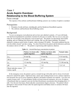
9
A 10 Year Retrospective Review of Digoxin Immune Fab Use in Digoxin Exposures
Mario Listiawan1, Grant Lackey2, Judith Alsop2, Timothy Albertson3, John Inciardi1, William
Ofstad1
1
California Northstate University College of Pharmacy, Rancho Cordova CA USA
2
UCSF California Poison Control Center - Sacramento Division, Sacramento CA USA
3
University of California Davis, School of Medicine, Sacramento CA USA
Background: Digoxin (DIG) is a cardiac glycoside indicated for the treatment of both systolic
heart failure and atrial fibrillation. Although the use of DIG has been declining in the past 10
years, reported DIG overdose cases reported to the Statewide Poison System have remained
stable. An analysis of the appropriate use of digoxin immune FAB (DIF) was initiated.
Methods: A 10-year retrospective poison center data study was completed on all cases of DIG
exposures for a period of January 1, 2001 to December 31, 2011. IRB approval was obtained and
cases were blinded prior to analysis. Inclusion criteria were DIG as a single ingestant and cases
followed to a known outcome. The parameters used in the case analysis were DIG as a single
substance, age, sex, year of reported cases, serum DIG level, serum potassium, serum
creatinine, FDA indications and dose for DIF, and patient outcome.
Results: A total of 798 cases of DIG ingestion without coingestants were identified. Of the 798
exposures, 543 (68%) were female, and 258 (32%) were male with a mean age of 72 years old
(range 4 days – 99 yo, SD 20 yrs). The mean DIG level reported was 4.3 ng/mL (range 0.1 –
37ng/mL, SD 3.0 ng/mL). The mean serum potassium reported was 5.1 mEq/L (range 2.3 – 8.7
mEq/L, SD 1.3 mEq/L) while the mean serum creatinine reported was 2.4 mg/dL (range 0.4 –
15.2 mg/dL, SD 1.8 mg/dL). Of 798 exposures, the use of DIF was reported in 292 cases (37%).
Of these 292 cases, 259 cases (89%) met the FDA indication for DIF use while 33 (11%) did not.
The remaining 506 exposures (63%) did not use DIF. Of the 506 cases, 293 (58%) met the
indication for DIF use while 213 (42%) did not. Of the 293 cases of patients who did not receive
DIF but met the FDA indication for use, 284 (97%) survived while 9 (3%) expired. Of the 213
(42%) cases that did not receive DIF and did not meet the FDA indication for use, 212 (99.5%)
survived while 1 (0.5%) died. The odds ratio calculated for the cases that did not use DIF with
clinical survival was 0.149 with a P value of 0.05 and CI of 0.007 – 1.154 and the number needed
to treat (NNT), was 38. 59 (20%) patients that used DIF were under dosed with 55 (93%)
survived and 4 (7%) expired. In 94 (32%) of the patients who were dosed correctly, 88 (94%)
survived and 6 (6%) expired (P value 0.92).
Conclusion: Healthcare practitioners should consult with their regional Poison Control Center
and closely follow the recommended FDA treatment guidelines regarding the use of digoxin
immune FAB.
Keywords: Antidote, Cardiac glycoside, Adverse drug event
10
Successful Management of Olanzapine-Induced Anticholinergic Agitation and Delirium with a
Continuous Intravenous Infusion of Physostigmine in a Pediatric Patient
Adebisi I Obafemi1, Stacey Hail1, Kurt Kleinschmidt1
1
UT Southwestern Medical Center, Dallas TX USA
Background.
Physostigmine effectively reverses anticholinergic delirium. However, continuous IV infusion of
physostigmine is rarely used due to concern for cardiotoxicity. We report the successful use of
continuous IV physostigmine in a 6-year-old male with anticholinergic delirium.
Case Report
A 6 y/o 30 kg male with ADHD ingested 15 to 20 5-mg olanzapine tablets. He was agitated and
was treated with lorazepam at a local hospital. His heart rate was 148, respiratory rate 32, blood
pressure 111/70, temperature 96.8ВєF, and O2 saturation of 98% on room air. His pupils were 5-6
mm and his skin was warm but not flushed. He had hypoactive bowel sounds. Blood chemistries
were normal. A 12-lead ECG showed sinus tachycardia with normal QRS and QT intervals.
The agitation worsened and did not respond to benzodiazepines. The patient was then given 2
doses of physostigmine 0.6 mg with reversal of the agitation; but the effect only lasted 45
minutes. The patient’s mother was disappointed that the physostigmine did not last longer and
asked for continuous physostigmine infusion.
The patient was transferred to the PICU and 6 mg of physostigmine was placed in 250 cc of D5W
and administered at 0.5 milligrams/hour. Overnight the patient became more agitated. But
instead of titrating the physostigmine drip, the PICU team discontinued it and started IV
dexmedetomidine 0.2 micrograms/kilogram/hour at 2100. The patient became over-sedated with
pinpoint pupils resulting in discontinuation of the dexmedetomidine at 0400. The patient again
became agitated and developed visual hallucinations. Three 1 mg boluses of physostigmine were
administered over 45 minutes and the physostigmine infusion was restarted at 1 milligram/hour
for 16.5 hours. He received a total of 19.5 milligrams of physostigmine with no return of
agitation or delirium. He was discharged home without further sequelae.
Discussion
There are limited data describing physostigmine to reverse anticholinergic delirium. There are
no studies involving its use in children. Pentel described asystole complicating physostigmine
use in TCA overdoses. Hence, physostigmine has been rarely used. Stern was the first to report a
case of a 20-year-old female with anticholinergic delirium successfully reversed with 77 mg of
physostigmine in a continuous infusion. Our patient received a total dose of 19.5 mg with
complete resolution of symptoms.
Conclusion
To our knowledge, this is the first case report using continuous infusion of physostigmine to
reverse anticholinergic delirium in a pediatric patient.
Keywords: anticholinergic, delirium, Physostigmine
11
Massive TCA ingestion treated successfully with a prolonged infusion of 20% lipid emulsion
Ravi Agarwala2, Syed Z Ahmed1, Timothy J Wiegand2
1
Northern Ontario School of Medicine, Thunder Bay ON Canada
2
University of Rochester Medical Center, Rochester NY USA
Background: Intravenous lipid emulsion (ILE) is increasingly used for severe intoxications of
lipophilic substances. However, data is limited on the optimal duration of treatment with ILE. We
present a case of prolonged use of ILE for a tricyclic antidepressant (TCA) overdose.
Case Report: A 42 year-old man was admitted hypothermic and hypotensive with QRS
widening to 200ms. He was also comatose, seizing, and suffered recurrent cardiac arrest,
ultimately developing vasopressor-dependent shock with persistent QRS widening despite
repeated administration of IV sodium bicarbonate and active rewarming. An initial [TCA]
returned at 7400 mmol/L. 20% ILE was administered as a 250mL bolus followed by a 24h
infusion at 100mL/h. Despite prompt normalization of the QRS interval and improvement in
blood pressure, the QRS widened following discontinuation of the infusion. ILE was resumed at
100mL/h with QRS normalization once again. Due to persistent toxicity and markedly elevated
TCA levels (8858 mmol/L day 2), ILE was continued at 18 mL/h, for 19 days, until levels were
no longer toxic. Whole bowel irrigation with polyethylene glycol was also performed due to
concern for persistent GI absorption after the levels increased on the second day. The patient
survived to discharge. Apart from a lipemic serum, there were no apparent complications from
the prolonged ILE infusion.
Case Discussion: We suspect that ongoing drug absorption due to prolonged ileus and delayed
elimination due to acute renal failure contributed to the persistent toxicity and elevated TCA
levels.
Conclusions: Prolonged ILE infusions may be useful and effective in massive TCA ingestions
with delayed absorption and/or clearance of the drug. In our patient, a prolonged ILE infusion (19
days) appeared to be safe and effective when used along with standard therapy for TCA toxicity.
Keywords: Lipid therapy, Tricyclic antidepressants, Intoxication
12
Benefits of Symptom-Triggered Phenobarbital Therapy in Refractory Delirium Tremens: A Case
Report
1
1
Debbie Gallo , Crystal Lui , Anastasia Shields1, Talia Pindyck1, Kelly Freer1, Brenda Benally1
1
Tsehootsooi Medical Center, Fort Defiance AZ USA
Background: Delirium tremens (DTs) can manifest days after the cessation of chronic alcohol
abuse. Symptoms such as change in cognition, disturbance of consciousness, and autonomic
instability are usually treated with benzodiazepines, however other modalities may be required.
We present a case of a patient with benzodiazepine refractory DTs who responded to symptomtriggered phenobarbital therapy.
Case Report: A 30 year-old male with a history of polysubstance abuse arrived to the Emergency
Department (ED). The extremely agitated patient reported using alcohol one day prior. The
patient is known to use alcohol chronically. The patient was sedated in the ED with lorazepam 4
mg IVP every 15 minutes as needed. Four hours later, he was transferred to the ICU. Within
hours, DTs quickly developed. Symptoms resolved with lorazepam 4 mg IVP every 15 minutes
(cumulative dose of 26 mg) and he was placed on a CIWA protocol. On hospital day 2, DTs
returned and intensified. A total of 46 mg of lorazepam was given over 4 hours with minimal
improvement; a lorazepam drip was then initiated. Despite all efforts to treat the patient with
high-dose lorazepam (> 40 mg in 2 hours), the DTs persisted. A phenobarbital protocol was
initiated. A total of 1170 mg of phenobarbital was given on hospital day 2. The patient remained
sedated with no DT's. On hospital day 3, a total of 1300 mg of phenobarbital was administered
for agitation. At this time, due to a back order on phenobarbital, supplies were exhausting and
could not be replenished. A CIWA-A protocol was used to supplement the remaining
phenobarbital.
When phenobarbital supplies exhausted, the patient was intubated and started on a propofol
infusion. With minimal symptom relief, approximately 38 mg of midazolam was given over 18
hours in addition with propofol, to control withdrawal symptoms. The remainder of the hospital
course was complicated by ventilator-associated pneumonia and ICU delirium.
Case Discussion: This patient responded well to symptom-triggered phenobarbital therapy. It was
not until after phenobarbital supplies were exhausted that mechanical ventilation with propofol
was necessary. The patient's DTs were uncontrolled with the change in therapy. Due to the
misfortune of exhausted hospital supplies, we were forced to intubate, begging the question;
would have mechanical ventilation been necessary if phenobarbital was continued?
Conclusion: Phenobarbital delayed the need for mechanical ventilation and reduced
benzodiazepine use. In this case, phenobarbital was the drug of choice for refractory DTs.
Keywords: Withdrawal, Alcohol, Delirium
13
Dexmedetomidine as an adjunct in patients undergoing treatment for ethanol withdrawal in the
critical care setting
2
Olga Amusina , Michele Zell-Kanter1, Amber Clouse2, Jerrold B Leikin3
1
Cook County Hospital-Stroger
2
NorthShore University HealthSystem-Highland Park Hospital
3
NorthShore University HealthSystem-OMEGA
BACKGROUND
The ideal management of ethanol withdrawal, a potential medical emergency, still eludes the
practitioner. The stigmata of ethanol withdrawal have not been fully comprehended and involve
the interplay of numerous neurotransmitters including Оі-aminobutyric acid (GABA), glutamic
acid, and norepinephrine. The benzodiazepines have been the mainstay in treating patients
withdrawing from ethanol because of their effect on GABA and glutamate receptors. The
adjunctive role of dexmedetomidine, a presynaptic О±2-agonist, has yet to be elucidated in
managing the hyper-adrenergic state of patients undergoing ethanol withdrawal.
METHODS
All adult patients in a suburban hospital critical care unit who exhibited the stigmata of ethanol
withdrawal and who received continuous intravenous infusions of dexmedetomidine as part of
their pharmacologic management were studied. Medical records were retrospectively examined
from July 2009 until November 2011 for the following parameters: demographic information,
outcome, complications, and the Richmond Agitation and Sedation scores (RAS).
RESULTS
Seventeen patients fulfilled the criteria and were studied. There were 16 males (age range 34-79
years, mean 55.4) and 1 female. Ethanol withdrawal was the primary diagnosis in 6 patients.
Dexmedetomidine was infused for an average of 67.2 hours (range 1.5-167 hours). One patient
received 5 separate infusions, 2 patients received 2 infusions. Fourteen patients had RAS scores
before and after dexmedetomidine therapy; all scores had improved with therapy. Transient
asymptomatic bradycardia occurred in 2 patients; 1 patient developed bradycardia and 5 beats of
ventricular tachycardia necessitating cessation of dexmedetomidine. One patient developed
supraventricular tachycardia requiring 3 doses of adenosine. Intubation was avoided in 5 patients;
dexmetedtomidine facilitated extubation in 1 patient.
CONCLUSION
Dexmedetomidine appears to be a worthwhile adjunct in the treatment of patients with ethanol
withdrawal in the critical care setting. Airway management was facilitated in 6 patients (35%).
Four of 17 patients developed either changes in pulse rate or rhythm while receiving
dexmedetomidine. Patients treated with dexmedetomidine for ethanol withdrawal should receive
cardiac monitoring.
Keywords: Ethanol, Withdrawal, dexmedetomidine
14
Physostigmine for Reversal of Iatrogenic Atropine Overdose From a Dental Procedure
Christine Worrall1, Jon Cole1, Benjamin Orozco1, Heather Ellsworth1
1
Hennepin County Medical Center, Minneapolis MN USA
Background: Atropine is described in the dental literature for use as an anti-sialogogue, with a
recommended dose range of 0.4-0.8mg in adults. Typically this is administered as 1-2 drops of
ophthalmic atropine, which has a concentration of 10mg/mL. This is the first reported case of
iatrogenic overdose of oral administration of ophthalmic atropine treated with physostigmine.
Overdose of opthalmic atropine is sparsely reported in the literature and in the only reported case
of dental administration resulting in toxicity went unrecognized, and resulted in a fatality.
Case: A 57-year-old male presented to the ED with acute delirium after he was seen by his dentist
at 7 a.m. and fitted for a dental prosthesis. A coworker brought him to the ED four hours later
with dysarthria, and concern for possible stroke. On arrival, his heart rate was 128 BPM, blood
pressure 148/94 mmHG, and temperature 37.7 C. His physical exam was notable for marked
mydriasis, dry mucus membranes, tachycardia, dysarthria, and a distended bladder. The dentist
was contacted, and reported that he had administered almost 2mL of ophthalmic atropine
(10mg/mL) sublingually to assist in drying the gingiva for moulding of the prosthesis. We gave
physostigmine 2.5mg IV with resolution of his anticholinergic syndrome. We observed him in
the ED, and at 6 hours after atropine administration, his anticholinergic symptoms recurred. He
was given a second dose of physostigmine 2.5mg, again with resolution of his symptoms. Due to
the efficacy of treatment with physostigmine, he did not require an extensive work-up for his
acute delirium. He was observed for 24 hours, with no further recurrences.
Case Discussion: Atropine is a tropane alkaloid extracted from several plants of the Solanaceae
family, and was used centuries ago to induce mydriasis for cosmetic effect. Currently, atropine is
used for therapeutic mydriasis, as well as for its cardiovascular effects. The ophthalmic
preparation has been adopted for use by dentists and oral surgeons to minimize salivation during
procedures. Atropine toxicity is characterized by anticholinergic crisis with delirium,
tachycardia, dry skin, and mydriasis. Physostigmine is a carbamate acetylcholinersterase inhibitor
effective at reversal of atropine induced anticholinergic syndrome. Repeat dosing is safe and
effective for recurrent symptoms.
Conclusion: Ophthalmic atropine drops are highly concentrated, and can result in symptomatic
overdose with relatively small volume administration. Anticholinergic delirium should be
considered in cases of acute altered mental status after dental procedures. This case highlights the
therapeutic and diagnostic utility of physostigmine.
Keywords: Iatrogenic, Atropine, Overdose
15
Successful treatment of severe diethylene glycol poisoning with fomepizole and hemodiaylsis
Joseph Turner1, Laura Tormoehlen1, Louise Kao1
1
Indiana University, Indianapolis IN USA
Background. Several instances of mass poisoning by diethylene glycol (DEG) due to medication
contamination have been described. However, there are few case reports of intentional ingestion
and even fewer reports of successful treatment. Case report. We report a case of a 43 year old
woman who ingested most of a 32-ounce can of "Prestone Super Heavy Duty High Temperature
Break Fluid" in a suicide attempt. The patient was found unresponsive and with agonal
respirations. She had three episodes of PEA arrest en route to our hospital. After initial
resuscitation she arrived at the emergency department unresponsive, hemodynamically unstable,
and severely acidotic. Her initial heart rate was 130 and systolic blood pressure was 65. Pupils
were fixed and dilated. Initial laboratory investigation was remarkable for pH of 6.95, HCO3 of
15, BE of -18, potassium of 2.7, and osmolal gap of 30. Aggressive treatment was undertaken
with 3.5 liters of normal saline, 100 mEq of sodium bicarbonate, and 1200 mg of fomepizole.
Emergent hemodialysis was initiated and after 6 hours of dialysis the acidosis had nearly
resolved, hemodynamic status was improved, and the osmolal gap had decreased to 12.
Ultimately, resuscitation and treatment were successful and the patient was discharged
neurologically intact 15 days after arrival. Case discussion. Prestone Super Heavy Duty Break
Fluid is a complex mixture of glycol ethers. Notably, it contains up to 8% diethylene glycol. DEG
is known to be extremely toxic, but most reports in the literature describe accidental
contamination. Cases of large-volume intentional ingestion are rare. It has been proposed that
such ingestions be treated similar to other toxic alcohols, mainly utilizing fomepizole and
hemodialysis. This is largely based on animal models, and to our knowledge there is only one
report of such treatment in humans. Our case illustrates successful treatment of a life-threatening
DEG ingestion using these treatment modalities. Conclusion. DEG is an extremely toxic
substance and ingestion can be life-threatening. Fomepizole and hemodialysis, along with
aggressive supportive care, may be an effective treatment.
Keywords: Diethylene glycol, Fomepizole, Ingestion
16
Treatment of severe toxic alcohol and glycol poisoning in the UK
Harry R Thanacoody1, Ian Weatherall1, Jeremy Davies2, Simon Thomas1
1
National Poisons Information Service(Newcastle), Newcastle-upon-Tyne UK
2
National Poisons Information Service(Cardiff), Cardiff UK
Background: This prospective study was undertaken to investigate the management of systemic
exposures to toxic alcohols and glycols reported to the UK National Poisons Information Service
(NPIS).
Methods: Details of exposures to products containing toxic alcohols and glycols reported to NPIS
were collected from 1 January 2010 to 31st December 2010 and cases of significant systemic
exposure were followed up to obtain information on antidote use and patient outcome.
Results: Of the 488 individual exposures to toxic alcohols and glycols resulting in 608 enquiries
to NPIS over the 12 month period, 182 met the criteria for case follow up. Antidote treatment was
provided to 99 patients. 36 patients received fomepizole alone, 49 ethanol alone, and 14 both
ethanol and fomepizole. Extracorporeal elimination by intermittent haemodialysis and/or
continuous haemodiafiltration was instituted in 33 patients- alone without antidotal treatment in
3, in conjunction with antidotes in 29 patients. Antidotal and/or extracorporeal treatment was
instituted in 36 of the 39 patients where the PSS was recorded as severe. Outcomes were
generally favourable regardless of treatment modality. However there were 4 cases resulting in
patient death (2 were treated with ethanol and 2 did not receive an antidote). 5 patients developed
renal failure (4 were treated with ethanol and 1 with fomepizole).
Adverse reactions to antidotes were reported in 10 patients: 8 receiving ethanol and 2 receiving
fomepizole. Adverse reactions reported as probably related to ethanol use were intoxication (4),
reduced conscious level (1), agitation (1) and withdrawal symptoms (1). Adverse reactions were
possibly related to fomepizole in 2 patients (shaking in 1 patient which recovered with supportive
treatment and angio-oedema which recovered following fomepizole discontinuation in 1
patient). Problems reported with use of ethanol during the study included difficulty in making the
infusion, calculating and adjusting infusion rate, difficulty in obtaining ethanol concentrations to
monitor treatment, sub-therapeutic ethanol concentrations, lack of availability or inadequate
stocks to complete full treatment. Problems reported with use of fomepizole included lack of
availability or inadequate stocks and lack of familiarity with its reconstitution and dosing
schedule.
Conclusions: Antidote and/or extracorporeal treatment for severe toxic alcohol or glycol
poisoning is required infrequently in the UK with around 100 reported cases per year. Ethanol
and fomepizole are used in similar numbers of patients without any differences in outcome.
Ethanol use may be associated with more frequent adverse reactions and practical difficulties
with its use.
Keywords: Ethylene glycol, Antidote, Hemodialysis
17
Successful treatment of massive ethylene glycol poisoning with fomepizole only
Anthony Burda2, Kamila Truitt1, Carol DesLauries2, Erin Pallasch2, Cindy Howard2
1
Southern Illinois University-Edwardsville School of Pharmacy, Edwardsville IL USA
2
Illinois Poison Center, IL
Background: Treatment of ethylene glycol (EG) poisoning consist of administration of an alcohol
dehydrogenase inhibitor such as fomepizole or ethanol and the possible addition of hemodialysis
(HD) to enhance elimination of the parent compound and metabolites. Several cases have been
published where patients exhibiting high blood levels of EG who presented prior to the
development of metabolic acidosis and nephrotoxicity were treated with fomepizole alone. We
present a case that is the highest EG level reported to date in a patient who was successfully
treated with fomepizole without HD. Case Report: A 43 y/o, 75 kg female presented to the ED 1
hour after ingestion of a ВЅ gallon of a 50:50 mixture of EG antifreeze and water along with 20
fluoxetine tablets and alcohol. Her physical exam was unremarkable other than appearing slightly
drowsy with slurred speech. Initial vitals signs were BP 137/88 mmHg, HR 92 beats/minute, RR
12 breaths/minute, PaO2 96% on room air. Her labs were: Na 136 mEq/L, K 3.7 mEq/L, Cl 99
mEq/L, CO2 22 mEq/L, BUN 2 mg/dL, Cr 0.7 mg/dL, glucose 110 mg/dl, osmolality 411
mOsm/kg, and ethanol 71 mg/dl. Her anion gap was 15 with an osmolar gap of 117 mOsm/kg. A
loading dose of 15mg/kg fomepizole was initiated followed by 4 doses of 10 mg/kg q 12 hours
then a repeat dose of 15 mg/kg. The patient's ethylene glycol level on arrival was 944 mg/dl.
Subsequent levels were 405 mg/dl, 94 mg/dl and non-detectable at 5.5, 30, and 64 hours post
ingestion respectively. The patient's acid/base status and renal function remained normal
throughout her hospital stay. The patient was admitted to the intensive care unit for the first 24
hours then transferred to a general medical floor. Case discussion: Fomepizole effectively blocks
the metabolism of EG to its toxic metabolites. Although treatment with the alcohol
dehydrogenase inhibitor, fomepizole prolongs the elimination half-life of EG, complete
elimination within three to five days is possible in the setting of normal renal function. It would
be impractical to treat asymptomatic patients with very high methanol levels with fomepizole
alone since the elimination half-life would extend to 40-60 hours thus requiring a protracted
length of stay in the hospital. Conclusion: We present a case of EG poisoning with a peak blood
level of 944 mg/dl treated with fomepizole alone. This case, together with other published reports
suggest that patients with massive EG poisoning who arrive early and demonstrate no evidence of
a wide anion gap metabolic acidosis or presence of toxic metabolites maybe safely treated with
fomepizole as sole therapy without HD.
Keywords: Ethylene glycol, Fomepizole, Antifreeze
18
Predictors Of Death And Renal Failure In Ethylene Glycol Poisoning
Derrick Lung1, James Brasiel1, Kent Olson1
1
University of California, San Francisco, San Francisco CA USA
Background: Despite the severity of ethylene glycol poisoning, little is known about what
factors influence survival and prolonged renal insufficiency. Our study aims to describe clinical
signs, times of door-to-antidote (D2A) administration, and times of door-to-dialysis (D2D)
associated with the outcomes of death and prolonged renal insufficiency.
Methods: This was a retrospective, observational California Poison Control System (CPCS)
study. Over a ten-year period (1999-2008), the CPCS database included 517 consultations for
ethylene glycol exposure. Of these, we studied 121 patients who had all of the following:
recorded serum level of ethylene glycol, arterial pH measurement, serum creatinine
measurement, and times of patient arrival, and antidote (ethanol and or fomepizole) and dialysis
initiation. We defined as "cases" 59 patients who died (9) or had prolonged renal insufficiency
(50). Prolonged renal insufficiency was defined as kidney injury in which dialysis was required
for greater than 3 days after hospital presentation. The remaining 62 patients were "controls." In
addition to descriptive statistics, to evaluate the association of D2A and D2D with dying or
prolonged renal insufficiency, we estimated odds ratios (ORs) and 95% confidence intervals
(CIs) using logistic regression models.
Results: Cases, compared to controls, were more likely to present comatose (57.6% compared to
12.9%), have seizures (40.7% compared to none), and require vasopressors (13.6% compared to
4.8%).
Cases presented with a higher mean ethylene glycol level of 240.4 mg/dL (SD 307), compared to
171.8 mg/dL (SD 168.3) for controls.
Cases presented with a lower mean initial arterial pH of 7.03 (SD 0.20), compared to 7.27 (SD
0.14) for controls. Cases had a higher peak creatinine within the first 24 hours of hospitalization
(2.4 mg/dL, SD 1.0), compared to controls (peak Cr 1.1 mg/dL, SD 0.5).
Compared to patients with a D2A within 3 hours, patients with a D2A greater than 3 hours had a
higher odds of dying or having prolonged renal insufficiency (OR = 3.34, 95% CI = 1.21 - 9.26).
Compared to patients with a D2D within 6 hours, patients with a D2D greater than 6 hours had a
lower odds of dying or having prolonged renal insufficiency (OR = 0.36, 95% CI = 0.15 - 0.87).
However, no patient who received dialysis within 3 hours of arrival died.
Conclusion: Compared to survivors without any prolonged toxic sequelae, patients poisoned
with ethylene glycol who died or had prolonged renal insufficiency were more likely to exhibit
severe clinical signs such as coma, seizures, hypotension, and acidosis. Earlier antidote
administration appears to lead to better outcomes. Benefit from earlier dialysis initiation could
not be demonstrated.
Keywords: Ethylene glycol, Fomepizole, Hemodialysis
19
CLINICAL AND ECONOMIC IMPACTS OF HEMODIALYSIS IN THE TREATMENT OF
PHENOBARBITAL OVERDOSE
Catherine Savard Rn, Marie-Pier Ferland Rn, Sophie Gosselin Md
BACKGROUND
In most instances, patients with phenobarbital (PB) overdose (OD) can be managed with
supportive care, activated charcoal and urine alkalinisation. Given the extended half-life of PB,
patients may be unconscious for several days even with aggressive symptomatic care.
Hemodialysis (HD) may be useful in such cases. We attempt to determine the clinical and
economic impacts of HD in severe PB OD.
CASE REPORT
A 29 year old female was found unresponsive. She was known to take quetiapine and
aripiprazole; no pills were missing. She was intubated on arrival. Causes of coma were
eliminated with time, standard laboratory tests, CT scan and lumbar puncture. Patient remained
comatose during the next 4 days. On day 4, a serum toxicological screen requested by our Poison
Centre revealed PB at 378 umol/L. HD was recommended until return to therapeutic PB level or
awakening. Dialysis duration was of 4h and the patient regained consciousness 2.5 hours after it
began. She was extubated 9h after HD ended and she recovered with no deficits.
RESULTS
Multiple serum samples were analyzed for PB prior to dialysis (5), during (4) and after dialysis
(4). Apparent half-lives were 71h pre HD, 7h during HD and 26h post HD. No clearance data was
available at the time of writing.
DISCUSSION
HD is generally recommended in cases where [PB] serum is ≥ 500 umol/L, and for patients who
are hemodynamically unstable. According to local data, the cost of 24h in ICU is 9300$
excluding expenses related to complications such as venous thromboembolism or ventilator
associated pneumonia which increase with intubation duration (Grap, 2012). One episode of HD
at our institution is 1332$. Complications of HD are minimal and mostly related to catheter
insertion (Randriamanantsoa, 2011). Considering the length of PB-associated coma following
OD, it seems that HD is a cost-effective, risk-minimizing solution for clinical and economical
perspectives. In our case it decreased apparent half-life by 10-fold while shortening intubation
and ICU stay. It could have been even more effective if started shortly after admission.
CONCLUSION
Our case shows that HD significantly decreases the plasma half-life of PB. One episode of HD
costs a fraction of an ICU stay while minimizing risk of complications due to prolonged coma.
Measuring plasma PB, early in coma of unclear origin would help clinicians weigh the risks and
benefits of HD early after admission. Evaluating the efficiency of other treatment modalities such
as multiple doses activated charcoal against HD is warranted.
Keywords: Hemodialysis, Coma, Intoxication
20
USEFULNESS OF LATE HAEMODIALYSIS IN PARAQUAT POISONING
Marie-Edith Ouellette2, Ami Grunbaum1, Marc Ghannoum3, Sophie Gosselin3
1
McGill University Health Centre - Medical Biochemistry, Montreal QC Canada
2
Centre Antipoison du Quebec, Quebec QC Canada
3
McGill University Health Centre- Emergency Department & Clinical Toxicology, Montreal QC
Canada
CASE PRESENTATION A 53-year-old man accidentally drank Gramoxone containing 37%
paraquat (PQ) 90 minutes before the call to Poison Control; he had already vomited 12 times.
Paramedics were on site but not interested in any recommendation. According to family, the
Gramoxone was an old product kept for professional use. Product was diluted by son and poured
into a drinking glass. The patient mistook the glass as his own and "accidentally drank and spat
out a mouthful". He was admitted to a community hospital. Poison Control recommended
activated charcoal, IV fluids, and antiemetics for persistent vomiting. Haemodialysis (HD) was
not initially recommended given the time elapsed since ingestion. At 18 hours (hrs.) postingestion (PI) the Poison Centre toxicologist recommended N-Acetylcysteine (N-AC)
administration, and transfer to tertiary care hospital for ICU admission. Patient arrived at the
tertiary care facility 23 hrs. PI. He began to deteriorate at about 45 hrs. PI. Treatment included IV
steroids for progressive upper airway oedema, and haemodialysis for renal failure. At 52 hrs.
patient was hypoxic and chest X-ray revealed pulmonary infiltrates. A cyclophosphamide IV
protocol was started in addition to N-AC and antioxidants. At 69 hrs. patient was intubated in the
OR. At 89 hrs. sepsis was suspected from aspiration pneumonitis. HD and Continuous
Venovenous Haemodiafiltration (CVVHDF) were administered in alternating cycles. At 115 hrs.
severe hypoxia and refractory hypotension developed. Comfort care measures were implemented.
Death was pronounced 117 hrs. PI. PQ levels in plasma, urine and dialysate were measured
before, during and after extracorporeal treatment (ECTR). The apparent half-life was 25 hrs.
Estimated PQ body burden on admission was 204 mg. PQ removal by HD was 13 mg in 6 hrs.
(HD#1) and 18 mg in 15 hrs. (HD#2). CVVHDF removed 4.2 mg in 26.4 hrs, whereas urine
excretion was 2.5 mg in 96hrs. Calculated clearances were 236 ml/min (HD#1), 250 ml/min
(HD#2), 43.8 ml/min (CVVHDF#1), 26.7 ml/min (CVVHDF #2) and 5.4 ml/min (urine).
DISCUSSION While PQ use and intoxications are more common in developing countries, it is
still used by the landscaping industry in North America. HD has been advocated only in the first
few hours after ingestion. However, analysis of our toxicokinetic data seems to indicate that
significant amounts of PQ are removed by ECTR even at a late stage, and that HD is more
effective than CRRT. Given that PQ ingestions are potentially lethal, that alternative treatments
have no definite benefit, and that HD has been associated with survival, we suggest that HD for
poison removal should be considered for all PQ ingestions, as it is unknown at which point in
time it becomes futile.
Keywords: Clearance, Hemodialysis, Paraquat
21
Hydroxocobalamin Hinders Hemodialysis
Samuel J Stellpflug1, Rebecca L Gardner1, Jenna M Leroy1, Michael D Zwank1
1
Regions Hospital Dept. of Emergency Medicine, St. Paul MN USA
Background: Hydroxocobalamin, an antidote used for cyanide exposure, has been FDA
approved since 2006. It functions by binding cyanide, forming cyanocobalamin, which is then
excreted in the urine. Its side effect profile is limited, however it can alter accuracy of laboratory
measurements and impact hemodialysis. We present a patient whose hemodialysis treatment was
delayed due to a false "blood leak" alarm following hydroxocobalamin administration.
Case Report: A 22 year-old male was running from police, and upon being caught he suffered a
respiratory arrest. He was transported by ambulance with respirations assisted by bag valve mask.
On arrival to the emergency department (ED) he had palpable pulses and Glasgow Coma Scale
(GCS) score of 3. ED providers promptly intubated the patient and initiated other resuscitative
measures. Vital signs included heart rate 70 beats/minute, blood pressure 76/50 mmHg, and O2
saturation 100%. Blood drawn from both peripheral IVs and a central line was bright red; venous
blood gas revealed pH <6.8, pCO2 47.5 mmHg, pO2 245 mmHg, bicarbonate 6.4 mmol/L, and
O2 saturation 97%. Lactate concentration was 16.4 mmol/L. With the constellation of low GCS,
hypotension, bright red venous blood with high O2 content, and lactic acidosis, cyanide
poisoning was considered and 5 gm hydroxocobalamin was given. Hemodialysis was initiated
based on persistent acidosis, hyperkalemia, and renal insufficiency. The dialysis machine
experienced an auto-shutdown due to a "blood leak" alarm. This alarm persisted and finally had
to be manually overridden, at which point dialysis commenced. The patient ultimately
deteriorated and expired 24 hours after initial presentation.
Discussion: A known effect of hydroxocobalamin administration is the discoloration of body
fluids. During dialysis it also discolors the dialysate, creating a problem because of an internal
safety measure in the dialysis machine. Normally blood passes on one side of a semi-permeable
membrane and dialysate on the other. Photosensors monitoring the dialysate are meant to alarm
and shut down the machine if red blood cells leak across the membrane. Hydroxocobalamin
changes the light refraction in the dialysate enough to set off the alarm and cause the auto-stop.
This "blood leak" phenomenon of hydroxocobalamin has been reported previously in a case
report; in that case it appears as though manual override was not done in time to perform dialysis.
Conclusion: Hydroxocobalamin administration can induce a false "blood leak" alarm preventing
hemodialysis until manually overriding the alarm. Providers caring for patients treated with
hydroxocobalamin should be aware of this as inability to perform dialysis may make for poor
outcomes.
Keywords: Cyanide, Hydroxocobalamin, dialysis
22
Physostigmine continuous infusion for the treatment anticholinergic toxicity in combined
diphenhydramine and bupropion overdose
1
Michelle Phillips , Nicole M Acquisto1, Rachel M Gorodetsky2, Timothy J Wiegand1
1
University of Rochester Medical Center, Rochester NY USA
2
D'Youville College School of Pharmacy, Buffalo NY USA
Background: Once a popular and widely used antidote for the treatment of anticholinergic
toxicity, the publication of case reports reporting an association of physostigmine use with
asystole in severe tricyclic antidepressant poisoning resulted in a paradigm shift and dramatic
decrease in physostigmine use beginning in the early 1980s. Recent literature reviews and cohort
studies have demonstrated that physostigmine, either administered alone or with
benzodiazepines, is a safe and effective antidote for the treatment of anticholinergic toxicity.
Despite this, there is minimal information in the literature regarding the use of physostigmine
administered as a continuous intravenous infusion for persistent anticholinergic toxicity. We
present a case of continuous physostigmine infusion, used with adjunctive GABAergic
medications, for the treatment of hyperthermia and agitation in a combined bupropion and
diphenhydramine overdose.
Case Report: A 13 year-old female presented with severe agitation and hyperthermia after
ingesting a large amount of diphenhydramine and bupropion. Despite incremental and increasing
doses of lorazepam, the patient remained febrile (up to 39 C via foley catheter). In conjunction
with additional doses of benzodiazepines, 2 mg of physostigmine was administered over 10
minutes. Coinciding nearly with the end of the infusion, along with the formation of sweat, there
was marked improvement in the patient's temperature and a decrease in agitation. Two additional
doses of physostigmine 2 mg were administered for recurrence of symptoms. Ultimately, a
continuous infusion of physostigmine at 2-4 mg/hour was required for a period of 6.5 hours to
maintain control of her agitation and attenuation of hyperthermia. The physostigmine was
discontinued when the patient was able to maintain her ability to sweat and her agitation
remained under control, albeit with high-doses of adjunctive benzodiazepines. The patient did not
experience vomiting, seizures, bradycardia or conduction block.
Case Discussion: A combination of sympathomimetic and anticholinergic poisonings may be
particularly problematic due to the impairment of evaporative heat loss in the setting of an
agitated delirium. Severe anticholinergic symptoms may not respond well to benzodiazepines.
Conclusion: Physostigmine administration allowed for evaporative heat loss to occur in our
patient. Due to the severe and persistent toxicity, along with the relatively short half-life of
physostigmine, a continuous infusion was required and used safely.
Keywords: Physostigmine, Anticholinergic, Infusion
23
Lipid Rescue for Tricyclic Antidepressant Toxicity: A Case Report
Jeffrey L Bargeon2, Erin Brennan1, Lydia Baltarowich2, Bram Dolcourt2
1
Sinai Grace Hospital DMC, Detroit MI USA
2
Childrens Hospital of Michigion Regional Poison Control Center, Detroit MI
Background: Intravenous fat emulsion (IFE) therapy is accepted therapy for local anesthetic
toxicity. Its use has expanded to other lipophilic drug poisonings, including calcium channel
blockers, beta adrenergic antagonists, antidepressants and neuroleptics. We report the case of an
intentional amitriptyline overdose with cardiovascular collapse, which was reversed following the
administration of intravenous fat emulsion.
Case Report: A 54 year old man was found unresponsive following a 2.25 gram amitriptyline
ingestion of fifteen 150mg tablets. Upon admission, He became hypotensive with a wide complex
tachycardia at 106 bpm, and having a QRS of 166ms, and a QTc of 566ms. Resuscitation
consisted of 4L of IV normal saline, Na bicarbonate, norepinephrine and phenylephrine infusions.
The patient remained hypotensive with prolonged QRS and QTc following resusitation. A 100 ml
bolus of 20% IFE was given followed by an infusion of 0.25ml/Kg/min over 60 min. Within 9
minutes of the IFE bolus, the QRS narrowed to 114 msec and vasopressor support was weaned
over the next several hours. The patient was discharged neurologically intact on hospital day 11.
Case Discussion: Our case describes severe cardiac instability following a massive overdose of
the lipophilic, antidepressant amitriptyline. The patient had significantly improved cardiac
function and hemodynamics following initiation of IFE therapy. The wide complex tachycardia,
previously refractory to sodium bicarbonate, narrowed within 9 minutes of the IFE infusion. Most
previous reports of tricyclic ingestions with hypotension and QRS widening were refractory to
vasopressors and sodium bicarbonate, have shown improvement in blood pressure with IFE
therapy. Many cases do not report significant narrowing of the QRS complex. Our patient had
refractory hypotension with prolongation of the QRS complex that was refractory to sodium
bicarbonate but narrowed after the IFE was administered. The patient also had improved blood
pressure with reduction in his vasopressor requirements following IFE.
Conclusions: IFE may be a valuable adjunct to treating patients with acute ingestions of
lipophilic drugs, such as tricyclic antidepressants, refractory to traditional therapy. Further
investigation is necessary to determine if these benefits of IFE therapy can demonstrate a
consistent response in other acute lipophilic drug poisonings.
Keywords: Lipid therapy, Overdose, Antidepressant
24
Intravenous Fat Emulsion (IFE) for Refractory Hypotension due to Venlafaxine Overdose
Christine Worrall1, Jon Cole1, Benjamin Orozco1, Stacey Bangh1
1
Hennepin County Medical Center, Minneapolis MN USA
Background: Venlafaxine is a serotonin-norepinehprine reuptake inhibitor, with a relatively welldescribed overdose syndrome, including seizures and serotonin-like syndrome. Cardiac
dysfunction, including tachyarrhythmias, Takot-subo morphology, and refractory hypotension are
also described. We report the first case of IFE for treatment of refractory hypotension due to
intentional venlafaxine overdose.
Case Report: A 43-year-old woman presented to an outside hospital with altered mental status
after ingestion of alcohol, venlafaxine and zolpidem. She was intubated for declining mental
status. On arrival to a teritiary ED, she had a heart rate of 108 BPM, BP of 82/45 mmHg,
temperature of 96 F, and was sedated with lorazepam. Serum ethanol was 0.05mg/dL. She was
given 1L of intravenous normal saline. Her blood pressure dropped to 51/27 mmHg. Naloxone 2
mg was given. A dopamine drip of 1mcg/kg/min was ordered but not available before 150 mL of
IFE 20% was administered. Within 5 minutes her BP improved to 120/47 mmHg, and she never
required pressors. A urine drug screen that utilizes liquid chromatography and mass spectrometry
was positive only for zolpidem and venlafaxine. Her BP remained normal throughout the rest of
her hospitalization, and she was extubated the next day and transferred to psychiatry.
Case Discussion: Venlafaxine toxicity has been well reported in the literature. A syndrome of
altered mental status, seizures, and hypertension is typical initially, followed by hypotension in
severe cases. Treatment with single-dose activated charcoal, whole bowel irrigation, and pressors
have been used with variable success in treating hypotension due to venlafaxine overdose. The
experimental logP hydrophobicity of venlafaxine is 2.8, which is similar to other compounds that
are known to be successfully treated with IFE in overdose. This contriuted to our decision to give
IFE, as well as the few contraindications to IFE. To our knowledge, this is the first reported case
of IFE for hypotension due to venlafaxine overdose.
Conclusion: IFE may be useful for treatment of hypotension due to venlafaxine overdose. This
case also supports the theoretical efficacy of IFE in patients with toxicity due to a compound with
similar hydrophobicity.
Keywords: Venlafaxine, Intravenous Fat Emulsion, Overdose
25
Novel use of Intralipid in the treatment of severe baclofen toxicity
Tomislav Jelic1, James Price, Milton Tenenbein1, Wesley Palatnick1
1
University of Manitoba, Winnipeg MB Canada
Backround
We present a case of severe baclofen toxicity, with hemodynamic instability, and decreased
level of consciousness that was treated successfully with intralipid.
Case Report
A middle-aged female was brought by paramedics to a rural hospital, after an overdose of an
unknown amount of baclofen tablets. The patient was initially stable, however her GCS declined
en route to hospital. Upon arrival to the ED, the patient had a GCS of 8, BP: 101/60 P 65 R 16 O2
saturation of 99% on 3L nasal prongs. Initial laboratory studies were unremarkable, in particular
there was no increased anion gap, and no salicylates or acetaminophen were detected. An ECG
demonstrated a sinus rhythm with first degree AV block, but normal QRS and QTc intervals.
Over the next hour, the patient deteriorated, with a declining GCS, and BP of 81/39. A fluid bolus
was initiated with no improvement. The patient was intubated for airway protection, and the next
BP was 59/32. Intralipid was administered as a 20% solution at 1.5cc/kg as a bolus followed by
0.25cc/kg/min infusion for 30 minutes. The BP rose to 89/57 within 30 minutes of the bolus
being given. The blood pressure continued to normalize with no pressor support. The heart rate
ranged between 44 to 58 bpm. Within 2 hours of intralipid administration, the patient's
hemodynamic status stabilized. She was subsequently extubated the following day and
discharged.
Discussions/Conclusion
This case demonstrates the first reported use of lipid emulsion therapy in the treatment of
hemodynamic instability due to a baclofen overdose. Baclofen is a GABA agonist, and is often
associated with neuromuscular dysfunction such as hypotonicity, decreased LOC, and coma like
states. Baclofen has been shown to cause hypotension (1), but it is rare (1,2). Lipid emulsion
therapy provided rapid resolution of the cardiovascular collapse, associated with the baclofen
ingestion in this patient.
References
1) Sein Anand J, Chodorowski Z et al. Selected clinical aspects of acute intoxication with
baclofen. Przegl Lek, 2005; 62(6): 462-4
2) Leung, NY, Whyte Im et al. Baclofen overdose: defining the spectrum of toxicity. Emergency
Medicine Australasia, 2006, February; 18(1):77-82
Keywords: Lipid therapy, Overdose, Baclofen
26
Immediate Chelation with DMSA for Acute Arsenic Overdose Prevents Chronic Toxicity from
Arsenic Poisoning
Michael G Holland1
1
SUNY Upstate Medical Univeresity, Syracuse NY USA
A 52 yo male with severe mental illness overdosed on trazodone, DEET, and an old arseniccontaining pesticide found in his garage. The ED initially was not concerned about the pesticide,
since the product was >40 yrs old, and therefore was felt to be non-toxic.
Patient presented with severe watery vomiting and diarrhea, but no blood was seen. VS were
stable with only slight tachycardia, which responded to fluid replacement. EKG showed no
abnormalities, no QTc prolongation. Vomiting had resolved once Poison Center was called for
treatment advice.
Medical Toxicology consult through the Poison Center recommended obtaining a stat whole
blood As and urine arsenic, then to start immediate chelation with DMSA due to signs and
symptoms consistent with acute inorganic As poisoning. The patient was immediately started on
DMSA 10 mg/kg q 8 hr (30mg/kg/d) for 5 days, then 10mg/kg q 12 hr (20mg/kg/d) pending lab
test results.
The initial stat whole blood Arsenic was 186 Вµg/L (nl= 2-23), and spot urine Arsenic was 1,936
mcg/L (nl <50mcg/L), all inorganic arsenic. Continued DMSA chelation was recommended until
urine and whole blood arsenic were normal.
The following lab results were obtained while chelation was ongoing:
Repeat whole blood arsenic on day 5 was 54 Вµg/dL (nl= 2-23)
24hr Urine arsenic from day 9 was 131 Вµg/dL (nl <50mcg/l)
24hr Urine arsenic on day 14 was 112 Вµg/L (nl <50mcg/l)
24hr Urine arsenic on day 29 was normal, < 50Вµg/L, and no inorganic arsenic was detected via
speciation
DMSA therapy was stopped after results came back normal, with the patient asymptomatic; he
developed no signs or symptoms of chronic arsenic posioning, with no alopecia, and no
peripheral neuropathy. He remained well after follow-up examination.
Keywords: Arsenic, Chelation, DMSA
27
Clinical Outcomes in Newer Anticonvulsant Overdose
Brandon K Wills1, Christine M Murphy1, Penny S Reynolds1, Eileen H Chu1, Kirk L Cumpston1,
Paul E Stromberg1, Rutherfoord S Rose1
1
Virginia Commonwealth University Health System, Richmond VA USA
Objectives: Clinicians have limited experience with treatment of overdose from newer
anticonvulsant medications. The aim of this study was to evaluate clinical effects of newer
anticonvulsants in overdose, determine if a relationship exists between dose and clinical effect,
and if there are drug(s) particularly more toxic in overdose.
Methods: This was a retrospective study using poison center data, evaluating clinical outcomes.
The Toxicallв„ў database from 1/1/2002 - 12/31/2011 was queried using key words: gabapentin,
lamotrigine, levetiracetam, tiagabine, topiramate, zonisamide, pregabalin, and oxcarbazine.
Polypharmacy overdose and children less than 15 years of age were excluded. Charts were
reviewed by two abstractors for pharmaceutical, reported dose, clinical outcome score, recorded
clinical signs/ symptoms, and vital signs.
Results: Out of 501 cases identified, 347 met final inclusion criteria. There was one death
reported from intentional overdose of topiramate. Proportion of overdoses exhibiting a clinical
effect by pharmaceutical are listed in table 1.
Data were modeled by cumulative logit ordinal regression on a 3-level ordinal scale response (0
no effect, 1 minor effect, ≥ 2 moderate to severe effect) using log-transformed standardized
doses. There was no significant effect of dose on severity of outcome (ОІ=0.12, p=0.23). However
risk of a more severe outcome score was significantly increased with tiagabine relative to other
drugs (ОІ=2.8, p=0.001).
Ranking of drug toxicity based on clinical signs and symptoms was performed by calculating an
information index H based on the proportional occurrence (1/0) of clinical outcome signs and
symptoms in nine categories of toxicity (seizures, AMS, pupillary response, neuromuscular, GI,
dermatological, cardiac, hypotension, metabolic). Lamotrigine ranked highest in terms of toxicity
(HT = 1.66) and number of interventions performed (HI= 1.17), and levetiracetam the lowest (HT
=0.98; HI = 0.88).
Conclusions: Overdose of newer anticonvulsants frequently results in altered mental status.
Seizures appear to be more common with tiagabine, lamotrigine, and oxcarbazepine. We could
not identify a dose-effect in these data which likely reflects the limitations of self-reported doses.
Risk of more severe outcome score was higher with tiagabine overdose while lamotrigine
overdose appears to result in more reported signs, symptoms, and interventions.
Seizure AMS Tachy/brady Hypotension GI
Gabapentin
2
41
11
16
6
Lamotrigine
5
45
16
9
36
Levetiracetam
0
60
0
7
7
Tiagabine
20
67
13
20
7
Topiramate
0
25
11
7
4
Pregabalin
0
35
22
4
13
Oxcarbazepine
4
53
31
5
24
Keywords: Anticonvulsant, Overdose, Poison center
28
Recommendation and Use of High-Dose Insulin and Intralipid following Beta and Calcium
Channel Blocker Toxicity
1
Michael A Darracq , Stephen L Thornton1, Han M Do2, Dennis Bok2, Richard F Clark1, F L
Cantrell3
1
University of California, San Diego, San Diego CA
2
Skaggs School of Pharmacy, University of California, San Diego, San Diego CA USA
3
California Poison Control System, San Diego Branch, San Diego CA USA
Background: Calcium channel (CCB) and beta-receptor (BB) blockers may cause significant
morbidity and mortality in overdose. Hyperinsulinemic euglycemia (HIE) and intravenous fat
emulsion (IFE) may be beneficial in treating toxicity from CCBs and BBs. Many poison control
centers (PCC) now recommend these modalities. However, health-care providers and may not
institute them despite PCC recommendations. We sought to determine how often HIE and IFE
are recommended by a statewide PCC in CCB and BB toxicity, how often those
recommendations are implemented, and if a faxable information sheet is associated with
improved adherence.
Methods: We performed a retrospective review of a statewide poison system's database for cases
of CCB and BB exposures from January 2005 – July 2011. Gender, age, drug type, co-ingestants,
interventions, and outcomes were recorded. HIE or IFE recommended by PCC, whether therapy
was implemented before or after recommendations, and whether a written protocol was faxed to
the treating physician was also abstracted. Exclusion criteria included an incomplete PCC record.
Results: There were 215 CCB and/or BB exposures identified during the study period that met
criteria for evaluation. Patients were predominantly female (64%) with an average age of 54
years. Death occurred in 25 (12%) cases. The PCC recommended HIE in 71 cases and it was
started in 1 case prior to PCC discussion. HIE was subsequently used in 30 of these cases after
PCC recommendation. IFE was recommended by the PCC in 30 cases. IFE was implemented 10
times. In no case, was IFE started prior to discussion with PCC. In 6 cases both HIE and IFE
were implemented, all after recommendation. A protocol for HIE was faxed to the treating
provider in 7 cases while a protocol for both HIE and IFE was faxed in 13 cases. HIE (8 cases) or
IFE (5 cases) was implemented in 13 cases following faxed protocol. There was no statistical
difference between HIE or IFE being implemented when a faxable protocol was available vs just
when PCC advised by phone. See Figure 1.
Conclusions: HIE and IFE is increasingly recommended by PCC for the treatment of CCB and
BB toxicity. An increased use by treating providers follows a similar temporal trend. The
presence of a faxable protocol was not associated with significant increased adherence to PCC
recommendations regarding the use of HIE or IFE.
Additional efforts to improve clinician education regarding HIE and IFE as well as implementing
improved faxable protocols may be required to increase the utilization of these potentially lifesaving antidotes.
Figure 1
2006 2007 2008 2009 2010
HIE recommended 8
14 5
17 27
HIE given
2
3
4
5
16
IFE recommended -IFE given
--
---
1
0
10
2
19
8
Keywords: Beta blocker, Calcium channel blocker, Insulin
29
Critical Care Management of Verapamil and Diltiazem Overdoses at a Single Center
Michael Levine1, Anne Michelle Ruha1, Steven C Curry1, Adam Bosak1, Angie Padilla-Jones1,
Rachel Levitan1
1
Banner Good Samaritan Medical Center, Phoenix AZ
Background: Overdose (OD) of the calcium channel blockers (CCB) verapamil and diltiazem is
associated with severe illness. Traditional management has used vasopressors (VASO), although
recently hyperinsulinemic euglycemia (HIE) has become popular. We describe a large series of
patients (pts) with CCB OD.
Methods: Retrospective review of all adult pts (age > 14) admitted to one toxicology service
over 22 yrs, for treatment of non-dihydropyridine CCB OD. Pts were identified via review of pt
encounter logs. Data were abstracted using standard methodology; analyses were mostly
descriptive, although medians (interquartile range (IQR)) were determined as appropriate.
Results: 59 verapamil or diltiazem ODs were identified. The mean (SD) age was 50 (+/- 16) yrs;
48% were men. 53% were verapamil ODs. Median (IQR) initial and max glucose were 150 (123189) and 219 (159-330) mg/dL, respectively. Median (IQR) initial and nadir K+ were 3.7 (3.34.1) and 3.2 (2.9-3.5) mEq/L, respectively. 41% presented with acute kidney injury (creatinine >
1.5 mg/dL). VASO were used in 61% pts; mean number used 2.1. VASO use was as follows (n
pts/total; mean max dose +/- SD; max dose): norepinephrine (24/59; 26 +/- 26 mcg/min; 100
mcg/min;), dopamine (22/59; 20 +/- 20 mcg/kg/min; 100 mcg/kg/min), epinephrine (16/59; 47
+/- 85 mcg/min; 325 mcg/min;), isoproterenol (12/59; 16 +/- 18 mcg/min; 60 mcg/min;),
phenylephrine (5/59; 282 +/- 296 mcg/min; 800 mcg/min); 9/56 received dobutamine. HIE (1
U/kg bolus, then 1 U/kg/hr) was used in 3 pts who were already on VASO. An intra-aortic
balloon pump was placed in 1 pt. 5 ischemic complications occurred in 4/59 pts: 3 pts had GI
bleeds (GIB), 2 of which were apparent prior to starting VASO. 1 pt who was slow to awaken
had a brain MRI which revealed mild ischemia, without clear evidence of infarction. Another pt
had ischemic bowel, which was suspected on admit (lactate 9.3 mmol/L with abdominal pain). Of
note, HIE was used in 1 pt with a GIB present on admit, as well as in the pt with possible stroke
who had GIB during therapy. 2 pts had in-hospital cardiac arrest and were resuscitated. 1 pt died
due to complications from pulmonary artery catheter placement. All pts with ischemic
complications were discharged to in-patient psychiatry, fully recovered. 4 pts were sent to shortterm rehab, including 1 with in-hospital cardiac arrest, due to deconditioning, but all were
neurologically intact.
Conclusions: In this case series of 59 non-dihydropyridine CCB ODs, hypotension was common,
and often managed with multiple high-dose VASO, without HIE. Ischemia was uncommon, and
death occurred in only 1 pt due to a procedural complication. VASO use following CCB OD was
associated with good outcome in this series
Keywords: Calcium channel blocker, Insulin, vasopressor
30
Effect of Acetaminophen Overdose on Prothrombin Time
Michael Levine1, Ayrn O'Connor1, Angie Padilla-Jones1, Michael D Menchine2
1
Banner Good Samaritan Medical Center, Phoenix AZ
2
University of Southern California, Los Angeles CA USA
Death/OLT
Initial elevated PT
Initial elevated AST
Liver injury
Initial elevated PT
Initial elevated AST
Test characteristics for development of d/OLT
Sensitivity
Specificity
PPV
NPV
100 (75.3-100)
100(75.3-100)
47.2 (41.2-53.2)
61.7 (55.9-67.3)
8 (4.3-13.3)
10.4 (5.7-17.3)
100 (97.3-100)
100(98-100)
91.7 (84.2-96.3)
62.8 (55.7-55.9)
54.3 (46.3-62.2)
94 (88.5-97.4)
91.7 (84.2-96.3)
82.6 (76.7-87.5)
71 (62.1-78.8)
95.5 (91.4-98.1)
BACKGROUND: APAP toxicity can result in transaminitis, coagulopathy, liver failure and
death. This study examined the test characteristics of a high initial PT and AST for hepatic injury,
death or liver transplant. METHODS: We conducted a chart review of patients ≥ 14 years old
with known APAP overdose, treated with NAC between 1 Jan 2006 and 31 Dec 2010 at a tertiary
care referral center. Inclusion criteria were APAP overdose established by history of APAP
ingestion plus either an APAP level above the treatment line on the Rumack-Matthew nomogram
or elevated transaminases in cases of suspected delayed presentation. Exclusion criteria were
concurrent use of warfarin, history of hepatitis with previously documented transaminitis,or CK >
1000 IU/L. Standardized data abstraction methods were utilized. We defined: high PT as a PT
value > the upper limit of reference normal; high AST = > 50 IU/L; liver injury = AST > 1000
IU/L. Initial PT was performed at various labs so the specific reference ranges for normal were
used. RESULTS: 304 pts were included, mean age 31 (range 14-88), with 67% women. The
mean (SD) and median (IQR) initial AST was 1208 (+/- 3186) and 37 (22-217) IU/L, while the
mean (SD) and median (IQR) max AST was 2426 (+/- 4429) and 48.5 (25-2991) IU/L
respectively. The mean (SD) and median (IQR) initial PT was 17.4 (+/-13.2) and 13.8 (11.8-16.4)
sec. while the mean (SD) and median (IQR) max PT were 21.7 (+/- 17.7) and 15.4 (13.8-21) sec.
respectively. 13 pts died or had a liver transplant (D/OLT). Test characteristics are in table I. 8
pts had initially normal AST but high PT and developed hepatic injury. 8 others had initially
normal PT but high AST and developed hepatic injury. CONCLUSION: In this cohort of
patients treated for APAP toxicity, an elevated initial PT and AST each had similar test
characteristics for predicting the outcome of d/OLT. A high initial PT has little clinical relevance,
but a normal initial PT is associated with a very high NPV for predicting d/OLT. Neither a
normal initial AST nor an initial normal PT can definitively exclude the risk of developing
hepatic injury.
Important limitations are small numbers of pts with the endpoint of d/OLT, and all pts were
treated with NAC. Further study is needed to determine if an initial normal PT can be used in the
decision to treat.
Keywords: Acetaminophen (paracetamol), prothrombin time, coagulopathy
31
Occurrence of Thrombocytopenia Following Acetaminophen Toxicity
Michael Levine1, Ayrn D O'Connor1, Angie Padilla-Jones1, Adam Bosak1, Steven H Thomas2
1
Banner Good Samaritan Medical Center, Phoenix AZ
2
University of Oklahoma, School of Community Medicine, Tulsa OK USA
Background: Thrombocytopenia has been described following APAP ingestions. Theories
proposed, include antibody formation to APAP metabolites and altered concentrations of
thrombopoietin. Neither the prevalence of thrombocytopenia, nor the significance has been
investigated. This study examines the prevalence of thrombocytopenia in a consecutive case
series of adults admitted with confirmed APAP overdose.
Methods: A retrospective analysis of data from a prospectively-derived registry of adults (age >
14 yrs) treated for confirmed APAP ingestion over a 1-yr span was performed. Primary outcome
was to determine the occurrence of thrombocytopenia. Secondary outcome included correlation
of thrombocytopenia and AST >1000. Standardized data abstraction methods were used. Median
and interquartile range (IQR) were calculated for categorical variables. Fisher's exact test was
used to assess the correlation between thrombocytopenia (PLT < 100,000/mm3) and hepatic
injury (AST > 1000 IU/L).
Results: 92 consecutive pts were identified and included in the analysis. The median (IQR) age
was 28.5 (19-52) yrs. The majority were women (65%) with acute ingestions (85%). Platelet
(PLT) counts 21 hrs post-admission were available in 91.3% of cases. The median (IQR) initial
and nadir PLT counts were 285,000 (203,000-312,000)/mm3 and 198,000 (139,000239,000)/mm3. With nadir PLT count < 100,000/mm3 in 12/84 (14.3%) and < 50,000/mm3 in
9/84 (10.7%). The mean (+/- SD) and median (IQR) initial AST values were 1195 (+/- 3334)
IU/L and 38 (23-175) IU/L respectively. The mean (+/-SD) and median (IQR) max AST values
were 1822 (+/- 3945) IU/L and 42 (25-1391) IU/L. The initial AST was > 1000 IU/L in 19/92
(20.7%), while the max AST was > 1000 IU/L in 26/92 (28.3%). Among the 26 patients with a
maximal AST > 1000 IU/L, 16/26 (61.5%) had nadir platelet counts < 100,000/mm3 vs. 10/26
(38.4%) with a nadir platelet count > 100,000/mm3 (p<0.001). When excluding those patients
whose presenting AST was > 1000 IU/L, 2/58 (3.4%) of patients whose maximal AST was <
1000 IU/L had PLT < 100,000/mm3 vs.2/7 (28.5%) of patients whose maximal AST was > 1000
IU/L developed PLT < 100,000/mm3 (p=0.054). 1/75 (1.3%) of pts with a PLT nadir >
50,000/mm3 died, while 3/9 (33%) of pts with a nadir PLT < 50,000/mm3 died; p=0.003.
Conclusions:Thrombocytopenia following APAP overdose in this study population was
relatively common with 14.3% developing PLT<100,000/ mm3 and 10.7% with PLT
<50,000/mm3. Decrease in PLT below either 100,000 or 50,000/mm3 was strongly associated
with the likelihood of peak AST>1000 IU/L. Etiology and prognostic utility of
thrombocytopenia could not be evaluated by the current study future investigation is needed.
Keywords: Acetaminophen (paracetamol), thrombocytopenia, platelets
32
Pharmacokinetics of N-acetylcysteine (NAC) during CVVH, HD and in the absence of renal
replacement therapy
2
Stephanie H Hernandez , MaryAnn Howland1, Thomas D Schiano2, Robert S Hoffman3
1
St. John's University College of Pharmacy, Queens NY USA
2
Mount Sinai Medical Center, New York NY USA
3
New York University, Bellevue Hospital Center, New York NY USA
Background: APAP induced fulminant hepatic failure (FHF) is associated with kidney injury,
acidosis and severe fluid and electrolyte imbalances. As a result, varying renal replacement
therapies (RRT) are increasingly used in severe APAP poisonings. Although there is a potential
for NAC extraction with RRTs, data are lacking and it is unclear whether NAC dosing should be
altered during RRT. We report extensive data from the first patient in a multicenter
pharmacokinetic study of the extracorporeal removal of NAC by RRTs in patients with APAP
induced FHF.
Methods: A 29 year old, 74 kg woman presented with right upper quadrant pain and vomiting
after unintentional chronic supratherapeutic dosing of APAP and oxycodone-APAP. Initial lab
values: [APAP] 31 mcg/mL, AST 3972 U/L, ALT 10270 U/L, total bilirubin 2.7 mg/dL, INR 3.1,
creatinine 3.2 mg/dL. She was started on a standard IV NAC regimen and over a 48 h period we
studied the pharmacokinetics of NAC during a maintenance infusion of 6.25 mg/kg/hr IV.
Kinetics were determined while she was on CVVH, in the absence of RRT, and lastly during HD
(with minor ultrafiltration). At nine points in time (T1-T9) simultaneous urine, blood, dialysate
and/or ultrafiltrate specimens were collected for [NAC], and measured via HPLC. During RRTs
blood was collected from both access and return, to obtain average [NAC] (Cp). Standard
pharmacokinetic calculations were applied.
Results: NAC concentrations in the absence of RRT (T3-6) were consistent with published
literature on standard NAC kinetics and averaged 31.6 mg/L. CVVH effect was minimal, with
extraction ratios of 0.03 and 0.09 (T1 and T2 respectively). This produced fractional clearances
of 5% and 3%, and removed 24.2 mg and 15.1 mg hourly. However, HD clearance was
substantial, with extraction ratios of 0.30 (T8) and 0.34 (T9). This produced fractional clearance
61% (T8) and 85% (T9) and removed in the dialysate (plus ultrafiltrate) 279 mg and 395 mg of
the 462.5 mg hourly NAC dose, (T8 and T9). Relatively low HD clearance at T7 was likely due
to sampling prior to equilibration. By extrapolation, 24 hours of CVVH and 4 hours of HD would
have removed 470 mg and 1348 mg of NAC, respectively.
Conclusions: These results suggest that CVVH clearance of NAC is minimal and unlikely to
require altered dosing. In contrast, HD extracted a significant amount of NAC. If more than 50%
of the hourly NAC dose is removed in dialysate during HD, a substantial dose adjustment may be
indicated. These data are limited based on the number of samples and the use of a single patient
whose clinical condition was changing over time. Ongoing patient enrollment and data collection
will enable us to better define NAC dose adjustments during RRTs.
Keywords: N-acetylcysteine, Pharmacokinetics, Acetaminophen (paracetamol)
33
Refractory Hypoglycemia in an Acetaminophen, Acetyl-salicylic Acid, and Glyburide Overdose
joseph yanta1, Nathan Menke1, andrew king1, anthony pizon1
1
University of Pittsburgh Medical Center, pittsburgh PA USA
Background: Refractory hypoglycemia is a well-described consequence of overdoses involving
acetaminophen (APAP), aspirin (ASA), and sulfonylureas. We describe a 17 day course of
recurrent hypoglycemia after an overdose of APAP, ASA, and glyburide (GBU).
Case: A 57 year-old man was found unresponsive by EMS. Initial capillary blood glucose was 52
mg/dL. Despite 25 g of dextrose by EMS, glucose upon arrival to the ED was 42 mg/dL. Vital
signs were normal. GCS was 10T. The rest of his examination was normal. ABG: pH 7.36,
PaCO2 30 mmHg, and bicarbonate 16 mEq/L. Serum APAP and salicylate measured 396 ug/mL
and 61.4 mg/dL, respectively. The patient was initiated on 5% dextrose (D5W) with 150 mEq/L
NaHCO3 infusion, 21-hour N-acetylcysteine (NAC), and subcutaneous octreotide (OCT) (50 ug
q 8hr). INR peaked at 4.3 on hospital day (HD) 2 and AST peaked at 1098 IU/L on HD 4.
NaHCO3 infusion was discontinued on HD 4 and NAC on HD 5. Despite resolution of ASA
toxicity and evidence of adequate hepatic synthetic function, the patient continued to have
episodes of hypoglycemia requiring dextrose boluses. Therefore, his D5W infusion was increased
to D20W on HD 6. Serum sulfonylurea panels on hospital days 1, 6, 12, 13, and 17 screened
positive for GBU (>3 ng/mL). On HD 7 his insulin level was 26 uIU/L [normal high < 25 uIU/L]
and C-peptide was 10.6 ng/mL [0.8 – 4 ng/mL] during a documented episode of hypoglycemia.
On HD 10, OCT was converted to an infusion at 25mcg/hour. The treatment goal was a blood
glucose of 80-200. The following titration parameters were utilized: 1) if glucose is at goal,
increase OCT by 5mcg/hr and decrease D20W by 10mL/hr. 2) If glucose is <80 increase OCT by
5mcg/hr and increase D20W to previous hour's rate and restart titration in 1 hour. 3) If glucose is
>200, decrease OCT by 5mcg/hr and decrease D20W by 10mL/hr. Verapamil 80 mg PO TID was
initiated on HD 13 and continued until HD 19. His last episode of hypoglycemia was on HD 17.
He was discharged to home on HD 21.
Discussion: The etiology of refractory hypoglycemia in this case is multifactorial. Contributing
are acute liver injury, impaired metabolism of GBU, depletion of glycogen stores, and catabolism
of acute illness. We also postulate that verapamil aids in reducing GBU-related secretion of
insulin by antagonizing pancreatic L-type calcium channels.
Conclusion: We describe the longest duration of refractory hypoglycemia due to sulfonylurea
poisoning and the first reported uses of an octreotide infusion and oral verapamil in sulfonylurea
poisoning. We propose that higher doses of octreotide may be required for severe cases of
sulfonylurea poisoning. Adjunctive therapy with verapamil may also be considered.
Keywords: Hypoglycemic, Sulfonylurea, Aspirin
34
The Ascending Hegemony of IV N-Acetylcysteine for Acetaminophen Poisoning: Why?
Richard J Geller1, Donny Wyatt2
1
California Poison Control System at Chilren's Hospital Central California, Madera CA USA
2
University of the Pacific School of Pharmacy, Stockton CA USA
Background. After the introduction of AcetadoteВ®, in 2004, US use of IV N-acetylcysteine
(NAC) dramatically increased, and has surpassed that of the oral preparation as reflected in
NPDS data. We find this surprising. Methods. Reasons cited for the preferential use of IV NAC
include purportedly shorter therapy (along with lower cost), convenience and less emesis. We
examined, from 1985-2010, NPDS (US PCC) yearly totals of patients receiving IV v. PO NAC.
We informally surveyed local hospitals re: relative acquisition cost of Acetadote versus generic
oral NAC. We searched the medical literature to compare relative incidence (IV v PO) of serious
ADRs and deaths, and to assess relative efficacy. Finally, we examined if there really is a
difference in average duration of treatment. Results. The US NPDS logged, from 1985-2003,
158,332 patients who received NAC PO and 10,787 who received NAC IV, a ratio of 15:1.
By2010, IV NAC cases (16,961) now out-numbered PO NAC cases (8,362) by a ratio of 2:1.
Local hospitals report in 2012 an average acquisition cost per gram of $30.21 for Acetadote, and
$1.40 per gram for generic NAC 20% oral solution. While vomiting is more frequent with PO
versus IV NAC, serious ADRs are more common with IV administration. Multiple deaths have
been reported since 2004 caused by Acetadote dosing errors, which appear more consequentially
grave than PO dosing errors. Cumberland, Acetadote's manufacturer, has reported no data
indicating that Acetadote is safer than the formerly administered generic oral NAC formulation
through a Millipore filter. Both PO and IV NAC are of established efficacy. While it has been
suggested that IV NAC may be more efficacious when initiating treatment within 12 hours of
ingestion, and that PO 72 hour protocol more effective when initiating after 18 hours, these
differences are not firmly established and, if they exist, are probably small. It is conceptually
untrue that all IV NAC patients need only 21 hours of therapy, or that PO NAC patients need 72
hours of therapy. Current literature advocates that duration of NAC therapy be patient-tailored,
with endpoints of both no remaining acetaminophen and improving liver health. The California
Poison Control System (CPCS) makes no durational distinction between PO v IV NAC, and
stops either one when both endpoints are reached, as early as after only 20 hours of therapy.
Discussion/ Conclusion. When examining relative acquisition cost, frequency of severe ADRs
with IV NAC, comparable efficacy and comparable duration of treatment, we find no justification
for the preponderant preferential use of IV NAC. We suggest routinely employing a 20 hour oral
regimen, and reserving the use of IV NAC for patients unable to take the drug by mouth.
Keywords: Acetaminophen (paracetamol), Overdose, Antidote
35
Acetaminophen overdose in profound hypothermia
Amber Rollstin1, Steven A Seifert1
1
University of New Mexico Health Sciences Center, Albuquerque NM USA
Background: There are few reports of acetaminophen overdose in hypothermic patients and
fewer in patients with profound hypothermia. The metabolism of acetaminophen, the risk of
hepatotoxicity with delayed treatment, and the kinetics and possible dose adjustments of Nacetylcysteine therapy are not well-studied in this setting.
Case report: A 37-year old female was found unconscious outside during the winter. She was
transported by EMS to a tertiary care facility where she was noted to be profoundly hypothermic,
with a core rectal temperature of 17oC. Her initial serum acetaminophen concentration was 232
mcg/mL. Active rewarming was started immediately and in the absence of guidelines from the
manufacturer regarding the use of IV N-acetylcysteine (N-AC) in profound hypothermia, it was
initiated with the standard treatment protocol. The patient did not develop signs of hepatic or
renal injury or of N-acetylcysteine toxicity. She later confirmed that she had taken an overdose of
acetaminophen/diphenhydramine ("Tylenol PM") 18 hours prior to discovery.
Discussion: Despite the significant ingestion of acetaminophen and delayed presentation, the
patient did not develop any signs or symptoms of liver or kidney injury. Acetaminophen's
combination with diphenhydramine may have delayed systemic absorption initially and her
profound hypothermia likely slowed heptatic and renal metabolism once acetaminophen
concentrations had reached peak levels. Although there are no guidelines for its use in profound
hypothermia, N-acetylcysteine was administered in a standard dose during her rewarming period.
Despite a blood concentration of acetaminophen greater than 200 mcg/mL and a late
presentation, she did not develop hepatotoxicity or nephrotoxicity and there were no observable
adverse effects from IV NAC.
Conclusions: Profound hypothermia may be protective of hepatic and/or renal injury in
acetaminophen overdose by slowing its metabolism. IV N-acetylcysteine was given in a standard
dose to a profoundly hypothermic patient without apparent adverse effects and appeared to be
protective of acetaminophen-induced hepatic and renal toxicity during rewarming.
Keywords: Acetaminophen (paracetamol), Environmental, N-acetylcysteine
36
Safety of Non-Therapeutic Atomoxetine Ingestions-A National Poison Data System Study
Andrew A Monte1, Alessandro Ceschi2, Michael Bodmer3
1
University of Colorado Denver School of Medicine, Aurora CO USA
2
Swiss Toxicological Information Centre, Zurich Switzerland
3
University Hospital, Basel Switzerland
Background: The safety of medications used to treat attention deficit-hyperactivity disorder has
been questioned resulting in US Food & Drug Administration "black box" warnings for increased
suicidality and cardiovascular events. The purpose of this study is to describe epidemiology and
clinical course of non-therapeutic atomoxetine exposures reported to the US National Poison
Database System (NPDS).
Methods: Retrospective database study of non-therapeutic single agent ingestions of atomoxetine
in children and adults reported to the NPDS between January 1, 2002 and December 31, 2010.
Adverse drug reactions (ADRs) were excluded from the analysis. Severity and clinical effects
were recorded by poison center providers using a standardized data collection system; effects
noted only in case notes were not captured.
Results: 20,032 atomoxetine exposures were reported during the study period, and 12,500 of
these were single agent, non-ADR exposures. The median age was 9 years (IQR 2, 14) and 7,454
were males (59.6 %). 8,933 (71.5%) were acute exposures, 3,325 (26.6%) acute-on-chronic, 166
(1.3%) chronic, and the remaining 76 (0.6%) cases were unknown chronicity. In 10,732 (86.2%)
cases, exposure was unintentional, in 1,082 (8.7%) were suicide attempts, and in 630 (5.1%)
abuse. The majority of cases were managed on-site [n=8,619 (69.0%)], and 3,667 (29.3 %) were
referred to/managed in health care facilities. Acute on chronic ingestion was associated with an
increased risk of a suicidal reason for exposure in comparison to acute ingestions (OR 1.44, 95%
CI 1.26, 1.65). The most common clinical effects were drowsiness/lethargy (709 cases; 5.7%),
tachycardia (555; 4.4%), and nausea (388; 3.1%). Exact dose information was available for 2,749
cases (22.0%). The maximum ingested dose was 48,000 mg in an 18 year male in a suicide
attempt resulting in a moderate outcome. Major toxicity was observed in 21 cases; seizures in 9
(42.9%), tachycardia in 8 (38.1%), coma in 6 (28.6%), and ventricular dysrhythmia in one case
(4.8%). There were no deaths.
Conclusions: Non-therapeutic atomoxetine ingestions reported to US poison centers are largely
safe and significant overdoses were tolerated with few clinical effects, however, seizures were
rarely observed. Major cardiovascular events were exceedingly rare in our study.
Keywords: National Poison Data System, Overdose, Adolescent
37
Massive Ibuprofen ingestion with refractory Hypotension, Hyperlactemia, and CNS Depression.
Adebisi I Obafemi1, Kapil Sharma2
1
UTSouthwestern Medical center, Dallas Texas, Dallas TX USA
2
UT Southwestern medical center, Dallas TX USA
Ibuprofen, a propionic acid NSAID is one of the most commonly used non prescribed
medications in the world. Although the pathophysiology of the common complications of
NSAIDS such as GI and renal effects are clearly defined, the data on the cardiovascular risk and
toxicity remain controversial. We report on the case of a 16 year old girl with refractory
hypotension and hyperlactemia following Ibuprofen overdose.
Case Report.
A previously well 16 year old girl ingested 300 to 350 tablets of 200mg (60-70 grams) Ibuprofen
in a suicide attempt following an argument with her parents. She was found unconscious with
two empty bottles of Ibuprofen. She vomited several times on the way to the hospital without
hematemesis.
She arrived at the emergency department (ED) 6 hours post ingestion with GCS of 11. BP 68/27
HR 110, RR, 18, Temp 36.6 C. She denied abdominal pain or diarrhea and no fever, cough or
shortness of breath. Initial venous blood gas was PH 7.27/PC02 46.8/PO2 62/BE -5. She was
bolused with 2 liters of fluid without improvement in her blood pressure and subsequently started
on dopamine drip at 5mcg/hr. She was then transferred to PICU on cardiac monitor.
Other laboratory results showed, Na 146 Meq/L, K 3.8 Meq/L, HCO3 18Meq/L, Chloride 102
Meq/L, Anion gap 26. Comprehensive Urine drug screen was only positive for Ibuprofen and her
serum Ibuprofen level 7-8 hrs post ingestion was >250 mcg/ml (normal 10-50 meq/L); serum
lactate 2.7, TSH 1.44. Acetaminophen, salicylate, creatinine and ethanol levels as well as
coagulation profiles were normal. Her EKG showed sinus rhythm with a QTc of 455. Chest Xray was normal.
Her blood pressure improved initially to 107/50 and then dropped down to 82/35, necessitating
increased dopamine dose to 10 mcg/hr to maintain MAP at >60. She subsequently sustained a
stable blood pressure and dopamine drip was turned off 18 hours from admission. She was then
transferred to psychiatry for inpatient treatment two days post admission.
Discussion.
While Ibuprofen overdose usually present with GI, renal and electrolyte disturbances, massive
ingestion can present with refractory hypotension, QT prolongation, anion gap metabolic
acidosis and neurological effects. Our patient’s serum Ibuprofen level was reported as greater
than 250 mcg/ml the highest level this particular laboratory could determine. The exact
mechanism of the cardiovascular toxicity of Ibuprofen is not known but there is no evidence of
direct cardiotoxic effect.
Conclusion.
Massive Ibuprofen overdose can present with refractory hypotension and neurologic effects, but
the exact mechanism of the cardiotoxic effect is not known.
Keywords: Ibuprofen, refractory hypotension, metabolic acidosis
38
Long term cohort of patients with Lamotrigine Toxicity
Philip W Moore1, David A Haggerty1, Amanda Cresswell1, Louis Cantilena1, J Ward Donovan1,
Keith K Burkhart2
1
PinnacleHealth Toxicology Center, Harrisburg PA USA
2
U.S. Food and Drug Administration, MD USA
Background: Lamotrigine (LTG) is a phenyltriazine anti-epileptic drug which inhibits voltagegated sodium channels, decreasing presynaptic release of glutamate and aspartate. Additionally,
a concentration dependent inhibition of serotonin uptake by lamotrigine has been reported in rats.
Reports of toxicity from the literature are limited to case reports and primarily involve
coingestants.
Case Series: Fifty-three patients were identified between 2003 and 2012 with possible LTG
toxicity at a single toxicology center. Forty-four patients (83%) had coingestant toxicity with one
fatality. Nine patients had LTG as the only significant ingestant. Two of the 9 patients had
seizures during transport or while in the emergency department. Six of the 9 patients
demonstrated myoclonic-type movement disorders. At presentation, 5 of the 9 patients were
hypertensive. Four patients were tachycardic with rates greater than 100 bpm. Four of the
patients were tachypneic with rates greater than 24 per minute. Mental status was assessed using
the Riker sedation scale, and scores ranged from 1-7 with an average of 3.6. Four of the 9
patients were hyperreflexic and 3 had inducible clonus. Six of the 9 patients had an
electrocardiogram, and two of these patients had QRS greater than 100 and the average QRS
duration was 101 ms. Five of the patients had QTc>460 and the average QTc was 487.
Therapeutic blood levels of LTG are less than 12 mg/L, and 6 of the 9 patients had LTG blood
levels with confirmed toxicity (>12.0 mg/L). Three patients did not have documented LTG blood
levels. Initial LTG levels ranged from 17.1 to 90 mg/L (mean 33.8) with half-lives varying from
6.9 to 51.7 hours. The patients who experienced seizures had initial LTG levels of 26 mg/L and
90 mg/L (both had seizure disorders). The patients with myoclonic movement disorders had
LTG levels from 17.8 to 90 mg/L.
Discussion: This is the largest cohort of LTG ingestions reported to date. The degree of LTG
toxicity was variable, likely reflecting dose-dependency. Mental status ranged from coma to
severe agitation, but usually was described as lethargic with intermittent myoclonic type
restlessness. Patients generally were hypertensive, tachycardic, and tachypneic. Some patients
with LTG toxicity have seizures, clonus and myoclonic movement disorders as well as QRS and
QTc prolongation.
Conclusions:
1) LTG toxicity was associated with altered mental status
2) LTG toxicity was associated with increased vital signs and neuromuscular irritability
3) LTG toxicity was associated with prolonged QRS and QTc
4) These clinical presentations are consistent with serotonin toxicity and sodium channel
blockade, both pharmacologic effects of LTG.
Keywords: Lamotrigine, Pharmacokinetics, Toxicity
39
Predicting mortality in severe aluminium phosphide poisoning.
Ashish Bhalla, Arvind Sharma, Kirandeep Gill, Surjit Singh
Background
Aluminium Phosphide poisoning is an important cause of morbidity and mortality in India. In this
study , tried to develop a model for predicting mortality in patients with severe aluminium
phosphide poisoning.
Methods
A prospective study including all patients with aluminium phosphide poisoning was carried out in
the medical emergency attached to Nehru Hospital at our centre from June 2010 till December
2011. At admission, heart rate, blood pressure, blood sugar, electrocardiogram , blood gases, liver
functions, renal functions were recorded and all the patients was followed up till discharge or
death. Primary outcome was mortality. Parametric variables like blood pressure, pulse rate, heart
rate was analyzed by student's t test. For categorical data like mortality , relative risk of death was
calculated with 95% confidence intervals ,with chi square test. Further correlation was carried out
by univariate, multivariate and multiple logistic regression analysis.
Results:
A total of 105 patients were enrolled during the study period. majority patients were males (62%)
in the age group 15-35 years (78%). The mean dose consumed was 3.6 grams. 51% patients died
in the study group. Important parameters, after multivariate analysis, correlating with mortality
were Blood pH<7.2, Systolic blood pressure <90, blood sugar <50 or >200 mg/dl and Glasgow
coma scale <4. On multivariate regression, none of these parameters were independently
associated with mortality. The odds ratio was 12.614 for pH<7.2, 17.600 for SBP <90mmHg,
13.23 for Blood sugar <50 or >200 and 18.621 for GCS <4. A score of 2 each was assigned to
SBP <90 and pH <7.2 and score of 1 was assigned to Blood sugar <50 or >200 and GCS <4. The
maximum score was 6. When applied to the study group it was noticed that 96.4% with score of 0
and 69% with score 2 survived, while 70% with score of 4 and 100% with a score of 6 died.
Conclusion:
pH<7.2, Systolic blood pressure <90, blood sugar <50 or >200 mg/dl and Glasgow coma scale <4
are important predictors of poor outcome in patients with severe aluminium phosphide poisoning
and a scoring system based on these parameters may be a useful adjunct in assessing severity of
poisoning.
Keywords: Pesticide, Shock, Medical toxicology
40
Salicylate Induced Proximal Tubular Dysfunction.
Adebisi I Obafemi2, Sing-Yi Feng1
1
UT Southwestern Medical Center, Dallas TX USA
2
UT Southwestern Medical Center, Dallas TX USA
Background.
Fanconi Syndrome (FS) is characterized by multiple proximal tubular transport defects resulting
in aminoaciduria and glycosuria despite euglycemia, hypouricemia and phosphaturia. The
absence of one or more manifestations defines partial FS. We report a case of a 14-year-old
female with salicylate toxicity who present with persistent glycosuria despite euglycemia.
Case Report.
A 14-year-old female ingested 9.75 g aspirin in a suicide gesture. She presented 14 hours postingestion with tinnitus, nausea, abdominal pain and hyperpnea.
Initial vital signs were: BP 121/65 mmHg; temperature 37.10c; HR 105 beats/minute; respirations
24 breaths/minute; weight: 53.1 kg; height: 161 cm. She had fresh lacerations on her wrists.
Initial laboratory results showed a serum salicylate 53 mg/dL; venous blood gas pH 7.46, CO2 29
mmHg. Serum electrolytes showed sodium (Na) 140 mEq/L, potassium (K) 3.4 mEq/L,
bicarbonate 20 mEq/L, blood urea nitrogen 14 mg/dl, creatinine 0.9 mg/dL, glucose 100 mg/dl.
A comprehensive urine drug screen was negative. Urinalysis showed glucose 301-1000 mg/dl,
3+ proteinuria, and negative ketones
The patient was treated with multi-dose activated charcoal and intravenous sodium bicarbonate.
Her serum salicylate was monitored and gradually declined to 1mg/dL. However, her glycosuria
and proteinuria persisted. She also had hypouricemia with serum uric acid 1.6 mg/dL. Urine
chemistry showed phosphorus 7.8 mg/dL; K 33 mEq/L; Na 102 mEq/L; uric acid 10.7 mg/dL.
She remained normoglycemic during her hospital stay. The patient was discharged home after 2
days with persistent glycosuria and proteinuria. Three weeks later, she was asymptomatic and
her glycosuria and proteinuria resolved.
Discussion.
Drug induced FS has been described but the literature about the effects of salicylate toxicity on
proximal tubular function is rare. A case series of 5 patients with reversible aminoaciduria
following salicylate toxicity was reported in 1961. The only case of reversible salicylate-induced
FS in a pediatric age group was reported in a 17-year-old female who ingested 12.5 gram of
aspirin. Our patient had reversible salicylate-induced proximal renal tubular dysfunction with
significant glycosuria (despite euglycemia), proteinuria and hypouricemia. The mechanism is
unknown but interference with proximal tubular basolateral Na-K-ATPactivity is a possible
mechanism.
Conclusion.
Clinicians should be aware that reversible proximal renal tubular dysfunction can be rarely
associated with salicylate toxicity. The mechanism is not known.
Keywords: Salicylate, Glycosuria, renal tubular dysfunction
41
Takotsubo Cardiomyopathy Associated with Opiate and Benzodiazepine Overdose
Jeremiah Ray1, Benjamin Williams1, Martin Caravati1
1
University of Utah, Salt Lake City UT USA
Background: Takotsubo cardiomyopathy ("broken heart syndrome") is a clinical syndrome
defined by left ventricular dysfunction, elevated serum troponin and ischemic patterns on
electrocardiogram but with patent coronary arteries. This syndrome was first described by
Japanese investigators in 1991 and is classically seen after severe emotional stress. We report a
case associated with drug overdose.
Case Report: A 34 year old woman with past medical history of depression and multiple suicide
attempts was found unresponsive with empty pill bottles (zolpidem 10mg #60, oxycodone 15mg
#90 and lorazepam 1mg #120). On the day prior to presentation, she was noted to have normal
mental status. In the ED, the patient had temperature 99.0 F, heart rate 146 bpm, blood pressure
159/100 mmHg, and oxygen saturation 94% on room air. Two doses of 0.4mg naloxone were
given without effect. Physical examination revealed an unresponsive woman without evidence of
trauma. Pupils were 3mm and sluggishly reactive to light bilaterally. The remainder of the exam
was significant for the presence of 11 fentanyl patches placed dermally. Her Glascow Coma
Scale was 7 and she was intubated. Laboratory findings showed a Troponin-I of 6.53 ng/mL that
peaked on day 2 at 15.33 ng/ml. Urine toxicology was positive for benzodiazepines and negative
for other drugs of abuse, including cocaine and stimulant amines. A 12-lead electrocardiogram
showed T-wave inversion in leads V1-V3. An echocardiogram revealed severe apical left
ventricular hypokinesis and an ejection fraction of 10-20%, consistent with Takotsubo
cardiomyopathy. The patient recovered without event and required only supportive management.
Follow up echocardiogram 6 days post-ingestion showed improvement of left ventricular
function with an ejection fraction of 50%.
Discussion: Drug related Takotsubo has been rarely associated with sympathomimetics like
epinephrine, vasopressors, amphetamine salts, nasal decongestants and with withdrawal from
opiates or benzodiazepines. These scenarios are consistent with the presumed pathophysiology of
catecholamine induced myocyte toxicity. We present a unique case of Takotsubo cardiomyopathy
that was associated with acute opiate and benzodiazepine overdose, which is not explained by the
commonly supported mechanism of sympathetic nervous system induced myocyte toxity.
Conclusion: This patient had Takotsubo cardiomyopathy associated with severe opiate and
benzodiazepine overdose. In the overdose setting, there is potential for emotionally induced
sympathetic nervous system up-regulation, thus the provider should be wary of a "broken heart".
Keywords: Overdose, Cardiac toxicity, Benzodiazepine
42
A Massive Overdose of Dalfampridine
Laura Fil1, Steven Sattler1, Stephen Havasy1
1
Good Samaritan Hospital Medical Center, West Islip NY USA
Background
Currently, about 400,000 Americans are living with multiple sclerosis (MS). MS is an
immune mediated inflammatory disease that attacks myelinated axons in the central nervous
system. One of the newer medications on the market is Dalfampridine or 4-aminopyridine (4AP). AmpyraВ® was approved by the FDA In January 2010 for treatment of patients with MS.
Case Report
Our patient was a 34 year old male with a past history of MS, who was brought to the
Emergency Department after being found unresponsive with bottles of valacyclovir, temazepam
and dalfampridine at his side. The patient had no other pertinent past medical history. His
current medications were valacyclovir, temazepam, dalfampridine (4-AP) and a tysabri IV
infusion. The patient had a temperature of 97.4, pulse 106, respiratory rate 24, BP 155/82. The
patient was tremulous, awake, but not responding to questioning or following simple commands.
He only responded to tactile stimulation by localizing to pain. The remainder of the physical
exam was only remarkable for tachycardia and diaphoresis with bedside blood glucose of
144. Fifteen minutes after arrival the patient lost consciousness and began to have a seizure. The
patient was administered 8 mg of lorazepam IV without control of his seizures. He was intubated
and placed on a lorazepam drip and started on a propofol drip and loaded with phenytoin and
phenobarbital (bolus and drip). The patient continued to have seizures. The patient’s lab values
were positive for a white blood count of 31.1 x10³/mcL. A urine toxicology screen was positive
only for benzodiazepines. The CT of the brain and the ECG were both normal. The 4-AP level
was 530 ng/mL (25 ng/mL and 49 ng/mL). The valacyclovir level was 7.5 mcg/mL (2.0-4.0
mcg/mL). The patient stopped seizing on hospital day 3 and was discharged 14 days later with
normal mental status and neurologic exam.
Discussion
4-AP is a potassium channel blocker that blocks the potassium ion current of repolarization
following an action potential. The blockade of the potassium channel at the level of the
membrane widens the action potential and enhances the release of acetylcholine, thus increasing
post-synaptic action potentials. Due to the ability of 4-AP to reverse synaptic blockade it has
been used as a treatment in many different neuromuscular disorders. The 4-AP level of our
patient was 530 ng/mL. The treatment of patients with 4-AP overdose is supportive. Animal data
suggests that patients with toxic levels of 4-AP may respond to phenytoin. Our patient did not
respond to phenytoin.
Conclusion
Our case illustrates the highest recorded level of 4-AP in an overdose. Our patient appeared to be
refractory to a combination of high doses of anticonvulsants and only improved after the drug
was metabolized.
Keywords: Dalfampridine, 4-Aminopyridine, Seizure
43
The Minoxidil Brothers: Severe Hypotension Following Intentional Ingestions of a 5% Topical
Preparation
1
Kelsey Moser , Heather Ellsworth1, Stacey Bangh1
1
Hennepin Regional Poison Center, Minneapolis MN USA
Background: Topical minoxidil (RogaineВ®) is an over-the-counter medication used to treat
male-pattern baldness. Minoxidil, which was initially marketed as an anti-hypertensive
medication, is a vasodilator that works by relaxing arteriolar smooth muscle, increasing
cutaneous blood flow, and stimulating resting hair follicles. This topical product became
available without a prescription in 1996 as a 2% solution, and a 5% solution was approved in
1997. Because these formulations are highly concentrated, ingestions may result in significant
toxicity. We present 2 cases of intentional minoxidil ingestions causing profound hypotension
requiring vasopressor support.
Case Report: Two brothers, ages 50 and 46, presented to the ED with nausea, vomiting, and
lightheadedness within 30 minutes of drinking unknown amounts of a 5% topical minoxidil
solution. Both had a history of alcohol abuse and drank the solution for its ethanol content. At
presentation, the elder brother had a blood pressure (BP) of 74/30 mm Hg and a heart rate (HR)
of 127 bpm, and the younger brother had a BP of 60/32 mm Hg and a HR of 113 bpm.
Intravenous fluid resuscitation was promptly initiated; due to persistent hypotension, dopamine
infusions were started and transfers to a tertiary care center arranged. The elder brother required
vasopressor support with dopamine overnight. His initial EKG was notable for ST depression in
the lateral leads and non-specific T wave changes. Cardiac biomarkers were undetectable. The
younger brother required dopamine and norepinephrine for blood pressure support for > 48 hrs.
His troponin peaked at 0.12 ng/mL; an echocardiogram revealed an EF of 65-70% and no gross
wall motion abnormalities. Both brothers developed alcohol withdrawal during hospitalization
but were discharged on hospital days 4 and 5 in stable condition with no permanent sequelae.
Discussion: Minoxidil is well absorbed and peak effects are typically seen within 1 hour of
ingestion. Despite a half-life of 4.2 hours, hypotensive effects may persist for more than 24 hours.
A 60 ml bottle of 5% solution contains 3 g of minoxidil; this is 30 times the maximum daily dose.
Both of our patients presented with significant and persistent hypotension and tachycardia.
Although both brothers were initially started on dopamine, a vasopressor with strong a1 agonist
activity, phenylephrine or norepinephrine would be preferred in a minoxidil overdose, as the use
of positive chronotropes may worsen tachycardia.
Conclusion: Healthcare providers should be aware that ingestions of topical minoxidil
preparations have the potential to cause severe, life-threatening hypotension requiring
vasopressor support and intensive cares.
Keywords: Minoxidil, Overdose, Vasodilator
44
Apparent Oral Methylphenidate Fatality with Postmortem Concentrations
Lee Cantrell1, Patricia Ogera2, Phyllis Mallett2, Iain McIntyre2
1
California Poison Control System, San Diego Division, CA
2
County of San Diego Medical Examiner's Office
Methylphenidate (MPD) is a widely prescribed stimulant used primarily for the treatment of
attention-deficit/hyperactivity disorder (ADHD). While typically used in the pediatric/adolescent
population, there has been an increase in therapeutic use among adults in recent years. Suicide
attempts involving methylphenidate ingestion have been well described; however deaths
attributed solely to MPD ingestion have not been reported. We report a case of a suspected fatal
MPD intoxication with corresponding postmortem liver, gastric, vitreous, peripheral and central
blood concentrations.
Case Report
A 62-year-old female was last known to be alive three days prior to a welfare check when she
was found dead on the floor near her bed with obvious rigor mortis. Her past medical history
included schizophrenia, depression, hypertension, fibromyalgia, knee osteoarthritis, chronic
lower back pain, Hepatitis C and mitral valve prolapse. She had a distant history of ethanol abuse
and her medications found at the scene included: acyclovir, atenolol, duloxetine, furosemide,
gabapentin, potassium chloride, levalbuterol tartrate, levothyroxine, MPD, morphine,
oxycodone/APAP, conjugated estrogen, prochlorperazine, ranitidine, and trazodone. The 10 mg
MPD IR tablets were dispensed 12 days prior to death, and 331 of 360 were missing. There were
no other significant medication discrepancies.
An autopsy revealed only mild coronary artery and aortic atherosclerosis with mild fatty changes
and moderate chronic inflammation in the liver. Histopathological studies were unremarkable.
Postmortem specimen analysis revealed MPD concentrations: peripheral blood 1.1 mg/L, central
blood 0.98 mg/L, liver 3.6 mg/kg, vitreous humor 0.80 mg/L and 1mg in stomach contents.
Diphenhydramine was confirmed at less than 0.10 mg/L in peripheral blood, no other drugs were
detected.
Discussion
Deaths related to excessive isolated MPD ingestion have not been previously reported. Reported
MPD-attributable deaths have all involved self-administration via parenteral or nasal insufflation
routes in an effort to abuse MPD rather than to commit suicide. As with our case, the exact
mechanism by which excessive exposure to MPD results or directly contributes to death is
unclear. Reports of postmortem MPD concentrations are rare and in our case we found markedly
elevated blood MPD concentrations, higher MPD liver concentrations than previously reported
and the first postmortem MPD vitreous humor concentration reported.
Conclusion
With no other identifiable cause of death, we report the first isolated MPD ingestion associated
with a fatality.
Keywords: Methylphenidate, Overdose, Death
45
Baclofen overdose resulting in prolonged alteration of mental status
Mohan Punja1, Kimberly A Clark1, Brent W Morgan1
1
Georgia Poison Center, Atlanta GA USA
Background: Baclofen is a gamma-aminobutyric acid (GABA) analogue that acts on GABAb
receptors in the central nervous system (CNS). There are reports of baclofen overdose causing
prolonged coma mimicking brain death with serum baclofen levels 2-6 times higher than the
upper limit of therapeutic. We report a patient with an intentional baclofen overdose who had
altered mental status for 7 days and a corresponding urine baclofen level.
Case Report: A 52 year old female with a history of hypertension and alcohol abuse was brought
to the hospital for altered mental status. On exam she was hypertensive, alternatively agitated and
somnolent, unable to follow commands, hypotonic and hyporeflexic. Evaluation including head
CT and serum testing for ethanol, salicylates and urine screening for drugs of abuse was negative.
EEG showed occasional frontal intermittent rhythmic delta activity (FIRDA) consistent with
cerebral dysfunction and possible toxic or metabolic encephalopathy. During admission the
patient remained disoriented, agitated, hypertensive and minimally verbal. She was treated with
benzodiazepines for presumed ethanol withdrawal. Her family then discovered a new empty
baclofen pill bottle in the patient's home. Urine baclofen level from the day of admission was 79
mcg/ml (minimum level of detection 2mcg/ml). At 5 days her sensorium began clearing and she
returned to her baseline 7 days after admission. She then admitted to consuming 12 baclofen
tablets of unknown dosage prior to admission.
Case Discussion: Previous case reports describe prolonged sedation and coma from central
nervous system depressant effects of baclofen despite a short half-life of 2-4 hours. Half-life in
overdose has ranged from 8 to 36 hours. The etiology of this prolonged clinical effect is
unknown, and animal studies seem to refute the theory of delayed CNS clearance. Baclofen
overdose and withdrawal are known to present similarly, and patients who present with overdose
may progress to withdrawal. Urine levels of 760 mcg/ml and 774mcg/ml were reported in two
baclofen overdose cases resulting in fatality. While we cannot completely exclude concomitant
ethanol withdrawal in this patient, the presence of baclofen in the urine, the neuromuscular
physical exam findings, and the ingestion history provided by the patient all suggest that her
prolonged symptoms were due to baclofen overdose.
Conclusion: Baclofen is a GABA analogue that in overdose may result in prolonged CNS effects
by an unknown mechanism. We report an overdose of at least 120 mg of baclofen with a urine
level of 79 mcg/ml resulting in 7 days of alteration of mental status and complete recovery.
Keywords: baclofen, Overdose, Delirium
46
4-Aminopyridine: A Novel Toxidrome
Andrew King1, Nathan Menke1, Anthony Pizon1, Kenneth Katz1
1
Division of Medical Toxicology, Department of Emergency Medicine, University of Pittsburgh,
Pittsburgh PA USA
BACKGROUND: 4-Aminopyridine (4-AP) selectively blocks voltage-gated potassium channels,
prolongs the action potential, increases presynaptic calcium influx, and subsequently, enhances
interneuronal and neuromuscular synaptic transmission. It also enhances the release of
acetylcholine from nerve endings and has been shown to increase dopamine transmission in rat
striatum. This medication has been studied and used in many disease processes with poor
neuronal transmission including multiple sclerosis (MS), spinal cord injuries (SCI), botulism,
Lambert-Eaton syndrome, and myasthenia gravis. To date, there have been limited reports of
either intentional or accidental 4-AP toxicity in humans. CASE REPORT: A 37 year old man
with a medical history of chronic progressive MS and depression was brought to a local
emergency department (ED) after he was found by his brother naked and confused with pills both
in his mouth and scattered on the floor. The patient's prescribed medications were 4aminopyridine, paroxetine, and fingolimod. Upon arrival to the tertiary care hospital ED, initial
vital signs included: blood pressure 125/104 mmHg, heart rate 133/minute, respiratory rate
30/minute, temperature 36.8В° C and room air SpO2 of 97%. Physical examination revealed the
patient to be agitated and delirious with slurred and incomprehensible speech. Other physical
exam findings included: mid-range and reactive pupils, dry mucous membranes, diffuse
diaphoresis, choreoathetoid movements and tremulousness with diffuse hyperreflexia. A 4-AP
level obtained six hours after suspected ingestion was 140 (ref range 17.3-42.7 ng/mL) with a
level 24 hours later measuring 6 ng/mL. He was treated with diphenhydramine (DPH) and
diazepam for presumed dystonia. A comprehensive urine drug screen by gas chromatographymass spectrometry (GC/MS) identified small amounts of DPH, caffeine, nicotine, and
acetaminophen metabolites. DISCUSSION: The toxidrome associated with 4-AP agrees with
what is known about its mechanism of action, combining cholingergic features (diaphoresis,
agitation, and seizures) with dopamine-related movement abnormalities (tremor, choreathetosis,
and dystonias). This case combines and exemplifies the clinical features described in previous
case reports. Management should be centered upon airway management, controlling agitation and
seizures with benzodiazepines, and judicious use of antipsychotics and/or anticholinergics for the
dopaminergic and cholinergic effects. As 4-aminopyridine recently received Federal Drug
Administration approval for the treatment of ambulation in patients with MS, physicians should
be keenly aware of this novel toxidrome, it's mechanism of action, and it management in
overdose.
Keywords: Neurotoxicity, Delirium, Overdose
47
ACCIDENTAL USE OF MISTAKEN INSULIN PRODUCTS: A RETROSPECTIVE REVIEW
OF POISON CENTER DATA.
1
Diane Glogan , Rachel Levitan1, Daniel E Brooks1, Richard D Gerkin1
1
Banner Good Samaritan Poison Control and Drug Information Center, Phoenix AZ USA
Background: There are limited data concerning accidental mis-identification and administration
of insulin. We hypothesized that home monitoring by a poison control center (PCC) was safe for
asymptomatic patients who use the wrong insulin product. Methods: A retrospective review of
PCC charts from 1/1/00 - 12/31/10 identified 652 cases of accidental mis-identification and
administration of insulin. Patients already taking at least one form of insulin were included;
intentional exposures were excluded. Data abstracted from charts included: patient age and
gender, caller site, 'intended' and 'mistaken' insulin formulations and doses, use of oral diabetic
agent, management site, emergency department (ED) referral, symptoms, blood glucose values
and treatments. Outcomes were defined as the development of symptoms (altered sensorium,
dizziness, diaphoresis or nausea), hypoglycemia (< 60 mg/dL; compared to 'normoglycemia' =
60-300 or 'hyperglycemia' = >300), management site, admission to a health care facility (HCF)
and death. Descriptive statistics were used with continuous variables reported as means and
standard deviations, and categorical variables are reported as percentages; multiple logistic
regression was used to determine outcome predictors. The validity of data abstraction was
assessed by a secondary evaluation of 70 (10.7%) charts. Results: A total of 652 charts met
inclusion criteria. Mean age was 56.4 years (range 2-92; SD 15.4) and most (58.5%) were
women. Accuracy of data abstraction was 98.2% (95% CI 97.2-98.9). Most (n=580; 89%) calls
originated from home, 70 (10.7%) from a HCF and 2 (0.3%) from EMS. Overall, 397 (60.9%)
cases were managed at home and 201 (30.8%) were immediately referred to a HCF; 26 (4.0%)
patients refused referral or were lost to follow up. Two (0.5%) patients initially managed at home
were later evaluated by EMS; neither became hypoglycemic or required admission. Symptoms
developed in 56 (8.6%) of all patients; 14/41 (34.1%) of those with hypoglycemia, 32/402 (8.8%)
with normoglycemia, and 10/135 (7.4%) with hyperglycemia. Dextrose (5-50%) was used in 69
(10.6%) cases. Of the 40 (6.1%) patients admitted to a HCF only 18 (45%) had hypoglycemia.
There were no deaths. Hypoglycemia (OR 5.94; p< 0.001) and insulin dose administered (OR
1.04; p< 0.001) were risks for HCF referral. The type and dose of insulin administered did not
predict symptom development. Conclusions: Based on a retrospective analysis of PCC data,
home observation of asymptomatic patients following the unintentional use of a wrong insulin
formulation appears safe. Further work is required to improve risk assessment.
Keywords: Insulin, Adverse drug event, Poison center
48
Adverse Events with Cough/Cold Product Use in Children: Four Years of Surveillance
J L Green1, R C Dart1, K M Reynolds1, Pediatric Cough/Cold Medication Safety Surveillance
1
Rocky Mountain Poison & Drug Center - Denver Health, Denver CO USA
Background: In efforts to analyze the risks associated with the use of cough/cold (CC)
medications in children, a multisystem surveillance model was developed to detect and evaluate
adverse events (AEs) associated with exposures to these medications. This study provides an
update on the safety profile of these medications based upon 4 years of active surveillance.
Methods: Five case sources are routinely monitored for CC medication exposures in children:
National Poison Drug System, FDA/Adverse Event Reporting System, English language medical
literature, news/media reports and manufacturer internal safety reports. Case inclusion criteria:
age <12y, exposure to ≥1 oral CC ingredient, ≥1 AE which occurred in the US. The Pediatric CC
Medication Safety Surveillance Team reviewed each case to reach consensus on the causal
relationship of each AE to each reported ingredient, estimate dose involved in the exposure,
judge therapeutic intent of the exposure, and identify contributing factors.
Results: The Team reviewed 3140 unique cases. 2368 (75%) cases were determined at least
potentially related to a CC ingredient: 38 (2%) fatal and 2330 (98%) non-fatal. Of the 2368 cases,
63% were <4 years of age. The majority (68%) involved accidental unsupervised ingestions
(AUIs). 2% were off-label administration by others (i.e. use for sedation or other off-label use).
Use for labeled indications was reported in 22% and unknown indication in 9%. Dose was
estimated supratherapeutic (75%), therapeutic (9%), or unknown (17%). The total number of
exposures has decreased from 756 in 2008 to 519 in 2011. While 38 fatal cases were detected
during this study period, only 8 deaths actually occurred since 2008 (5 in 2008, 1 in 2009, 2 in
2010). All deaths involved supratherapeutic or unknown doses.
Conclusions: While AEs are reported with appropriate use of CC products in children, the vast
majority (75%) of AEs were determined to involve a supratherapeutic dose. Reason for exposure
is critical when understanding what can be done to reduce these AEs. AUI was the most common
root cause of supratherapeutic dosing among the cases we reviewed. Further interventions are
underway to educate consumers about safe product storage and to discover advanced packaging
technology to minimize the frequency and amount of drug accessed during AUIs. Continued
surveillance will help determine the success of these interventions.
Keywords: Drug Safety, Pediatric, Surveillance
49
Arginine Hydrochloride overdose in an infant
Benjamin W Hatten1, Nathanael J McKeown1, Robert G Hendrickson1
1
Oregon Health and Science University, Oregon Poison Center, Department of Emergency
Medicine, Portland OR
Background: Arginine hydrochloride is used diagnostically to test for growth hormone deficiency
and therapeutically for treatment of metabolic alkalosis. We report a 5 month old who was given
an inadvertent overdose developing a metabolic acidosis, mental status changes, and a subdural
hemmorhage.
Case Report: A 5 month old male with ornithine-transcarbamylase deficiency was admitted to the
hospital with URI and mild metabolic crisis and was started on an arginine infusion. Instead of
the intended dose of 200 mg/kg arginine, the patient was inadvertently given 2 grams/kg. He
developed hyperglycemia with peak glucose 900 mg/dL, acidosis (pH 7.20), hypernatremia (peak
152 mmol/L), hyperchloremia (peak 125 mmol/L), elevated lactate (peak 5. 8 mmol/L),
hyperosmolality (346 mOsm/kgH2O), and hyperammonemia (peak 636 umol/L). He was given
two 1 meq/kg sodium bicarbonate boluses. He became increasingly irritable, had several episodes
concerning for seizure activity, and was subsequently intubated for mental status changes. CT
showed a small subdural hemorrhage. The patient underwent one episode of hemodialysis
immediately after the error was recognized, for the hyperammonemia, acidosis and metabolic
abnormalities, which subsequently improved. At time of discharge he had returned to baseline
neurologic status.
Case Discussion: There is only one previous case report of a pediatric arginine hydrochloride
overdose. It resulted in the death of a 21 month-old girl who received a dosing error of 3.9 g/kg
over 30 minutes during a provocative test for growth hormone deficiency. She developed cardiac
arrest at the end of the infusion with subsequent spontaneous return of circulation. She then
developed seizures and central pontine myelinolysis with death after 8 days. In our case, the
patient received half that amount given over 2 hours. Although he did develop seizures and a
small amount of intracerebral bleeding, the patient experienced improvement in mental status. In
normal human subjects given arginine, metabolic abnormalities have included transient
asymptomatic hyperkalemia, hyponatremia and hypophosphatemia. It is presumed that the life
threatening effects of an arginine hydrochloride are due to extreme hyperosmolality of the
infusion (950 nmol/L) and the potential to cause intracellular sodium shifts. It is possible that
early hemodialysis prevented a fatal outcome in this case and should be considered as soon as a
therapeutic error is recognized.
Conclusions: We report an infant who was given an inadvertent overdose of arginine
hydrochloride, developing mental status changes and metabolic derangements, which improved
with hemodialysis. This case reaffirms the rare but serious nature of Arginine Hydrochloride
overdoses.
Keywords: Hemodialysis, Pediatric, Adverse drug event
50
Lead Poisoning in an Infant from a Nigerian Traditional Remedy
Mateusz P Karwowski1, Terence Law1, Mark Kellogg1, Alan D Woolf1
1
Children's Hospital Boston, Boston MA USA
Background – Childhood lead poisoning is most often due to lead-based paint exposure and
typically develops after a child learns to walk. We present an unusual case of an elevated blood
lead level (BLL) in a 6-month-old infant due to a Nigerian traditional remedy applied to the
infant's eyes.
Case Report – A 6-month-old infant born in the US to Nigerian parents was found to have an
elevated BLL (13 mcg/dL). The infant was started on iron and referred to the Region 1 New
England Pediatric Environmental Health Specialty Unit (PEHSU), where one week later the
confirmatory BLL was 13 mcg/dL. Other laboratory indices were normal except for microcytosis.
The infant was asymptomatic, with normal developmental milestones. Other potential sources of
lead poisoning in the family's residence and environment were ruled-out. Further questioning
revealed that since age 2 weeks, a Nigerian folk remedy and cosmetic called 'tiro' had been
applied to the infant's eyelids 3-4 times weekly to promote visual development and improve
attractiveness. A grandparent had purchased the 'tiro' from a street vendor in Ilorin, a city in
Kwara State, Nigeria. The PEHSU recommended immediately ceasing application of 'tiro' to the
infant. Three months later, the BLL had fallen to 8 mcg/dL. Scanning electron microscopy of the
'tiro' was consistent with lead sulfide and quantitative analysis found the 'tiro' consisted of 82.6%
lead (826 mg/g). A single application was estimated to deliver approximately 8 mg of lead to the
infant's face.
Case Discussion – Several factors implicate 'tiro' as the agent responsible for this infant's elevated
BLL: other sources of lead were ruled out, the infant's developmental age precluded him from
moving independently to potential hazards, the 'tiro' was >80% lead, and when the practice was
discontinued, the BLL dropped. This case was reported to public health authorities at the Centers
for Disease Control & Prevention (CDC) in 2011. CDC officials have subsequently been working
with the Nigerian Ministry of Health to conduct outreach education discouraging this cultural
practice in specific districts of Nigeria.
Conclusions – The Nigerian folk remedy and cosmetic, 'tiro,' can be a source of lead poisoning in
infants. Clinicians should carefully query caregivers regarding their health beliefs and culturallybased practices. Use of ethnic remedies on infants should prompt clinicians to consider BLL
screening before the age typically recommended by federal and state guidelines.
Keywords: Lead, Pediatric, Public health
51
Phenol Toxicity Following Cutaneous Exposure to CreolinВ®
David Vearrier1, Erica J Adams1, Michael I Greenberg1
1
Drexel University College of Medicine, Philadelphia PA USA
Background: Phenol is a caustic that may cause cutaneous or gastrointestinal burns, depending on
the route of exposure. Significant absorption may result in systemic toxicity. We present a case of
cutaneous phenol exposure resulting in cutaneous and systemic phenol toxicity.
Case Report: A 9-year-old girl was exposed to CreolinВ®, a general purpose disinfectant
containing 40-50% phenol, when her mother applied this product to her head and upper torso in
an attempt to delouse the child. The patient presented to the emergency department with
depressed mental status requiring endotracheal intubation and skin erythema in the distribution of
contact with the cleanser. An initial EKG revealed sinus tachycardia with brief runs of
monomorphic premature ventricular contractions. On hospital day (HD) 1, the area of erythema
extended to both upper extremities and hyperpigmentation developed over the affected areas,
which continued to darken during the hospital course. The patient was extubated late on HD 1.
On HD 2, the patient’s urine was noted to be a dark green color that resolved later that day. By
HD 3, areas of desquamation and decreased sensation were noted in areas of maximal contact
with the cleanser. The patient developed a mild transaminitis with peak AST and ALT levels of
84 units/liter and 99 units/liter, respectively. The patient was stable through the remainder of her
hospital course and was discharged to home on HD 4.
Case Discussion: Our patient presented with signs of cutaneous and systemic phenol toxicity
characterized by dermal burns, depressed mental status, cardiac dysrhythmias and elevated
hepatic transaminases. Phenol exposure is known to cause systemic toxicity following even
limited dermal exposure. This may include CNS depression, seizures, hypotension, dysrhythmias,
hypothermia, metabolic acidosis, methemoglobinemia, and direct renal and hepatic injury.
Patients with significant phenol exposure often present with decreased mental status or seizures
and require prompt airway management. After stabilization, decontamination is of critical
importance. Exposed skin should be copiously irrigated with soap and water. If available, a
mixture of 75% low-molecular-weight polyethylene glycol and 25% ethanol is recommended by
the manufacturer and may be superior to irrigation with water alone. Patients with significant
dermal toxicity may require referral to a regional burn center.
Conclusions: Phenol is still used widely in manufacturing processes and is also present in less
concentrated formulations such as topical and oral analgesics. High concentration formulations
such as CreolinВ® are still available and misinformation about their appropriate use can have
adverse consequences.
Keywords: Pediatric, Caustic, Neurotoxicity
52
A 10-Year Retrospective Review of Venlafaxine Ingestions in Children Under 4 Years of Age
Shadi Doroudgar2, Grant Lackey1, Judith Alsop1, Timothy Albertson3
1
UCSF California Poison Control System - Sacramento Division, Sacramento CA USA
2
California Northstate University College of Pharmacy, Rancho Cordova CA USA
3
University of California Davis School of Medicine, Sacramento CA USA
Background: Serotonin-norepinephrine reuptake inhibitors (SNRIs) are a widely utilized class of
neuropsychiatric medications used for anxiety, major depression, obsessive compulsive disorder,
and attention deficit hyperactivity disorder. Venlafaxine (EffexorВ®) use has been associated with
cardiovascular problems, cognitive decline, and seizures with overdose in adults. There are few
studies on venlafaxine exposures in pediatrics and currently the drug is not FDA approved in this
population. Objective: The goal of this study was to describe adverse events related to pediatric
ingestion of venlafaxine.
Methods: A 10-year retrospective poison center study of ingestions of venlafaxine in children 4
years of age or younger from January 2001 to December 2011 was performed. IRB approval was
obtained and cases were blinded prior to analysis. Inclusion criteria were venlafaxine as a single
ingestant in children 4 years and younger followed to a known outcome. Amount ingested, age,
sex, weight, time to call, clinical
symptoms and patient outcomes were collected.
Results:
A total of 99 cases of ingestion were identified. 53 (53%) were female and 47 (47%) were male
with a mean age of 1.83 years (range 7 months to 4 years, SD 0.75 years). The mean amount
ingested per weight was 15.37 mg/kg (range 2.23 to 120.97, SD 17.60). The mean time to call
was 51 minutes (range 5 to 600 min, SD 102). Of the 99 children, 11 (11%) had hypotension <
90/60, 2 (2%) had hypertension >140/90, 53 (53%) experienced heart rate > 100, 4 (4%) had a
temperature greater than 100, 29 (29%) had a respiratory rate > 20, and 1 (1%) had a seizure.
Activated charcoal was administered to 60 (60%) of the patients.
Outcomes: No effect in 65 (65%), minor effects in 13 (13%), minimal effects possible in (20)
20%. moderate effect in 1 (1%), and major clinical effects in 1 (1%). The incidence of adverse
effects was: 11 (11%) lethargy/drowsiness, 5 (5%) mydriasis, 5 (5%) nausea/vomiting, 4 (4%)
activation, 3 (3%) somnolence, 3 (3%) diaphoresis, 1 (1%) ECG change, and 1 (1%) seizure.
Conclusion: Venlafaxine toxicity at doses up to 60 mg/kg in children 4 years of age and younger
manifested primarily as minor effects such as lethargy and drowsiness. One patient (1%) at 110
mg/kg manifested symptoms such as hypotension and mild tachycardia. One patient (1%) at 120
mg/kg
(1500mg) manifested symptom of a tonic-clonic seizure. In this age group ingestions <60mg/kg
showed
favorable outcomes with supportive care and gastric decontamination with activated charcoal.
Keywords: Antidepressant, Pediatric, Selective serotonin reuptake inhibitors
53
First Case Report of Pediatric Dabigatran Poisoning
Ilene B Anderson1, Michelle Fleurat2, Barbie J Barrett3, Matthew S Petrie4
1
California Poison Control System; San Francisco Division, San Francisco CA USA
2
University of California, San Francisco, San Francisco CA USA
3
Mills Peninsula Medical Center, San Mateo CA USA
4
San Francisco General Hospital, San Francisco CA USA
Background: Dabigatran etexilate (PradaxaВ®) is an oral, direct thrombin inhibitor used to prevent
stroke and embolism in patients with non-valvular atrial fibrillation. This is the first report of
pediatric poisoning with laboratory confirmation.
Case Report: A 2 year old African American F was found chewing on a maximum of one
PradaxaВ® 150mg capsule from a medication tray. Most of it was retrieved but some was eaten.
The Poison Control Center was called and referred patient to the Emergency Department. Vital
signs (1 hr post ingestion): BP 107/76, HR 99, RR 20, O2 saturation 99% on room air, T 36.2 C;
weight 13.15 kg. Physical exam showed an alert, well-appearing child without vomiting,
bruising, bleeding or other complaints. She had no significant past medical history. Labs (3 hrs
post ingestion): aPTT (activated partial thromboplastin time) 45.1 seconds (normal range 23.534.2 seconds), BUN 10, creatinine 0.3, total protein 7.3, albumin 3.7, alkaline phosphatase 234,
aspartate aminotransferase 37 IU/L, alanine transaminase 37 IU/L, hemoglobin 12.1, hematocrit
36, platelets 315. At four hours, normal saline 264 mL and activated charcoal 25grams with
sorbitol were given. She was transferred to a tertiary pediatric hospital for observation. Her 18 hr
aPTT = 37.5 seconds; 24 hr aPTT = 34.8 seconds. She was discharged on Day 2, 30 hrs post
ingestion in her normal state of health. Serum samples (from 3hrs post ingestion) were analyzed
for dabigatran utilizing high resolution mass spectrometry; a concentration of 25 ng/ml
dabigatran (active form) was measured.
Discussion: Dabigatran, a direct thrombin inhibitor, may pose a significant bleeding risk
following inadvertent pediatric ingestion. Overdose experience is limited and, to date virtually
nothing is known about the effects and metabolism in pediatric patients. Dabigatran etexilate, in
adults, is rapidly hydrolyzed by esterases in both plasma and the liver to its active form
(dabigatran) with peak levels at 1.5-3 hrs. However, biotransformation in the pediatric population
is unknown. In our 2 yr old patient, dabigatran (active form) was measured in the serum 3 hours
post ingestion. Notably, none of the dabigatran etexilate (prodrug) was detected, indicating
efficient biotransformation in the pediatric population. Our patient experienced slight aPTT
elevation at 3 hrs with no clinical symptoms of anticoagulation after ingestion of < 150mg
capsule with complete resolution by 24 hrs.
Conclusion: This case highlights the potential danger of dabigatran ingestion in pediatric patients,
the usefulness of aPTT monitoring given the early elevation following ingestion and evidence
that biotransformation of dabigatran etexilate in the pediatric population is similar to adults.
Keywords: Pediatric, Dabigatran, metabolism
54
Severe pediatric lead toxicity after ingestion of three intact rifle cartridges
Benjamin W Hatten1, Antonio Bueso1, B Zane Horowitz1
1
Oregon Health and Science University, Oregon Poison Center, Department of Emergency
Medicine, Portland OR USA
Background. This is the first pediatric report and second ever report of lead toxicity due to
ingestion of intact cartridges.
Case Report. A developmentally delayed, non-verbal 15-year-old male with a history of pica
behavior presented to the ED after vomiting a piece of a plastic fork. Abdominal X-Ray revealed
a foreign body in the stomach. EGD was performed to retrieve what was presumed to be the
remainder of the fork, but 3 intact, partially corroded 30mm rifle cartridges were found and
removed. No fork remnant was identified. The medical toxicology service was consulted. Upon
focused questioning of the parents, it was discovered that the patient had access to rifle shells on
a single day 1 month prior to presentation. He had exhibited a decreased level of activity,
anorexia, diarrhea, constipation and nausea with emesis for the past 3 weeks. Labs demonstrated
a new normocytic anemia with heavy basophilic stippling and mild renal insufficiency. The blood
lead level (BLL) returned 12 hours after EGD and was 146 mcg/dl. The patient was chelated with
a regimen of British anti-lewisite (BAL) IM and CaNa2-EDTA IV. On day 4 the BLL was 71
mcg/dl, so therapy was transitioned to PO Succimer. The patient was discharged on hospital day
9 on PO Succimer with a BLL of 53 mcg/dl. 36 days after removal, the BLL was 51 mcg/dl. At
this time, the patient had completed his course of Succimer and was at his baseline per parents
with resolution of the renal insufficiency and improving anemia. The BLL was 38 mcg/dl on post
EGD day 98. The patient is currently being followed with serial lead levels and multivitamin with
iron supplementation.
Case Discussion. This case was challenging due to the patient's developmental delay and baseline
nonverbal state, as determining symptoms of encephalopathy was difficult. However, the subtle
behavioral changes in concert with the extremely high lead level led to the decision to treat with
BAL/CaNa2-EDTA for severe plumbism. It is of interest that the cartridges remained in the
stomach for a month without breaking apart or passing the pylorus. The patient's persistently
elevated BLL is likely due to lead uptake into the RBC and bone compartments. This patient did
well with rapid transition from parenteral to oral chelation therapy and appears to have returned
to baseline neurologic status. X-Ray image and endoscopy photos will be presented.
Conclusion. This is the first pediatric case of lead toxicity due to intact rifle cartridge ingestion
and the first time intact cartridges have been removed by endoscopy. Intact rifle cartridges can
result in severe toxicity if ingested, but can be safely removed from the stomach by EGD.
Determining the severity of lead toxicity can be challenging in the pediatric population.
Keywords: Lead, Chelation, Pediatric
55
Pink Disease Resurfaces
Mary Wittler1
1
WFUBMC, Winston Salem NC USA
Background: A case of acrodynia is described, a rare presentation of chronic mercury (Hg)
exposure in a child.
Case Report: A previously healthy 5-year-old boy presented with 7 days of fatigue, dull
abdominal pain, anorexia, and red rash on his face, trunk, and extremities. His vital signs (VS),
exam, and office workup were unrevealing. He was diagnosed with viral syndrome. Over the next
4 weeks, further symptoms developed, including increased fatigue, irritability, profuse sweating,
and extremity pain. A 5-pound weight loss, hypertension (127/97 mmHg) and tachycardia (155
bpm) were noted. He was admitted; workup revealed: WBC, 25,800 (nl differential); Hg, 16.1
g/dL; Hct, 47.8%; metanephrine, 90 pg/mL (nl ≤57 pg/mL); normetanephrine, 235 pg/mL (nl
≤148 pg/mL); VMA, 4.6 mg/24h (nl ≤2.3 mg/24 h); normal CMP, UA, CRP, TFT, gastrin and
lead levels; and normal abdominal ultrasound. He was discharged with presumed viral syndrome.
One week later, he developed excessive drooling, and a red rash on his trunk, hands, and feet.
Exam revealed an extremely irritable child who repeatedly rubbed his hands and feet. VS were
BP 122/86, HR 160, RR 18, T 98.3. He had a blanching, erythematous, pinhead papular rash on
his trunk, and nonblanching, erythematous, excoriated macular rash on his palms and feet.
Dad, a HVAC contractor, previously brought home 3 Hg switches. Mom recalled vacuuming
elemental Hg from the kitchen floor 1 week prior to symptom onset. His 9-year-old sister had a
concurrent similar illness. The EPA was contacted, and subsequently both children were
hospitalized and toxicology was consulted. The patient's blood Hg level was 12 mcg/L (nl <10
mcg/L). Home and vacuum exhaust Hg air levels were 5,000 ng/m3 and 37,000 ng/m3,
respectively (nl <1,000 ng/m3). Succimer was administered per standard protocol. After
treatment and environmental remediation, the patient's symptoms resolved.
Case Discussion: Acrodynia (Gr: "painful extremities") was not uncommon in children the early
20th century, when teething powders often contained calomel. Since not all exposed children
developed the systemic and cutaneous symptom complex, an idiosyncratic hypersensitivity
reaction was postulated. Cutaneous findings include initial bright pink hue of the fingers, toes,
and nose ("pink disease"); subsequent duskiness, swelling and desquamation may occur.
Evanescent truncal rashes also occur. Extremity pain and pruritis are common. Systemic
symptoms include irritability, insomnia, fatigue, anorexia, photophobia, and hypersalivation.
Catecholamine excess accounts for sympathetic over activity, and is related to Hg's inhibition of
catechol methyl transferase.
Conclusions: This case highlights classic symptoms of a now rare disease.
Keywords: Mercury, Pediatric, Acrodynia
56
LISINOPRIL INGESTIONS IN CHILDREN LESS THAN 6 YEARS OF AGE
Justin C Lewis1, Judith A Alsop2
1
California Poison Control System - Sacramento Division, Sacramento CA USA
2
University of California San Francisco, School of Pharmacy, San Francisco CA USA
Background: Lisinopril is an angiotensin converting enzyme inhibitor used for hypertension,
congestive heart failure and acute myocardial infarction. For adults, the maximum recommended
dose is 40 mg/day. Reports of clinical experience with pediatric ingestions are minimal.
Method: A 13 year retrospective study of lisinopril ingestions in children was analyzed. Cases
included children less than 6 years (yrs) who were treated at a HCF. CHR approval was obtained
and cases were blinded. Inclusion criteria: lisinopril as a single ingestant, age < 6 yrs, treatment
site HCF, case followed to a known outcome. Data collected: age, sex, weight, amount (amt)
ingested (reported as exact, estimate, maximum, unknown amt), vital signs, symptoms, and
outcome.
Results: A total of 296 cases of lisinopril ingestions without coingestants were identified.
Demographics: male 51%; mean age 1.97 yrs (range 9 mo – 5 yo, SD 0.65 yrs). Mean amt
ingested was 7.0 mg/kg (range 0.1 mg/kg to 76.2 mg/kg, SD 10.5 mg/kg) and 84.3 mg total
(range 2.5 – 800 mg, SD 132.0 mg). Therapy: activated charcoal (AC) 151 patients (pts) (51.1%);
gastric lavage 3 pts (1.0%); and IV fluids (IVF) 9 pts (3.0%). Of the 296 pts, 8 pts (2.7%)
developed hypotension (HOTN) (range 55-74 systolic and 22-48 diastolic), 6 pts (2.0%)
developed tachycardia (range 159-174 bpm), and 4 pts (1.4%) developed bradycardia (range 6791 bpm). Of the 9 pts who developed HOTN, 3 pts (30%) were drowsy. The lowest blood
pressure of 55/22 was recorded in a 1.8 yr male who ingested an estimated 120 mg (13.3 mg/kg).
The lowest dose of lisinopril causing HOTN was 50 mg or 3.9 mg/kg. 282 pts (95.3%) were
treated and released from the ED, 14 pts (4.7 %) were admitted.
Ingestion of an exact amount of lisinopril was reported in 61 pts (20.6%); mean amt ingested 3.0
mg/kg (range 0.1 – 10.9 mg/kg, SD 2.7 mg/kg); and 33.4 mg total (range 2.5 - 160 mg, SD 32.3
mg). None of the pts with exact lisinopril ingestions developed HOTN although tachycardia
(range 160-170 bpm) was noted in 3 pts (4.9%). No pts received IVF or were admitted (100%
treated and released). Therapies in the exact group: AC 20 pts (32.7%), lavage 1 pt (1.6 %), and
ipecac administered prior to ED arrival in 1 pt (1.6%).
Of all pts, no effect was observed in 256 pts (86.5 %). Cardiovascular effects were seen in 19
total pts (6.4 %). Minor unrelated effects were seen in 21 pts (7.1%): drowsiness, vomiting,
diarrhea from GI decontamination.
Conclusion: The lowest dose of lisinopril to cause HOTN was 50 mg or 3.9 mg/kg. Although
continued evaluation of pediatric lisinopril ingestions is essential to determine more specific
thresholds for toxicity, these results indicate that lisinopril ingestions < 50 mg total or < 4 mg/kg
may be safely managed at home.
Keywords: Lisinopril, Pediatric, Hypotension
57
A Review of Pediatric Exposures to Anti-Dementia Drugs Reported to a State Wide Poison
Control System.
1
Stephen L Thornton , Julia Pchelnikova2, Frank L Cantrell3
1
Unversity of California - San Diego, San Diego CA USA
2
UCSD Skaggs School of Pharmacy and Pharmaceutical Sciences, San Diego CA
3
California Poison Control System - San Diego Division, San Diego CA USA
Background: There are currently four anti-dementia drugs commonly used in the United States.
Three (donepezil, galantamine, and rivastigmine) are centrally acting acetylcholinesterase
inhibitors. The other, memantine, is an NMDA antagonist. Their use has increased over the last
decade in response to the growing prevalence of dementia. There is a paucity of literature
regarding toxicity from these agents, especially in pediatric patients. We sought to characterize
pediatric exposures to these anti-dementia drugs. Methods: We performed a retrospective review
of a statewide poison system's database for all cases of pediatric (less than 19 years of age)
exposures to anti-dementia drugs from Jan 2001 – Sept 2011. Data collected included age, sex,
drug name, route of exposure, reason for exposure, adverse effects, interventions, hospitalization,
and death. Results: There were 126 cases identified. Sixty-eight (54%)were male. One hundred
and one (80%) patients were two years of age or less. The mean age was 2.3 years. One hundred
and twenty three (98%) cases were unintentional exposures. Donepezil was involved in 59 (47%)
cases, memantine in 37 (29%) cases, galantamine in 15 (12%) cases, and rivastigmine in 15
(12%) cases. Four of the rivastigmine cases involved the transdermal patch. All cases were single
substance exposures to the anti-dementia drug. No symptoms were reported in 103 (82%) of the
cases. Reported symptoms were GI upset (n=16), sedation (n=7), drooling (n=5), and diaphoresis
(n=3). No other symptoms were reported. In cases with known amounts ingested, the average
dose and number of symptomatic cases were: donepezil 5.4 mg, 2; memantine 6.2 mg, 1;
galantamine 6.2 mg, 0; rivastigmine 6 mg, 1. Symptoms were reported in 7 of the 11 exposures to
oral rivastigmine. Forty-seven (37%) cases were evaluated at a healthcare facility including 9 of
the 11 oral rivastigmine exposures. Four patients (3%) were admitted for an average of 1 day.
Activated charcoal was given 13 (10%) times while atropine was given once. There were no
serious outcomes or deaths in this series. Conclusions: In our review, pediatric exposures to antidementia drugs resulted in minimal symptoms and no serious outcomes or death. Exposures to
oral rivastigmine appear to be associated with a higher rate of symptoms and health-care
evaluation.
Keywords: Pediatric, Poison center, Ingestion
58
Compounded Ointment Results in Severe Toxicity in a Pediatric Patient
Ross Sullivan1, Matthew Ryzewski2, Enisa Hodzic2, Michael G Holland1, Jeanna M Marraffa1
1
Upstate Medical University, Upstate New York Poison Center, Syracuse NY USA
2
Upstate Medical University, Department of Pediatrics, Syracuse NY USA
Introduction:
Dermal drug delivery is becoming more common, as evidenced by the increased numbers of
compounding pharmacies preparing topical products for chronic pain management. Consumers
may not appreciate the potency or dangers associated with some of the drugs in these
preparations. Pediatric patients are especially at risk for significant toxicity with accidental
exposures due to their relatively large skin surface area, allowing significant systemic absorption
from even small dermal exposures. We report a case of severe toxicity in an 18 month old male
from exposure to his father's compounded pain ointment.
Case:
An 18 month old previously healthy child had an ointment applied topically to a diaper rash by
his mother, consisting of a single pump of a prescription ointment that her husband received from
a compounding pharmacy for neck pain. Approximately 20 minutes later, when the child had
been put down for a nap, he had gasping respiration but was otherwise unresponsive. EMS was
called and the child was unresponsive. In the ED, vital signs were pulse of 57 bpm , BP 74/35
mmHg, RR 21 breaths/min, O2 saturation 98% on a non-rebreather. Fingerstick glucose was 105
mg/dL. In the ED, physical examination was significant for: unresponsiveness; pinpoint pupils
and hyporeflexia. The patient's mental status continued to deteriorate with depressed respirations
and he was intubated. Laboratory results were non-contributory. Electrocardiogram revealed only
sinus bradycardia.
The patient was transported to a pediatric intensive care unit. He did well over the next several
hours with supportive care and had return to normal vital signs over the following 12 hours. He
was extubated the following morning without problems.
Blood taken at the time of ED presentation had a serum clonidine level of 9.2 ng/ml (reference
range 0.5-4.5 ng/ml) and a norketamine level of 41 ng/ml (reporting limit >20 ng/ml).
Discussion:
Dermal absorption of drugs leading to significant toxicity in children is well-known . Our patient
had toxicity from a topical pain medication compounded with several potent drugs known to
cause CNS depression.
Conclusion:
There has been an increase in the use of this drug delivery system for management of chronic
painful conditions. The popularity and attractiveness of such preparations may be the perception
that they are somehow safer and more natural than taking pills. This perception, the fact that
these are not dispensed in child-proof containers, and are often mailed to the patients without
pharmacist counseling, can lead to increased inadvertent exposures in the pediatric population.
Keywords: Pediatric, Dermal toxicity, Adverse drug event
59
Encephalopthy from unintentional donepezil and memantine ingestion in a 2 year child with
confirmatory serum levels.
Stephen L Thornton1, Richard F Clark1
1
University of California - San Diego, San Diego CA USA
Background: Donepezil and memantine are commonly prescribed anti-dementia drugs. There is a
paucity of literature concerning pediatric ingestions of these drugs. We describe a case of 2 year
old child who developed an encephalopathy after an unintentional ingestion of donepezil and
memantine. Case Report: A previously healthy 2 year old female was found by family agitated
and reporting visual hallucinations. Medications in the house were donepezil, memantine, orlistat,
a calcium supplement, acetaminophen, and an unknown antibiotic. They were unable to provide
strengths or dosages. There was no witnessed ingestion. She was taken to an emergency
department. There she became sedate and had rightward eye deviation but no tonic-clonic
activity. A head CT, lumbar puncture (LP), and urine drug screen were normal. She was
transferred to a children's hospital and remained sedated with occasional rightward eye deviation
and rapid breathing, concerning for seizure activity. Her vital signs were: heart rate 150 bpm;
blood pressure 97/36 mmHg; respiratory rate 17 breaths per minute; SaO2 98% on room air;
temperature of 37.8 oC. She was given intravenous lorazepam and started on levetiracetam. A CT
head, MRI brain and repeat LP were normal. An EEG showed no seizure activity but was
consistent with mild diffuse encephalopathy of nonspecific etiology. Other blood and chemistry
tests, including a viral encephalopathy panel, were normal. Twenty-four hours later she was more
appropriate. She continued to improve and was discharged 72 hours after admission with the
diagnosis of unknown encephalopathy. Serum collected approximately 24 hours from the onset of
her symptoms was sent for comprehensive drug testing. Memantine and donepezil were detected
at concentrations of 32 ng/ml and 470 ng/ml, respectively. Therapeutic memantine levels after a
single 20 mg dose vary between 80 and 120 ng/ml. Therapeutic donepezil serum levels vary from
25-50 ng/ml depending on dose. Case Discussion: There is a paucity of published literature
detailing pediatric exposures to these drugs or other anti-dementia agents. Memantine is a
NMDA receptor antagonist. There are no prior pediatric exposures reported. Donepezil is a
centrally acting acetylcholinesterase inhibitor. There is only one pediatric case report in the
literature; a 2 year old reportedly ingested a 10 mg donepezil tablet and developed sedation,
drooling, and fasciculations that resolved with observation. No donepezil serum level was
reported. Conclusion: This case demonstrates that unintentional ingestions of memantine and
donepezil can cause significant neurological symptoms in pediatric patients.
Keywords: Pediatric, Ingestion, Neurotoxicity
60
Circumstances and characteristics of poisoning in infants up to three months of age
Christine Rauber-Luethy1, Hugo Kupferschmidt1, Alessandro Ceschi1
1
Swiss Toxicological Information Centre, Associated Institute of the University of Zurich, Zurich
Switzerland
Introduction: 70% of all poisonings occurring in children ≤ 12 years involve toddlers 12 to 36
months of age. Infants ≤ 3 month are rarely involved in cases of poisoning. Moreover, the causes
of exposure to toxicants are different. The aim of this study was to identify the circumstances of
exposure, involved substances, and characteristics of cases of poisoning in infants ≤ 3 month.
Methods: A retrospective case study of all inquiries involving infants ≤ 3 month reported to the
Swiss Toxicological Information Centre (STIC) between Jan 2001 and Dec 2010. Analysis of
poisoning severity, included only cases with written feedback from the treating physicians.
Results: A total of 136945 pediatric (<16y/o) poisonings were registered by the STIC during the
study period. 1096 (0.8%) cases involved children ≤ 3 month - 432 females and 384 males
(unknown 280). Exposure was acute in 968 (88.3%) cases, chronic in 106 (9.6%), and unknown
in 22. The caller was a layperson in 603 (55%) cases, a physician in 454 (41.4%), and another
health professional in 39 (3.6%) cases. Pharmaceuticals were involved in 650 (59.3%) cases,
household products in 223 (20%), cosmetics in 70 (6.4%), food/beverages in 47 (4.3%), and other
agents in 106 (9.7%) cases. The routes of exposure were oral (776, 70.8%), inhalational (109,
9.9%), rectal (44, 4.0%), intravenous (42, 3.8%), and other (125, 11.4%). Agents involved in
more than 10 cases were cholecalciferol (154), essential oils (108), detergents (67), smoke gas
(55), acetaminophen (40), amidosulfonic acid (26), acetic acid (19), phytomenadione (18),
chlorhexidine (18), alcohol (16), amoxicillin (14), and zidovudine (11). The most frequent
circumstances of exposure were confusion of medicinal drops with essential oils or other
pharmaceuticals (147, 13.4%), milk/tea prepared with water contaminated with a decalcifier or
dish detergent (92, 8.4%), 10-fold dose error (58, 5.3%), exposure related to breast-feeding (54,
4.9%), administration of 1 pipette instead of 1 drop (48, 4.4%), giving the wrong strength of the
drug (47, 4.3%), and administration of substances by an older sibling (46, 4,2%). Severity of
poisoning (based on 174 cases with written medical follow-up) was mild in 41, moderate in 9,
and severe in 6 cases. 118 children were asymptomatic. No fatalities were reported. Substances
that caused severe symptoms were cholecalciferol (2 cases), dimenhydrinate (2 cases, twins),
digoxin (1), and illicium anisatum (1). Conclusions: Infants up to 3 months of age were involved
in less than 1% of all pediatric poisonings. Severe symptoms were rarely observed, and the most
common source of error was confusion of medicinal drops with essential oils or other
pharmaceuticals.
Keywords: infants, cholecalciferol, essential oils
61
Iatrogenic error: Survival after Amphotericin B overdose
S Banerji1, A C Bronstein1
1
Rocky Mountain Poison & Drug Center - Denver Health, Denver CO USA
Background: Amphotericin B deoxycholate (AB) is an IV polyene macrolide antifungal agent
isolated from Streptomyces nodosus. AB is still considered the drug of choice for most systemic
fungal infections. Normal dosing is 0.25-1.5 mg/kg/day. Adequate tissue concentrations are
needed for treatment success, especially in immunosuppressed patients. Therefore, dose limiting
nephrotoxicity is common. Newer AB preparations are available with lower renal toxicity
profiles at therapeutic concentrations. Liposomal amphotericin B (LAB) is a lipid complex
formulation. LAB is dosed higher than AB which results in enhanced efficacy and reduced
toxicity. The similar names of the 2 drugs can be the basis of dispensing and other iatrogenic
errors.
Case Report: 60-year old, 57 kg female renal transplant recipient presented with suspected fungal
infection. 250mg of intravenous conventional AB (4.4 mg/kg) was inadvertently substituted for
LAB [Day 0]. The poison center was contacted 16 hours later and recommended hemodialysis
followed by plasmapheresis and/or exchange transfusion. Labs – see table.
Hospital Day Cr (mg/dl) Amphotericin (mcg/ml)
0
2.1
4.9
1
2.9
1.7 after plasmapheresis
2
3.0
1.5
3
1.1
5
2.1
0.4
8
0.2
15
1.4
The patient received multiple rounds of plasmapheresis and hemodialysis during a 15-day period.
Her baseline creatinine was 2.1 mg/dl and peaked on Day 1 at 3.0. Highest serum potassium level
was 5.1 meq/L which also occurred on Day 1 . Serum amphotericin levels peaked at 4.9 mcg/ml
on Day 1, and dropped to 0.2 mcg/ml on Day 8 (therapeutic: 1-2 mcg/ml). She remained
hemodynamically stable throughout the hospital course. Urine output was not compromised. Her
mental status remained normal. She received 1 course of packed red blood cells for a drop in
hemoglobin and hematocrit and did not require subsequent transfusions. Broad spectrum
antibiotics were started for fever on Day 4. Corticosteroids were not administered. She was
discharged on Day 15 with serum blood urea nitrogen and creatinine of 18 mg/dl and 1.4 mg/dl
respectively.
Discussion: AB overdose can be fatal. Aggressive treatment with repeated hemodialysis and
plasmapheresis most likely limited nephrotoxicity in our patient. Enhanced elimination was
successful as reflected by the rise and fall of serum AB levels. Drug dispensing errors involving
drugs with similar names are common and problematic with drugs such as AB.
Conclusion: Aggressive treatment with hemodialysis and plasmapheresis may limit morbidity
and mortality of AB overdose. The different formulations of amphotericin B marketed have a
variety of dosing recommendations. Hospitals and pharmacies must maintain vigilance for lookalike drug names to avoid errors.
Keywords: Amphotericin, Dosing Error, Renal Toxicity
62
Triad of toxicity following topical application of EMLA
S Banerji1, A A Larson2, A C Bronstein1
1
Rocky Mountain Poison & Drug Center - Denver Health, Denver CO USA
2
Children's Hospital Colorado, Aurora CO USA
Background: Topical anesthetics are commonly used in pediatrics. EMLA Cream (AstraZeneca;
Wilmington, DE) is a eutectic mixture of prilocaine (2.5%) and lidocaine (2.5%) packaged in 5
and 30 gram tubes intended for use on intact skin. Although generally well tolerated, systemic
absorption can result in a clinical triad: seizures, methemoglobinemia (MetHb), and respiratory
failure. We report a case of an infant who suffered serious sequelae after excessive EMLA
application.
Case Report: 4-month-old female with Klippel-Trenaunay syndrome was undergoing a series of
laser treatments for port wine stains (torso and lower extremities). Despite pre-treatment with 15
gm of EMLA and occlusion with plastic wrap, she had significant discomfort after first two
sessions. Parents were advised to apply 60 gm of EMLA with occlusion prior to third visit –
resulting in EMLA covering 50% of body surface area (BSA). While waiting in the dermatology
clinic, 75 minutes after EMLA application, the patient became cyanotic and limp. She was
unresponsive with no respiratory effort and displayed generalized tonic clonic movements.
Resuscitation included bag-mask ventilation and lorazepam. She was intubated and transported to
the pediatrics ICU. Seizure activity terminated after 5-10 minutes. Serum lactate was 1.56
mmol/L. Methemoglobinemia (22.8%) was treated with methylene blue (1.5 mg/kg). One dose of
IV lipid emulsion was given. Brain CT scan was normal and EEG was negative for continued
seizure activity. MetHb fell to 6% one hour after methylene blue. Initial plasma lidocaine
concentration was 7.7 mcg/ml (therapeutic: 1.5-5 mcg/ml). Prilocaine was undetectable. G6PD
deficiency was not detected. She was extubated 48 hours after presentation with normal
neurological exam and discharged 4 days after admission.
Discussion: There are several contributing factors that lead to EMLA toxicity in this patient.
Manufacturer's directions specify a maximum of 2 grams applied over 20 cm2 BSA. Our patient
received 60 grams on 150 cm2 BSA. Prilocaine and lidocaine are amide-linked local anesthetics
that bind to sodium channels, blocking sodium current responsible for nerve conduction. In
concentrations exceeding therapeutic ranges, lidocaine may cause CNS and cardiovascular
toxicity. MetHb may occur from systemic prilocaine absorption. While our patient exhibited
toxicity from both agents due to excessive dosing, it is also postulated that absorption was
enhanced due to her vascular malformation.
Conclusions: Dermal absorption of topical anesthetics can cause a systemic toxicity triad.
Providers need to be aware that this risk is elevated with excessive application. Dermal vascular
abnormalities may further enhance absorption.
Keywords: Topical Anesthetics, Pediatric, Methemoglobin
63
Prolonged Hypocalcemia Refractory to Calcium Gluconate Following Ammonium Bifluoride
Ingestion
1
J Maddry , D Brozovich1, K Heard1
1
Rocky Mountain Poison & Drug Center - Denver Health, Denver CO USA
Background: Ammonium bifluoride is used in glass etching cream and has caused fatal ingestions
secondary to fluoride toxicity. Fluoride binds avidly to calcium and magnesium and may result in
hypocalcemia, hypomagnesemia, QT prolongation, and fatal cardiac dysrhythmias. We describe a
case of prolonged hypocalcemia in a pediatric patient refractory to multiple boluses of calcium
gluconate following the ingestion of ammonium bifluoride.
Case Report: A 2 year-old female ingested "a couple of fingers full" of Armour EtchВ® glass
etching cream (21-27% ammonium bifluoride) resulting in five episodes of vomiting over the
next hour. One hour after ingestion, the child's father contacted Poison Control and was
instructed to call 911 for emergent transportation. Upon arrival to the Emergency Department
two hours after ingestion, the patient was asymptomatic and no longer vomiting. Laboratory
evaluation revealed an ionized calcium of 1.21 mmol/L and the patient was administered calcium
gluconate per Poison Center recommendations. The patient remained asymptomatic but
developed multiple episodes of hypocalcemia with a nadir ionized calcium of 0.94 mmol/L 24
hours after the time of ingestion despite 5 repeat boluses of calcium gluconate (see table).
Hours After
Ingestion
Serum Ionized
Calcium (mmol/L)
2.25
1.21 *
5.25
1.36
9.75
1.01 *
12.50
1.00 *
14.75
1.10 *
15.25
1.24
18.25
0.98 *
20.75
1.36
23.50
0.94 *
26.00
1.36
28.00
1.32
30.00
1.33
* Indicates the administration of 0.186 mEq/kg of calcium gluconate
Case Discussion: Hydrofluoric acid is a weak acid that is absorbed passively from the stomach
and small intestine. When ammonium bifluoride reacts with body fluids it forms hydrofluoric
acid and fluoride ion. In a fasting state, peak absorption of hydrofluoric acid occurs in 30-60
minutes and the elimination half-life is 2.4-4.3 hours. Previous reports of hypocalcemia
secondary to ammonium bifluoride ingestion report calcium nadirs within 6 hours of ingestion. In
our case the nadir occurred 24 hours after ingestion despite 5 boluses of calcium gluconate. The
difference may be secondary to absorption kinetics. Previous reports of ammonium bifluoride
toxicity were secondary to the ingestion of liquid wheel cleaner. Our case involved the ingestion
of ammonium bifluoride paste. The greater viscosity of the paste form may have prolonged the
gastric absorption. As such, ammonium fluoride paste ingestions may require prolonged
observation and treatment compared to other fluoride products.
Conclusions: Ingestion of ammonium bifluoride paste may result in prolonged hypocalcemia
greater than 24 hours after exposure requiring the frequent administration of intravenous calcium
gluconate.
Keywords: Ammonium Bifluoride, Hypocalcemia, Pediatric
64
Intrathecal Insult with Iothalamate Meglumine
Michael E Nelson1, Emilio C Tayag2, Jason A Morell2, Jerrold B Leikin3
1
Cook County Hospital (Stroger), Toxikon Consortium, Illinois Poison Control Center, Chicago
IL USA
2
Blessing Hospital, Quincy IL USA
3
NorthShore University Health System - OMEGA, Glenview IL USA
Background:
Toxicity from medications inappropriately administered intrathecally occurs rarely. We present
an accidental exposure of an ionic contrast material infused intrathecally that was treated
successfully with benzodiazepines and cerebral spinal fluid (CSF) drainage.
Case report:
A 71-year-old male with history of spinal stenosis was undergoing a myelogram of his lumbar
spine when 18 mL of iothalamate meglumine was accidentally injected intrathecally for contrast
enhancement. Over the course of the following nine hours, the patient developed sinus
tachycardia (134 beats per minute), elevated blood pressure (183/103 mmHg), and
spasms/contractions beginning in his lower extremities. His mental status remained normal
sparing mild progressive drowsiness. Physical exam noted stiffness and rigidity in the patient's
lower extremities greater than upper extremities. Management consisted of high doses of
benzodiazepines and lumbar drain placement. Reverse Trendelenburg position was maintained at
greater than 30 degrees to facilitate gravitational forces and CSF flow through the lumbar drain.
He was intubated for sedation and on day 2 was noted to have spasms in the extremities with
stimulation and position changes but no seizures. The creatinine phosphokinase (CPK) rose to
2800 IU/L. He developed rapid atrial fibrillation, which converted back to normal sinus rhythm
with amiodarone and diltiazem. CSF drainage was maintained at roughly 10 – 20 mL per hour
over the course of 3 days. Daily head computed tomography images demonstrated decreasing
CSF attenuation of radiocontrast material until no longer visible after 3 days. Sedation was
discontinued and the patient was successfully extubated. He was later transferred to inpatient
physical rehabilitation for mild generalized weakness.
Case Discussion:
Iothalamate meglumine is an ionic hyperosmolar radiocontrast material. Intrathecal
administration of these agents has been associated with significant morbidity and high mortality
due to neurotoxic reactions. The ionic nature of the contrast material causes the excitatory effects,
yet the hyerosmolarity may also have inhibitory effects. This results in an ascending tonic-clonic
seizure syndrome and can lead to fatal complications unless treated aggressively. CSF drainage
or exchange has also been reported as a method of treatment.
Conclusion:
Intrathecal medication errors occur infrequently but can have deleterious consequences. We
present a patient with intrathecal administration of iothalamate meglumine that was aggressively
treated with benzodiazepines, intubation, and CSF drainage preventing seizures and its associated
complications.
Keywords: Iothalamate meglumine, Intrathecal, CSF drainage
65
Coagulopathy after accidental pediatric rivaroxaban ingestion
Pratik D Mehta1, Michael R Christian2, Michele Z Kanter3
1
Resurrection Medical Center, Chicago IL USA
2
The Toxikon Consortium, Chicago IL USA
3
Cook County Hospital (Stroger), Chicago IL USA
Background: Rivaroxaban is an approved oral factor Xa inhibitor for the prophylaxis of deep vein
thrombosis (DVT) in recent hip- or knee-arthroplasty patients. Routine measures of coagulation
parameters prothrombin time (PT), international normalized ratio (INR), and partial
thromboplastin time (PTT) are unnecessary, and rivaroxaban induced coagulopathy cannot be
excluded by these routine measures of coagulation. There are no definitively effective reversal
agents in cases of rivaroxaban toxicity. We have found no pediatric rivaroxaban exposures
documented in the literature.
Case report: A 21 month-old 12 kg female presented to the emergency department approximately
2 hours after ingesting rivaroxaban. The patient's grandfather had recently undergone total hip
arthroplasty and had been prescribed 35 tablets of 10mg ribaroxaban. The patient was found
playing with an open prescription bottle. One intact tablet was removed from the patient's mouth
and 2 additional tablets were unaccounted for (20mg or 1.67mg/kg). The patient was
asymptomatic upon arrival. She was given 1gm/kg of activated charcoal PO. Initial CBC, BMP,
and PTT were unremarkable. The patient's initial INR was 2.1, and she was admitted for
observation. INR peaked at 3.5 about 12 hours after ingestion. Repeat INR measurements were
2.6 at 15 hours post-ingestion and 1.7 and at 20 hours post-ingestion. The patient remained
asymptomatic without signs of bleeding and was subsequently discharged home without any
additional coagulation studies.
Discussion: Routine coagulation studies are not recommended in patients taking rivaroxaban for
DVT prophylaxis after recent hip- or knee-arthroplasty. Furthermore, data is lacking on pediatric
exposures to rivaroxaban. We report a case of an asymptomatic 21 month-old patient who
experienced an elevated INR after ingesting 2 10mg tablets of rivaroxaban. The patient's INR
may have had a higher peak if activated charcoal had not been administered approximately 2
hours after ingestion.
Conclusion: It may be reasonable to obtain routine coagulation studies in pediatric patients
exposed to rivaroxaban. An elevation in these parameters may warrant admission and serial
laboratory measurements until coagulation studies have normalized. Given the lack of
definitively effective reversal agents, activated charcoal may benefit patients who seek medical
care shortly after ingestion of rivaroxaban. Further clinical description and research is warranted.
Keywords: Anticoagulant, Pediatric, Activated charcoal
66
Transient toddler trance: Acute gamma butyrolactone toxicity in a toddler ingesting nail polish
remover
Travis D Olives1, Jon B Cole2
1
Hennepin County Medical Center, Minneapolis MN USA
2
Hennepin Regional Poison Center, Minneapolis MN USA
Background
Оі-butyrolactone (GBL) is a lactone ring that is quickly metabolized to Оі-hydroxybutyrate (GHB)
by peripheral lactonases, with clinical effects similar to GHB. GBL is widely used as an
industrial solvent, and in residential environments may be found in acetone-free nail polish
removers. Its neurodepressant effects are well studied, with greater potency and more rapidly
achieved peak effect than GHB.
Case Report
The mother of a 2-year old boy contacted the Poison Center shortly after her son ingested 1 ounce
of liquid nail polish remover to inquire if she should induce vomiting. On reassessing the child,
she found him unresponsive. With emergency medical services (EMS) en route, she was advised
not to induce vomiting. A first responder confirmed the unresponsiveness of the child, who he
found with eyes half-open, with gurgling respirations. The nail polish remover was identified as
"Once REMOVED." An internet search revealed that the product was known to contain GBL.
The child's initial vitals revealed a heart rate of 90bpm and blood pressure of 100/67mmHg. En
route to the accepting emergency department (ED), EMS provided supportive cares including O2
delivery. Upon arrival to the ED the child had regained consciousness and was awake and alert.
He was observed in the ED for several hours and subsequently discharged home without
permanent sequelae.
Discussion
GBL is more rapidly and completely orally absorbed than GHB, with dose-dependent plasma
concentrations. Once REMOVEDВ® and similar products (Boots Conditioning Nail Polish
Remover PadsВ®, MAVALA Extra Mild Nail Polish RemoverВ®) have previously been implicated
as high-GBL concentration products (as high as 84% GBL in some cases); a recent, less potent
reformulation of Once REMOVEDВ® is recently described in the literature. In June of 2004,
several bottles of Once REMOVEDВ® were seized in a raid of a suspected clandestine GHB
laboratory in Metairie, LA. High performance liquid chromatography revealed pure or near-pure
GBL. Ever-lifetime abuse of GBL is common in some groups but less so in the general
population; inadvertent toxicity such as this is rare.
Conclusion
Few substances cause such rapid onset of effective comatose state and respiratory depression as
GHB, GBL, and the precursor compound 1,4-butanediol. Although availability of these toxins
has been reduced, recreational use continues and products containing them remain available.
Ingestion should be considered in children with a rapidly decreasing level of consciousness.
Poison specialists and providers who care for poisoned children should be aware that GBL
toxicity is a possible complication of nail polish ingestion.
Keywords: Pediatric, Ingestion, GBL
67
Energy Drinks: The Wrong Kind of Fuel
Joanne Masur1, Matt Mink1, Miriam Weber1
1
PADIS (Poison and Drug Information Service), Calgary AB Canada
Background: Energy drinks represent a growing portion of the consumer beverage market
accounting for 5 to 6 billion dollars in sales annually. Packaging and volumes of energy drinks
vary. Some are marketed in miniature sizes (e.g. 2 ounces, 60 milliliters [mL]) and are designed
to be portable and consumed all at once. Similar packaging is employed for other consumer
goods such as travel-sized personal care items (e.g. shampoo, conditioner) and motor vehicle
products (e.g. fuel additives). The similarities in packaging can lead to unintentional ingestion of
products that may cause toxicity. We report two cases of unintentional poisoning from products
which were mistaken as energy drinks.
Case Reports: Case 1: A 43 year old male presented to the emergency room (ER) after ingesting
9 mL of what he thought was a small energy drink. The product he drank was similarly packaged
to an energy drink but was actually a bottle of Rushв„ў liquid incense, a commonly abused sexual
enhancement inhalant, containing isobutyl nitrite (95 to 100%). The patient admitted to
purchasing the product at an adult entertainment store. The energy drink and the Rushв„ў were
both packaged in bright colored miniature containers. After drinking the liquid incense at 2300
hours he realized his mistake, drank milk and spontaneously vomited. He presented to the ER and
the Poison Center was contacted at 0058 hours. His blood pressure and heart rate were 98/58 and
135 respectively. On follow up the patient had become cyanotic with a methemoglobin level
peaking at 23.6%. He was administered methylene blue 215 milligram (2 milligrams per
kilogram) intravenously at 0120 hours. The patient recovered, was observed as an inpatient for a
couple of hours and subsequently discharged well.
Case 2: A 46 year male presented to the ER complaining of nausea, vomiting and anxiety 40
minutes after ingesting 30 mL of a fuel additive called Eco Fuel Saverв„ў (65% ethanol, 25%
benzyl alcohol) which he thought was an energy drink. The packaging of the fuel additive was
brightly colored and contained 60 mL, which was similar to his energy drink. Blood work
revealed an anion gap of 15. The patient was treated symptomatically with benzodiazepines,
pantoprazole, and dimenhydrinate, and was discharged.
Discussion: Both cases presented were precipitated by incorrect identification and subsequent
ingestion of a potentially toxic product due to similar product packaging to miniature energy
drinks.
Conclusion: The wide scale popularity of energy drinks and their similarities in packaging with
other potentially toxic products may expose consumers to unintentional poisoning.
Keywords: Energy Drinks, Ingestion, Stimulant
68
Tiki trouble: Fulminant respiratory failure and cardiac necrosis in a 22-month-old ingesting Tiki
torch fuel
1
Travis D Olives , Christian P Lintner2, Jon B Cole2
1
Hennepin County Medical Center, Minneapolis MN USA
2
Hennepin Regional Poison Center, Minneapolis MN USA
Background
Hydrocarbon ingestions may progress rapidly to severe pulmonary damage and death with
aspiration of even trivial amounts. Severe hydrocarbon toxicity is characterized by marked
respiratory symptoms including coughing, cyanosis, respiratory distress, and vomiting.
Case Report
A 22-month-old girl aspirated an unknown amount of Tiki torch fuel, a source of long chain
aliphatic hydrocarbons. Immediately she began to cough, and was taken to a local emergency
department (ED). There she was found to be hypotensive and in significant respiratory distress.
Frothy, bloody oral secretions were noted. Initial labs were as follows:
Potassium: 8 mEq/L
Glucose: 700 mg/dL
Lactate: 12 mmol/dL
Venous blood gas:
pH: Acidotic, pH "not recordable"
pCO2: 248mm
pO2: 33mm
Solvent and volatile screens: negative
Shortly after arrival the child turned dusky and her eyes "rolled back into her head." Chest
radiograph revealed complete opacification of the right lung field. She was suctioned for frothy,
bloody secretions, emergently intubated and transferred to a higher level of care.
Arriving to the receiving facility, the patient had an immediate cardiac arrest. Pediatric advanced
life support was undertaken for 1 hour. Oxygen saturation never surpassed 50%, and extreme
hypercapnia (pCO2 240mm) was noted. Despite resuscitation efforts, the patient expired.
Post-mortem microscopic exam revealed extensive alveolar wall damage, neutrophiic and
eosinophilic infiltrates, acute hemorrhage and extensive edema throughout. Cardiac microscopy
revealed focal lymphocytic infiltrates, scant histiocytes and eosinophils, and cardiac myocyte
necrosis.
Case Discussion
This case illustrates an extreme case of aliphatic hydrocarbon toxicity from an unintentional
ingestion in a child. Hydrocarbon aspiration is not known to cause myositis, although
arrhythmogenesis secondary to myocardial catecholamine sensitization is hypothesized. A subset
of hydrocarbon ingestions will require observation and supportive cares for subacute toxicity
related to ingestion and delayed pneumonitis. A small subset develops fulminant respiratory
symptoms, respiratory failure and death. Our case demonstrates the acute course of aspirated
aliphatic hydrocarbon toxicity, with rapid deterioration and death despite maximal medical
management.
Conclusion
In patients suspected of hydrocarbon aspiration, acute severe respiratory symptoms bode poorly
and portend rapid deterioration. Early aggressive management is mandatory in these patients,
who may expire despite aggressive efforts.
Keywords: Hydrocarbon, Pediatric, Poison center
69
Severe Hyperthermia Following Misoprostol for Postpartum Hemorrhage
Travis Keeling1, Benjamin Orozco1, Jon Cole1
1
Hennepin County Medical Center, Minneapolis MN USA
Background: Misoprostol is a prostaglandin E1 derivative commonly used for its uterotonic
effects in postpartum hemorrhage (PPH). Hyperthermia is a known side effect but reports of
severe hyperthermia and autonomic instability are scant. We report a case of acute transient
hyperthermia, delirium and tachycardia attributed to misoprostol toxicity sustained during an
urban home birth.
Case: A 28-year-old woman presented to Labor and Delivery immediately following buccal
administration of 1600mg misoprostol for PPH following home delivery. Her only home
medicine was ArmourВ® natural thyroid replacement. On arrival she was alert and oriented and in
no acute distress with a blood pressure of 140/80 mmHg and heart rate of 150 bpm. No initial
temperature was recorded. Within one hour she became confused and agitated and was found to
be oriented to self only. Her heart rate was 210 bpm, blood pressure 141/99 mmHg and oral
temperature 40.7В°C. She then received intravenous saline, empiric broad-spectrum antibiotics,
droperidol for agitation and magnesium sulfate for eclampsia prophylaxis. She had gradual
decline in temperature and heart rate with normothermia and normal heart rate achieved 5 hours
after initial hyperthermia. Her mental status returned to baseline over 24 hours. Evaluation
included electrocardiogram (ECG), chest x-ray, coagulation and hemolysis studies, complete
blood count, troponin I, creatinine kinase (CK), thyroid stimulating hormone and blood cultures.
These data were negative except for the following: ECG with sinus tachycardia, white blood cell
count of 33.1 k/cmm, hemoglobin 10.5 g/dL, troponin I of 0.779ng/mL and CK of 3,082 IU/L.
Discussion: Misoprostol is well suited for resource-poor settings such as rural locations or home
births given its potent uterotonic effects and stability at ambient temperatures. Its pyrogenic
effects are due to prostaglandin-mediated modulation of the hypothalamic set point. Reports of
temperatures over 40В°C are limited to scattered case reports and one series of 58 Ecuadorian
women. The clinical course appears to be acute, transient and self-limited. This case is
noteworthy for the remarkable tachycardia and delirium as well as the initial setting of an urban
home birth. This is also the first report of myocardial ischemia associated with misoprostol
toxicity, and likely represents myocardial supply-demand mismatch of oxygen secondary to
tachycardia.
Conclusions: Clinicians should be familiar with the potential for dramatic thermoregulatory,
autonomic and central nervous system effects of misoprostol toxicity in the immediate
postpartum period.
Keywords: Misoprostol, Hyperthermia, Postpartum Hemorrhage
70
Allopurinol Overdose and Associated Acute Renal Failure
Alexa R Gale1, Ann-Jeannette H Geib1
1
Robert Wood Johnson University Hospital, New Brunswick NJ USA
Background: Allopurinol is a common therapy for gout. No significant effects from allopurinol
overdose (OD) have been previously reported. We report an unintentional OD of allopurinol with
associated hemorrhagic gastroenteritis and acute renal failure (ARF).
Case Report: A 58 year old male travelled to Nigeria. His daily medications were allopurinol,
furosemide, carvedilol, simvastatin, colchicine, clopidogrel, and aspirin. He normally organized
his daily medications into a pill bottle. In addition he brought stock bottles due to his prolonged
stay in Nigeria. Three days prior to hospital admission in the Untied States (US), he had confused
his stock bottle of allopurinol (100 mg) with his daily medication bottle and ingested
approximately 40 tablets. On realizing this, he sought medical treatment in Nigeria, and
underwent a form of gastric lavage. He returned to the US for further treatment. In flight he
reported bloody stools, lower chest pain (CP) and emesis.
Upon arrival in the Emergency Department in the US, he complained of CP and rectal bleeding.
His physical exam was significant for bilateral chest wall tenderness and heme positive stool.
Laboratory findings were significant for creatinine protein kinase of 552 U/l, Blood urea nitrogen
of 43 mg/dl and creatinine of 3.6 mg/dl (Baseline 1.5-1.9). He was fluid resuscitated and admitted
to the medical service. An allopurinol concentration on admission was undetectable and an
oxypurinol level was 21 mcg/ml.
During hospitalization he received intravenous hydration and his renal function improved. Upper
endoscopy revealed diffuse gastritis. Renal ultrasound showed evidence of medical renal disease
and non-specific cysts. He was discharged on hospital day 4.
Discussions: Allopurinol competitively inhibits xanthine oxidase, which is responsible for the
pathway of hypoxanthine to xanthine and then uric acid. Its metabolite oxypurinol is a noncompetitive inhibitor of xanthine oxidase. The half-life of allopurinol is approximately 1-2 hours
and 18-30 hours for oxypurinol. This is prolonged in kidney disease and in patients on
furosemide. A number of cases report ARF from allopurinol hypersensitivity syndrome.
Conclusion: To our knowledge no studies correlate allopurinol OD with ARF. The secondary
effect of bloody gastroenteritis has not been reported in allopurinol OD. This is a unique case of
allopurinol OD with both renal and gastrointestinal consequences
Keywords: Allopurinol, Overdose, Renal toxicity
71
Atypical antipsychotic exposures in young children
Bruce D Anderson2, Elana K Schwartz1, Yolande Tra2, Wendy Klein-Schwartz2
1
University of Maryland School of Pharmacy, Maryland Poison Center, Baltimore MD USA
2
University of Maryland School of Pharmacy; Maryland Poison Center, Baltimore MD USA
Introduction: In 2010 there were over 3,000 exposures to atypical antipsychotics in children < 6
years reported to poison centers . Most published studies in children focus on one drug or involve
a relatively small number of cases. A study comparing atypical antipsychotic exposures in young
children using a single database would provide important information concerning relative
toxicity. The purpose of this study is to evaluate atypical antipsychotic exposures in children < 6
years of age reported to a regional poison center and to compare toxicity, treatments required and
outcomes.
Methods: A retrospective study of poison center data on atypical antipsychotic exposures in
children under 6 years of age was performed. The study included cases from 2005 to 2011
involving aripiprazole (ARIP), olanzapine (OLAN), quetiapine (QUET), risperidone (RISP) or
ziprasidone (ZIPR) treated in a healthcare facility (HCF). Only single substance exposures (no
co-ingestants) were evaluated. Disposition and coded medical outcomes were compared.
Results: Of 254 atypical antipsychotic cases in children, 145 (57%) were managed in an HCF
(olanzapine 74.1%, ziprasidone 73.3%; aripiprazole 60.6%, quetiapine 55.9%, risperidone
46.3%) and these cases comprised the study sample. The majority were treated in the emergency
department and released (see table). There were no major effects or deaths; 10% experienced
moderate toxicity. Treatments included activated charcoal in 54 (37%) and antihistamines in 8
(6%) cases. One exposure to olanzapine required intubation.
Conclusions: Olanzapine cases were most likely to be admitted while quetiapine were most often
treated/released from emergency department (ED). Coded medical outcomes were similar.
ARIP
OLAN
QUET
RISP
ZIPR
(N=43)
(N=20)
(N=33)
(N=38)
(N=11)
TOTAL (%)
Disposition+
Treated/Released ED
Admit Critical Care
Admit Non-Critical
Lost to follow-up
Disposition Total
Outcome++
No Effect/Expect No Effect
Minor Effect
Moderate Effect
Minimal Effects Possible
Outcome Total*
31 (72)
1 (2)
11 (26)
0 (0)
8 (40)
5 (25)
5 (25)
2 (10)
30 (90)
1 (3)
2 (7)
0 (0)
27 (71)
2 (5)
7 (19)
2 (5)
8 (73)
1 (9)
2 (18)
0 (0)
104 (71)
10 (7)
27 (19)
4 (3)
145 (100)
14 (33)
18 (42)
8 (18)
1 (2)
5 (25)
7 (35)
4 (20)
3 (15)
17 (51)
8 (24)
1 (3)
7 (21)
14 (37)
15 (39)
1 (3)
5 (13)
6 (55)
4 (36)
0 (0)
0 (0)
56 (41)
52 38)
14 (10)
16 (11)
138 (100)
+ (comparing Treated/Released with Admission) p=0.011; ++(comparing no effect/expect no
effect with other outcomes) p=0.239
* 7 cases with unrelated-probably not responsible were excluded
Keywords: Antipsychotic, Pediatric, Overdose
72
Compounding Error Leads to Clonidine Toxicity Associated with Hyperglycemia
Heather Ellsworth1, Christian P Lintner1, Shin K Kwon1, Benjamin S Orozco1, Rachel Brandt1,
Kelsey L Moser1, Jon B Cole1
1
Hennepin Regional Poison Center, Minneapolis MN USA
Background: Clonidine (C) is a centrally acting alpha 2-adrenergic agonist that is used in the
treatment of hypertension, attention deficit hyperactivity disorder, and opioid withdrawal.
Following overdose, CNS and respiratory depression, miosis, bradycardia, and hypotension may
be observed. We present the case of a child who developed lethargy, transient apnea,
bradycardia, and hyperglycemia after the administration of what was thought to be a therapeutic
dose of an improperly compounded C suspension.
Case report: A 23 month-old female with a past medical history significant for autism presented
to the emergency department for evaluation of lethargy and apnea. Her symptoms began 10
minutes after receiving her normal dose of 3 mL of C, her only prescribed medication, from a
newly filled bottle. After arrival, she had intermittent episodes of apnea; definitive airway
management was contemplated but ultimately deferred. Her vital signs at presentation were
notable for a temperature of 36.2В°C, pulse of 79 bpm, blood pressure of 129/66 mm Hg,
respiratory rate of 24 bpm, and oxygen saturation of 95% on room air. The only notable
examination finding was bradycardia. There was no history of antecedent infectious symptoms or
physical evidence of trauma. Her laboratory evaluation revealed a WBC of 27.9 K/uL (70.6%
neutrophils) and platelet count of 540 (K/uL). Her basic metabolic panel was notable for a
glucose of 323 mg/dL, bicarbonate of 13.2 mmol/L, and an associated anion gap of 18.8. The
urine analysis was notable for glucosuria but was without ketonuria or evidence of concomitant
infection. Her CXR was normal. She received 0.1 mg/kg of naloxone with no observed clinical
change. An aliquot of her C suspension was sent for quantification, the intended concentration
was 0.5 mg/5 mL and the measured concentration was 75 mg/5 mL. Her clinical condition
improved and she was discharged on hospital day #3.
Case Discussion: Due to the increasing use of C in the pediatric population and available liquid
preparations, significant toxic effects following inadvertent ingestions may occur in this
population. Moreover, compounding errors, as highlighted by this case, may result in toxicity.
The observed laboratory aberrations are noteworthy; of relevance, rat studies have demonstrated
a dose-dependent hyperglycemic effect in rats receiving C. This child ultimately did well with
supportive cares.
Conclusions: Healthcare providers should be cognizant of the potential for compounding errors
when toxic effects are noted in children taking liquid formulations of medications therapeutically,
and hyperglycemia may be observed in children presenting with clonidine toxicity.
Keywords: Clonidine, Pediatric, Adverse drug event
73
A Retrospective Review of Supratherapeutic ADHD medication ingestions in Pediatric Patients
Jean C Lo1, Tri Vo2, Brenda Cortez-Gomez2, Lee Cantrell1
1
University of California, San Diego, San Diego CA USA
2
UCSF School of Pharmacy, San Francisco CA USA
Objective: Approximately 3% of American children receive attention deficit hyperactivity
disorder (ADHD) medications, including stimulants, alpha 2 agonists, and antidepressants.
Thousands of patients present to health care facilities (HCF) annually for supratherapeutic
consumption of ADHD medications. The purpose of this study is to better understand the
characteristics of supratherapeutic ADHD medication exposures in pediatric patients. Methods:
For this study, we queried the electronic database of a statewide poison system- for cases from
Jan 2006 to Dec 2010, for patients 18 years old and under, using the following search terms:
methylphenidate, dexmethylphenidate, amphetamine, lisdexamfetamine, atomaxetine, clonidine,
and guanfacine. An Excel spreadsheet was used to record the following variables: age, sex,
substances ingested, sustained release (SR) preparation or not, dose of exposure, co-ingestants,
where the ingestion occurred, reason for exposure (taking additional dose, given additional dose,
taking sibling's meds, dispensing error), symptoms, duration of symptoms, HCF evaluation,
admit vs discharge, length of hospitalization, treatment, and number of follow up. Results: 301
cases of pediatric patients who consumed supratherapeutic amounts of ADHD medications were
reported. The most common age of ingestions were 6 to 10 years (N= 26-35). The top 3 reported
ADHD substances ingested were methylphenidate -28 (42.5%), amphetamine/
dextroamphetamine- 55 (18.3%), and clonidine -52 (17.3%). 161 were SR formulations. The dose
ranged from 0.5 to 10 tablets. Co-ingestion was reported in 78 patients. The ingestion occurred at
home in 298 cases and school in 3 cases. Reason for exposure included taking additional dose –
103(34.2%), given additional dose –145 (15%), taking sibling's meds –28 (9.3%), dispensing
error –25 (8.3%). Symptoms were recorded in 92 (30.6%) patients with the top 3 symptoms:
lethargy -39 (13%), tachycardia -21 (7%), hypotension -19 (6.3%). 120 patients (40%) were
evaluated in HCF. Of these, 29 were admitted for 1 day or less. Alpha agonists result in more
HCF visits and symptoms. Treatment included AC -28 (9.3%), IV fluid -9 (3%), and
benzodiazepine -3 (1%). Conclusion: The most commonly reported ADHD medications taken
were methylphenidate and amphetamine/dextroamphetamine. Thirty percent of patients had
reported symptoms. Alpha 2 agonists result in more HCF visits and symptoms. Most pediatric
patients with supratherapeutic ingestions of ADHD, excluding alpha 2 agonists, do not require
HCF visits.
Keywords: Ingestion, Pediatric, ADHD medications
74
Management of Unintentional Methanol Ingestions; Kids Aren't Little Adults?
Christopher Lim1, Sean M Bryant3, Michael Wahl2
1
Cook County Hospital (Stroger)
2
Illinois Poison Center
3
Toxikon Consortium
Background
Methanol is not uncommonly ingested by pediatric patients. These unintentional exposures may
result in utilization of antidotal therapy, admission, and transfer to higher levels of care. The aim
of this study is to characterize and compare unintentional methanol ingestion in young patients
vs. older pediatric and adult patients.
Methods
Utilizing Crystal Reports (Version 11.0), all methanol exposures reported to the Illinois Poison
Center over a 10 year period (2002 – 2011) were retrospectively searched. Two groups of
patients with documented methanol levels met inclusion criteria: Patients ≤ 6 years of age and
patients > 6 years of age. Only unintentional ingestions were queried for the comparison.
Results
Twenty-four patients ≤ 6 years of age met inclusion criteria. Age range was 14 months – 5 years
with an average of 28 months (54% female). Methanol concentrations were undetectable in 20
(83%) patients. The highest concentration obtained was 24 mg/dL. This patient had a second
level of 16 mg/dL after treatment with fomepizole. Three other patients had detectable levels (5,
6, and 17 mg/dL). No patient was treated with ethanol or hemodialysis, however, 14 patients
(58%) were treated with fomepizole. Twelve patients were admitted to the hospital for a
minimum of 24 hours. By comparison, 36 patients > 6 years of age met inclusion criteria. Age
range was 9-68 years with an average of 37 years (69% male). Methanol concentrations were
undetectable in 23 (64%) patients. The highest concentration obtained was 150 mg/dL. Three
other patients had levels of 23, 35, and 47 mg/dL, with the remaining measuring between 5 and
10 mg/dL. Two patients were treated with ethanol, 21 patients (58%) were treated with
fomepizole and the patient with a level of 150 mg/dL was managed with hemodialysis. Eleven
patients were admitted to the hospital for at least 24 hours.
Conclusion
Unintentional methanol poisoning can be a difficult diagnostic and treatment dilemma. For
pediatric methanol exposures under the age of 6, 83% of cases had undetectable levels; those
with detectable levels typically had insignificant concentrations unlikely to cause harm.
Comparatively, several unintentional exposures in older children and adults, resulted in elevated
methanol concentrations requiring focused therapy. Given these results, it may be reasonable to
observe most children ≤ 6 years of age while waiting for methanol concentrations or following
serial chemistries without administering fomepizole empirically.
Keywords: Methanol, Pediatric, Laboratory
75
Do Unintentional Ethylene Glycol Exposures Require Intentional Management Measures?
Alex Kogan2, Sean M Bryant1
1
Illinois Poison Center
2
Cook County Hospital (Stroger)
Background
Ethylene glycol (EG) is not uncommonly ingested unintentionally. While many exposures are
merely a "lick or taste", others may include larger ingestions because of the color and palatability
of many products. Cases of exposure to EG being stored in inappropriate containers may also
exacerbate this problem. The aim of this study is to characterize and compare unintentional EG
ingestions in patients ≤ 6 years of age to those > 6 years of age.
Methods
Utilizing Crystal Reports (Verson 11.0), all EG cases reported to the Illinois Poison Center over a
10 year period (2002 – 2011) were retrospectively searched. Two groups of patients with
documented EG levels met inclusion criteria: Patients ≤ 6 years of age and patients > 6 years of
age. Only unintentional ingestions were queried for the comparison.
Results
Eight patients ≤ 6 years of age met inclusion criteria. Age range was 11 months – 5 years with an
average of 24 months (50% female). EG concentrations were undetectable in 6 (75%) patients.
The highest concentration obtained was 9.9 mg/dL. Only 1 other patient had a detectable
concentration (2.5 mg/dL). Two patients were treated with ethanol (one of whom also received a
dose of fomepizole), 3 patients were administered fomepizole alone, and none received
hemodialysis. Three patients (38%) were transferred to a higher level of care for admission. By
comparison, 6 patients > 6 years of age met inclusion criteria. Age range was 17 -45 years with
an average of 30 years (100% male). EG concentrations were undetectable in 3 (50%) patients.
The highest concentration obtained was 30 mg/dL. Two other patients had levels of 14.8 and 3.8
mg/dL, with the remaining concentrations measured being undetectable. Two patients were
treated with ethanol, 3 patients (50%) with fomepizole, and the patient with a concentration of
14.8 mg/dL was acidemic (HCO3 7 mEq/L) and received hemodialysis. One patient was admitted
to the hospital for at least 24 hours and no patient required transfer to a higher level of care.
Conclusion
Interestingly both groups were equally benign overall. A greater number of younger patients had
concentrations that were undetectable (75% vs. 50%). Additionally, the administration of alcohol
dehydrogenase (ADH) inhibitors was also greater in the older group (75% vs. 83%). Out of the
entire 14 patients managed, 71% received ADH inhibition (with 4 administrations of ethanol).
One patient was an outlier in that he presented late with acidemia from an alleged unintentional
exposure. While these data are limited, the requirement of antidotal therapy and need for hospital
admission is exceedingly rare in these patients. A prospective study would help further
characterize this dilemma.
Keywords: Ethylene glycol, Pediatric, Laboratory
76
CHILDRENS’ ACETAMINOPHEN DOSING: A NEW SET OF PROBLEMS
Maria Mercurio-Zappala1, ROBERT S HOFFMAN1, LAUREN SCHWARTZ1, MARY ANN
HOWLAND2, LEWIS S NELSON3
1
NYC POISON CONTROL CENTER, BELLEVUE HOSPITAL CENTER, NYC
DEPARTMENT OF HEALTH AND MENTAL HYGIENE, NEW YORK NY USA
2
NYC POISON CONTROL CENTER, ST. JOHN'S UNIVERSITY, COLLEGE OF
PHARMACY, NEW YORK NY USA
3
NYC POISON CONTROL CENTER, NEW YORK UNIVERSITY HOSPITAL, NEW YORK
NY USA
Background: Medication errors related to the use of pediatric formulations of acetaminophen
(apap) often involve misunderstanding the dosing directions and calculation errors. Dosing errors
were primarily due to the availability of two pediatric formulations, the concentrated infant's
drops (80 mg/0.8 mL) and the children's suspension (160 mg/5mL). To address these concerns, in
June 2011 the FDA considered, and industry voluntarily embraced, creating a single dosage
formulation that can be given to both infants and children. However, the public message
announcing this formulation change may not have been optimal, leading to some of the
confusion. We present a review of calls to our poison center related to pediatric apap dosing
before and after the introduction of the new formulation. Methods: We reviewed all of our
Center's Toxicall data from January to December 2011 involving a therapeutic error related to
apap in children under 6 years. The Managing Director and Educator identified the cause of the
error and classified the findings using a blinded data abstraction sheet. Given the limited time
since introduction, data from the latter time period is limited. Results: A total of 212 cases were
reviewed. The majority of calls (89%) came from parents or caregivers and 11% from the
medical provider. Almost all exposures occurred in the home and were managed on site (91%).
Of the total sample of 212 cases involving acetaminophen, 30% were coded as incorrect dose;
18% inadvertently took/gave medicine twice; 10% incorrect formulation given; 12% confused
units of measure; 8% dispensing cup error and 5% involved doses given/taken too close together.
Products were coded as original children's 160 mg/5 mL (47%), concentrated infant's 80 mg/0.8
mL (41%), or new formulation children's 160 mg/5 mL (13%). Qualitative analysis of the 26
calls after the formulation change revealed dosing concerns. Parents felt they were giving too
much apap based on the less concentrated volume and pediatricians' made recommendations
based on the original infant's formulation. Conclusions: Since its release less one year ago,
confusion about the new children's apap product dosage and instructions remain an issue for both
parents and providers. An integrated and enhanced effort is needed to raise awareness and
provide education to both groups regarding the proper use of the new formulation to reduce
medication errors during this transition period.
Keywords: Acetaminophen (paracetamol), MEDICATION ERRORS, DOSING
77
Ten-Fold Enoxoparin Overdoses: Two Post-Operative Pediatric Patients
Sean Ragone1, John Devlin1, Gaylord Lopez1, Robert Geller1
1
Georgia Poison Center, Atlanta GA USA
Background: Enoxaparin
is a Low molecular weight heparin (LMWH) that is commonly used postoperatively in pediatrics
for the prophylaxis of thromboembolic events. We report the first cases of an iatrogenic ten-fold
pediatric dosing error of enoxaparin, administered twice to two children over 24 hours without
significant bleeding complications.
Case Reports: Patient 1, a 7 month-old male, and patient 2, a 22 month-old male, on a postoperative recovery ward, were receiving enoxaparin (Lovenox) for postsurgical venous
thromboembolism prophylaxis. Each was given a tenfold dosing errors dose twice before
pharmacy recognized the error. Patient 1 was accidently given 36 mg of enoxaparin
subcutaneously instead of 3.6 mg and patient 2 was accidently given 47 mg instead of 4.7 mg.
Both children remained asymptomatic and without evidence of bleeding at the time the error was
reported. Laboratory coagulopathy evaluation at the time of error recognition was unremarkable
for both patient 1 (prothrombin time = 13.7, activated partial thromboplastin time = 27.3,
international normalized ratio = 1.0) and patient 2 (PT = 15.9, aPTT = 42.7, INR = 1.2).
Thrombocytosis (platelets = 740K) was identified in patient 2. Enoxaparin was discontinued for
24 hours in both cases. Subsequent coagulation studies remained unremarkable and both
patients remained asymptomatic.
Case Discussion: Enoxaparin is a low molecular weight heparin used for the prophylaxis and/or
treatment of thromboembolic events. Although LMWH such as enoxaparin has been proven to be
efficacious and safe in prophylaxis of thromboembolic events in children, overdose of LMWH
has resulted in bleeding complications. We report a ten-fold dosing error that occurred in two
children without any bleeding complications.
Conclusion: Despite the recognized adverse effects of LMWH overdose, we report two ten-fold
dosing errors with enoxaparin without significant bleeding complications.
Keywords: Adverse drug event, Anticoagulant, Pediatric
78
Acetaminophen mis-dosing in children younger than 7 years of age
Sara Kazim1, Sophie Gosselin2
1
McGill University- Emergency Medicine, Montreal QC Canada
2
McGill University- Clinical Toxicology, Montreal QC Canada
Background; Fever is one of the most common reasons for pediatric emergency room (ER)
visits. Acetaminophen is widely available over-the-counter for the treatment of fever.
Pharmacodynamic efficacy studies suggests that doses between 10 to 15 mg/kg are needed for
optimal antipyretic efficacy when given at 4 hours intervals. Studies have shown that 66% of
patients presenting to emergency departments with fever had received inaccurate doses of
acetaminophen. There is little information about the proportion of children at risk of chronic
supra-therapeutic acetaminophen toxicity in this age group (>75mg/kg/day). In clinical practice,
acetaminophen doses are based on the child's weight measured during the visit. Children weights
differ across age groups. We describe local practices to develop recommendations addressing
home acetaminophen dosing in children Objectives; To identify the proportion of patients and
factors contributing to inaccurate doses of acetaminophen in children younger than 7 years.
Methods; Cross-sectional questionnaire of 230 patients, under 7 years, with Canadian Triage and
Acuity Scale category 4 or 5 presenting to the emergency department with history of fever,
having received acetaminophen at home. Results; 41% of children did not receive an optimal
dose of acetaminophen. Acetaminophen doses of more than 15mg/kg were noted in 15% of our
population. Acetaminophen doses less than 15 mg/kg were reported in 30% of age-base and in
12% of weight-base groups. Doses more than 15 mg/kg were reported in 19% of age-base and in
12% of weight-base groups. Inadequate doses were more often seen in children aged 5 to 7 years
(52%) then in children aged 1 to 3 years (36%). Factors positively associated with accurate
dosing was presence of siblings and higher parental education. Conclusion; In our survey, 41%
of children coming to our ER for fever received inappropriate acetaminophen doses as per our
gold standard of 15mg/kg/dose. The main factor found to be contributing to this is age-based
calculation. Even with weight-based dosing, many are receiving inaccurate amount of
acetaminophen compared to our gold standard. Although in this study we did not address
specifically chronic supra-therapeutic ingestion of acetaminophen, our results suggest a potential
risk of toxicity in this subset of patients. Despite limitations, we think that this issue is
multifactorial but better parental education regarding appropriate acetaminophen dosing and
regular kids weighing would contribute greatly to better parental satisfaction regarding
acetaminophen effect at home for the treatment of fever in their children and reduce risk of supratherapeutic dosing if given for many days.
Keywords: Acetaminophen (paracetamol), Children, Fever
79
Pediatric Zinc Chloride Ingestion with Metabolic Acidosis and Elevated Zinc Levels
Richard G Thomas1, Molly A O'Gorman2, Raghu Varier2, Allison A Keller2, Amy Hanson2,
Martin E Caravati2, Jennifer Plumb2
1
Primary Children's Medical Center, Salt Lake City UT USA
2
University of Utah College of Medicine, Salt Lake City UT USA
Background: Pediatric ingestions of zinc chloride (ZnCl) containing products are rarely
reported. Ingestions are characterized by repeated vomiting, metabolic acidosis and corrosive
gastrointestinal tract injury. Care is usually supportive. This case report documents the effects of
a ZnCl ingestion including zinc and copper levels.
Case Report: A 30-month old boy ingested an unknown amount of soldering flux (<60 ml)
containing ZnCl. He immediately began vomiting and was taken to a local emergency department
(ED) for evaluation. In the ED he had 10+ episodes of vomiting and was noted to have a venous
blood pH=7.28, Na=139 mmol/L, K=3.2 mmol/L, Cl=102 mmol/L, bicarb=14 mmol/L,
glucose=184 mg/dl, lactate=2 mmol/L and an anion gap=23 mmol/L. The blood zinc level was
>800 ug/dl (range=60-120). He was then transferred to a pediatric hospital for further evaluation
and care. In the second ED, he was alert and awake with normal vital signs. His physical exam
was unremarkable with no visible oral burns. He received ondansetron 2 mg IV and 20 ml/kg of
normal saline IV. Laboratory testing: Na=137 mmol/L, K=4 mmol/L, Cl=110 mmol/L,
Bicarb=15 mmol/L, and an anion gap=12 mmol/L. At 5 hours after the ingestion, a venous blood
sample showed a lactate=1 mmol/L, pH=7.31 and pCO2=31 mmHg. The zinc level had dropped
to 576 ug/dl. Six hours post ingestion, an esophogastroduodenoscopy (EGD) was performed. The
examination was notable for diffuse gastropathy with exudate, gross edema and friability. The
patient was admitted for observation and started on pantoprazole and sulcralfate. He was
observed overnight and discharged the next day without further symptoms. Twenty-three days
after the exposure, the patient had an upper gastrointestinal barium study performed which was
normal. Normal blood levels of zinc, 99 ug/ml (range 60-120), copper, 130 ug/dl (range=75-153)
and magnesium, 2.2 mg/dl (range=1.6-2.3) were also determined at this time. Follow up at 42
days post ingestion showed an asymptomatic child with no obvious sequelae.
Case Discussion: ZnCl ingestions in children can produce significant injury and toxicity
including nausea, vomiting, and gastric damage with strictures. Additional complications include
hyperglycemia, metabolic acidosis and systemic zinc toxicity. This patient had many of these
clinical symptoms, notably a metabolic acidosis, elevated zinc levels and gastric damage. Chronic
but not acute zinc exposure can produce lower copper levels as this case demonstrates.
Conclusions: Any pediatric ZnCl ingestion should be carefully evaluated with endoscopy, and
monitoring for the effects of elevated zinc blood levels and metabolic acidosis.
Keywords: Pediatric, Caustic, Acidosis
80
Adverse Opioid Analgesic Exposures among the Elderly
Mathias B Forrester1, Melody Gardner2
1
Department of State Health Services, Austin TX USA
2
North Texas Poison Center, Dallas TX USA
Background: Opioid analgesic use, and its consequences, is increasing in the US. At the same
time, the elderly population in the US is increasing. However, there is little information on
adverse exposures to opioid analgesics among the elderly. This study describes opioid analgesic
exposures among the elderly reported to poison centers. Methods: Opioid analgesic exposures
among patients age 60 years or more reported to a statewide poison center system during 20002011 were identified. Exposures involving additional substances and those not followed to a final
outcome were included. The distribution of exposures was determined for various demographic
and clinical factors. Results: There were 4,126 total exposures. The annual number of exposures
increased 201% from 167 in 2000 to 502 in 2011. The most frequently reported opioids were
hydrocodone (49.1%), tramadol (14.0%), propoxyphene (13.8%), oxycodone (8.3%), morphine
(6.1%), codeine (5.0%), and methadone (4.0%). The patients were 53.0% in their 60s, 29.5% in
their 70s, 14.9% in their 80s, and 2.9% in their 90s or older. The patients were 65.7% female.
Ingestions were involved in 97.7% of the exposures. The circumstances of the exposures were
43.9% therapeutic error, 21.2% suspected attempted suicide, 9.6% intentional misuse or abuse,
9.2% adverse drug reaction, and 16.0% all other/unknown. The management site was 41.8%
already at/en route to a healthcare facility, 45.7% managed on site, 11.5% referred to a healthcare
facility, and 0.9% other/unknown. The medical outcome was 17.6% no effect, 19.0% minor
effect, 14.4% moderate effect, 4.4% major effect. 0.7% death, 3.2% not followed (nontoxic),
28.5% not followed (minimal effects), 9.0% unable to follow (potentially toxic), and 3.2%
unrelated effect. Conclusions: Adverse opioid analgesic exposures among the elderly reported to
this poison center system tripled during this time period. The highest proportion of the exposures
involved hydrocodone and occurred by ingestion. The majority of the patients were in their 60s
and female. The highest proportion of exposures was therapeutic error, and only a fraction of the
exposures were intentional misuse or abuse. Almost half were managed on site and the majority
were known or expected to not result in a serious outcome. Although most of these exposures can
be managed at home and probably will not result in serious effects, actions might need to be
taken to attempt to reduce the number of these exposures that occur.
Keywords: Opioid, Poison center, Elderly
81
Combined Synthetic Cannabinoid-Synthetic Cathinone Exposures Reported to Poison Centers
Mathias B Forrester2, Kurt Kleinschmidt1
1
University of Texas Southwestern Medical Center, Dallas TX USA
2
Department of State Health Services, Austin TX USA
Background: Much has been published on the new designer drugs synthetic cannabinoids and
synthetic cathinones. No literature has examined the outcomes when the two drugs are used in
combination. The purpose of this study is to describe patients who have been exposed to both
agents.
Methods: Synthetic cannabinoid-synthetic cathinone combination exposures reported to a
statewide poison center system during 2010-2011 were identified. The distribution of exposures
was determined for various demographic and clinical factors.
Results: Fourteen combination exposures were identified, representing 1.3% of total synthetic
cannabinoid and 3.9% of total synthetic cathinone exposures. Two of the exposures also involved
alcohol; the remaining cases reported no additional substances. The patients were 85.7% male
and had a mean age of 24 years (range 16-46 years). The route of the exposure was inhalation and
ingestion (57.1%), inhalation alone (28.6%), and ingestion alone (14.3%). All of the patients
were at or en route to a healthcare facility when the poison center was contacted. The medical
outcome was 7.1% minor effect, 85.7% moderate effect, and 7.1% major effect (compared to
13.7%, 43.7%, & 7.9% for Synth Cathinones alone and 24.4%, 41.4%, & 4% for synth
cannabinoids alone). The most commonly reported adverse clinical effects were agitation (71.4%,
vs. 37% with Synth Cathinones or 21.8% with Synth Cannabinoids, alone), tachycardia (35.7%),
hallucinations (28.6%), hypertension (21.4%), confusion (14.3%), and mydriasis (14.3%, vs.
1.7% with Synth Cathinones or 2.8% with Synth Cannabinoids, alone). The most frequently
reported treatments were administration of IV fluids (78.6%), benzodiazepines (57.1%), and
oxygen (14.3%); similar to the treatment used for either agent alone.
Conclusion: Synthetic cannabinoid-synthetic cathinone combination exposures accounted for a
small fraction of all exposures to these drugs reported to this poison center system. Compared to
either agent alone, the combination resulted in more patients with moderate effects, agitation, and
mydriasis. This finding may reflect synergistic effects. A limitation of the study was that the
exposures were not verified by toxicological tests.
Keywords: Cannabinoid, synthetic, Drug of abuse, Designer drug
86
The Effectiveness of a State Designer Drug Ban One Year Later
Mark L Ryan1, Thomas Arnold2
1
Louisiana Poison Center, Shreveport LA USA
2
Louisiana State University Heatlh Sciences Center, Shreveport LA USA
Background: Cathinone is a monoamine alkaloid with stimulant and psychoactive properties
found in the plant Catha edulis. Synthetic cathinones were first noted in the U.S. in 2010, being
sold on the internet, in head shops, smoke shops and convenience stores. The products were
labeled as bath salts, insect repellent, plant food, and stain remover to disguise their intended use
and to skirt existing drug laws. The first synthetic cathinone calls were received by U.S. poison
centers in July 2010. The first cathinone call to the Louisiana Poison Center (LPC) occurred in
September 2010. The number of cases reported in our state increased dramatically over the next
three months. The LPC responded to over fifty five percent of the cathinone calls reported to all
U.S. poison centers in 2010. As a result of the number of calls and the severe effects exhibited by
many users of these products a ban on six specific cathinones was enacted by emergency rule in
Louisiana on January 6, 2011. The ban was expanded to cover additional stimulant/hallucinogen
drugs during the spring legislative session and became law in July 2011.
Methods: The National Poison Data System (NPDS) was queried to determine the number of
synthetic cathinone calls the LPC received by month from September 2010 through December
2011. The numbers of calls prior to the ban were compared to those post-ban in an attempt to
gauge whether regulating these designer drugs could help quell the outbreak of calls occurring in
Louisiana.
Month
September 2010
October 2010
November 2010
December 2010
January 2011
February 2011
Calls
1
4
23
110
30
6
March 2011
April 2011
May 2011
June 2011
July 2011
August 2011
September 2011
October 2011
November 2011
December 2011
7
3
7
4
7
1
5
2
3
0
Conclusion: A ban placing six cathinones into Schedule I, making the manufacture, distribution,
or possession of those substances illegal in Louisiana went into effect January 6, 2011. The
number of synthetic cathinone calls decreased 94.5 % from 110 in December 2010, the last
month prior to the ban, to 6 in February 2011, the first full month after the ban was put in place.
While the number of cases has decreased, they have not ceased. These products are still available
and being abused. It appears that the ban in Louisiana helped decrease the number of calls related
to these substances. Vigilance by poison center staff to detect new emerging drugs of abuse and
education about the dangers of these designer drugs at all levels is critical to guard public health.
Keywords: Legislation, Designer drug, cathinone
87
Rural vs. urban distribution of synthetic marijuana cases
Erin Pallasch1, Lindsay Hendricks2, Anthony Burda1, Michael Wahl1, Arthur Kubic1
1
Illinois Poison Center, Chicago IL USA
2
The University of Iowa College of Pharmacy
Background:
Since 2010, U.S. PCCs have managed calls related to synthetic marijuana (SM) exposures. SM is
sold as "legal marijuana", easily accessible from convenience stores and gas stations.
The substances bind to CB1 and CB2 receptors with an affinity 60-80 times that of THC. Known
by several street names such as spice and K2 and with a price as much as 4 times that of
conventional marijuana, SM has emerged as a popular drug of abuse. The study goal is to
determine the distribution of SM calls based on population density in our state.
Methods:
We identified 334 human exposure cases involving SM in 2011. Seven cases were excluded
since caller site was unknown. The remaining 327 cases were sorted by original caller's
county. Using the guidelines listed by the U.S. census bureau the cases were further grouped into
the following four categories based on number of inhabitants: Large metropolitan (> 1,000,000);
medium metropolitan (250,000-999,999); small metropolitan (50,000-249,000); and
micropolitan/rural (<50,000). Adjusting for population base, we divided the number of calls per
100,000 people within that respective region.
Results:
Data collected are tabulated below:
Statewide SM cases stratified by population density.
Geographic area
# of SM cases
population
#of SM cases/100,000
Large metropolitan
Medium metropolitan
Small metropolitan
Micropolitan/rural
Large + medium
Small+micro+rural
71
123
60
73
194
133
5,194,675
3,936,816
1,922,605
866,051
9,131,491
2,788,656
1.37
3.12
3.12
8.43
2.12
4.76
These results reflect a marked difference in the rate of SM cases reported to our PCC when
compared to population density. The rate of SM cases reported per 100,000 population
originating from micropolitan/rural and small/medium metro areas was over 6.1x and 2.3x
greater respectively than the population corrected rate for macro/urban areas.
Conclusions:
It is unknown why there are more SM exposure cases originating from rural areas compared to
larger metropolitan areas when corrected for population density. Possible factors include: 1. A
higher rate of synthetic drug abuse in rural populations. 2. Rural populations may be more likely
to seek ER care for drug related issues. 3. Smaller rural hospitals may not have the resources to
manage these cases, thus rely on PCC consultation and call more frequently. Further study as to
the underlying cause of increased SM reporting rates to regional PCCs will be needed.
Keywords: Epidemiology, Substance abuse, Marijuana
88
A Comparison of Synthetic Cannabinoid Exposures Near the Mexican Border vs. Those Distant
from the Border
2
Mathias B Forrester , Kurt Kleinschmidt3, Melody Gardner1
1
North Texas Poison Center, Dallas TX USA
2
Department of State Health Services, Austin TX USA
3
University of Texas Southwestern Medical Center, Dallas TX USA
Synthetic cannabinoids are new substances of abuse. The 14 counties that border Mexico differ
demographically from the 240 counties in the rest of the state. It is unclear if this difference may
be associated with different clinical presentations. This study compared synthetic cannabinoid
exposures reported in the two geographic areas.
Methods: Synthetic cannabinoid exposures reported to our poison centers during 2010-2011 were
identified and grouped by whether the calls originated from border or non-border counties.
Circumstances & clinical effects were compared between the two areas.
Results: There were 100 border & 937 non-border exposures, resulting in a rate per 100,000
population of 4.1 in each region. The patients in the border and non-border counties were,
respectively, 51.0% & 55.5% 20 years or older and 72.0% & 75.0% male. The exposure occurred
through inhalation alone in 82.0% of the border and 78.0% of the non-border cases. The border
exposures, compared to the non-border exposures, had more serious outcomes (76.0% vs. 62.4%,
RR 1.22, 95% CI 1.08-1.37) and more moderate (vs. minor) outcomes (67.0% vs. 41.9%, RR
1.60, 95% CI 1.37-1.87) The table includes the rates for the most common clinical effects and
treatments.
Conclusion: Synthetic cannabinoid exposures reported from border and non-border counties were
similar with respect to exposure rate, patient age and sex, & route of exposure. However, patients
from border counties had significantly higher rates of (1) potentially serious outcomes, (2)
drowsiness & vomiting, & (3) administration of naloxone & antiemetics.
Clinical effects
Border counties (%)
Non-border counties (%)
RR, 95% CI
Tachycardia
41.0
38.8
1.06,
0.82-1.35
1.01,
Agitation
23.0
22.8
0.69-1.47
Drowsiness
1.48,
27.0
18.2
1.04-2.10
1.52,
Vomiting
22.0
14.5
1.02-2.26
0.74,
Hallucinations
8.0
10.8
0.37-1.48
Confusion
1.28,
13.0
10.1
0.75-2.20
Nausea
10.0
9.1
1.10,
0.59-2.05
Hypertension
0.51,
5.0
9.8
0.21-1.22
Chest pain
1.32,
9.0
6.8
0.68-2.57
Dizziness
1.08,
7.0
6.5
0.51-2.29
Treatments:
IV fluids
1.22,
52.0
42.6
1.00-1.50
Benzodiazepines
0.96,
21.0
21.9
0.64-1.43
1.99,
Antiemetics
10.0
5.0
1.04-3.82
Naloxone
2.96,
6.0
2.0
1.21-7.23
Keywords: Drug of abuse, Designer drug, Public health
89
Do Mosquito Swarms Lead to Increased Potentially Adverse Insect Repellent Exposures?
Mathias B Forrester1, Melody Gardner2
1
Department of State Health Services, Austin TX USA
2
North Texas Poison Center, Dallas TX USA
Background: On October 9, 2011, after a long period of drought, the Houston, Texas, area
received rain. On October 24, 2011, the Houston Chronicle newspaper reported that this rain,
along with warm temperatures, had led to a swarm of "floodwater mosquitoes." The newspaper
suggested that the public use insect repellent when outdoors. This study examined whether this
mosquito swarm affected the number of insect repellent exposures reported to state poison
centers. Methods: All exposures involving insect repellents (Generic codes 0201048, 0201049,
0218000) reported to Texas poison centers during 2011 were identified. Only those received from
Public Health Region 6 (the 13 counties that comprise Houston and the surrounding area) were
included in the study. The daily number of exposures during September-December 2011 and the
weekly number of exposures during the entire year were determined. The daily and weekly
pattern of exposures was compared to the dates of the rain and media report of the mosquito
swarms. Results: The highest weekly number of insect repellent exposures (13) was reported
during October 22-28. The next highest weekly number of exposures (4) was reported during
March 12-18, October 15-21 (the week immediately before the week with the highest number of
exposures), and October 29-November 4 (the week immediately following the week with the
highest number of exposures). During October 21-27, insect repellent exposures were reported
every single day. During September 1-October 20, exposures were reported on 9 out of 50 days
(mean every 5.6 days). During October 28-November 30, exposures were reported on 7 out of 34
days (mean every 4.9 days). Conclusions: The highest number of insect repellent exposures in the
Houston area during 2011 was reported around the time the media reported a mosquito swarm.
The insect repellent exposures appeared to increase in number and frequency before the media
reported the swarm and recommended use of insect repellent. Although not definitive, this study
suggests that increases in mosquito populations will lead to an increase in potentially adverse
insect repellent exposures. This suggests that when increases in mosquito populations are known
or expected to occur, public health authorities might want to advise the public to use insect
repellents with care and what to do should potentially adverse exposures to the products occur.
Moreover, monitoring of insect repellent exposures reported to poison centers might prove useful
for surveillance of mosquito populations.
Keywords: Poison center, Pesticide, Insecticide
90
Impact of 2011 Drought on Snake Bites Reported to Poison Centers
Mathias B Forrester1, Melody Gardner2
1
Department of State Health Services, Austin TX USA
2
North Texas Poison Center, Dallas TX USA
Background: In 2011, Texas experienced the hottest June-August on record for any state in the
United States and the longest drought on record for the state with November 1, 2010, to October
31, 2011, being the driest one-year period in the state's history. It has been suggested that
encounters with snakes increase during drought and hotter temperatures because the reptiles are
searching for food or seeking different habitats. This study examined whether the extreme
conditions in 2011 affected the number of snake bites reported to Texas poison centers. Methods:
All snake bites reported to Texas poison centers during 2008-2011 were identified. Snake bites
reported from outside of the state were excluded from the study. The annual number of snake
bites was determined for all snakes as well as non-exotic venomous snakes and the 4 major types
of venomous snakes found in Texas (copperheads, rattlesnakes, cottonmouths, coral snakes). The
number in 2011 was compared to that in 2008-2010. The mean and standard deviation for the
snake bites reported during 2008-2010 were calculated. If the number of snake bites in 2011 was
higher than this mean plus 2 standard deviations, then the 2011 number was considered to be
significantly higher than expected. Results: The associated table provides the annual number of
snake bites. The number of snake bites in 2011 was higher than any of the previous 3 years for
total snakes, non-exotic venomous snakes, copperheads, and cottonmouths, and the number was
significantly higher than expected for total snakes, copperheads, and cottonmouths. Conclusions:
The number of total snake bites and bites by copperheads and cottonmouths reported to Texas
poison centers were elevated during 2011 when compared to the previous 3 years. A similar
pattern was not observed for rattlesnakes and coral snakes. It is unclear why all types of
venomous snakes did not demonstrate an increase in reported bites in 2011, although differences
in habitat and geographic distribution across the state might be involved. Although a causal
relationship was not definitely demonstrated, this study suggests that conditions of drought and
high temperatures might result in higher numbers of snake bites, particularly copperheads and
cottonmouths, reported to poison centers.
Table 1. Annual number of snake bites reported to Texas poison centers
Year
2008
2009
2010
Total snakes
860
898
848
Non-exotic venomous snakes
475
551
474
Copperheads
259
274
227
Rattlesnakes
95
128
130
2011
944
554
313
96
Cottonmouths
Coral snakes
Keywords: Snake bite, Poison center, Drought
30
36
31
43
41
23
54
29
91
The Interaction between Social Media & a Poison Network during a Public Health Event
Melody Gardner2, Kurt C Kleinschmidt1
1
University of Texas Southwestern, Dallas TX USA
2
North Texas Poison Center, Dallas TX USA
Background
Poison Centers have long been involved in public health event surveillance & communication.
Recent examples include the Columbia shuttle disaster, H1N1 pandemic, Gulf Oil Spill, & the
emergence of designer drugs. In recent years, social media has become a new communication
platform for individuals & organizations in both daily life & times of disaster. This case reflects
the impact of social media on a public health event & subsequent calls to the Texas Poison Center
Network (TPCN).
Case Report
On Oct 3, 2011 a large chemical factory located near Dallas was destroyed by a fire that started
when flammable vapors ignited. The fire was contained within 8 hours; however the site
smoldered for several days. Over 225,000 pounds of chemicals, including bulk pesticides &
heavy metals, were consumed in the fire and > 1,000 residents were evacuated.
The TPCN received 7 calls that day regarding the fire. Clinical effects included: bad taste in
mouth (3), throat irritation (2), headache (1), & numb lips (1). Many members of the public used
social networking to discuss the incident, including acute health effects. A community activist
group started a FacebookВ® page dedicated to this event. Within 24 hours, there were 35 posts
from the public reporting adverse health effects from the smoke including: throat irritation (17),
cough (16), headache (16), shortness of breath (11), nausea (8), rash (4), fatigue (3), dizziness
(1), & vomiting (1). On the FacebookВ® page was a Google MapВ® used to show the location of
the posts, which were consistent with the plume map provided by the National Weather Service.
On Oct 18 the community activist group started contacting different public health agencies
regarding the response to the health concerns of the community. The group became aware of the
TPCN's services and started notifying the public via FacebookВ® to contact the TPCN for any
health concerns related to the fire. The TPCN received 9 additional cases within 24 hours of the
posting. Clinical effects reported included: throat irritation (6), cough (6), headache (5), dyspnea
(2), & vomiting (1).
Discussion
Based on a literature review, there are little data about the impact of social media in disasters. We
could not find any reports on poison centers using social media as a public health surveillance
tool during disasters. Based upon our experience, poison centers can increase their surveillance
by using social networking sites.
Conclusion
In future public health events, poison centers can monitor social network sites and interact with
the public via these sites. This will enable poison centers to deliver timely and correct
information to the public & encourage the public to contact the center with questions.
Keywords: Public health, Poison center, Social Media
92
"Unquestionable Answers" : A Social Media Campaign Promoting the Use of Poison Centers by
College-Age Adults
1
Wendy B Stephan , Richard S Weisman1, Jeffrey N Bernstein1, Loreeta Canton2, Deborah Carr2
1
Florida Poison Information Center - Miami, Miami FL USA
2
American Association of Poison Control Centers, Alexandria VA USA
Background: Young adults frequently find themselves in new and unexpected situations as they
leave home for college, work or military service. Some of these situations involve potential
poisonings, whether that means a venomous bite or sting, drug abuse, alcohol poisoning, or a
medication mistake. According to a national survey of adults conducted by HRSA, with the
exception of senior citizens, people ages 18 through 24 are:
•
•
•
•
Less likely than other adults to be familiar with poison control centers and the 1-800-2221222
Poison Help number.
Less likely to be aware that the poison control hotline is a free service, available 24 hours
a day.
Less likely to know that poison centers take calls about adult medication mistakes.
People ages 18-24 are also much more likely than other groups to seek information on poisonings
from the Internet via a search engine such as GoogleВ® or YahooВ®.
Poison prevention outreach has historically focused on the prevention of pediatric poisonings and
has concentrated on reaching parents and caregivers. However, as poisoning statistics have come
to be dominated by deaths in adults (including young adults), and frequently involve substances
of abuse, it is clear that efforts are needed to promote the hotline in adults well before they
become parents.
Study: The University of Miami hosts a 25-hour "blitz" in which teams of communications
students create free, made-to-order ad campaigns for non-profit clients. The Florida Poison
Information Center - Miami requested a social media awareness campaign geared toward people
exactly like the team members – students aged 18-24. The final campaign, called
"Unquestionable Answers," consists of 24 "e-posters" designed for sharing via Facebook. The
colorful posters show an internet search box with a poison-related question typed in. Topics
ranged from accidental misuse of household products, to food poisoning and popular drugs of
abuse. Each poster features the tag line, "Stop searching. Just call 1-800-222-1222."
Results: The AAPCC launched the campaign on April 10, 2012 with a press release to 4,173
media outlets. The release was picked up in 378 publications. As of April 16, the campaign had
logged 16,949 online impressions. The materials remain available for sharing and posting from
the AAPCC website.
Conclusions: The campaign has received very positive feedback and has raised awareness of
poison centers among organizations promoting safety in college and university students. It
also highlights that, in order to reach younger people who use the phone less frequently for
interpersonal communication, new methods are needed to encourage the continued use of the
phone for poison emergencies.
Keywords: Social Media, Education, Young Adults
93
Internet Shopping: A Vast Resource for Dangerous Poisons
Brad Riley1, Emily Seif2, Aaron Ziegler1
1
Michigan State University-College of Human Medicine, Grand Rapids MI USA
2
University of Michigan, Ann Arbor MI USA
Background: While the sale and distribution of most toxic substances is tightly regulated within
the Unites States, highly dangerous chemicals and drugs can still be purchased with relative ease.
Many antique poison containers for sale by collectors still hold their original contents. The
purchase of these products may potentially place members of the general public at risk for serious
poisonings if unintentional or intentional exposures occur. Our objective was to quantify the wide
variety of dangerous toxins and toxicants available for sale to the general public on a popular
online auction Web site.
Methods: Over a 6-month period, weekly listings on the online auction Web site eBayВ® were
searched using the term ''poison bottles". Products advertised as containing any of their original
contents were identified. Product name, formulation, toxic ingredient(s), the amount of the
product in the container, and relative toxicity rating were recorded in an Excel spreadsheet.
Relative toxicity ratings were determined using Clinical Toxicology of Commercial Products, by
Gosselin, et al. Descriptive statistics (frequency tables, confidence intervals) were used to
summarize the data.
Results: A total of 105 individual products were identified during the study period. Fifty-three
(50%) were liquids, 31 (30%) were in solid/tablet form, and 20 (19%) were powders. Product
containers were full for 48 items (46%) and partially full for the remaining 57. At least 12 (11%)
of the containers were described by the seller as cracked or poorly sealed. Sellers were made up
of both private individuals and businesses. Fifty-eight products (55%) contained 29 ingredients
rated as "extremely toxic" which possess a probable oral lethal dose (POLD) of less than 50
mg/kg. Examples included arsensic, cyanide, mercury, morphine, strychnine, nicotine,
pilocarpine, and phosphorus. Twenty-nine products (28%) contained ''very toxic'' ingredients
with a POLD of 50–500 mg/kg and included lead, camphor, digitalis, pyrogallic acid, quinine,
lindane, and warfarin. The remaining 18 products (17%) contained ingredients that were
classified as ''moderately-slightly toxic'' with POLDs of 500 mg/kg or greater.
Conclusions: While the vast majority of the products identified were being sold for the nostalgic
appeal of their containers, there is no guarantee that purchasers of these products would not
attempt to discard the contents or use them in some way. Closer scrutiny of the existing rules,
regulations, and policies regarding the sale of poisonous product containers via the Internet is
warranted.
Keywords: Abuse, Education, Public health
94
What do I do when my child gets into poison? Ask the Internet!
Christian P Lintner1, Shin K Kwon1, Heather Ellsworth1, Kirk A Hughes1, Jon B Cole1
1
Hennepin Regional Poison Center, Minneapolis MN USA
Background: Poison control centers (PCC) are a resource for parents and caregivers in
accidental pediatric exposures, reducing unnecessary emergency department (ED) visits and
health care costs. In the last few years, PCCs have noted a reduction in call volumes nationwide.
Although the cause of this trend may be multifactorial, the increased availability of the Internet
resources and portable devices, such as smart phones and tablets computers, may be contributing
to the reported call volume decline. Entering “my baby ate silica gel” into a search engine yields
over 80,000 hits. While the accuracy of content on these sites with respect to management and
referral is highly variable, users may not validate information with a reliable source. We propose
that increased Internet access and availability of Internet ready portable devices is a contributing
factor to decreased call volumes. A review of the total number of exposure calls to the local PCC
and annual number of hits to its website from 2007 to 2011 was used to examine this relationship.
Methods: The local PCC database was searched from January 2007 to December 2011 for all
human exposures, and the total number of calls for each year was tallied. The total number of hits
on the PCC’s website, which contains information on household exposures, during the specified
time period was also extracted.
Results: The number of annual exposure calls demonstrated a steady decline during the
aforementioned timeframe from 54,596 to 47,350. In contrast, the annual number of hits to the
local PCC website increased from 5,048,426 to 7,024,691. The results are summarized in Table
1.
Table 1
Year
2007
2008
2009
2010
2011
Total Exposure Calls
54,596
54,693
52,264
49,512
47,350
Annual Hits to Local PCC Website
5,048,426
5,076,646
5,033,120
5,620,455
7,024,691
Conclusion: The decrease in PCC exposure calls inversely correlates with hits to the same PCC's
website over a 5-year period. Recent technologic advances have resulted in improved access to
information, likely decreasing PCC utilization for questions regarding common household
exposures. Adopting innovative ways to disseminate information to the public is likely necessary
for PCCs to remain viable first-line resources in this era.
Keywords: Poison center, Call Decline, Internet
95
From the ED and Beyond: Successful Collaboration Leads to a Prompt Response
Jeanna M Marraffa1, Manpreet Singh2, Amy Macher3, Brennan Ellis3, Christine M Stork1,
Cynthia Morrow4
1
Upstate Medical University, Upstate NY Poison Center, Syracuse NY USA
2
Upstate Medical University, Syracuse NY USA
3
Upstate Medical University, Department of Emergency Medicine, Syracuse NY USA
4
Onondaga County Department of Health, Syracuse NY USA
Background:
An important function of poison centers is to utilize the information obtained to provide
toxicosurveillance. By virtue of being an organization that provides 24-hour service to local
populations, poison centers can play a sentinel role by alerting local health departments of
unusual cases or occurrence of a common case above the threshold level for a particular time and
place. We report an instance of prompt, effective collaboration with an Emergency Department
(ED); Regional Poison Center and the Local Health Department after patients presented with
symptoms consistent with strychnine after heroin use.
Case Reports:
Patient 1 presented to the ED with symptoms of tremors, muscle rigidity, high blood pressure,
and increased heart rate. The patient reported that he had used heroin via insufflation earlier that
evening and that the symptoms began shortly thereafter. The treating physicians rapidly
recognized that the patient was experiencing symptoms not consistent with heroin toxicity and
immediately considered that this may indeed be heroin contaminated with strychnine. The
regional poison center was contacted who then subsequently contacted the local Health
Department. The health department facilitated qualitative urine testing for strychnine.
The same day, a second patient (Patient 2) presented to a different ED (in the same city) with
history of heroin abuse and symptoms similar to Patient 1. Similar steps were undertaken for
Patient 2 as for Patient 1. Within 24 hours, strychnine was identified in urine samples of both
patients. Quantitative tests conducted later on serum and urine samples of Patient 1 showed very
high but non lethal Strychnine levels. The local health commissioner alerted the State Health
Department and all local Emergency Departments of possible strychnine poisoning in people
abusing heroin. Law enforcement was notified of these occurrences and the dangers of strychnine
poisoning.
Within four days of the first patient encounter, a health advisory was issued by the local Health
Department and a press release was generated alerting of possible strychnine contamination of
heroin.
Discussion:
We describe a scenario where effective collaboration between healthcare facilities, poison centers
and health departments resulted in a prompt public health response and likely mitigated further
exposures and toxicity. Cases received by the Emergency Departments or PCCs can initiate a
series of activities undertaken by other agencies such as health departments and law enforcement
to stem the spread of such poisonings.
Keywords: National Poison Data System, Public health, Heroin
96
Call Center Call Volume Decreases Differ by Age Group and by Type of Caller
Robert J Geller1, Lloyd Herrington2, Alison C Jones2, Cyndie Roberson3, Daniel A Hirsh1
1
Emory University Dept of Pediatrics, Atlanta GA USA
2
Georgia Poison Center, Atlanta GA USA
3
Childrens' Healthcare of Atlanta, Atlanta GA USA
Background: Call volumes reported by most US poison centers (PC) have been decreasing for 5
years or longer, despite increased population. Historically, more than half of PC calls have
concerned children <5 years of age. The reason for this decrease in PC calls remains unknown.
Objective: To establish whether call volume changes differ by age groups and types of call,
during the period 2000-2010.
Methods: Call volumes to one large PC and to one large pediatric nurse advice center (PNAC)
were analyzed by age of patient, site of call, and type of call. The PC served the entire state. The
PNAC, operated as a free service during 2003-2008 by the children's health care system with
>95% regional market share of pediatric inpatient and ED care, served the state's largest
metropolitan area (47% of state population). PC data from 2000-2010 was utilized; PNAC data
was only available for 2003-2008. US Census Bureau annual population estimates for the state by
age group were used.
Results: During years 2000-2005, estimated child population <5yr (CL5) increased 16% while
call volume about CL5 from homes to the PC was level and from healthcare increased 11%.
PNAC CL5 volume decreased 6% from 2003 to 2005. During 2005-2010, CL5 population
decreased 0.5% while CL5 calls to the PC from homes decreased 11.3%, and from healthcare
increased 12.5%. PNAC CL5 calls decreased an additional 13% from 2005-2008.
During years 2000-2005, estimated adult population > 65yr (AG65) increased 10% while calls
about AG65 increased 37%. During 2005-2010, AG65 population increased 19% while calls
about AG65 increased 6.5%.
Population est <5yr
Population est >65yr
2000 2003 2005 2008 2010
595150 659238 692726 740521 688521
788885 826506 870422 981024 1036578
Calls from Residence re <5yr 37266 36897 36759 35871 31715
Calls from Healthcare re <5yr 3281 3443 3637 4024 4092
Lay Calls re >65yr
PCNA Calls re <5yr
1533
1754 2109 2161 2286
237408 221410 191149
Discussion: The trend of decreased PC volume is predominantly driven by calls from homes
about children <5 years old. The same decrease in call volume from lay callers has affected the
area's largest PNAC as well. Call volume about older adults increased as did population. This
trend may reflect a change in information seeking practices by young adults as compared to older
adults. Most of the parents of the children <5 years are younger adults who have been raised
during an era of technological change and ascendancy of the internet and social media, and who
have been shown to be more likely to have internet access. Given that the trends observed span
across different types of health call centers, the etiology is not specific to PCs and the need for
their services. Further study is needed about these trends.
Keywords: Poison center, Pediatric, Epidemiology
97
Population Adjustment for US Poison Center Call Volume Changes: 2000-2010
Daniel A Spyker1, David Gummin2, Henry Spiller3, Alvin C Bronstein4
1
Uniform Services University of Health Sciences, Bethesda MD USA
2
Wisconsin Poison Center, Milwaukee WI USA
3
Kentucky Regional Poison Center, Louisville KY USA
4
Rocky Mountain Poison & Drug Center, Denver CO USA
Objectives: We examined the National Poison Data System (NPDS) of the American Association
of Poison Control Centers (AAPCC) change over time (COT) in closed human exposure, noncorporate encounters for the years 2000-2010 to assess the impact of population changes to the
decrease in exposure call volume since 2008.
Methods: Calls from health care facilities (HCF) have continued to increase, thus countering the
overall volume trend from 2008-10, so we first identified a "signal" subset of exposures (SSE)
that comprised the subset chiefly responsible for the exposure volume decline since 2008 via a
stepwise subsetting of Caller Site, Reason, Outcome, and Exposure Site. Population data were
available for 2000-2010 for the 7 age groups shown in the Table. Quadratic regression was fit to
each population group, each age subset of the SSE, and the population corrected SSE groups. The
slope at the year 2010 was calculated for each from the regression curve.
Results: The SSE comprised 60% of all NPDS exposures. The table shows the mean number of
exposures, slope for exposure counts, slope of exposures %, population corrected exposures,
population, and the % increase (adjusted to unadjusted). All Ages SSE decreased at 3.62%/year,
Population increased at 0.95%/year, and Exposures/1000 population decreased at 4.45%/year.
The last column shows that the 3.62% to 4.45% overall exposures declined 22.9% more rapidly
than would be evident without population correction.
Exposures
Mean
Slope @
Exposures/1000
% Increase
AgeGroup
Population slope
slope @
Exposures 2010
slope @ 2010
Adjusted
(years)
@ 2010 (%/year)
2010
(/year) (cases/yr)
(%/year)
/Unadjusted
(%/year)
0-4 y-o
997,420
-18677
-1.87%
-2.07%
0.18%
-10.3%
5-13 y-o
118,181
-4658
-3.94%
-4.53%
0.61%
-14.9%
14-17 y-o
22,155
-2499
-11.28%
-10.04%
-1.03%
11.0%
18-24 y-o
38,762
-3450
-8.90%
-8.90%
0.50%
0.04%
25-44 y-o
96,804
-8397
-8.67%
-8.91%
0.06%
-2.75%
45-64 y-o
56,609
-3971
-7.01%
-7.99%
2.13%
-13.8%
65 and >
24,887
-1113
-4.47%
-6.77%
2.66%
-51.4%
All Ages 1,409,452
-51020
-3.62%
-4.45%
0.95%
-22.9%
Conclusions: For this SSE: 1) All 7 age groups showed a strong COT signal and all 7 had a
negative slope at year 2010, 2) 0-4 y-o were the largest group and the largest contributor to the
decrease; 3) the population is increasing in most groups and the population correction served to
increase the rate of decline for All Ages, and for 5 of the 7 age groups; 4) The oldest age group
(65 and >) was most effected by the population correction.
Thus population correction tends to make the call volume "look worse" (demonstrates a more
rapid decline in exposure calls at year 2010). These SSE analyses excluded HCF and other call
classes which have continued to increase.
Keywords: Epidemiology, Poison Control Centers, Exposures
98
Are Emergency Department visitors receptive to obtaining information and engaging in political
advocacy in support of Poison Control Centers?
1
David C Lee , Nima Majlesi2, Deven Unadkat3, Jessica Mounessa1, Alvin Lomibao1, Andrew
Loftus1, Sandra DeCicco1, Megan McCullough1, Mary F Ward1, Andrew E Sama1
1
North Shore University Hospital - Hofstra University Medical School, Manhasset NY USA
2
Staten Island University Hospital, Staten Island NY USA
3
Long Island Jewish Hospital - Hofstra University Medical School, Queens NY USA
Background: Poison control centers (PCC) throughout the country have been closing due to
reduced governmental funding. This has been associated with a negative impact on the delivery
of health care to the communities they service. As compared to the general lay public, visitors to
the hospital may be more receptive to obtaining information and engaging in political advocacy
in health care policy.
Objective: Our objective was to determine if visitors in the waiting area in hospital emergency
departments (ED) were willing to receive information on PCC funding and willing to generate a
letter to the Governor of New York to reinstate funding for New York PCC.
Methods: We performed a multi-center IRB-approved, convenience-sample, survey study at three
hospitals across New York State from June 2010 to April 2012. All hospitals in the New York
State American College of Emergency Physicians Research Consortium were invited to
participate. Research interns approached adult, English-speaking visitors in the ED waiting area.
In Stage 1, visitors were asked to complete an anonymous nine-question survey on health care
issues. Questions were extracted from a national survey (Kaiser Family Foundation/Gallup Poll,
2007). In Stage 2, visitors were offered an informational brochure from the American Association
of Poison Control Centers on the importance of poison control centers. In Stage 3, visitors were
asked to sign a letter to the Governor of New York State in support of continued funding for PCC
and to indicate whether they wanted their letter to be sent. Data were analyzed employing
descriptive statistics.
Results: A total of 1077 subjects were approached. 705 subjects (65.5%) agreed to complete the
survey in Stage 1. Of these subjects, 454 (64.4%) were willing to read about PCCs in Stage 2.
Most subjects in Stage 2 also agreed to sign a letter in support of PCCs in Stage 3 (n=359,
79.1%), and have the letter sent to the Governor (n=343, 75.6%).
Conclusion: Over two-thirds of all visitors who were approached were willing to read
information on PCC and over one-third of them were willing to send a letter of support of PCC.
Hospital visitors are a willing cohort to engage in political advocacy in support of PCC.
Keywords: Poison center, Legislation, Public health
99
Time Requirements for Management of Hospital Cases by Poison Centers
Cheryl Thomas1, Marsha Ford1, Michael C Beuhler1
1
Carolinas Poison Center, Charlotte NC USA
Background: Although the majority of calls to poison centers (PC) result in home management, a
significant number of calls involve patients managed at health care facilities (HCF). PC
assistance in management and documentation of HCF cases may require more time than home
cases. Time-work data are needed to accurately gauge FTEs needed to handle HCF cases.
Methods: Human HCF exposure calls managed by one poison center were randomly selected for
study at the time of the initial call. Specialists (SPIs) involved with each case tracked all on-andoff phone time spent managing, researching, consulting and documenting the cases using a digital
clock. Times for each activity were recorded on a worksheet which was maintained until a case
was closed. One researcher entered the data into an Excel database and utilized the PC medical
records and phone call database to ensure all calls related to a case were included. An initial trial
period for data collection was performed for three months to provide feedback and training
reinforcement to staff to ensure accurate data collection. Study data was then collected in blocks
of 10 cases. The end point was pre-determined to be the point after 100 cases when the median
management time no longer changed with the addition of 10 cases and the mean management
time changed by less than 2%.
Results: There were 140 cases included in the analysis during the 3 month study period. This
represents 3.4% of all calls to the PC that were managed at a HCF during that time period. Mean
total management time was 46.0 minutes. Median total management time was 29 minutes with an
interquartile range of 19.2 to 53.5 minutes. 23% of cases required more than 1 hour for total
management.
Conclusions: Although further work (planned) is needed to directly compare time required to
manage a patient at home versus in a HCF, the amount of time required for PC management of a
HCF case appears substantial. Accurate measurements of time required for total handling of the
different types of calls will permit more accurate estimations of SPI FTEs required for PC
operations.
Keywords: Poison center, Time, Health Care Facilities
100
Professional Carpet Cleaners Do Not Comprehend the Implications of Spilled Mercury in the
Home
1
Brittany A Griffin , Gerald O'Malley, D.O.1
1
Thomas Jefferson University Hospital, Philadelphia PA USA
Background: Glass thermometers containing elemental mercury are a source of accidental
mercury exposure because many people attempt to clean the mercury using a standard vacuum.
This aerosolizes the mercury, creating the risk of inhalation exposure and potential pulmonary
toxicity, especially for children.
According to the CDC, adequate ventilation and avoidance of spreading or aerosolizing the
mercury are key measures to be taken during cleanup, preferably by a contractor trained in
mercury removal.
Objectives: To determine how knowledgeable carpet cleaner companies are in dealing with
mercury removal.
We hypothesize that less than half of the cleaning contractors contacted will answer the following
question correctly, "Is there any special equipment or methods necessary to clean up a class
thermometer from a carpet?"
Methods: Research Assistants identified and created a list of carpet cleaning agencies in the
Philadelphia area by using telephone books and Internet search engines. Researchers posing as a
concerned parent contacted the agencies via telephone interviews. Through a scripted
conversation, they were asked the following questions:
•
•
•
Do you need to use any kind of special vacuum or is there some special method to clean
mercury?
Is there anything I should do in the meantime?
Can I let me 2-year old baby play there?
Results: 75 companies within the Philadelphia area were contacted. Of those, 56 companies
responded to the telephone calls and were able to participate. Only 28.57% of companies (16/56)
knew that there was a special method to clean up mercury; however, of these companies only
31.25% (5/16) gave a proper/safe method. Overall, only 8.93% (5/56) of companies gave a safe
method of disposal.
Only 32.14% (18/56) established that there was anything that could be done in the meantime. Of
this percentage, 66.67% (12/18) gave an approved technique.
Almost 95% (53/56) carpet cleaning companies indicated that it was unsafe for a child to come
into contact with the soiled area.
Conclusions: Of 56 carpet cleaning companies contacted, only 28.57% indicated that there was a
proper methodology to safely clean up a mercury spill. More importantly, 91.07% that indicated a
specific technique gave an unsafe or inappropriate method of disposal. 95% of companies
indicated that it was hazardous for children and that they should be kept out of the vicinity of the
spill. However, the data collected from this preliminary study suggests that barely 9% of carpet
cleaning "professionals" actually provided a safe technique, which could be detrimental to the
health of the community.
Keywords: Mercury, Carpet, Clean up
101
Disaster preparedness of poison control centers in the United States.
Michael A Darracq1, Richard F Clark1, Gary M Vilke1, Irving Jacoby1, Gerard Demers1, F L
Cantrell2
1
University of California, San Diego, San Diego CA
2
California Poison Control System, San Diego Branch, San Diego CA USA
Objective: We sought to determine changes in the demographics of the US poison control center
(PCC) and PCC disaster preparedness as compared to Vilke et al. (Disaster Preparedness of
Poison Control Centers in The United States, Clinical Toxicology, 1996; 34(1). 53-58). We also
sought to determine the extent to which PCCs are engaged in disaster and terrorism preparedness
planning and other public health roles and responsibilities.
Methods: A computerized questionnaire was sent to the managing director of the 57 member
PCCs of the American Association of Poison Control Centers.
Results: A response was received from 37/57 (65%) members. PCCs serve a population greater
than 1 million (65%) or greater than 5 million (35%) and have an annual call volume between
25,000 and 60,000 (45%) or greater than 60,000 (55%). A written disaster plan was reported by
100% of respondents with 65% of centers conducting drills annually to rehearse the plan.
Arrangements in place for back-up coverage with another poison center was reported by 97%.
Arrangements for a loss of phone coverage was reported by 95% of centers and 67% have a
backup phone system in place. The presence of a backup generator (76%), uninterrupted power
supply (70%), plan for loss of power (95%), physical plant damage (84%), or increased phone
traffic (73%) were also reported. Fifty-four percent have been involved in a county, state, or
federal declared disaster. Hurricane (27%) was the most frequent disaster. An inability to operate
at some point in the previous 5 years was reported by 70% of centers with 46% reporting a loss of
services between 1 and 12hours in length. Loss of physical structure (16%) and loss of computer
services (14%) were the most frequent reasons for an inability to operate. Involvement in
terrorism preparedness and response policy(70%) and disaster preparedness and response policy
(86%) at the local, state or federal level was reported. Involvement in public health functions
such as reportable illness surveillance or answering "after hours" public health calls was reported
in 92% of respondents. Comfort with the ability of the PCC to operate during a disaster and meet
the public's need was reported by 90% of managing directors.
Conclusions: Despite a decrease in the number of PCCs nationwide, an increase in calls received
and population served per center, US PCCs appear more prepared for disaster than previously
described when measured by the presence and components of a disaster plan and regular ongoing
drills. PCCs are actively involved in terrorism and disaster preparedness and response planning
and traditional public health responsibilities such as illness surveillance.
Keywords: Poison center, Education, Public health
102
The Use of ECMO in Hydrocarbon Aspiration: A Case Report
Ghazal Shafiei1
1
John H. Stroger Hospital of Cook County, Chicago IL USA
Background:
Hydrocarbons are
a common source of accidental ingestion in the pediatric population, leading to
subsequent lung injury. The use of ECMO in the pediatric population is an underutilized
and novel tool in oxygenating the ventilated hydrocarbon toxic patient. We
present a case of 23 month old Male with lamp oil aspiration and subsequent
pneumonitis, right pneumothorax and pneumomediastium who was successfully
oxygenated with the use of ECMO.
Case:
A 23 month old male presents to a community hospital
with a main complaint of sleepiness, vomiting, and tachypnea, requiring intubation
for airway protection. Mother states that patient ingested unknown amount of a
decorative lamp oil that was at home. CXR done at the time showed aspiration
pneumonitis. Patient subsequently developed right pneumomediastinum and
pneumothorax, and ARDS with subsequent chest tube placement. Patient was
admitted to the PICU and had persistently elevated PaCo2 despite adequate
ventilation, during which time patient was switched to ECMO. Patient was subsequently
extubated 2 months later with residual neurological deficits thought to be due
to hypoxic encephalopathy. Patient recovered well in a rehabilitation home and
was discharged home with family in good condition.
Discussion:
The main
hazard associated with ingestion of hydrocarbons is a chemical pneumonitis following
aspiration of vomitus. Ingestion of small
amounts can still result in the oil "creeping" into the lung, causing a severe
chemical reaction and inflammation. In a retrospective study evaluating
kerosene ingestion in Iraqi children, 90% of the children had pulmonary
complaints and 43% had pneumonia. The treatment of oil lamp ingestion is mostly
supportive. The use of conventional mechanical ventilation usually leads to
high airway pressures and air leaks, leading at times to the need for high jet
ventilation and more advanced ventilation techniques. Failure to adequately
oxygenate and ventilate is an indication for ECMO, yet
ECMO in the older child is controversial and its use in the hydrocarbon toxic
patient has not been studied well. Rarely is the pneumonitis in hydrocarbon
toxicity severe enough to need advanced oxygenation than what mechanical
ventilation can provide. Several recent studies showed that the expanded
application of ECMO into hydrocarbon toxicity did in fact improve survival for
older children and which the above case illustrates well.
Conclusion:
In the severely hydrocarbon poisoned patient, the
use of ECMO can be novel and life saving when other conventional therapy fails.
Only with further research on severe hydrocarbon poisoning and the criteria
needed for use of ECMO can its benefit be truly put to the test.
Keywords: Hydrocarbon, Ingestion, Education
103
Your FIRST CALL in a Poisoning Emergency?
Lloyd Herrington1, Alison C Jones1, Stephanie L Hon1, Robert J Geller2
1
Georgia Poison Center, Atlanta GA USA
2
Emory University Dept of Pediatrics, Atlanta GA USA
Background: This Regional Poison Center (RPC) experienced an 11% decrease in exposure calls
between 2001 and 2011, despite an 18.3% increase in population. Lay callers occasionally
recount internet use prior to calling the PC. Residential internet access in the US has increased
four-fold since 1997, with over 70% of US households being "on-line".
Objective/Methods: To ascertain the role that the internet or other resources may have on
utilization of PC services, callers were asked, "Did you call anyone else or access the internet
prior to calling the Poison Center?"
Results: All calls from 2/8/2012 and 4/1/2012 between 07:00 and 18:00, handled by the same
CSPI, were analyzed. Of 462 calls, 6.2% of all exposure calls from the study period, 262 were
eligible for inclusion. Calls from HCFs and health care workers were excluded, as were lay calls
that required immediate medical attention. Sixty-two percent of the calls occurred during the
week, 38% on the weekend. Mothers and fathers were 66% of callers (155 & 19 respectively),
6% grandparents. 17% called about themselves. Of the 39% who called someone else prior to
calling the PC, 76% had called a health care professional; 22% had called a family member or
friend; 8% had called more than one person. Of the 27% who stated they had accessed the
internet, 58% did so to get the PC number. Prior to calling the PC, 37% had neither called anyone
else nor accessed the internet. Of the 7115 lay calls to this RPC during the study period, only
1.93% of the callers were transferred directly by EMS or an Advice Nurse Line.
In 2011, 27% of the callers to this RPC who participated in a satisfaction survey stated they got
the PC number from the internet; 29% from a HCF, Advice Nurse Line or 911. When asked, "If
the Poison Center was unavailable, what would you have done?", 85% responded they would
either call or go to a HCF or call 911 or an Advice Nurse Line. Only 5% would search on-line.
Discussion: It is impossible to know the actual incidence of poisonings that occur based on PC
data. The number of potential callers who access the internet or some source of information in
lieu of contacting a PC is likewise unknown. The exact cause of the decrease in calls to this RPC
remains uncertain. The role of internet access in decreasing call volume is unknown, though the
respondents in this study used the internet primarily to get the PC number. Based on the
responses, the PC appears to be the "first call" for poisonings only in some cases. Continued
publicity about PCs, their services, and the National 800 number is needed.
Keywords: Poison center, Health care utilization, Epidemiology
104
Overall quality of Poison Center data for acetaminophen-containing products is high, but coding
of product-specific fields needs improvement
K M Reynolds1, J L Green1, R C Dart1, B Bucher-Bartelson1, V Khatri1
1
Rocky Mountain Poison & Drug Center - Denver Health, Denver CO USA
Background: Research suggests that poison center (PC) data quality is suboptimal. However, no
systematic quality assessment of PC data across multiple centers has been performed. Exposure
information related to acetaminophen-containing products represents approximately 8% of annual
PC data and provides a targeted set of data to assess for quality. We measured the accuracy of PC
data collection of acetaminophen-containing products.
Methods: 12 US PCs randomly selected 100 acetaminophen-containing product exposure cases
that occurred during March 2011. Audio recordings were compared to the documented case fields
to assess accuracy of 18 demographic, exposure characteristics, and outcome/management fields,
and 7 product-specific fields for each substance involved. Accuracy was determined based on
standard field definitions provided in the NPDS reference manual. The percent correct was
calculated for topical subgroups of the data (patient demographics, exposure characteristics,
outcome/management information, substance information) and for overall. Mean Quality Scores
(MQS) are reported for the 12 PCs combined.
Results: 1200 acetaminophen-containing product cases were reviewed. 3 cases were excluded for
incomplete data, leaving 1197 cases (35,063 fields) eligible for analysis. 784 (65%) cases
involved 1 product; 413 (35%) cases involved ≥ 2 products. Overall MQS were high (94.0%; 595th percentile 83.3-100%). MQS by topical subgroups were highest for outcome/management
fields (97.2%; 5-95th percentile 75-100%) and lowest for substance information fields (89.0%; 595th percentile 57.1-100%). Among substance fields, route (98.7%; 5-95th percentile 100-100%)
and formulation (97.4%; 5-95th percentile 100-100%) had the highest MQS, while product
name/code (67.4%; 5-95th percentile 0-100%) had the lowest.
Mean Quality Score
Data Field Category
Patient demographics
Exposure characteristics
Outcome/management information
Substance information
Product name/code
Generic name/code
Quantity
(% Correct)
96.8
95.1
97.2
89.0
67.4
94.1
88.1
5th- 95th Percentiles
80.0-100
75.0-100
75.0-100
57.1-100
0-100
50.0-100
0-100
Quantity unit
Certainty
Formulation
Route
Overall score
91.6
85.4
97.4
98.7
94.0
0-100
0-100
100-100
100-100
83.3-100
Conclusion: Overall accuracy of PC data collection for acetaminophen-containing product
exposures is high, but opportunity for improvement is evident, especially regarding product code
identification. Product identification is critical in poison center case management as well as drug
safety surveillance, therefore additional efforts are needed to improve accuracy.
Keywords: Quality, Acetaminophen, Poison Center
105
Temporal Trends in Fatal Motor Vehicle Crashes Associated with Driving While Intoxicated by
Vehicle Class
David Vearrier1, Michael I Greenberg1
1
Drexel University College of Medicine, Philadelphia PA USA
Background: Driving while intoxicated (DWI) is associated with significant morbidity and
mortality due to motor vehicle crashes (MVC). Substantial public health efforts have been
directed at educating commercial and non-commercial drivers with regard to the dangers of DWI.
We analyzed data collected by the Federal Motor Carrier Safety Administration (FMCSA) to
determine whether there has been a significant trend in the percent of fatal MVC associated with
DWI.
Methods: Using annual data released by the FMCSA, we performed bivariate regression for trend
on the percent of fatal MVC associated with DWI for drivers of large trucks, light trucks,
passenger cars, and motorcycles for the years 1985 – 2008 at blood alcohol concentrations (BAC)
of 0.01 g/dL and 0.08 g/dL. We also performed Analysis of Variance (ANOVA) to determine if
there was a statistically significant difference between drivers of large trucks, light trucks,
passenger vehicles, and motorcycles in the percent of fatal MVC in which the driver had a BAC
of greater than 0.01 g/dL or 0.08 g/dL.
Results: Bivariate regression on each class of drivers at the two tested BAC levels demonstrated a
quadratic regression best fit the data with a decline in the percent of fatal MVC associated with
DWI from 1985 until approximately 2003 and a slight up-trend from 2003 – 2008, with R2 values
varying between 0.62 and 0.92 depending on the driver class and BAC level analyzed. Trends at
the BAC of 0.01 and 0.08 g/dL levels paralleled each other such that the same regression line fit
both data for each class of vehicles. ANOVA demonstrated that, as compared to drivers of light
trucks and passenger cars, motorcyclists had a significantly higher percent of fatal MVC
associated with DWI, while drivers of large trucks had a lower percent of fatal MVC associated
with DWI.
Conclusions: The decrease in percent of fatal crashes associated with DWI suggests that public
health and regulatory efforts directed at decreasing the mortality associated with DWI have been
successful. Limitations of our analysis include the increasing important of electronic distractions,
such as cell phones and texting, that may confound the temporal trends. Alcohol intoxication
associated with fatal MVC is less common in commercial drivers of large trucks and more
common in motorcycle drivers than in drivers of other types of vehicles, suggesting that greater
public health efforts should be directed at motorcycle drivers to target this particularly at risk
group.
Keywords: Occupational, Alcohol, Death
106
Improving the Percentage of Known Outcomes: A Successful Quality Improvement Initiative
Cathie L Smith1, Brad Dahl1, Karen C Thomas1, Barbara I Crouch1
1
Utah Poison Control Center, University of Utah, Salt Lake City UT USA
Objective: The objective of this study is to describe a quality initiative to increase the number of
follow-up calls and improve known outcomes.
Methods: This study was carried out in a regional poison control center between January 2009
and December 2011. Cases included were human exposures where the reason for exposure was
unintentional or adverse reaction. The baseline percent of cases with known outcomes was 49%
at the end of 2008. The staff collaborated to develop a goal to increase the percentage of cases
followed to a known outcome of 55%. Known outcomes were calculated as 100% minus the
percentage of cases that were coded as not followed (judged as nontoxic, minimal clinical effects
possible, or potentially toxic). Monthly reports describing individual and team results were
presented to all specialists and highlighted regularly at staff meeting. As the study progressed,
ongoing individual and center results were provided regularly to staff.
Results: Individual results were available from 2008 (baseline) to 2011 for 9 (6 full-time, 3 parttime) specialists (table). Four additional specialists joined the staff after this project was initiated.
Their data are included in the center total results but their individual improvement over baseline
was not quantified. Known outcomes increased to 53.4% in 2009; 58.6% in 2010, and 59.9% in
2011. Results for the nine specialists participating during the entire study showed an average
individual increase of 12.6% for known outcomes, with a range of 1 to 25% increase in known
outcomes. For the year 2011, a 10% increase in known outcomes is approximately 4047
additional cases followed. Every specialist improved their individual known outcomes over the
study period.
Table:
Yearly
Improvement
Over Baseline (%)
2008
2011 2009 2010 2011
(Baseline) (Final)
SPI 1
56%
64%
2%
8%
SPI 2
48%
71%
6%
15% 23%
SPI 3
42%
44%
-1%
2%
1%
SPI 4
50%
57%
3%
6%
7%
SPI 5
53%
61%
7%
9%
8%
SPI 6
52%
66%
3%
4%
13%
SPI 7
44%
69%
12% 29% 25%
SPI 8
47%
50%
1%
SPI 9
42%
66%
10% 14% 24%
Center
Total
50%
60%
4%
3%
9%
3%
9% 10%
Conclusion: We describe a quality improvement initiative that was developed with staff input
that lead to an increase in the number of cases followed and an increase in known outcomes.
Keywords: Poison center, Quality Improvement, Known Outcomes
107
Retrospective Analysis of Consultations data in a Poison Control Center
Jiang shaofeng1, Sun Chengye1
1
National Institute of Occupational Health and Poison Control, Chinese Center for Disease
Control and Prevention, China
Background: Poison Control Center in China CDC opened a 24 hours hotline, and provided
poison information service for the whole country in 1999. We established a database for
collecting the information of poison consultations in January , 2008.
Objectve: To evaluate the epidemiologic characteristics of 24h poisoning consultations in the
last 4 years, find common and most harmful toxic substances, and provide reference for clinic
treatment and poison control.
Methods: We reviewed the collected consultationS data from 2008 to 2011, and performed a
descriptive analysis.
Results: A total of 15303 valid records, 49% of them occurred from May to August. 66.08% by
physicians, 22.00% by relatives. Most exposures were intentional or suicide(56.94%),
unintentional and accident(24.94%) was the secondary cause. The commonest substances were
pesticide(86.47%, permethrin28.23%), chemical(6.23%),medicine(3.35%)ï ¼Œpoisonous
plants and animals(2.00%). The highest mortality was caused by paraquat. Sex ratio of male and
female was 2.2:1 in age group(≤17), infant and preschool children occurred were unintentional
and accident. Sex ratio of male and female was 0.83:1 in age group(ï ¼ž17,ï ¼œ60),
1.2:1 in age group(≥60), and occurred were intentional. There were significant differences in sex
ratio among different age groups.
Conclusions: Pesticide poisoning is an important status in China today. Infant and preschool
children unintentional and accidental poisoning cases occurred in increasing numbers, intentional
poisoning of school-age children and teenagers are increasing. Greater national efforts should be
directed to improve poison prevention and monitoring, strengthen management of toxic
substances and take measures to improve health education.
Keywords: Poison center, Pesticide, Child abuse
108
THE EDUCATIONAL BFR-GARDEN OF POISONOUS PLANTS - A PUBLIC SHOW OF
PLANTS BASED ON THE GERMAN CLASSIFICATION OF POISONOUS PLANTS
Axel Hahn1, Kathrin Begemann1, Ronald Keipert1, Avina Graefe2
1
Federal Institute for Risk Assessment (BfR), Berlin Germany
2
Gymnasium St. Augustin zu Grimma, Grimma Germany
Objectives:
Plants delight us in forests and gardens, they produce oxygen, provide comfortable climatic
conditions and constitute the most important fraction of human food. Findings made by mankind
have resulted in the knowledge that only a defined share of the plants is inedible or poisonous.
Deadly poisonous in Middle Europe are only few plants like e.g. Aconitum napellus. Most of the
fatal cases refer to mistakes or misuse in adults, whereas fatalities among children are extremely
rare. To get an objective assessment of plants, the BfR run a Federal research project which led to
the first German Classification of Poisonous Plants (GCPP) in 2000. After a review of data,
provided by German poison centres, completed by new literature and under the competence of a
working group of the German "Poison Committee", we are about to publish the first review. To
refresh the knowledge around poisonous plants, we are now under the construction of a public
garden for poisonous plants.
Method:
Following the assessment of the toxicity of chemicals in analogy to the German Regulations on
Dangerous Substances, we re-classified more than 80 plants into three categories, namely plants
which could lead to (+) minor poisoning, (++) moderate poisoning and (+++) severe or deadly
poisoning. For all plants, the habitat (garden, park, great outdoors) and a comparable amount of a
possible ingestion have been borne in mind. Based on the first GCPP, we listed plants for the
garden under educational reasons into two garden beds: 1) plants with moderate poisoning and
severe or deadly poisoning 2) plants belonging to the category minor poisoning.
Results:
The garden is situated in a protected institute's area, but with a public access for an interested
audience. The raised garden beds will have small trails and visit points, where informative
signboards - close to the plants- show details about the name, family, toxic parts of the plant, the
category of toxicity, etc.. For the garden bed (ca. 4 times 6 meter) of the moderate, severe or
deadly poisoning plants, we chose 15 plants from Aconitum napellus to Convallaria majalis. For
the counterpart, the garden bed (ca. 4 times 4 meter) of the minor or non-poisoned plants, we
chose another 15 plants from Cyclamen to Clematis.
Conclusion:
The German Classification of Poisonous Plants contains useful information more than 150
different plant species The final table of poisonous plants will find its counterpart in a list of more
than 60 non-poisonous plants appropriate to be used in the close proximity of children's
playgrounds, kindergartens, schools, public parks etc. The design of the garden is for educational
purpose and will be opened in summer 2012.
Keywords: Plants, Education, Public health
109
BFR-HUMAN CASE REPORT DATABASE FOR POISONINGS -THE PUBLIC ACCESS
PROTOTYPE
1
Axel Hahn , Esther Feistkorn1, Kathrin Begemann1, Frank Buchert1
1
Federal Institute for Risk Assessment (BfR), Berlin Germany
Background:
German Physicians and Poison Centres report human data of poisonings to the BfR
Documentation and Assessment Centre for Poisonings (BfR-DocCentre). This realistic data
obtained from the evaluation of cases of poisoning in humans is especially important for an wellfounded assessment of risks in human health. The existing data is condensed in a harmonized and
standardized data file for analysis in the BfR-Poison Information Database. In addition cases of
special toxicological and scientific interest (e.g. rare poisonings, high- / low-dose exposures,
cases with unexpected clinical course, substances of special interest etc.) are regular prepared for
standardized case reports. To manage these case reports, the BfR developed a bilingual case
report database, which has been improved step by step.
Methods:
Selected cases are documented in a standardized form (accident / situation of poisoning /
symptoms/signs / exposure data / clinical course/ assessment / remarks), indicated by the
substance / product involved and supplemented with important references. After co-checks for
correctness, completeness and readability, the German text is translated into English and
transferred into the database. Significant case reports from literature were transferred as pdf-files
into the database as well as case reports from international scientific congresses / conferences.
The recent development step was to find an easy and ergonomically retrieval procedure for the
human data, compile the documents and transform the database for the www-Access via the BfRinternet portal.
Results:
Based on an Informix 9.2 database (web-browser technology / user-interfaces with Java Server
Pages) the BfR-Human Case Report Database is developed as a module of the datamanagement
system of the BfR-DocCentre. The case reports with related images (pictures, labels etc.) are
directly linked to the case report database. On selection of different items the processed cases and
case reports can be retrieved in both German and English language. The selected case reports are
listed with their literature or case records and a short significant abstract. The BfR-Human Case
Report Database offers the documents in English or German language and additionally posters or
presentations of case reports. Since July 2002 more than 750 cases have been selected, prepared
and processed with additional data.
Conclusion:
The BfR-case reports were written down in uniform documents, provided with keywords and
additional information and can be easily retrieved in the BfR-Human Case Report Database by
index words. Since April 2012 we implemented a public access. The prototype will be
demonstrated via the BfR-internet portal.
Keywords: Human Case Reports, National Poison Data System, Intoxication
110
Characteristics analysis of acute poisoning from 6 hospitals in 5 provinces in China between
2009 and 2010
1
Congshen MENG , Hongshun ZHANG1, Chengye SUN1
1
National Institute of Occupational Health and Poison Control, Chinese Center for Disease
Control and Prevention, Beijing China
Background To analyze the characteristics (type, occupation, cause, outcome,etc) of acute
poisoning cases. Methods Analysis data were obtained from all acute poisoning cases between
2009 and 2010 from 6 hospitals selected from five provinces in China, analyzed statistically with
SPSS18.0. Results Proportion between males and females was 1:1.28, mortality was 2.09%. The
top three toxic materials were pesticides (42.96%), chemicals (30.55%) and medicine (20.41%);
Paraquat poisoning, accounting for 83.14% of all the deteriorated cases, ranked the top. In view
of season, pesticide ranked the top in summer, while carbon monoxide in winter, others had little
seasonal variations; The top three occupations were farmers (60.41%), workers (14.91%) and
students (5.79%); The top three poisoning cause were suicide (61.60%), accident (30.14%) and
occupational exposure (5.42%), female dominate in cases of pesticide poisoning, otherwise male
dominate in cases of accidents and occupational exposure. Conclusion Pesticide poisoning
ranked the top among all the acute poisoning. Paraquat poisoning had poor prognosis. Suicide
was the most common cause of poisoning. Most poisoned patients came from urban area.
Pesticides and carbon monoxide poisoning occurred seasonally.
Keywords: acute poisoning, poisoning surveillance, paraquat
111
Lessons Learned from Weather-related Events and Implications for Poison Centers
Lauren Salinger1, Amy Hanoian-Fontana1, Bernard Sangalli1, Marc J Bayer1
1
Connecticut Poison Control Center, Farmington CT USA
Background: On August 27th and October 29th, 2011, a tropical storm (TS) and nor'easter (NE),
caused massive power outages in our state, for up to 12 days. Each event significantly impacted
our call volume and tested our emergency resources. Early on, poison specialists (SPIs) identified
an influx of carbon monoxide (CO) exposures, due mainly to portable gasoline-powered
generators and unsafe heat sources. They realized the immediate need to alert authorities and
consumers. We evaluated our response and actions taken during and after these events and the
lessons we learned.
Methods/Results: A retrospective analysis of CO cases to our center confirmed call clusters
matched to event date and utility company power restoration data. The TS resulted in 14 reported
CO exposures, from 8/27 to 9/2, compared to a prior 4 year historic mean of 3 reports for the
same period. The NE resulted in 193 reported CO exposures, from 10/30 to 11/7 (4 year historic
mean 7 exposures).
Discussion: In both events, case analysis majorly implicated generator use, while the NE
included fire pits, charcoal grills and other substitute heating sources. It was apparent that lessons
learned from the TS were forgotten during the NE, as desperate citizens had to cope with
prolonged cold temperatures. In each event, the PC sent public health alerts about CO dangers
and collaborated with the Department of Public Health (DPH) and other state agencies.
Communication methods included Facebook, emailing and fax blasts to emergency departments.
Radio interviews expanded messaging and callers reported listening in the dark on batteryoperated radios. Of note, both storms resulted in higher call volume several days post-landfall.
The added caseload limited communication among SPIs and resulted in staffing implications. The
NE alone generated $3,500 in overtime costs. Post-storm collaboration with the DPH led to
expanded creation of multilingual CO materials including hangtags for house doors and better
inclusion of the PC in their disaster infrastructure.
Conclusions: PCs play a vital role in providing emergent public awareness and education. Early
identification of the problem was integral in alerting health partners and consumers. A final "Two
Storm Panel" committee report to the Governor confirmed 13 fatalities and other medical issues
as CO-related and implicated the use of portable generators and improper heat sources. Alternate
communication mediums are essential in a crisis. Partnerships with media and public health
agencies were critical and further strengthened. The integral position of PCs in state-wide
emergency preparedness was demonstrated. Lastly, PCs may need to increase staffing several
days after a catastrophic event.
Keywords: Carbon monoxide, Poison center, Public health
112
Taking It to the Streets: Toxicology Training for Emergency Medical Services Professionals
Ann S Lystrup1, Marty C Malheiro1, Karen C Thomas1, Barbara I Crouch1
1
Utah Poison Control Center, Salt Lake City UT USA
Objective: To develop a toxicology-related education program for Emergency Medical Services
(EMS) personnel statewide.
Methods: An on-line needs assessment survey was conducted to evaluate toxicology/poisoning
education needs and training format preferences of EMS agencies in our state. Based on survey
results, we designed an educational program about toxidromes specific to pre-hospital care
providers in a train-the-trainer format. The program was initially presented to EMS personnel at
an annual conference for emergency medical technicians. Based on feedback, program content
was modified and then offered to EMS instructors and trainers who are required to attend one of
the semi-annual state EMS training conferences. Pre-and post-tests and program evaluations were
completed at each class. The slide presentation and script were made available to EMS trainers
on CD. A pocket-sized toxidrome reference booklet was produced for use by individual EMS
providers. An additional survey was distributed electronically one year after launch of the
training to evaluate its usefulness.
Results: More than 300 people, representing all 29 counties in the state, attended in-person trainthe-trainer presentations taught by personnel from our poison control center (PCC). Scores on
two pre- and post-test questions showed a significant increase in toxicology knowledge. The
overall mean satisfaction score on evaluations from all trainings was 4.7 on a Likert Scale of 1-5.
One hundred percent of participants who completed evaluations said they would recommend this
training to a colleague. Fifty-three professionals, representing both rural and urban EMS
agencies, completed the survey distributed one year after commencement of the training. Thirtyeight respondents (72%) found the training to be useful or very useful, and 33 (62%) reported
they provided this training to additional EMS personnel (an average of 14 additional people
trained per respondent). Twelve respondents (23%) said the training increased their use of the
regional PCC. In addition, our state's largest EMS agency has made this training a requirement
for both orientation and annual education.
Conclusion: We successfully developed a toxidrome training program for EMS providers who
deemed it valuable and worth sharing with colleagues. The curriculum improved participants'
toxicology knowledge, while the train-the-trainer format allowed for extensive statewide
distribution and ongoing opportunities for local EMS trainers to provide the education to
members of their agencies according to their specific needs.
Keywords: Emergency medical services, Toxidromes, Education
113
Acing the CSPI Exam 2011 - Development of an In-house CSPI Exam Prep Series
Carolyn M Blume-Odom, Rebecca L Tominack, Julie A Weber
Background: To establish an in-house series of education sessions to prepare team members for
the annual CSPI Exam that would be cost-effective, easy to attend, and productive of passing test
scores.
Methods: Based on multiple requests from staff for a convenient, systematic, and gradual
method of review for the CSPI Exam, an education program utilizing in-house resources was
developed. The topics listed in the 'AAPCC CSPI Exam Objectives' were organized by the course
master into a series of 13 individual 2-hour sessions. Each test candidate and other interested
CSPI staff members were invited to prepare and present one of the sessions. The underlying
premise was that preparing a session and its associated handouts would encourage in- depth study
of the topics. Presenters utilized toxicology texts and journals, Micromedex, and on-line
resources to research and develop an educational handout and supporting materials which would
be presented orally. All handouts were reviewed for content by the medical director prior to
making them available for study. Additionally the medical director was in attendance at all
sessions for backup support and to facilitate the learning process through impromptu discussion.
The series was open to all team members in addition to exam candidates. Sessions were offered
by teleconference; these were held at a set time each week over a 13-week period beginning in
January 2011 through April 2011. This allowed staff to anticipate the session and easily attend
from any location with telephone access. Recordings from each session and associated handouts
were promptly packaged and made available by email for later study to those unable to attend
during the original presentation.
Results: Over the 13-week series there was overwhelming response with 144 attendances
(representing 288 contact hours) for all sessions. Not only did all 7 candidates from our center
who took the 2011 CSPI Exam receive passing scores, but additionally national exam results
indicated that 6 of the top 11 scores in the country were from members of our center.
Conclusions: The development of an in–house education series is a cost effective, methodical
approach to effective learning and successful test scores on the CSPI Exam. Far-reaching benefits
from this endeavor were exemplary test scores and enhanced team engagement in the process of
education regardless of whether or not a team member was taking the CSPI Exam. An additional
follow-up benefit was staff demand for ongoing monthly education sessions with the same
teleconferencing format, based on topics of interest submitted by members of the team.
Keywords: Education, CSPI Certification, Teleconferencing
114
Evaluating a Public Health Media Campaign in a Rural Community
Gayle Finkelstein3, Melissa Heinen1, Carrie Ames2
1
Salus Consulting, LLC, Minneapolis MN USA
2
Fletcher Allen Health Care, Burlington VT USA
3
Northern New England Poison Center, Portland ME USA
Background: Educational efforts in rural areas can be challenging. As part of a media plan,
cinema advertising may be effective for delivering a message to an attentive, captive audience.
Method: A movie theater advertisement was developed and then played in a small rural movie
theater that serves two counties with a population of 37,500. The still-image advertisement
included head-shots of four people of different ages and a cell phone with the poison center
number. The key message was "We help people of all ages – in all types of situations." The goal
of the advertisement was to raise awareness of poison center services. Poison center magnets and
stickers were also provided in the theater lobby for customers to take home and place on or near
their phones. Households in 25 towns within a 25-mile radius of the theater were selected for an
IRB-approved random telephone survey. The phone numbers were purchased from a survey
sampling company. Calls were made by a volunteer medical student from October 2011 to March
2012 between 8 a.m. and 8 p.m. (avoiding 6-7 p.m.). Participants needed to be at least 18 years
old, give verbal consent to participate in the survey and have viewed a movie at this theater in the
last six months. Participants were given a questionnaire with six questions that included
demographics (age and gender), recall of key message (four choices were given) and changes in
behavior. Three attempts were made to reach potential participants and all were documented.
Results: The volunteer attempted to call 329 people and reached 319 who were eligible to
participate. Of the 319, 32% (n=102) agreed to participate. 81% (n=81) of participants had
viewed a movie at the theater within the last six months. 77% (n=62) of these remembered seeing
the ad. 39 % (n=24) remembered correct message, "We help people of all ages – in all types of
situations"; however none selected it as their only choice. There was an average of 1.9 messages
chosen per respondent. Other response options included "store medicines up high and out of
reach" (68%, n=42), "keep medicines locked up" (65%, n=40) and "program your cell phone" (21
%, n=13). Of the 62 that remembered seeing the ad, 42% (n=26) took a sticker from the lobby but
only 23% (n=6) of those that took a sticker posted it on or near their phone.
Conclusion: Cinema advertising is effective for reaching a rural audience. This study reinforces
the importance of field testing a message with a clear action step for a desired behavior change.
The study was limited by targeting only landline phone users and those 18 and older for the
survey. It was also limited by not specifying an action step for the magnets and stickers and not
assuring their availability throughout the campaign (the supply was replenished only once).
Keywords: Evaluation, media, Education
115
Enhancing first responder preparedness for hazardous chemical incidents: Training that combines
high fidelity medical simulation with clinical decision-support tools
Mark A Kirk1, Andrea Fiumefreddo1, Patti Reynard1, Pertti Hakkinen2
1
University of Virginia Medical Simulation Center, Charlottesville VA USA
2
Office of Clinical Toxicology, Specialized Information Services, National Library of Medicine,
Bethesda MD USA
BACKGROUND: Effective preparedness for mass casualties requires rehearsal. Mental rehearsal
and clinical experience (simulated or real) are effective in preparing responders for critical
decision making and for taking appropriate clinical actions. OBJECTIVE: Describe a pilot
training program that provides first responders with active learning and hands-on training using
decision-support tools and high fidelity simulation. The intended outcomes for this 3 hour
workshop are for learners to demonstrate competency to recognize important toxic syndromes
associated with hazardous chemical accidents and terrorism and to take appropriate therapeutic
actions. METHODS: The course consists of learning and practicing recognition of 5 important
toxic syndromes applying the Chemical Hazards Emergency Medical Management Intelligent
Syndromes Tool (CHEMM-IST: a National Library of Medicine on-line decision support tool,
http://chemm.nlm.nih.gov/chemmist.htm) to a set of case scenarios. Five syndromes were used:
1) "knock down" (cyanide poisoning); 2) nerve agent poisoning; 3) chemical burns (hydrofluoric
acid); Irritant gas syndrome (chlorine gas poisoning); 5) acute solvent exposure (inhaled
chlorinated hydrocarbon exposure). Using high fidelity simulation, learners then practiced a
focused toxicological patient assessment looking for clues to chemical exposure leading to toxic
syndrome recognition. Challenging simulated cases gave the learners an opportunity to recognize
and treat chemically exposed, critically ill patients. RESULTS: 20 first responders demonstrated
a good-to-strong ability to recognize toxic chemical syndromes and take appropriate actions in a
simulated patient scenario after completing a 3 hour workshop. CONCLUSION: Hands-on
training combining high fidelity simulation with clinical decision-support tools enhances
emergency preparedness by integrating knowledge into clinical practice. Medical simulation
serves as a tool for learners to demonstrate competency in clinical practice.
Keywords: Public health, Cyanide, Organophosphate
116
911: Interfacing with the Poison Center - Working Better Together
Amy Hanoian-Fontana1
1
Connecticut Poison Control Center, Farmington CT USA
Background
A retrospective analysis of Toxicall data for 10 years (2002 – 2011) demonstrates that less than
1% of calls to the poison center (PC) are from 911. A strategic analysis shows the importance of
addressing 911 systems in outreach. The goal is improving the value, reputation, and utilization
of PC services in the 911 community by partnering with a range of 911 organizations to establish
protocols and procedures for calling the PC.
Methods
A literature search was completed using keywords poison control center, emergency medical
system, cost savings, benefit and cost effective. An inverted funnel format survey was sent
electronically to 103 Public Safety Answering Point (PSAP) managers statewide. A contracted
third party conducted 1 focus group with 12 PSAP managers and 5 key informant interviews with
medical control directors. All were convenience samples and used open and closed questions.
Results
Forty six participants are managers (19) and dispatchers (27). Survey respondents place a high
value on PC advice and have confidence in our service. 93% would be likely or highly likely to
recommend the PC to a colleague. They like our 24/7 access; the ease, speed and dependability;
and the knowledge and information we dispense. Dispatchers don't frequently call about carbon
monoxide, illicit drugs, hazmat, overdoses, or patients who are not infants or children. About half
do not have a policy or procedure for calling the PC. Participants indicated their training
preferences and needs for clarification on when and how to call the PC.
Focus group and key informant interviews find the PC professional, responsive and capable.
About half did not know whether PSAPs use PC services. Reasons to interface include better
customer service; closely aligned missions; faster intervention; expert patient care; increasing
safety; and saving money, time and resources. Barriers include resistance to policy change, fear
of liability, narrow perception of poisoning, training costs, longer dispatch times, and
bureaucracy.
Conclusions
Respondents were very satisfied with PC services but not comfortable with their current level of
knowledge of the PC. In the 911 system the PC is underutilized and is not viewed as an
emergency service. We are not part of the emergency medical dispatching, PSAP, or medical
control worlds. Marketing implications point to the importance of building awareness of the full
breadth and depth of PC services in the 911 community with special emphasis on the fact that the
PC can help at any point during the 911 call per the PSAPs protocols. Notably, the findings
shaped future communications with PSAPs and the design of a 2 hour training curriculum based
on participant input.
Keywords: 911, Poison center, utilization
117
Characterizing Risk Factors for Pediatric Lamp Oil Product Exposures
Sophia Sheikh1, Stephanie Kieszak1, Royal Law1, Heather K Bennett2, Elaine Ernst3, Randall
Bond4, Henry Spiller5, Helen Schurz-Rogers1, Alvin Chu6, Alvin C Bronstein7, Joshua G Schier1
1
Centers for Disease Control and Prevention, Atlanta GA USA
2
Utah Poison Control Center, Salt Lake City UT USA
3
Wisconsin Poison Center, Milwaukee WI USA
4
Cincinnati Drug and Poison Information Center, Cincinnati OH USA
5
Kentucky Regional Poison Center, Louisville KY USA
6
New Jersey Poison Information and Education System, Newark NJ USA
7
American Association of Poison Control Centers
Optimal practices for preventing unintentional pediatric exposures to lamp oil have yet to be
identified. Objective: To identify commonalities and potential risk factors for unintentional
pediatric lamp oil product (LOP) exposures. Study Design: Data collected from poison centers
(PCs) and reported to the National Poison Data System (NPDS) regarding pediatric LOP
exposure calls from 1/1/2000 -12/31/2010 were reviewed for age, gender, state, exposure date,
exposure site, and management site data. Regional penetrance was calculated by grouping states
into regions and dividing number of exposure calls by pediatric population per region. Temporal
analysis was performed by comparing number of exposures by season and Chanukah (a
previously identified risk factor). In the second part of this project, five PCs administered a
telephone-based survey to the parents of children from the 10 most recent LOP calls to their
individual PC. Calls in which a parent or guardian witnessed a pediatric LOP ingestion were
eligible for inclusion. Demographics, exposure information, behavioral traits, and health data
were collected. Frequency and descriptive analysis, Chi-square analysis, and Poisson regression
were performed. Results: Among NPDS data, two years was the most common patient age
reported and exposure calls increased in summer (p<0.0001). States in the Western and Midwestern regions had the highest numbers of exposure calls compared to other regions. Most
occurred at home, were managed in non-healthcare facilities, and had a “no effect” medical
outcome. Of the 50 PC administered surveys to parents or guardians, most met inclusion criteria
(n=47; 94%). Notable findings which were most commonly associated with LOP ingestions
included an age of 2, inadequate supervision, improper storage, and ineffective child resistant
closures, whereas color and scent were not. Conclusions: Prevention should focus on improving
supervision, storage practices, and product closures. Targeting color and fragrance would likely
not reduce exposures. Campaigns may have a greater effect in West and Mid-western regions.
They should be intensified in summer and within communities routinely using LOP.
Keywords: Hydrocarbon, Public health, Pediatric
118
"What's in your bottle?" A survey of consumer knowledge of OTC analgesics
Joan F McVoy2, Chris Fladby2, Ronald I Kirschner1, Kathy L Jacobitz2
1
University of Nebraska Medical Center, Omaha NE USA
2
Nebraska Regional Poison Center, Omaha NE USA
Background
Over the counter (OTC) analgesics are among the most common pharmaceuticals found in
households. Acetaminophen (APAP) is the leading cause of liver failure in the United States,
with nearly half of these cases due to repeated supratherapeutic ingestions. Chronic misuse of
APAP and other analgesics is more likely to occur when patients lack awareness and
understanding of the active ingredients in their medications.
Methods
This was an oral survey conducted by Poison Center (PC) RN CSPIs at 4 retail locations in
different neighborhoods of a single metropolitan area. Two were national discount department
stores, one was the pharmacy at a regional supermarket chain, and one was a national pharmacy
chain. Shoppers at these facilities were invited to participate if they spoke English and were ≥ 19
years of age. Subjects were asked to match containers of the OTC analgesics TylenolВ®, BayerВ®,
AdvilВ®, and AleveВ® with placards listing the active ingredients "acetaminophen", "aspirin",
"ibuprofen", and "naproxen". The active ingredients on the labels were obscured so participants
were unable to read the ingredients. After completing the survey subjects were given Poison
Center brochures, magnets, and an illustrated flyer describing the variety of medications that
contain APAP.
Results
A total of 225 individuals completed the survey. Most (161) were women. Only 26.7% were able
to match all 4 products with the correct ingredient, although 68% and 64.9% correctly identified
the active ingredients in BayerВ® and TylenolВ® respectively. Survey responses are summarized
below:
Drug name
Bayer
Tylenol
Advil
Aleve
Total Correct
% Correct
68.0
64.9
49.3
46.2
26.7
Number correct
153/225
146/225
111/225
104/225
60/225
Active ingredient
Aspirin
Acetaminophen
Ibuprofen
Naproxen
Discus
sion
Overal
l
knowledge of the active ingredients in OTC analgesics was low, although the proportion of
individuals aware that TylenolВ® contains APAP was higher than in some previous studies.
Participants were not queried on the adverse effects associated with specific ingredients. Our
results suggest that consumers pay little attention to the active ingredients in OTC products. A
survey containing less easily recognized brands or generic products would likely have resulted in
even lower scores. OTC analgesics can result in significant poisoning morbidity and mortality
when taken inappropriately. Poison Center programs focused on educating consumers to be more
aware of the active ingredients in the OTC products they purchase may be cost-effective tools to
reduce unintentional poisonings.
Keywords: Education, NSAID, Acetaminophen (paracetamol)
119
Improving Patient Ability to Identify Acetaminophen-containing Products: Preliminary Data of a
Brief ED Educational Intervention
1
Negean Afifi , Joanna W Davidson1, Jenny J Lu1, Sean M Bryant1
1
Cook County Hospital (Stroger), Chicago IL USA
Background: A profound gap exists in patient recognition of prescription and over-the-counter
(OTC) products containing acetaminophen (APAP). We investigate if a brief emergency
department (ED) educational intervention can improve patients' ability to identify APAPcontaining products.
Methods: Adult patients were recruited from the ED waiting room and included in the study if
they were English-speaking, had basic reading skills, and in no apparent distress. Further
categorization into control or intervention groups was based on the day of presentation. All
participants were initially asked the generic name for Tylenol, after which they were told the
answer. The intervention group then received a 5-minute teaching session guided by a visual aid,
prior to taking both a written and interactive exam. Control groups did not receive teaching prior
to exams. Written exam asked about target organ in APAP toxicity and asked patients to circle
APAP-containing products from a list of product names. Interactive exam required subjects to
physically sort bottles containing APAP from an array of OTC and prescription bottles. Total
possible score from both the written and interactive exam was 20 points (10 points each).
Results: Preliminary data from 100 participants (target sample size 200) were demographically
similar in age (average: 42 years) and race (predominantly African American). Average level of
education was high school. Prior to any intervention, 12% of all participants knew the generic
name. Intervention group scored 78% overall and 68% on the written portion. Control group
scored 56% overall and 33% on the written exam. Both groups performed similarly in physically
sorting bottles (control group: 79%; intervention group: 88%). While only 31% in the control
group identified the liver as the affected organ in toxicity, 78% in the intervention group
answered this question correctly.
Conclusion: The teaching session utilized improved patient ability to identify APAP-containing
products. Interestingly, both groups may have performed similarly in the interactive portion of
the exam merely from having been told the generic name initially and being able to physically
look for the name on the bottles. Even very brief teaching opportunities may be beneficial. A
future intervention may be including the visual aid employed in ED discharge instructions and
seeing if it improves patient retention of this vital information. Although still preliminary, our
data support the use of a brief educational intervention to increase knowledge about APAPcontaining products and potential liver toxicity.
Keywords: Acetaminophen (paracetamol), Education, Intervention
120
SUCCESSFUL SHORT AND LONG-TERM EDUCATIONAL OUTCOMES IN RESIDENTS
USING INTERNET TOXIDROMES CURRICULUM
Andrew Stolbach1, Bryan Hayes2, Harry Heverling1, Stephen Sisson1, Ellen Lemkin2
1
Johns Hopkins University, Baltimore MD USA
2
University of Maryland School of Medicine, Baltimore MD USA
Background: Internet learning may replace or supplement live teaching, but there is little
research on educational effectiveness of internet learning in medical toxicology.Many poisoned
patients are treated by emergency physicians without toxicology subspecialty training. Medical
Toxicology is co-sponsored as a subspecialty by the American Board of Emergency Medicine
and many toxicologists gained interest in their subspecialty during EM residency training.
Despite the relationship between med tox and EM, most EM residencies do not have a faculty
toxicologist to teach residents. Our objective was to assess immediate and longer-term
educational outcomes in residents from two different residencies completing an Internet learning
module on the topic "Toxidromes." Methods: We developed a self-directed Internet-based
module on basic toxicologic syndromes ("toxidromes.") The learning objectives of the module
were developed by a board-certified toxicologist. A pre-test, posttest, and a "follow-up" test were
created with the assistance of an expert in educational assessment. Tests were formatted in 4choice best answer format. To assess construct validity of the assessment questions, the questions
(without the teaching component) were administered to board-certified toxicologists and
compared to PGY1 residents without teaching. Residents at two different residencies were
assigned to take the teaching modules. Pre-test and posttest were taken immediately before and
after the module, respectively. The follow-up tests were administered to residents a minimum of
three-months after taking the initial curriculum. Results: The mean score of 9 attending
toxicologists taking the assessments without teaching was 91.4% for the pre-, post- and follow-up
tests respectively. The mean score for PGY1 (without teaching) was 57.1%. Sixty-six residents
took the curriculum. The mean pre-test scores for PGY1-4 residents was 65.1% (95% CI: 62.2%70.0%.) The mean posttest score (immediately after curriculum) was 98.7% (97.8%-99.7%.) The
mean follow-up score (minimum 3 months later) was 88.8% (85.3%-92.3%.) Conclusion: The
difference in score between untrained PGY1 subjects and experts suggests that this assessment
tool has some construct validity for measuring medical knowledge in "toxidromes" in this
population. The improvement from pre-test to posttest and from pre-test to follow-up tests
suggests that emergency medicine residents who took this module gained medical knowledge in
"toxidromes" management and retained much of the knowledge 3 months later. Internet learning
may replace or supplement live learning for EM resident toxicology education.
Keywords: Education, Internet, Toxidromes
121
Poison Center Collaboration with College of Pharmacy to Increase Educational Outreach
Jill Michels1, Christina DeRienzo1, Amy Grant2
1
Palmetto Poison Center, University of South Carolina, Columbia SC USA
2
South Carolina College of Pharmacy, University of South Carolina, Columbia SC USA
Background: A Regional Poison Center (RPC) partnered with its host College of Pharmacy
(COP) to not only provide poison prevention outreach to children and providers at daycare
centers throughout the state, but also provide a service learning experience for first year
pharmacy students (P1s).
Case Report: After completion of their first year in pharmacy school, pharmacy students
complete an Introductory Pharmacy Practice Experience (IPPE) in a community pharmacy
setting. The RPC contacted the COP experiential department concerning a collaboration to
provide students the opportunity for community outreach during their P1 IPPE. The COP had yet
to develop a community outreach program for students, and considering there are two campuses
100 miles apart, it was felt the students should still be provided the same experience. The RPC
developed a program for children ages 3-5 years of age for daycare facilities throughout the state.
The RPC initially contacted the daycare centers for consent of the program. Pharmacy students
were given a one hour orientation session which included information on poison center services
and information on common poison exposures. They also received a packet of materials which
included a script and activities for the children. The students were paired based on geographical
location and presented the program during their IPPE.
Discussion: One hundred-seventy nine students completed the service learning project in the
summer of 2011 at 95 sites. Programs were conducted in 13 out of 46 counties in the state.
Seventy-seven percent of the students believed that the service learning project increased their
ability to communicate with non-healthcare professionals. Evaluations from 54% of the daycares
were returned, and of those, 100% reported the students were knowledgeable of the topic and
96% reported the students interacted well with the children.
Conclusion: The partnering of a RPC with a COP allowed for a large increase of poison
prevention programs to be performed throughout the state. The pharmacy students were able to
improve their communication skills with non-healthcare professionals while also learning about
poison center services. Partnering with health profession schools allows for a RPC to increase its
outreach efforts while providing awareness and education to future healthcare professionals.
Keywords: Education, Outreach, Pediatric
122
Results of a Medicine Safety Program Pilot Targeting English, Spanish and Chinese Speaking
Older Adults
1
1
Lauren Schwartz , Luz Martinez , Jean Louie1, Eduardo Torres1, Andrea Elam1, Maria MercurioZappala1, Mary Ann Howland2, Melissa Heinen3, Robert S Hoffman1
1
NYC Poison Control Center, Bellevue Hospital Center, NYC Department of Health & Mental
Hygiene, New York NY USA
2
St. John's University College of Pharmacy, Queens NY USA
3
Salus Consulting LLC, Minneapolis MN USA
Background: The Poison Center (PC) developed a medicine safety program targeting older
adults. A patient education guidebook was created and field-tested for content in English, Spanish
and Chinese. An instructor manual was created to accompany the medicine safety guidebook that
emphasized teaching areas identified during the field test including the meanings of drug
interactions, auxiliary labels and active ingredients. We report the findings of pilot programs
conducted with English, Spanish and Chinese-speaking older adults. Methods: Workshops were
conducted by PC educators with native speaking older adults. Educational sessions were
delivered with a target sample of 150 participants (50 in each language) using the medicine safety
guidebook and instructor's manual. A 14 question pre-workshop survey asked 4 demographic, 5
knowledge-based, 2 behavior-based, 2 PC related and 1 workshop related question. A 15 question
follow-up telephone survey repeated the pre-test with an additional question for PC number
recall. The follow-up survey was conducted at least two weeks after the session by a PC educator
in the participants' native language. Participants received a tote bag with PC information, a
medicine box, and a magnet with the PC number. The pre and post data were analyzed (ChiSquare and T-test) for each language group using EpiInfo Version 3.5.3. Results: Educational
sessions were held reaching a total of 158 older adults; Chinese N=81, English N=32, and
Spanish N=45. Follow-up results (n=70) showed that all languages showed a significant increase
in knowledge about active ingredients. Chinese and Spanish participants reported increased
knowledge of auxiliary labels and the qualifications of PC staff. In addition, both groups reported
an increase in use of a medicine box after the intervention. The English group reported a
significant increase in comfort with calling the PC about medicines and use of a medication list.
The meaning of drug interactions remains a problem across all language groups. Conclusions:
Overall, the medicine safety program pilot was well received and showed improvements in both
knowledge and self-reported medication management strategies across languages. PC educators
will continue to emphasize the meaning of drug interactions throughout the session and employ
strategies that reinforce participant understanding.
Keywords: Education, medicine safety, older adults
123
A Community Based Mobile Video Challenge
Kristina M Hamm1, Iana M Simeonov1, Josh A Stein1, Stuart E Heard1
1
UCSF-CA Poison Control System, San Francisco CA USA
Background: Poison exposure is now the 2nd leading cause of injury and death to children under
the age of 5. Research suggests communities and individuals actively engaged in their health
have better outcomes. Education and prevention are integral components of good health
outcomes, but these messages do not reach many remote and at-risk communities because they
lack cultural meaning and have been crafted for, rather than by, their intended audiences. The
California Poison Control System is the largest provider of poison control services in the U.S.
and constantly seeks to provide meaningful, actionable information that improves health and
prevents injury.
Methods: Through an innovative approach to meeting the needs of disadvantaged and at-risk
populations, the CPCS challenge sought to cultivate scalable poison prevention education
initiatives in remote and underserved areas. Using a social entrepreneurship model, at-risk
communities were spurred to create, and provided the opportunity to share with each other and
the broader public, novel approaches to poisoning prevention education. Community groups were
invited to concept poison prevention messages using cell phones, webcams or flip cameras as part
of a statewide grassroots health challenge. Videos on poison prevention were collected and
shared through YouTube.
Results: A panel of community members judged the submissions for cultural relevance,
creativity and clarity of message. Three winners were selected and received cash prizes. All
submissions are on the CPCS YouTube channel and have received nearly 2,000 views.
Conclusion: Community based participatory education is a proven method to behavior
modification and knowledge increase. The video challenge involved community partners and
created innovative poison prevention education tools by and for the community. Research and/or
practice implications: Low-cost and large reach education efforts using technology are needed to
implement successful public health programs.
Keywords: Community Based Participatory Education, Video, Mobile
124
Likes, tweets and follows: California Poison Control's Obsessive use of Social Media
Iana M Simeonov1, Kristina M Hamm1
1
UCSF-CA Poison Control System, San Francisco CA USA
Background: Social media is now integral to business, education, and even public health. There
are hundreds of millions of users around the world accessing social media daily. Facebook has
more than 850 million active users and accounts for 1 out of every 5 page views on the Internet.
In the U.S., 72% of Internet users are on Facebook and share over 100 billion connections.
Twitter has over half a billion registered users, 106 million of whom are in the U.S. Everyday,
175 million tweets and 600 million queries are generated. YouTube has 800 million users who
watch 3 billion hours of video per month, more than anywhere else on online.
Methods: Lithium and Radian6 were used to mine social media for discussions around keywords
"poison", "poisoned" and "poison control". Observations led to the development of Facebook
pages in English and Spanish for the California Poison Control system with custom-built quizzes,
badges, daily updates/tips, videos, and frequent "Ask an Expert" segments. "@poisoninfo"
Twitter feed was also established for real-time news and answers to home and safety questions. A
Poison Info YouTube channel now hosts videos on home safety in English, Spanish, and
Hmong.
Results: There are more than 600 Facebook likes, over 600 Twitter followers and over 5,000
"tweets", 2025 views on the Poison Info YouTube channel and a newly launched "Show us
What's in Your Purse" campaign on Pinterest.
Conclusion: Social media is a valuable, influential, and cost-effective strategy to building and
maintaining the CPCS brand, poison prevention education and receiving feedback from
consumers in a diverse and geographically large state. It must, however, be part of an overall
strategy and reflect an organization's goals to work.
Keywords: Social Media, Educational Strategy, Education
125
Use of Mobile Devices by Low-Income, Low-English Proficiency Hispanic Consumers
Iana M Simeonov1, Kristina M Hamm1, Joshua A Stein1
1
UCSF-CA Poison Control System, San Francisco CA USA
Background: In 2011, reports from Google and the Nielson Company found 93% of U.S.
Hispanics use a mobile phone regularly and 45% of users have smartphones. When compared to
34% of the general market, Hispanics have the highest rates of smartphone use among all ethnic
groups, including Whites. Such data on the Hispanic population, however, includes acculturated
and U.S.-born residents, whose adoption of mobile technology may be more consistent with the
general population. Our qualitative study sought to discover the level of mobile phone use among
low-income (at or below 125% of Federal Poverty), low-English proficiency Hispanics.
Methods: A qualitative study of over 100 low-income Hispanics was conducted across
California, 31 of who were agricultural workers, to assess mobile device utilization. A total of 10
focus groups were conducted across 5 California markets: Los Angeles, Fresno, Bakersfield,
Salinas and San Diego. Of the 10 groups, 6 were with consumers and 4 with
promotoras/community health workers
Results: All but two respondents had mobile phones and over half had web-enabled phones.
Agricultural workers ranked their mobile phone among their most important possessions and
strongly preferred mobile over land phones. Respondents talked, texted, shared photos, used
email, social media, calendars and alerts; watched and recorded videos; listened to music and
played games. Nearly all accessed the Internet primarily through mobile devices.
Conclusion: Receiving health information on a mobile phone was perceived as highly desirable.
Respondents stated a preference for receiving such information via text message and indicated
they would pay up to $5/month for personalized health information. This study also found that
promotoras working with low-income Hispanics reported using mobile phones extensively in
outreach and voiced a desire for mobile-based teaching tools. Low-income, low-English
proficiency Hispanics are sophisticated users of mobile, which has important implications for
outreach.
Keywords: Mobile Devices, Health Communication, Education
126
Learning How to Get Legally High
A Cima1, T N Valdez1, A C Bronstein1, S Banerji1
1
Rocky Mountain Poison & Drug Center - Denver Health, Denver CO USA
Background
Various legislative actions have been taken to ban the sale of tetrahydrocannabinol (THC)
homologs and synthetic designer stimulants such as "bath salt" type products. A previous
metropolitan investigation confirmed that these products are still being sold in head shops. We
examined the ease of availability and proximity to educational institutions in our city.
Methods
We used an Internet search engine (www.Google.com) to query for the terms 'head shop' and
'public schools'. The geographic locations from the search results were plotted on a metropolitan
map. The distances between the schools and shops were calculated using an Internet geographical
information system (www.Bing.com).
Results
Twenty-four head shops were located in our central metropolitan area. Fourteen of the shops
were within 2500 feet of a school (Table).
In our metropolitan area, 22 schools were located within 2500 feet of a head shop.
Proximity of Schools to Head Shops
Type of School 500 feet 1000 feet 1500 feet 2000 feet 2500 feet
Elementary
5
3
1
1
Middle
2
1
1
2
High
1
1
2
University
2
Conclusions
The close proximity of these shops to schools affords students easy access and temptation. The
prices of these products ($8-14.95/0.25 gram) are within many students' budgets. These factors
create the perfect setting for experimenting with getting "legally high". This information can be
used by poison centers to raise the awareness of government officials and public service agencies
to create improved zoning regulations. Poison center education programs for schools can be
directed to those within closest proximity to head shops.
Keywords: Bath Salts, THC Homolog, Education
127
Comparison of high-fidelity medical simulation to short-answer written examination in the
assessment of emergency medicine residents in medical toxicology
Michael R Christian2, Michelle J Sergel1, Steven E Aks1, Mark B Mycyk1
1
Cook County Hospital (Stroger), Chicago IL USA
2
The Toxikon Consortium, Chicago IL USA
Background: Simulation is a popular and accepted teaching tool for post-graduate education, but
data are limited regarding the use of simulation as a tool for competency assessment.
Objectives: To compare the accuracy, precision, and satisfaction of high-fidelity medical
simulation (sim) to short-answer written examination (written) in the assessment of emergency
medicine residents (EMR) on a medical toxicology rotation.
Methods: Knowledge-based assessment tools using sim and written examinations were
developed, piloted, and revised. From July 2011 to April 2012, all consecutive rotating EMR on a
month-long toxicology rotation were eligible for participation in the study. On odd months, EMR
were assessed using a sim case of an aspirin (ASA) overdose patient and a written examination of
a tricyclic antidepressant (TCA) overdose patient. On even months, testing modalities were
switched (TCA sim and ASA written). The assessment tools each contained ten knowledge-based
questions. For each toxin (ASA and TCA), the scenario, questions, and answers were exactly the
same; only the assessment tool (sim v. written) varied. At the end of the assessment, the EMR
completed a survey. EMR were blinded to the purpose of the study. Scores were not used in the
final rotation grade. Using a ten-point scale, the third author scored both the simulation and
written tests to ensure consistency in grading.
Results: 45 EMR were enrolled [19 females, 26 males; PGY 2(2), 3(30), 4(10), and 5(3)].
Examination number, mean scores, and standard deviations were: ASA sim: N=24, mean=6.17
+/-2.32; ASA written: N=21, mean=7.19 +/-0.93; TCA sim: N=21, mean=6.57 +/-1.25; and TCA
written: N=24, mean=6.92 +/-2.14. Both the written tests (42/45; 93%) and sim tests (45/45) were
judged to be fair (as opposed to not fair) by EMR. However, on a three-point Likert scale, the sim
tests had a higher satisfaction rate (40/45 very satisfied; 89%) than the written tests (22/45 very
satisfied; 49%). Although only 5/45 EMR (11%) had been assessed using simulation previously,
the majority (31/45; 69%) would prefer to see simulation incorporated into the American Board
of Emergency Medicine (ABEM) certifying (oral) examination.
Conclusions: In this pilot study, assessment by sim has similar accuracy (demonstrated by the
similar mean scores) and precision (demonstrated by similar standard deviations) but higher
satisfaction rates when compared to a written examination. Most participants felt simulation
should be incorporated into the ABEM certifying (oral) examination. Further study is warranted
to see if these findings are reproducible.
Keywords: Education, Salicylate, Antidepressant
128
Does the training of 911 Center personnel impact calls to a Poison Center from 911 Centers?
Gail Banach1, Lee Livermore1
1
SUNY Upstate Medical University, Syracuse NY USA
Background
In a recent survey conducted by Edelman Public Relations 59% of participants responded they
would call 911 with a poisoning. The American Association of Poison Control Centers (AAPCC)
estimates that 85% of calls to US Poison Centers (PC's) are managed by phone, reducing
unnecessary hospitalizations. A study by Lovecchio F, et al., indicated that millions of dollars
were saved by a PC's ability to prevent unnecessary hospital visits. Therefore, PC's may look to
911 training to ensure proper management of poisonings, while reducing unnecessary hospital
visits. This study looks at a one PC's training model and at PC call volume from 911 Centers pre
and post training.
Methodology
The PC developed methodology for PC/ 911 interface, offering it to all 911's in the PC's coverage
area. Each 911 Center trained, reviewed and approved this policy to assure understanding. Over
14 months, training was conducted at twelve 911 Centers for 307 call-takers. Training described:
accurate identification of a poisoning; appropriate call disposition based on Clausen procedure;
appropriate utilization of the PC and effective interface. The impact of the curriculum was
assessed through pre and post-testing.
Results
The pre and post-tests administered to trainees contained the same nine multi-part questions.
McNemar's testing was applied to analyze responses. Most respondents accurately identified the
routes of poisoning exposures in both the pre and post-test. However, significant change was
demonstrated in the post-test on responses related to case disposition, i.e., emergency transport
with/without calling PC. In comparing response change from pre to post-testing for all questions,
the majority of responses demonstrated statistical significance and nearly 100% of the test-takers
answered "correctly" following training.
To evaluate the significance of overall change in case referral pattern from the year prior to
training and post-training, an analysis of variance (ANOVA) was conducted. As data were not
available for two of the counties for the year prior, these counties were not included in the
analysis. While a comparative of pre and post-testing seems to indicate the efficacy of 911
training, differences in PC call volume from the 911's over the three years were not statistically
significant, F(1.6, 9.6) = 1.43, p > .05.
Conclusion: With mutual cooperation and appropriate training 911 staff can learn to effectively,
efficiently and appropriately respond to the poison emergency call to 911. However, there seem
to be many intervening variables that impact change in call volume to a PC from a 911 Center
and training is only one variable. Further study may identify these variables.
Keywords: Education, 911 training, Public health
129
Mobile Technology use by Promotoras: A Small Qualitative Study in California
Iana M Simeonov1, Kristina M Hamm1
1
UCSF-CA Poison Control System, San Francisco CA USA
Background: The purpose of this qualitative research was to understand the extent to which
promotoras and community health workers (CHWs) engaged in outreach to low-income
Hispanics use mobile devices in their work and determine if mobile devices have a role as
teaching tools.
Methods: Four focus groups (two of promotoras serving urban populations, two serving rural)
with a total of 26 respondents (24 women, two men) were conducted in English and Spanish in
the two largest Hispanic areas of California.
Results: Every promotora had a mobile phone. The majority had web-enabled phones and made
extensive use of personal cell phones in outreach work. Promotoras accessed websites for clients,
took pictures during home visits, texted information and showed photos and videos to teach.
Although only three had used a tablet computer, all expressed excitement about tablets and
educational applications, particularly games and interactive experiences.
Conclusions: Promotoras stated that digital content accessible on mobile devices is now
essential, as it is a better fit for their teaching style and takes advantage of their Hispanic clients'
communications preferences. Games with low or no text were most desirable. Many felt poorly
served by and expressed frustration with the limited impact of print materials. In the words of two
respondents, using mobile devices "reduces the amount of tools that we have to carry" and
"makes it less boring" . Promotoras look for tools that are fast, simple and concise and feel
mobile devices fit these criteria. This has important implications for the development of
education materials.
Keywords: Community Health Promotoras, Technology, Mobile Health
130
Preparedness and Poison Control: Emergency Readiness in Families with Special Needs
Teresa Dodd-Butera1, Molly Broderick2
1
California State University, San Bernardino, San Bernardino CA USA
2
California Poison Control System, San Diego CA USA
Background: There is an emerging body of scientific evidence that health care inequities exist for
families experiencing disabilities; with particular vulnerability and potential for adverse
outcomes in emergencies. Poison control centers address the core functions of public health, and
opportunities exist for assessment, assurance, and outreach to families with special needs. The
purpose of this study was to survey knowledge of poison control center resources, as part of an
overall investigation of emergency readiness in those families with medical vulnerabilities.
Methods: Workshops and events on emergency preparedness, including poisoning emergencies
and environmental disasters, were conducted in this IRB-approved study, using a convenience
sample, at facilities serving families with special needs. Participants were family members and
staff caring for those with medical vulnerabilities including physical, mental, and emotional
needs. Pre and post workshop surveys were conducted, addressing poison center awareness and
preparation for poisoning emergencies. The approach was three-tiered, addressing: Potential,
Preparation, and Protection regarding emergency situations. Lecture content included potential
poisoning scenarios, prevention and preparation strategies, and efforts for response offering the
best protection in an emergency. All participants were given website resources and Poison
Hotline information for use in an emergency; or to obtain poison prevention information. Results:
In the pre-presentation survey, 40% of participants reported being either "very" or "moderately"
concerned about poisoning emergencies. However, 76% responded they did not know the Poison
Center hotline number. After the workshop presentation, 72% of respondents reported that they
would utilize poison center resources to obtain updated information about a disaster or outbreak;
and 80% reported that they would seek non-emergency prevention information on poisoning
exposures. All participants received phone stickers and/or magnets, provided at the educational
events and workshops. Conclusions: Concern for poisonings in families with special needs is an
area offering opportunities for expanded community outreach and education about poisoning
emergencies and poison control center services. Education on poisoning emergencies for families
with special needs can be effectively integrated with disaster preparation strategies. Future
studies on poisoning could target this population with medical vulnerabilities to expand on
further needs, services, and community penetrance.
Keywords: Public health, Poison center, Education
131
SUCCESSFUL SHORT AND LONG-TERM EDUCATIONAL OUTCOMES IN RESIDENTS
USING INTERNET ACETAMINOPHEN POISONING MANAGEMENT CURRICULUM
Andrew Stolbach1, Ellen Lemkin2, Stephen Sisson1, Harry Heverling1, Bryan Hayes2
1
Johns Hopkins University, Baltimore MD USA
2
University of Maryland School of Medicine, Baltimore MD USA
Background: Internet learning may be a good supplement or substitute for live teaching, but there
is little research on educational effectiveness of Internet learning in med tox. Many poisoned
patients are treated by emergency physicians without toxicology subspecialty training. Medical
Toxicology is co-sponsored as a subspecialty by the American Board of Emergency Medicine
and many toxicologists gained interest in their subspecialty during EM residency training.
Despite the relationship between med tox and EM, most EM residencies do not have a faculty
toxicologist to teach residents. Our objective was to assess immediate and longer-term
educational outcomes in residents from two different residencies completing an Internet learning
module on the topic "acetaminophen poisoning." Methods: We developed an Internet-based, selfdirected module on acetaminophen poisoning. The learning objectives of the module were
developed by a board-certified toxicologist. A pre-test, posttest, and a "follow-up" test were
created with the help of an expert in educational assessment. Tests were formatted in 4-choice
best answer format. To assess construct validity of the assessment questions, the questions were
administered to board-certified toxicologists and compared to PGY1 residents without teaching.
Residents at two different residencies were assigned to take the teaching modules. Pre-test and
posttest were taken immediately before and after the module, respectively. The follow-up tests
were administered to residents a minimum of three-months after taking the initial curriculum.
Results: The mean score of 9 attending toxicologists taking the assessments without teaching was
100%, 95.6%, and 97.77% for the pre-, post- and follow-up tests respectively (p >0.05.) The
mean score for 14 PGY-1 residents (without teaching) was 52.9%. The mean pre-test scores for
70 PGY 1-4 residents was 62.0% (95% CI: 57.3%-66.7%.) The mean posttest score (immediately
after curriculum) was 74.7% (70.0%-79.4%) The mean follow-up score (minimum 3 months) was
74.6% (69.2%-79.9%.) Conclusion: The large difference in score between untrained subjects and
experts suggests that this assessment tool has some construct validity for measuring medical
knowledge in acetaminophen poisoning in this population. The results of posttest and follow-up
tests suggests that EM residents who took this module had a statistically significant increase in
medical knowledge of acetaminophen poisoning management and retained the knowledge 3
months later. Internet learning may be a good substitute for or supplement to live learning for EM
resident toxicology education.
Keywords: Acetaminophen (paracetamol), Education, Internet
132
RADARS System Poison Center Intentional Exposures to OxyContin: A look at the differences
in rates of abuse via unintended routes before and after reformulation intervention
K R Brown1, S G Severtson1, R C Dart1, RADARS System Poison Center Group1
1
Rocky Mountain Poison & Drug Center - Denver Health, Denver CO USA
Background: OxyContin was reformulated in August of 2010. The new formulation is intended to
deter abuse through inhibiting use via unintended routes, such as chewing, injecting, and
inhalation. The purpose of this analysis is to compare routes of administration before and after
reformulation within the RADARSВ® System Poison Center (PC) Program.
Methods: The RADARS System PC data for route of administration in OxyContin abuse cases
from October 2009 – December 2011 were analyzed. Swallowed whole was considered "intended
route of administration"; all other routes were considered "unintended". To adjust for changes in
study coverage, rates per 100,000 population were calculated for each year/quarter using the 2000
US Census data. To adjust for changes in drug availability, rates per 1,000 unique recipients of
dispensed drug (URDD) were calculated for each year/quarter. Abuse rates were calculated by
intended and unintended routes of administration. Abuse rates from before the reformulation
(October 2009 – September 2010) were compared with those after reformulation (October 2010 –
December 2011) using negative binomial regression.
Results: For unintended routes of administration, there was a 43% (95% CI: 24 to 57%, p<0.001)
decline in the average abuse rate per 100,000 population and a 36% (95% CI: 14 to 52%,
p<0.002) decline in the average abuse rate per 1,000 URDD after the introduction of the new
formulation. For intended routes of administration, there was a 29% (95% CI: 8 to 45%, p=0.004)
decline in the average abuse rate per 100,000 population and a 19% (95% CI: 0 to 37%, p=0.091)
decline in the average abuse rate per 1,000 URDD after the introduction of the new formulation.
All decreases were statistically significant with the exception of the decrease in URDD rates for
intended routes of administration. The declines in unintended routes of administration rates were
not significantly different from the declines in intended routes of administration.
Conclusion: Results suggest that overall abuse rates for OxyContin have declined since the
introduction of the reformulated product. However, the rate of change is not different between the
routes of administration.
Keywords: RADARS System, Routes of administration, OxyContin Reformulation
133
Systematic Misclassification in Product-Specific Coding of Poison Center Data: The Example of
Buprenorphine in the RADARS System Poison Center Program
1
E J Winter , J M Davis1, B Bucher-Bartelson1, R Westberry1, J L Green1, E J Lavonas1, R C
Dart1, RADARS System Poison Center Group
1
Rocky Mountain Poison & Drug Center - Denver Health, Denver CO USA
Background: Misclassification of product-specific codes affects the accuracy of poison center
(PC) data. Apparent differential misclassification associated with Specialists in Poison
Information (SPI) choosing the first code option listed in MicromedexВ® has been described.
Using the example of codes for specific buprenorphine formulations we sought to quantify the
accuracy of product-specific coding within the RADARSВ® System Poison Center Program.
Methods: The RADARS System Poison Center Program captures information regarding drug
exposures from participating PCs in the United States. SPIs use standardized electronic systems
to record case data, including product codes and narrative notes. RADARS System staff then
perform quality control checks to verify product coding accuracy. Exposures to SuboxoneВ®
tablets and oral film from the second and third quarters of 2011 were reviewed. A trained
researcher reviewed the substance code and narrative notes for each case. Discrepancies were
verified by a second researcher. When narrative notes and product codes were discrepant, the
narrative notes in combination with substance formulation were considered authoritative. The
totals of initial and final product code classification were compared using McNemar's test for
correlated proportions to assess differential misclassification.
Results: A total of 1088 cases were reviewed. During the study period, Suboxone oral film was
the first Suboxone formulation listed in Micromedex. During RADARS System review, 4.1%
(16/395) of cases initially coded by SPIs to Suboxone tablets were re-coded to Suboxone oral
film, and 56.3% (390/693) of cases that were initially coded to Suboxone oral film were re-coded
to Suboxone tablets. Suboxone tablets were accurately coded in 95.9% of cases (379/395), while
43.7% (303/693) of Suboxone oral film cases were accurately coded. This differential
misclassification was significant (X2 = 942, df = 1, p<.0001).
Conclusion: Data from the National Poison Data System and individual PCs are frequently used
to study adverse events related to product-specific medication use. The reliability of this research
relies on accurate product coding. This study shows that differential misclassification may
introduce systematic bias, in which PC data over-reports the first listed formulation in a product
class. Quality control measures can identify and correct these errors. The accuracy of
buprenorphine product-specific coding in PC data varies differentially. Until data verification
steps are applied, PC data may over-report the first formulation listed in a product class.
Keywords: Poison Control Centers, Buprenorphine, Analgesics, Opioids
134
Opioid overdoses treated in the military community - complications, admission rates, and health
care resources consumed
1
Vikhyat S Bebarta , Shawn M Varney1, Sandra Valtier2, Kathy Carey1, Rosemarie G Ramos3
1
San Antonio Military Medical Center, San Antonio TX USA
2
Clinical Research Division, Wilford Hall Ambulatory Surgical Hospital, San Antonio TX USA
3
Office of the Chief Scientist, 59th Medical Wing, San Antonio Military Medical Center, San
Antonio TX USA
Background: Opioid overdoses are the leading cause of unintentional death in the US and visits
to the emergency department (ED) for opioid overdose have increased over the last 10 years in
civilians. Military veterans also have an increased opioid overdose rate; however, clinical
complications, health care resources consumed, and the proportion of opioid overdoses treated in
the military ED has not been reported.
Methods: We performed a retrospective cohort study on cases evaluated in the ED of one
military hospital. The military hospital is a tertiary referral center and Level 1 trauma center with
an annual volume of 75,000 pts/year. Using a search of the ICD-9 codes, we obtained records of
patients that were coded with overdose, suicide attempt, substance abuse, opioid use, intent of
self-harm, and poisoning. We included patients with opioid ingestion with the intent of self-harm.
Variables collected included demographics, military service, method of arrival, vital signs,
clinical complications, hospital admission, naloxone use, and drug ingested. One trained
abstractor abstracted each chart and used a standard data collection form. Periodic training was
performed and 10% of cases were reviewed by a second author for accuracy.
Results: In 2011, we detected 114 opioid overdose cases. The mean age was 34.7 yrs (range 1889), 44% were male, 51% active duty, 15% family members of active duty, and 40% arrived by
ambulance. 25% of overdoses involved opioids. 18% of opioid cases received naloxone and one
patient was intubated in the ED. No patient died or had cardiac arrest. Compared to non-opioid
cases, opioid cases were more likely to arrive by ambulance (53 vs 36%, p<0.045), more likely to
be admitted (57 vs 41%, p<0.048), more likely to be admitted to the ICU (p<0.047), and had a
longer hospital stay (4.4 days vs 1.8 days, p<0.014). Incidence of hypoxia, hypotension, and
bradycardia were similar between groups. Kappa score was 0.85 (0.75-0.98).
Conclusions: Opioid overdose comprised of 25 % of all overdoses treated in a military ED and
were more likely to arrive by ambulance, be admitted, be admitted to the ICU, and have a longer
hospital stay. Opioid overdoses consumed greater military hospital resources than non-opioid
overdoses.
Keywords: Opioid, Drug of abuse, Overdose
135
Misuse of prescribed opioids in a military population - is there a correlation with deployment,
active duty, or combat illnesses or injury?
1
Vikhyat S Bebarta , Victoria Ganem1, Shawn M Varney1, Rosemarie G Ramos2, Sandra Valtier3,
Sasha Ramirez1
1
San Antonio Military Medical Center, San Antonio TX USA
2
Office of the Chief Scientist, 59th Medical Wing, San Antonio Military Medical Center, San
Antonio TX USA
3
Clinical Research Division, Wilford Hall Ambulatory Surgical Center, San Antonio TX USA
Background: Opioid abuse and misuse has increased over the last 10 years in the civilian
population. Recent military veterans have an increased opioid abuse rate. However, there are no
reports about the military community (both active duty and non-active duty) reporting the
incidence of opioid use/misuse, reasons for misuse, and correlation with deployment injuries and
illnesses.
Methods: We conducted a prospective, anonymous survey in the emergency department waiting
room of a military tertiary care hospital. The survey was approved by our IRB. We created an 11
question survey tool with fixed response (closed end) and multiple choice questions that included
validated questions from the 2008 and 2010 DoD Survey of Health Related Behaviors. The
survey was revised through a pilot sample and revision. A research nurse and emergency
medicine resident used a standardized verbal script to greet subjects and then they offered the
subjects the survey form to complete anonymously. Survey collection was conducted 9 hours a
day over 3 weeks. Opioid misuse was defined as taking more than prescribed, obtaining opioids
from others, and taking an opioid for a recent injury/illness that was prescribed for an old
injury/illness. We expected a margin of error < 6% and a response rate > 98% based on our
sample size of 500 subjects. Proportions were compared with chi-square and fisher's exact test.
Results: Thus far we have collected 300 completed surveys with a 99% completion rate. The
mean age was 34.5 yrs (SD 13.6), 63% were male, 61% (CI 54-68%) were active duty, and 39%
had deployed with an average aggregate deployment duration of 17.5 months. 15% of subjects
reported a diagnosis of TBI and 19% PTSD. 36% of subjects reported a diagnosis of a physical
injury in the last 5 yrs. 70% (CI 64-77%) had taken an opioid in the last 5 year and the most
common opioids were hydrocodone (26%), codeine (23%), and oxycodone (14%). 10% of
respondents had taken more opioids than as prescribed and 20% had consumed opioid pills for a
recent injury/illness that was prescribed for an old injury/illness. Subjects that were active duty,
diagnosed with PTSD, diagnosed with a physical injury, or who had deployed were more likely
to have taken an opioid recently than non-active duty (p<0.005). These groups were not more
likely to have misused or taken more than prescribed. Female subjects were more likely to have
taken a previously prescribed opioid for a new illness or injury.
Conclusions: Opioid drug misuse occurs in the military community. In our survey, we did not
detect a correlation between opioid misuse and a self-reported diagnosis of PTSD, TBI, history of
deployment, or being on active duty.
Keywords: Drug of abuse, Opioid, Substance abuse
136
Alcohol Use Disorders and Previous history of deliberate self-poisoning are associated with
greater degree of toxicity in deliberate self-poisonings.
Timothy J Wiegand2, Rachel A Gorodetsky1, Kenneth R Conner2
1
D'Youville School of Pharmacy, Buffalo NY USA
2
University of Rochester Medical Center, Rochester NY USA
Background: Alcohol Use Disorders (AUDs) have previously been shown to increase the risk of
self-poisoning however it is not known how AUDs, or the acute use of alcohol, influences the
severity or outcome of an act of DSP. The Poisoning Severity Score is a standardized, previously
validated and generally accepted way of comparing poisoning severity. The PSS grades severity
as (0) none, (1) minor, (2) moderate, (3) severe, and (4) fatal poisoning. The PSS takes into
account review of 12 specific organs/systems.
Methods: Retrospective descriptive case review of Toxicology bedside consult case records over
a one year period (1/1/2011-12/31/2011) with analysis of variables including age, sex, history of
self-injury, previous AUD, drug use disorder (DUD), or history of mental disorder as well as
blood alcohol level (BAL) on Poisoning Severity Score and number of organ systems affected.
Results: 558 unique patient encounters were seen by the Toxicology Consult service from
1/1/2011-12/31/2011. These included 315 acts of DSP. 22% (69) of these directly involved
alcohol. Mean age of DSP with alcohol involved was 36.7 years (STD 11.99). 46.7 % were male.
29.3% had mild poisoning as identified by the PSS, 37.9% were moderate, 29.3% severe and
there was one fatality. 70% had a previous history of DSP and 76.7% had an identifiable AUD.
50% had other chemical dependency or DUD. 93.3% had history of mental disorder. T-tests of
variables and poison severity showed AUD (p 0.026) and history of self-injury (p 0.019) were
independently associated with increased medical severity of the incident. AUD (p 0.008) and
history of self-poisoning (p 0.003) were also shown to be associated with a greater number of
organs affected in the DSP. Continuous variables including age and BAL were also analyzed for
correlation with poisoning severity and number of organs affected in the DSP however neither
was shown to be significant. BAL was positive 46 of 69 individuals with mean level of 86.45
mg/dL (STD 95.19 mg/dL). 17 patients had identified use of alcohol during DSP but had levels <
10 mg/dL on presentation. 5 individuals did not have BAL obtained on presentation.
Conclusions: While AUD and history of DSP were shown to correlate with PSS and number of
organs affected in DSP directly involving alcohol, other variables, including BAL, did not. BAL,
however, is a limited measurement in terms of amount of alcohol consumed in DSP due to its
rapid metabolism (e.g. 15 mg/dL/hour in average individuals) as well as an often delayed
presentation to the hospital in many acts of DSP.
Keywords: Alcohol, Substance abuse, Overdose
137
Prescription Opioid and Stimulant Use Among Pregnant Women: Surveillance by Poison Control
Centers Participating in the RADARS System
1
1
L J Fischer , J M Davis , B Bucher-Bartelson1, L Poppish1, R C Dart1, RADARS System Poison
Center Group
1
Rocky Mountain Poison & Drug Center - Denver Health, Denver CO USA
Background: Concern is being raised about prescription drug use among pregnant women. The
Florida Agency for Health Care Administration recently reported a 388% increase in newborns in
Florida being treated for withdrawal from prescription painkillers between 2006 and 2010.
Similar trends have been reported in other states such as Maine and Tennessee. The purpose of
the current study was to compare characteristics of pregnant and non-pregnant women who have
a prescription opioid or stimulant exposure reported to Poison Centers (PCs) participating in the
RADARSВ® System.
Methods: The RADARS System Poison Center Program collects drug exposure cases involving
prescription opioids and/or stimulants, as well as demographics including pregnancy, from 48 of
57 PCs nationally. Exposure cases analyzed in this study were collected from 1Q2011-3Q2011
and restricted to ages 13-49 years. For continuous variables, t-tests were used to compare means
for the pregnant and non-pregnant groups. For categorical variables, chi-square tests were used to
compare proportions.
Results: Of the 17917 cases analyzed, 195 (1.11%) were pregnant women and 17722 (98.9%)
were non-pregnant women. Pregnant women had a lower proportion of intentional exposures,
including suspected suicide, misuse, abuse, withdrawal, and intentional unknown (70.3% vs.
76.8%) compared to non-pregnant women (c2 = 14.1 df=2 p=0.0009). For chronicity of exposure
(acute, acute-on-chronic, chronic, and unknown), a higher proportion of pregnant women had
acute exposures (74.9% vs. 64.0%) compared to non-pregnant women (c2 = 20.1 df=3
p=0.0002). On average, pregnant women were 4.3 years younger (95%CI 2.9-10.5 p<.001) than
non-pregnant women (26.0 vs. 30.3 years). Pregnant women also had fewer moderate, major, and
death medical outcomes (32.8% vs. 39.4%) (c2 = 3.5 df=1 p=0.0619). There was no significant
difference of drug class between groups. For example, methadone exposures were similar in both
pregnant and non-pregnant women (5.1% and 3.5%). Furthermore, the frequency of route
mentions, such as crush/chew (2.1% and 2.3%), were similar between pregnant and non-pregnant
women.
Conclusion: Information gathered by PCs suggest women who are pregnant may have better
medical outcomes potentially due to a lower proportion of intentional exposures. However, the
type of medication and route of administration suggests a level of abuse that is comparable
between non-pregnant and pregnant women. Small cell sizes for pregnant women and lack of
data on the babies born to these mothers limit some conclusions of this study. The public health
impact on both mother and child warrant continued research.
Keywords: Prescription Drugs, Pregnant Women, Poison Centers
138
Poison Center Efforts to Decrease Prescription Drug Abuse
JoAnn Chambers-Emerson1
1
Tampa General Hospital, Tampa FL USA
Background: In 2009, the state had an average of 8 deaths a day from prescription drug
overdoses. Of the top 100 oxycodone prescribers in the USA, 93 were in this state. Statewide
poison center calls regarding intentional misuse, abuse or suicide had increased 58 % from 2003 2009. When poisoning became the second highest cause of unintentional injury death statewide,
health agencies turned to the poison centers for explanations.
Objectives: Deaths classified as unintentional poisoning were actually due to intentional misuse
or abuse by adults. Our center's education outreach had always focused on unintentional
poisonings. In response to this crisis, we broadened our education mission to include prevention
of intentional poisonings due to drug abuse and misuse. In 2009, we joined CADCA coalitions
(Community Anti-Drug Coalitions of America funded by Drug-Free Communities grants) in
three neighboring counties. Coalition members created Logic Models to decrease substance abuse
problems. The poison center supported outreach to assist these efforts.
Methods: Poison center outreach methods included:
•
•
•
•
•
•
•
•
Statewide presentations to increase awareness of the prescription drug crisis
Lectures about synthetic drug toxidromes to medical and detox professionals
Providing poison center data to partners
Identification of state resources for drug abuse data
Media interviews
Participation at pill-take-backs, rallies and televised Town Hall meetings
Creation of flyers about pill disposal, synthetic drugs and overdose risk reduction
Creation of "Don't Let Them Sleep It Off" campaign to prevent overdose deaths.
Outcomes/results for the poison center:
Drug coalitions adopted four poison center flyers and applied their logos. Poison center data was
published in coalition Profile of Alcohol and Drug Indicator Reports. Counties instituted bans on
synthetic drugs based on poison center data. Poison Center had increased media presence on
drug-related stories.
Impact/results for the community: Our work with CADCA coalitions were part of the efforts
that initiated the state Prescription Drug Monitoring Program, enacted the 911 Good Samaritan
Bill, established permanent pill-drop boxes and banned new "pill mills."
Conclusions: Our poison center led the way in the state as the first injury prevention agency to
provide education about prescription medication abuse. After two years of focused outreach with
new partners, the poison center is an integral part of the substance abuse
prevention community and is valued for credible data and health information related to emerging
hazards.
Keywords: Drug of abuse, Education, Public health
139
MDMA in Urban Homicides from 2000-2010: It is Not Just a Club Drug Anymore
Patil Armenian1, Nikolas P Lemos2
1
University of California, San Francisco-Fresno, Fresno CA USA
2
Office of the Chief Medical Examiner, City and County of San Francisco, San Francisco CA
USA
Background: MDMA is a phenethylamine with stimulant and entactogenic properties, commonly
associated with dance club culture. A small pilot study demonstrated violent penetrating trauma
deaths in the presence of MDMA. This study aims to further characterize this observation.
Methods: This is a retrospective study examining medical examiner (ME) records of an urban
city from 2000-2010. Any case with the presence of MDMA in blood, urine or tissue was
included in the study sample. Initial ELISA positives were confirmed and quantitated with GCMS. Additional case information was tabulated and further analysis was made using ME annual
reports. Results: 98 cases met inclusion criteria. 86/98 were male. The mean and median ages
were 28 and 26 years, respectively. 55/98 cases were African-American (AA), 30 Caucasian, 9
Asian and 4 Hispanic. Homicide was the predominant manner of death (59/98), followed by
accident (29/98), suicide (9/98), natural (2/98) and undetermined (1/98). Accident was the
majority manner of death in Caucasian (67%), Asian (44%) and Hispanic (75%) cases. Homicide
was the manner of death in 52/55 (95%) of AA cases. The cause of death in all AA homicide
victims was gunshot wounds. MDMA blood concentrations were quantitated in 79 cases. The
mean blood concentration of MDMA was 0.6 mg/L (median: 0.28 mg/L, range: 0.01-5.6 mg/L).
In the homicide deaths, mean [MDMA] was 0.45 mg/L (n=48, median: 0.29 mg/L, range: 0.012.1 mg/L). Accidental deaths had a mean [MDMA] of 0.77 mg/L (n=25, median: 0.28 mg/L,
range: 0.06-5.6 mg/L). Co-ingestants were present in 88/98 (90%) of cases. In the 27 accidental
deaths with co-ingestants, 32 different drugs were identified. In the 53 homicide cases with coingestants, 8 different drugs were identified. The most commonly detected compounds were other
amphetamines in 29/53 cases and cocaine in 25/53 cases. In all years, MDMA was encountered
in less than 1.2% of all ME cases. MDMA accidental deaths constituted 0.3 to 1.3% of all-cause
accidental deaths without any discernible trend from 2000-2009. MDMA homicide deaths
steadily increased, accounting for 1.5% of all-cause homicides in 2000 to 14% in 2008, with a
slight drop-off to 10% in 2009. Although only 1.5% of all homicide cases in 2000 were AA
patients with detectable MDMA, this number increased to 13% in 2008. In 2008, 29% of all AA
homicide victims tested positive for MDMA. Conclusions: In this urban population, MDMA was
found more often in homicide victims than accidental deaths. The majority of cases were young
AA men who died due to gunshot wounds. Our data does not imply that MDMA toxicity is
responsible for the violent deaths. Rather, it represents a new subset of MDMA users which must
be examined in further studies.
Keywords: MDMA, Forensics, Trauma
140
Regional differences in seasonal trends in suicide exposures to prescription opioids as reported to
poison centers participating in the RADARS System
1
J M Davis , B Bucher-Bartelson1, S G Severtson1, R C Dart1, RADARS System Poison Center
Group
1
Rocky Mountain Poison & Drug Center - Denver Health, Denver CO USA
Background: Suspected suicide is the predominant reason coded for intentional prescription
opioid exposures reported to poison centers in the RADARSВ® System. Extant literature suggests
suicides follow a seasonal pattern peaking in late spring and early fall. We sought to determine if
suicide attempts using prescription opioids followed similar seasonal patterns and if these
patterns differed by latitude.
Method: Opioid exposures with a reason code of suicide were selected from 3-digit ZIP codes
consistently covered from the 4th quarter of 2006 to the 4th quarter of 2011 by the Poison Center
program. ZIP codes were used to classify cases in northern states, middle states, and southern
states. Suicide exposure population rates were assessed in a negative binomial harmonic
regression model. Suicide exposures were regressed on month, latitude (north, middle, and
south), harmonic terms, and two level interactions of each covariate. A correlation structure that
assumed higher correlation between points closer in time was used. Population was adjusted for
average monthly growth with a linearized correction using state specific growth rates between the
2000 and 2010 Census.
Results: Suicide was the reason for exposure in 62,195 cases; 54.2% of all intentional exposure
cases. The median reported age was 36 years (25th -75th percentile range 25 - 47). Females
accounted for 61.2% of cases. Consistent with existing literature, the first and second harmonic
terms were significant (p<0.05), suggesting a seasonal pattern to suicide exposures to opioids
detected using data from the Poison Center program. The interaction of the first harmonic and
latitude was significant (p<0.05), suggesting seasonal patterns differed by latitudes. The peak of
the harmonics occurred in different times in the three different latitudes: April in the northern
latitude, May and September in the middle latitude, and October in the southern latitude.
Conclusion: Suicide exposures detected by Poison Centers follow a seasonal pattern. This pattern
varies by latitude with peaks generally occurring later in southern states. The differing patterns by
latitude further support a general seasonal nature of suicide previously observed, but also suggest
that this pattern could be related to different climatological factors in different latitudes such as
the degree of which seasons change.
Keywords: Prescription Opioids, Suicide, Seasonal Trends
141
International Trends in Designer Amphetamine Abuse in UK and US, 2009-2012
Daniel A Spyker1, Simon Thomas2, D N Bateman4, John Thompson5, Gillian Cooper6, Rosemary
Spears6, Alvin C Bronstein3
1
Uniform Services University of Health Sciences, Bethesda MD USA
2
Newcastle University, Newcastle UK
3
Rocky Mountain Poison & Drug Center, Denver CO USA
4
University of Edinburgh, Edinburgh UK
5
Cardiff University, Cardiff UK
6
National Poisons Information Service (Cardiff), Cardiff UK
Objectives: Designer amphetamine (bath salts) exposures were identified as emerging public
health threats in North America and the European Community. We compared the experience in
the US via the American Association of Poison Control Centers' National Poison Data System
(NPDS) and UK via the National Poisons Information Service (NPIS).
Methods: We examined the change over time (COT) in NPIS and NPDS health care facility
human exposure telephone encounters for designer amphetamines by month for the last 4 years
(March 2009 through March 2012). NPIS calls queries were assigned to 1 of 12 classes while the
NPDS data were assigned to 1 of 5 classes. Three product classifications were common to both
coding systems: ivory wave, mephedone, and methylone. Because of this product code
difference, encounters were aggregated and corrected for population (interpolated by month). We
calculated the correlation coefficient for time shifted population corrected NPIS counts for 0 to
16 weeks versus the NPDS counts.
Results: Results are shown in the table for total number of calls, the maximum calls per month,
and the monthly call volume means. The last 3 columns show the same data corrected for
population. The UK peak of 2.46 calls per million population per month occurred in March 2010
while the US max of 1.64 per million population occurred in June 2011 (15 months later). The
correlation coefficient for NPIS versus NPDS population corrected COT was -0.391 and reached
a clear maximum of 0.775 (p<0.0001, N=24) with a 14 month shift of the NPIS COT.
UK and US Designer Amphetamine HCF Exposure
Encounters by Month, 2009–2012
Encounters/million
population served
_Country_ Total Maximum Mean Total Maximum Mean
UK-NPIS 870
128
22.9 16.7
2.46
0.438
US-NPDS 5227
521
145.2 16.4
1.64
0.457
Conclusions: While unadjusted aggregate exposure encounter counts were greater in the US than
UK, population adjusted calls/month/million population served were similar for the UK and US.
Peak exposures to these agents occurred more than a year earlier in the UK than the US. Global
real-time data analysis and cooperation can provide early warning data of emerging trends. Our
analyses revealed differing product categories underscoring the need for a common approach to
products classification, especially for emerging products of abuse.
Keywords: Abuse, Bath salt, Epidemiology
142
FDA's ban on ephedra results in plummeting calls to poison centers
Michele Zell-Kanter1, Jerrold B Leikin2
1
Cook County Hospital-Stroger, Chicago IL
2
NorthShore University HealthSystem-OMEGA
Background
The sale of ephedra and ephedra containing products ceased in April 2004 when the Food and
Drug Administration (FDA) implemented its rarely used capacity to remove a product from the
market because of safety concerns. Ephedra had been used either alone or in combination with
other sympathomimetics as an aid in weight reduction and as a "safe high". Cases of
cardiovascular toxicity sometimes resulting in death were reported as a result of its use. The
United States Court of Appeal upheld this ban in 2006. Ephedra can still be obtained from
complementary medicine practitioners for the treatment of nasal congestion and wheezing.
Prior to its ban here was no standardization in strength or purity of ephedra products because they
were regarded as dietary supplements. The 2 primary alkaloids in ephedra were ephedrine and
pseudoephedrine. These alkaloids caused CNS stimulation, cardiovascular stimulation, and heat
production. Ephedra was purported to enhance athletic performance and was abused by several
professional athletes resulting in their deaths.
Methods
Toxic Exposure Surveillance System (TESS) data from 2001 through 2007 was examined to
measure the number of cases of ephedra related calls to regional poison centers in the United
States. The logs were scrutinized to determine how often ephedra's use resulted in a major effect
and the number of fatalities that were recorded.
Results
The number of calls to poison centers related to the use of ephedra peaked in 2002 (10,326), the
year it was banned, and steadily declined through 2007 (830). Major effects related to ephedra
also peaked in 2002 (107) and only 4 major effects were reported in 2007. The number of
fatalities from the use of ephedra peaked in 2004 (7). No fatalities were reported in 2006 while
there was 1 fatality reported in 2007.
Conclusion
The FDA's ban on the sale of ephedra had a direct impact on ephedra related calls to poison
centers. There was more than a 1200% decrease in the number of calls made, greater than a
2600% decrease in the severity of effects, and a 700% decrease in the number of deaths in the
TESS data from 2004 through 2007. This ban was a very effective means to diminish ephedra's
availability and therefore its potential toxicity within the United States.
Keywords: Drug of abuse, Cardiac toxicity, Stimulant
143
Intentional and Unintentional Prescription Stimulant Exposures in Italy, Germany and the United
States
1
1
2
J L Green , B Bucher-Bartelson , H Desel , G Ochsenfahrt2, F Sesana3, G Milanesi3, R C Dart1
1
Rocky Mountain Poison & Drug Center - Denver Health, Denver CO USA
2
GIZ-Nord Poisons Centre, University Medical Center, Gottingen Germany
3
Poison Control Centre of Milan, Milan Italy
Background: Stimulant abuse in the US is reportedly rising yet abuse in other countries is not
well studied. This study provides abuse rate data for two stimulants over a 5 year period in Italy,
United States (US) and Northwest Germany.
Methods: Human exposures to methylphenidate and amfetamines from 2007-2011 were obtained
from Milan and Göttingen PCs. US data were obtained from PCs participating in the RADARS®
System. PCs in all 3 countries manage calls from healthcare providers as well as the public.
Milan PC handles 65-70% of all calls in Italy. Rates are expressed as number of exposures per
100,000 population separately for intentional and unintentional exposures. Intentional exposures
are those with a reason of suicide, abuse, misuse, and unknown intentional. Unintentional
exposures include unintentional general, unsupervised ingestions and therapeutic errors.
Results (Table): In Italy, trends for amfetamines and methylphenidate for both intentional and
unintentional exposures showed no clear trends over the 5 year time period. Since 2009-2010
amfetamine drugs were gradually retired as prescribed drugs in Italy. In Northwest Germany,
amfetamine exposure trends were variable with no change in the rates of intentional or
unintentional exposures from 2007 to 2011. For Methylphenidate intentional exposures rates for
2011 were 32% increased from 2007 levels and unintentional exposures were 58% increased
from 2007 levels. In the US increasing rates are seen for both intentional and unintentional
amfetamine and methylphenidate rates.
Conclusions: Amfetamine exposures reported to PCs appear to be on the rise only in US, not Italy
or Northwest Germany. However, intentional and unintentional methylphenidate exposures
reported to PCs appear to be on the rise in US and Northwest Germany. Stimulant abuse does not
appear to be a pressing issue in Italy based upon calls to PCs. Transatlantic data collection is
possible with comparably few resources.
Country
Exposure
Type
Amfetamine
Germany
Intentional
Italy
United States
Exposures/100,000 population
2007
2008
2009
2010
2011
%change
2007 to 2011
0.023
0.010
0.678
0.023
0
2.168
0.0
-100
219.9
0.015
0.007
1.455
0.015
0.007
1.683
0.030
0.005
2.033
Unintentional
Germany
Italy
United States
Methylphenidate
Germany
Intentional
Italy
United States
Germany
Unintentional
Italy
United States
0.008
0.005
1.026
0
0.005
2.198
0.015
0.002
2.667
0
0.003
3.080
0.008
0
2.918
0.0
-100
184.4
0.311
0
0.429
0.326
0
1.165
0.288
0
0.921
0.394
0
2.259
0.371
0.005
0.915
0.394
0.002
2.380
0.333
0.002
0.988
0.462
0.002
2.428
0.409
0
1.020
0.515
0.002
2.371
31.7
ND
138.1
58.1
ND
103.5
ND = Not Defined; % change calculated from rate data prior to rounding
Keywords: Poison center, Stimulant, Prescription Drug Abuse
144
Synthetic Cannabinoid laced Brownies
Adebisi I Obafemi1, Collin Goto1, Fout Drew2, Kurt Kleinschmidt1
1
UT Southwestern Medical Center, Dallas TX USA
2
Texas Department of Public Safety, Garland TX USA
Background.
Synthetic cannabinoids (SC) are emerging designer drugs of abuse. Most reports on the health
effects of these drugs are case reports. Marijuana has classically been used via many routes of
exposure including oral, such as in brownies. We report on eleven symptomatic patients who
unknowingly ingested brownies laced with analytically confirmed SC.
Case Series.
The toxicology service was asked to see eleven workers from a unit at a major hospital who were
brought to the emergency department (ED) following ingestion of brownies brought to work by
one of their colleagues. The brownies were allegedly doctored by the son of the lady that brought
in the brownies. The brownies were eaten by staff during their lunch break.
All 11 employees who ate the brownies had been well until symptoms began 30-60 minutes
following the ingestion and arrived to the ED within 2 hours of symptom onset. There were 5
males and 6 females; age range 20-57 years. Neuropsychiatric and cardiovascular symptoms
predominated: memory impairment (91%, 10/11), inappropriate giggling (36%, 4/11). All the
patients had lightheadedness, perioral and facial numbness and tingling sensation, dry mouth,
difficulty focusing/blurring of vision, and sluggishness. One patient had 5mm pupils; all the
others had normal pupils. No patient had depressed consciousness. Two patients had heart rates
>100 and 4 of 11 (36%) had BP > 140/90. One patient had chest pain. None were short of breath
and there were no gastrointestinal symptoms. All the symptoms were completely resolved four
hours following their onset except two patients had ongoing weakness and fatigue
All patients had negative urine drug screens, ethanol, acetaminophen and salicylate
concentrations; as well as normal EKGS and basic metabolic panels. All the patients were
discharged from the ED in stable condition within ten hours of the exposure.
The remaining brownie was analyzed by Department of Public Safety laboratory. Preliminary
analysis showed that it contained the synthetic cannabinoid AM 2201.
Discussions.
To our knowledge, this is the first case series of symptomatic ingestion of brownies laced with
SC. These patients were unaware of the contents of the brownies but they all shared common
clinical features, mostly neuropsychiatric and cardiovascular symptoms. These cases reflect that
oral SCs’ can cause adverse effects and that they do not degrade with cooking in an oven.
Conclusion.
Oral exposure of eleven patients to brownies laced with analytically confirmed SC resulted in
neuropsychiatric and cardiovascular symptoms. This series reflects that like Marijuana, oral
exposures to SC can lead to symptoms.
Keywords: Synthetic Cannabinoid, brownies, Neuropsychiatric symptoms
145
FATALITIES FROM ACETAMINOPHEN COMBINATION PRODUCTS REPORTED TO
POISON CENTERS
1
Samantha C Lee , Wendy Klein-Schwartz1, Michael C Beuhler2, Suzanne Doyon1
1
Maryland Poison Center, University of Maryland School of Pharmacy, Baltimore MD USA
2
Carolinas Poison Center, Carolinas Medical Center, Charlotte NC USA
Background: Supratherapeutic dosing or intentional overdose of acetaminophen (APAP), single
or combination products, can cause hepatoxicity or death. The objectives of the study are to
determine the relative contribution of APAP compared to other active drugs in fatalities involving
APAP combination products and related risk factors.
Methods: A 10-year retrospective analysis of NPDS fatality abstracts of only undoubtedly
responsible and probably responsible cases was conducted. Four raters independently examined
the abstracts for the cause of fatality as most likely due to APAP, the other active drugs, or both.
Results: Interrater reliability was substantial with overall Kappa of 0.74. Of the 339 cases, 59.9%
involved APAP-opioid, 30.0% APAP-antihistamine, 3.6% cough-and-cold/headache/other pain
relief products, and 6.5% more than one APAP combination product. The raters determined that
the overall fatality was caused by APAP in 60.3%, other drugs in 29.7%, and both drugs in
10.0% of cases. APAP was responsible for death in 79.7% of chronic use cases. For acute or
acute-on-chronic exposures, APAP and other drugs were responsible for deaths in 55.8% and
34.5%, respectively. Most deaths were due to suicides (68.1%) and nonmedical use (intentional
misuse/abuse) (19.2%). See Table for comparison of suicides and nonmedical use deaths.
Conclusions: The majority of deaths are attributed to APAP. Products containing antihistamines
and opioids are most often involved in suicides and nonmedical use, respectively. The proportion
of deaths from the non-APAP drugs increases when the chronicity is acute or acute-on-chronic.
Suicides
Nonmedical Use
(n=231) No. (%)
(n=65) No. (%)
Chronicity
Acute
177 (76.6)
Acute-on-Chronic
29 (12.6)
Chronic
18 (7.8)
Unknown
7 (3.0)
Overall Fatality Contribution Rating Categories
10 (15.4)
16 (24.6)
38 (58.5)
1 (1.5)
Category 1: Acetaminophen
Category 2: Other drugs
Category 3: Both
Category 1 Products
Opioid
Antihistamine
Cough and Cold or Headache/other pain
More than one product
Category 2 Products
Opioid
Antihistamine
Cough and cold or Headache/other pain
More than one product
125 (54.1)
78 (33.8)
28 (12.1)
57 (87.7)
7 (10.8)
1 (1.5)
46 (36.8)
71 (56.8)
3 (2.4)
5 (4.0)
43 (75.4)
6 (10.5)
1 (1.8)
7 (12.3)
58 (74.4)
10 (12.8)
5 (6.4)
5 (6.4)
6 (85.7)
--1 (14.3)
Keywords: Acetaminophen (paracetamol), Overdose, Death
146
STATUS EPILEPTICUS AFTER ACUTE ENDOSULFAN POISONING: A STUDY OF 25
CASES
1
2
SHYAM P LOHANI , PANKAJ PANT , RAIS VOHRA3, CASEY HERRFORTH4, MARCEL J
CASAVANT5
1
Nepal Drug and Poison Information Center, Kathmandu Nepal
2
Kathmandu University Teaching Hospital, Dhulikhel Nepal
3
California Poison Control System, Fresno- Madera Division, Fresno CA USA
4
University of California San Francisco, Fresno CA USA
5
The Ohio State University, Columbus OH USA
Background: Endosulfan, a chlorinated hydrocarbon, is a widely used insecticide with a high
mortality rate. The toxicological effect mainly results from over stimulation of the Central
Nervous System (CNS) by inhibiting Ca++ and Mg++ ATPase and antagonizing chloride ion
transport in gamma-aminobutyric acid (GABA) receptors. Due to its easy availability, it is
emerging as a common self-poisoning agent in adults.
Method: All calls to the Nepal Drug and Poison Information Center (NDPIC) involving
endosulfan ingestion resulting in status epilepticus (SE) during the period of March 2011 to
February 2012 were included in the study. Data entry and analysis was done using SPSS 16.00.
Results: A total of 25 exposure cases were reported to the NDPIC. There were 14 females (56%)
and 11 males (44%). Ages ranged from 15 to 61 years with a mean age of 31.8 years (В±1.3 years).
Time post exposure ranged from 30 to 120 minutes (median= 60 minutes). Reported dose
ingested ranged from 25 to 70 grams (mean= 34.6, median= 35). Besides SE, other initial
presenting symptoms were abdominal pain, vomiting, dizziness and headache. Thirteen (52%)
patients developed Refractory Status Epilepticus (RSE) and, of which eight (61.5%) died.
Development of RSE was significantly associated with mortality (OR: 2.6, 1.3-5.2). However,
there was no significant association between sex, time post exposure and amount ingested with
mortality.
Conclusion: Endosulfan poisoning continues to carry a high case-fatality rate in resource-limited
settings. Patients who develop seizures should be treated aggressively in the early stages of
endosulfan poisoning to prevent status epilepticus. More research is needed to identify high-risk
features for patients with organochlorine pesticide poisonings.
Keywords: Insecticide, Seizure, Poison center
147
Outcome Analysis of Exposures to Methylsalicylate-Containing Liniments
Mark A Kostic1, David D Gummin2
1
Medical College of Wisconsin, Milwaukee WI USA
2
Wisconsin Poison Center, Milwaukee WI USA
Background: Practitioners appropriately regard methylsalicylic acid (MeSA), the notorious
constituent of oil of wintergreen, with respect and trepidation. Topical balms such as Ben Gayв„ў,
Icy Hotв„ў, Thera-gesicв„ў, and Flexallв„ў may contain up to 30% MeSA. Just 5 ml of a 30%
preparation provides as much as 150 mg/kg of salicylate to a typical toddler, so even a trivial
ingestion could theoretically produce toxicity. Outcomes from human exposures to these
liniments are not adequately characterized.
Methods: The National Poison Data System (NPDS) was queried for all closed human exposure
cases given AAPCC generic codes 041790 (MeSA) or 008790 (camphor and MeSA in
combination), between 1/1/2000 and 12/31/2011. Cases involving “Oil of Wintergreen” (98%
MeSA) were excluded because of the well-described toxicity of this highly concentrated and
bioavailable preparation, relative to topical liniments. Special attention was given to the analysis
for the population at greatest risk for unintentional, single, acute ingestion (< 6 year-old age
group).
Results: 98% of all exposures in NPDS involve unintentional, predominantly pediatric,
ingestions. Only 10% were evaluated in a health care facility.
All ages
< 6 years old
Incidence of
# of exposures
reported to NPDS Acidosis
133,221
13 (0.01%)
108,462 (81%)
4 (0.004%)
% coded as
“Major Effect”
27 (0.02%)
9 (0.008%)
Deaths
None
None
Conclusion: Twelve years of NPDS data suggest that there is little, if any, risk of death after
ingestion of available MeSA liniments. There appears to be extremely low risk of acidosis or
other major clinical effects after ingestion. While most cases involved unintentional ingestion by
young children, risk appears nominal in other age groups as well.
Professionals should appreciate the incomplete consistency of coding and of follow-up practices
among reporting centers. And appropriate caution should be taken not to over-interpret the value
of retrospective clinical (non-research) data for prospective decisions about case management.
Nevertheless, routine ED referral for such exposures does not appear warranted in the vast
majority of cases.
Keywords: Salicylate, methylsalicylate, National Poison Data System
148
An 11-year retrospective comparison of dihydropyridine and non-dihydropyridine calcium
channel blockers
1
David H Jang , Meghan B Spyres1, Lewis S Nelson1, Robert S Hoffman2
1
New York University School of Medicine, New York NY USA
2
New York City Poison Control Center, New York NY USA
BACKGROUND
Calcium channel blockers (CCBs) are the leading cause of fatal cardiovascular drug poisoning,
representing approximately 40-50% of cardiovascular drug-related deaths reported to the
American Association of Poison Control Centers (AAPCC). CCBs are categorized as
dihydropyridines (amlodipine) versus non-dihydropyridines (verapamil) with the latter
commonly considered responsible for most CCB-related deaths. Although many view
dihydropyridine poisoning as inconsequential when compared to the non-dihydropyridines, we
hypothesize that dihydropyridine CCBs are associated with substantial morbidity and mortality.
METHODS
We conducted an 11-year retrospective review of data from one Poison Control Center
encompassing all calls received between January 1, 2000 and December 31, 2010 for reported
poisoning exposures from CCBs. Exclusion criteria included information calls, calls from outside
health care facilities, non-human exposures and cases where the outcome was unknown.
Remaining cases were separated into dihydropyridines versus non-dihydropyridines. Clinical
effects were categorized as no effect, minor effect, moderate effect, major effect, and death based
on the definitions established by the AAPCC. We dichotomized no, minor and moderate effect as
good outcome and major effect/death as poor outcome. We then performed a chi square test
statistical analysis.
RESULTS
A total of 1346 cases of calcium channel blockers met inclusion criteria. There were 863 cases of
dihydropyridine versus 483 cases of non-dihydropyridine CCBs. P value for chi square test was
<0.001. Table 1 summarizes our results:
Table 1
Good outcome Poor outcome (Deaths) Total cases
Dihydropyridines
748
115 (14)
863
Non-dihydropyridines 341
142 (19)
483
1346
CONCLUSION
Our 11-year retrospective study show that while non-dihydropyridine CCBs are associated with
morbidity and mortality, contrary to many clinicians' belief, the dihydropyridine CCBs are also
associated with morbidity and mortality. Dihydropyridine CCB poisoning should not be
considered benign and should be treated appropriately.
Keywords: Calcium channel blocker, Poison center, Epidemiology
149
Fatal Cesium Chloride Toxicity after Subcutaneous Administration of an Oral Preparation
D Sessions1, M Kosnett1, K Heard1
1
Rocky Mountain Poison & Drug Center, Denver Health, Denver CO USA
Background: Cesium Chloride (CsCl) is sold as a treatment for several types of cancers. The
purported mechanism of action is alkalinization of relatively acidic neoplastic cells. The efficacy
of CsCl has not been demonstrated in controlled experiments. Oral and IV CsCl use has been
associated with seizure, cardiotoxicity, syncope, and death. However, this appears to be the first
reported case of CsCl toxicity secondary to subcutaneous injection.
Case Report: A 61 year old woman presented in cardiac arrest 20 hours after subcutaneous use of
9 mL of an oral CsCl preparation. The patient had been taking oral CsCl, selenium, silymarin,
vitamin D, folic acid, and potassium for one year, to treat a breast mass. She was treated with
ACLS and had return of pulses. She received lidocaine, amiodarone, and magnesium prior to
transfer to an intensive care unit. During transfer, she suffered multiple episodes of ventricular
tachycardia requiring cardioversion. On arrival to the ICU, her vital signs included HR 58 and BP
110/61. Her initial ECG showed sinus bradycardia at 45 bpm and a QT of 744ms. She had
disconjugate gaze and decorticate posturing. She was ponatremic and hypokalemic. She was
treated with amiodarone, magnesium, and rapid correction of hyponatremia and hypokalemia.
She was also treated with Prussian Blue 3 g TID. Debridement of the injection was
recommended, but refused by her family. The patient remained in sinus rhythm for the remainder
of her course except for one episode of atrial fibrillation that resolved with metoprolol. Her QT
decreased to normal on day 4 after exposure. Unfortunately, she remained in a neurovegetative
state and died 11 days after exposure. Serum cesium levels drawn on day 3 after exposure were:
whole blood 100,000 mcg/L (ref. range <10 mcg/L), plasma 27,000 mcg/L (ref. range <10
mcg/L) , and urine 270,000 mcg/L (ref. range <20mcg/L). Her serum selenium level was 163
mcg/L (ref. range 23 – 190 mcg/L).
Case Discussion: Cesium Chloride is widely sold as an alternative cancer treatment despite a lack
of demonstrated efficacy and clear evidence of harm. Cesium causes QT prolongation and
arrhythmia via by blocking potassium rectifier channels on atrial and ventricular myocytes. To
our knowledge, this is the first reported case of cesium toxicity secondary to subcutaneous
administration. Prussian Blue has been demonstrated to increase elimination of radioactive and
non-radioactive cesium. However, this patient had already suffered anoxic brain injury and was
unlikely to benefit from increased elimination. Likewise, debridement of the injection site may
have decreased her exposure, but it is unlikely that she would have had any increased recovery in
neurologic function.
Keywords: Fatal, Cesium, Supplement
150
Energy Drink Exposures in the American Association of Poison Control Centers (AAPCC)
National Poison Data System (NPDS) Database
1
Sara M Seifert , Steven A Seifert2, Judy Schaechter1, Kris Arheart1, Blaine E Benson2, Eugene R
Hershorin1, Alvin C Bronstein3, Steven E Lipshultz1
1
University of Miami Miller School of Medicine, Miami FL USA
2
University of New Mexico Health Sciences Center, Albuquerque NM USA
3
American Association of Poison Control Centers, Alexandria VA USA
Background: Energy drink (ED) consumption is raising health concerns, particularly the rates
of consumption and physiologic effects in children and adolescents. There are reports associating
adverse events with ED exposure, but there are no systematic studies. In 2010, AAPCC began
tracking ED exposures reported to poison centers.
Methods: Retrospective case series. We included all human exposures to EDs reported to the
AAPCC NPDS database from 10/1/10 - 9/30/11. EDs were categorized as: Alcoholic (AED) or
non-alcoholic (NAED); caffeine-containing (CC); containing caffeine from a single source (C);
and containing caffeine from multiple herbal additives (C+). Cases were also categorized as
single product (SP) exposures when the only substance of exposure was an ED product. Non-CC
sports drinks, and ED exposures to substance of unknown formulations, were excluded.
Results: There were 4,854 total ED exposures reported to the NPDS database. Of those, 1,480
(31%) were exposures to SP, CC, NAEDs and 182 (4%) to AEDs. Compared with all human
exposures in NPDS, the SP, NAED exposures were disproportionately male (61% v 48%) and
also disproportionately male in preteens (71% v 58%) and teens (60% v 46%). Compared with all
human exposures, there was a disproportionately greater percentage of ED exposures in pre-teens
(11% v. 6%), and adolescents (26% v 7%), and also a disproportionately greater percentage of
cases coded as an intentional exposure (26% v 15%). Overall, a greater proportion of patients
were managed in a healthcare facility (31% v 25%) but a smaller proportion were admitted (3% v
9%) (all prior comparisons, p < 0.0001). There were no significant outcome differences between
C and C+ exposures, suggesting that clinical effects were solely the result of caffeine content.
When SP, CC, NAED outcomes were compared with the overall database, there was no
difference in the proportion of major outcomes but there was an increase in the proportion of
moderate outcomes. Major effects included ventricular dysrhythmias (1), other dysrhythmias (2),
hypertension (1), seizures (3), and tachypnea (1). There were no deaths following a SP, ED
exposure. There were two deaths in cases involving multiple substances that included EDs.
Conclusions: Energy drink exposures reported to poison centers disproportionately involve
males, pre-teens and adolescents, and were disproportionally intentional, relative to their
representation in the overall NPDS database. Clinical effects, some major, appear to be secondary
to the caffeine content. The apparent increased incidence of ED exposures in pre-teens and
adolescents relative to other exposures is a cause for concern.
Keywords: Epidemiology, Pediatric, Stimulant
151
Toxic alcohol and glycol poisoning in the UK- a 12 month prospective follow-up study
Harry R Thanacoody1, Ian Weatherall1, Jeremy Davies2, Simon Thomas1
1
National Poisons Information Service(Newcastle), Newcastle-upon-Tyne UK
2
National Poisons Information Service(Cardiff), Cardiff UK
Background: Enquiries regarding poisoning with ethylene glycol or methanol are frequently
referred to a consultant clinical toxicologist in the UK, because of diagnostic difficulty, severe
toxicity or inconsistent availability of specific assays and antidotes. This prospective study was
undertaken to investigate the epidemiology of systemic exposures reported to the National
Poisons Information Service (NPIS).
Methods: Details of exposures to products containing toxic alcohols and glycols reported to NPIS
were collected from 1 January 2010 to 31st December 2010 and cases of significant systemic
exposure were followed up to obtain information on antidote use and patient outcome.
Results: There were 608 enquiries about toxic alcohols and glycols exposures, involving 488
individuals over the 12 month period. 309 (63%) patients were male and 89 (18%) were under 5
years of age. Exposures were mainly by ingestion (n=434; 89%) and occurred mainly at home
(n=431; 88%). There were 328 (67%) accidental cases, 119 (24%) intentional cases and 4 cases
of recreational use. The most common products were surgical spirits, antifreeze and screenwash
products with ethylene glycol identified as the most common ingredient. At the time of the
enquiry 409 (84)% patients had no or minor symptoms and 71 (15%) had moderate/severe
symptoms using the Poisons Severity Score. Enquiries about 238 exposures originated from
hospital, of which 182 cases met the criteria for case follow up. Follow-up was not undertaken in
9 cases and was incomplete or failed in a further 51 cases. The majority (n=112) of the 122
patients for whom outcome data were available made a complete recovery, 5 had residual renal
impairment and there were 4 deaths. Antidote treatment was provided in 99 patients and
extracorporeal treatment (haemodialysis or haemofiltration) in 33 patients.
Conclusion: Serious poisoning with toxic alcohols and glycols occurs infrequently in the UK and
is associated with morbidity and mortality in a small number of cases. Exposures are
predominantly acute ingestions involving ethylene glycol and occur more frequently in males.
The majority of patients show few symptoms at the time of the enquiry and most make a
complete recovery.
Keywords: Ethylene glycol, Methanol, Epidemiology
152
An Elderly Woman with Trismus and Painful Spasms
Adebisi I Obafemi1, Sing-Yi Feng1
1
UT Southwestern Medical Center, Dallas TX USA
Background
Trismus associated with painful spasms in the setting of normal mental status has a narrow
differential diagnosis of tetanus, strychnine poisoning, neuroleptic malignant syndrome, dental
infection and stiff person syndrome. We present a case of a 91-year-old female who presented
with left lower extremity infection, trismus and painful spasms.
Case Report
A 91-year-old Hispanic female with history of hypertension and diabetes presented with a 2-day
history of dysphagia and inability to open her mouth with pain and spasms of her neck and jaw.
She had just come from Mexico.
Two weeks before her presentation, she cut her left leg and a scrap of metal was removed. She
did not receive antibiotics or a tetanus booster. There was no history of dental infection, fever or
ingestion of rat poison. She was not on antipsychotic medications. Her immunization history
was unclear.
Initial vital signs were BP 212/119 mm Hg; HR 123 beats/minute ; RR 14 breaths/min;
temperature 35.4В°C; O2 saturation 79% on room air, 90% on a non- rebreather mask on 15L O2.
She was alert and had board-like rigidity of the neck and spine as well as intermittent spasms.
Her oropharynx could not be examined due to her trismus. She had partially healed wounds on
the lateral left leg.
She was intubated and transferred to the ICU. She received a provisional diagnosis of tetanus
and was given tetanus immune globulin (TIG) and tetanus toxoid vaccine (Td). Her wound
debrided by the surgical team and she was started on metronidazole, zosyn and vancomycin.
Blood and urine strychnine testing was negative. Her tetanus antibody concentration was 0.100
IU/mL (reference Ві 0.1500 IU/mL); creatine kinase at its highest level was 1869 units/L; and her
wound culture was negative for Clostridium tetani. No radio-opaque foreign bodies were found
on x-rays of the left leg
Her hospital course was complicated with ventilator-associated pneumonia and bacteremia. Due
to her poor prognosis, her family decided to withdraw care and she died 16 days post-admission.
Discussion
Tetanus is rare in the developed world. Most of the cases in the US are in recent immigrants or
visitors. It is usually a clinical diagnosis once other causes are excluded. Our patient had
generalized tetanus as evidenced from her clinical presentation. Her tetanus antibody level was
low suggesting lack of immunity and she had undetectable strychnine concentration in her blood
and urine thereby excluding strychnine poisoning.
Conclusion
Tetanus is an uncommon toxin induced disease in the developed world that is often lethal despite
therapy. Because tetanus is rare in the US, diagnosis and management are often a challenge to
clinicians.
Keywords: Trismus, Spasms, Tetanus
153
TOXINZ: Comparison of Pharmaceutical Searches by Subscribing Countries
John S Fountain1
1
National Poisons Centre, Dunedin New Zealand
Background
TOXINZ (www.toxinz.com) is an Internet based poisons information database containing in
excess of 190,000 chemicals, pharmaceuticals, plants and hazardous creatures. Used throughout
Australasia and by Poisons Centres in over 20 countries, the product has recently been made
available via subscription access in Canada. This has made it possible to compare the drugs that
are most accessed in Australia, New Zealand and Canada to determine if there are differences.
Methods
User data collected from 1 April 2011 to 31 March 2012 from the three countries was extracted
then analysed using MS ExcelВ® and the ATC (www.whocc.no) classification system. Statics for
international use were downloaded from Google Analytics (www.google.com/analytics) for the
same period.
Results
During the period, TOXINZ managed 80,007 visits from 26,379 unique visitors providing
380,223 page views. In Australia, 39,365 clinical management documents were accessed with a
further 25,228 and 5,352 viewed in New Zealand and Canada respectively. Pharmaceuticals
accounted for 58.3% (22,961), 66.8% (16,862) and 70.1% (3,751) of the total each for Australia,
New Zealand and Canada. Table 1. lists the 20 top clinical monographs accessed for single
pharmaceuticals in each country.
Table 1. Comparison of Pharmaceutical Searches by Subscribing Countries
Australia
1
2
3
4
5
6
Drug
Acetaminophen
Quetiapine
Valproate
Venlafaxine
Duloxetine
Sertraline
%*
7.9
4.7
2.2
1.7
1.6
1.6
New Zealand
Drug
Acetaminophen
Quetiapine
Zopiclone
Citalopram
Venlafaxine
Codeine
Canada
%* Drug
14.3 Acetaminophen
7.3 Quetiapine
3.6 Venlafaxine
3.5 Aspirin
2.6 Bupropion
2.6 Digoxin
%*
7.9
2.4
2.0
1.7
1.5
1.5
7
8
9
10
Amitriptyline
Ibuprofen
Diazepam
Lithium (Therapeutic)
1.5
1.5
1.5
1.5
Ibuprofen
Fluoxetine
Valproate
Amitriptyline
2.5
2.2
1.9
1.8
Ibuprofen
Citalopram
Amitriptyline
Dimenhydrinate
1.4
1.4
1.2
1.2
[*Percent of drug searches]
Conclusions
It has been identified that the top 20 pharmaceuticals viewed by Australia, New Zealand and
Canada are broadly similar. The greatest variation was noted in Canada where the database had
only been available via subscription for four months; as use increases this variance may reduce.
While acetaminophen has historically been at the top of such tables, the appearance of quetiapine
as the second most accessed toxicology advice document in all three countries is striking. While
it should be recognised that use of this type of resource can only infer the rate of differing types
of poisonings, systems such as TOXINZ have potential to provide ongoing monitoring of clinical
toxicology challenges as they arise in a range of user countries (e.g. novel recreational drugs, new
pharmaceutical agents, etc).
Keywords: TOXINZ, Database, Information
154
Variation in suicide occurrence by day and during major American holidays
Gillian Beauchamp1, Mona Ho2, Shan Yin2
1
University of Cincinnati, Cincinnati OH USA
2
CIncinnati Children's Hospital, Cincinnati OH USA
Background
Because of the observed seasonality and daily variations in completed suicides, it has been
suggested that environmental factors and how they relate to mood may play a role in suicide.
Suicide attempts may be more influenced by impulsivity and environmental factors. The purpose
of this study is to determine if particular days of the week, months, or holidays were associated
with increase number of suicide attempts by poisoning.
Methods
All calls recorded in the National Poison Database System that were coded as "suspected suicide"
during the period 1/1/2006 – 12/31/2010 were included. The Newman-Keuls test was used to
determine if exposures were reported more commonly during a day of the week. The results were
also stratified by age group: <13, 13-19, 20-39, 40-59, 60-79, >80. In addition, the number of
exposures occurring on the following holidays: New Year, Martin Luther King Jr. Day,
Valentine's Day, Easter, Mother's Day, Memorial Day, Father's Day, Independence Day, Labor
Day, Thanksgiving, and Christmas, as well as the 1 day (3 days inclusive) and 3 (7 days
inclusive) days before and after each holiday were compared to three control dates, March 15,
June 15 and September 15.
Results
There were 1,065,067 exposures during the study period for an average of 583 per day. Deaths
occurred in 3790 (0.4%). Females were 63% of the total. Sundays (617/day) and Mondays
(614/day) were the most common and statistically different from the other days but not from each
other. Fridays (540/day) were the least common. Sundays and Mondays remained the most
common days for all age groups except for the <13 and 13-19 age group in which Mondays and
Tuesdays were the most common. New Year days (668/day) were associated with higher number
of exposures when compared to control days (598/day). Independence (544/d), Thanksgiving
(491/d), and Christmas (456/d) days were associated with lower number of exposures. All others
were not statistically different. None of the 3 or 7 day periods surrounding each holiday were
associated with higher number of exposures when compared to 3 and 7 day periods around
control days. Independence (3 day), MLK (7 day), Father's (7), Thanksgiving (3 and 7 day),
Christmas (3 and 7 day) day periods were associated with lower number of exposures when
compared to 3 and 7 day periods around control days.
Conclusions
The beginning of the week was associated with higher number of suicide attempts which has
been similarly seen in studies on completed suicides. Holidays had relatively little effect on
suicide attempt with more periods around holidays having lower number of attempts. This has
implications for clinicians and therapists for advising patients and potential victims.
Keywords: National Poison Data System, Suicide, Overdose
155
RISK IDENTIFICATION OF EYE INJURIES IN GERMANY -THE IMPORTANCE OF THE
BRANDNAME FOR CLEAR PRODUCT IDENTIFICATION
Axel Hahn1, Miriam Friedemann1, Kathrin Begemann1
1
Federal Institute for Risk Assessment (BfR), Berlin Germany
Background:
The BfR Documentation and Assessment Centre for Poisonings (BfR-DoCCentre) as part of the
toxicological network with German Poison Centres is dealing in general with a broad range of
acute intoxications. Eye injuries account for the foremost share of reports by physicians under
В§16e Chemicals Act (ChemG).
Method:
A specific German analysis of products involved in eye injuries was carried out for all cases
reported in 2010. The study has analysed the regular summary reports of the BfR-Product
Information System (PRINS) and included medicinal products as well as chemical products. The
severity of health impairment was assessed by the Poison Severity Score.
Results:
For only 282 (12.4%) of the 2 266 cases of eye injury reported to BfR in 2010, the product
involved was specifically named and could be clear identified on a formulation level. Of the 282
products reported, 264 (93.6%) had caused minor eye injuries. Six (2.1%) cases were classified
as moderate and for another six (2.1 %), a degree of severity was not specified or could not be
assessed. In the category of severe eye injuries, no definite specific product names were reported.
The moderate eye injuries had been caused by an agricultural surface disinfectant (quicklime), an
herbicide, a commercial surface/metal cleaner, a hardener and a two-component adhesive,
respectively. Among the causative agents of the cases of minor severity, disinfectants (102 cases,
corresponding to 38.6%) and cleaning agents (97 cases, corresponding to 36.7%) were
predominant. Of the disinfectants involved, ca. 40 % represented surface and hand disinfectants
and 20%, instrument disinfectants. In 21 out of 264 (7.9%) cases, eye injury had been caused by
medicinal products. Herbicides and pesticides were reported to have been involved in 17 (6.4%)
of the cases of minor severity. The cases of eye injuries lacking data on the degree of severity had
been caused by a medical sclerosing agent, a cosmetic and food additive, a commercial
dishwashing detergent, an instrument disinfectant and an agricultural surface disinfectant. Other
cases that were not assigned a degree of severity included three reports involving an alcoholic
hand disinfectant, two involving two surface disinfectants and one involving a crack filling
material used as an auxiliary product in the construction sector.
Conclusions:
The BfR study on cases of eye injuries showed that only in a minor part of the reported cases, the
products having caused these accidents could be systematically analysed on a formulation basis.
The clear brand name of the product involved is highly important for an early recognition of risks
and related preventive measures. This should be improved.
Keywords: Eye Injuries, Intoxication, National Poison Data System
156
Major Morbidity and Death Risk in Older Adults with Suicidal Intent
Daniel J Cobaugh1, Michael J Miller2, Timothy Pham2, Edward P Krenzelok3
1
ASHP Foundation, Bethesda MD USA
2
University of Oklahoma College of Pharmacy, Tulsa OK USA
3
University of Pittsburgh Schools of Pharmacy and Medicine, Pittsburgh PA USA
Objective: To describe suicide cases in older adults reported to U.S. poison centers over a 10–
year period and to estimate the relative risk of major morbidity and death for pharmaceuticals as
compared to non-pharmaceuticals following single-substance and multiple-substance exposures.
Methods: Data from the National Poison Data System involving individuals >60 years with a
reported exposure to a pharmaceutical or non-pharmaceutical substance and suicide as the
exposure reason were analyzed. Sample demographics and medical outcomes were described.
The probabilities of major morbidity and death were calculated for single-substance and multiplesubstance exposures to pharmaceuticals and non-pharmaceuticals. Relative risk of major
morbidity or death was estimated for the pharmaceutical compared to non-pharmaceutical
substances for single-substance and multiple-substance exposures. Results: The analytical sample
included 46,494 cases. The majority involved pharmaceuticals (92.3%), single-substance
exposures (53.3%), and females (62.2%). Females comprised 63.8% of pharmaceutical and
43.5% of non-pharmaceutical cases. For single-substance pharmaceutical exposures, the
probability of major morbidity (91.9/1000 exposures) was significantly lower compared to nonpharmaceuticals (170.9/1000 exposures) [RR=0.54 (95% CI: 0.49–0.59)] as was death (12.9 vs.
52.7/1000 exposures) [RR=0.25 (95% CI: 0.20–0.30)]. In contrast, the probability of major
morbidity from pharmaceuticals (156.7/1000 exposures) was similar to that for nonpharmaceuticals (169.9/1000 exposures) [RR=0.92 (95% CI: 0.80–1.06)] for multiple substances.
The probability of death from pharmaceuticals (20.5/1000 exposures) was significantly lower
compared to non-pharmaceuticals (37.1/1000 exposures) [RR=0.55 (95% CI: 0.40–0.77)] in the
multiple-substance group. Conclusions: In poison center cases, pharmaceutical agents were more
likely to be used in suicide attempts by older adults. For single-substance exposures the relative
risk of major morbidity or death was lower with pharmaceuticals when compared to nonpharmaceuticals. The relative risk of death following multiple substance exposures was also
lower with pharmaceuticals when compared to non-pharmaceuticals. However, for multiplesubstance exposures there was no difference in the relative risk of major morbidity when
pharmaceuticals and non-pharmaceuticals were compared. The use of multiple substances
appears to moderate the differential risk in major morbidity and death between pharmaceuticals
and non-pharmaceuticals.
Keywords: Suicide, Aged, Relative Risk
157
COMPARISON OF CONCURRENT NONMEDICAL USE OF BENZODIAZEPINES AND
METHADONE OR BUPRENORPHINE
1
Samantha C Lee , Wendy Klein-Schwartz1, Lawrence Gonzales1, Suzanne Doyon1
1
Maryland Poison Center, University of Maryland School of Pharmacy, Baltimore MD USA
Background: Nonmedical use of benzodiazepines is widely reported among opioid-dependent
drug users, including populations in opioid maintenance treatment, where life-time concurrent
use of benzodiazepines is 66-100%. Studies of these populations consistently identify that
patients taking benzodiazepines concurrently experience greater levels of drug-related harm.
Respiratory depression and death following nonmedical use of benzodiazepines and methadone
or buprenorphine is reported. This study will compare the toxicity of benzodiazepines with coingestion of methadone or buprenorphine.
Methods: A retrospective analysis of buprenorphine and methadone cases reported to NPDS
from November 1, 2002 to December 31, 2010. Inclusion criteria were ≥18 years old, nonmedical
use of buprenorphine or methadone with or without benzodiazepines, and managed in the health
care facility. Cases with other co-ingestants or unknown outcomes were excluded. Outcomes
measures were clinical effects, treatments, disposition and final medical outcomes.
Results: Of the 764 cases, 692 were in the methadone-benzodiazepine (methadone-BZD) group
and 72 were in the buprenorphine-benzodiazepine (BUP-BZD) group. Frequently reported
clinical effects were lethargy 71.1% vs 59.7%, respiratory depression 29.0% vs 15.3%, and
cardiac arrest 1.9% vs 0% for methadone-BZD and BUP-BZD groups, respectively. The
frequency of respiratory depression was significantly higher in the methadone-BZD group
(p=0.019). Patients in the methadone-BZD group were four-times more likely to receive
naloxone (60.4% vs 15.3%) or to be intubated (16.3% vs 4.2%) than BUP-BZD group.
Hospitalization rates were 67.3% for methadone group and 43.3% for BUP-BZD group.
Disposition and final medical outcomes can be found in the Table. Sixteen patients in the
methadone-BZD group died vs none in the BUP-BZD group. There were significant differences
in distribution of disposition and medical outcome.
Conclusions: Nonmedical use of benzodiazepines with methadone is associated with higher
hospitalization rates, greater ICU utilization rates and considerably worse outcomes when
compared to nonmedical use of benzodiazepines with buprenorphine, as reported to poison
centers.
MethadoneBenzodiazepine
BuprenorphineBenzodiazepine
(n=692) No. (%)
(n=72) No. (%)
Disposition (p<0.001)
Admission to critical care (ICU)
Admission for medical care
Treated and released from emergency
department
Medical Outcomes (p<0.001)
No effect
Minor
Moderate
Major
Death
360 (52.0)
106 (15.3)
17 (26.6)
12 (16.7)
190 (27.5)
37 (51.4)
28 (4.1)
178 (25.7)
341 (49.3)
129 (18.6)
16 (2.3)
12 (16.7)
37 (51.4)
22 (30.5)
1 (1.4)
0
Keywords: Benzodiazepine, Buprenorphine, Methadone
158
Pesticides and Suicides: Demographic Disparities
Lizette Villarreal1, Miguel C Fernandez1, Mathias B Forrester2
1
University of Texas Health Science Center San Antonio; South Texas Poison Center, San
Antonio TX USA
2
Texas Department of State Health Services, Austin TX USA
Objective: The suicide rate continues to increase across the US and varies by methods. The
World Health Organization identifies pesticide ingestion as one of the leading methods of
suicide. Pesticide exposures are often reported to poison centers(PC) and a significant number of
these exposures are related to attempted suicide. This study aims to evaluate the prevalence of
suicide by intentional pesticide exposure in a large, demographically diverse state bordering
MГ©xico.
Methods: Exposure cases for the most recent 12-year period (2000-2011) were retrospectively
analyzed from our PC network database. Cases were evaluated for their exposure characteristics,
outcomes and the relationship between suspected attempted suicides and pesticide exposures. A
range of 119-139 cases of suicide by pesticides were reported each year. The study excluded
patients under age twelve.
Results: Over a twelve-year period, a total of 1,495 cases were reported in which patients
attempted suicide by exposure to pesticides. Insecticides (46%) and rodenticides (46%)
accounted for the type of pesticide used in the majority of cases. Cases were distributed by sex in
a 3:2 ratio of males to females. Patient age was 20-years-old or greater in 84% of cases. These
attempted suicide exposures by pesticide occurred in the patients' residence (91%) and the route
of exposure was by ingestion in 97% of cases. Analysis of the initial management site reported
showed that 77% of the patients were en route or already in a healthcare facility(HCF), while
21% were referred to a HCF. Major or moderate effects were present in 18% of cases, 51% of
cases had minor or no effects, 7% of cases were not followed, and 23% were not able to be
followed. Death was the medical outcome in only 1% of cases. When examining demographic
variations for these exposures, reported attempted suicide by pesticide exposure occurred in
urban communities in 85% of cases and 15% in rural communities. Upon further examination,
based upon population in rural vs urban areas the rate/100,000 population was 7.29 in rural
communities and 6.85 in urban communities. The comparative exposure rate/100,000
population for cases occurring in counties that are located along the US-MГ©xico border was 8.11
versus 6.79 in non-border counties.
Conclusions: Rural areas and border communities are at greater risk for attempted suicide by
means of pesticide exposure. This study reveals that with a steady frequency of pesticide
poisoning by suicide, there is a need to train and equip healthcare facilities to work with PCs in
the evaluation and treatment of these poisoned patients. Because pesticides are easily accessible
and stored without any precautions in most households, the frequency of these exposures is likely
to continue.
Keywords: Pesticide, Suicides, Ingestion
163
A Retrospective Study of Scorpion Stings Presenting to an Emergency Department in Jeddah,
Kingdom of Saudi Arabia over a 12-year Period
1
Kholoud A Babkair , Mujahed Qari1, Abdulhadi Tashkandi1, Ziad N Kazzi2, Anwar Tashkandi1,
Anas Hamam1
1
King Abdulaziz Medical City, National Guard Hospital-Western Region, Jeddah Saudi Arabia
2
Emory University, Atlanta GA USA
Background: Saudi Arabia is known to be the habitat for at least 23 scorpion species, the
majority of which are venomous. Even though a number of studies exist in Saudi Arabia on
scorpion stings, none have described the frequency and general characteristics of these
envenomations in the second largest city in Saudi Arabia, Jeddah. Methodology: Retrospective
cohort study of all scorpion stings that presented to an Emergency Department in Jeddah between
1999 and 2011 and required admission. Cases were identified by searching the electronic medical
record database for the ICD 85 diagnosis of scorpion sting Results: 82 cases were identified
during the study period but 71 had complete medical records. 82% (n=58) of patients were under
the age of 14 years and 42% (n=30) were under the age of 5. The male to female ratio was 1:1.2
and 61% (n-43) of the stings occurred at night. The most common sting site was the toe (55%
n=39), followed by the finger (20% n=14) and unknown in 1 patient. Yellow colored scorpions
(Lerius quinquestriatus) were found to be the most common identifiable type (27% n=19)
followed by black colored scorpions or Androctonus crassicauda (10% n=7). Pain at the sting site
was the most common symptom (70% n=50), followed by vomiting (37% n=26), nausea with no
vomiting (11% n=8), and numbness at the sting site (7% n=5). Local erythema was the most
common sign (51% n=36), followed by anxiety (34% n=24), local swelling (30% n=21),
diaphoresis (28% n=20), local muscle spasms (6% n=4), convulsions (6% n=4) and myoclonus
(3% n=2). Serum creatine phosphokinase and amylase were both elevated in 24% (n=17) and
11% (n=8) of cases respectively. Polyvalent antivenin was administered in 65% (n=46) of the
cases according to the Saudi Ministry of Health guidelines. 44% (n=31) were admitted to the
Intensive Care Unit (ICU), of which 48% (n=15) were younger than 5 years old. 80% (n=25) of
the patients who were admitted to the ICU, received the antivenin. Conclusions: Scorpion stings
are likely underestimated in Jeddah and most commonly occur in children based on our study.
Our findings are compatible with other studies in Saudi Arabia. Future National studies
evaluating the clinical characteristics of these stings will better define secondary morbidities and
appropriate management.
Keywords: Scorpion, Stings, Jeddah
164
Non-native (exotic) snake envenomations in the U.S., 2005 - 2011
Blaine E Benson1, Leslie V Boyer2, Steven A Seifert1
1
University of New Mexico Health Sciences Center, Albuquerque NM USA
2
VIPER Institute, Tucson AZ USA
Background: Non-native (exotic) snakes are a problematic source of envenomation in the U.S. A
prior analysis of the database of the American Association of Poison Control Centers (AAPCC
NPDS) database for the period 1995 - 2004 found a large number of species involved, that the
majority of envenomations occurred in private residences, a sizeable percentage of pediatric bites
suggesting at-risk household members, and a 60% coding error rate, with native snakebites
erroneously coded as exotics and numerous case duplications, among other findings. This study
was designed to determine whether the demographics of the bites or rates of coding errors had
changed.
Methods. Retrospective case series in the AAPCC NPDS database from 2005 through 2011, with
primary center verification of suspect coding errors.
Results. Between 2005 and 2011, there were 259 human exposures exotic snakes in the United
States reported to U.S. poison centers. There were at least 68 exotic species involved, with 42%
to viperid snakes and 40% to elapids. The number and spectrum of exotic species was similar to
the previous study. The demographics of exposure were also similar to the previous study: Most
poison center contacts occurred during the warmer months; there were similar proportions
of exotic envenomations involving males (80%), occurring in the home (70%), and involving
children (<18 years, 17%; < 6 years, 9%). Of the cases followed to outcome, there was also
a similar pattern of medical outcomes. There were no deaths in this series. The coding error rate
for the years 2005 - 2008 was 56% (v 60% for 1994 - 2005; p = ns). The prior study was
published in 2007 and POISINDEX(R) changed the way it listed non-native venomous snakes at
the end of 2008. The coding error rate for 2009 - 2011 fell to 14% (p < 0.0001), with the
remaining coding errors mainly being the Deinagkistrodon code being erroneously used for
native copperhead or cottonmouth envenomations, and case duplications, where more than one
center coded itself as primary.
Conclusions. Exotic snake envenomations occur with a consistent frequency in the United States.
Numerous, diverse species are involved. There has been no significant change in the
demographics or outcomes of such bites since 1994. Poison centers should be aware of the
potential for such envenomations and the resources available to assist in their management. A
change in the way non-native snake codes were displayed in POISINDEX(R) resulted in a
significantly reduced rate of native snakebites being coded as exotic envenomations. More
prominent designation of Deinagkistrodon snakes as non-native and safeguards against
duplication of cases may help to further reduce coding errors.
Keywords: Snake bite, National Poison Data System, Poison center
165
First report of envenomation by the Great Lakes Bush Viper (Atheris nitschei)
Benjamin W Hatten1, Antonio Bueso1, L Keith French1
1
Oregon Health and Science University, Oregon Poison Center, Department of Emergency
Medicine, Portland OR
Background. The Great Lakes Bush Viper (Atheris nitschei) is a venomous pit viper from Central
Africa. No prior published reports of human envenomation by this species have been reported.
Case Report. A 30 year-old male was bitten by Great Lakes Bush Viper on the third digit of his
left hand. On presentation, the patient had a dramatically swelling of left upper extremity (LUE)
extending above the elbow and a hematoma in the left axilla. His left hand was edematous with
surrounding hemorrhagic bullae and mild bleeding from the bite site and the oral mucosa. He
reported nausea but denied other symptoms. See the table for laboratory results and blood
products transfused. A surgeon evaluated the patient for LUE compartment syndrome. No
fasciotomy was performed. Mucosal bleeding ceased within 12 hours of admission. No further
bleeding was observed. His smear demonstrated hemolysis. He was discharged from the hospital
on post-envenomation day 6 and has not returned for follow up visits.
Day Hct
1
46
35.4
2
28.6
25.7
3
25
4
27.3
5
29.1
6
26.8
Plt
29k
14k
34k
45k
75k
118k
194k
183k
INR
>9
>9
2.8
1.8
1.6
1.4
1.3
1.4
PT
>120
>120
28.6
20.2
18.3
17.3
16.1
16.8
PTT
>200
>200
35.6
29.5
27.1
29.7
31.5
30.2
DDimer
>4500
>4500
>4500
>4500
3493
1509
1061
990
Fibrinogen
<20
<20
<20
29
50
48
78
74
Cr Transfusion
0.9 Factor VIIa
2u FFP/2u Cryo
0.8 2u FFP/2u Cryo
1u Cryo
0.7 1u Cryo/1u PRBC
0.8
Case Discussion: Envenomation by the Great Lakes Bush Viper is uncommon, even in its home
range of Central Africa. The venom of other Atheris species is associated with hemolysis,
coagulopathy, and renal failure. There is no Atheris specific antivenom available, although other
antivenoms have been administered in Atheris sp. envenomations with an unclear impact on the
natural course of these cases. Both exotic antivenoms and CroFab were considered for this
patient, but neither was given. The consulting Hematologist suggested transfusing cryoprecipitate
to a goal fibrinogen of >200, but this was never achieved. Plasmapheresis has been suggested in
prior case reports of Atheris envenomation. This intervention was considered but deemed
unnecessary as the patient demonstrated clinical improvement within the first 24 hours. The
patient was discharged with improving tissue edema, paresthesias, limited range of motion in the
left hand, and persistent mild hematologic abnormalities.
Conclusion: This is the first case report of an envenomation by the Great Lakes Bush Viper
(Atheris nitschei). It resulted in local tissue damage and hematologic abnormalities that included
hemolysis, thrombocytopenia, hypofibrinoginemia, coagulopathy, and elevations in d-dimer. He
was treated without antivenom or plasmapheresis but with transfusion of multiple blood products.
Keywords: Snake bite, Envenomation, Venom
166
Sonographic Signs of Snakebite
Rais Vohra1, Rimon Bengiamin1, Cyrus Rangan2
1
UCSF-Fresno Medical Center, Fresno CA USA
2
Children's Hospital of Los Angeles, Los Angeles CA USA
Introduction. Crotalid bites are routinely assessed with serial measurements of limb
circumference and physical examination of soft tissue damage. The purpose of this pilot
investigation was to develop a protocol correlating external findings of rattlesnake bites with
internal anatomic changes visible on ultrasound imaging of a bitten extremity.
Methods. IRB approval was obtained to enroll snakebite victims treated at our hospital. Clinical
findings were serially recorded. Then, using an 8-mHz linear-array soft tissue probe, static
ultrasound images and video clips of both the bitten and contralateral (control) extremities were
taken during the same assessment.
Results. Four patients were enrolled for this pilot phase of the study (see Table). In all cases, the
ultrasound imaging of the affected extremity was consistent with gross visual inspection findings.
The most common abnormal ultrasound findings were hyperechogenicity and thickening of the
skin and subcutaneous layers, consistent with edema.
Other interesting sonographic findings: In two patients (A and B) with fasciculations, myokymia
was apparent in all visualized striated muscle layers, and fine contractions were apparent on
ultrasound even in areas with no external findings. Further, the diffuse, progressive edema in the
envenomated legs of patients A and B was clearly subcutaneous, sparing the fascial planes,
deeper muscle bundles, and perimysium. In two patients (C and D) with finger bites, small volar
tendons and pulp spaces were more readily visualized by immersing the imaged hand in a water
bath to facilitate imaging.
Discussion. This pilot study suggests that ultrasound imaging can facilitate a more complete view
of the depth and severity of tissue damage after snakebite. For instance, normal deep muscle
bundles and fascial integrity were seen in 2 cases with diffiuse leg swelling. Neither of these
patients was clinically suspected of compartment syndrome, so the prognostic value of such
isolated findings is uncertain; however, these intriguing initial echoes do warrant further
exploration.
Conclusions. In addition to the standard external assessments, sonographic examination could
aid the internal examination of an envenomated limb. Additional experience with a greater
diversity of bites and anatomic locations will fully elaborate the strengths and limitations of this
technique as a diagnostic adjunct in snakebite severity assessment.
Patient (age/sex) Bite Site
Edema Border/ Clinical Effects
A (48 yo M)
Medial left heel
thigh; fasciculations; bullae
B (16 yo M)
Lateral right mid-shin knee; fasciculations
C (29 yo M)
Left middle finger
shoulder; delayed necrosis
D (55 yo M)
Right middle finger
metacarpal-phalyngeal joint; blister
Keywords:
Envenomation,
Rattlesnake,
ultrasound
167
Black mamba envenomation treated with neostigmine
John Devlin1, Adam Wood2, Thomas Kunisaki2, Dawn Sollee2, Michael Schwartz1
1
Georgia Poison Center, Atlanta GA USA
2
Florida/USVI Poison Information Center-Jacksonville, Jacksonville FL USA
Background: Black mamba (Dendroaspis polylepis) bites are rare in North America.
Envenomation produces rapidly progressive paralysis. Management consists of prompt
antivenom administration. Some authorities recommend consideration of neostigmine in
neurotoxic elapid envenomation as a bridge to or adjunct with antivenom administration. We
present a patient's clinical course after Dendroaspis polylepis envenomation treated with
neostigmine.
Case Report: A 22 year-old exotic snake owner presented to a rural emergency department 10
minutes after being bitten on the left forearm by a black mamba. His medical history included
treatment for multiple snakebites, both crotalid and elapid, including a bite from an Egyptian
cobra (Naja haje). On presentation, he complained of tunnel vision, nausea and craniofacial
burning. He was tachycardic and hypertensive with slurred speech but had an otherwise
unremarkable neurologic exam. The poison center recommended pressure immobilization and
neostigmine 0.5 mg IV every 20 minutes up to four doses until appropriate antivenom was
administered. At 60 and 80 minutes post-envenomation, he received neostigmine. By 90 minutes
post-envenomation, the patient experienced left hand numbness, ptosis, dysarthria, "throat
tightness," and difficulty breathing, prompting endotracheal intubation. The patient was
transported to a regional medical center with bedside toxicology consultation and antivenom
obtained from the local zoo. The patient received SAIMR polyvalent antivenom in two 4 vial (40
mL) doses 3 and 6 hours post-envenomation. 32 hours post-envenomation, he was extubated. At
discharge on hospital day 3, he exhibited no neurologic sequelae.
Case Discussion: Among African snakes, black mamba envenomations have the highest
mortality. Death may occur despite the prompt administration of antivenom. Adjunctive
carbamates appear beneficial in elapid neurotoxicity from species whose venoms contain only
post-synaptic nicotinic neurotoxins. However, carbamate use in mamba envenomation is
controversial as Dendroaspis venoms also contain dendrotoxins and fasciculins, pre-synaptic
neurotoxins. Although the natural course of venom-associated neurotoxicity varies, neostigmine
use in our patient did not prevent the need for endotracheal intubation and mechanical ventilation.
This lack of effectiveness mirrors that of neostigmine use in envenomation from kraits, another
elapid with both pre- and post- synaptic neurotoxins.
Conclusion: Adjunctive neostigmine may not obviate the need for airway management and
ventilator support in patients experiencing neurotoxicity secondary to envenomation from
Dendroaspis polylepis.
Keywords: Envenomation, Snake bite, Neurotoxicity
168
Garter snake envenomation mimicking Crotalid envenomation
Nicholas B Pitts2, Mohan Punja2, Brad Lock1, Brent W Morgan2, Ziad N Kazzi2
1
Zoo Atlanta, Atlanta GA USA
2
Emory University, Atlanta GA USA
Background: Garter snakes (Thamnophis species) are usually considered non-venomous and
bites from the species are typically associated with minimal effects. We report an eastern garter
snake bite of the hand causing significant envenomation.
Case Report: A 57 year-old school teacher was bitten in the right thumb while attempting to
remove a snake from the school playground. The patient reported the snake chewing on her
thumb for 5 minutes despite efforts to detach it. Within fifteen minutes she experienced pain and
swelling at the site with development of edema, ecchymosis and discoloration progressing over
the next two hours up her forearm and into the antecubital fossa. No systemic manifestations
occurred. The reptile was transported in a container to her physician who emailed pictures of the
snake and the affected extremity to our Regional Poison Center staff. Aided by a herpetologist,
the snake was identified as an Eastern Garter snake, or Common Garter (Thamnophis sirtalis).
No blood tests were obtained. The patient was discharged home and was examined by the
physician twice during the following 4 days. Her symptoms and clinical findings were reported to
improve significantly. At two month follow-up, she had no complaints and her hand function was
normal.
Case Discussion: Many genera of the Colubridae family such as Thamnophis possess Duvernoy's
glands, which secrete compounds that aid consumption and digestion rather than hastening prey
death. Duvernoy's gland is distinguished from the venom gland and is not found in viperidae or
elapid snakes. The secretions have variable toxic effects which range from minor pain and local
irritation to the presentation of this patient. Since 1981, only 3 accounts of Thamonphis species
bites resulting in envenomation have been reported. All three cases were associated with a
prolonged bite duration, mild edema, and resolution of symptoms without systemic signs of
envenomation or coagulation abnormalities. In only one of these reports was the snake identified
definitively.
Conclusion: This case demonstrates that patients may develop significant symptoms and findings
that mimic Crotalid envenomation but, consistent with previous case reports, experience
resolution of symptoms with only symptomatic care. Thorough descriptions and pictures in
conjunction with local herpetology resources were instrumental in avoiding unnecessary use of
antivenom.
Keywords: Snake bite, Envenomation, Venom
169
Pygmy Rattlesnake Envenomation Successfully Treated with Crotalidae Polyvalent Immune Fab
Antivenom
1
Andrew King , William S Crim1, Nathan Menke1, Anthony Pizon1
1
University of Pittsburgh, Department of Emergency Medicine, Division of Medical Toxicology,
Pittsburgh PA USA
INTRODUCTION: The pygmy rattlesnake (Sistrurus miliarius barbouri) is a pit viper native to
the southeastern United States. It belongs to the subfamily Crotalidae which includes the western
diamondback (Crotalus atrox), copperhead (Agkistrodon contortrix), and the more closely related
massasauga rattlesnake (Sistrurus catenatus). The pygmy rattlesnake is the smallest pit viper in
the US. There have been a total of three unconfirmed cases describing pygmy rattlesnake
envenomation. All previous cases have described serious adverse reactions including renal
failure, hypotension, and significant swelling. All three were treated with equine derived
polyvalent antivenin. CASE REPORT: We describe a 28-year-old man who presented to an
outlying hospital 1 hour after being envenomated on the right hand by his friend's pet pygmy
rattlesnake. On presentation to an outside hospital he was noted to have significant right hand and
forearm swelling. The patient received an initial dose of 6 vials of crotaline polyvalent immune
fab antivenom (CroFabВ®). He subsequently received a second dose of 4 units of antivenom for
progression of right upper extremity swelling and then was transferred to our tertiary care center.
The patient reported improvement after the loading doses and he was started on maintenance
dosing of the antivenom, 2 vials every six hours for a total of 3 doses. The coagulation studies all
remained normal. All electrolytes remained normal. He had both objective and subjective
improvements in pain and swelling and was discharged the following day with recommendation
to have repeat complete blood count (CBC), fibinogen and prothrombin time (PT) in 3-5 days.
Unfortunately, the patient was lost to follow up. DISCUSSION: Previous analysis of doselethality of American pit viper venoms in mice has suggested that pygmy venom is more lethal
than the venom of the southern copperhead (Agkistrodon contortrix). In vitro hemotoxicity has
also been described. Crotalidae polyvalent immune fab antivenom has demonstrated efficacy
against pygmy rattlesnake venom in a murine-lethality model. This case represents the first time
in the documented medical literature that CroFabВ® has been used in the treatment of a pygmy
rattlesnake envenomation. CONCLUSION: Based on our patient's clinical and laboratory
response to CroFabВ®, its use, when indicated, appears to be safe, effective, and appropriate for
pygmy rattlesnake envenomation.
Keywords: Antivenom, Envenomation, Rattlesnake
170
Acute Renal Failure After a Pigmy Rattlesnake Bite
JoAnn Chambers-Emerson1, Cynthia Lewis-Younger1
1
Tampa General Hospital, Tampa FL USA
Background: Renal failure is rare following pigmy rattlesnake bites. Acute renal failure is a
major cause of morbidity and mortality after exotic pit viper bites. We report a case of renal
failure after a pigmy rattlesnake bite treated with CroFab.
Case report: A 42-year old male was bitten on the left little finger. He did not have a known
history of renal failure. The patient identified the snake as a pigmy rattlesnake. Within 30 minutes
after the bite, he experienced vomiting, diarrhea, hypotension (96/64) and tachycardia. He had
pain and swelling to the knuckles. Initial labs revealed an elevated BUN (25 mg/dl), elevated
creatinine (1.66 mg/dl), elevated fibrinogen (900 mg/dl), elevated INR (2.3), prolonged
prothrombin time (23.7 sec) and hypokalemia (2.8 mEq/L). CroFab was started immediately.
Blood pressure normalized (138/113) within an hour. The patient was transferred to a trauma
center. A fluid challenge of 7 liters resulted in very little urine output and the attending physician
was concerned about acute tubular necrosis.
By 29 hours post-bite, 13 vials of CroFab had been infused. Vital signs and clotting factors had
stabilized. Creatinine kinase (CK) increased to 131 u/L. The patient continued to rely on opiates
for pain relief and had small increases in swelling.
Two days post-bite, the fibrinogen was 502 mg/dl, platelets were 96 mm3 and prothrombin
time was 9.9 sec.
By discharge 12 days post-bite, the patient had received 6 dialysis treatments and was discharged
to outpatient dialysis.
Discussion: It is possible that the patient identified the snake incorrectly. It is possible that the
patient had a pre-existing renal insufficiency. However there was no previous history and the
patient was relatively young. A renal insult from a brief period of hypotension and hypoperfusion should normally have cleared with IV fluids. The patient tested positive for
myoglobinuria and had 2-5 red blood cells in the urine, but his CK was not in the range to be
classic rhabdomyolysis.
Conclusion: This patient exhibited renal failure which began within hours of sustaining a
reported pigmy rattlesnake bite and was documented prior to administration of antivenom. To our
knowledge, this is the first report of renal failure following a pigmy rattlesnake bite.
Keywords: Renal toxicity, Hemodialysis, Rattlesnake
171
Impact of a Novel Rapid Reconstitution Method on Protein Content for CroFab Polyvalent
Immune Fab, Ovine
1
Thomas King , David Gerring2, Richard Branton3, Emmanuel Mahlis1
1
BTG International Inc, West Conshohocken PA USA
2
BTG International Inc, Wales UK
3
BTG International Inc, London UK
BACKGROUND
CroFabВ® has been clinically demonstrated to be most effective when administered within 6 hours
of snake envenomation, and improved clinical outcomes are correlated with quicker timing of
administration. A new method of preparation allows for reconstitution of CroFabВ® in less than 4
minutes and involves injection of an increased amount (18 mL) of 0.9% saline and manual
inversion of the vial. The originally labeled method for reconstitution used 10 mL sterile water
for injection and required gentle swirling for up to 20 minutes. Manual inversion may result in
foaming of the product. The increased amount of injected saline was chosen to reduce
reconstitution time whilst minimizing the potential for foaming and thereby allaying concern over
loss of protein content in the reconstituted product.
METHODS
An analytical study (UV absorbance) was performed to compare the protein content of samples
from 3 separate batches of CroFabВ®. Three batches (A, B, and C) were each reconstituted using
the original labeled procedure (10 mL WFI with gentle swirling for 20-23 minutes) and then
using a modified rapid procedure (18 mL 0.9% saline and manual inversion for ~3-4 minutes).
RESULTS
Protein content of the batches reconstituted using the original labeled method (gentle swirling for
~23 minutes) was comparable to batches reconstituted with the new rapid method (manual
inversion for ~3 minutes) using analytical methodology. The total protein (В±10% mean total
protein of each batch with each reconstitution condition, measured by UV absorbance at
A280nm) in mg/vial was: for Batch A, B, and C (original method: 839, 834, 846) (new method:
848, 833, 827).
CONCLUSION
When comparing batches that were reconstituted using the original and new reconstitution
methods,
the total protein yield was demonstrated to be comparable. This suggests that any foaming that
occurs in conjunction with the newer, more rapid method does not result in loss of protein.
Keywords: Antivenom, Envenomation, Reconstitution
172
Snakebite on a Plane-Central American Coral Snake
Jean L Cleary2, Rhandi Stanford1, Wayne R Snodgrass2, David Morgan3
1
Scott and White Hospital, Central Texas Poison Center, Temple TX USA
2
University of Texas Medical Branch, Southeast Texas Poison Center, Galveston TX USA
3
Scott and White Hospital, Texas A&M Health Science Center Department of Emergency
Medicine, Temple TX USA
Background: Poison Centers (PC) frequently receive telephone calls from patients outside their
geographical areas. A few PCs regularly receive calls from othercountries seeking toxicology
advice. We are unaware of any previous PC calls pertaining to a passenger on an international
airline flight with a serious snakebite envenomation.
Case Report: A caller from Texas notified a local PC that a family member was on board a flight
returning from Honduras where he had been bitten by a coral snake 5 hours earlier. This 21-yearold, previously healthy man traveled as a missionary to an island off the coast of Honduras and
was bitten on the right thumb. He was examined by a Honduran physician who notified the
patient's family in the US. Arrangements were made for the patient to take a 3-hour commercial
flight back to the US to be treated. In flight, the patient was asymptomatic except for localized
hand pain and blurred vision. The PC determined that diverting the flight would not benefit the
patient based on multiple factors. He arrived in Texas 8 hours post-bite, and he was taken to the
hospital by helicopter. The patient had endotracheal intubation and received 6 total units of
North American Coral Snake Antivenin (Micrurus fulvius) (Equine) in two doses. He was
extubated the following day, but he had a hospital course complicated by a fever of 101.5 degrees
F. This was treated with antibiotics, and he continued to improve. He was discharged home after
6 days without symptoms and given warnings about developing serum sickness.
Case Discussion: Approximately 60 species of venomous coral snakes exist in Central and South
America, with the greatest variety occurring from Mexico to northern South America. There are
at least 5 species in Honduras, but only one genus: Micrurus. Our patient was treated with North
American
coral snake antivenom, and he did well. Others have demonstrated that this antivenom may be
successfully used to treat bites from snakes not indigenous to North America. The manufacturer
has stated that this antivenom will not be available in the future.
Conclusions: PC personnel should be aware that bites from coral snakes outside the US may be
effectively treated with NA coral snake antivenin (equine). This treatment may be important if
the specific exotic coral snake antivenom is not available. PC personnel should also be aware that
they may receive calls pertaining to patients from far outside their geographic region such as
from an international airline flight. Because coral snake envenomation potentially may be fatal,
there is need for continuing availability of antivenom in the US.
Keywords: Central American coral snakebite, North American coral snake antivenom,
International flight
173
Are Coral Snakes Really More Dangerous Than Rattlesnakes in the US?
Anthony James1, David L Morgan2
1
Scott & White Memorial Hospital, Temple TX USA
2
Texas A&M Health Science Center, Temple TX USA
Background: Approximately 8,000 venomous snakebites occur in the US annually. The most
common source is the rattlesnake, but a small fraction of these are from coral snakes. Rattlesnake
venom contains hemotoxins which can cause tissue necrosis, coagulopathy and death. Coral
snake venom contains potent neurotoxins which can cause paraly sis and may lead to subsequent
death from respiratory failure. Although death from coral snake bite is extremely rare in the US,
anecdotal evidence suggests that coral snake bites have a higher morbidity than rattlesnake bites.
However , no study has examined the morbidity of coral snakes bites compared to rattlesnake
bites.
Objective: Our goal was to determine the morbidity and mortality of coral snake bites and
rattlesnake bites in the US over a ten year period.
Methods: This study was an observational, case-control study of telephone calls to all US poison
centers (National Poison Data System) for coral snake and rattlesnake bites from 2000 to 2009.
Data were extracted from this database and analyzed to determine morbidity (defined as major,
moderate, minor, and no effect) and mortality (death) rates for coral and rattlesnake bites. There
were 64,698 total snakebites in the database for this time period.
Results: There were 672 (1.0%) coral snake bites and 9,474 (14.6%) rattlesnake bites. These two
groups of victims had similar average ages (33.2 and 35.1 years) and male gender (82.4% and
79.4%). Of the coral snake bite victims, 21.2% were under 18 years compared to 17.0% of
rattlesnake bite victims (p=.006). No deaths were reported following coral snake bi tes compared
to 21 (0.22%) for rattlesnake bites (p=.44). Major effects were seen after 4.0% of coral snake
bites and 9.8% of rattlesnake bites (p=.001). There were 28.9% and 60.1% moderate effects
(p<.001) and 56.9% and 26.9% minor effects (p<.001 in the two groups, respectively. No effect,
a presumed "dry bite", was seen in 8.6% of coral snake bites and 2.9% of rattlesnake bites
(p<.001).
Conclusions: Victims of rattlesnake bites were significantly more likely than victims of coral
snake bites to have major (life-threatening) outcomes or moderate outcomes. Victims of coral
snakes were significantly more likely to have minor outcomes or dry bites. Limitations of this
study include the reliance on information from callers to the poison centers and the lack of
treatment information (use of antivenom, time from bite to antivenom administration, surgical
treatment). The low number of dry bites suggests that these victims do not call the poison centers
compared to envenomated victims. The anecdotally assumed high morbidity of coral snake bites
is not supported by this data.
Keywords: coral snake, Rattlesnake, morbidity
174
Update on the coral snake antivenom shortage
Cynthia Lewis-Younger1
1
Florida Poison Information Center -Tampa/Tampa General Hospital, Tampa FL USA
Background: In 2003, Wyeth Pharmaceuticals discontinued manufacturing of North American
Coral Snake AntiveninВ®. Set to expire October 31, 2008; vials of one lot have been repeatedly
extended, but that supply is nearing exhaustion. Wyeth was merged into Pfizer, and began
manufacturing their product again in 2011, but new supplies are not anticipated to be available
until 2013. There is still no other FDA approved product available. Because the area with the
largest number of significant coral snake envenomations is in this center's region, this center has
chosen to prioritize this issue and work actively to address this critical shortage.
Methods: The Poison Center developed a coral snake antivenin discontinuation task force to
address the shortage. Based on the plan developed by that task force, the Center has been actively
working to monitor supply using focused phone surveys of hospital pharmacies, monitor and
assist in management of cases in an attempt to improve treatment while maximizing supply,
educate health care providers, and work toward developing a reliable supply of antivenom. A
dedicated webpage was developed and updated as needed. Contact with the antivenom
manufacturer was maintained. New treatment guidelines were developed and instituted. A study
was developed, and IRB approval obtained.
Results: A total of 43 coral snake envenomations were reported in the state in 2011. By fall of
2011, the supply of antivenin reached a critical level. Delays in receiving the antivenom have
resulted in patients needing ventilators and expensive prolonged ICU care. Legal and financial
issues have delayed the acquisition of an alternative supply. The study has been delayed by
supply issues. Pfizer's new antivenom will not be available before 2013 at the earliest.
Conclusion: This poison center continues to work actively to address a significant regional
shortage of an orphan drug. Outreach is now focused on minimizing transport times for
significantly envenomated patients and more closely monitoring care. Requests for support have
been submitted to multiple agencies to reverse this costly and life-threatening situation. The
Poison Center has been instrumental in responding to this crisis.
Keywords: Antivenom, Snake bite, Surveillance
175
Clinical Effects and Antivenom Use for Snakebite Victims Treated at Three U.S. Hospitals in
Afghanistan
1
1
Jason D Heiner , Shawn M Varney , Vikhyat S Bebarta1, Jason D Bothwell2, Aaron J Cronin3
1
San Antonio Military Medical Center, San Antonio TX USA
2
Madigan Army Medical Center, Seattle WA USA
3
Womack Army Medical Center, Fort Bragg NC USA
Background: The Afghanistan terrain harbors several venomous snakes. Annually, over 100,000
U.S. civilians and U.S and international military members work in Afghanistan. Snake
envenomations occur, but the paucity of published reports of Americans and others treated for
Afghan snakebites are insufficient to support hospital antivenom (AV) treatment and stocking
guidelines.
Objectives: To report the clinical effects and treatments recorded for snakebite victims
presenting to 3 U.S. military hospitals in Afghanistan.
Methods: A case series of all snakebite victims presenting to three U.S. military hospital EDs
between July 2010 and August 2011 in northern and southern Afghanistan using a standardized
data collection tool to extract cases from ED patient logs and available medical records.
Abstracted data included age, gender, bite date and anatomical location, snake description,
hematological and biochemical blood test results, AV use, AV adverse effects, bite
complications, procedures performed, and hospital course.
Results: 17 cases were collected and included. Median patient age was 20 years (IQR 12-30), 16
were male, and most (82%) were Afghans. All bites were to an extremity (76% lower extremity).
On arrival 8 had tachycardia and none had hypotension or hypoxia. A viper was implicated in 5
cases. Median time from bite to ED care was 2.8 hours (IQR 2-5.8). Ten were treated with at
least one dose of Iranian or French polyvalent AV, most commonly for coagulopathy, and no
adverse effects were detected. Six received additional AV for elevated INR. Five had an INR >
10 and none developed delayed onset coagulopathy after ED presentation. Three received blood
transfusions. Hospital length of stay ranged 1-4 days. All had resolution of coagulopathy and
improved swelling and pain at discharge. None required vasopressor support, fasciotomy, other
surgery, or died.
Conclusion: We report the largest series of Afghan snake envenomations treated by U.S.
physicians. In our study, AV resolved coagulopathy and improved local symptoms without
adverse effects. No patient required fasciotomy, aggressive vasoactive support, or died. The
retrospective nature of much of the data collection, source record variability, and short follow-up
period are notable study limitations.
Keywords: Antivenom, Snake bite, Envenomation
176
Report of a bite from a new species of the Echis genus-Echis omanensis
Said A Al Mazroui2, Badria A Alhatali2, Abdullah S Alreesi3, Robert J Geller1, Brent W
Morgan1, Ziad N Kazzi1
1
Department of Emergency Medicine, Emory University and The Georgia Poison Center, Atlanta
GA USA
2
Emory University and The Georgia Poison Center, Atlanta GA USA
3
Department of Emergency Medicine, Sultan Qaboos University, Muscat Oman
Background: In Oman, 9% of snake envenomations are caused by the saw-scalded viper (Echis
carinatus). This snake is distributed throughout the country, especially in rocky areas. Recently, a
new species belonging to the Echis genus, Echis omanensis has been recognized in Oman. Not
much is known about clinical manifestations of envenomation from its bite. Even though, past
reports of Echis omanensis may have been falsely attributed to Echis coloratus, we describe the
first case that is accurately linked to this newly recognized species and that responded to the
Saudi polyvalent antivenom without any complications.
Case Report: A 63-year old snake keeper presented to the Emergency Department shortly after
being bitten by a captive specimen of the Oman carpet viper (Echis omanensis). The incident
occurred during expression of the venom at a research center. The patient complained of severe
pain and swelling of the left index finger. The swelling extended to the mid forearm within one
hour. His vital signs remained stable with no evidence of systemic manifestations. The bedside
clotting test after 20 minutes was normal. He was treated initially with analgesics and tetanus
toxoid. Based on the concern that this snake bite could cause delayed coagulopathy and due to the
rapidly progressive swelling, the Saudi National Guard polyvalent snake anti-venom was
administered. This antivenom is raised from horses inoculated with the venom of 6 terrestrial
snakes including Echis carinatus and Echis coloratus. As per protocol, he received 40mL diluted
in 5mL per kilogram of body weight of normal saline, infused intravenously slowly over 30
minutes. The patient was admitted to the intensive care unit, where he received another similar
dose of the antivenom because his swelling and pain did not improve after four hours. He
remained hemodynamically stable, had normal serial coagulation tests, and had resolution of his
swelling within 48 hours.
Conclusion:
This case describes the clinical consequences of a bite from a newly recognized species of Echis
snakes that primarily consisted of local manifestations and that responded to the Saudi polyvalent
antivenom.
Keywords: Snake bite, Venom, Rattlesnake
177
Multiple Organ Failure Following Vespa basalis Stings & The Potential Benefits of
Plasmapheresis
1
Li Po-Sung , Mao Yan-Chiao1, Wang Lee-Ming1
1
2. Emergency Department, Taichung Veterans General Hospital, Taichung Taiwan
Introduction: There are more than 20 species of Vespa worldwide while 7 could be found in
Taiwan including Vespa basalis, affinis, velutina, ducalis, analis, manderinia and V. wilemani.
Among them, V. basalis is believed to be the most dangerous since its aggressiveness and highly
toxic venom. There were several deaths caused by multiple V. basalis stings however the
entomological details and clinical manifestations frequently were unavailable. Case report: We
present a case experienced multiple Vespa basalis stings, who developed hypotension, hemolysis,
acute renal failure, hepatic injury, coagulopathy and myocardial dysfunction as well as acute
respiratory distress syndrome. Plasmapheresis one session was conducted 19 hours post-stinging.
The patient’s liver function, coagulopathy and hemodynamic status improved soon after the
plasmapheresis. Discussion: In mice, the median lethal dose (LD50) of the V. basalis venom is
430 µg/kg (i.p.), in contrast to ï ¼ž2 µl/kg of V. affinis, 2.8 mg/kg of V. tropica (i.v.), and
2.5 mg/kg of V. orientalis (i.p.). Most of human deaths were caused by V. affinis & V. tropica in
Papua New Guinea and southeast Asia, and V. orientalis from the middle east and India while the
time to death varies from hours to 9 days. The case fatality rate was 33 % or even higher when
patients developed multiple organ failure, pulmonary edema or shock. Treatment is generally
supportive and/or temporary hemodialysis. In the literatures however, plasmapheresis had been
successfully performed in 2 cases of massive envenomation from honeybee or wasp stings in an
attempt to remove the circulating venoms or inflammatory mediators. Although it is not listed as
a definitive indication by the American Society for Apheresis for insect envenomations, given the
fact that large molecules are removed more efficiently during plasmapheresis than dialysis, it
might pose beneficial effects in the treatment of multiple organ failure caused by multiple hornet
stings. Conclusion: Multiple organ failure induced by massive envenomation of hornet stings is a
life-threatening event. Plasmapheresis might have beneficial effects in addition to supportive
care.
Keywords: Envenomation, Venom, Hemodialysis
178
Gila Monster (Heloderma suspectum) envenomations reported to the American Association of
Poison Control Centers' National Poison Data System
1
Robert French , Daniel E Brooks1, F. Mazda Shirazi2, Keith Boesen2, Rachel D Levitan1
1
Banner Good Samaritan Medical Center, Poison Control and Drug Information Center, Phoenix
AZ USA
2
University of Arizona Health Sciences Center, Arizona Poison and Drug Information Center,
Tucson AZ USA
Background: The Gila monster (Heloderma suspectum) is a venomous lizard native to the
deserts of southwestern United States and northern Mexico. While the venom delivery system
differs considerably from that of snakes, it too is capable of causing systemic envenomation.
Published reports of Gila monster bites are rare and likely represent more severe cases.
Commonly reported effects include pain, local edema, weakness, dizziness and nausea. Severe,
life threatening symptoms have also been reported. The aim of this study was to describe calls
reported to US Poison Control Centers (PCCs) concerning human H. suspectum exposures.
Methods: A retrospective review of PCC calls, received between 1/1/00 and 10/31/11, reported to
the National Poison Data System (NPDS) regarding Gila monsters. This study was approved by
our institutional review board.
Results: Of over 45 million calls reported to NPDS during this time period 319 concerned Gila
monsters. A total of 197 were exposure calls, of these 92 involved animals and were excluded.
There were 105 human exposures; most 79% were male. Arizona’s PCCs received 69.5% of the
calls; the rest were handled by 16 other PCCs. Of the human exposure calls, 71 (68%) were
referred to a health care facility (HCF) and 30 (29%) were managed onsite; there were 4
unknowns. Of the 71 HCF referrals, 36 (51%) were discharged home and 17 (24%) were
admitted. Most (n=11; 65%) admissions were to an intensive care unit. A total of 18 (25.3%)
patients refused referral or were lost to follow up. Limitations included the retrospective design
and reliance on the validity of the data reported to, and recorded by, PCCs.
Conclusions: Human envenomation by Gila monsters represents a very small proportion of calls
to US PCCs. Of those patients with a known outcome, many (29.7%) were managed at home and
most (83.2%) did not require hospitalization.
Keywords: Envenomation, Gila monster (Heloderma suspectum), Poison center
179
Dinosaur Envenomation? A case of a Komodo dragon bite
Heather A Borek1, Nathan P Charlton2
1
Einstein Medical Center, Philadelphia PA USA
2
University of Virginia School of Medicine, Charlottesville VA USA
Background
Komodo dragons (Varanus komodoensis), native to the islands of Indonesia, are the world's
largest lizards. It is often debated if the death of their prey is due to direct trauma from a bite,
envenomation, or sepsis due to the pathogenic bacteria which are present in their saliva. There are
no reports in the medical literature of a human exposure to a Komodo dragon bite.
Case Report
A 34 year-old female zookeeper was bitten on the hand by a Komodo dragon during a routine
feeding. The animal bit and clenched down for about 3 seconds before its bite was disengaged.
She received a dose of amoxicillin/clavulanate and was transported to the emergency room. Her
physical exam was notable for multiple small lacerations to the distal left 5th digit, the largest
being 3 cm overlying the distal interphalangeal (DIP) joint on the dorsal surface. She was unable
to fully extend at the DIP joint, but was neurovascularly intact. The wound was irrigated and
allowed to heal by secondary intention. She was discharged home on amoxicillin/clavulanate and
a finger splint with the IP joints in extension. Multiple wound checks revealed a well-healing
wound without signs of infection. After physical therapy and splinting for over 6 weeks, the
patient did not regain the full ability to extend the finger.
Discussion
The patient's mallet finger was most likely due to a traumatic tendon injury inflicted by the bite.
However, she was also at high risk for infection and complications of envenomation. A study of
wild and captive Komodo dragons showed a total of 57 species of bacteria isolated from their
saliva. Escherichia coli and Staphylococcus species were the most commonly isolated, however
Pasteurella multocida has also been isolated and thought to be a cause of prey mortality (1). Fry,
et al described mandibular venom glands in Komodo dragons that deliver their complex mixture
of proteins by a "grip-and-rip" mechanism, meaning the serrated teeth create wounds to allow for
entry of the venom (2).
Conclusion
This patient, who sustained a Komodo dragon bite and received immediate care, did not show
evidence of bacterial infection or significant envenomation. Management should be aimed at
evaluation of traumatic injuries, proper wound care, healing by secondary intention, and early
antibiotic therapy.
References
1. Montgomery, J et al. "Aerobic Salivary Bacteria in Wild and Captive Komodo Dragons."
Journal of Wildlife Diseases 38, no. 3 (July 2002): 545–551.
2. Fry, B et al. "A Central Role for Venom in Predation by Varanus Komodoensis (Komodo
Dragon) and the Extinct Giant Varanus (Megalania) Priscus." Proceedings of the National
Academy of Sciences 106, no. 22 (June 2, 2009): 8969–8974.
Keywords: Envenomation, Venom, Komodo dragon
180
Pause-dependent ventricular tachycardia and torsades de pointes after ibogaine ingestion.
Lauren K Shawn1, Kenneth Alper3, Shalin P Desai2, Kent Stephenson2, Aydin M Olgun2, Lewis S
Nelson1, Robert S Hoffman1
1
NYC Poison Control/ NYC DOHMH, NYU School of Medicine/ Bellevue Hospital Center,
New York NY USA
2
North Shore/ LIJ University Hospital, Manhasset NY USA
3
NYU School of Medicine, New York NY USA
Background: Ibogaine, an indole alkaloid isolated from the root bark of the African shrub
Tabernanthe iboga has been used increasingly over the last 25 years in medical and nonmedical
settings, most often for opioid detoxification. It reduces opioid withdrawal symptoms and drug
self-administration in humans and preclinical models, but is illegal in the US due to its
classification as a hallucinogen. Although ibogaine has been associated with fatalities and cardiac
arrhythmias, in most cases underlying cardiac disease, concomitant coingestants and use of
alkaloid extracts or dried root bark of uncertain composition were contributing or proximal
causes. Ibogaine is associated with QT prolongation presumably due to hERG channel blockade.
It is also associated with bradycardia, but the mechanism remains unknown. We present the first
case of well-characterized torsades de pointes (TdP) and pause-dependent ventricular tachycardia
(VT) following the ingestion of ibogaine in a patient with a normal pretreatment medical
evaluation.
Case Report: A 63-year-old man with a history of IV heroin use and subsequent methadone
maintenance presented to the emergency department (ED) due to an irregular pulse after
ingesting ibogaine in a nonmedical setting. The patient had transitioned off methadone to shortacting opioids one month prior. He received 10.5mg/kg of ibogaine HCl (85-98% purity) and
14mg/kg T. iboga root bark extract (50% alkaloids) over an interval of 15 hours, with the last
dose 3 hours prior to ED presentation. On arrival, ECG showed sinus bradycardia at 36 bpm with
a QTc interval of 498 ms. Two episodes of TdP associated with loss of consciousness as well as
multiple episodes of pause-dependent VT occurred. A temporary transvenous pacing wire was
placed. The initial ED potassium (K+) level was 4.1 mEq/L, however subsequent K+ levels were
as low as 2.9 mEq/L. The hypokalemia persisted despite repletion through hospital day four,
which was likely secondary aggressive cathartic use prior to starting ibogaine. The magnesium
level remained normal. Dysrhythmias resolved by day five, and the QTc stabilized at ≤ 450 by
day six. The pretreatment evaluation showed an ECG with sinus rhythm of 56 bpm, QRS interval
of 84ms and QTc of 422ms and normal stress echocardiography. Electrolytes were normal four
days prior to admission.
Case Discussion: Although previous reports indicate ibogaine is associated with bradycardia,
QT prolongation and ventricular tachyarrhythmias, this is the first report of pause-dependent VT
and TdP as well as a normal pretreatment screening.
Conclusion: Patients treated with ibogaine are at risk for pause-dependent VT and TdP despite
pretreatment medical screening.
Keywords: Alternative medicine, Cardiac toxicity, Electrocardiogram
181
Tea Tree Oil Ingestion Treated with Surfactant
D B Richards1, G S Wang2, J A Buchanan2
1
University of Colorado School of Medicine Department of Emergency Medicine, Denver CO
USA
2
Rocky Mountain Poison & Drug Center - Denver Health, Denver CO USA
Background: Tea tree oil is an essential oil used topically for its antifungal, antiviral, and
antibacterial properties. It contains a mixture of aromatic hydrocarbons sold over the counter in a
variety of concentrations. We describe an 18-month old male who ingested tea tree oil, developed
central nervous system (CNS) depression and respiratory distress, and received early treatment
with surfactant.
Case Report: A previously healthy 18-month old presented to the pediatric emergency
department (ED) by ambulance immediately following the oral ingestion of 15 mL of 100% tea
tree oil. On EMS arrival, he was lethargic and in respiratory arrest. Bag-valve-mask ventilation
was begun and the patient regained spontaneous but inadequate respirations. Upon ED arrival he
was unresponsive with a BP of 104/50, HR of 159, RR of 25, and an oxygen saturation of 98%
on 10L non-rebreather mask. His breath sounds were rhonchorous throughout. Chest x-ray
revealed a left lower lobe infiltrate. He was intubated due to his altered mental status and poor
oxygenation. A toxicology consult was obtained and surfactant administration was
recommended. Sixty-two minutes after his ingestion, 80 mL/m2 of surfactant was given via
endotracheal tube without complication. During his PICU admission, his respiratory status and
ventilator settings remained stable without changes in airway pressures or oxygenation. The
following day he was extubated and promptly developed inspiratory stridor and respiratory
distress, which was treated with three doses of racemic epinephrine and steroids. His stridor
improved and he was discharged home in good condition on hospital day three.
Case Discussion: Tea tree oil is an essential oil that can cause severe CNS depression, as well as
chemical injury to the airway and lung parenchyma. Surfactant has been shown to be an effective
treatment for hydrocarbon aspiration in animals, and also shown to improve oxygenation and
mortality rates when used in pediatric acute lung injury patients. Human case reports have
illustrated benefit with pediatric hydrocarbon aspiration, although most administered surfactant
hours to days into their clinical course. In this case, surfactant was given soon after aspiration
without side effects. The patient never developed any difficulties with oxygenation or increased
airway pressures while intubated, and was extubated the following day. We postulate that giving
surfactant early may prevent further lung injury.
Conclusion: Essential oils can cause profound CNS depression and aspiration pneumonitis. Early
administration of surfactant for significant toxicity after hydrocarbon ingestion may mitigate the
inflammatory effects on the lower airway and prevent the development of further lung injury.
Keywords: Essential Oil, Pediatrics, Surfactant
182
Cinnamania: 15 Seconds of Internet Fame, 3 Days in the ICU
Shana Kusin1, Charisse Pizarro-Osilla2, Benjamin W Hatten1, Robert G Hendrickson1, Patrick L
West2
1
Oregon Health and Science University, Oregon Poison Center, Dept. of Emergency Medicine
2
Oregon Poison Center
Background: The Cinnamon Challenge is an internet-based phenomenon where participants
attempt to swallow a spoonful of cinnamon then post recordings of the explosive results on
YouTube. The Cinnamon Challenge has gone viral in the last year, with 50,000 videos posted on
YouTube as of April 2012. Celebrities including Illinois Governor Pat Quinn, professional
basketball players Nick Young and JaVale McGee of the Washington Wizards, and comedian
Jimmy Kimmel have contributed to the growing cultural fascination by recording their own
attempts or featuring the phenomenon in their discourse. The AAPC issued a statement in March
2012 warning of the risks of the Cinnamon Challenge associated with choking or aspiration. We
present a case of a patient who became critically ill after attempting the Cinnamon Challenge.
Case Report: A 43-year old female called 911 after becoming dyspneic upon attempting to
complete the Cinnamon Challenge by swallowing a spoonful of cinnamon without water. She
was transported to the ED where she required intubation for impending respiratory failure. She
was noted to have aspiration pneumonia on chest x-ray, and secretions suctioned from the
endotracheal tube were described as thin and "cinnamon-like." (Chest X-ray image available.) On
hospital day (HD) 2, she developed refractory shock requiring 3 pressors with subsequent
hypotension-related liver injury (AST 1724, ALT 1135), and she was treated with moxifloxacin
and piperacillin/tazobactam. An echocardiogram showed decreased cardiac contractility. On HD
3, she was successfully extubated but continued to cough up brownish secretions. She was
discharged on HD5.
Case Discussion: The internet is teeming with videos of people, often teenagers, attempting to
swallow cinnamon then exhaling large puffs of brown smoke as they cough or vomit afterwards.
Although watching these videos may be entertaining, consideration of the health risks of
aspiration cannot be ignored. We present the case of a patient requiring intubation, ICU
admission, and suffering multi-system organ failure as a result of attempting the Cinnamon
Challenge.
Conclusions: Health care providers should educate patients and parents regarding the serious
health risks of completing the Cinnamon Challenge: go big, and you may go to the ICU.
Keywords: Dietary supplement, Public health, Internet
183
Purified diphtheria toxin complication in an immunized individual
Betty C Chen1, Lewis S Nelson1, Robert S Hoffman1, Steven M Marcus2
1
New York City Poison Control Center, New York NY USA
2
New Jersey Poison Information and Education System, Newark NJ USA
Background: Diphtheria toxin is produced by Corynebacterium diphtheriae, an anaerobic grampositive bacteria. Infection with this bacteria causes diphtheria, an upper respiratory infection that
can progress to multi-organ dysfunction from toxin absorption. Although diphtheria has largely
been eradicated due to the administration of the diphtheria-pertussis-tetanus vaccine, we report a
case of a student who was unintentionally exposed to diphtheria toxin in a laboratory.
Case: A 29 year-old diphtheria immunized graduate student presented to the ED following an
unintentional stick to her finger with a needle used to transfer laboratory grade diphtheria toxin.
She aggressively irrigated her finger under running water before going to the hospital. On
examination, she had normal vital signs. At the site of the injury, there was a 5 mm blister with
associated tingling in the finger. The rest of her physical examination was normal. The MSDS
cites a minimum lethal dose of ≤ 100 ng/kg. Because her immunization status was unknown
initially, the treating physician called the the poison center and the diphtheria duty officer at the
Centers for Disease Control (CDC). The patient received a Tdap booster and was discharged after
the toxicologist and the CDC recommended outpatient observation without antitoxin
administration. However, the patient developed swelling of her affected hand over several days.
The edema progressed to her wrist before improving, but she never developed systemic
symptoms.
Discussion: Multi-organ dysfunction can occur as a result of diphtheria toxin's ability to block
protein synthesis by inhibiting elongation factor-2. Intradermal injection of diphtheria toxin is
used as part of the Schick test, which was developed over 100 years ago to test immunity to
diphtheria. Subjects who developed significant skin reactivity with wheals and erythema were
deemed lacking immunity. Our patient's extensive hand swelling may be due to exposure to a
significant amount of toxin, larger than the standard dose used in the Schick test, or it may
indicate either a partial immunity or a pseudoreaction. Exposure to diphtheria toxin is potentially
lethal, particularly in non-immunized patients, and antitoxin is available through the CDC for
treatment in severe cases.
Conclusion: Percutaneous exposure to diphtheria toxin may cause skin swelling in individuals
lacking immunity. Intramuscular and intravenous exposures are riskier and may result in systemic
toxicity. While our patient's exposure occurred in a laboratory, we are reminded of diphtheria's
potential for re-emergence from poor herd immunity. This case renews our awareness of the
disease and the need for vigilance regarding universal immunity.
Keywords: Occupational, Diphtheria, Public health
184
Ibogaine for Opioid Addiction: A Deadly Treatment
Brandon Warrick1, Lydia Baltarowich1
1
Children's Hospital of Michigan Poison Center, Detroit MI USA
Background: Ibogaine is a naturally occurring psychedelic indole alkaloid affecting
dopaminergic, serotonergic, cholinergic, and GABA neuroreceptors. Since the 1960s, ibogaine
has used to treat opioid addiction and is readily available on the Internet. Although there are few
documented cases of lethality, it remains illegal in the United States and portions of Europe. We
report the death of a 25 year old male who took 2 grams of ibogaine to self-treat heroin addiction.
Case Report: 25 year old male with a past history of heroin abuse and cardiac dysrhythmias with
AICD placement, was found in a PEA arrest after ingesting 2 grams of ibogaine. He was
resuscitated but he remained unresponsive and intubated with a temperature of 97.9 F, pulse of
120 bpm, and BP of 131/111 mmHg. His pupils were dilated and bowel sounds were present.
Initial labs were remarkable for hypokalemia, hyperglycemia, an anion gap metabolic acidosis,
and lactate of 18 mg/dL. His troponin was 0.33 ng/ml. ECG showed a sinus tachycardia with a
prolonged QRS of 208 msec, and QTc of 521 msec. While in intensive care, he developed
seizures,hypotension and hyperthermia requiring norepinephrine, sodium bicarbonate, and
phenylephrine. Over the next 8 hours, his blood pressure continued to fall. Echocardiography
showed an EF of 10% and labs showed a worsening acidosis and rising anion gap, pH 7.2, pCO2
47, lactate 9, AST 491, and ALT 1099. The patient continued to deteriorate, and the decision was
made to provide comfort care followed by his death. According to the patient's AICD, the cardiac
arrest was from Vtach/Vfib. GC/MS of a capsule from the same shipment provided by the family
contained ibogaine and ibogoa. Post mortem analysis showed ibogaine: heart blood concentration
of 2.2 mcg/ml, iliac blood 1.8 mcg/ml, vitreous 0.98 mcg/ml, and liver 4.2 mcg/g. The patient's
death was attributed to ibogaine intoxication and preexisting heart condition.
Discussion: In the face of dwindling resources for opioid addiction treatment, more patients will
turn to social media to find financially accessible treatment for their addictions. As ibogaine
enjoys internet support for opioid addiction therapy, it may become more popular as patients look
for home treatment options. Health care providers need to be aware of ibogaine's unregulated use
in the community.
Keywords: Postmortem, Substance abuse, Alternative medicine
185
Nattokinase: A novel anticoagulant
Michael A Darracq1, Charlotte Sadler2, Binh T Ly3
1
VA Medical Center San Diego, San Diego CA USA
2
University of California San Diego Department of Emergency Medicine, San Diego CA USA
3
University of California San Diego Healthcare System, San Diego CA USA
Introduction: Nattokinase (NK) is claimed to have fibrinolytic activities similar to plasmin. It is
marketed as an alternative to aspirin and coumadin in the prevention and treatment of heart
disease, stroke, and thromboembolic disorders. There is a paucity of published literature
regarding safety, efficacy, or complications related to use. We present a case of traumatic
subarachnoid hemorrhage in a patient using NK for primary stroke prevention.
Case: A 73 year-old male with history of atrial fibrillation, congestive heart failure, 5 vessel
coronary bypass, and implanted cardiac defibrillator presented after a mechanical fall from
standing height with a periorbital hematoma. Medications included oral NK 2000 "fibrinolytic
units" twice daily, enalapril, digoxin, carvedilol, and furosemide. The patient denied aspirin or
other anticoagulant ingestion or overdose of nattokinase. He had been taking NK previously for
13 months. His head CT 3 hours post injury was unremarkable. Repeat head CT at 8 hours post
injury demonstrated a small subarachnoid and intraparenchymal hemorrhage. Physical
examination was only significant for a left periorbital hematoma. His initial coagulation studies
demonstrated an elevated PT 14.4 (9.7-12.5) INR 1.4, aPTT 35.2 (25-34), fibrinogen 282 (200450), and Thrombin Time 15.1 (12.8-16.2). The patient had no history or signs of liver disease.
The patient was discharged two days after presentation. At no time did the patient have any
neurological symptoms.
Discussion: Nattokinase, a novel soybean derived nutraceutical anticoagulant, is available overthe-counter and is in use by patients in the United States. The mechanism of action has not been
clearly described. Previous studies have suggested that it may decrease fibrinogen, factor VII,
and Factor VIII levels. Animal studies suggest that it may inhibit plasminogen activator inhibitor.
Further studies are needed to clarify the mechanism of action, safety, and indications.
Conclusion: Despite marketing as a safe and natural alternative to aspirin and coumadin therapy,
NK represents an alternative anticoagulant of concern. Routinely available clinical laboratory
tests may not be sufficient to detect clinically significant anticoagulation from NK. Bleeding
complications, coagulopathies, and intracranial hemorrhage may occur with use.
Keywords: Adverse drug event, Alternative medicine, Anticoagulant
186
Retrospective review of castor bean plant exposures reported to a state-wide poison control
system.
1
1
Stephen L Thornton , Michael A Darracq , Jean C Lo1, Richard F Clark1, Frank L Cantrell2
1
University of California - San Diego, San Diego CA USA
2
California Poison Control System - San Diego Division, San Diego CA USA
Background: Castor bean plant (Ricinus communis) exposure carries the risk of toxicity from the
toxalbumin ricin, a potent inhibitor of protein synthesis. Given the paucity of available data
regarding human exposures to the castor bean plant, we sought to describe to the experience of a
state-wide poison control system. Methods: We reviewed a state-wide poison control system's
database for exposures to castor bean plants from 2001-2011. Data collected included age,
gender, circumstances surrounding exposure, number of castor beans consumed, symptoms
described, laboratory values, treatments and patient outcomes. Results: There were 115 cases
identified. Seventy-six (55%) were male with an average age of 36 years. Eighty-six cases
involved ingestion of castor beans. Sixty-six (77%) of these were reported as intentional
ingestions. Crushing or chewing of seeds in took place in 27 cases. On average three and five
beans were consumed in unintentional cases and intentional cases, respectively. Gastrointestinal
symptoms predominated: vomiting (n=37), diarrhea (n=15) abdominal pain (n=15), and
hemotochezia (n=1). AST/ALT values were documented in 14 (16%) cases. Only one
abnormality was noted: an asymptomatic patient one week following ingestion had AST/ALT of
93 U/L and 164 U/L respectively. Ricin was confirmed in the urine of one patient who had GI
symptoms but recovered uneventfully. Twenty-two (26%) received activated charcoal, 7 (8%)
received intravenous fluids, and 5 (6%) received antiemetics. Twenty-two (26%) patients were
admitted for an average of 2.6 days (range 1-10 days). In 25 (29%) cases the poison center
advised the treating health care providers of possible delayed symptoms as per Poisindex. No
delayed symptoms, serious outcomes or deaths were reported. There were 29 non-oral exposures.
Cutaneous exposures to components of castor bean plants occurred in 23 (82%) patients with rash
or pruritis reported in 15 of these cases. Inhalational exposure to burning or decaying plant
material occurred in 5 (17%) patients with no symptoms reported. One patient reportedly injected
ground castor beans intravenously and developed hematemesis and abdominal pain. All patients
were managed at home with the exception of the intravenously exposed patient who was admitted
for 2 days with complete resolution of symptoms. No deaths or other serious complications were
reported. Conclusions: In this retrospective review, gastrointestinal and dermal symptoms were
most commonly described after reported exposures to castor bean plants. These exposures were
not associated with serious morbidity, mortality, or delayed symptoms.
Keywords: Plants, Poison center, Environmental
187
Severe Chronic Vitamin A Toxicity in an Infant Receiving Dietary Supplements
Akram Al Nababteh2, Amy C Young1, Paul Sheeran3
1
University of Texas Southwestern
2
Parkland Memorial Hospital
3
University of Texas Southwestern
Background: Acute and chronic vitamin A toxicity have been reported in children and adults with
either no or mild clinical effects to hepatic effects. To our knowledge, we report the first case of
an infant with severe multisystem effects after a seven-month history of ingestion of greater than
200,000 IU/day of supplemental vitamin A drops (recommended daily infant allowance 1,000
IU/day).
Case report: A previously healthy 10-month old girl presented with a three week history of loss
of developmental milestones, macrocephaly, and hypotonia. The mother reported that for the past
seven months the child was given unpasteurized goat's milk with multiple supplements: to each
eight ounce bottle of milk (at least four bottles/day) was added 10 drops of vitamin A (each drop
contains 5,000 IU of vitamin A palmitate), flax oil, molasses, cod liver oil, and an unknown
quantity of vitamin E.
The physical exam was significant for an irritable child who had macrocephaly, desquamation of
orange-tinted skin, and hepatomegaly. Initial abnormal laboratory findings were ionized calcium
2.06 mmol/L, calcium 14.2 mg/dl, hemoglobin 5.5 g/dL (normal MCV), INR 2.5, direct bilirubin
1.17 mg/dL, and alkaline phosphatase 513 U/L. Long bone roentgenograms showed rachitic
changes. On hospital day 2 (HD#2), the retinyl palmitate was 2.71 mg/L (normal 0-0.1 mg/L),
retinol was 1.48 mg/L (normal 0.2-0.5 mg/L) and alpha tocopherol was 12.0 (normal 3.5-8.0).
On HD#2 oliguria and congestive heart failure developed after the administration of plasma and
packed red cells. She was intubated due to increased work of breathing. Fluid overload and
hypercalcemia were corrected. Brain MRI noted macrocephaly and expanded subarachnoid
spaces. Abdominal ultrasound showed nephrocalcinosis. The patient was extubated on HD#4.
She was sent home on HD#10 on baby formula without any supplements. Appropriate follow-up
was arranged.
Case discussion: Vitamin A, a fat soluble vitamin that is stored in the liver, is essential for visual
function and cell growth. Vitamin A toxicity is rare, but affects many organs. Hypercalcemia and
rachitic changes due to a direct effect of Vitamin A on osteoclasts may occur. Vitamin A toxicity
may also cause irritability, pseudotumor cerebri, bone marrow suppression, and hepatic
dysfunction. The present infant had hypercalcemia, macrocephaly (due to pseudotumor in the
presence of non-fused skull sutures), irritability, anemia, and hepatic dysfunction due to
documented vitamin A toxicity.
Conclusion: We report a 10-month old infant with multiple severe systemic effects secondary to
chronic vitamin A toxicity. A detailed history of over-the-counter and dietary supplements is
essential to clinical history taking.
Keywords: Dietary supplement, Chronic overdose, Pediatric
188
Not-So-Edible Susumber Berries
James Willis1, Fiona M Garlich2, Bonny J Baron1, Sage W Wiener1, Robert S Hoffman2, Adam R
Aluisio1, Silas W Smith2
1
SUNY Downstate Medical Center and Kings County Hospital Center, Brooklyn NY USA
2
New York City Poison Control Center, New York University School of Medicine, New York
NY USA
Background: Solanum torvum (Susumber berries; SB) grow in tropical climates and are a
popular food in the West Indies. Berries are boiled and served with salted fish. SB are normally
edible but may cause toxicity under certain conditions. Solanaceous glycoalkaloids (SGAs) have
been isolated from SB in previous toxic ingestions, and are associated with gastrointestinal (GI)
symptoms and neurotoxicity.
Cases: A couple shared a traditional Jamaican meal of codfish and SB, which were transported
from Jamaica and frozen for weeks prior to preparation. Ten hours after ingestion, the 65-yearold husband (H) and his 49-year-old wife (W) developed GI and neurological symptoms,
prompting ED evaluation. H, who ate more of the berries, presented with nausea, vomiting,
unsteadiness and speech difficulty. His initial vitals were BP 154/93, HR 63, RR 16, SpO2 98%.
Physical examination showed opsoclonus, ataxia, past pointing and dysarthria with profound
speech difficulties. W complained of nausea, mild dysarthria, and had rotary nystagmus on exam.
Her vitals were BP 182/117, HR 63, RR 20, SpO2 97%. Both patients had normal brain MRIs.
Respiratory testing showed decreased vital capacity (VC) of 1.5 (H) and 1.8 L (W), and impaired
negative inspiratory force (NIF) of – 20 (H) and -40 (W) cm H20, without clinical respiratory
failure. Their hospital courses were complicated by hypertension, transaminitis and
rhabdomyolysis (see table). H's blood pressure peaked at 219/91, and improved with calcium
channel blockers.
AST ALT
(U/L)
Initial (I) 46
H
Peak (P) 489
406
I
W
959
P
(U/L)
31
163
237
1,205
CK
(U/L)
181
39,450
NA
5,281
Laboratory abnormalities improved with IV fluids and supportive care. VC increased to 2.8 (H)
and 3.6 L (W), and NIF improved to -43 (H) and -89 (W) cm H20. All respiratory, GI and
neurological symptoms resolved in 24 hours.
Discussion: Poisoning from SB is rare. While normally edible, SB growing conditions and postharvest stress during transport or storage may contribute to toxic SGA formation and subsequent
toxicity. As in previously reported cases, the affected patients displayed dose-dependent,
delayed-onset GI and neurological symptoms, and developed respiratory compromise,
hypertension, hepatotoxicity and rhabdomyolysis. Respiratory failure requiring mechanical
ventilation has been reported.
Conclusions:
Multiple patients presenting with neurological and GI symptoms should raise suspicion for a
shared toxic ingestion. A history of SB exposure should be sought, especially in patients of West
Indian descent. Treatment of SB toxicity is supportive, but early recognition may better anticipate
delayed complications such as hepatotoxicity, rhabdomyolysis, and respiratory failure.
Keywords: Susumber Berries, Food poisoning, Neurotoxicity
189
Toxic Squash Syndrome: A case series of diarrheal illness following ingestion of bitter squash,
1999-2011
3
1
Shana Kusin , Teddy Angert , Katie Von Derau1, B. Zane Horowitz3, Sandy Giffin2
1
Washington Poison Center
2
Oregon Poison Center
3
Oregon Health and Science University, Oregon Poison Center, Dept. of Emergency Medicine
Background: The cucurbitacins are a class of tetracyclic triterpenoids produced by members of
the Cucurbitaceae family, which includes pumpkin, zucchini, cucumbers, squash, melons, and
gourds. Cucurbitacins impart a bitter taste and are thought to protect plants against insects.
Cucurbitacin-containing plants have played a role in various folk and alternative medicine
practices with touted anti-inflammatory, antipyretic, and antimicrobial properties. The
cucurbitacins have known cytotoxic effects and have been investigated as potential
chemotherapeutic agents since the '60s.
Case series: We established a case definition of Toxic Squash Syndrome that included 1)
exposure to Cucurbitaceae within <24 hrs, 2) specific description of a bitter taste, and 3) diarrhea
with abdominal pain or nausea/vomiting and no other more likely cause of the syndrome. We
conducted a retrospective review of our poison centers' databases from 1999-2011 using the
search criteria of human exposure with a substance code indicating an ingestion of one of the
following: cucumber, zucchini, or a member of the Cucurbitaceae family. 17 cases met the case
definition of Toxic Squash Syndrome. 11 (65%) were women. All cases were adults. In most
cases, only 1 or 2 bites of squash had been ingested. 4 patients required hospital admission for
control of their symptoms, and 2 required ICU level care for severe dehydration.
Case series discussion: Cucurbitacins occur naturally in members of the Cucurbitaceae family.
Wild-growing varieties may contain high cucurbitacin levels, but those bred for human
consumption are usually nontoxic. Levels may be increased, however, due to changes in growing
conditions, over-ripeness, or other factors. The toxins are heat stable and will not be destroyed by
cooking. They are associated with an unpleasant bitter taste that can produce significant GI
distress and associated volume loss after even a single bite of affected squash. Our series is
limited by our narrow search terms and by its dependence on Poison Center reporting and
consistent coding by specialists. The true incidence of this syndrome is unknown.
Conclusion: This case series represents the first published case definition of a "Toxic Squash
Syndrome" following ingestion of bitter tasting squash.
Keywords: Plants, Ingestion, Food poisoning
190
Characterizing herbals and dietary supplements toxicities using the Toxicology Investigators
Consortium (ToxIC) Registry
1
Amy Zosel , Heidi Ludtke1, Kathryn Sullivan Dille1, Casey Ippen1, Jeffrey Brent2
1
Medical College of Wisconsin
2
University of Colorado, School of Medicine
Background: Toxicities resulting from herbals and dietary supplements are likely underrecognized. The ToxIC Registry is a national database of consultations provided by medical
toxicologists.
Objective: To characterize herbals and dietary supplements toxicities using preliminary data
from the ToxIC registry.
Methods: The ToxIC Registry began collecting data on bedside toxicology consults in January
2010. Patient demographic and healthcare data is de-identified and entered into a prepared
password protected
electronic database with fixed fields and is maintained by the American College of Clinical
Toxicology. For this study, the database was retrospectively queried for all herbals, dietary
supplementsand vitamins cases between January 15, 2010 and March 30, 2012.
Results: During period studied, the ToxIC Registry collected data on 12,125 cases. Herbals,
dietary supplements and vitamins accounted for 65 (0.5 %) cases. The largest age group involved
in these cases was 19-65 years (35, 53.8%), while 43% (28) were 18 years of age or younger.
Thirty-six patients (55%) were female. Substances ingested most commonly were multi-vitamins
(9, 13.8%) followed by caffeine (8, 12.3%), iron formulations (7.7%), vitamin C (4, 6%) and
echinacea (3, 4.6%). Twenty-seven cases (42%) involved co-ingestants: most commonly nonopioid analgesics (13, 20%) then sedative/hypnotics (9, 13.8%) and either EtOH,
anticholinergics/antihistamines or cardiovascular
medications (5, 7.7%). Nine (13.8%) patients exhibited toxidromes on presentation, including 2
anticholinergic, 5 sedative-hypnotic and 2 sympathomimetic. The most commonly associated
end-organ
toxicities were neurologic (27, 41.5%) followed by cardiovascular effects. Five (7.7%) patients
had a metabolic acidosis and 3 (4.6%) had electrolyte abnormalities. Three (4.6%) patients
developed coagulopathies. Five (7.7%) patients developed acute kidney injury. Two (3.1%)
developed rhabdomyolysis. Regarding treatment, 9 patients (13.8%) received benzodiazepines, 7
(10.7%) received N-Acetylcysteine, 2 (3.1%) received activated charcoal and 2 (3.1%)underwent
urinary alkalinization. Fifteen (23%) patients received IVF resuscitation and 4 (6%) required
endotracheal intubation.
Conclusions: Herbals, dietary supplements and vitamins poisoning cases are a small but
important proportion of cases of sufficient seriousness to require care by medical toxicologists.
Although further research is needed, this initial characterization of patients poisoned by herbals,
dietary supplements and
vitamins may contribute to the development of educational and prevention efforts.
Keywords: Herbals, Dietary supplement, vitamin
191
Not Just for Elementary School: a Field Trip and a New Antidote Solidify Trainees' Knowledge
of Amatoxin Poisoning
Hannah Hays1, Marcel Casavant2, Heath Jolliff3
1
The Ohio State University Medical Center, Columbus OH USA
2
The Ohio State University College of Medicine, Columbus OH USA
3
Nationwide Children's Hospital, Columbus OH USA
Background
Acute poisoning after ingestion of amatoxin-type mushrooms is an uncommon emergency
department (ED) presentation. Accurate identification may prove difficult, as assistance of an
experienced mycologist may not be readily available and patients may damage identifying
characteristics of the mushroom when picking them or preparing them to eat. We present a case
of accidental poisoning by Amanita virosa, in which a field trip to the mushroom source was the
impetus for patient transfer and use of a new antidote.
Case Report
A 74 y.o. female presented to an ED for evaluation of vomiting and diarrhea that started 9 hours
after ingesting 2 wild mushrooms. Physical examination was remarkable only for mild
tachycardia (104 bpm). Laboratory evaluation included normal chemistries, renal function,
complete blood count and liver function tests (LFTs). Due to intractable vomiting and diarrhea
and concern for poisoning with amatoxin, the patient was admitted.
Pictures of the mushrooms were e-mailed from the local ED to the poison center (PC), who
contacted an experienced mycologist to aid in identification. Because of the picture quality and
because the patient had picked them from the ground, the mycologist was unable to discern
whether the mushrooms were Amanita spp. or Lepiota spp.
The medical toxicology fellow contacted the family of the patient, who gave directions to the
area from which the mushrooms were picked. Survey of that area revealed numerous mushrooms.
Samples were dug from the ground and taken to the PC, where the mycologist identified them as
Amanita virosa.
LFTs obtained on hospital day 2 were elevated. The patient was transferred to a tertiary care
center and received treatment for 5 days with intravenous silibinin as part of a multicenter clinical
trial. LFTs (AST 5403 U/L) and INR (4.0) peaked on hospital day 4 and then trended down. The
patient was discharged home in good condition and without permanent sequelae.
Case Discussion
Intravenous silibinin is an antidote currently under investigation for amatoxin-induced hepatic
failure. Because amatoxin poisoning is uncommon and because clinical and laboratory evidence
of poisoning may not occur for hours, diagnosis and antidotal therapy could be delayed. In our
case, notification of the PC and a field trip to identify the mushroom served as an impetus for the
patient's receipt of antidote.
Conclusions
Intravenous silibinin is a promising antidote for amatoxin-induced hepatic failure. A field trip to
gain hands on experience regarding features of amatoxin-containing mushrooms can positively
affect patient care and help to solidify knowledge gained by medical trainees.
Keywords: Mushroom poisoning, Silibinin, Hepatotoxicity
192
Status epilepticus and acidosis after niacin misuse
Ronald I Kirschner1, Jean M Wissig2, Chad A Duval3, Jayesh C Thakker1
1
University of Nebraska Medical Center, Omaha NE USA
2
Nebraska Regional Poison Center, Omaha NE USA
3
BryanLGH Medical Center, Lincoln NE USA
Background
Ingestion of niacin in the belief that it will defeat urine drug screens has gained popularity in
recent years. We present a patient who was initially hypoglycemic, and remained in status
epilepticus for hours after glucose correction.
Case presentation
A 15-year-old female with history of bipolar disorder and marijuana use was found unresponsive
by her mother around noon after complaining of nausea earlier that day. Home medications were
escitalopram, lamotrigine, and risperidone. There was no history of prior seizures. Point of care
(POC) glucose by EMS was <20 mg/dL. Five minutes after dextrose 25 g IV, repeat POC glucose
was 210 mg/dL. In the Emergency Department the patient was not hypoglycemic but had
generalized seizures that recurred despite multiple lorazepam doses. Her initial vital signs were
BP 117/76 mm Hg, heart rate 115/min, respirations 40/min, temperature 94.6 degrees F. She was
intubated, loaded with fosphenytoin and phenobarbital, and propofol infusion was begun. Labs
were notable for arterial pH 6.97, pCO2 18 mm Hg, pO2 216 mm Hg, serum bicarbonate 5
mEq/L, anion gap 29, lactate 17 mmol/L, and WBC 27,600/mm3. Electolytes were otherwise
normal. Urine drug of abuse screen was negative. Acetaminophen, salicylate, ethanol, ethylene
glycol, and methanol were not detected. Head CT and LP results were unremarkable.
The patient was transferred to a children's hospital. Anticonvulsants were held and EEG showed
no seizure activity. The patient was given pipercillin/tazobactam, and acyclovir. She was
extubated the next morning and admitted to taking extended release niacin, approximately 1.5 g
daily for 7-10 days to defeat an upcoming drug screen. She denied intentional self-harm and
substance abuse other than smoking marijuana 2-3 weeks earlier. Acid-base status normalized
within 24 h, and hypoglycemia did not recur. Serum nicotinic acid, nicotinamide, and nicotinuric
acid levels were at 49, 8,000, and 14 ng/mL respectively. The patient had dizziness and blurred
vision that, along with leukocytosis, resolved by hospital day (HD) 4. Mild elevations of AST
(156 U/L) and ALT (100 U/L) peaked on HD 4, and normalized by HD 6. Creatinine was initially
normal but rose to 2.99 mg/dL on HD 6, and then improved. This may have been a complication
of acyclovir given for the first 3 days.
Discussion
Large niacin ingestions have been associated with early hypoglycemia, metabolic acidosis,
leukocytosis, and liver enzyme elevation. Recurrent seizure activity despite correction of
hypoglycemia has not, to our knowledge, been reported following niacin misuse.
Conclusion
Although the mechanism remains unclear, status epilepticus after correction of hypoglycemia
may be a previously unrecognized effect of niacin overdose
Keywords: niacin, drug screen, Seizure
193
Am I blue? Highest reported methemoglobin level after boiler water ingestion
Andrew King1, Nathan Menke1, Kenneth Katz1
1
Division of Medical Toxicology, Department of Emergency Medicine, University of Pittsburgh,
Pittsburgh PA USA
INTRODUCTION: Methemoglobinemia secondary to ingestion of boiler water is exceedingly
rare and is believed due to nitrate preservatives. A case of a patient who developed profound
methemoglobinemia after drinking boiler water is presented. CASE REPORT: A 50 year old man
without medical history and prescribed no medications who was living in his car entered his
workplace on a cold night in order to keep warm and obtain hot liquids. He drained a boiler pipe
into a cup. The first and second cups were brown so he discarded them. He mixed 2 cups of
yellowish water into a bowl of noodles and drank it. He subsequently developed palpitations and
a feeling of "impending doom". The patient drove himself to the ED where his skin appeared
blue-gray. Vital signs included: temperature 36.4В°C, heart rate 106 bpm, respiratory rate 20
breaths/min, blood pressure 105/72 mmHg, and room air oxygen saturation 82% (minimally
improved with 02). His blood was dark brown, and cooximetry measured 59% methemoglobin
and carboxyhemoglobin level of 2.9% (normal 0 – 1.5%). His hemoglobin was 15 g/dL (normal
14-17.5 g/dL). 150mg IV methylene blue was administered; repeat cooximetry measured 12%
methemoglobin. He was admitted and recovered uneventfully. UCDS (GC/MS) detected no
exogenous agents. Attempts to obtain a boiler water sample for analysis were unsuccessful.
DISCUSSION: Rare reports of inadvertent boiler water ingestion (used to make coffee in an
office and soup in a school) resulting in methemoglobinemia were due to nitrates and nitrites.
Commercial conditioner fluid and some coolants are known to contain nitrite and sodium
metaborate. To date this is the highest reported level of methemoglobin from boiler water
consumption. CONCLUSION: Physicians should be aware of uncommon, yet potentially
dangerous sources of oxidizing agents capable of generating life-threatening
methemoglobinemia. Further, proper labeling of these potential hazards is paramount to minimize
potential exposure.
Keywords: Methylene blue, Ingestion, Public health
194
Fatal Restaurant Carbon Dioxide Exposures
Gilbert P Smith1, Sara J Miller1, Gaylord Lopez1, Brent W Morgan2
1
Georgia Poison Center, Atlanta GA USA
2
Emory University, Atlanta GA USA
Background: CO2 is a simple asphyxiant gas not classified as toxic or harmful in accordance
with Globally Harmonized System of Classification and Labeling of Chemicals. Concentrations
higher than 1% (10,000 ppm) will make some people feel drowsy. Concentrations of 7% to 10%
may cause suffocation, dizziness, headache and unconsciousness within a few minutes to an hour.
One source of CO2 comes from commercially available carbonation systems used to carbonate
soft drinks and soda water. The purpose of this case series is to publicize this potentially toxic
scenario, find any reports of CO2 exposures, investigate the similarities of these exposures, and
determine if new guidelines or measures should be taken to prevent recurrence.
Case Report: Our Poison Center was recently involved in an eleven-person exposure to
restaurant-grade CO2 at a local fast-food chain restaurant where one victim died. The CO2 was
used in the carbonation system of the soft drink machine. Upon investigation, it was discovered
that a leaking hose caused release of CO2 into restrooms adjacent to where the leak was found.
Utilizing a basic internet search, we reviewed recent restaurant CO2 exposures. In addition to the
case reported to our Poison Center, two other reports were identified in other states. Poison
centers and local authorities were contacted for further information on each case. The second case
also involved a leaking hose, and the third case involved a leak while refilling CO2 tanks in an
enclosed area.
Case Discussion: Of these known cases, those with fatalities happened at the same restaurant
chain and involved the same CO2 supply company. The leaks were due to either a faulty valve or
a tear in the hose connected to the CO2 tank. All exposures occurred in poorly-ventilated areas of
the restaurants.
Conclusion: Based on the cases described here, we believe that restaurant soft drink machines
and CO2 tanks may pose a significant public health hazard. Safeguards are needed to alert
management if carbonated systems installed in restaurants develop leaks. The systems need to be
placed in adequately vented areas where CO2 would escape outdoors if a leak were to occur.
Lastly, inspections of food service establishments should include safety inspection of the CO2
carbonation system to ensure they are installed properly and working correctly.
Keywords: Public health, Carbon dioxide, Restaurant
195
Detection of a DMPS-MMA(III) complex during treatment of massive arsenic ingestion
Nancy G Murphy2, Michael J Kosnett3, X. Chris Le1, Xiufen Lu1, Cherie Colicott4, William
Gallacher4, Marnie Wood5, Simon Field4, Kimberley A Sheppard2
1
University of Alberta, edmonton AB Canada
2
IWK Regional Poison Centre, Halifax NS Canada
3
university of colorado school of medicine, denver CO USA
4
Dalhousie university, halifax NS Canada
5
Nova Scotia Medical examiner service, halifax NS Canada
Background: Unithiol (DMPS) has been advocated for the treatment of acute inorganic arsenic
intoxication but study of its in vivo metabolism and direct chelation of arsenic is limited. We
report urinary arsenic speciation during DMPS treatment of a fatal massive arsenic ingestion.
Case Summary: An 85 kg 34 year old male was transported to the ED five hours after an
intentional ingestion of 5 grams of arsenic trioxide. En route he became unresponsive and
profoundly hypotensive. He was intubated, and dimercaprol (BAL) 300 mg IM was administered
within 20 minutes. BP improved with IV fluid and norepinephrine. Initial ABG revealed pH
<6.8/pCO2 84/pO2 505. Lactate was 15.6. Abdominal films revealed metallic densities in the
upper abdomen that persisted despite 8 hours of attempted whole bowel irrigation. Chelation was
continued with dimercaprol 300 mg IM for the second dose, followed by unithiol (DMPS,
DimavalВ®) 250 mg IV q4h x 9 doses. Despite initial stabilization, within 12 hours multi-organ
failure ensued, characterized by severe metabolic acidosis, ARDS, oliguria, and coagulopathy. A
prolonged QTc was present, although the preterminal rhythm was a monomorphic wide complex
tachycardia followed by asystole 52 hours post ingestion. Autopsy revealed acute hemorrhagic
enterocolitis and acute renal tubular necrosis, and were attributed to the direct toxic effects of
arsenic. The associated hypovolemia and ischemia resulted in hepatic necrosis and splenic
infarctions. Diffuse soft tissue edema was also noted.
Urine arsenic species were analyzed by HPLC combined with ICPMS. On day 2, urinary arsenic
excretion was as high as 62,531 Вµg/L, consisting of As(III) 47.8%, As(V) 0.6%, MMA(V) 1.2%,
and DMPS-MMA(III) complex 50.4%. No DMA(V) or DMPS-As(III) were detected.
Discussion: Multi-gram ingestions of inorganic As are often fatal despite intensive care and
chelation. Prior work has identified the in vivo formation of a DMPS-MMA(III) complex, and
this case reports observation of this complex during treatment of massive inorganic As
intoxication. The presence of a large quantity of DMPS-MMA(III) combined with the relatively
low concentration of MMA(V), and the absence of DMA(V) or DMPS-As(III) are consistent
with DMPS exerting antidotal properties by binding in vivo to MMA(III), a highly toxic
metabolite of inorganic arsenic. Although not widely available in North America, DMPS is the
only arsenic chelator demonstrated to directly form an in vivo complex with an arsenic
metabolite.
Keywords: Arsenic, Chelation, Ingestion
196
800 Pebbles in a Stream: Whole Bowel Irrigation and Colonoscopy for Staggered Lead Shot
Ingestion
1
Hannah Hays , Marcel Casavant3, Heath Jolliff2
1
The Ohio State University Medical Center, Columbus OH USA
2
Nationwide Children's Hospital, Columbus OH USA
3
The Ohio State University College of Medicine, Columbus OH USA
Background
Acute poisoning after ingestion of lead containing foreign bodies is uncommonly reported.
Various techniques for reduction of systemic lead exposure have been described; however due to
the paucity of experience, optimal guidelines for management have not been established. We
present a case of staggered lead shot ingestion accompanied by a rapid elevation in blood lead
levels (BLL).
Case Report
A 13 year old, previously healthy male presented to the emergency department after a staggered
ingestion of lead shot in a suicide attempt. He reported ingesting 0.5 ounces on three occasions
over 9 days, approximately 800 pellets.
Four days after the last lead intake, the patient presented for evaluation. He denied symptoms and
had not seen any pellets pass in his stool. His physical examination was unremarkable. The initial
abdominal x-ray (AXR) was consistent with the reported history and is shown (Fig. 1). The
patient's whole BLL was 23 ug/dL and the remainder of his laboratory studies was unremarkable.
The patient was admitted and underwent whole bowel irrigation at a rate of 1.5 L/hr for 7 days.
Daily BLLs (Table 1) and AXRs (Fig. 2) were obtained.
On hospital day 8, the patient underwent colonoscopy, which removed a large burden of lead
pellets (Fig. 3). A post-procedure BLL was obtained and was not significantly changed from
prior (23 ug/dL). The patient was sent home with close monitoring, which included frequent
BLLs and clinic follow up.
An AXR at his one month clinic follow up appointment showed no residual lead pellets and his
two month BLL was 10 ug/dL.
Case Discussion
This case raises several issues pertinent to the management of lead-containing foreign body
ingestion. First, significant elevation of whole blood lead levels can be achieved after ingestion of
lead containing foreign bodies, necessitating intervention to prevent sequelae. Second, WBI may
be insufficient to expel the poison from the gastrointestinal (GI) tract. In our case, due to
insufficient progress with WBI after 7 days and a large burden of pellets near the cecum and fear
of appendicolith formation, we attempted removal of the large peri-cecal congregation with
colonoscopy, which was ultimately successful in freeing that area of lead.
Conclusions
Elevated blood lead levels can occur after ingestion of lead-containing products. When the
number of ingested foreign bodies is high, WBI may be insufficient to clear the GI tract.
Colonoscopy may be a safe and acceptable method for removing collections of lead pellets from
intestinal tortuosities, avoiding prolonged hospitalization and laparotomy.
Keywords: Lead, Decontamination, Ingestion
197
Toxic vaults: Chloropicrin theft deterrents in a historic midwestern county courthouse
Travis D Olives1, Heather Ellsworth2, Stefan Saravia3, Deborah Anderson4, Jon B Cole4
1
Hennepin County Medical Center, Minneapolis MN USA
2
Regions Hospital, Department of Emergency Medicine, Toxicology Education and Clinical
Service, St. Paul MN USA
3
Minnesota Department of Health, St. Paul MN USA
4
Hennepin Regional Poison Center, Minneapolis MN USA
Background
Chloropicrin is a colorless, oily liquid first synthesized in 1848 as a fungicide, insecticide, and
nematocide. It was historically used in chemical warfare, and is responsible for clusters of illness
outbreaks related to agricultural use. Exposure is tolerated for less than one minute at 15 ppm.
Higher concentrations are associated with lacrimation and nausea, and may result in death.
Limited human data suggest an LC50 of 120 ppm at 30 minutes. We present a mass chloropicrin
exposure during the renovation of a historic courthouse.
Case Report
The Department of Health (DOH) was notified of nine contractors remodeling a 1905 rural
courthouse who presented to an emergency department (ED) after violating a theft deterrent plate
containing two glass ampules behind the lock dial of a vault door. All presented with mucous
membrane and eye irritation, but appropriate respiratory rates and oxygen saturations on room
air. Five of nine required eye irrigation in the ED; a sixth declined. One received outpatient
fluticasone for an asthma exacerbation. All nine were discharged from the ED. Local emergency
providers knew of no additional sequelae.
The theft deterrent plate and two similar plates from additional vault doors were sent to DOH, a
Centers for Disease Control (CDC) level 1 Laboratory Response Network laboratory. An intact
vial was scored and sampled under a class III biosafety hood. Fourier transform infrared
spectroscopy (FT-IR) revealed a visual spectral match with chloropicrin. Gas
chromatography/mass spectroscopy revealed pure chloropicrin. Preliminary results were
communicated to the treating medical team on the same evening.
Case Discussion
With the advent of acetylene torches in the 1900s, irritant theft deterrent devices were installed
on an unknown number of safes. Few chloropicrin exposures have resulted from violation of
chemical theft deterrent devices in turn of the century safe boxes. This unexpected occupational
exposure to chloropicrin resulted in minor irritant effects. The DOH laboratory worked in
collaboration with county and hospital officials and the regional poison center to quickly identify
the offending substance, eliminating concerns for further anticipated medical sequelae.
Conclusion
We report an unusual mass exposure of nine construction workers to chloropicrin as they
remodeled a historic courthouse. Chloropicrin is a potential occupational hazard when renovating
historic buildings containing vaults, and personal protection equipment should be considered in
these circumstances. Close local, laboratory, and poison center collaboration were critical in the
response to this mass chemical exposure.
Keywords: Occupational, Forensics, Chloropicrin
198
A comparison of occupational fatalities from inhalation injury in confined versus non-confined
spaces from 1992 through 2010
Dorian Schuman-Werb1, David Vearrier1, Michael I Greenberg1
1
Drexel University College of Medicine, Philadelphia PA USA
Background: Confined spaces include underground tanks, storage bins, manholes, pits, silos,
process vessels, and pipelines. Confined spaces are generally considered to be the most
dangerous setting for the inhalation of chemicals. Methods: The Census of Fatal Occupational
Injuries (CFOI) part of the Bureau of Labor Statistics Occupational Safety and Health Statistics
program, uses diverse state, federal, and independent data sources to identify all fatal work
injuries in the US. We queried the CFOI for fatalities from inhalation injury in confined spaces
and non-confined spaces from 1992 through 2010. Poisson probabilities were calculated to
determine the statistical significance of changes in mortality related to open versus closed work
settings as reported by the CFOI. Results: Table 1 lists fatalities for occupationally related deaths
in both confined and non-confined spaces from 1992 through 2010. From 1992 through 2001
deaths in closed spaces exceeded those in non-confined spaces. From 2002 through 2010, deaths
in non-confined spaces exceeded those in confined spaces. For the closed space death data set,
the change appears to have occurred starting in 1997, when the fatality rate decreased by half, a
change that is statistically significant at the p < 0.001 level. Open space deaths data apparently
increased starting in 2003, which was also significant at the p < 0.001 level. Conclusions:
Confined spaces are generally thought to be more hazardous than non-confined spaces as
confined spaces may have limited ventilation, increased escape difficulty, and increased difficulty
for rescue. CFOI data reveal that fatalities due to inhalational injury became more prevalent in
non-confined spaces beginning in 1997. It is likely that multiple factors including better
regulatory protection and improved awareness for workers operating in closed spaces is
responsible for the apparent increase in non-confined space deaths. Analysis of the raw data
representing each of the deaths at issue will be required to determine the factors influencing this
change. Occupational inhalational exposures in non-confined spaces represent serious risks for
workers. Consequently, worker education as well as regulatory guidance should be adjusted to
reflect this previously unrecognized risk for occupational death.
Table 1. Occupational deaths in confined versus non-confined spaces
Year Confined spaces Non-confined Total
spaces
1992 51
18
69
1993
1994
1995
1996
1997
1998
1999
2000
2001
2002
2003
2004
2005
2006
2007
2008
2009
2010
43
44
35
56
25
27
23
22
25
20
27
13
21
15
32
17
14
25
15
24
21
12
19
14
23
20
18
23
32
32
45
38
32
35
30
30
58
68
56
68
44
41
46
42
43
43
59
45
66
53
64
52
44
55
Keywords: Occupational, Death, Epidemiology
199
Environmental Anthrax: Where the Buffalo Roam
JoAn Laes1, William Marinelli1, Mark Sprenkle1, Joseph Keenan1
1
Hennepin County Medical Center, Minneapolis MN USA
Background:
Environmentally acquired inhalational anthrax caused by Bacillus anthracis occurs through
exposure to contaminated animal products (e.g. wool, hair, or hide), direct contact with infected
animals, or from soil. After inhalation, anthrax spores release lethal factor (LF) and edema factor
(EF), which combine with protective antigen (PA) to cause a hemorrhagic mediastinitis and
occasionally a necrotizing pneumonia. The course of disease is biphasic including a prodromal
phase of myalgias and fever and a fulminant phase, characterized by worsening respiratory
symptoms. Treatment includes doxycycline, ciprofloxacin, or penicillin G procain, given as a
multidrug regimen. Anti-protective antigen immunoglobulin is available from the CDC as an
adjunctive treatment.
Case Report:
A 62 year-old Male with past medical history significant for remote chemical pneumonitis from
pesticide exposure presented with malaise, fever, dyspnea, and productive cough for 4 days.
Blood cultures obtained revealed Bacillus anthracis, confirmed by the State Department of
Health. An FBI investigation ruled out bioterrorism and the CDC concluded that his exposure
was environmental. Possible sources of exposure for this patient included collecting rocks from
dry riverbeds and contact with wild mule and bison during a three-week vacation in national
parks in the Western United States.
Showing no improvement on azithromycin and ceftriaxone, he subsequently developed hypoxic
respiratory failure and required mechanical ventilation. Chest computed tomography (CT)
revealed extensive right lung infiltrates with bilateral effusions, which were drained by
interventional radiology. The CDC supplied anti-anthrax immunoglobulin to be given with a
multidrug antibiotic regimen of ciprofloxacin, clindamycin, and meropenem. Less than 20 people
worldwide have received anthrax immune globulin since its development. Under this regimen,
the patient sustained a full recovery and was discharged with a course of ciprofloxacin.
Case Discussion:
Inhalational anthrax is a rare disease. (Review of pubmed using search term "anthrax case
reports" from 2004-2012, revealed only 3 published case reports of environmentally acquired
inhalational anthrax). Due to successful implementation of a rapid response laboratory network,
appropriate treatment for this patient was instituted in a timely manner. He survived despite
progression to the fulminant phase, which has had a reported mortality rate of 97% without
antibiotic treatment.
Conclusions:
Environmentally acquired inhalational anthrax is rare but lethal. Mortality is improved by rapid
diagnosis, early antibiotic therapy and potentially by administration of anti-anthrax
immunoglobulin.
Keywords: Environmental, Public health, Anthrax
200
Consumption of pesticide-treated wheat seed by a rural population in Malawi
Joshua G Schier1, James J Sejvar2, Emily C Lutterloh3, Andrew Likaka4, Eugenia Katsoudas5,
Yelena D Karaseva5, Beth T Barr6, Yanique Redwood1, Stephan Monroe2
1
National Center for Environmental Health, CDC, Atlanta GA USA
2
National Center for Emerging and Zoonotic Diseases, CDC, Atlanta GA USA
3
CDC Epidemic Intelligence Service Officer, assigned to Kentucky Dept for Public Health,
Frankfort KS USA
4
Malawi Ministry of Health, Lilongwe Malawi
5
Northeast Regional Laboratory, U.S. Food and Drug Administration, Department of Health and
Human Services, Jamaica NY USA
6
Global AIDS Program, Center for Global Health, Centers for Disease Control and Prevention,
Lilongwe Malawi
Background: An outbreak of typhoid fever in rural Malawi triggered an investigation by the
Malawi Ministry of Health and the Centers for Disease Control and Prevention in July 2009.
During the investigation, villagers were directly consuming washed, donated, pesticide-treated
wheat seed meant for planting. The objective of this study was to evaluate the potential for
pesticide exposure and health risk in the outbreak community. Methods: A sample of unwashed
(1430 grams) and washed (759 grams) wheat seed donated for planting, but which would have
been directly consumed, was tested for 365 pesticides. Results were compared to each other
(percent change), the US Environmental Protection Agency’s (EPA) health guidance values and
estimated daily exposures were compared to their Reference Dose (RfD). Results: Unwashed and
washed seed samples contained (respectively): carboxin, 244 and 57 ppm; pirimiphos methyl,
8.18 and 8.56 ppm; total permethrin, 3.62 and 3.27 ppm and carbaryl, 0.057 and 0.025 ppm.
Percent change calculations (unwashed to washed) were: carboxin, -76.6%; pirimiphos methyl,
+4.6%; total permethrin, -9.7%; and carbaryl -56.1%. Only carboxin and total permethrin
concentration among washed seed samples exceeded US EPA health guidance values (285x and
seven times respectively). Adult estimated exposure scenarios (1 kg seed) exceeded the RfD for
carboxin (8x) and pirimiphos methyl (12x). Adult villagers weighing 70 kg would have to
consume 0.123, 0.082, 1.06, and 280 kg of washed seed daily to exceed the RfD for carboxin,
pirimiphos methyl, permethrins and carbaryl, respectively. Conclusions: Carboxin, pirimiphos
methyl, permethrins and carbaryl were detected in both unwashed and washed samples of seed.
Carboxin, total permethrin and carbaryl concentration were partially reduced by washing. Health
risks from chronic exposure to carboxin and pirimiphos methyl in these amounts are unclear. The
extent of this practice among food insecure communities receiving relief seeds meant for planting
and resultant health impact needs further study.
Keywords: Pesticide, Food poisoning, Environmental
201
Chelation Therapy by Medical Toxicologists in the U.S.
Adebisi I Obafemi1, Paul Wax1, Kurt Kleinschmidt1, On behalf of the ACMT ToxIC Case
Registry Investigators
1
UTSouthwestern Medical Center, Dallas TX USA
Background.
Chelation therapy remains a controversial treatment. While National Health Statistics (NHS) data
(2008) reported 72,000 children (<18 years) and 111,000 adults (≥ 18years) were given chelating
agents in 2007 the AAPCC National Poison Data System (NPDS) data showed that relatively
few cases of chelation therapy are reported to a poison control center. In 2010 out of the 10,638
possible heavy metal exposure reported to the NPDS, only 358 (3.4%) were treated with
chelating agents. It is unclear as to how often medical toxicologists actually prescribe chelation
therapy. We investigated the Toxicologic investigators Consortium (ToxIC) Registry data of the
American College of Medical Toxicologist (ACMT) to see the frequency and pattern of chelation
therapy among medical toxicologists in the US.
Methods.
ACMT maintains a registry of encounters seen by participating medical toxicologists at the
bedside and in the clinic. A total of 34 centers throughout the U.S. regularly contribute data into
the registry. Based on a 2009 survey of ACMT members who practice medical toxicology at the
bedside, centers who participate in the Registry account for a majority of active medical
toxicology practices. We queried the registry data for the frequency of toxic metal exposure and
pattern of chelation therapy from January, 1st 2010 to December 31st, 2011 using search terms
such as metals/metalloids, individual metal name and chelating agents
Results.
10,414 encounters were reported in ToxIC during the 2 year period. 328/10,414 (3.1%) were for
exposure to metals. Chelating agents were used in 31 cases, 25 of which were prescribed by
medical toxicologists and 6 which presented after urine chelation challenge test by naturopaths or
self- chelation. During the same period 3,982/10,414 (39%) received other antidotes. Chelators
represent only 25/4007 (0.6%) of all antidotes used by medical toxicologists.
A chelating agent was prescribed in 25 (7.6%) of the 328 metal exposures; 18 children and 7
adults Dimercaptosuccinic acid (DMSA) was used in 18 cases, 15 of these were for lead, one was
for arsenic, one for mercury and one for an unknown metal. Ethylenediaminetetraacetic acid
(EDTA) was used in 5 cases, all for the treatment of lead poisoning. Deferoxamine was used in 4
cases, 2 for iron, one for aluminum and one for sucralfate. British anti Lewisite was used once to
treat lead encephalopathy in combination with EDTA.
Conclusions
Medical toxicologists rarely prescribe chelation therapy. This is consistent with uncommon use of
chelating agent reported to the NPDS. Medical Toxicologist’s prescribing of chelating agents
likely accounts for a tiny proportion (<<1%) of all use of chelating agents as reported in the NHS
data.
Keywords: Chelation, medical toxicologists, Metals
202
Accidental Cartap poisoning: Our experience
Ashish Bhalla, Vikas Suri, Navneet Sharma, Surjit Singh
Background:
Cartap Hydrochloride is a pesticide commonly used to control weevil and caterpillars. It is
frequently used by farmers producing cauliflower and cabbage. Human toxicity due to this
compound is uncommon. We report four cases of the poisoning managed at our centre.
Methods:
Retrospective analysis of 4 cases presenting to the medical emergency at out institute over a
period of 3 years was carried out. The demographic data, presenting symptoms, vitals, baseline
laboratory parameters and final outcome was recorded. The patients were followed up for
development of any complications daily till discharge.
Results.
4 patients, all males in the age range of 24-45 were admitted over past 3 years. All 4 patients
were farmers and had accidental exposure to the compound. All of them were exposed during the
dusting of crop with the pesticide. None of the patients were using protective measures while
dusting. All of them presented with nausea, vomiting, and breathlessness. There was no history of
excessive salivation, bronchorrhea, seizures, altered behavior, chest pain or palpitations. All the
patients had a characteristic green colored staining of the hands. All of them had tachycardia and
tachypnoea. Wheezing was noted in 3 patients but there were no crepititions. All the patients
showed evidence of mild hypoxia on pulse oximetery but had respiratory alkalosis on arterial
blood gas analysis performed after initiation of treatment. Chest X ray was normal in all.
Decontamination was performed using tap water and all the patients were given oxygen
supplementation along with symptomatic treatment in the form of nebulized salbuatamol and a
single dose of 100mg hydrocortisone. All of them were observed in the emergency for
development of complications. No systemic complications were noted and they were discharged
after 24 hours.
Conclusion:
Accidental exposure to Cartap hydrochloride powder may result in mild respiratory symptoms
with nausea. No severe toxicity was seen in our patients with mild exposure.
Keywords: Insecticide, Inhalant, Public health
203
Spinosad Insecticide Exposures Reported to Poison Centers
Mathias B Forrester1, Melody Gardner2
1
Department of State Health Services, Austin TX USA
2
North Texas Poison Center, Dallas TX USA
Background: Spinosad (spinosyn A and spinosyn D) is a biologically-derived insecticide created
from the soil-dwelling bacteria Saccharopolyspora spinosa. It is used to control a variety of
insects. Spinosad is reported to have low acute mammalian toxicity. In 2011 the US approved
spinosad for the treatment of head lice in humans. This study describes spinosad exposures
reported to poison centers. Methods: Spinosad exposures reported to a statewide poison center
system during 2000-2011 were identified. This was done by searching the PoisIndex code
description field and the verbatim reported substance field name of all of the records for any
products known to contain spinosad. The distribution of exposures was determined for various
demographic and clinical factors. Results: Thirty-five exposures were identified during 20002011, of which 85.7% occurred during 2008-2011. Patients 5 years and younger accounted for
65.7% of the cases; 57.1% of the patients were male. All of the exposures were unintentional.
The exposure route was ingestion alone (91.4%), inhalation alone (2.9%), dermal alone (2.9%),
and ingestion and dermal (2.9%). The exposure occurred at the patient's own residence (97.1%)
or at school (2.9%). All of the patients were managed on site. The medical outcome was no effect
(42.9%), minor effect (8.6%), not followed but judged nontoxic (17.1%), and not followed but
minimal clinical effects possible (31.4%). The adverse clinical effects, reported in 1 case each,
were erythema, abdominal pain, diarrhea, vomiting, and coughing. Conclusions: Spinosad
exposures are rarely reported to poison centers, although the number may be increasing. The
exposures can usually be handled on site with little or no adverse effects expected.
Keywords: Insecticide, Poison center, spinosad
204
Poison Center Calls Related to Water Treatment Facilities
Mathias B Forrester1, Susan Prosperie1, Melody Gardner2
1
Department of State Health Services, Austin TX USA
2
North Texas Poison Center, Dallas TX USA
Background: Water treatment facilities treat water to make it potable for public consumption or
process waste water to release it back into the environment. A variety of chemicals are used in
water treatment, thus creating the potential for hazardous chemical exposures. This study
describes exposures involving water treatment plants reported to poison centers. Methods: The
electronic notes field of each of the exposures reported to a statewide poison center system during
2000-2011 was searched for the terms "water treatment," "water plant," "water facility," or
"waste water." The records containing these terms were further reviewed to identify those
exposures that occurred in or were otherwise related to a water treatment facility. The distribution
of these exposures was determined for various demographic and clinical factors. Results: A total
of 55 patients were identified in 51 separate incidents. The annual number of patients ranged
from 1 to 7 with no annual trend. There was no seasonal trend. One patient was age 17 months
and the remaining had a mean age of 37 years (range 17-66). The patients were 87.3% male. The
exposures involved inhalation (60.0%), dermal contact (25.5%), ingestion (20.0%), and ocular
(16.4%), with 21.8% of the exposures involving multiple routes. All of the exposures were
unintentional; 89.1% involved individuals at work, 3.6% visitors, and 7.3% people at home
where the substance was brought home or was thought to have drifted from the facility. The
management site was 63.6% patient already at/en route to a healthcare facility, 21.8% on site,
12.7% referred to a healthcare facility, and 1.8% unspecified. The medical outcome was 12.7%
no effect, 43.6% minor effect, 18.2% moderate effect, 1.8% major effect, 16.4% not followed
(minimal effects possible), and 7.3% unable to follow (potentially toxic). The most common
products involved in the exposures were chlorine (52.7%), hypochlorite (9.1%), sodium
hydroxide (7.3%), aluminum sulfate (5.5%), ferrous sulfate (5.5%), and sulfuric acid (5.5%).
Conclusions: In spite of the large number of water treatment facilities in the US, few incidents
involving them are likely to be reported to poison centers. Individuals involved in water treatment
facility-related incidents tended to be adults, male, and workers at the facilities. Most of the
exposures involved inhalation of substances. The exposures that were reported were not likely to
have serious outcomes and could be managed on site. Most of the exposures involved chlorine or
related substances. Continued education of water treatment facility staff would be useful in
preventing and managing such exposures.
Keywords: Poison center, Water treatment, Chlorine
205
White Halloween: Mass Carbon Monoxide Poisoning Following a Nor'easter
Bernard C Sangalli, Marc J Bayer
Background: On October 29th 2011, an unusual snowstorm, caused power loss to over 900,000
residences in our state and was not restored for up to 12 days. This event led to an influx of
reports of carbon monoxide (CO) poisoning. We report on the characteristics of these exposures.
Method: This is a descriptive study using data collection software (ToxicallВ® Aurora, Co). A
scan statistic was used to identify associated call clusters matched to event date and utility
company power restoration data. 193 storm-related CO exposures were identified, with all
clusters occurring between 10/30/11 and 11/7/11. The prior 4 year historic mean for the same
period was 7 CO exposures.
Results: 168 (87%) of the 193 involved multiple patients exposures (73% acute), patient gender
was female (55%) (Pregnant N=7 [9 to 37 weeks]); male (40%); unknown (5%); age range (26
days-89 yrs). Call site was health care facility (HCF) (78%); Exposure site was residence (99%).
88% of patients were treated in a HCF. 104 (54%) patients had carboxyhemoglobin (COHb)
concentrations measured (range <2 – 33%). The most common clinical effects included:
headache (32%), nausea (17%), dizziness (12%), vomiting (10%); 6% of patients experienced
syncope. Treatments included 100% O2 (83.6%); HBO2 (7.7%).
Discussion: In this large case series, we were able to capture objective measurements of CO
poisoning for a large percentage of patients. Patient histories revealed that poisonings often
occurred inside the home even when generators were located outdoors. Another significant cause
of CO poisoning was indoor use of charcoal grills as heating sources.
Conclusions: In this case of prolonged storm-related power outages, the use of portable
generators was implicated as the major source of CO poisoning. This differs from the more
common environmental occurrences of CO poisoning from faulty furnaces and water heaters.
Our analysis revealed that CO poisoning occurred even when generators were used outdoors.
Keywords: Carbon monoxide, case series, environmental
206
Convulsion: a Rare, maybe Overlooked presentation of Caprolactam Poisoning
Chun-Chi Lin1, Ya-Hsueh Wu2, Jou-Fang Deng2
1
National Yang-Ming University Hospital
2
Taipei Veterans General Hospital
Background
Caprolactam is widely used in nylon fiber industry, not only an irritant to respiratory tract, eye,
skin and
mucous membrane but also a convulsant poison after extensive expose. We reported a young
women
exhibiting dermatitis and convulsions after exposure to caprolactam.
Case report
A 30 year-old female referred to emergent department with initial presentations of convulsion
and conscious change. The patient was well in the past and had been working in the plastic
recycling industry for 3 months. She experienced vomiting and lethargy while finishing works.
Seizure attack was noted on the way to hospital. Physical examination revealed erythematous
change and pigmentation of bilateral elbows and thighs. Laboratory tests showed
leukocytosis,hypokalemia and metabolic acidosis. Electroencephalogram revealed abnormal
cerebral activity in bilateral temporal area. Urine tests by GC/MS reported caprolactam. The
maximum caprolactam concentration(778,189 ng/mL) was observed at 17 h after seizure attack.
She recovered uneventfully after conservative treatment.
Discussion
Caprolactam demonstrated an antagonic property of GABAï ¼ˆgamma- aminobutyric
acidï ¼‰ mediated
inhibition in animal studies and resulted in central nervous toxicities, such as seizure, dystonia
and paresthesia. Half of workers exposed to caprolactam might develop occupational dermatitis
through prolonged skin contact. Coincidence of the typical skin lesion with unexplained CNS
disorder in the patient
strongly suggested caprolactam poisoning was the main cause of the illness.
Conclusion
Occurrence of neurological disorder of workers in the plastic industry should take consideration
of caprolactam poisoning, especially when dermatological manifestations are encountered.
Keywords: Caprolactam, Neurotoxicity, Seizure
207
Baseball's Deadly Hazard: Brain Cancer
Michael E Nelson1, Patrick M Lank1, Scott M Leikin3, Cory Franklin2, Jerrold B Leikin2
1
Cook County Hospital (Stroger), Chicago IL USA
2
NorthShore University Health System, Glenview IL USA
3
DeBusk College of Osteopathic Medicine - Lincoln Memorial University, Harrogate TN USA
Background:
Baseball is lauded as a game of statistics. Perhaps one of the most stunning statistics is not based
on player performance but on the incidence and mortality from brain tumors. In a recent Chicago
Tribune article, it was suggested that professionals in the game of baseball are more likely to
develop brain tumors. We reviewed baseball players, managers, and umpires diagnosed since
1985 with primary central nervous system (CNS) tumors in comparison to the general population.
Multiple hypotheses exist for the etiology of tumors and one premise is the introduction of
artificial turf (AT) in 1966.
Methods:
Review of baseball records reveals that 12 on-field professionals have been diagnosed and died
from primary CNS tumors, 9 of which occurred after 1985 with careers that spanned 1961 to
2004. There have been 17,760 major league baseball players from 1870 – 2012, giving an
average number of 1269 players per decade. There are currently 17 four-man umpire crews and
30 managers for an additional 98 individuals on the playing surface daily. Accounting for
fluctuations in rosters, managerial staff, umpires, and expansion teams; upwards of 1500
individuals per decade (6000 between 1960 and 2000) played, managed or umpired major league
baseball. Inferential statistics were performed using chi-squared test with Yates' continuity
correction comparing baseball's population versus the general population of the United States.
Results:
According to Central Brain Tumor Registry of the United States, the annual incidence rates of
primary CNS tumors range from 7.3 – 19.9 cases per 100,000 individuals, with a slight male
predominance. During the period AT was used, the baseball population had a significantly higher
rate of developing CNS tumors than the general population (9/6000 vs. 20/100,000, P < 0.0001)
with an odds of developing CNS tumor of 7.5 times that of the general population. Further
analysis of these individuals reveals similarities all being Caucasian males that were
occupationally exposed to AT for at least 2 years.
Discussion:
The incidence of primary CNS tumors in baseball players, managers, and umpires is significantly
higher than the average US population. Exposure to earlier generation AT could possibly explain
this heightened incidence as these players were exposed for at least 2 years. The adhesive
substance used to bind AT to the cement was polyvinyl chloride and in monomeric form is
associated with carcinogenesis. The correlation with vinyl chloride and hepatic angiosarcoma is
established, however the connection with specific brain cancers is debatable. Regardless of the
etiology, the number of primary CNS tumors among the baseball community is truly alarming
and requires further investigation.
Keywords: Baseball, Brain Tumor, artificial turf
208
Dimethyl Sulfate Exposure
Elizabeth J Scharman1, V. Dalton Noffsinger1
1
West Virginia Poison Center WVU Charleston Div, Charleston WV USA
Background Although dimethyl sulfate (DMS) is used/transported in the United States (U.S.), all
peer reviewed exposures have occurred outside the U.S. (series of 62 pts in China, 9 in England,
1 in Iran). 1 victim of a multiple victim incident in the US is described.
Case Report ED called the Poison Center (PC) regarding a 36 y/o male presenting 10 hrs postexposure to DMS. Lines containing DMS were being purged in the room in which he was
working; respirators were not in use. The duration of exposure could not be determined. He had
irritated eyes/hoarseness; HR 101, RR 20, O2 sats 94% on room air, T 36.5oC, BP 133/77. 4 hrs
later, lung sounds described as "not very good"; humidified O2 40% v-mask started, RR 16.
Methylprednisolone (MP) given per PC recommendations. In ICU, reported to be short of breath
(SOB) and choking. Facial edema/erythema, nasal congestion, mucous production/post-nasal
drainage reported. The uvula/ oropharynx were red/edematous associated with dysphagia. CXR
showed "lots" of fluid in lungs. Receiving 60 mg MP IV every 6 hrs and albuterol nebs every 4
hrs. pH 7.42, pCO2 38, pO2 58, HCO3 24, O2 sats 91% with humidified O2 40% v-mask; unable
to tolerate nasal cannula. BUN 15, SCr 1.2 mg/dL.
Day 2, productive cough persists but improved; sputum previously watery, now yellow. Now
receiving MP (125 mg IV every 6 hrs), albuterol nebs, enoxaparin and pantoprazole. Hoarseness,
sore throat, conjunctivitis and photophobia remained; VS: HR 105, BP 122/72, RR 12-20, O2 sats
94-98% with humidified O2 40% v-mask. CXR: right pleural effusion/lower lobe infiltrate; left
lung was clear.
Day 3, reported to be better; O2 sats 97% on RA. Productive cough diminishing. CXR: right
lower lobe opacity but overall improvement; started incentive spirometry. Facial edema abated.
Denied SOB and tolerating soft diet. MP dose was now 60 mg IV every 8 hrs and being tapered.
Eyes "better" but still red. Day 4, VS: HR 66, BP 123/86, RR 14, O2 sats 94-95% on RA. Sputum
culture reported gram (+) cocci /gram (-) rods; started on levofloxacin. MP continued; no formal
eye exam. Discharged that night.
Case Discussion Case demonstrated the classic presentation of delayed onset of toxicity and
significant respiratory and mucous membrane involvement. Early/aggressive steroid therapy was
utilized based on recommendations from the case series in China. Although this 1988 paper
recommended the use of prophylactic antibiotics (abx), it was decided to await evidence of
infection as part of current abx stewardship practices.
Conclusion The potential for delayed effects following DMS exposures should not be ignored.
Limited numbers of cases limit ability to prospectively evaluate the use of steroids; however, the
potential benefits in these cases should be considered.
Keywords: Occupational, Dimethyl Sulfate, Steroids
209
OCCUPATIONAL EXPOSURE OF LABORATORY WORKERS TO DRUGS OF ABUSE
Yedidia Bentur1, Ehud Wolf2
1
Israel Poison Information Center, Rambam Health Care Campus, Haifa Israel
2
Analytical Laboratory, Division of Identification and Forensic Science, Israel Police, Jerusalem
Israel
Background: The Analytical Laboratory (AL) of the Israeli Police handles investigation files
suspected to involve drugs of abuse (DOA). At any given moment there are in the AL tens of
kilograms of DOA, including cannabis, heroin, cocaine, amphetamines, methamphetamines and
cathinone derivatives. These DOA are notorious for their cardiotoxic, neurotoxic and psychiatric
effects. In recent years workers in the AL have complained of health problems such as dizziness,
headache, nausea, cough, eye irritation and pruritus; most of these complaints have been reported
in the literature after consumption of the above DOA. Limited information exists on the effect of
occupational exposure to DOA, especially in anesthesiologists, and law enforcement personnel
investigating clandestine laboratories. These studies did not use biological monitoring of DOA.
Objective: To evaluate the extent of exposure of workers in the AL of the Israeli Police to DOA
and the health effects of such exposure. Methods: A prospective open study using health and
occupational questionnaires, clinical assessments and monitoring of the urinary excretion of
DOA was performed. The study included three blocks of one week each. At the beginning of
each week baseline assessments were done, and the same assessments were repeated at the end of
three working days of that week. Demographic, clinical, occupational and laboratory data were
subjected to descriptive analysis and were compared using paired Student's t-test. The urine
samples were analyzed by immunochemical and chromatographic assays in another laboratory
whose workers were blinded to the DOA identified by the AL. Results: Fifty one workers aged
24-53 years old, 72.5% females, were sampled. A significant reduction in diastolic blood pressure
was found post-shift compared with the beginning of the working week, 71.2В±11.2 mmHg and
77.2В±13.6 mmHg, respectively, p<0.0001. Small significant decrease in spirometry parameters
were found post-shift compared with baseline values: FEV1 98.3В±14.6% and 100.7В±12.7%
respectively, p<0.0001, FVC 101.4В±13.7% and 103.7В±13.9%, respectively, p=0.003, FEF25-75
85.7В±19.4% and 89.6В±18.7%, respectively, p=0.006. No changes were recorded in the ECGs. The
main health complaints included headache, fatigue and dry eyes. No DOA were detected in urine
samples. Conclusion: The combination of health effects reported by the AL workers, significant
post-shift decrease in diastolic blood pressure and significant minor decrease in spirometry
parameters, and the lack of DOA in the workers' urine hint to an environmental problem.
Assessment of the environmental protection in the AL (e.g., turnover of air, use of hoods and
adequate protective masks) is warranted.
Keywords: Drug of abuse, Occupational, Biomonitoring
210
Significant Hydrogen Sulfide Exposure From A Kitchen Sink
akram al nababteh2, Kurt Kleinschmidt1
1
University of Texas Southwestern, dallas TX USA
2
Parkland Memorial Hospital, dallas TX USA
Background: Hydrogen sulfide (HS) is a toxic gas leading to lethal outcomes. It is encountered in
sewer systems, oil refineries and in the maritime, asphalt, and mining industries. We report the
first case of a significant HS exposure due to working on a clogged kitchen sink.
Case report: a 55 year old plumber who was feeling well (no chest pain, shortness of breath,
nausea) that day when he suddenly developed loss of consciousness while working on a clogged
kitchen sink. It occurred soon after he poured concentrated sulfuric acid in the sink. He was
found down by a coworker who smelled a rotten egg odor. The coworker called 911 and he
started CPR on the victim till the emergency medical services (EMS) arrived. The fire department
confirmed the presence of high concentrations of hydrogen sulfide at the scene. In the emergency
department he was found to have blood pressure of 180/110, tachycardia, O2 saturation: 100% on
room air. He was intubated for airway protection. The patient developed hypotension and was
started on pressors. He developed acute kidney injury, mild elevation in liver enzymes and
troponin elevation with an EKG with non-specific changes. He underwent a cardiac
catheterization which showed coronary artery disease (CAD) but without acute occlusion. He did
well and was discharged home.
Discussion: the patient's collapse was likely due to chemical asphyxia due to inhibition of the
electron transport chain by the HS. HS exposure is well reported due to working with sewer
systems but not from working on kitchen sinks. The scene data clearly reflected HS exposure. It
is unclear what else was in the sink that combined with the sulfuric acid to result in the release of
the HS.
Another possible etiology for the patient's collapse was a spontaneous cardiac arrhythmia. This is
unlikely because the patient did not have cardiac symptoms prior to his collapse. His EKG on
presentation did not have changes that were clearly consistent with cardiac ischemia and there
were no abnormal rhythms. Also, while he did have CAD on heart catheterization, there were no
acute occlusions. The hypoperfusion with the cardiac arrest is the likely cause of the
troponinemia and the multiorgan failure.
Conclusion: HS poisoning is a risk for plumbers or anybody else working with sulfuric acid to
unclog sinks.
Keywords: hydrogen sulfide, kitchen sink, multiorgan failure
211
Propofol Use for Pediatric Procedural Sedation in a General Emergency Department
Kimberly Wells1, Nashley Steele1, James Laham1, Maria Stephan1
1
University of Kentucky Medical Center, Lexington KY USA
Background: Propofol (Ppf) is an ultra-short non-barbiturate non-opioid sedative with amnestic
and anticonvulsant properties. Many tertiary pediatric emergency departments (ED) have not
instituted routine use due to its adverse effects of dose-dependent respiratory depression and
hypotension. We reviewed Ppf use in the pediatric population requiring deep sedation for painful
procedures. To our knowledge, this is the largest retrospective review evaluating the safety and
efficacy of this drug in children in a general ED.
Methods: A 4 year retrospective medical record review from 1/1/2007-12/31/2010 in children
<18 years was conducted. Patients (pt) were identified via the ED medication Pyxis system.
Data collection included: age, race, sex, history, procedure, weight, dosing, vital signs, sedation
level and adverse events. Chi-square or Fisher exact test and analysis of variance were used to
assess differences among data parameters where appropriate.
Results: 331 pts were identified: 66% were male, 80% Caucasian. 71% underwent fracture
reduction and 9% laceration repair. Mean initial bolus administered was 1.54 mg/kg (range 17mg/kg) and mean total dose 239 mg (6mg/kg) (range 144-860 mg). 96% received no adjunct
analgesic medication. 12 pts (4%) received analgesic medication prior to Ppf: 9 opioids, 1
nitrous, 2 ketamine. Recovery times were: mean 18 minutes (range 12-66 minutes). 95%
obtained moderate to deep sedation. Adverse events occurred in 16% (52 pts): of these 82% of
these required chin lift/jaw thrust; 13% had oxygen desaturation > 5% while on 3 liters/minute of
oxygen, 8% apnea, 4% bag-valve- mask ventilation (BVM), 4% hypotension, 1 pt with
bradycardia and ectopy. 2 procedures were aborted:1 allergic reaction and 1 pt with severe
agitation with initial bolus. 2 pts required intubation:1 for laryngospasm and 1 for hypoxia and
apnea not resolved with BVM. Adverse events by age were:<6 years-15%, 6-13 years -17%, >13
years-10% (p=0.38). Adverse events by weight were: < 75% weight for age -15%, >75% weight
for age -16.5%. (p= .11). Nursing documented adverse events in 16% of pts, physicians in 4%.
Conclusions: Ppf administered to children for painful procedures in a general ED is safe and
effective. Only 3% experienced a serious adverse event. Although adverse events were higher in
this study population than previously reported by others, this may be reflected in the increased
dosing required to achieve deep sedation when analgesics are not co-administered.
Keywords: Pediatric, propofol, Sedation
212
Five cases of airway angioedema after a Gila monster (Heloderma suspectum) bite
Robert French1, Daniel E Brooks1, Peter B Chase2, Anne-Michelle Ruha1, F. Mazda Shirazi2
1
Banner Good Samaritan Medical Center, Poison Control and Drug Information Center, Phoenix
AZ USA
2
University of Arizona Health Sciences Center, Arizona Poison and Drug Information Center,
Tucson AZ USA
Background:
Gila monster (Heloderma suspectum) is a venomous lizard found in the southwestern United
States and northern Mexico. Airway angioedema has been reported after a Gila monster bite. We
report 5 cases that were reported to poison centers in Arizona between 1/1/2000 and 10/31/2011.
Case 5 has been previously published.
Case 1
August, 2004
A 35yo man was bitten while playing with a Gila monster that he found while hiking. As
documented by home video, the lizard was removed from the man’s finger after 42 seconds by
prying its jaw open with a flat rock. Within 5 minutes, the patient became dizzy, vomited and had
diarrhea. At the hospital, he was noted to have facial and tongue edema, necessitating intubation.
Hypotension was treated with vasopressors. Two days later he was discharged.
Case 2
June, 2008
A 26yo man placed a Gila monster on his shoulder; it bit him on the neck. The animal dropped to
the ground. He picked it up, and placed it in a hat. It bit him again on his finger through the hat.
In the ED he was found to have edema on both sides of his tongue; he had no trouble swallowing
or breathing. He was admitted for observation. The following day the edema resolved and he
was discharged.
Case 3
January, 2009
A 29yo man was bitten on the arm. He developed a sensation of tongue swelling. He was
transported to an ED where tongue edema was noted. After several hours of observation, he was
discharged.
Case 4
May, 2009
A 49yo man found a Gila monster at a lake. He placed the animal inside his shirt; it bit him on
left upper quadrant of his abdomen. It let go immediately. Within 30 minutes he developed
respiratory problems. Upon arrival to the ED he lost consciousness. Severe edema was noted of
the tongue and glottis. The airway was secured by cricothyrotomy. He was discharged 5 days
later.
Case 5
July, 2011
A 29yo man was bitten on the forearm by a Gila monster that he found at a dry riverbed. The
lizard made chewing movements during the several minutes that it was attached. The patient
presented to an ED within an hour and later was transferred to the medical toxicology service at
another hospital. About 13 hours after the bite he described a sore throat. Angioedema developed
at the lower lip and uvula. The patient was discharged on hospital day 4.
Discussion:
Gila monster venom contains kallikrein-like toxins that convert kininogens to bradykinin.
Activation of bradykinin receptors produces vasodilation, vascular leak, inflammation, pain, and
angioedema.
Conclusion:
Airway angioedema, a potentially life threatening consequence of Gila monster envenomation,
was noted upon presentation in most cases. In one, it developed over several hours. Angioedema
was seen with brief as well as prolonged bites.
Keywords: Envenomation, Heloderma suspectum, Angioedema
213
Tapentadol vs tramadol: Comparative toxicity utilizing data reported to the National Poison Data
System
1
Ben Tsutaoka , Raymond Ho1, Thomas Kearney1
1
California Poison Control System - San Francisco Division, San Francisco CA USA
Background:
Tapentadol (TAP) (NucyntaВ®) is a C-II medication approved for the treatment of moderate to
severe pain in patients >18 years old. Immediate release TAP became available in June 2009 and
extended release TAP was approved in August 2011. TAP is orally active, with mu-opioid
receptor agonism and norepinephrine reuptake inhibition, in addition to weak serotonin reuptake
inhibition. The only similar FDA approved medication indicated to treat moderate to moderately
severe pain is tramadol (TRA), marketed since 1995. Because of their similar mechanisms of
action, the goal of this study was to determine if there were differences in the reported toxicity of
TAP and TRA.
Methods:
A retrospective cohort study was conducted analyzing data from the National Poison Data
System for single agent (TAP or TRA) cases reported to the American Association of Poison
Control Centers (AAPCC) from June 2009 through December 2011. Excluded cases included
those with multiple ingestions, non-ingestion route, outcomes that were unrelated, confirmed
non-exposure, and no follow-up. Case outcomes as defined by AAPCC criteria, as well as the
presence of specific symptoms were compared. Odds ratios and 95% confidence intervals were
calculated between TAP and TRA for non-significant and clinically significant
(moderate/major/death) adverse outcome severity and for common symptoms.
Results:
There were 298 TAP and 11,699 TRA cases that met inclusion criteria. The majority of patients
were female, 61.1% for TAP and 57.5% for TRA. Median age was 36 years old (0.75-92) for
TAP, and 25 years old (6 days to 98) for TRA. The odds of having a clinically significant adverse
outcome was significantly higher for TAP (OR=1.317, 95% CI=1.016-1.706). In the TAP
patients, there were significantly greater odds of developing confusion, slurred speech, sedation,
hallucinations, coma and respiratory depression. TRA patients had significantly greater odds of
having seizures (OR=8.879, 95% CI=3.305-23.86).
Conclusion:
In this study TAP patients had greater odds of having clinically significant outcome severity
which included coma and respiratory depression, than TRA patients. This likely relates to the
parent drug, TAP, being orally active. In contrast, TRA possesses weak affinity for the mu-opioid
receptor. For its opioid effect TRA is bioactivated to a more potent metabolite, O-desmethyltramadol, which has much greater mu-opioid receptor affinity. The increased seizure liability of
TRA deserves further research. The limitations of the study include the retrospective nature of the
study, reporting bias, small relative number of TAP cases, and inability to confirm exposures
with drug levels.
Keywords: tapentadol, tramadol, National Poison Data System
214
An 11 Year Review of Oral Tilmicosin Exposures Using National Poison Data System (NPDS)
Data
1
1
Shin K Kwon , Heather Ellsworth , Christian P Lintner1, Benjamin S Orozco1, Jon B Cole1,
David J Roberts1
1
Hennepin Regional Poison Center, Minneapolis MN USA
Background: Tilmicosin (TMC) is a veterinary macrolide antibiotic and its toxicity in humans is
primarily cardiac, mediated via a decrease in inotropy. In parenteral human exposures, the case
fatality is 10 times greater than the average mortality from all other exposures in the AAPCC
database. However, effects after small oral exposures are not well described. A review of data
from the NPDS is presented to augment current knowledge regarding outcomes and adverse
effects following small oral exposures to TMC.
Methods: The NPDS database was searched from January 2000 to December 2010 for all human
exposures to TMC. Single-substance oral exposures of taste amount were included for analysis;
patient age, exposure reason, clinical effects, medical outcome, and therapy employed were
abstracted. Confirmed non-exposures were excluded.
Results: A total of 247 cases satisfied inclusion criteria; there were 36 pediatric exposures (11
months-18 years) and 211 adult exposures. All pediatric exposures were unintentional. Reason
for exposure in adults was coded as unintentional (209), intentional misuse (1), and adverse drug
reaction (1). 52% (129) were managed on site and 42% (104) were either referred to or already
in a healthcare facility when the poison center (PC) was contacted. Medical outcome was coded
as no effect (138), minor effect (47), moderate effect (4), and unrelated effect (17). Thirty-six
cases were not followed, and 5 cases were considered potentially toxic but follow up was not
possible due to lack of patient/care giver/hospital cooperation. No moderate effects were
reported in children; the 2 minor effects in this population were oral irritation and vomiting. In
adults, documented effects are summarized in Table 1. Of note, no hypotension, bradycardia,
dysrrhythimas, or ECG changes were documented. No patients received GI decontamination or
antidysrhythmics. Dilution/irrigation/wash was performed in 115 exposures, food/snack
administered in 2, and IV fluids and oxygen administered in 1.
Table 1
Chest pain (incl. noncardiac)
Cough/choke
Dizziness/vertigo
Drowsiness/lethargy
Dyspnea
Fever/hyperthermia
Headache
Hypertension
Hypothermia
Nausea
Oral irritation
Other
Rash
Syncope
Tachycardia
Vomiting
Unknown if Related
3
0
3
0
1
1
5
2
1
3
1
17
2
1
2
3
Related
0
1
3
1
0
0
0
0
0
5
10
11
0
0
2
1
Conclusion: This review of 247 cases suggests that small oral exposures, unlike parenteral
exposures, are not associated with serious toxicity. Limitations include lack of detailed
information (no access to actual PC notes), number of cases lost to follow-up, and a small sample
size. These data suggest asymptomatic patients exposed to small, unintentional taste amounts of
TMC may be managed at home.

Keywords: Tilmicosin, Ingestion, Toxicity
215
Development of a droperidol application guideline: influence of a policy on continuous
droperidol usage
1
1
Jean C Lo , Sally Rafie , Stephen Thornton1, Christian Tomaszewski1, Stephen Thornton1
1
University of California, San Diego, San Diego CA USA
Objective: Droperidol is an effective agent against headache and refractory vomiting, but also
promotes QT prolongation. The black box warning of QT prolongation has led to the
discontinuation of droperidol use in many hospitals. This report describes a successful
collaboration between the Departments of Medical Toxicology, Emergency Medicine, Pharmacy
and a hospital regulatory agency that created a policy to ensure continuous availability of
droperidol for patients at our institution. Methods: In 2008, a working committee was formed at
our institution to address the consideration of retaining droperidol on our hospital formulary.
Many medical centers in our state had withdrawn droperidol from their formularies under
regulatory and hospital administration scrutiny at that time. Our committee conducted an
intensive review of medical literature surrounding the use of droperidol, and produced a guideline
addressing safe and effective droperidol use in preparation for regulatory evaluation. Within the
guidelines, IV droperidol less than 2.5 mg is considered safe and requires no cardiovascular
monitoring in patients less than 65 years of age. When used in higher doses, ECG monitoring for
QT prolongation is required, but allows an ECG to be performed as soon as possible after
droperidol administration for severely agitated patients where patient or staff welfare is at risk.
Strict monitoring of droperidol use was instituted to insure guideline compliance. Cases not
meeting compliance were identified and individual counseling and feedback provided to
prescribers. Results: From Jul 2010 to Dec 2011 droperidol was used 437 times at our medical
center. 96.6% of droperidol was used in the ED. The average age was 40 years (range 13-89
years). 411 of providers (94%) were compliance with dosing guidelines. 26 patients (6%)
received > 2.5 mg IV droperidol. The indications were agitation-15, vomiting-6, and HA-5. 8 of
the 26 patients had documented ECGs pre and post droperidol administration. The difference
between pre and post QT durations ranged -89 to 21 ms. Post droperidol only QT duration was
reported in 20 of the 26 patients, of which 505 ms was the longest. No dysrhythmias were
reported. Case discussion: This illustrates the proactive role that pharmacists and physicians can
take in the event a medication with adverse effects faces potential restriction. By guiding the safe
and efficacious use of droperidol, its availability has been preserved at our medical center.
Conclusion: Collaboration between the toxicology physicians and the hospital regulatory agency
ensure continuous availability of droperidol in the ED. Compliance with low dose droperidol
administration is safe.
Keywords: development of application guideline, collaboration with regulatory agency,
droperidol
216
Multi-System Pediatric Cough/Cold Product Surveillance Methodology: Evaluation of Data
Sources
1
1
1
1
V Khatri , J L Green , R C Dart , D Albert , K M Reynolds1, Pediatric Cough/Cold Medication
Safety Surveillance Team1
1
Rocky Mountain Poison & Drug Center - Denver Health, Denver CO Canada
BACKGROUND: While real-time data surveillance can be used to monitor drug safety trends
after media, industry and regulatory events, quality data sources are challenging due to the
uniqueness and limitations of each source. Data from an ongoing multi-system pediatric
cough/cold (CC) medication safety surveillance project was used to evaluate the quality of source
data that enables adverse event (AE) causality assessment and evaluation of trends over time.
METHODS: Between 1Q08-4Q11 cases were obtained from 5 data sources: English language
medical literature, NPDS, FDA/AERS data, manufacturer safety records, and media reports. Case
inclusion criteria: age <12 y, exposure to ≥1 CC ingredient, ≥1 AE, occurred in US. The Pediatric
CC Medication Safety Surveillance Team reviewed each case to reach consensus on the
relationship between the drug(s) involved in the exposure to the AEs reported. Evaluable cases
contained history of ingestion, drug concentration levels, and clinical course consistent with
exposure as well as case notes indicating other possible contributing factors. Non-evaluable cases
lacked adequate detail regarding these characteristics to reach panel consensus. For purposes of
analysis, the data was limited to cases detected from only 1 source.
RESULTS: Of 3046 single-source cases reviewed by the Team, 2677 (87.9%) were evaluable;
369 (12.1%) were non-evaluable. NPDS contributed the largest proportion of cases (80.5%), of
which only 8.8% were non-evaluable. Media reports contributed the smallest proportion of cases
(1.0%), with 56.5% non-evaluable. 99.7% of detected NPDS cases occurred during the
surveillance period (1Q08-4Q11), while event date was either not reported or occurred prior to
2008 in all medical literature cases and 60.8% of media reports.
Manufacturer
Safety Records
Medical
Literature
N (%)
N (%)
75 (69.4)
33 (30.7)
313 (75.9)
99 (24.0)
79 (73.1)
8 (7.4)
21 (19.4)
209 (50.7)
24 (5.8)
179 (43.4)
AERS
N (%)
Causality
Evaluable
Non-Evaluable
Event Date
1Q08-4Q11
Pre-2008
Not Reported
NPDS
Media
T
N (%)
N (%)
42 (84.0)
8 (16.0)
2237 (91.2)
216 (8.8)
10 (43.5)
13 (56.5)
26
36
0 (0.0)
0 (0.0)
50 (100.0)
2446 (99.7)
1 (0.0)
6 (0.2)
9 (39.1)
9 (39.1)
5 (21.7)
27
42
26
TOTAL
108
412
50
2453
23
CONCLUSIONS: Cases occurring during the detection period contributing sufficient information
to evaluate causality are important for surveillance because they allow for accurate analysis of the
impact of media or regulatory activities. While the utilization of multiple data sources is
recommended, this study illustrates the relative quality and timeliness of data sources often used
to make regulatory and clinical determinations regarding drug safety.
Keywords: Data Sources, Quality, Research Methodology
30
217
A 48 Month Retrospective Review of Pregabalin Ingestions in Adults
Grant Lackey2, Tahir Ashraf1, Judith Alsop2, Mario Listiawan1, Timothy Albertson3
1
California Northstate University College of Pharmacy, Rancho Cordova CA USA
2
UCSF California Poison Control Center - Sacramento Division, Sacramento CA USA
3
University of California Davis School of Medicine, Sacramento CA USA
Background: Pregabalin is an anticonvulsant drug used for neuropathic pain and as an adjunct
therapy for partial seizures with or without secondary generalization in adults. There are few
published reports regarding the toxicity in ingestions of pregabalin.
Method: A 48 month retrospective review of ingestions of pregabalin in adults reported to a
regional poison center during the period of January 1, 2009 to December 31, 2011was performed.
IRB approval was obtained and cases were blinded prior to analysis. Inclusion criteria were
pregabalin as a single ingestant and cases followed to a known outcome. Data collected were age,
sex, amount ingested, clinical symptoms, and patient outcome.
Results: A total of 147 cases of pregabalin ingestions without coingestants were identified. 48
patients (33%) were male, and 99 patients (67%) were female with a mean age of 54 years old
(range 22 – 99 years old, SD 21.7 yrs). The mean amount ingested was 762 mg (range 25 - 9000
mg, SD 1366 mg. Of the 147 patients, 35 patients (24%) developed drowsiness, 14 patients
(9.5%) developed dizziness, 10 patients (6.8%) developed tremors, 3 patients (2%) developed
nausea/vomiting, 6 patients (4%)developed other minor side effects such as slurred speech and
agitation, headache, rash, dyspnea, erythema and agitation, and mild confusion. 1 patient (0.7%)
developed respiratory depression at a dose of 4500 mg. 25 patients (17%) were treated in the ED.
Activated charcoal was administered to 12 (8%)of the 147 patients. All 25 patients (100%) in the
ED were discharged without sequelae. Outcome: No effect in 98 patients (66.7%), minor effects
in 38 patients (25.8%) drowsiness, dizziness, rash, and nausea/vomiting, moderate effect in 10
patients (6.8%) tremors, and a major effect in 1 patient (0.7%) that was reported as respiratory
depression.
Conclusion: Based upon the results of this retrospective study, pregabalin toxicities usually
manifest as minor effects. Moderate and major effects such as tremors and respiratory depression
were also observed in doses exceeding the recommended daily dose. The results did not show a
clear correlation between doses and toxicity. Pregabalin ingestions showed favorable outcomes
with supportive care and gastric decontamination with activated charcoal in the ED. Continued
evaluation of adult ingestions of pregabalin is essential to determine more specific thresholds for
toxicity.
Keywords: Anticonvulsant, Ingestion, Poison center
218
Clinical Findings of Patients Exposed to Spice and Bath Salts with Laboratory Confirmation
Kevin Maskell1, Carl Skinner2
1
Madigan Army Medical Center, Tacoma WA USA
2
Washington Poison Center, Seattle WA USA
Background: Little data is known on the clinical course of patients exposed to spice and bath
salts. Much of the known data is not supported by laboratory confirmation. Our hospital has had
several cases of young adult patients exposed to these agents. In this retrospective chart review,
we sought to quantify the signs and symptoms of those patients who have tested positive for the
parent compound or metabolite of either spice or bath salts.
Methods: We retrospectively reviewed the electronic charts of patients who presented to our
hospital from January 2008 to November 2011 and were found to be positive for the metabolites
of the spice products JWH – 018 and JWH – 073 and the bath salts product MDPV. Our study
was approved as exempt status by the Institutional Review Board. Variables collected and
analyzed included age, sex, symptoms, laboratory data, and treatments.
Results: Of 25 cases reviewed, two cases were excluded for incomplete records. One patient
presented twice within 24 hours and is included as a single case. The mean age was 24 years
(range 20-29 years), 88% of patients were males. Of the included cases, 21 tested positive for
spice, six for bath salts, and four were positive for both. In 13 cases (57%) the patient or a
companion at the bedside admitted to use of one or both compounds. Treatment primarily
consisted of intravenous (IV) fluids (43%) and benzodiazepines (43%) with rare cases requiring
other treatments. Fourteen cases (61%) required hospital admission, including five patients
admitted to the intensive care unit. Most patients had effects last less than eight hours.
Clinical Findings of Patients Exposed to 'Spice' and 'Bath Salts'
Spice,
Bath Salts,
Both,
Sign/Symptom
n=21
n=6
n=4
Hypertension
14 (67%) 5 (83%)
4 (100%)
Somnolence
14 (67%) 4 (66%)
3 (75%)
Tachycardia
13 (62%) 5 (83%)
3 (75%)
Leukocytosis
10 (48%) 5 (83%)
3 (75%)
Elevated CK
6 (29%)
3 (50%)
2 (50%)
Acidosis
5 (24%)
2 (33%)
2 (50%)
Positive for other agents on urine/serum
drug testing
11 (52%)
5 (83%)
1 (25%)
Conclusion: Based on this retrospective review, patients exposed to spice often present with
HTN, somnolence, and tachycardia. Those patients with bath salts exposure may have HTN,
tachycardia, leukocytosis, and are more likely to test positive for coingestions. HTN occurred in
all four cases exposed to both agents, and tachycardia, leukocytosis, and somnolence were
documented in three of the four. Based on these findings treatment suggestions after ensuring no
other causes might include benzodiazepines, IV fluids, and anti-emetics.
Keywords: Bath salt, Cannabinoid, synthetic, Drug of abuse
219
Correlation between tachycardia and seizures in bupropion exposures
Edward Bottei1, Rick Geller2
1
Iowa Statewide Poison Control Center, Sioux City IA USA
2
California Poison Control System, Fresno CA USA
Objectives: While creating a poison center guideline for managing bupropion exposures, a
relationship between tachycardia and seizures was noted. NPDS data was searched to determine
if there is a correlation between tachycardia and seizures in bupropion exposures.
Methods: AAPCC's NPDS data for July 1, 2004, through June 30, 2011, was searched for all
human exposures to bupropion. All ages were included. The exposures included both singlemedication and multiple-medication exposures. Each case's clinical effects were searched to
determine (a) whether "tachycardia" was present and (b) whether "seizure (single)" or "seizure
(multiple/discrete)" or "seizure (status)" was present. Every bupropion exposure fell into one of
four categories: (1) YES tachycardia & YES seizure, (2) YES tachycardia & NO seizure, (3) NO
tachycardia & YES seizure, (4) NO tachycardia & NO seizure.
Results: 24,693 bupropion exposures were identified. 4002 patients had at least one seizure, and
20,691 did not have any seizures.
Tachycardia Present Tachycardia Absent
Seizure Present 3960
42
Seizure Absent 54
20637
Statistical analysis of the NPDS bupropion exposure data revealed the following. For those with
bupropion exposure and tachycardia, the positive predictive value for having a seizure in this
series was 98.655%. For those without tachycardia, the negative predictive value was 99.797%.
The sensitivity of tachycardia as a predictor of seizures was 98.951%, and its specificity was
99.739%. The relative risk of having a seizure if the patient was tachycardic was 485.7.
Conclusion: As bupropion is a cathinone derivative, it is the indirect sympathomimetic effects
that cause both the tachycardia and the subsequent seizures. The association between tachycardia
and seizures in bupropion exposures in this data was very strong, and it is possible that
tachycardia may be a harbinger of impending seizures There is no implication of causality in this
data. Further research is needed to evaluate the possible clinical utility of this observed
association
The AAPCC maintains the national database of information logged by the country's 57 poison
centers. Case records in this database are from self-reported calls: they reflect only information
provided when the public or healthcare professionals report an actual or potential exposure to a
substance, or request information/educational materials. Exposures do not necessarily represent a
poisoning or overdose. The AAPCC is not able to completely verify the accuracy of every report
made to member centers. Additional exposures may go unreported to PCs and data referenced
from the AAPCC should not be construed to represent the complete incidence of national
exposures to any substance(s).
Keywords: Bupropion, Seizure, Tachycardia
220
Hypoglycemia: substances to consider in differential diagnosis
Karen C Thomas1, Barbara I Crouch1
1
Utah Poison Control Center, University of Utah, Salt Lake City UT USA
Objectives: Hypoglycemia often occurs after exposure to insulin and oral diabetes medications,
but other substances may also cause hypoglycemia. The objective of this study was to
characterize exposures reported to US poison centers where hypoglycemia is coded as a related
clinical effect.
Methods: Data from the American Association of Poison Control Center's National Poison Data
System (NPDS) were obtained for exposures between January 1, 2000 and December 31, 2010 in
which hypoglycemia was coded as related to the exposure. Cases with more than one substance
were excluded.
Results: A total of 12794 single substance exposures with hypoglycemia coded as related were
reported to NPDS. The ten most frequent causes of hypoglycemia were identified (see Table). Six
of the most frequent causes of hypoglycemia are medications used to treat diabetes or
miscellaneous/unknown drugs. For the remaining four substance categories of APAP, ethanol,
opiates and beta blockers, we report the most frequently associated clinical effects in addition to
hypoglycemia. For APAP exposures related clinical effects were elevated liver enzymes > 1000
(n=304, 73%), prolonged prothrombin time (n=267, 64%) and acidosis (n=251, 60%). For
ethanol containing products related clinical effects included drowsiness (n=151, 45%), acidosis
(n=91, 27%), or increased anion gap (n=56, 17%). For opiate exposures, common related clinical
effects were coma (n=65, 56%), respiratory depression (n=48, 41%) and hypotension (n=45,
39%). For beta blocker exposures, related clinical effects were bradycardia (n=50, 50%),
hypotension (n=40, 40%) and drowsiness (n=26, 26%).
Table:
Total: 12794
Rank
Substance Category
#
%
1
Insulins
5051 39.5%
2
Sulfonylureas
4452 34.8%
3
Miscellaneous/unknown Drugs
429
3.4%
4
APAP alone or in combination (including opiates)
416
3.3%
5
Biguanides
373
2.9%
6
Ethanol containing products
333
2.6%
7
Other/unknown oral hypoglycemics
324
2.5%
8
Thiazolidinediones
143
1.1%
9
Opiates alone or in combination (excluding APAP) 116
0.9%
10
Beta blockers
0.8%
101
Conclusions: Exposures resulting in hypoglycemia varied and mostly resulted from diabetes
medications. However, APAP, ethanol, opiates and beta blockers were also associated with
hypoglycemia in addition to other related clinical effects.
Keywords: Hypoglycemic, Medical toxicology, differential diagnosis
221
Use of Ultrasound in Identification and Management of a First-Trimester Pregnant Body Packer
May Yen1, Russell E Berger1, Bryan Canterbury, Bryan2, Melisa Lai Becker2
1
Childrens Hospital Boston, Boston MA USA
2
Cambridge Health Alliance, Cambridge MA USA
Background:
"Body packers" transport illicit drugs across international borders through internal concealment.
Drug packets are generally detected using plain film radiography or computed tomography (CT).
We report a case of a 27 year old pregnant, body-packer whose drugs were detected using
abdominal ultrasound.
Case report:
A 27 year old woman was brought to the emergency department by US Customs Agents shortly
after arriving from the Dominican Republic. At customs, she admitted to having ingested roughly
75 shotgun pellets packed with cocaine prior to her flight. She was asymptomatic at the time of
presentation with normal vitals signs and a benign physical exam. Routine screening revealed
that she was pregnant. In an attempt to quantify total drug burden without unnecessary fetal
irradiation, as well as to verify that the patient had an intrauterine pregnancy, a complete
abdominal ultrasound was performed.
Ultrasound showed numerous areas of acoustic shadowing within the bowel consistent with the
stated history of ingested pellets (fig 1). Urine drug screen was positive for cocaine metabolites.
The patient was admitted to the medical service for five days and underwent whole bowel
irrigation resulting in spontaneous passage of 78 intact pellets. The patient was asymptomatic
throughout her stay and was subsequently discharged to police custody.
Case Discussion:
Radiographic detection of ingested packets is often made using plain film radiography (the gold
standard) or CT. Recent advances in drug packaging has made xray detection increasingly
difficult. Although contrast CT provides a higher sensitivity for the detection of drug packets, it
has failed to detect known ingestions in the past. In addition, there are increasing reports of both
children and pregnant women serving as drug transporters. In these patient populations, judicious
use of radiographic imaging is required to minimize radiation exposure.
Abdominal ultrasound is a noninvasive, rapid and inexpensive method of assessing patient
anatomy without ionizing radiation. The presence of drug packets appears as hyper-echoic
structures with characteristic acoustic shadowing. The positive predictive value of
ultrasonography for the detection of ingested drug packets has been reported as comparable to
that of plain radiography and as high as at 97.6%. Abdominal ultrasound has also been used to
confirm equivocal plain film findings for drug packets.
Conclusion:
With continued concerns regarding excessive radiation due to repeat CT imaging particularly in
at-risk patient populations, we propose the use of abdominal ultrasonography as an alternative
diagnostic tool for drug packers presenting to the emergency room.
Keywords: Foreign body, Cocaine, Ingestion
222
Middle cerebral artery stroke associated with use of synthetic cannabinoid K2
May Yen1, Russell E Berger1, Jonathan Roberts2, Michael Ganetsky2
1
Childrens Hospital Boston, Boston MA USA
2
Beth Israel Deaconess Medical Center, Boston MA USA
Background:
There has been increased recreational misuse of synthetic cannabinoids in the United States over
the past several years. In 2011 the American Association of Poison Control Centers (AAPCC)
received 6959 number of calls regarding synthetic cannabinoids in comparison to 2906 in 2010.
While there have been case reports of cannabis induced cerebrovascular accident (CVA), there
have been no reports, to date, due to synthetic cannabinoids. We report a case of a synthetic
cannabinoid induced CVA.
Case Report:
A 22 year-old woman with a history of bipolar disorder and ADHD was transferred to our
institution for an evaluation of CVA. She had smoked K2 for the first time earlier that day and
then suffered an apparent anxiety attack. She fell asleep and then awoke with left sided weakness
and slurred speech. At the transferring institution, she had a normal noncontrast head CT
followed by a MRI that demonstrated a large right sided stroke. Her standard urine drug of abuse
screen was negative.
The patient was then transferred to our institution, where a CT angiogram of the head was
performed and demonstrated an acute infarct of the right middle cerebral artery. The next day, a
repeat head CT demonstrated a left parafalcine herniation. She was then started on hyperosmolar
therapy with mannitol; successive imaging demonstrated small amounts of hemorrhagic
transformation. A hypercoagulability workup failed to demonstrate a clotting predisposition.
Throughout the course of her hospitalization, the patient’s mental status improved; however, she
remained hemiplegic and was subsequently discharged to a rehabilitation facility.
Case Discussion:
K2 is a synthetic cannabinoid which has grown in popularity after initially having escaped
attention and regulation. In March of 2011, citing increasing health concerns, the DEA exercised
its emergency scheduling authority, banning the sale and possession of these substances; this ban
was recently extended for another 6 months. However, despite the DEA’s proactive stance, K2
remains easy to obtain and abuse.
Chest pain and myocardial infarctions have been seen with both cannabis as well as synthetic
cannabinoids; the postulated mechanism is secondary to vasospasm as these occur in young,
otherwise healthy, individuals with a negative atherosclerosis work up. Synthetic cannibinoids
has been reported to cause seizures and episodes of paranoia; however, this is the first reported
case of a CVA secondary to K2 use.
Conclusion:
Despite recognition of the danger of these substances, they remain readily available. We
recommend the consideration of synthetic cannabinoids in the differential diagnosis for CVA in
the young patient.
Keywords: Cannabinoid, synthetic, Stroke, Substance abuse
223
Medical Toxicology Referrals in the Acute Care Setting
Evan S Schwarz2, Paul M Wax1, On behalf of the ACMT ToxIC Case Registry Investigators 1
University of Texas Southwestern School of Medicine, Dallas TX USA
2
Washington University School of Medicine, Saint Louis MO USA
Background: Medical Toxicologists (MT) evaluate patients in several different acute care
settings. The types of providers whom request such services have not been defined. Knowledge
of who requests our services, and why, may enable MT to better market themselves. The
Toxicology Investigators Consortium (ToxIC) is a registry that was created 2 yrs ago by the
American College of Medical Toxicology. De-identified, descriptive data obtained by MT
during bedside encounters or office evaluations is entered into the database. We queried the
database to better understand the source of toxicology referrals.
Methods: The ToxIC database was queried according to the source of referral using the advanced
search option. The entire database was searched to obtain the total number of encounters in the
registry. To determine the total number of referrals from each source of referral, multiple
searches of the entire database were done with each source of referral as the main search term.
Patients seen in the outpatient setting were subtracted from those numbers so only patients seen
in the acute care setting were included. The referral sources included the emergency department
(ED), other hospital services (OHS), self-referral, outside transfer, poison center (PC), and
primary care provider (PCP). The sources of referral were stratified according to the type of
encounter. Data was compared to similar data available from the registry one year ago.
Results: The registry included 12,511 patient encounters. Of the 12,511 encounters, fifty-seven
percent were from the ED and 18% from OHS. Compared to 1 year ago, fewer referrals were
from the ED and more were from OHS when the percentages were 69% and 13%, respectively.
Transfers were responsible for 14% of referrals, constant from a year ago. Few referrals were
from the PC (5%) or a PCP (3%).
Nearly 50% of referrals from the ED were for intentional overdoses. Adverse drug events and
reactions represented <1% of ED referrals. There were 825 ED referrals (6.6%) for patients in
withdrawal.
Discussion: The ED is the source of referral for most acute care consultations. MT are
infrequently consulted by other services. The number of referrals initiated by the ED may be
misleading as ED physicians might order the consultation at the request of the inpatient service.
However, this may reflect that MT need to better market themselves to other providers. Most
ED consultations were for management of drug ingestions. Only a few were for patients actively
withdrawing or for adverse drug events or reactions. This could be evidence that MT can
improve their marketing.
Conclusion: The ED is the source of referral for most acute care toxicology consultations. MT
may want to improve marketing of their services to hospital providers.
Keywords: Medical Toxicologists, ToxIC, consultations
224
Propofol for Procedural Sedation in Obese Children in the Emergency Department
Nashley Steele1, Kimberly Wells1, James Laham1, Maria Stephan1
1
University of Kentucky Medical Center, Lexington KY USA
Background: Propofol (Ppf), a short-acting non-narcotic non-barbiturate sedative with amnestic
properties is used for procedural sedation in the emergency department (ED). Typically, dosing is
weight-based with titration starting at 1-2mg/kg followed by 0.5mg/kg infusions as needed to
achieve or maintain sedation. Limited data exist examining Ppf use and adverse effects in the
obese pediatric population. We describe the safety and efficacy of Ppf in obese children requiring
moderate to deep sedation for painful procedures in a general ED.
Methods: A 4 year retrospective medical record review was conducted from 1/1/200712/31/2010 in obese children< 18 years of age who received Ppf for procedural sedation in the
ED. Patients (pt) were identified via the ED medication Pyxis system. Data obtained included
age, weight, vital signs, medication dosing, sedation level, and adverse events. Using guidelines
from the Centers for Disease Control of pediatric growth charts for weight by age (WBA), pts
were grouped into 5 levels: Group1:<25% WBA, Group2: 26-50% WBA, Group 3: 51-75%
WBA, Group 4: 76-95% WBA and Group 5: >95% WBA. Obesity was defined as weight > 95%
(Group 5). To assess differences in complications and Ppf dosing by weight group, chi-square
tests and analysis of covariance adjusted for age was used.
Results: 331 pts charts were reviewed. 66% were male and 80% Caucasian. 92pts (28%) were in
Group 5. The majority of patients underwent fracture reduction (71%) followed by laceration
repair (9%). 96% of all pts received no adjunct analgesic agents. Initial mean bolus of Ppf
administered to Group 5 was 1.4 mg/kg, with a mean total dose of 5.5mg/kg compared with
1.6mg/kg induction bolus and total dose of 6 mg/kg for pts in Groups 1-3 (p>.05 for both).
Adequate sedation was achieved in 95% of all pts. Mean recovery and total procedure time
in Group 5 was 16 minutes (range 2-66) and 42 minutes (range 5-130) respectively, compared to
23 and 46 minutes in Group 1. In Group 5, adverse events occurred in 17% of pts. Of these 80%
were minor, requiring chin lift/jaw thrusts. Additionally, 27% had decreased oxygen saturations
>5% while receiving oxygen at 3Liters/minute. One pt required intubation for hypoxia and apnea
not resolving with assisted ventilation. Adverse events for pts in Groups 1-3 were 15% vs pts in
Groups 4 -5 of 16.5% (p=.11)
Conclusions: Weight-based Ppf is a safe and effective medication for obese children requiring
sedation for painful procedures in the ED. No increase in complications or dose of Ppf by weight
was found in this study population.
Keywords: Pediatric, Obese, Propofol
225
Serotonin Toxicity: Case characteristics and associated medications / drugs
Robert G Hendrickson2, Deirdre Witte1
1
Oregon Health and Science University School of Medicine, Portland OR USA
2
Oregon Health and Science University, Portland OR USA
Background:Serotonin toxicity is a common cause of drug-induced altered mental status in
hospitals nationwide. Unfortunately, the initial description of serotonin toxicity was non-specific
and may have led to misdiagnoses in early reports. It remains unclear which patient
characteristics are associated with serotonin toxicity and which medications are responsible. In
the past several years, a more specific definition has emerged and a national database of
prospectively collected data on patients who are diagnosed by a bedside medical toxicologist has
become available. We sought to determine the characteristics of patients who develop serotonin
toxicity as well as the agents that are associated with its development.
Methods:We accessed the ToxIC registry database and searched for "serotonin syndrome" in the
"Syndromes, symptoms and signs" section for the 2y period between 1/1/2010-1/1/2012. ToxIC
is a registry of cases that are seen and diagnosed by medical toxicologists throughout the country.
Data is entered prospectively by over 30 geographically-diverse toxicology services.
Results:Our database search identified 209 cases of serotonin syndrome diagnosed by medical
toxicologists. Patients were predominantly adults(1%<6yo; 1%7-12yo; 20%13-18yo; 76%1965yo; 3%>65yo) and were mostly female (60%). 56% were seen in an ED, 44% were seen in a
hospital ICU or ward(total of 176 reported location). 68% of cases were reported after an
intentional overdose, 7% after unintentional overdoses, 15% were adverse effects, and 24% were
drug abuse. Of those cases reporting symptoms/effects(200/209), 60% reported
hyperreflexia/myoclonus/clonus, 45% delirium, 41% agitation, 40% tachycardia, 39%
hypertension, 18% mydriasis, 14% hyperthermia, 19% developed rhabdomyolysis, 16% required
intubation and 3% had ventricular dysrhythmias. Most cases were treated with
benzodiazepines(52%) and 11% were treated with cyproheptadine. There were 51 cases where a
single drug was noted. The drugs noted in single-drug cases of serotonin toxicity are listed in
table 1.
Conclusions:We describe the clinical characteristics of, and the drugs associated with, serotonin
toxicity as described by medical toxicologists nationwide over a 2y period.
Table 1: Medications noted in single-drug cases of serotonin syndrome. (# mentions/51 cases)
Citalopram(9)
Dextromethorphan(7)
Bupropion(4)
Fentanyl/sufentanil(4)
Paroxetine(2)
Sertraline(2)
Tramadol(2)
Valproate(2)
Venlafaxine(2)
Medphedrone(2)
1 each of:
Adderall
Clomipramine
Cocaine
Diphenhydramine
Duloxetine
Fluoxetine
Lithium
MDMA
MDPV (bath salts)
Methamphetamine
Mirtazapine
Nortriptyline
Quetiapine
Keywords: Serotonin syndrome, Epidemiology, Medical toxicology
226
Ketamine: An alternative agent for the management of Agitated Delirium.
Kathryn Kopec1, J. Michael Kowalski1, Joseph D'Orazio1
1
Albert Einstein Medical Center, Philadelphia PA USA
Background:
Ketamine is well known for its use in procedural sedation and general anesthesia. As a
noncompetitive N-methyl-D-aspartic acid (NMDA) antagonist, ketamine is an effective agent for
achieving a dissociative analgesic state. While ketamine has been reported to control agitation in
traumatic injury, it has not yet been reported for the management of an agitated delirium due to
poisoning. We report a case of ketamine administration for sedation of a toxicologic-induced
agitation following unsuccessful management with intravenous benzodiazepines.
Case Report:
A 15-year-old male presented to the emergency department in an agitated state with complaints
of visual hallucinations. He reportedly ingested four tabs of “acid” two hours prior to arrival.
Vital signs were normal except for a sinus tachycardia of 107 beats per minute. Laboratory
studies were unremarkable except for a positive cannabinoids test on urine drug screening.
Intravenous (IV) benzodiazepines (BZDs) were administered as a first-line agent to control
agitation, but were ineffective. The patient required physical restraints for control of agitation,
despite receiving 24mg of lorazepam, 80mg of diazepam, and 8mg of midazolam over eight
hours. Adequate sedation was achieved only after administration of IV ketamine (1mg/kg).
Chemical sedation allowed for a complete physical evaluation, diagnostic testing, and
observation.
Discussion:
Lysergic Acid Diethylamide (LSD) or “acid” is a serotonin agonist that has been associated with
anxiety, paranoia, visual hallucinations and agitation. Uncontrolled agitation is a dangerous
clinical situation that places patients and medical providers at risk for serious harm. Patients may
develop rhabdomyolysis, metabolic derangements or sustain physical injuries. Gammaaminobutyric acid (GABA) agonists, like BZDs, are appropriate first-line sedative agents for
agitated patients regardless of the ingested toxic agent. Alternative agents must be considered
when there are shortages of IV BZDs or when BZDs fail to achieve adequate sedation. Using
alternative agents that are also sedating may result in respiratory depression necessitating
endotracheal intubation. Ketamine, an NMDA antagonist, preserves airway reflexes obviating the
need for endotracheal intubation. Like BZDs, intravenous and intramuscular ketamine has rapid
onset to peak effect and is titratable.
Conclusions:
Ketamine may be considered as an alternative agent for sedation of the toxicologic-induced
agitated patient.
Keywords: Sedation, Drug of abuse, Ketamine
227
Drug-Induced Seizures in Children Presenting to the Emergency Department
Yaron Finkelstein1, Janine R Hutson1, Jeffrey Brent2, Paul M Wax3
1
Hospital for Sick Children, Toronto ON Canada
2
University of Colorado, Denver CO USA
3
University of Texas Southwestern Medical School, Dallas TX USA
Background: Seizures are a worrisome, though not uncommon in
children presenting to the emergency department (ED). Seizures may be drug-induced,
and can be associated with exposure to a wide range of medications and
non-pharmaceutical agents. This etiology should be considered in any new-onset
seizure in a child, especially in an afebrile child. Many drugs are classified as
having the potential to provoke seizures, however, it is unknown which drugs are
commonly responsible for this acute pediatric presentation in real-life
context. Using the Toxicology Investigators Consortium (ToxIC) Case Registry as
a toxico-surveillance tool, we aimed to describe common agents responsible for
pediatric drug-induced seizures in the United States.
Methods: in 2010, the American College of Medical Toxicology
(ACMT) established a nationwide database, the Toxicology Investigators
Consortium (ToxIC) Case Registry, exclusively cataloging all cases in which
bedside consultation by a medical toxicologist was provided in any of the 31
American registry sites, in a prospective manner. Using the ToxIC registry
database, we identified all children consulted by a medical toxicologist for
drug-induced seizures over a two-year period between April 1, 2010 and March 31,
2012. Demographic and clinical parameters were collected and analyzed.
Results: We identified 143 drug-induced seizure cases
reported to the ToxIC registry in patients 18 years or younger (57% male). The majority of cases
(75%) occurred in children
aged 13-18 years, 13% were in children less than 2 years, and 12% in those between
2-12 years. Forty-three percent were consulted
while in the Emergency Department. The most common drug classes involved in
seizure activity were antidepressants (42%), anticholinergic/antihistamines
(22%) and non-opioid analgesics (15%). Thirty-nine percent of cases involved ingestion
of multiple agents. The majority of seizure episodes (84; 59%) resulted from
intentional overdoses, all of which occurred within the 13 to 18 year age group
(i.e., 79% intentional in this group). Of the intentional overdoses leading to
seizures in children, 61% involved antidepressants.
Conclusion: Antidepressants are the most common drug class
leading to pediatric drug-induced seizures in the United States, more so in
intentional overdose. In the context of adolescents presenting with new-onset
seizures of unknown etiology, the possibility of deliberate self-poisoning
should be explored, since the majority of drug-induced seizures in this age
group are intentional.
Keywords: Pediatric, Seizure, Adverse drug event
228
Medical Toxicologists (MTs) Involvement in Medication Management
Brenna M Farmer1, Lewis S Nelson2
1
New York Presbyterian/Weill-Cornell Medical Center, NY USA
2
New York University School of Medicine, NY USA
Background/Objective: MTs are being increasing looked to as experts regarding adverse drug
reactions, medication errors, formulary decision-making, and other medication-related concerns.
The extent and scope of such involvement is unknown. We sought to determine the roles of MTs
in their institution's medication management programs. Methods: An anonymous 12-question
survey was distributed via email with reminders to all American College of Medical Toxicology
(ACMT) members using an electronic internet-based survey tool. Questions, which were both
keyed and open ended, regarded their involvement in medication management issues in their
institutions. Topics included: role on different medication management committees, participation
in medication guidelines/policies formulation, and evaluation of medication errors and adverse
drug events. The survey was not designed for quantitative analysis and only those participating in
these efforts were asked to respond. Results: There were 84 respondents though not all
respondents answered all questions. See Table 1 for summary data. Range of time served on such
committees was less than 1 year (15); 1-5 years (63); 5-10 years (23); 10-15 years (19); greater
than 15 years (13). MTs contributed to the development of guidelines related to acute pain
treatment (39), alcohol withdrawal management (45), antidote usage (76), sedation for agitated
patients (32), procedural sedation (29), rapid medical response team (9) and
anticoagulation/reversal of anticoagulation (5). Respondents also report involvement in
administrative aspects of medication management including: quality assurance (52),
regulatory/disciplinary (32), and root cause analysis (44).
Table 1.
Committee
Pharmacy and Therapeutics (or
Equivalent)
Medication Safety
Adverse Drug Reaction/Event
Other (e.g., medication use, high alert
drugs, anticoagulation, antihypertensive
therapy, pediatrics, oncology, critical
care)
Chair/Co- Assistant Current
chair
Chair
Member
Alternate
Member
Past
Member
10
3
42
1
1
7
10
0
1
13
8
2
0
1
0
10
0
27
0
0
Conclusion: Based on a limited sample we have identified a wide range of involvement of MT in
their institution's medication management process. Many have important administrative and
leadership roles. MT are ideal candidates for these positions and responsibilities given the broad
nature of the specialty and expertise developed through training and practice.
Keywords: Medical toxicology, Medication Management, Formulary and therapeutics
229
Caustic Esophagitis: A Porcine Model
Matthew S Salzman1, Philip Katz1, Doraid Jarrar1, Adam K Rowden1
1
Einstein Medical Center, Philadelphia
Introduction: Caustic ingestions are uncommon in the United States, but have potentially
devastating consequences. Evaluation and treatment is primarily aimed at esophageal injury and
the complications associated therewith. Treatment with dilution or neutralization to minimize
esophageal injury is theoretically beneficial, but not usually recommended because of the lack of
safety and efficacy data. Small animal data, such as ex vivo rat esophagi undergoing caustic
injury, have demonstrated
benefit with these treatments. However, it is difficult to extrapolate this experiment to humans
because the conditionsare very dissimilar to true ingestion, and because the rat esophagus is very
different than human. We describe a new porcine model of caustic esophageal injury and pilot a
study to determine efficacy of milk dilution after sodium hydroxide induced esophageal injury.
Methods: Five, 60-80 lb, Yorkshire pigs underwent anesthesia and endotracheal intubation.
Subsequently, an orogastric (OG)tube was placed into the hypopharynx. Four out of 5 pigs
received 25 mL of a 50%NaOH solution into the esophagus, one subject was uninjured to serve
as a control.
Two subjects then received 75 mL of 2% milk via OG tube. Flexible endoscopy was then
performed at 1, 3 and 6 hours after the injury. The endoscopist was blinded. Injury severity was
determined by the
endoscopist using a standard grading scale. Animals that survived to the end of the protocol were
euthanized.
Results: Two of the four injured animals died proximal to the first endoscopy one hour after
injury.
One subject that survived the protocol and did not receive milk dilution had a severe, grade 3b
injury, while the one subject that survived the protocol and did receive milk therapy had a slightly
less severe, grade 3a injury on final endoscopy. The control pig had no injury on edoscopy.
Limitations: The two early deaths were believed to be due to early esophageal perforation and
subsequent bilateral pneumothorax. This was likely due to misplacement of the OG tube.
Conclusions: We describe a new model for caustic esophagitis that more reasonably
approximates human experience. While strong conclusions cannot be drawn from such a small
sample size, we believe that there is some benefit to milk dilution after caustic ingestion.
Additionally, we believe that this model can be used to better determine the effects of emergent
treatment of caustic ingestions and has advantages over previously described animal models.
Keywords: Caustic, Ingestion, Decontamination
230
The Role of Early Notification from a Social Networking Site in Intentional Cyanide Ingestion
Taj Melson1, Benjamin S Orozco2, Jamie M Hess1, Jon B Cole2, Samuel J Stellpflug3
1
Hennepin County Medical Center, Minneapolis MN USA
2
Hennepin Regional Poison Center, Minneapolis MN USA
3
Regions Hospital, St. Paul MN USA
Background: Cyanide is a potent and rapid acting chemical asphyxiant with varied industrial and
laboratory uses. We report a case of intentional cyanide overdose by a university student
laboratory worker who was successfully resuscitated due to prompt recognition of his ingestion
on Facebook.
Case Report: A 21 year-old college student notified friends through a Facebook conversation of
his plans to ingest cyanide while alone in his dormitory. Campus police arrived minutes later and
he was drowsy with slurred speech. He arrived to the hospital comatose and erythematous. He
was tachycardic at 113 beats per minute, blood pressure 109/70 mmHg, and had Kussmaul
respirations at a rate > 30 breaths per minute. He was promptly paralyzed, sedated, intubated, and
placed on a ventilator with a minute ventilation of 20 L/min. He was given 5 g of
hydroxocobalamin and 50 g of activated charcoal followed by 12.5 g of sodium thiosulfate.
Initial pH was 7.04, and lactate returned at 20.5 mmol/L. Two and a half hours later his lactate
was 5.2 mmol/L and pH was 7.54. The patient was extubated the following day with a full
neurologic recovery. A blood cyanide concentration ultimately returned at 4.990 mg/L. Death has
been associated with concentrations above 3 mg/L.
Case Discussion: Cyanide is a potent inhibitor of mitochondrial cytochrome oxidase and is often
fatal due to profound cellular hypoxia. Exposure is possible through oral, inhalational, and
dermal routes. Clinical presentation includes rapid onset of nausea, confusion, syncope, coma,
seizure, with acidosis and eventual cardiovascular collapse. The presentation and serum cyanide
concentration are consistent with an expected fatal outcome. Aggressive supportive care and
rapid administration of antidotal therapy likely contributed to a favorable outcome in this case.
Social networking media has the ability to rapidly disseminate information and was instrumental
in the positive outcome of this case. Networking sites, like Facebook, are aware of this
phenomenon and have material to assist members with appropriate responses when suicidal
gestures are presented to the general network community. Only one indexed article in the
literature addresses the importance of social networking sites with regard to suicidality. In this
case, rapid acquisition of information assisted in the appropriate treatment with lifesaving
benefits.
Conclusion: There is a paucity of literature on medical implications of social networking sites.
This case highlights how such sites may help healthcare workers identify and treat patients with
otherwise unknown exposures in a timely fashion.
Keywords: Cyanide, Overdose, Social networking
231
Medication and Drug Use in Early Pregnancy
Kavita M Babu1, Sarah D Fox1, Nichole L Korpi-Steiner3, Stephen Higgins2, Heather Howard4,
Amanda J Jenkins3, Barry M Lester1
1
Alpert Medical School of Brown University, Providence RI USA
2
Providence College, Providence RI USA
3
UMass Memorial Medical Center, Worcester MA USA
4
Women and Infants Hospital, Providence RI USA
Background:
The identification of prescription pharmaceutical and illicit drug misuse among pregnant women
is complicated by concerns about stigmatization, custody issues, criminal prosecution, and forced
rehabilitation which may compel pregnant women to conceal or minimize their substance abuse
in interviews with medical providers. Urine drug testing has been established as an adjunct in the
evaluation of drug use during pregnancy. The goal of this study is to identify the prevalence of
medication and drug use in our prenatal clinic population, utilizing comprehensive urine drug
testing methodologies.
Methods:
After IRB approval, anonymous urine residuals were collected during first prenatal visits to our
women's primary care clinic in a tertiary care, university hospital in Providence, RI. In cases
where subjects were unable to void, an empty specimen cup was collected Specimens were stored
frozen until urine drug testing was performed for pharmaceutical and illicit drugs. Samples were
screened qualitatively for amphetamines, benzodiazepines, buprenorphine, cocaine, ethanol,
methadone, opioids, oxycodone using an EMIT immunoassay. In addition, all specimens were
subjected to single-step liquid-liquid extraction with gas chromatography mass spectrometry
analysis. Drug confirmation was conducted using multiple analytic methods for specimens that
screened positive.
Results:
During the study period, there were 560 first pre-natal visits. Clinic staff collected 245 urine
specimens, including 45 empty cups, leading to a capture rate of 47.5%. In this population, 64%
of specimens tested positive for caffeine, and 17% for cotinine. Antibiotic use was identified in
2.5%, including TMP-SMX, metronidazole, and fluconazole, Anticonvulsant use was found in
1.5%. SSRIs were found in 1%. Ethanol was found in 0.5%, and cocaine metabolites in 1%. THC
was identified in 13%. Opioids, methadone, and buprenorphine were found in 2%, 2.5% and 1%,
respectively. Illicit drugs were identified in 14% of all specimens, and in 40% of methadone
users.
Discussion:
In this group, caffeine, cotinine and THC were the most commonly detected substances. We
found a low prevalence of benzodiazepines and opioids in the tested specimens. A high rate of
concurrent substance abuse was found in the small population of patients who tested positive for
methadone exposure. Maternal substance abuse remains the leading preventable cause of fetal
morbidity. Identification of the prevalence of drug abuse in a target population using anonymous
specimen collection with urine drug testing methods may help guide substance-specific
interventions.
Keywords: Substance abuse, Epidemiology, Surveillance
232
5-OXOPROLINEMIA MAY NOT BE THE ONLY CAUSE OF UNDIFFERENTIATED
ANION GAP METABOLIC ACIDOSIS IN THE SETTING OF ACETAMINOPHEN USE
Andrew King1, Lindsey Tillack1, Nathan Menke1, Anthony Pizon1, Michael Abesamis1
1
Division of Medical Toxicology, Department of Emergency Medicine, University of Pittsburgh,
Pittsburgh PA USA
Background: Case reports suggest that high levels of 5-oxoproline is responsible for an elevated
anion gap (AG) metabolic acidosis in both acetaminophen (APAP) overdose and therapeutic
dosing in the absence of liver injury. 5-oxoproline (pyroglutamic acid) is an organic acid
intermediate produced during the gamma-glutamyl cycle and as an intermediary during
glutathione synthesis. In the setting of glutathione deficiency, increased production and decreased
elimination of 5-oxoproline results in its accumulation and subsequent acidosis. We present a
case of a massive APAP ingestion with profound metabolic acidosis without predominantly
elevated 5-oxoproline levels or liver injury.
Case Report: A 69-year-old female with a history of chronic osteomyelitis presented to an outside
emergency department (ED) for shortness of breath. The patient admitted to taking a total of 9
grams of APAP over the prior 24 hours. She was found to be profoundly acidemic with a pH of
6.8 and bicarbonate of 3 mEq/L. Laboratory results showed: BUN 42 mg/dL, creatinine 2.19
mg/dL, AST 21 IU/L, and APAP 177 Вµg/mL. Ethanol and salicylate levels were undetectable.
The patient was treated with sodium bicarbonate, N-acetylcysteine (NAC), and fomepizole. Upon
arrival at our ED, the patient was alert but hyperpneic. Initial venous blood gas was: pH 7.01,
pCO2 18 mmHg, bicarbonate 4 mEq/L, lactate 3.4 mMol/L. Ibuprofen, ethylene glycol and
methanol levels were undetectable. The urine drug of abuse screen was negative. Urinary organic
acid evaluation revealed lactate, beta-hydroxybutyrate, succinate, hippuric acid, and only trace
amounts of 5-oxoproline. She was treated with IV fluids, sodium bicarbonate and NAC and
admitted to the ICU. She had complete recovery over the next 3 days. Her transaminases never
elevated. She was discharged to a psychiatric facility on hospital day 7.
Discussion: The patient's clinical presentation was consistent with previously described cases of
5-oxoprolinemia. However, evaluation of the patient's urine identified many different organic
acids with a relatively small amount of 5-oxoproline. The predominant organic acid in our case
was succinate. Previous reports have suggested that severe acidosis in the setting of APAP
ingestion is due to electron transport chain uncoupling or inhibition of essential mitochondrial
enzymes. The lack of significant 5-oxoproline urine levels in this patient suggests this as an
alternative etiology of her acidosis.
Conclusion: Succinate and not 5-oxoprolinemia was the predominant cause of undifferentiated
AG metabolic acidosis in our case of a massive APAP ingestion without signs of acute liver
injury.
Keywords: Acetaminophen (paracetamol), Acidosis, Laboratory
233
Using analysis of pooled urine samples from stand-alone urinals (pissoirs) in London to confirm
significant use of novel psychoactive substances
John R Archer1, Paul I Dargan1, Simon Hudson2, David M Wood1
1
Guy's and St Thomas' NHS Foundation Trust, London UK
2
HfL Sports Science, Fordham UK
Background:
Information on the pattern of use of classical recreational drugs and novel psychoactive
substances is typically based on self-reported user surveys. However, this can be unreliable as a
number of studies have shown that the content of both classical and novel recreational drugs is
variable and therefore users may not be aware of what they are using. There is increasing interest
in the use of waste water analysis, typically at the level of water treatment plants, to understand
population / sub-population patterns of drug use. This is limited to classical drugs such as
MDMA and cocaine, because of poor understanding of the metabolism and stability of novel
psychoactive substances. There is the potential to use standalone urinals (also known as pissoirs)
to collect a pooled urine sample closer to the point of elimination and therefore detect novel
psychoactive substances.
Methods:
Urine samples were collected from 12 different stand alone urinals deposited around the City of
Westminster area in London, UK (SoHo, West End, Westminster areas) for use by men over a
single weekend night in March 2012. After collection, each of the 12 pooled urine samples were
stored at 4ВєC and subsequently analysed using liquid chromatography tandem mass spectrometry
(LCMSMS). The study was discussed with the local IRB and deemed not to require formal
approval.
Results:
All urine samples were positive for nicotine and/or its metabolite cotinine. The following drugs
were detected (with the frequency of detection): 2-aminoindane (2-AI) (11 urinals), cocaine and
metabolites (11), amphetamine (10), MDMA and metabolites (8), methylhexaneamine (1,3dimethylamylamine, DMAA) (7), ketamine and metabolites (6), 4-methylmethcathinone (4MMC) (5), methamphetamine and metabolites (4), cathine (2), methiopropamine (2),
methoxetamine and metabolites (1) and 4-methylamphetamine (1).
Conclusions:
Analysis of pooled urine samples from stand-alone urinals in a large inner city can detect the use
of both classical recreational drugs and novel psychoactive substances. This study shows a high
detection rate of novel psychoactive substances including 2-aminoindane, methylhexaneamine
and methiopropamine, which are currently uncontrolled in our country. There is the potential to
expand this study to provide analytical confirmation of the actual novel psychoactive substances
being used and look at trends both over time and between different geographical regions to allow
legislative authorities to consider whether they should be controlled.
Keywords: Substance abuse, Surveillance, Bath salt
234
Pediatric Marijuana Exposures in Medical Marijuana Legal State
G S Wang1, K Heard1
1
Rocky Mountain Poison & Drug Center - Denver Health, Denver CO USA
Background/Objectives: Sixteen states and Washington DC have enacted laws to legalize medical
marijuana. Until October 2009, the use of medical marijuana was limited by concerns about
enforcement of federal drug laws. In October 2009, federal prosecutors were instructed not to
arrest medical marijuana users and suppliers who comply with state laws. The objective of this
study is to determine if the change in federal policy was associated with an increase in pediatric
accidental marijuana exposures evaluated at our children's hospital.
Methods: A retrospective cohort study of patients evaluated from January 2005 through
December 2011 at our children's hospital. All patients younger than 12 years with emergency
department (ED) visits for ICD-9 codes representing accidental ingestions or poisonings by
psychedelics/hallucinogens were identified and reviewed. The following information was
abstracted for cases identified as marijuana exposure: demographics, product ingested (medical
vs nonmedical), clinical effects, disposition, and testing. The monthly rate of visits before and
after the change in policy was calculated using a Poisson regression.
Results: Between January 2005 and October 2009, there were no patients younger than 12 years
evaluated in the ED for marijuana ingestion. Between October 2009 and December 2011, there
were 14 patients evaluated for marijuana ingestion. The median age of cases was 3 years, with a
range from 8 months to 12 years and 65% were male. Nine patients (64%) were documented
exposures to medical marijuana (8 as a food product); in 5 cases the product was not documented
as medical or non-medical. One patient presented with a history of a minimal exposure and had
no symptoms or workup. The remaining 13 had central nervous system effects such as lethargy
and ataxia; one case developed respiratory insufficiency. All symptomatic patients had marijuana
detected on urine testing. Other evaluations included: laboratory tests (13), CT (8), and lumbar
puncture (2). Five patients (36%) were observed in the ED and eventually discharged; 8 were
admitted (57%), 2 (14%) of which to the intensive care unit. The change in federal policy was
associated with an increase in the incidence of cases from 0 (95% CI 0 to 0.06) to 0.5 (0.3 to 0.9)
per month.
Conclusions: The increased availability of medical marijuana in our community was associated
with an increase in visits to the emergency department for marijuana exposures in pediatric
patients < 12 years of age after October 2009. The majority of patients were younger than 6,
ingested a food product containing medical marijuana, had CNS effects, received extensive
testing and were ultimately admitted.
Keywords: Marijuana, Pediatric, Medical Marijuana
235
Injectable Tanning Toxicity: More than Skin Deep
Michael E Nelson1, Sean M Bryant1, Steven E Aks1
1
Cook County Hospital (Stroger), Toxikon Consortium, Illinois Poison Control Center, Chicago
IL USA
Background:
The plethora of recreational and designer drugs available through the Internet can baffle even the
most highly trained physicians. These substances may be used to achieve states of euphoria, to
avoid detection of the law, and to enhance physical performance and appearance.
Case Report:
A 39-year-old male stated that he injected 6 mg of Melanotan subcutaneously in an effort to
darken his skin during wintertime. This was his first use of this product and was a 6-fold dose
injection. He denied any other drugs or intent of self-harm but did take a "pain pill" earlier in the
day. In the emergency department 2 hours post injection, he complained of diffuse body aches
and anxiety. Vital signs included blood pressure of 151/85 mmHg, heart rate of 130 beats per
minute, and a temperature of 97.8 В°F. Physical exam was significant for an anxious and restless
appearing male with mydriasis (7 mm), diaphoresis, diffusely flushed skin, tachycardia, and fine
muscle tremors. Significant lab results included new elevation of creatinine to 2.25 mg/dL,
creatine phosphokinase (CPK) to 1760 IU/L, troponin to 0.23 ng/mL, leukocytosis to 19.1
THO/mm3. Urinalysis revealed red cell casts and 3+ blood with 0 – 2 RBC and a qualitative
urine drug screen was negative for metabolites of cocaine or amphetamines but positive for
opiates. The patient was given benzodiazepines for agitation and anxiety with improvement of
symptoms and was admitted to the intensive care unit. CPK peaked at 17773 IU/L twelve hours
after hospitalization but clinical symptoms improved with normalization of heart rate and
resolution of diaphoresis and tremulousness. He continued to receive intravenous fluids for
rhabdomyolysis and the CPK decreased to 2622 IU/L and creatinine improved to 1.23 mg/dL.
The patient was then discharged after hospital day 3. The substance, which the patient provided
and stated that he injected, was analyzed via mass spectrometry and was confirmed to be
melanotan II when compared with a standard sample.
Case Discussion:
Melanotan induces melanocyte production thus enhance darkening of the skin and create sunless
tanning. Additionally, it is marketed as a supplement to treat sexual dysfunction in men and
women. It is easily purchased over the Internet, and law does not regulate delivery to consumer's
residence. To our knowledge, this is the first reported case of systemic toxicity from melanotan
usage in the medical literature.
Conclusion:
This case highlights an unregulated substance available over the Internet and its potential for
systemic toxicity. Medical toxicologists must maintain a high index of suspicion for these new
Internet based drugs. Reporting these exposures and related toxicities will aide in the
management of these emerging hazards.
Keywords: Melanotan, Adverse drug event, Dietary supplement
240
Exposures to Direct Thrombin Inhibitors and Factor Xa Inhibitors Reported to United States
Poison Control Centers
1
Karen C Thomas , Barbara I Crouch1, Richard G Thomas2
1
Utah Poison Control Center, University of Utah, Salt Lake City UT USA
2
Primary Children's Medical Center, Salt Lake City UT USA
Objective: The objective of this study was to characterize exposures to direct thrombin inhibitors
(e.g. dabigatran) or Factor Xa inhibitors (e.g. rivaroxaban) reported to US poison centers.
Methods: Data from the American Association of Poison Control Center's National Poison Data
System (NPDS) for exposures to direct thrombin inhibitors and factor Xa inhibitors between
January 1, 2009 and December 31, 2011 were obtained. Cases were excluded if the number of
substances was greater than one.
Results: A total of 929 exposures to direct thrombin inhibitors were reported to NPDS, of which
376 were single substance exposures. Reason for Exposure: 309 (82%) of exposures were
unintentional, 6 (2%) were intentional, 57 (15%) were adverse reaction and 4 (1%) had an
unknown reason. The most common reason for exposure was unintentional-therapeutic error,
which occurred in 262 cases (70%). The associated scenario was most often that medication was
inadvertently given twice (180 cases, or 69% of therapeutic errors) or doses given too closely
together (49 or 19% of therapeutic errors). Age: Children 6 years and younger were 25 (7%)
exposures, no exposures for ages 7 to 20 years, and 11 (3%) exposures occurred in patients
between 20 and 49 years. The majority of patients were over age 50 (306 patients or 81%), with
the largest proportions between 70 and 79 years (109 or 29%) and 80-89 years (96 or 25%).
Management Site: Although most cases were managed on site (255 cases or 68%), 31 (8%)
patients were referred to a health care facility, and 81 (21%) patients were already in or en route
to a health care facility when the call was taken. Clinical Effects: Effects occurred in 56 (15%)
exposures. The most frequently reported clinical effects included bleeding (16 patients or 4%),
PT prolongation (18 patients or 5%), other coagulopathy (16 patients or 4%), and hematemesis (9
patients or 2%). Outcomes: There were 6 deaths (1.6%), 11 exposures with major effects (3%),
32 exposures with moderate effects (8%), 7 exposures with minor effects (2%), and 80 exposures
with no effects (21%) coded. Most cases (214 or 57%) were not followed, judged to be either
nontoxic or minimal clinical effects possible.
Conclusions: Exposures to direct thrombin inhibitors and factor Xa inhibitors were primarily
accidental and occurred in older adults. At least 15% of exposures resulted in symptoms,
although most cases were not followed to known outcomes.
Keywords: Anticoagulant, National Poison Data System, Direct Thrombin Inhibitors/
Factor Xa Inhibitors
241
Case Series of Patients Presenting with Warfarin Overdose
Michael Levine1, Anne-Michelle Ruha1, Anthony F Pizon2, Rachel Levitan1, Angie PadillaJones1
1
Banner Good Samaritan Medical Center, Phoenix AZ
2
University of Pittsburgh, Pittsburgh PA
BACKGROUND: Warfarin is a vitamin K antagonist commonly used in the prophylaxis and
treatment of venothromboembolic disease. Guidelines for the management of patients on
warfarin who present with supratherapeutic international normalized ratios (INRs) exist.
However, these guidelines are not designed for managing acute warfarin overdose. We sought to
describe the clinical presentation of patients with warfarin overdose and the use of vitamin K in
these patients.
METHODS: Patients who were admitted between 1 January 1987 and 31 March 2012 with
diagnosis of acute warfarin overdose at 2 teaching hospitals were identified. Charts were
reviewed and data were extracted, for the following: demographic information; reported dose of
warfarin ingested; reason for anticoagulation; initial and peak prothrombin time (PT)/INR; doses,
amount, and route of vitamin K administered; time to medical clearance; and bleeding or
thrombotic complications. For cases whose PT/INR were > 100/10, a PT/INR of 100/10 were
used respectively for calculations.
RESULTS: 20 patients were identified with a mean (SD) age of 44 (+/-16.7; range = 2-60) years;
65% were men. The warfarin belonged to the patient in 17/20 (85%) of cases. The most common
reason for anticoagulation was venothromboembolic disease. The amount ingested was reported
in 14/20 (70%) cases. The median (IQR) amount of warfarin ingested was 106 (90-265) mg. 5
patients had a peak PT/INR that exceeded the lab's limit of quantification. The median (IQR)
initial and maximal PT/INR were 19.5 (14-40) seconds/1.7 (1.1-3.9) and 58.6 (33.3-100)
seconds/6.3 (3.3-10), respectively. 2 patients had epistaxis on admission. Vitamin K was
administered to 15/20 (75%) patients. Among those patients who received vitamin K, the median
total (IQR) amount of vitamin K administered was 10 (10-55) mg, and the median (IQR) number
of doses was 3 (2-4). 2 patients had hemorrhagic complications; epistaxis which was present in
each on admission, and a retroperitoneal hematoma that developed during the hospitalization in 1
of the patients with epistaxis. No patient had subsequent thrombotic complications from overcorrection during their hospitalization stay. The vitamin K was administered orally in 8 cases,
intravenously in 3 cases, and via multiple routes in 4 cases. The mean (range) time to medical
clearance was 5 (2-25) days.
CONCLUSIONS: In this series of patients with acute warfarin overdose, patients received
higher doses of vitamin K than would have been given based on published guidelines for
management of warfarin over-anticoagulation. Published guidelines for management of patients
on warfarin who present with supratherapeutic INRs may not be applicable in the setting of acute
warfarin overdose.
Keywords: Anticoagulant, warfarin, vitamin K
242
Verification of Vitamin-K Dependent Factor Activity for Marker of Brodifacoum Poisoning
Andrew King1, Jeremiah Escajeda1, Nathan Menke1, Anthony Pizon1, Michael Abesamis1,
Michael Lynch1
1
Division of Medical Toxicology, Department of Emergency Medicine, University of Pittsburgh,
Pittsburgh PA USA
Background: Brodifacoum, a readily available rodenticide, is a four-hydrooxycoumarin
derivative that is long-acting and 100 times more potent than warfarin. Ingestion causes lifethreatening coagulopathy via inhibition of vitamin K-2,3-epoxide reductase and reduced
production of the vitamin-K dependent clotting factors II, VII, IX and X. Treatment of
brodifacoum often requires large amounts of vitamin K for weeks to months to reverse the
associated coagulopathy. Timely identification of brodifacoum and other "superwarfarin"
anticoagulant rodenticides is limited by access to appropriate testing facilities. We present a case
that demonstrates the use of vitamin-K dependent factor activity as a surrogate for the diagnosis
of superwarfarin anticoagulant rodenticide poisoning.
Case Report: A 49 year-old female with a history of recurrent GI bleeds, uterine cancer, radiation
proctitis and partial colectomy, and recurrent hematochezia, presented to an outside hospital with
hematochezia. Labs demonstrated a prothrombin time (PT) of 163 sec (11.7 – 15.3), INR 16.7
(0.8 - 1.2), partial thromboplastin time (PTT) of 130 sec (22.7- 35.6) and a hemoglobin was 7.3
gm/dL.. The patient received fresh frozen plasma, packed red blood cells and was transferred to
our facility for further management. She denied warfarin ingestion as well as any rodenticide
exposures. PT and PTT mixing studies were normal. Factor V (97 %; range 70-150), factor VIII
(133 %; range 60-150) and thrombin time (16.7 sec; range 16-22) were normal. Vitamin Kdependent clotting factors II (40 %; range 70-150), VII (6 %; range 70-160), IX (7%; range 60150) and X (29 %; range 70-150) were all found to be low. These findings were highly
suggestive of a vitamin-k inhibition coagulopathy. A brodifacoum drug level was subsequently
found to be positive via high performance liquid chromatography/tandem mass spectrometry.
These findings contributed to transfer of the patient to a local psychiatric hospital after medical
optimization on 60 mg of vitamin K daily.
Discussion: Brodifacoum induced vitamin K-2,3-epoxide reductase inhibition will demonstrate
low II, VII, IX and X coagulation factor activity, whereas non-vitamin K dependent factors VIII,
V and thrombin time will remain normal.
Conclusion: This case suggests the utility of measuring and comparing vitamin K dependent
clotting factor activity to non-vitamin K dependent factor activity as a surrogate marker of
brodifacoum poisoning. Superwarfarin levels generally take several days for results and may
delay appropriate follow up and treatment. In facilities that can obtain clotting factors, this may
be a more expeditious way to make a superwarfarin diagnosis.
Keywords: Anticoagulant, Laboratory, Rodenticide
243
Melanotan II: an unusual cause of drug-induced priapism
John J Devlin1, Adam C Pomerleau1
1
Georgia Poison Center, Atlanta GA USA
Background: The synthetic melanotropic peptides, Melanotan I (afamelanotide) and Melanotan II
(bremelanotide), are non-selective melanocortin receptor agonists originally developed as tanning
agents. A chance discovery revealed melanotan II could induce penile erection. Benefit for
patients with erectile dysfunction has been investigated. Only mild adverse events have been
reported.
Case Report: A 60 year-old man presented to a urologist with refractory priapism after selfinjecting an indeterminate dose of Melanotan II, obtained from an online source, into the
subcutaneous (SC) tissue of his left lower abdomen. Within 30 minutes of injection, he developed
a painful erection. Temporary detumescence was achieved after 0.25 mg SC terbutaline, corporal
aspiration, and irrigation with a vasoconstrictor. He developed diaphoresis, tachycardia, and
rigors, prompting transfer to the ED. Vital signs signs were normal except HR 113 and BP
142/81. He was intensely diaphoretic, complaining of 10/10 penile pain, nausea, and a "need to
stretch." He exhibited odd voluntary back-arching behavior. He had WBC 12.4, 10% bandemia,
anion gap 13, and creatinine 1.35 mg/dL. His tachycardia, diaphoresis, hypertension, and backarching abated with repeated doses of benzodiazepines. Priapism recurred requiring Winter's
shunting by a urologist. The patient was admitted for 24 hours with continued benzodiazepine
treatment and recovered completely.
Case Discussion: The prevalence of bremelanotide use is unknown. Online communities of users
exist, the most popular of which report more than 5000 members. In addition to concerns about
potential impurity or contamination of unregulated products, adverse effects of these compounds
are minimized on websites. Published accounts of adverse events in the medical literature include
nausea and vomiting, flushing, headache, hypertension, taste disturbance, stretching and yawning
syndromes, darkening or eruption of melanocytic nevi, and melanoma. In addition to previously
published side effects of bremelanotide, our patient presented with, tachycardia, diaphoresis,
back-arching, and recalcitrant priapism, requiring surgical shunting. Medication review did not
reveal an alternative explanation for his priapism.
Conclusions: Melanotan II is an unregulated xenobiotic used for tanning, enhanced penile
erection, and increased libido. The medical literature reports only minor adverse effects which are
minimized by the procurement websites. We report a case of bremelanotide-associated
recalcitrant priapism, tachycardia, and leukocytotic effects. To our knowledge, these are the most
severe adverse effects yet reported with melanotan II use.
Keywords: Adverse drug event, Dietary supplement, Internet
244
Multisystem Organ Failure Following Lipodissolve Injections
Brandon Libby2, Anne-Michelle Ruha1, Robert Raschke1, Mark Wong1
1
Banner Good Samaritan Medical Center, Phoenix AZ USA
2
Boston Medical Center, Boston MA USA
Background:
Lipodissolve, also known as injection lipolysis, is a procedure marketed as a means to rid the
body of unwanted pockets of fat. The process entails a series of injections of phosphatidylcholine
and deoxycholate; however, other substances such as vitamins, herbal extracts and drugs are
sometimes added. This product has not been approved by the FDA, yet is available and
sometimes used without any credible scientific evidence that supports its safety or efficacy.
Case Report:
A 63-year-old woman, on lisinopril, citalopram, solifenacin, herbal medications (raspberry
ketone, acai, cranberry) and naturopathic vitamins, underwent her first lipodissolve procedure at a
home spa run by a nurse. She received 27 subcutaneous injections in her abdomen, back,
buttocks, and thighs, which were reported to contain a total of 135 mg lidocaine, 27 mL of a
phosphatidylcholine & deoxycholate mixture (provided by a compounding pharmacy), and 284
mL of 0.9% NaCl. Immediately following the injections she became dizzy. Vital signs (VS)
documented by the RN were BP 107/64 mmHg and HR 48 bpm. Vomiting and back pain
developed; she was sent home, but persistent vomiting prompted her to go to an ED, where VS
were normal and she was afebrile. Labs 6 hr after the procedure revealed AST 6992 IU/L, ALT
3467 IU/L, TBili 1.7 mg/dL, PT 17.2 sec, CPK 8 IU/L, BUN 27 mg/dL, Cr 0.81 mg/dL, lipase 38
IU/L, and normal electrolytes. WBC was 5.8 K/mm3, Hgb 13.3 g/dL, plt 287 K/mm3. APAP and
ASA were negative. ECG, CXR and abdominal US were normal. Over the next 6 hr AST and
ALT climbed to 22,560 IU/L and 12,032 IU/L, and Cr to 1.75 mg/dL. Hepatitis work-up was
negative. LFTs 3 months earlier had been normal. Liver function recovered over days, but renal
failure progressed, with Cr = 9.9 mg/dL on day 5. Hemodialysis was begun. Renal biopsy
showed ATN. GCMS analysis of the phosphatidylcholine & deoxycholate solution revealed
lidocaine and benzyl alcohol, and sterility testing was negative. The patient's urine showed only
lidocaine and meds she was given in the ED.
Discussion:
Lipodissolve is continuing to be marketed as a fat reduction technique despite FDA warnings that
there is no evidence to show it is safe or effective. This report highlights a severe complication
following this injection therapy. The exact cause of ATN in this patient is unknown. It is possible
the patient had undocumented hypotension prior to ED presentation or that the product contained
an unidentified nephrotoxic agent.
Conclusion:
Lipodissolve treatments may cause severe complications such as acute hepatitis and renal failure.
Keywords: Alternative medicine, Hepatotoxicity, Renal toxicity
245
Pancreatitis following treatment with intravenous lipid emulsion therapy for severe TCA toxicity
Michael Levine1, Kim Graeme1, Aaron Skolnik1
1
Banner Good Smaritan Medical Center., Phoenix AZ
Background: Intravenous lipid emulsion (ILE) therapy is advocated as an antidote for lifethreatening toxicity due to lipophilic drugs, including tricyclic antidepressants. Complications
from ILE are rarely described. We describe a case of ILE-induced pancreatitis.
Case report: A 20 y.o. man was found unresponsive 75 min after last being seen well and 3 days
after release from inpatient psychiatric care. The patient had known access to doxepin (>3 g),
levetiracetam, and citalopram (1.2 g). EMS reported a HR of 130 and SBP of 87. He was
intubated without medications and experienced a brief, generalized tonic-clonic seizure prior to
arrival in the ED. Upon arrival, his BP was 68/32 with a HR of 150. Initial ED treatment included
lorazepam and fosphenytoin, followed by sodium bicarbonate 150 mEq IV for a prolonged QRS
(126 msec). He developed brady-asystolic arrest and was resuscitated with additional bicarbonate
and epinephrine. After a second brady-asystolic arrest, a 1.5 mL/kg of 20% ILE bolus, followed
by 0.25 mL/kg/min for 30 minutes was given. The patient was transferred to our ICU with a HR
of 131 and SBP of 100. Upon arrival to the ICU, he became bradycardic with a wide QRS. 2
hours after the first dose of ILE was administered, a second bolus and infusion of ILE were
given. In total, the patient received 1550 mEq of sodium bicarbonate and 1210 cc of 20% ILE.
Norepinephrine and amiodarone infusions were utilized.
Labs on arrival to the ED were pH 6.66, CO2 15 mmol/L, and arterial lactate 14.9 mmol/L. Labs
on arrival in the ICU were total tricyclic 2368 ng/mL, levetiracetam 57.2 mcg/mL, and lipase 32
IU/L. Urine drug testing (GCMS) revealed doxepin, phenytoin, citalopram, and levetiracetam. No
evidence of shock liver occurred. Triglycerides peaked at 3,648 mg/dL, when tested hours after
ILE, but fell to 85 mg/dL the following day. Bilirubin began to rise on HD#3 and peaked on
HD#4 at 2.9 mg/dL. Lipase level began to rise on HD# 5 and peaked on HD#6 at 2,951 IU/L.
(See graph).
The patient made a full recovery. He was transferred to inpatient psychiatry on HD#8, with a
lipase of 2942 IU/L, no abdominal pain, and tolerating a low-fat diet. The patient was readmitted
to a medical bed when he began vomiting and complaining of epigastric pain after eating on
HD#9. CT scan on HD#11 revealed pancreatitis, no pseudocyst or hemorrhage, and a normal
gallbladder. His lipase was 456 IU/L , 24 days after ILE.
Discussion: Pancreatitis may follow ILE. Hyperlipidemia and hypertriglyceridemia are expected
after ILE.
Despite their known association with pancreatic disease, pancreatitis following ILE therapy for
overdose has been reported only once previously. We recommend serial assessments for
pancreatitis follow ILE rescue.
Keywords: Lipid therapy, Antidepressant, pancreatitis
246
Exposures to Newer Injectable Diabetes Medications Reported to United States (US) Poison
Control Centers
Karen C Thomas1, Barbara I Crouch1
1
Utah Poison Control Center, University of Utah, Salt Lake City UT USA
Objectives: The objective of this study was to characterize exposures to glucagon-like peptide-1
(GLP-1) receptor agonists (exenatide or liraglutide) and human amylin analogs (pramlintide)
reported to US poison centers.
Methods: Data from the American Association of Poison Control Center's National Poison Data
System (NPDS) for exposures to GLP-1 receptor agonists and amylin analogs between January 1,
2000 and December 31, 2010 was obtained. Cases were excluded from analysis if the number of
substances was greater than one.
Results: Nine-hundred nineteen exposures to GLP-1 receptor agonists (exenatide and liraglutide)
and 205 exposures to pramlintide were reported. Most (n=819, 89%) exposures to GLP-1
receptor agonists were unintentional, with 723 therapeutic errors (79%). The reason was adverse
reaction in 70 exposures (8%), intentional in 21 exposures(2%), and 5 exposures (1%) were
other-contaminant or tampering exposures. Most exposures (n=203, 99%) to pramlintide were
unintentional, with 191 therapeutic errors (93%), and the remaining 2 (1%) exposures were
adverse reactions. GLP-1 receptor agonist and pramlintide exposures occurred mostly in adults in
their 50s (n=221, 25% and n=75, 37% respectively) and 60s (n=207, 23% and n=50, 24%
respectively), with only 18 (2%) GLP-1 receptor agonist and 4 (2%) pramlintide exposures in
children 6 years and under. Most exposures to GLP-1 receptor agonists and pramlintide were
managed on site (n=703, 76% and n=147, 72%, respectively). The most commonly reported
clinical effect was nausea for both GLP-1 receptor agonists and pramlintide (n=158, 17% and
n=18, 9% respectively). GLP-1 receptor agonist exposures also resulted in vomiting (n=87, 9%)
and hypoglycemia (n=76, 8%). There were no deaths in either group. GLP-1 receptor agonist
exposures resulted in 5 major effects (1%), 97 moderate effects (11%), 144 minor effects (16%)
and 245 exposures with no effects (27%). Most GLP-1 agonist receptor cases (n=353, 37%) were
not followed. Pramlintide exposures resulted in moderate effects for 7 patients (3%), minor
effects in 21 patients (10%), and no effects in 84 patients (41%). Sixty-eight pramlintide cases
were not followed (33%).
Conclusions: Exposures to GLP-1 receptor agonists and human amylin analogs were primarily
therapeutic errors and occurred in adults 50 years of age and older. Clinical effects were noted in
more than 25% of GLP-1 exposures, although most cases were not followed to known outcomes.
Clinical effects were less common in the pramlintide group.
Keywords: GLP-1 Receptor Agonists, Injectable diabetes medications, National Poison
Data System
247
Cyproheptadine Withdrawal with Features of Serotonin Toxicity
Shana Kusin1, Robert G Hendrickson1
1
Oregon Health and Science University, Oregon Poison Center, Dept. of Emergency Medicine
Background: Cyproheptadine is an antihistamine with serotonin (5-HT) antagonist properties
used to treat Serotonin Syndrome. Sparse literature describes its use for pediatric migraine. We
describe a case of discontinuation of high-dose cyproheptadine that resulted in features of
serotonin toxicity.
Case Report: An 8 year old male without previous psychiatric history had been taking
cyproheptadine 4 mg PO BID for headaches for 2 months. The medication was abruptly
discontinued when his local pharmacy ran out of its supply. Approximately 36 hours after his last
pill, he developed agitation and diaphoresis. He presented to the ED, received lorazepam 1 mg
PO, and was discharged. Later that evening, he returned to the ED after becoming progressively
agitated and was described as unable to sit still, inconsolable, and undirectable. Physical exam
was notable for tachycardia and psychomotor agitation with no tremor or clonus. Basic labs were
normal; UDS was positive for benzodiazepines. He received midazolam 2 mg IV, lorazepam 2
mg IV, and was treated with cyproheptadine 4 mg PO; he subsequently slept comfortably and
was discharged the next morning with plans to resume a 4 mg PO BID regimen. Shortly after
returning home, he again became agitated; on return to the ER, he required 5-point restraints,
olanzapine 5 mg IM, and cyproheptadine 8 mg PO prior to calming. After discussing his dosing
with his mother, it became clear he had been taking a higher dose of 8 mg PO BID for 2 weeks
prior to drug discontinuation. He was discharged the next morning on 8 mg PO BID with plans
for a prolonged taper. Comprehensive serum drug screening sent at the time of his second ED
presentation was positive only for caffeine, theobromine, and ibuprofen.
Case Discussion: There is minimal published data regarding adverse outcomes from long term
use of cyproheptadine. This case describes a discontinuation syndrome exhibiting features of
serotonin toxicity (mental status changes, neuromuscular hyperactivity, autonomic instability).
We postulate that our patient became hypersensitized to 5-HT in the setting of chronic 5-HT
antagonism and that abrupt removal of the antagonist resulted in functional serotonin
overstimulation.
Conclusions: This is the first published account of cyproheptadine withdrawal with features of
serotonin toxicity.
Keywords: Adverse drug event, Serotonin syndrome, Pediatric
248
Methemoglobinemia and Carboxyhemoglobinemia associated with Zosyn-induced Bite Cell
Hemolytic Anemia
John J Devlin1, Brent W Morgan1
1
Georgia Poison Center
Background:
Bite cell hemolytic anemia, AKA drug-induced immune hemolytic anemia (DIIHA), is a rare
adverse drug effect. Piperacillin is the third leading cause of DIIHA. Although
methemoglobinemia (MetHb) has been observed in DIIHA from known MetHb-inducers, MetHb
and carboxyhemoglobinemia (COHb) have never been described in DIIHA secondary to nonMetHb inducing xenobiotics. We describe a case of MetHb and COHb associated with Zosyn
administration.
Case Report: A 37 year-old male with cardiomyopathy and heart failure was admitted for
abdominal pain. His medication regimen did not include known MetHb-inducers. Diagnostic CT
resulted in contrast nephropathy and subsequent urinary tract infection. On hospital day (HD) 3,
he was started on Zosyn. On HD 6, his hemoglobin dropped from 12.2 to 6.1 mg/dL, he
desaturated to 85%, and co-oximetry indicated MetHb of 9.5% and COHb of 4.7%, while
schistocytes and bite cells appeared on his peripheral smear. G6PD activity was 4.90 U/G Hb
(range 4.60 – 13.50). His haptoglobin level was < 30 mg/dL, consistent with hemolysis. Ascorbic
acid was initiated while transfusing packed RBCs. A Coombs test revealed positive direct
antibody testing (DAT +) and negative eluate testing, consistent with piperacillin-induced
immune hemolytic anemia. On HD 9, Zosyn was discontinued, followed by gradual resolution of
the patient's hemolysis, MetHb, and COHb. Unfortunately, the patient died on HD 43 secondary
to a pulmonary embolism.
Case Discussion: Piperacillin and tazobactam are not considered MetHb-inducers. Zosyn-induced
DIIHA is associated with IgG-coated RBCs, leading to opsonized RBC destruction by the
reticuloendothelial system. This makes direct xenobiotic-mediated oxidant stress an unlikely
cause of our patient's MetHb. Although RBCs are opsonized with IgG in piperacillin-associated
DIIHA, the antibiodies do not react during eluate testing. This produces a characteristic Coombs
DAT + and eluate negative test pattern. COHb up to 9.7% is reported with antibiotic-associated
DIIHA. The suggested mechanism is inability to clear endogenous COHb. Similarly, our patient's
uremia and low G6PD activity may have contributed to endogenous MetHb formation.
Hemolyzed RBCs liberate heme and free iron. This labile iron pool is a known oxidant stress.
Further, phagocytic cells possess NADPH oxidase and release superoxides during DIIHA cell
lysis. Therefore, our patient may have been unable to cope with his multifactorial oxidative
stress, resulting in elevated MetHb levels. The temporal link suggests Zosyn is the inciting agent.
Conclusion: We present the first case of combined MetHb and COHb secondary to Zosynassociated DIIHA.
Keywords: Adverse drug event, Antibiotic, Hemolysis
249
A Poison Center Retrospective Review of Duloxetine-Exposed Patients
Michael A Darracq1, Alycia K Clark2, Lily Qian2, F L Cantrell2
1
University of California, San Diego, San Diego CA
2
California Poison Control System, San Diego Branch, San Diego CA USA
Background: Duloxetine is a serotonin norepinephrine reuptake inhibitor (SNRI) approved in the
US for the treatment of major depression, generalized anxiety, fibromyalgia, diabetic peripheral
neuropathy, and chronic musculoskeletal pain. Given the limited published information regarding
human overdoses to this medication, our goal was to characterize such exposures.
Methods: We retrospectively reviewed a state poison system's database for all single agent
exposures to duloxetine from 2004-2011. Data collected included age, gender, circumstances
surrounding exposure, dose taken, symptoms, outcome, medical management and patient
disposition.
Results: There were 159 cases identified. After exclusion for errors in coding, non recorded data,
and loss to follow-up, 118 patients were included for review. Of the 61 pediatric and adolescent
(0-19years old) identified, 8 cases involved intentional overdose with a dose of 80-1200mg when
known. All intentional ingestions were seen in the emergency department with 7 cases coded as
no effect, one minor effects and no patients requiring admission. Of the remaining 53
unintentional ingestions, 17 were seen in the emergency department with no hospitalizations
required. All 53 patients had no effect recorded as outcome. See Table 1 for details.
Of 57 adult cases, 33 involved intentional ingestions with a dose of 100-2000mg when known.
Four of these patients were admitted to the hospital for an average of one day. There were 16
patients with no effect, 15 minor outcomes, 1 moderate outcome, and 1 major outcome. The
remaining unintentional ingestions (24 patients) represented an exposure of 20-300mg when
known, 4 patients were seen in the ED with one hospitalization. Outcomes included 2 no effect,
22 minor effect. There was one death following unintentional ingestion of 60mg however this
was felt unlikely due to duloxetine ingestion as the patient had concomitant renal failure,
recurrent episodes of hypoglycemia and elevated temperature combined with a relatively low
ingested dose. See table 1 for details.
Conclusion: Serious effects were rare following duloxetine ingestions of supratherapeutic doses
in this case series. Gastrointestinal and mild neurological symptoms were the most frequent
adverse effects seen.
Table1
0
0
Pediatric
Unintentional
(N=53)
1
0
Adult
intentional
(N=33)
3
4
Adult
Unintentional
(N=24)
4
3
0
0
2
0
0
1
1
0
0
0
0
1
0
1
0
0
0
0
0
0
1
9
1
2
0
2
0
1
2
3
4
1
3
0
5
4
1
0
6
0
0
0
0
0
1
0
3
1
0
0
0
0
1
0
1
0
0
0
1
0
Pediatric Intentional
(N=8)
Nausea
Vomitting
Abdominal
pain
Diarrhea
Drowsiness
Agitation
Confusion
Dizziness
Seizure
Tremor
Headache
Hypertension
(SBP>150)
Hypotension
(SBP<90)
Tachycardia
(HR>110)
Bradycardia
(HR<60)
Diaphoresis
EKG Changes
Keywords: Antidepressant, Overdose, Duloxetine
250
The Potential Use of Poison Centre Data for the Vigilance of Herbal Medicinal Products in
Canada
1
2
Scott A Jordan , Anne M Gallo , Liza Lee1, Delilah T Marotta2, Stephanie A Jack1
1
Health Canada, Marketed Health Products Directorate, Ottawa ON Canada
2
Ontario Poison Centre, The Hospital for Sick Children, Toronto ON Canada
Background:
Herbal medicinal products (HMPs) are widely used by consumers, and monitoring adverse
reactions (ARs) to health products is key in their safety monitoring. Side effects to HMPs are not
always reported to regulatory authorities, including Health Canada (HC). Poison centre (PC) data
may complement the AR information currently submitted to HC. A collaborative study was
conducted to investigate the nature and amount of data on HMPs collected by a Canadian PC and
the potential usefulness of this information in the vigilance of these products.
Methods:
The Canadian PC's electronic database was searched retrospectively from 2005-2009 for finished
commercial HMPs, using American Association of Poison Control Center (AAPCC) codes.
Symptomatic exposures, which included adverse reactions, were retrieved and analyzed. A
comparison was also made between the type of information collected by the PC and that which is
important for regulatory safety assessment.
Results:
Of the total number of exposure calls received by the Canadian PC (58-76 calls per year), 20
were defined as "adverse reactions" according to AAPCC criteria (definition excludes cases of
misuse/abuse). However, 70% of all symptomatic exposure calls met the minimum criteria for an
AR report to HC (a patient, a reporter, a product, and symptoms) and included reactions
associated with abuse and misuse (included in the HC definition of "AR"). In addition, the call
records contained information useful for safety/causality assessment. Specific product,
ingredient, and manufacturer identification was not always possible; this could theoretically be
improved through the collection of HC authorisation numbers for HMPs.
In total, 176 different HMPs were associated with symptomatic exposures to the Canadian PC
between 2005-2009. Many products (43%) were associated with only a single exposure during
this time period, while 15 products (9%) were associated with five or more. In some cases, the
PC data confirmed known adverse reactions.
Conclusions:
The data held in one Canadian PC database contains information from symptomatic exposure
calls related to HMPs, including useful information for safety/causality assessment. Many of the
symptomatic exposures meet the minimum AR reporting criteria for HC. Some gaps exist
between what is collected for PC purposes, and what is ideal for regulatory assessment. These
gaps are largely seen as resolvable; however discussions are required with PCs to determine the
feasibility of collecting additional data given their primary role is the management of the
poisoned patient.
Keywords: Adverse reactions, Herbal, Poison center
251
Boozy Liver: A Therapeutic Misadventure With Alcohol Sclerotherapy
Shin K Kwon1, Heather Ellsworth1, Christian P Lintner1, Benjamin S Orozco1, Jon B Cole1,
David J Roberts1
1
Hennepin Regional Poison Center, Minneapolis MN USA
Background: Symptomatic simple hepatic cysts are rare; however, if treatment is indicated,
alcohol sclerotherapy has been reported to be beneficial. With this technique, the cyst is
completely aspirated, and a volume of 95-99% ethanol 25-40% that of the liver cyst is instilled.
The dwell time in a single session is typically between 20 and 60 minutes but large cysts may
require more than one cycle. Complications of this procedure are usually minor and may include
pain, nausea and vomiting, and transient increases in basal temperature. Elevated blood alcohol
concentration (BAC) secondary to systemic absorption is another concern but is not often
described. We report a case of a patient who became clinically intoxicated after receiving alcohol
sclerotherapy.
Case report: A 79-year-old woman with a history of multiple large hepatic cysts associated with
right upper quadrant pain underwent alcohol sclerotherapy. Using ultrasound guidance, the
interventional radiologist drained 1500 mL of serous fluid from a cyst in the right hepatic lobe
and subsequently instilled 100 mL of absolute ethanol. The procedure was complicated by the
presence of an external leak in the drainage catheter. After the catheter was replaced, attempts to
aspirate the instilled ethanol were unsuccessful because no fluid was found. A computed
tomography (CT) scan of the abdomen was performed and revealed a small sliver of free fluid
around the dome of the liver, and 10-15 mL of fluid was aspirated. While preparing to discharge
the patient, she was noted to become lethargic and hypoxemic and developed slurred speech. Due
to concern for airway compromise, she required close monitoring in the intensive care unit. Her
BACs obtained 4.5, 6.5, and 17.5 hours post procedure were 187, 172, and less than 10 mg/dL
respectively. She was discharged home the following day.
Case Discussion: Because alcohol sclerotherapy is a relatively rare procedure, potential
complications and defined observation periods are not well established. Clinical intoxication is
infrequently anticipated because the instilled ethanol is aspirated after sufficient contact time with
the cyst wall. However, systemic effects, including CNS and respiratory depression requiring
intervention, may occur with this therapeutic modality. We postulate the delayed absorption and
intoxication is secondary to intraperitoneal and/or transperitoneal absorption.
Conclusions: Healthcare providers should be cognizant of the potential systemic effects of
alcohol sclerotherapy and ensure an adequate period of monitoring following the procedure.
Keywords: Ethanol, Intoxication, Hepatic Cyst
252
Propofol Extravasation: Tissue Necrosis Requiring Muscle Flaps and Skin Grafts
Kimberlie A Graeme1
1
Banner Good Samaritan Medical Center, Phoenix AZ USA
Background:
Cases of propofol extravasation are rarely reported. A PubMed search reveals 3 previously
reported cases in 19 years. In one case, propofol leaked into the subcutaneous (SQ) tissues of a
hand, with pain and swelling, but eventual full recovery, reported. Another case reports SQ
extravasation of clear propofol, with erythema, severe pain, and hand swelling ensuing. This hand
became tense and cyanotic. Incision, flushing, and drainage were instituted. This patient
recovered without complication. Another case reports dorsal foot extravasation of propofol in an
infant, associated with blister formation and tissue necrosis by day 2. Early wound debridement
and delayed skin grafting were utilized. This was the only previous case to note tissue necrosis
and the need for skin grafting. We report more severe injury.
Case Report:
A 62 y.o. man presented for scheduled hip surgery. Propofol (150 mg), fentanyl, midazolam and
rocuronium where injected into a left hand IV. Induction failed to occur. Extravasation of
medications was suspected. The hand was dressed. A new IV was placed. The surgical case was
complicated by hemorrhagic shock. Postoperatively, his SBP was in the 70s. Blood,
norepinephrine, and IV fluids were given.
The left hand dressing was removed 48 hours later. Deep-purple discoloration of the radial and
ulnar hand was noted. (See photos) Areas of discoloration were in dependent areas, lateral to the
IV site, not in the fingertips. Bleb formation was seen 72 hours after extravasation. The patient
complained of pain with movement of the hand, but the hand was not tense. Necrosis of the
underlying tissues was suspected. A hand surgeon was consulted.
Hand debridement was done 14 days later. Two areas were debrided (ulnar and radial hand). The
first webspace and ulnar aspect of left hand were incised and drained. Dense, deep tissue necrosis
was seen. Skin, SQ tissues, and muscle were debrided. An intrinsic muscle was freed and placed
over an exposed 2nd metacarpal bone. A skin graft was placed. Next, the 5th metacarpal was
exposed via debridement. A 5th dorsal metacarpal artery flap was harvested and placed. A skin
graft was placed.
The next day, his BP fell to 69/48 mmHg. Dopamine was started. His cumulative problems
included acute kidney injury, shock liver, anasarca, metabolic acidosis, and encephalopathy. He
was transferred to hospice for comfort care.
Discussion:
Most previous reports and reviews suggest that propofol extravasation produces minimal tissue
damage. Our case demonstrates otherwise.
Conclusion:
Propofol extravasation may produce severe tissue necrosis requiring muscle flaps and skin
grafting.
Keywords: Propofol, Extravasation, Tissue Necrosis
253
Transdermal clonidine toxicity from a compounded cream
Adam C Pomerleau2, Corinne R Fantz1, Casey Gooden1, Brent W Morgan2
1
Emory University School of Medicine, Department of Pathology and Laboratory Medicine
2
Georgia Poison Center, Atlanta GA USA
Background: Clonidine is an О±2-adrenergic and imidazoline receptor agonist used for chronic
pain management. It is administered with local anesthetics, opioids, and other agents by a variety
of methods including intrathecally, epidurally, and transdermally.
Case Report: The PC was consulted about a 23 year-old man with altered mental status,
bradycardia, and hypotension after he reportedly applied an unknown amount of his compounded
pain cream to his entire body. The cream was reported to contain clonidine 0.2%, ketamine 10%,
gabapentin 6%, imipramine 3%, lidocaine 2%, and mefenamic acid 1% (weight/weight) with 1
pump equaling 1 g of a 120 g container. Vital signs were HR 40s, BP unobtainable, RR 15, SpO2
99% on room air, and rectal temperature 95 F. He responded only to pain, with mydriasis and a
rash on the neck and chest. ECG showed sinus bradycardia with HR 40, QRS 104 ms, QTc 481
ms. Electrolytes, renal and liver function tests were normal. Salicylate, APAP, and ethanol levels
were undetectable. Urine drug screen was positive for TCAs, THC, and amphetamines.
Recommendations included skin decontamination, fluid resuscitation, warming, atropine, and
serum alkalinization. CT head revealed a moderate sized subarachnoid hemorrhage. He was
intubated and transferred to a tertiary care hospital. No surgical interventions were performed. He
made a full recovery. Initial serum had a clonidine level of 5200 ng/mL (therapeutic range 0.54.6 ng/mL), imipramine and desipramine 13 ng/mL (reference range 150-300 ng/mL), and
undetectable lidocaine and monoethylglycinexylidide.
Case Discussion: Clonidine is a centrally acting antihypertensive agent that decreases
sympathetic outflow from the central nervous system to peripheral tissues. Toxicity may produce
depressed mental status, hypotension or hypertension, bradycardia, respiratory depression,
miosis, and hypothermia. Clonidine pharmacokinetic studies have demonstrated dose linearity
with increased topical application. Our patient covered his entire body with a cream containing
0.2% clonidine and presented a few hours later with altered mental status, bradycardia, and
hypotension. His serum clonidine level, 5200 ng/mL, is the highest published level by any route
of exposure. A case involving a 3 year-old with ingestion of a clonidine suspension resulted in a
serum level of 300 ng/mL 18 hours after exposure; an error in compounding was suspected to
contribute to the toxicity. In this case, the cream was not available for testing so a compounding
error cannot be ruled out.
Conclusions: Topically applied clonidine can result in serious systemic toxicity. It is unclear
whether a compounding error, in addition to excessive use, resulted in this patient's toxicity.
Keywords: Clonidine, Dermal toxicity, Laboratory
254
PARASPINAL COMPARTMENT SYNDROME AND ACUTE KIDNEY INJURY
COMPLICATING MDPV INGESTION
Michael Levine1, Frank LoVecchio1
1
Banner Good Samaritan Medical Center, Phoenix AR
A 37 year-old, 85kg male with a history of a right nephrectomy due to trauma ingested an
unknown quantity of "bath salts" approximately four hours prior to seeking medical care. Per the
patient's report,
he had no prior history of underlying abnormal renal function, although he had not been
evaluated by a physician in several years. Following the ingestion, he described feeling "hot" and
subsequently sat in a bathtub for approximately four hours. He was found agitated, combative,
and brought to an emergency department (ED) where he was tachycardic (HR 153 beats per
minute), tachypneic (RR 50 breaths per minute) and febrile (T39oC). Given the agitation, he was
intubated to facilitate evaluation and for airway
protection prior to transfer to a tertiary care medical center. Initial laboratory studies in the ED
were
notable for a sodium 165 mmol/L, chloride 114 mmol/L, CO2 18 mmol/L, BUN 49 mg/dL,
creatinine 6.2 mg/dL (new), and an AST/ALT 863/514 IU/L. No CK was performed.
Upon arrival in the ICU, the patient's CK was 90,168 IU/L, with a troponin-I of 10.9 ng/mL. The
patient was extubated, and complained of mild diffuse myalgias, but had soft thigh, calf, forearm,
and paraspinal muscle compartments. Approximately 12 hours later, the CK was > 350,000 IU/L,
creatinine 5.1 mg/dL, and troponin-I 14.1 ng/mL. He had received 8545 cc of fluids, with a urine
output of 345 cc over the preceding 24 hours (0.17 cc/kg/hr). His exam was notable for
tenderness and firmness in the lumbar paraspinal region. He was started on hemodialysis, and
was taken to the operating room (OR) where the deep paraspinous compartments of lumbar spine
were noted to be tight. Necrotic muscle from the deep erectors were resected.
The patient was taken back to the OR two days later for re-exploration, where additional necrotic
muscle was resected, and surgical bleeding was controlled. A comprehensive urine drug screen
via GC-MS, which was obtained from the initial urine on admission, which confirmed the
presence of caffeine, hydrocodone, methylenedioxypyrovalerone (MDPV), and propofol. Serum
MDPV (via LC-MS/MS) 7 hours after first seeking medical care was 120 ng/mL, and 89 ng/mL
10 hours after seeking medical care.
Five months later, the patient remained in renal failure on hemodialysis.
Keywords: Bath salt, compartment syndrome, rhabdomyolysis
255
Tipsy Toddler: A Case of Metronidazole-Induced Peripheral Neuropathy
Heather Ellsworth1, Benjamin S Orozco1, Christian P Lintner1, Rachel Brandt1, Stacey A Bangh1,
Andrew R Topliff1
1
Hennepin Regional Poison Center, Minneapolis MN USA
Background: Metronidazole, a protozacide and bactericidal agent, is used in the treatment of
inflammatory conditions, Clostridium difficile colitis (CDC), and bacterial vaginosis. Despite a
relatively safe therapeutic profile and widespread clinical use, case reports documenting
peripheral neuropathy after exposure to metronidazole exist. In most instances, the neuropathy
develops after prolonged use and high doses. We present the case of a child who developed a
painful peripheral neuropathy after receiving supra-therapeutic doses of metronidazole for 1
week.
Case report: A 20 month-old girl presented to clinic for evaluation of painful hands and feet and
an abnormal gait. Her symptoms began after she completed a 7-day course of metronidazole for
treatment of CDC that occurred after receiving antibiotic therapy for streptococcal pharyngitis.
Due to an iatrogenic dosing error using an electronic prescription writer, she received 120 mg/kg
daily instead of the intended dose of 30 mg/kg/day. While receiving treatment, she experienced
nausea and vomiting; however, these symptoms had resolved by the time of presentation. She
subsequently developed an unsteady gait and would consistently point to her distal extremities
and proclaim "owie." Treatment with folic acid was initiated (150 mcg/day). Over the next 2
weeks, her neuropathy resolved and she returned to baseline. At home, she had a mechanical fall,
attributed to her abnormal gait, and sustained a head injury prompting an Emergency Department
evaluation. No head imaging was obtained, and she suffered no long-term sequelae.
Case Discussion: Due to the increasing use of electronic prescription writers, there is potential for
iatrogenic errors. In this case, the prescribing physician intended to write for 30 mg/kg/day of
metronidazole in 4 divided doses but inadvertently wrote for it to be administered 4 times daily.
This child developed peripheral neuropathy after a relatively short course of treatment; this is in
contrast to the majority of published case reports. It is suspected that peripheral axonal
degeneration is the etiology, and while it is usually reversible with drug cessation, the
administration of folic acid may shorten the time to resolution.
Conclusions: Healthcare providers should be aware of the potential for iatrogenic dosing errors
when using electronic prescription writers and the development of peripheral neuropathy in
patients receiving short courses of metronidazole. The mainstay of management is drug cessation
but folic acid supplementation should be strongly considered.
Keywords: Antibiotic, Neuropathy, Pediatric
256
A case of Charles Bonnet syndrome possibly induced by esomeprazole
Rika N O'Malley, Gerald F O'Malley
Background: Psychiatrist Charles Bonnet reported visual
hallucinations described by his blind grandfather in 1769. Patients with
Charles Bonnet Syndrome (CBS) have normal consciousness and insight and their
hallucination is not accompanied by sound. CBS is commonly associated with
senile macular degeneration and glaucoma.
Case report: 84yo male presented to the ED complaining of
vivid visual hallucinations that started suddenly while riding in a taxi. He
described a blue beard growing from the ED staffs' faces and flowers silently
blooming out of the air. He was aware that all his bizarre images were not
real. The patient was not confused or disoriented.His wife denied noticing any
change in his personality. The patient had recently been hospitalized for
bleeding esophagitis and GE junction ulcers that required transfusion,
discontinuation of warfarin and initiation of esomeprazole while in the hospital.
Esomerazole was changed from IV to oral formulation. The hallucinations started
in the taxi ride home from the hospital. PMHx includes macular degeneration,
Afib and atrial and mitral valve replacements. In the ED, the patient had
normal vital signs. PE was insignificant except irregular cardiac rhythm noted
to be atrial fibrillation at a controlled rate.
He was alert and oriented, with no focal neurological deficit. Brain CT
showed no disease and basic labs were unchanged from the time of previous discharge. The
patient was re-admitted for observation.
Neurology and psychiatry were consulted and psychiatry suggested the diagnosis
of Charles Bonnet syndrome. Esomeprazole was replaced by pantoprazole and the
hallucinations gradually subsided and disappeared. Patient was readmitted a few
more times for the next several months for his cardiac conditions, but
hallucination was never reported.
Case discussion: In 2009 an Italian pharmacovigilance system
reported 'psychiatric adverse reactions' related to proton pump inhibitor (PPI)
medications. Esomeprazole was the most common PPI associated with those cases.
Administration of PPIs increases serum gastrin level. Gastrin-induced peptide
receptors exist in the brain and can be affected by PPIs to cause neuropsychiatric
symptoms. Our patient demonstrated many of the classic characteristics of
Charles Bonnet Syndrome including macular degeneration, absence of any previous
history of hallucination, rapid onset of hallucination temporally related to
exposure to esomeprazole and remission of the hallucinations with
discontinuation of esomeprazole. It is
plausible to associate his hallucinations with his exposure to esomeprazole.
Conclusion: This is a case report of Charles Bonnet syndrome
possibly associated with the use of esomeprazole.
Keywords: Adverse drug event, Charles Bonnet syndrome, esomeprazole
257
Adverse events from misuse of clenbuterol for weight loss and body building
Henry A Spiller1, Kyla E James2, Douglas J Borys3, Stephen Scholzen3
1
Kentucky Regional Poison Control Center, Louisville KY USA
2
Sullivan University College of Pharmacy, Louisville KY USA
3
Concordia University Wisconsin, Mequon WI USA
Background: Clenbuterol is a b2-adrenergic agonist approved in the US for veterinary use in nonfood animals. In addition clenbuterol may have anabolic and lipolytic activity. Animal studies
have attributed these effects to activity at b3-receptors. A trend of clenbuterol use is emerging
among body builders and fitness enthusiasts attracted to the hypertrophic and lipolytic effects.
Methods: retrospective chart review of clenbuterol exposures reported to the Kentucky Regional
Poison Control Center involving clenbuterol. Clenbuterol was analyzed via tandem HPLC/MS
Results: Misuse was reported in 5 of 7 clenbuterol users: all male, mean age 25.2 years. Two
remaining exposures were accidental: during veterinary use and an infant's ingestion of a
mother's clenbuterol obtained for weight loss. Clenbuterol source among misusers, when known,
was from internet purchase (n = 2). Weight loss/body image was cited as reason of the 5 misuse
patients. Ingested dose (n = 3) ranged from 300-4500mcg [mean = 2600mcg]. Reported clinical
effects included tachycardia [n=4, max HR 118-170], widened pulse pressure [n=4, 65-77
mmHg], tachypnea [n = 3, 20-44 bpm], hypokalemia [n=2, 2.7 mEq/L], hyperglycemia [n=3,
max BG 159-334 mg/dL], ST changes on EKG (n=2), elevated troponin [n=1, 10.4], elevated
CPK [n=2, 256-1549 IU/L], palpitations (n=2), chest pain (n=1), and tremor (n=1). Measured
serum clenbuterol concentration (n = 1) was 2,983 pg/ml post 4.5 mg ingestion. Coingestants (n =
2) included T3 (7.5 pg/mL) and anabolic steroids Two patients were treated and released from the
ED while three were admitted to inpatient service. Treatments included activated charcoal (n=1),
benzodiazepines (n = 2), beta-blockers (n = 3), potassium replacement (n = 2), and IV fluid (n =
3). Duration of clinical effect exceeded 24 hours in 3 of 5 patients
Discussion: there is an increasing use of the internet for illicit drug use for body-building and
weight loss purposes, with widespread use of these drugs within the body building community
without full understanding of the risks. These patients may not view use as drug abuse or present
as the stereotype of illicit drug abusers, but may present as healthy athletic low-risk patients.
Clinical effects persisted greater than 24-hours with evidence of myocardial injury in 2 patients.
Ingested doses may be enormous and vary widely based upon anecdotal advice within the body
building community. Co-abused drugs may include anabolic steroids, estrogen antagonists,
thyroid hormones, and caffeine.
Conclusion: clenbuterol is increasingly being abused within the body-building subculture. An
increasing number of patients are presenting to emergency departments with adverse or overdose
effects
Keywords: Abuse, Adverse drug event, beta agonist
258
Corticosteroids continue to be inappropriately used in the management of NAC related
anaphylactoid reactions.
1
Takahiro Yamamoto , Tanya Spencer2, Paul I Dargan1, David M Wood1
1
Guy's and St Thomas' NHS Foundation Trust, London UK
2
King's College London, London UK
Background:
Intravenous (IV) N-acetylcysteine (NAC) is the antidote of choice for acetaminophen
(paracetamol) poisoning in the UK and recently has been more widely accepted in IV form in the
US. Previous reports have suggested anaphylactoid-like adverse reactions (ADR) develop in 350% of patients who receive IV NAC. Risk factors identified include past history of
allergy/asthma and low paracetamol concentration. There is controversy over the appropriate
management of these reactions particularly over the role of steroids and histamine-2 receptor
antagonists.
Methods:
We conducted a retrospective review of all cases where there was documentation of an ADR to
IV NAC in patients where it was being used for acetaminophen poisoning (February 2005 - June
2011). Data was extracted from hospital records on the timing of the reaction in relation to NAC
treatment, clinical features seen and management.
Results:
A total of 82 patients identified. 59 patients had complete case records available for review, of
which 34 (58%) were female, 25 (42%) were male. 12 (20%) had a history of asthma. The ADR
occurred during the 15minute (150mg/kg) infusion in 36 (61%) cases, 22 (37%) in the 4 hour
(50mg/kg) infusion and 1 (2%) in the 16 hour (100mg/kg) infusion. The time from NAC
initiation to ADR onset was 0 to 122 minutes (median 32.5 minutes). Clinical features seen
included: nausea (n=19, 32.2%), vomiting (23, 39.0%), abdominal pain (2, 3.4%), flushing (15,
25.4%), pruritis (12, 20.3%), urticaria (16, 27.1%), angioedema (7, 11.9%) breathlessness (14,
23.7%), respiratory distress (1, 1.7%), bronchospasm (6, 10.2%), hypotension (1, 1.7%),
tachycardia (6, 10.2%), chest pain (7, 11.9%), headache (1, 1.7%), and paraesthesiae (3, 5.1%).
Management included administration of anti-emetics (n=36, 61%), histamine H-1 antagonists
(26, 44%), corticosteroids (16, 27%), inhaled beta-2 agonists (6, 10%) and adrenaline (3, 5%); no
patients were treated with an histamine H-2 antagonist.
Conclusions:
The clinical features seen in this series were similar to those in previously published series.
However, management varied widely – this is likely to be due to poor understanding of the
mechanisms involved. In particular 27% of cases were treated with corticosteroids which have no
role in the management of NAC ADRs. Clinical Toxicologists need to ensure that ED physicians
are aware of the mechanisms and management of NAC ADRs.
Keywords: Acetaminophen (paracetamol), Adverse drug event, N-acetylcysteine
259
Muscle relaxant adverse effects seen in medical toxicology practices - Data from the ToxIC Case
Registry
Kathryn Bird, Jeffrey Brent1
1
University of Colorado, Aurora CO USA
Background: Muscle relaxants are primarily centrally acting medications used to treat muscle
spasm. The two most popular muscle relaxants are cyclobenzaprine and carisoprodol, with
approximately 22 and 11 million prescriptions dispensed in 2011, respectively. While these
muscle relaxants pose a health risk, a description of their use and side effects have not been
previously evaluated and systematically reported by toxicologists. Objectives: This study
compares reported use and adverse effects of different muscle relaxants as reported by consulting
medical toxicologists. Methods: The American College of Medical Toxicology ToxIC Case
Registry, from its inception in 2010, was reviewed for cases involving muscle relaxants.
Descriptive statistics were used to evaluate single exposure cases involving this class of
medications. Results: Of the 275 ToxIC Case Registry entries involving muscle relaxants, 62
were for single exposures, most frequently involving cases for carisoprodol (52%) or
cyclobenzaprine (39%). The genders were equally represented with 52% of cases involving
females. The majority of exposures were in adults (82%), with less minors (16%), and few adults
over age 65 (2%). Most cases were due to intentional pharmaceutical overdoses (74%), while
only 13% of cases were for unintentional pharmaceutical overdoses. Among serious adverse
events was coma/CNS depression, which was reported to have occurred in 32% of cases, due to
cyclobenzaprine and carisoprodol equally. Respiratory failure requiring mechanical ventilation
was noted in twice as many cases for carisoprodol than cyclobenzaprine. Both medications were
associated with rhabdomyolysis and delirium. However, although rare, acute coronary syndrome,
aspiration pneumonitis, and death were only reported with cyclobenzaprine. Conclusion:
Although cyclobenzaprine was prescribed significantly more often than carisoprodol, certain side
effects were reported more frequently with carisoprodol. Similarly, the American Association of
Poison Control Centers' National Poison Data System Annual Report for 2010 shows 35% more
single exposures for cyclobenzaprine then carisoprodol but 3% fewer Major Outcomes. This
raises concern for greater toxicity of carisoprodol. This is evident in the DEA's recent change of
carisoprodol to a schedule IV drug in February 2012. However, cyclobenzaprine also has
potential for adverse effects and should not be looked upon as a harmless alternative to
carisoprodol, especially in light of widespread usage of the drug.
Keywords: muscle relaxant, carisoprodol, cyclobenzaprine
260
Profound Hypothermia Induced by Paliperidone
Caridad Ponce1, Nassima Ait-Daoud1, Christopher P Holstege1
1
University of Virginia School of Medicine, Charlottesville VA USA
Background: Hyperthermia related to antipsychotic drug use is a well-known adverse effect,
especially in relation with neuroleptic malignant syndrome. A lesser known adverse effect,
hypothermia, may also occur. We present a case of profound hypothermia induced twice
following administration of the appropriate dose of paliperidone.
Case Report: A 48 year old female with a history of ruptured cerebral arteriovenous
malformation presented with new onset altered mental status. She developed increasing lethargy
over the previous 2 weeks, which culminated in sleep during 30 continuous hours and inability to
attend her activities of daily living. She presented alert only to person with incoherent speech and
miotic pupils. Vitals : pulse 46/min; BP 86/51mmHg; resp 12/min; temp 29.9oC via bladder
probe. She had no exposure to cold. Abnormal laboratory data: platelets 53,000/mm3, PTT 55.6 s,
alk phos 206 U/L, AST 168 U/L, ALT 357 U/L. EKG had sinus bradycardia with rate of 40/min
and Osborn waves. Head CT and brain MRI showed no acute changes. Endocrinology studies
were significant only for hyperprolactinemia of 155.7 ng/mL. Further history revealed that
paliperidone had been introduced for behavioral disturbances 9 weeks prior and increased from 3
to 6 mg daily, three weeks after initiation. Patient was passively re-warmed and by day 10 of
hospitalization, the temperature was stable and thrombocytopenia and transaminitis had resolved.
She was discharged to a nursing facility on day 11, but, mistakenly, paliperidone was restarted.
Patient presented 2 days later with altered mental status with pulse 59/min, BP 90/55 mmHg, resp
14/min and temp 34.3oC via bladder probe. Laboratory data: AST 75 U/L, ALT 122 U/L, alk
phos 226 U/L. Patient was passively re-warmed and discharged from hospital 4 days later with
return to baseline function.
Discussion:
The Naranjo Causality Scale was used to estimate the probability that paliperidone caused the
adverse clinical event of hypothermia in this case, with a total score of 7 indicating probable
adverse drug reaction. Review of the literature revealed no case of paliperidone-induced
hypothermia. However numerous reports are attributed to risperidone; paliperidone is the primary
active metabolite of risperidone. Proposed mechanisms for hypothermia include 5-HT2A receptor
antagonism and peripheral alpha2-adrenergic blockade. There is an increased susceptibility to
hypothermic effects induced by the atypical antipsychotics in those with pre-existing brain
damage.
Conclusions:
Paliperidone can induce a hypothermic crisis. Recognition of antipsychotic drugs that may cause
hypothermia is essential, as management of the condition requires a prompt discontinuation of
the offending agent.
Keywords: Hypothermia, Antipsychotic, Adverse drug event
261
Effect of acute aluminum phosphide exposure on rats- a histological and biochemical correlation
Anand R1, Kumari Priyanka1, Alka Kaushal1, Amanjit Bal1, Aditya Sunkaria1, Willayat Y Wani1,
Raina Dua1, Surjit Singh1, Ashish Bhalla1, Kiran D Gill1
1
Postgraduate Institute of medical education and research, Chandigarh India
Background:
Aluminum phosphide (ALP), a widely used fumigant and rodenticide leads to high mortality if
ingested. Its toxicity is due to phosphine which is liberated when it comes in contact with
moisture. The exact mechanism of action of phosphine is not known, though, it is widely
believed that it inhibits mitochondrial oxidative phosphorylation.
Methods:
In this study male albino Wistar rats, weighing 150–200 g were procured from the institute
animal house and divided into two groups (ALP group and Control). Animals were housed in
polypropylene cages and fed on a standard diet and water ad libitum. Ethical clearance for killing
of animals was duly obtained from the institute's animal ethics committee. The animals were
subjected to overnight fast. On the day of experiment, ALP group- received a single LD100 dose
of ALP (20 mg/kg) powdered, weighed and stuffed inside moist wheat flour pellets, orally.
Control group- received an equal volume of plain wheat flour pellet. After administration,
animals were observed for a period of 12 hours or till death, whichever occurs earlier. Control
animals were sacrificed by cervical dislocation. Basic serum biochemical parameters, activity of
mitochondrial complexes, antioxidant enzymes and parameters of oxidative stress, individual
mitochondrial cytochrome levels were measured along with tissue histopathology and
immunostaining for cytochrome c.
Results:
The serum levels of creatinine kinase-MB and lactate dehydrogenase were higher whereas as the
activities of mitochondrial complexes I, II, IV were observed to be significantly decreased in
liver tissue in ALP group rats. The antioxidant enzyme activity of catalase was lower with a
significant increase in lipid peroxidation whereas superoxide dismutase and glutathione
peroxidase were unaffected in them. There was also a significant increase in tissue lactate
dehydrogenase activity. Histopathology revealed congestion in most organs with centrizonal
hemorrhagic necrosis in liver. There was a significant decrease in all the cytochromes in brain
and liver tissues with the exception of cytochrome b in brain, the levels of which remained same.
There was marked reduction in the cytochrome c staining in ALP group as compared to the
controls.
Conclusion:
Following ALP exposure, not only cytochrome c oxidase but other complexes involved in
mitochondrial electron transport and enzymes are also inhibited, in particular the heme based
ones.
Keywords: Medical toxicology, Rodenticide, Cardiac toxicity
262
Evaluation of Crotalidae Polyvalent Fab Antivenom for the Treatment of Middle Eastern and
South Asian Viperidae Envenomation in a Murine Model (Mus musculus)
J. Dave Barry2, Chis E Hanson1, Suzanne McCall1, Jerry Merril1
1
Brooke Army Medical Center, San Antonio TX USA
2
Naval Medical Center Portsmouth, Portsmouth VA USA
Background: Worldwide, snakebite is the single most important cause of human injury from
venomous and poisonous animals, with an estimated 2.5 million bites per year and approximately
125,000 deaths each year. With ongoing conflicts throughout the Middle East involving hundreds
of thousands of US forces, our military population is at greater risk for exotic snakebite due to
frequent worldwide deployments and harsh living conditions. Although foreign antivenom exists,
it is expensive, difficult for rapidly deploying units to acquire, they lack documented safety
profiles and military providers are generally unfamiliar with their use. Based on in-vitro and
anecdotal evidence suggesting that cross-neutralization between Viperidae and Crotalidae
antivenoms exists, we performed a blinded, randomized, placebo controlled study comparing the
ability of Croatlidae polyvalent immune Fab Antivenom (CrofabTM) to decrease lethality, from
intraperitoneal injection of preincubated Viperidae venom and CrofabTM, by 50%, compared to
controls injected with preincubated venom and saline only, in a murine model.
Methods: LD50 (median lethal dose) concentrations for vemon from the following snake species
were determined using a standard method:
Gloydius halys caucasicus
Macrovipera lebetina cernovi
Cerastes cerastes cerastes
Pseudocerastes persicus
Echis carinatus sochureki
The lethality of each venom was investigated using sham control (antivenom only), lethal control
(venom only) and study (venom + antivenom) groups for each venom. Venom (2LD50) and/or
antivenin (enough to neutralize 10LD50) were pre-incubated with enough normal saline to equal
a total of 1.5 milliliter injection volume. Mixtures were warmed to 37 degrees Celcius then
injected intraperitoneally. Mice were observed 24 hours for survival.
Results:
#
died lived lethality
G halys
AV only
6
venom only 22
AV + venom 22
0
15
0
6
7
22
0%
68%
0%
M lebetina
AV only
6
venom only 22
AV + venom 22
0
16
4
6
6
18
0%
73%
18%
C cerastes
AV only
6
venom only 22
AV + venom 22
0
21
1
6
1
21
0%
95%
5%
P persicus
AV only
4
venom only 22
AV + venom 22
0
0
0
4
22
22
0%
0%
0%
E carinatus
AV only
0
venom only 22
AV + venom 22
0
21
2
0
1
20
0%
95%
9%
Conclusions: The lethality of simulated envenomation using intraperitoneal injection, for 4
Middle Eastern viper venoms, was significantly reduced using species non-specific antivenom
(CrofabTM) in this murine model. We suspect our LD50 determination for the
Pseudocerastes venom was inaccurate. Future research using a similar design comparing species
nonspecific Crotalidae-derived venom to species-specific Viperidae-derived venom is underway.
Keywords: Antivenom, Snake bite, Venom
263
Polyethylene glycosylation prolongs the stability of recombinant human paraoxonase-1
Tiegang Li2, Nana Wang1, Min Zhao2
1
Endocrinology Department, Shengjing Hospital of China Medical University, Shenyang China
2
Emergency Department, Shengjing Hospital of China Medical University, Shenyang China
Paraoxonase-1 (PON1) is a native enzyme that is synthesized in the liver and is capable of
hydrolyzing organophosphates (OPs). It is regarded as part of a promising approach for the
pretreatment and therapy of OP poisoning. Previous experiments with purified rabbit serum
PON1 have established that it can protect rats against many OP exposures. In the current paper,
we described a preparation of active recombinant human PON1 (rHuPON1) by engineering an
Escherichia coli expression system. Recombinant HuPON1 was purified by Ni-NTA affinity
chromatography followed by DEAE sepharose fast-flow chromatography. After purification,
rHuPON1 was chemically modified with polyethyleneglycol (PEG)-20K. Recombinant HuPON1
exhibited a mean residence time (MRT) of 8.9 h, which was threefold shorter than that of native
HuPON1 in rats. However, rHuPON1 chemically modified with PEG-20K displayed an MRT of
19.5 h, suggesting that PEG modification can prolong the circulatory stability of rHuPON1. PEGrHuPON1 had a catalytic efficiency sufficient in protecting rats against OP poisoning, as
measured by acetylcholinesterase activity in tissues and signs after poisoning.
Keywords: Organophosphate, Pharmacokinetics, Environmental
264
Metabolites and potential metabolic pathways for the novel psychoactive substance
methoxetamine.
1
Paul I Dargan , Simon Hudson2, David M Wood1
1
Guy's and St Thomas' NHS Foundation Trust, London UK
2
HfL Sports Science, Fordham UK
Background:
Methoxetamine is a ketamine analogue increasingly being used as a recreational drug in Europe
and the US during 2011/2. Small case series suggest acute methoxetamine toxicity includes
cerebellar and sympathomimetic features in addition to ketamine-like hallucinogenic and
dissociative features. Currently there is no published data on the pharmacology of
methoxetamine. The aim of this study was to determine potential methoxetamine metabolites in
urine samples from 3 patients with analytically confirmed lone acute methoxetamine toxicity.
Methods:
Patient urine samples and blank urine were diluted with 1:4 with ultrapure water. These were
analysed on an Accela UPLC system interfaced to a LTQ Orbitrap Discovery operating 30,000
mass resolution. Chromatographic separation was performed on a Waters Atlantis T3 C18
column using a water/acetonitrile 0.1% acetic acid gradient. Data were acquired in full scan mode
over a mass range of 50-650Da. Components identified by accurate mass as possible metabolites
were investigated further by positive-ion LCMS/MS using the LTQ and Orbitrap. Postulated
metabolites were based on the known phase 1 metabolism of ketamine. Accurate masses for these
possible methoxetamine metabolites were generated and used for data interpretation.
Results:
The presence of methoxetamine was confirmed and responses for all postulated metabolites were
seen: N-Desethyl (nor), Dehydro, Dehydro-nor, Hydroxy, Hydroxy-nor, Hydroxy-dehydro and
Hydroxy-nor- dehydro metabolites. Greatest responses were seen for the N-Desethyl (nor) and
Hydroxy-nor metablites (response relative to methoxetamine of 38.3 and 13.3%). Further data
interrogation using data file comparison software highlighted additional metabolites (e.g. Odesmethyl, Dihydro-nor, O-desmethyl-hydroxy-nor and O-desmethyl-dehydro metabolites)
produced through O demethylation and hydrogenation of the ketone function.
Conclusions:
In this analysis of urine specimens from three patients with acute methoxetamine toxicity we
determined the presence of a number of Phase I metabolites of methoxetamine. In addition to
expected metabolites based on the metabolism of ketamine, we detected additional metabolites
produced through O-demethylation and hydrogenation. Further work is required to determine the
potential for both acute and chronic toxicity associated with these methoxetamine metabolites.
Keywords: Substance abuse, Pharmacokinetics, methoxetamine
265
Ammonium Nitrate Cold Pack Ingestion Presenting with a Negative Anion Gap
Adam C Pomerleau1, Michael Schwartz2
1
Georgia Poison Center, Atlanta GA USA
2
National Center for Environmental Health, Atlanta GA USA
Background: Large, negative anion gaps are uncommonly encountered in clinical practice.
Laboratory error is the most frequent cause of a negative anion gap; other causes include bromide
or iodide intoxication, and multiple myeloma. Ammonium nitrate cold pack ingestion is an
infrequent human poisoning, with one prior case series of five patients published.
Case Report: A 45 year-old man presented to the emergency department after a suicide attempt
in which he ingested the contents of a Medline cold pack containing ammonium nitrate crystals.
He also ingested an unknown amount of spray deodorant containing 10% aluminum
chlorohydrate. He vomited a short time after the ingestion. Initial vital signs were T 97.8 F, heart
rate 97, blood pressure 147/102, respiratory rate 16, and oxygen saturation of 96% on room air.
Serial serum chemistries were obtained using a Siemens Advia 1800 analyzer (table 1). Serum
bromide and iodide were undetectable by ion chromatography and total protein was 7.2 g/dL
(normal 6-8.5 g/dL). Serum nitrate by ion chromatography was 1100 mcg/mL (normal 0.6-5.7
mcg/mL). The patient was treated with intravenous fluids, electrolyte replacement and
bicarbonate infusion. He was discharged on day 4 with a normal chloride of 110 mmol/L.
Case Discussion: A prior case series of ammonium nitrate cold pack ingestion describes findings
that include gastrointestinal (GI) upset, metabolic acidosis, diuresis, vasodilation, and
methemoglobinemia. Our patient had vomiting, metabolic acidosis, and a large negative anion
gap. His acidosis likely resulted from ammonium ion conversion to urea in the liver, a process
that generates hydrogen ions and dissipates bicarbonate. The Siemens chemistry analyzer uses a
chloride ion-selective electrode that is known to have interferences from halide ions, however our
patient had negative halide ion assays. Aluminum chlorohydrate is a polymer and unlikely to be
well absorbed from the GI tract, hence it probably contributed little to elevated chloride levels.
Therefore, we suggest the nitrate ions caused interference resulting in a falsely elevated chloride
level with corresponding negative anion gap.
Conclusions: This case of ammonium nitrate ingestion resulting in a large negative anion gap
suggests possible nitrate interference on an automated chloride ion-selective electrode-based
chemistry analyzer. If a negative anion gap is encountered in the setting of ingestion and a high
chloride concentration, halide and nitrate interference with chemistry analyzers should be
considered.
Table 1
Time
Na (mmol/L)
Cl (mmol/L)
HCO3 (mmol/L)
Anion Gap
Day 1 17:05
141
154
16
-29
Day 1 19:30
141
164
14
-37
Day 2 03:20
141
157
11
-27
Day 2 11:00
140
153
16
-29
Day 3 07:00
140
118
26
-4
Keywords:
Laboratory,
Ammonium
Nitrate,
Negative Anion
Gap
266
ANALYTICAL INTERFERENCES RESULTING FROM INTRAVENOUS LIPID EMULSION
Ami Grunbaum1, Sophie Gosselin2, Brian M Gilfix1, David Blank1
1
McGill University Health Centre, Division of Medical Biochemistry, Montreal QC Canada
2
McGill University Health Centre, Department of Emergency Medicine, Montreal QC Canada
Objectives
Lipid resuscitation therapy (LRT) using intravenous lipid emulsion (IVLE) for drug overdoses
has gained widespread use. However, there is little information regarding its adverse effects.
Dosing protocols supported by the American College of Medical Toxicology call for 1.5 mL/kg
of IVLE as a bolus (repeatable), followed by infusion of 0.25 mL/kg/min. For a 70kg individual,
this would be in excess of 230g of lipids over the course of 1 hour (compared to typical infusions
of 4 g/hr in parenteral nutrition therapy). Kinetic studies of lipid clearance have described a
maximal clearance of 6 mmol/L/hr. With a typical plasma volume of 3L, we would expect
triglyceride (TG) levels of approximately 80 mmol/L after 1 hour of treatment. Lipemic
interference indexes generally go up to 4 g/L of IVLE , far below the levels attained in LRT. We
performed lipemic interference studies on typical automated platforms to investigate the potential
of LRT to interfere with the reliability and turn around time of analytes that would be of interest
in acute intoxications. We also tested methods to minimize interferences.
Methods
Serum pools were spiked with increasing concentrations of Intralipid-20% (0 – 30%). Analyses
were performed on Beckman-Coulter DXC800 and DXI and Roche Modular-P. Analytes
demonstrating >5% interference were re-measured after dilution or centrifugation (14000xg for
10 minutes).
Results
TG and glycerol-blanked TG concentrations were similar in IVLE-free samples. However, with
addition of IVLE, concentrations were markedly different (139 vs. 76 mmol/L).
The following demonstrated significant interference: albumin (150% of expected), lipase (162%)
and Mg (344%). Amylase, creatinine, PO4, total protein, ALT, CK and bilirubin became
unmeasurable in IVLE-spiked samples.
There was no appreciable interference on the troponin-I, Na, K, Cl, Ca, CO2, urea or CRP assays.
Whereas glucose measurement by potentiometry was free of interference, colorimetric
methodology was error prone (500%).
Diluting with saline or water was of no benefit. Centrifugation removed >90% of glycerolblanked TG (max = 5.8 mmol/L), dramatically reducing lipid interferences.
Conclusions
IVLE results in appreciable analytical interferences at concentrations demonstrated in LRT. Of
particular concern is the marked interference on glucose and Mg, which may result in disastrous
interventions. These interferences can be minimized by brief centrifugation at relatively low
speeds on equipment readily available in most core labs. Of note, Intralipid contains 2.25%
glycerol, and will cause falsely elevated TG results if a glycerol-blanking method is not used.
Keywords: Laboratory, Lipid therapy, Interference
267
Chronic methoxetamine exposure in a mouse model demonstrates that methoxetamine is not a
"bladder friendly" alternative to ketamine.
David M Wood1, David T Yew2, Wai P Lam2, Paul I Dargan1
1
Guy's and St Thomas' NHS Foundation Trust, London UK
2
Chinese University of Hong Kong, Hong Kong Hong Kong
Background:
There is increasing evidence that chronic ketamine use is associated with significant lower
urinary tract symptoms including increased frequency of small volume micturition, dysuria,
supra-pubic pain and painful micturition; bladder biospies demonstrate cystitis with urothelial
ulceration and eosinophilic ulceration. The novel psychoactive substance Methoxetamine, an
arylcyclohexylamine derivative of ketamine, is currently marketed to recreational drug users as a
"bladder friendly alternative to ketamine". There is currently no evidence to support this
statement.
Methods:
A validated mouse model used by our group to demonstrate chronic lower urinary tract pathology
associated with chronic ketamine exposure was used. Two month old Institute of Cancer
Research (ICR) mice were administered either 30mg/kg of methoxetamine per day (5 mice) or
saline control (7 mice) by intraperitoneal injection for a period of three months. On completion of
the three months, the animals were sacrificed and the structures of the urinary tract were removed
for macroscopic examination and histological analysis. This study was reviewed and approved by
the local equivalent of an institutional review board (IRB).
Results:
Kidney: There was hydropic degeneration in both the proximal and distal convoluted tubules and
spotty infiltration with inflammatory cells in the kidneys of all of the five mice exposed to
methoxetamine. In addition, there was glomerular atrophy in three of the methoxetamine treated
mice. Bladder: All of the methoxetamine treated mice had random infiltration of mononuclear
inflammatory cells in the submucosal layer and in the muscle layer of bladder. Animals treated
with saline control showed none of the kidney or bladder features described above.
Conclusions:
This mouse model of chronic methoxetamine exposure has demonstrated significant histological
changes in both the kidney and bladder developing after three months of daily exposure to
methoxetamine. This suggests that chronic use of methoxetamine in humans is likely to be
associated with the same lower urinary tract symptoms that have been described for chronic
ketamine use. It is important that animal models such as this are developed to allow rapid
screening of novel psychoactive substances in an attempt to determine the potential for chronic
toxicity associated with the use of these substances.
Keywords: Substance abuse, Renal toxicity, methoxetamine
268
The Effects of L-Carnitine in a Murine Model with a Higher Degree of Verapamil Toxicity
Sean P Donovan1, Jason Chu1, Eliot L Wall1, Viral C Patel1, Daniel M Rolston1, Theodore C
Bania1
1
St Luke's-Roosevelt Hospital Center, New York NY USA
Objective: L-Carnitine (CAR) is an amino-acid derivative that plays a key role in the energy
utilization of free fatty acids by cardiac and skeletal muscle. In the setting of shock states such as
verapamil (VER) toxicity, it has been theorized that the heart shifts away from fatty acid
utilization for energy. Our prior work showed that CAR improved survival and hemodynamic
parameters in a murine model of VER toxicity. Following up on that, we wished to determine if
CAR was beneficial in a model with a higher degree of VER toxicity. The primary objective was
to determine if CAR increases survival in a model with a higher degree of VER toxicity. The
secondary objectives were to determine CAR's effect on hemodynamic parameters.
Methods: This was a controlled, blinded, randomized animal study utilizing 20 male SpragueDawley rats. Each animal received general anesthesia, active ventilation and instrumentation to
allow for recording of heart rate (HR) and mean arterial pressures (MAP). For the testing phase
animals were divided into two groups, CAR and normal saline. To induce toxicity all animals
received a constant infusion of 10mg/kg/hr of VER at time zero. Five minutes after
commencement of the initial infusion, animals were randomized to receive either 50 mg/kg CAR
or an equal volume of normal saline (NS). The experiment concluded with the development of
pulseless electrical activity or asystole or at the end of a 150 minute observation period. Data was
analyzed using Kaplan Meier survival analysis and ANOVA.
Results: The median survival for the CAR group was 70.64 minutes (95% CI 45.83 - 95.47)
while the median survival for the NS group was 71.90 minutes (95% CI 47.16 - 96.64), (log rank
p=0.767). One animal in the CAR group and 2 animals in the NS group survived to the end of the
150 minute observation period. There were no significant differences in HR or MAP at measured
time points up to 25 minutes after the start of the verapamil infusion.
Conclusions: Carnitine did not improve survival or hemodynamics in this model with a higher
degree of verapamil toxicity.
Keywords: carnitine, Calcium channel blocker, verapamil
269
Contribution of Serum Ethanol Concentration to the Osmol Gap: A Prospective Volunteer Study
Shaun D Carstairs1, Jeffrey R Suchard2, Travis Smith3, Leslie V Simon3, Colleen Kalynych3, Mai
Shimada4, David A Tanen1
1
Naval Medical Center San Diego, San Diego CA USA
2
University of California, Irvine, Irvine CA USA
3
University of Florida-Shands Jacksonville, Jacksonville FL USA
4
University of Tokyo, Tokyo Japan
Background: The contribution of ethanol (EtOH) to the osmol gap (OG) is commonly described
by [EtOH (mg/dL)]/k, where k is assumed to be 4.6 (one-tenth of its molecular weight) if ethanol
behaves ideally in solution. However, several studies on convenience samples of intoxicated
patients presenting to the emergency department suggest that ethanol does not behave ideally and
that k may be as low as 3.7.
Objectives: To determine prospectively the relationship between serum ethanol concentration
and total serum osmolality in a group of healthy volunteers.
Methods: A total of 10 volunteers (5 men and 5 women) participated in the study. Eight subjects
(4 men, 4 women) ingested 20 mL of 100% ethanol diluted in sugar-free soda at a rate of 1 drink
every 10 minutes, up to a maximum of 8 drinks. The 2 controls ingested 20 mL of water diluted
in sugar-free soda at the same rate. Blood samples were obtained at baseline and then every 20
minutes for 180 minutes to measure serum [EtOH], electrolytes, glucose, and osmolality (via
freezing-point depression). Predicted osmolality was calculated using the formula:
2*[Na]+[BUN]/2.8+[Glu]/18. OG was determined by subtracting calculated osmolality from
measured osmolality. The OG was divided by [EtOH] to determine the coefficient of ethanol's
contribution to total serum osmolality.
Results: Baseline OG among all subjects was variable (mean 2.84, median 4.71, IQR 0.47-5.38).
OG also varied within the 2 controls over 180 minutes (range, -0.2 to 10.4). Mean peak [EtOH]
was 229 mg/dL (median 223.5 mg/dL, IQR 171-273 mg/dL). There was a linear relationship
between [EtOH] and OG (Pearson coefficient of 0.99). Assuming a baseline osmol gap of zero, k
was calculated to be 3.98 (95% CI, 3.89-4.06) averaged over all participants.
Conclusions: Our data demonstrate that the coefficient describing the contribution of ethanol to
serum osmolality (k) is 3.98. This indicates that ethanol contributes more to total serum
osmolality than predicted for an ideal solute.
Keywords: Alcohol, Laboratory, Osmol gap
270
CYP drug interactions in overdose: how simulation software helps to determine the severity of
poisoning
1
1
Irma DeVries , Claudine C Hunault , Agnes G VanVelzen1, Esther F Brandon2, Jan Meulenbelt1
1
National Poisons Information Center, University Medical Center, Utrecht Netherlands
2
National Institute for Public Health and the Environment, Bilthoven Netherlands
Background: CYP enzymes play a major role in the phase I metabolism of many xenobiotics.
Their contribution to adverse events and interactions in therapeutic drug use has been extensively
studied, but their significance in drug interactions in overdose, and to what extent they influence
the severity of poisoning, is largely unknown. Antidepressants and antipsychotics are often
involved in multiple drug overdose situations, and many of these drugs are metabolized by the
same CYP enzymes. In this study the frequency of such multi-drug intoxications is analyzed. In
addition, simulations of drug interactions involving several antidepressants metabolized through
CYP2D6 (as an example), are carried out to quantify the changes in blood concentrations of these
drugs.
Methods: From 2009-2011 all multi-drug overdoses reported to the Poisons Information Center
were analyzed. All consecutive cases with at least one antidepressant or antipsychotic agent in
combination with other drugs metabolized by CYP enzymes were included. The ingested
compounds were categorized as substrate, inhibitor and/or inducer for the CYP enzymes.
Simulations were performed using the SIMCYP software in a normal adult population. The
simulated ingested doses were according to dose ranges that would cause a mild, moderate or
severe poisoning in a single drug overdose.
Results: In 3 years time, a total of 56.657 patients with a drug overdose were identified; 9.456
patients ingested an antidepressant, and 7.082 patients an antipsychotic drug. Among those
patients, 5.868 in the antidepressant group and 4.521 in the antipsychotic group ingested multiple
drugs (N=10.389, overlap in 2.011 patients). Interactions on CYP level were recorded in 4.671
out of 8.378 patients (56%). The order of involvement of CYP enzymes was CYP3A4 (45%),
CYP2D6 (34%), CYP2C19 (13%), CYP1A2 (8%), CYP2C9 (3%), CYP2E1 (3%), CYP2B6
(0.1%), CYP2C8 (0.01%). Simulations show that drug-drug interactions can be significant. For
instance, the peak concentration of imipramine (single dose 5 mg/kg) is increased by 63% when
combined with fluoxetine (relative strong inhibitor, single dose, 1.4 mg/kg). With an increase in
the fluoxetine dose to 25 mg/kg, the peak serum concentration of imipramine is increased by
146%.
Conclusions: The number of drug interactions through CYP enzymes in overdose is potentially
high. The use of dose-effect relations for individual drugs may well lead to underestimation of
the severity of the poisoning. Population-based simulators like SIMCYP are useful to simulate
interactions in case of multi-drug intoxications, particularly in situations in which several
metabolism pathways exist.
Keywords: drug interactions, CYP enzymes, Antidepressant
271
Detection of Acetaminophen-Protein Adducts in Decedents with Opiate-Acetaminophen
Combination Product Overdose
1
Karen C Thomas , Diana G Wilkins2, Steven Curry3, Todd C Grey4, Douglas E Rollins2
1
Utah Poison Control Center, University of Utah, Salt Lake City UT USA
2
Center for Human Toxicology, University of Utah, Salt Lake City UT USA
3
Department of Medical Toxicology, Good Samaritan Medical Center, Phoenix AZ USA
4
Office of the Medical Examiner, Utah Department of Health, Salt Lake City UT USA
Background/Objectives: A biomarker of acetaminophen-protein adducts (APAP-CYS) is
measurable in serum from patients taking acetaminophen (APAP). Serum APAP-CYS
concentrations > 1 ВµM have been associated with APAP toxicity. This serum biomarker assay
was used to determine if persons who die of suspected opiate-APAP combination product
overdose have measurable serum concentrations of the APAP-CYS biomarker and whether there
are differences in serum concentration between heart and femoral samples.
Methods: The State Medical Examiner's office collected liver tissue and heart and femoral blood
samples at autopsy from decedents suspected of opiate-APAP combination product overdose.
Samples from 22 decedents were collected over a 4-month period. Serum samples were
processed and the biomarker APAP-CYS was quantified using previously validated liquid
chromatography-tandem mass spectrometry (LC-MS/MS) procedures. Histology was performed
on liver samples.
Results: Heart and femoral blood samples were unusable from 2 decedents, so 20 decedents were
included for analysis. The biomarker APAP-CYS was measurable in samples that were not
degraded or hemolyzed. Three decedents did not have measurable APAP-CYS in either heart or
femoral serum (lower limit of quantification 0.01 ВµM or 2.7 ng/mL). APAP-CYS was measurable
in either heart or femoral serum in samples from 17 decedents. Heart serum APAP-CYS
concentrations ranged from 0.04 ВµM to 28.9 ВµM (11.7 ng/mL to 7817 ng/mL). Femoral serum
APAP-CYS concentrations ranged from 0.03 ВµM to 9.2 ВµM (8.7 ng/mL to 2477 ng/mL). In 12
decedents, APAP-CYS was quantifiable in both serum samples and in 11/12 of these decedents,
heart serum APAP-CYS concentrations were the same or greater than femoral serum APAP-CYS
concentrations. Opiates available in combination with APAP (hydrocodone, oxycodone,
tramadol) were detected at autopsy for 18/20 decedents. Liver histology did not suggest
significant hepatic injury or necrosis even in decedents with high serum APAP-CYS
concentrations.
Conclusions: Postmortem APAP-CYS concentrations tended to be higher in heart samples than
in femoral samples. In the absence of hepatotoxicity, postmortem serum APAP-CYS
concentrations can be elevated into the range associated with hepatic necrosis in living patients
with APAP toxicity. These results suggest postmortem release of APAP-CYS adducts into blood
with postmortem redistribution. Future directions include quantification of acetaminophen and
acetaminophen metabolites in these samples and the quantification of the biomarker APAP-CYS
in liver samples.
Keywords: Acetaminophen (paracetamol), Laboratory, Postmortem
272
Trends in Nicotine Content and Relationship to Smoke Nicotine Yield among Popular Japanese
Cigarette Brands
Mariko Fukumoto1, Taichi Fujimori1
1
Clinical Toxicology, School of Pharmacy, Kitasato University, Tokyo Japan
Background: Accidental cigarette ingestion by children is the most frequent occurrence in Japan.
To estimate amount of nicotine ingested, we measured the actual nicotine content of tobacco in
33 popular cigarette brands (Vet Human Toxicol, 1997). In 2007, the US study confirms
increased machine-measured levels of smoke nicotine over the period 1998-2005 as a result of
increased nicotine in the tobacco rod. We sought to determine whether nicotine content would
show an overall increase over time and would associate with smoke nicotine yield in Japanese
cigarette brands.
Methods: Samples; 36 filtered and one non-filtered brands. Analytical method; The papers and
filters were removed and the tobacco weighed. After crushing in a blender for 60 sec, the
contents were quantitatively suspended in 100 ml of methanol:0.1 N NaOH (1:1) solution,
ultrasonically vibrated for 60min, and filtrated through polytetrafluoroethylene filters (pore size
0.45 Вµrn). Then the filtrate was diluted 50 times with a 0.067 M phosphate buffer (pH 7.0) and 20
Вµl were injected into the HPLC. Apparatus; Hitachi L-7100 pump, L-7400 UV detector set at 262
nm, and Hitachi D-7500 data integrator. The column; Inertsil ODS-3 (150Г—4.6 mm, 5Вµm).
Mobile phase; 0.067 M phosphate buffer (pH 7.0): acetonitril (88:12 v/v), containing 2 mM
sodium hexanesulfonate. Flow rate; 1.0 ml/min. Retention time for nicotine; 9.4min. Three
cigarettes from each pack were measured. The correlation between these values and the nicotine
yield on the label was measured by linear regression.
Results: The 37 brands sampled account for more than 50% share of the domestic sales in fiscal
2009. Average nicotine content and smoke nicotine yield were as follows: 14.18mg В± 2.60
(range: 9.03-28.31mg) and 0.62mg В± 0.52 (0.1-2.3 mg), respectively. Compared to the average
value, nicotine content increased by 18.9% over the period 1997-2010 (1997: 12.09mgВ±3.09), the
same trend was seen with the United States. On the other hand, nicotine yield has been reduced
21.5% (1997: 0.79mg В± 0.56).
Conclusions: Nicotine yield is not correlated with nicotine content (r2=0.39). Therefore when
based on the guide (yield) number on the label, there is a risk of low intake estimate in cigarette
ingestion. From the results of this study, the ratio of the yield and content from that it has
expanded to 23-fold from about 15 times, that risk was further increased in 13 years.
Keywords: Nicotine contents, Cigarettes, Tobacco poisoning
273
The Liability of Opioid Likeability
Rachel S Wightman1, Jeanmarie Perrone2, Lewis Nelson1
1
New York University School of Medicine, Department of Emergency Medicine, New York NY
USA
2
Perelman School of Medicine at the University of Pennsylvania, Department of Emergency
Medicine
Despite the availability of medicinal oral forms of morphine for nearly a century, morphine abuse
is not a significant national concern. The alteration of morphine to create semisynthetic
derivatives, such as heroin and the prescription opioids (PO), has been associated with
widespread opioid misuse. PO abuse has become a public health epidemic with over 15,000
annual deaths. Although it is recognized that POs have sufficient reinforcing properties to sustain
their misuse, we sought to evaluate the supporting likability science.
Methods: We obtained the English-language articles identified on a PubMed search for “opioid
abuse liability” from 2001-2011. Additional articles were derived from their references. Articles
were selected that discussed comparative likability of the POs hydrocodone, oxycodone, and
morphine. Because design and terminology varied among studies, those directly comparing abuse
liability of two or more POs in individual subjects were considered as reflecting likability.
Results: 17 studies were found that met search criteria and on further review 6 were deemed
relevant. The majority of study subjects were current or former opioid abusers. Funding for most
was provided by NIDA. Details are in the Table.
Study
Comer SD.
Neuropsychopharmacol
2008; 33:1179-91.
Design
Outcome Measures
Likability
DBPC crossover: IV
OXY > MOR
VAS, Opioid Symptom
fentanyl, oxycodone,
Checklist, Subjective Opioid
morphine, buprenorphine, Withdrawal Scale, Drug
heroin
Effect Questionnaire, Drug
vs. Money Breakpoint
Comparison
Stoops WW.
Psychopharmacol 2010;
212:193-203.
DBPC crossover IV
VAS, ARCI, 17 Item
oxycodone, morphine, andAdjective Checklist
hydrocodone, placebo
MOR = HYD =
OXY
Zachny JP.
Psychopharmacol 2003;
170:242-54.
DBRPC crossover PO
oxycodone, morphine,
lorazepam, placebo
VAS, ARCI, Adjective
Rating Scale (ARS), Drug
Liking Questionnaire
(DLQ), 24-hr post-session
sequelae questionnaire
OXY > MOR
Zachny JP.
Psychopharmacol 2008;
196:105-16.
DBRPC crossover PO
oxycodone, morphine,
placebo
VAS, ARCI, ARS,
OXY > MOR
questionnaire drug
liking/drug effect/take again,
24-hr post-session sequelae
questionnaire
Zachny JP.
Psychopharmacol 2003;
165:146-56.
DBPC cross over study
PO hydrocodone,
morphine, placebo
VAS, ARCI, ARS, DLQ,
24-hr DLQ
HYD = MOR
Zachny JP.
Drug DBRPC crossover PO
VAS, ARCI, ARS, DLQ,
Alcohol Depend 2005;
hydrocodone, morphine, 24-hr DLQ
78:243-52.
placebo
HYD = MOR
Conclusion: Oxycodone demonstrates a higher likability, and greater abuse potential, than
morphine or hydrocodone across the majority of studies assessed. Abuse potential, as a
component of safety, should be more closely scrutinized, and the risk benefit profile should be
considered when prescribing opioids.
Keywords: Opioid, Abuse, Substance abuse
274
The novel development of an experimental model of dihydropyridine calcium channel blocker
poisoning using intravenous amlodipine
1
David H Jang , Sean P Donovan2, Theodore C Bania2, Lewis S Nelson1, Robert S Hoffman1,
Jason Chu2
1
New York University School of Medicine, New York NY USA
2
St Luke's-Roosevelt Hospital, New York NY USA
BACKGROUND
Cardiovascular drug overdoses are among the leading causes of poisoning fatality reported to the
American Association of Poison Control Centers (AAPCC). Within this class, calcium channel
blockers (CCBs) account between 50-60% of deaths. CCBs are characterized as dihydropyridines
or non-dihydropyridines. The majority of experimental animal models utilize nondihydropyridines (verapamil) to study CCB poisoning. Since dihydropyridines are
physiologically different from non-dihydropyridines and do account for morbidity and mortality
we sought to establish an experimental model of dihydropyridine poisoning.
METHODS
Healthy Sprague-Dawley rats weighing 400-500 g were anesthetized with isoflurane via
tracheostomy. Intravenous access was established in the left femoral vein. Arterial blood pressure
was measured through the right carotid artery. ECG was recorded continuously using 3 skin
electrodes.
1. Normal saline, tween, and DMSO solvents were used to attempt to dissolve amlodipine.
2. Successful solvents were tested for cardiovascular toxicity.
3. LD50 was estimated using the up-and-down method as described by Dixon, which uses an
iterative dose-selection algorithm. Starting with an initial exposure of 25 mg/kg, each subsequent
dosage was raised or lowered based on the survival of the preceding animal. The maximum
likelihood estimate for LD50 with SE was established using the following equation: LD50 =
average (Xi) + d / N (A + C), where average (Xi) is the average test level (in mL/kg) for the last n
trials, N is the nominal number of samples or total number of samples, minus 1 less than the
number of identical samples at the beginning of the trial, A and C values are acquired from
Dixon's tables after the series of experiments are performed, and d is the distance between data
points.
RESULTS
1. We were able to dissolve amlodipine in 40% DMSO.
2. No changes in hemodynamics with DMSO concentrations as high as 40% after two hours of
observation (which is our established period of observation).
3. The majority of animals died from PEA arrest about one hour after administration of
amlodipine.
4. The Dixon up-and-down method for N > 6 gave a maximum likelihood estimate for the LD50
of 8.65 mg/kg (SE, +/- 2.67 mg/kg) mg/kg. The distance, d, between data points was 5 mg/kg.
The total number of samples was 7.
CONCLUSION
We describe a novel preparation and (LD50) dose of IV amlodipine in a rat model of amlodipine
poisoning. The applications are wide and can be used to study various treatment options such as
high-insulin-euglyemic therapy.
Keywords: Calcium channel blocker, Shock, Cardiac toxicity
275
Characterizing Methanol Elimination in Toxic Overdose
Tammy T Nguyen1, Renee Petzel Gimbar1, Frank P Paloucek1, Carol DesLauriers2
1
University of Illinois at Chicago College of Pharmacy, Chicago IL USA
2
Illinois Poison Center, Chicago IL USA
Background: Review of common toxicology textbooks has shown that there is limited
information concerning the characteristics of toxic alcohol elimination. Published reports state
that methanol is removed by zero order kinetics via alcohol dehydrogenase (ADH). However,
when treatment with ethanol (EtOH) or fomepizole is initiated and ADH is blocked, elimination
changes to first order before reverting to zero order when treatment effect decreases. This duality
has caused confusion and resulted in reported summative, zero order clearances ranging from
0.88 to 20 mg/dL/hr. These calculations may be inappropriate as the properties of methanol
elimination at different methanol concentrations has yet to be elucidated. Clarification of these
characteristics may aid in determining the need and duration of fomepizole, ethanol, and
hemodialysis (HD) treatment. Methanol was chosen due to its longer half-life allowing easier
assessment of its elimination characteristics. Method: Data collected from case reports included
age, sex, amount of methanol ingested, coingestants, pertinent treatment, and blood methanol
levels. Of 1,189 methanol poison center reports from 2001-2009, 35 contained 3 or more
methanol levels. However, 17 were excluded due to poor documentation. In addition, using
predefined search criteria, over 140 abstracts were reviewed using MEDLINE. This resulted in 7
overdose reports containing 14 patients with 3 or more methanol levels. Results: Published case
reports and local poison center data were separated into four treatment groups: no treatment,
EtOH, fomepizole, and HD.
Published: No treatment
Published: EtOH
Poison Center: EtOH
Published: Fomepizole
Poison Center: Fomepizole
Published: HD
Poison Center: HD
N
6
2
2
2
7
6
11
Half Life (hrs)
7.1 - 24.3
12.9 - 33.5
16 - 28
32.2 - 39.8
8.9 - 70
2.2 - 34.3
3.8 - 18
Clearance (mg/dL/h)
0.93 - 3.33
4.32 - 7.33
0.2 - 1.05
0.54 - 0.98
0.5 - 1.94
1.82 - 20.59
0.79 - 20.52
Surprisingly, a mixture of zero and first order kinetics was found in each treatment group in both
the published and poison center data. This may suggest inadequate characterization or a mixed
order elimination process.
Conclusion: This is the largest retrospective review of published and local poison center data on
methanol overdoses to date. The result supports the ambiguity of current toxicology textbooks,
but is hampered by the small number of patients. Due to the inability to characterize methanol
elimination, regardless of treatment modality, caution should be used when interpreting
pharmacokinetic data in the overdose setting. Methanol levels should be drawn frequently to best
characterize patient specific elimination, as assumptions made using the current pharmacokinetic
model may be inaccurate.
Keywords: Methanol, Pharmacokinetics, Clearance
276
Survival following methanol poisoning with initial serum pH < 6.5
Aaron B Skolnik1, Daniel E Brooks1, Michael D Beck2, Gabriel Quiros1, Adam Bosak1
1
Banner Good Samaritan Medical Center, Phoenix AZ USA
2
Casa Grande Regional Medical Center, Casa Grande AZ USA
Background
Methanol (MeOH) is metabolized to formic acid causing ocular, central nervous system, and
renal toxicity. Lower arterial pH at presentation correlates with increasing mortality in MeOH
poisoning. We present the first reported case of survival from severe MeOH poisoning with
initial arterial pH less than 6.5.
Case Report
A 50 year-old man presented to an ED after being found altered. On arrival, the patient (pt) had
fixed and dilated pupils, localized to noxious stimuli, and had unintelligible speech. HR was 106
bpm, respiratory rate 18 breaths/min, and BP 126/79 mmHg. The patient was rapidly intubated.
Arterial blood gas (ABG) results, using a Siemens Rapid Point 405 blood gas analyzer were: pO2
218 mmHg and pH <6.5. On initial labs, sodium was 153 mmol/L, potassium 5.9 mmol/L, CO2
<5 mmol/L, creatinine 2.4 mg/dL, ammonia 341 umol/L, lactate 12.8 mmol/L, and ethanol was
18 mg/dL. CT of the brain showed old right frontal infarct. EKG demonstrated sinus tachycardia
with qrs prolonged at 145 msec. The patient was given 2 liters of normal saline, 100 mEq of
sodium bicarbonate, and 15 mg/kg of fomepizole (4-MP) IV. He became hypotensive and
norepinephrine infusion was started. The pt was transferred to our medical toxicology service.
On arrival, the pt was comatose, without sedation. Temperature was 37.8 В°C, HR 154 bpm (atrial
fibrillation), BP 91/53 on 30 mcg/min norepinephrine infusion, ventilated at 20 breaths/min.
ABG pH was 6.915, pCO2 38 mmHg, pO2 358 mmHg. CO2 was 9 mmol/L, creatinine 1.96
mg/dL, ammonia 236 umol/L, phosphorus 11.1 mg/dL, and lactate 8.7 mmol/L. Acetaminophen,
salicylate, valproate, and ethylene glycol levels were undetectable. Serum MeOH level was 51
mg/dL.
Continuous veno-venous hemodiafiltration (CVVHDF) was initiated. 4-MP was dosed every 4
hours during CVVHDF.
Serial MeOH levels were followed down to 14 mg/dL. The pt's neurologic examination
improved, and he was off vasopressors, alert, and successfully extubated on hospital day (HD) 3.
Ophthalmologic consultation found no evidence of retinal toxicity. The pt made a full recovery
and was transferred to an inpatient psychiatric facility on HD 8.
Case Discussion
This pt presented after MeOH ingestion with profound hyperammonemia and hyperphosphatemia
likely due to dehydration and severe acidosis. Metabolism of MeOH was blocked by
administration of the competitive alcohol dehydrogenase inhibitor 4-MP. Rapid initiation of
CVVHDF corrected the pt's acidosis and metabolic disarray as well as enhancing clearance of
MeOH.
Conclusions
Survival without sequelae from MeOH poisoning despite initial arterial pH <6.5 is possible with
early initiation of renal replacement and maximal intensive supportive care.
Keywords: Methanol, Acidosis, Fomepizole
277
Missed isoniazid exposure secondary to inappropriate serum testing leads to repeat overdose
Ashleigh Fay, John Devlin
Background: Isoniazid (INH) overdose is a well-known cause of seizures and lactic acidosis. We
report a patient with repeat INH overdoses over 13 months, initially undiagnosed secondary to
inappropriate serum testing.
Case Report: A 14 year-old female presented to the emergency department (ED) on Christmas
Day 2010 with seizures, vomiting, and altered mental status. She received 8mg midazolam en
route. Initial testing revealed a metabolic acidosis with a pH of 6.9 and a lactate of 60.8 mg/dl.
She was currently taking 300 mg/day of INH for latent tuberculosis. Given the history of recent
INH use, 3500 mg of pyridoxine was administered empirically along with intravenous fluids. The
patient denied intentional ingestions and an inpatient workup did not reveal an etiology of her
seizures. A serum INH level, obtained 36 hours after admission, was < 0.1 mcg/mL. She was
discharged with rectal diazepam and instructed to continue INH.
The patient returned to the ED in January 2012 in status epilepticus. She received midazolam en
route and after arrival was treated with phosphenytoin and bicarbonate. The patient was found to
have a mild acidosis with a pH of 7.29 and lactate of 11.2 mg/dl. Again, she was empirically
treated with 4130 mg of pyridoxine. A serum INH level obtained 24 hours after presentation was
1.8 mcg/dL. After stabilization, the patient admitted to intentional INH overdoses, precipitating
both presentations.
Case Discussion: Seizures and metabolic acidosis complicate INH overdose. INH reaches a peak
serum concentration in 1-2 hours and is metabolized by acetylation and hydrolysis, with an
elimination half-life of 70 minutes in normal acetlyators and 180 minutes in slow acetylators.
Approximately 75-95% of metabolites are excreted renally within 24 hours. Detection of urine
metabolites is used in monitoring INH compliance and has a 96-99% sensitivity at 24 hours, and
76% at 48 hours. However, serum levels obtained >24hours may be misleading.
Conclusion: An understanding of INH pharmacokinetics dictates the likelihood of INH exposure
confirmation with either serum or urine testing. To confirm an INH exposure serum levels should
be obtained within 24 hours of ingestion. Clinicians caring for patients presenting with status
epilepticus without a clear etiology or suspected of INH toxicity should consider the timing of
specimen collection in order to determine the most appropriate diagnostic strategy.
Keywords: Overdose, Seizure, Laboratory
278
Severe Lamotrigine Toxicity Treated With Intralipid Emulsion Therapy
Philip W Moore1, Megan Urquhart2, Dale McMillion3, J. Ward Donovan1, Keith K Burkhart1,
Louis R Cantilena, Jr1
1
Pinnacle Health Harrisburg Hospital, Harrisburg PA USA
2
Lehigh Valley Health Network, Lehigh Valley PA USA
3
York Hospital, York PA USA
Background: Lamotrigine (LTG) is a phenyltriazine anti-epileptic compound that inhibits
voltage-gated sodium channels, presynaptic release of the excitatory neurotransmitters glutamate
and aspartate, as well as serotonin reuptake. To date, the highest reported pre-mortem LTG level
was 74.7 mcg/ml in a patient who expired on hospital day (HD) 4 after experiencing seizures,
widened QRS and cardiovascular collapse. This case describes a LTG poisoned patient with a
LTG serum level of 90 mcg/mL measured 16 hours post ingestion.
Case Report: A 23 year-old male was found shaking and foaming at the mouth with empty bottles
of LTG and fludricortisone. Initial vitals included BP 141/58 mmHg, respirations 24/min, and
normal oxygen saturation. The patient was agitated and delirious, with generalized myoclonic
activity. Laboratory data showed bicarbonate 17 mg/dl with anion gap 18, and lactate 6.5. ECG
demonstrated a QRS of 117 with terminal R-wave of 4 mm in lead aVR and S-wave in lead I and
aVL. The patient was transferred to a tertiary toxicology management facility where he continued
to exhibit intermittent generalized myoclonic restlessness and agitation. On HD 3, his LTG level
resulted at 90mg/L (collected and sent out at 16 hours post-ingestion). Intralipid emulsion (ILE)
therapy was administered because of the continued agitation. Serial measurements of the LTG
level demonstrated an apparent half-life of 43 hrs. The clearance of LTG appears to increase after
ILE was administered and toxicity resolved on HD 6.
Case Discussion: Case reports have associated LTG toxicity with hyperkinetic and hypokinetic
movement disorders, seizures, confusion, agitation, hyperthermia, tachycardia, hypertension,
hyperreflexia, clonus, prolonged QRS, and elevated CK. These reports often include
coningestants which may confound the presentation. We present a less confounded LTG
ingestion (assuming fludricortisone has low toxicity) with a markedly elevated LTG blood level.
This is a unique opportunity to observe LTG kinetics and symptomatology before and after the
administration of ILE therapy.
Conclusions:
(1) LTG toxicity is associated with movement disorders characterized by intermittent myoclonic
restlessness and agitated delirium, prolonged QRS, likely the result of sodium channel blockade
on electrocardiogram and possibly seizures.
(2) The apparent half life of LTG is 43 hours, however the clearance appeared to increase at 120
hours post ingestion.
(3) Intralipid therapy may hasten the recovery from lamotrigine toxicity as decreased agitation
and myoclonic restlessness was noticed in our patient shortly after treatment with this modality.
Keywords: Lamotrigine, Intralipid Emulsion, Pharmacokinetics
279
Method Validation of a Tricyclic Antidepressant Drug Panel in Urine by LC-MS/MS
Chia-Ni Lin2, JoEtta Juenke1, Kamisha Johnson-Davis2
1
ARUP Laboratories, Salt Lake City UT USA
2
University of Utah, Salt Lake City UT USA
Background: Tricyclic antidepressants (TCA) can cause serious cardiac complications and can
be lethal if misused at high doses; as a result, TCA should be monitored. To monitor patient
compliance to therapy, urine specimens may be preferred since the collection is non-invasive and
the specimen can provide a longer detection window. TCA are frequently monitored by
immunoassay; however false positives and cross reactivity to other cyclic compounds may
compromise results. The purpose for this study was to develop a confirmation method by liquid
chromatography-tandem mass spectrometry (LC-MS/MS) to determine TCA in urine specimens.
This LC-MS/MS method can quantify amitriptyline, nortriptyline, doxepin, desmethyldoxepin,
desipramine, imipramine, clomipramine, desmethylclomipramine and protriptyline.
Methods: Standards were obtained by Cerilliant and prepared in drug-free urine. The internal
standard which includes: nortriptyline-D3, doxepin-D3, desipramine-D3, imipramine-D3,
clomipramine-D3, protriptyline-D3 were added to the patient urine, standards, and controls in
microcentrifuge tubes. Protein precipitation was performed with a methanol/ acetonitrile solution,
then samples were centrifuged to pellet the proteins. The supernatant was then injected into a LCMS/MS (Waters Acquity TQD). The concentration of analyte(s) was calculated from the
calibration curve and ion ratios between the analyte(s) and the internal standards.
Results: The calibration curves obtained with human urine were linear with a correlation
coefficient of over 0.99 in the range of 100– 2000 ng/mL. The coefficient variation for inter- and
intra-day precision was within 12% for each analyte at five different concentrations: 100, 300,
1000, 2500 and 5000 ng/mL. There was no carryover observed in this assay, with the high
concentration of 5000 ng/mL. To evaluate accuracy, drug-free urine was pooled and used a
matrix to spike 22 samples with TCA at different concentrations. An additional 33 patient
samples were collected and sent to an alternative laboratory for split sample comparison by gas
chromatography-mass spectrometry (GC/MS). In a total of 55 results, there were three false
negative results for nortriptyline and two false negative results for amitriptyline were observed
when comparing to GC/MS method. The false negative results could be due to the difference in
sample preparation, and positive cut-off concentrations, between the two laboratories. No false
positive result was observed.
Conclusions: Urine TCA testing can be a useful approach in identifying patterns of compliance,
misuse, and abuse. We provide an option to confirm and quantify the TCA concentration in urine
by LC-MS/MS.
Keywords: Antidepressant, LC-MS/MS, Urine
280
Ethylene Glycol Poisoning and Hypocalcemia; Is There a Correlation?
David Stasior1, Sean M Bryant2
1
Cook County Hospital (Stroger)
2
Illinois Poison Center, Chicago IL
Background
Ethylene glycol (EG) gets metabolized to oxalate which subsequently combines with calcium to
form calcium oxalate crystals. Several medical toxicology texts advocate close monitoring of
calcium levels and warn of the dangers and morbidity associated with hypocalcemia in the
context of EG poisoning. The aim of this study is to characterize this relationship.
Methods
Utilizing Crystal Reports (Version 11.0), all EG cases reported to the Illinois Poison Center over
a 10 year period (2002-2011) were retrospectively queried. Those cases where an EG
concentration was reported >/= 20 mg/dL, and a serum calcium level was measured and recorded,
were included. Hypocalcemia was defined as a serum level less than 8.5 mg/dL. Any
hypocalcemia related morbidity (QTc changes on EKG, muscle spasms or cramps, etc) or
requirement for calcium supplementation was also recorded.
Results
A total of 589 cases of EG were managed over the 10 year period. Thirty cases had documented
EG concentrations >/= 20 mg/dL and a recorded calcium level. Within this group, EG
concentrations ranged from 23 – 575 mg/dL (average 170 mg/dL). Serum calcium levels ranged
from 7.1 – 10.4 mg/dL (average 8.9 mg/dL). No ionized calcium levels were reported. Five
patients (17%), by definition, were classified as hypocalcemic (range 7.1 – 8.1 mg/dL; average
7.6 mg/dL) with corresponding EG levels (range 28 – 575 mg/dL; average 222 mg/dL). The
patient with the highest EG concentration (575 mg/dL) had a calcium level of 7.7 mg/dL, and the
patient with the lowest calcium level (7.1 mg/dL) had an EG of 28.7 mg/dL. No morbidity related
to hypocalcemia was reported and no calcium supplementation was required in any of the 30
patients.
Conclusion
Contemporary medical toxicology textbooks recommend serum calcium levels in patients
exposed to EG. While measuring oxalate concentrations may best determine if a relationship
between EG poisoning and hypocalcemia exists, this was impossible with these retrospective
data. These data additionally depend on the requirement of an EG concentration and calcium
level to have been recorded within the poison center record. The timing of these laboratory values
was also impossible to address. A minority of patients in this series was considered
hypocalcemic. No direct trend between EG concentration and serum calcium level was
appreciated. No patient experienced hypocalcemic related morbidity nor did any patient require
calcium supplementation. A prospective study would help better define a relationship between
EG exposure and the potential for hypocalcemia and the existing prescription for obtaining
calcium levels in EG poisoned patients.
Keywords: Ethylene glycol, Hypocalcemia, Laboratory
281
Correlation of Osmolar gap and Ethylene Glycol Concentration During Dialysis
Benjamin S Orozco1, Kristin M Engebretsen1, Samuel J Stellpflug1, Heather E Ellsworth1
1
Regions Hospital, Emergency Medicine Dept., St. Paul MN USA
Background: Ethylene glycol (EG) is the chief ingredient in antifreeze solutions. EG ingestion
may lead to seizures, coma, acidosis, renal failure and death. Hemodialysis (HD) removes EG,
toxic metabolites and corrects acidosis. The use of osmolar gap as a surrogate for serum EG
concentration could improve upon currently varied practices and HD run times especially in
situations where EG concentrations cannot readily be obtained. We present a case that
demonstrates correlation between EG concentration and
osmolar gap (OG).
Case Report: 47 year-old woman presented to the ED after being found obtunded. She arrived
with a pulse of 115 beats/minute, blood pressure 115/81 mmHg, O2 saturation of 100% and
Kussmaul respirations. She was intubated and given IV saline. Laboratory values were significant
for pH 7.19, anion gap 33 mmol/L, serum osmolality 373 mosm/kg, and a calculated osmolarity
of 299 mosm/L. She was given 15 mg/kg fomepizole; her EG level returned at 341 mg/dl. She
was placed on HD for 6 hours with resolution of metabolic acidosis and normalization of osmolar
and anion gaps. Four EG concentrations were drawn during HD and correlated to the osmolar
gaps. The patient was extubated the next day. Her creatinine peaked at 5.3 mg/dL; she required
intermittent HD for 10 days and ultimately recovered renal function.
Discussion: EG elimination by HD is described by the formula t= [-V ln(5/A)/0.06k]; t is time, V
is the Watson estimate of total body water, A is the initial toxin concentration in mmol/L, and k is
80% of the manufacturer specified dialyzer urea clearance. EG concentrations as high as 905
mg/dL have been published making a wide potential range of needed time on HD. It is prudent to
continue HD until the anion gap metabolic acidosis resolves and EG concentration is < 20 mg/dl.
Practice varies greatly with regard to serial EG concentrations during HD. While the availability
of measuring serum EG concentration is limited, the OG is rapidly obtainable at most centers. A
published case series highlighting the correlation of OG and serum methanol concentration
during HD proposed that using OG to guide HD duration could have reduced HD times. Similar
correlation with EG may help prevent over or under utilization of HD and lab resources. A bestfit trend line applied to a scatter plot of the four values each of EG concentrations and OG from
the above case correlated with an R2 value of 0.995.
Conclusion:
We present an EG intoxication where there is strong correlation between osmolar gap and serum
EG concentrations during HD. Further study is warranted as use of osmolar gap rather than
frequent EG concentrations has the potential to optimize resource utilization and minimize risk to
the patient during HD.
Keywords: Ethylene glycol, Hemodialysis, Osmolar Gap
282
On the Whole, Kinetics of Rectal Vicodin are Unreliable
Jill Topeff1, Rachel Brandt1, Jon Cole1, Heather Ellsworth1
1
Hennepin County Medical Center, Minneapolis MN USA
Background:
Analgesics, including acetaminophen (APAP) containing products, are consistently the leading
exposure substances according to the National Poison Data System. Apap-containing products
accounted for at least 13% of Poison Center deaths in 2010. While the pharmacokinetic profile of
orally ingested APAP is well known, and the Rumack-Matthew Nomogram is widely used to
determine potential toxicity, information regarding other routes of exposure is lacking. APAP
may be administered orally or rectally, and the dosing and frequency is equivalent. We report a
case of unexpected pharmacokinetics after rectal administration of an APAP-containing product.
Case Report:
A 42 year old man presented with lethargy 5 hours (h) after the ingestion and rectal
administration of a mixture containing an unspecified Raid product and eliminator weed and
grass killer and the insertion of an unknown quantity of Vicodin rectally. His APAP level was 50
mcg/ml. The patient was admitted to psychiatry. He became tachycardic and diaphoretic 10.5
hours post-ingestion, and his APAP level measured 95mcg/ml. Treatment with N-acetylcysteine
was initiated. Subsequent APAP levels were 132 mcg/ml, 84 mcg/ml, and 30 mcg/ml at 16.5 h,
22 h, and 27 h post-ingestion respectively. His APAP level was undetectable 24 h after
presentation. At 39 h post exposure, his AST and ALT peaked at 74 and 44, and his INR peaked
at 1.23 on hospital day four.
Discussion:
The recommended dose of APAP is 10-15mg/kg every 4-6 hours irrespective of route. Rectal
suppositories contain glycerin to facilitate absorption. The bioavailability of APAP is between
60% and 98% with an estimated time to peak of 30-120 minutes for oral preparations. In this
case, the patient administered an oral preparation rectally; the anticipated pharmacokinetic profile
following this exposure is largely unknown. In general, however, rectal absorption is slower and
the peak serum concentration lower. In a study with 8 children, a 13mg/kg APAP dose resulted in
a maximum concentration of 7.7mg/L at 1.6 h and 4.9mg/L at 2.0 h following oral and rectal
administration[1]. This data is based on formulations intended for rectal use. In this case, the
peak serum concentration occurred between 5 and 16 hours and the elimination half-life was
prolonged.
Conclusion:
Providers should be aware of the altered kinetics of rectally administered acetaminophen
containing products and consider obtaining serial APAP levels.
[1] Coultard KP, et al. Relative bioavailability and plasma paracetamol profiles of Panadol
suppositories in children. J Paedeatric Child Health. 34(5): 425, 1998.
Keywords: Acetaminophen (paracetamol), Pharmacokinetics, Rectal
283
The Effect of Pre-hospital Nebulized Naloxone on Suspected Heroin-Induced Bronchospasm
Katie L Tataris1, Joseph M Weber1, Leslee Stein-Spencer3, Steven E Aks2
1
Department of Emergency Medicine, Cook County Hosptial, Chicago IL USA
2
Toxikon Consortium, Chicago IL USA
3
Chicago Fire Department, Chicago IL USA
Background: Snorting or smoking heroin is a known trigger of acute exacerbations of asthma in
addition to usual factors (tobacco smoke, upper respiratory infections weather changes, etc). In
fact, heroin abuse may be a risk factor for more severe asthma exacerbations, as these users have
a higher rate of intubation compared to nonusers. Research has demonstrated that heroin, and
other opioids, provoke pulmonary bronchoconstriction. Investigations in the basic science
literature show that naloxone may play a role in decreasing opioid-induced bronchospasm. There
are no known clinical cases where the effect of naloxone on opioid-induced bronchospasm has
been described.
Methods: This is an observational study in which nebulized naloxone was administered to
patients with suspected heroin-induced bronchospasm. In 2010, the Chicago Region XI EMS
system added a new route of naloxone administration for the treatment of patients with suspected
opioid intoxication or undifferentiated altered mental status. Patients with spontaneous
respirations were administered 2 mg of naloxone with 3 ml of normal saline by nebulization.
While the majority of cases reviewed were administered nebulized naloxone for depressed
respirations or altered mental status, here we describe a case series of administrations for
suspected heroin-induced bronchospasm.
Results: Out of 129 cases of nebulized naloxone administered over a 6-month period (January
1st-June 30th 2010), there were 21 administrations to patients with suspected heroin-induced
bronchospasm. Of these, 19 patients had a clinical response to treatment documented. 13 patients
displayed clinical improvement (68%), 4 patients had no improvement (21%), and 2 patients
worsened (10%). Of the two patients that had clinical decline, none required intubation. Of the
patients that improved, 1 patient received only nebulized naloxone and one patient received
naloxone and albuterol together. Seven patients showed clinical improvement after the
administration of albuterol, atrovent, and naloxone together as a combination. Four patients
showed additional improvement when the naloxone was administered after the albuterol and
atrovent combination.
Conclusions: This case series indicates that naloxone may play a role in reducing acute opioidinduced bronchoconstriction, either alone or in combination with albuterol. Future controlled
studies should be conducted to determine if the addition of naloxone to standard treatment
improves bronchospasm without causing adverse effects.
Keywords: Naloxone, Opioid, Asthma
284
CASE SERIES OF 25I-NBOMe EXPOSURES WITH LABORATORY CONFIRMATION
Adam Kelly1, Bernard Eisenga1, Brad Riley1, Bryan Judge1
1
Grand Rapids Medical Education Partners/Michigan State University, Grand Rapids MI USA
Background: 25I-NBOMe is a derivative of the phenethylamine hallucinogen 2C-I. We report a
case series of four patients with adverse effects from 25I-NBOMe exposure. Three cases of
exposure are supported by laboratory confirmation.
Case Series: Four males between the ages of 18 and 19 simultaneously presented to the
emergency department after recreational exposure to 25I-NBOMe. They purchased the drug from
a dealer who obtained it through the internet. The substance was either snorted or ingested orally.
Upon arrival, all patients were tachycardic and displayed varying levels of psychomotor
agitation. None were capable of providing a clear history. Three patients experienced prolonged
seizure activity which required pharmacologic therapy, intubation, and mechanical
ventilation. Patient D developed rhabdomyolysis and renal failure requiring hemodialysis. Urine
specimens were analyzed from patients A, C, and D. Using a sample of 25I-NBOMe that was
obtained by law enforcement as a standard, our laboratory was able to use liquid
chromatography-mass spectrometry (LC-MS) to confirm and quantify the presence of 25INBOMe in the biologic specimens. Pertinent data for the four patients recorded at the time of
presentation is summarized in Table 1.
Table 1.
Patient
Heart
BP
Rate
bpm
A
B
C
D
122
108
153
184
mmHg
121/56
140/60
148/49
107/82
Urine
Serum
Urine
Glucose Seizure
[25IIntubated
Drug
Activity
NBOMe]
Screen
mmol/L mg/dL
ng/mL
22
239
no
no
2
caffeine
22
484*
yes
yes
n/a
n/a
13
286
yes
yes
36
caffeine
11
79
yes
yes
28
caffeine
Serum Serum
CPK HCO3
U/L
n/a
826
292
30000
nicotine
n/a = specimen not available or test not performed
* = Patient B has known diagnosis of type 1 diabetes mellitus
Discussion: This is the first reported series of symptomatic patients after 25I-NBOMe exposure
with quantitative urine confirmation. 25I-NBOMe is a ring substituted phenylethylamine
derivative of 2C-I with potent agonist activity at 5HT2A receptors. Like 2C-I and other
phenylethylamines, use of this emerging “legal high” can result in serotonergic and
sympathomimetic toxicity including hypertension, tachycardia, psychomotor agitation,
hyperglycemia, seizures, and rhabdomyolysis. Especially concerning with these exposures was
the development of protracted seizures that occurred in 3 of 4 patients and necessitated
aggressive pharmacologic therapy and supportive measures.
Conclusion: The recreational use of 25I-NBOMe can result in significant adverse effects
including prolonged seizures. This “legal high” can be detected in the urine of users with LC-MS.
Clinicians should be cognizant of this drug and its potential for toxicity in order to provide
appropriate care.
Keywords: Bath salt, Seizure, Designer drug
285
Fatalities following parenteral injection of MDPV sold as "hookah cleaner"
Ronald I Kirschner2, Henry C Nipper1, Patricia K Studts3, Kathy L Jacobitz4
1
Creighton University Medical Center, Omaha NE USA
2
University of Nebraska Medical Center, Omaha NE USA
3
Creighton Medical Laboratories, Omaha NE USA
4
Nebraska Regional Poison Center, Omaha NE USA
Background
Designer stimulants sold as "bath salts" have proliferated in recent years. We report two fatalities
following parenteral administration of a purported water pipe cleaner with documented serum
methylenedioxypyrovalerone (MDPV) levels and detection in the product.
Case 1
A 43-year-old man was found dead by police at the edge of a lake. He was not brought to the
Emergency Department (ED). His girlfriend told police they arrived the previous night to go
camping. He was "acting weird" so she locked herself in a vehicle and found him the next
morning. Earlier that day he had self-injected glass cleaner obtained at a "head shop." Police
reviewed security video that showed him purchasing "hookah cleaner." The shop owner told
police its active ingredient was MDPV.
Multiple small abrasions and contusions were seen at autopsy. There was no evidence of
drowning or major traumatic injury, but single vessel coronary disease was noted. Urine was
negative for drugs of abuse (DOA). Blood ethanol was <10 mg/dL. Serum MDPV was 160
ng/mL.
Case 2
EMS responded to the home of a 37-year-old man when a neighbor reported he was naked and
screaming. He was found apneic and pulseless. EMS initiated CPR and intubated the patient.
Bedside glucose was 236 mg/dL. Naloxone 1 mg IV and multiple doses of epinephrine were
given. Initial cardiac rhythm was asystole. Paramedics found needles and drug paraphernalia at
the scene. They worked on the patient for 45 minutes in the field. Resuscitation was terminated in
the ED 10 minutes after arrival.
The patient had a history of drug abuse. His wife told police that when his chronic pain was not
relieved by tramadol, he self-treated by injecting "Crystal Clean Hookah & Pipe Cleaner"
obtained from a head shop.
Autopsy revealed 3 vessel coronary stenosis and rib fractures consistent with CPR. Urine was
negative for DOA. Blood ethanol was <10 mg/dL. MDPV, tramadol, and caffeine were detected
in the blood. Serum MDPV was 340 ng/mL.
MDPV was detected in the "Crystal Clean" product found at his home.
Discussion
Excited delirium and death have been reported following parenteral abuse of MDPV-containing
bath salts. These decedents injected a product labeled as "hookah cleaner" according to histories
obtained by police. Both had documented serum MDPV levels. Detection in the original product
supports this being the source. While the immediate cause of death is unclear, MDPV toxicity is
associated with tachycardia, and may have precipitated dysrhythmias. The presence of coronary
disease could have been a predisposing factor in both cases.
Conclusion
As public awareness of bath salt abuse increases, the active ingredients may be sold in other
forms that are less recognizable.
Keywords: Abuse, Bath salt, MDPV
286
NORMAL MRI WITH SYMPTOMS AND DIAGNOSIS OF SUBACUTE COMBINED
DEGENERATION FROM NITROUS OXIDE ABUSE
David Prigge1, Andrew King1, Nathan Menke1, Anthony Pizon1
1
University of Pittsburgh, Department of Emergency Medicine, Division of Medical Toxicology,
Pittsburgh PA USA
Background: Subacute combined degeneration (SCD) refers to the degeneration of the posterior
and lateral columns of the spinal cord caused by a deficiency in vitamin B12. SCD has been
described both with the use of nitrous oxide for anesthesia and with abuse of nitrous oxide using
whipped cream propellant canisters ("whippits"). Nitrous oxide causes a functional B12
deficiency via oxidation of the cobalt in the corrin ring, rendering it inactive. In SCD from
nitrous oxide, B12 levels may be low or normal, and other sequelae of B12 deficiency may be
present. Treatment involves cessation of nitrous oxide abuse and B12 supplementation. We
describe a case of symptomatic SCD of the spinal cord caused by abuse of nitrous oxide with an
initially normal MRI. Case Report: A 31 year- old woman presented with a one week history of
lower extremity weakness and paresthesias in both her legs up to her mid back and hands. Upon
exam, she had 3/5 strength in her lower extremities with hyperreflexia, decreased vibratory and
light touch sensation up to the mid abdomen, and decreased proprioception in her toes. An initial
MRI of the spine was normal and the cause of her symptoms was unclear. She was discharged
with neurosurgery follow up scheduled. Her symptoms worsened and a repeat MRI one month
later revealed extensive enhancement and edema of the central and dorsal aspects of the spinal
cord from the craniocervical junction to the level of the T8 vertebral body with scattered areas of
sparing. Only after a large number of nitrous oxide canisters were found in her purse was the
diagnosis of SCD made. She was started on B12 shots, but was lost to follow up. Discussion: It is
known that patients with all-cause SCD may have persistent symptoms despite normal MRI
studies; however, this is the first known case of symptomatic nitrous oxide-induced SCD in the
setting of an initially normal MRI. The neuropathologic changes seen with SCD exist along a
spectrum, from initial edema and irregularity of the myelin sheaths, to eventual demyelination of
the axons and cell death. The first MRI in this case was likely due to the patient presenting early
in the disease process, as spinal cord edema and demyelination are later findings. Additionally,
our patient was not forthcoming with her drug abuse history, further confounding her diagnosis in
the setting of normal imaging. Conclusion: This case is the first to demonstrate that symptomatic
SCD from nitrous oxide abuse may present with a normal MRI. Consequently, normal imaging
should not preclude diagnosis of this rare disease. If clinical suspicion is high, treatment with B12
supplementation should not be delayed, even if a history of nitrous oxide abuse is not
forthcoming.
Keywords: Inhalant, Abuse, Neurotoxicity
287
SUDDEN DEATH FOLLOWING CONFIRMED PHENCYCLIDINE OVERDOSE:
TOXICOLOGIC AND PATHOLOGIC ANALYSIS
Karl Huesgen1, Brad Riley1, Stephen D Cohle2
1
Grand Rapids Medical Education Partners / Michigan State University, Grand Rapids MI USA
2
Spectrum Health Blodgett Hospital, Grand Rapids MI USA
Background: Sudden death due to phencyclidine (PCP) toxicity is rare and is typically
associated with medical comorbidities or physical restraint. It is usually attributed to excited
delirium, stress cardiomyopathy, or catecholamine-induced arrhythmia. However, establishing a
causal relationship is difficult given low incidence. We present a case of a minimally-restrained,
35 year old man who died suddenly following PCP intoxication and arrest.
Case Report: A 35 year old man died in police custody following arrest for bizarre behavior at a
bar. During arrest observers noted that he had strength well beyond expected and Taser
deployments had minimal effect. Eventually he was handcuffed, taken to jail, and placed in a
cell. Handcuffs were removed and the subject's only restraint was a shackle and chain attached to
a wall. The subject paced, danced, and yelled for approximately one half hour, after which he
appeared to be breathing heavily. Shortly thereafter officers found him pulseless and apneic.
CPR was immediately initiated and an AED was attached. The AED did not advise shock. EMS
personnel provided CPR for approximately 20 minutes before death was pronounced. Autopsy
revealed abrasions and contusions but no major injuries. Internal examination demonstrated
pulmonary vascular congestion, congested lung parenchyma, and pulmonary edema. Histologic
evaluation confirmed pulmonary edema, intraalveolar hemorrhage, and hepatic sinusoidal
congestion. The cardiovascular, central nervous system, and renal systems were grossly and
microscopically unremarkable. Toxicologic analysis included GC/MS blood screen which
identified PCP, nevirapine, and caffeine. Blood PCP concentration was quantified at 710 ng/mL.
Discussion: Sudden death from PCP intoxication is very rare. Reported cases of sudden death
attributed to PCP intoxication almost universally include four-point or 'hog-tie' restraints.
However, this subject was only minimally restrained prior to death. He did not appear to have
any comorbid medical conditions, though the presence of the antiretroviral nevirapine may
indicate HIV infection. Postmortem findings suggest sudden death due to acute cardiac failure.
The AED did not recommend shock, suggesting that neither ventricular fibrillation nor
ventricular tachycardia were present. His sudden death is consistent with acute stress
cardiomyopathy secondary to high serum PCP or endogenous catecholamine concentration.
Conclusions: Unexpected cardiopulmonary collapse of this individual with high-level PCP
intoxication illustrates the need for close monitoring by EMS and law enforcement personnel in
cases of suspected PCP intoxication.
Keywords: Phencyclidine, Death, Substance abuse
288
Psychosis from a bath salt product containing flephedrone and MDPV with serum, urine, and
product quantification
1
Stephen L Thornton , Roy R Gerona2, Christian A Tomaszewski1
1
University of California - San Diego, San Diego CA USA
2
University of California - San Francisco, San Francisco CA USA
Background: The use of designer drugs commonly marketed as bath salts or plant food has risen
dramatically in recent years. Several different synthetic cathinones have been indentified in these
products, including 3,4-methylenedioxypyrovalerone (MDPV) and 4-fluoromethcathinone
(flephedrone). We report a case of bath salt intoxication with quantitative MDPV and
flephedrone levels in a patient's serum and urine, and from the bath salt product. Case Report: A
23 year old male with a prior psychiatric history arrived via EMS for bizarre behavior,
suicidality, and hallucinations after reportedly insufflating a bath salt. Serum, urine, and the bath
salt product were sent for testing using liquid chromatography-time-of-flight mass spectrometry
(LC-TOF/MS) (TOF 6230, LC 1260, Agilent). He was found to have MDPV levels of 186 ng/mL
and 136 ng/ml in his serum and urine, respectively, and flephedrone levels of 346 ng/mL and 257
ng/mL in the serum and urine, respectively. A comprehensive LC-TOF/MS screen for 39 other
cathinones and 309 other drugs was negative. The white powder in question was found to contain
143 mcg MDPV and 142 mcg flephedrone per mg powder. His psychosis and agitation resolved
with lorazepam, droperidol, and observation in the emergency department. Case Discussion:
Agitation, psychosis, movement disorders, tachycardia, and hypertension have all been attributed
to the use of MDPV; there are no prior reports detailing clinical experience with flephedrone.
Considering that our patient's serum flephedrone levels were two-fold higher than his MDPV
level, it is likely flephedrone contributed to his clinical toxicity. Halogenation of
phenylethylamines at the para-position, such as with flephedrone, prevents metabolism via parahydroxylation and potentially increasing clinical effects. Conclusion: This case suggests the
possibility that fluorinated cathinones, such as flephedrone, may have altered metabolism and/or
elimination which may affect their course of clinical toxicity. This case also highlights the
evolving composition of synthetic cathinones found in bath salt products.
Keywords: Designer drug, Drug of abuse, Laboratory
289
Sudden Cardiac Death Associated With Methylone Use
Peter N Carbone1, David L Carbone2, Shaun D Carstairs1, Scott A Luzi1
1
Naval Medical Center San Diego, San Diego CA USA
2
University of Arizona College of Medicine -- Phoenix, Phoenix AZ USA
Background: Methylone is a synthetic cathinone derivative that, along with other structurally
related substances, can be found in so-called “bath salt” products. Toxicity due to these
substances has increasingly been reported in the medical literature in recent years. There have
been few reports of severe toxicity due to methylone. We report a case of sudden cardiac death
associated with the use of methylone.
Case Report: A previously healthy 19-year-old male without known medical problems collapsed
suddenly while jogging. Witnesses noted him to be pulseless and apneic and immediately started
CPR and activated the emergency medical services system. Paramedics arrived within 5 minutes
and cardiac monitoring revealed pulseless electrical activity. He was transported to a nearby
emergency department where ACLS efforts were continued, but he was pronounced dead soon
after arrival. An autopsy was performed and showed no anatomic cause to explain the patient’s
sudden death. Comprehensive toxicological analysis for drugs of abuse was performed on the
patient’s urine and central blood within 24 hours of death using GC/MS and detected methylone
in the urine at a concentration of 70 mcg/dL. No other drugs were detected in urine or blood
including pseudoephedrine, ephedrine, amphetamine, methamphetamine, MDMA, MDA, or
cocaine or its metabolites. Analysis was also negative for other bath salt components including
flephedrone, n-ethylcathinone, mephedrone, methedrone, ethylone, butylone, MDPV, and
naphyrone.
Discussion: Methylone toxicity has rarely been described in the medical literature. Only one
other methylone-associated fatality has been reported, in a 24-year-old female who developed
serotonin syndrome and disseminated intravascular coagulation after ingesting a combination of
methylone and butylone. No cases of sudden cardiac death associated with methylone use have
been described previously. While a definitive cause-and-effect relationship cannot be established
in this case, the lack of anatomic abnormalities found on autopsy and the failure to detect other
cardioactive agents in the patient’s blood or urine suggests strongly that methylone contributed to
the patient’s death.
Conclusion: Methylone has the potential to result in severe toxicity including sudden cardiac
death. Specific testing for methylone and other bath salt components should be considered in
such cases, particularly when other causes for sudden cardiac death are not found.
Keywords: Bath salt, Death, Forensics
290
Clinical Presentations and Medical Complications after Exposure to Substances Labeled as "Bath
Salts": a ToxIC Preliminary Report
4
Blake A Froberg , Michael Levine1, Kristin M Engebretsen2, Nathanael J McKeown5, Mark
Kostic6, Christopher D Rosenbaum3, Daniel E Rusyniak4
1
Banner Good Samaritan Medical Center, Phoenix AZ USA
2
Regions Hospital, St. Paul MN USA
3
University of Massachusetts Medical School, Worcester MA USA
4
Indiana University School of Medicine, Indianapolis IN USA
5
Oregon Poison Center, Portland OR USA
6
Wisconsin Poison Center, Milwaukee WI USA
Background: The increasing usage of synthetic cathinones, sold as bath salts, has resulted in
reported medical complications. Most reports involve small numbers of patients. We are
conducting a multi-center study of clinical presentations, complications, and dispositions in
patients presenting to Emergency Departments after reported bath salt exposure. Although a
preliminary report, our data represents the largest cases series to date.
Methods: Bath salt cases were identified from the ToxIC Network Database using the search
terms: bath salt, methylenedioxypyrovalerone, MDPV, mephedrone, methylone, or
dimethoxyphenethylamine. Centers reporting cases were contacted and, after IRB approval,
reviewed patient medical records and filled out a uniform data abstraction form for each
identified case. The data were consolidated and summarized using descriptive statistics.
Results: 126 cases were identified in ToxIC. At the time of this writing, 40 cases from 6
sites have been received. The majority of cases involved young (median age 29) men (75%).
Most patients had a prior history of mental illness (57.5%). 75% of patients used bath salts
recreationally. Insufflation or unknown (both 40%) were the most frequent routes of exposure.
40% of cases involved a single drug exposure; the most commonly reported co-ingestant was a
synthetic cannabinoid (17.5%). Tachycardia (HR>100 bpm) was present in 70% of cases. Other
sympathomimetic signs included systolic hypertension (SBP>140mmHg, 35%) and hyperthermia
(Temp>100.1В°F, 15%). Agitation was reported in 57.5% of the cases. The majority of initial
basic metabolic panels were normal, abnormal findings included hypokalemia (K<3.5 mmol/L,
27.5%) and acidemia (HCO3<22 mmol/L, 37.5%). 57.5% of cases had confirmatory testing with
MDPV identified in 78% of these cases. The majority of patients had normal EKGs. Most
patients were treated with benzodiazepines (72.5%) with a smaller percentage receiving an
antipsychotic (30%). 22.5% of patients were intubated with agitation cited as the most common
reason (67%). Most patients were admitted to the hospital (85%) with the mean length of stay
being 5.1 days (В±8.6, Stdev); of those admitted 85% were admitted to an ICU. Patient
dispositions were most commonly to home (57.5%) or to a psychiatric unit (37.5%). There was
one death reported.
Conclusion: Tachycardia and agitation were the most common abnormalities reported after
exposure to bath salts. The majority of patients required benzodiazepines and hospitalization.
Although still legal in some states, drugs commonly sold as bath salts may represent a
particularly toxic form of synthetic cathinone.
Keywords: Bath salt, Stimulant, Drug of abuse
291
Pneumorachis, Pneumomediastinum, and Subcutaneous Emphysema after Synthetic Cannabinoid
Use
1
Blake A Froberg , Benjamin D Bauer1
1
Indiana University School of Medicine, Indianapolis IN USA
Background: Pulmonary complications including pneumothorax and pneumomediastinum are
well reported after the inhalational use of illicit drugs. Pneumorachis (epidural pneumatosis) has
been reported after thoracic trauma, lumbar puncture, weight lifting, and epidural injection.
There are 3 medical case reports of pneumorachis after the inhalational use of illicit drugs. This is
the first reported case of pneumorachis after synthetic cannabinoid use.
Case Report: A 16 year old previously healthy male presents with throat pain, chest pain and
upper body swelling that began the morning prior to presentation. His pain is worse with
coughing and with movement. He reports 1 day of cough and high-pitched voice. He smoked 1
cigar of "spice" yesterday. He does not report any recent physical trauma or vigorous physical
activity.
On exam his temperature is 97.8В°F, pulse is 72 bpm, blood pressure is 136/72 mmHg,
respiratory rate is 14/min., and oxygen saturation is 98% on room air. The patient is awake and
alert and in no distress. His exam is notable for slight decrease in breath sounds at the lung bases
bilaterally and for crepitus on his neck, shoulders and upper chest wall.
Chest x-ray shows bilateral neck and upper chest subcutaneous emphysema and
pneumomediastinum. Chest CT shows subcutaneous emphysema in the soft tissue of the neck,
chest, and abdomen as well as pneumorachis. A barium esophagram is normal.
The patient is hospitalized for 48 hours. During the hospitalization he is stable on room air
with resolution of his pain, cough, and high-pitched voice. His crepitus over his neck is still
palpable and his pulmonay exam is normal at the time of discharge. His repeat chest x-ray on the
day of discharge shows decrease in his subcutaneous emphysema and pneumomediastinum.
Case Discussion: This patient developed subcutaneous emphysema, pneumomediastinum, and
pneumorachis after inhalational use of a synthetic cannabinoid. Although the exact mechanism
for these findings is unclear, 2 plausible explanations exist: excessive coughing leading to
barotrauma or forced inspiration against resistance leading to barotrauma.
Case conclusion: Barotrauma associated with inhalational use of synthetic cannabinoids can
result in pneumorachis, pneumomediastinum, and subcutaneous emphysema. Our patient had
spontaneous improvement in symptoms and radiographic findings with supportive care.
Keywords: Cannabinoid, synthetic, Drug of abuse, Pneumorachis
292
Comparison of Ingested Versus Inhaled Synthetic Cathinone Exposures
Mathias B Forrester1, Liza Leung2, Kurt Kleinschmidt2
1
Department of State Health Services, Austin TX USA
2
University of Texas Southwestern, Dallas TX USA
Background: Among the novel classes of synthetic "designer" drugs that have gained popularity
in recent years are synthetic cathinones ("bath salts"). Synthetic cathinones (SC) are used by
various routes. It is unclear if the clinical outcomes and adverse events are affected by the route
of exposure. The purpose of this study was to compare ingested and inhaled SC exposures.
Methods: SC exposures reported to a statewide poison center system during 2010-2011 were
identified and grouped by whether the exposure was by ingestion alone or inhalation alone.
Exposures by other routes or involving other substances were excluded. The distribution of
exposures was determined for various demographic and clinical factors and comparisons made
between the two routes.
Results: There were 80 SC ingestions and 155 inhalations. The ingestion and inhalation patients
were 88.8% and 86.5% 20 years or older and 73.8% and 74.2% male, respectively. The patient
was already at or en route to a healthcare facility when the poison center was contacted in 66.3%
of the ingestions and 72.3% of the inhalations. A potentially serious outcome was reported in
72.5% of the ingestions and 70.3% of the inhalations. The table provides the rates for the most
common clinical effects and treatments. The only comparison with a significant difference was
that those who inhaled the SC were more likely to be treated with benzodiazepines (42.6% vs.
26.3%, RR=1.62, 95% CI 1.07-2.44)
Conclusions: There are no prior data that examined SC exposures based upon the route of
exposure. Synthetic cathinone ingestion and inhalation patients were both likely to be adults and
male. The adverse clinical effects were similar between the two routes. However,
benzodiazepines were more likely to be administered in inhalations. The reason for this
difference is unclear.
Ingestions (%)
Inhalations (%)
Clinical Effects
Tachycardia
43.8
45.8
33.8
36.8
20.0
19.4
16.3
17.4
13.8
11.6
7.5
6.5
Fever
8.8
6.5
Drowsiness
6.3
Agitation
Hypertension
Hallucinations
Confusion
Chest pain
6.5
Treatments
IV fluids
40.0
52.3
Benzodiazepines
26.3
42.6
Oxygen
3.8
9.7
Other sedation
3.8
Keywords: Bath salt, Drug of abuse, Stimulant
6.5
293
Comparison of Synthetic Cathinone and Methylenedioxymethamphetamine (MDMA) Exposures
Mathias B Forrester1, Liza Leung2, Kurt Kleinschmidt2
1
Department of State Health Services, Austin TX USA
2
University of Texas Southwestern, Dallas TX USA
Background: Among the novel classes of "designer" drugs are synthetic cathinones ("bath salts").
Synthetic cathinones (SC) purportedly produce pharmacological effects similar to
methylenedioxymethamphetamine (MDMA). This study compared demographics, adverse
effects, treatment, and clinical outcomes of SC and MDMA exposures reported to poison centers.
Methods: SC and MDMA exposures reported to a statewide poison center network during 20102011 were identified. Exposures involving only SC or MDMA were included; any with
additional substances were excluded. The distribution of exposures was determined for various
demographic and clinical factors and comparisons made between the two drugs [with relative risk
(RR) and 95% Confidence Intervals (CI)].
Results: There were 292 SC and 265 MDMA exposures. The SC and MDMA patients were,
respectively, 86.6% and 50.9% 20 years or older and 75.0% and 63.0% male. The most common
route of exposure was inhalation for SC (57.2%) and ingestion for MDMA (89.1%). The patient
was already at or en route to a healthcare facility when the poison center was contacted in 71% of
the SC and 60% of the MDMA exposures. A potentially serious outcome occurred in 73.3% of
the SC and 64.5% of the MDMA exposures (RR 1.13, 95% CI 1.01-1.27). The accompanying
table provides the rates for the most common clinical effects and treatments.
Conclusions: Compared to MDMA, SC exposure patients were more likely to be adults and male.
SC was most likely to have been inhaled while MDMA was most likely to be ingested. Since SC
and MDMA are both phenylethylamines, one would expect similar side effects. The data support
this hypothesis. However, the data also show that SC were significantly more associated with
adverse effects and serious outcomes.
Synthetic
Cathinones
(%)
MDMA
(%)
RR,
95%
CI
Tachycardia
45.5
37.3
27.2
1.67,
1.32.1
18.1
2.06,
1.532.77
9.4
2.03,
1.303.16
9.4
1.88,
1.202.95
3.0
4.10,
1.938.62
4.1
2.30,
1.184.48
7.9
1.05,
0.591.87
2.7
2.48,
1.15.6
2.7
2.34,
1.035.34
Agitation
Hypertension
19.2
Hallucinations
Confusion
17.8
12.3
Vomiting
9.4
Chest pain
Dizziness
Dyspnea
7.5
6.8
6.4
Treatments
IV fluids
50.0
Benzodiazepines
Oxygen
Other sedation
38.4
9.6
6.8
34.0
1.47,
1.21.8
21.5
1.78,
1.352.34
4.2
2.31,
1.174.54
1.9
3.63,
1.389.53
Keywords: Bath salt, MDMA, Drug of abuse
294
Comparison of Synthetic Cannabinoid and Methylenedioxymethamphetamine (MDMA)
Exposures
2
Mathias B Forrester , Liza Leung1, Kurt Kleinschmidt1
1
University of Texas Southwestern
2
Department of State Health Services, Austin TX USA
Background: Synthetic cannabinoids have become a popular substance of abuse over the last
several years. Synthetic cannabinoids purportedly produce pharmacological effects similar to
methylenedioxymethamphetamine (MDMA). This study compared synthetic cannabinoid and
MDMA exposures reported to poison centers.
Methods: Synthetic cannabinoid and MDMA exposures reported to a statewide poison center
network during 2010-2011 were identified. Exposures involving only synthetic cannabinoids or
MDMA were included;any with additional substances were excluded. The distribution of
exposures was determined for various demographic and clinical factors and comparisons were
made between the two drugs [with relative risk (RR) and 95% Confidence Intervals (CI)].
Results: There were 952 synthetic cannabinoid and 265 MDMA exposures. The synthetic
cannabinoid and MDMA patients were, respectively, 55.0% and 50.9% 20 years or older and
75.3% and 63.0% male. The most common route of exposure was inhalation for synthetic
cannabinoids (88.4%) and ingestion for MDMA (89.1%). The patient was already at or en route
to a healthcare facility when the poison center was contacted in 73.9% of the synthetic
cannabinoid and 59.6% of the MDMA exposures. A potentially serious outcome was reported in
62.4% of the synthetic cannabinoid and 64.5% of the MDMA exposures. The accompanying
table provides the rates for the most common clinical effects and treatments.
Conclusions: There are no prior data comparing these agents. Compared to MDMA, patients who
used synthetic cannabinoids were more likely to be male. Synthetic cannabinoids were most
likely to have been inhaled while MDMA was most likely to be ingested. Synthetic cannabinoids
were more likely to have hemodynamic and neurological effects. The treatments for both
exposures appear to be similar.
Clinical effects:
Tachycardia
Agitation
Drowsiness
Vomiting
Hallucinations
Nausea
Confusion
Hypertension
Synthetic Cannabinoids (%)
MDMA (%)
RR, 95% CI
37.5
21.8
17.9
15.4
9.9
9.8
9
8.5
27.2
18.1
8.7
9.4
9.4
8.7
3
9.4
1.38, 1.11-1.7
1.2, 0.9-1.6
2.05, 1.36-3.11
1.63, 1.09-2.44
1.04, 0.68-1.6
1.12, 0.72-1.74
2.99, 1.46-6.09
0.9, 0.59-1.38
Treatments:
IV Fluids
Benzodiazepines
Oxygen
38.9
20.1
9.1
Keywords: Cannabinoid, synthetic, MDMA, Abuse
34
21.5
4.2
1.14, 0.95-1.37
0.93, 0.7-1.2
2.2, 1.19-4.0
295
Prolonged, severe agitation and rhabdomyolysis after intravenous injection of a synthetic
cannabinoid
1
Shani K Smith , Michael R Christian2, Steven E Aks3
1
Midwestern University Emergency Medicine Residency, Chicago IL USA
2
The Toxikon Consortium, Chicago IL USA
3
Cook County Hospital (Stroger), Chicago IL USA
Background: Use of synthetic cannabinoids in the US is common. However, most abusers
pyrolyze or orally ingest synthetic cannabinoids, and intravenous use is rare. We report a case of
prolonged, severe agitation and rhabdomyolysis after the intravenous injection of a synthetic
cannabinoid.
Case Report: A 24 year-old female with a prior history of polysubstance abuse purchased incense
containing a synthetic cannabinoid from a "head shop" in Illinois. She mixed the incense with
water and self-administered the solution three times intravenously via insulin syringes. Shortly
thereafter, she became very agitated and ran through a nearby cornfield. The patient sought help
from a neighbor who called EMS. Upon arrival to the emergency department, the patient was
extremely agitated requiring several doses of benzodiazepines. Initial vitals signs were
remarkable for: T=99.7, BP=127/58, HR=140, RR=30, and O2=98%RA. Initial labs were
significant for a CPK of 1,842. EtOH and UDS were negative. The patient was admitted to the
intensive care unit and required intermittent doses of benzodiazepines for agitation over 48 hours.
CPK peaked at 68,744 on hospital day #2. AST/ALT also increased to 572 and 154 respectively:
hepatic synthetic function remained normal. The patient did well with supportive care, and all
symptoms and laboratory abnormalities resolved by hospital day #3. The patient was later
discharged to a psychiatric facility.
Discussion: Synthetic cannabinoids are known to cause agitation and rhabdomyolysis. However,
we report a patient with a prolonged course of severe agitation and significant rhabdomyolysis
after intravenous injection of a synthetic cannabinoid. Intravenous injection of a synthetic
cannabinoid may make titration of the drug more difficult when compared to the typical abuser
who pyrolyzes the drug. Intravenous injection also bypasses first-pass hepatic metabolism
compared to patients who may orally ingest cannabinoids. Injection of pyrogens may also
complicate this route of use.
Conclusion: Agitation and rhabdomyolysis may be more pronounced in patients who parenterally
abuse synthetic cannabinoids. Further clinical description and research is warranted.
Keywords: Cannabinoid, synthetic, Delirium, Drug of abuse
296
Severe Poisoning Following Self-Reported Use of 25-I, a Novel Substituted Amphetamine
Rutherfoord S Rose1, Kirk L Cumpston1, Paul E Stromberg1, Brandon K Wills1
1
Virginia Commonwealth University Health System, Richmond VA USA
Background
Abuse of synthetic amphetamine and cathinone derivatives resulting in significant toxicity and
occasional fatality is being increasingly reported by poison centers. Chemically unique
molecules are developed as soon as existing ones become illegal. Haphazard manufacturing and
lack of testing complicate toxicologic assessment. We describe a series of self-reported
exposures to 25-I, a novel amphetamine derivative allegedly made in a local makeshift
laboratory.
Case Series
Ten patients with an average age of 17 yrs (range 14 – 20 years) presented to local emergency
departments in our community after ingestion and/or insufflation of a drug referred to as “25-I”.
Six of 10 reported 25-I alone; other substances admitted to by the other 4 included ethanol, 2-CE,
THC and ketamine. Most common effects included tachycardia (90%), hypertension (70%),
agitation (60%) and hallucinations (50%). The average heart rate was 123 beats per minute
(range: 78 – mid 150s). Two patients were found in status epilepticus and another was found
unresponsive. One of the patients who had a seizure was found to have multiple, discrete
intraparenchymal hemorrhages and acute kidney injury. Hyperthermia was not documented in
any case. In eight patients with available CBC results, the average WBC was 19,913 (range
10,700 – 29,400). Sixty percent of patients were admitted to the ICU, two were treated in the
ED and released, and one each was admitted to psychiatry or managed in a clinical decision unit
and subsequently discharged. Three patients required emergent intubation, and all admitted
patients (7/10) were given intravenous benzodiazepines for sedation. All patients were
discharged in good condition once symptoms resolved. Urine and blood specimens were
obtained from several patients are being analyzed for synthetic phenylethylamines and
cathinones.
Case Discussion
25-I is thought to represent 25I-NBOMe, which is a (n-benzyl) phenylethylamine in the 2C
“family”. Although synthesized for research several years ago, there are no published reports of
its use as a drug of abuse. In addition to sympathomimetic effects from the phenylethylamine
backbone, methoxy and other substituent groups impart serotonergic effects resulting in
hallucinogenic properties. 25-I appears to be potent with a reported “dose” of 500mcg and can
result in greater potential for inadvertent overdose.
Conclusion
This case series describes significant morbidity following self-reported use of 25-I, a newly
identified drug of abuse in a cluster of young patients.
Keywords: Amphetamine, Designer drug, Drug of abuse
297
Adverse Effects from Pediatric Exposures to Spice (Cannabinoid Agonists)
Jennifer Plumb1, William M McDonnell1, Kathleen T Anderson1, Barbara I Crouch1, E Martin
Caravati1
1
University of Utah, Salt Lake City UT USA
Background: A new and potentially harmful intoxicant known as "Spice" has emerged in recent
years. Spice contains plant material mixtures, sold as "herbal incense," and is smoked for
potential euphoric effect. Marketed under a variety of names, it is readily available to children
despite trends toward legal prohibitions. These plant blends contain compounds that function as
potent cannabinoid receptor agonists. Limited data on the clinical effects associated with Spice is
available, particularly in children.
Objective: To describe the demographics, clinical effects and medical interventions associated
with pediatric/adolescent exposures to Spice, and to determine whether a state law making Spice
illegal is associated with changes in reported complications from such exposures.
Methods: This is an observational case series. A retrospective chart review was conducted of all
intentional Spice (THC analogues) exposures in patients 0-19 years of age reported to a single US
poison center from 11/09-03/12. Data collected and reviewed included age, sex, evaluation at a
health care facility (HCF), clinical effects, medical interventions, outcomes and disposition.
Results: 67 reports of pediatric Spice exposures occurred during the study period. Mean age was
17 years (range 11-19), and 66% (n=44) were male. The most commonly reported adverse effects
were: lethargy 28% (n=19), anxiety/agitation 25% (n=17), nausea/vomiting 21% (n=14),
confusion 16% (n=11), chest pain 13% (n=9), and tremor 12% (n=8). 15% (n=10) suffered
seizure and 9% (n=6) had hallucinations. Of the 46 patients (69%) evaluated at a HCF, 74%
(n=34) had tachycardia, mean heart rate 128 (range 96-220), and 7% (n=3) had hypokalemia.
There were single cases of pneumomediastinum, atrial fibrillation, and rhabdomyolysis reported.
30% (n=14) of HCF cases received medical interventions including naloxone, benzodiazepines,
supplemental potassium, oxygen, antiarrhythmic medication, alkalinization of urine and
intravenous fluids; and 22% (n=10) were admitted to the hospital. In the 16 months proceeding
the substance being made illegal in this state, an average of 3.19 pediatric exposures were
reported to the poison center per month, and in the 13 months after the law was passed, 1.23 per
month were reported (rate ratio 2.59; 95% CI: 1.48-4.54). There was a marked reduction in the 4
months immediately after the law was passed, with 0.5 exposures per month reported (0 in the
first 2 months).
Conclusions: Children and adolescents exposed to Spice show a broad range of adverse effects.
Many require emergency medical interventions and hospitalization. Fewer exposures and adverse
events were reported to the poison center after Spice was made illegal.
Keywords: Pediatric, Drug of abuse, Poison center
298
Status Epilepticus Following Use of Synthetic Marijuana
Kenneth Butler2, Bryan D Hayes1, Floyd Howell1
1
University of Maryland Medical Center, Baltimore MD USA
2
University of Maryland School of Medicine, Baltimore MD USA
Background: Synthetic cannabinoids are sold as incense in convenience stores and on the
internet. These products are smoked as an alternative to marijuana. They appear to have far more
dangerous side effects than their illicit counterpart.
Case Report: EMS responded to a call for a 46-year-old man who had experienced a seizure at
home. Prehospital personnel noticed no evidence of trauma, overdose, or environmental
abnormalities. During transport to our ED, he experienced two more seizures without a lucid
interval, which were aborted by administration of midazolam, 4mg (total) IV. His blood glucose
concentration was normal.
In the ED, two more tonic-clonic seizures occurred, again, with no lucid interval. The seizure
activity ceased after administration of diazepam, 10mg IV. Vital signs: temperature, 36.8 C;
pulse, 140 beats/min and regular; respiratory rate, 10 breaths/min; blood pressure, 177/119
mmHg. Rapid-sequence endotracheal intubation was performed for airway protection and
management of status epilepticus (SE) (etomidate, 20mg, and rocuronium, 100mg).
Fosphenytoin, 1gm IV, was administered. Propofol was used for continuous sedation and to
protect against subsequent seizure activity.
A family member reported that the patient had been "smoking incense" earlier in the day and
presented a packet labeled "BLACKJACK WILD, 1+ Grams potpourri, legal 50 states."
A chest film showed aspiration in the left lower lung; head CT scan was negative. Laboratory
revealed normal electrolyte concentrations and metabolic acidosis, attributed to the seizure
activity. Urine toxicology screen was positive for benzodiazepines. Common causes of SE (e.g.,
infection, endocrine and metabolic abnormalities, structural deformities, illicit drugs) were
considered but not suspected. SE has not been reported with synthetic marijuana. The patient
exhibited no further seizure activity, was admitted to the medical ICU, and discharged the next
day with a diagnosis of SE secondary to use of synthetic marijuana.
Case Discussion: Synthetic cannabinoids, e.g., "Spice" and "K2," give users a marijuana-like
high. Physical signs of their abuse are consistent with sympathomimetic toxicity. The mechanism
involves interaction with the CB1 and CB2 receptors. Synthetic compounds have a 3-fold higher
affinity for CB1 than CB2 and a 4-fold higher affinity for CB1 than THC, which may explain the
seizure threshold alteration. Synthetic cannabinoids are not regulated drugs in most states and do
not test positive on routine toxicology screens. This makes them popular among patients
subjected to random drug screens.
Conclusion: Emergency physicians and toxicologists should be aware of these substances and the
potential side effects, including SE.
Keywords: Marijuana, Substance abuse, Abuse
299
Self-medication with methoxetamine as an analgesic resulting in significant toxicity
Johanna M Wilde1, S R Rose1, Kirk L Cumpston1, Brandon K Wills1, Paul E Stromberg1
1
Virginia Commonwealth University Health System, Richmond VA USA
Background: Methoxetamine (MXE) is a non-scheduled arylcyclohexylamine drug of abuse
with clinical effects similar to ketamine or phencyclidine. We report a case of MXE toxicity after
self-medicating for treatment of chronic foot pain.
Case: A 29 year old man was brought to the emergency department (ED) for altered mental
status. He was apprehended by police after running into the street and lying down in front of
oncoming traffic. The patient admitted to sampling a new bag of MXE an hour earlier by dipping
a wet finger in the bag and licking it. A plastic bag containing white powder, labeled MXE with
CAS number 1239943-76-0 was found in his pocket. On presentation to the ED vital signs
included a HR of 117, BP 140/81, RR 28, and Temp of 37.1. He was agitated, combative, and
noted to have vertical nystagmus. Neurologic symptoms included: dysphoria, psychomotor
agitation, labile mood, and dissociative confusion, including partial amnesia to the preceding
events. The patient was given 10mg IV diazepam in the ED and was admitted for monitoring and
was back to baseline by 24 hours. Laboratory analysis for confirmation is pending. He reported
using MXE for its analgesic effects to treat chronic foot pain. He had ingested 5-10mg every four
hours x 5 days the week prior for analgesia and a one-time recreational insufflation of 100mg two
weeks before. With all previous use, he denies ever having a dysphoric reaction.
Discussion: MXE is a dissociative anesthetic that has a similar chemical structure and effects as
ketamine, including euphoria, dysphoria and analgesia. Recreational use of MXE has been
reported and appears to be relatively uncommon. A presumably small dose of MXE taken for
chronic pain resulted in toxicity similar to previously described recreational use. This highlights
the potential for dosing errors in non-standardized designer drug manufacturing.
Conclusion: This was a case of significant MXE toxicity from self-medicating as an analgesic.
Clinical effects were similar to previous case reports from recreational use.
Keywords: Neurotoxicity, Abuse, Designer drug
300
Internet snapshot survey to understand the availability of alpha-methyltryptamine (AMT) in the
UK
David M Wood1, Paul I Dargan1
1
Guy's and St Thomas' NHS Foundation Trust, London UK
Background:
Alpha-methyltryptamine (AMT) was investigated in the 1960s as an antidepressant, but
psychedelic, stimulant and entactogenic effects limited further development. It is a Schedule I
controlled substance in the US, but is legal in the UK as it is not covered by generic tryptamine
control. Novel psychoactive substances are often sold from Internet suppliers. Using established
European Monitoring Centre for Drugs and Drug Addiction (EMCDDA) Internet snapshot
methodology we investigated UK Internet availability of AMT.
Methods:
The Internet search engine "google.co.uk" was searched (March 2012) for AMT Internet
suppliers. Data was extracted on the amount (mass of powder, number of capsules/pellets) and
cost of purchase. Sampling was undertaken to exhaustion: the first 100 Internet sites were
reviewed in full then sampling continued until 20 successive unrelated sites were identified.
Results:
A total of 44 Internet sites sold AMT: 30 powder, 10 pellets, 4 capsules; 41 quoted prices in
UKВЈ, 3 in US$; none sold in Euros. One site sold combined AMT and MDAI (5,6Methylenedioxy-2-aminoindane) powder (ВЈ121 for 10g).
AMT powder: The majority (30/44) of sites sold AMT powder (29 UKВЈ, 1 US$), 7 sites sold bulk
quantities ≥100g. Price per gram of AMT powder reduced from £75-249.90 (100mg purchase) to
ВЈ30-80 (500mg) to ВЈ20-64 (1g) to ВЈ15-45 (5g) to ВЈ13-40 (10g). Bulk purchases (100-250g) were
ВЈ8.50-ВЈ15 per gram. On the site selling in US$, AMT was $119.90 per gram (100mg purchase).
AMT pellets: 10/44 sites sold AMT in pellets (9 UK£, 1 US$), 3 sites sold bulk quantities ≥1000
pellets. Price per AMT pellet reduced from ВЈ6.50 (1 pellet) to ВЈ3.50-ВЈ5.00 (10) to ВЈ2.00-ВЈ4.00
(100). Bulk purchases (1000 pellets) were ВЈ1.50-ВЈ3.00 per pellet. The site selling in US$, price
reduced from $7.20 (2 pellets) to $4 (100); bulk purchases (1000 pellets) were $2.40 per pellet.
There was no indication on the quantity of AMT per pellet on any site.
AMT capsules: 4/44 sites sold AMT in pellets (3 UKВЈ, 1 US$). Price per capsule reduced from
ВЈ8.49 (1 capsule) to ВЈ2.33 per capsule (15) to ВЈ2.00 per capsule (40) to ВЈ1.67 per capsule
(150).On the site selling in US$, AMT was $11.90 for a single capsule. 2/4 sites provided
information on capsule content
(30-34mg AMT/capsule).
Discussion:
This study showed that AMT was widely available in powder, capsule and pellet from in the UK
in March 2012 from Internet sites selling in both UKВЈ and US$, with decreasing price with
increasing purchase size. Users report desired effects with 20-30mg of AMT, therefore it appears
AMT is sold in both user and dealer quantities. Use of Internet snapshot survey methodology can
be useful in determining availability of novel psychoactive substances.
Keywords: Substance abuse, Designer drug, alpha-methyltryptamine
301
Pseudo-Ergotism Resulting in Quadruple Amputation after DOI Abuse
J. Michael Kowalski1, Matthew S Salzman1
1
Einstein Medical Center, Philadelphia PA USA
Background:
2,5-dimethoxy-4-iodoamphetamine (DOI) is a weak serotonin agonist predominately used in
neuropsychiatric research. Because of its serotonergic properties, this drug has abuse potential as
a hallucinogenic agent. Currently, DOI is not regulated in the United States and is occasionally
sold as a substitute for lysergic acid diethylamide (LSD). To date, there are no published reports
of DOI abuse. We report a case of a patient who underwent quadruple limb amputation after DOI
abuse.
Case Report:
A 21-year-old male presented to a suburban community hospital after reportedly ingesting, then
injecting DOI intramuscularly then intravenously. Shortly thereafter, he sought evaluation for
worsening paresthesias in his upper and lower extremities. Reportedly, he injected the drug
because he was not getting the desired hallucinogenic effect.
Our regional poison center was consulted five days into the patient's hospital course, after having
developed myocardial ischemia with elevated troponins and a reduced ejection fraction. He also
had progressive cyanosis in all four extremities and a creatine kinase of 38,000. The patient was
subsequently transferred to a tertiary care facility, where he received NTG paste to the
extremities, heparin, dobutamine and dextran. Additional vasodilator therapy with a
dihydropyridine calcium channel blocker was further recommended upon consultation with the
on-call toxicologist.
Despite aggressive support and peripheral vasodilating agents, the patient developed ischemia in
all his extremities. After demarcation of the ischemic injury, he underwent surgical amputation of
four extremities below the knees and elbows.
Discussion:
Ergotamines and ergot derivatives agonize alpha-adrenergic and serotonin receptors.
Serotonergic agonists are believed to induce vasoconstriction through direct activation on smooth
muscle, activation of alpha-adrenoceptors, and activation of additional vasoconstrictors such as
norepinephrine. Resulting vasospasm has been associated with cardiac ischemia, as well as
extremity paresthesias with peripheral artery constriction progressing to gangrene. DOI, like an
ergot, is a serotonergic agent which structurally resembles 4-bromo-2,5-dimethoxyamphetamine
(DOB) and Bromo-dragonfly, both of which have been reported to cause vascular insufficiency
and subsequent bilateral amputation of the lower extremities.
Conclusion:
This is the first reported case of DOI induced ergotism resulting in extremity necrosis and
subsequent quadruple amputation. Treatment options may include intra-arterial vasodilators,
dihydropyridine calcium channel blockers, anticoagulants, hyperbaric oxygen and surgical
sympathectomy.
Keywords: Abuse, DOI, Hallucinogen
302
A retrospective study of cases of 'bodypackers' and 'bodystuffers' admitted to the Cardiff Poisons
Unit from 2001 to 2009
Rosemary Spears1, John Thompson1
1
National Poisons Information Service, Cardiff UK
Background
The Cardiff Poisons Unit is a ward dedicated to the treatment of adult poisoned patients. The
management of individuals who swallow illicit drugs for concealment is difficult, with risk of
package rupture leading to a large quantity of drugs being rapidly released into the body. Such
patients are often admitted whilst in police custody and may not give a reliable history.
We aim to study patients admitted to the poisons ward from 2001 to 2009 suspected of
concealing drugs within the body.
Method
The admissions database was searched for all patients classified as bodypackers or body stuffers.
Patient notes were retrieved and details regarding treatment and outcome recorded.
Results
Sixty cases (49 male and 11 female) recorded as body packers or stuffers were identified for this
period. Of patients questioned regarding current drug use, 37 admitted to heroin use, 20 regularly
used cannabis and 12 used cocaine; 5 did not use drugs. The majority of patients (n=33) were
suspected of swallowing packages to avoid detection by police (stuffers), 16 had inserted drugs
per rectum and 6 had swallowed drugs packaged for smuggling purposes (packers).
Ten patients refused imaging investigations, 20 did not receive imaging for unspecified reasons.
Fifteen chest x-rays were negative for packages. Of 20 abdominal x-rays performed 4 were
positive and 2 undecided. Two patients received computed tomography, one negative and the
other showing multiple packets. A urine toxscreen was performed in 26 of the patients: 11 were
positive for benzodiazepines; 9 for cocaine and opiates; 8 for cannabis; 3 for methadone and 1
each for amphetamine and barbiturates.
Five of the patients refused treatment. A laxative was given to 31 patients. One patient underwent
emergency surgery secondary to package rupture and did not survive. Three individuals were
treated for opiate withdrawal whilst admitted.
In 18 cases packages were retrieved, in the majority of cases (n=9) just one package. The largest
number of packages retrieved was 50 (in the patient who died).
Conclusion
Other cases may have been admitted but not coded as 'packers' or 'stuffers'. As 92% of cases
admitted to some recreational drug use, urine screening is often not of great value in assessing
package rupture. Many patients are drug users and may suffer withdrawal symptoms whilst in
hospital and so may require treatment unrelated to the actual drug concealment. Lack of patient
cooperation or reliable medical history adds to the difficulty of managing patients to a standard
protocol.
Keywords: Drug of abuse, Bodypacker, Foreign body
303
Nasopharyngeal necrosis after chronic opioid (oxycodone/acetaminophen) insufflation
Christopher D Rosenbaum1, Katherine L Boyle1, Edward W Boyer1
1
University of Massachusetts Medical School, Worcester MA USA
Background
Nasopharyngeal necrosis results from narcotic insufflation, but cocaine use is more commonly
associated with this pathology than opioid abuse. Physical exam findings associated with severe
tissue destruction are not routinely seen on physical examination or available in the medical
literature. We present a case of chronic oxycodone/aceteminophen insufflation and images of a
severely damaged soft palate.
Case Report
A 25 year-old female presented to the Emergency Department (ED), with stable vital signs,
complaining of a headache. She stated that her primary care doctor, working in concert with an
otolaryngologist, instructed her to return to the ED any time she had a significant headache. Her
past medical history was significant only for crushing and insufflating diverted Percocet В® tablets
for many years. She states that she has been sober for almost a year and denies taking any
prescription or illicit drugs during that time. She denies insufflating any other medications or
illicit drugs besides Percocet В®. On physical exam, she has hypernasal speech (i.e. cleft palatelike misarticulations), no visible external facial deformities, and no skin changes. Visual
inspection of her oropharynx can be seen in the attached images. She denies any complaints other
than a mild general headache.
Case Discussion
The attached images demonstrate soft palate necrosis, with no appreciable damage to the hard
palate. Her soft palate destruction was a continuation of the cartilaginous destruction evidenced
by her obliterated nasal septum. Despite her young age, this patient had a significant opioid
addiction, and her only route of administration was insufflation over > 10 years. Due to an
increased risk of infection (resulting from loss of facial structures), the patient was started on oral
clindamycin and instructed to follow up with outpatient otolaryngology consultation. Per the
patient, there were plans for her to have a corrective surgery in the near future. This patient gave
consent for her images to be used for publication and education.
Conclusion
Without a patient's specific description of drug diversion and insufflation, it is possible to miss
the physical exam findings of nasal septum destruction or soft palate necrosis. Performing a
careful physical exam of the nasal turbinates and the oropharynx may help the treating physician
to better assess the patient's risk factors for infection. This patient had hypernasal speech, no
visible nasal septum, and a defect in her soft palate. Treating physicians should be aware of the
risk for infection, need for possible antibiotics and neuro-imaging, and be in contact with
otolaryngology or corrective surgical specialty.
Keywords: Abuse, Opioid, Drug of abuse
304
Significant hand sanitizer intoxication following crude extraction method with in vitro ethanol
concentration analysis.
1
Michael A Darracq , Nasim Ghafouri2, Amadeo Pesce3, F L Cantrell2
1
University of California, San Diego, San Diego CA USA
2
California Poison Control System, San Diego Branch, San Diego CA USA
3
Millennium Laboratories, San Diego CA USA
Background: Alcohol-based hand sanitizers are recommended by many public health authorities
as an alternative to washing for hand hygiene. Containing ethanol and isopropanol, these products
provide a source of intoxication in otherwise controlled settings such as prison or hospital. We
describe a case of an incarcerated psychiatric patient who consumed sanitizer and became
significantly intoxicated. Anaylsis of the ethanol and isopropanol content of the product
following the separation method described by the patient was performed.
Case Report: A 40 year-old patient in a locked psychiatric facility was found minimally
responsive in her room along with two empty 1L bags of hand sanitizer. She was intubated for
respiratory depression. Vital signs were normal. Toxicology studies revealed a blood alcohol
concentration of 382 mg/dL. Renal and liver function, chemistry panel, blood count, blood gas
analysis and a head CT were unremarkable. The patient was extubated after approximately 24
hours of supportive care and reported mixing the contents of hand sanitizer gel with table salt,
then running the solution through a sock to obtain a consumable liquid. She denied any other
ingestions and had no further adverse effects.
Methods: 2 TBS of table salt were added to 2 TBS of hand sanitizer (labeled 62% ethyl alcohol),
and then filtered through a cotton sock. Both the gel and resulting liquid were sent for
determination of ethanol and isopropanol concentration by two different methods: NADH
generating enzymatic (EM) and headspace gas chromatography (H-GC).
Results: Prior to separation, the ethanol content was 4084 mg/dL by EM and 4380 mg/dL by HGC. After separation, the ethanol content via EM was 5278 mg/dL and 4200 mg/dL via H-GC.
Discussion: Previously published reports detail intentional hand sanitizer ingestions and
subsequent intoxications in a variety of at-risk populations; however none have included
psychiatric patients in locked facilities. When tested, the parent hand sanitizer contained less than
the stated 62% ethanol by volume via both methods and was 29% more concentrated with EM
and slightly less concentrated with H-GC.
Conclusion: Despite the conflicting results from our in vitro analysis following a crude extraction
method, alcohol-based hand sanitizer ingestions continue to represent a source of significant
intoxication for patients in otherwise controlled settings. Tamper-resistant packaging should be
employed when utilizing these products with at-risk populations.
Keywords: Alcohol, Drug of abuse, Ethanol
305
Death from Ingestion of an Ethanol-Based Hand Sanitizer in the Emergency Department Waiting
Room
Aaron Schneir1, Richard F Clark1
1
UCSD Medical Center, San Diego CA USA
Background: Alcohol-containing hand sanitizers (HS) are present throughout hospitals and
ingestions have caused significant intoxication. One report details a death from a non-ETOHcontaining HS. We report the first death from the ingestion of an ETOH-based HS.
Case Report : 36-yr-old presented with ETOH intoxication. 3 days prior he was administered 50
mg chlordiazepoxide for ETOH WD and DC/ed without prescription. Meds included aripiprazole
and quetiapine for schizophrenia (compliance unknown). Vital signs: T- 98.2В°F, P-106, BP114/76, RA pulse ox 98%. He was alert, uncooperative, with slurred speech. Blood glucose was
131 mg/dL and a breathalyzer registered 278 mg/dL. 4.5 hrs after ETOH measurement he was
calm, cooperative, and left the ED with a steady gait. 30 minutes later he was found
unresponsive, pulseless, and asystolic in the ED waiting room bathroom adjacent to an empty
container (1000 mL) of HS (PurellВ® Instant HS) removed from the wall (unknown pre-existing
quantity). The label describes 62% ETOH, with isopropanol and propylene glycol as other
ingredients. No other ETOH-containing products were found. Soon after CPR, ET intubation, and
IV administration of epi, vasopressin, and Na bicarb he regained circulation. Initial ECG
revealed: sinus tachycardia (137), normal QRS interval, QTc 555 ms, no ischemic changes. Abnl
labs soon after resusc: pH-7.05, pCO2 85; K-3.1, bicarb-19, Ca 1.02 mmol/L (1.13-1.32), ALT
1037 U/L, lactate 101 mg/dL (4.5-19.8). Na, Cr, INR/PTT, bili, trop, CK, Mg, and CBC were nl.
Serum ETOH was 526 mg/dL; salicylates and APAP were not detected. Serum ketones were
positive at 1:2 (osmolarity , isopropanol not measured). Brain/cervical spine CT, and chest xry
were nl. Echocardiogram day 2 was unremarkable, trop (1.58 ng/mL) and liver enzymes peaked
day 2 and were attributed to initial hypoperfusion. Day 3 an MRI revealed findings consistent
with anoxic brain injury. Care was withdrawn day 7 and he expired. UDS following resusc was +
for benzos only and LC-MS/MS revealed nordiazepam (69 ng/mL), and oxazepam (393 ng/mL);
chlordiazepoxide metabolites and concentrations consistent with therapeutic use.
Case Discussion: The pt appeared to suffer a respiratory arrest following the acute ingestion of an
ETOH-containing HS. It seems likely that individuals who ingest HS may not anticipate the
potential for rapid absorption of a large quantity of ETOH. This appears to be the first death
reported from the ingestion of an ETOH-containing HS.
Conclusion: The case highlights the potential for harm after the intentional ingestion of a HS
easily accessible in a hospital. Health care facilities may consider preventive steps to limit the
access of HS to patients.
Keywords: Ethanol, Alcohol, Hand sanitizer
306
Neuro-psychiatric features of acute withdrawal from Gamma-hydroxybutyrate and its analogues
David M Wood1, Debajeet Choudhury2, Gopinath Ranjith2, Sean Cross2, Paul I Dargan1
1
Guy's and St Thomas' NHS Foundation Trust, London UK
2
South London and Maudsley NHS Foundation Trust, London UK
Background:
Regular use of gamma-hydroxybutyrate (GHB) or its analogues gamma-butyrolactone (GBL) and
1,4-butanediol (1,4BD) is associated with physical dependency and withdrawal on cessation of
use. We describe here the neuro-psychiatric features seen in a large cohort of individuals with
presentations to hospital with acute GHB/analogue withdrawal.
Methods:
Patients presenting to our large inner city hospital with acute GHB or analogue withdrawal
between May 2008 and September 2011 were identified retrospectively from our prospective
clinical toxicology database. The following data was then extracted from the medical records of
these presentations: age, sex, dose of GHB/GBL/1,4-BD used per day, concurrent use of
alcohol/other drugs, any neuro-psychiatric symptoms and their management.
Results:
There were 31 acute GHB or analogue withdrawal presentations in 20 different patients; 17
(85%) male and 3 (15%) female. Mean В± SD age was 27.5 В± 4.3 years. The median (interquartile
range) amount of GHB/analogue used per day was 27 (18-37)ml. 22 (71%) reported use of other
drugs including methamphetamine (32.2% of individuals), cocaine (22.5%), alcohol (19.4%),
MDMA (9.7%), mephedrone (6.5%), ketamine (3.2%) and cannabis (3.2%). 17 (54.8%)
presentations were reviewed by liaison psychiatry; 29.4% of those required review by an
attending level doctor. The commonest neuro-psychiatric symptoms during withdrawal were
anxiety and agitation in 19 of presentations (61.3%) and 15 (48.4%) respectively; other neuropsychiatric symptoms recorded included tremor (38.7%), hallucinations (35.5%), paranoia
(19.4%), confusion (16.1%), amnesia (12.9%), delusions (12.9%) and sexual dis-inhibition
(9.7%). Five individuals required one-to-one observation by a mental health nurse due to their
symptoms during their withdrawal. In addition to our standard use of benzodiazepines and/or
baclofen for GHB/analogue withdrawal, 2 individuals required treatment with anti-psychotics
(olanzapine, quetiapine). 7 (22.6%) were admitted to the Intensive Care Unit due to the severity
of their symptoms and 3 required sedation and endotracheal intubation for ongoing withdrawal
management.
Conclusions:
Acute GHB or its analogue withdrawal can be associated with severe neuro-psychiatric
symptoms, which may require high-levels of liaison psychiatry intervention, use of anti-psychotic
medication and/or admission to an intensive care unit for symptom control. Clinical toxicologist
involvement to improve GHB/analogue withdrawal management pathways may help to reduce
the severity and/or occurrence of these neuro-psychiatric symptoms.
Keywords: Substance abuse, Delirium, Withdrawal
307
Acute cardiomyopathy in a teenager using "Molly's Mosquito Cap" identified as
fluoroamphetamine and prescription modafanil
Kirk L Cumpston1, Lisa Dodd1, Carl Wolf1, Brandon K Wills1, Paul E Stromberg1
1
Viriginia Commonwealth University Health System, Richmond VA USA
Background: Recently there has been a surge of cleverly disguised substituted amphetamines,
with unpredictable clinical responses. Anecdotal reporting by patients and healthcare
professionals without positive identification has only increased the uncertainty and inaccurate
speculation about the clinical effects and epidemiology. We present a case of an 18 year old
female who developed life threatening cardiomyopathy after ingesting fluoroamphetamine and
prescription modafanil.
Case Report: An 18 year old female reportedly drank two capfuls of "Molly's Mosquito Cap,"
insufflated 110 mg of methylphenidate and ingested 800 mg of modafinil with recreational intent.
Once the euphoric effects resolved she experienced headache, nausea, vomiting, weakness and
diaphoresis. Her presenting vital signs were BP 95/65 mmHg, HR 110-124 bpm, T 97.7В° F.
Relevant diagnostic testing included: ECG sinus tachycardia 115 bpm, no ischemic changes,
QRS 82 ms, QTc 470 ms; serum bicarbonate 20 mg/dL, AG 17, glucose 166 mg/dL, urine drug
screen positive for amphetamines and THC. Troponin I increased from 0.15 ng/mL to 5.4 ng/mL
in five hours and decreased to 5.36 ng/mL 10.5 hours after the initial troponin. The CK increased
from 74 mg/dL to 335 mg/dL over the same time period. An echocardiogram > 36 hours from
exposure revealed EF of 15-20%, dilation of the left ventricle, markedly reduced systolic
function, diffuse hypokinesis and mild mitral regurgitation. Chest CT revealed no pulmonary
embolism, but diffuse extensive ground glass opacities greatest in lower lobes. She was treated
with benzodiazepines and supplemental oxygen and she recovered in four days. The urine was
tested by GCMS and fluoroamphetamine and modafinil were detected, but methyphenidate was
not.
Case Discussion: Cardiomyopathy has been reported with MDMA, ephedrine and
methlyphenidate, even with therapeutic doses. The mechanisms of amphetamine-induced
cardiomyopathy are multifactorial and may include supply-demand ischemia and myocyte cell
death leading to global dysfunction of the left ventricle. It is unclear if the presence of the
modafanil represented a therapeutic or toxic amount. The presumptively positive urine
amphetamine screen was related to the fluoroamphetamine because the modafinil will not trigger
a positive finding, and no other amphetamines were detected on GC/MS. Both subtances in this
case may have contributed to her cardiomyopathy.
Conclusion: Acute dilated cardiomyopathy occurred in a young female exposed to "Molly's
Mosquito Cap" identified as fluoroamphetamine and prescription modafanil.
Keywords: Amphetamine, Cardiac toxicity, Designer drug
308
What? Did you say opioid induced hearing loss?
Kathryn Kopec1, Jane Miloradovich3, Jeanmarie Perrone2
1
Albert Einstein Medical Center, Philadelphia PA USA
2
University of Pennsylvania, Philadelphia PA USA
3
Philadelphia Poison Center, Philadelphia PA USA
Background: Opioid use and misuse is increasingly prevalent and misuse of prescription opioids
has been associated with specific complications that may contribute to the diagnosis of opioid
poisoning. Between 1999 and 2006, the number of fatal poisonings associated with opioid use
tripled. In 2009, there were 475,000 emergency department visits secondary to abuse of opioid
analgesics, a number that has doubled from 2004. Multiple drugs including aminoglycosides,
macrolides, vancomycin, loop diuretics, cisplatin, quinine, and salicylates are associated with
ototoxicity. Chronic abuse of opioids has been shown to be ototoxic. However, acute hearing loss
after acute opioid poisoning has been poorly characterized. We describe four patients who
presented with altered mental status and acute hearing loss; opioid exposures were suspected and
later confirmed.
Case Reports:
These four cases have multiple similarities despite the various type of opioid exposure. They
have been summarized in the following table.
Cases
28yo male
altered with
history of
oxycontin abuse
14yo female
lethargic
after
spending the
night at a
friend’s
home
Hearing Loss
Altered
Mental Status
Narcan
given/effect
Tachycardia
Hypotension
Negative
apap/aspirin
levels
+
+
+
+
Given, no effect Not given
+
+
+
+
18yo male 31yo male
unarousable unresponsive
heroin and
after
alprazolam
snorting
two lines of intoxication
crushed
morphine
tablets
+
+
+
+
Given, no
effect
+
+
+
Unknown
+
+
+
Renal
+
+
+
Dysfunction
Elevated
+
+
+
Troponins
Elevated
+
+
+
Transaminases
UDS positive Opioids,
Opioids Opioids benzodiazepines, oxymorphone morphine
cannabis
Follow Up
Unable to obtain Resolved
Resolved
+
+
+
Opioids,
benzodiazepines
Unable to
obtain
Case Discussion: These four cases demonstrate acute hearing loss following various acute opioid
exposures. This complication is being reported more commonly, but may be attributed to the
increased prescribing and misuse of opioids. There are multiple suggested mechanisms for the
hearing loss including temporal lobe ischemia, genetic polymorphisms of drug-metabolizing
enzymes, and cochlear ischemia. Although not well elucidated, the mechanism may be related to
cerebral hypoperfusion as many of the cases demonstrated other end organ toxic effects. It
appears to occur secondary to a wide range of opioid use including heroin, morphine, oxycodone,
and methadone.
Conclusion: With the increasing prescribing of opioids it is important for healthcare providers to
be aware of the association between opioid poisoning and hearing loss. Opioid toxicity should be
considered in any patient with acute hearing loss after poisoning.
Keywords: Opioid, Overdose, Drug of abuse
309
Oxymorphone insufflation associated with acute sensorineural hearing loss
Christopher D Rosenbaum1, Katherine L Boyle1, Edward W Boyer1
1
University of Massachusetts Medical School, Worcester MA USA
Background
Acute bilateral hearing loss is an uncommon presentation to the ED. Many xenobiotics have been
associated with acute sensorineural hearing loss, but opioids are rarely the cause. Case reports of
heroin, codeine, hydrocodone, methadone, and propoxyphene are linked with opioid associated
hearing loss (OAHL), but no case reports associate oxymorphone with OAHL. Oxymorphone is
a prescribed oral opioid that is increasingly being abused via crushing tablets for insufflation.
Case Report
A 37 year-old male, found in the basement of his mother's home, had miosis and sonorous
respirations. Naloxone produced rapid improvement of mental status and hypopnea. On arrival to
the ED, the patient's vital signs were stable, and he was alert and oriented to person and place. He
admitted to drinking alcohol earlier in the day ([EtOH] 113 mg/dL) and then snorting
oxymophone shortly before EMS arrived. He complained only of hearing loss, stating that he felt
like he was "in a tunnel." He denied trauma, headache, vision changes, ataxia, or tinnitus. On
physical exam, he had no neurologic deficit other than bilateral hearing loss that spontaneously
resolved after 5 hours. The patient's urine oxymorphone and oxycodone concentrations on arrival
were 1274 ng/mL and 1055 ng/mL respectively. After 22 hours in the hospital, those levels
dropped to 34 ng/mL and 16 ng/mL respectively. Urine toxicology testing confirmed the
presence of the following compounds: nicotine, diphenhydramine, ibuprofen, citalopram,
oxcarbazepine, and cannabinoids. Other than marijuana, the patient stated that he was prescribed
the remaining medications by his primary care doctor.
Case discussion
The presence of oxymorphone solely as a metabolite of oxycodone is unlikely, given the initial
ratio of oxymorphone to oxycodone. It is possible that oxymorphone, as an oxycodone
metabolite, contributed to the elevated oxymorphone level. The patient admitted to taking
Percocet В® as prescribed by a doctor, and his non-detectable acetaminophen levels argue against
acute Percocet В® exposure. He admitted to purchasing oxymorphone illegally from the Internet.
The patient did not know if he purchased oxymorphone IR or ER. He was therefore admitted for
24 hours of observation. We could not obtain a sample from the Internet for forensic analysis. He
experienced no recurrent hearing loss while in the hospital.
Conclusion
Oxymorphone insufflation appears to cause abrupt sensorineural hearing loss. Neither the
mechanism of action, nor the association between timing of opioid use and OAHL have been
established. Assuming zero order kinetics for oxymorphone elimination, the patient's hearing
recovered at a urine [oxymorphone] ~992 ng/mL.
Keywords: Opioid, Overdose, Abuse
310
Review of intentional abuse of quetiapine exposures
Bruce D Anderson1, Elana K Schwartz2, Yolande Tra1, Wendy Klein-Schwartz1
1
University of Maryland School of Pharmacy; Maryland Poison Center, Baltimore MD USA
2
University of Maryland School of Pharmacy, Maryland Poison Center, Baltimore MD USA
Introduction: There are several case reports of abuse of quetiapine, but no studies specifically
addressing disposition and outcomes from intentional quetiapine abuse/misuse. In addition, most
literature on the population that abuses quetiapine reports an older age group with previous
substance abuse history, many of whom are in jail. Documenting nonmedical use of quetiapine
using a single database would provide important information concerning toxicity in a typical
poison center population. The purpose of this study is to evaluate quetiapine exposures involving
misuse/abuse reported to a regional poison center.
Methods: A retrospective study of poison center data on single substance quetiapine exposures
was performed. The study included cases from 2005 to 2011 with reason coded as intentional
abuse or intentional misuse. Data were evaluated for age, disposition [e.g. on-site, emergency
department (ED), health care facility (HCF) admission], treatments performed and coded final
outcomes.
Results: There were 95 cases meeting inclusion criteria, of which reason was misuse in 71 and
abuse in 24. Age breakdown for misuse and abuse were 15 and 10 cases between 6 and 12 years;
50 and 13 cases between 13 and 19 years; and 6 and 1 cases > 19 years, respectively. No major
effects or deaths were documented; 9.5% experienced moderate toxicity. Disposition and medical
outcomes are displayed in the table. Activated charcoal was administered to 9,
benzodiazepines/other sedation to 3 and antihistamines to one patient.
Conclusions: The at-risk age group is adolescents who accounted for 66% of cases. Disposition
and coded outcomes indicate that most quetiapine misuse and abuse cases do well.
Disposition
On-Site Non HCF
MISUSE (%)
ABUSE (%)
TOTAL (%)
17 (23.9)
3 (12.5)
20 (21.1)
Managed in HCF
Treated/Released ED
Admit Critical Care
Admit Non-Critical Care
Admitted to Psych
Lost to follow-up
Refused referral/other
Disposition Total
Outcome
None
Minor
Moderate
Minimal Effects Possible*
Unable to follow-pot. toxic
Outcome Total
28 (39.4)
2 (2.8)
5 (7.0)
10 (14.1)
3 (4.2)
6 (8.4)
71 (100)
14 (58.3)
3 (12.5)
1 (4.2)
1 (4.2)
2 (8.3)
0
24 (100)
42 (44.2)
5 (5.3)
6 (6.3)
11 (11.5)
5 (5.3)
6 (6.3)
95
2 (2.8)
34 (47.9)
6 (8.5)
25 (35.2)
4 (5.6)
71 (100)
1 (4.5)
13 (59.1)
3 (13.6)
4 (18.2)
1 (4.5)
22 (100)
3 (3.2)
47 (49.5)
9 (9.5)
29 (30.5)
5 (5.3)
93**
* Coded as �not followed, minimal clinical effects possible’
** 2 cases with unrelated effect (exposure probably not responsible) were excluded
Keywords: Antipsychotic, Abuse, Poison center
311
Emergency Department Alcohol Intoxicated Patient Resource Utilization & Length of Stays
Vishal Gupta1, Asim Tarabar1, James Dziura1
1
Yale University, New Haven CT USA
Background: Alcohol contributes to many medicinal and surgical problems both in the short and
long term. Its use contributes to a significant morbidity and mortality to the American population.
Intoxicated patients require a significant amount of emergency department resources and stay in
the emergency department for prolonged periods. The purpose of our study was to quantify the
amount of emergency department resources utilized. Further, we looked to see if there was a
trend for increased resource utilization and length of stay that was dependent on the level of
intoxication.
Methods: Our study was a retrospective chart review of all emergency department visits between
April 6, 2005 to December 31, 2009 with a primary or secondary diagnosis of alcohol
intoxication. We randomly selected 125 patients from these dates. Data obtained included patient
demographics (age, sex, occupation, insurance status), triage level, mode of arrival, length of
stay, initial breathalyzer levels, initial and discharge vital signs, use of physical or chemical
restraints or a sitter, use of labs, use of diagnostic imaging, presence of trauma or agitation, need
for psychiatry evaluation, and evaluation by HPA (Health Project Assert). Subjects were then
divided into quartiles based on breathalyzer levels for data analysis.
Results: 115 subjects had complete data. The average breathalyzer was 0.174 with an average
length of stay of 388.62 minutes. 73% of our patients were male. We were unable to predict
length of stay based on breathalyzer levels. We found that with increasing levels of intoxication,
patients were more likely to be older, require physical restraints, require a sitter and were more
likely to be agitated. There was no correlation between intoxication and pharmacological
restraints, evidence of trauma, lab or diagnostic imaging utilization, need for psychiatry
evaluation, request for detoxification, or evaluation by HPA.
Conclusion: Alcohol intoxication is a diagnostic dilemma for many clinicians. Intoxicated
patients experience prolonged length of stays in our emergency departments waiting for sobriety
and repeat evaluations to assess safety. They utilize a significant number of emergency
department resources in what is an already scarce resource environment.
Keywords: Alcohol Intoxication, Resource Utilization, Emergency Department
312
Plastered on Pruno: Severe Intoxication from Prison Hooch
Stephen Zanoni1, Cari Matthews1, Michael Darracq2, Shaun D Carstairs1
1
Naval Medical Center San Diego, San Diego CA USA
2
University of California, San Diego, San Diego CA USA
Background: Ethanol is one of the oldest and most widely used drugs in the world and is often
used to alter consciousness. Produced by the fermentation of carbohydrates by yeast, ethanol can
be made from a wide variety of sugar-containing products including fruits and vegetables. Pruno,
also known as "prison wine" or "hooch," is an alcoholic beverage made from easily obtainable
ingredients such as ketchup, apples, oranges, potatoes, and sugar. Instructions for brewing pruno
can be readily found on the internet, and pruno has long been reported to be made in prison.
Case Report: An otherwise healthy 22-year-old male presented to an emergency department
(ED) from a military prison (brig) with acutely altered mental status. He had been found down in
his cell with a depressed level of consciousness, lying next to a bottle of mouthwash. In the ED
he was uncooperative and became increasingly agitated, requiring sedation with droperidol and
midazolam. The mouthwash bottle was noted to be filled with a foul-smelling, thick brown liquid
containing particulate matter. The patient's vital signs were normal with the exception of
tachycardia (111 beats/min). Physical exam was remarkable only for agitation. Laboratory
analysis revealed a serum ethanol level of 279 mg/dL. Chemistries, glucose, renal and liver
functions were normal. Toxic alcohol screening was negative for methanol, ethylene glycol, and
isopropanol. Urine drug immunoassay and noncontrast head CT were negative. The patient was
admitted and his mental status returned to baseline within 12 hours; he denied ingestion of other
substances besides pruno. He was discharged to the brig in his usual state of health.
Discussion: Multiple recipes for pruno can be found online. Typically, sugar-containing products
such as fruit, potatoes, ketchup, or table sugar are allowed to ferment in garbage bags or barrels
for up to 7-10 days. There is a general paucity of published literature on pruno and other
alternative ethanol sources in the incarcerated patient population. In addition to awareness of
pruno as a readily available source of ethanol in prison, clinicians should be aware of other
potentially serious complications from pruno ingestion. In 2004, several California prison
inmates suffered from botulism and required prolonged intubation after ingesting a potato-based
pruno product.
Conclusion: Pruno represents a potential source of ethanol for incarcerated individuals seeking to
alter their consciousness and should be considered in the care and treatment of the prisoner with
altered mental status. In addition to ethanol intoxication, unsanitary brewing conditions may
contribute to other complications from pruno ingestion, including previously reported cases of
botulism.
Keywords: Internet, Ethanol, Drug of abuse
© Copyright 2026 Paperzz