Understanding
NEUROLOGY
a problem-orientated approach
John Greene
Consultant Neurologist
Institute of Neurological Sciences, Southern General Hospital, Glasgow
Ian Bone
Professor of Neurology
Institute of Neurological Sciences, Southern General Hospital, Glasgow
MANSON
PUBLISHING
Further sources of information
ASSOCIATION OF BRITISH NEUROLOGISTS
www.theabn.org
Comprehensive, good for doctors and patients, links
to disease-specific websites.
PATIENT ADVICE
www.neuroguide.com
Good North American site with links to patient
information sites.
THE NEUROLOGICAL ALLIANCE
www.neural.org.uk
Umbrella organization bringing together various
neurological charities and interest groups.
www.theabn.org/public/patientcarer.php
Source of disease-specific information for patients
and carers.
GUIDELINES
www.nice.org.uk
Applies to the NHS in England & Wales.
www.sign.ac.uk
Evidence-based guidelines, does not cover all
neurological areas.
OTHER USEFUL INFORMATION
www.dvla.gov.uk/at_a_glance/content.htm
Invaluable as a source of driving regulations for all
neurological conditions, not just epilepsy.
Copyright В© 2007 Manson Publishing Ltd
ISBN-10: 1-84076-061-3
ISBN-13: 978-1-84076-061-3
All rights reserved. No part of this publication may be reproduced, stored in a retrieval system or
transmitted in any form or by any means without the written permission of the copyright holder or in
accordance with the provisions of the Copyright Act 1956 (as amended), or under the terms of any
licence permitting limited copying issued by the Copyright Licensing Agency, 33–34 Alfred Place,
London WC1E 7DP, UK.
Any person who does any unauthorized act in relation to this publication may be liable to criminal
prosecution and civil claims for damages.
A CIP catalogue record for this book is available from the British Library.
For full details of all Manson Publishing Ltd titles please write to:
Manson Publishing Ltd, 73 Corringham Road, London NW11 7DL, UK.
Tel: +44(0)20 8905 5150
Fax: +44(0)20 8201 9233
Website: www.mansonpublishing.com
Commissioning editor: Peter Altman
Project manager: Ruth Maxwell
Copy-editor: Ruth Maxwell
Cover design: Cathy Martin, Presspack Computing Ltd
Book design and layout: Cathy Martin, Presspack Computing Ltd
Colour reproduction: Tenon & Polert Colour Scanning Ltd, Hong Kong
Printed by: New Era Printing Co Ltd, Hong Kong
Contents
Preface
4
Contributors
4
Abbreviations
5
1 HISTORY TAKING AND
PHYSICAL EXAMINATION
Introduction
Concepts of history taking
Taking the history
Neurological examination
Cranial nerve examination
Motor examination
Sensory examination
Coordination
Stance and gait
References
7
Ian Bone, John Greene
8
8
8
11
20
23
25
26
26
26
2 NEUROLOGICAL
INVESTIGATIONS
27
Ian Bone, John Greene
Introduction
28
Investigating the head
28
Investigating the spinal cord
47
Investigating the peripheral nervous system
(nerve, neuromuscular junction, and muscle)
52
Investigating specific sites
56
DISORDERS OF COGNITION
Memory disorders
John Greene
Speech and language disorders
John Greene
DISORDERS OF SPECIAL SENSES
Visual loss and double vision
James Overell, Richard Metcalfe
Dizziness and vertigo
James Overell, Richard Metcalfe
63
DISORDERS OF CONSCIOUSNESS
Blackouts: epileptic seizures and other events
Rod Duncan
Acute confusional states
Myfanwy Thomas
64
64
76
94
104
104
131
DISORDERS OF MOTILITY
Weakness
Richard Petty
Tremor and other involuntary movements
Vicky Marshall, Donald Grosset
Poor coordination
Abhijit Chaudhuri
148
148
DISORDERS OF SENSATION
Headache
Stewart Webb
Spinal symptoms: neck pain and backache
John Paul Leach
Numbness and tingling
Colin O’Leary
187
187
4 MULTIPLE CHOICE QUESTIONS
Index
3 THE PROBLEMS
86
86
163
176
204
214
229
235
4
Preface
While traditional neurology textbooks tend to be
organized by disease process, patients, being unaware
of this, arrive with a complaint, (e.g. headache,
dizziness, memory problems), that requires an
explanation. This multi-author book adopts a
problem-oriented approach to the commonly
presenting complaints seen by neurologists. We have
drawn on the experience of practising clinicians in a
busy department based in the Southern General
Hospital, Glasgow.
The problem-based approach illustrates the
manner in which clinicians, in the real world, focus
on particular elements of history and examination in
order to narrow down their differential diagnosis and
by so doing formulate a diagnostic approach or
sometimes (quite often actually) offer no more than
confident professional reassurance.
This is not a comprehensive textbook of these
neurological conditions in themselves, nor a manual
of neuro-therapeutics. Neurology is a speciality
requiring a �good listener’ and a capable examiner, no
more and no less.
We hope that this book will demystify what
should have never been mysterious in the first place
and prove useful to medical undergraduates. It should
also be of benefit to junior doctors preparing for
MRCP. If trainee neurologists also derive benefit from
reading it, so much the better!
John Greene and Ian Bone
Contributors
John Greene
Consultant Neurologist, Institute of Neurological
Sciences, Southern General Hospital, Glasgow
Ian Bone
Consultant Neurologist and Honorary Professor of
Neurology, Institute of Neurological Sciences,
Southern General Hospital, Glasgow
Rod Duncan
Consultant Neurologist, Institute of Neurological
Sciences, Southern General Hospital, Glasgow
Vicky Marshall
Research Registrar in Neurology, Institute of
Neurological Sciences, Southern General Hospital,
Glasgow
Donald Grosset
Consultant Neurologist, Institute of Neurological
Sciences, Southern General Hospital, Glasgow
Abhijit Chaudhuri
Consultant Neurologist, Essex Centre of
Neurological Sciences, Oldchurch Hospital,
Romford, Essex
Myfanwy Thomas
Consultant Neurologist, Paris, France. Formerly
Consultant Neurologist, Institute of Neurological
Sciences, Southern General Hospital, Glasgow
Stewart Webb
Consultant Neurologist, Institute of Neurological
Sciences, Southern General Hospital, Glasgow
James Overell
Consultant Neurologist, Institute of Neurological
Sciences, Southern General Hospital, Glasgow
John Paul Leach
Consultant Neurologist, Institute of Neurological
Sciences, Southern General Hospital, Glasgow
Richard Metcalfe
Consultant Neurologist, Institute of Neurological
Sciences, Southern General Hospital, Glasgow
Colin O‚Leary
Consultant Neurologist, Institute of Neurological
Sciences, Southern General Hospital, Glasgow
Richard Petty
Consultant Neurologist, Institute of Neurological
Sciences, Southern General Hospital, Glasgow
We would like to thank colleagues in
Neurophysiology and Neuroradiology at the
Institute for help with providing figures.
Abbreviations
Abbreviations
ACE Addenbrooke’s Cognitive
Examination/angiotensin-converting enzyme
CTP CT perfusion
CTV CT venography
AChRAb anti-acetylcholine receptor antibody
DMD Duchenne muscular dystrophy
ACTH adrenocorticotrophic hormone
DNA deoxyribonucleic acid
AD Alzheimer's disease
DRG dorsal root ganglion
AICA anterior inferior cerebellar artery
DSA digital subtraction angiography
AION anterior ischaemic optic neuropathy
DWI diffusion-weighted imaging
ALP alkaline phosphatase
ECG electrocardiography
ALS amyotrophic lateral sclerosis
EEG electroencephalogram
ANA antinuclear antibody
EMG electromyography
AP antero-posterior
ENA extractable nuclear antigen
ARAS ascending reticular activating system
ENG electronystagmography
AST aspartate aminotransferase
ERG electroretinography
AVM arteriovenous malformation
ES epileptic seizure
BIH benign intracranial hypertension
ESR erythrocyte sedimentation rate
BOLD blood oxygen level dependent
FBC full blood count
BPPV benign paroxysmal positional vertigo
FLAIR (MRI) fluid-attenuated inversion recovery
BSAEP brainstem auditory evoked potential
CADASIL cerebral autosomal dominant
arteriopathy with subcortical infarcts and
leucoencephalopathy
(MRI)
fMRI functional magnetic resonance imaging
FP-CIT fluoropropyl carboxymethoxy iodophenyl
nortropane spectroscopy
CAT computer assisted tomography
GH growth hormone
CBD corticobasal degeneration
GnRH gonadotrophin releasing hormone
CBF cerebral blood flow
GP general practitioner
CBV cerebral blood volume
GT glutamyl transferase
Cho choline
HD Huntington’s disease
CJD Creutzfeldt–Jakob disease
HIV human immunodeficiency virus
CK creatine kinase
HMPAO hexamethylpropyleneamine oxime
CMAP compound muscle action potential
HMSN hereditary motor and sensory neuropathy
CNS central nervous system
HSV herpes simplex virus
COPD chronic obstructive pulmonary disease
ILAE International League against Epilepsy
Cr creatine
INR international normalized ratio
CRP C-reactive protein
IQ intelligence quotient
CSF cerebrospinal fluid
JME juvenile myoclonic epilepsy
CT computed tomography
LMN lower motor neurone
CTA CT angiography
LP lumbar puncture
5
6
LSD lysergic acid diethylamide
PNES psychogenic nonepileptic seizures
MELAS mitochondrial encephalopathy, lactic
PSP progressive supranuclear palsy
acidosis, and stroke-like episodes
MERRF myoclonic epilepsy and ragged red fibres
syndrome
PWI perfusion-weighted imaging
RAPD relative afferent pupillary defect
RF radiofrequency/ rheumatoid factor
MI myocardial infarction
SAH subarachnoid haemorrhage
MMSE Mini-Mental State Examination
SCA superior cerebellar artery/spinocerebellar ataxia
MND motor neurone disease
SEEG stereo-EEG
MRA magnetic resonance angiography
SNAP sensory nerve action potential
MRI magnetic resonance imaging
SPECT single photon emission computed
MRI-ADC magnetic resonance imaging apparent
diffusion coefficient
tomography
SSEP somatosensory evoked potential
MRS MR spectroscopy
SSPE subacute sclerosing panencephalitis
MS multiple sclerosis
SSRI selective serotonin reuptake inhibitor
MSA multiple system atrophy
TCD transcranial Doppler ultrasound
MTT mean transit time
TFT thyroid function test
NAA N-acetyl aspartate
TIA transient ischaemic attack
NART National Adult Reading Test
TRH thyrotrophin releasing hormone
NCS nerve conduction study
TTP time-to-peak
NMJ neuromuscular junction
UMN upper motor neurone
OCB oligoclonal band
VEP visual evoked potential
PD Parkinson’s disease
VER visual evoked response
PET positron emission tomography
WAIS-R Wechsler Adult Intelligence Scale-Revised
PICA posterior inferior cerebellar artery
CHAPTER 1: HISTORY TAKING AND
PHYSICAL EXAMINATION
Ian Bone, John Greene
INTRODUCTION
CONCEPTS OF HISTORY TAKING
TAKING THE HISTORY
NEUROLOGICAL EXAMINATION
CRANIAL NERVE EXAMINATION
MOTOR EXAMINATION
SENSORY EXAMINATION
COORDINATION
STANCE AND GAIT
REFERENCES
7
8
INTRODUCTION
A logical framework of history taking and examination
is the basis of the discipline of clinical medicine. The
increasing range, availability, and sensitivity of
diagnostic tests should never replace this �clinical
acumen’. This text adopts the problem-orientated
approach practised daily in clinics, surgeries, and
wards worldwide. While investigative resources may
vary from country to country, these diagnostic skills
should not. The nature of neurological disease is such
that patients are often difficult to rouse, their intellect
or expression of language and memory impaired, or
behaviour inappropriate. In such circumstances,
history taking may be impossible and an account from
a family member or friend will be essential. Specific
neurological deficits, e.g. anosognosia, may result in a
patient denying any symptoms (such as nondominant
limb weakness). Finally, with increasing travel, a
language
barrier
may
present
additional
communication problems.
This initial chapter is a brief generic guide to
history taking, examination, and the essentials of
functional neurology (neuroanatomy). The student
should use this route map not in isolation, but
alongside other texts such as Neurological
Examination made easy (1) and Neurology and
Neurosurgery Illustrated (2).
CONCEPTS OF HISTORY TAKING
The purpose of clinical examination is to diagnose the
condition responsible for the patient’s symptoms,
which will subsequently dictate treatment. In
neurology, probably more so than in any other
medical specialty, the history is vital in narrowing
down the differential diagnosis.
Ideally, using a hypothetico-deductive approach,
each question should be a sort of �magic bullet’ by
which the differential diagnoses are eliminated, until
a unique diagnosis is reached. This approach,
however, takes years to develop, and a student is
advised to follow a logical order of history taking, to
ensure that no important parts of the history are
missed. By following the same pattern of history
taking time and again, it is less likely that a student
will miss out an important element in a stressful
situation such as an exam. The history will usually
give the clinician a likely diagnosis, and physical
examination may then serve merely as confirmatory.
There are two key questions to be asked: firstly,
where in the neuraxis is a lesion likely to be, given the
findings on history taking and examination?
Secondly, what is the nature of the causal lesion
inferred by the tempo of illness presentation?
Insidious and progressive symptoms may be
compatible with tumour or degenerative illness. A
sudden onset and improving symptoms could be due
to vascular disease. A subacute onset of symptoms
lasting a few weeks before resolving may be due to
demyelinating disease such as multiple sclerosis.
In order to answer these questions, it is important
to have a good understanding of the anatomy and
physiology of the nervous system. Once understood,
neurology loses its mystery and is arguably the most
logical of all the medical subspecialties in terms of
utilizing information available from clinical
examination to localize the site and nature of
pathology.
TAKING THE HISTORY
THE PRESENTING COMPLAINT
Enquiry must be focused; a complaint of headache
should not be met with an endless list of apparently
unconnected questions about all other possible
symptoms. This will tire both the patient and doctor.
However, anticipating and enquiring into all
potentially relevant symptoms form a vital part of
history taking. For instance, memory loss,
personality change, hearing loss, and double vision
may accompany headache (to name but a few
possibilities). Each of these additional complaints
contributes importantly to defining a pathological
process, anatomical localization, and targeted
examination.
The pathological processes that affect the central
and peripheral nervous systems are relatively few.
Pathological processes
Conditions can be inherited, developmental, or
acquired:
Infection (e.g. meningitis)
Inflammation (e.g. multiple sclerosis)
Ischaemic (trauma and stroke)
Neoplastic (primary and secondary tumours)
Degenerative (e.g. Parkinson’s disease)
Toxic/metabolic
Knowledge of these pathological possibilities
leads to the first set of key questions.
History taking and physical examination
Onset
Did the symptom (e.g. headache) come on suddenly
(acutely), gradually (subacutely), or has it been
present for weeks (chronically)? The definitions of
acute, subacute, and chronic are arbitrary and reflect
our understanding of the supposed disease process in
hand. Several weeks of headache imply that the
complaint is chronic while several weeks of memory
loss or dementia indicate that this is acute.
Establishing the mode of onset is not always easy. A
patient complaining of weakness of the arm may
recollect the day, on carrying out a specific task, that
they first became aware of the problem. The manner
in which weakness behaves thereafter (e.g.
progressively worsening over the next year) indicates
that, despite an apparently acute onset, it is actually
chronic and progressive. The opinion of a family
member or friend will often help clarify when a
problem was first objectively noticed and the rate at
which it seems to be worsening.
Age of onset should be established next. This will
help separate developmental from acquired problems,
e.g. weakness of the right arm and leg due to cerebral
palsy from that due to stroke. Also certain disorders
are more likely in specific age groups, e.g. migraine or
epilepsy in childhood or adolescence, multiple
sclerosis in early adult life, and stroke and dementia
in later life. Age of onset is particularly important in
inherited disorders, which tend to occur at the same
age in families or earlier in subsequent generations
(anticipation).
Duration
The length of time for which a symptom has been
present helps to establish its chronicity, possible
underlying pathology, and the urgency with which
investigations should be sought. Duration is also of
help in establishing prognosis once diagnosis is
known. For example, worsening headache of short
duration is more likely to be due to serious
intracranial disease than headache of stable severity
established over many years. Loss of consciousness
due to epilepsy will normally be more prolonged than
that from uncomplicated syncope. Referrals of
patients to neurology clinics are commonly
designated urgent, semi-urgent, or routine on the
duration of history alone.
Frequency
Disorders can be divided into those in which the
symptom is unremitting, those that come and go in
the context of underlying progression or, finally, those
which are truly intermittent with well-being in
between. This latter group of paroxysmal disorders
comprise a significant component of neurological
outpatient practice: migraine, epilepsy/�funny turns’,
transient ischaemic attacks, and dizziness/vertigo.
With all these paroxysmal disorders great care must
be taken to consider non-neurological disorders (e.g.
cardiac arrhythmias, metabolic disease, and syncope).
Associated symptoms
What else may be complained of over and above the
cardinal symptom? Here �wheat has to be separated
from chaff’ and the real skill of asking the correct
questions and eliciting the appropriate reply comes
into play. Knowledge of a list of diagnostic
possibilities, even at this early stage, is needed to
target the appropriate questions. With intermittent
headache (migraine?) is sickness present? Does
tingling occur in one arm? With weakness in the legs
is bladder or erectile function disturbed? With loss of
consciousness does tongue biting occur? Are
involuntary movements present? The list of what
could be asked is endless and narrowing these down
forms the basis of the problem-orientated approach.
Aggravating or relieving factors
What makes a symptom worse and what makes it
better? This applies to a host of conditions.
Mechanical disorders (spinal degenerative disease,
disc prolapse) produce pains that are aggravated by
certain postures and improved by others. Multiple
sclerosis symptoms can be exaggerated or diminished
by changes in environmental and body temperature.
Migraine may be worsened by certain foods, stress, or
lack of sleep and improved by lying in a darkened
room or catnapping. The clinician should always
listen to the patient and respect that they have the
complaint and are best able to judge what affects it.
For instance, patients’ and parents’ observations on
diet and its effect on behaviour or seizure frequency
are so often dismissed out of hand as irrelevant when
in fact they may be indicating something of
fundamental importance to pathogenesis.
9
10
These five questions (onset, duration, frequency,
associated symptoms, and aggravating or relieving
factors) should be applied to all the possible
neurological scenarios that commonly arise in
practice. The common scenarios are:
вќЏ Headache.
вќЏ Visual disturbance.
вќЏ Loss of consciousness.
вќЏ Dizziness.
вќЏ Hearing loss.
вќЏ Unsteadiness.
вќЏ Memory loss.
вќЏ Speech disturbance.
вќЏ Numbness.
вќЏ Weakness.
вќЏ Bladder/bowel disturbance.
Prior medical history
This should be a standard component of all clinical
history taking. However, there are certain areas where
information is essential to the process of deriving the
neurological differential diagnosis:
вќЏ Prior neurological symptoms or diagnosis.
вќЏ Exposure to, or possession of risk factors for
neurological disease and occupational history.
вќЏ History of head injury.
вќЏ Alcohol or drug misuse.
вќЏ Psychiatric history.
вќЏ Other (e.g. clotting disorder, immunodeficiency
or at risk, thyroid disease).
Family history
Many neurological illnesses have a hereditary basis.
To explore this, a detailed family history is essential.
Death of a parent or sibling at an early age from an
unrelated cause may obscure this possibility. The
genotype represents the actual genetic basis for an
individual; the term phenotype refers to the outward
expression of the genotype. In some disorders, the
variability of the phenotype within a family may
result in a significant family history being discounted.
Also, with complex patterns of inheritance,
tendencies and traits within a pedigree are at risk of
being overlooked.
The student should have a working knowledge
of patterns of simple inheritance (dominant,
recessive, and sex-linked) and complex
inheritance (polygenic, multiple allelic,
incomplete dominant, and sex-influenced trait).
Social history
Awareness of social activities (smoking, drinking,
recreational drug use, sporting interests) may be
helpful to diagnosis. For instance, the mountain biker
presenting with ulnar nerve palsy (deep branch
compression in the palm of the hand), the heavy
drinker with seizures, or the drug user with HIVrelated infection.
Employment history informs on educational
achievements, behavioural and cognitive decline,
stigmatization due to illness (epilepsy), or progression
of physical disability (multiple sclerosis). Finally,
certain employments carry inherent risks of
neurological disturbance (e.g. working with
solvents/petrochemicals). In patients with disability
and handicap, the questioner should establish who
the main carer is, what level of care is required, how
independent an individual is in aspects of daily living
(cooking, feeding, dressing, and bathing/showering).
The following should also be documented: living
circumstances (is housing appropriate to current and
anticipated degree of future handicap?), provision of
state benefits, social worker, and aids (wheelchairs
and the like).
Current drugs and allergies
Many common complaints such as muscle pain
(myalgia), tingling (paraesthesia), headache, and
dizziness are recognized side-effects of commonly
prescribed drugs. This is becoming increasingly
recognized in the elderly who receive polypharmacy.
The drug history should be ascertained in detail. Are
all drugs required? Are there drug interactions that
could be harmful? Does the current complaint relate
in time to the commencement of a particular drug?
These questions can result in the recognition of cause
and halting a troublesome symptom without recourse
to unnecessary investigations.
The identification of allergies is vital to avoid
rechallenging the patient with a potentially harmful
drug. With the widespread use of contrast dye in
imaging investigations (CT and MRI), a history of
prior contrast sensitivity should be sought. Finally, it
is at this point that absolute or relative
contraindications to investigations should be
documented. Ferro-magnetic foreign bodies,
pacemakers, hip replacement, breast enhancement,
pregnancy, or claustrophobia must all be documented
where MRI is considered necessary.
History taking and physical examination
NEUROLOGICAL EXAMINATION
ANATOMY OF THE NERVOUS SYSTEM
The nervous system is hierarchically organized. The
sensory and special sensory nervous systems provide
ascending input to higher centres regarding the
position of the individual in the environment. This
includes vision, touch, smell, joint position, pain, and
hearing. Such modality-specific information is then
processed and coordinated in order to provide the
higher centres with a coherent model of the
environment. On this basis, higher processes allow
the individual to decide on and initiate a particular
response to the environment. This considered
response is then executed via the motor cortex,
through the spinal cord and peripheral nerves to
individual muscles to allow appropriate action.
Lesions to the nervous system may result in a
pathological excitation, such as a focal motor seizure
or, alternatively, a pathological inhibition, e.g.
paralysis due to stroke. These are termed positive and
negative phenomena.
MENTAL STATUS
Levels of consciousness
The understanding of consciousness is made
confusing by the use of terms such as stuporose or
obtunded. These subjective terms are best avoided,
and the following more meaningful terms should be
used: consciousness is particularly difficult to define,
but can be described as a state which allows the
individual to perceive and understand the
environment, and to respond to what is perceived. It
requires both arousal and awareness. Arousal
indicates how well a subject appears to be able to
interact with the environment, e.g. whether they are
awake or sleeping. Awareness, by contrast, relates to
the depth and content of the aroused state. Attention,
in turn, depends on awareness.
It is crucial for doctors to relay information
regarding a patient’s conscious state using reliable
scales. Although the Glasgow Coma Scale was initially
developed for patients with head trauma, it has proved
a reliable means of assessing and reporting on patients
with reduced conscious level regardless of the aetiology,
and has good inter-rater reliability. It comprises three
categories: eye opening, motor response, and verbal
response (Table 1). Although the three components can
be added up, e.g. GCS 9, it is best to describe the
individual components, e.g. E3M4V2.
Table 1 Glasgow Coma Scale
Eye opening (E)
Spontaneous
To speech
To pain
None
4
3
2
1
Motor response (M)
Obeys commands
Localizes to pain
Flexion withdrawal to pain
Abnormal flexion of upper limbs
(decorticate rigidity)
Extension (decerebrate rigidity)
No response
6
5
4
3
2
1
Orientated and converses
Disorientated and converses
Inappropriate words
Incomprehensible
No response
5
4
3
2
1
Best verbal response (V)
11
12
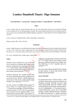
Anatomy of consciousness
Consciousness may be impaired by damage to the
reticular formation in the midbrain and thalamus, or
by bilateral damage to the cerebral hemispheres
resulting in brain displacement, which thereby
impairs the function of the reticular formation (1–3).
1
1 Diagram to show brain herniation, resulting in impaired consciousness.
2
2 Magnetic resonance image illustrating central pontine
myelinolysis, a brainstem cause of reduced consciousness.
3
3 Computed tomography scan showing subdural collection,
resulting in impaired consciousness.
History taking and physical examination
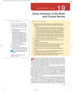
Loss of awareness in the context of being awake
is termed akinetic mutism, and can occur with frontal
lesions (4).
Brainstem tests
In a patient with a reduced conscious level, it must be
ascertained whether coma is due to bilateral
hemispheric disease or to brainstem pathology.
Utilizing brainstem reflexes can assess the integrity of
the brainstem. Pupillary responses to light ascertain
whether there is significant midbrain pathology. The
corneal reflex tests brainstem integrity at the pontomedullary level. Gag reflex assesses the lower
brainstem, while assessment of spontaneous
respiratory movements indicates the integrity of the
medullary respiratory centres. These four clinical
assessments allow the clinician to interrogate the
integrity of the brainstem (5). If an unconscious
patient exhibits normal brainstem reflexes, then it is
likely that the impaired consciousness is due to
bilateral hemispheric dysfunction.
Behaviour
In the conscious patient, behaviour and cognition
must be further addressed. Traditionally, behaviour is
part of the psychiatric examination, and in neurology
it is often neglected in favour of cognition. It is,
however, worth commenting on the patient’s
behaviour. Mood should be noted, whether
depressed, euphoric, or unduly anxious. Emotional
lability may also occur, with sudden swings in mood.
While taking the history, the organization and
content of thought processes should also be noted.
Muddled thinking may suggest a mild delirium.
Thought content can highlight the presence of
delusions (beliefs held which are at variance with the
patient’s environmental background) or illusions
(misperception of stimuli, e.g. mistaking a bush in
dim light for a person). These differ from
hallucinations, in which the imagined object is not
based on a misperceived real object (e.g. hearing
voices, seeing Lilliputian figures and so on).
4
5
Pupillary response
Corneal
reflex
Gag
reflex
Respiration
4 Computed tomography scan showing frontal haemorrhage,
resulting in the patient being awake but not aware.
Midbrain
(Edinger–Westphal)
nucleus
V
VII
Ponto
medullary
junction
IX
X
Medulla
Lower medulla
5 Diagram to illustrate the use of brainstem reflexes in
assessing the integrity of the brainstem at different levels.
13
14
The degree of psychomotor activity should also
be assessed. Hyperalert patients are restless and
exhibit increased motor activity, including speech that
may be accompanied by autonomic overactivity. By
contrast, hypoalert patients may sit motionless for
hours without speaking.
Cognitive function
This aspect of neurological history taking and
examination was neglected for most of the twentieth
century, and is still often poorly performed or even
omitted. Cognition embraces many higher order
activities, including attention, memory, and language.
It is difficult to examine adequately these complex
skills within the confines of a busy neurology clinic.
However, there are brief measures for assessing
cognitive function such as the Addenbrooke’s
Cognitive Examination (ACE). While being no match
for proper neuropsychological assessment, it is a very
good �snap-shot’ assessment of cognitive functions.
Cognitive functions are best divided into those that
require an extensive anatomical network (distributed),
and those utilizing a more localized brain area
Table 2 Cognitive functions
Distributed
Attention/concentration
Memory
(localized) (Table 2). Impairment of a distributed
function, such as attention, does not allow the clinician
to localize the lesion, but suggests that there is a deficit
somewhere in the network subserving attention, which
can then be further localized with subsequent examination and investigation. By contrast, a deficit in a localized function such as language allows the lesion site to
be pinpointed from history and examination alone.
Distributed cognitive functions
Attention/concentration
Attention is difficult to define, but implies
concentration and persistence. Impaired attention
implies inability to focus and selectively concentrate
on a topic, with impersistence, distractibility, and
often disorientation, e.g. as seen in the acute
confusional state. Anatomically, attention requires a
distributed system including neocortex (especially
prefrontal), thalamus, and brainstem, linked by the
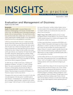
reticular activating system (6). Attention may be
disrupted by any focal lesion affecting this distributed
system, or by a diffuse disturbance, such as metabolic
upset or the effect of drugs.
6
Cortical areas
Pre-frontal, posterior parietal,
and ventral temporal
Thalamus
Intralaminar and reticular nuclei
Higher-order intellectual functions
Localized
Dominant
Language
Calculation
Praxis
Nondominant
Spatially-directed attention
Complex visuo-perceptual skills
Dopaminergic, cholinergic, and
serotinergic pathways
Constructional abilities
Brainstem
nuclei
Reticular formation, midline
raphe, locus coeruleus, and
tegmental nuclei
6 Schematic diagram to show the anatomy of the reticular
activating system.
History taking and physical examination
Bedside tests of attention/concentration include
orientation, digit span (i.e. asking the patient to
repeat number strings), the ability to recite months of
the year backwards (not forwards as this is
overlearned and not a true measure of attention), or
serial sevens (i.e. asking the patient to subtract 7 from
100 and keep subtracting 7).
Higher cognitive processes
The frontal lobes are particularly involved in
conceptual thinking, adaptation and set shifting (i.e.
adjusting to a change in rules), planning and problem
solving, and personality, motivation, and social
behaviour.
Patients with frontal lobe damage show deficits in
the above, in particular poor planning and goal setting,
distractibility, and perseveration (i.e. being unable to
discard old rules and start using new rules). Such socalled frontal behaviour can be present long before
there is any supporting evidence of frontal dysfunction
on neuropsychology or brain imaging (7).
In addition to the above cognitive deficits, frontal
lobe damage also has behavioural consequences.
Disinhibition may occur, resulting in social and
sexually inappropriate behaviour. Aggression and lack
of concern for others may be a feature. Loss of interest
in the world also occurs, with increasing passivity.
Applied anatomy
Anatomy of the frontal lobes is described in (8). The
frontal lobe syndrome may be further subdivided.
Orbitomedial damage is said to result in personality
and behaviour change, while dorsolateral damage
tends to have more effect on executive function, such
as problem solving. Such distinctions are rarely
clinically apparent.
Frontal lobe function may be tested in the clinic
by verbal fluency, e.g. FAS letter fluency (asking the
patient to generate as many exemplars [example
words] beginning with F in 1 minute, then repeating
this for both A and S). Asking patients what is meant
by proverbs such as �A rolling stone gathers no moss’
is also a measure of abstract reasoning and relies on
the frontal lobes. Motor sequencing, such as the
Alternating Hand Movements Test, also assesses
frontal function.
8
7
SMA
M1
FEF
DL
BA
OM
8 Diagram of the anatomy of the brain frontal lobes. BA: Broca’s area;
DL: dorsolateral pre-frontal area; FEF: frontal eye fields; M1: primary
motor cortex; OM: orbito-medial; SMA: supplementary motor area.
7 Magnetic resonance image of a frontal brain tumour.
15
16
Memory
Memory is not a unitary function, but is a broad term
which includes many subcomponents. For instance,
episodic memory refers to the ability to learn and
retain new information. Asking a patient to repeat a
name and address three times, and then asking the
patient to recall the name and address after at least 5
minutes can assess this. The ability to do this task is
crucially dependent on the hippocampus and other
limbic system structures. This is the first region of the
brain to be affected in Alzheimer’s disease, hence
impaired delayed recall is the earliest feature found on
testing patients with new-onset Alzheimer’s disease.
Semantic memory, by contrast, is the database of
knowledge an individual draws on to give meaning to
conscious experience, e.g. knowing the capital of
France or the boiling point of water. This can be
tested at the bedside by category fluency (e.g. naming
as many animals in the next minute, object naming
[whether real objects or line drawings], and reading).
Memory will be addressed more fully in the section
on Memory disorders (3.3).
Localized cognitive functions
Dominant
Language
Language is subserved by the left hemisphere in
nearly all right-handed people and in over 60% of
left-handed people. Language comprehension is
served mainly by the dominant temporal lobe,
especially Wernicke’s area (the posterior third of the
superior temporal gyrus), while language expression
relies on more anterior structures such as Broca’s area
(the posterior third of inferior frontal gyrus).
A full bedside assessment of language should
include the following: spontaneous speech, naming,
comprehension, repetition, reading, and writing. The
nature of any language impairment (aphasia) detected
by these determines the type of aphasia.
Language is more fully addressed in the section
on Speech and Language Disorders (page 94).
Calculation
Pathology at the angular gyrus (the posterior Sylvian
fissure straddling temporal and parietal lobes) of the
dominant hemisphere can result in inability to
understand or write numbers. This can be found in
association with aphasia. When occurring in
conjunction with the inability to write, right-left
disorientation and finger agnosia (i.e. being unable to
say which of the patient’s or examiner’s fingers is the
middle one), this is termed Gerstmann’s syndrome.
Praxis
Praxis refers to the ability to perform and control
skilled or complex motor actions. Apraxia is the
inability to execute such motor commands in the
context of good comprehension and cooperation,
together with functioning motor and sensory systems.
Apraxia is further subdivided into ideomotor and
ideational apraxia, terms that can generate confusion.
Ideomotor apraxia is the inability to carry out
motor acts to command, yet with preserved ability to
carry these out spontaneously, e.g. lighting a match. It
is thought that ideomotor apraxia occurs due to
stored programmes for specific motor acts, called
engrams, being damaged or disconnected. It is usually
due to dominant hemisphere, inferior parietal or
prefrontal pathology.
Ideational apraxia is the inability to synthesize the
individual components of a complex motor act into
one unified operation, with retained ability to perform
each component, e.g. able to open a matchbox to
command, strike a match to command, yet be unable
to effortlessly carry out the entire motor repertoire
seamlessly. It can occur with extensive left hemisphere
disease or lesions affecting the corpus callosum (the
fibres connecting the cerebral hemispheres).
Apraxia may affect only selected movements. For
example, orobuccal apraxia results in difficulty
performing motor commands involving buccal areas
(such as whistling, chewing) and occurs with inferior
frontal or insular lesions. Asking the patient to mime
certain actions, such as whistling, waving goodbye,
and pouring tea may test praxis.
Right hemisphere function
Visuo-spatial and perceptual function rely heavily but
not exclusively on the nondominant hemisphere.
These are required for visual attention, i.e. the ability
to attend to the visual environment. The ability to
dress, construct objects, and ability to understand
what one is seeing may be impaired with right
hemisphere damage. Such patients may fail to
recognize objects or people. They tend to ignore
objects to their left side and may fail to identify
objects or faces despite intact vision. Visuo-spatial
and perceptual function may be tested at the bedside
by asking the patient to draw a clock face,
overlapping pentagons, or a three-dimensional cube.
History taking and physical examination
Damage to the right hemisphere can impair
spatially directed attention leading to the disorder of
neglect, where patients attend less to left hemispace (9).
This may amount to a complete denial of the left side
(10), or lesser states such as sensory extinction where a
visual or tactile stimulus, when administered bilaterally,
fails to be perceived on the left side. Neglect usually
occurs due to right inferior parietal or prefrontal
pathology, but occasionally results from damage to the
thalamus, basal ganglia, or cingulate gyrus.
That neglect occurs almost always to the left may
be due to the left hemisphere monitoring right
hemispace, while the right hemisphere monitors both
hemispaces. Thus a right hemisphere lesion means
that only right hemispace is monitored, while the
converse does not apply with a left hemisphere lesion.
9
Explores
R
Left
hemisphere
Dressing apraxia is not an apraxia as such, but a
visuo-perceptual disorder in which the patient is
unable to dress (becoming entangled in clothing),
despite there being no motor disorder. It usually
occurs due to right posterior parietal damage.
The ability to copy a shape, e.g. overlapping
pentagons or a cube, requires vision, perception, and
visuo-motor output. Difficulties copying usually
reflect right parietal dysfunction. Patients with rightsided lesions tend to produce drawings with grossly
altered spatial arrangements, while patients with leftsided lesions make over-simplified drawings.
10
Explores
L+R
Right
hemisphere
9 Diagram to show the dominance of the right hemisphere for
directed attention.
10 Unilateral visual neglect analysis. The patient neglects the
left hemispace due to a right hemisphere stroke. (The original
drawing was of a double-headed daisy.)
17
18
Difficulties in higher order visuo-perceptual skills
may occur in right hemisphere damage. In visual
object agnosia, the patient is unable to identify an
object visually despite having normal basic
perception. This does not represent a general
semantic memory loss about the object, as
identification through tactile or auditory modalities
results in access to full semantic information about
the object. The deficit in access is therefore modalityspecific, i.e. inability to access semantic information
about an object when this is presented visually.
This deficit can apply specifically to person
recognition. In prosopagnosia, the patient is unable to
recognize familiar faces. Knowledge about the person
is not lost, however, as gait, voice, and so on cause the
patient to identify the person whose face is not
recognized. It is usually due to bilateral inferior
occipito-temporal lesions.
COGNITIVE HISTORY TAKING
Although cognitive history taking broadly follows the
general principles of history taking, the presence of
cognitive deficits (not always noticed by the patient
while causing them inexplicable distress) means that
there are differences in the conduct of this part of the
examination. It is worthwhile trying to interview both
patient and informant alone at some point; this
allows any sensitive issues to be mentioned by the
patient, and also allows the informant to give a clear
history of the nature of the presenting complaint
without risk of offending the patient. In view of the
possible lack of insight, it is worth asking the patient
if they know why they have been referred to the
clinic. In addition to the presenting complaints
themselves, it is useful to ask how these difficulties
are impacting on daily living activities.
A complaint such as �poor memory’ cannot be
accepted simply at face value, but the nature of the
complaint must be determined further. Patients may
use the term �poor memory’ to represent many
problems, including failing to keep appointments or
retain new information (true episodic memory
impairment), forgetting objects’ and people’s names
(anomia, usually in keeping with semantic memory
impairment), or forgetting where they have left their
keys or why they have gone into the kitchen (often
simply slips of attention or concentration). Enquiries
about anterograde memory (i.e. ability to retain new
information), retrograde memory (ability to retrieve
knowledge about previous holidays and so on), and
semantic memory (factual knowledge, knowing what
objects are used for) are important.
For language, in addition to asking whether
patients have difficulty expressing themselves or in
understanding others, it is also worth asking whether
there has been any impairment of reading or writing.
Difficulties with dressing, finding one’s way around
the house or the town, or difficulties constructing
objects suggest a visuo-spatial problem.
THE INFORMANT INTERVIEW
When speaking to the informant alone, information
on what was the initial symptom, and how symptoms
have changed with time is important. For example,
initial symptoms of no longer being able to retain new
information indicate an anterograde episodic memory
deficit, which could be the beginnings of Alzheimer’s
disease. By contrast, an initial symptom of wordfinding difficulty and forgetting the function of
objects suggests a semantic memory deficit such as
can occur in fronto-temporal dementia. The tempo of
evolution of symptoms can help to determine the
underlying pathology. Sudden-onset symptoms with
subsequent improvement suggest a vascular cause.
Insidious-onset progressive symptoms are more in
keeping with neurodegenerative disease, or perhaps a
slow-growing tumour.
Examples of how the cognitive deficit affects the
patient in the real world should be sought, such as
work, cooking and general household tasks, driving
and so on. The informant may well be able to provide
further history, which the patient cannot. Past history
should enquire as to whether there have been any
previous neurological or psychiatric illnesses, or
whether there has been any significant head trauma
(i.e. sufficient at the time to result in loss of
consciousness). An accurate drug history is essential,
particularly as sedating drugs can be an easily
reversible cause of cognitive impairment. Family
history must be thorough, and diagnoses should not
necessarily be taken at face value; institutionalization
or �nervous breakdown’ may indicate a previously
undetected neurological disease, while depression
may be secondary to a neurodegenerative process.
Social history provides the patient’s occupation,
which is of use in estimating premorbid IQ. Alcohol
habits are particularly relevant here.
History taking and physical examination
PHYSICAL EXAMINATION
In a busy clinic, the clinician may be hard pushed to
obtain a history from both patient and informant,
perform bedside cognitive testing, and also conduct a
physical examination. A detailed neurological
examination is not routinely necessary, but the
following features may be of particular relevance.
Visual field deficits may not be noticed by the
patient or relative, and can easily be screened for by
confrontation, preferably using a redheaded pin, but
if necessary using a wiggling finger. Eye movements,
both saccades (voluntary rapid movements) and
pursuit (tracking the examiner’s finger) should be
checked. For example, inability to look down to
command can occur in progressive supranuclear
palsy, while impaired pursuit movements can occur in
basal ganglia disorders such as Huntington’s disease.
Specific signs of frontal disease can include the
presence of so-called primitive reflexes such as grasp
reflex (grip occurring due to stroking palm), pout
reflex (puckering of lips when lips lightly tapped),
and palmo-mental reflex (chin quivering when palm
Pout reflex
Tap lips with tendon
hammer –
a pout
response
is observed
Grasp reflex
Stroking palm of hand
induces �grasp’
11 Diagram to illustrate primitive reflexes.
stroked) (11). Involuntary movements such as
fidgeting, which could indicate Huntington’s disease,
can be easily overlooked unless consciously looked
for. Gait analysis can also be useful. The stooping
festinant gait of Parkinson’s disease or the �feet glued
to the floor’ sign of normal pressure hydrocephalus
can be diagnostic.
BEDSIDE COGNITIVE TESTING
The assessment of higher cerebral function is carried
out during the neurological examination, but may
also be supplemented by a more detailed assessment
of cognitive function performed by a neuropsychologist (if available). The extent to which the
clinician is able to assess cortical function depends on
the time available in clinic, and also whether the
clinician has the backup of a neuropsychologist, as
these services are not universally available. The
clinician should be able to sample various aspects of
cognitive function, such as general functioning,
frontal function, memory, language, and visuoperceptual function.
Glabellar reflex
Patient cannot inhibit blinking in response to
stimulation (tapping
between the eyes)
Palmomental reflex
Quick scratch on palm of hand
induces sudden contraction of
mentalis muscle in face
11
19
20
A standardized test such as the Mini-Mental State
Examination (MMSE) can be used to test aspects of
cognitive function. Although this was originally
devised to be used as a screening tool for dementia, it
is of some use for a brief cognitive overview.
Some criticisms of the MMSE are that the
language and visuo-perceptual items are too easy,
there is not a proper test of delayed recall, and there
is no timed test to assess subcortical cognitive
slowing. In an effort to address these issues, the
Addenbrooke’s Cognitive Examination (ACE)
includes the 30 points of the MMSE, but the
additional 70 points improve the assessment of
memory and language and include timed fluency
tasks which are sensitive to subcortical dysfunction.
CRANIAL NERVE EXAMINATION
CRANIAL NERVE I (OLFACTORY NERVE)
The patient should be asked if taste and smell are
affected. Further testing is not necessary unless the
patient concurs or there is a special reason to test
olfaction. Before testing, the airway should be
checked that it is clear. Each nostril is tested with an
odour such as camphor or peppermint. Loss of smell
is termed anosmia. If nasal disease is excluded, a lack
of smell may be due to closed head injury or anterior
cranial fossa disease but is also a feature of certain
neurodegenerative disorders such as Parkinson’s
disease.
CRANIAL NERVE II (OPTIC NERVE)
The basic tools for assessment are a bright light, an
ophthalmoscope, coloured pins, and a vision reading
chart (e.g. Snellen). First assess visual acuity. The
patient is asked if they are aware of reduced vision in
either or both eyes. Visual acuity should be tested
wearing glasses if prescribed. Each eye is covered in
turn and its neighbour tested separately. When using
the Snellen chart, the patient stands 3–6 m from the
chart and reads from largest to smallest print, visual
acuity being measured as the distance from the chart
(3 or 6 m) over the distance at which the letters
should normally be seen, e.g. 6/6 for normal and 6/60
for poor vision. Alternatively, a near vision chart is
held 30 cm from the patient and again they are asked
to read the smallest print possible with each eye in
turn. Results are expressed as N6 and so on.
Visual fields testing requires a 7 mm coloured
(red) pin. The patient is asked if they are aware of a
gap or �blindspot’ in either eye and the clincian
establishes that the red pin target is visible to each
eye. The patient should then look into the examiner’s
eyes, standing 1 m away. The field of vision of the
examiner can then be compared with that of the
patient (confrontation). The extent of visual field can
be ascertained by testing each eye from each
quadrant, asking the patient to state as soon as they
can see the pinhead at all (regardless of colour).
Whether the patient can see red in central vision
should also be checked.
The following findings may be demonstrated:
вќЏ Constricted fields of vision, e.g. chronic
papilloedema, glaucoma, and functional illness
(tunnel vision).
вќЏ Central field defect (scotoma), e.g. optic neuritis,
retinal haemorrhage.
вќЏ Altitudinal (vertical) field defects in one eye, e.g.
retinal infarction.
вќЏ Hemianopia.
вќЏ Bitemporal (defect in the temporal fields in both
eyes), e.g. pituitary disease.
вќЏ Homonymous (defect to the same side in both
eyes), e.g. parietal, temporal, or occipital lobe
disease.
Quadrantanopia, congruous, and incongruous
field defects further localize defects (see page 104).
Fundoscopy
The ophthalmoscope is used to examine the fundus of
each eye separately, while the patient fixates, in a
darkened environment, into the distance. The
ophthalmoscope should be adjusted for the clinician’s
own vision. If myopic, the lens is turned
anticlockwise (red), if long-sighted clockwise (black).
The patient’s eye is then approached approximately
15В° from the line of fixation and the disc, blood
vessels, and retina are assessed and findings
documented accordingly.
The student should be aware of the following
fundoscopic findings:
Optic disc: normal, pale, swollen.
Blood vessels: normal, attenuated, swollen,
nipped, absent, containing emboli, cholesterol.
Retina: infarcts, haemorrhages, exudate,
retinopathy.
History taking and physical examination
CRANIAL
NERVES
III, IV,
AND
VI (OCULOMOTOR,
TROCHLEAR,
AND ABDUCENT NERVES)
These nerves are responsible for all eye movements.
The clinician should inspect for ptosis (drooping
eyelid), pupil size, strabismus (squint), and proptosis
(protuberance of the globe of the eye). If proptosis is
suspected, the eye should be inspected from above,
tilting the head back to contrast the prominence of
each eye. The pupil light reaction should be tested in
each eye separately, checking both the direct
(illuminated eye) and indirect (nonilluminated eye)
responses. The pupils’ reaction to converging (when
looking at the end of the nose the pupils constrict)
should also be assessed. The patient should be asked
if double vision is present; if so, confirmation that the
double vision is binocular (requires both eyes to be
present) can be obtained by covering one eye at a
time. The patient should be asked whether the two
images are separated vertically or horizontally, and in
which direction the two images (true and false)
maximally separate. The range of slow-pursuit
horizontal and vertical eye movements is assessed by
asking the patient to follow the clinician’s moving
finger or similar object. If double vision is present, in
which direction of gaze double vision is maximal
should be determined.
The following rules help assessment:
Double vision is worse (maximal) in the
direction of the affected muscle.
The false image is always the outermost one.
The false image is the product of the affected eye.
When evaluating eye movements:
вќЏ The position of the head should be noted
(patients with double vision will often tilt the
head to minimize this).
вќЏ The eyes should be checked in the primary
position (at rest) and ptosis or pupillary
asymmetry noted.
вќЏ The eyes should be assessed following an object.
Are abnormal movements (nystagmus) present?
Is there paralysis of one or more muscles? All
directions of gaze must be tested and knowledge
of the specific muscle innervation is essential.
вќЏ Nystagmus is defined as a slow drift of the eyes
to one side with a fast corrective movement in
the opposite direction. While physiological when
watching an object moving rapidly by, these
movements are generally abnormal and inform
on the presence of brainstem/cerebellar disease.
The patient should be asked to follow a moving
finger and any jerky movements observed.
Nystagmus can be:
– Vertical. Upbeat: upper brainstem, e.g.
pontine infarction. Downbeat: cervicomedullary junction, e.g. Arnold–Chiari
malformation.
– Horizontal. Ataxic: greater in the abducting
(looking outwards) rather than adducting
(looking inwards) eye, e.g. multiple sclerosis.
– Multi-directional. Present in all directions of
gaze (though maximal in one), e.g. druginduced.
– Unidirectional. Peripheral: labyrinthine
disease. Central: unilateral cerebellar disease.
CRANIAL NERVE V (TRIGEMINAL NERVE)
The patient is asked if they are aware of altered facial
sensation, and sensation of light touch with cotton
wool is tested in each of the three sensory divisions
(ophthalmic V1, maxillary V2, and mandibular V3).
The corneal reflex (V1&V2) is tested with a wisp of
cotton wool, touching cornea not sclera. Next, the
three divisions are tested with a pin. When defining
the territories of sensory loss, the clinician should
always move from the abnormal to the normal.
Evidence of wasting (temporalis muscles) should
be noted and motor function assessed. The pterygoids
are tested with the jaw open wide (thus avoiding any
minor deviation due to temperomandibular joint
asymmetry), then jaw opening resisted by pressing
against the joint. In order to test the jaw jerk, the
patient is asked to let the jaw hang loosely open and
a tendon hammer is used to percuss on the clinician’s
finger placed on the patient’s chin.
CRANIAL NERVE VII (FACIAL NERVE)
The nasolabial folds (from the corner of the mouth to
the sides of the nose) should be observed and
spontaneous movements such as blinking and smiling
noted. The following muscles are tested using the
instructions described: frontalis: �wrinkle the
forehead’; orbicularis oculi: �screw up the eyes’;
buccinator: �blow out the cheeks’; and orbicularis
oris: �show the teeth’. Ptosis (drooping of an eyelid) is
not due to weakness of facial nerve muscles, and
facial asymmetries without weakness is common so
the clinician should not be misled. The patient should
be asked about taste (absence or distortion) and
tested with a sugar or salt solution applied by a
cotton bud to the anterior two-thirds of the tongue.
21
22
The facial nerve also supplies the muscle to the
stapedius and the parasympathetic supply to the
lachrymal gland, though neither is tested at the
bedside. Four types of disturbance are found:
❏ Unilateral lower motor neurone, e.g. Bell’s palsy.
вќЏ Bilateral lower motor neurone, e.g. myasthenia
gravis.
вќЏ Unilateral upper motor neurone, e.g. hemisphere
stroke.
вќЏ Bilateral upper motor neurone, e.g. brainstem
stroke (pseudobulbar palsy).
Loss of emotional expression is a feature of
Parkinson’s disease, while excessive emotional
expression (emotional incontinence) occurs in
pseudobulbar palsy.
Distinction between unilateral upper and lower
motor neurone facial weakness is simple. In upper
motor neurone (UMN) weakness, forehead and eye
closure is relatively spared (bilateral supranuclear
innervation) while these are affected with lower
motor neurone (LMN) lesions (the final common
pathway for all that travels to the facial nucleus).
CRANIAL NERVE VIII (AUDITORY AND VESTIBULAR NERVES)
Assessment requires a 256 or 512 Hz tuning fork and
an auroscope. First, the patient is asked if they have
noticed any problem with their hearing. The clinician
then speaks in a whisper (counting in numbers) at
arm’s length from the patient, while occluding the
nontested ear with the hand and notes if hearing loss
is reported or demonstrated.
Weber’s test involves striking a 256 or 512 Hz
tuning fork on the examiner’s knee and placing it on
the patient’s forehead. Normally this should be heard
in the middle of the head and should not lateralize.
When the sound does lateralize, it does so to the side
of greater conductive loss or that with the intact
cochlea (to the opposite side) in sensori-neural
hearing loss.
The Rinne test again utilizes the vibrating tuning
fork. The tuning fork is applied firmly to the mastoid
process behind the ear and is then held in front of the
external auditory meatus. The patient is asked which
they hear loudest. Patients with normal middle ear
function hear well by air rather than by bone
conduction. Those with conductive deafness
experience the reverse. The external auditory meatus
and tympanic membrane are examined with the
auroscope. Conductive deafness is common (wax,
middle ear disease). Sensori-neural deafness is less
common and takes three forms:
❏ Cochlea, e.g. noise, Ménière’s disease.
вќЏ Nerve lesion, e.g. meningitis, acoustic neuroma.
вќЏ Brainstem, e.g. vascular, demyelinating disease.
Examination of the vestibular nerve includes
testing gait, nystagmus, and caloric testing (see
Chapter 2).
CRANIAL NERVE IX (GLOSSOPHARYNGEAL NERVE)
Sensation on the posterior wall of the tonsillar fossa
is examined with an orange stick. The motor
(stylopharyngeus) and autonomic (parotid glands)
components are not tested.
CRANIAL NERVE X (VAGUS NERVE)
Articulation, cough, and ability to elevate the soft
palate (�saying Ah!’) are tested. Touching the
posterior pharyngeal wall on each side with a throat
swab and comparing each response tests the gag
reflex. The autonomic and sensory (external auditory
meatus/external ear) are not tested at the bedside.
CRANIAL NERVE XI (ACCESSORY NERVE)
The sternocleidomastoid muscle is tested by tilting the
head to the opposite side while applying resistance
against the examiner’s hand, pressing on the angle of
the jaw. The muscle belly can be observed to stand
out. Asking the patient to shrug the shoulders against
resistance also tests the trapezius muscle.
CRANIAL NERVE XII (HYPOGLOSSAL NERVE)
The tongue at rest on the floor of the mouth is
examined for wasting and fibrillation. The patient is
asked to protrude the tongue and any deviation
noted. The tongue should also be observed for
reduced movement as seen in UMN lesions.
Multiple cranial nerve palsies
Patterns of multiple cranial nerve palsies due to extraaxial lesions reflect their anatomical relationships
(Table 3).
History taking and physical examination
Table 3 Relationship between cranial nerve palsies and location of extra-axial lesions
III, IV, VI, V1
Superior orbital fissure or (anterior) cavernous sinus
III, IV, VI, V1, V2
Posterior cavernous sinus
V, VI
Apex of petrous temporal bone
VII, VIII
Internal auditory meatus or cerebello-pontine angle
IX, X, XI
Jugular foramen
IX, X, XI, XII, and sympathetic
Below the base of the skull (retropharyngeal space)
MOTOR EXAMINATION
Evaluation of patterns of limb weakness is essential
for localizing disease. Weakness may affect a single
limb (monoplegia), arm and leg on the same side
(hemiplegia), or all limbs (tetraplegia). Weakness may
be proximal (muscle disease or inflammatory
neuropathy) or distal (axonal neuropathy).
Observations on muscle wasting, abnormal
movements, tone, and reflex state help to differentiate
UMN from LMN disorders. Finally, weakness that
does not follow a recognizable organic pattern may
be psychologically based. The components of
assessment are:
OBSERVATION
вќЏ Involuntary movements, e.g. extrapyramidal
disease, fasciculation, myokymia.
вќЏ Muscle symmetry.
вќЏ Left to right (mononeuropathy, e.g. carpal
tunnel).
вќЏ Proximal versus distal, e.g. myopathy or
neuropathy.
EXAMINATION OF MUSCLE TONE
The patient is asked to relax and the upper and lower
limbs are tested. The patient’s fingers, wrist, and
elbow are flexed and extended. Similarly, the patient’s
ankle and knee are flexed and extended. Normally, a
small, continuous resistance to passive movement is
felt, and decreased (flaccid) or increased
(rigid/spastic) tone should be noted. Failure to relax is
a common problem. A flaccid (weak) limb suggests a
LMN disorder, while a spastic (weak) limb suggests
UMN problems. Rigidity occurs in extrapyramidal
disease and fluctuating stiffness (paratonia or
Gegenhalten) with diffuse frontal lobe disturbance.
EXAMINATION OF MUSCLE STRENGTH
Muscle strength is under corticospinal (UMN) and
anterior horn cell/nerve root/nerve plexus/peripheral
nerve/neuromuscular junction (LMN) control. The
target of all pathways is the muscle itself. Muscle
strength is tested by having the patient move against
the examiner’s resistance. One side is always
compared with the other and strength is graded on a
scale from 0–5/5 (Table 4).
When testing, the following should be considered:
the overall distribution (proximal versus distal), the
pattern (flexor versus extensor), and the grouping
(single root versus multiple roots versus plexus versus
single nerve versus multiple nerves). There is no
avoiding knowing the detailed innervation of
muscles. In a minimal examination the following
should be tested:
Table 4 Grading of motor strength
Grade
Description
0/5
No muscle movement
1/5
Visible muscle movement but no
movement at the joint
2/5
Movement at the joint, but not against
gravity
3/5
Movement against gravity, but not
against added resistance
4/5
Movement against resistance, but less
than normal
5/5
Normal strength
23
24
Upper limbs
вќЏ Flexion at the elbow (C5, C6, biceps).
вќЏ Extension at the elbow (C6, C7, C8, triceps).
вќЏ Extension at the wrist (C6, C7, C8, radial
nerve).
вќЏ Finger abduction (C8, T1, ulnar nerve).
вќЏ Opposition of the thumb (C8, T1, median
nerve).
Lower limbs
вќЏ Flexion at the hip (L2, L3, L4, iliopsoas).
вќЏ Adduction at the hips (L2, L3, L4, adductors).
вќЏ Abduction at the hips (L4, L5, S1, gluteus
medius and minimus).
вќЏ Extension at the hips (S1, gluteus maximus).
вќЏ Extension at the knee (L2, L3, L4, quadriceps).
вќЏ Flexion at the knee (L4, L5, S1, S2, hamstrings).
вќЏ Dorsiflexion at the ankle (L4, L5).
вќЏ Plantar flexion (S1).
Pronator drift
The patient is asked to stand for 20–30 seconds with
both arms straight forward, palms up, and eyes
closed, keeping the arms still while the examiner
gently taps downwards. In pronator drift, the patient
fails to maintain extension and supination (and the
limb �drifts’ into pronation). Pronator drift is seen in
UMN disease.
REFLEXES
The tendon reflex comprises a stretch sensitive
afferent (from muscle spindles) via a single synapse
(anterior horn cell region) to the efferent (motor)
nerve. These reflexes are accentuated in UMN disease
and diminished or absent with LMN disorders.
Deep tendon reflex
Patients must be relaxed and positioned properly
before starting the test. No more force with the
tendon hammer should be used than is required to
provoke a definite response. Reflexes can be
�reinforced’ by asking the patient to perform
isometric contraction of other muscles groups
(clenched teeth, upper limbs or pull on hands, lower
limbs). Reflexes should be graded on a 0 to 4 �plus’
scale (Table 5).
The following reflexes should be tested:
❏ Biceps (C5, C6): examiner’s thumb or finger is
placed firmly on the biceps tendon and the
examiner’s fingers are struck with the reflex
hammer.
❏ Triceps (C6, C7): the patient’s upper arm is
supported and the patient’s forearm is allowed
to hang free; the triceps tendon is struck above
the elbow.
❏ Brachioradialis (C5, C6): the patient’s forearm
rests on their abdomen or lap and the radius is
struck about 3–5 cm (1–2 inches) above the wrist.
вќЏ Abdominal (T8, T9, T10, T11, T12): a blunt
object is used to stroke the patient’s abdomen
lightly on each side in an inward and downward
direction above (T8, T9, T10) and below the
umbilicus (T10, T11, T12), while noting
contraction of the abdominal muscles with
deviation of the umbilicus towards the stimulus.
вќЏ Knee (L2, L3, L4): the patient sits or lies down
with the knee flexed and the patellar tendon is
struck just below the knee.
❏ Ankle (L5 S1): the patient’s foot is dorsiflexed at
the ankle and the Achilles tendon struck.
CLONUS
If the reflexes are hyperactive, clonus is tested for.
This can be done at any joint but is usually performed
at the ankle. The knee is supported in a partly flexed
position and the foot then quickly dorsiflexed. Clonus
is manifest by rhythmic sustained oscillations.
PLANTAR RESPONSE (BABINSKI)
A positive Babinski sign indicates UMN disease while
a negative test is normal. The lateral aspect of the sole
of each foot is stroked with the end of a reflex
hammer and movement of the toes noted. The normal
movement is that of plantar flexion. Extension of the
big toe with fanning of the other toes is abnormal, a
positive Babinski.
Table 5 Tendon reflex grading scale
Grade
Description
0
Absent
1+ or 1++
Hypoactive
2+ or 2++
Normal
3+ or 3++
Hyperactive without clonus
4+ or 4++
Hyperactive with clonus
History taking and physical examination
SENSORY EXAMINATION
This is the most exacting and potentially misleading
part of the neurological examination. There are five
categories of sensation to test: pain, temperature
(small fibre/spinothalamic tract/thalamus/diffuse +
frontal cortex), vibration, joint position, and light
touch (large fibre/dorsal column/medial lemnisci/
parietal cortex). These anatomically separate
pathways explain why sensory loss is often
incomplete (dissociated).
The examination requires a cooperative patient
and an alert examiner! Each test should be explained
to the patient before it is performed. The patient
should have their eyes closed during testing.
Symmetrical areas both sides of the body are
compared as well as distal and proximal areas of each
limb. Where an area of sensory abnormality is found,
the boundaries should be mapped out in detail,
moving from the abnormal to the normal area.
вќЏ Distal and symmetrical impairment in limbs
suggests sensory neuropathy.
вќЏ A level on the trunk below which sensation
is lost localizes spinal cord disease.
вќЏ An area on the trunk or limbs confined to
one side suggests nerve root or single
peripheral nerve disturbance.
The five components of sensation are tested as
follows:
VIBRATION
A low-pitched (128 Hz) tuning fork is used to test
awareness at bony prominences such as wrists,
elbows, medial malleoli, patellae, anterior superior
iliac spines, spinous processes, and clavicles.
Vibration is normally lost at the ankles in those over
60 years. The patient must understand that it is the
sensation of vibration rather than the sound from the
fork that is being tested.
SUBJECTIVE LIGHT TOUCH
Cotton wool is used to touch the skin lightly on both
sides. Sometimes when a stimulus is presented
simultaneously on both sides it is not felt on one side,
yet it is felt on that side when the stimulus is
presented separately (sensory inattention, suggesting
parietal lobe disease). Several areas on both the upper
and lower extremities are tested and the patient is
asked if there is a difference from side to side.
POSITIONAL SENSATION
The patient’s big toe is held away from the other toes
to avoid �cueing’ and is moved to demonstrate to the
patient what is meant by �up’ and �down’. The
examiner then moves the toe and asks the patient to
identify the direction the toe has moved with eyes
closed. If position sense is impaired the ankle joint is
then tested and then the fingers in a similar fashion.
The examiner should then move proximally to test
the metacarpophalangeal joints, wrists, and elbows.
PAIN
A suitable sharp disposable object is used to test
�sharp’ sensation. The following areas are tested:
вќЏ Shoulders (C4).
вќЏ Inner and outer aspects of the forearms (C6 and
T1).
вќЏ Thumbs and fingers (C6 and C8).
вќЏ Front of both thighs (L2).
вќЏ Medial and lateral aspect of both calves (L4 and
L5).
вќЏ Little toes (S1).
Remembering these particular dermatomal areas
is helpful. Pinprick examination is critical to defining
a �level’ when localizing spinal cord disease.
TEMPERATURE
This is omitted if pain sensation is normal, but is
helpful in confirming the presence and distribution of
pinprick loss. A tuning fork heated or cooled by
water is used and the patient asked to identify �hot’ or
�cold’. Similar areas as above are tested. Temperature
loss is much less helpful when defining a precise
spinal cord level.
DISCRIMINATION
Tests of discrimination are dependent on intact touch
and position sense; they cannot be performed where
these are clearly abnormal. These tests assess cortical
(parietal) sensation.
Graphaesthesia
With the blunt end of a pen or pencil, a large number
is �drawn’ in the patient’s palm and the patient asked
to identify the number.
Stereognosis (an alternative to graphaesthesia)
A familiar object is placed in the patient’s hand (coin,
paper clip, pencil) and the patient then asked to name
the object.
25
26
Two point discrimination
An opened paper clip or divider is used to touch the
patient’s finger pads, alternating irregularly between
one point touch and touch in two places
simultaneously. The patient is asked to identify if they
feel �one’ or �two’ points. The minimum distance at
which the patient can discriminate is identified.
COORDINATION
These tests assess cerebellar function and require
relatively intact motor power. To test for lack of
coordination with weakness present is unsound, as
such tests cannot be performed to a level that allows
accurate interpretation. Associated signs, dysarthria,
and nystagmus are further pointers to cerebellar
disease.
RAPID ALTERNATING MOVEMENTS
The patient is asked to slap rapidly the palmar and
dorsal surface of the hand alternately on the thigh. In
the lower limbs, the patient taps the examiner’s hand
with the sole of each foot.
LIMB COORDINATION
This is assessed by �point-to-point’ tests. The patient
is asked to touch the tip of the examiner’s finger and
then their own nose. The accuracy in approaching
each target and the smoothness of movement between
the two targets is recorded (deficits being termed
past-pointing and dysmetria, respectively). In the
lower limbs, the patient is asked to touch the front of
the shin with each heel, and then to run the heel up
the front of the leg to the knee and similar
observations are made.
STANCE AND GAIT
GENERAL
The patient is asked to stand with feet together and,
once they feel comfortable, to close the eyes. If the
patient cannot balance well with eyes open, they
should not be asked to close the eyes. The clinician
stands close to the patient and must be ready to
support them should they fall to either side.
Instability with eyes open suggests cerebellar disease,
and with the eyes closed afferent (sensory ataxia)
disturbance. Unsteadiness only on eye closure is a
positive Romberg test.
POSTURAL STABILITY (THE RETROPULSION OR FORCED
PULL-BACK TEST)
The patient is asked to stand with eyes open and feet
apart. The clinician stands behind the patient and
warns that the patient will suddenly be pulled
backwards. An abnormal response should be
anticipated and the clinician must be prepared to
catch the patient! An abnormal reaction is seen in
extrapyramidal disorders (e.g. Parkinson’s disease)
with retropulsion (taking several steps backwards and
being unable to maintain stability).
The student should be able to recognize certain
walking disorders: the hemiplegic, ataxic,
extrapyramidal, and myopathic gaits; also the
high �steppage’ gait of unilateral or bilateral
footdrop.
REFERENCES
1 Fuller G (2004). Neurological Examination
Made Easy. Churchill Livingstone, London.
2 Lindsay K, Bone I (2004). Neurology and
Neurosurgery Illustrated. Churchill Livingstone,
London.
CHAPTER 2: NEUROLOGICAL INVESTIGATIONS
27
Ian Bone, John Greene
INTRODUCTION
INVESTIGATING THE HEAD
INVESTIGATING THE SPINAL CORD
INVESTIGATING THE PERIPHERAL NERVOUS SYSTEM
(NERVE, NEUROMUSCULAR JUNCTION, AND MUSCLE)
INVESTIGATING SPECIFIC SITES
28
INTRODUCTION
The evaluation of a clinical presentation often,
though not invariably, leads to the formulation of an
�investigative pathway’. Thoughtful and thorough
history taking and examination are central to
deciding not only �whether to investigate’ but also
�where to look’. Once the decision to investigate has
been reached the clinician must ask �Is it in the head,
the spine, in nerve, or muscle?’ The answer is crucial
as each of these sites or levels has specific
investigations. Getting it right is not always easy. For
example, the complaint of a �foot drop’ can indicate
cerebral, spinal, or peripheral nerve disease, the skill
being to decide from the history and examination
where the culprit lies and to investigate accordingly.
In this chapter the student will be introduced to
the basics of neurological investigation as an aid to
the problem-oriented text that follows. Rather than
list and describe these in order of complexity, cost, or
12
X-ray tube
moving in
10В° steps
Fixed array
of detectors
12 Diagram to illustrate a computed tomography gantry.
availability, each compartment (head, spine, nerve
and muscle) will be addressed separately. Obviously
some disease processes will be multifocal, multisystem, and straddle compartments (e.g. the
craniocervical junction) or medical disciplines (e.g.
the orbit and ear), and resultant investigations will be
more complex and diverse. What follows is therefore
a �rough guide’ to introduce the student to
techniques, indications, sensitivity, specificity, and
potential complications of the tools used to diagnose
and help treat neurological disease.
INVESTIGATING THE HEAD
A computed tomography (CT) or magnetic resonance
imaging (MRI) scan is now indicated in most workups of suspected intracranial disease. Indeed, it is
difficult to imagine practice without such imaging.
Before the 1970s, the ability to image the brain
depended on isotope studies, invasive injection of air
(air encephalography), or contrast (angiography/
myodil ventriculography). These techniques showed
no more than outlines and shadows and were risky.
As a result the clinician was much more conservative
and circumspect as to which patients were subjected
to such tests. The advent, easy availability, and low
risk of cross-sectional imaging have undoubtedly
diluted clinical skills but the advances in diagnosis
and improved patient care are well worth this price.
The current danger is that of over-investigation and
that clinical skills are reduced such that investigations
are targeted to the wrong site or incidental imaging
findings are mistaken as relevant.
COMPUTED TOMOGRAPHY
Also referred to as CAT (computer assisted
tomography) scanning, this revolutionary technique
was introduced into clinical practice in 1973. It is the
most commonly used form of cranial imaging, being
available on a 24 hour basis in almost all hospitals.
The word tomography refers to imaging of slices of
the brain (or indeed any other organ). The patient is
placed in a gantry (12) and a thin beam of X-rays,
created by a collimator, traverses each �slice’ to a
multichannel ionization detector.
The path from collimator to detector establishes a
single �beam’ passing though a patient’s tissues.
During the scan the computer takes brief readings
from all detectors. In the earlier scanners both
collimator and detector rotated around the patient
whereas in modern third and fourth generation units
the detectors are fixed with only the X-ray source
Neurological investigations
moving. Computer processing with multiple beams
and detectors completely encircling the head allows
absorption values for blocks of tissue (voxels) to be
established. Reconstruction of this by twodimensional display (pixels) results in the eventual CT
appearance (13). Slices studied can be varied in
thickness (1–10 mm) and can be rendered parallel to
the orbitomeatal line (a reference line drawn through
the orbit and ear), to allow anatomical interpretation
and reproducibility for repeated studies. �High
definition’ (1–2 mm) scans give greater anatomical
detail, take longer to acquire, and are reserved for
examination of specific sites (orbit, Circle of Willis,
pituitary). Alteration of window level will change the
contrast appearance and allow more detailed
assessment of bony structures or help visualize subtle
differences in tissue contrast, such as with an acute
cerebral infarct.
Falx cerebri
Frontal
lobe
Frontal horn
of lateral
ventricle
Lateral
ventricle
Septum
pellucidum
Parietal
lobe
Pineal gland
Occipital
lobe
3rd ventricle
Midbrain
Quadrigeminal
cistern
Frontal sinus
Orbital
roof
Frontal lobe
Sylvian fissure
Temporal lobe
Pons
Mastoid air
cells
Cerebellum
4th ventricle
Cerebellum
13 Computed tomography of normal brain, illustrating landmark anatomical details.
Temporal
lobe
Sulci
13
29
30
Knowledge of the display and normal
appearance along with anatomical structures is
essential.
Because each slice is oriented along the
orbitomeatal line, with the head tilted backwards,
lesions appear more posterior than is the case. Scans
can be displayed in coronal (ear to ear) or sagittal
(forehead to occiput) planes to look at specific
regions (e.g. orbits or foramen magnum).
Intravenous contrast medium should not be given
routinely but only where a noncontrast scan has
shown an abnormality and clarification is required
(e.g. a possible abscess, metastatic tumour, or
vascular anomaly). Reactions occur in 5% of
examinations, this being dose-dependent. Contrast
will show up areas of high vascularity and regions of
breakdown of the blood–brain barrier, and will
demonstrate areas of altered blood perfusion (see
later). A contrast-enhanced scan also highlights up all
the major intracranial vessels (14).
14
14 Contrast computed tomography scan showing left middle
cerebral artery stenosis (arrow).
The patient requires no special preparation for
CT other than reassurance that the procedure is safe.
Metal should be removed from the head (hairpins,
dental bridgework) as this will cause signal scatter
and result in artefacts. If contrast is to be given, the
clinician must enquire after a history of previous
reaction to injected contrast. Radiation dose is
comparable to a routine skull X-ray (now rarely
performed). Pregnancy is not a contraindication as
only a very small quantity of radiation �scatters’ in the
direction of the abdomen and uterus. The limitations
of CT are listed below.
Helical CT (spiral CT) is performed by �pulling’
the patient though the scan field of the rotating X-ray
source. This allows faster scanning time, better image
reformation, and the ability to perform newer
techniques such as CT angiography (CTA).
Limitations of CT imaging
вќЏ Uses ionizing radiation.
вќЏ Visualization of posterior fossa structures
(e.g. brain stem or cerebellar pontine angle)
inferior to MRI.
вќЏ Less sensitive than MRI to acute changes
(e.g. early infarction).
CT angiography
This examination uses X-rays to visualize blood flow
in arterial vessels. CT venography (CTV) studies
similar flow through veins (15). An automatic
injector controls the timing and rate of injected
contrast, continuing through the initial image
recording. The rotating scanner spins in a helical
manner around the patient, creating a fan-shaped
beam of X-rays. As many as 1000 of these pictures
may be recorded in one turn of the detector. Once
acquired, processing these images requires a powerful
computer programme making it possible to display
cross-sectional slices or three-dimensional �casts’ of
the blood vessels.
CTA is used to study blood vessel wall disease
(narrowing [16], dissection, atheroma, inflammation,
and aneurysm). Both extracranial (aortic arch and
branches) and intracranial vessels (Circle of Willis
and branches) can be studied. Intracranial aneurysms
sized ≥3 mm can be detected.
Neurological investigations
CT perfusion
CT perfusion (CTP) has been proposed as a method
of evaluating patients suspected of having an acute
stroke where thrombolysis is considered. It may
provide information about the presence and site of
vascular occlusion, the presence and extent of
ischaemia, and tissue viability. CTP provides accurate
measures of local cerebral blood volume (CBV),
blood flow (CBF), and mean transit time (MTT).
MAGNETIC RESONANCE IMAGING
The millions of water molecules within the human
body contain hydrogen ions which themselves have
protons which act as microscopic magnets. When
placed in a magnetic field, the protons within an
individual will align in part to that field. If a
radiofrequency (RF) pulse is then applied, these
protons become excited, start spinning and �flip’ their
orientation. When the RF field is turned off, the
spinning protons revert to their prior state (or
15
alignment), releasing energy. These microscopic
magnets, and the vectors that they set up by
realignment, form the basis for generating images.
Variability of these ions from tissue to tissue as well
as their response to applied electromagnetic pulses
creates images of organs (brain and spine) in health
and disease.
The student should understand two simple terms.
The time it takes for a proton to recover most of
its longitudinal alignment (or magnetization) is
referred to as T1 or spin-lattice relaxation time.
The time it takes for the proton to lose most of its
transverse alignment (or magnetization) is
referred to as T2 or spin-spin relaxation time.
A bewildering variety of RF pulse sequences (spin
echo, gradient echo, inversion recovery, and
saturation recovery) can be used, often with a specific
purpose (e.g. gradient echo to show previous
haemorrhage). The introduction of magnets with
increasing field strength (>1.5 Tesla) allows a better
signal to noise ratio, and faster scanning with a
reduction of imaging time. By acquiring data more
rapidly, these MRI scanners can perform MR
angiography and gain functional information on
diffusion and perfusion, as well as images of those
acutely ill patients whose physiological instability
previously precluded lengthy investigations.
16
1
15 Computed tomography venogram illustrating cerebral
venous drainage.
2
16 Computed tomography angiogram illustrating left middle
cerebral artery occlusion (1) and right middle cerebral artery
narrowing (2).
31
32
ADVANTAGES AND DISADVANTAGES OF MRI
(COMPARED TO CT)
Advantages
вќЏ Select any plane, e.g. oblique.
вќЏ No ionizing radiation.
вќЏ More sensitive (early
ischaemia/demyelination).
вќЏ No bony artefacts (looking at sites adjacent
to bone).
Disadvantages
вќЏ Cannot use with any ferromagnetic implant,
e.g. pacemaker.
вќЏ Claustrophobic.
вќЏ Does show bony artefact.
вќЏ Long-term hazards not known.
T1 and T2 images can easily be distinguished. On
T1, CSF is black (hypointense) while on T2 it is white
(hyperintense). Table 6 will help interpretation of MR
images (increase or decrease in signal is defined in
relation to normal grey matter). Figures 17–20
present MRI images.
Paramagnetic enhancement
The intravenous agents used (e.g. gadolinium) induce
a strong local magnetic field effect that will shorten
T1 signals. This will result in enhancement of the
image in areas of contrast leakage (across a damaged
blood–brain barrier) or within the vascular compartment (arteries, microcirculation, and veins).
Gadolinium and other such agents highlight
ischaemia, infection, and demyelination and can help
distinguish tumours from surrounding oedema.
Table 6 Guide to identifying the MRI sequence
T2-weighted image
вќЏ CSF bright.
вќЏ Grey matter brighter than white matter.
Perfusion-/diffusion-weighted image
вќЏ CSF grey.
вќЏ Grey matter brighter than white matter.
T1-weighted image
вќЏ CSF dark.
вќЏ White matter brighter than grey matter.
CSF: cerebrospinal fluid.
17
17 T1 sagittal magnetic resonance image of the brain.
18
18 T1 coronal magnetic resonance image of the brain.
Neurological investigations
19a–d Axial magnetic resonance image
(MRI) of the brain illustrating the
superiority of MRI over computed
tomography.
19a
19b
19c
19d
20 T2 magnetic resonance image showing a primitive neuroepithelial tumour in the
right anterior temporal lobe.
20
33
34
Fluid-attenuated inversion recovery
Fluid-attenuated inversion recovery (FLAIR) MRI
depicts areas of tissue T2 prolongation while
suppressing the signal from neighbouring cerebrospinal
fluid (CSF). It is helpful in assessing lesions that are
adjacent to CSF, such as at periventricular and
superficial cortical sites (21, 22).
Magnetic resonance angiography
Magnetic resonance angiography (MRA) can be
based on either time-of-flight methods (measuring the
contrast enhancement produced by inflowing blood),
or phase contrast methods, which utilize the flowinduced variations in signal induced by the motion of
21
21 Fluid-attenuated inversion recovery (FLAIR) magnetic
resonance image showing the white matter changes of multiple
sclerosis. Note that cerebrospinal fluid (CSF) is dark on FLAIR
unlike the bright CSF seen on T2.
blood alone. Time-of-flight measurements are better
suited for imaging arterial wall morphology. MRA
can demonstrate extracranial and intracranial
stenosis (narrowing) noninvasively, but tends to
overestimate the degree of a stenosis and
consequently misinterprets low flow through a tight
stenosis as an occlusion. It is usually used in
conjunction with ultrasound (or CTA) for the
presurgical evaluation of carotid artery stenosis (23).
MRA is used to screen for intracranial aneurysms but
will not show all of those seen on intra-arterial digital
subtraction angiography (see later). By selecting a
specific flow velocity, veins can also be imaged
(MRV) (24).
22
22 Fluid-attenuated inversion recovery (FLAIR) magnetic
resonance image showing left mesial temporal sclerosis causing
epilepsy.
Neurological investigations
Diffusion and perfusion MRI
MR scanning is superior to CT scanning in the
diagnosis of acute stroke. However, conventional MRI
does not demonstrate early acute cerebral ischaemia
particularly in a period when therapy may reverse or
limit the permanent brain injury. T2 sequences require
at least several hours from the onset of the stroke
before demonstrating signal change abnormalities.
Water diffusion has been introduced as an additional
contrast parameter in the MR imaging of acute stroke.
Diffusion-weighted imaging (DWI) can demonstrate
altered cellular metabolism and the reduction in the
microscopic water molecule motion (Brownian
motion) in acute and subacute stroke. Damaged tissue
may be visualized as early as 5–15 minutes after the
onset of stroke.
23 Magnetic resonance
angiogram of the carotid
arteries.
23
Perfusion-weighted imaging (PWI) allows
assessment of perfusion of the brain microvasculature. PWI requires intravenous gadolinium
contrast (bolus tracking) that causes a paramagnetic
susceptibility effect. Thus, MRI signal declines as
gadolinium travels through the microvasculature.
PWI is used to complement the information obtained
by DWI in acute stroke patients. PWI may show
reduced perfusion in a larger area of tissue than that
shown on DWI. This indicates a significant area of
tissue at risk of infarction, a diffusion–perfusion
mismatch. Using PWI, various perfusion properties of
the contrast bolus can be calculated, such as CBV,
CBF, MTT, and time-to-peak (TTP) of contrast
arrival. A very low CBV implies irreversible
ischaemia.
24
24 Magnetic resonance venogram showing superior sagittal
thrombosis.
35
36
25
25 T1 magnetic resonance image of a
subcortical infarct.
The combination of MRA to visualize vessels,
and DWI and PWI to define early infarction and
�tissue at risk’ (�the ischaemic penumbra’) represents
an ideal �package’ to help plan acute stroke
treatments (25–27).
26
26 Diffusion-weighted imaging (DWI)
magnetic resonance image of the same
subcortical infarct shown in 25.
Functional MRI
MRI is able to map functional cortical activity during
the performance of tasks. This is achieved by
detecting regional cortical tissue changes in venous
blood oxygenation. This technique is known as blood
oxygen level dependent (BOLD) imaging or simply
functional MRI (fMRI) (28). The key to fMRI is that
oxyhaemoglobin is diamagnetic whereas deoxyhaemoglobin is paramagnetic. The role of fMRI is
mainly in research. It has, however, been used to
guide surgical resection of brain in or adjacent to
eloquent areas.
brain tissue and have high nuclear magnetic
sensitivity compared with other magnetic nuclei).
Single voxel spectroscopy can be performed in many
clinical 1.5T MR units using commercially available
software. Two types of MRS finding are encountered.
Either the spectrum reveals a metabolite that is not
normally present or, more commonly, an abnormal
quantity of a normal metabolite is present.
Metabolites studied are N-acetyl aspartate (NAA),
choline (Cho), creatine (Cr), glutamate, and lactate.
NAA is present exclusively in neurones and declines
with neuronal and axonal damage and death. Choline
is important in the myelination of nerves and is
altered by demyelinating diseases. Lactate is a marker
of anaerobic respiration and increases in hypoxic
ischaemic states. In the future it is hoped for a
diagnostic �biochemical biopsy’ for specific tumour
types and other pathologies.
MR spectroscopy
Proton MR spectroscopy (MRS) is a noninvasive
method used to obtain a biochemical profile of brain
tissue (a biochemical biopsy). Although 31
phosphorous (31P) spectra are sometimes used, MRS
studies are usually obtained by proton spectroscopy
with water suppression (protons are abundant in
SINGLE PHOTON EMISSION COMPUTED TOMOGRAPHY
Single photon emission computed tomography
(SPECT) utilizes a gamma camera that can rotate and
computer reconstruction similar to CT, to produce a
two-dimensional image. Compounds labelled with
gamma-emitting tracers (ligands) have been developed
to study blood flow, map receptors, and evaluate
Neurological investigations
27
27 Apparent diffusion coefficient (ADC)
magnetic resonance image of the same
subcortical infarct shown in 25.
29 Ictal hexamethylpropyleneamine
oxime (HMPAO)
spectroscopy (axial slice
in the long axis of the
temporal lobe) with
injection 25 seconds
after the onset of a
right mesial temporal
lobe seizure, showing
hyperfusion of the
whole temporal lobe.
29
28
28 Functional magnetic resonance image of a
patient performing left finger tapping, activating
right motor cortex.
30
31
30 SPECT imaging showing reduced left hemisphere perfusion in primary progressive aphasia.
31 Fluoropropyl carboxymethoxy iodophenyl nortropane spectroscopy (FP-CIT) showing reduced dopamine transport in
corticobasal degeneration.
tumour growth. While a rotating scanner is most
commonly used, fixed multidetector systems produce
better image definition as well as axial, coronal, and
sagittal views. Ligands are labelled with radioactive
iodine or thallium and are given intravenously.
Radiation dose limits repeated investigations. SPECT
scans are interpreted visually, usually in conjunction
with structural imaging (CT or MRI), as an
anatomical abnormality will influence ligand uptake.
SPECT scanning is used to study cerebral blood
flow (in stroke, dementia, and seizures). CNS
receptors include benzodiazepine (epilepsy), glutamate (stroke), and dopamine (Parkinson’s disease and
other movement disorders). Rates of tumour growth
can also be studied (high-grade thallium, low-grade
tyrosine). The following figures illustrate the clinical
applications of SPECT imaging (29–31).
37
38
POSITRON EMISSION TOMOGRAPHY
The positron emission tomography (PET) technique
uses positron-emitting isotopes (radionuclides). These
have a short half-life and have to be generated on-site
by a cyclotron. Imaging and display also involve
cross-sectional data acquisition and reconstruction,
much like CT scanning. When a positron encounters
an electron (which will be almost immediately it is
emitted from the source) mutual annihilation occurs,
with the emission of photons (gamma rays). Multiple
detectors enable detection and quantification of
radioactivity. Labelled oxygen, fluorine, or carbon is
used given by intravenous bolus or by inhalation. As
well as studying receptor sites and receptor binding,
this technique can quantify CBF, CBV, and glucose
and oxygen metabolism. Lack of availability and
complexity have limited the wider application of PET;
however, it may be particularly helpful in detecting
small tumours (e.g. small cell lung cancer in
paraneoplastic syndromes).
CEREBRAL ANGIOGRAPHY
Angiography remains the �gold standard’ choice in the
evaluation of arterial and venous occlusive disease,
aneurysm (32), and arteriovenous malformation
(AVM). It is also useful in defining the blood supply of
some tumours (e.g. meningioma). Finally, it is necessary
for interventional neuroradiologic procedures such as
coiling aneurysms, stenting stenosis, and delivering
intra-arterial thrombolysis. Contrast is injected via a
flexible catheter introduced by the femoral artery, and
rapid film changers are used to capture its flow through
extra- and intracranial vessels.
Advances in the development of high-quality
digital subtraction units provide instantaneous
images for view on a cathode ray tube. This digital
subtraction angiography (DSA) utilizes analogue to
digital image conversion with digital image display.
Iodine-containing, water-soluble media provide the
necessary density to allow the visualization of the
cerebral arterial, capillary, and venous systems. The
32
32 Angiogram showing intracerebral aneurysm (arrow).
Neurological investigations
major complication of cerebral angiography is stroke,
either from damaging the vessel wall (dissection) or
dislodging thrombus (embolism). Meticulous care to
detail is mandatory. With ectatic and tortuous arteries
it may be difficult to catheterize these selectively, and
injections at the origin of such vessels may be safest.
Once the procedure is over, general care is
important, particularly with regard to blood pressure.
Elderly patients following a large contrast load may
become hypotensive, and with haemodynamically
significant carotid artery stenosis, relative
hypoperfusion will cause ischaemia and �watershed’
infarction. Systemic reactions to contrast occur,
ranging from urticaria to bradycardia and laryngeal
spasm. Local complications include haematoma at
the catheter site, femoral nerve damage, and arterial
thrombosis with distal embolism.
The appearances of a normal examination
should be noted with regard to major vessels
and their divisions. Oblique views may be used
in assessing aneurysms and cervical views for
evaluation (and precise measurement) of internal
carotid and vertebral artery disease.
Interventional angiography
Recent advances in endovascular procedures now
play an important part in patient management.
Tumours, aneurysms, fistulas (acquired openings
between arteries and veins), AVMs, and narrowed
intracranial arteries can be treated by an array of
particles, glues, balloons, coils, and stents.
ULTRASOUND
This noninvasive technique utilizes a probe
(transducer) that emits ultrasonic waves, usually at
frequencies of 5–10 MHz. When held over a blood
vessel these ultrasonic waves are reflected back to the
probe, which also doubles as a detector. Reflected
waves can then be displayed as a two-dimensional
image (B-mode). When the probe is held over a
moving column of blood, the frequency shift of
reflected waves (Doppler effect) informs on the
velocity of the column. There are four types of
Doppler ultrasound:
вќЏ Continuous wave Doppler measures how
continuous sound waves change in pitch as they
encounter blood flow blockages or narrowed
blood vessels. This is performed at the bedside
and provides a quick, rough guide of damage or
disease.
вќЏ
вќЏ
вќЏ
Duplex Doppler produces a picture of a blood
vessel and the organs that surround it. A
computer converts the Doppler signals into a
graph that provides information about the speed
and direction of blood flow through the blood
vessel being examined.
Colour Doppler is computer assisted and
converts the Doppler signals into colours that
represent the speed and direction of flow
through the vessel.
Power Doppler is a new technique being
developed that is up to five times more sensitive
than colour Doppler and is used to study blood
flow in vessels within solid organs.
Ultrasound is used to assess extracranial (carotid)
arterial disease (33).
Transcranial Doppler ultrasound
Transcranial Doppler ultrasound (TCD) is a safe,
reliable, and relatively inexpensive technology for
measuring intracranial blood flow velocities. Waves
with frequencies around 2 MHz are directed towards
intracranial vessels using a handheld probe. The
frequency shift (Doppler effect) in the reflected sound
indicates the velocity of the column of moving blood.
33
33 Carotid Doppler ultrasound showing 70% right internal
carotid stenosis.
39
40
Images are reconstructed from the time-dependent
intensity of the reflected sound, and vascular structures
visualized. Altering transducer location and angle, and
the instrument’s depth setting obtains velocities from
most intracranial arteries. The cranial �windows’ used
are the orbit (anterior cerebral artery) and in temporal
(middle cerebral artery) and suboccipital regions (basilar
and posterior cerebral arteries). Age and skull thickness
make insonation of vessels technically difficult. The
posterior circulation is more difficult to pick up signals
from than the anterior circulation. TCD is quick; 30–40
minutes is sufficient for detailed studies. The
instrumentation is inexpensive and portable to the
bedside. The applications of TCD are in the assessment
of intracranial haemodynamics (measuring autoregulation before and after carbon dioxide inhalation or
intravenous acetazolamide), detecting intracranial
stenosis, spasm in subarachnoid haemorrhage, and
detecting right-to-left shunts (pulmonary fistula or atrial
septal defect) following the injection of intravenous
agitated saline. Ultrasound may also have a therapeutic
effect on clot lysis (after embolic vessel occlusion).
NEUROPHYSIOLOGY
While imaging mainly informs on structure, the
function of the brain can be assessed using various
neurophysiological techniques. Mostly this involves
recording spontaneous electrical activity of the brain,
but also uses evoked responses, where a stimulus is
provided and the brain’s response to that stimulus is
measured.
ELECTROENCEPHALOGRAPHY
In electroencephalography (EEG), electrodes are placed
in arrays on the scalp, equidistantly and according to
an international convention (34). Each electrode is
wired to its own channel. Electrical differences between
pairs of electrodes, or one electrode and a combination
of others, are recorded, and can be displayed on a
screen as a series of parallel lines, each line representing
one electrode. A standard EEG takes about 30 minutes
to perform. Patients are usually awake, lying down
with eyes closed. In addition to passive recording,
provocation techniques are also routinely employed,
usually hyperventilation and photic stimulation, which
can sometimes induce epileptiform changes on EEG.
Normal rhythms
Detailed EEG analysis is beyond the scope of this
chapter, but Tables 7 and 8 summarize normal and
abnormal electrical rhythms.
34
1
5
2
6
3
7
4
8
a
1 2 3 4
5
6
7
8
b
34 Diagram to show the placement of electroencephalography
scalp electrodes.
Table 7 Normal electroencephalography
rhythms
Alpha rhythm
❏ 8–13 Hz
вќЏ Symmetrical
вќЏ Seen posteriorly with eyes closed
Beta rhythm
вќЏ >13 Hz
вќЏ Symmetrical
вќЏ Present frontally, not affected by eye opening
Theta and delta rhythms
вќЏ Seen in children and young adults
вќЏ Frontal and temporal predominance
❏ Theta: 4–8 Hz
вќЏ Delta: <4 Hz
Table 8 Abnormal electroencephalography
rhythms
Spikes or sharp waves
Epileptiform discharges
Combined spike and
slow wave discharges
Commonly occur in epilepsy
Slow waves, localized
Seen over any focal lesion,
e.g. tumour, encephalitis, stroke
Slow waves;
generalized
Seen in metabolic or toxic coma
Periodic discharges
Seen in CJD, SSPE, metabolic
encephalopathies
Absent brain waves
Severe hypoxia, hypothermia,
intoxication
CJD: Creutzfeldt–Jakob disease; SSPE: subacute sclerosing
panencephalitis.
Neurological investigations
Clinical use of the EEG
EEG is mainly of use in the diagnosis of altered
conscious states including epilepsy. Advances in
imaging have largely supplanted its use in localizing
lesions. It has a niche role in diagnosis of rare cortical
disorders such as Creutzfeldt–Jakob disease (CJD)
and subacute sclerosing panencephalitis (SSPE), but is
of little use in the differential diagnosis of dementing
disorders.
Epilepsy
It must be stressed that the diagnosis of epilepsy is a
clinical one. EEG is normal in 50% of patients with
epilepsy, and is abnormal in 2–4% of the population
who do not have epilepsy. Nevertheless, in a patient
who has had a single seizure, EEG can be of some
prognostic value in predicting future seizures. Even in
definite seizure activity, the EEG can be of use in
classification. For example, an apparent tonic–clonic
seizure may indicate primary generalized epilepsy, but
may also represent a focal seizure, which generalizes
so rapidly that the focal onset cannot be appreciated
clinically. EEG may be helpful here in differentiating
between a focal (35) or generalized (36) epilepsy
disorder, which has implications for the choice of
anticonvulsant.
35 Electroencephalograph showing focal
seizure onset (top two lines).
35
36 Electroencephalograph showing
generalized seizure onset.
36
41
42
Other altered conscious states
EEG is particularly useful in other altered conscious
states (37). It may reveal the cause of coma, and can be
of use prognostically. It may also suggest neurological
disorders mimicking unconsciousness such as locked-in
syndrome, where the EEG will be normal.
Sleep deprived EEG
Sleep deprived EEG involves the patient remaining
awake overnight, and then having EEG done first
thing the following morning. Sleep can bring out
epileptiform abnormalities on EEG, which may not
be apparent on routine EEG. The patient tends to
become drowsy quickly, or to fall asleep. Its main use
is in terms of seizure classification.
Video-telemetry
As will be covered in the section on blackouts (page
64), the diagnosis of epilepsy is a clinical one, and
relies on detailed questioning of the patient and
relatives as to what happened during the event. There
can, however, arise times when a confident clinical
diagnosis cannot be made from the information
available. If events are occurring sufficiently
frequently, then the patient can be admitted for
continuous video recording with simultaneous EEG
recording. Should the patient have an event while
being monitored, then careful analysis of behaviour
as seen on video together with EEG data will almost
always yield a diagnosis.
Short video-EEG with suggestibility
The differential diagnosis of epilepsy includes
nonepileptic attacks that are of psychological origin.
These turns can be induced by suggestion. On
account of long waiting times for video-telemetry, it is
possible to make a diagnosis of nonepileptic attacks
on an outpatient basis by doing an EEG, but using
suggestion techniques which increase the likelihood
of a patient having an attack while being recorded.
Intracranial EEG recordings
If a patient is being considered for epilepsy surgery, the
clinician must be confident of the localization of the
focus of seizure activity. This is usually achieved using
a combination of supporting information, including
clinical semiology of the seizure, structural imaging,
SPECT imaging of brain blood flow at the time of the
seizure, and video-EEG. Sometimes, however, it is
necessary to obtain more detailed electrophysiological
data than is obtainable using surface recordings. It is
then necessary to record electrical data from the brain
itself. While nasopharyngeal and sphenoidal electrodes
were used previously, increasing use is being made of
depth electrodes, which are inserted through the brain
substance. A clear hypothesis about possible sites of
seizure origin must be in place, and electrodes are then
placed in appropriate structures. Each electrode has
several recording sites along its length. The information is then summated as a stereo-EEG (SEEG).
Page 1
AVG 100.0 75 Hz
AVG 100.0 75 Hz
T6
AVG 100.0 75 Hz
C2
AVG 100.0 75 Hz
Fp1
AVG 100.0 75 Hz
F7
AVG 100.0 75 Hz
T3
AVG 100.0 75 Hz
T7
AVG 100.0 75 Hz
C1
AVG 100.0 75 Hz
F4
AVG 100.0 75 Hz
C4
AVG 100.0 75 Hz
P4
AVG 100.0 75 Hz
F3
AVG 100.0 75 Hz
C3
AVG 100.0 75 Hz
F5
AVG 100.0 75 Hz
EKG
AVG 100.0 75 Hz
13:43:18
F8
T4
13:43:15
AVG 100.0 75 Hz
13:43:12
37
Fp2
37 Electroencephalograph in
encephalopathy.
Neurological investigations
In addition to recording passively from each of
the electrode points, it is also possible to stimulate at
each of the points along the electrode. Should
stimulation at one point result in a seizure, then this
makes it likely that the seizure focus is in close
anatomical proximity to the electrode point.
POLYSOMNOGRAPHY
Sleep disorders are a major source of morbidity. While
facilities in the UK for the investigation of respiratory
causes of sleep impairment are well developed,
facilities for the investigation of neurological causes of
sleep disorder are relatively poor.
While some sleep disorders such as
narcolepsy–cataplexy can sometimes be diagnosed
with confidence in the clinic, polysomnography is the
optimal technique for the investigation of sleep
disorder. This involves the patient being admitted
overnight, and various physiological parameters are
measured during sleep. These include EEG to
determine the stage of sleep, chin electromyography
(EMG), electrocardiography (ECG), respiratory
movements, and oxygen saturation.
EVOKED POTENTIALS
Evoked potentials are electrical signals produced by
the nervous system in response to external stimuli.
The stimulation may be visual, auditory, or
somatosensory. Given the low voltages, it is often
necessary to use computer averaging in order to
discriminate the evoked potential of interest from
background noise.
The main use of evoked potentials is in detecting
what may be clinically silent neurological
dysfunction. While they are useful for detecting
abnormality in a set of brain structures, they are
usually not helpful in determining the pathological
cause of any such identified dysfunction.
Visual evoked potentials
In this test, the patient is asked to look at a television
screen with a black and white chequer board
appearance. Pattern reversal of the image is
employed, and the neural signal generated is
detectable with scalp electrodes over the occipital
cortex as visual evoked potentials (VEPs). The clinical
interpretation of the signal achieved relies primarily
on the latency of the signal and whether it is delayed.
Reductions in amplitude are of lesser clinical
importance. Delayed visual evoked responses (VERs)
indicates nervous system dysfunction somewhere
between retina and occipital cortex, and can be a
manifestation of subclinical demyelination (38).
Brainstem auditory evoked potentials
Brainstem auditory evoked potentials (BSAEPs) occur
in response to an acoustic stimulus, usually a click,
and arise from subcortical structures. On account of
their very small size, many such stimuli must be
summated in order to achieve a recordable signal.
38
N145
N75
P100
N75
P100
1
200 ms 5 uV
86(15)
N145
2
200 ms 5 uV
58(9)
Right eye
1
200 ms 5u V
86(15)
N75
N145
P100
N145
N75
P100
2
200 ms 5 uV
58(9)
Left eye
38 Visual evoked response showing delayed response on the
left due to optic neuritis.
43
44
While VIIIth nerve damage may impair BSAEPs, their
main use is in assessing the integrity of the brainstem.
Any such pathology there, such as pontine glioma or
demyelination, can result in abnormal signals.
Somatosensory evoked potentials
Using similar rationale to the above, the integrity of the
somatosensory system may be assessed by stimulating
sensory nerves peripherally, and measuring the time for
such a signal to be recordable over the somatosensory
cortex (somatosensory evoked potentials, SSEPs).
Common sources of stimulation are the median nerve,
common peroneal nerve, or posterior tibial nerve.
Again, delayed SSEPs may occur due to disturbance
anywhere in the sensory system from peripheral nerve,
through spinal cord, up to cortex, and can be due to
many different pathologies (39).
NEUROPSYCHOLOGY
Cognitive function is investigated by the clinician as
part of the neurological examination, i.e. bedside
cognitive testing. Due to constraints of clinic time,
this allows only a brief assessment of cognition. A
neuropsychologist, who has the time to fully assess
cognition using standardized tests, does more detailed
investigation of cognitive function.
Traditionally,
neuropsychological
testing
employed a battery approach, where a standardized,
well-validated test battery would be administered to
the patient. This results in collation of much
information regarding cognitive function, but is not
specific to the patient’s cognitive presenting
complaint. An alternative approach is individualized
testing, where the choice of tests is tailored to the
patient. Although such tests may not be as well
standardized as in the battery of tests, it does allow a
dynamic, fluid approach to investigating patients.
Often the neuropsychologist uses a combination of
the two approaches. There are hundreds of individual
neuropsychological tests, and the following only
provides a brief representative sample of what is
available.
LOWER
MEDULLA
cervical
thoraci
c
lum
bar
sa
cra
l
The
dorsal
columns
LEG
3
THALAMUS
FACE
2
Nucleus
gracilis
Medial
lemniscus
MIDBRAIN
Internal
arcuate
fibres
PONS
Ascending
reticular
formation
Lateral lemniscus
Nucleus
cuneatus
Spinothalamic tract
MEDULLA
SPINAL
CORD
SPINAL CORD
1
M
AR
FACE
PONS
TRU
NK
K
b
M
AR
Fasciculus
gracilis
Fasciculus
cuneatus
1
2
SPINAL
CORD
39 Diagram to show the anatomy of the sensory system. a: dorsal columns; b: spinothalamic pathway.
cer
v
tho ical
lum racic
sa bar
cra
l
3
UN
a
TR
LEG
39
Somatotopic
organization
(cervical level)
Neurological investigations
Orientation
This is usually assessed as part of bedside cognitive
testing, where it comprises a third of the total score of
the Mini-Mental State Examination (MMSE). The
MMSE acts as a brief assessment of cognitive
function, but is used more by the neurologist in clinic
rather than by the neuropsychologist.
Attention
A simple test of attention is digit span. While this may
be done forward or backward, backward digit span
also involves short-term memory in addition to
attention, and forward digit span is therefore the
purer test of attention. The examiner simply reads out
a digit sequence, each number given at one-second
intervals. The length of the digit span is increased
until the patient makes errors. A nonverbal equivalent
of the digit span test is the Corsi Block Test, in which
the subject watches the examiner touching blocks in a
certain order, and must reproduce the order.
The Trail Making Test A involves connecting
randomly positioned numbered circles in numeric
order as quickly as possible. The Letter Cancellation
Test involves cancelling selected letters from a
background of nontarget letters.
Intellectual abilities
Intelligence is thought to be the ultimate expression of
cognitive ability. The most widely used measure of
intelligence is the Wechsler Adult Intelligence ScaleRevised (WAIS-R). This comprises various subtests.
Verbal scales comprise information, digit span,
vocabulary, arithmetic, comprehension, and
similarities, while performance scales comprise picture
completion, picture arrangement, block design, object
assembly, and digit symbol. Given that a patient’s
current performance may reflect impairment due to
neurological disease, it is also worthwhile trying to
estimate premorbid IQ (intelligence quotient). One
such measure is the National Adult Reading Test
(NART). This utilizes the fact that IQ is correlated
with the ability to pronounce irregular words, e.g.
pint. It was previously thought that this ability to
pronounce words is insensitive to cerebral damage,
and thus provides a measure of premorbid IQ.
Concerns arise, however, that semantic memory
impairment (as occurs in many neurodegenerative
illnesses such as Alzheimer’s disease) can lead to a
surface dyslexia, which results in mispronunciation of
irregular words, thus leading to a false underestimate
of premorbid IQ.
Reasoning, problem solving, and executive function
Executive function is complex, and involves many
abilities, such as formation and planning to achieve
goals, set-shifting, and searching stimuli for relevance
to achieving goals. Measures of verbal reasoning
include the comprehension and similarities subtests of
the WAIS-R. Such tests involve questions such as
�What does �A rolling stone gathers no moss’ mean?’
or �How are an orange and a banana alike?’
Raven’s Progressive Matrices may measure
nonverbal reasoning. In this test multiple designs are
presented, with a final design missing. The patient has
to choose from a series of alternative designs which
one should occupy the missing space.
The Wisconsin Card Sorting Test involves the
patient being given four target cards with designs.
Probe cards are then given to the patient, who is
asked to sort these according to the target card
template. There are three ways of sorting, but the
patient is free to sort by whichever strategy they first
adopt. The only feedback to the patient is whether
their sorting is correct. In this way, set shifting is also
tested in addition to concept formation.
The Stroop Test comprises three parts. Firstly, the
patient reads the names of colours printed in black
ink. Secondly, the patient must name the colour of
coloured dots as quickly as possible. Lastly, the
patient must name the colour of ink of colour name
words. Interference is caused by the colour name not
matching the colour of the ink. The ability to inhibit
this interference effect is a measure of frontal
executive function.
Verbal function
An assessment of language should include
spontaneous speech, naming, comprehension,
repetition, reading, and writing. There exist aphasia
batteries such as the Boston Diagnostic Aphasia
Examination, and aphasia screening tests such as the
Token Test. In this latter test, the patient is given
various tokens of different shape, size, and colour.
The patient is asked to point, touch, and pick up
certain tokens. It is a very sensitive test to impaired
language, but does not indicate which component of
language is impaired. Individual language components may be tested by specific tests. An example is
the Boston Naming Test, where the patient is asked to
name 60 line drawings.
45
46
Memory
There are many different forms of memory, both
conscious (explicit) and unconscious (implicit).
Explicit memory may refer to episodic memories (i.e.
learning new information, or recalling discrete
episodes such as previous holidays), or semantic
memory (i.e. the database of knowledge we draw on
to give meaning to our conscious experience).
Practically, assessing memory as part of neuropsychological testing usually involves asking the
patient to learn new information, and then testing
recall after a delay. Semantic memory is also assessed,
but there is considerable overlap with language.
The Wechsler Memory Scale-Revised comprises
various subscales, which assess different components
of memory. Verbal memory is tested using logical
memory, which involves the patient reading a story
paragraph, and then having to recall as much as
possible. Verbal paired associates involve the
presentation of pairs of words. At recall, one word is
shown, and the patient must provide the other word
in the pair. Nonverbal memory may be assessed in a
similar fashion, but by asking for recall of sequences
of taps rather than a story. Short-term verbal memory
is assessed using digit span.
40
A further measure of nonverbal memory is the
Rey–Osterrieth complex figure (40). This is an
abstract design, which is not easily encoded verbally.
The ability to copy the Rey figure is a measure of
visuo-perceptual function. The ability to draw from
memory after a 30-minute delay is a measure of
delayed nonverbal memory.
Perception and construction abilities
While perception involves all sensory modalities, in
practice the neuropsychologist is interested in visual
and auditory perception.
Visual perception and construction
Object and face recognition, colour perception, and
visual search activities may all be assessed
neuropsychologically.
The Benton Facial Recognition Test involves the
patient being shown one face, with six others on the
adjacent page, one of which matches the target, and
is a measure of facial recognition. The Judgement of
Line Orientation Test involves a test line having to be
matched for orientation with one of a series of lines
arranged like a protractor. The Line Bisection Test
requires the patient to put a cross halfway along
several lines. It is sensitive to patients with neglect.
The Clock Drawing Test is a measure of visual
construction (41).
40 Diagram of the Rey–Osterrieth figure.
Neurological investigations
SPECIFIC LABORATORY TESTS
While the diagnostic sieve usually involves the above
modalities to narrow down the differential diagnosis,
there is a place for specific blood tests, which either
confirm or refute specific diagnostic possibilities. An
example of this would be dementia, where it would
be appropriate to perform a blood screen to look for
treatable causes of dementia, such as vitamin B12
deficiency and thyroid function. Depending on the
clinical presentation, there may be a need to look for
specific diseases. For example, if abnormal
movements accompanied dementia, this would raise
the possibility of Huntington’s disease, and therefore
genetic screening for this disorder would be necessary.
In similar vein, if there were a family history of
dementia, migraine, and stroke-like episodes, and if
characteristic white matter lesions were seen on MRI,
then it would be appropriate to test for the notch 3
mutation on chromosome 19, which would lead to a
diagnosis of cerebral autosomal dominant arteriopathy with subcortical infarcts and leucoencephalopathy (CADASIL).
INVESTIGATING THE SPINAL CORD
NORMAL ANATOMY
The vertebral column comprises 7 cervical, 12
thoracic, 5 lumbar, 5 sacral, and 4 coccygeal
vertebrae. The sacral bones fuse to form the sacrum
and the coccygeal bones the coccyx (42).
With the exception of both the atlas (C1) and the
axis (C2), all vertebrae share common anatomical
features. Each is made up of a body, pedicles, and pairs
of facets. The superior facet articulates with the inferior
facet by means of the pars interarticularis (pars: part;
inter: between; articularis: articulating surface). Finally,
each vertebra possesses two laminae and a spinous
process. The spinal cord, conus, and cauda equina lie
within the vertebral canal and emerging nerve roots
pass within the neural foramina. The C1 nerve root
42
Intervertebral
foramina
Cervical
roots
1–8
Cervical
segments
Thoracic
segments
Thoracic
roots
1–12
41
Lumbar
segments
Sacral
segments
Lumbar
roots
1–5
Coccygeal
segment
Sacral
roots
1–5
Coccygeal root
41 Clock drawing by a patient exhibiting neglect to the left
hemisphere.
42 Sagittal diagram of the spine and spinal cord, illustrating
the levels of exit for the nerve roots.
47
48
passes superiorly above the atlas, the C5 root passes
above the body of C5 into the C4–5 neural foramen, the
C6 root passes above C6 into the C5–6 foramen, and so
on. The lowest cervical nerve root, C8, passes below the
body of C7 and outwards through the neural foramen
of C7–T1. The presence of the C8 root means that the
T1 thoracic nerve root passes below the body of T1
through the foramen of T1–2 and all subsequent nerve
roots in this manner. The last (T12) nerve root similarly
passes below its vertebral body and then through the
foramen of T12–L1. The first lumbar root likewise
passes below L1 into the L1–2 neural foramen and the
L5 root passes below the body of L5 into the L5–S1
neural foramen.
In adults, the tip of the conus lies at or above the
L2–L3 intervertebral disc space. The filum terminale,
a thin glial-ependymal cord, is a direct continuation
of the conus terminating in the posterior aspect of the
coccyx. It measures <2 mm in diameter and, if larger,
is regarded as a thickened filum often associated with
developmental
neurological
deficits
(spinal
dysraphism). Not infrequently, the filum contains a
small amount of fat tissue, considered a normal
anatomical variant of no clinical significance, though
large masses, spinal lipomas, can occur.
Understanding the relationships between the
vertebral bodies and emerging nerves is
indispensable for the correct correlation between
the clinical examination and radiological findings.
Knowledge of spinal cord blood supply is helpful
in the diagnosis of �spinal stroke’ but also in the
interpretation of spinal angiography, a highly
specialized infrequently performed investigation (43,
44). Two paired posterior spinal arteries supply the
spinal cord, arising from the posterior inferior
cerebellar artery and merging to form a plexus of
vessels on the posterior surface of the cord. This rich
blood supply ensures protection against ischaemia and
explains the relative sparing of the posterior spinal
cord (dorsal columns) in spinal strokes. The arterial
blood supply to the anterior two-thirds of the spinal
cord is much more vulnerable, being through a single
vessel. The anterior spinal artery arises by fusion of a
branch from each vertebral artery. Seven to 10
unpaired radicular arteries leave it as it descends the
cord. The largest, variably arising between T9–T12
levels, is called the artery of Adamkiewicz. This less
efficient circulation renders the spinal cord most
vulnerable to ischaemia at the �watershed level’ T8.
RADIOLOGY
The role of plain radiology in the diagnosis of spinal
cord disease has become less valuable, particularly
with the increasing accessibility of CT and MRI.
Degenerative changes can be anticipated with
increasing age and the radiological presence of
cervical or lumbar spondylosis does not, in isolation,
result in a clinical diagnosis. A complete radiological
cervical spine assessment consists of lateral, anteroposterior (AP), open-mouth, and right and left
oblique views. The thoracic spine series consists of an
AP view and a lateral view, though the upper part of
the thoracic spine is very difficult to visualize on the
lateral view because of the overlying shoulders. A
complete lumbosacral spine series consists of AP,
lateral, coned-down lateral, and right and left oblique
views (oblique views are useful in evaluating the pars
interarticularis for spondylolysis). Guidelines on the
use of plain films must be followed, thus limiting
unnecessary radiation exposure.
CONTRAST MYELOGRAPHY
Contrast myelography in combination with CT remains
a useful investigation if MRI is contraindicated due to a
pacemaker or other reason (45). Indeed, some
neurosurgeons still rely on this investigation to visualize
small disc fragments in the lateral spinal recesses.
Nonionic water-soluble contrast agents are used. A
history of allergy or prior contrast reaction should be
enquired into (premedication with antihistamines and
steroids may be necessary). Contrast is injected at the
L3/4 level slowly over 1–2 minutes. Alternatively, a
lateral C1–2 puncture can be performed with care to
prevent intracranial flow (acute neurotoxicity).
Postmyelography, patients are maintained at a 30–45°
head up position to further prevent this.
SPINAL CT
CT is still preferred in evaluating acute spinal trauma.
Fractures are identified as lucencies without sclerotic
margins, and bone impingement on the spinal canal is
clearly visualized (46). Images are generally obtained
in the axial plane with angulation of the scanning
gantry where necessary. The spine is best viewed
when the gantry is perpendicular to the area of
interest. In the lumbar region, this necessitates
considerable angulation to evaluate the lumbar discs.
Images may be obtained using 1–10 mm-thick slices
and sagittal reconstruction can be made; however,
spinal MRI has all but ruled out the need for these.
CT can provide helpful information on vertebral
Neurological investigations
43
44
Vertebral artery
Posterior spinal artery
territory
– Posterior one-third of
spinal cord
– Dorsal column
Cervical arteries arise from vertebral
and subclavian vessels, form plexuses
and supply the cervical and upper
thoracic cord
T1
Intercostal artery branches supply
the midthoracic cord
7
10
L5
Virtually no
anastomotic
communication
Anterior spinal artery is at its
narrowest at T8. This level of the
spinal cord is liable to damage
during hypertension – watershed
area
Artery of Adamkiewicz, the largest
radicular artery, supplies the low
thoracic and lumbar cord. It usually
arises around T9–T12 levels and is
on the left side in 70% of the
population
Sacral arteries arise from the
hypogastric artery and supply the
sacral cord and cauda equina
Anterior spinal artery territory
Penetrating branches – anterior and part of posterior grey
matter
Circumferential branches – anterior white matter.
– Anterior two-thirds of spinal cord
44 Axial figure of the spinal cord, illustrating anterior and
posterior spinal artery territories.
Anterior radicular branches joining anterior spinal artery
43 Sagittal diagram of the blood supply to the spinal cord.
45 Myelogram showing
spondylolisthesis at L4–L5 and
L5–S1 with crowding of nerve
roots.
45
46
46 Coronal computed tomography scan of the
spine showing a fracture at C7 not visible on
plain X-rays.
49
50
body disease (tumours and infections) and may be
complementary to MRI, but the latter is generally the
spinal imaging modality of choice.
SPINAL MRI
MRI has revolutionized the investigation of disease of
the spinal cord, and recent advances have shortened
data acquisition times and reduced artefacts. Its
noninvasive ability to image the spinal cord and
surrounding structures (CSF, dura, and ligaments)
makes it indispensable in the investigative workup.
Classically there are three locations for spinal
neurological disease, extradural, intradural/ extramedullary, and intramedullary (47). MRI has the
capability to evaluate all three locations.
A good working knowledge of basic MR
imaging principles and spine anatomy is essential
for understanding the imaging observations seen
in various spine lesions.
For example, because of their anterior oblique
orientation, the neural foramina in the cervical spine
are exceedingly difficult to see on a sagittal image and
are best seen on oblique sagittal or axial views.
Conversely in the lumbosacral region, the neural
foramina are laterally orientated and therefore are
properly visualized on a sagittal T1-weighted image
(48). Anatomic changes inevitably occur to the neural
foramen with ageing. Posterior disk bulge, lateral
herniated disk, together with vertebral body
osteophytes can encroach on the neural foramen and
its contents. Although MRI is poor in showing dense
normal cortical bone, it shows infiltrative/infective
disease well. Contrast-medium enhanced imaging can
help differentiate tumours, inflammation, and active
demyelinating diseases.
A common mistake is failure to order views that
display the likely site(s) of the lesion. In most cases,
careful history and examination will decide which of
cervical, thoracic, or lumbosacral levels are to be
imaged. However, this is not always possible and �full
spinal imaging’ should then be requested. Some
disease processes can be multilevel (multiple sclerosis
[MS]) and, on occasion, the common (lumbar disc
disease) and the rare (thoracic meningioma) may
coexist. In any patient with a spinal cord syndrome of
unknown cause, the clinician must consider the
possibility of a lesion at the level of the foramen
magnum, a level that is neither spinal nor cranial and
consequently is often overlooked. Imaging here
should pay particular attention to the possibility of
malformations (Chiari malformations) and the
location of the odontoid process (rheumatoid
arthritis). A benign tumour of the foramen magnum
such as meningioma can be confused with MS or
degenerative cord diseases and, prior to correct
diagnosis, progressive disability passes beyond the
point of reversibility.
47
EXTRADURAL
Partial block
INTRADURAL
Extramedullary
block
Intramedullary
block
Cord displaced
to one side
Dura lifted off
vertebral body
�Shoulder’ of
contrast material
Contrast material is
splayed around the
dilated cord
47 Illustration of extradural and
intradural (extramedullary and
intramedullary) spaces.
Neurological investigations
SPINAL ANGIOGRAPHY
While in the head CT and MR angiography have
increasingly replaced digital subtraction angiography
(DSA), this is not the case with the spine. Resolution
and the complexity of the spinal circulation mean that
conventional angiography still remains the gold
standard. However, the indications for spinal
angiography are few (spinal vascular lesions such as
dural fistula). This infrequently performed investigation
requires considerable technical skill, and on occasion,
selective cannulation of several radicular vessels may be
required to demonstrate a dural fistula. CT angiography may show large dural fistulas (49).
48
48 Magnetic resonance image of the lumbar spine
showing lumbar canal stenosis.
NEUROPHYSIOLOGY
Neurophysiological investigations such as EMG can
be helpful in localizing the level of a lesion if this is
not apparent on imaging. It is a valuable adjunct to
imaging where there is uncertainty about the true
significance of a compressed nerve root (cervical or
lumbar radiculopathy).
Nerve conduction studies are of value in
differentiating peripheral nerve from nerve root
(radicular) lesions. Radicular lesions cause a
segmental distribution of muscle involvement, i.e.
affect muscles supplied by the same root, whereas
peripheral nerve lesions may affect muscles supplied
by that nerve, which may be innervated by several
nerve roots. Reduction in nerve conduction velocity
may indicate peripheral neuropathy. Fasciculation
and fibrillations can be detected in muscles that
49
49 Computed tomography angiogram showing a dural fistula.
51
52
appear clinically not involved, e.g. certain conditions
that appear purely spinal may have an anterior horn
cell component (motor neurone disease/amyotrophic
lateral sclerosis [MND/ALS]) and neurophysiology
can be diagnostic where imaging has been normal.
SOMATOSENSORY EVOKED POTENTIALS
Somatosensory evoked potentials (SSEPs) test the
integrity of the afferent sensory pathways, principally
the large nerve fibre dorsal column–medial lemniscal
pathway. They are elicited by electrical stimulation of
the median and posterior tibial nerves. Evoked peaks
from median nerve stimulation are detected in the
brachial plexus (Erb’s point), central grey matter of
the cervical cord, medial lemniscus, and primary
somatosensory cortex. Abnormalities of SSEPs result
from single level (e.g. cervical spondylosis) or diffuse
(e.g. MS) disease states. SSEPs can be used to monitor
the integrity of the spinal cord during spinal surgery.
LUMBAR PUNCTURE
Lumbar puncture (LP) is a useful test for measuring
CSF pressure and obtaining CSF samples, important
in the diagnosis of inflammatory disorders
(oligoclonal bands [OCBs] in MS) and infections
(borreliosis, syphilis, viral infections, spinal
tuberculosis) (50). If, however, a mass lesion is
suspected, particularly if it is high in the vertebral
canal, LP, without provision for prior spinal imaging
and �on-site’ surgical decompression, may be
contraindicated. Some patients with compressive cord
lesions become abruptly worse after LP due to
shifting of the cord, and they may show signs of a
complete cord syndrome. If the lesion is cervical,
respiratory paralysis may occur suddenly.
If spinal MRI is performed after LP, bleeding or
leakage of CSF into the epidural space may result in
meningeal enhancement after contrast, erroneously
interpreted as part of the disease process under
investigation. It is therefore normally preferable to
obtain imaging prior to CSF examination.
INVESTIGATING THE PERIPHERAL NERVOUS
SYSTEM (NERVE, NEUROMUSCULAR JUNCTION,
AND MUSCLE)
The investigation of disorders of the peripheral
nervous system relies primarily on neurophysiology,
with selected use of more invasive nerve and muscle
biopsy.
NEUROPHYSIOLOGY
The use of nerve conduction studies to investigate
peripheral nerve function, and EMG to investigate
muscle disease, allows extensive investigation of the
peripheral nervous system noninvasively.
50
L1
L2
L3
L4
Dura
Extradural fat
Ligamentum flavum
L5
50 Sagittal diagram of lumbar spine.
Nerve conduction studies
By administering an electrical stimulus to a peripheral
nerve, it is possible to measure signal conduction in the
nerve, and thus deduce how the nerve is functioning.
Motor and sensory nerves may be studied by recording
the distal latency (latency from stimulus to recording
electrodes), evoked response amplitude, and conduction velocity (51). Normal conduction velocity values
vary depending on age and body temperature.
Significant delay indicates impairment in nerve
conduction, as is seen in demyelinating neuropathies
such as Guillain–Barré syndrome or multifocal
neuropathy, and in nerve entrapments.
Neurological investigations
51 Diagram of nerve conduction
studies.
51
–
+
Stim. 1
Stim. 2
–
+
Record
hypothenar
eminence
(surface or
needle
electrode)
F waves and H waves
While neurophysiology is mainly directed at disorders
of nerve and muscle, it is possible to obtain indirect
evidence of spinal or nerve root impairment by means
of F and H waves (52). F waves require the motor
nerve to be stimulated antidromically, i.e. from distal
to proximal. The stimulus then travels proximally up
the motor nerve axon, and then returns
orthodromically (i.e. proximal to distal) back down
to the initial stimulation point.
H reflexes differ in that the initial nerve
stimulated is a sensory nerve. The signal in conducted
proximally up to where the sensory nerve synapses
with the motor nerve. The signal then returns down
the motor axon and is recorded peripherally.
These F-response and H-reflex studies can be of
use in peripheral neuropathies, particularly when the
pathological process is too proximal to be detectable
by conventional nerve conduction studies. However,
in conditions such as radiculopathy, while responses
may be abnormal, EMG would also be abnormal,
such that F and H studies would not usually add any
additional diagnostic value to EMG.
Blink reflex
In this test, the supraorbital nerve is stimulated, and
activity in orbicularis oculi is recorded. An abnormal
blink reflex can be helpful in showing evidence of a
subtle trigeminal or facial nerve lesion.
a
Stimulus 1
10 mV
Stimulus 2
10 mV
10 ms
52
Sensory
Motor
b
Sensory
Motor
52 Diagram to show F and H reflexes in nerve conduction
studies. a: in F waves, the signal travels from distal to
proximal up the motor nerve, and then from proximal to distal
down the same motor nerve; b: in H waves, the signal travels
from distal to proximal up the sensory nerve, and then
proximal to distal down the motor nerve.
53
54
Electromyography
In EMG, a fine bore needle is inserted directly into
muscle. The needle comprises a central recording
electrode, and the outer casing acts as a reference.
The potential difference between the two provides a
measure of spontaneous muscle activity.
Normal muscle is electrically silent. When muscle
is contracting normally, there appear motor unit
potentials. As more of these are recruited, an
interference pattern arises, which is the summation of
many motor unit potentials (53). Abnormalities
detected during EMG include the following:
spontaneous rest activity, motor unit potential
abnormalities, interference pattern abnormalities,
and other phenomena, e.g. myotonia.
53
Spontaneous rest activity
Fibrillation potentials are due to single muscle fibres
contracting, and indicate active denervation, as
occurs in neurogenic disorders such as neuropathy.
Sharp positive spikes may be seen in chronically
denervated muscle, such as in motor neurone disease,
but also occur in acute myopathy (54).
Motor unit potential abnormalities
In neuropathy, collateral reinnervation causes
potentials of large amplitude and long duration (55).
This is in contrast to myopathies and muscular
dystrophies, where potentials are of small amplitude
and short duration (56).
Abnormalities of the interference pattern
Neuropathy leads to a loss of motor units under
voluntary control, hence a reduction in interference.
By contrast, in myopathy, recruitment of motor units
and the interference pattern are normal.
An interference pattern
200 ВµV
20 ms
Special phenomena
In myotonia, voluntary movement results in high
frequency repetitive discharges. The amplitude and
frequency of the potentials fluctuate, resulting in the
typical �dive bomber’ sound on audio monitor (57).
53 Diagram to show normal interference picture in nerve
conduction.
54
55
–
200 ВµV
100 ВµV
+
20 ms
10 ms
54 Diagram illustrating positive sharp waves in nerve
conduction.
56
55 Diagram illustrating polyphasic, large amplitude, long
duration motor unit potentials, commonly seen in neuropathy.
57
200 ВµV
20 ms
56 Diagram illustrating polyphasic, small amplitude, short
duration potentials seen in myopathies and muscular
dystrophies.
200 ВµV
20 ms
57 Diagram illustrating myotonic discharge on
electromyography as seen in myotonia.
Neurological investigations
Repetitive nerve stimulation/jitter
The neuromuscular junction may be specifically
investigated using these techniques. In normal subjects,
repetitive stimulation of a motor nerve results in a
constant muscle potential, and decrement in the
amplitude only occurs when the rate of stimulation is
>30 Hz. Abnormalities on repetitive stimulation
indicate neuromuscular junction dysfunction. In
myasthenia, there occurs a decremental response at
stimulus rates as low as 3–5 Hz (58). In
Eaton–Lambert syndrome, an incremental response
occurs with rapid stimulation, i.e. at 20–50 Hz.
Single fibre EMG
A further way of investigating the neuromuscular
junction is using single fibre EMG. This requires a
very fine needle, which records from approximately
1–3 muscle fibres from a single motor unit, rather
than the 20 or so motor units sampled when using a
standard EMG needle. There is normally a degree of
variability in the action potentials recorded from
different muscle fibres in a single motor unit, on
account of variation in neuromuscular transmission.
In myasthenia, there is increased jitter due to greater
variability in neuromuscular transmission (59). This
can be helpful in supporting the diagnosis in those
cases of myasthenia where repetitive stimulation may
have been normal.
NEUROPATHOLOGY
Nerve biopsy
Although nerve conduction studies often give
sufficient information to result in the diagnosis of a
peripheral nerve disorder, it is occasionally necessary
58 Electromyograph in myasthenia showing
decremental response on repetitive stimulation.
58
59 Diagram illustrating jitter on single fibre electromyography.
The gap between the two muscle action potentials is variable,
i.e. jitter.
59
55
56
to proceed to biopsy. It may be of use in trying to
distinguish between segmental demyelination and
axonal degeneration, and also in the diagnosis of a
number of specific disorders such as amyloidosis,
vasculitis, and sarcoidosis. The decision to biopsy
must take into account the likelihood of the biopsy
leading to a change in treatment, and this must be
balanced against the morbidity which can be
associated with this test.
Traditionally, the sural nerve was biopsied. This
leads to permanent numbness affecting the lateral
border of the foot. Sometimes, this can result in
persistent unpleasant dysaesthesia, which can be
worse than the initial symptoms. Biopsies nowadays
tend to be fascicular, which at least spares a portion
of the nerve, with less attendant morbidity. Normally,
the specimen is analysed using light and electron
microscopy, often with immunohistochemistry.
Muscle biopsy
This should only be considered after a full
neurological examination, supplemented by
appropriate blood tests, and often EMG. A muscle
appropriate to the patient’s symptoms must be
chosen, and biopsy should only take place in centres
where the specimen can be processed and analysed
fully, i.e. histochemistry, electron microscopy, and
special studies such as immunohistochemistry.
The issue of whether open or needle biopsy is
better is still controversial. Open biopsies provide a
larger specimen, which can be fixed at its
physiological length. Needle biopsy, however, results
in less scarring, and the ability to sample multiple
sites. The specimen is, however, smaller and it is
more difficult to orientate.
Specific laboratory tests
Antibodies
In myasthenia, the presence of acetylcholine receptor
antibodies is diagnostic, although antibody-negative
myasthenia may occur, especially in more restricted
forms such as ocular or bulbar myasthenia. Voltage
gated calcium channel antibodies are detected in
90% of patients with Eaton–Lambert syndrome,
which can superficially mimic myasthenia. In certain
of the hereditary motor and sensory neuropathies
(HMSN some types termed Charcot– Marie–Tooth
disease), it has been possible to identify the genetic
mutations. Of those HMSN with a known genetic
basis, HMSN types 1 and 3 have been associated
with mutations in one of several genes expressed in
Schwann cells, which produce myelin for the
peripheral nervous system.
Mitochondrial diseases are a diverse group of
conditions
resulting
from
impairment
of
mitochondrial function, and leading to a wide range
of clinical disorders. Some patients have symptoms
that fulfil clearly delineated syndromes, such as
mitochondrial encephalopathy, lactic acidosis, and
stroke-like episodes (MELAS) or myoclonic epilepsy
and ragged red fibres syndrome (MERRF), but most
do not. Molecular genetic studies of deoxyribonucleic
acid (DNA) from blood may be analysed for
mitochondrial DNA mutations. Nearly all point
mutations may be detected from blood, but major
structural mutations, such as deletions, require
skeletal muscle for analysis.
Ischaemic lactate test
This is a physiological test of muscle function. The
patient is asked to grip repeatedly, with a cuff occluding
the circulation. Blood is drawn at regular intervals in
order to assess lactate levels. Normally, when exercise is
relatively anaerobic, then anaerobic metabolism should
produce lactate. In patients with deficits in the
glycolytic pathway, there is no rise in lactate levels with
ischaemic exercise. In contrast, in mitochondrial
disease, there can be excess lactate production.
INVESTIGATING SPECIFIC SITES
Certain constellations of clinical features are of great
localization value. The tempo of onset of symptoms is
then of use in determining the type of pathological
process responsible for these anatomically localizable
syndromes.
CRANIO-CERVICAL JUNCTION
Lesions at the cranio-cervical junction may result in
symptoms of poor balance (60). There may be a
history of loss of function on one side followed by
progression to signs to all four extremities.
Neurological examination may reveal ataxia and
cerebellar signs, together with brisk reflexes and
upgoing plantars. Down-beating nystagmus, if
present, points to the cranio-cervical junction.
Although CT may be sufficient, the cranio-cervical
junction is best imaged with MRI (61).
CEREBELLO-PONTINE ANGLE
A common cause of lesions at the cerebello-pontine
angle is acoustic neuroma or other tumours such as
meningioma (62, 63). Patients with lesions here tend
Neurological investigations
Type I
Type II
60
Type III
Medulla
Meningomyelocele
60 Diagram to show three types of Chiari malformation.
62
61
V nerve
VIII nerve
Aqueduct and
fourth ventricle
VII nerve
61 Magnetic resonance image showing Chiari malformation
resulting in secondary hydrocephalus.
63 Magnetic resonance image of cerebello-pontine angle lesions
(vestibular schwannoma).
IX, X, XI nerves
62 Diagram of a cerebello-pontine angle tumour, compressing
adjacent structures.
63
57
58
to present with mild vertigo or ataxia, and there may
be accompanying ipsilateral occipital pain.
Examination often reveals asymmetrical sensorineural hearing loss on examination. In the event of
delayed diagnosis, as the tumour progresses there
may be facial pain, numbness, and paraesthesia due
to involvement of the trigeminal nerve. Depressed
corneal reflex will be evident. Should the tumour
result in hydrocephalus, the symptoms and signs of
raised intracranial pressure will appear.
PITUITARY/CAVERNOUS SINUS
Lesions of the pituitary fossa may present with local
space-occupying effects, or with endocrine symptoms
depending on whether an active hormone is being
released. Local mass effect can result in nonspecific
headache. On examination, the finding of a visual field
defect should raise suspicion of a pituitary lesion.
Pressure on the inferior aspect of the optic chiasm
results in a superior temporal quadrantanopia, but
tumour progression will lead to bitemporal
hemianopia. In some instances, lateral expansion will
lead to compression of nerves lying in the walls of the
cavernous sinus, especially the third nerve (64, 65).
Endocrine effects depend on whether there is
hypersecretion or hyposecretion of hormones, and on
which hormone is being affected. Should there be
hypersecretion of growth hormone (GH), then this
will result in acromegaly. This presents clinically with
enlargement of face, hands, and feet, with coarsening
of the skin. A tumour producing increased prolactin
will result clinically in women with infertility,
amenorrhoea, and galactorrhoea. In men, impotence
may occur. An adrenocorticotrophic hormone(ACTH) producing pituitary tumour will result in the
features of Cushing’s syndrome. This includes facial
mooning, hirsutism, central obesity, and muscle
weakness.
Investigation of lesions affecting the pituitary or
cavernous sinus primarily involves neuroimaging and
tests of endocrine function. The imaging modality of
choice for investigating pituitary lesions is MRI (66).
This gives excellent anatomical detail, and can show
whether there is suprasellar extension and whether
adjacent structures such as the walls of the cavernous
sinus are involved. Given the close proximity of the
pituitary to the optic chiasm, it is important to test
visual fields as a means of monitoring involvement of
visual pathways. This is best done at the bedside by
assessing peripheral visual fields using a red hatpin. It
may be supplemented by more detailed Goldmann
perimetry, available in ophthalmology clinics.
Hypersecretion may be diagnosed by measuring
blood levels of the relevant hormone. While many
endocrine presentations of pituitary tumours are due
to hypersecretion, it is also possible for pituitary
lesions to present clinically with signs of impairment
of pituitary secretion. This may present clinically as
adult GH deficiency syndrome (weight gain, loss of
libido, fatigue), muscle weakness and fatigue, or with
the symptoms of hypothyroidism. Low levels of
pituitary hormone in the presence of low target gland
hormones confirm hyposecretion. This can be further
65
64
III nerve
Cavernous sinus
IV nerve
Carotid artery
V nerve
VI nerve
Sphenoid sinus
64 Diagram to show the anatomy of the cavernous sinus.
65 Computed tomography scan of cavernous sinus thrombosis.
Neurological investigations
66
investigated by combined pituitary function
stimulation tests comprising the insulin tolerance test,
also with gonadotrophin releasing hormone (GnRH)
and thyrotrophin releasing hormone (TRH) injection.
DISEASE AT �SHARED’ SITES
Orbit
Orbital disorders may present to the neurologist as
well as to the ophthalmologist, and it important that
neurologists are aware of the appropriate
investigations for this area.
Methods of visual field testing
It must be remembered that confrontation using a red
pin is a relatively crude means of mapping visual
fields, both for peripheral vision and for assessing the
blind spot. Peripheral visual fields are better tested
with a Goldmann perimeter (67). Here, a moving
target is brought in from the periphery, and the
patient must indicate as soon as he is aware of the
target. It is thus possible to map out peripheral visual
fields well.
While central fields may be assessed using a
Goldmann perimeter, this is more accurately done
using the Humphrey field analyser, which records the
threshold at which a static light source is seen at
various central points.
66 Magnetic resonance image of a pituitary tumour (arrow).
67
Fixation
point
120
135
105 90
75
60
70
45
60
50
150
Peripheral
field
30
Central field
40
30
165
15
20
10
0
90 80 70 60 50 40 30 20 10 10 20 30 40 50 60 70 80 90
180
10
345
20
195
30
40
210
330
50
60
225
240 255 270 285
Goldmann perimeter
67 Diagram to demonstrate Goldmann perimetry.
315
70
300
Right eye
Blind spot
59
60
68
69
69 Computed tomography scan showing a left optic nerve
meningioma. (Note Left is left on this scan.)
The ear
Many conditions such as dizziness may present
equally to a neurologist as to an ear nose and throat
surgeon. One should therefore be familiar with the
relevant investigations.
68 Magnetic resonance image showing a small tumour
superior to the left eye.
Imaging
Tumours of the orbit, or inflammatory conditions,
are best visualised using MRI (68) rather than CT
(69).
Neurophysiology
Visual evoked responses (VERs) have been described
above, but are a useful means of assessing the
integrity of the visual pathway from retina to
occipital cortex.
Electroretinography (ERG) is a means of
assessing rod and cone photoreceptor function, and is
of use in assessing retinal degeneration and
dystrophy.
Fluorescein angiography
This involves intravenous injection of aqueous
fluorescein. A photograph of the fundus is taken before
and after injection. This technique demonstrates
choroidal and retinal vasculature, and can detect
vascular occlusion and retinal haemorrhages. True
optic disc swelling results in leakage of fluorescein,
while pseudopapilloedematous discs do not leak.
Auditory system
Weber’s and Rinne’s tests done as part of the
neurological examination will usually allow
classification of hearing impairment as being
sensorineural or conductive, and will localize which is
the impaired ear. These tests are, however,
supplemented with further investigations including
audiometry. Pure tone audiometry involves air
conduction by means of a pure tone administered
through headphones, with masking noise applied to
the contralateral ear (70). Bone conduction is assessed
by means of an electromechanical vibrator. Airconducted sound requires a functioning ossicular
system as well as cochlea and VIII nerve, while bone
conduction bypasses the ossicles.
Speech audiometry uses pretaped words rather than
tones, but involves the same principles as the above.
Stapedial reflex decay
Normally, a loud stimulus causes reflex contraction
of stapedius, with subsequent reduced compliance of
the tympanic membrane. In this test, an activating
tone is applied to the ear under test, and the tympanic
membrane impedance is monitored in the
contralateral ear. Rapid decay of the reflex response
suggests an auditory nerve lesion.
Neurological investigations
70
Normal hearing
–10
0
dB
Air
Bone
Conductive deafness
–10
0
Sensorineural loss
–10
0
Bone
Air
dB
dB
Bone
Air
100
125 250 500 1000 2000 4000 8000
Hz
100
125
250
100
125
500 1000 2000 4000 8000
Hz
250
500 1000 2000 4000 8000
Hz
70 Audiograms of normal hearing, conductive deafness, and sensorineural loss.
Auditory brainstem evoked potential
This has been covered earlier in the chapter.
71
Normal
response
L
R
Vestibular system
L
Caloric testing
The vestibular system may be tested clinically using
the Hallpike manoeuvre. Further investigation of the
vestibular system relies on caloric testing, which
utilizes the vestibulo-ocular reflex. In this test, water
at 30В°C (86В°F) is irrigated into the ear. Nystagmus
usually occurs after 20 seconds, and lasts for more
than 1 minute. The test is then repeated 5 minutes
later with water at 44В°C (112В°F). Cold water reduces
vestibular output from one side, causing an imbalance
and producing eye drift towards the irrigated ear,
with rapid corrective movements to the opposite ear.
Hot water reverses this, increases vestibular output
and changes the direction of nystagmus. Time until
cessation of nystagmus is plotted for each ear, at each
temperature (71).
Reduced duration of nystagmus is termed canal
paresis. This may be due to a peripheral or central
lesion. A more prolonged duration of nystagmus in
one direction than the other is called directional
preponderance. It can be due to a central lesion
ipsilateral to the preponderance, or from a
contralateral peripheral lesion.
Combined with audiometry, these tests should
differentiate peripheral from central lesions.
Electronystagmography
Although nystagmus may be observed clinically, it is
min
30В°
44В°
min
R
Damage to the labyrinth, vestibular nerve
or nucleus results in one of two abnormal
patterns, or a combination of both.
L
30В°
R
1. Canal
paresis
Left
canal
paresis
L
44В°
R
L
2. Directional
preponderance
30В°
R
L
Directional
preponderance
to the right
44В°
R
71 Caloric testing illustrating normal response, canal paresis,
and directional preponderance.
possible to assess nystagmus in a more quantitative
manner by means of electronystagmography (ENG).
These studies of eye movements are performed in
darkness, in order to eliminate the stabilizing effects
of visual fixation.
61
This page intentionally left blank
CHAPTER 3: THE PROBLEMS
63
DISORDERS
OF
CONSCIOUSNESS
BLACKOUTS: EPILEPTIC SEIZURES AND OTHER EVENTS
ACUTE CONFUSIONAL STATES
DISORDERS
OF
COGNITION
MEMORY DISORDERS
SPEECH AND LANGUAGE DISORDERS
DISORDERS
OF
SPECIAL SENSES
VISUAL LOSS AND DOUBLE VISION
DIZZINESS AND VERTIGO
DISORDERS
OF
MOTILITY
WEAKNESS
TREMOR AND OTHER INVOLUNTARY MOVEMENTS
POOR COORDINATION
DISORDERS
OF
SENSATION
HEADACHE
SPINAL SYMPTOMS: NECK PAIN AND BACKACHE
NUMBNESS AND TINGLING
64
Disorders of consciousness
BLACKOUTS: EPILEPTIC SEIZURES
AND OTHER EVENTS
Rod Duncan
INTRODUCTION
Clinicians in the Western world work in a diagnostic
environment in which tests of one kind or another
play an increasingly important diagnostic role.
Disorders such as epilepsy, which manifest as
recurrent, paroxysmal dysfunction of the central
nervous system, continue to pose diagnostic
difficulties. Why should this be?
The problem is simple to state. Between events,
there are usually no abnormal examination findings,
and those tests that can be applied are limited in
terms of sensitivity, specificity, or both. The most
accurate diagnostic tests depend on recording the
disordered physiology at the time of the event (e.g.
recording the changes in the electrical activity of the
brain during an epileptic seizure). This type of test
requires that events are frequent enough for there to
be a realistic chance of capturing them, and that the
resources are available to allow this. For these
reasons, the use of such investigations is not
practical for the majority of patients and the
diagnosis of epilepsy and other disorders that come
into its differential diagnosis, remains primarily a
clinical one.
The differential diagnosis of epileptic seizures is
wide, but this chapter will deal principally with
conditions commonly considered at neurology or first
seizure clinics: epilepsy, syncope, psychogenic
nonepileptic seizures (PNES, previously termed
�pseudoseizures’), cardiogenic syncope, and panic or
hyperventilation attacks.
The most common and important diagnostic
distinction is between vasovagal syncope and
epileptic seizure. It is thought that 80% of people will
experience syncope at least once in the course of their
lives. Epilepsy has an incidence of approximately 50
per 100,000 per year in the teenage and adult
populations, with higher incidences in the paediatric
and elderly populations. PNES make up 10–20% of
patients who are thought to have uncontrolled
epilepsy (or 2–4% of all patients thought to have
epilepsy). For the nonspecialist, distinguishing PNES
from epilepsy may be enormously difficult. PNES
may coexist with hyperventilation or panic attacks.
Cardiogenic syncope presents less commonly to
neurological services, but has an importance out of
proportion to its incidence, as it is potentially fatal.
CLINICAL ASSESSMENT
The diagnostic approach
The principle of the diagnostic method is that the
account of the patient and that of the eyewitness allow
the clinician to put together a �video’ in his or her mind
of what happens to the patient during the event. This
virtual video can then be compared with the clinical
features of different types of event in order to try to
make a match. An understanding of the pathophysiology of disorders such as epilepsy and syncope,
and how that pathophysiology produces signs and
symptoms, can also provide a framework for diagnosis
from first principles. This can especially help where
events are atypical. In this chapter, pathophysiology
will, as far as possible, be related to the clinical features
(sometimes called the clinical semiology) of events.
In taking the history of an event, it is important
to be systematic. The account from the patient should
include the circumstances of the event (i.e. what the
patient was doing at the time of onset of the event,
and for a few minutes before), the patient’s experience
of the onset of the event, the patient’s experience of
the event itself (if any), and the patient’s symptoms
after the event, up to the point where the patient feels
back to normal. It is important that the duration of
each stage is established as accurately as possible. In
taking the history from the eyewitness, it is important
to begin at the point when the eyewitness first
realized something was amiss, and again to take the
witness stage by stage through the event, once more
taking care to establish durations. It is important to
establish whether all the events are the same. A
distinction must be made between events that vary in
severity, and events that are completely different in
kind. If there are both mild and severe events, it must
be established whether the severe events lead on from
mild ones, or occur independently.
While this chapter will deal mainly with the
clinical features of the events themselves, the overall
history of the disorder, and more specifically the
distribution in time of the events may be
diagnostically useful to some degree. Triggering of
events may be seen in a number of disorders, and is
crucial to the diagnosis of vasovagal syncope. In
Disorders of consciousness
contrast, information relating to the background of
the patient should not be given inappropriate
diagnostic weight. A teenage patient with one firstdegree relative with epilepsy, for example, has an
increased risk of having epilepsy, but the absolute risk
is of the order of a few percent, and syncope remains
the most common diagnosis in this population.
Where several first-degree relatives are affected, the
increased risk becomes more significant, and more
weight may be given to family history. Similarly, a
history of mild head injury is associated only with a
small increase in liability to develop epilepsy, and
should probably not be given significant diagnostic
weight (the more so as >50% of patients with PNES
give a history of mild head injury prior to the onset of
the disorder). If, however, the head injury involved a
depressed fracture with craniotomy, the risk of
epilepsy is substantial.
Syncopal attacks
Vasovagal syncope can be provoked by a number of
stressors, physiological and psychological:
вќЏ Postural change.
вќЏ Medical procedures.
вќЏ Physical trauma.
вќЏ Psychological trauma (sight of blood, bad news).
вќЏ Micturition.
вќЏ Defecation.
вќЏ Eating.
вќЏ Swallowing.
The role of clinical examination
Neurological signs suggestive of a cortical lesion
indicate a potential cause for epileptic seizures, but
should not be given undue weight in deciding whether
events are seizures or not. Cardiovascular
examination can, however, be important, and is dealt
with under the relevant disorders.
More than one factor may operate: for example,
postural change may provoke vasovagal syncope in
combination with predisposing factors such as heat,
stress, or alcohol. Some medications such as
antidepressant and antihypertensive drugs predispose
to syncope. If a predisposing factor such as heat is
present, then maintaining the upright posture for a
prolonged period may result in spontaneous syncope.
Postural syncope may be one of several symptoms
that might suggest disordered autonomic function.
Triggering is crucial to the diagnosis of syncope.
Without establishing the existence of a credible
trigger for each attack, the diagnosis cannot be made
with confidence.
Mechanisms of altered consciousness
Mechanisms in the brainstem are thought to
determine consciousness, but it is not clear how this
applies to disorders such as syncope and epilepsy. In
syncope, the whole brain is ischaemic and therefore
dysfunctional. In seizures, the whole brain may be
involved (the cortex being responsible for generating
the seizure discharge, which is then conducted down
subcortical pathways, presumably including the
brainstem reticular formation), or specific parts of the
brain may be involved. In the latter case, the history
may well fail to determine whether the patient is
amnesic for a period of time, or has been
unconscious. Unresponsiveness may be misleading:
patients not infrequently remember some of their
jerking movements. The author is aware of one case
where a patient claimed to be aware during his
tonic–clonic seizure and to remember this after the
event, and indeed could recount information
presented to him during generalized seizures. The
seizures presumably only appeared to be generalized,
in reality affecting only the motor strips bilaterally,
causing unresponsiveness.
Postural dizziness and �presyncope’
A change in posture to the standing position causes a
rise in the orthostatic pressure in the veins. The veins
passively dilate, accommodating a greater volume of
blood so that circulating volume to the rest of the
body falls. Cardiac output and perfusion pressure to
the head and brain therefore fall also. The brain has
effectively no metabolic reserve, so that symptomatic
dysfunction occurs within seconds. Where syncope is
triggered by a �noxious stimulus’ such as the sight of
blood, the pathophysiological sequence begins with
vagal overactivity, leading to bradycardia and
reduced blood pressure.
The initial symptoms (prodrome) of loss of
perfusion pressure to the head are also crucial to the
diagnosis of syncope. The retina is as sensitive to loss
of perfusion pressure as the brain, so visual symptoms
are common. Usually, these consist of blurring, dark
spots, or darkening of the visual image. However, a
wide variety of effects may occur, and the author has
encountered almost every elemental visual
modification imaginable: bright spots, coloured
spots, darkening from the periphery inward, from the
65
66
top down, and so on. Dizziness is usual (the word
may be the patient’s interpretation of unsteadiness,
disorientation, a feeling of unreality, or other
sensations). Auditory effects may occur, a buzzing
noise being the most common. Reduced perfusion
pressure in the carotid arteries activates the carotid
baroceptors. Signals are sent to the heart, to increase
cardiac output, and to the peripheral veins to increase
tone in their walls. In normal circumstances, this will
restore perfusion pressure to the head and any
symptoms will resolve. Symptoms may also resolve if
the patient sits with the head between the legs (sitting
upright is usually not sufficient) or lies down.
Postural dizziness provides a useful tool in the
diagnosis of syncope. Almost everybody experiences
postural dizziness at some point in their lives, and
therefore knows what it feels like. When a patient
presents with an episode of loss of consciousness with
a prodrome, the patient can be asked to compare the
prodrome with the feeling they get if they �stand up
too quickly’. Clearly, the duration and severity of the
feelings can vary, but if the patient identifies them as
qualitatively the same, then the clinician can be
clinically confident that the patient has lost
consciousness because of lack of perfusion pressure to
the brain. During presyncope, an eyewitness may not
notice anything at all, or may note that the patient
looks dazed or confused.
Vasovagal syncope
In some circumstances, the above sequence of events
may either be insufficient to restore cerebral
perfusion, or may trigger vagal overactivity, resulting
in loss of consciousness and fall. In elderly patients
and those with neuropathy involving loss of control
of blood pressure, a change to the upright position
may directly cause loss of perfusion pressure to the
head, and the onset of syncope may be rapid, with no
remembered prodromal symptoms. However, it is
more usual, especially when the trigger is postural
change, for the process to take some time. Thus, if a
person gets up from a chair to leave the room, the
onset of symptoms may be delayed for a few steps,
and loss of consciousness typically does not occur
until the patient has reached the next room. It is
therefore important in taking the history to be sure to
go back to precisely what the patient was doing and
what his or her posture was at least 1 minute or so
before the onset of symptoms.
When loss of consciousness occurs, the patient
normally falls. The orthostatic pressure differential
between the head and the legs is then lost, perfusion
pressure to the brain is restored, and the patient
recovers. Typically, therefore, the duration of
unconsciousness in syncope is short. However, if for
some reason a fall is prevented or if the patient is
propped up in the sitting position, then perfusion
pressure to the brain may not be restored, and the
patient may not regain consciousness for some time.
During syncope, brain ischaemia may be
sufficient to cause seizure-like manifestations, such as
brief stiffening and a few jerks. If the patient does not
fall or if hypotension is severe, then these
manifestations may be quite marked. The accurate
identification of this situation as an effect of syncope
depends crucially on obtaining a clinical history of
the triggering factor, and of the prodromal symptoms
typical of syncope.
Cardiogenic syncope
In cardiogenic syncope, the patient loses
consciousness when perfusion pressure to the brain is
lost due to arrhythmia or cardiac outflow
obstruction. However, because perfusion pressure
tends to be lost much more quickly, there may be no
prodrome. If loss of perfusion pressure is severe then
stiffness may occur, but a simple fall to the ground
with no convulsive movements is usual.
Cardiogenic syncope should be suspected in the
following situations:
вќЏ If the eyewitness account suggests syncope,
but no history of triggers can be elicited.
вќЏ If there is a history of cardiac disease.
вќЏ If there are additional cardiac symptoms.
вќЏ If there is a family history of sudden
unexplained death.
вќЏ If loss of consciousness is provoked by
exercise (very important).
Cardiogenic
syncope
presents
relatively
uncommonly to neurology clinics; long QT syndrome
usually presents in childhood (72). It may be
misdiagnosed as epilepsy and subsequently present as
longstanding epilepsy in an adult, probably because of
its tendency to cause loss of consciousness with a tonic
phase. It is important to identify, as it is associated with
attacks of ventricular fibrillation and death.
Carotid sinus hypersensitivity may cause episodes
of loss of consciousness. An exaggerated response to
physical stimulation of the carotid sinus, in some
cases provoked even by turning the head, may cause
Disorders of consciousness
sinus arrest. It occurs mainly in the elderly. The
patient often has no warning, is aware of a gap in
their awareness, and then of coming round with rapid
return to normality. An eyewitness seeing such a
72 Interictal encephalographic
recording in a boy aged 11
years, showing focal frontal
spikes on the right (second and
third channels from the top).
The electrocardiographic
recording (9th channel from
the top) shows normal sinus
rhythm but a prolonged QT
interval of 0.52 seconds (upper
limit of normal is 0.46
seconds). The spikes were
thought to be incidental as the
patient had never been
witnessed to have had an
epileptic seizure. He had
documented episodes of cardiac
arrest (ventricular fibrillation,
'torsades de pointe' pattern),
manifesting as loss of
consciousness with stiffness
and cyanosis. The diagnosis
was long QT syndrome.
collapse notes the patient lying still, without
convulsive movements, for a short period. Sometimes
the patient notes an association with head turning, or
a particular position of the neck.
72
FP2–F4
F4–C4
C4–P4
P4–O2
FP1–F3
F3–C3
C3–P3
P3–O1
ECG
FP2–F8
F8–T4
T4–T6
T6–O2
FP1–F7
F7–T3
T3–T6
T6–O1
100 ВµV
1 sec
67
68
EPILEPTIC SEIZURES
Classification of seizures
Epilepsy is usually defined as a tendency to have
recurrent epileptic seizures (ESs). It follows from this
that to diagnose epilepsy the patient’s events must be
identified as being epileptic seizures, and more than
one has to have occurred. The classification of the
epilepsy depends on identifying the type(s) of seizures
the patient suffers from. Table 9 identifies common
seizure types.
The relationship between the pathophysiology of
seizures and their clinical manifestations is most
easily illustrated by considering focal motor seizures.
A �spike’ refers to a phenomenon seen on electroencephalographic (EEG) recording, and is the basic
�unit’ of seizure activity. It is a sudden and brief
increase in the measured voltage at an electrode or
electrodes. It is due to an aggregate of neurones firing
simultaneously, or nearly so. The voltage and current
produced are much higher than those which occur
during normal brain function. If the current is
produced in a given part of the brain (for example
that part of the right motor cortex which corresponds
to the left hand), then it is conducted down pathways
which impulses from that cortex would normally be
conducted down. In the case of the motor cortex, the
impulse is conducted through the relevant parts of the
internal capsule, basal ganglia, brainstem, spinal
cord, brachial plexus, and peripheral nerves to the
muscles. The result is a jerk (a movement with a fast
contraction phase and a slower relaxation phase
[clonus or a clonic movement]) (73).
If the discharge takes the form of a series of spikes
very close together, the jerks will occur too frequently
for the muscle to relax between, and the result will be
a sustained contraction (stiffness, or tonus). If the
discharge consists of repetitive spikes (of whatever
frequency), then areas of cortex adjacent to that
originally involved may begin to be �recruited’. In this
way, as a discharge spreads (or propagates) along the
motor strip, the jerks or tonus correspondingly spread
to the parts of the body served by the cortex. For
Table 9 Common types of seizure
Seizure type
Description
EEG discharge
Type of epilepsy
Primary
generalized
tonic–clonic
Tonic–clonic seizure (vocalization, stiffness
followed by generalized jerks, with postictal
stupor and confusion). No focal onset
Generalized from onset,
spike, spike/wave
Primary generalized
Primary
generalized
absence
Also called petit mal: short interruption of
activity and contact
Generalized 3/sec spike and
wave
Primary generalized
Myoclonic
Jerk, usually of 1 or more limbs
Generalized or focal spike
Primary generalized or focal
Atonic
Abrupt loss of muscle tone and drop to
the ground
Variable. May be generalized
or focal spike
Primary generalized or focal
Simple partial
Focal seizure with no disturbance of
consciousness (usually clonic movements
of limbs or sensory symptoms)
May have no detectable
surface EEG change. May
show focal change (spike,
slow wave)
Focal
Complex partial
Focal seizures with disturbance of
consciousness (usually arrest of activity,
loss of contact with automatic movements)
Focal onset, usually with
bilateral spread
Focal
Secondary
generalized
tonic–clonic
Simple or complex partial seizure evolving
to clonic
Focal onset then generalized
Focal
Disorders of consciousness
example, if a seizure discharge begins in that part of
the motor cortex serving the hand, as it spreads
upward and downward through the cortex, the jerks
(or stiffness if the spikes are frequent enough) will
gradually spread to involve the rest of the arm, the
face, and the leg. This phenomenon is called the
�Jacksonian March’. The discharge may then go on to
involve the whole brain, producing a generalized
seizure. In this event, the spikes are synchronous
throughout the motor cortex, are conducted down
motor pathways, and reach the muscles simultaneously, producing a jerk that is synchronous in all
four limbs.
These are the basic processes by which the positive
elements of clinical seizure semiology are produced.
They are translatable pretty much directly to other
primary cortices (sensory and visual principally). In
cortices with complex integrative functions, �simple’
epileptic discharges feed in to networks, producing
complex effects that may mimic or parody normal
brain function. These complex effects may consist of
organized automatic movements, complex hallucinations, or other effects and behaviours.
Seizure discharges produce not only positive
phenomena (e.g. jerks) but also negative phenomena,
as they will prevent normal function. Dysfunction
may also occur in the postictal phase, thought to be
due to neuronal exhaustion. If a focal motor seizure
involves one arm, for example, then the arm may be
weak for a period postictally. If the seizure activity
involves Broca’s area, then the postictal deficit is
likely to be dysphasia. Seizures may be followed by
widespread brain dysfunction, causing effects such as
confusion and drowsiness.
Seizure discharge and the clinical manifestations of
tonic–clonic seizures
During a tonic–clonic seizure, the EEG shows an initial
discharge of high frequency generalized spikes, which
then gradually breaks up into frequent discrete spikes,
slowing as the seizure progresses. Once the spikes have
stopped, there is absence of, then slowing of, cerebral
rhythms. The clinical correlate of this is direct. The fast
discharge of spikes coincides with the tonic phase of the
seizure. As the fast discharge begins to break up into
discrete spikes, the tonic phase gives way to persisting
stiffness but with a superimposed fine jerky tremor. The
tremor and stiffness gradually break up further into
discrete jerks, which slow down as the seizure
progresses. The absence or slowing of rhythms in the
postictal period reflects cortical dysfunction, which is
clinically associated with confusion, drowsiness, and
headache. In the aftermath of the seizure, the patient
may report incontinence and having bitten the side of
the tongue or the cheek. The next day, the patient may
complain of myalgia.
73
73 Recording from a subdural grid of electrodes placed over the lateral frontal cortex just anterior to the motor strip. The
electrical manifestations of the seizure begin in channels 35–36, 36–37, and 37–38 as a high frequency spike discharge (arrow).
As the seizure progresses, the discharge spreads to adjacent cortical areas (arrows). The discharge evolves into a slower spikewave form and terminates (last arrow) with postictal flattening of the trace most severe in the channels most affected by the
seizure discharge (28–29, 29–30, 30–31, 31–32, 36–37, 37–38, 38–39). Clinically, the fast spike discharge was associated
with stiffening of the contralateral upper limb, and the spike-wave discharge with jerks. The postictal flattening represents
neuronal dysfunction, and was associated with weakness (i.e. Todd's palsy).
69
70
An understanding of this pathophysiological
sequence has an important clinical implication. A
generalized spike produces a jerk that is necessarily
synchronous in all four limbs, and is not capable of
producing alternating movements of limbs or side-toside movements of the head. If an event appears to be
a generalized seizure but the movements are
alternating, then the event is likely to be a PNES
(Table 10). It should nevertheless be remembered that
complex movements might occur during complex
partial seizures or during postictal confusion, again
illustrating the importance of having an eyewitness
account of the whole event.
Classification of the epilepsies
The International League against Epilepsy (ILAE)
classification of epilepsies includes a large number of
syndromes. However, many are relevant primarily to
epilepsies presenting in childhood, and in adolescents
and adults by far the most important classification
issue is whether the epilepsy is primary generalized or
focal. The importance of making this distinction lies
in the fact that primary generalized epilepsies respond
only to certain anticonvulsant drugs, tend not to
remit, and tend to relapse on withdrawal of current
treatment.
74
74 Interictal electroencephalographic recording with photic
stimulation at 19/second (flashes are marked on the trace at
the bottom of the picture). Photic stimulation induces a
generalized spike-wave discharge, typical of a primary
generalized epilepsy such as juvenile myoclonic epilepsy.
Primary generalized epilepsies make up 10% of
adult epilepsies as a whole, but up to 50% of
epilepsies presenting in the late teens. The most
common phenotype is juvenile myoclonic epilepsy
(JME). This presents with tonic–clonic convulsions in
combination with myoclonic jerks, which usually
occur in the morning. Some patients have a
photoconvulsive response (74), which may manifest
as a tendency to have seizures in response to flashes
of certain frequencies. A minority of patients also
have true petit mal absence seizures; these consist of
arrest of activity and loss of contact lasting several
seconds (75). In some patients, seizures occur in the
context of sleep deprivation, or following alcohol
excess. Other phenotypes, such as early morning
tonic–clonic seizures in adolescence, may be
encountered. It is rare for primary generalized
epilepsies to present after the age of 25 years.
Auras
The aura of a seizure is a subjective feeling that the
patient associates with the beginning of the seizure. It
is in fact the very beginning of the seizure, reflecting
the effects of the seizure discharge while it remains
very localized, and therefore is usually a feature of
focal or secondary generalized seizures. Auras are
75
75 Electroencephalographic recording during a generalized
'petit mal' seizure, showing generalized 3/second spike and
wave discharges.
Disorders of consciousness
particularly common where the seizure originates in
cortex with sensory or associative functions. If a
seizure originates in the sensory cortex, the aura is
likely to be one of tingling, the tingling being felt in
that part of the body served by the part of the sensory
cortex that is affected. If a seizure originates in the
primary visual cortex, then the aura is likely to
consist of an elementary visual hallucination, seen in
the contralateral visual field. In temporal lobe
seizures, a number of auras may occur, the most
common being a feeling of dГ©jГ vu and an abdominal
sensation of butterflies rising up through the chest.
Auras consisting of smells and sounds may occur.
Complex partial seizures
Complex partial seizures may originate from any part
of the cortex, but most commonly originate in the
temporal lobe. From the eyewitness point of view,
there is an interruption of activity and a loss of
contact. The patient tends to stare into space, and
may remain motionless. Automatic activity may
occur. This most commonly consists of chewing or lip
smacking movements of the mouth, or fiddling
movements of the hands, whereby the patient will
pluck at his clothing or fiddle with some object near
him. The seizure typically lasts 30–90 seconds, with a
variable phase of postictal confusion, drowsiness, or
sometimes headache.
Table 10 Factors which may lead to a clinical
suspicion of PNES
❏ �Fall down, lie still’ type attacks lasting >2 minutes*
вќЏ Recurrent attacks in medical situations (in scanner,
clinics)
вќЏ Many major attacks in the day with no morbidity
вќЏ Alternating movements
вќЏ Side-to-side head movements
вќЏ Emotional outburst associated with attack
вќЏ Situational response
вќЏ Eyes closed during attack
*If shorter may be syncope or cardiac syncope.
Frontal lobe complex partial seizures may be
associated with bizarre automatisms, causing them to
be mistaken for PNES, or may simply consist of short
periods of immobility.
PANIC AND HYPERVENTILATION ATTACKS
Hyperventilation is a common reaction to anxiety.
Hyperventilation decreases carbon dioxide levels in
the plasma and induces respiratory alkalosis. This
causes instability of excitable membranes and results
in a number of symptoms, including light-headedness,
or a sensation of unreality. Excitability of peripheral
nerves causes peripheral paraesthesiae. Where
hyperventilation is marked, spasm of some muscles
may occur, particularly those of the hands and
occasionally the eyes. Rubefaction may be marked.
Hyperventilation may induce vasovagal syncope,
confusing the diagnostic picture. If it is remembered
that it is often not possible to obtain a clear history of
a precipitating situation, the history is usually fairly
clear. While there is no diagnostic test as such, the
patient can be asked to hyperventilate under
observation, and the effects then compared to those
of the events complained of by the patient.
PSYCHOGENIC NONEPILEPTIC SEIZURES
Confirmation of the diagnosis of PNES is the function
of the specialist, but it is important that all clinicians
coming in contact with epilepsy and similar disorders
know when to suspect PNES (Table 10).
Most patients with PNES are young and most
(75%) are female. Only 10–15% have epilepsy. A
significant proportion (probably approximately
50%) has a background of sexual or physical abuse,
and psychopathology is common. While useful in
understanding the condition, these factors should be
used with care in diagnosing PNES, as their
discriminatory value is poor.
While the occurrence of social triggering is a
useful pointer to PNES, its absence should not allay
diagnostic suspicion; in many patients with PNES
there is no discernible social triggering. There is a
distinct tendency for PNES to occur in medical
situations, and the diagnosis should be considered in
patients who have a history of events during scans,
hospital outpatient visits and so on. Patients with
PNES may well have events in response to photic
stimulation.
71
72
The clinical semiology of PNES
PNES are usually frequent. Two types of event are
particularly common. They may be labelled
�convulsive’, where patients have variable movements
of limbs, head, and trunk, and �swoon’ where patients
fall down and lie still. PNES tend to be longer than
epileptic seizures, though the ranges overlap. Where
there are �convulsive’ movements, the observation of
alternating limb movements and side-to-side head
movements, especially with coordinated alternating
agonist and antagonist activity (effectively a tremor)
or thrashing movements, are highly suggestive of
PNES. Jerking movements do occasionally occur, but
are asynchronous. Forward pelvic thrusting is seen in
a variable proportion of patients. Signs of emotional
distress during or after the event suggest PNES.
�False friends’
Some clinical features widely thought to indicate
epilepsy do in fact occur in PNES, in particular urinary
incontinence and injury (with the exception of thermal
burns). PNES are often said not to be stereotyped, but
a number of studies have shown this not to be true.
Patients may have PNES while appearing to be asleep,
and a history of events arising during sleep should not
be taken as conclusive evidence for ES.
INVESTIGATIONS
Routine (interictal) EEG should be used with care in
the diagnosis of epilepsy. A single recording has a
false-negative rate of at least 50%. The test is
76
therefore of absolutely no use at all in excluding a
diagnosis of epilepsy, and should not be requested for
this purpose. Nonspecific abnormalities are common,
especially in certain patient groups (e.g. the elderly,
history of brain trauma) and should not be regarded
as relevant to the diagnosis of epilepsy. Epileptiform
abnormalities (76) occur rarely (1%) in the young,
healthy, nonepileptic population, although they are
less specific for epilepsy in the elderly and in patients
with a history of brain trauma.
EEG should be used as a diagnostic test when the
patient history and eyewitness account are suggestive
of epilepsy, but the clinician is not quite sure. Where
the history indicates some other type of event (e.g.
faint), then the prevalence of epilepsy in this
population will be low: few positives will be
obtained, and a high proportion of them will be false
positives. The test should not be carried out in this
circumstance.
There is no point in carrying out an EEG
recording for diagnostic purposes in a patient
who has an unequivocal history of seizure.
Neither a positive nor a negative result will
change the diagnosis.
In patients in whom the diagnosis of seizure is
made, however, EEG may usefully be carried out for
three indications other than diagnosis. It may aid the
classification of the epilepsy where that is clinically
uncertain. It can detect a photoconvulsive response,
76 Interictal electroencephalographic recording
showing left fronto-temporal spikes, maximal in
channel T3, typical of interictal discharge seen in
mesial temporal lobe epilepsies.
Disorders of consciousness
allowing the patient to be advised to avoid certain
situations. If carried out soon after a first seizure, the
finding of epileptiform abnormalities indicates an
increased probability of recurrence.
In patients presenting diagnostic difficulties,
simultaneous video and EEG recording of events is
usually diagnostic. Events do, however, have to be
reasonably frequent for this to be practicable.
Ambulatory EEG recording may also be used, and in
some circumstances simple video recording may be
useful. Electrocardiography (ECG) should be carried
out in any patient whose attacks include loss of
consciousness. If the clinical picture indicates a
significant possibility of cardiogenic syncope (see
below), then ambulatory ECG monitoring may be
indicated. Referral to a cardiologist should be
considered. Patients who have had a seizure should
have cranial imaging, to exclude a structural cause
that may require treatment. MRI is recommended, in
view of its greater sensitivity, though CT is more
generally performed.
SUMMARY
The diagnosis of epilepsy is normally made on
clinical grounds alone.
вќЏ Knowledge of and the ability to diagnose other
common attack disorders are required.
вќЏ Vasovagal syncope may present features
suggestive of epilepsy; the key to diagnosis is to
identify triggering factors and the aura.
вќЏ Cardiac syncope should be suspected when
attacks are provoked by exercise or when there
is a family history of sudden unexplained death.
вќЏ Psychogenic nonepileptic seizures are common
among patients who are thought to have
intractable epilepsy. It is important to know
what factors should trigger referral for
reassessment of diagnosis.
вќЏ Standard EEG can be useful in diagnosis and
classification, but should never be used to
�exclude’ epilepsy.
вќЏ
CLINICAL SCENARIOS
CASE 1
of loss of
d an episode
ha
e
al
m
d
ol
robtained
A 35-yea
ral practitioner
ne
ge
he
T
s.
es
's wife. She
consciousn
t from the man
en
ev
e
th
of
t
couch the
an accoun
sitting on the
en
be
d
ha
he
said that
nsciousness,
d
ing, had lost co
f, and had ha
previous even
. He went stif
de
si
e
g
on
in
el
to
slumping
me round, fe
d gradually ca
a few jerks, an
w minutes.
e of epileptic
groggy for a fe
diagnosis is on
e
th
t,
in
po
is
be clear on
At th
is important to
it
,
er
ev
ow
H
further
seizure.
any attack, so
of
s
ce
an
st
the circum
indicated.
t unreasonably
questioning is
titioner had no
ac
pr
l
ra
ne
ge
inic, however,
The
izure. At the cl
se
ic
pt
ile
ep
d
been feeling
diagnose
d how he had
ke
as
as
w
t
en
ld. He had
the pati
at he had a co
th
id
sa
e
H
t.
a warm fire,
that nigh
uch in front of
co
e
ll
th
on
g
in
got up to refi
been sitt
hisky. He had
w
g
of
in
s
liv
as
e
gl
th
a
to
lf way
drinking
ha
y
el
at
im
ox
appr
his glass, and
unwell.
lt
fe
d
room ha
There were three factors that might predispose
to syncope. Firstly, the patient had a cold. Secondly,
he had consumed a small amount of alcohol.
Thirdly, he was in a warm environment. A change in
posture interacted with three predisposing factors to
produce syncope. The initial history placed the onset
of the event on the couch. In reality, the onset was
after the patient had got up and walked a few steps,
and it was necessary to go back a few minutes in
time to elicit the history of postural change.
He turned round, and went back and sat on the
couch. There was then a gap in his awareness until
he came round. He was asked in what way he had
felt unwell just after he got up from the couch. He
said that he felt dizzy, and that his vision had
become blurred. He identified this feeling as being
similar to the one he had on standing up too
quickly, a feeling of dizziness accompanied by
blurring and darkening of the vision.
The patient fainted in the sitting position,
preventing an immediate fall, and preventing
restoration of perfusion to the brain.
Unconsciousness and recovery were longer than
usual, and the patient had stiffness and jerks.
73
74
CASE 2
h a history of
presented wit
e
al
m
fe
d
ol
rhese had
A 27-yea
nsciousness. T
co
of
ss
lo
of
and had
episodes
as 6 years old
w
ar.
e
sh
n
he
w
e or two per ye
started
on
at
,
nt
ue
eq
infr
ncy had
originally been
year, the freque
g
e
in
ed
ec
pr
e
as having thre
Over th
t where she w
in
po
e
th
to
d
increase
eek.
ars like
or four per w
e disorder appe
th
t
in
in the
po
is
th
At
but a change
y,
ps
ile
ep
e
bl
acta
worsening intr
prompt a
ilepsy should
ep
of
r
ou
behavi
reassessment.
of the attacks.
d no warning
ha
f
specific
el
rs
he
e
Sh
d, but had no
ne
ai
dr
lt
fe
e
sh
,
scription.
Following them
and gave a de
sb
hu
er
H
s.
om
a progressively
postictal sympt
agitated, with
e
m
co
be
ld
ou
ing both arms.
She w
ovement involv
m
s
ou
ul
em
tr
inutes, and
severe
over several m
se
ea
cr
rnating
in
ld
ou
This w
had violent alte
e
sh
re
he
w
t
in
side-to-side
spread to the po
ur limbs, with
fo
l
al
of
ts
en
ld go on for
movem
head. This wou
e
th
was
of
ts
en
longer. There
movem
casionally for
oc
,
es
ut
.
in
ce
m
l
en
severa
incontin
ngue biting, or
tible with
no cyanosis, to
co
n is not mpa
io
pt
ri
sc
possibly
de
s
hi
T
ggests PNES,
su
d
an
,
es
ur
iz
of a
tonic–clonic se
the background
on
t
se
on
nt
ce
of re
er.
seizure disord
longstanding
She was admitted for vid
eo EEG recording,
and several attacks we
re recorded in the first
3
days. All corresponded
to the husband’s
description, and all we
re PNES. Enquiry
discovered a backgroun
d of childhood sexual
abuse. No interictal EE
G abnormalities were
detected.
The diagnosis of PNES
is established, but
the possibility of a back
ground epilepsy
remains. Onset of PNES
aged 6 years is not
usual, and the events we
re infrequent for many
years, suggesting anoth
er cause.
Her medication was gra
dually withdrawn
after her discharge fro
m hospital. On review
at
1 month, her husband
said that the attacks ha
d
greatly reduced, but sai
d that he now realized
there had been two typ
es of attack, the more
recent, frequent attack
s being different from
the original ones. A de
tailed description of the
more recent attacks wa
s obtained. The
patient would fall abrup
tly to the ground,
initially floppy, then sti
ff. She would then go
blue. After 30 seconds
or so, she would
recover her colour, the
n relax and gradually
recover consciousness,
with a short phase of
confusion.
When different attacks are occurring eyewitnesses
may find it difficult to distinguish between them. In
this case, it was only when the frequent type of event
ceased that the husband could give a clear description
of the less frequent type. In this case it was not
possible to establish the diagnosis definitively, but the
eyewitness description of the recent attacks is highly
suggestive of cardiac arrest. The fact that the patient
had a history of sexual abuse illustrates the diagnostic
limitations of background factors.
An ECG was performed urgently, and showed a
prolonged QT interval.
There is a good argument for performing routine
ECG in all patients presenting with attacks of loss of
consciousness.
Disorders of consciousness
75
CASE 3
A 55-year-old female presented to the neurology clinic complaining of memory difficulties over the
previous few months. On taking a detailed history, it became clear that her problem was intermittent. She
was actually complaining of short gaps in her memory, at intervals of every few hours or so, though
sometimes with gaps of a few days. The gaps in memory were brief, lasting 2 or 3 minutes at most.
According to her own account, she behaved completely normally during these gaps, and appeared to carry
on normal activity. She felt completely normal before the attacks and after.
While the initial complaint was of poor memory suggesting a cognitive problem, a paroxysmal
disturbance of memory over a time of seconds or minutes should always arouse suspicion of seizures.
The eyewitness account was obtained. It transpired that most of the gaps in memory passed without
an eyewitness being aware of anything amiss. However, on one occasion she had a gap in her memory
while driving. The eyewitness reported that she had become disoriented, and seemed unaware of where she
was. She continued to drive, but could not carry on a sensible conversation for 30 seconds or so.
This illustrates the tendency for focal epileptic seizures to vary in severity (reflecting a variation in the
extent of the propagation of the discharge). In this case, it was only when a more severe seizure occurred
that it became evident that the functional disturbance extended beyond memory.
A standard EEG was normal. Ambulatory EEG monitoring was carried out. This showed frequent
spike discharges over the left temporal region, which correlated with periods of loss of memory.
REVISION QUESTIONS
1 Vasovagal syncope:
a Presents with lateralized visual aura.
b Causes biting of the tip of the tongue.
c Is usually infrequent.
d May cause myoclonic jerks.
e May be provoked by exercise.
Psychogenic nonepileptic seizures:
a Are usually frequent.
b May cause injury.
c Are not stereotyped.
d Are associated with synchronous clonic
movements.
e Are associated with head injury.
True petit mal seizures:
a Are associated with a generalized spike-wave
discharge.
b Usually last 30–60 seconds.
c Usually originate in the temporal lobe.
d May occur many times per day.
e May occur in juvenile myoclonic epilepsy.
2
2
3
a True.
b False. Seizures last several seconds at most.
c False. They are generalized seizures.
Answers
1 a False. Lateralized visual aura suggests
epileptic seizure.
b True. In tonic–clonic seizures, tongue biting is
usually lateral, or the inside of the cheek is
bitten.
c True.
d True.
e False. Provocation by exercise should suggest
cardiogenic syncope.
3
a True.
b True, though in a minority of patients.
c False.
d False. Movements are normally alternating or
tremulous. Asynchronous jerks may
occasionally occur.
e True. A history of minor head injury is elicited
in approximately 50% of patients.
d True.
e True.
76
ACUTE CONFUSIONAL STATES
Myfanwy Thomas
INTRODUCTION
About 20% of acute medical admissions come under
the broad heading of neurology, though not necessarily
due to a precise and easily defined primary neurological
condition. Alerted arousal (coma) and disturbed
content of consciousness (e.g. confusional states) are
encountered on a daily basis in the acute medical wards.
Many patients, particularly the elderly, have comorbid
illnesses that cross medical disciplines. This is
particularly true of the acute confusional state, which
can be produced by a systemic illness and disorders
remote from the central nervous system; in practice,
more than half of elderly patients with a confusional
state have a non-neurological disease underlying it.
However, the same is not true of younger persons where
primary neurological disease must be the prime suspect.
Definition of the acute confusional state
Confusional states are behavioural syndromes, which
can be caused by a number of physical illnesses.
Confusion results in the patient being unable to
formulate thoughts coherently; concentration is
impaired, attention cannot be maintained or is
constantly shifting, and thought processes are slowed
and disorganized. Orientation for time, place, and
person is disturbed. The immediate world becomes a
bewildering place as patients cease to comprehend and
register what is happening around them, put this in
context, draw inferences from such happenings, and
plan appropriately taking account of changing
circumstances. Sometimes, this is accompanied by
visual or auditory hallucinations. The sleep–wake cycle
is disturbed and there is either increased (agitated) or
decreased (hypoactive) psychomotor activity. The
onset is almost always acute or subacute. Once the
underlying physical cause is treated, confusion rarely
lasts more than 1 week. The prognosis depends on
causation, but if the underlying cause is eradicated then
recovery can be expected to be complete.
Some diseases which lead to stupor or coma
have as part of their evolution an acute confusional
state. Confusion is a characteristic feature in
dementia, but dementia is a chronic disorder and
does not form part of this chapter. Finally, it is
worth remembering that intense emotion may
interfere with coherence of thought.
Sometimes there is a practical difficulty in deciding
between confusion and dysphasia (see page 94).
However, features central to discriminating between
the confused and the dysphasic are retention of
concentration and the speed of attempting a reply. The
examiner should ask �are they able to listen attentively
to what is being asked?’; �Do they struggle to reply
while concentrating?’ If so, they are dysphasic rather
than confused.
Anatomy of attention
The ascending reticular activating system (ARAS) in
the upper brainstem and the polymodal association
areas of the cortex must be intact for normal
attention (77). The function of the ARAS in this
context is to prime the cortex for stimulus reception.
However, a lesion of the ARAS produces sleep or
coma rather than impaired attention or a confusional
state. Once primed, the polymodal association cortex
focuses this arousal energy for attention, and lesions
in these cortical areas will affect selective attention.
Other areas feed into these polymodal areas; the
prefrontal cortex is particularly involved in
77
Primary
sensory cortex
Polymodal
association
cortex
Limbic
system
Thalamic
nuclei
Reticular
system
Sensory
input
77 Diagram to show the parameters of attention.
Disorders of consciousness
maintaining attention. The limbic system and the
primary sensory cortex also feed into these polymodal
areas. It is thought that the limbic system has quite a
major role in delirium because of its connections with
the temporal lobe polymodal areas.
As well as maintaining attention to the subject in
hand, the attention system must also monitor the
environment to detect any new or changing factors to
which the brain must respond, must decide which
need a response and which can be ignored, and then
must be able to shift attention to any of these new
stimuli. Blood flow studies have shown that the
polymodal cortical areas are the regions of monitoring
and shifting. Specifically, the posterior parietal cortex
and other polymodal areas feed back to the reticular
nucleus of the thalamus, which itself modulates
sensory input to the cortex from the thalamus.
In summary, sensory input arrives in the
thalamus, which relays it to the primary sensory
cortex from where it is relayed to the polymodal
association areas (prefrontal cortex and posterior
parietal cortex) where selective attention takes place.
Additional input arrives directly from the reticular
system and the limbic system.
Pathophysiology
Theoretically, a structural lesion in the attention
system (though never in the ARAS) may cause a
confusional state, but the vast majority of cases are
due to a diffuse metabolic or infective cause. The
pathological changes found at postmortem are
minimal and entirely nonspecific. The diffuse causes
and the fact that there is a universal susceptibility to
developing acute confusional states (it can happen to
all of us) suggest that there is biochemical or
enzymatic impairment to a common metabolic
pathway in neurones. A diffuse metabolic cause can
interrupt neurotransmitters such as acetylcholine and
noradrenaline. These two neurotransmitters are
perhaps particularly significant, since acetylcholine
increases the responsivity of cortical neurones to
other inputs. Noradrenaline increases postsynaptic
evoked responses. Anticholinergic drugs, which were
widely used to treat Parkinson’s disease, are
particularly likely to induce confusion, suggesting
that perhaps cholinergic function is the key
dysfunctional neurotransmitter in the acute
confusional state. Furthermore, the pathways
involving the ARAS and the polymodal cortex may be
especially vulnerable, since these two areas have the
most polysynaptic chains.
Anatomy and pathophysiology
There is no one single abnormal finding in the
confused state; the pathology depends on the
underlying cause. For example, in a patient who is
confused as part of an evolving brain disorder such as
a metabolic disturbance or a severe systemic infection
such as pneumonia, there may be few or no
pathological changes in the brain. The electroencephalogram (EEG), however, may show marked
slowing, with the predominant rhythm being in the
slow theta (4–8 Hz) or, not uncommonly, the delta
range (<4 Hz). The student should remember that
confusion forms a consistent part of the postseizure
state, so primary pathological changes in the brain
may be at a molecular cellular level.
CLINICAL ASSESSMENT
Clinical characteristics
The development of a confusional state occurs over
hours or days. The state consists of two subtypes: the
lethargic and the delirious. These two subtypes are
distinguished by psychomotor activity. In the former,
there is psychomotor retardation with obtundation.
In delirium, there is a heightened state of arousal,
patients are hyperalert and over-react to stimuli with
agitation, tremulousness, and excitement, are
distracted by irrelevant stimuli, and cannot sustain
attention. Sometimes, this is coupled with wild
delusions and terrifying fantasies and insomnia. It
should be noted that even though the two subtypes
may seem superficially distinct, they often overlap
and indeed may occur alternately in the same patient
as well as have the same cause. Both types are
characterized by acute mental changes with
accompanying attentional deficits.
Attention and memory
Poor attention span is the cardinal feature: these
patients can be distracted, and they cannot sustain
attention to external stimuli, e.g easily lose the thread
of a conversation. Trivial stimuli may gain
inappropriate attention and important stimuli be
ignored. Patients cannot shift their attention or
manipulate material and concentrate on a task.
When looking for an attentional deficit in a
patient, the patient is asked to recite the days of the
week or the months of the year in reverse order. Digit
span is tested by asking the patient to subtract serial 7s
from 100. All these require sustained concentration
and the ability to manipulate material. Another good
test for attention is to ask the patient to produce
77
78
words beginning with a certain letter or from specific
semantic categories, e.g. animals. Patients with
impaired attention can produce few examples, and
may return to a previous category or perseverate.
Disorientation for time is always present at some
point in acute confusion and is usually an early
feature. There is invariably a disturbed appreciation
of the passage of time. As the confusional state
worsens, there is disorientation for place, and later
for person.
The memory disturbance is mainly due to poor
attention. New material or incoming stimuli are not
appropriately registered. Therefore, immediate
repetition of a name and address will be poorly
performed and the clinician may need to repeat the
information several times before the patient can
correctly or even approximately repeat. Long-term
memory is intact; the problem is to access it in the
presence of impaired attention. Sometimes confabulation (a wildly inaccurate account of events) occurs
as part of a confusional state.
Fluctuations
During the confusional state there are sometimes
marked swings in attention and arousal. These may
occur unpredictably and irregularly during the day
and worsen at night. �Sundowning’ with restlessness
and confusion at night is frequent. Sometimes a
confusional state is only apparent at night when the
sleep–wake cycle is disturbed, with drowsiness
occurring during the day and wandering at night.
Once the patient has recovered from the acute state,
there is a dense amnesia for the period of the illness,
though if the illness has been marked by fluctuations
there may be islands of memory remaining.
Thought and speech
The organization and content of thought are
disturbed. In the lethargic subtype, the stream of
thought is slowed. In delirium, the stream of
thought may be accelerated but, in both subtypes,
there is difficulty in ordering thoughts to carry out
a sequenced activity and goal-directed behaviour.
Even mildly confused patients will have problems
formulating a complex idea and sustaining a logical
train of thought. Confusion can be understood as
an inability to maintain a stream of thought with
the usual coherence, clarity, and speed. The patient
has problems with concept-formation and tends
towards a rather concrete type of thinking. Asking
the patient to explain a proverb such as �a rolling
stone gathers no moss’ can test this. The clinician
can test to see whether the patient can detect
differences and similarities between classes of
object (similarity judgement), and whether they can
define single words.
Sometimes, the thought content has a dream-like
quality and it may be dominated by the patient’s
concerns, beliefs, and desires. There are often brief,
poorly elaborated, and inconsistent delusions which
have a paranoid content, such as that their life is in
danger or that relatives have been killed.
The patient’s speech may reflect this muddled
thinking, in that it is imprecise, shifting from subject
to subject, is usually circumlocutory and goes off at a
tangent. There are hesitations, repetitions, and
perseverations. Often the rate is abnormal and
hurried with a dysarthria.
Perceptual deficits
Perception refers to the ability to extract appropriate
information from the environment and one’s own
body and to integrate this information so that it can
be useful and is meaningful. Clearly an attention
deficit accounts for many of the perceptual
disturbances; the most common disturbance is
decreased perception: the patients are missing things
going on around them. However, there may be
disturbances of vision and hearing, visual
hallucinations occurring particularly in the younger
patient. These are vivid and coloured. Sounds may be
distorted or accentuated. Body image may be affected
with a perceived alteration of size or shape. Feelings
of unreality and depersonalization are common, but
psychotic auditory hallucinations with voices
commenting on behaviour do not generally occur in
the acute confusional state. Generally, patients with
delirium rather than the lethargic subtype develop
hallucinations and those patients who are alcohol- or
benzodiazepine- dependent and have been withdrawn
from them are particularly likely to hallucinate.
There are some additional perceptual phenomena
worth mentioning: an altered time sense when
nonrelated events are juxtaposed. Sometimes the
unfamiliar is mistaken for the familiar. Bizarre
reduplicative phenomena may occur, when a patient
perceives a person with two heads but one body.
Similarly, reduplicative paramnesia, or the replacement
of persons or places, occurs because of an inability to
integrate recent observations with past memories.
Disorders of consciousness
Emotion and behaviour
Emotional disturbances are common: there is
emotional lability with swings from agitation and
fearfulness to apathy or depression. Generally, the
impression is of someone who is rather perplexed, but
this can suddenly change to a frightened, angry, and
aggressive patient. The mood changes occur with
delusions and impaired perception. It has been
suggested that these are a direct result of a
confusional state on the limbic system and its
regulation of emotions.
As described earlier in this chapter, behavioural
changes are always present with two contrasting
patterns. These patterns are not fixed and patients
commonly alternate between them. The first pattern
is the hyperalert state: this is the excitable patient
whose gaze and attention are constantly shifting.
These are the patients who develop vivid
hallucinations. Any stimulus will receive an
immediate and excessive response with pressure of
speech and abnormal and excess emotion such as
crying or laughing inappropriately. This is
accompanied by increased physical activity and
repetitive and purposeless behaviour such as plucking
at the bedclothes repeatedly. Attempts to restrain the
patient if they try to get out of bed produce an
aggressive outburst. Coupled with this is evidence of
autonomic hyperactivity such as pupillary dilatation
and tachycardia. The lethargic subtype is quite
different: these patients drift in and out of sleep if not
stimulated and lie quietly. Their replies are slow and
their speech is often incoherent. Although they may
be hypoalert, such patients can be experiencing
delusions.
CAUSES OF THE ACUTE CONFUSIONAL STATE
The easiest way to classify causes and formulate an
investigative approach is to subdivide causes into four
groups (Table 11).
Infectious non-neurological illness
Confusion can be an accompaniment to an infectious
non-neurological illness, particularly pneumonia or a
urinary infection in the elderly. Confusion often
accompanies a septicaemia and forms part of the
postoperative state. It is also worth remembering that
an acute confusional state in a child can be due to a
lobar pneumonia. Focal or localizing neurological
signs do not accompany this type of acute confusional
state.
Neurological disease
Neurological diseases producing focal or lateralizing
signs on neurological examination can cause
confusion. Vascular disease, particularly the so-called
�top of the brainstem’ syndrome, often due to small
infarcts involving the posterior circulation, where the
basilar artery divides to form the two posterior
cerebral arteries, is an example. There are small endarteries, the mesencephalic arteries that supply the
posterior part of the thalamus. Infarction of these
presents with a confusional state. Almost any
infarction or haemorrhage of the temporal or parietal
lobes (but particularly involving the right parietal
lobe) can present with confusion, as can lesions of the
upper brainstem. As this is �site-sensitive’ rather than
�pathology-sensitive’, any pathology, be it vascular,
neoplastic, or infective in this area may cause
confusion. Cerebral trauma with concussion can
produce acute confusion with disorientation.
Acute bacterial meningitis presents with headache
and increasing confusion. Tuberculous meningitis has
a rather more gradual onset than does bacterial or
purulent meningitis, and often presents with fatigue,
apathy, lethargy, and somatic symptoms, sometimes
Table 11 Causes of acute confusional states
Acute non-neurological illness:
вќЏ Commoner in the elderly
вќЏ No focal neurological signs
Neurological disease:
вќЏ Focal or lateralizing signs
вќЏ Vascular disease
вќЏ Trauma
вќЏ Meningitis
вќЏ Encephalitis
вќЏ Subarachnoid haemorrhage
вќЏ Drugs and alcohol
вќЏ Postictal confusion
Chronic confusion with lethargy or retardation:
вќЏ Metabolic disease
вќЏ Hypoxia
вќЏ Severe infectious disease
вќЏ Post-operatively
Drug intoxication
79
80
over days or even weeks. The somatic symptoms
(headache, neck stiffness, and vomiting) tend to be
predominant in bacterial meningitis, particularly in
younger patients, whereas confusion may outweigh
the somatic symptoms in the elderly, but this is not an
absolute rule.
Herpes simplex virus (HSV) encephalitis usually
begins with a seizure and lateralizing signs but other
viral causes may not, e.g. the encephalitis associated
with infectious mononucleosis or measles.
In subarachnoid haemorrhage, the accompanying
neurological signs should be apparent, but it is worth
remembering that in a young person rupture of a small
aneurysm along the course of the middle cerebral
artery as it winds along the Sylvian fissure may
produce no or very little neck stiffness. This is because
the reactive swelling of the brain surrounding the
aneurysm presses on the aneurysm and prevents much
blood leaking into the subarachnoid space. Postictal
confusion is usually short and should be self-limiting.
Confusion with lethargy or retardation
Confusion can occur with an underlying medical or
surgical disorder and can occur with any of the
metabolic disorders such as hepatic encephalopathy,
uraemia, or hypoglycaemia. It can arise as a part of
the hypoxic state following resuscitation after cardiac
arrest, or indeed in hypoxia of any cause such as
congestive cardiac failure. Equally, confusion may
occur in CO2 retention. This type of confusion can
occur as part of severe infectious diseases (the list of
potential infections is vast but rare �opportunistic
infections’ need to be considered in the
immunosuppressed).
Drugs and alcohol
More correctly, this should really be �withdrawal from
drugs and alcohol’. In the chronically habituated or
addicted patient, the withdrawal from alcohol will
produce an acutely delirious state, delirium tremens,
as described earlier. (Although benzodiazepine
withdrawal does not produce an acute confusional
state remember that acute withdrawal in a chronically
dependent patient can produce seizures.) The use of
barbiturates is now rare. Acute withdrawal produces
an initial improvement for 8–12 hours. This is rapidly
followed by tremor, nervousness, and insomnia, but
the initial improvement may last up to 2 or even 3
days in the chronically addicted patient. These
symptoms can be associated with generalized seizures
and these may be followed by a state very similar to
delirium tremens. This though is variable and some
patients have seizures without delirium while others
develop delirium without seizures. Acute psychosis
should make the clinician consider the possibility of
drug abuse. Opiates in large doses can produce a
clouded, agitated state of consciousness. Some of the
drugs previously commonly prescribed for
Parkinson’s disease, such as the anticholinergics, can
cause confusion. Also, unfortunately some of the
drugs which are currently used, such as L-dopa
preparations and dopamine agonists, have similar
side-effects.
вќЏ
Postoperatively, particularly in the elderly or
frail patient, confusion may be seen. The
pathophysiological basis for this is commonly a
mixture of drug (sedatives/analgesics)
intoxication and (possibly) a degree of hypoxia.
SUMMARY
In an elderly patient who has become confused,
consider an underlying infection.
вќЏ If there is no infection, look at the drugs they
may have been taking.
вќЏ In a young patient, consider a neurological
cause.
вќЏ Remember that an acute confusional state can
present either with agitation and tremulousness
or with lethargy.
вќЏ There are no typical postmortem features in the
acute confusional state; any pathological
changes are minimal and nonspecific.
❏ Remember that Wernicke’s encephalopathy does
not just occur in the alcoholic patient but can
occur in the context of repeated vomiting and
poor nutritional intake.
Disorders of consciousness
CLINICAL SCENARIOS
CASE 1
A 77-year-old right-handed male was admitted to the Acute Medical Receiving Unit in his local hospital from
home. He was unable to give any information himself but was accompanied by his son and daughter-in-law.
They said that he was normally an independent man, looking after himself and, since being widowed, living
on his own. His health was good for a man of his age.
This is therefore a sudden change and not someone who has slowly deteriorated over a period of weeks
or months. There is no suggestion that he has a dementing illness nor is there any suggestion of an acute
deterioration in a chronic disease.
He had telephoned them complaining of chest pain and when they arrived at his house, found he had
vomited and he said he had been off his food. His relatives were alarmed that he could give them no other
information and he did not seem able to answer their questions at all so they brought him to the local
hospital.
His symptoms suggest an acute physical cause plus some other factor.
When he was admitted to the ward he was conscious, but made no spontaneous complaints. When asked
directly whether he had any chest pain, or whether he had been sick, he gazed around him and eventually
replied 'yes' to the questions but was quite unable to add further details. When pressed for further
information he could not supply any, tending to ignore the questions and gaze around the ward (an oldfashioned Nightingale ward) which seemed to surprise him. It became clear that he was vague and distracted.
Attempts at a formal neurological examination were fruitless as he either ignored requests (e.g. to follow a
moving finger) or turned towards some other stimulus such as a noise outside the curtain. Intermittently, he
would make a brief effort to concentrate, only to be distracted by a fresh stimulus. However, when asked
whether he drank a lot, he indignantly denied this and his relatives equally indignantly denied this.
This man is not dysphasic. In spite of being distracted, it is clear that he comprehends and is capable of
a response, although intermittently. His distractedness, his vagueness and, most importantly, the testimony
of close relatives that this is quite unlike his usual behaviour, suggest that this is an acute confusional state.
An attempt was made to carry out some tests of higher cortical function. When asked to name the days
of the week, he gazed around before settling himself back on the pillow. He did not seem to understand when
asked to name five animals and became irritated when pressed to try. When asked where he thought he was,
he said 'the market' (he had been an auctioneer). He then started giving a rambling and confused story about
something being found in his garage but he could not elaborate on this. When his son tried to assure his
father that everything there was safe, the patient became agitated and accused him of stealing something, it
was not clear what, and he tried to get out of bed. At this point he started accusing the admitting clinician
and became more restless and difficult to manage. Further attempts at formal examination were fruitless.
This is now quite clearly an acute confusional state. Formal examination is often difficult, in fact
sometimes impossible. But we still have no clue as to why it has occurred. Earlier in the history he had
mentioned chest pain, when he phoned his relatives. It is possible he may have had a myocardial infarct (MI),
perhaps with hypotension.
At this point, noticing that he had Dupuytren's contractures, he was asked whether he would like a
drink. He brightened up immediately and said yes. On examination of his respiratory system, he was found
to have coarse crepitations bilaterally but particularly at the left base. Abdominal examination showed his
abdomen to be soft and his liver was not enlarged.
This seems to be an elderly man who drank regularly. Some of the clinical picture may be alcohol
withdrawal, perhaps combined with chest infection, although a MI has not been ruled out.
(Continued overleaf)
81
82
78
CASE 1 (continued)
after
tive. However, 48 hours
An MI screen was nega
. His
agitated and confused
admission, he became
med
nsu
co
he
t
tha
t point
relatives admitted at tha
ght be as
mi
s
thi
ht
ug
tho
sis; they
alcohol on a regular ba
was
whisky per day and he
much as half a bottle of
al.
ving alcohol withdraw
therefore treated as ha
lized
rea
s
wa
it
en
wh
ing day
However, on the follow
logical
tests were normal, neuro
that his liver function
t his
On the assumption tha
review was requested.
had been
he
al,
raw
thd
alcohol wi
confusion was due to
drowsy,
and had become very
given a benzodiazepine
ficult.
making assessment dif
thoric
was an elderly rather ple
he
n,
tio
ina
On exam
his
t
had no complaints abou
man who said that he
d as an
rke
wo
d
ha
he
t
said tha
health. When asked, he
tails
rs but gave no other de
auctioneer for many yea
s
h pressed to do so. Hi
about himself althoug
verate. He
rse
pe
to
d
de
d he ten
cerebration was slow an
bellar
mitive reflexes (pout, gla
had recurrence of all pri
re
responses). His fundi we
tap, and palmo-mental
c vessels
78 Computed tomography scan of right parietal
enuated arterioscleroti
normal with rather att
ver,
we
Ho
ts.
en
stroke.
vem
mo
r
of ocula
and he had a full range
no
s
wa
ere
Th
ld.
fie
l
t visua
he had neglect of his lef
was
limbs. Sensory testing
his
of
ss
ne
ak
objective we
ether or
not possible to say wh
carried out and it was
the
cause by that stage of
not he had neglect, be
g to
llin
wi
or
le
ab
t
no
was
examination the patient
apart from
were all very sluggish
cooperate. His reflexes
r
were absent. His planta
his ankle jerks, which
r
nta
pla
t
lef
his
t
al in tha
responses were abnorm
, the right equivocal.
response was extensor
ficult
considered the most dif
Sensory testing is often
nt
tie
l examination, as the pa
part of the neurologica
st be able
what is being done, mu
must first comprehend
le to
examination, and be ab
to cooperate with the
ssible in a
ate reply, clearly not po
formulate an appropri
l neglect,
focal signs, with visua
confused patient. The
n with a
lesion in an elderly ma
hint of a right parietal
signs of a
ady alcohol intake and
previous history of ste
uted
est X-ray and his comp
chest infection. His ch
confirmed this.
tomography scan (78)
Disorders of consciousness
CASE 2
A 54-year-old female with long-standing anxiety and depression was admitted to the Medical Unit at her
general practitioner’s (GP) request. Her husband provided all information on admission.
This is unusual for a woman of 54 years and immediately alerts the clinician to a new situation.
Whether it is an organic change or a functional change is as yet unknown.
He said that over the previous 6 months his wife had slowed down and she had started to shuffle. Her
mobility had deteriorated quite dramatically over 3 days and, because she had fallen many times at home,
he asked the GP for a home visit. He added that although her walking and her behaviour had been
unusual in the past, he had never seen his wife like this. Over 48 hours she had become confused and then
mute. The patient herself made no complaints of any sort but sat gazing in front of her. She could not or
would not answer any questions. In reply to a series of standard questions, her husband said that she had
not vomited, nor had she complained of headache and had never had a seizure.
This is a subacute situation. It is clear that this patient has been deteriorating gradually until the 3
days before admission, when matters had accelerated. This is not the history of a bacterial or a viral
infection, but could be due to trauma. Her GP thought that for some reason her chronic psychotic state
had decompensated, although he also wondered about a hysterical fugue state. Both these are good
suggestions, but the question is then, why should her psychotic state have decompensated?
Her husband was adamant that although she had fallen many times, she had not hit her head and had
never been unconscious.
Therefore, the recent deterioration is not due to trauma and there is no explanation for her symptoms
over the past few months. It is possible that due to her long history of anxiety and depression, drug
therapy may be relevant. Or the possibility of some superadded infection?
Her husband said that she had been taking phenelzine (45 mg) each day, zopiclone (15 mg) every
night, and one tablet of diazepam (5 mg) and one of prochlorperazine (5 mg) each three times a day. On
checking the referring letter from her GP, it emerged that she was also taking tramadol (50 mg), MST
Continus (15 mg), a cox-2 inhibitor, and beclomethasone and seretide inhalers.
Finally, the possibility of an inherited disorder should be pursued; this patient had no family history of
any neurological disease. There is now a good history, and an account of a gradual deterioration with a
more rapid change over the past few days. Trauma has been ruled out, and the history does not sound like
infection.
On examination, she was conscious, agitated, groaning, and sweating profusely, with a tachycardia of
128 per minute. Her temperature was 37.7В°C (99.9В°F). Her eyes were open and her pupils reacted to light
directly and indirectly. Her neck, trunk, and limbs were rigid with clonus in both legs and a generalized
hyperreflexia with extensor plantar responses. She moved around the bed spontaneously. There was no
facial weakness. There was a pout reflex but no other primitive reflexes. There was no rash, although
livedo reticularis was noted over the abdomen. Heart sounds were normal and her chest was clear. There
was no lymphadenopathy and no abdominal masses. Although the patient was conscious, she made no
verbal response to either questions or commands. Her concentration was limited and she was easily
distracted, repeatedly trying to sit up.
It was thought that infection was a possibility and so following a CT scan, which showed some atrophy,
her cerebrospinal fluid (CSF) was examined, and a range of blood tests were requested. Clinically, the picture
looked like the neuroleptic malignant syndrome, although the drugs and doses the patient had taken did not
wholly support this diagnosis.
(Continued overleaf)
83
84
CASE 2 (continued)
Initial investigations: 9
13.3 Г— 10 /l
WCC:
3.2 mm/h
ESR:
21 mg/l
CRP:
30 Вµmol/l
Bilirubin:
ALP:
AST:
CK:
306 IU/l
167 IU/l
9499 IU/l
aline
ctive protein; ALP: alk
tation rate; CRP: C-rea
en
im
sed
yte
oc
thr
ery
t; ESR:
ne kinase.
WCC: white cell coun
transferase; CK: creati
ino
am
ate
art
asp
T:
AS
phosphatase;
high CK level
aemic illness, the very
tic
sep
a
be
to
ht
ug
tho
en
gnosis clinically was
stopped and she was giv
Although the initial dia
prochlorperazine was
e
mTh
.
pto
me
sym
dro
ing
syn
be
nt
to
na
,
tic malig
from admission
te
sta
al
nic
cli
r
suggested the neurolep
he
in
t
th a rapid improvemen
supportive treatment wi
pression.
free, apart from her de
CASE 3
A 55-year-old female presented to a District General Hospital with a week’s history of a flu-like illness.
During the first 3 days she had been very lethargic and rather drowsy, but brightened up on the fourth day
and so returned to work the following day. At work she had had a seizure; eyewitnesses said she had become
very pale, her eyes rolled up, and she fell, becoming rigid. The seizure lasted about 1 minute and an
ambulance was called and she was brought to the Accident and Emergency Department. The information
was given by her daughter, the patient herself being very drowsy. The patient had no history of epilepsy, had
not had a head injury and her general health was excellent. She had not vomited and her complaints during
the previous few days had been of feeling 'shivery' and aching all over, including a headache.
This is not just a postictal state, though part of the confusion may be related to the seizure. This patient
has never had a seizure before and it was preceded by a febrile illness with an alteration in her alertness. No
medical help was sought initially as her family assumed she had the flu until she had the seizure.
On examination, she was confused and drifted into sleep unless stimulated. She answered questions
slowly but appeared to have little understanding. Her temperature was 37.7В°C (99.9В°F). There was no neck
stiffness; Kernig’s sign was negative. Her fundi were normal and there were no focal neurological signs.
There is the hint of a subacute confusional state; there is no clinical evidence of meningitis.
Over the next few hours there was no improvement in the patient’s clinical state, she remained drowsy and
confused, and her initial blood tests were normal apart from a gamma-glutamyl transferase (GT) of 87 IU/l.
Over the next few hours her temperature rose slightly to 38В°C (100.4В°F) and later that day to 39В°C (102.2В°F).
These results provide further confirmation that this is not a postictal confusion, but there is an
additional illness, which does not clinically look like acute meningitis, although this has to be considered.
There is no history of head injury or of any drug ingestion. The course and tempo of the illness suggest an
encephalitic illness.
A CT scan shortly after admission was normal and lumbar puncture was carried out. The CSF had a
mild increase in protein of 0.77g and WCC of 457 Г— 109/l, 99% of these were lymphocytes. Staining and
culture for bacteria were negative.
(Continued on page 85)
Disorders of consciousness
79
CASE 3 (conti
nued)
This is very
suggestive o
a mildly rais
f encephaliti
ed protein a
s, with
nd a lymph
Could this
ocytosis.
be tuberculo
us
reality is a
meningoence meningitis, which in
phalitis and
subacute or
often has a
even a grad
u
al onset? Th
unlikely giv
is is
en the short
history and
examinatio
the clinical
n gave no h
int of any m
features. M
eningitic
oreover, tub
ercu
predominan
tly basal men lous meningitis is
ingitis with
CSF protein
a high
and cranial
nerve signs.
HSV polym
Positive
erase chain
reaction (a
specific test
sensitive an
for
d
resonance im herpes simplex) and a
magnetic
age (79) sho
wing swelli
right tempo
ng of the
ral lobe con
firmed the cl
suspicion o
inical
f herpes sim
plex enceph
EEG showed
alitis. Her
the classica
l periodic sl
and sharp w
ow wave
ave discharg
es over the
temporal re
right
gion.
11
12
13
14
15
False.
True.
False.
False.
True.
False.
10
Visual hallucinations are rarely seen in the acute
confusional state.
In an acute confusional state there is
disorientation for time before that for place or
person.
The two subtypes of a confusional state never
coexist in the same patient.
Following recovery from an acute confusional
state, the patient has an amnesia for the period
of confusion.
There are specific features found at postmortem
following the acute confusional state.
Emotional lability is a frequent accompaniment
to the acute confusional state.
The acute confusional state lasts <24 hours.
6
7
8
9
10
11
9
Answers
1 True.
2 False.
3 True.
4 True.
5 False.
REVISION QUESTIONS
1 In an acute confusional state the stream of
thought can be either slowed or accelerated.
2 A lesion of the ascending reticular activating
system may cause a confusional state.
3 Fluctuations in a confusional state may occur by
day and by night, but are more marked by night.
4 In an acute confusional state, remote memory is
intact.
5 There is no universal susceptibility to developing
an acute confusional state.
6 The predominant EEG rhythm in an acute
confusional state is fast theta or alpha rhythm.
7 Autonomic hyperactivity, e.g. a tachycardia, is
usually present.
8 The acute confusional state is always
accompanied by increased psychomotor activity.
79 Diffusion-weighted magnetic resonance
image of herpes simplex virus encephalitis,
showing temporal lobe involvement.
85
12
13
14
15
True.
False.
True.
False.
86
Disorders of cognition
MEMORY DISORDERS
John Greene
INTRODUCTION
Perhaps the commonest cognitive complaint
presenting in the clinic is gradually worsening
memory. Symptoms of forgetfulness may simply be
due to mild depression, but equally can be the
harbinger of neurodegenerative illness such as
Alzheimer’s disease. In order to exploit fully the
clinical examination in making the diagnosis, it is
essential for the clinician to have a detailed
understanding of the anatomy, physiology, and
pathology of memory. There are many different types
of memory, e.g. learning a name and address,
recalling a previous holiday, knowing the capital of
France. These different types of memory rely on
different brain structures. Disease affecting different
areas of the brain can therefore result in different
types of memory impairment.
MEMORY AND ITS DIVISIONS
The taxonomy of memory is complex, but the
broadest distinction is between explicit and implicit
memory (80). Explicit memory refers to memories
which can reach consciousness. By contrast, implicit
memory refers to unconscious learned responses,
including conditioning and motor skills, which rely
on cerebellum and basal ganglia.
Knowledge of the neural substrates underlying
the above subcomponents of memory allows the
clinician detecting a memory deficit to know
which brain structures are affected.
Explicit memory is divided into short-term (or
working) and long-term memory. Strictly speaking,
short-term memory refers to a system that retains
information for seconds at most, while long-term
memory refers to memories held even for as short as
a few minutes. These terms are widely misused by
clinicians, who use short-term memory to mean
events of a few weeks’ or months’ standing, and longterm memory to refer to memories of years’ standing.
The terms will be used here as correctly defined by
neuropsychologists.
Short-term memory can be tested by digit span or
by asking a patient to immediately repeat a name and
address. Working memory requires the frontal lobes,
language areas for verbal material such as digit span,
80 Diagram
to show the
components
of memory.
LTM: longterm memory;
STM: shortterm memory.
80
Memory
Explicit
(declarative)
STM
(working)
Verbal
Implicit
(procedural)
LTM
Spatial
Episodic
(event)
Conditioning
Semantic
(fact)
Priming
Motor skills
Disorders of cognition
and nondominant hemisphere for visual material.
Asking a patient to recall a name and address given
10 minutes previously, or anything longer tests longterm memory. Long-term memory is further
subdivided into episodic and semantic memory:
episodic memory refers to personally experienced
events (e.g. recalling a conversation earlier in the day
or a previous holiday), while semantic memory refers
to facts, concepts, and words and their meaning (e.g.
boiling point of water, capital of France). Episodic
memory and semantic memory require different brain
regions. While semantic memory utilizes mainly
dominant temporal neocortex, the structures crucial
for establishing and retrieving episodic memories
appear to be components of the limbic system (81).
The limbic system comprises the hippocampus,
the thalamus and mamillary bodies, and the basal
81
Fornix
late gyrus
n gu
Ci
callosum
pus
r
Co
Memory disorders
The practical relevance of knowing the above anatomy
is that identifying a specific type of memory disorder
will allow the clinician to localize the site of the
pathology. Accepting the above subdivisions, it should
be noted that there are many different types of memory
disorder, the commonest of which will be described
here. Memory disorders may be pure (i.e. amnesias) or
mixed (with additional cognitive deficits, i.e. confusion
if acute, dementia if chronic). Table 13 presents the
causes of memory impairment.
Anterior
nucleus of
thalamus
Mamillothalamic
tract
Hippocampus
forebrain. Although all limbic structures are involved
in episodic memory, different components have
differing roles (Table 12). The hippocampus is
primarily involved with laying down new memories,
and consolidating recently acquired ones.
Hippocampal pathology can cause difficulty encoding
new ongoing memories (i.e. an anterograde amnesia)
and impaired consolidation of those very recently
acquired, before injury (i.e. a temporally-limited
retrograde amnesia). The thalamus, by contrast, is
involved not only in laying down new memories, but
also in retrieving previously acquired memories.
Thalamic pathology (e.g. Korsakoff’s syndrome,
thalamic infarction) will manifest clinically as an
anterograde amnesia with a temporally extensive
retrograde amnesia, i.e. patients have difficulty in
recalling events which occurred years or decades
before the onset of the pathology.
Mamillary body
81 Diagram illustrating the anatomy of the limbic system.
Table 13 Causes of memory impairment
Type
Pure amnesia
Mixed
Transient
Transient global amnesia
Transient epileptic amnesia
Drugs
Psychogenic amnesia
Delirium
Table 12 Divisions within long-term memory
Type
Role
Neural substrate
Episodic
New learning, retrieval
of autobiographical
events
Limbic system
Semantic
Knowledge of
the world
Temporal neocortex
Implicit
Motor skills,
e.g. playing a
musical instrument
Basal ganglia,
cerebellum
Persistent
Dementia
Hippocampal
вќЏ Early Alzheimer's disease
вќЏ HSV encephalitis
Diencephalic
вќЏ Korsakoff's syndrome
HSV: herpes simplex virus.
87
88
Acute transient disorders of episodic memory
Many pathological conditions are responsible for
transient impairment of episodic memory. Any disorder
that transiently impairs medial temporal function will
result in a reversible episodic memory deficit.
Transient global amnesia is a disorder in which
the patient becomes acutely amnesic for several
hours, usually with a cycle of repetitive questions. It
is benign, tends not to recur, and is thought to have a
migrainous basis. Psychogenic amnesia results in loss
of previous autobiographical memory and may result
in loss of personal identity, which almost never occurs
in organic disease. There is usually a major
precipitating life event, and usually a psychiatric
background. The condition may last for up to
months. Epilepsy can occasionally present as
transient episodes of pure amnesia, but this usually
occurs in the context of more obvious seizures.
Chronic disorders of episodic memory – the amnesic
syndrome
Here, there is inability to learn new information, but
there can also be a retrograde amnesia of variable
duration. It is broadly divided into hippocampal and
diencephalic amnesia. Hippocampal damage results in
dense anterograde amnesia with temporally limited
retrograde amnesia, while parahippocampal pathology
can cause a more extensive retrograde amnesia.
Hippocampal pathology may arise as a result of
conditions as varied as herpes simplex virus (HSV)
encephalitis, hypoxia, or early Alzheimer’s disease.
In diencephalic amnesia, in addition to
anterograde amnesia, there is an extensive retrograde
amnesia with a temporal gradient (i.e. relative
preservation of distant memories). A classic example
of this is Korsakoff’s syndrome, in which acute
thiamine deficiency results in damage to the
mamillary bodies. Bilateral thalamic infarcts or third
ventricle tumours may also damage the diencephalon.
Disorders of semantic memory
Damage to temporal neocortex classically occurs in
semantic dementia (temporal variant fronto-temporal
dementia) (82). The pathology affects the temporal
neocortex and tends to spare the hippocampus,
resulting in profound semantic memory impairment
with relative sparing of episodic memory. On
occasion, HSV encephalitis or stroke may selectively
impair semantic memory.
Mixed disorders of episodic and semantic memory
The above discrete syndromes are useful for
delineating the functional neuroanatomy of memory.
In practice, mixed episodic and semantic memory
disorders are more common, such as occur in neurodegenerative disease such as Alzheimer’s disease.
CLINICAL ASSESSMENT
History taking from the patient and relative is crucial
to the diagnostic process for memory disorders. In
assessing a patient complaining of memory
impairment, the following questions need to be
addressed:
вќЏ Is there a memory problem?
вќЏ If yes, what aspect(s) of memory is/are affected?
вќЏ What is the neuroanatomical substrate?
вќЏ Is the memory problem transient or chronic?
вќЏ Is it a pure amnesia or a mixed picture (i.e. is it
purely a memory problem, or are others aspects
of cognition such as language or visuoperceptual function also impaired)?
вќЏ Are the memory complaints organic?
вќЏ Does the tempo of the illness suggest the likely
pathological process?
вќЏ If the clinical picture suggests dementia, is the
pathology likely to be cortical or subcortical?
вќЏ Which disease is causing the dementia?
The patient interview
Patients with memory complaints are often aware
that the possibility of dementia is being considered.
Initial questions regarding background (occupation,
family circumstances) will establish rapport, and
establish whether history taking from the patients
themselves will be informative. It is helpful to
establish whether the patient knows why they have
been referred to the clinic. Further open-ended
questions regarding how symptoms began, what
current difficulties there are, and the effect this has
had on activities are worth recording, as they
illustrate the extent of the problem.
It is imperative to know what is meant by �poor
memory’. This can be used to mean forgetting the
names of things or people (semantic memory),
forgetting new information or past events (episodic
memory), or forgetting what one has gone into a
room to get (attention). A further useful distinction is
between memories acquired before and after onset of
pathology (i.e. anterograde and retrograde). It is,
however, not always easy to determine the date of
onset of pathology, especially in early dementia,
Disorders of cognition
whereas this is straightforward in the case of head
injury or acute stroke. Specific probing questions can
elucidate which aspects of cognition are impaired.
With regard to the subdivisions within memory, the
most important issue is whether the patient can learn
new information, as impaired anterograde episodic
memory is the first manifestation of Alzheimer’s
disease. This can be assessed by whether the patient
can recall conversations, describe current affairs, or
current TV programmes. Has the patient started to
make lists, become repetitive, or started frequently to
lose things at home? Retrograde memory can be
assessed by asking autobiographical questions (e.g.
holidays, previous houses) or asking about old news
events, which occurred before the onset of symptoms.
Semantic memory can be assessed by testing
general factual knowledge (e.g. capitals of countries,
names of people, places, and things). Is there a recent
history of word-finding and naming problems, or loss
82
82 Magnetic resonance image showing anterior temporal lobe
atrophy in semantic dementia, i.e. temporal variant frontotemporal dementia.
of meaning of less common words? The patient’s
employment, education, and premorbid intelligence
quotient (IQ) must be taken into account in assessing
a patient’s cognitive status, especially when testing
semantic memory. Specific difficulties indicating a
language disorder include word-finding problems,
word errors, grammatical mistakes, difficulty
understanding words and grammar, or problems with
reading and writing. Ability to dress and route
finding are a measure of visuo-spatial function. It is
always worth enquiring about mood, energy, sleeping
and eating patterns, as depressive pseudodementia
can superficially resemble early dementia.
The informant interview
Relatives can often identify when symptoms were first
noted, and what the initial symptom was. This is of
diagnostic use as different dementias present
specifically, while latterly they merge. The suddenness
of onset and rate of progression are again
diagnostically useful. How symptoms sequentially
have affected activities of daily living is also
informative.
Other history
Past medical history should pay particular attention
to previous head trauma, epilepsy, meningitis, or
psychiatric illness. Drug history is important, as is
family history. Alcohol and other drug consumption
is also relevant.
Clinical examination
In addition to the general and neurological
examinations, the bedside cognitive examination is
crucial in the patient with memory disorder. While it
is important to assess all aspects of cognition (e.g.
language, visuo-spatial function) to see if the memory
complaint is part of a more widespread dementing
illness, the following will focus on memory in
particular.
The Mini-Mental State Examination (MMSE) is a
widely used test, initially developed as a screening
tool for dementia. Due to its brevity it has several
limitations. In terms of this chapter, it has deficiencies
in assessing memory. The ability to learn and retain
new information is very important in bedside
cognitive testing of memory, as impaired delayed
recall is often the first indication of early Alzheimer’s
disease. In the MMSE, the patient is asked to
remember three items. The ability to subtract 7 from
100 repeatedly is then tested, and then the patient is
89
90
asked to recall the three items. This is not properly
delayed recall, as insufficient time is allowed to pass
before testing recall. An intelligent patient may do
serial 7s very quickly, and may in fact have been
holding the three items in working memory during
this task, so that recall of three items is not in fact
testing true delayed episodic memory.
In an effort to improve on this, the
Addenbrooke’s Cognitive Examination (ACE)
significantly expands on bedside testing of memory.
In addition to the MMSE, the patient is also given a
name and address to remember. To ensure that they
have had a chance to register the new information,
the address is given three times. Secondly, delayed
recall is not tested until towards the end of the ACE,
with many intervening items having been given in the
interim. It is thus a proper test of delayed recall.
The ACE also assesses semantic memory through
category fluency (as many animal names in 1 minute),
and in more rigorous testing of object naming. It is
brief enough to use in a busy outpatient setting. It is
clearly shorter than a full neuropsychological
assessment but serves to screen those patients who
might require more detailed neuropsychology.
Bedside cognitive function comprises tests of
orientation, attention, frontal executive function,
memory, language, calculation, and praxis and right
hemisphere function. Given that this chapter refers to
forgetfulness, comment is restricted to those
components that assess memory.
Working (or short-term memory) may be assessed
using digit span, with items presented at one per
second. The patient should be able to repeat at least
five digits. For practical purposes, the most important
part of memory testing is whether the patient can
learn and retain new information. This is best done
by giving a name and address three times, testing
immediate registration after each presentation, and
then testing recall after an interval of not less than 5
minutes. True hippocampal pathology, such as early
Alzheimer’s disease, should result in a patient scoring
7/7 on each of the trials of immediate recall, yet
scoring 0/7 on delayed recall. By contrast, subcortical
pathology, such as depressive pseudodementia, results
in impaired immediate registration, e.g. 1, 3, 5/7, yet
the proportion retained after an interval is above
baseline, e.g. 3/7. This distinction is by no means
absolute, but is a useful clinical pointer. Quizzing the
patient about previous conversations and so on may
also test anterograde memory.
Should a patient fail to recall a name and address,
this may be due to either impaired encoding of this
information, or failure to retrieve it. By then giving a
choice of three items, should the patient tend to
choose correctly, then a retrieval defect seems likely.
If, however, they score at no more than chance, then
it is likely that information was not encoded in the
first place. More detailed assessments of anterograde
verbal memory include story recall and word-list
learning.
Anterograde nonverbal memory impairment
usually parallels that of verbal memory disturbance.
Damage to nondominant hippocampus can cause
anterograde nonverbal memory impairment, but
there is no easy bedside test of this. Walking a route
outside the clinic and asking the patient to repeat it
can suffice. Delayed recall of the Rey figure (an
abstract design, see 40), also tests nonverbal memory.
Remote memory may also be assessed, e.g. previous
major news events, but also autobiographical
memory. This is necessarily more subjective and there
is significant variation in individuals’ knowledge of
famous events. Autobiographical memory also
requires cross-verification with relatives to ensure
that plausible responses are not merely confabulation.
Semantic memory may be assessed by category
fluency, asking for the names of as many exemplars as
possible in 1 minute, e.g. animals. Object naming also
taps semantic memory.
SUMMARY
❏ The clinician cannot accept a complaint of �poor
memory’ at face value, but must further clarify
what this means.
вќЏ Different areas of the brain subserve different
aspects of memory.
вќЏ It is often best to interview the patient and
informant separately for part of the
examination, as the presence of the patient can
inhibit the informant from relaying a clear
account of the symptoms.
вќЏ It is necessary to ascertain whether the cognitive
deficit is restricted to memory, or whether other
areas of cognition are involved.
вќЏ Inability to recall a name and address given 10
minutes previously is a useful screening test,
which can be of some use in detecting patients
with early Alzheimer’s disease.
Disorders of cognition
CLINICAL SCENARIOS
that his
ic complaining
lin
C
rs
de
or
is
to keep
CASE
tive D
has been failing
ory and Cogni
e
em
H
M
s.
e
th
th
on
to
m
. He has
few
male presents
eek to the next
with this for a
w
e
ed
on
bl
ou
om
tr
A 66-year-old
fr
en
ess for over a
V series
ng. He has be
ted forgetfuln
llow weekly T
no
s
fo
ha
to
e
t
if
memory is faili
ul
w
ic
ff
is
d. H
not learn new
and finds it di
nies low moo
tive, and does
de
ti
e
pe
H
re
e.
e
appointments
m
m
ti
co
t
rs
ing. He has be
lists for the fi
of personality.
started using
and is progress
t,
se
his memory
on
s
no alteration
ou
h
di
it
si
w
in
d,
of
ge
a. Examples of
as
an
ti
w
ch
en
m
un
e
de
is
y
year. It
rl
rw
ea
he
e week to the
tive of
He appears ot
mmes from on
ra
is very sugges
s
og
es
pr
ln
V
information.
fu
T
et
r
rg
ressive fo
or to remembe
g depressive
Insidious prog
appointments
mood, makin
ep
w
lo
ke
likely.
es
to
ni
ng
de
ili
t
of fa
tal dementia un
The patien
on
t.
fr
ci
a
fi
de
es
problems are
y
ak
or
m
deficits were
ality
episodic mem
te. Cognitive
ange in person
da
e
ch
ll
y
th
rl
r
next, i.e. true
ea
fo
o
pt
N
, delayed reca
ed exce
tia less likely.
s was normal
he was orient
es
n,
dr
io
ad
at
pseudodemen
d
in
an
am
e
m
gnitive ex
stration of na
On bedside co
immediate regi
le
hi
ecific deficit of
W
y.
or
em
onstrates a sp
m
he
de
ll
restricted to m
ca
re
d
of cognition,
delaye
ast two areas
ith very poor
le
w
at
g
in
in
st
t
was poor at 0.
e
te
en
th
e
m
iv
ct
e cognit
require impair
’s disease affe
Normal bedsid
s of dementia
s of Alzheimer
on
ge
ti
his
an
t
ni
fi
ch
bu
y
de
d,
rl
s
te
A
ea
en
ory.
not dem
, the very
er
is
ev
us
ow
th
episodic mem
H
e
H
d.
.
lly
tion demente
amnesia initia
is not by defini
lting in a pure
ase.
su
se
re
di
s,
while single
’s
ea
er
ar
l
im
pa
atrophy (83),
to Alzhe
l
e
pa
e
du
am
be
perihippocam
oc
to
pp
y
usion (84). H
teral hi
nesia is likel
ietal hypoperf
I) showed bila
ar
R
-p
(M
ro
progressive am
g
po
in
m
ag
te
l
nance im
vealed bilatera
Magnetic reso
tomography re
d
te
pu
m
co
on
rases.
photon emissi
anticholineste
on
d
ce
en
m
m
was co
1
83
83 Magnetic resonance image showing medial
temporal atrophy in Alzheimer's disease.
84
84 Single photon emission computed tomography scan
showing temporo-parietal hypoperfusion in Alzheimer's
disease.
91
92
CASE 2
A 55-year-old female presented complaining of
poor memory and concentration. This had come
on fairly suddenly 6 months previously. She
admitted to early morning wakening and poor
appetite. She felt low, but was adamant that this
was a consequence of her poor memory. She was
worried she had the beginnings of dementia. Her
family described her as having become very
apathetic and no longer attending to much around
her. She had also become more irritable.
Subacute onset of symptoms is not in keeping
with early dementia. She has some somatic
markers of depression, as well as low mood.
Apathy and irritability are in keeping with
depression.
On cognitive assessment, she was poorly
oriented for time. She had difficulty with serial 7s,
could not spell WORLD backwards and digit span
was reduced to four. Category and letter fluency
were both reduced. Anterograde memory was
impaired: she had great difficulty with immediate
registration of name and address. She had a
tendency to answer, 'I don’t know' to many
questions.
She has problems with tests of attention and
concentration. The tendency to say, 'I don’t know'
or to give up easily is suggestive of depressive
pseudodementia. In organic pathology such as
dementia, the patient will usually try to do the
test, even if they subsequently fail. This is typical
of depressive pseudodementia. Imaging was
normal, and she responded well to antidepressants.
CASE 3
A 60-year-old
male was adm
itted with acut
confusion unde
e
r the influenc
e of alcohol. H
found to be de
e was
hydrated, and
was given intr
dextrose infusi
avenous
on. On recove
ry, it was note
tended to ask
d that he
nursing staff th
e same questi
repeatedly. H
ons
e also insisted
that he was 25
and needed to
years old,
go back to his
army base.
Repeated ques
tioning sugges
His insistence
ts that he is am
that he is 25 in
nesic.
dicates that th
amnesia also
e
has a significan
t retrograde
component. T
he sudden on
set of his sym
precipitated by
ptoms
alcohol raises
the possibility
Wernicke’s en
of
cephalopathy
followed by K
psychosis.
orsakoff’s
His sister com
mented that he
the 1960s, an
was sure it was
d he appeared
to have no kn
of his personal
owledge
life events sinc
e then. He was
unable to lear
n new inform
ation about th
which she had
e family,
told him. He
had been livin
on an inadequa
g alone
te diet, and ha
d been drinki
heavily before
ng
admission.
Inability to le
arn new inform
he has antero
ation shows th
grade amnesia.
at
His lack of
knowledge of
personal even
ts since the 19
demonstrates
60s
retrograde am
nesia. The
inadequate di
et suggests th
at he may have
thiamine defici
ency. Adminis
tration of
intravenous de
xtrose in hosp
ital can precip
Wernicke–Kor
itate
sakoff syndro
m
e.
On general ex
amination, he
had evidence of
a mild peripher
al
ne
uropathy, but
broad based ga
also had a
it, and was un
able to tandem
bedside cognit
gait. On
ive examinatio
n, he was diso
time, thinking
riented for
it was 1968. L
etter fluency w
with perseverat
as reduced
ions. On testin
g memory, he
name and addr
registered
ess, but delaye
d recall was 0.
extensive retrog
He had an
rade memory
de
ficit, and coul
describe public
d not
or autobiogra
phical events m
than the 1960
ore recent
s. Language an
d visuo-spatia
were normal.
l function
(Continued on
page 93)
Disorders of cognition
85
CASE 3 (continued)
His cognitive deficit is restricted to memory.
Absent delayed recall confirms anterograde
amnesia. This is acute onset anterograde amnesia.
There is also a significant retrograde amnesia,
extending back 40 years. MRI showed altered
signal in the periaqueductal grey matter of the
midbrain (85). It transpired that when presenting
to the Accident and Emergency Department, he
had been given IV dextrose but no thiamine cover,
and this had precipitated acute Wernicke’s
encephalopathy with subsequent Korsakoff’s
syndrome. Ataxia and peripheral neuropathy also
occur in this condition.
85 Magnetic resonance image showing increased
signal in periaqueductal grey matter of the
midbrain, consistent with Korsakoff's syndrome.
11 Depression can superficially resemble early
dementia.
12 Hippocampal pathology primarily results in an
anterograde rather than retrograde amnesia.
13 Short-term memory relies on the frontal lobes.
14 Retrograde memory can be tested by asking
about autobiographical memories, and also by
asking about famous public events.
15 Semantic memory can become impaired only if
there is also episodic memory impairment.
Answers
1 False.
2 False.
3 False.
4 False.
5 False.
6
7
8
9
10
11
True.
True.
True.
True.
True.
True.
REVISION QUESTIONS
1 All memories reach conscious awareness.
2 Remembering last month’s holiday is short-term
memory.
3 Premorbid intelligence does not affect bedside
cognitive performance.
4 It is easy to determine the time of onset of
neurodegenerative disease.
5 A normal Mini-Mental State Examination score
excludes Alzheimer’s disease.
6 Digit span is a useful bedside test of short-term
memory.
7 Korsakoff’s syndrome results in a significant
retrograde amnesia.
8 With memory impairment, loss of personal
identity usually indicates a nonorganic cause.
9 Herpes encephalitis may selectively impair either
episodic or semantic memory.
10 Insight may be retained early in Alzheimer’s
disease.
93
12
13
14
15
True.
True.
True.
False.
94
SPEECH AND LANGUAGE DISORDERS
John Greene
INTRODUCTION
Speech and language are crucially important for
everyday life. The student must therefore have a good
grasp of the anatomy and physiology of language,
how the process may be upset by disease, and how to
assess the patient with a possible speech or language
problem. The role of clinical examination in
neurological diagnosis involves two stages. The first is
to know what level in the nervous system is
responsible for the symptoms. This can be at the level
of brain, spinal cord, peripheral nerve,
neuromuscular junction, or muscle. The second stage
is to ascertain the nature of the pathology responsible
for the symptoms. The tempo of onset of the
symptoms is of use here in that sudden onset
symptoms may well have a vascular basis, while
insidious onset of progressive symptoms may be due
to tumour or neurodegenerative disease.
In order to be able to deduce from the nature of
a language disorder where in the nervous system
the symptoms are arising from, the student must
first study normal language.
Anatomy and physiology of language
It is important firstly to distinguish between language
and speech. Language refers to the entire system used
for communication, including understanding others’
speech, reading, speech output and writing, as well as
other forms of expression such as gesticulation and
sign language. Speech simply refers to the oral
production (the mechanics) of language.
The understanding and production of language
utilize brain regions specializing in language working
in unison with the motor system responsible for the
articulation and expression of speech. The language
centres are located in the left hemisphere in nearly all
right-handers, and in about 60% of left-handers. The
following account of language is necessarily
simplistic. In truth, the brain does not work through
a linear set of actions, but functions through multiple
parallel streams of processing (parallel distributed
processing). Having said that, the following remains a
clinically useful teaching aid.
The comprehension of language will be described
first. The heard word is initially processed by
auditory cortex in the temporal lobes. The signal is
then sent upstream to Wernicke’s area in the superior
temporal gyrus. This area allows comprehension of
language and initiation of speech output. This is then
passed forward to Broca’s area (inferior frontal
gyrus), which is responsible for supervising the
production of language (86). Adjacent motor cortex
then prepares the motor act of speech, and signals are
sent down cortico-bulbar fibres to the muscles
controlling speech. These descending fibres are
modulated by the cerebellum and basal ganglia and
terminate on motor nuclei of cranial nerves
controlling tongue and larynx. The appropriate
muscle action results in desired articulation and
speech (87).
Reading and writing
Reading differs from listening in that the occipital
cortex processes language visually. Information is
then passed forward to Wernicke’s area for
understanding of what is being read. Normal
processing of written material involves two processes.
One is a superficial system in which words are read
and pronounced simply on the basis of the letter
strings, e.g. MINT is pronounced as one would
expect from the component letters. For the other
(deep) system, correct pronunciation is intimately tied
up with meaning, e.g. PINT can only be correctly
pronounced if the patient knows the meaning of the
word. With a regular word such as MINT, there is no
disparity between the superficial and deep systems.
With irregular words such as PINT, there is a
disparity in pronunciation between the surface and
86
AG
AF
BA
SMG
WA
86 Diagram illustrating the principal language areas of the
brain. AF: arcuate fasciculus; AG: angular gyrus; BA: Broca’s
area; SMG: supra marginal gyrus; WA: Wernicke’s area.
Disorders of cognition
87 Diagram to illustrate the
anatomy of articulation.
R
L
3
Speech initiated
2
Descending cortico-bulbar pathway from left
hemisphere to nuclei X and XII
Connection through corpus callosum to motor
cortex of right hemisphere
Descending cortico-bulbar pathway from right
hemisphere to nuclei X and XII
3
1
2
4
A
Cerebellum
B
C
87
1
4
A
B
C
D
-
Cortico-bulbar pathway
Cerebellum
Extrapyramidal system
Nuclei of lower neurons of X, XII cranial nerves
A
Hypoglossal nucleus and
nerve (XII) to tongue
D
Nucleus ambiguus of
vagus nerve (X)
supplying soft palate,
pharynx and larynx
The extrapyramidal and
cerebellar systems modulate
articulatory muscle action
deep systems, but the deep system overrules, and the
word is pronounced correctly (88). Should reading
aloud be required, Broca’s area is activated, and the
above cascade occurs. Writing requires motor areas
controlling hand movements to be involved in the
production of written symbols.
Pathology of language and speech
In caring for a patient with an apparent disorder of
speech or language, the clinician must first establish
whether there is a true language impairment (i.e. a
dysphasia), or whether the deficit is due to more
peripheral damage. If there is an apparent deficit in
comprehension of language, it is important to
establish whether basic hearing and vision are
sufficient to allow the heard or seen word to be
passed upstream to become accessible to language, or
whether it is a true language deficit. Similarly, in
terms of speech output, it is again important to
establish whether there is a true language output
disorder (i.e. expressive dysphasia), or whether the
problem is more peripheral (i.e. impaired motor
control of facial and vocal musculature to allow
speech expression). Dysphasia refers to a loss of
comprehension, production, or both of spoken
(and/or written) language. Dysarthria refers to poor
articulation of speech yet with language intact.
88
Written word
Visual analysis
Analysis by
meaning
Analysis by
sound
Speech output
Spoken word
88 Diagram to illustrate the dual route hypothesis of reading.
95
96
Dysphonia refers to impaired vocal cord control,
which affects speech. Pathology affecting any
individual part of this extensive system can result in
specific language or speech impairment. The nature of
the language deficit varies depending on the site of
pathology. Thus clinical examination can help the
clinician to localize what level in the neuraxis is
responsible for the impairment.
DISORDERS OF LANGUAGE
Classification of aphasias
Given the understanding of the systems involved in
language, it is possible to address how selective lesions
to the language and speech regions result in specific
aphasia syndromes. The prime factors to be assessed in
determining the type of aphasias are: fluency,
repetition, comprehension, and naming (89).
Fluent speech is of normal rate and normal phrase
length. Damage to anterior language areas results in
nonfluent (or expressive) aphasia, in which speech is
slow, and said to resemble �telegraphic’ communication. Fluency is impaired in Broca’s aphasia, but is
spared in Wernicke’s aphasia.
Impaired repetition indicates pathology in the
perisylvian language areas. An isolated impairment
of repetition is called conduction aphasia, while
selective sparing of repetition occurs in the
transcortical aphasias.
Comprehension is impaired in Wernicke’s
aphasia, and relatively spared in Broca’s
aphasia.While naming is impaired in all the aphasias,
the nature of the naming defect can provide clues as
to the type of aphasia, e.g. phonemic paraphasias in
Broca’s aphasia.
Neuroanatomy of the aphasias
Wernicke’s aphasia
Lesions to Wernicke’s area produce problems with
decoding of spoken and written language (fluent
dysphasia). Here, speech is fluent and there is little
effort in speaking. Paraphasias (i.e. word errors)
occur. These may be semantic paraphasias (e.g.
APPLE for ORANGE), or new words (neologisms)
may occur in the acute stage. The disorder is
sometimes described as jargon aphasia (or a word
salad). Syntax (correct use of grammar and
nonsubstantive words) is relatively preserved.
Auditory comprehension is always impaired and
repetition is impaired. For instance: �We redo, er, the
place, em, near the nettek, thing with wheels’, instead
of �We went to the seaside by car’.
Transcortical sensory aphasia
Transcortical sensory aphasia is identical to
Wernicke’s aphasia, except repetition is preserved.
The pathology usually involves cortical or deep white
matter damage at the periphery of the perisylvian
language areas.
89
Comprehension
Repetition
Wernicke’s
Fluency
Impaired
Reduced
Transcortical sensory
Normal
Fluent
Conduction
Preserved
Reduced
Normal
Anomic
Impaired
Global
Non-fluent
Preserved
Reduced
Normal
89 Diagram to show the classification tree of aphasias.
Broca’s
Transcortical motor
Disorders of cognition
Conduction aphasia
Speech is fluent but paraphasic, with significant
impairment of repetition, and is due to perisylvian
pathology. Asking the patient to repeat �No, ifs, ands
or buts’ can test this.
Broca’s aphasia
Broca’s aphasia is nonfluent, with distorted language
output and disturbed syntax. Speech is slow and
laboured, often described as telegraphic speech, with
phonemic paraphasias (e.g. SITTER for SISTER).
Broca’s aphasia occurs due to damage to the frontoparietal region in general, rather than specifically
isolated damage to Broca’s area. For instance: �The
tea in, um, cup. Put on tagle.’ instead of �Put the cup
of tea on the table’.
Transcortical motor aphasia
This is similar to Broca’s aphasia except that
repetition is preserved.
Anomic aphasia
Anomia is a poor localizing sign, and is present in all
aphasias. For instance: �It’s a, er, the thing you put
water in to, a cup’ instead of �It’s a glass’.
DISORDERS OF READING, WRITING, AND SPEECH
The dyslexias
Ability to read is one of the most sensitive markers of
language dysfunction, and in aphasia there is nearly
always some impairment of reading aloud or
comprehension of text.
There are several different types of reading
disturbances (dyslexias). Analysis of the type of
reading deficit allows classification of the dyslexia,
aiding anatomical localization. Broadly speaking,
dyslexia may be peripheral (i.e. due to impaired visual
decoding of the written word) or central (due to a
breakdown in true language resulting in deviation
from the normal meaning of words).
Peripheral dyslexias
Alexia without agraphia
In this disorder, written material cannot be
understood, yet the patient can recognize words spelt
aloud. Writing is preserved, but patients cannot read
what they have written. It is usually due to disease
(usually infarction) of the medial aspect of the left
occipital lobe and splenium of the corpus callosum,
and there is normally an accompanying right
homonymous hemianopia (see page 104). Due to the
left occipital insult, visual information is only relayed
to the right occipital cortex (90). The signal cannot be
conveyed from there to Wernicke’s area, thus the
patient cannot read. Pathways from right occipital
cortex can, however, reach more anterior language
and motor areas, and the patient is therefore able to
write down what is being seen.
Neglect dyslexia
Here, the patient does not attend correctly to left
hemispace, and the inability to read the left side of
words is therefore part of the more general
phenomenon of neglect.
Central dyslexia
It is possible to understand subtypes of central
dyslexia, by referring to the above dual route
hypothesis of reading. If there is damage to the deep
system, then the ability to pronounce irregular words
correctly will be impaired. Words can then only be
pronounced by the superficial system, i.e.
phonetically based on letter strings. PINT will be
therefore pronounced to rhyme with MINT, i.e. a
regularization error. Regularization errors are
90
L
R
Ventricle
Insula
Corpus
callosum
Wernicke’s
area
Site of lesion
producing pure
alexia
Splenium
Primary
visual
cortex
90 Diagram to show alexia without agraphia due to left
occipital lesions involving the splenium of corpus callosum.
97
98
therefore a sign of damage to the deep semantic
system. (Semantics is the referential meaning of words,
the database on which one draws to give meaning to
conscious experiences.) If the surface system is
impaired, words can no longer be processed letter-byletter. The only way words may be read is therefore
through meaning. Patients with such a deficit will be
unable to pronounce words for which they do not
know the meaning. (They will also be unable to
pronounce plausible nonwords, e.g. CHOG, but this
only becomes apparent on bedside testing.)
The dysgraphias
Drawing analogies with reading, writing disorders
(dysgraphias) may be due to true language
impairment, or may be peripheral in origin.
Central dysgraphia
Similar to reading impairment, damage to the deep
semantic system results in difficulties with spelling
irregular words, e.g. PINT. By contrast, damage to
the surface writing system results in patients only
being able to spell words they know the meaning of,
and they are unable to spell nonwords.
Peripheral dysgraphia
In a patient who has difficulty writing, the
preservation of oral spelling informs the clinician that
language is not impaired, so the dysgraphia must be
due to peripheral mechanisms. In dyspraxic
dysgraphia, written letters show errors, often inverted
or reversed, with abnormal copying. It usually occurs
with dominant parietal lobe damage, e.g. M T
instead of MEAT. Should writing exhibit a wide left
margin, or a tendency to miss out the first few letters
of individual words (e.g. WER instead of FLOWER),
this suggests that the dysgraphia is simply one
manifestation of a bigger picture of neglect, i.e.
neglect dysgraphia.
disorders result in monotonous quiet speech, socalled hypokinetic dysarthria. Damage to X or XII
nuclei or nerves results in a flaccid dysarthria. Speech
is nasal, and ultimately progresses to total anarthria
(loss of speech).
CAUSES OF LANGUAGE DISORDER
While the above classification helps to localize where
the problems causing language and speech disorder
are, the rate of evolution of the symptoms helps to
determine the nature of the pathologic process (Table
14). For example, sudden onset of speech or language
impairment is often due to stroke, which is the
commonest cause of speech and language impairment.
Twenty to 30% of all strokes have some degree of
language or speech impairment, and this can be the
only symptom, with no evidence of hemiparesis or
other neurological impairment. By contrast, insidious
onset and progressive impairment are suspicious of
tumour or neurodegenerative disease.
CLINICAL ASSESSMENT
Specific �focused’ history taking
The patient and carer should be asked specifically
about problems understanding others’ speech and
writing, and also whether the patient has a problem
Table 14 Causes of language impairment
Aphasia
AE
Dysarthria
Dysarthria
Dysarthria refers to impaired articulation of speech in
the context of normal language. Pathology at any
level below the language centres may be responsible.
Damage to cortex or cortico-bulbar fibres (the bulbar
upper motor neurones) results in spastic dysarthria.
Speech is strained, sometimes said to resemble the
speech of Donald Duck.
Cerebellar pathology results in scanning speech,
where syllables are not correctly spaced, and speech is
slow. This is called ataxic dysarthria. Basal ganglia
вќЏ Focal lesions
– Stroke
– Tumour
– Abscess
вќЏ Dementia
– Alzheimer's
– Fronto-temporal
вќЏ Spastic
– UMN, e.g. stroke, tumour
вќЏ Ataxic
– Cerebellar lesion
вќЏ Hypokinetic
– Basal ganglia, e.g. Parkinson's
disease
вќЏ Flaccid
– LMN, e.g. MND
LMN: lower motor neurone; MND: motor neurone disease;
UMN: upper motor neurone.
Disorders of cognition
with speech or written output. Problems in
understanding language may first arise during threeway conversations, or when on the phone (due to the
lack of nonverbal cues). Enquiries regarding reading
and writing should also be made.
Bedside cognitive testing of language
It is essential to cover the following areas:
spontaneous speech, naming, comprehension,
repetition, reading, and writing.
Spontaneous speech
This is the most important aspect of language
examination. In particular, attention should be paid to
fluency, grammar, any paraphasias (incorrect words),
word finding, and articulation. Fluency refers to phrase
length. This should not be confused with pauses due to
word-finding difficulty. The nature of any paraphasias
may also be illuminating. Phonemic paraphasias (e.g.
SPORAGE for ORANGE) occur in damage to anterior
language areas, as may neologisms (new words).
Semantic paraphasias (e.g. FRUIT or ORANGE for
APPLE) are indicative of semantic memory impairment.
Naming
Naming should comprise items of varying degrees of
frequency, e.g. nib, hand, as well as watch, as low
frequency words may be lost before high frequency
words. The type of naming error varies depending on
the type of aphasia. Patients with Broca’s aphasia
produce phonemic paraphasias, while semantic
paraphasias may occur in Wernicke’s aphasia.
Comprehension
Comprehension needs to be assessed at different
levels of complexity, e.g. �Point to the coin’, then �Put
the watch on the floor’ then �Point to the item used
for telling the time’.
Repetition
This should be tested for monosyllabic single words,
then polysyllabic single words, and finally sentences.
Phrases using small grammatical function words,
such as �No ifs, ands or buts’, are particularly difficult
for patients with conduction aphasia. Impaired
repetition indicates perisylvian pathology.
Reading
Impaired reading aloud does not necessarily imply
impaired reading comprehension. Asking the patient
to read, �Close your eyes’, may test the latter. While
reading skills usually parallel spoken language
abilities, this is not always the case. If a reading
problem is identified, it is important to then establish
what type of dyslexia is present. Although this is
beyond the scope of the undergraduate, analysis of
performance on letter identification, reading
exception words and nonwords, and analysis of the
type of reading errors allow subclassification of the
type of dyslexia.
Writing
Screening for the presence of dysgraphia may be
achieved by asking the patient to write a few sentences.
If a deficit is detected, then the ability of the patient to
write regular and exception words to dictation allows
further subdivision of the dysgraphia. If the formation
of letters hints at a motor problem in writing, then the
subsequent finding of normal oral spelling indicates
that this is a peripheral dysgraphia and is not true
language impairment.
Investigations
Bedside cognitive testing allows the clinician to
determine whether there is language impairment, and
what type of dysphasia is present. The choice of
instrument that the clinician should use depends on
available time.
The Mini-Mental State Examination (MMSE) is a
widely used test, originally designed to screen for
dementia in the community. On account of its brevity
it has several limitations. One major drawback of the
MMSE is that the language items are too easy, such
that a patient with early language impairment may
still perform at ceiling on the language items of the
MMSE. The Addenbrooke’s Cognitive Examination
(ACE) includes and expands on the MMSE. With
regard to language, it incorporates timed tests (verbal
fluency, producing as many exemplars in 1 minute).
Naming, comprehension, repetition, and reading are
more fully addressed.
While it is difficult for the clinician to do much
beyond this in the busy outpatient clinic, the ideal
measure of language performance would encompass a
formal neuropsychological assessment. This resource
is, however, scarce. Although clinical classification of
the aphasia should result in a confident localization of
the lesion, imaging is invariably done. Although
computed tomography (CT) is more widely available,
magnetic resonance imaging (MRI) remains the
imaging modality of choice for localizing brain lesions
and determining their type. Such structural imaging
99
100
can be complemented by functional imaging such as
single photon emission computed tomography
(SPECT), which shows cerebral blood flow. Functional
MRI (fMRI) identifies which parts of the brain are
active when performing a particular cognitive task, but
this is primarily used on a research basis.
вќЏ
вќЏ
вќЏ
SUMMARY
вќЏ A logical approach to history taking and clinical
examination will allow the student to determine
and classify the nature of a language or speech
disorder, as well as narrowing the list of
diagnostic possibilities.
It is important to distinguish between true
language impairment (aphasia) and disorders of
speech articulation (dysarthria).
All clinicians should be competent at bedside
cognitive testing, as back-up neuropsychology is
not universally available.
A competent bedside examination of language
should comprise the following areas:
spontaneous speech, naming, comprehension,
repetition, reading, and writing.
CLINICAL SCENARIOS
nd language
or to understa
k
ea
sp
to
ty
d, she seemed
inabili
CASE 1
owly recovere
sl
suddenly with
e
d
sh
te
s
en
A
es
.
er
pr
e
smok
femal
abetic, and a
right-handed
of a
pertensive, di
hy
A 60-year-old
unicating.
as
m
w
m
e
ngly suggestive
co
Sh
ro
es
st
a.
ti
e
gi
ul
ar
le
ic
t
ip
ff
en
m
di
ti
he
or
pa
and a right
t still had maj
right-handed
pairment in a
d language, bu
im
en
eh
ge
pr
ua
m
ng
co
la
d
to
hemiplegia an
of
Sudden onset
ere. The return
ph
is
m
he
ft
le
91
e
th
g
ou
in
uage tput
stroke affect
significant lang
h
it
w
t
bu
on
comprehensi
aphasia.
d
ggests Broca’s
su
es
ti
she had reduce
ul
ic
ff
di
e assessment,
iv
it
gn
co
al
e
rb
id
ve
On beds
kedly reduced
d also had mar
an
,
neous speech
an
sp
t
gi
di
nguage, sponta
la
g
in
in
am
ex
tion and
fluency. On
paired articula
im
h
it
w
t,
en
onemic
was nonflu
re were also ph
he
T
.
rs
ro
er
al
grammatic
ehension was
lthough compr
mmand with
paraphasias. A
as poor for co
w
it
,
ed
ar
sp
but improved
superficially
ing was poor,
am
N
.
ax
nt
sy
ition was poor
complicated
ic cues. Repet
em
on
ph
h
it
w
rface dyslexia
somewhat
ing reading, su
st
te
n
O
s.
ce
d grammar.
for senten
lved simplifie
vo
in
ng
ti
ri
W
Broca’s
was seen.
be expected in
ld
ou
w
as
l
al
shows that
This is
t of repetition
en
m
y,
ir
pa
im
he
a. In summar
aphasia. T
l motor aphasi
ca
ti
or
sc
t,
en
an
tr
lu
this is not
by being nonf
characterized
ly wellthe aphasia is
on and relative
ti
ti
pe
re
ed
ir
aracteristic of
with impa
on. This is ch
si
en
eh
pr
m
co
onto-parietal
preserved
owed a left fr
sh
T
C
a.
si
ha
gradually over
Broca’s ap
h she improved
ug
ho
lt
A
.
1)
stroke (9
h significant
e was left wit
the months, sh
.
speech output
impairment of
91 Computed tomography scan showing extensive left
hemisphere stroke resulting in Broca's aphasia.
Disorders of cognition
CASE 2
d
ccident an
d to the A
e
te
h
n
T
se
.
n
re
o
p
male
onfusi
c
ld
o
te
u
rc
a
a
e
-y
h
wit
mily
A 65
epartment
, but his fa d
D
ry
y
to
c
is
n
e
h
a
rg
Eme
give
berish, an
s unable to
talking gib
p
u
g
patient wa
in
k
a
viour
him as w
. His beha
m
e
described
th
d
n
understa
He was
unable to
propriate.
p
a
d
ise well.
e
m
e
se
ut otherw
b
,
c
ti
otherwise
e
b
ia
be in an
ve and d
peared to
p
a
y
hypertensi
ll
ia
it
normal
he in
otherwise
Although
is
h
,
te
a
st
a disorder
fusional
is may be
th
t
acute con
a
th
s
.
suggest
rehension
behaviour
age comp
u
g
lking
n
ta
la
s
f
a
o
w
y
nt, he
e
m
specificall
ss
e
ss
a
ommands.
ive
On cognit nable to carry out c
f
account o
and u
ifficult on
d
oted
s
n
constantly,
a
r,
w
e
v
n
e
ognitio
s, how
c
a
f
w
o
It
g
.
in
n
st
o
Te
erati
ith
tient coop h was fluent, but w
lack of pa
c
e
bey
e
o
sp
neous
nable to
that sponta logisms. He was u
d, and
e
ir
a
neo
ly imp
d
s
e
u
rk
ro
a
e
m
m
s
u
n
wa
ing
s. Naming ith repetition. Read
command
w
ly
p
ot com
d.
he could n ould not be assesse
e
c
g
n
n
onla guag
and writi
to assess n nce of
lt
u
c
fi
if
d
It is
the prese
function in s conveying what is
e
iv
it
n
g
o
c
nt, a
impairme
language.
language
eavily on
h
d
so
s
e
li
re
nomia, an a
required
logisms, a
o
h
e
it
n
w
e
le
th
b
r,
ti
compa
Howeve
re
a
n
o
ti
repeti
This
impaired
e disorder. ke’s aphasia.
g
a
u
g
n
la
primary
s Wernic
a,
on indicate
constellati tion of fluent aphasi tion is
a
eti
in
p
b
re
m
nd
The co
hension, a
re
p
a. CT
m
o
si
c
a
h
d
p
ke’s a
impaire
ic
rn
e
W
f
stic o
morrhage
characteri
phere hae
is
m
e
h
ft
a le
rea (left
confirmed
ernicke’s a
W
g
in
tt
u
) (92).
underc
ral region
o
p
m
te
r
o
superi
92 Computed tomography scan showing left hemisphere
haemorrhage resulting in Wernicke's aphasia.
92
101
102
CASE 3
to the clinic,
wife presented
s
hi
d
orating over
an
e
al
m
adually deteri
gr
en
be
A 55-year-old
d
ha
eech, and
at his speech
tand others’ sp
rs
de
un
complaining th
to
le
ly slurred
He was still ab
ing increasing
m
co
be
the last year.
as
w
me rather
is own speech
had also beco
ng
ti
ri
w
could read. H
is
H
.
ble to others
his feet.
and unintelligi
an
less steady on
lt
fe
he
rthria rather th
d
an
,
messy
ing with dysa
veral
ep
se
ke
at
in
s
is
on
si
ch
due to le
be
Slurred spee
n
ca
ia
hr
involvement
hough dysart
l nerves), the
ia
an
cr
,
dysphasia. Alt
lia
ng
sal ganglia)
lum, basal ga
(or possibly ba
r
lla
be
areas (cerebel
re
ce
ts
ptoms is
gait sugges
ogressive sym
pr
s
ou
of writing and
di
si
in
-growing
The tempo of
ness or a slow
ill
e
iv
involvement.
at
er
en
ith neurodeg
compatible w
red and slow,
eech was slur
sp
s
hi
tumour.
g,
in
st
te
gnitive
ble. Naming,
On bedside co
rly distinguisha
ea
cl
t
. Writing was
no
e
er
w
g were normal
in
ad
re
and syllables
d
an
,
. The
, repetition
gnitive deficits
comprehension
her specific co
ot
no
e
er
w
ed bilateral
untidy. There
ination reveal
am
ex
al
stagmus
ic
93
og
ol
neur
gaze-evoked ny
as
w
re
he
d
T
an
a.
t
en
em
m
ed
ir
llo
pa
papi
ose im
gaze. Finger–n
on left lateral
ent.
id
ev
were both
the
truncal ataxia
de tumour in
ra
-g
w
lo
a
ed
ow
ted
CT scan sh
is man presen
. Although th
3)
rather
(9
ia
m
hr
llu
rt
be
sa
cere
had a dy
he
s,
m
le
ob
gs were in
with speech pr
ological findin
ur
ne
er
th
O
rebellar
than aphasia.
ia being of ce
hr
rt
sa
dy
s
hi
keeping with
origin.
93 Computed tomography scan showing a left cerebellar
tumour.
Disorders of cognition
11
12
13
14
15
True.
False.
False.
True.
True.
False.
10
The ability to converse by phone is a very
sensitive marker of early language dysfunction.
Analysis of the type of naming error can identify
which part of the language system is impaired.
If reading aloud is impaired, then reading
comprehension must be impaired.
The ability to correctly read irregular words is
linked with knowledge of meaning of the word.
Normal performance on the Mini-Mental State
Examination excludes the possibility of language
impairment.
Difficulty naming objects occurs in most
aphasias.
Clinical classification of an aphasia renders
subsequent imaging unnecessary.
6
7
8
9
10
11
9
Answers
1 False.
2 True.
3 True.
4 False.
5 False.
REVISION QUESTIONS
1 The right hemisphere is dominant for language
in the majority of left-handers.
2 Phonemic paraphasias (spoonerisms) tend to
occur with Broca’s aphasia.
3 Repetition ability allows discrimination between
conduction aphasia and the transcortical
aphasias.
4 Reading ability is often spared in the aphasias.
5 The ability to write, yet be unable to read what
has been written, indicates functional rather
than organic disease.
6 Slow monotonous speech occurs in
extrapyramidal disease.
7 Language impairment cannot be due to stroke
unless there is also evidence of hemiparesis.
8 If a patient cannot write, yet has preserved oral
spelling, then their symptoms are not organic in
origin.
103
12
13
14
15
True.
False.
True.
False.
104
Disorders of special senses
VISUAL LOSS AND DOUBLE VISION
James Overell, Richard Metcalfe
INTRODUCTION
Most people take good vision for granted. Such is its
importance in everyday life that disturbance or loss of
vision, even for seconds, often causes major alarm. It
can be very difficult for even the articulate witness to
describe visual disturbances, particularly transient
episodes. Particular care should be taken when
listening to visual complaints. This chapter will
consider simple and relatively common disorders of
visual sensory and oculomotor function. It assumes
that there is no optical or retinal disorder, and does
not attempt any discussion of either visual integrative
function beyond the primary visual cortex, or more
complex disease of the oculomotor system above the
brainstem nuclei, concerned with eye movements.
The student must remain aware that patients do not
respect such artificial constraints.
Once the image of an object is centred on the fovea
of the retina, assuming there is no optical defect, the
high definition colour-sensitive cones of that region
provide neural information to the primary visual
cortex. Here, features of the object such as shape,
colour, and motion are encoded for use in those parts
of the occipito-parietal and occipito-temporal cortices
concerned with the integration of vision with other
aspects of sensation, both past and present.
Functional anatomy and physiology
Retina and optic nerve
Light (�information’) is detected by the retina, and
passed to the optic nerve, the head of which is visible
on fundoscopy as the optic disc (94). Information from
the temporal (outer) visual field is detected by the nasal
(inner) portion of the retina, while information from
the nasal visual field is detected in the temporal retina
(95). Similarly, light from the top half of the visual field
passes to the bottom half of the retina, and vice versa.
Retinal and optic nerve lesions can produce several
patterns of visual deficit, the common examples of
which are shown in 96.
Optic chiasm and optic tract
The anatomy of the visual field pathways and
resultant visual field defects are shown in 97. The
optic nerve leaves the orbit through the optic foramen
and passes posteriorly to unite with the opposite optic
94a
94b
94c
94d
94 Fundoscopy. a:
normal fundus;
b: papillitis in a
patient with acute
optic neuritis;
c: papilloedema
(swelling of the
optic disc) in a
patient with raised
intracranial
pressure from a
brain tumour;
d: Optic atrophy in
a patient with
multiple sclerosis
and multiple
previous episodes
of optic neuritis.
Disorders of special senses
95
Left
visual
world
a
96
Macula
Blind spot
Right
visual
world
L
R
b
L
Left
temporal
retina
Left and
right nasal
retina
L
R
Right
temporal
retina
c
R
L
R
96 Diagram to show patterns of monocular visual field loss.
a: central scotoma (a round defect centred at fixation),
commonly seen in optic neuritis; b: centrocaecal scotoma (a
central defect that extends to involve the blind spot),
commonly caused by toxins such as alcohol or tobacco;
c: arcuate scotoma (an arc-shaped defect extending from the
blind spot), common in glaucoma.
95 Diagram to show the relationship between anatomy and
physiology of the visual fields. Light information from the
temporal (outer) portion (shown in black) of both left and right
visual fields is detected by the left and right nasal retina. Neurones carrying this information go on to decussate
in the optic chiasm. Light information from the nasal (inner) portion (shown in red) of both visual fields is
detected by the temporal retina on each side.
97 Diagram to show visual
a
b
field pathways (a) and
L
visual field defects (b).
L
A’: monocular visual loss
R
secondary to optic nerve
or retinal disease. May be
1
L
complete or may follow
A'
one of the patterns shown 2
B'
in 96; B’: bi-temporal
hemianopia due to a lesion 3
L
of the optic chiasm. The
4
upper quadrants are
C'
5
usually affected first;
C’: left homonymous
D'
L
hemianopia from a lesion
of the optic tract. The field
defect is often incongruous
7
(the shape of the defect is
E'
6
different in the two half
L
8
fields); D’: superior left
homonymous
9
quadrantanopia from a
lesion of the lower fibres
L
F'
of the optic radiation in
the temporal lobe;
E’: inferior left homonymous quadrantanopia from a lesion of the upper fibres
of the optic radiation in the parietal lobe; F’: macula-sparing, left homonymous
hemianopia from an anterior visual cortex lesion. 1: retina; 2: optic nerve;
3: optic chiasm; 4: optic tract; 5: lateral geniculate body; 6: optic radiation (7: lower fibres in temporal
parietal lobe); 9: occipital cortex; L: left; R: right.
97
R
A'
R
B'
R
C'
R
D'
R
E'
R
F'
lobe; 8: upper fibres in
105
106
nerve at the optic chiasm. Here partial decussation
occurs, and it is this anatomical arrangement that
leads to both bitemporal hemianopia from lesions at
the chiasm, and to the various types of homonymous
hemianopia, when the pathology is posterior to the
chiasm. Axons that derive from the ganglion cells of
the nasal portion of the retina (and which carry
information from the temporal field of vision [95])
cross to the contralateral side of the chiasm, and go
on to form the optic tract with axons from the
temporal side of the ipsilateral retina (97a).
Thus, the left optic tract contains visual
information from the right field of vision (nasal
field of the left eye and temporal field of the right
eye), and the right optic tract contains visual
information from the left field of vision (nasal
field of right eye and temporal field of left eye).
Visual field defects that derive from lesions at
both the chiasm and optic tract are shown in 97b.
Optic radiation
The optic tract continues posteriorly to the lateral
geniculate body, where the axons synapse with cell
bodies whose subsequent axons go on to form the
optic radiation. These pass backwards, coursing deep
through the parietal and temporal lobes. Because the
arrangement of axons within the optic radiation
continues to follow the distribution that originated in
the optic nerve, the superior fibres (in the parietal
lobe) contain information from the superior retinal
ganglion cells, which comes from the lower half of the
visual field. Hence parietal lobe lesions, rather than
causing a full hemianopic defect, affect just the
inferior quadrant of the homonymous half visual field
(a homonymous inferior quadrantanopia). Similarly,
temporal lobe lesions affect just the superior quadrant
(a homonymous superior quadrantanopia). These
patterns are depicted in 97b.
Occipital cortex
The two bundles of optic radiation meet again at the
temporo-parietal junction, and go on to terminate in
the visual cortex of each occipital lobe. Total loss of
the visual cortex on one side produces a complete
congruous homonymous hemianopia. Most
commonly, the macular area (central vision), which
transmits information to the occipital pole or tip, is
spared because so much of the visual cortex subserves
this important central vision (97b). This is
particularly the case in vascular disease because the
occipital lobe receives blood from both the middle
and posterior cerebral arteries; if the posterior
cerebral artery is occluded, the posterior visual cortex
continues to receive blood from the middle cerebral
artery and so the macula is spared.
CLINICAL ASSESSMENT
Focused history taking
Initial localization of the pathological process
involves the distinction of monocular from binocular
visual loss. Detailed enquiry is worthwhile as very
often the history will initially be misleading, and the
possibility that the patient may have lost vision in half
of their visual field rather than in one eye will not
have occurred to them. Asking about the effect of
shutting either eye is helpful. Monocular pathology
must lie anterior to the chiasm (97a). Unless there is
a history suggestive of a process affecting both optic
nerves, the pathological process causing binocular
vision loss must lie where information from both eyes
is in close proximity, either at or posterior to the optic
chiasm. The pattern of binocular disturbance leads to
more precise localization (97).
The usual complaint is of loss of all or part of
vision (negative symptoms). Transmission of
information has been disturbed and the patient sees
nothing in all or part of their visual field. There are
fewer possibilities when symptoms are positive (for
example flashing lights); assuming there is no retinal
disorder, such problems are likely to be due to
migraine or epilepsy. Common aetiologies of visual
loss, divided by anatomical location and speed of
symptom onset, are described in Table 15. The
divisions charted should be applied with some
latitude; patients’ recollection of their story can be
uncertain, and often diagnoses are complicated by
coexistent pathology.
Clinical context is the final step in the solution of
the clinical problem. Various other aspects of the
patient’s history lead to modification of the list of
possibilities reached on the basis of tempo:
Age of onset: optic neuritis tends to occur between
the ages of 20 and 40 years. It may be the first
symptom of demyelination. Vascular disease tends to
be a disease of older people. Temporal arteritis occurs
in patients over 50 years. Tumours can occur at any
age, but specific examples present in childhood and
young adulthood (for example optic nerve glioma),
and in the older age group (for example metastatic
carcinoma).
Disorders of special senses
Past history: previous episodes of transient
neurological disturbance may have been caused by
previous episodes of demyelination (raising the
possibility of multiple sclerosis [MS]). Vascular risk
factors increase the likelihood that the current
complaint has a vascular basis. Atrial fibrillation is a
risk factor for embolism both to the middle and
posterior cerebral arteries (causing cerebral infarction
and hemianopia), and the ophthalmic or retinal artery
(causing amaurosis fugax), though this is a problem
more commonly seen as a consequence of embolism
from a carotid stenosis.
Evolution: sudden and static, or sudden and
improving describe acute insults, which are usually
vascular, and their aftermath. Level of improvement
after a vascular insult varies, but maximal disability is
reached at onset, usually over seconds. Anterior
ischaemic optic neuropathy (AION), which may be
secondary to vasculitis (arteritic), tends to follow this
classic vascular pattern.
Sudden and worsening generally describes a
situation in which the onset seemed sudden (�I noticed
it one day’; �I woke up with it’), but progression has
continued. Generally an active pathological process is
continuing to cause clinical worsening, and any
progressive problem should raise the suspicion of
neoplasia, e.g. chronic progressive monocular visual
loss is indicative of ocular nerve compression.
Demyelination will worsen over days before
typically showing improvement, which often starts
between 1 and 3 weeks after onset.
Associated ophthalmic history: a history of decreased
visual acuity and colour vision and pain, especially on
moving the eye, suggests optic neuritis. Symptoms
following optic neuritis are often worse in the heat or
with exercise (Uhthoff’s phenomenon).
Altitudinal defects (loss of either the upper or the
lower field in one eye) suggest a vascular cause. Often
the patient will describe �a shutter’ descending or
ascending over their vision. In AION, visual loss
tends to be more severe and complete in the arteritic
than in the nonarteritic form.
Bitemporal visual field loss caused by chiasmal
lesions may be accompanied by more profound visual
loss unilateral (suggesting unilateral optic nerve
pathology) if the lesion also compresses the optic
nerve on one side. Symptoms can develop very
insidiously, causing difficulties that are difficult to
characterize on history alone. Careful examination of
visual fields (see later) is important. Sometimes
symptoms caused by instability of the two preserved
nasal fields abutting the midline occur, and objects
Table 15 Common aetiologies of visual loss
Onset
Optic disc
Optic nerve
Chiasm/tract
Radiation/occipital lobe
Sudden
(seconds to
minutes)
вќЏ Ischaemic optic
neuropathy
вќЏ Ischaemia
вќЏ Trauma
вќЏ Pituitary apoplexy
вќЏ Stroke
вќЏ Trauma
вќЏ Stroke
Hours to
days
вќЏ Optic neuritis (papillitis) вќЏ Optic neuritis
вќЏ Ischaemic optic
вќЏ Aneurysm
neuropathy
вќЏ Pituitary tumour
(aggressive)
вќЏ Toxic
вќЏ Aneurysm
Days to
months
вќЏ Glaucoma
вќЏ Tumour
вќЏ Sarcoidosis
вќЏ Tumour (glioma,
pituitary tumour,
craniopharyngioma)
вќЏ Sarcoidosis
вќЏ Thyroid
ophthalmopathy
вќЏ Toxic (alcohol,
tobacco)
вќЏ Tumours (pituitary
adenoma, glioma,
craniopharyngioma,
metastasis)
Months to
years
вќЏ Carcinoma-associated
neuropathy
вќЏ Tumour (meningioma) вќЏ Tumour (meningioma) вќЏ Tumour (meningioma)
вќЏ Tumours (glioma,
metastasis)
107
108
may briefly double or the left side of images may slide
in relation to the right. Often close visual work such
as threading a needle is impossible.
Papilloedema (optic disc swelling secondary to
raised intracranial pressure) may be associated with a
history of visual blurring on changing posture (visual
obscuration), and loss of peripheral vision. Usually,
however, it is asymptomatic. Proptosis indicates an
enlarging lesion within the orbit.
Associated neurological history: damage to the optic
tract or radiation is often seen in the context of either
hemiparesis, aphasia, hemisensory loss, or neglect, and
these features both aid the process of localization (see
above), and narrow the differential diagnosis. Tumours
encroaching on visual pathway structures can cause
pain in that area; they may be associated with
headache worse in the morning and on lying, stooping,
bending, and coughing (�pressure headache’).
The presence of �pressure headache’ (and
papilloedema) in the absence of structural pathology
raises the possibility of cerebral venous thrombosis
(causing a blockage to the venous drainage system of
the brain) or idiopathic intracranial hypertension,
which is often seen in association with obesity.
Temporal arteritis (causing AION) is usually
associated with a headache, often temporal and
unilateral. Scalp tenderness, jaw claudication, and
aching shoulders are specific symptoms that should be
sought as supportive evidence.
Migraine can cause various visual disturbances
(often positive, see above), which usually precede
severe throbbing unilateral headache, photophobia,
and nausea.
Hemianopia can be caused by degenerative
conditions, such as Alzheimer’s disease (AD) and
Creutzfeldt–Jakob disease (CJD), but this is
uncommon. Agnosia (failure to recognize stimuli in the
environment) and syndromes of visual disturbance
caused by higher cortical dysfunction (Table 16) are
more common in these conditions, and occur in the
context of cognitive decline and other cortical features
such as apraxia and aphasia (in AD) or myoclonus and
seizures (in CJD).
Examinations (Table 17)
General examination
Pulse rate and rhythm should be assessed, and blood
pressure should be documented. Lymphadenopathy
may signify metastatic tumour or sarcoidosis.
Temporal arteries should be palpated to determine
whether they are patent (temporal arteritis can cause
Table 16 Visual disturbance resulting from
higher cortical dysfunction
Alexia without agraphia (able to write but not read)
вќЏ Damage to connections between primary visual
cortices and angular gyrus
вќЏ Accompanied by right homonymous hemianopia
Balint’s syndrome
вќЏ Gaze apraxia (inability to direct voluntary gaze)
вќЏ Visual disorientation
вќЏ Optic ataxia
вќЏ Simultanagnosia (loss of panoramic vision)
вќЏ Bilateral posterior watershed infarction
Prosopagnosia (inability to recognize faces)
вќЏ Bilateral inferior temporal lobe lesions
вќЏ May have superior quadrantinopic defects
Central achromatopsia (central colour vision loss)
вќЏ Bilateral inferior temporal lobe lesions
вќЏ May accompany prosopagnosia
Anton’s syndrome (blindness, but denial of blindness)
вќЏ Normal pupillary response
вќЏ Bilateral damage to occipital and parietal lobes
Table 17 Examination for visual loss
Assessment
Examination
General
Pulse (rhythm, rate)
Blood pressure
Lymphadenopathy
Temporal arteries
Ocular
Proptosis
Periocular inflammation
Anisocoria and pupillary response
Visual fields
Visual acuity
Colour vision (Ishihara plates)
Fundoscopy
Anterior chamber
Venous pulsation and vessels
Optic disc
Retina
Macula
Neurological
Cranial nerves
Motor nerves
Sensory system
Ocular movements
Lateralized weakness
Reflexes
Plantar responses
Vibration and position sense testing
Lateralized pain and temperature loss
Sensory inattention and cortical
sensory loss
Disorders of special senses
occlusion and pulseless temporal arteries) or
thickened, tender or tortuous, which can also be
features of arteritis.
bright light into one pupil (98a). The light source is
then removed and the procedure repeated with the
examiner concentrating on the other pupil to assess
the consensual response (98b). A brisk constriction
of the pupil should be seen in both eyes when light
is shone in either. Finally, the swinging light test
should be performed to determine the presence of a
relative afferent pupillary defect (RAPD), or Marcus
Gunn pupil (98c). An optic nerve lesion will disturb
the pupillary light reaction (causing an RAPD) often
permanently, and will usually reduce visual acuity.
Asking the patient to fix on a far and then a near
target assesses pupillary responses to accommodation: the pupils should constrict and the eyes
should converge.
Ocular assessment
Proptosis is assessed by standing behind the patient
and looking downwards over the patient’s brows,
comparing the positions of the two corneas relative
to each other and to the superior orbital rims. Pupils
should be assessed in a step-by-step process. Minor
degrees of anisocoria (pupillary inequality) are
common in the general population; acquired (new)
anisocoria indicates a disruption of efferent supply
(sympathetic or parasympathetic) to that pupil. The
direct light response is first assessed by shining a
a
8
7
L
b
R
L
98
c
R
L
R
1
4
6
3
2
5
98 Diagram to show pupillary response testing. The direct light response (a) impulses are conveyed in the left optic nerve (1) to
both Edinger–Westphal nuclei (2) in the midbrain (3). From here efferent fibres pass in the oculomotor nerve (4) to constrict the
left pupil. Since impulses from the left eye are relayed to both nuclei, efferent impulses also pass in the right oculomotor nerve to
constrict the right pupil, the �consensual light response’ (b). The swinging light test (c) assesses the relative power of the right
and left afferent response. Light is rapidly alternated between right and left eyes, and the response from an eye affected by an
optic nerve lesion will be shorter, slower, and less complete. 5: superior colliculi; 6: lateral geniculate body; 7: ciliary ganglion;
8: ciliary nerve.
109
110
Visual fields are assessed by the technique of
confrontation (99). The examiner should be directly
in front of the patient, sitting about 1 metre away. An
initial screening examination to detect homonymous
hemianopic defects is useful, particularly if such a
problem is suggested by the clinical history. Static and
moving targets are presented in the four quadrants of
binocular vision (99a). If fields have been found to be
intact by this technique, simultaneous binocular
targets are presented and the patient is asked which
they see: only seeing one indicates a deficit of visual
attention, seen in lesions of the nondominant parietal
lobe (99b). More detailed testing should follow,
particularly if the history suggests a chiasm or optic
nerve disorder; the patient’s left eye visual field will
correspond with the examiner’s right eye visual field.
The examiner and the patient each close or cover one
eye, and concentrate on the opposite pupil. The
patient is then asked to indicate when they can see a
visual target, and a red pin is advanced diagonally
across each visual quadrant, half way between
examiner and patient. The examiner’s field is thus
compared to that of the patient (99c, d). In the
temporal field of either eye the normal visual field of
both examiner and patient will extend beyond the
reach of even the longest arm, and so the target is
usually advanced from a position slightly behind the
patient. This is well within the examiner’s visual field,
and so an absolute comparison of examiner and
patient is impossible (in this part of the visual field),
but asymmetry and abnormalities become evident
with experience.
For central defects (central scotoma) a small pin
is used to map out any area of visual loss. In AION,
visual field loss tends to be altitudinal and has a sharp
demarcation at the horizontal meridian, whereas
optic neuritis is commonly associated with a central
area of field loss (scotoma) (96). In the temporal
portion of the visual field lies the physiological blind
spot: again a pin is used to compare the size of this
area between examiner and patient. This technique
requires cooperation on the part of the patient,
practice by the examiner, and the patience of both.
The patient’s gaze must not deviate from the
examiner’s pupil (99e). Papilloedema causes an
enlarged blind spot and constriction of the visual
field. Visual fields are more accurately tested with a
Goldmann perimeter machine or other types of visual
analyser. This is useful to confirm findings from
confrontation, especially the presence of central field
defects or an enlarged blind spot, and also to assess
change in visual fields over time.
Visual acuity should be tested in each eye with a
Snellen chart (for far vision) and a near reading card
for near vision. Each assessment is recorded in a
standard way. For neurological purposes it is
important that the patient wears glasses that they
have been prescribed, to correct any refractive error.
If these are unavailable, then looking through a small
pinhole in a piece of paper or card will eliminate
major refractive errors. If patients are severely
visually impaired, an assessment of whether they can
see how many fingers the examiner is holding up, or
can appreciate movements of the hand or a light
shone in the eye, is performed. Colour vision is
assessed using specialist colour charts known as
Ishihara plates. These were designed to detect
congenital colour vision disorders, but are also useful
for acquired defects.
Fundoscopy is a key skill in visual assessment. It
is easier when the pupil has been dilated
pharmacologically. The ophthalmoscope is held in the
examiner’s right hand, and the examiner’s right eye is
used to examine the patient’s right eye. The left hand
and eye are used for the patient’s left eye. The patient
is asked to fixate on a distant target and keep both
eyes open, and a strong positive lens is initially used
to examine the anterior chamber. The retina is then
brought into focus by reducing the lens strength. The
setting required to examine the retina will depend on
any refractive errors that the patient and/or examiner
may have. The retinal vessels are traced back to the
optic disc, and examined for nipping (which indicates
hypertensive eye disease) and venous pulsation
(which is lost early in disc swelling). The clarity of the
disc edge is then assessed, along with the disc colour
and pallor. The retina is examined for haemorrhages
and exudates (seen in hypertension and diabetes) by
systematically panning from disc to periphery in a
clockwise manner around the fundus. Finally, the
light beam of the ophthalmoscope is set to narrow
and the patient is asked to look directly into the light,
which brings the macula into view. Lesions in this
area tend to cause serious loss of vision.
A normal optic disc is shown in 94a. Myopia
makes the disc look enlarged and pale, and
hypermetropia makes it look pink and small. Fundal
examination in the acute phase of optic neuritis is
usually normal but sometimes shows swelling of the
optic nerve head (papillitis) (94b). Swelling may also
be seen in AION, often associated with pallor and
haemorrhages in the arteritic form. Optic disc
Disorders of special senses
99a
99b
99c
99d
99 Visual field testing. The initial screening assessment to detect
(gross) hemianopic defects involves presenting targets separately
in the fields of binocular vision (a). Simultaneous bilateral
targets are presented to test for visual inattention (b).
During uniocular testing, a target is advanced diagonally from
periphery to centre across each visual field (c, d). Small central
defects (scotoma) and the size of the physiological blind spot are
assessed as shown in (e).
a: binocular (screening) testing: a moving target in the patient’s
upper left field; b: testing for visual inattention; c: uniocular
testing: the upper temporal field of the left eye; d: uniocular
testing: assessing the lower nasal field of the left eye;
e: mapping out the blind spot of the left eye.
99e
111
112
swelling in the presence of normal visual acuity is
typically papilloedema (94c), which should be
presumed to be due to raised intracranial pressure
unless proved otherwise. Optic atrophy (94d) is the
end stage of a range of different optic nerve
pathologies.
Fundal and pupillary examination in patients
with visual defects caused by pathology
posterior to the chiasm that has not caused any
brain swelling will be normal.
Neurological examination
Cranial nerves III, IV, and VI should be examined
carefully in all cases of visual loss (see later for
details). Diplopia is sometimes mistaken for visual
loss or blurring and vice versa. Lesions posterior to
the chiasm will often be associated with lateralized
weakness, and so testing for tone and power may help
in localization (see page 148). Reflex asymmetry or
an extensor plantar may be present in the absence of
motor weakness. Long tract motor signs may indicate
previous lesions in other parts of the nervous system
in a patient with optic neuritis (indicating MS) or
AION (indicating possible previous vascular events).
Similar principles apply to the sensory examination
(see page 214). Testing for sensory inattention is
important in patients with a visual deficit that is
hemianopic, and other features of cortical sensory
loss such as astereognosis (inability to recognize
objects by touch) and agraphaesthesia (inability to
recognize numbers traced on the palm) may be
present. Various syndromes of higher cortical
dysfunction are associated with visual disturbance
(Table 16).
Investigations
These are guided by history and examination
findings. Specific tests that are useful in certain
situations are illustrated in Table 18.
Table 18 Clinical syndromes and their investigation
Acute monocular visual loss
FBC, ESR, CRP, glucose, cholesterol
ECG
Carotid Doppler
Echocardiography
Subacute monocular visual loss
FBC, ESR, CRP, glucose, cholesterol, vitamin B12
VER
MRI
Lumbar puncture for OCB
Mitochondrial DNA analysis (Leber’s hereditary optic neuropathy)
Progressive monocular visual loss
VER
MRI of orbits, orbital apex, and chiasm
Binocular visual loss
Chiasm
Homonymous
With associated higher cortical dysfunction
Functional visual loss
MRI suprasellar space, pituitary fossa, and sphenoid sinus
CT or MRI of parietal, occipital, and temporal lobes
ECG
FBC, ESR, CRP, glucose, cholesterol
Carotid Doppler
Echocardiography
EEG (if suggestive of visual seizure)
CT or MRI
Neuropsychological evaluation
VER
Goldmann perimetry
CRP: C reactive protein; CT: computed tomography; ECG: electrocardiogram; EEG: electroencephalogram; ESR: erythrocyte
sedimentation rate; FBC: full blood count; MRI: magnetic resonance imaging; OCB: oligoclonal bands; VER: visual evoked response.
This table excludes the investigation of ophthalmological disorders. In case of doubt, always consult an ophthalmologist.
Disorders of special senses
CLINICAL SCENARIOS
CASE 1
mputer
On examination, her Snellen visual acuity was
r-old female co
ea
-y
28
y,
th
al
he
ving difficulty
ha
A previously
as
w
in the left eye and 6/5 in the right eye.
6/36
e
sh
ticed that
her left
programmer no d small computer text with
visual fields were normal on the
Confrontation
xt
es an
ly over the ne
ow
seeing fine lin
sl
a central scotoma on the
demonstrated
but
right,
ed
ss
re
n
oms prog
inating betwee
m
ri
sc
eye. The sympt
di
in the left eye, and
intense
less
were
Colours
left.
ty
ul
ic
e had diff
day, so that sh
.
plates in that
Ishihara
7/13
read
only
could
she
es
lin
spaper head
history of a
r
ea
letters in new
cl
a
defect on
pupillary
afferent
an
was
There
eye.
te
no
s de
with
young patient
These symptom
a
in
The
bilaterally.
normal
was
Fundoscopy
left.
the
on
si
vi
teration of
oblems. The
pr
al
monocular al
ic
og
was
examination
neurological
her
of
rest
ol
ur
sual or ne
noted (reading
no previous vi
normal.
lem has been
ob
pr
a
e
th
ch
hi
ressed suggests
og
pr
manner in w
s
Afferent pupillary defects occur with any
ha
d
an
a screen)
or looking at
).
ys
nerve injury. Similarly, marked reduction
optic
da
t (hours to
symptoms she
r
he
subacute onse
of
t
acuity, central scotoma, and altered
visual
in
se
on
pain
r the
and developed
Two days afte
s
ur
lo
co
merely denote optic nerve injury,
vision
colour
of
in
perception
dness. The pa
re
noted altered
or
g
cause, but the combination of
its
than
rather
lin
el
but no sw
essure on the
pr
in her left eye,
or
t
en
of optic neuritis.
characteristic
is
features
em
h eye mov
was worse wit
be normal in optic
usually
will
Examination
est
ecific, and sugg
globe.
sp
e
or
of cases
one-fifth
about
in
but
neuritis,
m
e
ar
nts
These complai
papillitis (swelling of the optic nerve head)
optic neuritis.
will be seen. Eventually the disc will appear
pale (optic atrophy), especially the temporal aspect. The
fact that the rest of the neurological examination was
normal is extremely important, because optic neuritis is a
common presentation of MS, and a previous neurological
event at a different site may have been subclinical (i.e.
may not have caused symptoms).
Lumbar
puncture
left eye b
was norm
ut norma
al. Visua
l on the
l evok
MRI sho
right. M
w
s
agnetic r ed potentials wer
no evide
oligoclon
esonance
e delayed
nce o
al bands
imaging
(which a f previous episod
immuno
(MRI) w in the
re comm
es of dem
globulin
as norma
only dete
res
present.
l.
cted in M yelination, and
This dim ponse to central
S
and refle
nervous
inishes th
underlyin
sys
ct
e likeliho
g MS, an
od that th tem [CNS] antig
d
further e
ens) are
e optic n
pisodes o makes it less like
no
euritis w
ly that th
f optic n
elsewher
as second t
euritis or
e patient
e. It doe
a
ry
w
C
s not, ho
She was
wever, gu NS inflammation ill go on and hav to
n
o
t
a
e
/
rantee fr
treated, a
demyelin
improved
eedom fr
nd after
ation
. After 3
o
2
m
weeks he
m
scotoma
r visual s further episodes.
was gone onths, her vision
ymptom
was 6/6
. Colour
s gradua
in both e
perceptio
lly
yes, and
n remain
the centr
ed slightl
al
y reduce
d on the
left.
113
114
reported pain at each side of his jaw when he ate,
CASE 2
awoke with
an
m
st
po
d
especially towards the end of the meal. He was
re
e
male reti
per half of th
up
e
A 62-year-old
th
in
on
hypertensive, and on treatment with a beta-blocker.
si
on.
paired vi
ior to evaluati
pr
�darkened’, im
ys
da
3
e
He had never smoked.
ade
his left ey
s as �like a sh
om
visual field of
pt
m
sy
of
This shows the importance of background
d not
the onset
picture. He di
al
He described
su
vi
e
th
and the patient being given the opportunity
history,
er
wn’ ov
being pulled do
it. Symptoms that his GP has diagnosed as
relate
to
e
e pain.
ocular, there ar
on
complain of ey
m
n
ai
ag
is
take on a new importance when
headache
tension
lem
h
While the prob
e patient is muc
th
d
an
,
re
context of his visual loss. Jaw
the
in
considered
s
he
om
features
ake with sympt
w
other specific
at
th
s
nt
ie
at
above is pathognomonic of
described
as
claudication
(they
Case 1. P
or over hours
ly
older than in
en
dd
su
em
causes troublesome
usually
which
arteritis,
temporal
e,
loped th
lar problems ar
cu
as
may have deve
V
).
ep
le
as
arteritis is the
temporal
in
concern
The
headache.
ey were
cts (from
ltitudinal defe
don’t know, th
A
g.
in
ak
this must be
and
AION,
arteritic
of
development
w
mon on
e common in
ar
however, com
n)
w
do
p
is examined.
patient
the
before
even
suspicion
the
to
, or the
e head.
rv
the bottom up
ne
c
ti
op
e
,
acuity of
visual
corrected
revealed
Examination
hand
ers of th
6 weeks before
r
vascular disord
fo
he
ac
ad
The
eye.
left
the
in
6/36
and
eye
right
the
in
6/9
a he
on
He had noted
actitioner (GP)
pr
l
ra
ne
ge
and
thickened,
nonpulsatile,
were
arteries
temporal
s
was
ulted hi
as told that he
and had cons
w
e
H
.
is
th
afferent
left
A
bruits.
carotid
no
were
there
tender;
t
s abou
was prescribed
three occasion
headache, and
pupillary defect was present. The left optic nerve
n
io
ns
he
te
om
ed specifically
suffering fr
on
ti
es
qu
n
head demonstrated diffuse, pale swelling (100);
he
sics. W
simple analge
small haemorrhages were visible at the superior
disc margin and retinal arteriolar narrowing was noted.
100 Pallid
100
oedema of Confrontation testing revealed a superior altitudinal visual
field defect in the left eye. The remainder of the
the optic
neurological examination was normal.
disc.
The reduced visual acuity in the left eye is consistent
with optic nerve injury, and not secondary to a refractive
error (because refractive deficits were corrected). The
afferent pupillary defect confirms an optic nerve problem.
Temporal arterial pulsa
tion is sometimes
dif
ficult to feel in normal pa
tenderness imply an inflam
tients, but in this case thi
matory process occurring
ckening and
in the vessel that may als
pulselessness. Disc swelli
o have led to occlusion
ng is classically pale in AI
and
ON
, but may be hyperaemic
in the nonarteritic form.
(red and swollen), partic
The disc swelling is mo
st
ularly
oft
en diffuse, with a segme
frequently present. The
nt of more prominent inv
field defect suggests an
olv
an
ter
ement
ior problem (since it is mo
normal neurological exa
nocular and altitudinal)
mination provides furthe
.
Th
r
rea
e
ssurance that the proble
Full blood count reveal
m is anterior.
ed mild normochromic,
normocytic anaemia, an
sedimentation rate (ES
d a raised erythrocyte
R) of 89 mm/hr in the
first hour. Methylprednis
intravenously immedia
olone (1 g) was infused
tely after the receipt of
the ESR result, and the
resolved. Further doses
following day his head
of methylprednisolone
ache had
over the subsequent 2
prednisolone. Left tem
days were followed by
poral artery biopsy cond
hig
h-dose oral
uc
ted on the second day
intimal proliferation wi
of admission revealed
th a tunica media infiltr
are
as of
ate
d with lymphocytes, pla
multinucleated giant cel
sma cells, epithelioid cel
ls, consistent with a dia
ls, and
gn
osis of temporal arteritis
It is important to recog
.
nize temporal arteritis
quickly, and to treat it
ischaemia (of the same
on suspicion, because co
or of the other eye) co
ntinued
uld further jeopardize
Over the next week vis
sight.
ual acuity stabilized at
6/24 on the left. At 2 mo
loss, the optic disc becam
nths following onset of
e atrophic, worse in the
visual
inferior half, with comp
Oral prednisolone was
lete resolution of disc oe
subsequently reduced ov
de
ma.
er
the ensuing weeks and
disturbance.
months with no return
of visual
Disorders of special senses
His op
acuity, a tician found no
rm
no
intraocu rmal fundal ap al corrected vis
lar pres
p
ua
earance
sure
, and no l
visual fie
rmal
ld defec s. He did find
so
ts
referral
to an op in both eyes an me peripheral
d
h
t
recomm
h
almolog
The fin
ended a
is
problem dings of the op t.
tician su
is not o
cula
ggest th
problem
at t
is not co r, and suggest
that the he
nfined t
pathwa
y. They
o
do not lo one optic nerv
exclude
e or
calize th
anterior
e lesion
causes:
and is t
,
bu
the prob
herefore
lem is b t they
either a
to it.
in
t the ch
ocular,
iasm or
posterio
The oph
r
thalm
acuity f
indings ologist confirm
e
a
d
n
d arrang
(101). T
the visu
al
he
ed
problem patient had no visual field tes
ting
s and th
other co
e past m
mplaint
social h
s or
edical, f
istories
amily, a
were un
nd
remarka
ble.
CASE 3
A 56-year-old male with lifelong myopia had a
number of �near misses’ while driving his car and his
wife noticed that he was apparently not seeing vehicles
approaching from the left which were very obvious to
her. He had no difficulty seeing road signs or with
reading but was persuaded to go to see his optician to
check his glasses prescription.
The symptoms suggest a loss of the peripheral
visual field that could be due to a large number of
different causes. For example, glaucoma results in loss
of peripheral vision, secondary to raised intraocular
pressure at the optic nerve head, and retinitis
pigmentosa causes similar loss due to a retinal
degeneration. The difficulty seeing to the left would be
consistent with a left homonymous hemianopia due to
a lesion affecting the right optic tract, radiation, or
visual cortex. The fact that the patient did not notice a
specific time of onset argues against a sudden lesion
such as a right occipital lobe infarct.
(Contin
ued ove
rleaf)
101
120
105
135
90
75
120
60
30
I4E
40
I2E
10
180
90 80 70 60 50 40 30 20 10
I2E
20
345 195
315
70
270
210
330
50
60
60
285
300
0
345
30
40
330
50
255
10 20 30 40 50 60 70 80 90
10
40
240
15
10
30
225
30
90 80 70 60 50 40 30 20 10
0
20
210
30
20
180
10 20 30 40 50 60 70 80 90
10
195
45
50
150
15 165
20
60
40
30
165
75
60
I4E
50
90
70
60
150
105
135
45
70
115
225
315
70
240
255
270
101 Visual field charts showing bi-temporal hemianopia, consistent with a lesion at the optic chiasm.
285
300
116
102
d)
ral field
CASE 3 (continue
show a bitempo
ds
el
fi
al
su
vi
at the
The
nt with a lesion
te
is
ns
co
is
at
defect th
RI showed a
Subsequent M
optic chiasm.
tic chiasm
essing on the op
meningioma pr
iasm, most
of the optic ch
y gland,
(102). Lesions
om the pituitar
fr
g
in
is
ar
ly
hemianopia
common
a bi-temporal
e
uc
od
pr
lly
bres in
classica
the crossing fi
of
on
ti
up
rr
te
due to in
ripheral
s can involve pe lesion
the chiasm. Thi
th
If e
retinal fibres.
e optic
and/or central
chiasm then on
or
ri
te
an
e
th
the nasal
affects
ng fibres from
si
os
cr
e
th
d
nerve an
affected,
her eye may be
retina of the ot
on may
si
erior placed le
pia due to
whereas a post
ia
ruous hem no
ng
co
in
an
e
produc
lvement.
moved
optic tract invo
successfully re
as
w
on
si
le
The
sual field
solution of vi
re
le
ab
er
id
ns
with co
loss.
CASE 4
A 74-yea
r-old ma
le
of hyper
tension a retired lawyer wit
n
h a histo
dh
presented
ry
to the Ac ypercholesterola
emia
c
Departm
ent comp ident and Emerg
en
laining o
which ha
f visual lo cy
d develo
s
p
s to the le
e
gardenin
d sudden
ft,
g 6 hour
ly while
s
h
p
e was
present w
re
hen he sh viously. The visu
al loss w
ut either
The back
as
eye.
g
r
o
u
n
d
onset imm
o
ediately r f vascular risk an
d sudden
aises susp
cause. Th
icio
eh
binocular istory also sugges n of a vascular
ts that th
(b
e problem
eye), and ecause it was pre
sent whe
is
hemiano
n he shut
pic (�to th
will be m
either
e left’). O
uch more
ften the h
va
with my
istory
vision’), a gue (�there’s som
ething w
nd this u
be availa
r
s
ong
eful info
ble afte
He had n r careful probing. rmation will only
o other
loss, but
had recen symptoms at the
time of v
tly been d
neighbou
isual
isch
rin
vomiting g hospital. He ha arged from a
dp
an
been diag d imbalance 4 we resented there wit
eks previo
h
nosed wit
h an isch
usly, and
after hav
aemic cer
had
ing a bra
ebellar str
in scan. H
had been
oke
e had slo
started o
wly recov
n aspirin
tensives,
ered,
and incre
and had
ased anti
received
hypera course
of physio
therapy.
102 Magnetic resonance image showing a meningioma,
pressing on the optic chiasm from below.
His symptoms at the time of this first event are
certainly suggestive of posterior circulation stroke,
and the slow recovery after sudden initial
presentation is consistent with this diagnosis. The
fact that both events occurred in quick succession
suggests they may be connected. Moreover, if the
visual defect is consistent with the suggested
deficit of hemianopia, both could result from
posterior circulation ischaemia, since infarction of
the posterior cerebral artery causes occipital lobe
infarction and hemianopia.
Examination revealed xanthelasma and
hypertension (176/92 mmHg [23.5/12.3 kPa]).
Corrected visual acuity was 6/9 in each eye, and
pupillary and fundal examination was normal.
Confrontation visual field testing revealed a left
homonymous hemianopia. Goldmann perimetry
subsequently showed this to spare the macula. The
remainder of the neurologic examination revealed
past pointing and intention tremor, most marked
in the left upper limb. He was mildly ataxic on
heel-to-toe walking.
(Continued on page 117)
Disorders of special senses
103a
ed)
CASE 4 (continu
scular risk
markers of va
al
ic
ys
ph
s
ha
e
H
The visual
hypertension).
d
an
a
m
as
el
nopic, which
(xanth
ous and hemia
ym
on
m
ho
is
t
t, radiation, or
defici
the optic trac
in
on
optic
si
le
e
th
places
examination of
,
is
th
h
it
w
g
in
d fundi) is
cortex. In keep
ity, pupils, an
cu
(a
on
ti
that the
nc
fu
nerve
ture and fact
na
s
ou
ru
ng
cortex
normal. The co
anterior visual
an
t
es
gg
su
ed
macula is spar
likely to be
llar signs are
be
re
ly.
ce
is
H
.
lesion
eeks previous
first event 4 w
e
th
of
ng
ae
ni
el
sequ
T) scan
mography (C
he
Computed to
s (103a, b). T
on
ti
rc
screte infa
ted
di
o
ca
lo
tw
as
ed
w
al
reve
scribed,
um
rc
ci
e
or
m
d
,
first, older an
e. The second
llar hemispher
be
re
ce
e
ft
le
m
e
so
in th
ed with
d and associat
the
poorly define
cipital lobe in
oc
t
gh
ri
e
th
ed
swelling, affect
bral artery.
posterior cere
e
th
e
of
y
or
it
terr
sclerosis of th
vealed athero
re
r both
hy
fo
ap
ce
gr
ur
io
Ang
bolic so
em
al
im
ox
pr
a
as
basilar artery,
ardiography w
lesions. Echoc
al
ri
te
ar
e
es
h
it
of th
ted w
tient was trea
ents.
normal. The pa
ainst further ev
ag
t
ec
ot
pr
to
cs
portant
antithromboti
imaging is im
ar
ul
sc
va
to
g
me vascular
Proceedin
rred in the sa
cu
oc
ts
en
ev
stigate for a
because two
portant to inve
im
as
case
w
It
.
em
syst
source (in this
lic
bo
em
al
im
common prox
artery).
in the basilar
s
si
ro
le
sc
ro
athe
103 Computed tomography scan of vascular disease. a: older
infarction in the territory of the left superior cerebellar artery;
b: newer infarction in the territory of the right posterior
cerebral artery.
103b
117
118
DOUBLE VISION
Diplopia means double vision. Diplopia can be
horizontal (one image beside the other) or vertical (one
image on top of the other). Abnormalities of the ocular
media (e.g. cornea and lens) create monocular
diplopia, in which when the patient covers the normal
eye the double vision persists. Misalignment of eyes
causes binocular diplopia; covering either eye abolishes
the double vision. Neurological causes of double vision
are almost exclusively binocular. The physical
examination usually reveals the location of the
abnormality, and the history indicates its aetiology.
Disorders of the extraocular muscles, III, IV, and VI
cranial nerves and brainstem oculomotor pathways
cause binocular diplopia. Rarely, cerebral cortical
lesions can cause multiple images (polyopia). Many
patients with misalignment of the eyes beginning in
childhood (secondary to strabismus or �squint’) do not
experience diplopia because the image from the
deviated eye is suppressed (amblyopia or �lazy eye’).
Functional anatomy and physiology
Extraocular muscles
Six muscles control eye movement. Each moves the
eyeball or globe in a specific direction. These
directions of action are illustrated in 104, along with
the specific nerve supply of each muscle (see later).
Vertical is more complex than horizontal gaze with the
104
Superior rectus (oculomotor)
Orbit
The optic nerve enters the orbit through the optic
foramen, alongside the ophthalmic artery. The nerves
that supply ocular movement (see below) enter the
orbit through the superior orbital fissure. Orbital
apex anatomy is illustrated in 106. Masses (which
may be inflammatory, infective, or neoplastic) in the
orbit may displace the globe (causing proptosis or
forward displacement of the globe), may
mechanically interfere with the extraocular muscles,
or may compress the nerves supplying ocular
movement, so causing cranial nerve palsy. They may
also cause visual loss, due to interference with the
optic nerve at the orbital apex.
Inferior oblique (oculomotor)
Superior rectus (oculomotor)
Lateral rectus
(abducens)
Inferior rectus (oculomotor)
oblique muscles moving the eye up and down when it
is turned in (ADduction) and the recti moving the eye
up and down when it is turned out (ABduction). If an
extraocular muscle is weak, movement of the eye in
the direction of the muscle’s action will be impaired,
and the patient will see double when looking in that
direction, because conjugate eye movement has been
disrupted. The false image lies outermost and comes
from the underacting eye (105). Extraocular muscles
may be affected directly by muscle (e.g. myositis) or
neuromuscular junction (NMJ) disorders (e.g.
myasthenia gravis) that make the muscles weak or
restricted.
Inferior oblique (oculomotor)
Medial rectus
(oculomotor)
Superior oblique (trochlear)
Inferior rectus (oculomotor)
Superior oblique (trochlear)
104 Diagram to show the line of action of individual ocular muscles and their nerve supply. Medial and lateral rectus move the
eye medially and laterally, respectively. With the eyes in midposition, superior rectus and inferior oblique move the eye up, and
inferior rectus and superior oblique move the eye down. The oblique muscles move the eye up and down when it is turned in
(medially). The recti move the eye up and down when it is turned out (laterally).
Disorders of special senses
105
1
2
3
Looking
right
Looking
straight ahead
Looking
left
105 Diagram showing left abducens palsy, illustrating the mechanism of diplopia. When looking right and straight ahead, the
extraocular muscles adjust the alignment of the eyes to maintain centring of the visual object on the macula. Since the left eye
cannot look out (abduct), the image cannot be maintained on the macula of the left eye when the patient looks left. Thus the
image falls on the nasal retina and is projected into the temporal half field. 1: optic nerve; 2: macula; 3: extraocular muscles.
106 Diagram to show the
neuromuscular anatomy of
the orbit and orbital apex.
The orbital apex is shown in
cross-section in the box.
1: superior oblique, supplied
by the trochlear nerve (2);
3: optic nerve; 4: superior
orbital fissure; 5: superior
rectus and 6: medial rectus
(supplied by the oculomotor
nerve); 7: superior and
inferior divisions of the
oculomotor nerve; 8: lateral
rectus, supplied by the
abducens nerve (9); 10: optic
foramen; 11: ophthalmic
artery.
106
10
1
7
2
3
3
11
2
4
9
5
6
8
119
120
Cranial nerves
Three nerves supply the muscles that serve eye
movement. Their anatomical path towards the orbital
apex (106) is shown in 107.
The oculomotor (III) nerve is the major nerve
supplying eye movement, controlling the superior,
inferior and medial recti, and the inferior oblique
(106, 107). It also innervates levator palpebrae
superioris (which elevates the eyelid) and carries the
parasympathetic nerve supply to the pupil. On
leaving the midbrain anteriorly, the nerve passes
through the interpeduncular cistern in close relation
to the posterior communicating artery and runs
through the cavernous sinus. From here it enters the
orbit through the superior orbital fissure.
107
4
The trochlear (IV) nerve supplies the superior
oblique muscle, which depresses the eye when it is
turned in (or ADducted, (106, 107). It emerges
dorsally from the midbrain and passes laterally
around the cerebral peduncle to enter the cavernous
sinus. From here it enters the orbit through the
superior orbital fissure.
The abducens (VI) nerve supplies the lateral
rectus muscle, which ABducts the eye (106, 107).
After emerging from the pons, the nerve runs anterior
to the pons before piercing the dura at the basilar
portion of the occipital bone. Under the dura the
nerve runs up the petrous portion of the temporal
bone and, at its apex, passes through to the lateral
wall of the cavernous sinus. From here it enters the
orbit through the superior orbital fissure.
1
Midbrain
1
20
5
1
2
19
6
2
2
Midbrain
3
18
4
7
15
8
16
9
2
10
Pons
15
3
17
11
12
107 Schematic diagram to show the anatomy of the oculomotor (1),
trochlear (2), and abducens (3) nerves. Axial sections of each relevant
13
brainstem level are shown on the left. The cavernous sinus is shown in
cross-section in the box. The three nerves pass forward to the superior
orbital fissure (see 106). 4: aqueduct; 5: red nucleus; 6: cerebral
14
3
peduncle; 7: medial lemniscus; 8: trochlear nucleus; 9: cortico-bulbar
and cortico-spinal tracts; 10: medial longitudinal bundle; 11: abducens
nerve nucleus; 12: facial nerve nucleus; 13: facial nerve; 14: trigeminal nucleus and tract; 15: internal carotid artery; 16: external
carotid artery; 17: common carotid artery; 18: sphenoidal sinus; 19: trigeminal nerve; 20: pituitary.
Disorders of special senses
Many diseases or disorders can disrupt cranial
nerve functions, so causing diplopia. The nerves may
be compressed by neoplasms occurring at any point
along their anatomical path, by aneurysms of
adjacent vascular structures (for example a posterior
communicating artery aneurysm causing a third nerve
palsy), or by raised intracranial pressure causing
transtentorial herniation. Their blood supply may
become disrupted (microvascular occlusion), and they
may be affected by inflammatory and infective
disorders. As they pass into the cavernous sinus and
superior orbital fissure, function may be disrupted by
inflammatory masses, neoplasm, aneurysms, fistulae
(between carotid artery and cavernous sinus), or by
thrombosis of the cavernous sinus itself.
Brainstem
Each of the three cranial nerves supplying eye
movement (III, IV, VI) originates in the brainstem.
The oculomotor nucleus lies immediately anterior
to the cerebral aqueduct in the midbrain at the level
of the superior colliculus (107). Nerve fibres pass
anteriorly through the red nucleus and substantia
nigra to emerge medial to the cerebral peduncle.
The trochlear nucleus lies immediately anterior to
the cerebral aqueduct in the midbrain at the level of
the inferior colliculus (107). The nerve passes
posteriorly to decussate in the dorsal midbrain. It
emerges on the posterior aspect of the midbrain.
The abducens nucleus lies in the floor of the fourth
ventricle within the inferior portion of the pons (107).
It lies in close relation to the facial nerve nucleus and
facial nerve axons loop around it. The nerve passes
anteriorly to emerge from the brainstem on the
anterior surface of the pons without decussating.
Vertical and horizontal movements are initiated
via gaze centres in the brainstem, and the actions of
the three nerves supplying ocular movement are
united to produce a coordinated response. So when,
for example, the left eye adducts (to look right), an
action of the left medial rectus, the right eye abducts
(to look right), an action of the right lateral rectus.
Brainstem lesions can cause diplopia by affecting
the cranial nerve nucleus, or the cranial nerve fascicle
(nerve trunk). Common aetiologies include
demyelination and ischaemia or infarction. Infections
(encephalitis or abscess), haemorrhage, and neoplasm
may also affect the brainstem. Often nonocular
symptoms or signs, as a result of disturbance of
closely related structures, will accompany the eye
movement problem. Lesions affecting the structures
which control coordinated gaze (for example the
medial longitudinal fasciculus) can also produce the
symptom of diplopia, though they frequently do not.
CLINICAL ASSESSMENT
Focused history taking
Initial confirmation that the problem is indeed
diplopia must be obtained. Often patients use the
term �visual blurring’ or shadowing to mean a variety
of complaints. Diplopia means seeing two objects;
these may not be equally distinct, but there are two.
The clinician should then ascertain the effect of
eye closure. Binocular diplopia is caused by
misalignment of the images obtained from each eye; it
will be abolished by closure of either eye. The
presence of this feature (or confirmation of its
presence on examination) establishes the cause of the
diplopia as neurological. Monocular diplopia will
continue when the normal eye is covered and
disappear when the abnormal eye is covered.
The direction of the diplopia must be
investigated. Disorders of muscles or nerves supplying
muscles, that act in a vertical (up and down) plane
will cause vertical diplopia, while problems with
those that act in a horizontal (side-to-side) plane will
cause horizontal diplopia. Diplopia is maximal in the
direction of action of a paretic muscle. Thus trochlear
nerve palsy causes vertical diplopia maximal on down
gaze, for example when going down stairs, because it
affects the superior oblique muscle, which makes the
eye look down when ADducted.
Sudden onset implies a vascular or traumatic
cause. An expanding aneurysm causing compression
of a cranial nerve tends to occur suddenly or worsen
over hours. Gradual onset over days may occur in
infective, inflammatory, or demyelinating disorders.
Gradual onset over weeks commonly occurs in
neoplastic processes.
Progressive, relentless worsening implies a
progressive pathology: an expanding aneurysm, a
growing tumour, or worsening cavernous sinus
thrombosis. Vascular (arterial) occlusion causing
microvascular cranial nerve palsy or brainstem
stroke, tends to occur suddenly, and either remains
static or improves. Myasthenia, causing extraocular
muscle weakness, tends to worsen as the day goes on
or as a specific task (for example reading) continues.
Inflammatory diseases may have a progressive or
varying course.
121
122
Myasthenia and demyelination are usually painless.
Microvascular occlusion of a cranial nerve may be
accompanied by retro-bulbar pain. Local infective or
inflammatory diseases tend to be accompanied by pain;
thus granulomatous inflammation at the superior
orbital fissure or cavernous sinus in the Tolosa–Hunt
syndrome causes multiple cranial nerve palsies with
severe pain. Encephalitis and meningitis are usually
accompanied by more diffuse headache. Expanding
aneurysmal or neoplastic pathologies produce pain:
painful third nerve palsy should be assumed to be due
to compression by an aneurysm of the posterior
communicating or basilar artery until proved otherwise.
Other ocular symptoms should be investigated.
Proptosis and periocular oedema imply the presence of
an orbital or retro-orbital mass that may be infective
or inflammatory. It may also be caused by caroticocavernous fistula (in which case it may be pulsatile),
cavernous sinus thrombosis, and thyrotoxic eye
disease, which causes enlargement of extraocular
muscles. Loss of vision (see earlier) accompanying
diplopia occurs in the context of diseases of the orbit
that affect ocular movement, such as inflammatory or
granulomatous conditions, primary and metastatic
tumours, orbital cellulitis, and orbital wall fractures.
Weakness of eye closure is a clue that a NMJ or
muscle disease causing extraocular muscle weakness
may be the cause of the diplopia.
Ptosis can accompany disorders of the extraocular
muscles, for example myasthenia gravis (often
fatiguable) or chronic progressive external
ophthalmoplegia (progressive). Rarely, it accompanies
diseases that affect the cavernous sinus, because
sympathetic fibres to the eyelid and pupil are intimately
associated with the internal carotid artery, which
passes through the cavernous sinus. In this instance,
pupillary constriction may also be present (Horner’s
syndrome). Ptosis is also a feature of third nerve palsy,
because the third nerve supplies fibres to the levator
palpebrae superioris, which elevates the eyelid.
Pupillary dilatation occurs as a feature of third
nerve palsy, due to disruption of parasympathetic
supply to the pupil: it tends to be an early feature of
third nerve palsy if this is secondary to compression
by aneurysm or infiltration. Microvascular third
nerve palsy usually spares pupillary reactions. This
difference in presentation is explained by the
superficial arrangement of pupillary fibres within the
nerve trunk: they are affected first by extrinsic
compression and tend to be spared by central nerve
trunk infarction.
Other neurological symptoms can occur. Facial
sensory disturbance (V nerve dysfunction) in
combination with diplopia suggests either brainstem
or cavernous sinus disease. Facial weakness occurs
with diplopia in two contexts. Unilateral dysfunction
of facial power may accompany abducens nerve palsy
or internuclear ophthalmoplegia in brainstem lesions
because of the close proximity of the two structures in
the pons. Neuropathies, NMJ disorders (myasthenia)
and myopathies may result in facial and extraocular
muscle weakness. In these instances, the facial
weakness will usually, but not always, be bilateral.
Hemiparesis or hemisensory loss again usually
implies damage to the motor or sensory tracts that
pass through the brainstem in close proximity to
nerves III, IV, and VI. Ataxia in combination with
diplopia can occur due to damage to the cerebellar
peduncles or lobes from an ischaemic or
demyelinating event that has also affected the
structures involved in eye movement. It may also
occur due to Wernicke’s encephalopathy (caused by
acute thiamine deficiency, and often seen in
alcoholics), when it will usually be accompanied by
confusion and amnesia.
It is important to establish the patient’s medical
history and background. Vascular disorders, such as
microvascular cranial nerve palsy, tend to occur in the
context of vascular risk factors (age >60 years,
diabetes, hypertension, hyperlipidaemia). Inflammatory disorders of the orbital apex or cavernous sinus
tend to occur in middle age. Expanding aneurysms
causing cranial nerve palsy, myasthenia, metastatic
and most primary neoplasms tend to occur in middle
and old age. Demyelination is usually a disease of
young adults, and previous episodes suggestive of
transient neurological disturbance may have occurred
in the past.
Examinations
Focused examination
Directed clinical examination of the patient
presenting with diplopia is summarized in Table 19.
Other aspects of the history may have raised
questions that need to be answered by a more
comprehensive general or neurological examination.
General examination
Arrhythmias may suggest cardiac disease or embolic
source, and tachycardia occurs in the context of
systemic infection in association with fever.
Hypertension is the major risk factor for stroke,
Disorders of special senses
aneurysm formation, and microvascular cranial nerve
palsy. Raised intracranial pressure impairs
consciousness, increases blood pressure, and slows
the pulse (Cushing’s response). A goitre should raise
suspicion of thyroid eye disease or myasthenia, which
can occur in association with thyroid disorders.
Lymphadenopathy occurs in metastatic neoplasm and
lymphomas, sarcoidosis, and lupus. Patients with
paretic eye muscles often compensate for their
problem by tilting or turning their head away from
the direction of action of the paretic muscle, since this
minimizes the diplopia they experience. This is
discussed further below.
Table 19 Examination for diplopia
Assessment
General
Ocular
Diplopia
Neurological
Cranial nerves
Examination
Pulse (rhythm, rate)
Blood pressure
Goitre/lymphadenopathy
Compensatory head position
Visual acuity
Fundoscopy
Visual fields
Anisocoria and pupillary responses
Ptosis/eyelid retraction
Periocular inflammation
Proptosis
Monocular occlusion
Range of motion
Cover test (tropia)
Saccades
Doll's eye test
Facial weakness/palsy
Facial sensory loss and corneal reflex
Hearing
Motor system
Lateralized weakness
Reflexes
Plantar responses
Cerebellar system Finger-to-nose testing
Rapid alternating movement testing
Heel-shin testing
Sensory system Vibration and position sense testing
Lateralized pain and temperature loss
Ocular assessment
Visual fields, fundi and acuity should be assessed as
discussed in the first section of this chapter. Pupillary
size, anisocoria (unequal pupils), and response to
light and accommodation are assessed. Pupillary
dilatation can be subtle, and may not be fixed (in
other words the response is sluggish and abnormal,
but present). It occurs in (complete) third nerve
palsies (because the third nerve carries
parasympathetic nerves which constrict the pupil),
and should raise suspicion of a compressive aetiology
because pupillary fibres lie superficially in the nerve
trunk and are often affected first. Horner’s syndrome
(a small pupil [miosis] more evident in the dark and
ptosis) indicates disruption of sympathetic nerve
supply to the eye and, in the context of diplopia,
raises suspicion of pathology at the cavernous
sinus/superior orbital fissure or brainstem. Ptosis
(drooping of the eyelid) should be looked for and the
palpebral aperture (distance between the eyelids)
measured with a ruler to compare the two sides.
Asking the patient to look upwards for 30 seconds
before returning to the primary position and
remeasuring assesses fatiguable ptosis. Cogan’s lid
twitch sign is a useful addition: the patient is asked to
look down for 10 seconds, and then quickly up to a
central target; the upper eyelid overshoots its normal
position and then comes down like a shutter to its
resting state. Fatiguable ptosis indicates a NMJ
disorder, most commonly myasthenia. It is often, but
not exclusively, bilateral. It also occurs congenitally
and in chronic progressive external ophthalmoplegia.
Unvarying ptosis may be due to mechanical disorders,
a Horner’s syndrome (incomplete and with miosis) or
oculomotor nerve palsy (typically more severe, and
with a normal or dilated pupil). Lid retraction occurs
in thyroid eye disease, in association with lid lag
(lagging behind of the eyelid as it is shut). Proptosis is
best assessed by standing behind the patient and
looking downwards over the patient’s brows,
comparing the positions of the two corneas relative to
each other and the superior orbital rims.
Diplopia examination
A specific diplopia examination should also be
performed. Monocular occlusion is performed
initially, either looking straight ahead or in whichever
direction the diplopia occurs, to determine whether
this does abolish the double vision. For range of
motion (duction testing) the patient is asked to a)
look at the target, b) follow the target with their eyes
123
124
and not by moving their head, c) and to report if they
see double at any point. These instructions should be
clear and often need to be reiterated. The target
(preferably a light to allow observation of the corneal
light reflex) is held at arm’s length from the patient’s
eyes. It is moved upwards, downwards, to the left, to
the right, and then upwards and downwards when
the patient is looking to the left and to the right. After
testing with both eyes open (versions), it is useful to
test each eye separately, the other being covered by
the examiner’s hand (ductions). Often, duction failure
(loss of movement) of the eye in a certain direction or
series of directions is obvious. The common patterns
of ophthalmoplegia (failure of eye movement)
associated with III, IV, and VI cranial nerve palsies are
demonstrated in 108. Also illustrated is the pattern
seen in internuclear ophthalmoplegia; although this
often does not cause diplopia, it is an important
pattern to recognize, and denotes a lesion of the
medial longitudinal fasciculus in the brainstem.
108
a
Looking straight ahead
b
Looking down and
to the right
c
Looking straight ahead
Looking to the left
d
Looking to the right
=
If double vision is present, the examiner should
establish in which direction the images are maximally
separated. In this position the most peripheral
(outermost) of the two images is the false image (105).
By covering the eyes alternately and asking the patient
to say when the outermost (normally also the faintest)
image disappears, it is possible to establish which eye is
at fault (�the bad eye’). The weakened muscle is that
which normally moves the bad eye in the direction in
which maximal diplopia occurs.
A tropia is a misalignment of the eyes during
binocular vision. The cover test is a useful addition, as
it will detect subtle misalignment that may be
associated with diplopia, but which is not evident
when testing for range of motion as described above.
The patient is asked to fixate on a target straight
ahead, one eye is covered and the examiner watches the
other eye. If that eye makes a refixation movement, it
was not aligned on the target. If the eye moves nasally
(in), it was misaligned temporally (out) and so on.
108 Diagram to show the common patterns of ophthalmoplegia.
: indicate the movements that are lost in each presentation.
a: left oculomotor palsy: there is ptosis, pupillary dilatation, and
diplopia in all directions except on lateral gaze (since lateral
rectus function is intact). When the patient attempts to look
down, the eye intorts (inwardly rotates, ). Superior oblique
(intact since it is supplied by the trochlear nerve) has a rotatory
action when the eye cannot be adducted; b: left trochlear palsy:
frank diplopia occurs when the patient looks down and away
from the side of the affected eye; c: left abducens palsy: diplopia
is maximal when looking to the paralysed side; d: left
internuclear ophthalmoplegia is a disorder of conjugate gaze,
causing the eyes to move independently on lateral gaze. On
lateral gaze away from the side of the lesion (which lies in the
medial longitudinal fasciculus) the adducting eye fails to adduct,
and the abducting eye demonstrates coarse jerky nystagmus
(
). a: Looking straight ahead; b: Looking down and to
the right; c: Looking straight ahead; Looking to the left; d:
Looking to the right.
Disorders of special senses
Different patterns are illustrated in Table 20. If the eye
does not refixate, the same procedure is carried out for
the other eye, after a momentary pause to allow
binocular vision to be re-established. Nonparalytic
strabismus beginning in childhood commonly causes
tropia; in this situation there is no diplopia, because the
image from the deviated eye is suppressed or vision in
one eye is poor (amblyopic). If the patient has a nerve
or muscle weakness causing diplopia, the tropia
increases when the patient is asked to look in the
direction of action of the paretic muscle. Testing for
phorias (misalignment of the eyes during monocular
vision), which do not cause diplopia under normal
conditions, is beyond the scope of this text.
Saccades are rapid conjugate voluntary eye
movements between objects. The examiner holds
their right fist 30° to the patient’s left, and their left
palm 30° to the patient’s right. The patient is then
asked to look quickly back and forth between the fist
and the palm. The test is repeated with the fist held
30° above the patient’s line of vision and 30° below it.
Normal saccades have high velocities and are
accurate. Any ophthalmoplegia will cause slowing of
saccades: the examiner detects that the paralysed eye
reaches the target after the normal eye when the
patient is asked to look in the direction of action of a
weak muscle. This again is a more sensitive sign than
range of motion testing. It is particularly useful in the
detection of internuclear ophthalmoplegia.
The Doll’s eye test (vestibulo-ocular reflex) is useful
for evaluating eye movements when the patient is
unconscious, but is also helpful in the distinction of
supranuclear gaze palsy from nuclear gaze palsy. The
examiner rapidly oscillates the head horizontally and
vertically. The normal response is for the eyes to rotate
in the opposite direction, maintaining fixation. If a gaze
palsy has been identified on range of motion testing, but
on Doll’s eye testing the eyes move normally in the
�paralysed’ direction, then the problem must lie in
structures that control voluntary gaze, superior to the
cranial nerve nucleus that actually subserves movement,
with vestibular input being preserved.
Neurological examination
Cranial nerves V and VII should be examined carefully.
Cranial nerve V lies near the floor of the fourth ventricle
in the lateral part of the pons and so may be affected by
pontine lesions that also affect the medial longitudinal
fasciculus (causing internuclear ophthalmoplegia), and
cranial nerve VI. Only the first (ophthalmic) division of
the trigeminal nerve will be affected in cavernous sinus
Table 20 Classification of tropia
Type
Misalignment
Cover test
Exotropia
Temporal (outward)
Nasal movement
Esotropia
Nasal (inward)
Temporal movement
Hypertropia
Superior (upward)
Downward movement
Hypotropia
Inferior (downward) Upward movement
lesions, and the corneal reflex will often be depressed.
Cranial nerve VII loops around the VI nerve nucleus,
and so is commonly affected by processes disturbing the
VI nerve and nucleus in the pons. A contralateral
hemiplegia may accompany this pattern. It is also
important to examine facial power in patients suspected
of having myasthenia or generalized neuropathy or
myopathy: often facial weakness will be bilateral, and
this can make it more difficult to detect since there is no
asymmetry. Asking the patient to screw their eyes up
tight and to whistle can uncover mild bilateral weakness
in this situation.
A motor system examination should be
performed (see page 148). Long tract signs may occur
in pontine or midbrain lesions because the corticospinal tracts run through these structures in close
proximity to cranial nerve nuclei and fascicles. Lower
motor neurone weakness may occur in the context of
generalized neuropathies that affect eye movement
and skeletal muscle power (for example
Guillain–Barré syndrome), and both NMJ disorders
(e.g. myasthenia) and myopathies may affect limb
power as well as eye movements.
The integrity of the cerebellar system is also
assessed (see page 176). The patient should be tested
for upper limb cerebellar signs (finger-nose-finger test
and rapid alternating movements [dysdiadochokinesis]), lower limb cerebellar signs (heel-shin test)
and asked to tandem walk. Abnormalities indicate
disease of the cerebellum or its connections, and tend
to occur ipsilateral to any cerebellar lesion. It is
commonly seen in association with diplopia or eye
movement disorders in alcoholics who have developed
Wernicke’s syndrome and in multiple sclerosis (MS).
In the sensory system (see page 214), lateralized
sensory disturbance may occur in brainstem disease.
Peripheral loss of pain and temperature sensation is
commonly seen in neuropathies. Posterior column
sensation (vibration) is often particularly affected by
spinal MS, that may be subclinical.
125
126
Investigations
These are guided by history and examination
findings. Specific tests that are useful in certain
situations are illustrated in Table 21.
SUMMARY
вќЏ The speed of onset of visual symptoms is the
best indicator of the underlying pathology.
вќЏ Monocular visual loss is caused by pathology
anterior to the optic chiasm.
вќЏ
вќЏ
вќЏ
вќЏ
Lesions at or posterior to the optic chiasm affect
the visual fields in both eyes.
Neurological diplopia is binocular.
A painful third (oculomotor) nerve palsy is a
neurological emergency due to an aneurysm
until proven otherwise.
Neurological diplopia can be caused by nerve,
muscle, or neuromuscular junction disorders.
Think about the latter two if the first doesn’t fit.
Table 21 Investigation of clinical syndromes
Isolated diplopia
Acute diplopia
Subacute diplopia
Chronic progressive diplopia
Nonisolated diplopia
Diplopia
+ signs of raised intracranial pressure
+ proptosis and/or papilloedema
+ combination (2 or more) of
Horner’s, III, IV, V, or VI palsy
+ multiple other cranial nerve palsies
+ brainstem or long tract signs
+ generalized weakness or bulbar weakness
Glucose, FBC, ESR, CRP, ANA, RF, ENA, cholesterol
CT +/- MRI
Angiography
AChRAb
Tensilon test, EMG
Infection screen (Lyme, tuberculosis)
TFT, thyroid autoantibodies
ESR, CRP, ANA, RF, ENA, ACE
CSF including OCB and pressure
MRI +/- gadolinium
Angiography
AChRAb
Tensilon test, EMG
TFT, thyroid autoantibodies
CSF including OCB
MRI +/- gadolinium
MRI or CT +/- contrast
MRI (with gadolinium) of orbits, orbital apex and
cavernous sinus
MRI (with gadolinium) of orbits, orbital apex and
cavernous sinus
MRI (with gadolinium) of posterior fossa and meninges
CSF
MRI (with gadolinium) of posterior fossa
AChRAb, Tensilon test, NCS, EMG
ACE: angiotensin-converting enzyme; AChRAb: anti-acetylcholine receptor antibody; ANA: antinuclear antibody;
CRP: C-reactive protein; CSF: cerebrospinal fluid; CT: computed tomography; EMG: electromyography; ENA: extractable
nuclear antigen; ESR: erythrocyte sedimentation rate; FBC: full blood count; MRI: magnetic resonance imaging; NCS: nerve
conduction study; OCB: oligoclonal bands; RF: rheumatoid factor; TFT: thyroid function test.
Disorders of special senses
CLINICAL SCENARIOS
CASE 1
medical
relevant past
no
h
it
w
e
al
m
fe
zontal
A 30-year-old
binocular, hori
e,
ut
ac
h
it
w
d
te
history presen
.
These are the physical signs of complete
vere headache
se
d
an
ia
ant in
rt
po
diplop
im
is
palsy. Microvascular oculomotor palsy
oculomotor
re
word he
ems
se
Almost every
lly
ia
it
in
to hypertension or diabetes)
secondary
(often
at
fferential th
(therefore, in
e
ut
ac
narrowing a di
an
is
pupil. The fact that these very
the
spares
normally
s
y broad. Thi
ular and
sc
va
unmanageabl
ly
ab
in isolation is important:
present
are
signs
distinct
ob
trauma, pr
a young
in
t
en
the absence of
ev
d)
would affect other
stroke
circulation
posterior
te
y
tumour rela
refore unlikel
he
(t
unlikely to be
y
or
st
signs, as would
other
cause
so
and
structures
hi
no previous
vascular risk
or
e
as
woman with
se
di
or orbital apex.
sinus
cavernous
the
in
lesions
ar
lized vascul
likely to be
ss
le
is
t
to have genera
en
ev
pupil-involved, left
isolated,
an
of
diagnosis
A
the vascular
more likely to third nerve palsy was made. A CT scan of the head
d
an
),
factors). Thus
be
d
ul
(though it co
alformation.
an infarction
or vascular m
was normal, but a CT cerebral angiogram revealed
sm
ry
),
eu
an
an
re neurological
fo
re
be related to
he
(t
r
an aneurysm of the left posterior communicating
la
was binocu
than muscle,
er
th
The diplopia
ra
al
ur
artery (109). This was successfully obliterated by
refore, if ne
the eye in a
e
ov
horizontal (the
m
at
th
insertion of a coil the following day.
fects nerves
cens]), and
du
ab
the problem af
or
or
Painful acute oculomotor palsy should
ot
e [oculom
(which makes
he
ac
horizontal plan
ad
he
prompt urgent investigation and vascular
always
re
with seve
y).
el
lik
was associated
un
a
ni
the presence of pain in this situation
imaging:
he
e
and myast
ial ptosis on th
rt
pa
demyelination
a
as
w
the aneurysm is expanding and may
that
implies
e
e
n, ther
pil. The left ey
On examinatio
pu
ft
le
ve
ti
ac
rupture.
ted and unre
Eye
left and a dila
raight ahead.
hen looking st
w
ction of
ed
ct
ra
de
du
ab
un
e
was
moderat
ed
al
ve
re
g
in
left eye.
movement test
pression of the
de
d
an
n,
io
at
intorsion of
adduction, elev
ally. There was
rm
no
ed
ov
m
intact fourth
The right eye
suggesting an
,
ze
ga
n
w
do
the left eye in
neurological
he rest of the
T
e.
rv
ne
ion, was
l
ia
cran
ndal examinat
fu
g
in
ud
cl
in
e testing were
examination,
d blood glucos
an
re
su
es
pr
normal. Blood
normal.
109
3
1
109 A computed tomography angiogram showing an aneurysm
of the left posterior communicating artery (1). 2: basilar artery;
3: left middle cerebral artery.
2
127
128
CASE 2
sion and
ory of hyperten
st
hi
a
h
it
w
e
r,
It is important to appreciate the general
mal
inless, binocula
A 72-year-old
with acute, pa
d
.
te
context in which neurological events
medical
ft
en
le
es
e
pr
th
es
to
ing
diabet
worse on look
1.
ia
e
op
patient’s hypertension and diabetes are
this
occur;
as
pl
C
di
to
al
t
nt
ex
cont
horizo
etely different
pl
m
controlled. Eye abduction is an
poorly
both
co
to
a
is
is
s
n
ia
hi
T
ysic
s of a good ph
le
ill
hi
sk
w
function, and diplopia occurs on
nerve
abducens
y
’,
ke
on
e
m
th
m
e co
One of
mon things ar
m looking in the direction of action of the left
hi
om
�c
sh
at
pu
th
ht
te
ig
ia
m
apprec
signs that
abducens nerve. The fact that the false image
g for warning
forward
always lookin
on and straight
m
m
co
e
th
s
hi
om
disappears on occluding the left eye confirms that
T
fr
s.
si
ay
no
aw
r
ag
or he
ous di
ld
ou
ards a less obvi
w
w
this is a problem with left lateral rectus (supplied
to
at
d
th
d
an
an
er
e 1,
answ
e cause as Cas
m
ld
sa
by the left abducens nerve), not right medial
e
ou
th
w
ve
or
ct
ha
could
tute do
ng that the as
ea
hi
ak
et
m
rectus (supplied by the right oculomotor nerve),
m
es
so
ur
be
at
s
fe
alway
ber of
t
owever, a num
en
ti
H
which also acts in this direction. No oculomotor
pa
d.
a
in
is
m
s
in
hi
y. T
have
ogy more likel
ol
ti
s
functions are affected and the pupils are normal,
ae
hi
d
gn
an
ni
be
ile
prof
more
le vascular risk
ab
er
further reassurance that the diagnosis as in Case
ds
id
un
ns
so
co
n
a
with
m agai
ss. The proble
this
le
in
in
1 is unlikely. Again the problem is isolated,
y
pa
el
is
lik
m
e
le
or
prob
s is m
ischaemic basi
an
making structural pathology in the brainstem
t
bu
,
ar
ul
vasc
96
0/
t.
or cavernous sinus unlikely.
19
en
ti
as
w
pa
re
of
essu
type
n, his blood pr
ucose
gl
A diagnosis of an isolated, presumed
d
oo
bl
om
On examinatio
s rand
ightly microvascular left sixth nerve palsy was made.
.8 kPa) and hi
sl
12
as
3/
w
5.
e
(2
ey
g
ft
H
mm
). The le
ol/l (241 mg/dl
A CT scan of the head was normal. He was
testing eye
was 13.4 mm
ht ahead. On
ig
ra
st
g
in
ok
and
lo
e,
n
ey
started on low-dose aspirin, and his
he
ft
w
le
e
ed
th
ct
addu
abduct
to
was unable to
ok
he
hypertension and diabetes were controlled by
lo
,
ts
to
en
d
ke
em
mov
hen as
side by side w
d
es
re
additional antihypertensives and gliclazide.
ag
ea
im
pp
o
sa
tw
di
age
saw
the
r, more faint im
te
of
ou
er
For 2 weeks the patient wore a patch over
nd
he
ai
T
.
m
ft
re
le
the
. The
e was occluded
ral
ey
ne
the left eye to alleviate his symptoms, but by
ft
ge
le
d
e
an
th
ic
n
olog
whe
s and ophthalm
st
te
e
4 weeks both his symptoms and signs had
rv
ne
l
ia
cran
e normal.
er
w
completely resolved.
ns
io
at
in
exam
Microvascular nerve palsy generally has a
benign prognosis, and patients normally recover
fully. Patches are a simple and useful short-term
measure to obliterate the false image.
CASE 3
A 54-year-old male was referred to his local Ophthalmology Department complaining of double vision. This
had come on quite suddenly, but image separation (which was horizontal) increased over 48 hours. He had no
other complaints. He had hypertension (which was well controlled), but had been otherwise well in the past.
If this is binocular diplopia (as seems likely), the patient will need to be examined to identify the
affected muscles. Horizontal image separation suggests involvement of the medial and/or lateral rectus.
Diplopia noted to be worse on lateral gaze to the right or left can give clues as to the affected muscles,
but this information is not available here.
On examination, the abnormalities were confined to the left eye. The left eye was turned out in the
primary position (exotropia) and there was weakness of ADduction, elevation and depression. There was
a partial ptosis but the pupil was normal.
This looks like an isolated third nerve palsy, sparing the pupil and therefore likely to be due to
ischaemia of the nerve and is perhaps related to his hypertension.
(Continued on page 129)
Disorders of special senses
CASE 3 (continued)
Blood glucose (to investigate for diabetes), inflammatory markers (to investigate for arteritis), and
angiography (to investigate for a compressive vascular lesion) were all normal. On review 3 weeks later,
the symptoms had improved and, presuming that this was indeed due to microvascular disease of the
nerve, he was told that there was a good chance of complete recovery.
Most ischaemic mononeuropathies do recover well and at the present time there is nothing to suggest
any other disorder.
One month later, his diplopia had returned with both horizontal and vertical image separation and
drooping of both eyelids. On examination, he had weakness of varying degrees affecting all eye movements,
bilateral ptosis, and also weakness of eye closure. His symptoms were worse at the end of the day.
It appears that the initial diagnosis was incorrect. He now has widespread weakness, which cannot be
explained by a disorder of one or even two or three nerves. This, together with the weakness of eye closure,
suggests a disorder of muscles and the diurnal variation is highly suggestive of ocular myasthenia. The initial
presentation had been deceptive.
This diagnosis was confirmed by a dramatic response to intravenous edrophonium (Tensilon test) and
single fibre electromyography. He was subsequently treated with steroids and azathioprine, resulting in a
good clinical response.
REVISION QUESTIONS
1 Five lesion sites (a–e) and five visual loss
patterns (1–5) are stated below. Match the site
with the visual deficit that a lesion at that site
commonly produces:
a Right optic
1 Left incongruous
nerve.
homonymous
hemianopia.
b Optic chiasm.
2 Left macular sparing
homonymous
hemianopia.
c Right optic tract. 3 Right central scotoma.
d Right temporal
4 Bitemporal hemianopia.
optic radiation.
e Right visual
5 Left superior
cortex.
homonymous
quadrantinopia.
2
Which of the following statements about the
medical history of a patient with visual loss are
true:
a Binocular visual loss will be abolished by
shutting one eye.
b Optic neuritis characteristically occurs in
patients over 60 years.
c Tumours tend to cause progressive visual
disturbance over days or months.
d Migraine causes monocular or binocular
visual disturbance normally followed by
severe headache.
e Temporal arteritis usually causes a
hemianopic visual disturbance.
129
130
Neurological diplopia:
a Is abolished by shutting either eye.
b Always results in images appearing side by
side.
c Can be caused by a lesion affecting the
oculomotor, trochlear, or abducens nerves, or
the muscles that they supply.
d Occurs only as a symptom of brainstem
disease.
e Remains a lifelong problem for patients with
congenital strabismus (�squint’).
5
Which of the following statements about the
history of a patient with diplopia are true?
a Isolated trochlear palsy causes vertical
diplopia maximal when looking down.
b Myasthenic symptoms will be most marked
when the patient wakes up.
c Proptosis suggests aneurysmal expansion
causing diplopia.
d Unilateral ptosis indicates that the patient
must be suffering from an oculomotor nerve
palsy.
e Diplopia is maximal in the direction of action
of a paralysed muscle.
Which of the following investigation and
management plans, formulated by a Casualty
officer, would you agree with?
a A patient with a painful complete oculomotor
nerve palsy (pupil involved) was reassured
and sent home for review in the neurology
clinic the following week.
b A patient with diplopia and ophthalmoplegia
which worsened as the day progressed was
referred for a Tensilon Test.
c A patient with a sudden abducens nerve palsy
and a history of hypertension, whose CT scan
was normal, was started on aspirin, given a
patch to wear over his right eye, and
reassured. Plans were made for clinic review.
d A 20-year-old patient was found on
examination to have a left internuclear
ophthalmoplegia and a relative afferent
pupillary defect in the right eye. The patient
was told that he had probably suffered a
stroke and was referred for a CT scan.
e A patient with symptoms of unilateral pain
behind the eye, and a combined oculomotor
and abducens palsy and tingling on their
forehead, was referred for an MRI scan of
their orbital apex and cavernous sinus.
2
3
4
6
c,d
a True.
b False.
c True.
d False.
e True.
During a focused assessment for visual disturbance:
a A left relative afferent pupillary defect
suggests disease of the left optic nerve.
b Visual field testing may be omitted if visual
acuity is normal.
c An enlarged blind spot and constriction of the
visual field are features of papilloedema.
d Finding associated lateralized motor weakness
and upper motor neurone signs suggests that
the lesion lies in the optic chiasm.
e ESR and carotid Doppler should be ordered
to investigate acute monocular visual loss in a
70-year-old male.
Answers
1 a with 3
b with 4
c with 1
d with 5
e with 2
3
5
6
4
a True.
b False.
c True.
d False.
e False.
a, e
b, c, e
Disorders of special senses
DIZZINESS AND VERTIGO
James Overell, Richard Metcalfe
INTRODUCTION
Dizziness and vertigo are common problems both in
primary and secondary care. Both are terms that
mean different things to different people, and the
initial objective of the physician must be to identify
the exact nature of the patient’s complaint. This
requires patience, careful listening and questioning,
and a willingness not to jump to a (often incorrect)
conclusion. Even after a measured assessment of the
problem, both dizziness and vertigo have a number of
diverse aetiologies: serious and life-threatening
problems such as cardiac dysrhythmia need to be
distinguished from the more common vestibular
disorders. Anxiety may be both a cause and an effect
of dizziness, and patients with chronic problems often
develop a vicious cycle of anxiety and dizziness that
can be difficult to disentangle, and may prevent
resolution of symptoms long after any pathological
problem has resolved (110).
Sensory inputs (visual, vestibular, and joint
position – see below) are normally combined to
provide an accurate model of the physical world.
Symptoms can normally occur in the healthy individual
when exposed to an unusual combination of sensory
110 Diagram to show the
interrelationship between
dizziness and anxiety. A patient
may be dizzy because they are
anxious or fearful, or become
anxious because of an attack
of dizziness, particularly if this
occurs in a public place. A
vicious cycle may ensue. If the
patient eventually becomes
asymptomatic due to
resolution of the cause of
dizziness or cerebral
compensation for the altered
sensory inputs being received,
symptoms may return (i.e. the
patient may decompensate)
during anxiety-provoking
situations or due to
psychological distress.
inputs triggered by exposure to odd visual stimuli, such
as flickering images on screen or fast-moving traffic.
This leads to a mismatch between visual, vestibular,
and proprioceptive inputs, and to symptoms that are
termed visual vertigo. These occur in the absence of
demonstrable vestibular disease. This phenomenon is
the basis for the lay perception of vertigo as meaning
�being scared of heights’: a mismatch between sensory
inputs causes troublesome symptoms at the top of tall
buildings. Medical meanings are discussed below.
Dizziness is a broad and nonspecific term
describing an unpleasant sensation of imbalance or
altered orientation in space. When using the term
�dizziness’, patients may mean that they are suffering
from vertigo, disequilibrium, presyncope, or symptoms
that stem from psychological disturbance. Vertigo
describes an illusion of movement in relation to the
environment. It is usually rotatory (a sensation of
�spinning round the room’ or �the room spinning
around me’), but also sensations of body tilting,
swaying, or forceful movement (impulsion) may occur.
Disequilibrium is a sensation of altered static
(standing) or dynamic (walking) balance; common
terms used by patients are unsteadiness and loss of
balance, and they may fall as a consequence. Ataxia
means an unsteady gait, often described as �walking as
110
Cerebral
compensation
Vestibular
dysfunction
Asymptomatic
state
DIZZINESS
Anxiety
Psychological distress
Stressful situations
Rapid movements
131
132
though I am drunk’. Presyncope is a sensation of
impending loss of consciousness (or syncope). The
terms �light-headedness’, �feeling faint’, or �feeling
woozy’ are often used to describe this sensation. It is
commonly associated with sweating, nausea, pallor,
visual dimming or blurring, and generalized weakness.
Anxiety may cause aspects of all the sensations
described above, but generally causes a vague �giddy’,
�woozy’, or light-headed sensation that is typically
protracted with periodic exacerbations. It may cause
patients to hyperventilate, and this can result in
presyncopal and other frightening sensations. Anxiety
may exacerbate symptoms from other causes or lead to
decompensation and recurrence of symptoms in
patients who have recovered (110).
Functional anatomy and physiology
The vestibular system is a special sensory system that
detects rotational and linear acceleration. Along with
inputs from the visual system and joint and body
position sense from the proprioceptive system, body
orientation in space (equilibrium) is maintained. Loss
of any one of these three systems will produce clinical
symptoms referable to that system (for example
functional loss of the vestibular system will produce
vertigo), but loss of two of the systems will produce
profound imbalance and falling. Thus a patient with
severe proprioceptive disturbance will fall if vision is
eliminated (the basis for the Romberg sign, see
below). Mild impairment of all three systems is a
common cause of dizziness in older patients
(�multisensory disequilibrium of the elderly’).
The vestibular apparatus is a series of
intercommunicating sacs and ducts filled with
endolymphatic fluid, otoliths (granules), and hair cells,
the movement of which generates sensory action
potentials. The functional units of the system are
divided into the utricle, the saccule, and the semicircular
canals. The semicircular canals are arranged in three
different planes; this arrangement ensures that angular
movement in any direction results in displacement of
the hair cells imbedded in the endolymph (111a, b).
Linear acceleration results in displacement of otoliths
within the utricle and/or saccule, which again is
detected by the hair cells. Action potentials from the
hair cells are transmitted to the vestibular division of the
VIIIth cranial nerve. This travels (along with acoustic
information transmitted from the cochlea) through the
111
a Ampulla of semicircular
canal
Semicircular
canals
b Saccule
Head held upright
Anterior
Horizontal
Posterior
Gelatin layer
Cupula
Hair cells
Supporting
cells
Otoconia
Vestibular
nerve
Utricle
Head held bent forward
Saccule
Vestibular nerve
Cochlea
Hair cells bend
under gravitational
force
111 Diagram of the vestibular apparatus, comprising the semicircular canals, the utricle, and the saccule. The six semicircular
canals (three on each side) work as three matched pairs in three planes. a: The ampulla, a swelling at the end of each canal,
contains the sensory apparatus, comprising the cupula and hair cells. As the head moves, movement of endolymph causes the
cupulae on both sides of the head to bend in opposite directions. The difference in activity between the paired ampullae results in
the sensation of movement. b: The utricle and saccule contain the otolith organs. The organ in the saccule senses angular
acceleration of the head. Forward movement forces the crystals to attempt to slide down the slope, proportional to the speed
and angle of the movement. This displaces the hair cells, which is transmitted in turn to the vestibular nerve.
Disorders of special senses
petrous temporal bone to the internal auditory meatus.
From here the VIIIth cranial nerve passes through the
subarachnoid space in the cerebellopontine angle to
synapse in the vestibular nuclei (located on the floor of
the fourth ventricle at the pontomedullary junction) and
cerebellum. This arrangement is illustrated in 112.
The vestibular nuclei project the sensory
information to the following functional systems:
вќЏ Cerebellum. Spatial and motion sensation from
the vestibular system is transmitted to the
cerebellum, which is the functional centre for
balance and coordination. Ataxia (see below)
occurs as a result of disturbed sensory input to
this system.
вќЏ Parieto-temporal cortex. These projections are
responsible for the conscious perception of
motion and spatial orientation. A disturbance in
the inputs to this system will produce the
sensation of vertigo. Such a disturbance may be
due to disordered vestibular afferent information
(for example discordant information from the
two vestibular apparatuses), or a conflict
between vestibular and other sensory inputs.
вќЏ
вќЏ
Oculomotor nuclei. These mediate vestibuloocular reflexes: eye movements in the orbit are
produced that are equal in amplitude and
opposite in direction to head movements, so that
gaze remains steady. Vestibular nystagmus (see
below), a physical sign that often accompanies
vertigo, is produced by a disturbance in this
reflex system.
Spinal cord. Projections to the spinal cord form
the vestibulo-spinal tract, which mediates the
vestibulo-spinal reflexes that assist in
maintaining posture and balance, particularly on
the muscle groups that act against gravity. The
postural imbalance that often occurs in
vestibular disease is caused by abnormal
activation of these pathways.
Traditionally, the anatomical characteristics of the
vestibular system have led to the distinction of
peripheral (labyrinthine or vestibular nerve) from
central (brainstem or vestibular connections) vertigo.
The timing, context, and accompanying symptomatology usually lead to proper classification into one
112
Semicircular
canals
IV ventricle
Vestibular nuclei
Vestibular
ganglion
Cochlear nuclei
Vestibular nerve
Utricle
Olive
Medial lemnisci
Internal
auditory
meatus
Cochlear nerve
Sacule
Cochlea
112 Diagram to show the central connections of the vestibular system. Information from the utricle, saccule, and semicircular
canals is relayed to first-order vestibular neurones. The cochlea (acoustic) and vestibular divisions of cranial nerve VIII travel
through the internal auditory meatus, and then pass through the subarachnoid space in the angle between the pons and
cerebellum. Each enters the brainstem separately at the pontomedullary junction.
133
134
of these two categories, and to the correct diagnosis of
the cause of the vertigo. The nature and direction (113)
of the vertigo itself cannot be relied on absolutely to
make this distinction, but generally peripheral rotational
vertigo is in a yaw plane. Vertigo in other planes should
raise the suspicion of a central disorder.
CLINICAL ASSESSMENT
Focused history taking
There are numerous potential causes of dizziness and
vertigo, and the process of obtaining a history needs
to be primarily one of listening and understanding the
exact nature of each symptom. The standard pattern
of localization followed by pathophysiology (beyond
the distinction of central from peripheral above) tends
to lead to mistakes.
Definition
The specific meaning of the terms vertigo,
disequilibrium, and presyncope are described above.
It is very important to determine which of these broad
distinctions applies, since localization and pathological possibilities follow on from this. Nonspecific
complaints (�dizziness’, �giddiness’, �woozy’, �light-
113
Pitch
Roll
Yaw
113 Diagram to illustrate the three planes of head movement.
Yaw (horizontal about a vertical axis) is the common plane of
rotation in peripheral vertigo. Pitch (flexion and extension
about a vertical axis) and roll (lateral head tilt about a
horizontal axis) are less common in peripheral vertigo.
headedness’) should be explored. Direct and leading
questions may be necessary, but should be viewed and
recorded as such. Sometimes, despite the best
attempts of the physician to classify the
symptomatology more closely, it is impossible to
determine the specific complaint; in this situation it is
more sensible to enquire about the other features of
the problem described below than to �pigeonhole’ the
patient into a category that may lead to inappropriate
investigations and treatments.
Symptom complexes
Medical dizziness and vertigo often have specific
situational features or present as part of a group of
symptoms. It is useful to consider these �symptom
complexes’ as an aid to reaching a differential
diagnosis. The possibilities for each complex are
shown in Table 22, along with other features specific
to each diagnosis.
вќЏ Acute severe vertigo refers to sudden, usually
very disabling, vertigo accompanied by nausea
and often profound vomiting. Often there will
be a history of such an attack in a patient who
goes on to develop positional vertigo: such
patients have partially compensated (adapted),
but continue to have symptoms when the
vestibular system is provoked.
вќЏ Positional vertigo (bed spins). Patients complain
of brief spells of rotatory vertigo on changing
position, typically when getting into or out of
bed, or on rolling from one side to the other.
Most patients with this symptom will have
benign paroxysmal positional vertigo (BPPV).
вќЏ Vertigo with headache. The characteristics of the
headache may give more clues to the diagnosis
(for example vertebrobasilar migraine causing
recurrent throbbing headache or Chiari
malformation causing headache on stooping or
coughing) than the vertigo.
вќЏ Hydrops (hearing disturbance, vertigo, tinnitus).
Patients complain of recurrent periods of vertigo,
tinnitus (ringing or roaring in the ears), and
transient hearing loss, often preceded by a feeling
of fullness in the ear. Most patients with this
symptom complex will have Ménière’s disease.
❏ Medical dizziness. A �nonspecific’ and generally
acute presentation of giddiness, light-headedness,
or presyncopal symptoms caused by low blood
pressure, low blood glucose, and/or metabolic
derangements associated with systemic infection
or medications.
Disorders of special senses
Table 22 Differential diagnosis of vertigo and dizziness symptom complexes
Symptom complex
Diagnostic possibilities
Other features
Acute severe vertigo
вќЏ Acute peripheral vestibulopathy (often
referred to as labyrinthitis, vestibular
neuronitis, or vestibular neuritis)
вќЏ Posterior circulation stroke
вќЏ Nausea, vomiting, ataxia, and nystagmus
Positional vertigo
вќЏ BPPV
вќЏ Vestibular decompensation
вќЏ Central vertigo
вќЏ Postural hypotension
Vertigo or dizziness
with headache
вќЏ Vertebro-basilar migraine
вќЏ Post-traumatic vertigo
вќЏ Chiari malformation
вќЏ Central vertigo
Hydrops
(hearing disturbance,
vertigo, tinnitus)
Medical dizziness
вќЏ Anxiety
вќЏ MГ©niГЁre's disease
вќЏ Post-traumatic hydrops (MГ©niГЁre's variant)
вќЏ Syphilis
вќЏ Postural hypotension
вќЏ Cardiac arrhythmia
вќЏ Hypoglycaemia
вќЏ Medication effect
вќЏ Systemic infection
вќЏ Hyperventilation
вќЏ Other symptoms of acute brainstem disease;
focal brainstem signs
вќЏ Lack of other symptoms; short lived;
vertigo induced by changing head position;
Dix–Hallpike test
вќЏ History of acute event before positional
symptoms
вќЏ Other symptoms of brainstem disease; focal
brainstem signs
вќЏ Getting out of bed, rather than turning in
bed; systolic blood pressure drop of
>20 mmHg (2.7 kPa) on standing
вќЏ Severe throbbing episodic headache;
associated photophobia
вќЏ History of moderate to severe trauma;
associated memory disturbance and ataxia
вќЏ Posterior headache with pressure features;
down-beat nystagmus
вќЏ Other symptoms of brainstem disease; focal
brainstem signs
вќЏ Headache, often with tension features
вќЏ Vertigo duration usually a few hours; low
tone sensorineural hearing loss
вќЏ Significant ear trauma
вќЏ Bilateral hearing loss; risk factors for syphilis
вќЏ Occurs on standing; systolic blood pressure
drop of >20 mmHg (2.7 kPa) on standing;
may relate to medications
вќЏ Palpitations, breathlessness, chest pain,
autonomic symptoms
вќЏ Osmotic symptoms; history of diabetes
вќЏ May also present with true vertigo;
hypotensives, sedatives, antiepileptics,
vestibular suppressants
вќЏ Fever, malaise, myalgia, arthralgia
вќЏ Paraesthesiae around the mouth and in the
fingers
BPPV: benign paroxysmal positional vertigo.
Timing and duration
Are the symptoms episodic or constant? If they are
episodic, how long do they last? Many of the
conditions causing vertigo will occur episodically
(many times per day or week), and the duration of the
symptomatology when it occurs is a key factor in
reaching a diagnosis. Vertigo can occur suddenly or
appear gradually, and once present can disappear
quickly, resolve over minutes, hours, or days, remain
static, or progress. Some illnesses may only occur once
(monophasic), but their diagnosis may have major
implications because the physician must concentrate
on preventing further episodes (for example transient
ischaemic attacks, or TIAs). If such problems become
recurrent, perhaps occurring two or three times on
specific occasions rather than the episodic pattern
described above, accurate diagnosis becomes even
more important. Progressive disequilibrium is likely to
135
136
have a toxic cause (for example persistent treatment
with intravenous gentamicin, which is oto- and
vestibulo-toxic), and progressive vertigo is likely to
occur in the context of other worsening brainstem
neurology, raising the possibility of a neoplastic lesion.
Table 23 shows the diagnostic possibilities that are
raised by each type of presentation.
Triggering or exacerbating factors
Dizziness on standing up suggests postural hypotension.
Changes in the position of head or body often
exacerbate vertigo (see positional vertigo symptom
complex above). Walking in the dark will exacerbate
vestibular disturbances, but dizziness or imbalance only
in the dark suggests a problem with proprioceptive
inputs (a sensory ataxia). Coughing, sneezing, bending
down, or straining at stool raises intracranial pressure,
and so may worsen symptoms from a posterior fossa
mass lesion or malformation (for example Chiari
malformation). Situations or times when vertigo occurs
may lead to a pattern suggesting a relationship to
certain foods or medications (for example alcohol) or to
situational anxiety. Symptoms of dizziness and vertigo
may cause acute anxiety and hyperventilation, but
anxiety and hyperventilation can be the root of the
problem itself, and should be enquired about sensitively.
Associated otologic history
Hearing loss and tinnitus generally imply peripheral
dysfunction, usually involving the inner ear. It is
important to enquire closely about the components of
the hydrops symptom complex above. Acute
peripheral vestibulopathy (often attributed, though
without much evidence, to a viral infection of the
vestibular nerve – so called vestibular neuritis) presents
with vertigo, nausea, vomiting, ataxia, and nystagmus.
When combined with tinnitus and/or hearing loss it is
known as labyrinthitis. Tumours compressing the
VIIIth nerve (the most common example being acoustic
neuroma) generally produce progressive asymmetric
hearing loss and mild ataxia rather than vertigo.
Associated general and neurological history
A history of diabetes raises the possibility of
hypoglycaemia and a history of cardiac disease
should alert the physician to the possibility of cardiac
arrhythmia (see medical dizziness symptom complex
above). Systemic infection may cause medical
dizziness, and may be accompanied by either general
(fever, malaise) or specific (diarrhoea, cough) signs of
infection. Vascular risk factors (age, smoking,
diabetes, hypertension, hyperlipidaemia) increase the
likelihood of TIA or stroke as the cause of vertigo or
dizziness. A history of previous focal neurological
events raises the possibility of demyelination
(multiple sclerosis, MS). A history of migraine is
common in patients with Ménière’s disease.
Any of the following �brainstem’ symptoms imply
a central basis for vertigo: diplopia, facial numbness,
dysarthria, dysphagia, lateralized limb weakness or
numbness, and lateralized incoordination. Since facial
(VII) nerve fibres travel in close proximity to the
VIIIth nerve in the internal auditory canal,
cerebellopontine angle and brainstem, complete facial
nerve palsy can occur in conjunction with vertigo and
this indicates disease of one of these structures.
Ataxia (unsteadiness of gait) is very commonly
associated with both peripheral and central vertigo, is
frequent with cerebellar disease and diseases causing
loss of proprioceptive input, but is uncommon in
medical dizziness. Nausea and vomiting accompany
dizziness and vertigo in all contexts, but tend to be more
marked and dramatic in peripheral vertigo than in
central vertigo. Patients may experience oscillopsia,
which is an awareness of jumping of the environment.
This may occur as a consequence of rapid jerking eye
movements (nystagmus) or because of failure of the
vestibulo-ocular reflex. The complaint of unsteadiness
and imbalance (disequilibrium) may be caused by
vestibular dysfunction, but may also be secondary to
cerebellar disease or impaired proprioception due to
large fibre peripheral nerve or spinal cord posterior
column dysfunction. Presyncope is often accompanied
by visual dimming, palpitation, sweating and pallor,
and suggests one of the causes of the medical dizziness
symptom complex. Hyperventilation may be
accompanied by paraesthesiae around the mouth and in
the fingers. Enquiring about the symptoms of anxiety is
mandatory in the dizzy patient, but is particularly useful
in a patient who may be hyperventilating.
Age
Common causes of vertigo in the elderly include BPPV,
acute peripheral vestibulopathy, trauma, medication
(see below), and ischaemia of either the posterior
(brainstem) circulation or the labyrinthine system.
Common causes of vertigo in younger adults include
Ménière’s disease, acute peripheral vestibulopathy, head
trauma, and medication. Causes of medical dizziness
tend to occur more often in the older age group, since
heart disease, prescription of antihypertensive
medication, and diabetes are more common.
Disorders of special senses
Family history
A family history of vascular disease or risk factors,
Ménière’s disease, autoimmune disease in a patient
with Ménière’s disease, or migraine may be obtained.
cardiovascular side-effects including postural
hypotension), and sedatives (benzodiazepines,
narcoleptics). Drugs can also cause ataxia,
anticonvulsants being the most common example.
Medication history
Some medications are toxic to the vestibular nerve,
the most common example being aminoglycoside
antibiotics. Numerous medications can make patients
feel dizzy and unsteady, including anticonvulsants
(carbamazepine, phenytoin), antihypertensives
(which can cause symptomatic hypotension and
medical dizziness), antidepressants (which can have
Substance abuse history
Alcohol causes dizziness and vertigo during acute
ingestion. Heroin, benzodiazepines, and other
sedatives can cause dizziness, as can amphetaminebased drugs and lysergic acid diethylamide (LSD).
Chronic use of alcohol producing a cerebellar
syndrome is the most common cause of ataxia in the
United Kingdom.
Table 23 Differential diagnosis of dizziness and vertigo by timing and duration of symptoms
Presentation
Course
Duration
Onset
Episodic
Monophasic
Recurrent
<1 minute
Sudden
BPPV variants
Vestibular
decompensation
Epilepsy
Arrhythmia
Arrhythmia
TIA
TIA
Posterior circulation
stroke
Demyelination/
CNS inflammation
Vertebro-basilar
migraine
Posterior circulation
stroke
Demyelination/
CNS inflammation
Vertebro-basilar
migraine
Minutes to hours
Sudden
Gradual
Hours to days
Sudden
Gradual
≥2 weeks
Orthostasis
Vestibular
decompensation
Panic attacks and
situational anxiety
Hyperventilation
MГ©niГЁre's disease
Sudden
Gradual
Anxiety
Drug intoxication
Vertebro-basilar
stroke
Acute peripheral
vestibulopathy
Demyelination/
CNS inflammation
Bilateral vestibular
paresis
Drug intoxication
Acute peripheral
vestibulopathy
Demyelination/
CNS inflammation
Multisensory
disequilibrium
of the elderly
Drug intoxication
Chiari malformation
Progressive
Brainstem tumour
Chiari malformation
Bilateral vestibular
paresis
Multisensory
disequilibrium
of the elderly
Drug intoxication
BPPV: benign paroxysmal positional vertigo; CNS: central nervous system; TIA: transient ischaemic attack.
137
138
Examinations
Focused examination
The focused physical examination of the dizzy or
vertiginous patient is outlined in Table 24. The various
components of the assessment are discussed below.
General examination
Presence of a tachy- or bradyarrhythmia raises the
possibility that symptoms of medical dizziness are
attributable to cerebral hypoxia as a consequence of
reduced cardiac output and consequent hypotension. A
fall in systolic blood pressure of >20 mmHg (2.7 kPa)
on standing constitutes significant postural
hypotension. The presence of a third or fourth heart
sound may imply heart failure (leading to hypotension),
or significant hypertension (increasing vascular risk),
respectively. Heart murmurs can be caused by valvular
disease, which may be an embolic source. Arterial bruits
(carotid, subclavian, femoral) imply major vessel
stenotic arterial disease, which increases the probability
that symptoms are attributable to ischaemia, while not
being directly implicated in the aetiology of the
problem. Carotid sinus massage is useful if carotid sinus
hypersensitivity is suspected.
Balance assessment
The gait pattern of particular importance in the
assessment of dizziness and vertigo is the ataxic gait:
wide-based, stumbling, and unstable. Any instability
will be exaggerated by tandem walking (walking with
one foot in front of the other �as if on a tightrope’).
Young, fit patients should be able to do this
backwards as well as forwards. Ataxia can be caused
by significant cerebellar disturbance, significant
(particularly acute) vestibular disturbance, or
significant (particularly acute) proprioceptive
disturbance. Romberg’s test (illustrated in 114)
should be performed with the eyes open and then
with the eyes shut. Vision tends to compensate for
chronic vestibular and especially proprioceptive
deficit, so difficulty standing with the eyes closed (or
a �positive Romberg’s test’, 114) specifically indicates
a deficit in one or both of these systems.
Otologic examination
A brief assessment of hearing (for example rubbing
the thumb and forefinger beside each ear) is useful as
a screening test for hearing disturbance. Normally
young persons should be able to perceive this at arm’s
length from their ear, and elderly patients should be
able to perceive it at 15 cm (6 inches). An audiogram
(see below) quantifies and characterizes the hearing
loss, but simple clinical tests can be helpful. A
distinction is made between conductive (related to
disease or blockage in the external auditory meatus,
eardrum, or ear ossicles) and sensorineural (cochlear,
VIIIth nerve or brainstem) hearing loss. This
distinction is made clinically using Rinne’s and
Weber’s tests, illustrated in 115a, b. During Rinne’s
test, a vibrating high frequency tuning fork
(preferably 512 Hz) is held close to the ear, and the
patient is asked to compare the volume of the sound
Table 24 Examination for vertigo and dizziness
Assessment
Examination
General
Pulse (rhythm, rate)
Blood pressure lying and standing
Cardiac auscultation
Arterial bruits
Carotid sinus massage
Balance
Gait
Tandem walking
Romberg’s test
Otological
Hearing
Rinne and Weber tests
Tympanic membranes
Neurological
Cranial nerves
Motor system
Cerebellar system
Sensory system
Nystagmus
Fundoscopy
Eye movements
Vestibulo-ocular reflexes
Facial movement
Facial sensation and corneal reflex
Lateralized weakness
Reflexes
Plantar responses
Finger-to-nose testing
Rapid alternating movement testing
Heel-shin testing
Vibration and position sense testing
Spontaneous nystagmus
Gaze-evoked nystagmus
Hallpike manoeuvre
Head shake test
Vestibulo-ocular system testing
Disorders of special senses
114
a
b
Feet together
c
Eyes open
Eyes shut
114 Diagram to illustrate Romberg's test. The patient should be asked to stand upright, with feet together and eyes open (a).
Staggering and unsteadiness with the eyes open suggest a cerebellar lesion (b). The patient is then asked to shut their eyes;
unsteadiness at this stage is strongly suggestive of a defect of joint position sense, but can also occur in patients with vestibular
impairment (c).
115
b
a
Normal
Left conductive
deafness
115 Diagram to illustrate conductive and sensorineural hearing
loss tests. a: Rinne's test. The volume of the note from a
256 Hz or 512 Hz tuning fork is compared during air (left) and
then bone (right) conduction. In conductive deafness, bone
conduction is better than air conduction. b: Weber's test. The
tuning fork is placed on the vertex and the sound should be
heard equally in both ears. In conductive deafness, the sound is
heard louder in the affected ear. In nerve deafness, the sound is
perceived less well on the affected side.
Left sensorineural
deafness
139
140
heard with that perceived when the tuning fork is
pressed against the mastoid bone. Normally air
conduction of sound is more efficient than bone
conduction, but when there is conductive deafness the
reverse is the case. In Weber’s test, the vibrating
tuning fork is applied to the midline of the forehead
and the patient is asked whether they hear the sound
in the middle of their head, or in either ear.
If a patient has sensorineural deafness in one ear
the sound will appear to arise on the healthy
side (in the good ear). Conversely, if there is
conductive hearing loss on one side, the sound
will appear to arise from the affected side (the
bad ear): it is for this reason that Rinne’s test
must be performed first to exclude conductive
hearing loss before proceeding to Weber’s test.
Motor system (see page 148)
Testing for long tract motor signs (tone, power, and
reflexes) is important to document any subclinical
disease affecting the pyramidal tracts. This localizes
the cause of vertigo or imbalance in the brainstem
(for example in brainstem stroke or Arnold–Chiari
malformation), or indicates pyramidal pathology
elsewhere in the nervous system, which can occur in
demyelination.
Cerebellar system (see page 176)
Causes of hearing loss associated with vertigo
tend to be sensorineural. Tympanic membranes
should be inspected for wax, perforation, otitis, and
mass lesions, particularly if conductive hearing loss is
detected by Rinne’s test.
Cerebellar disease causes unsteadiness and ataxia,
rather than the sensations of dizziness or vertigo, but
because of the close interrelationship between the
cerebellar and vestibular systems both functionally
and neuroanatomically, it is mandatory to test for
upper limb cerebellar signs (finger-nose-finger test
and rapid alternating movements [dysdiadochokinesis]) and lower limb cerebellar signs (heel-shin
test) in all dizzy and vertiginous patients.
Abnormalities indicate disease of the cerebellum or its
connections, and tend to occur ipsilateral to any
cerebellar lesion.
Neurological examination
Sensory system (see page 214)
Cranial nerves
Proprioception (joint and position sense), mediated
by large fibre peripheral nerves and the posterior
columns of the spinal cord, is fundamental to
maintaining equilibrium. If reduced it will cause
imbalance and unsteadiness; this may be initially
apparent only in the dark because vision compensates
for any deficiency. Minor reductions in
proprioception, common as people age, may
exacerbate dizziness from other causes.
Fundoscopy should be performed to look for signs of
raised intracranial pressure and vascular changes
associated with diabetes and hypertension. Optic
atrophy (seen as a pale optic disc) should be sought as
evidence of previous, possibly subclinical,
demyelination. Eye movements should be examined,
firstly to look for nystagmus (see below), but also to
seek evidence of gaze palsy, internuclear
ophthalmoplegia, or III, IV, or VI cranial nerve palsy,
any of which would indicate structural brainstem
disease and a central cause of vertigo. Examination
for these problems is discussed in more detail on
pages 104–112. Facial nerve palsy accompanying
vertigo indicates structural disease of the internal
auditory canal, cerebellopontine angle, or brainstem.
The fifth (V) cranial nerve may also be affected by
lesions in the cerebellopontine angle, hence the
importance of testing for facial numbness and absent
corneal reflex. Generally, cerebellopontine angle
lesions (common examples being acoustic neuroma,
meningioma, and metastasis) present with hearing
loss rather than vertigo.
Nystagmus assessment
Nystagmus is an involuntary oscillation of the eyes.
Nystagmus may be pendular (equal velocity and
amplitude in both directions of movement
[sinusoidal]) or jerk (slow phase drift in one direction,
then a corrective quick phase in the other). The fast
phase of jerk nystagmus is used to define the direction
of the nystagmus, though the pathological movement
is the slow one. Nystagmus usually involves a to-andfro oscillating movement of the eyes, but there may be
torsional or rotatory components. Torsional
movements (around the viewing axis of the eye) are
seen with both end-organ and brainstem vestibular
Disorders of special senses
disturbance. Rotatory nystagmus (a variety of
pendular nystagmus, but occurring in two planes)
usually implies brainstem disease. A full discussion of
nystagmus is beyond the scope of this chapter, but a
distinction should be made between spontaneous
nystagmus and that observed on provocative testing.
Spontaneous nystagmus. Nystagmus seen in the
primary position is usually either congenital or due to
an acquired vestibular disturbance. Vestibular
nystagmus is typically brought out by the elimination
of visual fixation (e.g. covering one eye while viewing
the other with an ophthalmoscope), congenital the
reverse. Nystagmus only seen on eccentric gaze is
most often gaze-evoked nystagmus, and is commonly
seen in drug intoxication and cerebellar disease.
Provocative testing. Dix–Hallpike’s test for positional
nystagmus is vital to diagnose BPPV (116).
Headshake testing involves vigorously shaking the
head from side to side some 20 times. It stimulates the
vestibular system, and may �bring out’ a latent
asymmetry of the vestibular apparatuses, manifest as
a short-lived vestibular nystagmus.
The integrity of the vestibulo-ocular system can
be assessed in a number of ways. In the Doll’s eye
manoeuvre, the head is oscillated vertically or
horizontally, and normally a fully corrective
movement of the eyes in the opposite direction is
observed. This is particularly helpful in the
unconscious patient (to demonstrate intact vestibuloocular reflexes) or when looking for supranuclear
gaze disorders. The head thrust test involves a more
rapid head movement with the eyes fixed on a point
ahead. Abnormality of the reflex is demonstrated by
lagging eye movements relative to the head shift.
Dynamic visual acuity is an assessment of any
alteration in visual acuity when the head is turned
back and forth (at approximately 2 Hz). Normal
subjects drop a maximum of one line of Snellen visual
acuity with head movement.
116
a
b
116 Diagram to illustrate the Dix–Hallpike positional test. The
patient is positioned so that when lying flat, the head extends
over the end of the table. With the patient sitting upright, the
physician turns and holds the patient's head at 45В° to the side
(right or left). The patient is then rapidly laid down with the
head extended over the edge of the table (a, b). The eyes are
carefully observed for the development of positional
nystagmus, which is usually not immediate. Severe vertiginous
symptoms usually accompany a positive test. Finally, the
patient should be sat upright and the eyes observed for
nystagmus.
141
142
Investigations
These are guided by history and examination
findings. Specific tests that are useful in different
clinical situations are illustrated in Table 25.
вќЏ
вќЏ
SUMMARY
вќЏ Dizziness is a nonspecific term which should
provoke further enquiry to establish what the
patient means.
вќЏ Vertigo is an illusion of movement due to
disturbance of the vestibular system or its
connections.
вќЏ
вќЏ
Normal balance requires inputs from the visual,
proprioceptive, and vestibular systems;
impairment of any can contribute to impaired
balance. Assess all three.
Brief episodes of rotatory vertigo lasting seconds
are usually due to benign paroxysmal positional
vertigo or vestibular decompensation.
If the history suggests syncope or presyncope, a
cardiovascular cause should be sought.
Dizziness makes people fearful and anxious;
anxiety and concern can make people feel dizzy.
Sorting out the physical and emotional issues is
a challenge for the physician.
Table 25 Clinical syndromes and their investigation
Acute severe vertigo
FBC, ESR, CRP, cholesterol, glucose
ECG, echocardiogram
MRI posterior fossa +/- gadolinium
Audiogram (if hearing disturbance)
Positional vertigo
MRI posterior fossa +/- gadolinium
Tilt table testing
Vertigo or dizziness with headache
MRI head +/- gadolinium
Audiogram (if hearing disturbance)
Hydrops (hearing disturbance, vertigo, tinnitus)
Audiogram
TFT, ESR
VDRL / FTA
Medical dizziness
FBC, ESR, CRP, cholesterol, glucose
ECG, echocardiogram
24-hr Holter monitor
Tilt table testing
Autonomic function tests
CRP: C-reactive protein; ECG: electrocardiogram; ESR: erythrocyte sedimentation rate; FBC: full blood count; MRI: magnetic
resonance imaging; TFT: thyroid function test; VDRL/FTA: serological tests for syphilis.
Disorders of special senses
CLINICAL SCENARIOS
erk was
CASE 1
Since that time she complained of recurring
female bank cl
d
ol
rea
-y
spital
58
l
ho
el
e
w
th
ly
to
us
of the room spinning around her in a
attacks
io
P)
er (G
A prev
neral practition
ge
r
s.
he
es
by
in
(yaw) plane. This was happening on
horizontal
zz
ed
s of di
referr
r.
current attack
ea
re
cl
of
un
t
ng
en
ni
every day. Each attack lasted
occasions
multiple
es
ai
at pr
compl
the problem is
oser
cl
of
es
re
ir
tu
qu
na
re
seemed to occur whenever
and
seconds
10
about
he
d
T
an
n many things
ea
m
n
ca
’
ss
down, or sat up from the
lay
bed,
in
turned
she
ne
�Dizzi
ly
us
io
n.
ev
io
pr
at
s
position.
supine
ig
th
mon
invest
had started 5
ting of a
This disorder is episodic, and the complaint
Her problems
er days, consis
ov
ss
ne
ill
an
d
pe
r,
lo
he
ve
nd
de
e
ou
one of vertigo. The problem is
specifically
is
sh
ar
ing
when
room was mov
her,
e
th
en
se
at
d
th
ha
n
which immediately narrows
positional,
clearly
P
io
G
sensat
. Her
treated
g, and malaise
in
d
it
ha
m
d
vo
an
,
example cardiac
(for
possibilities
the
ea
n,
naus
fectio
e had a viral in
sh
ve
at
ti
th
da
r
to be positional). It
unlikely
are
arrhythmias
se
he
told
ibular
move
ics and a vest
to
ot
bi
le
ti
ab
an
un
h
problem
postural
a
be
to
appear
not
does
it
en
w
her
had be
ly
t the start she
ow
A
sl
s
).
ne
om
ti
pt
is
because
hypotension),
postural
(suggesting
m
ah
(bet
er sy
return
her so dizzy. H
to
e
le
ad
ab
m
it
en
e
th
as
difficulties
cause
down
lying
and
turning
us
beca
e was
weeks, and sh
2
er
ov
ed
each
of
duration
The
up.
standing
as
much
ov
impr
e bank.
quiry into the
episode is extremely short, and this is very
to work at th
very directed in
or
g
in
on
ti
ct of the
es
pe
as
l
ta
vi
characteristic of BPPV. It can also occur in
is
Careless qu
sed th
appears vestibular decompensation after acute events.
may have mis
ch
m
hi
le
w
ob
,
ss
pr
t
ne
en
ill
pres
of this
ajor symptom
o. It is
There were no other associated
history. The m
unds like vertig
so
c,
si
ha
op
sis of
on
ba
m
e
th
en
be
on
y
ve
including no visual symptoms, no
symptoms,
el
to ha
n mer
using
solutely certai
ca
ab
e
be
nc
to
ba
t
ur
ul
no weakness. Her general
and
numbness,
st
ic
diff
ular di
called
ther the vestib
re
he
s
w
ie
y
or
or
st
st
hi
in the past. She was a
excellent
was
health
hi
l:
this
centra
peripheral or
e
as
tl
w
lit
o
a
ig
be
rt
exercised regularly
who
nonsmoker
teetotal
ve
to
the
ely
ing, and
the event are lik
it
r
m
te
af
vo
e
,
m
ea
ti
us
She was taking
husband.
her
with
lived
and
e
som
s of na
l
other symptom
ra
he
nt
T
ce
n.
d
ai
an
rt
seen.
when
medication
no
ce
un
ripheral
cur in both pe
tem features
These are important negatives: central
malaise can oc
specific brains
no
e
ar
e
er
th
d the
t
an
bu
,
,
ke
es
ro
nc
st
ba
a
affecting vestibular tracts usually
disorders
ur
ed
dist
suffer
she may have
solved
re
at
th
ly
t
ow
es
sl
gg
s
cause other brainstem
always)
not
(but
su
to
mptom
of the
sudden. Her sy
re
t
tu
no
na
as
d
w
t
an
of past history and
lack
The
symptoms.
se
g,
on
timin
al
and the onset,
er
,
ks
ph
ri
ee
pe
w
e
2
make stroke
again
risk
vascular
low
ut
er
ov
with ac
ost consistent
fection.
m
in
l
ra
em
vi
se
to
ss
her initial illness.
of
cause
the
as
unlikely
y
ne
ill
ndar
, presumed seco
hy
at
op
ul
ib
st
ve
She had equal and reactive pupils. Extraocular movements were full and there was no spontaneous
nystagmus. Visual fields were full and fundoscopy was normal. There was no facial weakness or sensory
disturbance and hearing was normal. Motor examination showed normal bulk, tone, and power
throughout. There were no upper limb cerebellar signs and tandem gait was normal. Reflexes were
symmetric and plantar responses were flexor.
These are all important negatives: standard neurological examination was normal. The symptoms,
however, are clearly provoked by positional change, and so it is mandatory to perform positional
manoeuvres.
A Dix–Hallpike manoeuvre performed with the head turned toward the right did not produce any
nystagmus. On repetition with the head turned toward the left, she developed an up-beating torsional
nystagmus after a period of 5 to 10 seconds. This subsided after an additional 10–15 seconds. Repetition
of the manoeuvre resulted in identical symptoms of lesser severity.
(Continued overleaf)
143
144
r
ble to her left posterio
CASE 1 (continued)
gnosis of BPPV refera
dia
a
lish
ab
est
n
tio
ina
These findings on exam
n), no more
al.
of treating this conditio
s
can
an
me
ral
stu
semicircular
po
(a
ositioning procedure
symptoms resolved.
After a single Epley rep
manoeuvre.
could be provoked and
us
gm
sta
ny
d
an
cured by a single Epley
o
tig
are
PV
BP
th
wi
positional ver
nts
tie
success rate to
e that 50–80% of pa
and this increases the
,
ed
eat
rep
It is important to realiz
y
tel
dia
me
procedure should be im
If it is unsuccessful, the
approximately 90%.
CASE 2
A 51-year-old diabetic female developed episodes of
dizziness and falling, and was referred to the hospital
for investigation. She had been diabetic for 30 years,
and had been treated with subcutaneous insulin for
that time. She had a history of depression, and her
diabetes had been poorly controlled for many years,
in part due to poor compliance with medication. For
the last 3 years she had been on peritoneal dialysis
for end-stage renal disease, and she was awaiting a
kidney transplant. She had hypertension and had
developed numerous diabetic complications including
retinopathy, peripheral vascular disease, and
cerebrovascular disease. She had recently been in
hospital after a right subcortical stroke that had
caused a minor left hemiparesis.
It is often rewarding to enquire about complex
medical histories such as these at the start of
consultations, since this kind of background is likely
to have such an important bearing on the
interpretation of the current problem. Diabetic
patients can become dizzy because of hypoglycaemia,
and patients with renal disease (particularly those on
dialysis which involves large fluid shifts) often have
difficulties with blood pressure control. Retinopathy
may affect the visual component of the sensory
system, peripheral vascular disease may affect
sensation from the feet, and diabetic patients often
have peripheral neuropathy, which commonly affects
joint position sense. The patient’s recent stroke will
have affected motor control, making her more prone
to falls. Interpretation of the current problem is
much more straightforward if it is considered in the
context of the past medical history.
e had
previously, sh
About 3 weeks
ess, dizziness,
odes of weakn
developed epis
mptoms were
edness. Her sy
or meal
and lighthead
to time of day
d
te
la
re
un
,
severity.
intermittent
duration and
in
le
ab
ri
va
times, and
episodes had
arly all of the
ne
at
th
that a
lt
fe
e
Sh
standing, and
as
w
e
sh
n
he
occurred w
after she had
curred shortly
oc
d
ha
r
be
m
nu
When she felt
sitting down.
om
fr
up
and dark
d
oo
st
come blurred
be
d
ha
on
si
had said
dizzy her vi
s and friends
on
si
ca
oc
of
r
on a numbe
that she was
pale. She felt
en
be
d
said that
ha
e
sh
r balance, and
he
ng
si
lo
d
side to
unsteady an
e veered from
sh
ng
ki
al
w
as
when she w
of sudden and
had a number
d
gs
ha
e
Sh
.
de
si
glucose readin
lls. Her blood
fa
ed
ct
pe
ex
un
high.
er normal or
has
had been eith
mptoms. She
sy
of
r
be
m
nu
a
She has
at seem to be
presyncope th
of
s
om
ilibrium,
pt
m
sy
oms of disequ
pt
m
sy
s
ha
e
fficulty
postural, sh
and walking di
e
nc
la
ba
of
She does
and her loss
she is ataxic.
at
th
ty
ili
ib
ss
raise the po
ts are not
rtigo. The even
y, and
not describe ve
mes of the da
ti
ar
ul
ic
rt
pa
occurring at
d not shown
her glucose ha
monitoring of
aemia, so this
ggest hypoglyc
story is so
anything to su
her medical hi
se
au
ec
B
y.
el
stems, it is
seems unlik
ct so many sy
fe
af
d
ul
co
d
ogies are
complex an
mber of aetiol
nu
a
at
th
le
very possib
cture here.
the clinical pi
5)
contributing to
ued on page 14
(Contin
Disorders of special senses
CASE 2 (continued)
amins, an
She was taking oral iron, multivit
inhibitor
E)
(AC
yme
enz
angiotensin-converting
lin, and
insu
us
neo
cuta
sub
(enalapril 10 mg/day),
malized
nor
al
tion
rna
inte
warfarin adjusted to her
ced
odu
intr
ntly
rece
n
bee
ratio (INR). This had
while
d
urre
occ
had
ke
stro
because her subcortical
sician in charge
she was taking aspirin and the phy
nt to
tme
trea
r
nge
stro
of her care wanted a
occurring.
ts
even
ular
vasc
prevent further cerebro
(100 mg)
e
tilin
tryp
ami
ng
She had also been taki
ntly
rece
had
but
at night for depression,
ing been
discontinued this on her own, hav
her to feel
sing
cau
be
concerned that this may
s markedly,
ines
dizz
her
ed
dizzy. This had improv
.
falls
her
and she had had no furt
nt judge of
Often the patient will be an excelle
problem:
both the nature and cause of the
helped her
has
e
tilin
tryp
discontinuing the ami
se postural
cau
to
wn
kno
is
symptoms. This agent
more problems
hypotension, and probably caused
il. Dialysis
because she was also taking enalapr
hypotension,
l
tura
pos
to
ne
patients are more pro
n prescription is
as mentioned above. The warfari
to falls should
a concern. Patients who are prone
careful
very
r
afte
n
fari
only be prescribed war
e likely
mor
them
es
mak
it
e
consideration becaus
sustain
to have major haemorrhage if they
significant trauma.
sure was
She was in sinus rhythm. Blood pres
138/86
and
ine,
sup
)
kPa
2.3
146/92 mmHg (19.5/1
ding. There were
mmHg (18.4/11.5 kPa) when stan
legs were
no murmurs or bruits. Pulses in the
ble to
una
was
She
l.
ora
absent below the fem
itive.
pos
was
test
’s
berg
tandem walk, and Rom
s.
side
h
bot
on
6/24
was
ity
Corrected visual acu
ges
rha
mor
hae
blot
and
dot
Fundoscopy revealed
ula. Eye
and hard exudates around the mac
minor left
a
had
She
.
mal
movements were nor
esis with the
ipar
hem
left
mild
facial weakness and
were absent at
arm worse than the leg. Reflexes
with
ent
pres
ise
the ankles, but otherw
onse was
resp
tar
plan
reinforcement. Her left
in sensation to
n
ctio
redu
ked
extensor. She had mar
up to mid shin,
pin prick and temperature distally
ition sense loss
and markedly decreased joint pos
.
feet
the
in
loss
and vibration sense
There is no significant blood pres
sure fall on
standing now, but this does not neg
ate the
history of postural events, and the
symptomatic
improvement on stopping amitryp
tiline. She has
severe peripheral vascular disease,
and
retinopathy that appears to be affe
cting her
macula and acuity. The left hemipar
esis and
extensor plantar response are con
sistent with the
known history of recent right sub
cortical stroke.
The new and pertinent finding is
of signs
consistent with a diabetic periphe
ral neuropathy.
A selective serotonin reuptake inhi
bitor
(citalopram) was introduced for dep
ression in
place of amitryptiline. Anticoagula
tion was
discontinued (there was no real indi
cation for it,
and it posed significant risk if falls
recurred), and
she was started on a statin and clop
idogrel as
vascular prophylaxis. She was asse
ssed by a
physiotherapist and provided with
a stick.
Several factors contributed to the
dizziness
and falls in this case, including pres
yncope from
orthostatic hypotension, motor dise
quilibrium
related to her hemiparesis, and sens
ory
disequilibrium related to her neurop
athy and
impaired proprioception.
145
146
CASE 3
ferred for a
female was re
d
ol
rea
-y
34
A
of persistent
inion because
op
al
ic
ing of being
og
ol
ur
ne
a constant feel
d
be
ri
sc
de
e
the past 6
dizziness. Sh
severity over
g
in
at
tu
uc
fl
a
dizzy with
begun while in
problem had
thought
l,
el
w
un
ry
months. The
e felt ve
sh
n
he
w
re
nt
ed, and had
shopping ce
e very frighten
m
ca
be
t,
in
fa
me by her
she would
and driven ho
r
ca
e
th
to
ctant to go
to be helped
had been relu
e
sh
e
nc
si
r
ve
husband. E
r own.
shopping on he nothing in this history to
is
e
er
th
So far
eviously
s cause. As pr
ou
vi
ob
dizziness from
suggest an
ho experience
w
ts
en
ti
pa
,
ightening,
mentioned
e experience fr
th
nd
fi
e
us
ca
plain of
whatever
equently com
fr
ts
en
ti
pa
s
is was an
and anxiou
ility is that th
ib
ss
po
ne
O
rmation is
dizziness.
but more info
pe
co
yn
es
pr
episode of
r that she
needed.
ing, it was clea
on
ti
es
to
qu
ct
re
On di
er in relation
d vertigo eith
ce
en
ri
pe
ex
no
r
had neve
the past, had
e,
mptoms or in
ner ear diseas
the present sy
t middle or in
es
re
gg
he
su
T
.
to
er
s
disord
symptom
t a brainstem
es
gg
su
to
g
no
in
d
and noth
rhythmia an
y of cardiac ar
was no histor
or migraine.
pointer as to
previous faints
to give a clear
y
or
st
hi
no
,
al interview
Again
ess. The medic
in
zz
di
r
he
r
t to know a
a cause fo
attempt to ge
an
e
lv
vo
in
so
what is
should al
t as a person,
en
ti
pa
e
th
t
are feeling in
little abou
es, how they
liv
r
s
ei
th
in
g
ns are. It seem
happenin
t their concer
ha
w
d
.
an
re
,
es
he
themselv
important
be particularly
that she was
as if this may
iry, it emerged
qu
en
le
nt
ge
e. She was a
On
the present tim
at
in
ra
st
t
ea
also ran a
under gr
g children and
un
yo
e
re
th
of
as a busy
mother
er husband w
H
y.
er
rs
nu
l
for her elderly
preschoo
e had to care
sh
d
an
,
nt
ta
had become
accoun
arby and who
ne
ed
liv
ho
w
rself on her
mother
She prided he
.
rm
fi
in
ly
ng
sures, but had
increasi
h all these pres
it
w
pe
co
to
the day and to
ability
el tired during
fe
to
n
ode in
gu
be
recently
found the epis
d
ha
e
Sh
p.
ee
d sl
ecifically
have disturbe
ening, and sp
ht
ig
fr
ry
ve
l
e
mal
e of a stroke lik
the shopping
as going to di
w
e
e
sh
sh
t
ed
gh
at
ou
had th
t this ab
n concern abou
rder
a serious diso
her father. Whe
d
ha
that she
d
ce
in
nv
co
e
becam
our or MS.
such as a tum
The neurological and general medical
examinations were normal. Specifically,
there was no evidence of vestibular disorder,
nothing to suggest other neurological
disease, and the cardiac examination was
unremarkable.
All of the above suggests that the
complaint of dizziness is related to her life
situation and not disease of the nervous
system. The strict psychiatric terminology
for her symptoms would require expert
assessment and a great deal more
information, but physical symptoms of
�stress’ are very common, especially in
patients who �cope’ with difficult and
stressful lives. Such patients are often
resistant to a psychological or stress-related
interpretation of their symptoms, largely
because they seem so real and serious.
Terminology in this area is notoriously
difficult. �Somatization’ refers to the process
whereby mental distress is expressed in
physical symptoms. �Functional neurological
symptoms’ is a better term, implying a
deficit of functioning of the nervous system,
rather than a structural disorder of it. The
brain can frequently produce physical
symptoms that suggest a disorder of the
body and hence cause illness without
evident disease.
An explanation that there was no sign
of serious disease as feared by the patient
produced a degree of relief but the
subsequent suggestion that the symptoms
might be stress-related was met with
considerable scepticism. Eventually normal
investigations including cerebral imaging
together with the support of her family and
physician encouraged her to make
alterations in her life that led to the
ultimate resolution of her symptoms.
Disorders of special senses
Answers
1 a, c, d, e
c with 1
d with 2
e with 3
Five �symptom complexes’ and five common
�disease processes’ are shown below. Match each
symptom complex with a disease process that is
a common cause:
a Acute severe
1 Migraine with aura.
vertigo.
b Positional vertigo. 2 Ménière’s disease.
c Vertigo with
3 Postural hypotension.
headache.
d Hydrops (hearing 4 Acute peripheral
disturbance,
vestibulopathy.
vertigo, tinnitus).
e Medical dizziness. 5 Benign paroxysmal
positional vertigo.
Which of the following statements about the
assessment of a patient complaining of dizziness
are true?
a Cardiovascular examination may be omitted
if there is no history of altered consciousness.
b Weber’s test lateralizes to the right in a patient
with conductive hearing loss on the right.
c In a patient with benign paroxysmal
positional vertigo, Hallpike’s test usually
reproduces the symptoms.
d A history of imbalance in the dark is
suggestive of a proprioceptive (joint position
sense) deficit.
e Sudden severe vertigo in combination with
diplopia is most likely to be caused by acute
peripheral vestibulopathy.
3 b, c, d
2
3
2 a with 4
b with 5
REVISION QUESTIONS
1 Which of the following statements are true?
a Dizziness often coexists with vertigo, but is
not the same as vertigo.
b Dizziness always implies neurological disease.
c The vestibular apparatus projects sensory
information to the cerebellum and parietotemporal cortex.
d Peripheral vertigo is often rotational and in a
yaw plane.
e Vestibular sensory information is conveyed
with sensory information from the cochlea
(hearing) in the VIIIth cranial nerve.
147
148
Disorders of motility
WEAKNESS
Richard Petty
INTRODUCTION
The assessment of a complaint of weakness is
critically dependent on the correct interpretation of
patients’ subjective symptoms. The term weakness
means a reduction in strength but is rarely used in this
medical sense (Table 26); it is more often used to
describe a feeling of fatigue. The most important step
with complaints of weakness is to be certain to
understand what the patient is trying to describe. A
complaint of weakness and malaise is common but
disorders producing weakness are relatively
uncommon; motor neurone disease (MND) has a
prevalence of 1:20,000 and Duchenne muscular
dystrophy (DMD), the commonest muscular
dystrophy, is present in 1:3,500 male births. Nonneurological disease may also result in complaints of
weakness and fatigue (Table 27). Table 28 indicates
the range of symptoms arising from weakness in
differing distributions. Asking specifically about these
abilities will help to clarify the clinical problem.
Table 26 Symptoms giving rise to complaints
of �weakness’
вќЏ
Fatigue
вќЏ
Feelings of tiredness/sleep disorders
вќЏ
Depressive illness
вќЏ
Numbness leading to motor dysfunction
вќЏ
Incoordination
вќЏ
Stiffness in Parkinson’s disease
Table 27 Non-neurological causes of
complaints of weakness and fatigue
Table 28 Symptoms resulting from weakness in
differing distributions
Cardiac disease
Distal upper
limb
Pulmonary
disease
Systemic disease
Ischaemic cardiac disease: angina
pectoris
Low-output cardiac failure, congestive
cardiomyopathies
Low output secondary to obstruction:
aortic stenosis
Gas exchange failure: pulmonary
fibrosis
Bellows failure (diaphragm or
intercostals weakness) may be a
neuromuscular cause
Anaemia
Cachexia in malignancy
Depression with complaints of
weakness
Proximal upper
limb
Distal lower
limb
Proximal lower
limb
Unable to open door handles
Difficulty with zips and buttons
Unable to carry shopping
Deterioration in handwriting
Cannot put on shirt/jacket
Unable to lift objects down from a high
shelf
Difficulty in combing hair
Easy tripping if ankle dorsiflexors are
weak
Inability to run if ankle plantarflexors
are weak
Instability over rough ground
Difficulty in rising from a low chair or
bath
Difficulty ascending the stairs (glutei)
Difficulty descending the stairs
(quadriceps)
Waddling gait
Disorders of motility
Functional anatomy and physiology
The motor unit comprises the anterior horn cell or
motor neurone, its axon, and the muscle fibres
innervated by that axon (117). The number of muscle
fibres supplied by a single motor neurone is variable
and is dependent on the function of the muscle. Large
117 Diagram of a motor
unit showing axon
supplying many muscle
fibres.
muscles such as the quadriceps have motor units
comprising many hundreds of muscle fibres, whereas
in the facial muscles there may be single numbers. The
movement of interdigitating filaments of myosin and
actin generate muscle contraction (118). The
interaction of actin with myosin is triggered by the
117
Muscle fibres
Spinal cord
Neuromuscular
junction
Motor neurone
Motor axon
Muscle fibres
118 Diagram of a
sarcomere with
subcomponents.
118
Actin
Titin
Myosin
Nebulin
Z disc
Z disc
M line
I band
A band
Sarcomere
I band
149
150
release of calcium from the specialized endoplasmic
reticulum termed the sarcoplasmic reticulum (119).
This calcium release is triggered by the passage of an
119
Sarcolemma
I band
A band
Sarcoplasmic
reticulum
T tubule
119 Diagram of the sarcoplasmic reticulum.
electrical impulse along the muscle membrane.
Neuromuscular transmission is a complex process
involving influx of calcium into the terminal motor
axon, which then triggers the release of acetylcholine
stored in presynaptic vesicles. Acetylcholine diffuses
across the synaptic cleft, activates the acetylcholine
receptor on the muscle membrane to allow ingress of
sodium, and sets off the electrical impulse in the muscle
fibre. The energy for muscle contraction is generated
by glycolysis (the breakdown of glucose) occurring in
the sarcoplasm and by oxidative phosphorylation
taking place in the mitochondria.
The pattern of innervation determines many
physiological and biochemical characteristics of the
muscle fibre. The major division is between fast
twitch, glycolysis-dependent fibres and slow twitch,
fatigue-resistant, oxidative fibres. Disease or
dysfunction of any component may result in
weakness and it can be hard in practice to distinguish
between weaknesses due to the individual
components. Many systems determine the activity of
the motor unit, and dysfunction of these systems may
also give rise to weakness (120).
120 Diagram to show
the anatomy of motor
control.
120
Conscious thought
to move
Motor cortex
Basal ganglia
Cerebellum
Motor unit
Contraction/movement
Disorders of motility
A critical distinction that has to be made is
between weakness resulting from a motor unit
(lower motor neurone [LMN]) weakness and
from the upper motor neurone (UMN).
The distinction rests on aspects of the
examination and the presence or absence of
additional features (Table 28; see Introduction).
The following terms are used to describe the
localization of weakness and reflect important
anatomical distinctions:
вќЏ Intracranial; remember the cranial nerve nuclei
are within the skull.
вќЏ Spinal; taken to mean the process is localized
below the foramen magnum but with no better
localization.
вќЏ Disorders of the motor neurone itself are
referred to as motor neuronopathies.
вќЏ Disorders of the emerging motor axons (for
example disc protrusions) within the spinal canal
are referred to as root lesions or radiculopathies.
вќЏ A plexus lesion (often infiltrative) is due to
damage to all or part of the brachial or
lumbosacral plexus.
вќЏ Peripheral nerve disorders are classified
according to whether a single nerve is involved:
a mononeuropathy (often mechanical); many
individual nerves at discrete sites (mononeuritis
multiplex, seen in inflammatory diseases); or, if
all nerves are involved, a diffuse peripheral
neuropathy (as in diabetes mellitus). Further
refinements in description refer to the
involvement of sensory, motor, or autonomic
fibres. On occasion it is also possible to describe
whether the axon or myelin sheath is the site of
pathology, axonal or demyelinating
neuropathies.
вќЏ The neuromuscular junction is a potential site of
pathology.
вќЏ A myopathy is any disease where the muscle
cells are the primary site of pathology. A
muscular dystrophy is a genetically determined,
progressive disorder of muscle characterized by
muscle fibre necrosis.
CLINICAL ASSESSMENT
Focused history taking
History taking from a patient with a complaint of
weakness must start by characterizing the severity
and pattern of the weakness. The problems shown in
Table 28 offer a guide, but patients should be
encouraged to elaborate on the difficulties they have.
It is also critical in formulating a diagnosis to be
certain about the timing of onset of symptoms and
the pattern of evolution. Questions should be asked
of early development (of parents if necessary) and
abilities at games at school. Lost skills or abilities
noted either by the patient or other family member
will give an index of the rate of evolution of the
illness. In addition, the specific aspects described
below should be covered.
Pain
Pain arising as a direct consequence of primary
muscle disease is usually precisely localized by the
patient to muscle. They know it is muscle pain and
not joint or nonspecific deep pain. In metabolic
defects such as McArdle’s disease (a disorder of
glycogen breakdown), it is clearly related to exercise:
severe pain with contracture develops often within 1
minute of ischaemic exercise and affects the
exercising muscle only.
Cramp
Muscle cramps are universal and normal and are
most often felt in the calf muscles. They are
associated with high frequency, irregular bursts of
muscle fibre activity and originate in distal motor
axons. Fasciculations are the spontaneous discharge
of all the muscle fibres in a motor unit and are
common in motor unit diseases such as amyotrophic
lateral sclerosis (i.e. MND). Patients may be aware of
this spontaneous activity. Painful cramps on exercise
are a feature of some metabolic myopathies and, if
cramps are complained of, the relationship to exercise
should be clarified.
Fatiguability
This is another term rarely used in the medical sense
of an excessive failure of strength on repeated
contraction. It is more often used in the context of
systemic disorders such as anaemia or nonspecific
feelings of exhaustion. True fatigue is a feature of
neuromuscular transmission disorders and is also a
major feature of disorders of energy metabolism (such
as mitochondrial disorders), where it is often
associated with malaise, headache or nausea and
(sometimes) vomiting, reflecting an exercise-induced
lactic acidosis. The word is also often used to describe
the exercise limitation occurring in disorders
associated with cardiomyopathies or respiratory
muscle weakness (which may exist without clinically
151
152
obvious limb muscle involvement) such as myotonic
dystrophy or acid maltase deficiency.
It is also important to then ask about any possible
symptoms of weakness elsewhere that the patient may
not recognize as due to weakness, or may not have
recognized as important.
Complaints of double vision occur when control
of eye movement is lost. This can be as a result of
brainstem disease, a lesion affecting the oculomotor
nerves, a neuromuscular transmission disorder, or a
primary myopathy. Changes in the quality of speech
have been discussed in the Introduction, but it is often
helpful to ask the patient to talk for a period of time
if weakness of the bulbar muscles is suspected.
Relatives may also comment on a change not noted
by the patient. Changes in swallowing, like double
vision, can be due to problems at many sites but it is
important to ask directly whether the patient has
noted change. Questions concerning family history,
drug history, and social history are also critical:
вќЏ Family history. Inherited disorders of peripheral
nerve and muscle are among the most common
inherited disorders. It is important when taking
a history to ask about other family members.
Some disorders are mild and it may be necessary
to examine other family members to establish
the diagnosis of an inherited disorder.
Table 29 Myotomes of importance
C5
Deltoid, biceps
C6
Biceps, brachio-radialis
C7
Triceps
C8
Finger flexors
T1
All intrinsic hand muscles
L1, 2 Hip flexion
L3
Knee extension
L4
Knee extension, hip flexion, ankle dorsiflexion
L5
Ankle dorsiflexion and eversion, hip extension,
knee flexion
S1
Ankle plantarflexion
вќЏ Drug history. Drug toxicities are a major cause
of peripheral nerve disease. Some cytotoxic
drugs used in cancer treatment will almost
invariably cause some damage to peripheral
nerves. A full current and recent past drug
history must be taken. Steroid drugs are a
common cause of a proximal muscle weakness
although muscle toxicity is an unusual drug sideeffect.
вќЏ Occupational history. This is important not only
in establishing prior levels of physical ability but
will also determine the impact of a particular
deficit on an individual. Questions should also
be asked of current domestic circumstances and
how people are managing daily tasks within the
home.
Questioning should then return to the presenting
complaint and should involve direct questions
pertinent to the possible localization of the problem.
Such questions might include:
вќЏ Cortical: lateralized weakness may be due to a
cortical lesion where questions about headache
or focal seizures are important. A frontal lesion
may give rise to anosmia.
вќЏ Hemisphere: movement disorder if basal ganglia
are involved, headache if a mass lesion is
suspected.
вќЏ Brainstem: many possible features including
diplopia, dysarthria, dysphagia, facial sensory
abnormalities, vertigo, and disequilibrium.
вќЏ Spinal cord: the important observation is that
symptoms can be localized to a level; there will
be no symptoms or signs referable above and the
problems can be accounted for by disease at one
level. Problems may include sensory loss (which
may be dissociated, see below) and bladder
dysfunction (see below).
вќЏ Nerve root: symptoms and signs may involve a
sensory disturbance in the territory of the
affected root (121) and discomfort in the
innervated myotome, i.e. the muscles innervated
by that root (Table 29). The reflex arc subserved
by that root may be depressed or absent as the
process affects both afferent and efferent
pathways (122).
Disorders of motility
121a
L1
C3
C4
T2
C5
T11
121b
L2
T12
L1
S3
S4
S5
T3
S3
T4
T2
L2
L2
T6
C6
S4
T5
S2
T7
T1
L3
T8
L3
T9
T10
C6
T11
T12
L1
C7
C8
L5
L4
121 Diagram to show the dermatomal supply of the upper (a)
and lower (b) limbs.
S1
S1 L5
S1
L5
122 Diagram to show the anatomy of
the spinal cord.
Dorsolateral tract
Lateral
corticospinal tract
Rubrospinal
tract
Olivospinal
tract
Vestibulospinal
tract
Tectospinal tract
Anterior corticospinal tract
Fasciculus gracilis
122
Fasciculus cuneatus
Gelatinous substance
Posterior
spinocerebellar
tract
Anterior
spinocerebellar
tract
Lateral
spinothalamic tract
Fasciculus proprius
Anterior spinothalamic tract
153
154
вќЏ Peripheral nerve: symptoms and signs will be
restricted to the distribution of the affected
nerve. Again, motor and sensory function will be
affected (Table 30, 123).
вќЏ Neuromuscular junction: symptoms and signs will
be purely motor. Fatiguability, if sought, is usually
a prominent feature. These disorders, despite
affecting all muscles, may present with very focal
symptoms. Reflexes are usually preserved.
вќЏ Muscle: problems will be purely motor.
Weakness and wasting are prominent features.
Reflexes are usually unaffected; there is no
damage to the reflex arc. The distribution of the
weakness will be critical in making a diagnosis:
is it proximal, distal, or selective?
Focused examination
Observation
It is important to watch someone walking. This may
show the scissoring gait of spasticity, reveal a foot drop
gait, or the slow initiation and turning of a movement
disorder such as Parkinson’s disease. If specific tasks
are described as difficult then the patient should be
asked to perform them if possible, and any difficulty
observed. It is also critical to examine for muscle
wasting; the upper limbs should be first examined from
behind the shoulders so that the periscapular muscles
can be seen. The arms, forearms, and hands are then
looked at. These muscles are usually larger in the
dominant arm. The hands should be held supinated to
look at the forearm flexor muscles. The first dorsal
interosseus and thenar eminence should be examined
with care; wasting is easily missed. The limbs should be
examined for any evidence of joint contracture.
Palpation
This is less important in the assessment of weakness,
though inflammatory disorders may cause induration
and muscle tenderness.
Power
Table 31 lists the muscles that should be routinely
tested. This allows the major roots and peripheral
nerves to be included in the assessment. It is important
to recognize that the lower limb muscles are, in the
normal situation, much stronger than the examiner’s
upper limb muscles, and mild weakness will not be
detectable when testing strength on the couch. In this
situation, other tests are needed. Asking the patient to
rise from a crouch tests hip extensors and knee
extensors; to stand on tiptoes tests ankle plantarflexion;
to walk on the heels tests dorsiflexion.
Table 30 Patterns of deficit in major peripheral
nerve lesions
Radial nerve in radial groove on humerus
вќЏ Weakness of wrist extension
вќЏ Weakness of finger extension
вќЏ Loss of sensation in anatomical snuffbox
Ulnar nerve at elbow
вќЏ Weakness of flexor digitorum profundus III, IV
вќЏ Weakness of interossei in hand
вќЏ Sensory loss as shown (123)
Median nerve at wrist
вќЏ Weakness of abductor pollicis brevis
вќЏ Sensory loss as shown (123)
Lateral popliteal nerve at knee
вќЏ Weakness of ankle dorsiflexion
вќЏ Weakness of eversion
Table 31 Movements to be routinely tested
Movement
Muscle(s)
Peripheral
nerve(s)
Root(s)
Shoulder
abduction
Deltoid
Axillary
C5
Elbow flexion Biceps
Musculocutaneous C5,6
Brachioradialis Radial
Elbow
extension
Triceps
Finger flexion Long flexors
Radial
C7
Anterior
interosseus
C8
Ulnar
Finger
abduction
Interossei
Ulnar
T1
Thumb
abduction
Abductor
pollicis brevis
Median
T1
Hip flexion
Iliopsoas
L1,2
Hip extension Gluteus
maximus
Sciatic
L5,S1
Knee flexion
Sciatic
S1
Knee extension Quadriceps
Femoral
L3,4
Ankle
dorsiflexion
Deep peroneal
L4,5
Hamstrings
Tibialis
anterior
Ankle
Gastrocnemius Tibial
plantarflexion Soleus
S1,2
Disorders of motility
123a
123b
123 Diagram to show the sensory loss in lesions to the radial
(a), ulnar (b), and median (c) nerves.
123c
155
156
Tone, coordination, reflexes, and plantar responses
Muscle tone should assessed at the elbow, knee, and
ankles. The assessment of coordination is usually
regarded as part of the assessment of the motor
system. It is important to remember, however, that
coordination depends on an intact sensory system,
brainstem, and cerebellum as much as on muscle
power. Coordination can appear impaired if there is
severe weakness. The reflexes that should always be
assessed are shown in Table 32 with the localization
of the reflex arc.
The investigation of complaints of weakness is
clearly critically dependent on accurate anatomical
localization. It is usually possible to make a
distinction between upper and lower motor neurone
patterns (Table 33). Furthermore, careful history
taking and examination should allow a judgement to
be made as to whether a purely motor disorder is
present or one involving sensory or autonomic
function. If an anatomical localization is possible and
a structural lesion is suspected, then imaging can be
directed to that area. If muscle is thought to be the
site of pathology the investigations described below
may be considered.
Serum creatine kinase
Creatine kinase (CK) is an enzyme localized to the
muscle sarcoplasm. A process leading to a loss of
integrity of the muscle cell membrane will allow
leakage of CK. CK levels can be measured in blood;
however, it is important to recognize that the normal
distribution in the population is skewed and although
normal ranges are often quoted as 170 IU/l, many
normal individuals will have levels between 200 and
300 IU/l. It is also a very sensitive test. Unaccustomed
exercise can provoke a rise from normal to upwards
of 1000 IU/l. This means that an isolated elevation
must be interpreted with caution. Indeed, the changes
in muscle arising from loss of the nerve supply
(denervation) can be associated with rises of CK to
500–1000 IU/l.
Electromyography and nerve conduction studies
These neurophysiological studies address different
questions. Electromyography (EMG) detects the
electrical activity of muscle fibres using a needle
inserted into resting and active muscle. The pattern of
activity changes if there is ongoing denervation,
damage to muscle membranes, or abnormally small
muscle fibres. It also allows the detection of the large
motor units seen in disorders where denervation is
followed by reinnervation by surviving motor axons
(124). Nerve conduction studies (NCS) use electrodes
to record from both motor and sensory nerves. In
usual practice, only myelinated axons are studied.
Information is gathered on conduction velocity and
the amplitude of the nerve impulse under study. The
findings often allow a distinction to be made between
disorders affecting the axon and those affecting the
myelin sheath. They also allow motor and sensory
fibre populations to be studied independently.
Muscle biopsy
Pathological examination of muscle is used if a
primary disorder of muscle is suspected. It allows the
specific diagnosis of some metabolic disorders and
muscular dystrophies and is also important in the
diagnosis of inflammatory muscle disease. The biopsy
can be obtained by use of a wide bore needle (needle
biopsy) or under direct vision (open biopsy).
Table 32 Reflexes routinely assessed
Reflex
Nerve root level
Biceps
C5, 6
Triceps
C7
Finger jerks
C8
Knee jerk
L3,4
Ankle jerk
S1,2
Table 33 Clinical features of upper and lower
motor neurone weakness
Upper
motor neurone
Lower
motor neurone
Power
Weakness of
extensor muscles
in the upper limbs
and flexors in the
lower limbs
Weakness restricted
to muscles
innervated by
affected lower
motor neurone(s)
Tone
Increased with
spastic quality
Normal or reduced
if marked wasting
is present
Reflexes
Increased
May be reduced or
absent
Plantar
responses
Extensor
Flexor
Bulk
Normal
Wasting
Disorders of motility
124a
124b
124 Electromyography studies in myopathy, showing
myopathic potentials (a) and large neurogenic potentials (b).
SUMMARY
вќЏ Time must be given to listen to and understand
the quality of the symptoms the patient is
describing.
вќЏ The rate of evolution and date of onset of
symptoms must be carefully documented; it may
be longer than at first apparent.
вќЏ Any family history should be documented.
вќЏ The distinction between upper and lower motor
neurone weakness is critical to accurate
diagnosis.
вќЏ Fatiguable disorders kept in the waiting area
prior to seeing the clinician may return to
normal.
вќЏ A diagnosis should be formulated and localized
prior to requesting imaging or
neurophysiological studies.
вќЏ The motor unit is the functional lower motor
component; differentiating neurogenic from
myopathic weakness can be very difficult.
вќЏ It is necessary to remember that the spinal cord
terminates at the level of L1 or L2; lumbar
disease cannot produce upper motor neurone
signs in the legs.
вќЏ The patient should be observed walking.
вќЏ The shoulder and periscapular muscles should
always be examined from behind.
157
158
CLINICAL SCENARIOS
The following case histories illustrate how this approach allows localization and diagnosis in neurological
practice.
CASE 1
ng problem with her legs.
because of a 3-year slowly worseni
ice
adv
s
seek
ale
fem
d
r-ol
yea
56A
legs and looks as if she is
have told her she is dragging her
nds
Frie
go’.
’t
won
y
�the
says
She
ided to ask for help as she
right than the left and she has dec
the
on
se
wor
feel
ngs
Thi
ing.
limp
t ankle.
has begun to trip up over her righ
uired disorder, coming on in
information. It sounds like an acq
ul
usef
This history gives some
and the right is more affected
involvement of both lower limbs,
is
re
The
.
sing
gres
pro
and
life
mid
questioning reveals more
be localized at present but further
not
can
blem
pro
The
left.
the
than
information.
een her shoulder
k but has had some discomfort betw
She does not think her arms are wea
band around her chest going
s is worse at night and feels like a
Thi
ths.
mon
6
t
pas
the
for
es
blad
her legs she says they don’t
ed to describe what is wrong with
ask
en
Wh
.
side
d
han
t
righ
the
to
Further questioning reveals
trunks. I am walking like a robot’.
tree
like
f,
�stif
are
they
t,
righ
feel
denly very hot when she
the bath water properly; it feels sud
of
ture
pera
tem
the
feel
not
can
she
is sure her upper limbs are
ly tested it with her left foot. She
ious
prev
ing
hav
in,
ds
han
her
s
put
there is no history of
referable to her cranial nerves, and
ms
pto
sym
no
had
has
she
,
mal
nor
headache.
cle tone. The sensory
problems suggest an increase in mus
The words she uses to describe her
or and sensory systems. This
the process is involving both mot
that
s
gest
sug
and
ing
strik
is
y
stor
problem also affects sensory
(UMN) disturbance and that the
e
ron
neu
or
mot
er
upp
an
lies
imp
pathway (the pathway from
features to suggest that the UMN
ed
ciat
asso
no
are
re
The
s.
way
path
the head, and the upper limbs
horn cell) is being affected within
rior
ante
the
to
ex
cort
or
mot
the
thoracic cord at that level
pain is interesting as a lesion in the
st
che
of
ory
hist
The
.
mal
nor
are
traverses the thoracic cord.
to the lower motor neurones as it
way
path
N
UM
the
lve
invo
ld
cou
a scissoring motion,
abnormality and she walked with
The examination confirmed a gait
er limbs were normal.
ground. The cranial nerves and upp
the
on
toe
t
righ
her
f
scuf
to
ing
tend
e been found to imply
firmed and no abnormalities hav
A spastic gait disorder has been con
.
disease above the site of her pain
felt normally above the level
wed that pinprick sensation was
sho
k
Examination of her trun
d side. Examination of her
felt only as a prod on her left-han
was
l
leve
this
w
belo
but
1)
(12
T7
of
ked on the left than the
a spastic increase in tone more mar
had
She
.
ting
was
no
wed
sho
legs
ained on the right. She had
le at both ankles, which was sust
itab
elic
us
clon
was
e
ther
and
t
righ
sparing of antigravity
e marked on the right with relative
mor
,
legs
h
bot
in
y
usel
diff
ss
kne
wea
muscles.
typical of involvement
ed. The pattern of weakness is also
A UMN problem is being confirm
of the UMN.
r. Sensory examination
h plantar responses were extenso
The reflexes were all brisk and bot
toe, with reduced vibration
ition sensation at the right great
pos
t
join
ced
redu
had
she
wed
sho
sensation.
(Continued on page 159)
Disorders of motility
CASE
Bilater 1 (continued)
al dise
sensory
loss de ase has been
monstr
confirm
basis o
ate
fa
ed and
a patte
compre lesion affect d that can b
e expla
r
ing the
ssing th
ined on n of
spinal
e spina
expecte
c
t
l
o
d to:
h
cord fr
r
om the d. A mass les e
ion
right sid
• Da
e would
mage t
be
h
e
U
MN pa
left.
t
h
w
ays mo
• Da
re on t
mage t
he righ
h
e
ip
silatera
which
t than
contain
l ascen
ding po
fibres s
sensatio
sterior
ubser
n an
• Da
column
mage a d propriocep ving joint po
s,
s
t
s
it
io
c
e
io
n
n
.
n
d
lateral
ing but
s
p
c
in
r
ossed p
othalam
• Da
ain fib
mage
ic tra
res in t
functio descending fib ct.
he
n.
res con
trolling
bladde
r
All of t
h
e
s
e
pattern
finding
r
s have
b
Figures eferred to as
a Brow een demonst
125 an
ra
n-SГ©qu
d 126
a neuro
ard syn ted, a
sh
fibrom
drome.
a at T6 ow the MRI
right.
sca
compre
ssing th ns demonstr
at
e cord
from th ing
e
125 Sagittal
magnetic
resonance
image
showing
thoracic
neurofibroma.
125
126
126 Axial magnetic resonance image showing the dumb-bell
appearance of thoracic neurofibroma.
159
160
CASE 2
with a 2A 65-year-old male smoker presents
t arm and a
month history of soreness in his righ
ause he could no
poor grip. He had sought help bec
. Further
longer turn his car key in the lock
one (6 kg)
questions revealed a story of a 1-st
ths. He had
weight loss over the previous 4 mon
e no story
gav
also felt nonspecifically unwell. He
stioning
que
ct
of weakness elsewhere, but on dire
of his
side
r
remarked that sensation on the inne
right arm was odd.
context,
The story with weight loss is, in this
story is
The
m.
suggestive of an underlying neoplas
The
.
arm
t
progressive and localized to the righ
er sensory
pain implies the involvement of eith
es.
ctur
pathways or non-neurological stru
r than
othe
General examination was normal
region.
ular
avic
some tenderness in the right supracl
nial
Cra
.
mal
nor
The left arm and lower limbs were
n of
ptio
exce
the
nerve examination was normal with
t
righ
the
and
is
a right-sided mild and partial ptos
h reacted to light
Bot
left.
the
than
ller
sma
pupil was
had weakness
and accommodation. The right arm
d muscles.
han
and wasting involving all the small
and
bulk
The forearm muscles were of normal
xes
refle
or
inat
power. The biceps, triceps, and sup
rick
pinp
to
n
atio
were normal. There was loss of sens
.
arm
over the medial right arm and fore
the hand muscles
of
ting
was
and
ss
kne
wea
The
N) problem.
indicate a lower motor neurone (LM
bined median and
This is localized and while a com
ulnar nerve problem peripherally could account for
this, the sensory loss corresponds to a T1
distribution. Note that the dermatome of T1 (the
skin area corresponding to the sensory fibres entering
the spinal cord at T1) does not overlie the myotome
(the muscles innervated by the T1 motor fibres). The
T1 root supplies all the intrinsic muscles of the hand
so there may be localization. The ptosis on the right
can be explained. The eyelid is maintained in
position by the action of levator palpebrae superioris
(innervated by the IIIrd cranial nerve). The
sympathetic nerves also innervate fibres within this
muscle. These fibres also act as pupil-dilating. The
consequence of damage to the sympathetic nerve
fibres going to the eye is thus partial ptosis and
miosis. Additional effects of damage to these fibres
include a lack of sweating over the face and
enophthalmos, but these are less often clinically
apparent. These sympathetic fibres reach the orbit
having exited the spinal cord at the T1 root. They
synapse in the stellate ganglion and reach the orbit
by travelling in the sheath of the internal carotid and
ophthalmic arteries. Thus a lesion involving the T1
root could account for this clinical picture.
The tenderness in the right supraclavicular
region corresponds to the erosion of this area by a
lung tumour arising at the apex of the right lung,
as was seen on chest X-ray and MRI (127, 128).
This complex of symptoms and signs is referred to
as a �Pancoast’ tumour.
127
128
127 Chest X-ray showing right apical opacity due to a
Pancoast tumour, with partially destroyed second and
third ribs.
128 T2
magnetic
resonance image
showing soft
tissue mass
involving C7,
T1, and T2 with
vertebral
collapse and
moderate cord
compression.
Disorders of motility
CASE 3
story of a problem
n well in the past gives a 6-month
A 57-year-old female who has bee
r on it. It is
ove
trip
to
un
’t work and she has beg
with her left foot. She says it won
tive's insistence.
rela
her
at
d
but she has only attende
ome
bles
trou
e
mor
e
littl
a
ng
becomi
s or change
is not aware of any sensory problem
and
re
whe
else
ss
kne
wea
any
ies
She den
in bladder control.
lized but
blem so far and it seems to be loca
This sounds like a purely motor pro
ns weakness
mea
ally
usu
up
diagnosis. Tripping
the
e
mak
help
to
else
e
littl
very
is
there
context of an
e muscles will become weak in the
of the ankle dorsiflexors, but thes
lateral popliteal
the
of
n
lesio
L5 root disease or a
with
as
l
wel
as
n
lesio
N
UM
g
evolvin
nerve.
no drug
is otherwise medically well and on
There is no story of pain and she
, not only in
ntly
rece
ps
cram
of
she has noticed a lot
that
say
,
ever
how
s,
doe
She
.
therapy
n of the cranial
across her shoulders. Examinatio
her calf but also in both thighs and
nts visible. These
eme
mov
g
widespread twitchin
are
e
ther
s
limb
the
In
.
mal
nor
is
nerves
kness diffusely
She does have mild proximal wea
ns.
atio
icul
fasc
of
nce
eara
app
the
have
left. There is
of plantar and dorsiflexion on the
ss
kne
wea
w
sho
legs
The
s.
arm
in the
espread twitching
knee of the left leg and again wid
the
w
belo
cles
mus
the
all
of
wasting
re the wasting is
ct including the left ankle jerk whe
is visible. The reflexes are all inta
mal.
nor
is
on
sati
onses are extensor. Sen
most clearly seen. Both plantar resp
ss imply LMN
kne
wea
and
of signs. The wasting
y
arra
ng
fusi
con
htly
slig
a
is
s
Thi
disease; more
may be due to anterior horn cell
pathology. The fasciculations also
basis of either an L5
the
on
ed
cannot be explain
that
ent
pres
are
es
aliti
orm
abn
e
extensiv
upper and lower
Abnormalities are present in both
n.
lesio
e
nerv
l
litea
pop
ral
late
or
root
al cord from
A disease process involving the spin
limbs so a focal lesion is impossible.
LMN signs,
ead
espr
wid
such
e
ansion could produc
exp
bar
lum
the
to
on
regi
ical
the cerv
d. The reflexes,
der disturbance might be expecte
but some sensory features or blad
N pathology.
UM
g
lyin
responses extensor, imp
however, are brisk and the plantar
may be diseased
cord
al
spin
concerned that the
The investigating physicians were
e normal. EMG
wer
d
flui
al
mination of cerebrospin
exa
and
ges
ima
but
th
leng
its
g
alon
she also
the extent of LMN involvement and
studies were arranged to identify
evidence of
ed
trat
ons
dem
nerve function. EMG
underwent NCS to examine motor
present in all
was
s
Thi
.
legs
her neck, arms, and
of
cles
mus
the
in
tion
erva
den
ongoing
ence of
normal sensory function and no evid
wed
sho
S
NC
left.
and
t
righ
on
and
sites
e of motor
motor fibres, but did show evidenc
damage to the myelin sheath of the
axonal loss.
D). This is a
an with motor neurone disease (MN
This clinical scenario is of a wom
It is progressive
upper and lower motor neurones.
degenerative process involving the
cle. It will be
mus
ted
stria
easing weakness of all
and incurable leading to slowly incr
initial concern;
of
lack
her
ll
about the diagnosis to reca
her
to
king
spea
n
whe
nt
orta
imp
delicacy.
nticipated requires great tact and
the giving of bad news when una
161
162
11
12
13
14
15
True.
True.
True.
False.
True.
False.
10
A lesion of the right C5 sensory root would be
expected to be associated with sensory loss in
the axilla.
A lesion in the spinal cord may produce both
upper and lower motor neurone features.
It is appropriate to image the lumbar spine of a
patient with spastic leg weakness and numbness
involving the legs and up to just below the
umbilicus.
Transection of the ulnar nerve at the elbow will
cause weakness of all the intrinsic hand muscles.
Diseases of the motor unit would be expected to
give rise to sensory symptoms.
Some inherited neuromuscular disorders are so
mild affected individuals may never seek medical
advice.
Steroid administration may cause muscle
weakness.
6
7
8
9
10
11
9
Answers
1 True.
2 True.
3 False.
4 False.
5 False.
REVISION QUESTIONS
1 A lesion of the left S1 root would be expected to
reduce or abolish the left ankle jerk reflex.
2 Muscle wasting is a characteristic feature of
lower motor neurone weakness.
3 An elevation of serum creatine kinase activity to
twice the upper limit of normal invariably
indicates neuromuscular disease.
4 Extensor plantar responses are not a feature of
upper motor neurone weakness.
5 A motor nerve always innervates a single muscle
fibre.
6 A defect in oxidative muscle metabolism would
be expected to produce limitation of sustained
exercise rather than brief intense exercise.
7 Disorders of neuromuscular transmission may be
associated with fatigue.
8 Cramps may be a prominent feature of motor
neurone disease.
12
13
14
15
False.
False.
True.
True.
Disorders of motility
TREMOR AND OTHER INVOLUNTARY
MOVEMENTS
Vicky Marshall, Donald Grosset
INTRODUCTION
Disruption of the basal ganglia and extrapyramidal
system causes abnormal involuntary movements that
are either reduced (hypokinetic) or increased and
excessive (hyperkinetic). The general lack of
movement seen in Parkinson’s disease (PD) is an
example of a hypokinetic disorder. Chorea is an
example of a hyperkinetic movement disorder,
characterized by excessive involuntary movements.
This chapter covers clinical aspects of involuntary
movements, and how to use the clinical examination
to differentiate between the different types of
movement disorder. It provides anatomical and
physiological insights into the mechanisms of control
of movement, and therefore disturbance of
movement. Tremor disorders will be considered in the
main, but information on the taxonomy of other
involuntary movements will be provided.
TREMOR
Tremor, as defined by the Movement Disorder Society,
is a rhythmical, involuntary, oscillatory movement of a
body part. It is the rhythm that distinguishes tremor
from other abnormal involuntary movements. Usually
the differentiation of types of tremor is straightforward
based on history and examination, although difficulties
may be met particularly in the early stages of tremor.
Tremor classification
Tremors are most commonly classified according to
causes or clinical characteristics, but as the aetiology of
many tremors is not fully understood and needs to be
continuously updated, use of the clinical characteristics
tends to be favoured and is more clinically useful. Table
34 defines each tremor type (rest and action, postural
and kinetic) and details the main conditions in which
these tremors commonly occur. It should be recognized
that different tremor conditions may be �mixed’
comprising more than the main tremor type
Table 34 Tremor classification
Characteristics
Disorder
Rest
Parkinson's disease: may also have postural and intention
components but usually rest tremor is more prominent
Drug-induced parkinsonism (dopamine antagonists,
e.g. neuroleptics)
Drug-induced tremor
Palatal tremor
Seldom occurs in other conditions
Occurs when a body part is not voluntarily
activated and is fully supported against gravity,
e.g. arms and hands resting in the lap
Action
Any tremor occurring during voluntary contraction of muscle. Includes postural, kinetic (which includes intention), and
isometric tremor
Postural, present while maintaining position against
gravity, e.g. arms maintaining outstretched position
Enhanced physiological tremor
Essential tremor
Drug-induced tremor
Orthostatic tremor
Neuropathic tremor syndrome
Dystonic tremor
Psychogenic tremor
Kinetic, present during any voluntary movement; target (intention tremor) or nontarget directed movement
Intention tremor, e.g. finger–nose test
(Disturbances of cerebellar pathways)
Cerebellar tremor
Drug-induced tremor
Holmes tremor
Dystonic tremor
Task-specific kinetic tremor
Primary writing tremor
163
164
129
characteristic for that condition. For example, although
a rest tremor is characteristic for PD, action
components to the tremor can coexist.
Cortex
Specialist anatomy and physiology
The basal ganglion is a term which refers to four main
subcortical nuclei that regulate and coordinate
movement: the striatum (comprising the putamen and
caudate), globus pallidus, subthalamic nucleus, and
the substantia nigra (129, 130). These grey matter
nuclei maintain muscle tone needed to stabilize joint
positions or inhibit muscle tone during initiation of
movement. PD is a neurodegenerative disease of
unknown aetiology, in which degeneration of
dopaminergic neurones projecting from the substantia
nigra pars compacta to the striatum occurs, resulting
in deficiency of dopamine within the striatum. The
degeneration of the neurones within the substantia
nigra causes loss of the characteristic black (�nigra’)
pigmentation. Clinical symptoms occur when there is
depletion of 50–70% of striatal dopamine.
Striatum
Caudate
Putamen
Globus
pallidus
Externa
Interna
Thalamus
Subthalamic
nucleus
Substantia nigra
129 Neuroanatomical schematic diagram of extrapyramidal
structures.
130
CORTEX
Glutamate
Encephalin/
GABA
THALAMUS
STRIATUM
Acetylcholine
GABA
Glutamate
Dopamine
Interna Externa
SUBTHALAMIC NUCLEUS
GABA
SUBSTANTIA
NIGRA
Globus pallidus
Inhibitory neurone
Excitatory neurone
Parkinson’s disease
Disruption here of
dopaminergic fibres
from the substantia
nigra to the striatum
Huntington’s disease
Degeneration of fibres
from the striatum to the
globus pallidus externa.
(loss of neurones in the
caudate nucleus)
130 Schematic representation of the interaction between basal ganglia, thalamus, and cortex. GABA: gamma amino-butyric acid.
Disorders of motility
SPECIFIC TREMOR DISORDERS
Parkinson’s disease and �Parkinson’s plus’ disorders
PD has a prevalence of approximately 1% in those
over 65 years, but occasionally has younger onset as
low as 30 years (juvenile PD). In early disease,
patients
may
describe
small
handwriting
(micrographia), rest tremor, or stiffness of a limb.
Relatives may note a reduced facial expression,
diminished blinking, or an arm that does not swing
on walking. Some patients may notice a reduction in
sense of smell. Features are typically asymmetrical
and include bradykinesia, rigidity, tremor, and
postural instability (Table 35). Tremor is the first
symptom in up to 75% of patients with PD, but 20%
of patients never develop tremor. Up to 25% of
patients diagnosed with PD in life have an incorrect
diagnosis.
The characteristic tremor usually has a frequency
of 4–6 Hz, starting in an arm and spreading to the leg
on the same side then to the contralateral limbs. It is
also called a �pill-rolling’ tremor on account of it
being a back-and-forth motion of the thumb and
index finger occurring at rest. It becomes less during
posture holding and intention (movement).
Sometimes tremor is the most prominent feature with
little in the way of other features, and this is referred
to as benign tremulous PD. In other cases, where
tremor is absent, the patient is described as having the
akinetic-rigid form of PD. Primitive reflexes may be
present. Patients may be stooped. Walking may be
affected; patients may stop in mid stride with
difficulty in restarting movement, called �freezing’.
When a patient walks with small steps as if hurrying
forward to keep balance it is known as festination.
Patients may have reduced facial expression
causing some people to think they are depressed, and
a soft voice. Other symptoms that patients may
complain of are depression, emotional changes and
memory loss, constipation and urinary problems,
sexual difficulties, and sleep difficulties. As the
disease progresses, some patients may have trouble
swallowing or chewing and may develop dementia,
hallucinations, or delusions (caused by a
combination of medication and disease). Involuntary
choreiform movement of trunk, limbs, or head
usually occurs after many years of drug treatment,
and is referred to as dyskinesia.
�Parkinson’s plus’ disorders consist of
parkinsonism plus additional features, which result
from neuronal degeneration outwith the basal
ganglia. �Parkinson’s plus’ should be considered a
concept rather than a diagnosis. These conditions
only respond to antiparkinson drugs in the first few
years, unlike PD where a long-term treatment
response is maintained. Progressive supranuclear
palsy (PSP), previously known as Steele–Richardson–
Olszewski syndrome, is characterized by axial rigidity
(particularly neck rigidity), ophthalmoplegia (the
supranuclear palsy component of the name) with
reduced vertical eye movements (reduced downward
gaze is more specific), and early and frequent falls.
Multiple system atrophy (MSA) is classified
according to whether patients demonstrate primarily
parkinsonism (MSA-P) or cerebellar (MSA-C)
features, in which speech and limb movements
become ataxic. Autonomic features (e.g. postural
dizziness, bladder and sexual dysfunction) may be
prominent in either. Corticobasal degeneration (CBD)
may have limb dystonia, myoclonus, apraxias, and
the �alien limb’ phenomenon in addition to
parkinsonism. All the Parkinson’s plus disorders are
less common than PD and all have unique
neuropathological findings.
Drug-induced parkinsonism
The signs of PD may be mimicked by the use of
medications that cause dopamine depletion. These
include some antipsychotics (although the newer
atypical antipsychotics are less likely to cause the
same problems) and antiemetics such as
metoclopramide and prochlorperazine. The signs are
reversible on drug withdrawal and usually take weeks
to a couple of months to resolve. Occasionally, the
use of dopamine-depleting drugs may �unmask’ PD in
patients with preclinical disease (when clinical signs
were not present prior to drug treatment).
Table 35 Cardinal criteria for parkinsonian
syndrome
вќЏ
Bradykinesia
вќЏ
And at least one of the following:
– Muscular rigidity
– 4–6 Hz rest tremor
165
166
Many additional conditions can have
parkinsonian signs associated with them:
cerebrovascular disease affecting the subcortical areas
(causing lower body parkinsonism which, as the
name suggests, affects legs more than arms, while PD
is the other way around), hydrocephalus (a classical
triad of gait disorder, urinary incontinence, and
dementia), encephalitis, hypoxic brain injury; and
toxin exposure (e.g. carbon monoxide).
Table 36 Criteria for classic essential tremor
Inclusion criteria
вќЏ Bilateral, largely symmetrical postural or kinetic
tremor, involving hands and forearms that is visible
and persistent
вќЏ Additional or isolated tremor of the head may occur
but in the absence of abnormal posturing
Exclusion criteria
вќЏ Other abnormal neurological signs, especially dystonia
вќЏ Presence of known causes of enhanced physiologic
tremor
вќЏ History or evidence of psychogenic tremor
вќЏ Convincing evidence of sudden onset or evidence of
stepwise deterioration
вќЏ Primary orthostatic tremor
вќЏ Isolated voice tremor
вќЏ Isolated position-specific or task-specific tremors
вќЏ Isolated tongue or chin tremor
вќЏ Isolated leg tremor
Essential tremor
Essential tremor is at least 2–3 times commoner than
PD, though only about 10% of patients with essential
tremor seek medical advice. It is often a hereditary
disorder; autosomal dominance is common but many
cases are sporadic. Typically, there is a symmetrical
tremor of the upper limbs, with insidious onset and
slow progression. The tremor is worse on posture
holding and intention, and is rarely prominent at rest.
Patients may describe difficulty in holding a cup
without spilling it and are often embarrassed by the
tremor. The head, chin, jaw, and lips may be affected
and occasionally the legs, but never in isolation (Table
36). The tremor may be transiently improved with
alcohol. Parkinsonian signs are typically absent, but
in the older population a degree of age or arthritisrelated slowing may cause difficulty in distinguishing
this from parkinsonian disorders (Table 37).
OTHER TREMORS (131)
Physiological tremor occurs in every normal subject
around every joint that is free to move. It is of low
amplitude and of high frequency in the fingers and
hand (just visible to the naked eye) and low in
proximal joints, and is enhanced by muscular fatigue
and anxiety. It is called enhanced physiological
tremor when it is easily seen by the eye, usually
postural and of high frequency, and is reversible if a
cause is found (such as thyrotoxicosis, phaechromocytoma, drug withdrawal states, caffeine excess, and
alcohol intoxication).
Table 37 Typical features which aid differentiation between Parkinson’s disease and
essential tremor
Parkinson's disease
Essential tremor
Tremor character
Rest; 4–6 Hz
Postural, kinetic; 6–12 Hz
Site
Hands, legs
Hands, head, voice
Onset
Unilateral
Bilateral
Other features
Bradykinesia
Rigidity
Postural instability
Family history
Usually negative
Positive 50%
Alcohol
No effect
Marked reduction in tremor
Beta-blocker
May reduce tremor
Effective
Levodopa
Effective
No effect
Disorders of motility
Cerebellar tremor syndromes include intention
tremor and titubation. Many medications may cause
drug-induced tremor, e.g. amiodarone, lithium,
sodium valproate, prednisolone, and selective
serotonin reuptake inhibitors (SSRIs). A clear causal
relationship with onset of medication is sought but not
always found as tremor onset may be delayed. These
tremors are usually kinetic in nature. Drug-induced
parkinsonism has already been mentioned above.
Holmes tremor was previously labelled as rubral,
thalamic, and midbrain tremor and is caused by a
lesion of the brainstem/cerebellum and thalamus. It is
a slow (<4.5 Hz) rest and intention tremor which may
not be as rhythmic as other tremors. There may be
some delay between lesion (for example stroke) and
tremor onset. Orthostatic tremor may present with a
feeling of unsteadiness and is a fine fast tremor of the
thighs on standing. Electromyography is required to
confirm the typical 13–18 Hz frequencies.
Neuropathic tremor may occur in patients with
many peripheral neuropathies but most frequently
occurs in demyelinating and dysgammaglobulinaemic
neuropathies. Most often these are postural and
kinetic tremors. Dystonic tremor is tremor in a body
part that is affected by dystonia, e.g. a head tremor in
cervical dystonia. Psychogenic tremor is still mostly a
diagnosis of exclusion as there are no definite clinical
characteristics that are specific to a psychogenic
tremor. Many patients may have a variability of
tremor frequency or reduction of frequency or
amplitude by distraction, an unusual combination of
131 Tremor flow diagram: action tremor
encompasses postural and kinetic tremors
(Table 35).
rest/ kinetic tremors, somatization in past history, or
appearance of other unrelated neurological signs.
Tremors may be of acute onset with remissions. The
�coactivation’ sign may be present: a resistance to
passive movements around a joint, due to voluntarily
increased tone necessary to continue the tremor, which
disappears when the patient completely relaxes.
Wilson’s disease
Wilson’s disease is a rare, autosomal recessive
inherited disorder of copper metabolism with copper
accumulation. The liver is not capable of biliary
excretion of copper. It usually presents in early
adulthood (but may be later) with hepatic dysfunction (leads to cirrhosis if untreated), neurological
dysfunction, or psychiatric dysfunction. Patients may
show a rest tremor of parkinsonian type, usually
mixed with action tremor (bat’s wing tremor) and
other extrapyramidal signs (dysarthria, dystonia,
rigidity, chorea). Slit-lamp examination may reveal
corneal copper deposition (Kayser–Fleischer rings)
and serum caeruloplasmin level is low, with elevated
urinary copper. Copper-chelating drugs can be used to
prevent copper deposits.
TAXONOMY OF OTHER INVOLUNTARY MOVEMENTS
Chorea, athetosis, ballism, dystonia, and motor tics
are all hyperkinetic disorders associated with basal
ganglia dysfunction. These involuntary movements
characteristically disappear during sleep. Chorea is
derived from the Greek word for �dance’. Chorea is a
131
Rest
Tremor
Parkinsonian
Physiological
Essential
Action
Cerebellar
Holmes
167
168
nonrhythmic, involuntary, purposeless motion which
primarily involves the face or extremities and may
flow from one body part to another. Movements may
be jerky and relatively rapid. If several choreic
movements are present, they may appear slow and
writhing, resembling athetosis (choreoathetoid).
Some causes of chorea are listed in Table 38.
Huntington’s disease
Huntington’s disease (HD) is a progressive disorder
characterized by chorea and dementia. It is due to an
expanded and unstable trinucleotide repeat (CAG) on
chromosome 4, which is inherited in an autosomal
dominant manner. Mean age of onset is 40 years old
and survival time 15–18 years after onset. This disease
is characterized neuropathologically by striatal
degeneration. It manifests with problems of voluntary
and involuntary movements (typically choreiform),
emotional control, and cognitive ability. Patients
describe clumsiness with motor speed; fine motor
control and gait are affected. Choreiform movements
may be of distal muscles such as quick jerking
movements of the fingers resembling cigarette flicking;
later they may become slower and continuous (choreoathetoid) or flinging movements resembling ballism.
Other involuntary movements such as bradykinesia,
rigidity, and dystonia can occur with advancing disease
duration. Speech problems occur early and swallowing
difficulties later in disease duration. Death usually
occurs as result of infectious complications of
immobility. HD can usually be distinguished from other
diseases causing chorea based on family history, course
of disease, and associated findings.
Table 38 Causes of chorea
вќЏ
Sydenham’s chorea: sporadic, postinfectious chorea
associated with rheumatic fever
вќЏ
Chorea gravidarum (chorea of pregnancy)
вќЏ
Drug-induced (e.g. contraceptives, levodopa,
neuroleptics)
вќЏ
Structural lesions of subthalamic nucleus
(e.g. cerebrovascular accident)
вќЏ
Systemic lupus erythematosus
вќЏ
Metabolic disorders: thyrotoxicosis, Wilson’s disease
вќЏ
Huntington’s disease
вќЏ
Neuroacanthocytosis
Other basal ganglia dysfunctional disorders
Athetosis (derived from the Greek word �without
position’) is characterized by slow, writhing,
continuous movements of the distal parts of the
extremities. It is often associated with chorea
(choreoathetosis). Ballism (derived from the Greek
word for �jump or throw’) demonstrates abrupt,
flailing, and violent involuntary movements of
proximal muscles. Movements are often unilateral
(hemiballismus), contralateral to a lesion within the
subthalamic nucleus.
Involuntary
and
inappropriate
muscle
contractions of agonist/antagonist muscles when
changing or maintaining posture cause a twisting
movement with abnormal posture that may be
painful, dystonia. An example is cervical dystonia.
Movements are usually slow but may be more rapid
and repetitive. Motor tics are abrupt and rapid
purposeless involuntary movements that are repetitive
and irregular. Examples are eye blinking or shoulder
shrugging. They may be suggestible, worsened with
anxiety, and partially suppressible. Most are benign. A
chronic and severe type of motor and vocal tics
(including involuntary swearing) associated with
behavioural disturbance is Tourette’s syndrome.
OTHER INVOLUNTARY MOVEMENTS
Clonus, asterixis, and myoclonus are all involuntary
movements whose primary pathology is not disease
within the basal ganglia and are sometimes mistaken
for tremor. Clonus is rhythmic movement elicited
though the stretch reflex around a joint and is most
often looked for around the ankle by sharp
dorsiflexion of the foot, the presence of which
signifies an upper motor neurone (UMN) lesion. It is
usually associated with increased muscle tone.
Asterixis may resemble an irregular tremor. Asterixis
may be unilateral (caused by focal hemispheric
lesions) or bilateral (endocrine, metabolic dysfunction
[e.g. hepatic encephalopathy], intoxication, or focal
brainstem disease). It has a characteristic flapping
pattern during tonic contraction (noted for example
with dorsiflexion of the wrist), due to sudden lapses
of innervation. Patients may have other physical signs
such as those associated with liver failure. Myoclonus
is an intermittent, sudden, abrupt, brief muscle jerk
which may be irregular or rhythmic and of slow
frequency arising from the central nervous system.
Disorders of motility
CLINICAL ASSESSMENT
Focused history taking
Initial history taking should concentrate on onset of
involuntary movement (sudden or gradual), duration,
location and spread, and whether the condition is
progressive or intermittent. Family or friends may be
able to report if the disorder was present prior to the
patient noticing it. A family history is important as
many movement disorders have a familial tendency,
e.g. tremor of early adult onset suggests essential
tremor; a family history of chorea (and mental
impairment)
suggests
Huntington’s
disease.
Medication and illicit drug history is necessary, as is
noting a response to alcohol (essential tremor
improves with alcohol; the tremor of PD does not).
History should be focused according to tremor type,
e.g. a patient with a fine, symmetrical, postural
tremor should be questioned about symptoms of
thyrotoxicosis. History taking has been covered in
previous chapters of this book and so this chapter
concentrates on clinical aspects of involuntary
movements, especially using clinical examination to
differentiate between movement disorders.
Examination of tremor
While taking the medical history, the clinician should
watch for tremor unobtrusively, as the characteristics
of tremor may change if the patient is aware of being
scrutinized. An affected limb should be watched while
fully supported against gravity (at rest), while
maintaining a position against gravity (postural), and
during target directed movements (intention) (Table
34). When writing a sentence, micrographia may be
noted in a patient with PD, while in patients with
essential tremor the writing will be tremulous but not
small (132). Alternative measures to assess essential
tremor are by copying a spiral, which can be used to
assess progression or therapy response at intervals
(133). If appropriate from the history, an attempt to
bring on the tremor in a task-specific manner, e.g.
writing or in a standing position for orthostatic
tremor should be made. For any tremor, neurological
examination should look particularly for the
following signs:
Bradykinesia (slowness of movement). Assessment is made of repeated finger and heel taps,
watching particularly for reduction in amplitude or
132
133
a
132 Tremulous writing in a patient with essential tremor.
b
133 Normal Archimedes spiral (a) and abnormal tremulous spiral in a patient with
essential tremor (b).
169
170
hesitation, and reduced arm swing on walking.
Bradykinesia may occur to an extent in elderly
patients due to coexistent disease such as arthritis.
Rigidity. Assessment is made of passive
movements around joints. The patient should be as
relaxed as possible. Cogwheeling is a rhythmic brief
increase in resistance during passive movement.
Froment’s sign is increased tone felt during voluntary
movement of the other limb. Voluntary resistance to
passive movements may be found in psychogenic
tremors.
Postural instability. Typically found in PD,
impaired balance may cause patients to fall easily or
develop a forward or backward lean. If pulled gently
by the shoulders from behind and told to try to keep
their balance (the �pull’ test), patients with PD have a
tendency to step backwards (retropulsion) or fall (the
test must be fully explained to the patient and the
clinician must be ready to catch if necessary).
Gait. A shuffling gait with short steps and
difficulty turning corners or going through
doorways is seen in PD.
Bradykinesia, rest tremor, rigidity, and postural
instability are features of PD and atypical
parkinsonian disorders (MSA, PSP, CBD) and in other
disorders such as dementia with Lewy bodies.
Medication use with dopamine depletors (e.g.
antipsychotics or some antiemetics) may mimic PD.
Cerebellar signs. Essential tremor often has an
intention component to the tremor. The presence of
other cerebellar signs, e.g. dysdiadochokinesis, poor
coordination on heel–shin testing, nystagmus,
cerebellar speech disturbance, raises the possibility of
a structural disorder and suggests neuroimaging and
other investigations. Cerebellar features may represent MSA in the presence of parkinsonian signs.
Pyramidal signs. These may raise the possibility
of cerebral disorders of many aetiologies and should
prompt further investigation, such as structural brain
imaging. Pyramidal signs (e.g. brisk reflexes or
extensor plantar responses), in the context of a
parkinsonian disorder may be due to MSA.
Primitive reflexes. While no primitive reflex is
specific, the presence of them adds weight to a
diagnosis of PD in a patient with a tremor disorder.
The glabellar tap, pout, and palmomental reflexes
should be assessed.
Evidence for neuropathy, such as chronic
demyelinating neuropathies, polyneuropathies
(diabetes, uraemia), should be sought.
General examination. Signs of metabolic upset
(e.g. hyperthyroidism, hypocalcaemia, alcoholism,
and hepatic disturbance) should be looked for.
Investigations
Blood tests and structural brain imaging (CT or MRI)
are often needed to exclude metabolic disorders or
structural brain disease as a cause of tremor or other
involuntary movement. Functional neuroimaging
may be used to assess the integrity of the pre- or
postsynaptic dopaminergic neurone with SPECT or
PET. Nerve conduction studies or electromyography
may be required in some cases to clarify the diagnosis.
SUMMARY
вќЏ History taking in movement disorders should
include rate of onset, position, precipitants and
relievers, and full drug and family history.
вќЏ A full neurological examination is important to
perform in patients with tremor, for evidence of
other extrapyramidal signs, cerebellar features,
and pyramidal signs for instance.
вќЏ Tremor should be described in terms of site and
type (rest or action [postural and kinetic]) and
amplitude of movements.
вќЏ PD is due to degeneration of the dopamine
neurones from the substantia nigra in the
midbrain to the striatum. The aetiology is
unknown.
вќЏ PD is typically of unilateral onset spreading to
become bilateral and most patients will present
with resting tremor.
вќЏ The cardinal features of PD are bradykinesia,
tremor, rigidity, and postural instability.
❏ Mimics of PD include Parkinson’s plus disorders,
drug-induced parkinsonism, and vascular
parkinsonism.
вќЏ Essential tremor presents with postural and
intention tremor and often shows transient
improvement with beta-blockers or alcohol.
вќЏ Movement disorders presenting in early
adulthood should prompt a screen for Wilson’s
disease.
вќЏ Chorea may be due to metabolic disturbances,
structural lesions, inflammatory disorders,
medications, and neurodegenerative disorders.
❏ Huntington’s disease presents with chorea and
dementia and is an autosomal dominant
disorder.
Disorders of motility
CLINICAL SCENARIOS
CASE 1
insidious onset in
her GP having noticed a tremor of
by
rred
refe
is
ale
fem
d
r-ol
yea
A 72ths it has begun
previous year. Within the last 4 mon
the
r
ove
d
han
ant)
min
(do
t
righ
her
tinuous, and
it was intermittent but now it is con
ially
Init
l.
wel
as
d
han
left
the
ct
to affe
arent when she is
k and write. Tremor is more app
coo
to
ity
abil
her
with
ing
rfer
inte
general slowing that
worse with anxiety. She notices a
is
and
on
visi
tele
g
chin
wat
ng
sitti
. Handwriting is
any stiffness. She has had no falls
ies
den
but
’
�age
to
n
dow
s
put
she
oarthritis of shoulders,
Past medical history includes oste
al.
usu
than
ller
sma
no
but
ky
sha
hol and is an exacement. She takes negligible alco
repl
hip
left
a
with
hips
and
ws,
elbo
ache. Previously she
-inflammatory painkiller for joint
anti
an
s
take
she
ly
rent
Cur
ker.
smo
is negative for any
without any benefit. Family history
or
trem
the
for
r
cke
-blo
beta
a
d
trie
tremor disorders.
ing PD. Anxiety
et and is worse at rest, both suggest
Tremor had an asymmetrical ons
es with beta-blockers
se. Essential tremor often improv
cau
of
ss
rdle
rega
or
trem
sens
wor
lack of family
lude the diagnosis, neither does the
but the nonresponse does not exc
represent
may
ing
Slow
response to alcohol.
history. There is no information on
may
ritis
arth
ch
whi
to
nd in an older patient,
bradykinesia but may also be fou
contribute.
right was
m a reduction in arm swing on the
As the patient walked into the roo
. Walking
age
her
for
mal
nor
n
, which may have bee
ped
stoo
htly
slig
was
She
t.
aren
app
Facial expression
freezing, shuffling, or festination.
no
with
s,
step
ll
sma
with
slow
was
her hands fully
ation was normal. While sitting with
min
exa
eral
Gen
.
ced
redu
e
littl
a
was
asked to count
tremor initially but when she was
no
was
e
ther
lap,
her
on
ted
por
sup
the right arm. It was
or was present, more obvious in
trem
rest
l
tera
bila
a
20,
from
n
dow
finger but also
on between the thumb and index
a coarse tremor with a rubbing acti
rigidity was
no
st
wri
t
righ
movement around her
involved other fingers. On passive
t’s sign). Mild
men
(Fro
arm
left
y movement in the
ntar
volu
ed
orm
perf
she
ss
unle
ed
not
ia was noted in
t knee and ankle joints. Bradykines
righ
the
und
aro
ent
pres
was
dity
rigi
k and with good
taps; although initial taps were quic
er
fing
by
ssed
asse
as
arm
t
righ
the
dity or
some hesitation. There was no rigi
amplitude they quickly fatigued with
opulsion was
retr
and
ed
orm
The �pull’ test was perf
leg.
or
arm
left
the
in
ia
ines
dyk
bra
reflexes bilaterally.
ellar tap and positive palmomental
glab
itive
pos
a
was
re
The
t.
aren
app
cerebellar signs.
s were all normal. There were no
exe
refl
and
n,
atio
sens
er,
pow
b
Lim
metrical pattern
Examination reveals a very asym
The diagnosis is likely to be PD.
, and postural
rest tremor, bradykinesia, rigidity
ent:
pres
s
sign
inal
card
the
all
with
exes lend weight to the diagnosis.
instability. Abnormal primitive refl
age. Tremor
r initial slowness as being due to
Patients with PD often interpret thei
, a technique that
centrated upon subtracting numbers
con
she
as
t
aren
app
e
mor
ame
bec
noted in a hand and
e apparent. (Tremor may also be
mor
or
trem
e
mak
to
used
be
can
arm when walking.)
(Continued overleaf)
171
172
CASE 1 (continued)
over the presence of
When there is clinical uncertainty
CT brain scan shows
parkinsonian features a FP-CIT SPE
and Parkinson plus disorders,
presynaptic dopamine deficit in PD
(134). Structural imaging (CT
and is normal in essential tremor
unless there are atypical clinical
or MRI) is usually not performed
rfering with her life, treatment
features. As PD is functionally inte
e, levodopa (Sinemet or
should be offered with, for exampl
(Pramipexole or Ropinirole). A
Madopar) or a dopamine agonist
ms was noted after starting
significant improvement in sympto
ic PD) with resulting
treatment (in keeping with idiopath
improved quality of life.
134a
134b
134 Dopamine transporter imaging (presynaptic): FP-CIT single photon emission
computed tomography scans. a: normal uptake of ligand with bright signal in caudate
(anteriorly) and putamen (posteriorly). Uptake is normal in essential tremor, druginduced parkinsonism, and vascular parkinsonism (unless there is a focal infarction
within the striatum which then shows a 'punched out' lesion atypical of true
parkinsonian syndrome); b: abnormal with reduced uptake bilaterally in caudate and
putamen, but more reduced in left than right putamen. Asymmetrical finding is typical
of Parkinson's disease as dopaminergic neurones are lost in the putamen before the
caudate and on the side contralateral before ipsilateral to the affected side. Abnormal
uptake is also found in Parkinson's plus disorders thus this imaging technique will not
differentiate between Parkinson's and Parkinson's plus disorders.
Disorders of motility
CASE 2
ear-old male
hter of a 46-y
ug
da
nt
na
present with chorea and dementia, and should be
eg
al movements
The pr
out his abnorm
ab
ns
er
ed
rr
nc
considered if evidence of muscle wasting and
cu
co
d oc
raised
movements ha
e
se
or
he
m
T
g
P.
in
G
m
neuropathy is present; it can be diagnosed by
e
co
e be
with th
onths and wer
m
5
y
ar
er
nt
ov
lu
finding acanthocytes (derived from the Greek word
lly
vo
k in
gradua
nsisted of quic
co
lt
y
fe
he
e
T
.
sh
le
'acantha' meaning thorn) which are abnormally
at
ab
nds th
notice
fingers and ha
s
e
hi
es
of
th
ts
of
en
shaped red blood cells with finger-like projections
e
g som
movem
, incorporatin
g due to membrane instability, in a thick, wet, blood
se
in
ui
dy
sg
ti
di
as
to
ch
d
he trie
activity, su
to purposeful
film smear. Thyrotoxicosis needs to be excluded
movements in
y involuntary
of
e
unaware an
H
as
d.
w
e
te
H
no
which can also present with chorea and may
ir.
en
be
his ha
grimacing had
al
ci
Fa
,
.
ts
on
si
en
account for his anxiety and irritability; associated
es
depr
movem
ychiatrist with
h
ps
it
a
w
s
ed
ar
nd
ye
findings in history (diarrhoea, weight loss, heat
te
2
last
had at
bility over the
ta
as
ri
w
ir
d
he
an
ch
intolerance) and examination (postural tremor,
y,
hi
for w
anxiet
e of psychosis
s
od
is
hi
ep
h,
rt
nt
bi
tachycardia) should be apparent.
ce
at
re
one
pted
tic drug. Ado
ep
ol
ur
ne
a
Positive findings on examination were
taking
unknown.
c
as
ri
w
at
y
hi
or
yc
st
ps
hi
choreiform movements of the fingers
intermittent
of
ext
family
ility in the cont
d
un
so
at
th
grimacing was present but
Facial
hands.
and
ts
HD is a possib
emen
voluntary mov
in
d
ay
m
an
e
ts
nc
en
ti
movements typical of
stereotyped
the
without
ba
pa
distur
Some
he
e extremities.
T
th
.
ts
in
en
rm
em
fo
were generally
Reflexes
dyskinesia.
tardive
ov
ei
chor
ary m
ess of involunt
en
ve
ar
ha
aw
ld
ou
no
tests and
cognitive
on
poorly
scored
He
brisk.
w
show
story
sitive family hi
po
a
no
t
of
bu
ce
showed
brain
of
MRI
this.
into
insight
no
had
n,
en
atio
pres
irmatory inform
nf
co
l
ve
di
fu
lp
ar
T
Tests
putamen.
and
caudate
of
atrophy
bilateral
he
.
been
is case
available in th
is
y
by
or
ed
st
us
hi
function
thyroid
and
metabolism
copper
of
ca
ily
fam
can be
ndition which
y
were normal and no acanthocytes were found.
dyskinesia, a co
ts as involuntar
es
if
an
m
n,
io
d
at
ic
an
,
ed
ng
m
ki
c
ti
ac
ep
sm
This man should be referred for counselling
ol
neur
ing, lip
ts, with grimac
en
em
be
ov
ly
m
al
to genetic testing for HD. DNA-based
prior
al
on
faci
casi
e
ents and can oc
th
em
of
ov
ts
m
en
g
in
is currently 98.8% sensitive for HD and
testing
em
mov
chew
choreoathetoid
e
by
ud
cl
ed
in
ni
pa
es
ti
the CAG repeat length. His test was
detects
m
ili
acco
possib
d legs. Other
an
t
s,
an
m
in
ar
m
The daughter was referred for
positive.
k,
do
trun
mal
A rare autoso
e.
as
ay
se
m
di
s
’s
si
as she has a 50% chance of having
counselling
on
to
Wils
nthocy
n as neuroaca
ow
kn
n
io
it
gene and her unborn baby 25%.
the
inherited
nd
co
She opted not to have the test (135).
135 Schematic diagram of the
trinucleotide cytosine-adenineguanine (CAG) repeat in the short
arm of chromosome 4, which
encodes the protein Huntingtin.
The repeat is present in normal
alleles with up to 35 repeats.
Patients with >39 repeats will
develop Huntington's disease; those
with 35–39 repeats are
'indeterminate' and may or may
not develop the disease. The length
of the repeats determines the age
of onset.
135
Huntington’s disease (Huntingtin gene)
5`
AAAAA
(CAG)
up to 35 repeats = normal alleles
35–39 repeats
= indeterminate
39+ repeats
= Huntington’s disease
173
174
CASE 3
On examination he had parkinsonian facies
h stiffness and
it
w
d
te
en
positive frontal reflexes; moderately severe
with
es
9
male pr
gradually over
A 65-year-old
on
g
in
m
and rigidity were found symmetrically
bradykinesia
co
t
d
ovemen
icularly notice
rt
slowness of m
pa
as
w
s
not the neck. He was stooped with
but
limbs
all
in
hi
limbs. T
ok slow
to
months in all
he
d
ce
ti
postural instability. There was
and
gait
shuffling
a
no
s wife
m swing
ar
on walking; hi
d
ce
du
re
reflexes and no cerebellar
normal
had
he
tremor;
no
d
s and ha
. He had
or
shuffling step
em
tr
no
normal. There was
were
movements
Eye
features.
ed
report
d
ce
ti
bilaterally. He
no
e
H
.
with a lying blood
hypotension
postural
of
evidence
lls
veral fa
me period on
experienced se
ti
e
m
sa
e
kPa) and a
(14.7/8.0
mmHg
110/60
of
pressure
th
ss over
d this
an
lightheadedne
on
ti
si
po
(10.7/6.7
mmHg
80/50
of
pressure
blood
standing
g
a sittin
oms
urinary sympt
standing from
d
ha
e
H
brain
MRI
symptomatic.
was
he
which
.
from
kPa)
lls
his fa
uency. His GP
eq
contributed to
fr
d
an
ce
normal.
was
en
contin
she found
of dribbling in
preparation as
The autonomic variant of MSA is the likeliest
pa
do
vo
le
a
d
itial benefit to
had starte
in
e
th
;
Vascular parkinsonism is typically lower
diagnosis.
sm
ni
d
rkinso
short-lived an
features of pa
d
an
ild
m
is not the pattern seen here and the
which
body
as
e
ents w
headedness. H
motor movem
ht
lig
s
hi
d
no evidence of ischaemic
showed
brain
MRI
ne
worse
been
the treatment
oker. He has
sm
a
as
w
drugs are likely to be
Antihypertensive
lesions.
d
on an
had hypertensi
r for 4 years.
to
bi
hi
in
and should be
hypotension
his
aggravating
E
C
an A
levodopa,
on aspirin and
to
se
on
sp
scan that reflects
re
brain
SPECT
D2
A
out.
phased
od
ptoms
The lack of go
l onset of sym
ra
te
la
bi
dopaminergic
postsynaptic
of
integrity
the
d
an
or,
absence of trem unlikely and one of the
neurones is abnormal in �Parkinson’s plus’
is
D
P
at
ore likely. The
suggest th
m
is
s
er
rd
disorders and normal in PD and may be used if
so
oms
us’ di
�Parkinson’s pl
bladder) sympt
d
an
ss
ne
there is difficulty distinguishing the disorders
zi
iz
ic (d
than
early autonom
of MSA rather
t
an
ri
va
clinically (136). Use of antiparkinsonian drugs
ic
m
tono
de
suggest the au
did not coinci
t
se
on
e
th
was avoided because of the poor response and
as
ect,
state
a drug side-eff
parkinsonian
do
eu
ps
A
worsening of hypotension. Deterioration in his
.
iation
Both
with drug init
cular disease.
as
ov
br
condition occurred over 4 years with gait
re
ce
by
a
may be caused
sm may have
ni
so
in
rk
disorder and falls contributed to by his
pa
ular
MSA and vasc
.
pa
do
vo
postural hypotension.
le
se to
partial respon
136a
136b
136 Dopamine 2 receptor
imaging (postsynaptic):
iodobenzamide (IBZM) single
photon emission computed
tomography scans. a: normal
uptake of ligand as seen in
Parkinson's disease;
b: bilaterally reduced uptake
compatible with multiple
system atrophy (and other
Parkinson's plus syndromes).
Disorders of motility
10
11
12
13
14
15
True.
False.
False.
True.
False.
True.
9
Everybody with chorea should undergo genetic
testing for Huntington’s disease.
Thyroid function tests are indicated in a patient
presenting with bilateral postural tremor and
weight loss.
Structural brain imaging is not indicated when
pyramidal signs (e.g. hyper-reflexia and extensor
plantars) are found in conjunction with
parkinsonian signs.
Antiemetic medications may cause parkinsonism.
Dopamine-depleting medications may cause
Huntington’s disease.
Beta-blockers may cause tremor.
Lithium may cause tremor.
Essential tremor responds to levodopa.
6
7
8
9
10
11
8
Answers
1 False.
2 True.
3 False.
4 False.
5 True.
REVISION QUESTIONS
1 Parkinson’s disease typically presents with
unilateral postural arm tremor.
2 Essential tremor typically presents with bilateral
postural arm tremor.
3 Huntington’s disease is inherited in an
autosomal recessive manner.
4 Wilson’s disease often presents in patients aged
over 50 years.
5 A fine bilateral postural arm tremor can be
caused by sodium valproate.
6 An ischaemic lesion in the subthalamic nucleus
could be the cause of acute onset hemiballism.
7 Imaging of the presynaptic dopaminergic
neurone is expected to be abnormal in essential
tremor.
175
12
13
14
15
False.
False.
True.
False.
176
POOR COORDINATION
Abhijit Chaudhuri
INTRODUCTION
Coordination is an essential requirement for any
purposeful, goal-directed, motor movements. Poor
coordination or loss of coordination is known as
ataxia, which literally means �without order’. Patients
recognize ataxia as clumsiness of movement or poor
balance that is distinct from weakness. In clinical
practice, it is usual to restrict the term ataxia to
describe movements characterized by �motor error’,
i.e. an inaccuracy of movements and gait in the
absence of obvious paralysis, involuntary movements
of the limb, or visual impairment. A patient with a
weak arm from a corticospinal lesion or with athetoid
arm movements will appear clumsy, but these should
not be termed ataxia. Similarly, disturbed vision due
to blindness or squint will limit one’s ability to target
motor activities because of an inability to appreciate
the position of an object in space. As a corollary,
ataxia is always made worse under conditions of
visual deprivation and this is often an important
clinical observation in ataxia caused by the loss of
proprioceptive input (sensory ataxia).
Ataxia due to cerebellar lesions (cerebellar
ataxia), unlike that due to the involvement of
the sensory pathway, is always present even
when the eyes are open.
After weakness and tremors, poor coordination is
the next most common symptom of disorders that
affect the motor system. In a number of common
neurological conditions, ataxia is the dominant
symptom. Ataxia may also be the consequence of a
general medical disorder or a medication side-effect.
Alcohol misuse is the commonest toxic cause of ataxia.
Classification
Disorders of the cerebellum or its connections and
proprioceptive sensory pathway are not the only
causes of poor coordination. Some authors have
described an additional type of ataxia in frontal lobe
disorders, characterized by disturbed standing and
walking. This has been termed gait ataxia, but it is
probably more appropriate to consider it as an
apraxia of gait rather than a true ataxia. Middle ear
diseases affecting vestibular function may also cause
problems of balance. Destruction of the hair cells of
the vestibular labyrinth from advanced Ménière’s
disease or after prolonged administration of
aminoglycoside antibiotics results in vestibular
ataxia. Patients with vestibulopathy due to these and
other causes are unsteady while standing and walking
and usually find it very difficult to climb down the
stairs without holding the bannister. Frequently, they
complain of a sense of imbalance even when lying or
standing still and are liable to be dismissed as
hysterical. However, just like patients with cerebellar
and sensory ataxia, their staggering and unsteadiness
increase in the dark, occasionally to the point of
actual falling. This type of ataxia is caused by the loss
of vestibular input necessary for ocular fixation and is
a not uncommon problem in the elderly. For the
remainder of this chapter, however, only cerebellar
and sensory ataxias will be discussed, as these are
most common in neurological practice.
Pathophysiology
Cerebellar anatomy and physiology
The cerebellum regulates movement, posture, and
gait. It does so indirectly by influencing the output of
the descending motor tracts (e.g. corticospinal tract)
that are directly involved in voluntary movements.
Cerebellar lesions do not cause motor weakness
(paralysis) but disrupt the smooth execution of
normal movements. This leads to poor coordination
of limb, eye, and trunk movements, impaired balance,
and unsteady gait. A simple way to understand the
function of the cerebellum is to consider it as an
integrator that corrects errors in voluntary movement
by constantly comparing intended action with the
actual performance (137).
Anatomically, the cerebellum occupies most of the
posterior cranial fossa. It has an outer mantle of grey
matter (cerebellar cortex), internal white matter, and a
set of deep nuclei. There are two cerebellar hemispheres
connected by a midline strip termed the cerebellar
vermis. The part of the hemisphere closest to the vermis
is called the intermediate region and the rest of the
hemisphere comprises the lateral region. The cerebellum
receives input from all levels of the central nervous
system, mostly by way of two pairs of large fibre tracts
known as inferior and middle cerebellar peduncles. The
paired superior cerebellar peduncles comprise the major
cerebellar outflow tracts to the motor regions of the
cerebral cortex and brainstem. These fibres originate in
the deep cerebellar and vestibular nuclei before relaying
through the Purkinje cells.
The cerebellum receives its blood supply from the
posterior circulation. The vertebral and basilar
arteries give rise to three paired branches: the
superior (SCA), the anterior inferior (AICA) and the
Disorders of motility
posterior inferior cerebellar arteries (PICA). These are
interconnected by extensive arterial anastomoses.
There is a topical representation of individual
body parts within the cerebellum (the cerebellar
homunculus), and certain cerebellar regions are
correlated with distinct function. This may have some
significance in localizing lesions in cerebellar ataxia.
Midline cerebellar lesions affect stance, gait, and
truncal posture. Lesions in the lateral part produce
ipsilateral limb ataxia (this occurs because of a
double crossing effect of the superior cerebellar
peduncle and the corticospinal tracts respectively).
Cerebellar signs by themselves do not distinguish
lesions in the cerebellar hemispheres from lesions in
any of the cerebellar pathways; however, most severe
cerebellar dysfunction is often seen in the lesions
affecting the cerebellar outflow (deep cerebellar
nuclei and the superior cerebellar peduncle).
Middle cerebellar peduncle
Proprioceptive sensory pathway
Position and vibration sensations comprise the two
most important proprioceptive inputs. Proprioception is carried by the large, myelinated, fastconducting sensory fibres in the peripheral nerves.
These afferent fibres comprise the axons of the
bipolar nerve cells located in the dorsal root ganglia
outside the spinal cord. In the spinal cord,
proprioceptive fibres collect into a bundle in the
posterior part of the cord (dorsal column) ipsilaterally
and ascend to the lower medulla, where they synapse
into the second order neurones (within the nuclei
gracilis and cuneatus). Axons from these nuclei cross
over to the other side as arcuate fibres and ascend to
the thalamus as a large fibre bundle known as the
medial lemniscus. Third order sensory neurones from
the thalamus then pass through the posterior limb of
the internal capsule before reaching the primary
sensory (parietal) cortex (138).
138
137
Posterior column
Fasciculus
Fasciculus
cuneatus
gracilis
Superior
cerebellar
peduncle
C4 C8 T3 T6 T1 T12 L1 S1 S2
Temperature
137 A schematic diagram of the anatomy of the cerebellar
peduncles that carry the afferent and efferent fibres. The
inferior peduncle mostly contains all afferents, e.g. from the
vestibular system and the spinal cord. The middle peduncle
contains only afferents from the pontine motor nuclei, and the
superior peduncle contains mostly efferent fibres. Because
both the entering fibres (in the inferior and middle peduncle)
and the exiting fibres (in the superior peduncle) cross to the
other side, the cerebellar output control is ipsilateral due to
the double decussation.
ino
tha
lam
in
Pa
Inferior
cerebellar
peduncle
Sp
ic t
rac
Touc
h
t
Pressure
138 A schematic diagram showing lamination of the ascending
sensory fibres in the posterior column of the spinal cord. C:
cervical; L: lumbar; T: thoracic; S: sacral nerve roots. The
sensations of temperature, pain and nondiscriminatory touch
are carried in the anterolateral system (spinothalamic tract).
177
178
A lesion in the peripheral nerve, dorsal root ganglia,
the dorsal column within the spinal cord, medulla,
medial lemniscus, thalamus, or parietal cortex may
cause a similar sensory ataxia. Essentially, the
symptoms are due to loss of position sense and can be
compensated by normal visual input with information
informing on the position of the body in space.
It is only in the absence of the visual input on
body position that sensory ataxia becomes
obvious.
Differential diagnosis of ataxia
Ataxia needs to be distinguished from loss of
dexterity due to motor weakness (paralysis),
restricted movement due to pain, involuntary
movements, and from lack of cooperation due to
cognitive or behavioural problems. The nature of
ataxia (cerebellar, sensory, or vestibular) can usually
be identified from the history and neurological
examination. Poor coordination is only a symptom
and is not a syndrome or disease in itself. It is the
underlying problem or disease that needs to be
identified and the symptom of ataxia only provides a
clue in this exercise.
CLINICAL ASSESSMENT
Focused history taking
The onset, natural history, and the associated
symptoms of ataxia are of diagnostic relevance. In
particular, the effect of eye closure on ataxia must be
determined. Motor symptoms such as dysarthria (see
page 163) are invariably associated with cerebellar
ataxia and symptoms of peripheral sensory
disturbance (burning hands and feet, numbness) are
common with sensory ataxia. Age of onset and family
history are very important in slowly progressive
cerebellar ataxia as so many of these rare syndromes
have a hereditary basis. Cerebellar ataxia may be
acute, subacute, or chronic and a list of possible
causes is provided (Table 39). Rarely, patients may
experience symptoms of an intermittent ataxia,
appearing normal in between attacks (episodic
ataxia). Sensory ataxia may be acute (e.g. brainstem
stroke or demyelination), subacute (e.g. vitamin B12
deficiency, paraneoplastic neuropathy) or part of a
chronic progressive myelopathy (e.g. tabes dorsalis,
demyelination) or neuropathy (e.g. demyelinating
peripheral neuropathy). Common causes of sensory
ataxia are listed (Table 40).
Examination
Focused examination
The examination of the ataxic patient should be part
of a general medical and neurological examination.
At the outset, it is important to emphasise that tests
for coordination are only meaningful in the presence
of retained power (MRC grade 4 or above). Ataxia
may be most obvious to the patient when he/she tries
to button or unbutton their shirt, fasten clothes, or
use a knife and a fork or, in the case of sensory ataxia,
in the dark or when the eyes are closed.
A general medical examination is important
because it may aid in the aetiological diagnosis of
ataxia. Some examples are middle ear disease (with or
without cerebellar abscess), bradycardia (hypothyroidism or raised intracranial pressure), papilloedema (posterior fossa tumours), short neck, low hair
line, and related dysmorphic features (developmental
anomalies of the craniocervical junction such as Chiari
malformation), pes cavus (Friedreich’s ataxia and
hereditary neuropathy), scoliosis (Friedreich’s ataxia,
syringomyelia, and neurofibromatosis), cutaneous
telangiectasia (ataxia-telangiectasia) and dermatitis
herpetiformis (associated with coeliac disease).
Neurological examination
This includes special attention to the testing of
speech, external eye movements, upper and lower
limb coordination, assessment of involuntary
movements, and observation of gait. The anatomical
localization of ataxia is only possible by means of a
thorough neurological examination. The degenerative
hereditary and familial cerebellar ataxias have
cerebellar signs that may be marked or slight but, in
addition, patients also show posterior column,
corticospinal, and/or neuropathic signs (characteristic
of Friedreich’s ataxia). Thrombosis of the posterior
inferior cerebellar artery due to vertebral occlusion is
the commonest vascular lesion of the cerebellum, and
presents with acute vertigo and distinctive medullary
signs (cranial nerve IX/X palsy, Horner’s syndrome,
impairment of facial and contralateral pain and
temperature sensations). These features occur in
addition to more obvious cerebellar features of
nystagmus and limb ataxia. The same artery also
supplies the lateral part of the medulla.
Cerebellar dysarthria
There are two types of cerebellar dysarthria, one
characterized by �explosive’ speech as if the patient
was speaking with his mouth full of marbles. The
Disorders of motility
Table 39 Presentations of cerebellar ataxia
Acute (over hours to days)
Acute, reversible
Viral and postinfective cerebellitis (Case 1)
Acute, relapsing
Episodic ataxias
Multiple sclerosis
Metabolic encephalopathies (hyperammonaemias)
Toxic (alcohol, phenytoin, barbiturates)
Acute, persistent
Cerebellar infarcts
Cerebellar haemorrhage (e.g. hypertensive), abscess, or metastases
Wernicke–Korsakoff syndrome
Hyperthermia
Opsoclonus-myoclonus
Subacute (over days to weeks) or chronic (months)
Pure cerebellar syndrome
Paraneoplastic (associated with gynaecological or small-cell lung tumours)
Alcohol–nutritional
Toxic (lithium, mercury, gasoline, glue, cytotoxics)
Cerebellar syndrome associated
with additional signs or symptoms
Posterior fossa tumours (medulloblastoma, astrocytoma, haemangioblastoma,
acoustic Schwannoma)
Chronic subdural haematoma
Multiple sclerosis
Hypothyroidism
Hashimoto's encephalomyelitis
Lyme disease
Coeliac disease
Superficial siderosis
Cerebellar syndrome with dementia
Creutzfeldt–Jakob disease
Chronic (months to years)
Childhood or early-onset
Congenital cerebellar ataxia
Heredofamilial (e.g. Friedreich's ataxia)
Ataxia-telangiectasia
Ataxia associated with hereditary metabolic diseases
Cerebellar syndrome with myoclonic epilepsy (e.g. Lafora body disease,
Unverricht–Lundborg syndrome)
Adulthood or late-onset
Sporadic (olivopontocerebellar atrophy and inherited spinocerebellar degeneration
spinocerebellar ataxia 1,2,3 etc.)
Multiple system atrophy
Any age
Craniocervical junction anomalies (Chiari malformation, Dandy–Walker syndrome)
Table 40 Causes of sensory ataxia
Demyelinating peripheral neuropathies ('sensory ataxic
neuropathy'):
вќЏ Chronic inflammatory demyelinating neuropathy
❏ Inherited neuropathy (Charcot–Marie–Tooth disease)
вќЏ Paraproteinaemic neuropathy
вќЏ Sarcoidosis
Dorsal root ganglionopathy
вќЏ Infective (syphilis, Lyme disease)
вќЏ Toxic (e.g. pyridoxine)
вќЏ Paraneoplastic (subacute sensory neuropathy)
❏ Sjögren’s syndrome
Lesions affecting dorsal column in the spinal cord
вќЏ Compression of the cervical or thoracic spinal cord (e.g.
meningioma)
❏ Inherited spinocerebellar degeneration (e.g. Friedreich’s
ataxia)
вќЏ Multiple sclerosis
вќЏ Syphilis
вќЏ Vitamin B12 deficiency
Lesions affecting medial lemniscus or its central
projection
вќЏ Multiple sclerosis
вќЏ Vascular (infarct)
вќЏ Tumours
179
180
other type of speech is much slower with undue
separation of the syllables, known as staccato or
scanning speech (from a resemblance to the scanning
of the Greek verse). The former is more characteristic
of an acquired cerebellar problem (e.g. multiple
sclerosis) and occurs because the patient attempts to
speak normally but suffers from poor coordination of
the muscles of articulation of speech and control of
breathing. Scanning speech is relatively more
common in either a developmental cerebellar disease
(congenital or the early-onset type of heredofamilial
ataxia) or a lesion acquired early in childhood.
Cerebellar disease is often associated with distinctive
eye movement abnormalities and nystagmus; these
are discussed elsewhere (page 104).
Cerebellar ataxia is associated with reduced
muscle tone and this, together with the errors
imposed in the direction, range, speed, and force of
movements, results in a characteristic set of clinical
signs. A failure to integrate conjugate eye movements
produces nystagmus. Heel-to-knee and finger-to-nose
tests are the usual ways to demonstrate cerebellar
incoordination in the lower and upper limbs
respectively (Chapter 1). Sometimes, however, more
delicate movements are necessary to unmask the
impaired coordination, such as asking the patient to
make imaginary finger movements as if playing the
piano. Other examples include asking the patient to
bring the tip of each finger rapidly in turn to touch
the thumb of his own hand, or to pat the dorsum of
his clenched fist first with the palmar aspect and then
the dorsal aspect of his opposite hand with increasing
speed, repeating the manoeuvre with the other hand.
A number of terms have been used in the
literature to qualify cerebellar incoordination. These
include dyssynergia (a lack of synergy or coordinated
movements of synergistic muscles), dysmetria (an
ineffectual range of movement, often resulting in
target overshooting or hypermetria [�past-pointing’]),
dysdiadochokinesia (inability to perform complex
repetitive movements smoothly and rapidly such as
supination and pronation of forearms) and rebound
phenomenon (a failure to brake movement smoothly,
for example, when the external resistance to the
forearm kept flexed at 90Вє is suddenly released, a
patient with cerebellar disturbance may fail to check
the unopposed flexion movement and may be hit by
his own arm).
In mid-line cerebellar lesions, the trunk muscles are
more severely affected than the limbs and the ataxia
becomes obvious when the patient is asked to walk.
With a cerebellar lesion, the patient walks with a broad
base in order to maintain his balance. Often this is best
demonstrable when a patient is asked to turn suddenly,
when they tend to sway, reel to either side, or fall
backwards. This is especially so with lesions in the
cerebellar vermis. In less severe cases, as the patient
turns on a wide base they abduct their lower limbs
excessively to compensate for hypotonicity and then
replace the foot on the ground away from the intended
position (due to dysmetria). This results in the typical
decomposition of gait seen in cerebellar disorders.
Because such clumsiness is not corrected by vision
(unlike the patient with a posterior column disease), a
patient with cerebellar ataxia does not look to the
ground while walking but instead looks straight ahead.
In pure cerebellar lesions uncomplicated by central
nervous system involvement at other sites, reflexes are
not overtly affected but the tendon jerks may be mildly
affected qualitatively. When the knee jerk is tested with
the patient sitting at the edge of the bed, the knee may
swing back and forth three times or more due to a
combination of muscle hypotonia and rebound
phenomenon. This is known as a pendular knee jerk
and must not be interpreted as a sign of pathological
hyperreflexia associated with corticospinal lesions.
Testing for joint position sense and vibration is
covered elsewhere in the book (Chapter 1). Patients
with sensory ataxia may have muscle hypotonia and
diminished reflexes when the pathological process is
at the level of the peripheral nerves, dorsal root
ganglia or, rarely, in the posterior column without
extending into the corticospinal tracts. In
uncomplicated sensory ataxia, the patient walks on a
broad base in order to maintain their centre of
gravity. Limbs are lifted abnormally high due to the
inability to appreciate the normal range of
movements at the joints as a result of lack of
proprioception. For the same reason, the heels are
slammed back to the ground with excessive force,
resulting in a �stamping’ gait, originally described
with tabes dorsalis (literally meaning wasting of the
posterior column), a form of neurosyphilis. A
combination of proprioceptive loss, painless joint
deformity (Charcot joints), and perforating ulcers of
the lower limbs was virtually diagnostic of the
locomotor ataxia of neurosyphilis, a condition that is
now seldom seen but contributed a great deal to the
understanding of sensory ataxia. In patients with
sensory ataxia, the heels of the shoes tend to get worn
more quickly as this part of the foot strikes the
ground with so much force.
Disorders of motility
A positive Romberg’s test refers to the situation
where a patient is completely steady when standing
on a normal base with eyes open and the feet drawn
together but becomes unsteady as soon as the eyes are
closed.
Obviously, Romberg’s test cannot be performed
(or is meaningless even if performed) in a patient
with cerebellar ataxia who is unsteady even with
his eyes open.
In the upper limbs, sensory ataxia is best
demonstrated by asking the patient to shut their eyes
while keeping their pronated arms outstretched in the
forward position. Since in sensory ataxia one cannot
appreciate the limb position if the eyes are closed, the
patient’s forearms will drift away from the original
position in an athetoid movement (pseudoathetosis or
sensory limb ataxia). This swaying of the sensory
ataxic arms must be distinguished from the sideways
drift seen in the presence of motor weakness in
corticospinal lesions (pronator drift sign).
Investigations
As a general rule, all patients with progressive cerebellar
ataxia must be considered for imaging (preferably
magnetic resonance imaging [MRI]) scans of the
posterior fossa and the craniocervical junction (Table
41). Computed tomography (CT) scan of brain is
particularly useful in an acute setting when patients
present with increasing signs of ataxia due to cerebellar
infarction with oedema, cerebellar haemorrhage,
posterior fossa subdural haematoma, obstructive
hydrocephalus, cerebellar tumours, and metastases.
The utilization of more specific investigations for
cerebellar ataxia depends on the age of symptom
onset, positive family history, additional neurological
signs, and the potential differential diagnosis. A slowly
progressive cerebellar syndrome with bilateral
symmetrical involvement suggests a biochemical
(metabolic), toxic, or inherited cause. Genetic tests are
currently available for Friedreich’s ataxia and a
number of inherited spinocerebellar ataxias (SCA
mutations) and should only be considered after a
careful review of the family history with appropriate
pretest counselling. Chronic gait ataxia of months’ or
years’ duration usually suggests a metabolic or
inherited ataxia; hypothyroidism should also be
excluded in cases with a shorter history. Multiple
sclerosis may present with cerebellar and/or sensory
ataxia and the diagnosis is usually confirmed by MRI
scans (brain and cervical spinal cord) and, if required,
cerebrospinal fluid (CSF) examination and evoked
response studies. In a patient over the age of 40 years
who develops a subacute and progressive cerebellar
ataxia and has negative family history, a normal MRI
scan and normal CSF, and if alcohol and toxic causes
have been excluded, underlying malignancy requires
consideration. Paraneoplastic cerebellar degeneration
is a recognized presentation of breast and ovarian
carcinoma and in such cases serum samples should be
tested for paraneoplastic antibodies (anti-Yo
antibody). These are named after index cases. Another
paraneoplastic (or occasionally, postinfectious)
syndrome, opsoclonus-myoclonus, is associated with
incapacitating gait ataxia. Folic acid, vitamin B12 and
B1 concentrations in the blood should be measured in
patients in whom alcohol or a nutritional cause of
ataxia is suspected. The electroencephalogram (EEG)
Table 41 Initial investigations of an ataxic
patient
Cerebellar ataxia
All cases:
вќЏ Cranial CT and/or MRI of head
вќЏ Thyroid function test
Selected cases:
вќЏ CSF
вќЏ Genetic tests
вќЏ Toxicology or metabolic screen
вќЏ Paraneoplastic antibody (anti-Yo)
вќЏ Serology for coeliac disease
вќЏ Vitamin B1
вќЏ EEG
Sensory ataxia
All cases:
вќЏ Vitamin B12 and folic acid levels
вќЏ Syphilis serology
Selected cases:
вќЏ MRI (head and/or spinal cord)
вќЏ Nerve conduction studies
вќЏ CSF
вќЏ Paraneoplastic antibody (anti-Hu)
Serology for Sjögren’s syndrome, Lyme disease
CSF: cerebrospinal fluid; CT: computed tomography;
EEG: electroencephalogram; MRI: magnetic resonance
imaging.
181
182
is a valuable investigation in patients with myoclonus
and cerebellar ataxia (mitochondrial encephalopathies, inherited myoclonic epilepsies, and
Creutzfeldt–Jakob disease).
Nerve conduction studies and CSF examination
are indicated in patients with peripheral neuropathy
and sensory ataxia; these patients have absent or
diminished tendon reflexes (areflexic ataxia).
Conversely, sensory ataxia with hyperreflexia requires
MRI scanning of the spinal cord or lower brainstem
(medulla and the region of foramen magnum)
depending on the anatomical localization, often based
on the level of concomitant sensory loss. Rarely,
sensory ataxia produces lateralized imbalance that
involves one side of the body only. In such cases,
symptoms can be caused by lesions in the parietal lobe
or thalamus but alternatively may also be due to
unilateral posterior column disease affecting the spinal
cord as seen in Brown-SГ©quard syndrome (see page
148). Patients with sensory ataxia without a structural
lesion in the brain and spinal cord should have blood
samples tested for folic acid and vitamin B12, serum
protein electrophoresis, markers for Sjögren’s
syndrome, syphilis, and Lyme serology. Adult patients
with a smoking history and a progressive ataxic
neuropathy (Denny–Brown sensory neuropathy) will
require screening for small-cell lung tumour and serum
paraneoplastic antibody (anti-Hu antibody).
SUMMARY
❏ The patient’s symptoms of poor coordination
should be interpreted as part of a wider
neurological or medical problem.
вќЏ While specific attention should be given to
identifying the nature of ataxia, a complete
neurological examination is essential for the
clinical diagnosis of the ataxic patient.
вќЏ Type of ataxia (i.e. sensory or cerebellar) as well
as its possible anatomical localization is
important in choosing the right investigation in
a given patient.
вќЏ In rare cases (Case 3), cerebellar and sensory
ataxia may coexist in the same patient.
вќЏ The treatment is directed to the underlying cause
rather than to the ataxia itself.
CLINICAL SCENARIOS
CASE 1
h an abrupt
presented wit
e
al
m
fe
d
ol
ra period of 24
A 12-yea
it ataxia over
ga
d
an
b
lim
her classes 2
onset of
and attending
l
el
w
as
w
e
loped a mild
hours. Sh
n she had deve
he
w
ly
us
io
ev
weeks pr
sh. This was
at
ralized skin ra
she was kept
fever with gene
ken pox and
ic
ch
d
be
an
o
to
d
tw
y or
diagnose
led within a da
tt
se
r
r
ve
he
fe
to
er
home. H
oblem due
ve any other pr
y symptoms of
she did not ha
d not have an
di
e
Sh
x.
po
symptoms, or
chicken
cranial nerve
er
w
lo
,
on
si
ache with her
double vi
ve a mild head
ha
d
di
t
bu
g
trying to go to
vomitin
n twice while
lle
fa
as
d
ha
e
Sh
e poor. She w
ataxia.
balance becam
r
he
e
s
nc
it
si
g
t
the toile
hout spillin
g
hold a cup wit
ce with feedin
also unable to
quired assistan
re
so
al
nd
e
ha
Sh
r
.
contents
coordinate he
r inability to
because of he
d a knife.
using a fork an
movements by
She was born full-term, normal delivery, and
her developmental milestones were normal. She
was an intelligent girl and did not have any other
significant past medical history. She was not on
any regular medications. She has two brothers
(aged 9 and 16 years) who are healthy. There is no
family history of cerebellar ataxia.
On examination, she was afebrile. Her general
physical examination showed healed skin marks of
chicken pox with one or two lesions where the
scabs were still present. She appeared cheerful but
had cerebellar dysarthria as she spoke. Her optic
discs were normal and pupils were equal as well
as reactive. External ocular movements showed
bilateral jerk nystagmus in the horizontal plane.
Her lower cranial nerves were intact. Motor
examination showed normal limb tone, brisk
reflexes, and flexor plantar responses. She had
ataxia of both her arms and legs with quite
marked ataxia of gait.
(Continued on page 183)
Disorders of motility
CASE 1 (continued)
Clinically, a diagnosis of acute ataxia of childhood was made. This was considered to be due to an
acute cerebellitis that is well recognized after chicken pox in children.
To exclude other possibilities such as a cerebellar tumour (medulloblastoma) and demyelination, MRI
brain scan was advised. This was found to be entirely normal. She also had CSF examination by lumbar
puncture for inflammatory causes of her ataxia. Her CSF showed 20 lymphocytes (normally less than 5)/mm3
with a protein of 0.65 g/l. CSF polymerase chain reaction (PCR) was negative for Varicella zoster virus.
The patient and her parents were told that her ataxia was not due to a tumour, infection or brain
injury and was probably caused by local inflammation in her cerebellum as a reaction to her recent viral
exanthem (chicken pox). They were also told that she was expected to make a slow but full recovery
spontaneously and she did so in 6 months’ time. In a child who presents with acute cerebellar ataxia, the
first concern is to exclude a structural lesion (tumour or an abscess) in the posterior fossa or cerebellum.
CASE 2
A diagnosis of subacute combined
origin
ssman of Indian
ne
si
,
lls
bu
fa
d
ol
nt
rof the spinal cord due to dietary
degeneration
ue
ea
eq
of fr
A 52-y
-week history
10
a
h
it
rm
w
te
was considered.
deficiency
B
vitamin
d
tte
or
presen
12
ired sh
gue, and impa
ti
fa
e,
e
nc
by low levels of serum
confirmed
was
This
la
es
th
ba
poor
prior to
out 6 months
ns
ab
io
r
at
Fo
and supported by
pmol/l),
(110
B
vitamin
ns
y.
se
l
or
infu
mem
12
e
experiencing pa
H
as
s.
w
m
he
ar
s
s
(105 fl), macrocytosis,
volume
cell
mean
increased
hi
om
,
tent
sympt
d, to a lesser ex
2
an
st
et
fe
pa
s
e
hi
th
in the peripheral
neutrophils
hypersegmented
and
g
r
in
fo
affect
abetic
to be mildly di
n
ow
as
kn
showed an
cord
spinal
his
of
MRI
w
smear.
blood
so
It
al
one.
was
aged on diet al
an
m
to
as
e
w
du
the cervical
d
within
signal
increased
patchily
of
area
e
an
er
years
oms w
sensory sympt
s
hi
do
at
d
th
and
normal
was
CSF
His
cord.
the
of
segment
an
t
t
gh
weigh
thou
advised to lose
that
as
ed
w
e
er
H
ov
.
conduction
(nerve
electrophysiology
peripheral
sc
es
di
et
diab
ever, he
activities. How
down
studies) could not demonstrate any abnormality.
more physical
he did not look
if
ill
dm
ea
tr
of
a
e
on
us
ca
ly
si
be
He was found to be weakly positive for
ea
k
ll
al
he fe
ng a w
of
fficulty in taki
y
di
or
t
st
ea
hi
gr
al
antiparietal cell antibody.
d
ic
ha
r med
and
re was no othe
He
he
y.
T
er
e.
rg
nc
su
la
The patient was commenced immediately on
el
ba
w
bo
poor
d no history of
ha
e
ct
H
ri
.
st
ce
a
vitamin B12 injections and his
intramuscular
an
as
rt
w
impo
. He
ory of diabetes
st
hi
of
r
ily
m
be
fatigue, and poor
symptoms,
sensory
fa
m
a
nu
ra
had
e of his faith fo
us
ca
be
l.
an
rapidly. He made a good
resolved
concentration
ri
ho
ta
vege
k alco
l
t smoke or drin
ia
no
an
d
cr
di
e
al
ataxia by 3 months.
sensory
his
from
recovery
H
rm
s.
no
year
ination showed
ion
at
in
am
ex
uncommon in the
not
is
deficiency
B
Vitamin
or
Clinical exam
Mot
12
d optic discs.
an
,
al
ls
pi
rm
pu
no
or in patients
diet
vegetarian
strict
a
of
setting
,
h
es
it
nerv
gs w
d tone in his le
se
s
ea
rk
cr
je
in
ee
or
gastritis
(chronic
problems
bowel
with
ed
kn
k
show
t bris
ankle jerks, bu
nt
e
se
er
ab
w
of
symptoms
Neurological
disease).
Crohn’s
,
s
er
se
w
po
spon
his plantar re
h
d
ot
ke
B
.
ar
m
lly
any
before
appear
may
deficiency
B
vitamin
ra
ed
te
bila
12
ion show
ory examinat
e
ns
th
Se
to
.
or
ss
changes.
haematological
lo
ns
te
ex
eptive
feet, proprioc
berg’s
hyperalgesia of
ly positive Rom
ng
ro
st
a
d
an
,
ps
level of his hi
d sensory
limb motor an
r
pe
up
not have
is
H
.
test
al and he did
rm
no
e
er
w
examinations
signs.
any cerebellar
183
184
CASE 3
ause of her history of poor
the neurology outpatient clinic bec
in
seen
was
ale
fem
d
r-ol
yea
35A
rs. She realized this as she was
, developing over the past 6–8 hou
arm
t
righ
ss
her
g
ctin
affe
n
atio
rdin
coo
holding with her right hand unle
unable to appreciate what she was
was
She
h.
she
ch
teet
whi
her
sh
from
bru
ia
to
atax
ng
ar
tryi
of cerebell
previously, she had an acute attack
she had looked at it. Three years
and left leg. She was treated with
arm
left
her
in
ms
e residual sympto
som
with
den
very
reco
d
goo
a
e
mad
had
re the present episode, she had sud
at that time. About 10 years befo
oids
ster
s
nou
ave
was
intr
of
she
es
en
dos
Wh
e
nt.
larg
any treatme
had made a full recovery without
loss of vision in her right eye but
be abnormal and the observed
to
nd
fou
e
wer
s
her MRI brain scan
n,
asio
occ
last
the
on
ated
stig
inve
s diagnosis was further supported
nosis of multiple sclerosis (MS). Thi
diag
a
with
for
nt
siste
con
e
wer
s
nge
cha
delayed and her CSF was positive
response from her right eye was
ked
evo
al
Visu
s.
test
nal
itio
add
by
oligoclonal bands.
a relapse of her MS. She was also
concerned that she might have had
On this occasion, the patient was
easingly difficult to cope with her
le mother, she was finding it incr
feeling increasingly tired. As a sing
r her 5-year-old daughter.
a receptionist, and also to look afte
household work, full-time job as
She admitted that she was under 'a lot of stress' in
the recent weeks. She took simple analgesics for
pain relief but otherwise was not on any regular
medication. There was no other contributory past
or family medical history. She did not smoke and
took alcohol only on rare social occasions.
Neurological examination showed some
reduction of visual acuity in her right eye, a relative
afferent right pupillary defect, jerk nystagmus of the
abducting eyes in the horizontal plane bilaterally,
and brisk jaw jerk. Motor examination showed
minimally increased tone in the legs with reduced
tone in the arms, normal power, brisk reflexes (arms
and legs), ankle clonus, and extensor plantar
response bilaterally. Her left heel–knee test was
abnormal and she had past pointing with her left
arm. In addition, she had marked proprioceptive
loss in her right arm with pseudoathetosis.
The present problem was attributed to the
sensory ataxia of her right arm due to a new
demyelinating event. Her neurological examination
also showed features of previous optic neuritis in the
right eye, bilateral internuclear ophthalmoplegia,
and left-sided cerebellar hemiataxia.
MRI scans of her cervical cord and lower
brainstem revealed an area of demyelination
involving the upper two segments of the cervical
cord and extending to the right side of the medulla
in its lower half (139).
This lesion w
as considered
adequate to ex
the sensory at
axia in the righ
plain
t arm. She was
treated with a
short course of
intravenous st
injections for
eroid
her relapsing
MS symptoms.
advised to take
She was
time off her w
ork and part-t
child care for
ime
her daughter
was arranged
made a reason
. She
able recovery
from her uppe
sensory ataxia
r limb
and was able
to return to w
months later.
ork 4
Unilateral or
upper limb se
ataxia without
nsory
lower limb in
volvement is
suggestive of
a lesion in the
central nervou
system. A pati
s
ent may have
sy
mptoms of bo
cerebellar and
th
sensory ataxia
.
Disorders of motility
139
139 Magnetic resonance image of spinal cord of a patient with
multiple sclerosis similar to Case 3, showing plaques in the
cervical spinal cord and medulla.
REVISION QUESTIONS
1 The final common pathway in the cerebellar
output involves:
a Red nucleus.
b Purkinje cell.
c Dentate nucleus.
d Granule cell.
2
3
Ataxia on eye closure but not with open eyes is
suggestive of:
a Cerebellar ataxia.
b Sensory ataxia.
c A positive Romberg’s test.
d Middle ear disease.
Neurological symptoms of vitamin B12
deficiency may present:
a As noncompressive myelopathy, i.e. spinal
cord disease.
b With symptoms of burning feet.
c Without haematological changes of
megaloblastic anaemia.
d With cerebellar symptoms.
4
Paraneoplastic neurological diseases affecting
coordination may manifest as:
a Weakness of neuromuscular junction
(antivoltage gated calcium channel antibody).
b Subacute sensory neuropathy (anti-Hu
antibody).
c Opsoclonus, truncal ataxia (anti-Ri antibody).
d Cerebellar ataxia (anti-Yo antibody).
5
Blood supply of the cerebellum is derived from
the branches of:
a Carotid artery.
b Vertebral artery.
c Basilar artery.
d All of the above.
6
Within the spinal cord, fibres carrying
proprioception are located in the:
a Spinocerebellar tract.
b Spinothalamic tract.
c Posterior column.
d None of the above.
185
186
7
Ataxic hemiparesis refers to:
a Ipsilateral corticospinal weakness and
contralateral ataxia.
b Corticospinal weakness and ataxia on the
same side.
c Neither (a) nor (b).
d Both (a) and (b).
8
Sudden dizziness and vomiting, along with
marked truncal ataxia in a hypertensive patient
who is unable to stand upright, suggest a
diagnosis of:
a Ménière’s disease.
b Cerebellar haemorrhage.
c Internal capsular haemorrhage.
d Subarachnoid haemorrhage.
9
Ataxic neuropathies may be associated with:
a Monoclonal or polyclonal gammopathy.
b Paraneoplastic antibody.
c Sjögren’s syndrome.
d Inflammatory demyelinating neuropathy.
10 Cerebellar features present in conjunction with a
rapidly deteriorating level of consciousness is
suggestive of:
a Expanding posterior fossa mass.
b Posterior inferior cerebellar artery infarct.
c Anterior inferior cerebellar artery infarct.
d Viral encephalitis.
12 Cerebellar ataxia is inherited as an autosomal
recessive disorder in:
a Hypothyroidism.
b Friedreich’s ataxia.
c Spinocerebellar ataxia.
d Ataxia-telangiectasia.
13 A combination of cerebellar and sensory ataxia
may be seen in:
a Coeliac disease.
b Alcohol abuse.
c Lyme disease.
d B-vitamin deficiency.
14 Vertigo is a side-effect of toxicity from:
a Phenytoin.
b Aminoglycoside antibiotics.
c Salicylates.
d Alcohol.
15 The preferred neuroimaging for acute cerebellar
ataxia is:
a MRI.
b CT.
c CT followed by MRI.
d Plain X-ray of the skull base.
11 The following clinical signs would suggest a
diagnosis of acoustic neuroma (vestibular
Schwannoma):
a Hearing loss.
b Cerebellar ataxia.
c Nystagmus.
d Decreased facial sensation.
Answers
1 b
2 b, c
3 a, b, c
4 b, c, d
5 b, c
6
7
8
9
10
11
a, b, c
b
b
a, b, c, d
a
a, b, c, d
12
13
14
15
b, d
a, c
a, b, c, d
c
Disorders of sensation
Disorders of sensation
HEADACHE
Stewart Webb
INTRODUCTION
Headache is a common disorder, with 70% of the
population having at least one episode every month.
For most it is a benign self-limiting symptom, which
does not cause concern. However, it may be a
disabling complaint and, for a few, an indication of
potentially life-threatening disease. The question for
every doctor at some time is when to reassure, treat,
and investigate. This chapter will discuss the clinical
approach to patients with insidious worsening
headache who commonly present to the outpatient
clinic, and patients with acute onset headache who
require urgent hospital admission and assessment to
exclude potentially life-threatening conditions. A
useful division of headache is into primary and
secondary types (Tables 42 and 43).
Classification
The International Headache Society have classified
and produced diagnostic criteria for both primary
and secondary headache disorders. Although these
Table 42 Primary headache disorders
Lasting >4 hours
Lasting <4 hours
Tension type headache
(69%)
Cluster headache (2%)
Migraine (16%)
Chronic paroxysmal hemicrania
SUNCT
Hypnic headache
Idiopathic stabbing
Exertional headache (0.1%)
SUNCT: shortlasting, unilateral neuralgiform headache
attacks with conjunctival injection and tearing
Table 43 Secondary headache disorders
Systemic infection
(63%)
Head injury
(4%)
Medications and toxins
(3%)
Chronic analgesia abuse
Acute alcohol abuse or withdrawal
Vascular disorders
(1%)
SAH
Venous sinus thrombosis
Vertebral or carotid dissection stroke
Brain tumours
(0.1%)
Infection
Chronic CNS infection
Inflammation
Giant cell arteritis
CSF obstruction
Intraventricular tumour
Aqueductal stenosis
Raised CSF protein
Post meningitis
GBS
Spinal cord tumours
Cerebral oedema
Post head injury
Post cerebral anoxia
Benign intracranial hypertension
Others
Malignant hypertension
Hypercapnia
Metabolic disorders
Cervical spine
Acute glaucoma
CNS: central nervous system; CSF: cerebrospinal fluid; GBS: Guillain–Barré syndrome;
SAH: subarachnoid heamorrhage.
187
188
can be helpful in clinical practice, they are mainly
used for clinical trials (Table 44) and represent work
in progress rather then criteria that are written in
stone.
Anatomy and pathophysiology of headache
Pain of both primary and secondary type headaches
originates from either extra- or intracranial
structures. The brain itself has no pain receptors and
therefore lesions within brain parenchyma do not
produce headache unless they directly or indirectly
stretch or compress surrounding pain sensitive
structures (Table 45).
CLINICAL ASSESSMENT
History taking
The history is the most important part of the
assessment process (Table 46), as there are no
diagnostic tests for the primary headache disorders
and it is impractical, unwise, and anxiety provoking
to investigate all patients.
Persons may have more then one type of
headache (mixed) and, although it is reasonable to
concentrate on the most troublesome, it is important
to get a clear description of each type. It is crucial, in
patients who report a recent onset of headache, to
determine if the headache is new, or whether it is an
Table 44 Abbreviated International Headache Society criteria for common primary headaches
Episodic tension type headache
вќЏ <15 headaches a month
вќЏ Headache lasts from 30 minutes to 7 days
вќЏ At least two of the following pain characteristics:
pressing/tightening (nonpulsating)
mild to moderate intensity
bilateral location
not aggravated by routine physical activity
вќЏ Both:
no nausea or vomiting
may have photophobia or phonophobia but not both
Chronic tension type headache
❏ ≥15 headaches a month for ≥6 months
вќЏ At least two of the following pain characteristics:
pressing/tightening (nonpulsating)
mild to moderate intensity
bilateral location
not aggravated by routine physical activity
вќЏ Both:
no vomiting
no more than one of nausea, photophobia, or
phonophobia
Migraine without aura
❏ Headaches last 4–72 hours
вќЏ At least two of the following pain characteristics:
unilateral location
pulsating quality
moderate or severe intensity
aggravated by routine physical activity
вќЏ At least one of the following with the headache:
nausea and/or vomiting
photophobia and phonophobia
Migraine with aura
вќЏ At least three of the following:
reversible aura indicating focal cortical and/or
brainstem dysfunction
aura develops over at least 4 minutes, or two
symptoms occur in succession
no aura lasting >60 minutes
headache follows aura within 60 minutes (the
headache may also begin before or with the aura)
Familial hemiplegic migraine
вќЏ Fulfils the criteria for migraine with aura
вќЏ Aura includes some degree of hemiparesis and may be
prolonged
вќЏ At least one first degree relative with identical attacks
Basilar migraine
вќЏ Fulfils the criteria for migraine with aura
вќЏ Two or more aura symptoms of the following type:
visual symptoms in both the temporal and nasal fields
of both eyes
dysarthria
vertigo
tinnitus
decreased hearing
double vision
ataxia
bilateral paraesthesia
bilateral pareses
decreased level of consciousness
In all criteria secondary causes must be excluded; this is
usually on clinical grounds but where indicated, follows
investigations.
Disorders of sensation
existing headache which has become more frequent
or severe. Patients may consider some headache as
normal and fail to mention their �sinus headache’,
which has been present for several years. The
possibility of having a serious cause does not increase
in proportion to the severity, frequency, or duration
of the headache. Severe headache may be due to
migraine, while patients with tumours rarely present
with headache alone and then only occasionally
complain of having a dull or muzzy head on direct
questioning. Patients should be asked about the onset,
progression, frequency, and duration of each
headache (it may be useful for the patient to keep a
diary). The site and spread of the headache should be
established, its quality (e.g. dull, sharp, throbbing, or
Table 45 Pain sensitive structures of the head
Intracranial
pressure) and intensity (is the patient’s activity or
function limited?). Patients should be asked about
any associated features, in particular the presence or
absence of nausea, vomiting, visual disturbance,
photophobia, phonophobia, dysarthria, or ataxia and
should also report any precipitating, aggravating, or
relieving factors (e.g. lying, standing, movement,
coughing, stooping, or straining). Medication history
should be described, i.e. which drug(s) has been or is
being used, in what dose, frequency and duration,
and effect. It is also useful to enquire about
occupation, family history, level of stress and
depression, and any concerns or anxieties the patient
may have about the headache.
Table 46 Questions to ask the patient with
headache
вќЏ Dura at the base of the brain
вќЏ
Onset of headache
вќЏ Venous sinuses
вќЏ
Frequency and progression of headache
вќЏ Arteries (including): proximal part of anterior and
middle cerebral arteries
вќЏ
Duration of headache
вќЏ
Site and radiation
вќЏ
Quality of headache
вќЏ
Associated features
вќЏ
Precipitating factors
вќЏ
Aggravating factors
вќЏ
Relieving factors
вќЏ
Drug history
вќЏ
Occupation
вќЏ
Family history
вќЏ
Level of stress and depression
вќЏ
Concerns or anxieties
intracranial segment of the
internal carotid artery
вќЏ Middle meningeal artery
вќЏ Cranial nerves: Optic
(II)
Oculomotor
(III)
Trigeminal
(V)
Glossopharyngeal (IX)
Vagus
Extracranial
вќЏ Skin and subcutaneous tissue
вќЏ Muscle
вќЏ Extracranial arteries
вќЏ Periosteum of the skull
вќЏ First three cervical nerves
вќЏ Eyes
вќЏ Ears
вќЏ Nasal cavities
вќЏ Sinuses
(X)
189
190
Examination
A thorough, thoughtful examination is essential to
reassure patients with primary headache, eliminate
secondary causes, and identify comorbid disease such
as infection, hypertension, and depression (Table 47).
After taking the history the examiner should be in a
position to decide if the headache is of primary or
secondary type and, if secondary, if the underlying
pathology is likely to be intra- or extracranial or
systemic. Decisions made will then help in directing a
more focused neurological examination. In all
Table 47 Minimum examination in patients
with headache
Possible abnormal findings
Cranial nerves
patients, blood pressure should be recorded. The
optic discs should be examined for evidence of
papilloedema. The pupils should be noted to be equal
and reactive to light. Eye movements should be full
with no evidence of nystagmus or diplopia. Upper
and lower limbs should be assessed for focal
weakness and ataxia, asymmetry of reflexes, and
plantar responses. Both gait and tandem walking
should be assessed. Further examination will depend
on the patient’s symptoms and may include
assessment of neck movements, pain and stiffness,
local pain and tenderness (neck, occiput, scalp,
temporal arteries, sinuses, eyes, temporomandibular
joint), infection, and inflammation (meninges, eyes,
ears, nose, throat, teeth).
Investigations
Patients in whom a diagnosis of primary headache is
certain do not require investigation (Table 48). The
history and examination should identify those
patients with �red flag’ symptoms and signs (Table 49)
who need investigation.
Fundoscopy
Papilloedema
Visual fields
(to confrontation)
Visual field defects
Pupils
Equal and reactive to light
Eye movements
Nystagmus
Diplopia
Abnormal pursuit movements
Facial movements
Asymmetrical weakness
Table 48 Investigation of headache
Brisk/asymmetry
Bloods
Finger–nose test
Ataxia
ESR
Temporal arteritis
Plantars
Extensor plantar response
Thyroid function
Thyroid disease
Gait
Spasticity/ataxia
24 hour VMA
Phaeochromocytoma
Tandem gait
Ataxia
Imaging
Limbs
Reflexes
Comment
Scalp
Temporal arteries
CT
Compared to MRI it is poor in
visualizing the posterior fossa, base
of skull and foramen magnum. It
cannot exclude subarachnoid
haemorrhage completely
CT with contrast
Suspected mass lesion on plain CT
MRI
Availability limited
CT/MRI venography
Venous sinus thrombosis
CT/MRI angiography
Cerebral vasculitis or vascular
malformations
Lumbar puncture
To exclude meningitis and to
measure CSF pressure
In patient >50 years
Tenderness and absent pulsation
Additional examination depending on history
Scalp
Tenderness
Cervical spine
Tenderness
Restricted neck movements
Tenderness
Click
Temporomandibular
joint
Sinus
Tenderness
Nasal obstruction
Ears
Deafness
Infection
Teeth
Poor dentition
Wear
CSF: cerebrospinal fluid; CT: computed tomography; ESR:
erythrocyte sedimentation rate; MRI: magnetic resonance
imaging; VMA: Vanillyl-mandelic acid.
Disorders of sensation
INSIDIOUS WORSENING HEADACHE
Both primary and secondary headache disorders may
present to the clinician with insidious worsening
headache.
Primary headache disorders
Tension type headache
This is the commonest type of primary headache (lifetime prevalence 78%). Starting at any age, it can
continue throughout life. It is separated into episodic
and chronic forms, depending on whether patients
have periods of freedom. The headache is
nonpulsating and is described as a bilateral pressure
or tight sensation of mild to moderate severity that
may inhibit but not prohibit daily activities. Physical
activity does not aggravate the headache and there is
no associated vomiting. However, mild nausea,
photophobia, or phonophobia may occur. Patients
may also complain of being giddy, light-headed, and
have difficulty concentrating. In the episodic form it
may develop during, after, or in anticipation of stress,
and tends to last <24 hours.
Table 49 Red flag symptoms/signs
Symptoms
вќЏ
Increased or new-onset vomiting
вќЏ
Posture related headache or vomiting
вќЏ
Visual obscurations
вќЏ
Pronounced change in the character or timing of the
headache
вќЏ
Unusually abrupt onset
вќЏ
Persistent headache especially beyond 72 hours
вќЏ
Late onset >55 years
вќЏ
Developing after head injury
Signs
In the chronic form, patients may awaken with
headache, which then persists throughout the day
regardless of activity or stress. Four different varieties
maybe distinguished. Chronic daily headache is
tension type headache, which has become daily often
as a result of analgesia overuse or depression.
Transformed migraine applies to those patients with
typical migraine headaches that increase in frequency
until they are occurring daily, but still retain some of
the features of migraine such as unilaterality or
nausea. New daily persistent headache (Table 50)
applies to patients who were previously headache free
but who develop headache without any obvious
physical or psychological factor to account for the
headache. In this situation secondary causes (Table
43) need to be ruled out. Hemicrania continuum is a
constant unilateral headache of uncertain cause
responsive to indomethacin.
Migraine
Migraine headache affects between 10 and 20% of
the population and is more common in females.
Problems with diagnosis often arise because typical
features are absent or atypical migraine features are
present. Migraine may be associated with changes in
mood (irritability, depression, or elation), alertness
(drowsiness, yawning) and appetite (craving for sweet
foods), which may precede the onset of headache by
up to 24 hours.
Table 50 Causes of new daily persistent
headache
Primary
Migraine
вќЏ
Fever
вќЏ
Neck stiffness
вќЏ
Papilloedema
вќЏ
Drowsiness
Low CSF volume headache
вќЏ
Neurological deficit (e.g. weakness, ataxia, cognitive
dysfunction)
High CSF volume headache
Local tenderness (e.g. temporal artery
Chronic meningitis
вќЏ
tenderness)
Tension type
Secondary
SAH
Post traumatic headache
CSF: cerebrospinal fluid; SAH: subarachnoid haemorrhage.
191
192
Classical migraine is unilateral, fronto-temporal
and ocular in site and throbbing in nature. It builds up
over 1–2 hours, often progressing posteriorly and
becoming diffuse and typically lasts >4 hours or
subsides with sleep. However, migraine may also
present with a pressure-type pain deep behind the eyes
(orbital migraine), which can radiate backwards to the
occiput, neck, or shoulders or it may start as a dull
ache in the neck and radiate forward behind the eyes
(occipital-orbital migraine). With both these patterns
the pain is often bilateral. The pain can also affect the
face (facial migraine) involving the nostrils, cheek,
gums, and teeth. Rarely, it may affect the upper and
lower limbs on the side of the headache, suggesting
thalamic involvement. Attacks commonly occur during
the day but may wake the patient from sleep or be
present on wakening. Movement normally aggravates
the pain and patients prefer to lie quietly. This is in
contrast to tension headache, which is usually mild
enough for patients to carry on their normal activities,
or cluster headache where patients are usually restless.
As migraine without aura (�common migraine’) is
at least three times as common as migraine with aura
(�classical migraine’), visual disturbance is not a
feature in all. Visual aura generally arises in the
occipital lobe and is therefore hemianopic and
consists of hallucinations (fortification spectra, zigzag lines, incomplete jagged circles, unformed flashes
of light) or scotomas (holes in central vision). The
hallucinations are white more often than coloured,
and commonly shimmer or jitter as they gradually
enlarge leaving behind an area of impaired vision
(scintillating scotoma). These visual symptoms
usually evolve over 5–10 minutes and clear gradually
after 20–30 minutes. Less frequently, the aura may
arise from other areas of the cortex or brainstem and
produce symptoms such as dysphasia with unilateral
paraesthesia and hemiparesis, dГ©jГ vu and dreamlike
states, olfactory and gustatory hallucinations
(temporal lobe), distortions of body image (parietal
lobe), diplopia, vertigo, ataxia, and dysarthria
(brainstem). The hemiparesis is often described as a
sense of heaviness without true weakness of the limbs
and is usually associated with sensory symptoms
beginning in the hand and then spreading to the arm,
face, lips, and tongue. As with visual aura, these
sensory symptoms may take 10–20 minutes to evolve
and positive symptoms (tingling) are typically
followed by negative symptoms (numbness). The aura
in migraine may precede or accompany the headache,
or may occur in isolation (a so-called migraine
equivalent) often seen in middle age. In some patients,
migraine aura may be difficult to distinguish from
transient ischaemic attack (TIA) or epilepsy.
However, migraine aura is usually positive, of gradual
onset (5–10 minutes) and accompanied or followed
by migraine headache. Although a TIA may be
accompanied by headache, it is distinguished by its
negative symptoms (loss of vision, weakness, or
numbness), abrupt, nonevolving/spreading onset and
its confinement to a single blood vessel territory.
Photophobia, phonophobia, dizziness, and vertigo
are also commonly reported. However, in patients
with long-standing migraine, the development of new
or more severe headache with vomiting, particularly
if related to positional change or accompanied by
acute vertigo, should be treated with suspicion, and
scanning to exclude a posterior fossa lesion carried
out.
Specific forms of migraine can be identified
symptomatically:
Hemiplegic migraine
Hemiplegic migraine is a rare, but frequently over
diagnosed, autosomal dominantly inherited disorder.
The migraine aura is often associated with transient
focal weakness or other cortical symptoms (e.g.
dysphasia, dysarthria, drop attacks) that resolve
within 24 hours, usually without evidence of
infarction. These transient deficits may occur without
aura or headache and diagnosis of migraine may be
difficult unless there is a clear family history.
Vertebrobasilar migraine
This is associated with prodromal symptoms arising
from the vertebrobasilar territory: brainstem (vertigo,
tinnitus, diplopia, ataxia, dysarthria, cranial
neuropathies, pyramidal dysfunction); mid brain
reticular formation (fainting, sudden loss of
consciousness, agitation, acute confusion, or
prolonged stupor and coma); occipital lobe (bilateral
visual disturbance). These prodromal symptoms may
also occur without subsequent headache.
Ophthalmoplegic migraine
Here patients present with unilateral, periorbital
headache followed within hours by a third nerve
palsy with an enlarged pupil. Less commonly, the
fourth or sixth cranial nerve may be involved or a
partial Horner’s syndrome may occur. The resultant
double vision usually outlasts the headache by days
or weeks and can even become permanent.
Disorders of sensation
Retinal migraine
Patients experience recurrent monocular visual loss
lasting <30 minutes, normally limited to the same eye
and associated with a dull ipsilateral ache.
Cluster headache
Cluster headache may begin at any age, although
more commonly it occurs in the third and fourth
decades. Males are more affected then females in a
ratio of 7:1. Patients are affected by attacks usually in
clusters lasting 6–12 weeks, with variable periods of
remission. During each cluster the attacks are usually
of rapid onset and start with a prickling or tingling
sensation in the orbit and nostril. This is followed
within minutes by severe pain (�like a red hot poker’
or �acid’) in the same area, lasting 15–180 minutes.
The pain is strictly unilateral, although the side may
vary; the affected eye is bloodshot and the nose
congested with a watery discharge. The initial attacks
are usually nocturnal, waking patients from sleep
several times, and are usually at the same time each
night. Daytime attacks are less frequent, but again
occur at the same time of the day. Alcohol, exercise,
and heat may precipitate an attack during a cluster
period but not during remissions. A full range of
migrainous symptoms has been reported in patients
with cluster headache. However, unlike migraine
patients, cluster sufferers are usually restless and
prefer to move around rather than lie still.
Chronic paroxysmal hemicrania
Attacks of pain and associated autonomic features are
similar to cluster headache. However, females are predominantly affected, attacks are shorter (lasting 2–45
minutes), more frequent (>5 per day on most days) and
are responsive to treatment with indomethacin.
Hypnic headache
This benign headache disorder occurs mainly in
elderly patients. The attacks occur during sleep and
patients wake with a medium or severe generalized
headache, unaccompanied by autonomic features.
Attacks typically occur at fixed times each night, last
for 1 hour and respond well to indomethacin or
lithium (cautiously used).
Secondary headache disorders
Sinusitis
Sinusitis may cause pain in the affected sinus, and be
referred to other areas of the face and head. However,
pain in the affected sinus should be identifiable and
there should be evidence of nasal discharge or nasal
blockage. Chronic, subclinical sinus infection as a
cause of recurrent severe headache does not occur,
although so often headache is attributed to this.
Refractory errors
Refractory errors may cause mild periorbital or
frontal headache, but cannot account for intermittent
severe headache that is not linked to prolonged visual
tasks at a distance or angle where vision is impaired.
Apart from pain, patients will have evidence of an
uncorrected refractive error and may also complain of
visual blurring, �swimming’, or double vision.
Cervical spine
A number of conditions affecting the cervical spine,
such as osteo- and rheumatoid arthritis, cervical
instability following trauma, and Paget’s disease may
irritate the upper three cervical roots; pain arises in
the neck and back of the head on the ipsilateral side
with forward radiation to the orbits and frontal
regions. Certain neck movements may be painful and
restricted. Careful palpation of the occipital nerves
may produce local tenderness and induce the occipital
headache.
Post-traumatic headache
Post-traumatic headache may vary from mild to
severe. The intensity and duration of the headache do
not correlate with the severity of the head injury and,
indeed, some studies have suggested an inverse
relationship with mild head injury causing more
problems. The headache may be either acute or
chronic (persisting >8 weeks) and begins within 2
weeks of the injury (or termination of post-traumatic
amnesia). The chronic form evolves into a daily
headache and can be complicated by analgesia misuse
headache. The headache is variably characterized as a
tension type headache, a migraine type headache, or
a combination of both. It is often associated with
other post-traumatic problems such as dizziness,
depression, and cognitive impairment.
Analgesia misuse headache
This is perhaps the most common association with
chronic daily headache. Patients report that their
headache is frequently improved by analgesia, only to
return (rebound) as the drug effect wears off. This
leads to a vicious cycle, and it is not uncommon for
patients with chronic daily headache to end up taking
large doses of analgesia. Most analgesics have been
193
194
implicated, particularly over-the-counter treatments
containing aspirin or paracetamol with caffeine or
codeine phosphate, as well as triptan preparations.
After stopping the offending drug, the headache can
take some weeks to settle down.
Giant cell arteritis
Giant cell arteritis (or temporal arteritis) is an
inflammatory disorder affecting medium-sized, extra
cranial arteries, and should be considered in any
patient presenting with new headache over 50 years
of age. The affected arteries are painful, tender,
swollen, and pulseless. Although the temporal arteries
are commonly affected, other scalp arteries may be
involved and occipital pain with tenderness is not
uncommon. Tenderness of the scalp may cause
140a
difficulty with washing or brushing the hair. Patients
may complain of �jaw claudication’ when talking or
chewing, with pain in masseter muscles. General
unwellness with muscle aching, anorexia, weight loss,
and even low-grade pyrexia is common. Patients may
also develop monocular blindness or carotid and
vertebrobasilar territory transient ischaemic attacks.
Blindness occurs in 50% of patients if untreated, and
urgent investigations and treatment with steroids are
indicated. A raised erythrocyte sedimentation rate
(ESR) can help confirm the diagnosis but a near
normal ESR does not exclude it. Temporal artery
biopsy (140) may confirm the diagnosis, but a normal
biopsy again will not completely exclude it, as vessel
involvement is patchy. A dramatic immediate
response to steroids is also diagnostic.
140b
140 Temporal arteritis. a: biopsy of a temporal
artery in transverse section, showing thickening of
the intimal layer with stenosis of the vessel lumen.
There is also disruption of the internal elastic lamina
with thickening of the inner layers of the media
(haematoxylin and eosin, low power); b: higher
power magnification of the vessel wall at the junction
between intima and media, showing disruption of the
internal elastic lamina, multinucleated giant cell
formation, and a chronic inflammatory cell infiltrate.
Disorders of sensation
Raised crebrospinal fluid pressure headache
Raised intracranial (cerebrospinal fluid [CSF])
pressure headache is characterized by a bursting
sensation in the head that is normally present on
waking, aggravated by bending or coughing and
associated with visual obscuration (Table 51). It
responds poorly to analgesia. Although patients
presenting with these symptoms need further
investigation to exclude potentially serious disease
(Table 43), many of these features occur in migraine
and many patients with new onset migraine are
extensively investigated without any abnormality
being found. There are a number of �red flags’ or
warnings (Table 49) which should raise suspicion but
particular pointers to serious intracranial pathology
are the presence of very short-lived headaches with a
pounding quality related to changes in position or
straining, with no headache at other times, and
recent onset headache associated with positional
vertigo and repeated vomiting. Focal signs or
papilloedema may or may not be present but must
always be examined for.
Table 51 Causes of raised cerebrospinal fluid
(CSF) pressure headache
Mass lesions
Tumours
Haematomas
Abscess
CSF circulation block
Aqueductal stenosis
Intraventricular tumour
Fourth ventricular outflow block
Cerebral oedema
Post head injury
Post cerebral anoxia
Benign intracranial hypertension
Raised CSF protein
SAH
Post meningitis
GBS
Spinal cord tumours
Disorders of circulation
Lateral sinus thrombosis
Jugular vein thrombosis
Superior vena cava obstruction
Systemic causes
Malignant hypertension
Hypercapnia
GBS:Guillain–Barré syndrome; SAH: subarachnoid
haemorrhage.
Posterior fossa lesions
An evolving mass lesion in the posterior fossa (141),
with foramen magnum herniation, may present with
continuous neck and occipital pain. This can be
associated with neck stiffness, head tilt (for comfort
and to offset double vision), paraesthesia of the
shoulders, dysphagia, and loss of upper limb reflexes.
In more acute lesions, there may be episodes of tonic
extension and arching of the neck and back,
extension and internal rotation of the limbs,
respiratory disturbances, cardiac irregularity, and loss
of consciousness.
141 Axial
computed
tomography
scans with
contrast,
showing a left
posterior fossa
tumour with
obstructive
hydrocephalus
(a). There is
enlargement of
the temporal
horns, lateral,
third, and
fourth ventricles
(b). There is
also mid-line
shift, with the
fourth ventricle
pushed to the
right and
effacement of
the normal
sulcal pattern.
141a
141b
195
196
Low cerebrospinal fluid pressure headache
Although spontaneous CSF leaks can occur in
patients with low CSF pressure headache, most
develop after a lumbar puncture (LP), epidural
injection, or Valsalva manoeuvre (during lifting,
straining, coughing, or clearing the eustachian tubes).
The headache occurs daily, is not present on waking
but increases towards evening, and is relieved by lying
down to be aggravated again by standing up.
Benign intracranial hypertension
Benign intracranial hypertension typically occurs in
young, overweight females. Patients present with
raised intracranial pressure headache, visual
142
142 Right VI nerve palsy, with failure of the eye to abduct.
Table 52 Secondary causes of benign
intracranial hypertension
Vascular
Cerebral venous sinus thrombosis
Jugular vein thrombosis
Drugs
Tetracycline
Vitamin A
Anabolic steroids
Growth hormone
Nalidixic acid
Lithium
Norplant levonorgestrel implant
Endocrine
Addison’s disease
Hypoparathyroidism
obscuration or loss, bilateral papilloedema and,
occasionally, a sixth nerve palsy (142). The
neurological examination is otherwise normal.
Alternative diagnoses such as mass lesions, ventricular
enlargement, and venous sinus thrombosis must be
excluded by imaging. CSF pressure is >200 mm of
CSF, with a normal or low protein concentration and
normal cell count. Weight gain and drugs (e.g.
tetracycline) may be incriminated, but the majority of
patients have no identifiable underlying cause.
However, other secondary causes (Table 52) should be
considered, particularly in patients who do not fit the
normal expected phenotype.
ACUTE ONSET HEADACHE
Acute onset headache which is normally encountered
in the Accident and Emergency Department, with or
without neurological deficit and, regardless of the
severity, needs urgent assessment and investigation. A
third of these patients will have a potentially fatal or
disabling
intracranial
condition.
Although
subarachnoid haemorrhage (SAH) is the immediate
concern of most clinicians, a number of causes should
be considered (Table 53). Many of these conditions
can be excluded on the basis of the history,
examination, and investigations. However, some
patients require more directed investigations, such as
computed tomography (CT) or magnetic resonance
imaging (MRI) venography in patients suspected of
having cerebral venous sinus thrombosis.
Table 53 Causes of acute onset headache
вќЏ
Subarachnoid haemorrhage
вќЏ
Cerebral venous sinus thrombosis
вќЏ
Spontaneous intracranial hypotension
Renal
Renal failure
вќЏ
Pituitary apoplexy
Cardiovascular
Right heart failure
вќЏ
Carotid or vertebral artery dissection
Respiratory
COPD
вќЏ
Migraine
Haematology
Severe iron deficiency anaemia
вќЏ
Acute hypertensive crisis
Other
Sleep apnoea
вќЏ
Coital/cough/exertional/weight lifter’s headache
вќЏ
Thunderclap headache
COPD: chronic obstructive pulmonary disease.
Disorders of sensation
Subarachnoid haemorrhage
SAH (143) presents with headache, reaching its
maximum intensity instantaneously or at most over a
couple of minutes. The pain usually lasts more then 1
hour. Apart from headache, patients can be otherwise
well and the absence of neck stiffness, reduced level of
consciousness, focal deficit, vomiting, or photophobia is no indication of a more benign condition. In
the investigation of SAH (144) the sensitivity of both
CT and LP is largely dependent on the timing of the
tests and the methods used in acquiring and analyzing
the CSF sample. CT scanning should always be done
before a LP is performed. CT is reported to be
95–98% sensitive in detecting SAH if performed
within 12–24 hours of clinical onset. However, this
sensitivity decreases rapidly with time and by the end
of the first week is <50%. It is therefore important
that all patients with suspected SAH who have a
normal CT scan should go on to have CSF
examination. When blood is detected in the CSF
following a LP it is important to distinguish between
SAH and blood from a �traumatic tap’. When red
blood cells enter the CSF they start to lyse rapidly and
release pigments giving the appearance of
xanthochromia. These pigments such as oxyhaemoglobin, methaemoglobin, and bilrubin may be
detected in CSF when spun down and the red cells are
removed. Detection of xanthochromia by visual
inspection alone has <50% sensitivity; however,
detection by spectrophotometry is reported to be
100% accurate if performed on samples taken
between 12 hours and 2 weeks after ictus. Diagnostic
confusion may still arise if delay occurs in removing
the red cells from the CSF sample before performing
spectrophotometric analysis, as this may allow time
for traumatic red cell to lyse and release
xanthochromia. It is therefore important that samples
of CSF are processed urgently.
143
Consider other
conditions including:
Sagittal sinus thrombosis
Pituitary apoplexy
Intracranial hypotension
Malignant hypertension
Carotid or vertebral
dissection
Ischaemic stroke
Migraine/cluster headache
<12 hrs
Wait
CT brain
–
+ SAH
Neurosurgical
referral
>12 hrs and
<2 wks
>2 wks
LP
(process CSF immediately)
Xanthochromia on
spectrophotometry
Neurosurgical
referral
143 Computed tomography scan of subarachnoid
haemorrhage with early hydrocephalus.
144
Suspected SAH
Angiography
No xanthochromia on
spectrophotometry
Discharge
144 Algorithm for the investigation of suspected subarachnoid
haemorrhage.
197
198
Cerebral venous sinus thrombosis
Cerebral venous sinus thrombosis (145) should be
suspected in any patient with a hypercoaguable state
or infection of the paranasal sinuses, middle or inner
ear presenting with features of stroke or raised
intracranial pressure without ventricular enlargement
(Table 54). The presence of multiple cerebral infarcts
outwith arterial territories, the slower evolution of
symptoms, seizures, and a clotting disorder favour
venous over arterial thrombosis. In some patients,
thrombosis of a particular venous sinus can be
suggested from the clinical presentation.
Cavernous sinus thrombosis (146) presents with
chemosis, proptosis, and multiple cranial nerve
involvement (III, IV, VI, and ophthalmic division of
V). Saggital sinus thrombosis presents with
headaches, seizures, and a hemiparesis that
predominantly affects the leg. Later in the course of
the disease both lower limbs are often involved.
Posterior cavernous sinus and inferior petrosal sinus
thrombosis may present with multiple cranial nerve
palsies (V, VI, IX, X, and XI).
145a
145b
Pituitary apoplexy syndrome
When a pituitary adenoma has outgrown its own
blood supply it infarcts. This life-threatening
condition presents with acute onset headache,
ophthalmoplegia, bilateral blindness, reduced level of
consciousness, and pituitary failure. A CT scan shows
infarction and often haemorrhage within the tumour
(147).
Table 54 Causes of cerebral vein thrombosis
Infections
Ear (middle and inner ear)
Paranasal sinuses
Haematological
Sickle cell anaemia
Polycythaemia
Thrombocytopenia
Paroxysmal nocturnal haemoglobinuria
Antiphospholipid antibody syndrome
Protein S and C deficiency
Cancer
Cardiac
Cyanotic congenital heart disease
Drugs
Oral contraceptive pill
Others
Post partum
Post operative states
145 Axial contrast-enhanced computed tomography
scans, showing cerebral venous sinus thrombosis.
Note the filling defects within the right transverse
sinus (a, arrow) and superior sagittal sinus (delta
sign) (b, arrow), diagnostic of venous thrombosis.
Disorders of sensation
146a
147a
146b
147b
146 Axial computed tomography scans showing
cavernous sinus thrombosis secondary to sphenoid
sinusitis. The cavernous sinus on the left side is expanded
with clot, causing a number of filling defects (a, arrows).
There is also extension of thrombosis to involve the left
transverse sinus, and thrombosis in the left superior
ophthalmic vein (b). Enhancement in the left cerebellar
pontine angle reflects concomitant meningitis (arrow).
147 Pituitary apoplexy. a: axial postcontrast computed
tomography (CT) scan, showing a pituitary and suprasellar
high attenuation mass with focal haemorrhage (arrow);
b: postcontrast corneal CT scan confirming the pituitary and
suprasellar mass with invasion of the left cavernous sinus.
199
200
Acute hypertensive crisis
Chronic arterial hypertension of mild or moderate
degree does not cause headache. However,
hypertension may be a cause of headache in
circumstances where there is an acute, severe, and
prolonged elevation in diastolic blood pressure. In
malignant hypertension, diastolic blood pressure is
>120 mmHg (16 kPa), there is evidence of grade 3 or
4 retinopathy, the patient may be encephalopathic,
and the headache is temporally related to the rise in
blood pressure and disappears within 2 days of the
blood pressure normalizing (7 days if encephalopathy
is present). Patients with a phaeochromocytoma may
present with headache when there is an acute rise
(>25%) in diastolic blood pressure. This may be
associated with sweating, palpitations, or anxiety.
The headache disappears within 24 hours of blood
pressure normalization. Patients with pre-eclampsia
and eclampsia may also present with headache which
is associated with oedema or proteinuria, and a blood
pressure rise of 15 mmHg (2 kPa) from the prepregnant level or a diastolic pressure of 90 mmHg (12
kPa). The headache disappears within 7 days of blood
pressure normalization or termination of the
pregnancy. Some toxins or medications may cause an
acute rise (>25%) in diastolic blood pressure
associated with headache. The headache will
disappear within 24 hours of blood pressure
normalization.
Benign cough headache
Cough headache is bilateral, of sudden onset, and is
precipitated by coughing. It may be diagnosed only
after structural lesions have been excluded by
neuroimaging.
Stroke
Carotid and vertebral artery dissection may cause a
stroke or TIA and is commonly associated with
sudden onset headache ipsilateral to the affected
artery.
SUMMARY
вќЏ Headache is common and for most is a benign
self-limiting symptom.
вќЏ It is crucial to determine if the headache is new
or the exacerbation of an existing headache,
which has become more frequent or severe.
вќЏ The possibility of having a serious cause does
not increase in proportion to the severity,
frequency, or duration of the headache.
вќЏ Patients with tumours rarely present with
headache alone.
вќЏ History and examination should identify those
patients with red flag symptoms and signs who
need investigation.
вќЏ Sudden onset headache should be investigated
urgently.
вќЏ All patients over the age of 50 years with new
onset daily headache should have their ESR
checked.
Coital headache
Coital headache is bilateral at onset, precipitated by
sexual excitement, prevented or eased by ceasing
sexual activity before orgasm, and is not associated
with intracranial pathology such as an aneurysm.
There are three forms: dull, explosive, and postural
types. The dull form is characterized by a dull ache in
the head and neck that intensifies as sexual
excitement increases. In the explosive form, there is a
sudden severe headache that occurs at the time of
orgasm, and in the postural form the headache
develops after coitus and resembles that of low CSF
pressure headache.
Benign exertional headache
Benign exertional headache is specifically brought on
by any form of physical activity. Patients complain of
bilateral, throbbing pain that normally lasts between
5 minutes and 24 hours. It is prevented by avoiding
excessive exertion and is not associated with any
systemic or intracranial disorder.
Idiopathic stabbing headache
Idiopathic stabbing headache is predominantly felt in
the orbit, temporal or parietal region. It is stabbing in
nature, lasts a fraction of a second, and recurs at
irregular intervals. It should only be diagnosed after
structural lesions have been excluded by
neuroimaging.
Thunderclap headache
Thunderclap headache refers to patients who present
with suspected SAH but who have a normal CT scan
and CSF examination performed within the
appropriate time window. This is considered to be a
relatively benign condition, which does not require
any further investigations.
Disorders of sensation
CLINICAL SCENARIOS
CASE 1
A 69-year-old woman presented to Accident
and Emergency with a 12-day history of pain
and tenderness diffusely affecting her scalp
and associated with jaw claudication.
This elderly woman is presenting with
new daily persistent headache (Table 50).
Scalp tenderness and jaw claudication suggest
an extracranial vascular cause.
Five days later she developed diplopia and
complete ptosis of the right eyelid.
Diplopia and complete ptosis of the right
eyelid suggest III cranial nerve palsy. This
may be due to compression of the nerve by an
aneurysm or ischaemia of the nerve due to
narrowing of the blood vessel that supplies it.
On examination, she had a right III nerve
palsy with pupillary sparing. The right
temporal artery pulsation was absent, but
there was no overlying tenderness.
A III nerve palsy with pupillary sparing
suggests ischaemia of the nerve rather then
compression, and taken together with absence
of the temporal artery pulsation suggests
temporal arteritis.
CASE 2
al full blood
e had a norm
sh
n,
io
at
ig
st
ein (CRP)
On inve
C-reactive prot
er
H
e.
os
uc
gl
r ESR,
count and
0 mg/l) and he
<1
al
m
or
(n
l
casions,
was 17 mg/
a number of oc
on
ed
at
pe
re
scan of
which was
mm/hr. A CT
27
d
an
8
n
ee
ranged betw
phy were both
a CT angiogra
her brain and
mpressive
normal.
s excluded a co
ha
g
in
ag
im
arker
Neuro
flammatory m
in
ed
is
ra
e
th
teritis. She
lesion and
of temporal ar
s
si
no
ag
di
e
mediately
supports th
biopsy and im
ry
te
ar
l
ra
po
nisolone 60
had a tem
started on pred
as
w
e
sh
is
th
had gone and
following
her headache
s
ur
a
ho
24
In
as confirmed
mg daily.
ral arteritis w
po
m
te
ed
of
iz
s
al
si
the diagno
Her CRP norm
by histology.
recovered
few days later
lsy gradually
pa
e
rv
ne
d
ir
and her th
s is confirmed
completely.
mporal arteriti
te
of
s
si
no
ag
sponse to
The di
rapid clinical re
e
th
by
d
an
icantly raised
on biopsy
t always signif
no
is
SR
E
he
ude this
steroids.T
g does not excl
in
ad
re
al
rm
nose and treat
and a no
Failure to diag
s.
si
l.
no
ag
di
t
d may be fata
importan
t visual loss an
en
an
rm
pe
to
can lead
uent headache. She first
patient clinic with increasingly freq
out
the
to
d
ente
pres
ale
fem
d
ed to be associated with
A 39-year-ol
e were mild and infrequent and seem
thes
s;
teen
y
earl
her
in
hes
dac
d again at the age of 24
developed hea
stopped for a few years but restarte
then
hes
dac
hea
Her
.
ods
peri
her
a generalized headache
the time of
the time of her presentation she had
At
rity.
seve
in
d
ease
incr
y
uall
years and grad
urring up to eight times a
odes of more severe headache occ
epis
had
and
dull
and
t
stan
con
which was
she experienced in her teens.
although more severe than those
ilar
sim
e
wer
hes
dac
hea
re
seve
frontal region and behind
month. The
on and radiated forward to the left
regi
al
ipit
occ
left
the
in
an
beg
ement. They were
They usually
and aggravated by any type of mov
er
ract
cha
in
g
bbin
thro
e
wer
y
hobia. There was no
both eyes. The
vomiting, photophobia, and phonop
with
tly
uen
freq
and
sea
nau
e she frequently woke up
associated with
e headaches could occur at any tim
thes
h
oug
Alth
e.
anc
urb
dist
al
history of visu
obvious precipitating features.
tension type headache is
with a headache. There were no
headache types. The first, chronic
two
of
tion
bina
com
a
e
hav
to
s
She seem
t, severe, with focal
The second, migraine, is intermitten
ess.
urel
feat
and
,
dull
,
ized
eral
constant, gen
phonophobia.
nausea, vomiting, photophobia, and
throbbing, and is associated with
efit. She was also on a serotonin
ben
any
ptan per attack without
atri
nar
of
ets
tabl
two
ng
taki
She was
the frequency of attacks was
which although initially reducing
y,
dail
e
twic
mg
1.5
en)
otif
(piz
ngly frequent doses of
antagonist
year she had started taking increasi
ious
prev
the
r
Ove
act.
imp
e
littl
now having
ets a day. This gave her good
and was now on six to eight tabl
l)
amo
acet
par
ate/
sph
pho
eine
Solpadol (cod
ral hours.
(Continued overleaf)
daches normally returned after seve
symptomatic control but her hea
201
202
CASE 2 (continued)
h Solpadol is giving her
and Solpadol medication. Althoug
She is over-using both naratriptan
is requiring increasing doses
developed rebound headache and
good symptomatic relief, she has
he.
of analgesia to control her headac
doscopy was normal.
focal neurological deficit and fun
no
was
e
On examination ther
ic and naratriptan
headaches complicated by analges
She was diagnosed with migraine
ication. After 4 months
h her naratriptan and Solpadol med
misuse. She was advised to stop bot
two attacks of migraine a
ificantly and she was having only
her headaches had improved sign
tment and she was advised
was added to her prophylactic trea
month. Subcutaneous sumatriptan
to limit its use to twice a month.
CASE 3
A 55-year-old male, admitted to his district general
hospital, was referred to the visiting neurologist with a
history of new onset headache that had been constant
for 10 days, with only temporary relief from simple
analgesia. There was no history of head injury. He had a
past history of rheumatic fever and had a prosthetic
heart valve replacement 10 years earlier for which he
was on warfarin. He was also being investigated for a
raised prostate specific antigen.
This man is presenting with new daily persistent
headache (Table 50). Although there is no history of
head injury, he is at risk of intracranial haemorrhage
and subdural haematoma because of his anticoagulation
therapy. He is under investigation for raised prostate
specific antigen (a tumour marker) and the headache
may be a presentation of cerebral metastasis.
His headache had come on instantaneously while
watching television with his wife. There were no
associated symptoms, precipitating, or aggravating
features. On examination he was alert and orientated.
There was no neck stiffness and Kernig’s sign was
negative. Cranial nerves, fundoscopy, and upper and
lower limb examination were normal. A CT scan of his
brain performed on admission was normal.
vascular
set suggests a
on
en
dd
su
The
excluded
H needs to be
d lack of
cause and SA
an
al CT sc an
rm
no
he
T
.
ly
duced
urgent
iffness, and re
st
ck
ne
t,
ci
fi
focal de
t totally
ousness are no
level of consci
ude SAH.
d do not excl
fresh
reassuring an
ministration of
ad
e
th
g
in
w
Follo
verse his
temporarily re
to
a
m
as
pl
frozen
med which
d a LP perfor
warfarin, he ha
with a late
and consistent
was abnormal
cerebral
l
H. Four-vesse
t no
diagnosis of SA
bu
as performed,
angiography w
or
sm
rebral aneury
is
evidence of ce
was found. H
n
io
at
rm
fo
al
m
s
ou
en
arteriov
sly and
led spontaneou
headache sett
H do not
completely.
ith proven SA
w
ts
en
ti
pa
e
Som
aneurysm
demonstrable
ly
al
ic
og
ol
di
sis here is
have a ra
ation; progno
rm
fo
al
m
ar
ce rates.
or vascul
igible recurren
gl
ne
h
it
w
le
favourab
Disorders of sensation
REVISION QUESTIONS
1 In the assessment and investigation of
subarachnoid haemorrhage, which of these
statements is true?
a The headache reaches its maximum intensity
instantaneously or at most over a couple of
minutes.
b Meningism is an important sign.
c A normal CT scan done within the first
12 hours of ictus excludes the diagnosis.
d A normal lumbar puncture done within the
first 12 hours of ictus excludes the diagnosis.
e A normal lumbar puncture done a week after
the ictus excludes the diagnosis.
f A patient who presents with coital headache
for the first time can be reassured.
Which of these statements is true?
a Subarachnoid haemorrhage may present with
new-onset, chronic, daily headache.
b Sudden-onset, unilateral headache associated
with ipsilateral Horner’s syndrome and
contralateral hemiparesis suggests carotid
dissection ipsilateral to the headache.
c A normal temporal artery biopsy excludes the
diagnosis of temporal arteritis.
d Patients with cluster headache prefer to lie
still.
e Alcohol may precipitate a cluster headache
during periods of remission.
f Chronic arterial hypertension of moderate
degree does not cause headache.
In patients with chronic daily headache, which
of these statements is true?
a Patients presenting to Casualty with chronic
daily headache are unlikely to have migraine.
b Patients over the age of 50 years who present
with a new-onset, focal, daily headache
should be considered to have temporal
arteritis until proven otherwise.
c Migraine prophylactic medication is effective
in patients with migraine and analgesiainduced headache.
d Analgesia-induced headache may take up to 6
months to settle down after stopping the
offending agent.
e All analgesic and triptan medications can be
associated with analgesia-induced headache.
f Migraine may present with chronic daily
headache.
Answers
1 a, e
2 b, d, e, f
3 a, b, f
2
3
203
204
SPINAL SYMPTOMS: NECK PAIN AND BACKACHE
John Paul Leach
INTRODUCTION
Neck pain and backache are common causes of selfreferral in primary care, but are much less frequent
visitors to neurology outpatients, being seen mainly in
orthopaedic clinics. Almost anyone over the age of 40
years will be able to give a history of back or neck pain
at some point in their lives, as it would appear that one
of the flaws inherent in the design of the human spine
is the frequency with which it will produce pain,
stiffness, and a general creaking as age advances.
Among men over the age of 50 years, 90% will display
radiographic evidence of degenerative changes.
As a result of the profusion of frequent, often
nonspecific symptoms, spinal disease is one of the few
areas in clinical neurology where history taking seems
secondary to examination in terms of importance.
Since neck and back pain are common enough to be
almost physiological states, the role of the neurologist
is to determine which patients warrant further
investigation and which require reassurance alone.
Most cases of neck and back pain will have no
demonstrable neurological deficit. Localization of
spinal pathology on the basis of neurological findings
can be challenging, despite the common anatomy (a
bundle of nerves and nerve roots encased in a
segmented bony canal) (148). The patterns of deficit
caused by cervical, thoracic, and lumbar spinal
disease are different, and here will be addressed as
upper (cervical and thoracic) and lower (lumbar and
sacral) spinal syndromes.
CLINICAL ASSESSMENT
While patients’ histories may dwell on limb
symptoms and localized pain, care should be taken to
elicit any complaint of sphincter disturbance or
truncal sensory disturbance. General examination is
absolutely essential as this may identify features
suggesting generalized conditions in which the spinal
cord may be secondarily involved, e.g. neurofibromatosis, adrenal insufficiency, or primary
neoplasm. Such findings expand the differential
diagnosis of underlying spinal pathology. As with all
patients referred for an opinion, while the emphasis is
on possible spinal disease, a full neurological
examination is mandatory.
Cranial nerve examination
In patients with arm and/or leg symptoms, cranial
nerve examination may seem irrelevant. However, it
may provide vital clues to multilevel neurological
disease, or the intracranial masquerading as spinal
pathology. Abnormal eye movements, fundal
changes, pupillary abnormalities, and lower cranial
nerve/cerebellar signs should be particularly sought.
Subtle eye movement disorders may betray the
existence of multiple sclerosis presenting with
myelopathy, while cerebellar ataxia suggests posterior
fossa disease. Horner’s syndrome can be a helpful
localizing sign, ipsilateral to weakness at the cervical
level while contralateral at the cranial level.
Upper limb examination
If there is significant cervical root compression, motor
system examination may show signs of lower motor
neurone (LMN) involvement (wasting, weakness,
poor reflexes) which will vary with the level of the
lesion (Table 55).
Sensory examination can help elucidate the level
(149) although it should be stressed that it is motor
involvement (weakness and wasting) and radiology
148
Intervertebral
foramina
Cervical segments
Thoracic
segments
Cervical
roots
1–8
Thoracic
roots
1–12
Lumbar segments
Sacral segments
Coccygeal
segment
Lumbar
roots
1–5
Sacral
roots
1–5
148 Diagram of sagittal spinal cord, illustrating nerve roots in
relation to vertebral bodies.
Disorders of sensation
that will motivate the surgeon when to operate. Any
lesion above C8 may be associated with some evidence
of upper motor neurone (UMN) dysfunction in one or
both arms. In general, the higher the cervical lesion, the
more probable that there will be UMN signs in the
arms, often with a clear reflex level (absent or reduced
at the level, and brisk below the level).
Lower limb examination
Assessment of lower limbs in patients with upper
spinal cord lesions will show UMN signs. These take
the form of brisk reflexes, spread of reflexes both
above and below the tested level, clonus with
increased tone, and extensor plantar responses. It
would seem logical (although not definitive) that
patients with cervical vertebral problems will be more
likely to experience lumbar spine disease. Widespread
spinal degenerative disease is one of the recognized
causes of mixed UMN and LMN signs.
An additional test of lower spine function involves
stretch testing to indicate nerve root compression/
irritation. The commonest such test is straight leg
raising. This involves an assessment of the discomfort
and pain induced by hip flexion, when the knee is either
flexed or fully extended. When the leg is raised and the
knee extended, stretching of the nerve roots will cause a
shooting pain in the distribution of the affected nerve
roots. This pain will not occur (or will be much less)
when the knee is flexed during leg raising.
Sphincter and perineal examination
In patients with sacral root lesions, anal sphincter
tone may be lost. When there is suspected
involvement of these lower spinal roots, assessment of
anal tone is necessary. Where a central (within the
cord) spinal lesion is present, the positioning of the
spinothalamic pathways (more caudal fibres lying
most peripherally within the spinal cord) will mean
that there is widespread caudal loss of pinprick
sensation that spares the perianal region. This is
called �sacral sparing’. The reverse pattern of sensory
149 Dermatomal map
showing lesion
localization.
149
V
V
V
C2
C3
T2
T3
T4
T5
T6
T7
T8
T9
T10
T11
T12
C4
C5
T2
T1
C6
L1
C8
L2
Table 55 Reflexes and roots
Reflex
Jaw jerk
Pectoral jerks
Triceps
Biceps
Supinators
Abdominal reflexes
Crossed adductors
Knees
Ankles
Level
Trigeminal nerves
C3, C4
C6, C7
C5, C6
C5, C6
Thoracic roots
L2, L3
L3, L4
L5, S1
L3
C7
C7,6
T1
C8
C6,7,8
L4 L5
T1
C6,7
205
206
loss occurs when the cord is compressed from
without (150).
When there are symptoms suspicious of spinal
disease with accompanying neurological symptoms,
the main questions to be addressed are:
вќЏ At what level in the neuraxis is the lesion? (Use
of cord and radicular signs.)
вќЏ Where is the lesion: intramedullary,
extramedullary intradural or extradural (see
page 207)?
вќЏ What is the lesion (see page 209)?
Lesion level localization
This may be ascertained by differentiating the cord or
nerve roots signs. The constellation of symptoms will
vary depending on the region affected. In upper spinal
disease (cervical and thoracic spine), the spinal cord
or nerve roots may be affected.
At upper levels, therefore, the potential effects are:
вќЏ Upper motor problems in the legs.
вќЏ Lower motor neurone problems in the arms.
вќЏ Truncal sensory level changes.
вќЏ Sphincter disturbance.
❏ Horner’s syndrome (with cervical lesions).
The spinal cord finishes at approximately L1
level. As a result, neurological effects of lumbar or
sacral spine disease almost exclusively involve a
radicular pattern.
Pressure on the lower spinal cord and cauda
equina causes:
Cord involvement will cause LMN lesions at the
level of the lesion, with UMN signs below.
Involvement of the cauda equina (i.e. below the level
of L1) will cause only LMN symptoms.
Localization by cord symptoms
A lesion produces neurological deficit at or below its
level. Sometimes, however, a lesion may be at a higher
level than is apparent clinically, i.e. a sensory level
suggesting a thoracic level may be being produced by
a cervical lesion ( a �dropped level’). At the level of the
lesion, motor signs may be of a LMN type, given the
direct destructive effect on anterior horn cells.
Sensory signs may be soft or absent, but there may be
a band of dysaesthesia round the truncal
circumference at the level of spinal involvement.
Below the lesion, sensory changes will usually become
more likely and more severe. Motor signs will be of
UMN type below the level of any spinal lesion.
Bladder symptoms only occur where the cord is
affected bilaterally. Such cord lesions will initially
leave an atonic bladder, with absence of sensation of
fullness. With time, the bladder begins to undergo
Table 56 Pitfalls in root/peripheral nerve lesion
differentiation
L5–S1 versus peroneal nerve:
вќЏ Both will cause weakness of ankle dorsiflexion and
sensory changes over anterior shin and foot
❏ Peroneal nerve lesion may be associated with Tinel’s
sign round the fibular neck
❏ L5–S1 lesion may cause reduced ankle jerk and
weakness of foot inversion (and possibly a positive
straight leg raising test)
150
Upper
lesion
S3–S5
S2
Lower
lesion
вќЏ LMN problems in the legs, with clinical features
dependent on the particular roots affected.
вќЏ Truncal or sacral sensory level changes.
вќЏ Sphincter disturbance.
�Saddle’ area
150 Diagram of cauda equina lesions, illustrating the affected
saddle area.
C8–T1 versus ulnar nerve
вќЏ Both will cause weakness of intrinsic muscles of the
hand
вќЏ Isolated ulnar lesions should cause only hypothenar
eminence and interossei wasting. Sensory changes are
usually confined to the hand
❏ C8–T1 lesions may cause wasting also at the thenar
eminence and may have associated sensory change
along the medial border of the forearm
Disorders of sensation
reflex emptying causing urinary urgency and urge
incontinence. In general, a hypertonic bladder
indicates a UMN lesion, e.g. a lesion affecting the
spinal cord or brain.
Autonomic fibres in the cervical cord supply sympathetic function which can be clinically assessed by
checking sweating in limbs, trunk, and face. Another
measure of spinal sympathetic function is the presence
or absence of a Horner’s syndrome. There will sometimes be a localized pain or even tenderness overlying
cord pathologies which can help with localization.
Localization by radicular symptoms
Radicular localization can be done on the basis of
sensory or motor changes. Knowledge of the basic
dermatomal pattern (see below) will allow inference to
be drawn on the level of involvement. Sensory change
is usually a subjective electric shock-like pain along the
affected dermatome, although more chronic lesions
Sensory deficit
can lead to numbness in the affected area. Motor
changes will usually involve reduction or loss of the
relevant reflexes on the affected side (Table 55).
Differentiation of radicular symptoms from
peripheral nerve-related symptoms can only be done
when there is a knowledge of the characteristic
patterns of innervation of each of the peripheral
nerves and the functions supplied by each spinal root.
This can be difficult, however, and some
characteristic pitfalls are listed (Table 56). Direct
pressure on the sacral roots by a lumbar or sacral
lesion causes a LMN bladder, with loss of sensation
of fullness and an atonic overfilling bladder.
Lesion anatomical localization
The extent and pattern of involvement of the spinal
cord in disease may result in characteristic patterns of
deficit, which can give further clues to the nature of
the pathological process (151).
Useful localizing
features (if present)
Loss of pain, temperature and light touch
below a specific dermatome level (may
spare sacral sensation)
Loss of all modalities at one or several
dermatome levels
Lesion site
CONTRALATERAL
SPINOTHALAMIC
TRACT LESION
(Partial
spinothalamic
tract lesion)
BROWN-SÉQUARD SYNDROME
Loss of pain and temperature below a
specific dermatome level
Loss of proprioception and
�discriminatory’ touch up to similar level
and limb weakness
(Partial cord lesion)
Bilateral loss of all modalities.
Bilateral leg weakness
COMPLETE CORD LESION
�Suspended’
sensory loss
Bilateral loss of pain and temperature.
Preservation of proprioception and
�discriminatory’ sensation
CENTRAL CORD LESION
151 Diagram to show the characteristic patterns of sensory loss in various spinal cord lesions.
151
207
208
Brown-SГ©quard syndrome
Lesions affecting one or other half of the spinal cord
will cause UMN weakness and spasticity below the
lesion. There occurs typically a mixed bilateral
sensory deficit of spinothalamic loss (pain and
temperature) below and contralateral to the lesion,
and dorsal columnar loss (proprioception and
vibration) below and ipsilateral to the lesion (152).
152
LATERAL COMPRESSIVE LESION
Corticospinal
tract
Dorsal columns – gracile
and cuneate nuclei
Tumour
Complete transverse lesions
These result in bilateral UMN weakness and
spasticity below the lesion. There may be LMN signs
at the level of the lesion which can help in
localization. Corticospinal, spinothalamic, and dorsal
column tracts are affected (153). All modalities are
affected (there may frequently be sphincter
involvement, and sometimes Lhermitte’s sign).
Central cord lesions
Earliest motor effects of central cord lesions will involve
anterior horn cells at the levels of the lesion, with a
resultant LMN lesion pattern. Later, with further
expansion, the corticospinal tracts can be involved and
can cause caudal UMN signs. Early decussation near the
point of entry by spinothalamic fibres means that more
rostral fibres layers tend to be more centrally placed in
the cord. This explains why central cord lesions may
therefore spare spinothalamic fibres below the level of
the lesion (sacral or abdominal pinprick) (154). Central
cord lesions which extend anteriorly may also affect
second order spinothalamic fibres as they decussate in
front of the anterior ventral commissure.
Lateral spinothalamic tract
153
BROWNSÉQUARD
SYNDROME
Ipsilateral
root/segmental
signs
Tumour
Impairment of all
sensory modalities
up to the level of
the lesion
Contralateral
impairment
of pain and
temperature
sensation
Ipsilateral
pyramidal
weakness and
impaired joint
position sense
and accurate
touch
localization
152 Diagram of an incomplete lateral compressive lesion, with
the attendant pattern of sensory impairment.
Bladder
dilated
Limbs
flaccid
153 Diagram of a complete extrinsic spinal cord lesion, with
attendant clinical features.
Disorders of sensation
Anterior spinal cord
Thrombosis of the anterior spinal artery damages
anterior spinal cord only. The effects on corticospinal
tracts leave bilateral spasticity and weakness below
the lesion with, sometimes, LMN weakness at the
level of the lesion. There is a dissociated sensory loss,
i.e. there is a bilateral decrease in pinprick and
temperature sensation with sparing of light touch,
joint position sense, and vibration sense. This is
because of selective damage to the spinothalamic
tracts: dorsal column sensation is supplied by the
posterior spinal arteries, and is therefore unaffected.
Nature of the lesion
The nature of onset and rate of progression will
suggest the nature of the lesion; for example, vascular
lesions will occur abruptly, while demyelinating
lesions may demonstrate a more subacute onset.
Metastatic lesions will usually be slowly progressive,
CENTRAL CORD LESION
154
and may cause pain secondary to local destruction.
Pathologies around the spinal cord may be referred to
as either intrinsic to the spinal cord (intramedullary)
or extrinsic to the cord (extramedullary) (Table 57).
Extramedullary lesions may in turn be intradural or
extradural.
Investigations
As stated above, minor �wear and tear’ is seen in the
majority of plain X-rays of cervical and lumbar spine
in middle age. Plain X-rays of spine will be useful
where there has been acute spinal trauma, but not
otherwise. The imaging regime of choice is magnetic
resonance imaging (MRI). This will give both axial
(transverse cuts) and sagittal (longitudinal cuts) views
of the affected region, which will allow a good
assessment both of any spinal cord compression and
any root canal stenosis that may correlate with
radicular symptoms. In patients where MRI is
contraindicated (e.g. patients with prostheses in situ)
then computed tomography myelogram is a helpful
alternative. In differentiation of peripheral nerve and
root lesions, nerve conduction studies and
electromyography can be useful.
SUMMARY
вќЏ Minor spinal symptoms are common,
neurological sequelae are not.
вќЏ Imaging is best done where there is a possibility
of surgery; or where symptoms or signs suggest
serious neurological disease.
�CAPE’ sensory
deficit
Table 57 Causes of spinal cord pathology
Sacral
sparing
154 Diagram of a central cord lesion, with sensory findings.
Pathology around the cord
Vertebral/spinal column disease:
вќЏ Degenerative
вќЏ Infiltrative (neoplastic)
вќЏ Infective
вќЏ Dural disease
вќЏ Neoplastic
Pathology within the cord:
вќЏ Inflammatory
вќЏ Neoplastic (primary or secondary)
вќЏ Ischaemic/haemorrhagic
вќЏ Developmental (syringomyelia)
вќЏ Degenerative
209
210
CLINICAL SCENARIOS
CASE 1
inpatient
referred for an
as
w
e
al
m
fe
ck
A 66-year-old
ng history of ba
. She had a lo
ew
had
t
vi
re
bu
y
s,
og
ar
ol
ye
neur
evious 30
pr
e
th
er
ov
to the
and neck pain
eviously. Prior
pr
k
ee
w
e
th
ll
was now
sustained a fa
shopping, but
n
ow
r
he
do
rself
fall, she could
able to dress he
un
as
w
e
Sh
k.
were so
unable to wal
ir as her arms
ha
r
he
h
us
br
ea on mild
and could not
ained of dyspno
pl
m
co
e
Sh
k.
ted for a
wea
also being trea
as
w
of
e
Sh
.
on
ti
exer
n on account
y tract infectio
ar
were
in
ur
ns
ia
ed
ic
ys
um
pres
tending ph
at
he
T
s.
om
a stroke
urinary sympt
had sustained
e
sh
at
th
lly
ia
concerned init
of brain, which
zed a CT scan
ni
ga
or
d
ha
d
an
.
walking was
proved normal
terioration in
en
The sudd de
to stroke; the
ght to be due
ou
th
ly
al
in
ig
or
both arms,
both legs and
of
t
en
icion of a
em
lv
vo
in
tely raise susp
ia
ed
m
im
ld
noea in
however, shou
onset of dysp
he
T
.
on
si
le
al
ase may
cervical spin
y of chest dise
or
st
hi
y
an
of
the absence
ical lesion
an upper cerv
to
te
la
re
so
al
function. Her
diaphragmatic
g
in
is
peractive
om
pr
m
co
represent a hy
ay
m
s
om
pt
y rather
bladder sym
inal patholog
sp
to
y
ar
nd
bladder seco
ion.
cative of infect
than being indi
155
155 Sagittal magnetic
resonance image of
cervical spine, showing
severe cord compression
at C3–C5 levels.
The patient was alert and fully orientated.
Cardiovascular and respiratory examinations
were normal although the patient was breathless
on minimal exertion. Cranial nerve examination
was unremarkable. Examination of the upper
limbs revealed increased tone bilaterally,
symmetrically reduced power of all movements,
and brisk biceps, supinator, and triceps jerks.
Sensory testing was normal. In the lower limbs,
tone was increased, and there was mild
weakness of all movements with increased
reflexes and upgoing plantars.
Examination confirmed UMN signs involving
all four limbs: together with the sphincter
disturbance (mid-stream specimens of urine were
consistently sterile) this would immediately alert
the clinician to a spinal cord lesion at the cervical
level. Dyspnoea had become prominent and was
another localizing symptom (muscles of
respiration are supplied at C3,4, and 5 levels).
Speed of onset was quicker than would be
expected for a demyelinating or inflammatory
lesion. (In any event, demyelination is rare at this
age.) The timing of the weakness, coming
immediately after a fall, was felt to be more than
coincidental, and it appeared likely that there
had been significant mechanical damage to the
cervical spine sustained during (or perhaps
contributing to) her fall.
MRI of cervical spine showed there to be
severe spondylitic and degenerative changes, most
pronounced at higher levels (155). Referral was
made to the neurosurgeons, who initially refused
to offer operation in view of the risk of
exacerbation. She deteriorated over the next few
weeks, with increasing weakness in both arms
and a worsening in respiratory function.
It was decided that a conservative policy
posed an unacceptably high risk to her
respiratory function. Decompression was carried
out in order to prevent further deterioration, but
she did surprisingly well; she regained her
mobility, and has now begun to mobilize
outdoors with the aid of a zimmer frame.
Disorders of sensation
CASE 2
male was
45-year-old fe
l
at
el
w
ly
us
io
ter a collapse
A prev
edical wards af
m
e
of
t
th
se
to
on
d
te
admit
a sudden
embers having
t
home. She rem
ess on the righ
kn
ea
ed by a w
w
ke
llo
ro
fo
st
a
in
pa
ng
neck
d as havi
e
been diagnose
T of brain. Sh
side. She had
m
e a (nor al) C
on
of
rg
s
de
es
un
bn
d
m
and ha
s and nu
some weaknes
complained of
hich had been
w
,
r right arm
he
d
an
gs
le
no history of
both
roke. She gave
st
r
he
to
ed
ascrib
ms.
or neck proble
previous back
oms would be
pt
m
set of sy
on
en
a
dd
su
he
T
r disease, but
cerebrovascula
h
it
ck
w
ne
l
nt
ia
te
it
is
cons
the in
the history was
in without
crucial part of
pa
is
th
onset of
pt
ru
ab
e
th
:
pain
of a vascular
a is suggestive
m
au
tr
g
in
ed
prec
emorrhage.
y examinations
lesion or a ha
and respirator
Cardiological
alert and
he patient was
T
t
.
al
rm
no
e
wer
ler on the righ
pupil was smal
d
he
T
an
.
ht
ed
lig
at
nt
to
orie
reactive
e
er
w
ls
pi
pu
sided
side, but both
a slight rightas
w
re
he
T
n.
ovements.
accommodatio
range of eye m
ll
fu
a
t
function
bu
,
is
ptos
cranial nerve
d
an
al
rm
no
catch in
Acuities were
had a spastic
e
Sh
.
al
rm
no
s of her
was otherwise
ade 4 weaknes
gr
d
ha
d
an
g.
the right arm
in the right le
ild weakness
m
d
as
w
an
ck
m
ri
ar
t
np
righ
nt. Pi
was inconsiste
left
g
e
in
st
th
te
in
y
ly
or
ab
Sens
ss notice
le
t
bu
,
bs
lim
left ankle
altered in all
absent to the
as
w
g
in
st
te
leg. Vibration
.
and right knee
A helpful localizing sign was the Horner’s
syndrome: it would be very unusual for a stroke
to cause an isolated Horner’s syndrome. The
dissociated sensory loss (not quite the classical
pattern) was clue to the spinal origin of her
symptoms.
MRI scanning showed very marked
degenerative cervical spine disease (156).
After surgical review, the patient underwent
cervical spine decompression with good effect.
The Horner’s syndrome eventually resolved.
156 Sagittal
magnetic
resonance
image of
cervical
spine,
showing
severe cord
compression
at level
C4–C7,
with
marked
bulging of
disc at
C6–7.
156
CASE 3
A 61-year-old mal
e presented to th
e neurology servic
days previously. A
es after a subacu
fter the leg weakn
te onset of leg wea
ess had evolved,
on micturition. Th
kness 10
he began to notic
ere was no recent
e some terminal dr
onset of altered se
had been diagnose
ibbling
nsation, although
d as having a perip
3 years previously
heral neuropathy
There were no cr
he
�of abrupt onset’
anial or upper lim
which affected th
b symptoms, and
The limitation of
e
le
no
gs.
hi
st
ory of back pain
symptoms to legs
or trauma.
and bladder is a
of previous leg sy
clue to a spinal pa
mptoms suggests
thology. The hist
that current sympt
pre-existing lesio
ory
oms may be a fu
n. Stepwise progre
rther manifestatio
ssion of such defic
caused by dural ar
n of a
its can occur due
teriovenous fistula
to piecemeal infa
e.
rction
(Continued overlea
f)
211
212
157
157 Sagittal
magnetic
resonance
image,
showing
dural fistula
(arrowheads).
159
159 Computed
tomography
angiogram
illustrating the
length of dural
fistula.
CASE 3 (continued)
Examination revealed normal cranial nerve
and upper limb function. Tone was reduced in
the legs, with weakness of hip flexion to grade
3, and grade 4 weakness elsewhere. The right
knee jerk and both ankle jerks were lost and
both plantars were upgoing. Sensory testing
showed subjective distal sensory change; there
was a loss of vibration to both knees and lost
pinprick to mid shins.
Abrupt onset of neuropathy is rather rare.
The mixture of LMN and UMN signs limits the
diagnosis to a short list of possibilities. Given
these signs, imaging of the spine is essential.
Nerve conduction studies appeared to
confirm the presence of a peripheral neuropathy,
but other tests did not reveal any systemic or
nutritional cause of such a neuropathy. MRI and
CT scanning showed some serpiginous flow
voids over the lower thoracic cord and cauda
equina (157–159).
Such changes were consistent with the
presence of a dural arteriovenous fistula. Glue
embolism was attempted with some
radiographic success, but it had no effect on his
clinical symptoms. Further scans showed a
recurrence. Further surgical ligation was
undertaken with good effect.
158
158 Magnetic
resonance angiogram
of spinal vasculature,
hunting for the source
of dural fistula.
Disorders of sensation
REVISION QUESTIONS
1 In cervical spine disease which of the following
statements are true?
a A �clicking’ or �crunching’ sensation
experienced by the patient is a symptom of
serious pathology.
b Bony change on X-ray is unusual in patients
of middle age.
c Cranial nerve examination can provide useful
information.
d Jaw jerk is a sign of cervical myelopathy.
e Hyper-reflexia in the legs is a reliable sign of
myelopathy.
In lumbar and thoracic spine disease which of
the following statements are true?
a Hyper-reflexia is a common sign of lumbar
spine involvement.
b A normal straight leg raising test precludes
surgical intervention.
c Loss of knee jerks is a sign of L5
radiculopathy.
d Unilateral lesions commonly cause bladder
symptoms.
e A lower motor neurone bladder is small and
has a small residual volume when
catheterized.
In thoracic spine disease which of the following
statements are true?
a The sensory level is a reliable pointer to the
level of the involvement.
b Urinary urgency and incontinence is a sign of
radiculopathy.
c Loss of perianal sensation is a sign of
pathology in the central cord.
d Unilateral cord pathology causes loss of pain
and temperature sensation inferior and
ipsilateral to the lesion.
e Anterior cord lesions cause loss of vibration
and proprioception.
Answers
1 c, e
2 None, all false.
3 None, all false.
2
3
213
214
NUMBNESS AND TINGLING
Colin O’Leary
INTRODUCTION
Numbness and tingling are frequently reported
symptoms. They occur not only in neurological
disorders, but are a feature of metabolic disturbances
such as hypocapnia (e.g. due to over-breathing in
panic attacks) or hypocalcaemia; most people have
experienced these symptoms following either a dental
or other local anaesthetic, or resulting from lying
awkwardly on a limb. Thus, these are familiar
sensations. In neurological disorders, sensory
symptoms are frequent presenting complaints, even
though on examination patients may have evidence of
extrasensory involvement, or may subsequently
develop extrasensory symptoms. There are, however,
some disorders that can singularly or predominantly
affect the sensory system.
ANATOMY AND PHYSIOLOGY
A detailed consideration of the complexities and
function of sensory pathways is beyond the remit of
this chapter; what follows is a simplified overview
pertinent to clinical practice in facilitating the
localization and nature of a lesion(s). Special senses
(sight, hearing, smell, taste) are not considered here.
Sensation begins as a stimulus to a sensory
receptor; this message is then conveyed by peripheral
and central neuronal pathways back to the brain.
Disorders of sensation can be due to lesions at any
point along these pathways. Within this sensory
network, there are anatomical and physiological
divisions, a working knowledge of which permits
clinical localization. These factors are considered
below from a physiological viewpoint.
Sensory receptors
There are many different sensory receptors subserving
superficial and deep sensation. These include touch
and pressure receptors, Pacinian corpuscles
(vibration), pain and temperature receptors, and
stretch receptors in muscle spindles. These receptors
are the starting points of the sensory pathways and
convey different sensory information from the skin
(touch, pain, temperature), and deep structures such
as muscles, joints, ligaments, and organs
(proprioception [joint position sense], pressure, pain).
The receptors synapse with sensory nerve endings
that merge to form peripheral nerves.
Peripheral nerves
Peripheral nerves comprise many thousands of
individual nerve axons, and are mainly mixed, i.e.
contain motor nerves supplying muscles, sensory
nerves, and autonomic nerves. These axons vary in
diameter and in their degree of myelination, factors
which determine the speed at which impulses are
conducted through the nerve; the largest and most
heavily myelinated fibres conduct most quickly (up to
130 m/second), and unmyelinated fibres are the
slowest (2 m/second) (Table 58). Different sensations
are subserved by differing populations of nerves and
this determines how quickly these sensations are
perceived, e.g. proprioception is conveyed very
quickly and pain/temperature relatively slowly. A
practical example of this is that when touching
something very hot, one appreciates the sensation of
touch before one is aware of the temperature. Many
peripheral nerves lie deep and are well protected, but
some are prone to pressure damage at particular sites,
e.g. the median nerve at the wrist (deep to the flexor
retinaculum), the ulnar nerve at the elbow (medial
epicondyle), and the common peroneal nerve behind
the knee (neck of the fibula).
Nerve plexuses
The innervation of limbs and limb girdles is via
plexuses (brachial and lumbo-sacral) formed by
spinal nerves, which allows nerves from several
different spinal levels to form peripheral nerves. The
trunk (cage) is supplied by spinal nerves only: viscera
are supplied by the vagus nerve (X cranial nerve).
Spinal nerves and roots
At each level of the spinal cord, a pair of nerve roots
emerge on both sides, posterior and anterior. These
join together to form spinal nerves that then exit the
spinal canal as a pair at each level, right and left. The
anterior spinal nerve root conveys motor axons only.
All sensory nerves enter the spinal cord via the
posterior roots. The nerve cell bodies of all peripheral
sensory axons are in a ganglion protruding from each
posterior spinal root, the dorsal root ganglion (DRG).
These nerve roots and spinal nerves are particularly
prone to injury within the spinal canal and as they
exit it via the intervertebral foramina.
Disorders of sensation
Table 58 Classification of sensory nerve fibres
Maximum
diameter (Вµ)
Maximum
conduction
velocity (m/second)
Degree of
myelination
Ia
22
120
+++
Muscle spindle primary afferents
Ib
22
120
+++
Golgi tendon organs, touch and pressure
receptors
II
13
70
++
Muscle spindle secondary afferents,
touch and pressure receptors, Pacinian
corpuscles (vibratory receptors)
III
5
15
+
Touch, pressure, pain, temperature
IV
1
2
-
Pain, temperature
Fibre
type
Spinal cord
Up to the spinal cord, sensation follows a common
pathway (although the differing morphology of
peripheral axons renders them susceptible to different
pathological processes). In the spinal cord, the nerves
entering via the posterior spinal root (via the
posterior root entry zone) divide into two main
bundles. Those nerves subserving proprioception
ascend in the ipsilateral (same side) posterior columns
to the mid medulla oblongata, where they terminate
in the cuneate and gracile nuclei. From these nuclei,
second order neurones decussate and ascend to the
contralateral thalamus in the medial lemniscus. Fibres
subserving touch, pain, and temperature synapse with
second order sensory nerves and traverse the spinal
cord, anterior to the central canal. The fibres ascend
to the thalami as the anterior spino-thalamic tracts,
those nerves entering the more distal spinal cord
most, lying more superficially. Thus, within the cord,
there is functional and anatomical separation that is
of significance in localizing lesions (160).
Function
160
Primary
somatosensory
cortex
8
7
6
Lower
medulla
5
4
Spinal
cord
1
3
4
2
Proprioception
Pain
Temperature
160 Diagram to show a simplified outline of major sensory
pathways. 1: dorsal root ganglion; 2: fasciculus cuneatus;
3: fasciculus gracilis; 4: lateral spino-thalamic tract; 5: medial
lemniscus; 6: nucleus cuneatus; 7: nucleus gracilis;
8: thalamus.
215
216
Facio-cranial sensation
The face and deep cranial structures receive their
sensory supply via the V (trigeminal) cranial nerve (also
motor to the muscles of mastication) (161). The upper
cervical roots supply the posterior scalp. The cell
bodies of the sensory afferents of the trigeminal nerve
are contained in the trigeminal (gasserian) ganglion,
the equivalent of the spinal nerve DRG. The trigeminal
nerve enters the brainstem at the level of the mid pons
where the proprioceptive (and motor) nuclei are
located, and the second order sensory axons then
traverse the pons to ascend to the thalamus. Fibres
subserving pain, temperature, and touch descend as the
spinal tract of the trigeminal nerve to the ipsilateral
medulla and upper cervical cord where they form the
nucleus of the spinal tract of V. Second order sensory
neurones then traverse the cord/medulla and ascend to
the thalami. Thus, unilateral lesions in the upper
cervical spine/medulla can give rise to contralateral
trunk and limb pain, temperature and touch loss
(ascending spino-thalamic tract), as well as facial loss
of the same sensory modalities (descending spinal tract
and nucleus). This feature is characteristic of lower
brainstem lesions.
Thalami
These large, deep-brain nuclei are the first level at
which one becomes conscious of sensory stimuli.
Unilateral lesions here and above, give rise to partial
or complete hemi sensory syndromes. Lesions of the
thalami are often associated with pain (thalamic
syndrome). Third order neurones project from the
thalami to the ipsilateral sensory cortex via the
posterior limb of the internal capsule.
Sensory cortex
The primary sensory cortex is located at the
postcentral gyrus, and is where integrated fine
sensation (i.e. the texture and temperature of objects)
is appreciated (162). Associated sensory areas in the
parietal cortex are responsible for integrated
sensations such as stereognosis (the ability to
recognize objects through touch), graphaesthesia (the
ability to identify characters traced on the skin), and
two-point discrimination (the ability to recognize two
adjacent stimuli as separate). Clearly, these functions
depend on intact peripheral sensation.
161
Trigeminal nerve
divisions:
a
1
2
3
4
b
5
6
7
8
Trigeminal nerve
divisions:
3
2
1
161 Diagram to show the somatotrophic organization of the
trigeminal system. a: peripheral innervation territories of the
trigeminal nerve. 1: ophthalmic; 2: maxillary; 3: mandibular
trigeminal nerve; 4: innervation by cervical sensory roots.
b: organization of the spinal trigeminal tract in relation to the
three trigeminal nerve divisions and the glossopharyngeal
nerves for the portion of the medulla that includes the caudal
nucleus. 5: spinal trigeminal nucleus; 6: cuneate nucleus;
7: gracile nucleus; 8: glossopharyngeal nucleus.
Disorders of sensation
Other sensory pathways
The brainstem and cerebellum also receive sensory
input via specific spinal tracts. These subserve the
maintenance of smooth movements, balance, and
coordination, and are subconscious. These are not
considered further here.
CLINICAL ASSESSMENT
History and examination
History taking is particularly important when it
comes to elucidating sensory symptoms, as in many
cases there may be few, if any, objective neurological
signs. Although the history should focus on the
presenting complaint(s), it is important to ask about
other neurological symptoms, as well as
encompassing non-neurological causes. It is also
important to be aware that patients may describe
nonsensory symptoms in sensory terms: a common
example is the patient who describes weakness of a
limb as numbness. Equally, it is important to
complete a full neurological examination.
It is important to interrogate the patient as to the
precise nature of their sensory symptoms. As sensory
symptoms are often vague in nature, a superficial
history will lead to imprecise localization and
differential diagnosis. Armed with basic anatomicophysiological concepts, it is important to detail
important negatives in the history, in addition to
recording the positive findings. For example, in
someone presenting with symptoms suggestive of
162
13 11 9 7 6
15 12 10 8 5
14
4
17 16
3
20
18
2
21 19
1
23
22
24
25
26
27
28
162 Penfield homunculus: the primary somatic cortex
(postcentral gyrus). 1: genitalia; 2: toes; 3: foot; 4: leg; 5: hip;
6: trunk; 7: neck; 8: head; 9: shoulder; 10: arm; 11: elbow;
12: forearm; 13: wrist; 14: hand; 15: little finger; 16: ring
finger; 17: middle finger; 18: index finger; 19: thumb; 20:
eye; 21: nose; 22: face; 23: upper lip; 24: lower lip; 25: teeth,
gums, jaw; 26: tongue; 27: pharynx; 28: intra-abdominal.
217
218
spinal disease, it is vital to enquire about bladder
function. The main points of the history are as
outlined in Table 59. Thus, for example, with a
patient presenting with tingling of the hand, it is
important to describe what part of the hand is
affected, which digits, the frequency and duration of
the symptoms, whether the patient awakes at night
with the symptoms, or whether it is worsened by
certain wrist postures (as in carpal tunnel syndrome).
Sensory examination can be laborious. It should
follow a general neurological examination and be
directed to the presenting complaint. Table 60
indicates the main features of the sensory
examination. The following points should be
considered when carrying out a sensory examination:
вќЏ Ensure the patient is relaxed and not distracted.
вќЏ Explain to the patient what to expect and what
is expected of them prior to each component of
the examination; demonstrate initially (on an
area of normal sensation).
Table 59 Sensory history and loss
History
Where?
Onset/progression
Define
Focal
Multifocal
Distal limb
Truncal level
Additional
Specify
Timing: simultaneous,
evolutionary, migratory
Ascending, which limbs?
Upper level, lower level, pain?
Hemisensory
Face involved?
Face/contralateral limb
How long?
Acute
Other brainstem features?
How sudden, pain?
Rate of evolution?
Stable, progressive, stepwise?
Nature
Subacute
Chronic
Improving monophasic
Episodic
Associated features
Constant
Precipitating/provoking
factors
Pain
Variability?
Postural, coughing,
exercise, temperature?
At onset, distribution?
Frequency, duration, nocturnal?
Weakness
Bladder symptoms
Ataxia
Trauma
Others
Acute, chronic?
Examples
Root or peripheral nerve lesion
вќЏ Mononeuritis multiplex
вќЏ Polyradiculopathy
Polyneuropathy
вќЏ Spinal cord lesion
вќЏ Suspended sensory level with
central cord lesion
вќЏ Stroke
вќЏ Tumour
Stroke
вќЏ Trauma
вќЏ Vascular lesion
Inflammatory processes
Degenerative processes
вќЏ Multiple sclerosis
вќЏ Carpal tunnel syndrome
вќЏ
вќЏ
вќЏ
вќЏ
Compressive radiculopathy
Uhthoff's phenomenon
Radiculopathy – 'sciatica'
Thalamic syndrome
вќЏ Cord/cauda equina lesion
вќЏ Uncommon with peripheral
neuropathies
вќЏ Large fibre polyneuropathy
вќЏ Posterior column disease
вќЏ Vitamin B12 deficiency
вќЏ Saturday night radial nerve palsy
вќЏ Ulnar nerve palsy at elbow
Disorders of sensation
Table 60 Testing for sensory loss
Modality
Tool/technique
Testing
Touch
Fingertip
вќЏ Medium-large, myelinated вќЏ Light touch only, otherwise testing
peripheral nerves
pressure receptors
вќЏ Spino-thalamic tract
вќЏ Do not stroke
Cotton wool
Pain
Neuro-tip
Comments
вќЏ Small, lightly myelinated
peripheral nerves
вќЏ Spino-thalamic tracts
вќЏ Avoid too much pressure
вќЏ Small, lightly myelinated
peripheral nerves
вќЏ Spino-thalamic tracts
вќЏ Do not use very hot water
вќЏ Do not use hypodermic needles
вќЏ Do not reuse neuro-tips
вќЏ Never use hat pins
Temperature
Universals with hot and
cold water
Tuning fork
Joint position
sense
Fingers and toes
Vibration
128 Hz tuning fork
вќЏ Medium-large, myelinated вќЏ Start distally, use bony eminences
peripheral nerves
вќЏ Unilateral loss on rib cage and cranium is
вќЏ Posterior columns
suspicious of nonorganic disease
Pseudoathetosis
Arms extended, fingers
abducted, eyes closed
вќЏ Large, myelinated
peripheral nerves
вќЏ Posterior columns
вќЏ Observe spontaneous athetototic
movements of the fingers
Pronator drift
Arms extended, hands
supine, eyes closed
вќЏ Contralateral frontoparietal lobe
вќЏ Observe slow, downward drift of affected
arm, with pronation of the wrist
вќЏ Lack of pronation reduces the strength of
the sign
Sensory
inattention
Eyes closed, stimulate each
side independently, then
simultaneously
вќЏ Contralateral parietal lobe вќЏ Subject will report stimulus from both
sides when tested independently
вќЏ Only from unaffected side when tested
simultaneously
Stereognosis
Eyes closed, present key
or similar to hand
вќЏ Contralateral parietal lobe вќЏ Assumes normal peripheral sensation
Graphaesthesia
Eyes closed, draw letter/
digit on palm
вќЏ Contralateral parietal lobe вќЏ Use continuous characters, e.g. S, O, 3, 8
вќЏ Assumes normal peripheral sensation
2-point
discrimination
Dividers
вќЏ Contralateral parietal lobe вќЏ Light touch only
вќЏ Varies <5 mm on index finger to >1 cm
on back
Reflexes
Tendon hammer
вќЏ Large, myelinated
вќЏ Frequently lost early in peripheral
peripheral nerves, sensory
neuropathies
(afferent)and motor
вќЏ Retained or exaggerated with central
(efferent)
sensory loss (due to associated UMN
disease)
Romberg's sign
Stands with feet together,
hands by side, then closes
eyes
вќЏ Large, myelinated
peripheral nerves
вќЏ Posterior columns
вќЏ Subject falls (prevent fall): a sign of
sensory (not cerebellar) ataxia
Finger–nose
Extended arm, touches nose
with index finger, repeats
on eye closure
вќЏ Large, myelinated
peripheral nerves
вќЏ Posterior columns
вќЏ Stop if at risk of injuring eyes
Heel–shin
Lifts heel onto opposite knee вќЏ Large, myelinated
and runs down shin, lifts and
peripheral nerves
repeats with eyes closed
вќЏ Posterior columns
вќЏ Avoid using pressure
вќЏ Start with small joints, e.g. ring finger
DIP, 2nd toe
DIP: distal inter-phalangeal; UMN: upper motor neurone.
вќЏ Avoid letting subject use shin as a guide,
i.e. repeated passes up and down
219
220
❏ The following line of questioning is useful –
�Can you feel this? Does it feel normal? Does it
feel the same on both sides?’.
вќЏ Avoid using excessive pressure when testing light
touch, pain, and proprioception.
вќЏ Testing superficial sensation should be random,
and encompass radicular (nerve root) and
peripheral nerve territories.
вќЏ Remember that there is considerable overlap
between root territories and, to a lesser extent
peripheral nerve territories, resulting in areas of
sensory loss less than anticipated; e.g. a single
truncal root lesion may not result in
demonstrable sensory loss.
вќЏ Also, there is some crossover at the mid line,
such that in a unilateral lesion sensory
impairment may not reach the mid line.
вќЏ Always map an area of sensory loss from the
centre of the deficit outwards; doing the
opposite tends to underestimate the area of
disordered sensation.
вќЏ Always repeat the examination of a deficit,
preferably in a different order/direction to
confirm the findings.
вќЏ When testing proprioception:
– Ask the patient to close their eyes (or look
away).
– Start with small joints to elucidate the most
subtle deficits (e.g. ring finger, distal
interphalangeal joint or second toe) before
moving on to larger joints or joints with
greater cortical representation (e.g. the index
finger).
– After demonstrating, ask the patient to
indicate the last direction of movement, not
the final position.
– Avoid applying too much pressure on the digit
to indicate the direction of movement.
– Move the digit by holding it by the sides.
вќЏ The majority of the sensory examination
depends on the patient’s replies, and is therefore
subjective and relatively soft; associated motor
signs and reflex changes are therefore valuable.
вќЏ Certain sensory tests are useful as a screen
before embarking on a more detailed
examination, especially pain and proprioception.
Recording the history and findings is important.
It is usually best to record the patient’s own
description, especially when this is particularly
illustrative. The terminology of sensory symptoms is
extensive and there is an overlap between what is
reported and what is found on examination (Table
61). In general terms, sensory modalities may be
normal, reduced, absent, altered, or increased. Some
of these are described in Table 61, in addition to those
outlined in Table 60. Table 62 provides a list of useful
anatomical reference points.Classification of sensory
nerves is presented in Table 63.
Table 61 Sensory disturbance terminology
Term
Meaning
Example
Hypoaesthesia, anaesthesia
Reduced/loss of sensation
Numbness
Hypodynia/allodynia
Reduced/loss of pain sensation
Can result in painless burns, Charcot joints
Paraesthesiae
Positive sensory phenomena
Pins and needles
Dysaesthesia
Altered interpretation of sensory stimulus
Feeling hot as cold, touch as pain
Astereognosis
Unable to recognize an object through
touch alone
Subject unable to find keys in pocket, correct
coins
Agraphaesthesia
Unable to recognize a character traced on
the skin
Examined for: e.g. trace a figure 8 on patient’s
palm
Sensory neglect
Ignores sensory stimuli to one side, with
intact peripheral sensation
Parietal lobe infarct
Disorders of sensation
Nonorganic sensory loss
Sensory loss is a frequent manifestation of nonorganic
or elaborated disease, either as a feature of
conversion syndromes or in the malingerer. The lack
of objective clinical signs often makes this difficult to
exclude, and making a diagnosis of nonorganic
sensory loss depends a lot on clinical experience. The
clinician should beware of discounting sensory
symptoms too rapidly; nonorganic disease should be
a diagnosis of exclusion. However, there are a few
useful pointers:
вќЏ Nonorganic sensory loss frequently:
– Is complete, i.e. totally numb.
– Affects all modalities of sensation, i.e. small
and large fibre, spino-thalamic, and posterior
column.
– Does not respect anatomical/physiological
boundaries.
Table 62 Selected sensory localization
reference points
Site
Area subserved
C4 root
Shoulder
Additional
C7
Middle finger
T4
Trunk: nipple level
T8
Trunk: costal margin
T10
Trunk: umbilical level
Triceps reflex
L3
Leg: over knee
S1
Foot: sole
Ankle reflex
S2
Buttocks: over ischial
tuberosities
Stand on S1; sit on
S2
–
–
–
–
Has circumferential cut-off on the limbs.
Completely respects the mid line.
Respects patients’ modesty!
Is associated with unusual (nonorganic)
postures and movements.
– Has absent associated features/signs.
вќЏ On examination it is worth checking for:
– Vibration loss that respects the mid line on
face and thorax; in genuine sensory loss
vibration can be appreciated by the
contralateral (normal) side.
– Nonpronating arm drift.
– Withdrawal or an emotional response to
painful stimuli, while denying appreciation.
– Lack of trophic changes.
– Consistent reporting of the direction opposite
to the direction of actual movement when
testing proprioception.
None of these features individually points to
nonorganic sensory loss, and some are features of
true sensory loss. In addition, patients often try to
help or impress the examiner by elaborating their
signs. Also, patients manifesting gross nonorganic
features may have a genuine, often minor underlying
disorder. However, usually an overall pattern emerges
suggesting nonorganic disease.
Patterns of sensory disturbance
Patterns of sensory loss, taking into account their
distribution and evolution, suggest the site and nature
of the lesion. Single-site focal loss suggests a root or
peripheral nerve lesion. Compressive causes are
frequently painful (e.g. sciatica). Associated
Table 63 Classification of sensory nerve fibres
Maximum fibre
diameter (Вµ)
Maximum conduction
velocity (m/sec)
Degree of
myelination
Ia
22
120
+++
вќЏ Muscle spindle primary afferents
Ib
22
120
+++
вќЏ Golgi tendon organs
вќЏ Touch and pressure receptors
II
13
70
++
вќЏ Muscle spindle secondary afferents
вќЏ Touch and pressure receptors
вќЏ Pacinian corpuscles (vibratory sense)
III
5
15
+
вќЏ Touch, pressure, pain, and temperature
IV
1
2
-
вќЏ Pain and temperature
Type
Function
221
222
weakness, wasting, or reflex loss should be assessed
for. Nerve stretching manoeuvres (e.g. straight leg
raising) may exacerbate pain, as may certain postures
(bending over). Percussion or compression of nerves
over entrapment sites (Tinel’s sign) may increase
paraesthesiae or numbness, and/or cause pain.
Multiple focal sites can be affected in mononeuritis
multiplex, often sequentially, sometimes flitting, and it
is frequently due to vasculitis. Multiple spinal roots can
be affected by infiltrative disease within the spinal
canal/dura, often affecting the lumbo-sacral roots as in
lepto-meningeal carcinomatosis.
Distal limb sensory loss, progressing proximally, is
a feature of dying-back polyneuropathies, such as
those due to vitamin deficiencies and diabetes.
Pathologically, there is axonal degeneration affecting
the axon most distal to the cell body initially. Usually
there is sensory loss to the level of the knee before the
upper limbs are involved. Exceptions to this rule
include the sensory neuronopathy associated with the
sicca syndrome and paraneoplastic sensory neuropathy
(dorsal root ganglionopathies). Reflexes are frequently
lost early in peripheral polyneuropathies. A pseudoneuropathic (peripheral) pattern can be seen in cervical
cord compression whereby patients present with distal
limb paraesthesiae. On examination however, there
will be upper motor neurone signs (i.e. brisk reflexes
rather than depressed or absent reflexes). Distal limb
paraesthesiae can also be a feature of multiple sclerosis.
Sensory loss affecting the perineum suggests a
conus medullaris or cauda equina lesion. This is
associated with loss of sphincter control, i.e. bladder
and bowel dysfunction. Sensory loss with a level on
the trunk indicates spinal cord disease. Often patients
complain of pain or heightened sensation at the upper
level. Causes include cord compression (e.g. tumour or
disc protrusion), inflammation (transverse myelitis),
and vascular disease (anterior spinal artery syndrome,
posterior column function preserved). Hemicord
syndromes can present with ipsilateral posterior
column (proprioceptive loss), and contralateral spinothalamic loss (Brown-SГ©quard syndrome); there may
be ipsilateral motor loss. Central cord lesions (e.g.
intrinsic cord tumours and syringomyelia) tend to
cause bilateral spino-thalamic loss initially, due to
interruption of the decussating fibres, followed by
motor loss. These lesions may present with a
suspended sensory loss, i.e. with preserved sensation
distally. An example of this is a syrinx affecting the
cervical spinal cord that presents with �cape’
distribution spino-thalamic sensory loss.
The hallmark of brainstem sensory loss is crossed
spino-thalamic sensory loss, i.e. trigeminal (facial)
sensory loss ipsilateral to the lesion, with limb/trunk
loss on the contralateral side. The lateral medullary
syndrome of Wallenburg is a good example of this.
Thalamic lesions are associated with hemipain
disturbances (thalamic syndrome, is often burning in
nature), often with retained simple sensations.
Cortical sensory syndromes are again characterized
by unilateral symptoms (unless bilateral cortical
lesions), intact simple sensations (appreciated at the
thalamic level), but impaired integrated sensory
modalities (Table 60).
The hand
Sensory examination of the hand deserves particular
attention. Peripheral nerve lesions, especially those
due to trauma (acute and chronic), and less frequently
as a result of vasculitis and granulomatous disorders,
are especially common in the upper limb. Partial and
chronic lesions often present with sensory symptoms
prior to the onset of weakness and wasting. Radicular
and brachial plexus lesions are more unusual, and
often have coexisting motor features at presentation.
The hand receives its sensory innervation from three
peripheral nerves: median (C5–T1), ulnar (C8, T1),
and radial (C6, 7, 8) nerves, derived from three spinal
roots (C6, 7, 8) (163, 164). The median nerve
supplies the radial three and a half digits and
associated palmar surface; the ulnar nerve the little
and half of the ring finger, as well as the ulnar surface
of the palm. Sensation from the dorsal surface of the
hand and digits as far as the distal phalanges is
mediated via the radial nerve (may be more proximal
in ulnar innervated digits).
Sensory loss splitting the ring finger is a useful
feature to distinguish median and ulnar nerve lesions
from more proximal lesions (plexus and root), but in
a small percentage of people the ring finger is
supplied by either nerve completely. The median and
ulnar nerves supply the intrinsic muscles of the hand:
the median nerve supplies the two radial lumbrical
muscles and the muscles of the thenar eminence bar
the adductor pollicis and flexor pollicis brevis
muscles; these and the rest of the intrinsic hand
muscles are supplied by the ulnar nerve. The
myotomal innervation of the hand muscles is via the
C8 and T1 nerve roots. Thus, wasting in specific
muscle groups may be an aid to diagnosing focal
lesions and may precede frank symptomatic
weakness. Advanced ulnar nerve lesions will produce
Disorders of sensation
163 Diagram to illustrate
somatic innervation of the
hand.
163
C7
C8
C7
C6
C6
C8
Median nerve
C8
C8
C6
Ulnar nerve
C6
Radial nerve
Ulnar nerve
Palm of hand
characteristic clawing of the hand; median nerve
lesions cause wasting of the thenar eminence.
The commonest median nerve lesion is carpal
tunnel syndrome, where the median nerve is chronically
compressed under the flexor retinaculum at the wrist.
This condition can occur spontaneously, be bilateral, is
more common in women, and is associated with a
number of conditions, e.g. hypothyroidism, pregnancy,
rheumatoid arthritis, and acromegaly. It typically
presents as a painful sensory syndrome, patients often
complaining initially of pain and paraesthesiae in the
median innervated digits and hand, awakening them at
night. As the condition progresses, patients complain of
exercise- or posture-induced pain and numbness, often
radiating into the radial aspect of the forearm and
centred about the index finger in the hand. Typically,
these symptoms progress over many months or years.
Patients may have objective median territory sensory
loss on examination. Percussion over the flexor
retinaculum (Tinel’s sign) may reproduce the
symptoms, as may hyperextension of the wrist. Nerve
conduction studies confirm the diagnosis by
demonstrating slowing of conduction in the median
nerve across the wrist. Conservative management
includes treating any underlying disorder and wrist
splinting; surgical division of the flexor retinaculum
will relieve the symptoms.
Dorsum of hand
164
Nucleus
cuneatus
Thalamus
Nucleus
gracilis
Dorsal columns
Lateral spinothalamic
tract
164 Schematic diagram of the spinal cord sensory pathways.
223
224
The ulnar nerve is most prone to injury where it
passes across the medial epicondyle at the elbow (the
�funny bone’). Elbow fractures or repeated chronic
trauma (e.g. leaning on the elbows) can cause ulnar
distribution sensory symptoms and loss, progressing
to ulnar weakness. Compressing the ulnar nerve at
the medial epicondyle may reproduce these
symptoms. Radial nerve lesions rarely present as
sensory syndromes.
SUMMARY
вќЏ Numbness and tingling may arise from lesions
directly affecting the neuraxis, from the level of
the peripheral nerve and sensory receptor through
the spinal cord to cortical level: these symptoms
may be secondary to non-neurological disorders.
вќЏ Distinct patterns of sensory loss and associated
features facilitate clinical localization of the
lesions: an understanding of the basic
neuroanatomical pathways subserving sensation
and their physiology enables this.
вќЏ Separation of sensation into large fibre/posterior
column function versus small fibre/ spinothalamic function helps in considering peripheral
nerve and spinal lesions; assessment of
proprioception and pain are useful screening
examinations.
вќЏ Cortical and subcortical lesions produce loss of
integrated sensory functions but these functions
depend on intact spinal and peripheral sensory
pathways: gross sensation may be intact with
these higher level lesions.
вќЏ Sensory symptoms must not be considered in
isolation: enquiry regarding motor and
autonomic symptoms should be made where
appropriate, and a full neurological examination
should be conducted.
вќЏ An accurate sensory assessment may avoid
unnecessary investigation.
CLINICAL SCENARIOS
CASE 1
ale presented to
A 60-year-old, right-handed fem
h history of
ont
6-m
a
the neurology clinic with
feet
her
g
ctin
affe
ascending paraesthesiae,
l below the
leve
a
to
sing
initially, but then progres
iceable in
not
t
mos
e
wer
knees. These symptoms
re of being
awa
ome
bec
also
bed at night. She had
dark. She
slightly unsteady, especially in the
ptoms, or
sym
limb
er
upp
denied any weakness,
was
ory
hist
ical
med
t
bladder symptoms. Pas
any
ng
taki
not
was
unremarkable, and she
smoker, and
regular medications. She was a non
Her mother
k.
wee
per
hol
drank <4 units of alco
.
etes
diab
set
y-on
suffered from maturit
sensory
sing
gres
pro
ly
slow
a
The history is of
a
ing
gest
sug
,
only
legs
disorder affecting the
lack
The
.
athy
rop
neu
poly
’
peripheral �dying-back
a
es
mak
ms
pto
sym
of bladder and motor
. The slow
spinal/cauda equina lesion unlikely
iency state;
efic
lic/d
abo
met
a
progression suggests
ammatory,
note family history of diabetes. Infl
less likely.
seem
ses
cau
tic
vascular, and neoplas
ses unlikely.
cau
etic
gen
ital/
Her age makes congen
Neurological examination of the cranial nerves
and upper limbs was normal. In the legs there was
no wasting or fasciculation, tone was normal, and
power was well maintained. Both ankle jerks were
absent and the knee jerks were just present with
reinforcement. Sensory testing showed loss of
vibratory sensation at up to the knees. There was
blunting of pinprick sensation over both feet. Joint
position testing suggested a subtle deficit.
The examination confirms peripheral nerve
disease with impaired/absent reflexes and distal
sensory loss; there are no upper motor neurone
signs. Both large and medium sized sensory axons
are affected. Thus, clinically, the features are best
explained by a peripheral polyneuropathy. The
areflexia would be difficult to explain on the basis
of a chronic spinal process, and although the
sensory symptoms could be explained by
combined posterior column and spino-thalamic
damage, the lack of any motor or bladder features
would make this unusual.
(Continued on page 225)
Disorders of sensation
CASE 1 (continued)
but clinical features help to narrow the
The differential diagnosis of peripheral neuropathy is extensive,
s, vitamin B12 deficiency), pathological
field. Neuropathies are classified according to aetiology (e.g. diabete
, sensory, mixed, autonomic).
process (demyelinating, axonal, neuronal) and functional loss (motor
s. Predominantly or purely sensory
Demyelinating neuropathies usually have prominent motor feature
es. Frequently these are subacute or
neuropathies tend be axonopathies or, less commonly, neuronopathi
chronic processes.
s the most likely causes.
The investigation of peripheral neuropathy is initially directed toward
electromyography [EMG]), although more
Electrophysiological examination (nerve conduction studies and
pathophysiology of the neuropathy
useful in motor neuropathies, can provide information about the
delays in obtaining electrodiagnostic studies
facilitating the targeting of subsequent investigations. However,
tion studies can demonstrate slowing
often result in blood tests being requested first. In brief, nerve conduc
lination or vasculitis), or reduction in the
of conduction (due to demyelination), or conduction block (demye
normal velocities (axonopathy) and loss
amplitude of the compound muscle action potential (CMAP) with
pathy).
of sensory nerve action potentials (SNAPs) (axonopathy/neurono
distal lower limb SNAPs, with relatively
of
loss
strated
demon
studies
In this case, the nerve conduction
a high random serum glucose that was
normal motor studies and EMG. Initial investigations demonstrated
glucose tolerance test was abnormal,
confirmed on a fasting sample. Haemoglobin A1c was raised and
confirming a diagnosis of diabetes.
patient was initiated on appropriate
Thus, a diagnosis of diabetic neuropathy was confirmed and the
medical treatment.
165
CASE 2
A 45-year-old, right-handed male plumber presented with a 2-year history
of slowly
evolving numbness and pain affecting both hands, beginning on the ulnar
borders
and progressing up the forearms. He noted impaired dexterity in carryin
g out his
occupation, which he considered threatened by his disability. He was
still able to feel
objects but found it difficult to discern texture. On questioning, he also
described
loss of temperature sensation, and had unknowingly burnt his right index
and
middle fingers while smoking. He did not describe any positive sensory
symptoms
such as pain or paraesthesiae. He did not describe any weakness, or neurolo
gical
symptoms elsewhere.
Progressive symmetric distal upper limb only, spino-thalamic (pain, tempera
ture,
and touch) sensory loss is being described. This would be an unusual
pattern for a
peripheral sensory polyneuropathy (legs spared; other poorly myelinated/
unmyelinated fibres not symptomatically affected as no autonomic features
, e.g.
bladder/postural hypotension). However, rare sensory neuronopathy associa
ted with
Sjögren/ sicca syndrome frequently affects the upper limbs first. Consid
er
degenerative/metabolic/deficiency state, given the slow evolution. A possible
diagnosis is spinal cord disease, but there are no long tract symptoms,
e.g.
mobility/bladder features.
Neurological examination of the cranial nerves and lower limbs was normal
.
Examination of the upper limbs showed normal tone and power, with
some
suggestion of early intrinsic hand muscle wasting. Upper limb reflexes
were reduced.
Sensory testing showed preserved proprioception and vibratory sensatio
n. However,
pain (sterile neurotip), was reduced bilaterally from the shoulders to nipple
level
(C4–T4), including the upper limbs. Temperature sensation was also reduced
in this
distribution, with lesser impairment of touch (165).
(Continued overleaf)
165 Diagram to illustrate
suspended sensory loss in
syringomyelia. Syringomyelia
results in cape-like sensory
impairment selectively for pain
and temperature, often
accompanied by loss of reflexes
in the upper limbs.
225
226
CASE 2 (continued)
central spinal cord
ory loss, which is strongly indicative of
Examination confirms a suspended sens
loss is unusual in
adjacent dermatomes. Truncal sensory
disease. The signs extend over multiple
). The confluence of the
area
l
ilica
umb
e people in the peri
obes
in
seen
es
etim
som
(but
hies
opat
polyneur
y, the lesion is now localized
progressive structural lesion. Clinicall
ly
slow
a
est
sugg
n
utio
evol
slow
and
signs
to image this area and
s, the most appropriate investigation is
Thu
.
cord
ical
cerv
and
al
dors
er
upp
to the
this is best achieved by MRI.
iation of the cerebellar
cavity within the spinal cord and hern
ral
cent
a
s
rate
onst
dem
)
(166
I
MR
The
(a Chiari malformation).
tonsils through the foramen magnum
l cranio-cervical
d and it is believed that the congenita
ciate
asso
ly
uent
freq
are
s
ition
cond
se
The
the syrinx. As the syrinx (the
n) predisposes to the development of
atio
orm
malf
ri
Chia
(the
n
atio
orm
malf
sects the decussating spinoin the cord) expands and extends, it tran
rally
cent
ed
tion
posi
ty
cavi
filled
fluid
). With further expansion
spino-thalamic pattern sensory loss (165
d
ende
susp
the
in
lting
resu
s
fibre
thalamic
areflexia. At this stage the
lting in muscle wasting, weakness, and
resu
d,
roye
dest
are
cells
horn
rior
ante
limb upper motor neurone signs
become involved, giving rise to lower
ts
trac
long
ng
endi
desc
and
g
ndin
asce
the foramen magnum, and
red for neurosurgical decompression of
refer
was
ent
pati
This
loss.
ory
sens
and
his condition subsequently stabilized.
On cranial nerve examina
tion, she had unsustaine
d end-gaze nystagmus on
Ophthalmoscopy showe
d pallor of the temporal
lateral gaze bilaterally.
portion of the right optic
(Ishihara chart) demonst
disc and colour vision tes
rated impairment in the
ting
right eye. Jaw jerk was
limbs showed hyper-refl
positive. Examination of
exia only. In the legs, the
the upper
reflexes were brisk, with
dorsiflexion and an exten
minimal weakness of lef
sor left plantar response
t ankle
. Sensory testing showe
both ankles only. Gait wa
d
impaired vibratory sensat
s normal, apart from mi
ion at
nor difficulty with tande
Romberg’s sign was nega
m walking (heel–toe) an
tive. Abdominal reflexes
d
we
re
ab
sent.
The examination findin
gs �outweigh’ the clinic
al symptoms and provid
disease: right optic neuro
e evidence for multifoc
pathy (pale disc and im
al
paired colour vision), up
reflexia and extensor lef
per motor neurone signs
t plantar response), and
(hypersensory impairment (im
sensation). In addition,
paired distal lower limb
the history suggests disord
vibratory
ered bladder control.
As the history suggests
a multifocal disorder of
the central nervous sys
level of the brainstem (ey
tem, involving at least
e movement control), an
an upper
MRI of brain was reque
white matter lesions, bo
sted. This demonstrated
th recent (gadolinium en
multiple
hancing) and established
supportive evidence for
(167). This provided str
the diagnosis of multip
ong
le sclerosis. Visual evoke
slowing of conduction
d responses demonstrat
in the right visual pathw
ed significant
ays, consistent with demy
examination (obtained
elination. Cerebrospinal
by lumbar puncture) de
fluid (CSF)
monstrated 7 lymphocyte
glucose, but the presen
s per mm3, normal pro
ce of isolated oligoclona
tein and
l bands in the CSF alone
supportive of a diagnosi
, a finding consistent wi
s of MS (168).
th and
In the 6 months follow
ing presentation, the pa
tient suffered two furthe
neuritis and mild partia
r relapses (symptomatic
l transverse myelitis), an
left optic
d was commenced on int
erferon-beta therapy.
Disorders of sensation
166
167
168
1
2
166 Magnetic resonance image of
syringomyelia. 1, descended cerebellar
tonsils; 2, syrinx.
1
167 Gadolinium-enhanced T1-weighted
magnetic resonance image of the brain in
multiple sclerosis, demonstrating enhancing
(acute) lesions (arrows), established lesions
(short arrow), and 'black holes'
(arrowheads).
2
3
4
168 Cerebrospinal fluid (CSF)
IgG oligoclonal banding
patterns. Lanes 1 and 2
demonstrate isolated oligoclonal
bands, consistent with an
intrinsic central nervous system
response, the usual response
indicative of multiple sclerosis.
Lanes 3 and 4 show both
paired and isolated bands.
(Lanes 1, 3: CSF; lanes 2,
4: serum.) (Courtesy of
Mrs J Veitch, Neuroimmunology
Laboratory, INS, Southern
General Hospital, Glasgow.)
CASE 3
A 25-year-old, left-handed, Caucasian female presented with a 3-month history of intermittent tingling
affecting both hands and feet. She was most aware of this in the evenings, especially on retiring to bed.
However, she has also noted a worsening of her symptoms following a jog or a hot bath. These symptoms did
not interfere with her normal activities. On questioning, she did admit to some urinary urgency that had
persisted following the birth of her first (only) child 3 years earlier. She also recalled an episode of transient left
leg heaviness (possibly weakness), in her late teens, which resolved over a couple of weeks. She had no other
significant past medical history, and there was no relevant family or social history. She was taking no regular
medication, apart from the oral contraceptive pill.
In addition to the presenting sensory symptoms, a number of potentially important other symptoms are
described: previous leg weakness and on-going bladder symptoms. The intermittent nature of the sensory
symptoms, together with heat sensitivity (Uhthoff’s phenomenon), is more suggestive of a central rather than a
peripheral cause. Bladder symptoms suggest spinal cord disease. The history suggests remitting disease,
possibly at different sites within the central nervous system. Her age (gender), race, and symptoms postpartum
suggest multiple sclerosis (MS).
227
228
11
12
13
14
15
True.
True.
False.
False.
True.
False.
10
In sensory inattention, the patient is unable to
appreciate a sensory stimulus when presented to
the affected side only.
Pseudoathetosis can be caused by a dorsal root
ganglionopathy.
In suspended sensory loss, pain and temperature
sensation are retained until advanced disease.
C7 root usually mediates sensation from the ring
finger.
An infarct of the anterior limb of the internal
capsule causes contralateral hemisensory loss.
Lateral pontine lesions may cause ipsilateral
limb sensory loss.
Ataxia worsening on eye closure suggests a
sensory cause.
6
7
8
9
10
11
9
Answers
1 False.
2 False.
3 False.
4 False.
5 True.
REVISION QUESTIONS
1 Pain is mediated by fast-conducting, peripheral
sensory neurones.
2 Proprioception is impaired early in central cord
disease.
3 A (lateral) hemicord lesion will cause loss of
spino-thalamic sensation ipsilaterally below the
lesion.
4 Sensation can only be appreciated at a cortical
level.
5 Stereognosis depends on intact peripheral
sensory pathways.
6 Dorsal root ganglia contain the nuclei of
proprioceptive nerves.
7 Romberg’s sign is a test of proprioceptive
function.
8 Sensory loss in the ulnar two digits with
weakness of finger flexion suggests an ulnar
neuropathy.
12
13
14
15
False.
False.
False.
True.
CHAPTER 4 MULTIPLE CHOICE QUESTIONS
229
Note: answers are on page 234
BLACKOUTS: EPILEPTIC SEIZURES AND OTHER EVENTS
1 Vasovagal syncope:
a Presents with lateralized visual aura.
b Causes biting of the tip of the tongue.
c Is usually infrequent.
d May cause myoclonic jerks.
e May be provoked by exercise.
2
True petit mal seizures:
a Are associated with a generalized spike-wave
discharge.
b Usually last 30–60 seconds.
c Usually originate in the temporal lobe.
d May occur many times per day.
e May occur in juvenile myoclonic epilepsy.
3
Psychogenic nonepileptic seizures:
a Are usually frequent.
b May cause injury.
c Are not stereotyped.
d Are associated with synchronous clonic movements.
e Are associated with head injury.
ACUTE CONFUSIONAL STATES
1 In an acute confusional state the stream of thought can
be either slowed or accelerated.
2 A lesion of the ascending reticular activating system
may cause a confusional state.
3 Fluctuations in a confusional state may occur by day
and by night, but are more marked by night.
4 In an acute confusional state, remote memory is intact.
5 There is no universal susceptibility to developing an
acute confusional state.
6 The predominant EEG rhythm in an acute confusional
state is fast theta or alpha rhythm.
7 Autonomic hyperactivity, e.g. a tachycardia, is usually
present.
8 The acute confusional state is always accompanied by
increased psychomotor activity.
9 Visual hallucinations are rarely seen in the acute
confusional state.
10 In an acute confusional state there is disorientation for
time before that for place or person.
11 The two subtypes of a confusional state never coexist in
the same patient.
12 Following recovery from an acute confusional state, the
patient has an amnesia for the period of confusion.
13 There are specific features found at postmortem
following the acute confusional state.
14 Emotional lability is a frequent accompaniment to the
acute confusional state.
15 The acute confusional state lasts <24 hours.
MEMORY DISORDERS
1 All memories reach conscious awareness.
2 Remembering last month’s holiday is short-term
memory.
3 Premorbid intelligence does not affect bedside cognitive
performance.
4 It is easy to determine the time of onset of
neurodegenerative disease.
5 A normal Mini-Mental State Examination score
excludes Alzheimer’s disease.
6 Digit span is a useful bedside test of short-term memory.
7 Korsakoff’s syndrome results in a significant retrograde
amnesia.
8 With memory impairment, loss of personal identity
usually indicates a nonorganic cause.
9 Herpes encephalitis may selectively impair either
episodic or semantic memory.
10 Insight may be retained early in Alzheimer’s disease.
11 Depression can superficially resemble early dementia.
12 Hippocampal pathology primarily results in an
anterograde rather than retrograde amnesia.
13 Short-term memory relies on the frontal lobes.
14 Retrograde memory can be tested both by asking about
autobiographical memories and also by asking about
famous public events.
15 Semantic memory can become impaired only if there is
also episodic memory impairment.
SPEECH AND LANGUAGE DISORDERS
1 The right hemisphere is dominant for language in the
majority of left-handers.
2 Phonemic paraphasias (spoonerisms) tend to occur with
Broca’s aphasia.
3 Repetition ability allows discrimination between
conduction aphasia and the transcortical aphasias.
4 Reading ability is often spared in the aphasias.
5 The ability to write, yet be unable to read what has
been written, indicates functional rather than organic
disease.
6 Slow monotonous speech occurs in extrapyramidal
disease.
230
7
8
9
10
11
12
13
14
15
Language impairment cannot be due to stroke unless
there is also evidence of hemiparesis.
If a patient cannot write, yet has preserved oral spelling,
then their symptoms are not organic in origin.
The ability to converse by phone is a very sensitive
marker of early language dysfunction.
Analysis of the type of naming error can identify which
part of the language system is impaired.
If reading aloud is impaired, then reading
comprehension must be impaired.
The ability to correctly read irregular words is linked
with knowledge of meaning of the word.
Normal performance on the Mini-Mental State
Examination excludes the possibility of language
impairment.
Difficulty naming objects occurs in most aphasias.
Clinical classification of an aphasia renders subsequent
imaging unnecessary.
VISUAL LOSS AND DOUBLE VISION
1 Five lesion sites (a–e) and five visual loss patterns (1–5)
are stated below. Match the site with the visual deficit
that a lesion at that site commonly produces:
a Right optic nerve.
1 Left incongruous
b Optic chiasm.
homonymous
c Right optic tract.
hemianopia.
d Right temporal
2 Left macular sparing
optic radiation.
homonymous
e Right visual
hemianopia.
cortex.
3 Right central scotoma.
4 Bitemporal hemianopia.
5 Left superior
homonymous
quadrantinopia.
2 The following statements about the medical history of a
patient with visual loss are true:
a Binocular visual loss will be abolished by shutting
one eye.
b Optic neuritis characteristically occurs in patients
over 60 years.
c Tumours tend to cause progressive visual disturbance
over days or months.
d Migraine causes monocular or binocular visual
disturbance normally followed by severe headache.
e Temporal arteritis usually causes a hemianopic visual
disturbance.
3 During a focused assessment for visual disturbance:
a A left relative afferent pupillary defect suggests
disease of the left optic nerve.
b Visual field testing may be omitted if visual acuity is
normal.
c An enlarged blind spot and constriction of the visual
field are features of papilloedema.
4
5
6
d Finding associated lateralized motor weakness and
upper motor neurone signs suggests that the lesion
lies in the optic chiasm.
e ESR and carotid Doppler should be ordered to
investigate acute monocular visual loss in a 70-yearold male.
Neurological diplopia:
a Is abolished by shutting either eye.
b Always results in images appearing side by side.
c Can be caused by a lesion affecting the oculomotor,
trochlear, or abducens nerves, or the muscles that
they supply.
d Occurs only as a symptom of brainstem disease.
e Remains a lifelong problem for patients with
congenital strabismus (�squint’).
Which of the following statements about the history of
a patient with diplopia are true?
a Isolated trochlear palsy causes vertical diplopia
maximal when looking down.
b Myaesthenic symptoms will be most marked when
the patient wakes up.
c Proptosis suggests aneurysmal expansion causing
diplopia.
d Unilateral ptosis indicates that the patient must be
suffering from an oculomotor nerve palsy.
e Diplopia is maximal in the direction of action of a
paralysed muscle.
Which of the following investigation and management
plans, formulated by a Casualty officer, would you
agree with?
a A patient with a painful complete oculomotor nerve
palsy (pupil involved) was reassured and sent home
for review in the neurology clinic the following week.
b A patient with diplopia and ophthalmoplegia which
worsened as the day progressed was referred for a
Tensilon Test.
c A patient with a sudden abducens nerve palsy and a
history of hypertension, whose CT scan was normal,
was started on aspirin, given a patch to wear over his
right eye, and reassured. Plans were made for clinic
review.
d A 20-year-old patient was found on examination to
have a left internuclear ophthalmoplegia and a
relative afferent pupillary defect in the right eye. The
patient was told that he had probably suffered a
stroke and was referred for a CT scan.
e A patient with symptoms of unilateral pain behind
the eye, and a combined oculomotor and abducens
palsy and tingling on their forehead, was referred for
an MRI scan of their orbital apex and cavernous
sinus.
Multiple choice questions
DIZZINESS AND VERTIGO
1 Which of the following statements are true?
a Dizziness often coexists with vertigo, but is not the
same as vertigo.
b Dizziness always implies neurological disease.
c The vestibular apparatus projects sensory
information to the cerebellum and parieto-temporal
cortex.
d Peripheral vertigo is often rotational and in a yaw
plane.
e Vestibular sensory information is conveyed with
sensory information from the cochlea (hearing) in the
VIII cranial nerve.
2 Five �symptom complexes’ and five common �disease
processes’ are shown below. Match each symptom
complex with a disease process that is a common cause:
a Acute severe
1 Migraine with aura.
vertigo.
2 MГ©niГЁres disease.
b Positional vertigo.
3 Postural hypotension.
c Vertigo with
4 Acute peripheral
headache.
vestibulopathy.
d Hydrops (hearing
5 Benign paroxysmal
disturbance,
positional vertigo.
vertigo, tinnitus).
e Medical dizziness.
3 The following statements about the assessment of a
patient complaining of dizziness are true:
a Cardiovascular examination may be omitted if there
is no history of altered consciousness.
b Weber’s test lateralizes to the right in a patient with
conductive hearing loss on the right.
c In a patient with benign paroxysmal positional
vertigo, Hallpike's test usually reproduces the
symptoms.
d A history of imbalance in the dark is suggestive of a
proprioceptive (joint position sense) deficit.
e Sudden severe vertigo in combination with diplopia
is most likely to be caused by acute peripheral
vestibulopathy.
WEAKNESS
1 A lesion of the left S1 root would be expected to reduce
or abolish the left ankle jerk reflex.
2 Muscle wasting is a characteristic feature of lower
motor neurone weakness.
3 An elevation of serum creatine kinase activity to twice
the upper limit of normal invariably indicates
neuromuscular disease.
4 Extensor plantar responses are not a feature of upper
motor neurone weakness.
5 A motor nerve always innervates a single muscle fibre.
6 A defect in oxidative muscle metabolism would be
expected to produce limitation of sustained exercise
rather than brief intense exercise.
7
8
9
10
11
12
13
14
15
Disorders of neuromuscular transmission may be
associated with fatigue.
Cramps may be a prominent feature of motor neurone
disease.
A lesion of the right C5 sensory root would be expected
to be associated with sensory loss in the axilla.
A lesion in the spinal cord may produce both upper and
lower motor neurone features.
It is appropriate to image the lumbar spine of a patient
with spastic leg weakness and numbness involving the
legs and up to just below the umbilicus.
Transection of the ulnar nerve at the elbow will cause
weakness of all the intrinsic hand muscles.
Diseases of the motor unit would be expected to give
rise to sensory symptoms.
Some inherited neuromuscular disorders are so mild
affected individuals may never seek medical advice.
Steroid administration may cause muscle weakness.
TREMOR AND OTHER INVOLUNTARY MOVEMENTS
1 Parkinson’s disease typically presents with unilateral
postural arm tremor.
2 Essential tremor typically presents with bilateral
postural arm tremor.
3 Huntington’s disease is inherited in an autosomal
recessive manner.
4 Wilson’s disease often presents in patients aged over 50
years.
5 A fine bilateral postural arm tremor can be caused by
sodium valproate.
6 An ischaemic lesion in the subthalamic nucleus could be
the cause of acute onset hemiballism.
7 Imaging of the presynaptic dopaminergic neurone is
expected to be abnormal in essential tremor.
8 Everybody with chorea should undergo genetic testing
for Huntington’s disease.
9 Thyroid function tests are indicated in a patient
presenting with bilateral postural tremor and weight
loss.
10 Structural brain imaging is not indicated when
pyramidal signs (e.g. hyper-reflexia and extensor
plantars) are found in conjunction with parkinsonian
signs.
11 Antiemetic medications may cause parkinsonism.
12 Dopamine-depleting medications may cause
Huntington’s disease.
13 Beta-blockers may cause tremor.
14 Lithium may cause tremor.
15 Essential tremor responds to levodopa.
231
232
POOR COORDINATION
1 The final common pathway in the cerebellar output
involves:
a Red nucleus.
b Purkinje cell.
c Dentate nucleus.
d Granule cell.
2 Ataxia on eye closure but not with open eyes is
suggestive of:
a Cerebellar ataxia.
b Sensory ataxia.
c A positive Romberg’s test.
d Middle ear disease.
3 Neurological symptoms of vitamin B12 deficiency may
present:
a As noncompressive myelopathy, i.e. spinal cord
disease.
b With symptoms of burning feet.
c Without haematological changes of megaloblastic
anaemia.
d With cerebellar symptoms.
4 Paraneoplastic neurological diseases affecting
coordination may manifest as:
a Weakness of neuromuscular junction (antivoltage
gated calcium channel antibody).
b Subacute sensory neuropathy (anti-Hu antibody).
c Opsoclonus, truncal ataxia (anti-Ri antibody).
d Cerebellar ataxia (anti-Yo antibody).
5 Blood supply of the cerebellum is derived from the
branches of:
a Carotid artery.
b Vertebral artery.
c Basilar artery.
d All of the above.
6 Within the spinal cord, fibres carrying proprioception
are located in the:
a Spinocerebellar tract.
b Spinothalamic tract.
c Posterior column.
d None of the above.
7 Ataxic hemiparesis refers to:
a Ipsilateral corticospinal weakness and contralateral
ataxia.
b Corticospinal weakness and ataxia on the same side.
c Neither (a) nor (b).
d Both (a) and (b).
8 Sudden dizziness and vomiting, along with marked
truncal ataxia in a hypertensive patient who is unable to
stand upright, suggests a diagnosis of:
a Ménière’s disease.
b Cerebellar haemorrhage.
c Internal capsular haemorrhage.
d Subarachnoid haemorrhage.
9
10
11
12
13
14
15
Ataxic neuropathies may be associated with:
a Monoclonal or polyclonal gammopathy.
b Paraneoplastic antibody.
c Sjögren’s syndrome.
d Inflammatory demyelinating neuropathy.
Cerebellar features present in conjunction with rapidly
deteriorating level of consciousness are suggestive of:
a Expanding posterior fossa mass.
b Posterior inferior cerebellar artery infarct.
c Anterior inferior cerebellar artery infarct.
d Viral encephalitis.
The following clinical signs would suggest a diagnosis of
acoustic neuroma (vestibular Schwannoma):
a Hearing loss.
b Cerebellar ataxia.
c Nystagmus.
d Decreased facial sensation.
Cerebellar ataxia is inherited as an autosomal recessive
disorder in:
a Hypothyroidism.
b Friedreich’s ataxia.
c Spinocerebellar ataxia.
d Ataxia-telangiectasia.
A combination of cerebellar and sensory ataxia may be
seen in:
a Coeliac disease.
b Alcohol abuse.
c Lyme disease.
d B-vitamin deficiency.
Vertigo is a side-effect of toxicity from:
a Phenytoin.
b Aminoglycoside antibiotics.
c Salicylates.
d Alcohol.
The preferred neuroimaging for acute cerebellar ataxia
is:
a MRI.
b CT.
c CT followed by MRI.
d Plain X-ray of the skull base.
HEADACHE
1 In the assessment and investigation of subarachnoid
haemorrhage, which of these statements is true?
a The headache reaches its maximum intensity
instantaneously or at most over a couple of minutes.
b Meningism is an important sign.
c A normal CT scan done within the first 12 hours of
ictus excludes the diagnosis.
d A normal lumbar puncture done within the first 12
hours of ictus excludes the diagnosis.
e A normal lumbar puncture done a week after the
ictus excludes the diagnosis.
Multiple choice questions
2
3
f A patient who presents with coital headache for the
first time can be reassured.
In patients with chronic daily headache, which of these
statements is true?
a Patients presenting to Casualty with chronic daily
headache are unlikely to have migraine.
b Patients over the age of 50 years who present with a
new-onset, focal, daily headache should be
considered to have temporal arteritis until proven
otherwise.
c Migraine prophylactic medication is effective in
patients with migraine and analgesia-induced
headache.
d Analgesia-induced headache may take up to 6
months to settle down after stopping the offending
agent.
e All analgesic and triptan medications can be
associated with analgesia-induced headache.
f Migraine may present with chronic daily headache.
Which of these statements is true?
a Subarachnoid haemorrhage may present with newonset, chronic, daily headache.
b Sudden-onset, unilateral headache associated with
ipsilateral Horner’s syndrome and contralateral
hemiparesis suggests carotid dissection ipsilateral to
the headache.
c A normal temporal artery biopsy excludes the
diagnosis of temporal arteritis.
d Patients with cluster headache prefer to lie still.
e Alcohol may precipitate a cluster headache during
periods of remission.
f Chronic arterial hypertension of moderate degree
does not cause headache.
SPINAL SYMTOMS: NECK PAIN AND BACKACHE
1 In cervical spine disease:
a A �clicking’ or �crunching’ sensation experienced by
the patient is a symptom of serious pathology.
b Bony change on X-ray is unusual in patients of
middle age.
c Cranial nerve examination can provide useful
information.
d Jaw jerk is a sign of cervical myelopathy.
e Hyper-reflexia in the legs is a reliable sign of
myelopathy.
2 In thoracic spine disease:
a The sensory level is a reliable pointer to the level of
the involvement.
b Urinary urgency and incontinence is a sign of
radiculopathy.
c Loss of perianal sensation is a sign of pathology in
the central cord.
3
d Unilateral cord pathology causes loss of pain and
temperature sensation inferior and ipsilateral to the
lesion.
e Anterior cord lesions cause loss of vibration and
proprioception.
In lumbar and thoracic spine disease:
a Hyper-reflexia is a common sign of lumbar spine
involvement.
b A normal straight leg raising test precludes surgical
intervention.
c Loss of knee jerks is a sign of L5 radiculopathy.
d Unilateral lesions commonly cause bladder
symptoms.
e A lower motor neurone bladder is small and has a
small or undetectable residue.
NUMBNESS AND TINGLING
1 Pain is mediated by fast-conducting, peripheral sensory
neurones.
2 Proprioception is impaired early in central cord disease.
3 A (lateral) hemicord lesion will cause loss of spinothalamic sensation ipsilaterally below the lesion.
4 Sensation can only be appreciated at a cortical level.
5 Stereognosis depends on intact peripheral sensory
pathways.
6 Dorsal root ganglia contain the nuclei of proprioceptive
nerves.
7 Romberg’s sign is a test of proprioceptive function.
8 Sensory loss in the ulnar two digits with weakness of
finger flexion suggests an ulnar neuropathy.
9 In sensory inattention, the patient is unable to
appreciate a sensory stimulus when presented to the
affected side only.
10 Pseudoathetosis can be caused by a dorsal root
ganglionopathy.
11 In suspended sensory loss, pain and temperature
sensation are retained until advanced disease.
12 C7 root usually mediates sensation from the ring finger.
13 An infarct of the anterior limb of the internal capsule
causes contralateral hemisensory loss.
14 Lateral pontine lesions may cause ipsilateral limb
sensory loss.
15 Ataxia worsening on eye closure suggests a sensory
cause.
233
234
ANSWERS
BLACKOUTS
1 b, c, d
a False. Lateralized
visual aura suggests
epileptic seizure.
b True. In tonic–clonic
seizures, tongue
biting is usually
lateral, or the inside
of the cheek is
bitten.
c True.
d True.
e False. Provocation
by exercise should
suggest cardiogenic
syncope.
2 a, d, e
a True.
b False. Seizures last
several seconds at
most.
c False. They are
generalized seizures.
d True.
e True.
3 a, b, e
a True.
b True, though in a
minority of patients.
c False.
d False. Movements
are normally
alternating or
tremulous.
Asynchronous jerks
may occasionally
occur.
e True. A history of
minor head injury is
elicited in
approximately 50%
of patients.
ACUTE CONFUSIONAL
1 True.
2 False.
3 True.
4 True.
5 False.
6 False.
7
8
9
10
11
12
13
14
15
True.
False.
False.
True.
False.
True.
False.
True.
False.
2
3
4
5
6
MEMORY DISORDERS
1 False.
2 False.
3 False.
4 False.
5 False.
6 True.
7 True.
8 True.
9 True.
10 True.
11 True.
12 True.
13 True.
14 True.
15 False.
SPEECH
AND LANGUAGE
DISORDERS
1
2
3
4
5
6
7
8
9
10
11
12
13
14
15
False.
True.
True.
False.
False.
True.
False.
False.
True.
True.
False.
True.
False.
True.
False.
a
b
c
d
LOSS AND
with
with
with
with
WEAKNESS
1 True.
2 True.
3 False.
4 False.
5 False.
6 True.
7 True.
8 True.
9 False.
10 True.
11 False.
12 False.
13 False.
14 True.
15 True.
AND OTHER
INVOLUNTARY MOVEMENTS
DOUBLE VISION
1
DIZZINESS AND VERTIGO
1 a, c, d, e
2 a with 4
b with 5
c with 1
d with 2
e with 3
3 b, c, d
TREMOR
STATES
VISUAL
e with 2
c, d
a, c, e
a, c
a, e
b, c, e
3
4
1
5
1
2
3
4
5
6
7
8
9
10
11
12
13
14
15
False.
True.
False.
False.
True.
True.
False.
False.
True.
False.
True.
False.
False.
True.
False.
POOR COORDINATION
1 b
2 b, c
3 a, b, c
4 b, c, d
5 b, c
6 a, b, c
7 b
8 b
9 a, b, c, d
10 a
11 a, b, c, d
12 b, d
13 a, c
14 a, b, c, d
15 c
HEADACHE
1 a, e
2 b, d, e, f
3 a, b, f
SPINAL SYMTOMS
1 c, e
2 All false.
3 All false.
NUMBNESS AND
1 False.
2 False.
3 False.
4 False.
5 True.
6 True.
7 True.
8 False.
9 False.
10 True.
11 False.
12 False.
13 False.
14 False.
15 True.
TINGLING
Index
Index
Note: page numbers in bold refer to
the main discussion of a topic; those
in italic refer to content of tables
abdominal reflex 24
abducens nerve (VIth cranial nerve)
21, 120, 121, 128
accessory nerve (XIth cranial nerve)
22
acetylcholine 77, 150
acetylcholine receptor antibodies 56
achromatopsia, central 108
acoustic neuroma 56–8
acromegaly 58
Adamkiewicz, artery 48, 49
Addenbrooke’s Cognitive
Examination (ACE) 14, 20, 90, 99
adrenocorticotrophic hormone
(ACTH) 58
age 9, 136, 204
agnosia 108
agraphaesthesia 112, 220
akinetic mutism 13
alcohol use 81–2, 122, 137, 176, 181
alcohol withdrawal 80
alexia without agraphia 97, 108
allergies 10
Alzheimer’s disease 16, 18, 89, 90, 91,
108
amaurosis fugax 107
amblyopia 118, 125
aminoglycosides 176
analgesics, overuse 193–4, 201–2
aneurysms, cerebral 32–3, 80, 121,
127
angiography
cerebral 38–9
CT 30
digital subtraction (DSA) 38–9
fluorescein 60
interventional 39
magnetic resonance (MRA) 34
spinal 48–9, 51
angular gyrus 94
anisocoria 109
ankle
movements 154
reflexes 24, 156
anosomia 152
anterior ischaemic optic neuropathy
107
antibody tests 56
anticholinergic drugs 77, 80
antipsychotics 165
Anton’s syndrome 108
anxiety 71, 131, 132, 136, 137,
146–7
aphasias 45, 96–8, 101
apraxias 16, 17
arcuate fasciculus 94
arousal 11
arrhythmias, cardiac 135, 137, 138
arteriovenous fistula, dural 51,
211–12
arteriovenous malformations 38–9
ascending reticular activating system
(ARAS) 76–7
astereognosia 112, 220
asterixis 168
ataxia 122, 131–2, 136, 176–81
acute of childhood 182–3
aetiologies 122, 137
cerebellar 133, 176, 178, 179, 180,
204
clinical scenarios 182–5
differential diagnosis 178
examination 178, 180–1
Friedreich’s 178
investigations 181–2, 181
sensory 178, 179, 180–1, 182
vestibular 176
atherosclerosis, basilar artery 115–16
athetosis 168
atrial fibrillation 107
attention/concentration 11, 14
anatomy 76–7
assessment 15, 45, 77–8
audiometry 22, 60–1, 138, 139
auditory/vestibular nerves 22, 132–3
aura, migraine 188, 192
auras, epileptic seizures 70–1
awareness 11, 13
Babinski sign 24
back pain 204
balance 133, 170, 176
assessment 138, 139
Balint’s syndrome 108
ballism 168
barbiturates 80
basal ganglion 164
behavioural changes 13–14, 15, 79
benign intracranial hypertension 196
Benton Facial Recognition Test 46
benzodiazepines 80
biceps reflex 24, 156
bladder function 152, 159, 206
blink reflex 53
blood oxygen level dependent (BOLD)
imaging 36
Boston Diagnostic Aphasia
Examination 45
Boston Naming Test 45
brachial plexus 222
brachioradialis reflex 24
brain
anatomical landmarks 29
herniation 12
perfusion pressure 65–6
tumours 15, 56–8, 102, 187, 195,
195
see also named parts of the brain
brainstem auditory evoked potentials
(BSAEPs) 43–4
brainstem lesions 121
brainstem reflexes 13
Broca’s aphasia 97, 99
Broca’s area 69, 94, 95
Brown–Séquard syndrome 159, 182,
208, 222
bulbar muscles 152
C-reactive protein 112, 126, 142
calcium, muscle contraction 150
calcium channel antibodies 56
calculation, ability 16
caloric testing 61
CAPE sensory deficit 209
carbon dioxide retention 80
cardiac disease 122
altered consciousness 64, 66, 66–7,
67, 74
confusional states 80
dizziness/vertigo 135, 136, 137, 138
weakness/fatigue 148
carotid artery stenosis 34, 107
carotid sinus hypersensitivity 66–7,
138
carpal tunnel syndrome 223
cauda equina lesions 205–6, 222
cavernous sinus 58
thrombosis 122, 198, 199
cerebellar disorders 170, 176, 177
cerebellar system
function, assessment 26
testing 125
cerebellar tremor syndromes 167
cerebello-pontine angle 56–8
cerebellum 102, 133, 140, 176–7, 178
235
236
cerebral arteries
atherosclerosis 115–16
CT angiography 30–1
cerebral ischaemia 35
cerebral oedema 187, 195
cerebral venous thrombosis 108
cerebrospinal fluid (CSF)
blood in sample 197
lumbar puncture 52
obstruction 187
oligoclonal banding 227
pressure 195, 195, 196
protein level 187
cerebrospinal fluid (CSF) examination
181
cerebrovascular disease 35–7, 81–2,
100, 200
cervical spine disorders 193, 204–5,
210–11
Charcot joints 180
Charcot–Marie–Tooth disease 56
Chiari malformations 56–7, 225–6,
227
chickenpox 182–3
choline 36
chorea 163, 167–8
Clock Drawing Test 46–7
clonus 24, 168
Cogan’s lid twitch sign 123
cognitive function 14
distributed 14–16, 14
history taking 18, 88–9
localized 14, 16–18
cognitive testing 19–20, 44–6, 89–90
language assessment 99
colour vision 108, 110
computed tomography (CT) 28–30
angiography 30
brain anatomical landmarks 29
spinal cord 49–50
computed tomography perfusion
(CTP) 31
concentration see
attention/concentration
confabulation 78
confrontation 59, 110, 111
confusional states 76
causes 79–80, 79
clinical assessment 77–9
clinical scenarios 81–5
definition 76
fluctuations 78
with lethargy/retardation 77, 80
pathophysiology 77
consciousness
anatomy 12–13
brainstem tests 13
levels of 11
consciousness, altered
diagnosis 42, 65
mechanisms 65
see also epilepsy; syncope
conus medullaris 222
coordination 156, 176–85
tests 26
copying, shapes 17
corneal reflex 13, 21, 140
corpus callosum 16, 97
Corsi Block Test 45
corticobasal degeneration 165
cramps 151, 161
cranial nerves
examination 20–2, 112, 125, 140,
190, 204
eye movements 120–1
palsies 22, 23, 128, 196
see also individual nerves
cranio-cervical junction lesions 56–7
creatine kinase (CK) 84, 156
Creutzfeldt–Jakob disease 108
Cushing’s syndrome 58
cytotoxic drugs 152
dopaminergic neurones 164, 172, 174
Doppler ultrasound 39–40, 112
dorsal root ganglion 214, 215
double vision (diplopia) 21, 112,
118–29, 192
causes 121, 152
clinical scenarios 127–9
examinations 21, 122–5
history taking 121–2
investigations 126, 126
drug abuse 80, 137
drug history 10, 18, 89
drugs
analgesia overuse 191, 193–4,
201–2
causing confusional states 77, 80,
83–4
causing dizziness/imbalance 137,
176
causing headache 187, 196
causing weakness/fatigue 152
inducing tremor/parkinsonism 165,
167
and syncope 65
Duchenne muscular dystrophy (DMC)
deafness see hearing loss
148
delirium 13, 77, 78
dural venous sinus
delirium tremens 80
cavernous 58
delusions 13
thromboses 122, 198, 199
dementia 16, 18, 47, 88, 89–90, 91,
dysaesthesia 220
108
dysarthria 95, 98, 102, 178, 180
demyelination 107, 121, 122
dysdiadochokinesia 125, 180
Denny–Brown sensory neuropathy 182 dysgraphia 98, 99
depression 91–2, 144–5
dyskinesia 165
dermatomes 152–3
dyslexias 97–8
dextrose, intravenous infusion 92–3
dysphasias 76, 95, 96
diabetes mellitus 128, 136, 144, 224–5 dyssynergia 180
diet 9, 181, 183
dystonia 168
digit span 45, 77, 86, 90
digital subraction angiography (DSA)
ear disorders 60–1, 176, 178
38–9
Eaton–Lambert syndrome 55, 56
diplopia see double vision
echocardiography 112
discrimation tests 25
eclampsia 200
disequilibrium 131, 136
elaborated disease 221
disinhibition 15
elbow movement 154
Dix–Hallpike positional test 141, 143
electrocardiography (ECG) 66, 67, 73,
dizziness 131–42
112, 142
anatomy and physiology 132–4
electroencephalography (EEG) 40–3
and anxiety 131, 132, 136, 137
abnormal rhythms 40
clinical scenarios 143–6
ataxia 181–2
examinations 138–41
confusional states 77
history taking 134–7
epilepsy 41–2, 68, 68, 69, 70, 72–3
postural 65–6, 73, 132, 144–5
intracranial recordings 42
specific investigations 141
normal rhythms 40
symptom complexes 134, 135
sleep-deprived 42
timing/duration of symptoms 135–6,
video 42, 73
137
visual loss 112
Doll’s eye test 125, 141
electromyography 54–5, 156–7
Index
electronystagmography 61
electroretinography (ERG) 60
embolism, cerebral arteries 107
emotion 13, 76, 79
expression 22, 165
employment history 10, 152
encephalitis, HSV 80, 84–5
epilepsy 64
classification of seizures 68–71
classification of syndromes 70–1
clinical diagnosis 64–5
diagnostic difficulties 42, 64
EEG diagnosis 41–2, 68, 68, 69,
72–3
memory disorders 75, 88
risk factors 65
Erb’s point 52
erythrocyte sedimentation rate (ESR)
112, 114, 142, 194
evoked potentials 43–4, 52
executive function 45
extraocular muscles 118–19
eye movements
disorders 124–5, 152, 204
examination 21, 140
muscles 118–19
nerve supply 120–1
F waves 53
facial expression 22, 165
facial muscles 21, 22, 122
facial nerve (VIIth cranial nerve)
21–2, 136, 140
facial recognition 18, 46, 108
facial sensation 122, 216
falls 66, 83
family, reporting of signs/symptoms
18, 89
family history 10, 18
ataxias 181
epilepsy 65
weakness 152
FAS letter fluency 15
fatigue 148, 151–2
finger-to-nose test 180, 219
fingers
movements 154
reflexes 156
sensory loss 222–3
folic acid 181
forced pull-back test 26, 170
frontal lobe 15, 19, 176
full blood count 112, 114, 142
functional neuroimaging 36–7, 100,
170, 172, 174
fundoscopy 20, 104, 110, 112, 140
gag reflex 13
gait 19, 26, 154
ataxic 180
Parkinson’s disease 165, 170
spinal cord lesion 158–9
gamma camera 36–7
Gerstmann’s syndrome 16
giant cell (temporal) arteritis 106,
108–9, 114, 194, 201
glabellar reflex 19
Glasgow Coma Scale 11, 11
glossopharyngeal nerve (IXth cranial
nerve) 22
Goldmann perimetry 58
graphaesthesia 25, 219
grasp reflex 19
growth hormone 58
Guillain–Barré syndrome 52
H waves 53
Hallpike manoeuvre 61
hallucinations 13, 78, 192
hands 154, 154, 155, 222–3
head
injury 65, 79, 193
pain sensitive structures 188, 189
sensory innervation 216
see also brain
headache 187
acute onset types 196–200
analgesic overuse 191, 193–4,
201–2
classification 187, 187
clinical assessment 188–90, 189,
190, 191
clinical scenarios 201–2
diagnostic criteria 187–8, 188
with dizziness/vertigo 134, 135, 142
insidious worsening types 191–6
red flag symptoms/signs 191, 195
hearing loss 136
assessment 22, 60–1, 138–40
conductive 22, 61
sensorineural 22, 61, 140
with vertigo 140
heel-shin test 219
heel-to-knee test 180
hemianopia 58, 104–6, 108
bitemporal 105, 106, 115–16
homonymous 97, 105, 106
hemicrania, chronic paroxysmal 191,
193
hemiparesis/hemiplegia 100, 192
hereditary motor and sensory
neuropathies (HMSN) 56
herpes simplex virus (HSV)
encephalitis 80, 84–5
hip flexion/extension 154
hippocampus 16, 87, 90
history taking 8–10
cognitive disorders 18, 88–9
concepts 8
informant interview 18, 89
Holmes tremor 167
Horner’s syndrome 122, 123, 192,
204, 207, 211
Humphrey field analyser 59
Huntingtin gene 173
Huntington’s disease (HD) 19, 47,
164, 168, 173
hydrocephalus 57, 58
hydrops 134, 135, 142
hyperalert state 79
hyperalertness 14
hyperreflexia 182
hypertension 122–3, 128
benign intracranial 196
causing headache 200
malignant 200
hyperventilation 71, 136, 137
hypoaesthesia 220
hypodynia/allodynia 220
hypoglossal nerve (XIIth cranial
nerve) 22
hypotension, postural 65–6, 138,
144–5
hypoxia 80
infective diseases 79–80, 84–5, 182–3,
187
inflammatory disorders 187
informant interview 18, 89
inherited disorders 10
ataxias 178, 181
migraine 192
peripheral nerves/muscle 152
insight, loss of 18
intellectual ability 45
ischaemic lactate test 56
Ishihara plates 110
�Jacksonian March’ 69
jaw claudication 114, 194
jaw jerk 21
joint position sense 219
Judgement of Line Orientation Test
46
knee
movements 154
reflexes 24, 156, 180
Korsakoff’s syndrome 87, 88,
92–3
laboratory tests 47
double vision 126
headache 190
237
238
laboratory tests (continued)
peripheral nervous system disorders
56
visual loss 112
lactate levels 36, 56
language 16
anatomy and physiology 94–5
pathology 95–6
language disorders 89, 96–7
causes 98, 98
clinical assessment 16, 18, 45,
98–100
clinical scenarios 100–2
stroke 100
left hemisphere function 16, 96–8
Letter Cancellation Test 45
lid retraction 123
limb coordination 26
limb examination 190
muscle power 23–4, 23
reflexes 24, 156, 156, 219
spinal cord lesions 204–5
limbic system 16, 77, 87
long QT syndrome 66, 67, 74
lower motor neurone (LMN)
disorders 156, 161, 204–5, 206,
207, 209
lumbar puncture 52, 112, 197
lung tumour 160
lymphadenopathy 123
McArdle’s disease 151
magnetic resonance angiography
(MRA) 34
magnetic resonance imaging (MRI)
31–6
advantages/disadvantages 32
Alzheimer’s disease 91
dementia 88, 89, 91
diffusion and perfusion-weighted 35
fluid-attenuated recovery (FLAIR)
34
functional 36, 100
Korsakoff’s syndrome 93
language disorders 99–100
paramagnetic enhancement 32–3
pituitary lesions 58
spinal cord 50–1, 209
syringomelia 226
magnetic resonance spectroscopy
(MRS) 36
mamillary bodies 87, 88
Marcus Gunn pupil 109
mass lesions
causing headache 195
see also tumours
median nerve lesions 154, 155, 223
medications see drugs
memory 16
and attention 78
episodic 16, 18, 87, 88
semantic 16, 45, 87, 88, 89, 90
short-term 86–7, 88
taxonomy 86–7
memory disorders 86–93
causes 87
clinical assessment 16, 18, 46,
88–90
clinical scenarios 91–3
in epilepsy 75, 88
types 87–8
Ménière’s disease 135, 136, 137, 137,
176
meningioma, optic chiasm 115–16
meningitis 79–80
mental status, assessment 11–20
mesencephalic arteries 79
metabolic disorders 80, 151, 170, 214
migraine 9, 108, 188, 191–3, 201–2
Mini-Mental State Examination
(MMSE) 20, 45, 89–90, 99
mitochondrial diseases 56
mononeuritis multiplex 222
mood 22, 76, 79
assessment 13
motor neurone disease (MND) 148,
161
motor sequencing 15
motor system
anatomy 150
examination 23–4, 140, 154, 154,
156, 156
motor tics 168
motor unit, structure 149–50
movements
involuntary 19, 167–8
see also tremor
routine tests 154, 154
multiple sclerosis (MS) 9, 52, 104,
107, 181, 184–5, 204, 227
multiple system atrophy (MSA) 165,
170, 174
muscle
biopsy 56, 156
contraction 149–50
cramps 151, 161
fasciculations 51–2, 151, 161
structure 149
tone 23, 112, 156, 180
muscle strength 23–4, 23, 154, 154
muscle wasting 154, 161
myasthenia 55, 56, 121, 122, 123,
128–9
myelography, contrast 48
myoclonus 168
myopathy 151, 156, 157
myotomes 152, 152
myotonia 54
N-acetyl aspartate (NAA) 36
naming 45, 99
National Adult Reading Test (NART)
45
nausea 135, 135
neck pain 204
neglect 17, 97, 220
nerve biopsy 55–6
nerve conduction studies 40, 51–2,
52–3, 156
nerve plexuses 214
lesions 151, 222
nerve roots 207, 214, 215
lesions 151, 152, 206, 206–7
nervous system, anatomy 11
neurofibroma, thoracic 158–9
neuroleptic malignant syndrome 83–4
neuromuscular junction (NMJ)
disorders 55, 122, 123, 154
neurophysiology 40, 156–7
peripheral nerve disorders 52–3
spinal cord lesions 51–2
neuropsychology 44–6
noradrenaline 77
numbers, understanding 16
numbness and tingling 214, 217–24
nutritional disorders 181, 183
nystagmus 21, 136, 180
dizziness/vertigo 138, 140–1
down-beating 56
quantitative assessment 61
occipital cortex 97, 105, 106
occupational history 10, 152
oculomotor nerve (IIIrd cranial nerve)
21, 120
nucleus 121, 133
palsy 127
oedema
cerebral 187, 195
periocular 122
olfactory nerve (Ist cranial nerve) 20
onset of symptoms 9
ophthalmoplegia 124–5
opiates 80
opsoclonus-myoclonus 181
optic chiasm 58, 104–6, 107, 115–16
optic disc 104, 108
optic nerve (IInd cranial nerve) 20,
104, 109
optic neuritis 106, 113
optic radiation 106
orbit 59–60, 118–19
orbital apex 118, 119
orientation, tests 45
Index
oscillopsia 136
pain, muscular 151
pain sensitivity 25, 219
palmomental reflex 19
Pancoast tumour 160
panic attacks 71
papillitis 104
papilloedema 104, 108
paraneoplastic syndromes 181, 182
paraphasias 97, 99
parieto-temporal cortex 133
parkinsonism, drug-induced 165, 167
Parkinson’s disease (PD) 19, 22, 163,
164, 165, 171–2
drugs 80
tremor 165, 166
Parkinson’s plus disorders 165–6, 174
paroxysmal disorders 9
perception 78
perfusion, cerebral 65–6
perineum, sensation 205–6, 222
peripheral nerve disorders
classification 151
differentiation from root lesions
206, 206–7
investigations 52–6
patterns of deficits 154–5, 154,
222–3
peripheral nerves 214, 215
peroneal nerve lesions 206
petit mal seizures 68, 70
phaeochromocytoma 200
physical examination 19
pinprick examination 25
pituitary apoplexy syndrome 198–9
pituitary fossa 58
plantar response 24, 156
point-to-point test 26
polymodal association cortex 76
polyneuropathies 222, 224–5
polyopia 118
polysomnography 43
popliteal nerve, lateral 154
positional sensation 25
positron emission tomography (PET)
38
postoperative confusion 80
postural dizziness/hypotension 65–6,
73, 132, 144–5
postural stability 26, 170
pout reflex 19
praxis 16
pre-eclampsia 200
presenting complaint 8–10
presyncope 65–6, 73, 136, 144–5
primary sensory cortex 76–7
prochloroperazine 83–4, 165
progressive supranuclear palsy (PSP)
165
prolactin 58
pronator drift 24, 181, 219
pronunciation 45, 94–5, 97
proprioception 177–8, 215
deficits 132, 140
testing 220
proptosis 21, 108, 109, 122, 123
prosopagnosia 18, 108
pseudoathetosis 181
psychogenic nonepileptic seizures
(PNES) 64, 65, 71, 71–2
psychomotor activity 14
pterygoids 21
ptosis 21, 122, 123
pull-back test 26, 170
pulmonary disease 148, 160
pupil, responses 13, 21, 109, 122, 123
pyramidal signs 170
quadrantanopia 58, 105, 106
radial nerve lesions 154, 155
radiculopathies 151, 152
radiology, spinal cord 48, 209
Raven’s Progressive Matrices 45
reading 45, 94–5, 97–8, 99
rebound phenomenon 180
recall, delayed 89–90
reduplicative phenomena 78
reflexes 24, 156, 156, 219
brainstem 13
loss 222
primitive 19, 170
spinal roots 207
repetition 96, 99
respiratory alkalosis 71
respiratory movements 13
reticular activating system 14
retina 60, 104
Rey–Osterrieth complex figure 46, 90
right hemisphere function 16–18
rigidity 23, 170
Rinne’s test 22, 60, 138–40
Romberg’s test 26, 132, 138–9, 181,
219
saccades 125
sacral root lesions 204–5
sarcomere, structure 149–50
sarcoplasmic reticulum 150
scalp 190, 194, 216
scotoma 110, 192
seizures
psychogenic nonepileptic 64, 71,
71–2, 74
see also epilepsy
semantic memory 16, 45, 87, 88, 89,
90
semicircular canals 132
sensory cortex 216
sensory loss 17, 125, 140, 214–28
assessment 25–6, 112, 217–24, 219
and dizziness 140
nonorganic (elaborated) disease 221
peripheral nerve lesions 154–5,
224–5
spinal cord lesions 158–9, 207,
221–2
terminology 220
sensory nerve fibres, classification 215
sensory pathways 214–17
sensory receptors 214
septicaemia 79
serial 7’s 90
shoulder abduction 154
single photon emission computed
tomography (SPECT) 36–7, 100,
172, 174
sinusitis 193
sleep disorders 43
smoking 182
Snellen chart 20, 110
social history 10, 18
somatosensory evoked potentials
(SSEPs) 44, 52
speech
anatomy and physiology 94–5
assessment 98–9
disorders 78, 95–8, 102, 152, 178,
180
fluency 15, 99
sphincter function 205–6, 208, 222
spinal cord
anatomy 47–8, 153, 204, 215
dermatomes 152–3
imaging/investigations 47–51
sensory pathway 214–17
spinal cord lesions
anatomical localization 207–9
clinical scenarios 210–12, 225–7
investigations 48–52, 209
level localization 206–7
sensory loss 158–9, 207, 205,
221–2
tumours 158–9
weakness 152, 153, 158–9, 160
spinal sympathetic function 206
spino-thalamic tracts 209, 215
stance 26
stapedial reflex decay 60–1
Steele–Richardson–Olszeweki
syndrome 165
stereognosis 25, 219
sternocleidomastoid muscle 22
239
240
steroids 152
strabismus 125
straight leg raising 205
stress 146–7
see also anxiety
�stretch’ tests 205
stroke 35–7, 81–2, 100, 200
Stroop Test 45
subarachnoid haemorrhage (SAH) 80,
197, 202
substantia nigra 164
�sundowning’ 78
sural nerve, biopsy 56
swallowing disorders 152
sweating 206
symptoms
aggravating/relieving factors 9
associated 9
duration 9
frequency 9
mode of onset 9, 18
syncopal attacks 65–7, 71
syringomelia 225–6, 227
systemic disease
causing headache 195
causing weakness/fatigue 148
taste 21
temperature sensation 25, 158, 219
temporal arteries 190
temporal arteritis 106, 108–9, 114,
194, 201
tendon reflexes 24, 180, 182
thalamic nuclei 216
thalamus 77, 79, 87, 88, 164
thiamine 88, 181
thought, content/organization 13, 78
thrombosis
anterior spinal artery 209
dural venous sinus 122, 198, 199
thumb abduction 154
thyroid disorders 123
thyroid function tests 142
time orientation 78, 92
Tinel’s sign 206, 222, 223
tinnitus 136
Token Test 45
Tolosa–Hunt syndrome 122
tongue, examination 22
�top of the brainstem’ syndrome 79
touch sensitivity tests 25, 219
Tourette’s syndrome 168
Trail Making Test 45
transient ischaemic attack (TIA) 192
trauma, head 65, 79, 193
tremor 163
classification 163–4, 163
clinical assessment 169–70
tremor (continued)
essential 166, 166, 170
Parkinson’s disease/Parkinson’s plus
disorders 165–6, 166, 171–2
physiological 166–7
triceps reflex 24, 156
trigeminal nerve (Vth cranial nerve)
21, 140, 216
trochlear nerve (IVth cranial nerve)
21, 120, 121
tropia 124–5, 125
tumours
brain 15, 56–8, 102, 187, 195, 195
headache 195, 195
lung 160
orbit 60
paraneoplastic syndromes 181
spinal cord 158–9
visual loss 106, 107, 115–16
2-point discrimination 26, 219
Uhthoff’s phenomenon 107, 227
ulnar nerve lesions 154, 155, 206,
222–3, 224
ultrasound 39–40, 112
upper motor neurone (UMN)
disorders 156, 158–9, 205, 206,
207, 208
vagus nerve (Xth cranial nerve) 22,
214
vascular disease
confusional states 79
double vision 121, 122, 127, 128
headache 187, 195, 200, 202
spinal 51, 211–12
visual loss 107, 116–17
vasovagal syncope 64, 65–6, 71
verbal fluency 15, 99
verbal function, assessment 45
verbal reasoning 45, 78
vertebrobasilar migraine 192
vertigo 131
benign positional 134, 135, 143–4
clinical assessment 134–41
defined 131
symptom complexes 134, 135
vestibular nerve 22, 132–3
vestibular system
anatomy 132–4
disorders 176
testing 61
vestibulo-ocular reflex 61
vestibulo-spinal tract 133
vestibulocochlear nerve (VIIIth cranial
nerve) 22, 132–3, 136
vibration, sensitivity 25, 219
video-telemetry 42
vision, and headache 193
visual acuity testing 10, 20, 110
visual evoked responses 43, 60, 112
visual field defects 19, 58, 104–6
visual field testing 20, 58–9, 110–11,
115
visual loss
aetiologies 106, 107
clinical assessment 106–12, 112
clinical scenarios 113–17
with diplopia 122
higher cortical dysfunction 108,
108, 112
temporal arteritis 114, 194
visual neglect 17
visual object agnosia 18
visual symptoms
epilepsy 71
migraine 192
presyncope 65–6
visuo-perceptual function 16–18,
46–7, 89
vitamin B1 (thiamine) 88, 181
vitamin B12 181, 183
vomiting 135, 136
Wallenburg, lateral medullary
syndrome 222
weakness
anatomical localization 151, 152–4,
156
clinical assessment 151–7
clinical scenarios 158–61
non-neurological causes 148
symptoms arising from differing
distributions 148
Weber’s test 22, 60, 138–40
Wechsler Adult Intelligence ScaleRevised (WAIS-R) 45
Wechsler Memory Scale-Revised 46
Wernicke’s aphasia 96, 99, 101
Wernicke’s area 16, 94, 97
Wernicke’s encephalopathy 80, 92–3,
122
Wilson’s disease 167
Wisconsin Card Sorting Test 45
writing disorders 16, 98, 99
xanthochromia 197
© Copyright 2026 Paperzz