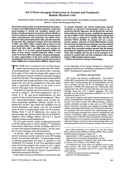
From www.bloodjournal.org by guest on February 2, 2015. For personal use only.
Ontogeny of Hematopoietic Stem Cell Development: Reciprocal Expression of
CD33 and a Novel Molecule by Maturing Myeloid and Erythroid Progenitors
By Carolyn Brashem-Stein, David A. Flowers, Franklin 0. Smith, Steven J. Staats, Robert G. Andrews, and Irwin D.Bernstein
W e have identified a molecule expressed by human
marrow granulocyte/monocyte colony-forming cells (CFUGM), erythroid colony-forming cells (CFU-E), and erythroid
burst-forming units (BFU-E), but not their precursors detectable in long-term bone marrow culture. This antigen,
detected by flow microfluorimetry using monoclonal antibody 789, is coexpressed with CD33 on many CD34+
CFCs, although only the 7B9 antigen was detected on a
portion of BFU-E and CFU-E, whereas only CD33 was
found on a portion of CFU-GM. Antibody 7B9 appears to be
useful for isolating subsets of progenitors based on their
common or selective expression of 7 8 9 antigen and CD33.
0 1993 by The American Society of Hematology.
D
(Burroughs-Wellcome, Research Triangle Park, NC) at 2 pg/mL,
for 7 days.23
To prepare marrow samples for immunization, normal bone
marrow cells were enriched for CD34+ cells using an immunoadsorbant column as previously described,24or by magnetic bead enrichment. Using this latter method, cells were incubated with 12-8
antibody (anti CD34) at 25 pg/mL for 20 minutes, washed twice
with phosphate buffered saline (PBS) plus 2% human AB serum
(GIBCO, Grand Island, NY), and then incubated with biotin conjugated goat-antimouse IgM (p chain specific) (Tago, Burlingame,
CA), at a dilution of 1530. After washing twice, magnetic streptavidin (Advanced Magnetics, Inc, Cambridge, MA) was added to cells
suspended at 107/mL,at a ratio of 5 beads/cell. Cells and beads were
rocked gently for 20 minutes at 4°C and bound cells were separated
from unbound cells in a 25 cm2tissue culture flask using a magnet.
When analyzed by immunofluorescence, CD34+-enriched marrow
cells were shown to contain at least 70% CD34-positive cells.
Leukemic cell lines were maintained in continuous culture in
RPMl I640 with 10%Bovine Calfserum (BCS) (Hyclone Laboratories, Logan, UT). The KG-1 and KG- 1 a cell lines were supplied by
Dr David Golde, University of California, Los Angeles. The other
cell lines, HL-60, HEL, K-562, U-937, Jurkat, HSB-2, Raji, Nalm1 , and Nalm-6 were kindly provided by Dr John Hansen, Fred
Hutchinson Cancer Research Center, Seattle, WA. The cell line
FMY 9S5 clone 7 is a transfected mouse L-cell line that expresses
the CD33 antigen.25It was kindly provided by Dr Thomas Look, St
Jude Children’s Research Hospital, Memphis, TN.
Antibodies. The IgM antibodies 5F1 (CD36), lGlO (CD15),
L4F3 (CD33), 12-8 (CD34), and H I2C12 (antimouse Thy 1.2), and
the IgG antibodies p67-6 (CD33), 7B9, and 31.A (antimouse Thy
1.1) were prepared as previously d e s ~ r i b e d . ~ , ~The
~ , ~anti-CD34
~,~’
antibody MY-10 was kindly provided by Dr Curt Civin, Johns
Hopkins Oncology Center, Baltimore, MD. Antibody 1F5 (CD20)
was provided by Dr Paul Martin, Fred Hutchinson Cancer Research Center. All the above antibodies were purified from ascites
fluid, and were used at a concentration of 10 pg/mL, except for
antibody 12-8, which was used at 20 pg/mL. The anti-CD2 antibody (35.1), and the anti-CD4 antibody (66.1) were used in the
form of ascites fluid at a I: 1000 dilution, and were kindly provided
by Dr Paul Martin. The directly conjugated antibodies fluorescein
isothiocyanate (F1TC)-1F5 and Cy-5-7B9 were prepared in our laboratory and were used at 50 pg/mL. The fluorochrome Cy5 was
kindly provided by Dr Swati Mujumdar, Carnegie Mellon University, Pittsburgh, PA, and antibody was conjugated with Cy-5 as
described.”
For flow cytometry studies, the following conjugated antisera
were used: FlTC goat-antimouse IgG and IgM (Tago, Inc), FlTC
goat-antimouse IgG (7chain specific, Kirkegaard & Perry, Gaithersburg, MD), and phycoerythrin (PE) goat-antimouse IgM ( p
chain-specific, Calbiochem, La Jolla, CA). All of the above antisera
were used at a dilution of 1 :40.
Monoclonal antibod-v screening and production. Balb/c mice
were immunized via the intraperitoneal route (IP) with 0.2 to 1 .O X
URING EARLY hematopoiesis, pluripotent stem
cells give rise to progenitor populations that display
progressively decreased proliferative potentials, accompanied by an increased commitment to differentiate along a
single hematopoietic lineage.’,’ Progenitor cells have been
distinguished in vitro based on their proliferation in response to specific growth factors and the nature of their
mature p r ~ g e n y . ~More
- ~ recently, it has been possible to
characterize human hematopoietic progenitors on the basis
of cell surface antigen expression.6-2’
Progenitor cells and their precursors express the CD34
antigen, whereas most progenitors express the CD33 antigen.’ In this report, we describe a novel 72 Mr molecule
(detected by antibody 7B9) that is also expressed by hematopoietic progenitors but not their precursors. A population of
progenitors, including a portion of granulocyte/monocyte
colony-forming cells (CFU-GM), erythroid colony-forming
cells (CFU-E), and erythroid burst-forming units (BFU-E),
express both the CD33 and 7B9 antigens. Those cells expressing only detectable amounts of the 7B9 antigen are
mainly committed to erythroid differentiation, whereas
those expressing only detectable amounts of CD33 are
mainly GM progenitors.
MATERIALS AND METHODS
Cells. Samples of blood and bone marrow were obtained from
normal healthy volunteers following informed consent under an
Institutional Review Board approved protocol. Peripheral blood
lymphocytes (PBL), monocytes, and granulocytes were isolated as
described by Ficoll-Hypaque density centrifugation (density of
1.077) (Pharmacia, Inc, Piscataway, NJ).” Normal thymocytes
were obtained following informed consent from patients undergoing cardiac surgery at Children’s Hospital and Medical Center,
Seattle, WA. Activated T lymphocytes were obtained as previously
described by incubating PBL in phytohemagglutinin (PHA)
From the Programs in Pediatric Oncology, Fred Hutchinson
Cancer Research Center, and the Department of Pediatrics, University of Washington, Seattle.
Submitted November 2, 1992; accepted April 6 , 1993.
Supported by the National Cancer Institute Grant No. CA39492.
Address reprint requests to Irwin D. Bernstein. MD, Pediatric
Oncology Program, Fred Hutchinson Cancer Research Center,
1124 Columbia St, Seattle, W A 98104.
The publication costs of this article were defayed in part by page
charge payment. This article must therefore be hereby marked
“advertisement” in accordance with 18 U.S.C.section 1734 solely to
indicate this fact.
0 1993 by The American Society of Hematology.
0006-4971/93/8203-0016$3.00/0
792
Blood, Voi 82,No 3 (August l ) , 1993:pp 792-799
From www.bloodjournal.org by guest on February 2, 2015. For personal use only.
793
CD33 AND 709 EXPRESSION ON PROGENITOR CELLS
Table 1. Expressionof 7 8 9 Antigen on CFCs
in Human Bone Marrow
Cells Sorted
(%)
Experiment 1
7B9+
709Unseparated
Experiment 2
709+
709Unseparated
13
85
13
86
CFU-GM
BFU-E
CFU-E
ND
564 zt 89
(54)’
74 IT 7
(46)
133 ? 7
ND
1095 f 28
(65)
89 i 8
(35)
132 i 10
687 f 127
(78)
29 2 6
(22)
60 f 7
ND
and SchleyerMand screened for the ability to support the growth in
vitro of the desired types of CFCs.
Long-term marrow cultures. Separated and unseparated marrow cells were placed in a long-term marrow culture system
(LTMC) as previously de~cribed.~’
Briefly, irradiated adherent cell
layers from 2 to 4-week-old LTMC were used as feeder layers for
isolated marrow cells. Cells were inoculated onto irradiated layers
and cultured for 5 to 7 days at 37”C, after which time cultures were
maintained in a 33°C incubator. At weekly intervals, the cultures
were fed by removing half of the culture supernatant and replacing
it with fresh medium. The nonadherent cells removed with the
culture supernatant were assayed for the presence of CFCs.
Immunofluorescence and sorting. Cells were stained usingprevi-
A
Results are the mean ? standard error of colonies expressed per 1O5
cells.
Abbreviation: ND, not done.
In parentheses are the percentage of all CFCs sorted that are in that
population. Bone marrow cells were stained and separated by indirect
immunofluorescence and fluorescence activated cell sorting, as described in Materials and Methods.
IO6 CD34+ normal bone marrow cells on days 0,7, 14 and 21, and
with 1.5 X IO6 cells intravenously(IV) on day 32. The spleens were
removed 4 days after the last immunization, and spleen cells were
fused with SP2/0 cells, as described.” Supernatants from fusion
wells were tested IO to 14 days later by enzyme-linked immunoadsorbent assay (ELISA). Supernatants were diluted 1:5 and tested
using the SBA Clonotyping System 111 (Southern Biotechnology
Assoc, Inc, Birmingham, AL) to select wells that were producing
IgG or IgM antibodies. Supernatants were then tested by two-color
indirect immunofluorescenceon normal human bone marrow by
staining the test antibody with FITC goat-antimouse IgG, and
counterstaining with antibody 12-8 followed by PE goat-antimouse
IgM for the IgG-producing wells. For the IgM-producingwells, test
antibodies were stained with the anti-IgM reagent, and CD34
stained with MY-10 followed by the anti-IgG second step. Hybridomas were cloned by limiting dilution techniques and selected for
further study if they secreted antibody that bound to a portion of
the CD34+cells, but not a large percentage of the CD34- cells. Cells
from the hybridoma line producing 7B9 were expanded in vitro and
injected into the peritoneal cavities of pristane-primedBalb/c mice.
Ascites fluid was collected from these mice and 7B9 was purified
from the ascites fluid by high pressure liquid chromatography
(HPLC).
Colony-forming assays. CFU-GM, CFU-E, and BFU-E were
identified by culturing cells in Iscove’s modified Dulbecco’s medium (IMDM) (GIBCO) supplemented with 20% fetal bovine
serum (FBS), (Hyclone), 20% human placental conditioned medium (HPCM), 2 IU/mL human erythropoietin (Amgen, Inc,
Thousand Oaks, CA),
mol/L 2-mercaptoethanol(2-ME)(BioRad Laboratories, Richmond, CA), and 0.3% agar (Seaplaque;
FMC Corp, Rockland, ME), or in methylcellulose (0.88% Terry
Fox Cancer Research Center, Vancouver, BC, Canada) with 20%
FBS, 2% Bovine Serum Albumin (Intergen CO, Purchase, NY),
HPCM, erythropoietin, and 2-ME. All cultures were incubated at
37°C with 5% CO2in air, in a humidified incubator. CFU-E were
scored after 7 to 9 days, and CFU-GM and BFU-E were scored after
14 to 16 days of culture using an inverted microscope as previously
de~cribed.2~
HPCM was prepared using the methods of Schlunck
5
n
E
3
z
Fluorescence Intensity
Fig 1. Fluorescence histogram of 7 8 9 staining on bone marrow
cells showing log fluorescence intensity (x axis) versus cell number
(y axis). W i d line is 789, dashed line is 31.A, an isotype-matched
nonspecific control antibody. The vertical lines show the sorting
windows used, with all cells to the left of the left line sorted as the
7 8 9 population and the cells to the right of the right line sorted as
the 7B9+ population. (A) One color 7B9 staining. (B) Two-color
staining with 7B9 and anti-CD34 antibody 12-8, showing 7 8 9
staining on the CD34+ population.
-
From www.bloodjournal.org by guest on February 2, 2015. For personal use only.
BRASHEM-STEIN ET AL
794
.
.. .
31.A
31.A
N
7
0
I
789
789
Log FIuoresce nce
ously described indirect immunofluorescent antibody staining techn i q u e ~ . ~ , ~For
, * * ,single-color
~~
fluorescence studies, I O7 cells/mL
were incubated with antibody for 20 minutes at 4"C, washed twice,
and then incubated in FITC-conjugated goat antimouse IgG plus
IgM antiserum for 20 minutes at 4°C. Cells were then washed twice
and kept at 4°C until analysis and cell sorting.
For two-color staining to study the coexpression of 7B9 antigen
and CD33 or CD34, cells were incubated with a mixture of 7B9
(IgG1) and the anti-CD33 antibody L4F3 or the anti-CD34 antibody 12-8 (IgM). Cells were then washed and incubated with a
mixture of FITC goat-antimouse IgG and PE goat-antimouse IgM
antisera. All stainingwas done with cellsresuspended in PBS supplemented with 2% human AB serum.
Sorting techniques for one- and two-color fluorescence have been
previously described.'." Cells considered positively stained displayed a fluorescence intensity greater than 96% to 99% of cells
stained with an isotype-matched control antibody. All analysis and
sorting was done on a modified Becton Dickinson FACS I1 flow
cytometer (Becton Dickinson, San Jose, CA).
Radiolabeling, immunoprecipitation. and gel analysis. Radioimmunoprecipitation studies were performed as de~cribed.~'
Cells
were labeled by cell surface lactoperoxidase iodination.33Cells were
washed 3 times with PBS pH 7.0 and resuspended to 5 X lo7 cells/
mL in PBS containing glucose (J.T. Baker, Phillipsburg, NJ) (90
mg/ 100 mL PBS). Iz5I(2 mCi), 20 pL glucose oxidase (Calbiochem)
(5 mg/mL) and 20 pL lactoperoxidase (Calbiochem) (70 IU/mL)
were added, in order, to 1 mL of cells at room temperature. The
cells were incubated for 20 to 25 minutes at room temperature and
then washed three times with PBS. The radiolabeled cells were lysed
with 50 mmol/L Tris-HC1 pH 7.6 (Sigma, St Louis, MO), 150
mmol/L NaC1, 2% Triton X-100 (Sigma), 2 mmol/L phenylmethylsulfonylfluoride (Boehringer Mannheim, Indianapolis, IN) and
I % (wtivol) aprotinin. The lysate was centrifuged and the supernatant was precleared with irrelevant antibody (3 l .A) sepharose con-
Fig 2 . Fluorescence scatter
analysis of bone marrow cells
stained with H12C12 (IgM control), 31.A (lgG control). 7B9.
and L4F3 (anti-CD33) is shown
as a two-dimensional representation of red (L4F3) y axis fluorescence versus green (7B9) x
axis fluorescence. The windows used for sorting have
been outlined and labeled.
(1) 7B9-CD33+, (2) 7B9+
CD33+, (3) 7B9+ CD33-. (4)
7 8 9 - CD33-.
jugate (100 p L of a I :1 suspension containing 50 pg antibody) for 20
minutes at 4"C.'4 The radiolabeled precleared lysate (200 pL) was
then incubated with each monoclonal antibody sepharose conjugate for 2 hours at 4°C. The beads were washed twice with lysis
buffer (50 mmol/L Tris, pH 8.0, 0.15 mmol/L NaCI, 20 mmol/L
EDTA, 2 mmol/L phenylmethylsulfonyl fluoride, 2% TX-100) and
twice with 50 mmol/L Tris-HC1 containing 0.5% NP-40 (Sigma)
and 150 mmol/L NaCl.
The radiolabeled protein was released by addition of 60 pL sample electrophoresis buffer (0.125 mol/L Tris-HC1,2.5% sodium dodecyl sulfate (SDS) 25% glycerol, 0.002% bromophenol blue with or
without 2.5% 2-ME) and was heated at 100°C for 5 minutes. Immunoprecipitated proteins were separated by electrophoresis in 8%
polyacrylamide gels in the presence of SDS under reducing and
nonreducing condition^.^^ The gels were dried and radiolabeled
bands identified by radioautography.
RESULTS
Expression of 7B9 antigen by normal marrow hematopoietic progenitors. Table 1 shows two of three experiments
in which normal bone marrow was separated by flow cytometry into populations that stained positively or negatively
with 7B9, and tested for the presence of CFC. The 7B9+
population was found to contain 90% of the BFU-E in both
experiments, and 78% of the CFU-E (experiment 2). This
7B9+ population was also found to contain 54% and 65% of
the CFU-GM in experiments 1 and 2, respectively. A fluorescence histogram of the staining of 7B9 on bone marrow
cells is shown in Fig 1A.
Expression oj'7B9 antigen on CD34' normal marrow progenitor ceLls. To better determine the reactivity of 7B9 on
the bone marrow progenitor population known to be vir-
From www.bloodjournal.org by guest on February 2, 2015. For personal use only.
CD33 AND 789 EXPRESSION ON PROGENITOR CELLS
795
Table 2. ExDression of 7B9 and CD33 on CFCs in Human Bone Marrow
Experiment 1
Cells sorted (%)
CFU-GM
BFU-E
CFU-E
Experiment 2
Cells sorted (%)
CFU-GM
BFU-E
CFU-E
Experiment 3
Cells sorted (%)
CFU-GM
BFU-E
CFU-E
Experiment 4
Cells sorted (%)
CFU-GM
BFU-E
CFU-E
Experiment 5
Cells sorted (%)
CFU-GM
BFU-E
CFU-E
Experiment 6
Cells sorted (%)
CFU-GM
BFU-E
CFU-E
769-033-
7B9-CD33'
18
0
0
18 2 8 (5)
66
23 f 3 (25)'
0
3 f 3 (3)
14
0
2+2(1)
BO I 12 (13)
70
81 f 13 (55)
1 i l(1)
3 t 1 (2)
10
0
0
0
7B9TD33-
7B9+CD33+
Unseparated
2
0
300 f 100 (8)
900 2 200 (30)
14
317 i 6 7 (75)
500 f 29 (92)
267 f 33 (62)
5924
35 f 6
33 2 3
2
140 F 76 (3)
842 k 182 (34)
3,333 rt 333 (75)
15
291 f 3 (42)
209 f 91 (64)
58 f 21 (10)
52 f 5
21 f 3
20 f 4
15
2,300 f 322 (93)
0
40 f 40 (22)
47 f 5
9 f 3
62 f 5
71
20 f 20 (4)
0
0
4
260 k 78 (3)
200 f 71 (100)
520 rt 136 (78)
17
7 2 7 (2)
20+0(15)
7 f 7 (10)
71
13f 13 (15)
0
0
2
250 rt 50 (8)
933 rt 17 (85)
517 60 (90)
9
500 f 60 (74)
0
0
96 f 5
4 9 f 16
17f7
11
60 f 23 (2)
27 f 7 (10)
293+47(17)
69
40 i 23 (10)
0
0
2
333 I 101 (2)
1,267 I 101 (90)
6,317 f 368 (68)
20
1,200 f 231 (85)
0
133 f 88 (14)
196 i 12
3 B i 10
179 f 10
7
0
13 i 7 (2)
220f 31 (8)
67
13 f 1 3 (7)
0
0
17 rt 17 (0.2)
683 f 109 (16)
2,900f 1,102(21)
22
533 f 186 (93)
233 f 88 (83)
633 k 120 (71)
130f2
48 2 0
136 f 12
1
Bone marrow cells were stained and separated by indirect immunofluorescence and fluorescenceactivated cell sorting, as described in Materials and
Methods. Results are the mean f standard error of triplicate cultures, expressed as colonies per 1O5 cells.
The percentage of sorted CFC detected in each population is shown in parentheses.
tually all CD34+, we analyzed the expression of 7B9 on
CD34+ bone marrow cells by two-color immunofluorescence (Fig 1B). In each of two experiments, approximately
half of the CFU-GM were in the 7B9+CD34+group (52%
and 49%), whereas the majority of BFU-E were in the
7B9TD34' group (90% and 82%).
Expression of CD33 and 7B9 antigens by normal marrow
progenitor cells. Because CD33 is also known to be expressed by most progenitors, we compared the expressionof
CD33 and 7B9 antigens on these cells using two-color immunofluorescence. Cells were separated into groups that
Table 3. Long-Term Bone Marrow Cultures
of 7B9+ and 789- Cells
Start of Culture
Unseparated
789+
789-
Week 5 of Culture
GM
BFU-E
GM
BFU-E
132210
1,095f28
89 f 8
52f5
815f67
142 1
42f19
0
29511
7f5
0
I f 1
Results are the mean +_ standard error of colonies expressed per 1O5
cells plated, cultured in triplicate. The data shown were from the same
experiment as 2 in Table 1.
did or did not stain with 7B9 and/or anti-CD33 antibody
(Fig 2). The majority of sorted BFU-E (85% to 100%)and
CFU-E (82%to 100%)was found in either the 7B9+CD33+
or 7B9+CD33- groups. In six experiments, a range of 0%to
Table 4. Reactivity of 7B9 W i t h Normal Bone Marrow Cells
Determined by Indirect Immunofluorescence and Cell Sorting
Myeloblast
Immature myeloid
Mature myeloid
Erythroblast
Immature erythroid
Monocyte
Eosinophil
Basophil
Lymphocyte
7B9+
789.
8.7 i 5.5
43.3 rt 18.8
8.3 f 3.2
4.0 1 .O
4.7 f 4.6
9.7 f 10.3
19.7f 13.9
1 .o rt 1 .o
0.7 f 0.6
1 .o f 1 .o
53.3 f 11.5
30.7 i 4.0
0.3 0.6
9.0 i 6.9
1 .o i 1 .o
1.3 1.5
0
4.0 f 5.3
*
*
*
Numbers represent the mean and standard deviation of three different
sorts in which 100 cell differentials were performed on Wright-Giemsa
stained cytocentrifuge preparations. The immature myeloid group contains promyelocytes, myelocytes, and metamyelocytes; mature myeloid is bands and PMN's; and immature erythroid is all nucleated red
blood cell precursors except erythroblasts.
From www.bloodjournal.org by guest on February 2, 2015. For personal use only.
BRASHEM-STEIN ET AL
796
KG-I
I
HL-60
Fluorescence Intensity
92% of the BFU-E was in the 7B9+CD33+group, with 8% to
100%ofthe BFU-E in the 7B9+CD33- group. Similarly, 0%
to 7 1% of the CFU-E sorted into the 7B9+CD33+group, and
21% to 90% of the C N - E sorted into the 7B9TD33group. A minor portion of BFU-E and C N - E expressed
neither the 7B9 or CD33 antigens (Table 2).
The majority of sorted CFU-GM (89% to 100%)was
found in the 7B9-CD33+ or the 7B9+CD33+groups, with
only 0% to 10%of CEU-GM found in the 7B9+CD33- or
7B9-CD33- groups. The CFU-GM were predominantly in
the 7B9+CD33+group in the six experiments (42% to 93%).
In these six experiments, we also found that most of the
CFU-GM were 7B9+ (45% to 96%). In one experiment, we
also assessed the morphology of the CFU-GM by picking
colonies from the methylcellulose plates and performing
differentials on Wright-stained cytospin preparations. Colonies derived from both the 7B9-CD33+ and 7B9+CD33+
groups showed 5 of 10 colonies picked contained granulocyte precursors, with the remaining cells being monocytes
and macrophages (data not shown).
Lack of expression of 7B9 antigen by LTMC initiating
cells. We tested the ability of isolated 7B9+ and 7B9marrow cells to generate or maintain CFC in a long-term
culture system. Sorted cells were cultured in the presence of
previously established irradiated marrow stromal cell layers.
The entire layers were harvested after 5 weeks of culture and
tested for CFC activity. The cultures of 7B9- cells were
found to contain CFU-GM and BFU-E after 5 weeks in
culture, whereas the 7B9+-sorted cells did not, suggesting
the presence of precursors of both CFU-GM and BFU-E in
the 7B9- but not the 7B9+ population (Table 3).
Normal peripheral blood and bone marrow cells. In four
experiments looking at peripheral blood mononuclear cells
by immunofluorescence and gating on cells with high forward light scatter, 68.4% k 2.9% ofthe cells reacted with the
anti-CD36 antibody 5F1, and 81.3% +- 5.4% of the cells
reacted with 7B9. In contrast, no reactivity with granulocytes was found in two-color studies with 7B9 and the anti-
Fig 3. Fluorescence histogram of 7B9 staining
on hematopoietic cell lines, showing fluorescence
intensitv (x axis) versus cell number I..
v axis).
. Solid
line is .769, dashed line is 31.A. an isotypematched nonspecific control antibody.
CD15 antibody 1GIO. Platelets, which stained positively
with the anti-CD42b antibody C7E10, also all reacted with
7B9 (data not shown).
Reactivity of 7B9 with a portion of T lymphocytes was
shown by gating around the lymphocyte sized population
based on forward and right angle light scatter characteristics. In four experiments, 82.7% f 0.5% of this population
consisted of T cells, as determined by staining with antiCD2 antibody 35.1. In this same population, 36.9% f 5.4%
of the cells reacted with 7B9; therefore, at least 19.6%of this
population must coexpress 7B9 and 35.1. In two color studies, no reactivity of 7B9 was seen with CD4+ cells, detected
by antibody 66.1. Antigen 7B9 was also detected on 80% of
PHA-activated T cells, and 25% of thymocytes. Studies of
B-lymphocyte reactivity, tested by using direct conjugates
of an anti-CD20 antibody (FITC-IF5) and 7B9 (Cy-5-789)
revealed low levels of 7B9 antigen on 34% of CD20+ cells
(data not shown).
In bone marrow, 7B9 stained 10% to 15% of mononuclear cells, of which 70% to 80% displayed high forward light
scatter. Cytospin preparations of positively stained marrow
cells selected by cell sorting showed mainly myeloid cells
including myeloblasts, immature myeloid cells and eosinophils, erythroblasts, and small percentages of immature erythroid cells (Table 4).
Leukemic cells and cell lines. Our panel of human hematopoietic cell lines was analyzed three times for 7B9 reactivity (Fig 3). The myeloid cell lines HEL, HL-60, and
U-937, the B-lymphocyte lines Raji and Nalm-6, and the
T-lymphocyte line HSB-2 all were reactive with antibody
7B9. In addition, the myeloid line KG-I showed a slight
shift in its fluorescence intensity when stained with 7B9
compared with the negative control antibody, and a small
number of the cells of the T-cell line Jurkat expressed 7B9
antigen. The cell line K562 was negative for 7B9. The myeloid cell line KG-la and the mouse L cell line FMY 9S5
Clone 7, which expresses CD33, were 7B9 negative (data
not shown).
From www.bloodjournal.org by guest on February 2, 2015. For personal use only.
CD33 AND 789 EXPRESSION ON PROGENITOR CELLS
797
HEL
/.*
RAJ1
.
.. ,,..,,...
'.*:s$r
.
-200
-97
-69
97
69
-46
,46
-30
.30
__
R
NR
R
NR
Fig 4. Immunoprecipitation of the 789 antigen from HE1and Raji cells under reducing (R) and nonreducing (NR) conditions. Antibodies
789. p67-6 (anti-CD33). 1F5 (anti-CDPO), and 31.A (an irrelevant control antibody) were tested. Molecular weight was determined with
['4ClLeucine-labeled markers (New England Nuclear, Boston, MA). On Raji cells, 789 precipitates a broad band of 63 to 73 Mr under
reducing conditions, and a band of 60 to 70 Mr under nonreducing conditions. On HEL cells, 789 precipitates a band of 56 to 90 Mr under
reducing conditions, and a band of 56 to 82 Mr under nonreducing conditions. On the Raji cells, the positive control, precipitated with
antibody 1F5 (anti-CDPO),yielded a band at 37 Mr, just above a nonspecific band in all lanes. The negative control lysate, precipitated with
antibody 31 .A, showed a nonspecific band at 75 Mr in the reducing lanes and severalfaint bands in the nonreducinglanes. On the HELcells,
the positive control p67-6 (antLCD33 antibody) precipitated a broad band just below the 69 Mr marker on reduction and slightly lower in the
nonreducinglane. Both 789 and p67-6 precipitated a nonspecific band at 35 Mr, and antibody 31 .A precipitated one nonspecific protein at
58 Mr under reducing conditions.
Leukemia cells in IO of 14 acute lymphocytic leukemia
(ALL) specimens, including 3 of 3 T-cell leukemias, bound
7B9 with between 35% and 85% ofthe cells showing reactivity. Four of five bone marrows from patients with acute
myeloid leukemia (AML) were also found to express 7B9
antigen (data not shown).
Characterization of the cell &ace molecule recognized
by 7B9. Radioiodinated Raji cell lysates were precipitated
with antibody 7B9 and electrophoresed. Under reducing
conditions, a broad band of 63 to 73 Mr was consistently
observed, whereas nonreducing conditions showed a band
of slightly lower molecular weight, 60 to 70 Mr. (Fig 4).
HEL cell lysates precipitated with 7B9 showed a broader
band in the reducing lane of 56 to 90 Mr. The nonreducing
band is slightly lower at 56 to 82 Mr. The broad bands ofthe
7B9 antigen precipitated from both cell lines are consistent
with profiles seen for heavily glycosylated proteins.
DISCUSSION
The identification of cell surface antigens expressed by
hematopoietic precursors at different stages of maturation
has led to the development of methods for purifying distinct
subpopulations of these cells. Thus, it has been possible to
distinguish CFCs committed to express a single lineage from
less mature cells capable of establishing hematopoiesis in
long-term culture or giving rise to multipotential blast colony-forming
The former cells have been
shown to express the CD34 antigen as well as a variety of
determinants associated with differentiative expression
along the various hematopoietic lineages, including CD33
and HLA/DR, whereas the less mature cells appear to lack
these differentiation antigens.
In the present study, we have identified a novel epitope,
identified by 7B9, that within the population of CD34+
marrow cells is, like CD33, expressed by CFCs and not their
precursors detectable in long-term culture. In these experiments, virtually all of the BFU-E were 7B9+,whereas a variable percentage of CFU-GM were 7B9+. Of particular interest is the observed differential expression of the 7B9 and
CD33 antigens on subsets of CFCs, with sole detection of
7B9 antigen on a portion of BFU-E and CFU-E, but only
rarely on CFU-GM; and sole detection of CD33 on a portion of CFU-GM, but only rarely on BFU-E or CFU-E. The
remaining progenitors display a 7B9+CD33+ phenotype
From www.bloodjournal.org by guest on February 2, 2015. For personal use only.
798
and consist of a mixture of erythroid and myeloid progenitors. Thus, the populations expressing only one of the two
antigens displayed greater enrichment for either erythroid
or myeloid differentiation than the cells expressing both antigens, which displayed both differentiative potentials.
These observations are consistent with the idea that CFCs
selectively lose o r decrease their expression of 7B9 antigen
as they differentiate along the granulocyte/monocyte pathway, or lose or decrease their expression of CD33 as they
differentiate along the erythroid lineage. Furthermore, we
do not know if the cells completely cease to express one of
the antigens, or if they continue to express one antigen in a n
amount that falls below our threshold ofdetection. Nonetheless, it is possible for antibodies detecting these antigens to
be used to enrich for myeloid or erythroid subsets of CD34'
progenitors.
More intriguing is a potential role of these surface antigens in regulating the differentiative and proliferative potential of these cells. To date, we have been unable to influence the proliferation of CD34+ marrow cells in the
presence ofhematopoieticgrowth factors by adding antibodies against these antigens either in solution or by adherence
to plastic (data not shown). Although the CD33 gene has
been cloned,"' sequence data has not provided sufficient
information to determine function. However, sequence homology between CD22 and myelin-associated glycoprotein
has been observed."' The former has been found to have a
role in signaling B cells to proliferate, whereas the latter is
thought to play a role in cell adhesion. Nevertheless, it is
tempting to speculate that the common expression of 7B9
antigen and CD33 followed by exclusive decrease o r loss of
one of these antigens by hematopoietic progenitors of different types suggests these molecules may play a role in influencing the differentiative expression of these cells. This
could result, for example, if binding of these molecules to
the appropriate ligand provided a synergistic signal in the
response to growth factors, or if this binding interaction
would cause these cells to bind in a n appropriate niche in
the marrow space.
The chemical nature of the 7B9 determinant is uncertain.
Although it has been possible to precipitate a protein or
glycoprotein from cells, the broad band o n polyacrylamide
gel electrophoresis and differences in the molecular weight
observed using different cell lines is consistent with the possibility that the epitope detected represents a carbohydrate
determinant. This would also explain the lack of success in
efforts to clone the gene encoding for this antigen (datanot
shown). However, the antigen that we detected does appear
novel because a similar one has not been previously described. Antibody 7B9 shows some similarity in its patterns
of reactivity to the previously described antigen RFB- 1,'5,41
but differences in cell line reactivity suggest that this may
not be the case, and biochemical data on the nature of the
RFB- I antigen is not available.
The development of antibody 7B9 has practical implications. Subsets of maturing progenitors could be isolated
based o n the common o r selective expression of 7B9 antigen
and CD33. Availability of these highly purified progenitors
as well as those expressing variable amounts of 7B9 antigen
BRASHEM-STEIN ET AL
and/or CD33 should provide cell populations useful for further assessment of the selective actions of recombinant hematopoietic growth factors o n subsets of progenitor populations.
REFERENCES
1. Till JE, McCulloch EA: A direct measurement of the radiation sensitivity of normal mouse bone marrow cells. Radiat Res
14:213, 1961
2. Dick JE, Magli MC, Huszar D, Phillips RA, Bernstein A: Introduction of a selectable gene into primitive stem cells capable of
long-term reconstitution of the hematopoietic system in W/Wv
mice. Cell 42:71, 1985
3. Bradley TR, Metcalf D The growth of mouse bone marrow
cells in vitro. Aust J Exp Biol Med Csi 44:287, 1966
4. Pike BL, Robinson WA: Human bone marrow colony growth
in agar gel. J Cell Physiol 76:77, 1970
5. Stephenson JR, Axelrad AA, McLeod DL, Shreeve MM: Induction of coloniesof hemoglobin-synthesizingcells by erythropoietin in vitro. Proc Natl Acad Sci USA 68: 1542, 197 1
6. Andrews RG, Torok-Storb B, Bernstein ID Myeloid-associated differentiation antigens on stem cells and their progeny identified by monoclonal antibodies. Blood 62:124, 1983
7. Andrews RG, Singer JW, Bernstein ID: Precursors of colonyforming cells in humans can be distinguishing from colony-forming
cells by expressionof the CD33 and CD34 antigens and light scatter
properties. J Exp Med 169:1721, 1989
8. Griffin JD, Ritz J, Nadler LM, Schlossman S F Expression of
myeloid differentiation antigens on normal and malignant cells. J
Clin Invest 68:932, 1981
9. Griffin JD, Linch D, Sabbath K, Larcom P, Schlossman S F A
monoclonal antibody reactive with normal and leukemic myeloid
progenitor cells. Leuk Res 8:52 1, 1984
10. Civin CI, Strauss LC, Brovall C, Fackler MJ, Schwartz SJ,
Shaper JS: Antigenic analysis of hematopoiesis. 111. A hematopoietic progenitor cell surface antigen defined by a monoclonal antibody raised against KG- la cells. J Immunol 133:157, 1984
11. Tindle RW, Nichols RAB, Chou L, Campana D, Catovsky
D, Birnie GD: A novel monoclonal antibody B1-3C5 recognizes
myeloblasts and non-B non-T lymphoblasts in acute leukemias,
CGL in blast crisis, and reacts with immature cells in normal
marrow. Leuk Res 9:1, 1985
12. PapayannopoulouTh, Brice M, Yokochi T, Rabinovitch PS,
Lindsley D, Stamatoyannopoulos G: Anti-HEL cell monoclonal
antibodiesrecognize determinants that are also present on hemopoietic progenitors. Blood 63:326, 1984
13. Strauss LC, Stuart LC, Civin CI: Antigenic analysis of hematopoiesis. I. Expression of the MY-l granulocytesurface antigen on
marrow cells and leukemic cell lines. Blood 61:1222, 1983
14. Strauss LC, Brovall C, Fackler MJ, Schwartz JF, Shaper JH,
Loken MR, Civin CI: Antigenic analysis of hematopoiesis IV. The
MY-1 1 hematopoietic cell surface antigen is expressed by myelomonocytic and lymphoid, but not erythroid, progenitor cells. Exp
Hematol 14:935, 1986
15. Bcdger M, Izaguirre CA, Blacklock HA, Hoffbrand AV: Surface antigenic determinants on human pluripotent and unipotent
hematopoietic progenitor cells. Blood 6 1: 1006, 1983
16. Ferrero DH, Broxmeyer HE, Pagliardi GL, Venuta S , Lange
B, Passano S, Rovera G: Antigenically distinct subpopulations of
myeloid progenitor cells (CFU-GM) in human peripheral blood
and marrow. Proc Natl Acad Sci USA 80:4114, 1985
17. Ferrero D, Gabbianelli M, Peschle C, Lange B, Rovera G:
Surface phenotypes of human hemopoietic progenitor cells defined
by monoclonal antibodies. Blood 66:496, 1985
From www.bloodjournal.org by guest on February 2, 2015. For personal use only.
CD33 AND 7 8 9 EXPRESSION ON PROGENITOR CELLS
18. Sutherland HJ, Eaves CJ, Eaves AE, Dragowska W, Lansdorp PW: Characterization and partial purification of human
marrow cells capable of initiating long-term hematopoiesis in vitro.
Blood 74:1563, 1989
19. Verfaille C, Blakolmer K, McGlave P: Purified primitive human hematopoietic progenitor cells with long-term in vitro repopulating capacity adhere selectively to irradiated bone marrow
stroma. J Exp Med 172:509, 1990
20. Brandt J, Baird N, Lu L, Svour E, Hoffman R: Characterization of a human hematopoietic progenitor cell capable of forming
blast cell containing colonies in vitro. J Clin Invest 82:1017, 1988
21. Knapp W, Diirken B, Gilks WR, Rieber EP, Schmidt RE,
Stein H, Von dem Bome AEG Kr, editors: Leucocyte typing IV:
White Cell Differentiation Antigens. Oxford, UK, Oxford, 1990
22. Bernstein ID, Andrews RG, Cohen SF, McMaster BE Normal and malignant human myelocytic and monocytic cells identified by monoclonal antibodies. J Immunol 128976, 1982
23. Brashem-Stein C, Nugent D, Bernstein I D Characterization
of an antigen expressed on activated human T cells and platelets. J
Immunol 140:2330, 1988
24. Berenson RJ, Andrews RG, Bensinger WI, Kalamasz D,
Knitter G, Buckner CD, Bernstein ID: CD34+marrow cells engraft
lethally irradiated baboons. J Clin Invest 81:951, 1988
25. Look TA, Peiper SC, Douglas EC, Trent JM, Scherr CJ: Amplification of genes encoding human myeloid membrane antigens
after DNA-mediated gene transfer. Blood 67:637, 1986
26. Andrews RG, Singer JW, Bernstein I D Monoclonal antibody 12-8 recognizes a 115-Mr molecule on both unipotent and
multipotent colony-forming cells and their precursors. Blood
67342, 1986
27. Denkers EY, Badger CC, Ledbetter JA, Bernstein ID: Influence of antibody isotype on passive serotherapy of lymphoma. J
Immunol 135:2183, 1985
28. Mujumdar RB, Ernst LA, Mujumdar SR, Lewis CJ, Wagner
A: Cyanine dye labeling reagents: Sulfoindocyanine succinimidyl
esters. Bioconjugate Chem 4:105, 1993
29. Powell JS, Fialkow PJ, Adamson JW: Human mixed cell
colonies: unicellular or multicellular origin-analysisby Gd-PD. Br
J Haematol 57:89, 1984
30. Schlunck T, SchleyerM: The influence of culture conditions
799
on the production of colony stimulating activity by human placenta. Exp Hematol 8:179, 1980
3 I . Andrews RG, Takahashi M, Segal GM, Powell JS, Bemstein
ID, Singer JW: The L4F3 antigen is expressed by unipotent and
multipotent colony-forming cells but not by their precursors. Blood
68:1030, 1986
32. Pesando JM, Ritz J, Lazarus H, Tomaselli KJ, Schlossman
S F Fate of a common acute lymphoblastic leukemia antigen during modulation by monoclonal antibody. J Immunol 126:540,
1981
33. Hubbard AL, Cohn Z A The enzymatic iodination ofthe red
cell membrane. J Cell Biol 55:390, 1972
34. Axen R, Porath J, Ernbach S: Chemical coupling ofpeptides
and proteins to polysaccharidesby means of cyanogen halides. Nature 241:1302, 1967
35. Laemmli U K Cleavage of structural proteins during the assembly ofthe head of the bacteriophage T-4. Nature 227:680, 1970
36. Andrews RG, Singer JW, Bemstein I D Human hematopoietic precursors in long term culture: Single CD34+ cells that lack
detectable T, B, and myeloid antigens produce multiple colony
forming cells when cultured with marrow stromal cells. J Exp Med
172:355, 1990
37. Bemstein ID, Leary AG, Andrews RG, Ogawa M: Blast colony forming cells and precursors of colony forming cells detectable
in long term marrow culture express the same phenotype
(CD33-CD34'). Exp Hematol 19:680, 1991
38. Reinherz EL, Haynes BF, Nadler LM, Bernstein ID (eds):
Leukocyte typing 11, vol 1-111. New York, NY, Springer-Verlag,
1986
39. McMichael AJ, Beverley PCL, Cobbold S, Crumpton MJ,
Gilks W, Gotch FM, H o g N, Horton M, Ling N, MacLennan
ICM, Mason DY, Milstein C, Spiegelhalter D, Waldmann H, editors: Leucocyte typing 111: White cell differentiation antigens. Oxford, UK, Oxford, 1987
40. Simmons D, Seed B: Isolation of a cDNA encoding CD33, a
differentiation antigen of myeloid progenitor cells. J Immunol
141:2797, 1988
4 1. Bodger MP, Francis GE, Delia D, Granger SM, Janossy G A
monoclonal antibody specific for immature human hematopoietic
cells and T lineage cells. J Immunol 127:2269, 1981
From www.bloodjournal.org by guest on February 2, 2015. For personal use only.
1993 82: 792-799
Ontogeny of hematopoietic stem cell development: reciprocal
expression of CD33 and a novel molecule by maturing myeloid and
erythroid progenitors
C Brashem-Stein, DA Flowers, FO Smith, SJ Staats, RG Andrews and ID Bernstein
Updated information and services can be found at:
http://www.bloodjournal.org/content/82/3/792.full.html
Articles on similar topics can be found in the following Blood collections
Information about reproducing this article in parts or in its entirety may be found online at:
http://www.bloodjournal.org/site/misc/rights.xhtml#repub_requests
Information about ordering reprints may be found online at:
http://www.bloodjournal.org/site/misc/rights.xhtml#reprints
Information about subscriptions and ASH membership may be found online at:
http://www.bloodjournal.org/site/subscriptions/index.xhtml
Blood (print ISSN 0006-4971, online ISSN 1528-0020), is published weekly by the American
Society of Hematology, 2021 L St, NW, Suite 900, Washington DC 20036.
Copyright 2011 by The American Society of Hematology; all rights reserved.
© Copyright 2026 Paperzz