2
Pediatric
Dermatology
Contents
2.1
Neonatal Dermatology ................................................................................................................................................
28
2.2
Childhood Infectious Diseases ...................................................................................................................................
32
2.3
Papulosquamous and Eczematous Dermatoses .......................................................................................................
36
2.4
Pigmented Lesions ......................................................................................................................................................
38
2.5
Bullous Diseases ..........................................................................................................................................................
41
2.6
Epidermal, Appendageal, and Dermal Tumors .......................................................................................................
43
2.7
Tumors of Fat, Muscle and Bone ...............................................................................................................................
47
2.8
Vascular Disorders ......................................................................................................................................................
48
2.9
Genodermatoses ..........................................................................................................................................................
52
S. Jain, Dermatology,
DOI 10.1007/978-1-4419-0525-3_2, В© Springer Science+Business Media, LLC 2012
27
DERMATOLOGY: ILLUSTRATED STUDY GUIDE AND COMPREHENSIVE BOARD R EVIEW
2.1
NEONATAL DERMATOLOGY
A
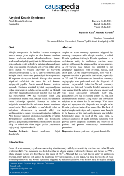
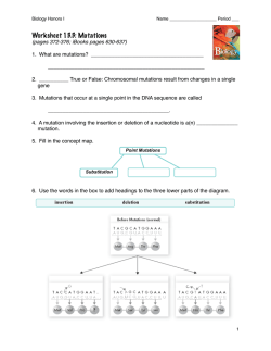
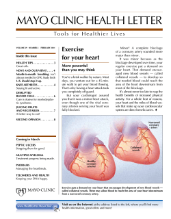
Transient Neonatal Pustular Melanosis (Figure 2.1A)
• Onset at birth; common in darkly pigmented infants
• Presents with small pustules or residual hyperpigmented macules with
collarette of scale
• Smear of sterile pustule shows numerous neutrophils
• Histology: subcorneal pustules with neutrophils
Erythema Toxicum Neonatorum
• Onset typically 24–48 h after birth; occurs in half of all full-term infants
• Presents with blotchy erythematous macules, papules, pustules, and
wheals
• Smear of sterile vesicle/pustule shows eosinophils
• Histology: subcorneal pustules with eosinophils, associated with
pilosebaceous unit
B
Neonatal Cephalic Pustulosis (Neonatal Acne) (Figure 2.1B)
• Onset typically within first 30 days; Malassezia spp. implicated in
pathogenesis
• Presents with erythematous follicular comedones, papules, and pustules on
face
• Histology: follicular pustules with neutrophils
Sclerema Neonatorum
• Onset usually within first week of life; form of panniculitis in severely ill,
premature infants; often fatal
• Presents with diffuse woody hardening of skin; spares genitalia, palms,
and soles
• Histology: needle-shaped clefts with necrotic adipocytes with
little surrounding inflammation
C
Subcutaneous Fat Necrosis of the Newborn (Figure 2.1C)
• Onset within first weeks of life; localized form of sclerema neonatorum in
healthy infants
• Presents with indurated subcutaneous nodules favoring cheeks, shoulders,
back, buttocks, and thighs
• Associated with hypothermia, perinatal hypoxemia (from preeclampsia,
meconium aspiration, etc.), hypoglycemia
• Calcification may occur; ± profound hypercalcemia with resolution, so
prudent to monitor calcium levels until 1 month after full resolution of
lesions
• Histology: panniculitis with prominent inflammatory infiltrate,
needle-shaped clefts and fat necrosis
Pedal Papules of Infancy
• Soft, non-painful papules involving heels
28
Figure 2.1
A: Neonatal pustular melanosis*
B: Neonatal cephalic pustulosis
(Reprint from Boekhout T, GuehoKellerman E, Mayser P, Velegraki A.
Malassezia and the Skin. New York, NY:
Springer; 2010)
C: Subcutaneous fat necrosis*
*Reprint from Laxer RM, ed. The Hospital
for Sick Children: Atlas of Pediatrics.
Philadelphia, PA: Current Medicine; 2005
PEDIATRIC DERMATOLOGY
Seborrheic Dermatitis (Figure 2.2A)
• Onset typically 1 week after birth; lasts several months, mostly resolves by
1 year of age
• Presents with ill-defined erythematous patches with waxy scale over scalp
(“cradle cap”), ± axillae and groin; lesions may appear psoriasiform
A
Miliaria Crystallina (MC) or Miliaria Rubra (MR)
• Onset within first few weeks of life; due to obstructed sweat glands and
associated with ↑ temperature (i.e., occlusion)
• Presents with clear vesicles favoring head, neck, and upper trunk (MC) or
erythematous papules/vesicles grouped in intertriginous areas or occluded
areas (MR)
Aplasia Cutis Congenita (ACC) (Figure 2.2B, C)
• Onset before birth; localized defect in epidermis, dermis and/or fat;
variable appearance, typically along midline
• Presents with erosion, ulceration, scar, or membranous defect
(ovoid lesion covered by an epithelial membrane)
• Hair collar sign: ring of dark long hair encircling lesion; ± marker of
underlying neural tube defect
• Typically isolated abnormality, but may be associated with developmental
anomalies or following disorders:
Bart Syndrome
Adams–Oliver
Syndrome
Seitles Syndrome
B
ACC of lower extremities + epidermolysis bullosa
(dominant dystrophic)
ACC on scalp (with skull ossification defect)
+ extensive CMTC + limb defects (reductions,
syndactyly) + cardiac abnormalities
Bilateral temporal ACC + abnormal eyelashes,
“leonine” facies, upward-slanting eyebrows
Cutis Marmorata Telangiectatica Congenita (CMTC)
• Onset at birth; typically improves with age
• Presents with blanching reticulated vascular pattern on trunk/extremities
with segmental distribution
• Associated anomalies in ½ of patients (varicosities, nevus flammeus, macrocephaly, ulceration, hypoplasia, and/or hypertrophy of soft tissue and bone)
C
Sucking Blister
• Onset at birth or soon after; due to sucking
• Presents with solitary blister (hand, wrist, or lip)
Congenital Infections of the Newborn (see Table 2-1)
Differential Diagnosis of �Diaper Dermatitis’ (see Table 2-2)
Figure 2.2
A: Seborrheic dermatitis
B: ACC, cicatricial
(Courtesy of Dr. Michelle B. Bain)
C: ACC, bullous
(Courtesy of Dr. Michelle B. Bain)
29
DERMATOLOGY: ILLUSTRATED STUDY GUIDE AND COMPREHENSIVE BOARD R EVIEW
Infection
Cytomegalovirus
(CMV)
Herpes Simplex
Virus
(HSV)
Rubella
Toxoplasmosis
Varicella
Syphilis, Early
Congenital
Syphilis, Late
Congenital
30
Table 2-1 Congenital Infections of the Newborn
Clinical Findings
Extracutaneous Findings
Important Points
Petechiae, purpura,
Intrauterine growth retardation, в‡’ Leading infectious cause
vesicles, and “blueberry
chorioretinitis, intracranial
of deafness and mental
muffin” lesions
calcification
retardation
в‡’ Typical findings on
histology: enlarged
Blueberry muffin lesions: red-blue papules/nodules due to dermal
endothelial cells with
erythropoiesis
intranuclear inclusions
Localized or disseminated
Encephalitis (predilection for
в‡’ Majority HSV2, 85%
skin lesions (vesicles,
temporal lobes), multi-organ
acquired perinatally
erosions, scarring)
failure, ocular infection
⇒ 50–75% mortality if left
untreated
“Blueberry muffin”
Cataracts, deafness, congenital в‡’ 50% chance of deafness
lesions
heart disease, CNS findings
в‡’ Severe birth defects if
(microcephaly, hydrocephaly),
within first 16 weeks of
hepatosplenomegaly (HSM)
pregnancy
в‡’ Non-immune pregnant
woman transfer the virus
to the fetus
“Blueberry muffin”
Ocular abnormalities
lesions favoring the
(chorioretinitis, blindness),
trunk
CNS abnormalities
(deafness, mental
retardation, seizures),
thrombocytopenia,
intracranial calcification
Cicatricial skin lesions
Ocular abnormalities
в‡’ Greatest risk in first
(chorioretinitis, cataracts),
20 weeks
cortical atrophy, psychomotor в‡’ 2% risk of embryopathy
retardation, hypoplastic limbs
in women with infection
within first two trimesters
Syphilitic pemphigus,
Snuffles (rhinitis, secondary
в‡’ Early congenital syphilis
rhagades (radial furrows/
to ulcerated mucosa),
occurs from birth to
fissures in perioral area,
enlarged lymph nodes and
2 years of age
spleen, neurosyphilis
turn into parrot lines),
в‡’ Only congenital syphilis
papulosquamous macules/
may show bullous
Be able to differentiate early
papules (like secondary
lesions
and late congenital syphilis
syphilis)
в‡’ Papulosquamous lesions
findings
common in the diaper
area
Hutchinson’s teeth,
Interstitial keratitis, gummas
в‡’ Includes permanent
Higoumenakis sign, mulberry along long bones/skull, tabes
sequelae of early
molars, saddle nose, saber
dorsalis, generalized paresis
congenital signs
shins, parrot lines and furrows
в‡’ Higoumenakis sign:
congenital thickening of
the medial aspect of the
clavicle
PEDIATRIC DERMATOLOGY
Table 2-2 Differential Diagnosis for Diaper Dermatitis
Clinical Findings
Bright red patches with pustules and satellite papules,
В± intertriginous involvement (including scrotum), В± thrush
Irritant Dermatitis
Poorly demarcated erythematous plaques, spares inguinal folds
Seborrheic Dermatitis
Typical salmon-covered scaly patches and plaques involving
the scalp, groin, and other intertriginous areas
Psoriasis
Sharply demarcated bright pink to red plaques involving
inguinal creases, minimal scale; most common psoriatic
presentation in infants
Allergic Contact Dermatitis
Rare in infants, В± related to topical preparations or foods
Atopic Dermatitis
Increased incidence of diaper dermatitis in atopic patients
Miliaria
Clear vesicles or erythematous papules/pustules due to blocked
eccrine ducts from heat or humidity in diaper area
Granuloma Gluteale Infantum
Red to violaceous granulomatous nodules over the vulva,
perianal area, buttocks, В± scrotum; due to irritation, occlusion,
candidal infection
Perianal Pseudoverrucous Nodules
Erythematous nodules and papules in children with fecal
incontinence
Acrodermatitis Enteropathica
Erythematous crusted patches/plaques with flaccid bullae in
perineal, periorificial, and distal extremities; due to ↓ zinc level
(also ↓ alkaline phosphatase as zinc-dependent); may occur in
following settings:
1. Premature infants (poor absorption and ↑ requirement
of zinc) when weaned off breast milk (which has adequate
zinc level)
2. Inherited form (AR) manifests when weaned off breast milk
3. Healthy infants if low zinc level in maternal milk
4. Acquired form if malabsorption or inadequate nutrition
Cystic Fibrosis
Resembles acrodermatitis enteropathica, also due to zinc
deficiency В± pedal edema, failure to thrive, infections and
malabsorption
Multiple Carboxylase Deficiency
Both resemble acrodermatitis enteropathica (periorificial
dermatitis); treatment for both forms (listed below) is biotin
Biotin Deficiency
1. Neonatal form: AR, holocarboxylase synthetase deficiency,
В± erythroderma with alopecia, fatal if not treated
2. Juvenile form: biotinidase deficiency, В± seizures, alopecia,
hearing loss, developmental delay
Langerhans Cell Histiocytosis
Yellow-brown crusted papules with purpura in seborrheic
distribution; В± systemic involvement; Langerhans cells
(CD1a +, S100+)
Kawasaki Disease
Tender erythema in perineal area which later desquamates
Perianal Strep
Bright red, well-demarcated perianal erythema and involving
creases
Bullous Impetigo
Honey-colored crusts and flaccid bullae
Scabies
Erythematous nodules involving diaper area, В± genitalia
Congenital Syphilis
Reddish-brown papulosquamous eruption, may be erosive or
bullous
Entity
Candidal Dermatitis
31
DERMATOLOGY: ILLUSTRATED STUDY GUIDE AND COMPREHENSIVE BOARD R EVIEW
2.2
CHILDHOOD INFECTIOUS DISEASES
Disease
Acute Hemorrhagic
Edema of Infancy
(Finkelstein Disease)
Erythema Infectiosum
(�Slapped Cheek’ or
Fifth Disease)
Gianotti–Crosti
Syndrome
Hand-Foot-Mouth
Disease
Henoch–Schönlein
Purpura (HSP)
Herpangina
Kawasaki Disease
(Mucocutaneous Lymph
Node Syndrome)
Measles (Rubeola or
First Disease)
32
Table 2-3 Childhood Infections
Exanthem
Etiology/Course
Large circinate painful purpuric
Etiology: likely infectious (viral or bacterial)
plaques involving face, ears, distal
Age: 6 months–3 years; self-limited
extremities в†’ evolve into edematous
Leukocytoclastic vasculitis seen on histology
targetoid lesions
May be hypersensitivity reaction to infection
(medication/vaccination less likely)
Bright red macular erythema over cheeks
Etiology: parvovirus B19 (ssDNA) also causes
в†’ lacy eruption mainly on the extremities hydrops fetalis; peaks in spring and winter
Age: school-age children; self-limited
Mild prodrome, 10% with arthralgias
Etiology: likely infectious (HBV, EBV)
Abrupt onset of skin-colored to pink-red
edematous papules to cheeks, buttocks,
Age: 6 months–2 years; self-limited
extremities
May have low-grade fever and
lymphadenopathy
Elliptical grayish vesicles, pustules,
Etiology: coxsackievirus A16
and erosions on hands, feet, and buttocks
(enterovirus 71 less often)
Age: children <10 years (В± adults); self-limited
Oral: vesicles/erosions red base
Fever, sore mouth, anorexia, abdominal pain;
enteroviral infection may also cause myocarditis, pneumonia, meningoencephalitis
Purpuric macules and papules
Etiology: possibly infectious (viral, strep)
favoring lower extremities and
Age: peaks at 4–7 years (± adults); self-limited
buttocks
Presents 1–2 week after upper respiratory
infection
Arthralgias, GI bleeding, abdominal pain,
nephritis with hematuria в†’ IgA vasculitis
Exanthem: often absent
Etiology: various enteroviruses
(often coxsackie group A/B and echovirus)
Oral: painful gray vesicles on
Age: 3–10 years old; self-limited
tonsillar, palate, buccal mucosa
Polymorphous eruption
Etiology: unknown but likely infectious
(morbilliform, erythema multiforme-like
Age: children <5 years of age
or bullous); В± edema and erythema of
Arthritis, abdominal pain, GI symptoms
distal extremities; can be generalized or
localized (groin, LE)
Complications: cardiac aneurysm (in Вј of
untreated patients), myocarditis, pericarditis
Oral: red swollen or dry fissured lips;
Need 5 of 6 criteria for diagnosis: rash • fever
strawberry tongue; pharyngeal erythema
>5 days • conjunctivitis • palmoplantar
erythema, edema, or desquamation • swollen
lips or red tongue • cervical lymphadenopathy
Erythematous macules/papules over
Etiology: measles virus (paramyxovirus)
forehead, hairline, and behind the ears
Age: unvaccinated children
в†’ spreads downward
Prodrome: fever, cough, nasal congestion,
rhinorrhea, conjunctivitis; rash appears after
Koplik spots
Oral: Koplik spots (gray papules on
Complications: encephalitis, otitis media,
buccal mucosa)
pneumonia, myocarditis, В± subacute sclerosing
panencephalitis
PEDIATRIC DERMATOLOGY
Disease
Infectious
Mononucleosis
Papular Purpuric
Gloves and Socks
Syndrome
Roseola
(Exanthem Subitum
or Sixth Disease)
Rubella
(German Measles or
Third Disease)
Scarlet Fever
(Second Disease)
Unilateral
Laterothoracic
Exanthem
Varicella (Chickenpox)
Table 2-3 Childhood Infections (cont’d)
Exanthem
Etiology/Course
Polymorphous: morbilliform (common),
Etiology: infectious (EBV)
urticarial, petechial, or erythema
Age: children, young adults (15–25 years);
multiforme-like lesions
self-limited
Of note, morbilliform eruption may
Fever, pharyngitis, fatigue, myalgias, headoccur after treatment with ampicillin
aches, hepatosplenomegaly, lymphadenopathy
Complications: splenic rupture, airway
obstruction, hepatitis
Etiology: parvovirus B19
Erythema, edema, petechiae, and purpura
on palms/soles (В± extension to dorsal
Age: children and young adults; self-limited
aspect), + burning and pruritus
Mild prodromal symptoms, occurs mainly in
young adults; peaks in spring
Circular to elliptical “rose red” macules
Etiology: human herpesvirus 6 (HHV6)
or papules involving trunk, occasionally
Age: 6 months–3 years
surrounded by white halo
Sudden-onset high fever; rash begins as fever
subsides
Complications in healthy patient: mainly
seizures
Erythematous macules and papules on
Etiology: togavirus (ssRNA)
face в†’ spreads acrally, accompanied by
Age: unvaccinated children/adults; self-limited
tender lymphadenopathy (occipital,
Usually mild prodrome
postauricular, cervical)
Complications: arthralgia/arthritis, hepatitis,
myocarditis, pneumonia
Erythema of axilla, neck, chest в†’ evolve
Etiology: group A b-hemolytic streptococci
to pink papules with erythematous back(erythrogenic toxin A, B, C)
ground (sandpaper-like) в†’ hand and foot
Age: children (1–10 years old)
desquamation (7–10 days later);
Pastia’s lines (linear petechial streaks
Extracutaneous: sore throat, headaches, chills,
in body folds)
fever, nausea, abdominal pain, anorexia
Oral: “red strawberry” tongue
Treatment: PCN 10–14 days
(erythromcyin in PCN- allergic pts)
Morbilliform or eczematous eruption in
Etiology: likely viral
axilla and lateral trunk with unilateral
Age: children (6 months–10 years);
dominance (В± bilateral involvement)
self-limited
Pruritic, erythematous macules/papules of Etiology: varicella zoster virus (VZV)
scalp, face в†’ spreads to trunk and extremiAge: children and adults; self-limited in
ties, evolves into vesicles with narrow red
halo (“dew drops on rose petal”), central healthy children
crust or necrosis seen within lesions
Complications in children: secondary bacterial
infection
Adults with more severe presentation
(pneumonia, 10–30% mortality if untreated)
All stages of development seen
simultaneously
33
DERMATOLOGY: ILLUSTRATED STUDY GUIDE AND COMPREHENSIVE BOARD R EVIEW
A
B
C
D
E
F
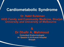
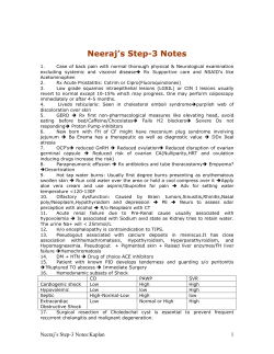
Figure 2.3
A: Dermal hematopoiesis (Courtesy of Dr. Vandana Mehta)
B: Congenital syphilis (Courtesy of Dr. Paul Getz)
C: Congenital syphilis (Courtesy of Dr. Paul Getz)
D: Congenital syphilis (Courtesy of Dr. Paul Getz)
34
E: Candidiasis (Courtesy of Dr. Paul Getz)
F: Langerhans cell histiocytosis (Reprint from Morgan MB,
Smoller BR, Somach SC. Deadly Dermatologic Diseases. New York,
NY: Springer; 2007)
PEDIATRIC DERMATOLOGY
A
B
C
D
E
F
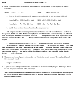
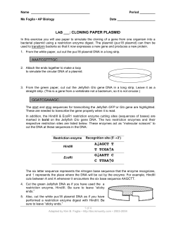
Figure 2.4
A: Acrodermatitis enteropathica
(Courtesy of Michelle B. Bain)
B: Acrodermatitis enteropathica
(Courtesy of Michelle B. Bain)
C: Gianotti–Crosti syndrome
(Courtesy of Dr. Michelle B. Bain)
D: Gianotti–Crosti syndrome
(Courtesy of Dr. Michelle B. Bain)
E: Varicella
(Reprint from Abdel-Halim AW. Passing the USMLE.
New York, NY: Springer, 2009)
F: Papular purpuric gloves and socks syndrome
(Reprint from Burgdorf WH, Plewig G, Wolff HH,
Landthaler M, eds.. Braun-Falco’s Dermatology.
3rd ed. Heidelberg: Springer; 2009)
35
DERMATOLOGY: ILLUSTRATED STUDY GUIDE AND COMPREHENSIVE BOARD R EVIEW
2.3
PAPULOSQUAMOUS AND ECZEMATOUS DERMATOSES
A
Psoriasis (Figure 2.5A)
• Approximately 25% patients will have presentation before age 15
• Presents as erythematous well-demarcated plaques with micaceous scale
• Guttate psoriasis more common in children; presents with raindrop-like
papules in an eruptive pattern; common triggers include strep infection,
viral infection, stress, and trauma
Pityriasis Lichenoides (PL)
• Two diseases forming spectrum of PL: pityriasis lichenoides et varioliformis acuta (PLEVA) and pityriasis lichenoides chronica (PLC)
• PLEVA: abrupt onset of erythematous papules and vesicles with crusted
or necrotic centers, often involuting within weeks to months; treat with
oral erythromycin, phototherapy, and/or topical corticosteroid
• PLC: reddish-brown papules with adherent scale, heals with dyschromia;
more chronic course lasting months to years
B
Acropustulosis of Infancy (Figure 2.5B)
• Onset from 6 months to 2 years; resolves by age 3
• Presents with recurrent crops of pruritic pustules on palms, soles, distal
extremities (may mimic scabies infection so prudent to perform mineral
oil scraping)
• Treatment: topical corticosteroid
Pityriasis Rubra Pilaris (PRP) (Figure 2.5C)
• Three juvenile forms in addition to two adult forms (I/II)
Classic Juvenile
Form (III)
Circumscribed
Juvenile Form (IV)
Atypical Juvenile
Form (V)
Resembles classic adult form but with early onset
(first 2 years of life); most resolve within 3 years;
10% cases
Lesions on extensor surfaces and present in
prepubertal years; 25% cases (50% persist into
adulthood)
Similar to type III + scleroderma-like changes
of hands/feet, familial basis; presents in early
childhood with unrelenting course; 5% cases
C
Pityriasis Rosea (PR)
• Self-limited papulosquamous eruption; likely viral pathogen (human
herpesvirus 7, less likely HHV 6)
• Presents with initial herald patch (precedes eruption by 1–2 weeks)
followed by salmon-colored oval patches and plaques with inner scale
along long axis of Langer’s lines of cleavage (“Christmas tree” pattern on
posterior trunk); variants include inverse pattern (flexural accentuation)
and papular PR (young children and darker-skinned patients)
Figure 2.5
A: Guttate psoriasis
B: Acropustulosis of infancy
C: Pityriasis rubra pilaris
(Courtesy of Dr. Paul Getz)
36
PEDIATRIC DERMATOLOGY
Lichen Striatus (Figure 2.6A, B)
• Self-limited, linear inflammatory condition in children
• Presents with small erythematous scaly papules forming linear
band в†’ spreads down extremity or trunk and typically follows lines of
Blaschko, В± nail involvement
• Hypopigmentation may persist for months to years after lesions resolve
and points to diagnosis
A
Keratosis Pilaris (KP)
• Excessive keratinization causing horny follicular plugs on upper arms,
thighs, and cheeks; associated with atopy
KP Atrophicans
• Group of disorders in children with faulty follicular keratinization
followed by atrophy and scarring
o KP atrophicans faciei: erythema with follicular spiny papules of
eyebrows, cheeks, and scalp; involute and leave pitted atrophic scars;
term ulerythema ophyrogenes if limited to lateral 1/3 of eyebrows,
associated with Noonan syndrome
o Atrophoderma vermiculata: pit-like atrophic scarring of follicles on
face (“honeycomb” atrophy), associated with Rombo syndrome and
Down syndrome
B
Rombo syndrome: milia, atrophoderma vermiculata, acral cyanosis, trichoepitheliomas,
multiple BCCs, hypotrichosis, alopecia
Atopic Dermatitis (AD) (Figure 2.6C)
• Occurs in 10–15% children, often presenting at 2–3 months of age;
multifactorial pathogenesis but includes ↑ secretion of TH2 cytokines
(IL-4, IL-5)
• Triad of atopy: AD, allergic rhinitis, asthma
• Few may have allergy to specific foods, which may exacerbate AD
(eggs, milk, soybeans, fish, wheat, peanuts)
• Presents with eczematous lesions, xerosis, and lichenification
• Distribution varies with age
o Infants: face, scalp, and extensors
o Children: antecubital/popliteal fossae, neck, wrists, ankles
o Adults: typically hands (chronic hand eczema)
C
Atopic patients with ↓ amount of innate antimicrobial peptides: human b-defensins
(HBD) and cathelicidins (LL37)
Figure 2.6
A: Lichen striatus
(Courtesy of Dr. Michelle B. Bain)
B: Lichen striatus
(Courtesy of Dr. Paul Getz)
C: Atopic dermatitis
37
DERMATOLOGY: ILLUSTRATED STUDY GUIDE AND COMPREHENSIVE BOARD R EVIEW
• Pityriasis alba: hypopigmented patches with minimal scale; may be only
manifestation of AD (Figure 2.7A)
• Complications: keratoconus (conical deformity of cornea), eyelid dermatitis, ↑ risk of infection (impetigo, eczema herpeticum, molluscum contagiosum) (Figure 2.7B)
• Treatment: topical corticosteroid, topical calcineurin inhibitor, oral
corticosteroid (short course), oral antihistamine, phototherapy
A
Juvenile Plantar Dermatosis
• Typically in children with an atopic diathesis; related to increased humidity from impermeable material in shoes
• Presents with dry, scaly glazed patches with fissures involving forefoot
plantar surface
• Chronic but typically self-limited
2.4
PIGMENTED LESIONS
CafГ© Au Lait Macule (CALM)
B
• Presents as a light to dark brown macule or patch
• Single lesion in 10–20% of normal population; multiple lesions ± associated with different genodermatoses (McCune-Albright syndrome,
neurofibromatosis)
Lentigines
• Presents as brown macules with increased number of melanocytes; no
relationship to sunlight
• Multiple lentigines may be associated with the following:
LEOPARD Syndrome
Carney Complex
(LAMB or NAME
syndrome)
Peutz–Jeghers
Syndrome
Laugier–Hunziker
Syndrome
Bannayan–Riley–
Ruvalcaba Syndrome
38
AD, PTPN11 gene, cafГ©-noir macules, EKG
changes, ocular hypertelorism, pulmonary
stenosis, abnormal genitalia, growth retardation,
deafness
AD, PRKAR1A gene, psammomatous melanotic
schwannomas, cardiac/cutaneous myxomas, blue
nevi, endocrine overactivity
AD, STK11 gene (serine threonine kinase),
mucocutaneous (oral/acral) lentigines, intestinal
polyposis, В± intussusception, various malignancies
Mucocutaneous lentigines, longitudinal
melanonychia, genital melanosis
AD, PTEN gene, penile > vulvar lentigines,
lipomas, hemangiomas
Figure 2.7
A: Pityriasis alba
(Courtesy of Dr. Paul Getz)
B: Molluscum contagiosum
PEDIATRIC DERMATOLOGY
Ephelides (Freckles)
• Present as light brown macules in sun-exposed areas; more prominent in
children with fair skin and during summer time; onset typically within first
3 years of age
• Can be a marker for UV-induced damage if acquired
• Histology: normal number of melanocytes, increased pigment in
keratinocytes
A
Congenital Nevus (CN) (Figure 2.8A)
• Onset at birth or first year typically; 1–2% of population
• Categorized as small (<1.5 cm), medium (1.5–20 cm), and large
(>20 cm or 10% BSA)
• Slight ↑ risk of melanoma (highest in large CNs); 3–12% of giant (large)
CNs may develop melanoma (different studies show varying percentages);
axial nevi with greatest risk
• If large nevus over scalp, rule out neurocutaneous melanosis with MRI
B
Neurocutaneous melanosis: ↑ intracranial pressure, leptomeningeal melanoma, spinal
cord compression
Spitz Nevus (Epithelioid or Spindle Cell Nevus) (Figure 2.8B)
• Presents as dome-shaped red-brown or tan-pink smooth surfaced papule;
typically occurs within first two decades
• Pigmented, congenital, and agminated variants reported
• Histology: Kamino bodies (PAS + globules)
• Characteristic starburst dermoscopic finding in pigmented Spitz nevi
Halo Nevus (Sutton’s Nevus)
• Melanocytic nevus with surrounding hypopigmented halo in which central
nevus either persists or involutes
• Typically appears in adolescence; may appear in setting of vitiligo; prudent
to rule out concomitant melanoma (rare) by performing full skin exam
C
Nevus Spilus (Speckled Lentiginous Nevus) (Figure 2.8C)
• Presents as tan, regularly bordered patch with darker macules within
lesion
• Melanoma rarely arises within nevus component
• Associated with phakomatosis pigmentovascularis and pigmentokeratotica
(latter with organoid nevus + hemiatrophy + neurologic defects)
Melanoma
• 0.3–0.4% of melanomas in prepubertal children
• ↑ Risk with fair skin, blue eyes, blonde/red hair, CDKN2A or p16 mutation, xeroderma pigmentosum, dysplastic nevus syndrome, large congenital nevus, or neurocutaneous melanosis
Figure 2.8
A: Congenital nevus
B: Spitz nevus
(Reprint from Laxer RM, ed. The Hospital
for Sick Children: Atlas of Pediatrics.
Philadelphia, PA: Current Medicine; 2005)
C: Nevus spilus
(Courtesy of Dr. Paul Getz)
39
DERMATOLOGY: ILLUSTRATED STUDY GUIDE AND COMPREHENSIVE BOARD R EVIEW
Becker’s Nevus (Becker’s Melanosis) (Figure 2.9A, B)
• Acquired unilateral lesion found in adolescent males (second or third decade)
typically on shoulder, upper chest, or back
• Presents as hyperpigmented hypertrichotic patch or plaque associated with
underlying smooth muscle hamartoma (arrector pili)
• Histology: ↑ melanin in epidermis, often smooth muscle hamartoma
present in dermis
A
Blue Nevus (Figure 2.9C)
• Congenital or acquired (typically early childhood)
• Different types: common, cellular, and combined
• Multiple blue nevi associated with Carney complex (LAMB/NAME
syndrome)
• Histology: normal epidermis, many elongated dendritic melanocytes
within dermis, large amounts of melanin often seen within melanocytes
B
Nevus of Ota (Nevus Fuscoceruleus Ophthalmomaxillaris,
Oculodermal Melanocytosis) (Figure 2.9D)
• Onset either near birth or during puberty
• Most common in Asian population, mainly women
• Presents as unilateral, blue-gray macules typically involving V1 and V2
distribution of trigeminal nerve
• Most common extracutaneous sites: sclera > tympanum > nasal mucosa >
pharynx > palate
C
Nevus of Ito (Nevus Fuscoceruleus Acromiodeltoideus)
• Similar presentation to nevus of Ota but typically occurs in shoulder
region (supraclavicular, scapular, and deltoid)
D
Hori’s Nevus (Acquired Nevus of Ota-like Macules)
• Onset in late adolescence, mainly in Asian women
• Bilateral nevus of Ota-like macules of the zygomatic region; may be
misdiagnosed as melasma
Congenital Dermal Melanocytosis (Mongolian Spot)
• Common in infants with pigmented skin
• Presents with blue-gray macules or patches typically over lumbosacral
skin or buttocks
• If extensive, consider phakomatosis pigmentovascularis
• Histology: dendritic melanocytes situated in lower half of dermis, cells
arranged parallel to epidermis
40
Figure 2.9
A: Becker’s nevus
(Courtesy of Dr. Paul Getz)
B: Becker’s nevus
C: Blue nevus (Courtesy of Dr. Paul Getz)
D: Nevus of Ota (Courtesy of Dr. Paul Getz)
PEDIATRIC DERMATOLOGY
2.5
BULLOUS DISEASES
EB Subtype
Dowling-Meara
(EBS Herpetiformis)
Weber-Cockayne (Localized)
Koebner (Generalized)
EBS Muscular Dystrophy
EBS Mottled Pigmentation
Herlitz (EB Lethalis)
Premature termination codon
Non-Herlitz
(Generalized Atrophic Benign EB
or GABEB)
JEB with Pyloric Atresia
Hallopeau-Siemens Recessive
DEB (RDEB-HS)
Premature termination codon
Non-Hallopeau-Siemens
(RDEB-nHS)
Cockayne-Touraine (DDEB-CT)
Pasini Variant (DDEB-P)
Table 2-4 Epidermolysis Bullosa
Inh
Gene
Clinical Features
EB SIMPLEX (EBS)
Split: Epidermal Basal Layer
AD
K5/K14
Onset at birth, grouped or herpetiform
blisters (figurate), significant mucosal
membrane and laryngeal/esophageal
involvement (В± hoarseness), nail
dystrophy, confluent PPK, scarring,
early death
EM: clumped tonofilaments in basal
keratinocytes
AD
K5/K14
Onset typically childhood/adolescence,
palmoplantar bullae/erosions, heal
without scarring
AD
K5/K14
Generalized bullae at birth, PPK, nail
dystrophy, mucosal erosions, heals
without scarring
AR
Plectin
Widespread bullae at birth, muscular
dystrophy, scarring, hair/nail/tooth/oral
disease, early death
Resembles localized and generalized
EBS + reticulated hyperpigmentation
over trunk
JUNCTIONAL EB (JEB)
Split: Basement Membrane (Lamina Lucida)
AR
Laminin 5
Severe, widespread bullae, nonhealing
(laminin-332)
exuberant granulation tissue (perioral,
axillae, neck), enamel defects, absent
nails, mucosal involvement (respiratory/
GI tract with hoarseness), early death
AR
Laminin 5
Widespread bullae at birth, heal with
or BPAG2 (BP180)
atrophic scars, mild oral involvement,
scarring alopecia, nail dystrophy,
improves with time
AR
Severe congenital blistering, hydronepha6b4 integrin
rosis, pyloric atresia, mucosal erosions
DYSTROPHIC EB (DEB)
Split: Dermal (Sublamina Densa)
AR
Type VII collagen
Severe widespread bullae at birth, heals
with atrophic scarring (on hands/feet в†’
“mitten deformity”), milia, nail
dystrophy, mucosal strictures, oral,
esophageal, cutaneous SCCs
AR
Type VII collagen
Skin changes localized to acral bony
prominences, Hallopeau-Siemens
symptoms but less severe
AD
Type VII collagen
Bullae mainly over extremities, heal
with milia/atrophic scars/keloids, nail
dystrophy
AD
Type VII collagen
Similar to Cockayne subtype + albopapuloid lesions (white perifollicular
papules, slowly enlarge)
41
DERMATOLOGY: ILLUSTRATED STUDY GUIDE AND COMPREHENSIVE BOARD R EVIEW
A
B
C
D
E
F
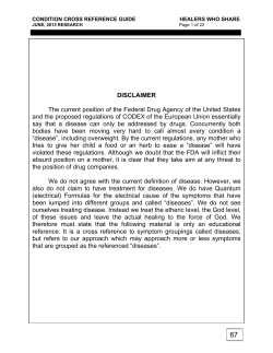
Figure 2.10
A: EB simplex (Weber–Cockayne) (Courtesy of Dr. Paul Getz)
B: Dominant dystrophic EB (Cockayne–Touraine)
(Courtesy of Dr. Paul Getz)
C: Recessive dystrophic EB
D: Recessive dystrophic EB
42
E: EB simplex (Dowling-Meara) (Reprint from Laimer M et al.
Epidermolysis bullosa hereditaria. Monatsschrift Kinderheilkunde
Zeitschrift für Kinder und Jugendmedizin. 2008: 156 (2);110–21)
F: EB simplex (Dowling-Meara) (Reprint from Has C et al.
Hereditare Blasen bildende Hauterkrankungen. Der Hautarzt. 2004:
55(10);920–30)
PEDIATRIC DERMATOLOGY
Chronic Bullous Disease of Childhood (Figure 2.11A)
• Blistering disorder with onset typically before age 5
• Target antigen: 97 kDa Ag (LAD-1 or LABD97): cleaved ectodomain of
BPAG2
• Presents with annular and herpetiform bullae favoring extensor surfaces/
groin (“crown of jewels” configuration)
• Histology: subepidermal bullae with neutrophils in dermal papillae
(similar to dermatitis herpetiformis)
• Treat with dapsone or sulfapyridine
A
Neonatal Pemphigus
• Presents in infants whose mothers have pemphigus vulgaris; due to passive
transfer of maternal IgG to fetus
• Self-limited; resolves within few weeks of birth
Hailey–Hailey Disease (Familial Benign Chronic Pemphigus)
(Figure 2.11B)
B
• AD, ATP2C1 gene (encodes Golgi-associated Ca2+ ATPase hSPCA1),
results in abnormal intracellular calcium signaling; onset typically second
to third decade
• Presents with flaccid vesicles initially on erythematous base over intertriginous areas, ruptures easily, and gives rise to macerated or crusted
erosions
• Histology: extensive epidermal acantholysis “dilapidated brick wall”
Think of “Hailey’s Comet” to remember ATP2 C 1
2.6
EPIDERMAL, APPENDAGEAL, AND DERMAL TUMORS
Epidermal Nevus (EN) (Figure 2.11C)
C
• Hamartoma of epidermis and papillary dermis; onset typically at birth
(В± adolescence, rare in adulthood)
• Presents as hyperpigmented papillomatous papules and plaques along
lines of Blaschko
• Ichthyosis hystrix: extensive bilateral systematized lesions
• ILVEN (inflammatory linear verrucous epidermal nevus): erythematous
scaly plaque along lines of Blaschko; not associated with any neurologic
defects
• Epidermal nevus syndrome (Schimmelpenning syndrome): sporadic;
epidermal nevus, underlying CNS, ocular, cardiac, and skeletal defects,
biopsy to r/o epidermolytic hyperkeratosis (EHK)
Of note, if biopsy of EN shows EHK, the patient may be at risk with offspring with
full-blown EHK
Figure 2.11
A: Chronic bullous disease of childhood
(Courtesy of Dr. Michelle B. Bain)
B: Hailey–Hailey disease
(Courtesy of Dr. Paul Getz)
C: ILVEN (Courtesy of Dr. Paul Getz)
43
DERMATOLOGY: ILLUSTRATED STUDY GUIDE AND COMPREHENSIVE BOARD R EVIEW
Nevus Sebaceus (Figure 2.12A, B)
• Presents as solitary yellow-orange slightly raised plaque typically on scalp
or face; plaque typically thickens and becomes more verrucous or pebbly
during childhood
• Mutation in PTCH gene has been reported (deletion)
• Benign tumors (trichoblastoma, syringocystadenoma papilliferum) and
malignant tumors (BCC < 1% cases) can arise within lesion
A
Basal Cell Carcinoma
• Seen in children with xeroderma pigmentosum (XP) and basal cell nevus
syndrome (BCNS)
Squamous Cell Carcinoma
• Seen in children with XP, dystrophic EB, and albinism
Pilomatricoma (Calcifying Epithelioma of Malherbe)
B
• Onset typically in childhood
• Presents as solitary firm, skin-colored to faint blue papule or cyst on face
or upper trunk
• Histology: anucleate cornified cells (“ghost” or “shadow” cells),
calcification seen in late lesions
• Multiple pilomatricomas may be associated with myotonic dystrophy
(b-catenin defect)
Trichoepithelioma (Figure 2.12C)
• Benign adnexal neoplasm usually appearing in childhood
• Presents as skin-colored translucent papules (usually multiple) along the
nasolabial folds or periorbital regions
• Multiple lesions in Brooke–Spiegler syndrome (trichoepitheliomas,
cylindromas, spiradenomas)
C
Angiofibroma (Fibrous Papule)
• Skin-colored firm papule on face
• Multiple lesions associated with tuberous sclerosis (once known as
adenoma sebaceum) with onset in early to mid-childhood
Figure 2.12
A: Nevus sebaceus*
B: Nevus sebaceus*
C: Trichoepitheliomas*
* Courtesy of Dr. Paul Getz
44
PEDIATRIC DERMATOLOGY
Neurofibroma (NF) (Figure 2.13A)
• Presents as skin-colored, soft or rubbery papulonodule with positive
“buttonhole” sign (easily invaginated)
• Commonly seen as solitary lesion; multiple lesions associated with
neurofibromatosis
• Plexiform NF considered pathognomonic for NF1, malignant transformation in 2–13%
A
Connective Tissue Nevus (Figure 2.13B, C)
• Also known as shagreen patch (tuberous sclerosis) collagenoma, elastoma,
or dermatofibrosis leticularis disseminata (latter in Buschke–Ollendorff
syndrome)
• Onset at birth or early childhood; likely hamartoma
• Presents as firm, solitary, or multiple skin-colored papules, nodules, or plaques
B
Infantile Digital Fibroma (Figure 2.13D)
• Onset within 1 year of age
• Presents as multiple firm, smooth dome-shaped nodules on dorsolateral
fingers/toes (spares thumb and great toe)
• Benign with spontaneous regression within 2–3 years typically; high local
recurrence rate with surgical excision
• Histology: eosinophilic intracytoplasmic perinuclear inclusions within
spindle cells
C
Infantile Myofibromatosis (Congenital Generalized Fibromatosis)
• Rare, onset at birth or within first 2 years
• Presents as one or more firm, rubbery skin-colored to purple papulonodules on head, neck, or trunk
• Two types: localized with no visceral involvement, good prognosis;
visceral involvement with high mortality
D
Fibrous Hamartoma of Infancy
• Onset at birth or within first year of life
• Presents as painless, solitary skin-colored subcutaneous nodule typically
involving axilla, shoulder, or upper arm (less likely groin area)
• Treat with local excision
Fibromatosis Colli
• Infiltration of fibrous tissue involving the lower third of the sternocleidomastoid muscle at birth
• Typically spontaneous remission within few months
Juvenile Hyaline Fibromatosis
• Due to mutation in capillary morphogenesis protein 2
• Multiple firm papules and nodules involving the face, extremities, and
scalp; hypertrophic gums and disfigurement with flexion contractions
Figure 2.13
A: Neurofibromas
(Courtesy of Dr. Paul Getz)
B: Connective tissue nevus
(Reprint from Laxer RM, ed. The Hospital
for Sick Children: Atlas of Pediatrics.
Philadelphia, PA: Current Medicine,
2005)
C: Connective tissue nevus
(Courtesy of Dr. Paul Getz)
D: Infantile digital fibroma
(Reprint from Burgdorf WH, Plewig G,
Wolff HH, Landthaler M, eds. BraunFalco’s Dermatology. 3rd ed. Heidelberg:
Springer; 2009)
45
DERMATOLOGY: ILLUSTRATED STUDY GUIDE AND COMPREHENSIVE BOARD R EVIEW
Juvenile Xanthogranuloma (JXG) (Figure 2.14A, B)
• Non-Langerhans cell histiocytosis with Touton giant cells; onset typically
within first year of life
• Two types: micronodular (small, multiple) or macronodular (larger size,
few in number)
• Presents as single or multiple firm, pink-red papulonodules with yellow
hue on head/neck > trunk/upper extremities
• Regression typically seen in children (not in adults)
• 0.5% with ocular involvement: glaucoma, hyphema (may rarely result in
blindness)
• Association with NF1 and juvenile myelomonocytic leukemia (JMML)
A
Langerhans Cell Histiocytosis (LCH) (Figure 2.14C)
• Clonal proliferative disease of Langerhans cells (comma-shaped nuclei,
S100+, CD1a+, intracytoplasmic Birbeck granules seen on EM), four
overlapping syndromes
• Current classification by number of organ systems involved (single vs.
multisystem), but historically grouped as follows:
– Multisystem involvement, (acute disseminated
form); onset typically before 2 years of age
– Small, pink papules, pustules, vesicles with
scale/crust/petechiae in seborrheic distribution
Hand–Schuller–Christian – Onset between 2 and 6 years of age
Disease
– Typical triad: diabetes insipidus, bone lesions,
exophthalmos
– Osteolytic bone lesions (cranium)
Eosinophilic Granuloma – Onset in older children, localized LCH variant
– Asymptomatic granulomatous lesions involving bone (cranium), spontaneous fractures
Congenital Self-Healing – Onset at birth or soon after, limited to skin;
Reticulohistiocytosis
also known as Hashimoto-Pritzker disease
– Widespread, red-brown papulonodules
– Self-healing within weeks to months
B
Letterer–Siwe Disease
C
Benign Cephalic Histiocytosis
• Self-limited histiocytosis (S100 negative non-LCH); onset within first
3 years of life
• Presents with small red-brown macules and papules on face, spreading to
neck and ears > trunk and arms; spontaneous resolution after months or
years
46
Figure 2.14
A: Juvenile xanthogranuloma
(Courtesy of Dr. Michelle B. Bain)
B: Juvenile xanthogranuloma
(Courtesy of Dr. Paul Getz)
C: LCH
(Reprint from Morgan MB, Smoller BR,
Somach SC. Deadly Dermatologic
Diseases. New York, NY: Springer; 2007)
PEDIATRIC DERMATOLOGY
Dermoid Cyst (Figure 2.15A)
• Seen typically in infants along embryonic fusion plane
• Presents as discrete, subcutaneous nodule commonly around eyes or nasal
root
• Histology: lined by stratified squamous epithelium (with granular layer)
containing appendageal elements
• CT/MRI should be performed to rule out connection to CNS before
excision
A
Mastocytosis (Figure 2.15B, C)
• Spectrum of disorders with mast cell hyperplasia in skin and other organs
• Childhood mastocytosis – onset before puberty (50% before age 2), c-kit
alteration (proto-oncogene, tyrosine kinase subfamily); several forms in
children:
Solitary Mastocytoma
Urticaria Pigmentosa (UP)
Diffuse Cutaneous
Mastocytosis
Telangiectasia Macularis
Eruptiva Perstans (TMEP)
– Tan to brown, minimally infiltrated plaque
or nodule; spontaneous resolution over
months
– Positive Darier sign
– Onset early childhood, may occur in adults
– Hyperpigmented to pink pruritic macules
or papules on trunk; positive Darier sign
– Variant: bullous UP
– Doughy or boggy skin texture with
lichenification and yellow hue
– Extreme pruritus, friction may cause
bullae
– Systemic symptoms: bronchospasm,
diarrhea
– Persistent eruption of macules and papules
with red-brown hue
– Rare in childhood
B
C
• Avoid mast cell degranulators: aspirin, alcohol, opiates, quinine, polymyxin B sulfate, amphotericin B, tubocuraine, scopolamine
2.7
TUMORS OF FAT, MUSCLE AND BONE
Lipoma
• If located over lumbosacral region at birth, consider underlying spinal
dysraphism (incomplete closure of mesenchymal, osseous, and nervous
tissue of the spine) в†’ perform MRI
Associated syndromes with lipomas: Bannayan–Riley–Ruvalcaba syndrome, Gardner
syndrome, MEN I
Figure 2.15
A: Dermoid cyst
(Reprint from Laxer RM, ed. The Hospital
for Sick Children: Atlas of Pediatrics.
Philadelphia, PA: Current Medicine; 2005)
B: Urticaria pigmentosa
(Courtesy of Dr. Michelle B. Bain)
C: Urticaria pigmentosa
(Courtesy of Dr. Paul Getz)
47
DERMATOLOGY: ILLUSTRATED STUDY GUIDE AND COMPREHENSIVE BOARD R EVIEW
Cutaneous Calcification
• Solitary nodular calcification: benign nodule in infants typically from
heel sticks
• Osteoma cutis: idiopathic or associated with Albright’s hereditary
osteodystrophy
• Supepidermal calcified nodule: solitary firm nodule on scalp or face
(ears) of children
2.8
VASCULAR DISORDERS
Hemangiomas and Vascular Malformations
Hemangiomas are vascular tumors arising in infancy with true cellular proliferation, which eventually regress. Vascular malformations represent errors in vascular morphogenesis (dysplastic vessels) without true cellular proliferation and
without regression.
Vascular Tumors
Infantile and Congenital
Hemangiomas
Kaposiform
Hemangioendothelioma
Pyogenic Granuloma
Tufted Angioma
Congenital
Hemangiopericytoma
Vascular Malformations
Capillary Malformation (slow flow):
Port-Wine Stain (Nevus Flammeus)
Venous Malformation (slow flow):
Cavernous Hemangioma, Phlebectasia
Lymphatic Malformation (slow flow):
Lymphangioma (Lymphangioma Circumscriptum
Cystic Hygroma, Cavernous Lymphangioma)
Arteriovenous Malformation (fast flow):
Cirsoid Aneursm
Combined Malformation (slow or fast flow)
A. VASCULAR TUMORS
Hemangioma of Infancy (Figure 2.16A)
• Benign vascular tumor presenting soon after birth (first few weeks after life)
• More common in premature infants, 15% have multiple lesions with
higher risk for visceral involvement, GLUT1 positive (endothelial marker,
useful in differentiating from malformation)
• Precursor lesion: pink or bruised macule or patch with surrounding
telangiectasias
• Superficial hemangioma (strawberry hemangioma) situated in the
superficial dermis and bright red in color during the proliferative phase
• Deep or cavernous hemangioma (located deep dermis and/or subcutis)
presents as blue-purple mass with normal overlying skin, В± bruit
• Involution: 30% by age 3, 50% by age 5, 70% by age 7, 90% by age 9
• Complications: ulceration (most common), anatomic distortion with
interference of normal function, high-output congestive heart failure
(greater risk with visceral hemangiomas, especially if in liver)
• Regionally significant hemangiomas: periocular (obstruct vision and cause
ophthalmologic complications), beard region (clue for laryngeal hemangiomatosis with airway obstruction), segmental hemangioma over lumbosacral area (MRI of spine to r/o GU/GI/spinal/skeletal abnormalities),
nasal tip (textural changes and scarring)
48
PEDIATRIC DERMATOLOGY
PHACES
• Posterior fossa malformation, hemangioma, arterial anomalies, cardiac
defect, coarctation of the aorta, eye abnormalities, sternal defects, and
supraumbilical raphe
• Hemangiomas tend to be plaque-like on the face involving more than one
dermatome
• Most common posterior fossa malformation: Dandy–Walker malformation
A
Diffuse Neonatal Hemangiomatosis
• Cutaneous and visceral hemangiomas; liver hemangioma may be complicated by obstructive jaundice
• If multiple cutaneous hemangiomas, perform ultrasound, urinalysis, stool
guiaic, CBC to r/o systemic involvement
• If no visceral involvement → benign neonatal hemangiomatosis
• ↑ Mortality with systemic form due to high-output cardiac failure, GI
bleeding, and respiratory compromise
Tufted Angioma (Figure 2.16B)
• Onset during infancy or early childhood
• Presents as ill-defined red-brown plaque or patch over neck or upper trunk;
plaque slowly extends with time (typically does not regress)
B
PELVIS Syndrome
• Perineal hemangioma, external genital malformation, lipomyelomeningocele, vesicorenal anomalies, imperforate anus, and skin tag
Pyogenic Granuloma (Figure 2.16C)
• Presents as rapidly growing, friable red papule of skin or mucosa with
frequent ulceration
• Common in children and young adults
• Associated with antecedent trauma, pregnancy, oral medications (retinoids, inidinavir, EGFR inhibitors)
Kaposiform Hemangioendothelioma (Figure 2.17A)
• Usually onset before age 2
• Presents as vascular macules, plaques, nodules, or bulging indurated
masses
• Associated with Kasabach–Merritt syndrome → consumptive coagulopathy with thrombocytopenia (platelet sequestration) and purpura; deepseated tumors (i.e., retroperitoneal) likely to cause above syndrome
C
Glomeruloid Hemangioma
• Distinct vascular proliferation in POEMS syndrome (polyneuropathy,
organomegaly, endocrinopathy, monoclonal gammopathy, and skin
lesions)
• Presents as firm, red-purple papules over trunk or extremities
Figure 2.16
A: Hemangioma
(Courtesy of Dr. Michelle B. Bain)
B: Tufted angioma
C: Pyogenic granuloma
49
DERMATOLOGY: ILLUSTRATED STUDY GUIDE AND COMPREHENSIVE BOARD R EVIEW
B. VENOUS MALFORMATIONS
A
Capillary Malformation (Nevus Flammeus, Port-Wine Stain, PWS)
(Figure 2.17B)
• Presents as a well-demarcated erythematous patch or plaque that grows in
proportion to general growth of the body; does not spontaneously recede
(unlike “salmon patches” over forehead, glabella, nose/philtrum, nape or
eyelid which typically disappear by age 3)
• Facial PWS follows sensory CN V distribution (V1–V3); over time, skin
changes from pink to deep purple and thickens with ↑ nodularity and
pyogenic granulomas
• GLUT1 negative
• PWS can be seen with combination of epidermal or melanocytic abnormalities: phakomatosis pigmentovascularis (see below)
• Associated syndromes: Sturge–Weber syndrome, Klippel–Trénaunay
syndrome, Proteus syndrome
Phakomatosis Pigmentovascularis
• Type 1: PWS + epidermal nevus
• Type 2: PWS + dermal melanocytosis ± nevus anemicus
• Type 3: PWS + nevus spilus ± nevus anemicus
• Type 4: PWS + dermal melanocytosis + nevus spilus ± nevus anemicus
Glomangioma (Glomuvenous Malformation) (Figure 2.17C)
• Arises in children and adolescents; may be sporadic or inherited (autosomal dominant with incomplete penetrance; defect in glomulin gene)
• If solitary lesion (glomus tumor), onset typically in adulthood with
subungual location
• Presents as soft pink to deep blue papules or nodules in segmental
distribution; tender to palpation, В± attacks of pain with pregnancy or
menstruation
• Histology: resembles vascular malformation but vessels lined with one or
more rows of cuboidal glomus cells
B
C
Figure 2.17
A: Kaposiform hemangioendothelioma
(in Kasabach-Merritt)
(Reprint from Laxer RM, ed. The Hospital
for Sick Children: Atlas of Pediatrics.
Philadelphia, PA: Current Medicine;
2005)
B: Port-wine stain (Reprint from
Abel-Halim AW. Passing the USMLE. New
York, NY: Springer; 2009)
C: Glomangiomas (Courtesy of Dr.
Michelle B. Bain)
50
PEDIATRIC DERMATOLOGY
Angiokeratoma (Figure 2.18A)
• Ectasias of dermal capillaries
• Presents as a dark red to purple papule; either solitary or multiple;
distribution varies by type
Solitary
Angiokeratoma
Angiokeratoma
Circumscriptum
Angiokeratoma
of Mibelli
Angiokeratoma
Corporis Diffusum
(Fabry Disease)
Angiokeratoma
of the Scrotum
A
Single red to dark brown papule usually on the lower
extremity
Large verrucous papules or plaques typically involving
the extremity, onset in early childhood/infancy
Rare, presents as several 1–5 mm dark, red-gray
papules over acral areas with verrucous surface
Numerous tiny telangiectatic red papules associated
with hereditary lysosomal storage disease, XLR,
a-galactosidase A deficiency
Multiple small red-violaceous papules studding the
scrotum, less often the vulva, onset in adulthood
(Fordyce)
Lymphangioma
• Uncommon congenital malformation of the lymphatic system; either
superficial (lymphangioma circumscriptum) or deep-seated (cavernous
lymphangioma)
• Lymphangioma circumscriptum: multiple translucent vesicles with clear
lymph fluid (resembling frog spawn)
• Cystic hygroma (variant of cavernous lymphangioma): deep-seated large
translucent soft mass typically over neck, axilla, or lateral chest
B
C. TELANGIECTASIAS
Spider Angioma (Spider Nevus) (Figure 2.18B, C)
• Common acquired lesion seen in children and adults
• Comprised of central arteriole with radiating thin walled vessels; temporary obliteration seen with compression
• Presents as bright red papule with central papule surrounded by distinct
radiating vessels
• Multiple lesions associated with liver disease, pregnancy, and estrogen
therapy
C
Angioma Serpiginosum
• Onset typically within first two decades of life
• Presents as small, red punctate asymptomatic macules in serpiginous
pattern typically over extremity
Figure 2.18
A: Angiokeratoma
B: Spider angioma
C: Spider angioma
51
DERMATOLOGY: ILLUSTRATED STUDY GUIDE AND COMPREHENSIVE BOARD R EVIEW
2.9
GENODERMATOSES
X-Linked Recessive
CHAD’S Kinky WIFE, CHANdra
• C: Chronic Granulomatous Disease
• H: Hunter Disease
• A: Anhidrotic (Hypohidrotic) Ectodermal Dysplasia
(Christ-Siemens-Touraine)
Of note, type IX EDS
• D: Dyskeratosis Congenita
(occipital horn syndrome)
• S: SCID
is NOT part of the revised
EDS classification (since
• Kinky: Kinky Hair Disease (Menkes Disease)
it is NOT due to a
• W: Wiskott–Aldrich Syndrome
collagen defect) and type
• I: Ichthyosis, X-linked
V is classified as “other”
• F: Fabry Disease
in EDS classification
• E: Ehlers–Danlos Syndrome (type V and IX)
• C: Chondrodysplasia Punctata (not Conradi–Hünermann type)
• H: Hypohidrotic ED with Immunodeficiency
• A: Agammaglobulinemia, Bruton
• N: Lesch-Nyhan Syndrome
X-Linked Dominant
A
B
BIG ChOMP
• B: Bazex Syndrome (do not confuse with acrokeratosis paraneoplastica
{Bazex syndrome})
• I: Incontinentia Pigmenti (Bloch-Sulzberger Syndrome)
• G: Goltz Syndrome (Focal Dermal Hypoplasia)
• C: CHILD Syndrome
• h: –
• O: Oro-Facial-Digital Syndrome
• M: MIDAS Syndrome (micrognathia, dermal aplasia, sclerocornea)
• P: Chondrodysplasia Punctata (Conradi–Hünermann type)
A. SYNDROMES WITH DEFECTIVE DNA REPAIR
C
Xeroderma Pigmentosum (XP) (Figure 2.19A, B)
• AR, due to defect in DNA repair
• Seven complementation groups (A–G) and one XP variant described, each
encoding different proteins in the nucleotide excision repair (NER)
pathway (except XP variant)
• Presents with marked photosensitivity, early onset of all major skin malignancies, exaggerated sunburn following minimal sun exposure, solar lentigines
by age of 2, ocular abnormalities (photophobia, keratitis, corneal opacification, vascularization), neurologic abnormalities (progressive deafness)
• XP variant (mutation in DNA polymerase): no neurologic abnormalities
• DeSanctis–Cacchione syndrome (Gr. A): severe neurologic abnormalities
(MR, deafness, ataxia)
Cockayne Syndrome
• AR, defective excision repair: unable to repair cyclobutane pyrimidine
dimer products after irradiation, ↑ chromosomal breaks
• Two complementation groups: CS-A (ERCC8) and CS-B (ERCC6)
• Presents with photosensitivity, mental retardation, cachectic dwarfism,
peripheral neuropathy, sunken eyes, prominent ears, “salt and pepper”
retinitis pigmentosa, dental caries, thinning hair, basal ganglia calcification
COCKAYNE – eight letters (ERCC8), Cachectic dwarfism, Ocular (salt/pepper RP),
Cataracts, Avoid sun, Ears (“mickey mouse”)
52
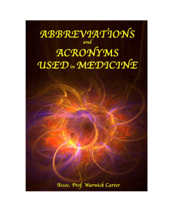
Figure 2.19
A: Xeroderma pigmentosum
(Courtesy of Dr. Michelle B. Bain)
B: Xeroderma pigmentosum
(Courtesy of Dr. Michelle B. Bain)
C: Rothmund-Thomson
(Reprint from Burgdorf WH, Plewig G,
Wolff HH, Landthaler M, eds. BraunFalco’s Dermatology. 3rd ed. Heidelberg:
Springer; 2009)
PEDIATRIC DERMATOLOGY
Trichothiodystrophy (PIBIDS)
• AR, mutation in gene ERCC2 (XPD protein) and ERCC3 (XPB protein)
in NER pathway, sulfur deficiency in hair
• PIBIDS: Photosensitivity (50%), ichthyosis (variable severity), brittle hair
(alternating bright and dark bands known as “tiger tail,” flattened hair
shafts like a ribbon), intellectual impairment, decreased fertility, short
stature, receding chin, protruding ears
A
TrichoThiodystrophy – Tiger Tail abnormality
Bloom Syndrome
• AR, BLM gene mutation, RecQ protein-like two (RecQL2, some sources say
RecQL3 {Spitz}), DNA helicase family, mutation results in ↑ spontaneous
sister chromatid exchanges, breakage, and rearrangements
• Presents with photodistributed erythema/telangiectasias over cheeks
within first few weeks of life, short stature, normal intelligence, immune
deficiency causing chronic respiratory/GI infections, ↓ fertility, ↓ IgM/
IgA, high-pitched voice
• ↑ Risk cancer: leukemia, lymphoma, GI adenocarcinoma
B
BLooM – 2 O’s (RecQL2)
Butterfly rash, Leukemia, iMmune deficiency, ↓ IgM
Rothmund–Thomson Syndrome (Poikiloderma Congenitale)
(Figure 2.19C)
• AR, RECQL4 (DNA helicase)
• Presents with photodistributed erythema and vesicles on face in first few
months of life, evolves into poikiloderma and extends to buttocks and
extremities, premalignant acral keratoses, alopecia, cataracts, hypoplastic
thumbs/ radii/ulnae, ↑ risk osteosarcoma, normal intelligence
Rothmund Thomson – Reduced Thumbs
ROTH (4 letters) – RecQL4
C
Dyskeratosis Congenita (Zinsser-Engman-Cole Syndrome)
• Two forms: XLR and AD
• XLR, DKC1 gene mutation, encodes protein dyskerin (interacts with
telomerase), ↑ sister chromatid exchanges
• AD, hTR (human telomerase RNA component) and hTERT (human
telomerase reverse transcriptase) mutations
• Cutaneous poikiloderma (face, trunk, thighs), nail dystrophy (atrophy,
pterygium), premalignant leukoplakia (buccal mucosa most common),
frictional bullae, palmoplantar hyperhidrosis
• Bone marrow failure with anemia, thrombocytopenia, or pancytopenia
в†’ major cause of mortality
• ↑ CA: mucosal SCC, Hodgkin’s lymphoma, AML
DYSkeRaTOSis – DYStrophy (nails), mR, Thrombocytopenia,
Oral premalignant leukoplakia, Sun avoidance (poikiloderma)
Ataxia-Telangiectasia Syndrome (Figure 2.20A)
• AR, ATM gene mutation, inability to repair chromosomal strand breaks,
sensitivity to ionizing radiation
Figure 2.20
A: Ataxia-Telangiectasia
(Reprint from Morgan MB, Smoller BR,
Somach SC. Deadly Dermatologic
Diseases. New York, NY: Springer; 2007)
B: Basal cell nevus syndrome*
C: Palmar pits (BCNS)*
*Courtesy of Dr. Paul Getz
53
DERMATOLOGY: ILLUSTRATED STUDY GUIDE AND COMPREHENSIVE BOARD R EVIEW
• Presents first with ataxia (2–3 years old) → telangiectasias on bulbar
conjunctivae (spreads to cheeks/ears), premature aging (atrophic/sclerotic
face), ↓ Purkinje fibers in cerebellum
• Defects in cellular and humoral immunity (↓ IgA, IgG, IgE), severe and
frequent sinopulmonary infections, ↑ lymphoreticular malignancy, ↑ breast CA
A
Fanconi Syndrome
• AR, ↑ chromosomal breakage
• Presents with diffuse hyperpigmentation, multiple CALMs, pancytopenia,
↑ SCC, ↑ solid organ CA, ↑ leukemia, hypoplasia of radius/thumb
FanCONi – CONe-shaped defect (hypoplasia of distal structures – radius/thumb)
B. SYNDROMES OF TUMOR SUPPRESSION
Basal Cell Nevus Syndrome (Gorlin Syndrome) (Figure 2.20B, C)
• AD, PTCH (PATCHED) gene, inhibits sonic hedgehog signaling (unbound
PTCH inhibits Smoothened (SMO) signaling; when inactivating mutation
occurs in PTCH в†’ repression of SMO removed в†’ constitutive activation
of Gli and downstream targets)
• Presents with numerous BCCs, palmar/plantar pits, odontogenic keratocysts of jaw, characteristic facies (frontal bossing, hypertelorism), cataracts, glaucoma, bifid ribs, calcification of falx cerebrum, agenesis of
corpus callosum, ovarian fibromas, medulloblastoma, meningioma
B
Neurofibromatosis, Type I (Von Recklinghausen Disease)
(Figure 2.21A–C)
• AD, NF-1 gene, encodes neurofibromin (tumor suppressor protein)
• Criteria: two or more of the following six:
Six or more CALMs
Cafe au lait macule (CALM):
or two or more neurofibromas or
> 0.5 cm prepubertal, >1.5 cm postpubertal
one plexiform neurofibroma
Axillary or inguinal freckling (Crowe’s sign)
Optic glioma
Lisch nodules
Sphenoid wing dysplasia or thinning cortex of long bone
First degree relative with NF
C
• ↑ Risk of tumors: optic glioma, malignant peripheral nerve sheath tumor,
neurosarcoma, juvenile myelomonocytic leukemia, rhabdomyosarcoma
• ± Hypertension, mental retardation (MR), seizures, kyphoscoliosis,
endocrine disorder (precocious puberty, acromegaly, thyroid/parathyroid
abnormalities)
Neurofibromatosis, Type II (Bilateral Acoustic NF)
• AD, NF-2 gene, encodes merlin/schwannomin
• Diagnosis requires bilateral CNVII masses OR first degree relative AND
either unilateral CN VIII mass OR two of the following: schwannoma,
optic glioma, meningioma, juvenile posterior subcapsular opacity
Carney Syndrome (NAME or LAMB Syndrome)
• AD, PRKAR1A gene
54
Figure 2.21
A: CALMs*
B: Neurofibromatosis*
C: Neurofibromatosis*
*Courtesy of Dr. Paul Getz
PEDIATRIC DERMATOLOGY
• Presents with ephelides, blue nevi, lentigines, cutaneous myxomas
(flesh-colored papules over ears, eyelids, nipples), primary pigmented
nodular adrenocortical disease (results in Cushing syndrome)
• Tumors: testicular tumors, pituitary GH-secreting tumors, psammomatous
melanotic schwannomas
A
NAME: nevi, atrial myxoma, myxoid tumors, ephelides
LAMB: lentigines, atrial myxomas, mucocutaneous myxomas, blue nevi
Muir–Torre Syndrome
• AD, mutation in MLH1 and MSH2 (DNA mismatch repair genes) causing
microsatellite instability
• Multiple sebaceous neoplasms and keratoacanthomas
• ↑ Risk of colon adenocarcinoma, less common GU, lung, breast or heme
malignancy
Muir–Torre: think of “more” and more sebaceous neoplasms
Tuberous Sclerosis (Figure 2.22A, B)
• AD, TSC1 gene mutation (hamartin), and TSC2 (tuberin)
• Ash-leaf macules (earliest finding), facial angiofibromas, connective tissue nevi
(shagreen patch), fibromas (gingival and subungual), CALMs, dental enamel pits
• Renal angiomyolipomas, retinal hamartomas, seizures, pulmonary
lymphangioleiomyomatosis, cortical tubers, cardiac rhabdomyoma
Cowden Syndrome (Multiple Hamartoma Syndrome) (Figure 2.22C)
• AD, PTEN gene mutation, encodes tyrosine phosphatase protein, mutation
causes cell proliferation
• Trichilemmomas (smooth to verrucous small papules on face),
“cobblestone” appearance of the mucosa including tongue
(oral papillomas), acral keratotic papules
• ↑ Breast fibroadenoma, ↑ CA: breast, thyroid follicular; GI polyps
B
C
COWden – trichileMOOmas; other PTEN syndromes: Lhermitte–Duclos and
Bannayan–Zonana syndrome
Multiple Endocrine Neoplasia (MEN)
Type 1
(Wermer Syndrome)
Type 2a (Sipple
Syndrome)
Type 2B (Multiple
Mucosal Neuroma
Syndrome)
– AD, MEN1 mutation (menin: tumor suppressor)
– Angiofibromas, collagenomas, lipomas, CALMs
– Pituitary, parathyroid, pancreatic tumors
– AD, RET mutation (tyrosine kinase receptor)
– Lichen or macular amyloidosis, hemangiomas,
genital lentigines, hamartomas, lipomas
– Parathyroid tumor, thyroid medullary carcinoma,
pheochromocytoma
– AD, RET mutation
– Multiple mucosal neuromas, thickened lips
– Thyroid medullary carcinoma, pheochromocytoma,
marfanoid habitus, diffuse ganglioneuromatosis
(megacolon, diarrhea)
MEN 1: 3 P’s (pituitary, pancreas, parathyroid) + CALMs
MEN 2A: Amyloidosis (“sipple” syndrome: think “rippled” macular amyloid)
MEN2B: Blubbery lips due to mucosal neuromas
Figure 2.22
A: Angiofibromas in TS
(Courtesy of Dr. Michelle B. Bain)
B: Koenen tumor in TS
(Courtesy of Dr. Paul Getz)
C: Cowden syndrome (Reprint from
Morgan MB, Smoller BR, Somach SC.
Deadly Dermatologic Diseases. New York,
NY: Springer; 2007)
55
DERMATOLOGY: ILLUSTRATED STUDY GUIDE AND COMPREHENSIVE BOARD R EVIEW
Bannayan–Riley–Ruvalcaba Syndrome
• AD, PTEN mutation
• Genital lentigines, hamartomas, lipomas, hemangiomas, mental retardation, macrocephaly
A
Bannayan – think of an old banana with dark spots on the outside
resembling lentigines
LEOPARD Syndrome (Multiple Lentigines Syndrome)
• AD, PTPN11 gene mutation, encodes tyrosine phosphatase Shp2
• Lentigines, ECG abnormalities, ocular hypertelorism, pulmonic stenosis,
abnormal genitalia, retarded growth and deafness
• Multiple lentigines at birth/early infancy (sun exposed and protected areas,
including genitalia, hands, feet)
Peutz–Jeghers Syndrome (Figure 2.23A)
• AD, STK11/LKB1 gene mutation, encodes serine-threonine kinase tumor
suppressor
• Hyperpigmented macules on lip/oral mucosa/fingers (starts in infancy/
early childhood) and intestinal polyposis (В± bleeding, intussusception)
• ↑ GI adenocarcinoma, ↑ other solid organ malignancies
B
PeuTz(S) Jeghers – Threonine Serine kinase
Gardner Syndrome
• AD, APC gene encoding tumor suppressor gene (ras proto-oncogene)
• Cutaneous epidermoid cysts, osteomas (mandible, maxilla), supernumerary teeth, odontomas, fibromas, congenital hypertrophy of the retinal
pigment epithelium (CHRPE)
• Tumors: GI adenocarcinoma (inevitable), osteochondromas, thyroid
papillary carcinoma, hepatoblastoma, adrenal adenomas
Gardner – birds CHiRP in the GARDen
Birt–Hogg–Dubé Syndrome (Figure 2.23B, C)
• AD, BHD gene (encodes folliculin)
• Multiple fibrofolliculomas, trichodiscomas, acrochordons on the face,
scalp, neck, and upper trunk
• Associated with renal cell carcinoma, medullary carcinoma of thyroid,
spontaneous pneumothorax (multiple pulmonary cysts)
C
Birt HOGG Dube – think of a hog with rough textured skin (because of fibrofolliculomas and trichodiscomas)
Dysplastic Nevus Syndrome
• AD, CDKN2A (p16 tumor suppressor gene, inhibits cyclin-dependent
kinase 4 [CDK4])
• Dysplastic nevi, melanoma, pancreatic CA, astrocytomas
56
Figure 2.23
A: Peutz–Jeghers syndrome
(Courtesy of Dr. Paul Getz)
B: Birt–Hogg–Dube syndrome*
C: Birt–Hogg–Dube syndrome*
(*Reprint from Morgan MB, Smoller BR,
Somach SC. Deadly Dermatologic
Diseases. New York, NY: Springer; 2007)
PEDIATRIC DERMATOLOGY
C. SYNDROMES WITH PREMATURE AGING
A
Werner Syndrome (Adult Progeria) (Figure 2.24A)
• AR, RECQL2 gene mutation (WRN gene), encodes RecQ DNA helicase,
genomic instability (↑ aging/cancer)
• Normal growth until second decade, then short stature/thin limbs, graying
of hair in adolescence, central obesity, pinched facial expression, beaked
nose, micrognathia, high-pitched voice, mottled hyperpigmentation,
sclerodermoid changes, cataracts, diabetes mellitus, premature atherosclerosis, chronic leg ulcers
• ↑ Soft tissue sarcomas, osteosarcomas, SCCs
Werner – tWo (recql2)
Progeria (Hutchinson–Gilford Syndrome) (Figure 2.24B)
• AD, lamin A gene mutation (LMNA), encodes lamin A and lamin C
(nuclear envelope protein)
• Markedly premature aging (median lifespan 12 years), large appearing
cranium, frontal bossing, prominent scalp veins, beaked nose, micrognathia, “plucked bird” appearance, loss of subcutaneous tissue, sclerodermoid skin; alopecia, high pitched voice, average intelligence, severe
premature coronary atherosclerosis
B
D. DISORDERS WITH IMMUNODEFICIENCY
Familial Chronic Mucocutaneous Candidiasis (FCMC)
• Recurrent, progressive candidal infections (skin, nails, and mucosa)
presenting with recurrent oral thrush, nail dystrophy, crusted cutaneous
plaques
C
Hyper-IgE Syndrome (Job Syndrome) (Figure 2.24C)
• AD, mutation in gene encoding STAT3 (signal transducer and activator of
transcription 3), AR (gene encoding tyrosine kinase 2 TYK2)
• ↑ IgE levels, peripheral eosinophilia, cold abscesses, coarse facies,
eczematous dermatitis, lung abscesses, pneumonia, retained primary teeth,
pneumatocele, otitis media, osteopenia with recurrent fractures
Figure 2.24
A: Werner syndrome
(Reprint from Baykal C, Yazganoglu KD.
Dermatological Diseases of the Nose and
Ears. Berlin: Springer; 2010)
B: Progeria
(Courtesy of the Howard family)
C: Hyper-IgE syndrome
(Reprint from Burgdorf WH, Plewig G,
Wolff HH, Landthaler M, eds. BraunFalco’s Dermatology. 3rd ed. Heidelberg:
Springer; 2009)
57
DERMATOLOGY: ILLUSTRATED STUDY GUIDE AND COMPREHENSIVE BOARD R EVIEW
Wiskott–Aldrich Syndrome (WAS)
• XLR, WASP gene, encodes WAS protein (controls assembly of actin
filaments)
• Thrombocytopenia and platelet dysfunction (since birth) → petechiae and
ecchymoses of skin, epistaxis, melena, hematemesis, hematuria
• Atopic dermatitis (face, scalp, flexures), excoriated areas with crust/
petechiae, recurrent bacterial infections
• Hepatosplenomegaly, lymphadenopathy, ↑ lymphoma (non-Hodgkin’s
lymphoma)
• Death from infections > hemorrhage > malignancy
• Treatment: bone marrow transplantation
Severe Combined Immunodeficiency (SCID)
• XLR, deficiency of g chain of IL2 receptor (IL2RG); AR, defect in
tyrosine kinase JAK3 or adenosine deaminase (ADA); heterogeneous
disorders with severely impaired humoral and cellular immunity
• Deficiency or total absence of circulating lymphocytes
A
B
Autoimmune Polyendocrinopathy-Candidiasis-Ectodermal
Dystrophy (APECED)
• AR, AIRE gene (autoimmune regulator gene) mutation
• Candidal infections, endocrinopathy (thyroid/parathyroid abnormality,
diabetes mellitus, hypoadrenocorticism), cutaneous and other autoimmune
disorder (alopecia areata, vitiligo, pernicious anemia)
• Varied cutaneous presentations: seborrheic-like dermatitis or morbilliform
eruption, recurrent candidiasis and bacterial infections, chronic diarrhea,
failure to thrive
E. DISORDERS OF PIGMENTATION
Oculocutaneous Albinism (OCA) (Figure 2.25A)
Type
OCA, Type 1a
(Tyrosinase-negative)
Inheritance/Defect
AR
TYR (Tyrosinase
enzyme deficiency)
OCA, Type 1b (Yellow AR
mutant)
TYR
OCA, Type 2
(Tyrosinase-positive)
AR
P gene (↓ Eumelanin
synthesis)
OCA, Type 3 (Rufous) AR
TYRP-1 (Tyrosinaserelated protein 1)
58
Clinical
No melanin in skin/hair/
eyes, white hair (over time
may turn slightly yellow),
milky white-pink skin,
blue-gray eyes, amelanotic nevi (pink), extreme
UV sensitivity, ↑ skin CA,
nystagmus, strabismus,
↓ visual acuity
↓ Tyrosinase activity, little
or no pigment at birth,
develop some pigment
over time, milder eye
findings
Most common OCA,
broad clinical phenotype
(minimal to moderate
dilution), pigmented nevi
develop over time, light
brown hair/skin
Light brown hair/skin,
blue or brown irides,
nystagmus, ↓ visual acuity
C
Figure 2.25
A: Oculocutaneous albinism
(Courtesy of Dr. Paul Getz)
B: Hypomelanosis of Ito
C: Incontinentia pigmenti
(Reprint from Burgdorf WH, Plewig G,
Wolff HH, Landthaler M, eds. BraunFalco’s Dermatology. 3rd ed. Heidelberg:
Springer; 2009)
PEDIATRIC DERMATOLOGY
Chédiak–Higashi Syndrome
• AR, LYST/CHS1 gene mutation (lysosomal trafficking regulator), defect
in vesicle trafficking
• Giant intracytoplasmic granules (involving melanocytes, platelets,
leukocytes)
• Onset at infancy: oculocutaneous albinism with immunologic deficiency,
silvery metallic hair (clumps of melanin microscopically), recurrent
infections, easy bruising, progressive neurologic deterioration, giant
lysosomal granules, slate-gray skin color
• “Accelerated phase”: pancytopenia, lymphohistiocytic infiltration of
reticuloendothelial system
• Treatment: stem cell transplantation
A
B
Hermansky–Pudlak Syndrome
• AR, HPS gene mutation (lysosomal transport protein) or AP3B1 (formation of vesicles and protein trafficking)
• Oculocutaneous albinism, hemorrhagic diathesis (absent dense bodies in
platelets) with epistaxis, ecchymosis, menorrhagia, pulmonary fibrosis,
granulomatous colitis, renal failure, cardiomyopathy
Griscelli Syndrome
• AR, myosin Va or Rab27a gene mutation, encodes GTPase (ras family)
• Variable pigmentary dilution, silvery metallic hair, recurrent pyogenic
infections, pancytopenia, neurologic involvement, immunodeficiency
• Uneven clumps of melanin in medulla on microscopy of hair; giant
melanosomes NOT seen
Hypomelanosis of Ito (Figure 2.25B)
• Sporadic, due to somatic mosaicism
• Onset at birth/early childhood, whorled/linear/patchy hypopigmentation
(unilateral or bilateral) following lines of Blaschko; В± CNS, eye, skeletal,
or tooth abnormalities
Figure 2.26
A: Waardenburg syndrome
(Reprint from Levine N, Levine CC.
Dermatologic Therapy: A–Z Essentials.
New York: Springer; 2009.)
B: Clinodactyly of 5th finger
Incontinentia Pigmenti (Bloch–Sulzberger Syndrome) (Figure 2.25C)
• XLD, NEMO gene mutation (NFkb essential modulator), lethal in males;
cutaneous lesions follow lines of Blaschko
• Four stages:
Vesicular stage: vesicles in linear/whorled streaks
Verrucous stage: hyperkeratotic linear plaques
Hyperpigmented: linear/whorled hyperpigmentation
Hypopigmented: hypopigmented thin streaks
• Associated with patchy scarring alopecia, absent or peg-shaped teeth, CNS
abnormalities (seizures, delayed psychomotor development), ocular
disease (retinal vascular abnormalities, blindness)
Piebaldism
• AD, c-kit gene mutation (proto-oncogene, tyrosine-kinase receptor
family), defective melanocyte migration and development
• White forelock, irregularly shaped leukoderma favoring anterior trunk,
extremities, forehead (leukoderma spares hands, feet, hips, shoulders),
otherwise healthy
59
DERMATOLOGY: ILLUSTRATED STUDY GUIDE AND COMPREHENSIVE BOARD R EVIEW
Waardenburg Syndrome (Figure 2.26A)
• Four types below:
Type
WS,
Type 1
Inh
AD
Defect
PAX3
(transcription factor)
WS,
Type 2
AD
MITF
(transcription factor)
WS,
Type 3
AD
PAX3
WS,
Type 4
AD
AR
SOX10 (TF)
EDN3 (endothelin-3)
EDNRB (endothelin
receptor)
A
Clinical
White forelock, leukoderma,
heterochromia iridis,
synophrys, dystopia
canthorum (characteristic),
broad nasal root, deafness
uncommon
Similar to WS1 but dystopia
canthorum absent, deafness
common
Similar to WS1 + upper limb
abnormalities (hypoplasia,
syndactyly, flexion
contractures)
Similar to WS1 +
Hirschsprung disease,
deafness common
B
F. DISORDERS WITH PIGMENTED LESIONS
McCune–Albright Syndrome (Polyostotic Fibrous Dysplasia)
• Sporadic, GNAS 1 gene mutation, encodes a subunit of Gs adenylate cyclase
• Large café-au-lait macules (geographic border), precocious puberty,
pathological fractures, endocrine abnormalities (hyperparathyroidism,
hyperthyroidism, acromegaly), sclerosis at base of skull
MCCune –Café au lait macules, preCocious puberty; do NOT confuse with Albright
hereditary osteodystrophy (pseudohypoparathyroidism)
Russell–Silver Syndrome (Figure 2.26B)
• Presents with triangular facies, hemihypertrophy, clinodactyly of the fifth
finger, syndactyly of second/third toes
C
G. VASCULAR DISORDERS
Sturge–Weber Syndrome (SWS) (Figure 2.27A)
• Sporadic neurologic disorder, facial PWS associated with ipsilateral ocular
and leptomeningeal anomalies
• Facial PWS typically involves V1 distribution (can be more extensive or
bilateral), congenital or acquired ocular abnormalities (glaucoma),
neurologic abnormalities (seizures, motor dysfunction, mental retardation), intracranial “tram-track” calcification
• 10–15% patients with PWS of V1 distribution have underlying SWS
Klippel–Trénaunay Syndrome (KTS) (Figure 2.27B, C)
• Sporadic, vascular malformation of a limb associated with bone and soft
tissue hypertrophy of the affected extremity with lymphatic and deep
venous insufficiency
• Gigantism of the involved limb; may become painful and edematous, even
ulcerate, В± recurrent cellulitis
60
Figure 2.27
A: Sturge–Weber syndrome
(Reprint from Burgdorf WH, Plewig G,
Wolff HH, Landthaler M, eds. BraunFalco’s Dermatology. 3rd ed. Heidelberg:
Springer; 2009)
B: Klippel–Trenaunay syndrome
(Courtesy of Dr. Michelle B. Bain)
C: Klippel–Trenaunay syndrome
(Courtesy of Dr Michelle B. Bain)
PEDIATRIC DERMATOLOGY
• Can also have urinary/GI vascular lesions, less frequently can have
intermittent claudication, venous ulcers, lymphedema, recurrent pulmonary emboli
• If multiple arteriovenous fistulas associated with skeletal and soft tissue
hypertrophy в†’ Parkes Weber syndrome
Proteus Syndrome
• Sporadic, mosaic mutation in PTEN
• Named after the Greek god, Proteus, who could change his shape at will
(due to dramatic variation in manifestations of syndrome)
• Cutaneous findings: hyperkeratotic epidermal nevi, palmoplantar cerebriform connective tissue nevi, capillary malformation, hemangiomas,
lipomas
• Systemic findings: asymmetric growth with partial gigantism of hands/
feet, hyperostoses of epiphyses and skull (especially external auditory
canal), bilateral ovarian cystadenomas
A
B
Cobb Syndrome
• Rare, nonfamilial disorder with capillary malformation on the posterior
trunk in association with spinal arteriovenous malformation (most
common intramedullary)
• Kyphoscoliosis common, spinal AVM can cause neurologic deficits and
can affect vertebral body (pain, weakness, muscular atrophy)
Von Hippel–Lindau Syndrome (VHL)
• AD, VHL gene (tumor suppressor)
• Bilateral retinal/cerebellar hemangioblastomas, PWS rarely of face, ↑ renal
and pancreatic CA, pheochromocytoma, progressive and fatal by age 40
Beckwith–Wiedemann Syndrome
• AD, KIP2 gene (inhibitor of G1 cyclin)
• Circular depression over rim of helices, linear earlobe crease, facial
vascular malformation, macroglossia, visceromegaly, hemihypertrophy of
tissue/viscera with associated Wilms tumor and hepatoblastoma
C
BECK WITH – think of a baby named BECKy WITH earlobe creases, circular
depressions (ears), protruding tongue, and Wilms tumor
Rubinstein–Taybi Syndrome (Figure 2.28A)
• Sporadic, CREB binding protein
• Vascular malformation, broad thumbs, beaked nose, mental retardation,
congenital heart defects, cryptorchidism
Rubinstein Taybi – Roomy (broad) Thumbs
Mafucci Syndrome
• Sporadic, PTH/PTHrP type I receptor
• Venous malformations (superficial/deep) of hands/feet, benign endochondromas (benign cartilaginous tumor), ↑ risk of chondrosarcomas within endochondromas and other less common sarcomas; angiosarcomas usually fatal
MafuCCI – Cartilaginous tumor (endochrondroma), Chondrosarcoma
Figure 2.28
A: Rubinstein–Taybi syndrome
B: Blue rubber bleb syndrome
(Courtesy of Dr. Michelle B. Bain)
C: Cornelia de Lange syndrome
(Courtesy of Dr. Karen Bryson)
61
DERMATOLOGY: ILLUSTRATED STUDY GUIDE AND COMPREHENSIVE BOARD R EVIEW
Blue Rubber Bleb Nevus Syndrome (Bean Syndrome) (Figure 2.28B)
• Sporadic (sometimes AD), TIE2 gene mutation (tyrosine kinase activating
mutation)
• Multiple tender cutaneous and GI venous malformations
• Presents with compressible, blue papulonodules on trunk/arms, painful
with ↑ lesional hyperhidrosis, + nocturnal pain characteristic, GI malformations can cause GI bleeding, intussusception
A
Cornelia de Lange Syndrome (Figure 2.28C)
• AD, but mainly sporadic, NIPBL (nipped-beta-like gene)
• Cutis marmorata, synophrys, trichomegaly, craniofacial abnormalities,
MR, deafness, low-pitched cry, clinodactyly
Hereditary Hemorrhagic Telangiectasia (Osler–Weber–Rendu)
(Figure 2.29A, B)
• AD, HHT1 (endoglin), and HHT2 (ALK1) gene mutation
• Multiple mucocutaneous and GI telangiectasias: epistaxis, telangiectasis
(skin/mucosa), GI bleeding, pulmonary arteriovenous malformations
B
Hereditary Lymphedema (Milroy Disease)
• AD, FLT4 gene mutation, encodes VEGF receptor-3 (tyrosine kinase R in
lymphatic vessels)
• Congenital lymphedema, chylous ascites, ± cystic hygroma
Lymphedema–Distichiasis Syndrome
• AD, FOXC2 mutation, encodes transcription factor
• Late-onset lymphedema, double row of eyelashes (distichiasis),
В± trichiasis
Noonan Syndrome
• AD, PTPN11 gene, encodes protein tyrosine phosphatase SHP2
• Webbed neck (mimics Turner syndrome), characteristic facies (hypertelorism), undescended testicles, low posterior neck hairline, pulmonary
stenosis, lymphedema, keloid formation, KP atrophicans (ulerythema of
the eyebrows)
C
Turner Syndrome
• XO genotype
• Webbed neck, low posterior hairline, congenital lymphedema, abnormal
sexual development, primary amenorrhea, aortic coarctation
Meige Lymphedema (Hereditary Lymphedema II)
• Late-onset lymphedema (around puberty)
H. DERMAL DISORDERS
Osteogenesis Imperfecta (OI)
• AD/AR, mutation in type I collagen gene (a1 and a2 chains)
• Decreased elasticity, easy bruising, hearing loss secondary to otosclerosis,
mitral valve prolapse
• Type I: fractures, bowing, kyphoscoliosis
• Type II (severe): beaded ribs, crumpled humeri, abducted thighs
Ehlers–Danlos Syndrome (Figure 2.29C, Table 2-5)
62
Figure 2.29
A: HHT (Courtesy of Dr. Paul Getz)
B: HHT (Reprint from Morgan MB,
Smoller BR, Somach SC. Deadly
Dermatological Diseases. New York, NY:
Springer; 2007)
C: Molluscoid tumors in EDS
(Reprint from Burgdorf WH, Plewig G,
Wolff HH, Landthaler M, eds. BraunFalco’s Dermatology. 3rd ed. Heidelberg:
Springer; 2009)
PEDIATRIC DERMATOLOGY
EDS Type
Classic
Hypermobility
Vascular
Kyphoscoliosis
Arthrochalasia
Dermatosparaxis
Other
Table 2-5 Classification of Ehlers–Danlos Syndrome (EDS)
Traditional
Inh
Gene Defect
Clinical Findings
Classification
I (Gravis)
AD
COL5A1 or
Hyperextensible skin, joint laxity, skin fragility
COL5A2
with fish-mouth scars and cigarette paper
(Type V collagen) texture, + Gorlin sign (touch tip of nose with
tongue), absence of frenulum (inferior labial
II (Mitis)
(AR)
(Tenascin X
or lingual), molluscoid pseudotumors (spongy
deficiency)
tumors over scars/pressure points), В± mitral
valve prolapse, В± premature rupture of membranes in labor (type I)
III (Benign
AD
TNXB (Tenascin X Striking joint hyperextensibility (subluxations/
hypermobile)
in 10%)
dislocations), minimal skin involvement,
degenerative joint disease
IV (ArterialAD
COL3A1 (Type III Thin translucent skin, visible veins under skin,
ecchymotic)
collagen)
vascular fragility (arterial, GI, uterine
rupture), extensive bruising, hypermobility of
small joints (hands/feet)
VI
AR
PLOD (Lysyl
Kyphoscoliosis, respiratory problems, muscle
hydroxylase)
weakness, joint laxity, ocular fragility
(glaucoma, retinal detachment)
VIIA, VIIB
AD
COL1A1 or
Marked joint hypermobility with moderate
COL1A2 (Type I
cutaneous elasticity, dislocation of large joints
collagen)
(bilateral congenital hip dislocations),
scoliosis, easy bruising
VIIC
AR
ADAMTS2
Extremely fragile and sagging skin, easy
(Procollagen
bruising, hernias
N-proteinase)
V, VIII, X
Of note, type IX reclassified as occipital horn syndrome, allelic with Menkes
disease (ATP7A, lysl oxidase defect)
Type XI reclassified as familial joint hypermobility syndrome (new type X)
V
XLR
Hyperextensible skin, orthopedic abnormalities, bruising
VIII
AD
?
Periodontitis + EDS I/II findings
X
Fibronectin
Bruising, joint hypermobility
deficiency
EDS, cardiac
AR
Heart valve defects + EDS I findings
Collagen I (a2
valvular
chain)
EDS,
AR
B4GALT7
Progeroid facies, osteopenia, MR, growth
progeroid
(Galactosyl
retardation, skin hyperextensibility, joint
transferase 1)
hypermobility
Marfan Syndrome
• AD, fibrillin 1 and 2 defect
• Tall stature, ectopia lentis (upward dislocation), myopia, arachnodactyly,
long limbs, aortic dilation with rupture, mitral valve prolapse (MVP), striae,
elastosis perforans serpiginosa (EPS); death from cardiac complications
63
DERMATOLOGY: ILLUSTRATED STUDY GUIDE AND COMPREHENSIVE BOARD R EVIEW
Pseudoxanthoma Elasticum (PXE) (Figure 2.30A)
• AR (most common) or AD, ABCC6 gene mutation (transmembrane
transporter gene)
• Fragmented/calcified elastin of skin/eyes/arteries, “plucked chicken” skin on
flexures, angioid streaks (rupture in Bruch’s membrane) with retinal hemorrhage, gastric artery hemorrhage, MVP, hypertension, myocardial infarction
A
Cutis Laxa
• AR, FBLN5 gene, fibulin 5, AD (elastin gene and FBLN5), XLR (ATP7A
gene)
• Presents with loose, pendulous skin (inelastic), arterial rupture, lung
abnormalities, visceral diverticulae/hernia, joint dislocation, pulmonary
emphysema (AR inheritance), newborn with hypoplastic lungs
• Acquired form: Marshall syndrome
Congenital Contractural Arachnodactyly
• AD, fibrillin 2, crumpled ears, long limbs, arachnodactyly
B
Focal Dermal Hypoplasia (Goltz Syndrome) (Figure 2.30B, C)
• XLD, lethal in males
• Presents with linear atrophy following Blaschko’s lines following areas
of fat herniation, osteopathia striata, colobomas, oral papillomas, lobster
claw deformity of hands, syndactyly, alopecia, notched nasal ala
Goltz – think of a lobster using its claw along the sand causing linear striations
(osteopathia striata)
Berardinelli–Seip Congenital Lipodystrophy
• BSCL2 gene mutation (nuclear lamins)
• Generalized lipodystrophy, hyperlipemia, acanthosis nigricans, insulinresistant DM, hepatomegaly
C
Familial Partial Lipodystrophy
• AD, LMNA gene mutation (lamin A/C)
• Symmetric lipoatrophy of trunk/limbs, tuberoeruptive xanthomas, acanthosis nigricans, hypertriglyceridemia
Buschke–Ollendorf Syndrome (Figure 2.31A, B)
• AD, LEMD3 (MAN1) gene mutation, encodes inner nuclear membrane
protein
• Elastomas (dermatofibrosis lenticularis disseminata) presenting as yellow
papules involving trunk, buttocks, arms, and osteopoikilosis (ectopic
calcifications in bone), not prone to fracture
BUSHke – think of small bush-like opaque areas within the bone (osteopoikilosis)
64
Figure 2.30
A: Pseudoxanthoma elasticum
(Courtesy of Dr. Sophie M. Worobec)
B: Osteopathia striata
(Reprint from Offiah AC, Hall CM.
Radiological diagnosis of constitutional
disorders of bone. Pediatric Radiology.
2003; 33(3): 153–61)
C: Focal dermal hypoplasia
(Reprint from Burgdorf WH, Plewig G,
Wolff HH, Landthaler M, eds. BraunFalco’s Dermatology. 3rd ed. Heidelberg:
Springer; 2009)
PEDIATRIC DERMATOLOGY
Lipoid Proteinosis (Urbache–Wiethe Disease)
• AR, ECM1 gene mutation (extracellular matrix protein 1)
• “String of pearls” over eyelids, hoarse voice, bean-shaped
temporal/hippocampal calcification (with occasional seizures), large
wooden tongue, waxy yellow papules of face/oropharynx
A
Beare-Stevenson Cutis Gyrata Syndrome (Figure 2.31C)
• FGFR2 gene mutation (fibroblast growth factor receptor 2)
• Cutis gyrata, acanthosis nigricans, anogenital anomalies, craniosynostosis,
furrowed palms/soles
I. DISEASES OF THE HAIR AND NAILS
Menkes Disease
• XLR, ATP7A mutation, encodes ATP-dependent copper transporter
• ↓ Serum copper levels, pili torti (most common), trichorrhexis nodosa,
short brittle “steel wool” hair, sparse eyelashes/eyebrows, cupid’s bow
upper lip, progressive CNS deterioration, seizures, tortuous arteries
B
Monilethrix
• AD, human hair keratin hHb1 and hHb6
• Beaded hair with elliptical nodes along hair shaft, keratosis pilaris
MoneliThRIX – think of trix cereal and each piece as an elliptical node causing a beaded
appearance
Trichorhinophalangeal Syndrome
• AR/AD, TRPS1 gene
• Sparse hair, pear-shaped nose, cone-shaped epiphyses
TrichoRhinoPhalangeal (TRP) –think of TRiPping so many times that your nose
becomes pear-shaped
C
Uncombable Hair Syndrome (Figure 2.32A)
• AD, AR or sporadic
• Pili trianguli et canaliculi (triangular cross-sectional appearance, longitudinal groove), blonde “spun glass” hair
• Possible improvement with biotin
Tricho-dento-osseous Syndrome
• AD, DLX3 homeobox gene, curly/kinky hair at birth (may straighten after
puberty), dental pits, ↑ bone density
Björnstad Syndrome
• AR, pili torti, deafness, normal intelligence and lifespan
Figure 2.31
A: Elastomas
(Reprint from Burgdorf WH, Plewig G,
Wolff HH, Landthaler M, eds. BraunFalco’s Dermatology. 3rd ed. Heidelberg:
Springer; 2009)
B: Osteopoikilosis
(Reprint from Dheedene A. The heterozygous Lemd3+/GT mouse is not murine
model for osteopoikilosis. Calcified Tissue
Int. 2009; 85 (6): 546–51)
C: Cutis verticis gyrata
(Courtesy of Dr. Michelle B. Bain)
65
DERMATOLOGY: ILLUSTRATED STUDY GUIDE AND COMPREHENSIVE BOARD R EVIEW
Papular Atrichia
• AR, human homolog of mouse hairless gene mutation
• Loss of natal hair with subsequent generalized atrichia
A
Nail–Patella Syndrome (Figure 2.32B)
• AD, LMX1B mutation
• Triangular lunulae, absent/hypoplastic patella, posterior iliac horns,
thickened scapulae, glomerulonephritis, Lester iris (hyperpigmented
papillary margin of iris), radial head subluxation
PATELLa – Posterior iliac horns, Absent patella, Thickened scapula, Eye (lester iris),
Lunulae (triangular), gLomerulonephritis
Pachyonychia Congenita
• Mainly AD, K6a/16 mutation (type I), K6b/17 (type II)
Jadassohn–Lewandowsky
(Type I)
Jackson–Lawler (Type II)
Dystrophic nails, palmoplantar keratoderma
(PPK), oral leukokeratosis (benign)
Dystrophic nails, PPK, steatocystomas,
epidermal cysts, natal teeth
B
J. DISORDERS OF CORNIFICATION
Ichthyosis Vulgaris (IV) (Figure 2.32C)
• AD, decreased/absent profilaggin (keratohyalin granules)
• Presents few months after birth to early childhood with fine, white scales
on extensor surfaces; flexures spared, hyperlinear palms/soles, atopic
diathesis
• Histology: attenuated/absent granular layer, retention hyperkeratosis
• Acquired form of IV associated with internal disease, malignancies, and
some medications
C
Figure 2.32
A: Uncombable hair syndrome
(Reprint from Burgdorf WH, Plewig G,
Wolff HH, Landthaler M, eds. BraunFalco’s Dermatology. 3rd ed. Heidelberg:
Springer; 2009)
B: Triangular lunulae (NPS)
(Reprint from Tosti A, Ralph DC, Piraccini
BM, Iorizzo M. Color Atlas of Nails.
Heidelberg: Springer; 2010)
C: Ichthyosis vulgaris
(Courtesy of Dr. Paul Getz)
66
PEDIATRIC DERMATOLOGY
X-linked Ichthyosis (XLI)
• XLR, defect in steroid sulfatase (STS, arylsulfatase C)
• Presents around infancy with mild erythroderma and large translucent
scales → evolves into adherent brown “dirty” scales over extremities,
trunk, neck; variable involvement of flexures, sparing of palms/soles/face
• Mother (with affected fetus): low/absent estrogen in urine/amniotic fluid
в†’ labor fails to progress
• Other associations: comma-shaped corneal opacities, cryptorchidism
(↑ risk of testicular CA)
• Histology: hyperkeratosis or parakeratosis overlying normal or slightly
thickened granular layer
• Tests: serum lipoprotein electrophoresis (detects accumulation of cholesterol sulfate)
Lamellar Ichthyosis (Nonbullous Congenital Ichthyosiform Erythroderma,
Nonbullous CIE) (Figure 2.33A–C)
• AR, mutation in TGM1 gene (transglutaminase deficiency) or ABCA12
mutation (ATP binding cassette A12)
• Presents at birth with collodion membrane with underlying erythroderma
в†’ evolves to thick, dark scales with prominent flexural involvement; no
improvement with age
• Associated ectropion, eclabium, scarring alopecia
• PPK, heat intolerance (heat stroke), hypernatremia
• Histology: massive orthokeratotic hyperkeratosis, acanthosis
A
B
Congenital Ichthyosiform Erythroderma (Nonbullous CIE)
• AR (some AD), TGM1 gene, few ALOXE3 or ALOX12B gene mutation
(encode lipoxygenase 3 and 12R-lipoxygenase, respectively)
• Presents at birth with collodion membrane → generalized erythroderma
and persistent scaling, flexures involved, PPK; no improvement with age
• Associated scarring alopecia, ectropion, nail dystrophy (similar to LI but
milder), heat intolerance
Ichthyosis Bullosa of Siemens
• AD, keratin 2e (K2) gene defect
• Presents at birth with mild erythroderma and mild blistering → evolves
into brown hyperkeratotic plaques over joints, flexures, dorsal hands and
feet; spares palms/soles
C
Figure 2.33
A: Lamellar ichthyosis*
B: Lamellar ichthyosis*
C: Lamellar ichthyosis*
* Courtesy of Dr. Paul Getz
67
DERMATOLOGY: ILLUSTRATED STUDY GUIDE AND COMPREHENSIVE BOARD R EVIEW
Epidermolytic Hyperkeratosis (EHK or Bullous CIE) (Figure 2.34A–C)
• AD, keratin 1 and keratin 10 gene mutations
• Presents at birth with initial erythroderma, bullae, denuded skin → evolves
into verrucous hyperkeratotic plaques, flexural involvement, PPK
• Histology: massive orthokeratotic hyperkeratosis, hypergranulosis,
cytolysis of suprabasal/granular layers, clumped tonofilaments (keratin
intermediate filaments)
• Failure to thrive, hypernatremic dehydration, recurrent infections (bronchopneumonia, sepsis)
Harlequin Ichthyosis
• AR, ABCA12 mutation (ATP binding cassette A12)
• Presents at birth with encasement of hard, thickened restrictive stratum
corneum with severe ectropion, eclabium, mitten-like hands and feet
• Death within few days of birth due to respiratory difficulties and sepsis
• Oral retinoid may prolong survival
A
B
Netherton Syndrome (Figure 2.35A)
• AR, SPINK5 gene defect (encodes serine protease inhibitor LEKT1)
• Presents at or near birth with generalized erythroderma and scaling,
В± collodion membrane
• Triad of congenital ichthyosis (ichthyosis linearis circumflexa {ILC} or
congenital ichthyosiform erythroderma {CIE}), trichorrhexis invaginata
(TI, bamboo-like or ball-and-socket appearance of hair shaft), and atopy
• ILC: serpiginous or circinate erythematous plaques with double-edged
scale
• TI: most specific hair finding (eyebrow with high yield), trichorrhexis
nodosa is most common
Sjögren–Larsson Syndrome
• AR, FALDH gene defect (encoding fatty aldehyde dehydrogenase)
в†’ involved in synthesis of epidermal lipids and catabolism of sphingolipids in the brain
• Presents at or near birth with erythema, generalized ichthyosis and pruritus
в†’ evolves into dark scales on lower abdomen, flexures, and neck with
persistent pruritus, palmoplantar keratoderma (PPK)
• Ichthyosis, spastic ditetraplegia (scissor gait), MR, perifoveal “glistening
white dots” in ocular fundus
C
SJO Gren – Showy Glistening white dots
sJOGren – think of trying to JOG with a spastic gait
CHILD Syndrome (Figure 2.35B)
• Congenital hemidysplasia with ichthyosiform erythroderma and limb
defects
• XLD, NSDHL gene defect, encodes NADPH steroid dehydrogenase-like
protein (enzyme 3b-hydroxysteroid-dehydrogenase)
• Presents at or near birth with striking unilateral ichthyotic erythroderma
(face typically spared); over time erythema fades while hyperkeratosis
persists
• Ipsilateral alopecia, ipsilateral organ aplasia/agenesis, ± cleft palate
• Ipsilateral skeletal defects such as hypoplasia of digits or ribs to complete
amelia, stippled epiphyses (seen in early infancy and resolves during
childhood)
68
Figure 2.34
A: EHK*
B: EHK*
C: EHK*
* Courtesy of Dr. Paul Getz
PEDIATRIC DERMATOLOGY
Conradi–Hünermann–Happle Syndrome (XLD Chondrodysplasia
Punctata) (Figure 2.35C)
• XLD (different from severe AR rhizomelic form), mutation in EBP gene,
coding emopamil-binding protein (sterol isomerase activity) в†’ accumulation of 8(9) cholesterol and 8-dehydrocholesterol (impaired cholesterol
synthesis)
• Presents at birth with ichthyosiform erythroderma → hyperkeratosis
replaced by linear/patchy follicular atrophoderma and ice pick–like scars
• Chondrodysplasia punctata: stippled or punctate calcification of the
epiphyses or “stippled epiphyses” (detected during infancy)
• Cataracts, deafness, scarring alopecia, frontal bossing with flat nasal
bridge
A
CONradi – think of a CON man who becomes crippled with stippled epiphyses
Chondrodysplasia Punctata (distinct from XLD CP)
• XR, arylsulfatase E defect, also can be AD
B
Rhizomelic Chondrodysplasia Punctata
• AR, PEX7 gene defect (peroxisomal biogenesis disorder)
• Presents with diffuse fine scaling and erythema; alopecia
• Punctate chondrodysplasia, cleft vertebrate, respiratory compromise
KID Syndrome (Keratitis–Ichthyosis–Deafness Syndrome)
• AD (few AR), GJB2 gene defect (encoding connexin 26)
• Presents at or near birth with symmetric erythematous hyperkeratotic
plaques on knees, elbows, and face; PPK with grainy or stippled
appearance
• Congenital sensorineural deafness, vascularizing keratitis with secondary
blindness, photophobia, abnormalities of teeth/nails, ↑ infections, ↑ risk
(rare) of SCC
KID Syndrome – Konnexin 26
C
Figure 2.35
A: ILC in Netherton syndrome
(Courtesy of Dr. Michelle B. Bain)
B: CHILD syndrome
(Reprint from Burgdorf WH, Plewig G,
Wolff HH, Landthaler M, eds. BraunFalco’s Dermatology. 3rd ed. Heidelberg:
Springer; 2009)
C: Chondrodysplasia punctata
(Reprint from Laxer RM, ed. The Hospital
for Sick Children: Atlas of Pediatrics.
Philadelphia, PA: Current Medicine;
2005)
69
DERMATOLOGY: ILLUSTRATED STUDY GUIDE AND COMPREHENSIVE BOARD R EVIEW
Refsum Disease (Figure 2.36A)
• AR, mutation of PAHX (PHYH) gene (peroxisomal phytanolyl-CoA
hydroxylase) or PEX7 gene (biogenesis factor 7) в†’ excessive accumulation of phytanic acid
• Presents at childhood/adolescence with variable symptoms but typically
mild ichthyosis (like ichthyosis vulgaris), cerebellar ataxia, peripheral
neuropathy, “salt and pepper” retinitis pigmentosa, deafness
• Infantile Refsum (onset at birth): mutation in PEX1, PEX2, or PEX26
• Treat with dietary restriction of phytanic acid
A
Ref SUM – REtinis pigmentosa, SOME salt and pepper please
Darier Disease (Keratosis Follicularis) (Figure 2.36B, C)
• AD, ATP2A2 gene mutation, encodes SERCA2 (sarcoendoplasmic
reticulum calcium ATPase)
• Presents with hyperkeratotic papules coalescing into warty plaques in a
seborrheic distribution
• Acrokeratosis verruciformis of Hopf: verrucous papules on dorsum of
hands
• Palmar keratosis/pits
• Nails: red and white alternating longitudinal bands, V-shaped nicks at
distal nail plate, subungual hyperkeratosis
• Oral: cobblestoning of oral and anogenital mucosa
• Histology: acantholysis with corp ronds and grains
B
dArier – 2A2
K. OTHER CONDITIONS
C
Palmoplantar Keratodermas (see Tables 2-6, 2-7)
Ectodermal Dysplasias (see Table 2-8)
Metabolic and Enzyme Deficiency Diseases (Table 2-9)
Signs of Spinal Dysraphism (Table 2-10)
Keratinopathies (Table 2-11)
Figure 2.36
A: Retinitis pigmentosa
(Reprint from Hoffman GF, Zschocke J,
Nyhan WL. Inherited Metabolic Diseases.
Berlin: Springer; 2010)
B: Darier disease
(Courtesy of Dr. Paul Getz)
C: Darier disease
(Courtesy of Dr. Paul Getz)
70
PEDIATRIC DERMATOLOGY
Disease
Non-epidermolytic PPK
Table 2-6 Diffuse Palmoplantar Keratodermas
Type
Inh
Mutation
Diffuse
AD
K1
(Unna-Thost Syndrome)
Epidermolytic PPK (Vörner Epidermolytic
AD
K1 or K9
(most common)
Syndrome)
Mal de Meleda
Transgredient
AR
SLURP-1 gene
(encodes protein:
Secreted Ly-6/uPar
related protein)
Vohwinkel Syndrome,
Classic
Mutilating
keratoderma
+ deafness
AD
GJB2 (encodes
connexin 26)
Vohwinkel Syndrome,
Variant
Mutilating + ichthyosis
AD
Papillon–Lefèvre
Syndrome
PPK + periodontitis
AR
Loricrin
(cornified
envelope protein)
Cathepsin C
(lysosomal
protease)
Haim–Munk Syndrome
PPK + periodontitis
+ onychogryphosis
AR
Cathepsin C
Naxos Disease
PPK + woolly hair
+ cardiomyopathy
AR
Plakoglobin
Carvajal Syndrome
PPK + woolly hair
+ cardiomyopathy
Mutilating PPK +
periorificial plaques
AR
Desmoplakin
?
? (possible K5
and K14)
?
Connexin 26 or
A7445G
(mitochondrial)
(Keratoderma Hereditaria
Mutilans)
Olmsted Syndrome
Non-epidermolytic PPK
with deafness
PPK + sensorineural
deafness
Clinical Appearance
PPK with erythematous border,
hyperhidrosis, secondary tinea
infections, pitted keratolysis, no
transgrediens
Clinically similar to nonepidermolytic PPK but histology shows epidermolytic
hyperkeratosis
Transgredient PPK (hands, feet,
elbows, knees), hyperhidrosis
with malodor and secondary
infections, perioral erythema,
thickened nails
Diffuse honeycomb-like PPK,
pseudoainhum, starfishshaped keratoses of joints,
sensorineural deafness, linear
keratotic plaques of knees,
scarring alopecia
Similar to classic Vohwinkel,
but no deafness and more
generalized ichthyosis
Periodontitis, early loss of
teeth, transgredient erythematous PPK with psoriasiform
lesions on extremities, calcification of falx/tentorium,
hyperhidrosis
Papillon–Lefevre syndrome
+ onychogryphosis, arachnodactyly, acroosteolysis
Woolly hair, right ventricular
cardiomyopathy with arrhythmias, PPK
Dilated cardiomyopathy, PPK
in first year of life, woolly hair
PPK (initially focal, then
widespread) leading to flexion
deformities, autoamputation,
erythematous hyperkeratotic
perioral plaques
PPK, progressive sensorineural
deafness
71
DERMATOLOGY: ILLUSTRATED STUDY GUIDE AND COMPREHENSIVE BOARD R EVIEW
Disease
Howel–Evans Syndrome
Inh
AD
Richner–Hanhart
Syndrome
AR
(Tyrosinemia Type II)
Punctate PPK
AD
(Keratosis Punctata Palmaris Et
Plantaris)
Acrokeratoelastoidosis
AD
Striate PPK
AD
Erythrokeratoderma
Variabilis
AD
(Mendes da Costa)
Progressive Symmetric
Erythrokeratoderma
AD
Disease
Hidrotic Ectodermal
Dysplasia (Clouston Syndrome)
Hypohidrotic (Anhidrotic)
Ectodermal Dysplasia
(Christ-Siemens-Touraine)
Ankyloblepharon-Ectodermal
Dysplasia-Clefting Syndrome
(AEC) (Hay-Wells)
Ectodermal DysplasiaEctrodactyly-Clefting Syndrome
(EEC) (Split Hand-Split FootEctodermal Dysplasia-Clefting)
Rapp-Hodgkin Syndrome
Ectodermal Dysplasia/Skin
Fragility Syndrome
72
Table 2-7 Focal Palmoplantar Keratodermas
Mutation
Clinical Appearance
TOC gene
Focal PPK over pressure areas
(tylosis-oesophageal carcinoma)
(balls of feet > hands), oral leukokeratosis, ↑ esophageal CA
Hepatic tyrosine amino-transferase
Pseudoherpetic keratitis, dendritic
(TAT)
corneal ulcers (tyrosine crystal deposition in eyes), painful focal PPK,
progressive MR, treat with diet
restricted in tyrosine and phenylalanine
?
Begins during or near adolescence,
punctate keratoses on palms, can also
occur in palmar creases of patients of
African origin
Skin-colored papules involving hands
and feet
Desmoglein 1 and desmoplakin 1
Onset in teens/early adulthood, hyperkeratotic linear plaques on volar fingers,
diffuse/focal plaques on proximal
palms/soles
GJB3, GJB4
Erythematous migratory patches (may
(connexin 30.3 and 31)
last minutes to days), fixed hyperkeratotic plaques, 50% with PPK, flexures
spared
Likely loricrin mutation or
Fixed hyperkeratotic erythematous
connexin 31
plaques over joints/extremities, 50%
with PPK
Table 2-8 Ectodermal Dysplasias
Mutation
Clinical Appearance
GJB6 (connexin 30)
Hypotrichosis, diffuse PPK, nail
dystrophy, NORMAL teeth and
sweating, MR, ocular abnormalities
XR
EDA (ectodysplasin A)
Hypotrichosis, periorbital
hyperpigmentation, ABSENT or
AD, AR
EDAR gene
conical teeth, sweating with heat
(ED-A receptor)
intolerance, NORMAL nails,
saddle nose, everted thick lips,
↑ bronchopulmonary infections
AD
p63
Chronic erosive scalp dermatitis,
abnormal granulation tissue, recurrent
bacterial infections, ankyloblepharon,
hypotrichosis, 80% cleft lip/palate
AD
p63
Ectrodactyly (split hand/foot), hearing
loss, nail dystrophy, В± PPK, 70% cleft
lip/palate, sparse and dry hair,
hypodontia
AD
Mid facial hypoplasia, cleft lip/palate,
scalp dermatitis, ↓ sweating, nail
dystrophy, hypodontia
AR
Plakophilin-1
Trauma-induced bullae (most prominent
during infancy), sparse hair, thick
dystrophic nails
Inh
AD
PEDIATRIC DERMATOLOGY
A
B
C
D
Figure 2.37
A: Anhidrotic ectodermal dysplasia
(Courtesy of Dr. Michelle B. Bain)
B: Anhidrotic ectodermal dysplasia
(Courtesy of Dr. Michelle B. Bain)
C: Pseudoainhum in Vohwinkel syndrome
(Courtesy of Dr. Paul Getz)
D: Palmoplantar keratoderma in Vohwinkel syndrome
(Courtesy of Dr. Paul Getz)
73
DERMATOLOGY: ILLUSTRATED STUDY GUIDE AND COMPREHENSIVE BOARD R EVIEW
Disease
Alkaptonuria
Inh
AR
Biotinidase
Deficiency
Fabry Disease
AR
Fucosidosis
AR
Gaucher Disease
AR
Hartnup Disease
AR
Holocarboxylase
Synthetase
Deficiency
Hunter Disease
AR
Hurler Disease
AR
Lesch–Nyhan
Syndrome
Lipoid Proteinosis
XLR
Neimann–Pick
Disease
AR
Phenylketonuria
AR
Prolidase
Deficiency
AR
Wilson’s Disease
AR
74
XLR
XLR
AR
Table 2-9 Metabolic and Enzyme Deficiency Diseases
Defect
Clinical Findings
Homogentisic acid (HA)
Blue-gray pigmentation of cartilage (helices),
oxidase
sclera and skin (axilla); urine darkens on standing,
arthritis
Alopecia, periorificial dermatitis, developmental
delay, seizures; treat with biotin
Glycosphingolipids in vascular endothelium:
a-Galactosidase A
multiple angiokeratomas, extremity pain/
paresthesias, whorl-like corneal
and lenticular opacities, birefringent lipid
globules in urine (“maltese crosses”), MI,
cerebrovascular accident (CVA)
Multiple angiokeratomas, coarse facies, growth
a-Fucosidase
retardation, dysostosis multiplex, mental
retardation
Type I (adult): no CNS findings + diffuse
a-Glucosidase
brown skin pigmentation, thrombocytopenia,
(Glucocerebrosidase)
hepatosplenomegaly (HSM), bone pain,
ehrlenmeyer flask deformity of femoral midshaft
Type 2 (infant): no skin findings, severe, rapid
death
Type 3 (juvenile): chronic neuropathy
SLC6A19
↓ Renal reabsorption of neutral amino acids,
pellagra-like dermatosis with photosensitivity,
ataxia, tremors
Alopecia, perioral/perianal dermatitis, metabolic
encephalopathy, metabolic acidosis; treat with
biotin
Iduronidate sulfatase
Firm, flesh-colored to white papules coalescing
over scapula
Mental retardation (MR), HSM, hernia, opacities,
a-L-iduronidase
gargoyle-like features
HGPRT deficiency
Self-mutilation, orange crystals in the diaper, gout,
choreoathetosis, MR
ECM1 defect
Pearly papules, hippocampal calcification,
infiltration of deposits on lips and tongue
(wooden), hoarseness
Sphingomyelinase
Type A: failure to thrive, HSM, neurologic
deficiency (SMPD1)
deterioration
Type B: minimal neurologic disease, xanthomas,
histiocytic infiltration in viscera, psychomotor
delay, muscle weakness, blindness (cherry red
spots)
SLC39A4
Diffuse hypopigmentation, eczema, MR, sclero(zinc transporter)
dermoid changes, blonde hair, blue eyes, urine and
skin with mousy odor
Prolidase
Skin fragility, ulceration and scarring over lower
extremities, photosensitivity, MR, recurrent
infections
ATP 7B (ATPase copper
Copper accumulation in liver/brain/cornea,
transporting enzyme)
cirrhosis, blue lunula, Kayser–Fleischer rings,
ataxia, dementia, hepatomegaly
PEDIATRIC DERMATOLOGY
Table 2-10 Signs of Spinal Dysraphism (High Risk for Dysraphism if Ві2 of the Following)
Hypertrichosis
Tails/pseudotails
Hemangiomas
Dimpling
Lipomas
Dermoid cysts/sinuses
Type II Keratin
1
Type I Keratin
10
1
9
2 (2e)
10
3
4
5
12
13
14
6a
16
6b
17
8
K81 and K86
18
19
6
16
Skin tags
Aplasia cutis
Telangiectasia, capillary malformation, nevi (less likely)
Table 2-11 Keratinopathies
Location of Expression Associated Disease
Suprabasal keratinocytes Epidermolytic hyperkeratosis (Bullous CIE)
Unna-Thost PPK (K1)
Ichthyosis hystrix of Curth-Macklin (K1)
Palmoplantar, supraEpidermolytic PPK (Vörner)
basal keratinocytes
Granular and upper
Ichthyosis bullosa of Siemens
spinous layer
Cornea
Meesmann corneal dystrophy
Mucosal epithelium
White sponge nevus
Basal keratinocytes
Epidermolysis bullosa simplex (EBS)
Dowling-Degos disease (K5 alone)
Outer root sheath
Pachyonychia congenita I (Jadassohn
Lewandowsky)
Focal PPK
Nail bed
Pachyonychia congenita II (Jackson-Lawler)
Steatocystoma multiplex
Simple epithelium
Cryptogenic cirrhosis
Hair
Monilethrix
Simple epithelium, bulge
cells
Hyperproliferative keratinocytes
References
1.
2.
3.
4.
5.
6.
7.
8.
9.
10.
11.
12.
13.
14.
15.
Altman RS, Schwartz RA. Childhood cutaneous hemangiomas. Cutis. 2003;72:201-205.
Barron KS. Kawasaki disease: etiology, pathogenesis, and treatment. Cleve Clin J Med. 2002;69(suppl 2):S1169-S1178.
Bergman JN, Eichenfield LF. Neonatal acne and cephalic pustulosis: Is Malassezia the whole story? Arch Dermatol. 2002;138:255-257.
Berlin AL, Paller AS, Chan LS. Incontinentia pigmenti: a review and update on the molecular basis of pathophysiology. J Am Acad Dermatol.
2002;47:169-187.
Bianca S, Ingeynosi C, Bonaffini E. Harlequin foetus. J Postgrad Med. 2003;49:81-82.
Brandt O, Abeck D, Gianotti R, Burgdorf W. Gianotti-Crosti syndrome. J Am Acad Dermatol. 2006;54(1):136-145.
Burton BK. Other genodermatoses: enzyme deficiency diseases. In: Bolognia JL, Jorizzo JL, Rapini RP, eds. Dermatology. 2nd ed. St. Louis,
MO: Mosby Elsevier; 2008:893-899.
Caceres-Rios H, Tamayo-Sanchez L, Duran-Mckinster C, et al. Keratitis, ichthyosis and deafness (KID syndrome): review of the literature and
proposal of a new terminology. Pediatr Dermatol. 1996;13:105-113.
Chan LS. Blistering Skin Diseases. London: Manson Publishing Ltd; 2009:82-102.
Cohen BA. Pediatric Dermatology. 3rd ed. Philadelpha, PA: Elsevier Mosby; 2005:39-42. 67–74, 201–214.
DiGiovanna JJ, Robinson-Bostom L. Ichthyosis: etiology, diagnosis, management. Am J Clin Dermatol. 2003;17:81-95.
Dorfman DH, Glaser JH. Congenital syphilis presenting in infants after the newborn period. N Engl J Med. 1990;323:1299-1301.
Dorton DW, Kaufmann M. Palmoplantar pustules in an infant: acropustulosis of infancy. Arch Dermatol. 1996;132:1365-1366.
Enjolras O, Mulliken J. Vascular tumors and vascular malformations, new issues. Adv Dermatol. 1997;13:375-423.
Feng E, Janniger CK. Miliaria. Cutis. 1995;55:213-216.
75
DERMATOLOGY: ILLUSTRATED STUDY GUIDE AND COMPREHENSIVE BOARD R EVIEW
16. Fine JD. Epidermolysis bullosa. In: Bolognia JL, Jorizzo JL, Rapini RP, eds. Dermatology. 2nd ed. St. Louis, MO: Mosby Elsevier; 2008:
491-500.
17. Freyschmidt G, Freyschmidt J. SKIBO-Diseases: Disorders Affecting the Skin and Bones. Heidelberg: Springer; 1999.
18. Frieden IJ. Aplasia cutis congenita: a clinical review and proposal for classification. J Am Acad Dermatol. 1986;14:646-660.
19. Haas N, Henz BM, Weigel H. Congenital miliaria crystallina. J Am Acad Dermatol. 2002;45(5 suppl):S270.
20. Heide R, Tank B, Oranje AP. Mastocytosis in childhood. Pediatr Dermatol. 2002;19:375-381.
21. Hu JC, Takahashi S. Mastocytosis. In: Schwarzenberger K, Werchniak AE, Ko CJ, eds. Requisites in Dermatology: General Dermatology.
Philadelphia, PA: Elsevier Ltd; 2009:75-86.
22. James WD, Berger TD, Elston DM. Andrews’ Diseases of the Skin: Clinical Dermatology. 10th ed. Philadelphia, PA: Saunders Elsevier Inc;
2006:69-90.
23. Kang K, Polster AM, Nedorost ST, Stevens SR, Cooper K. Atopic dermatitis. In: Bolognia JL, Jorizzo JL, Rapini RP, eds. Dermatology. 2nd
ed. St. Louis, MO: Mosby Elsevier; 2008:199-214.
24. Kimberlin DW. Neonatal herpes infection. Clin Microbiol Rev. 2004;17:1-13.
25. Kormani M, Lindgren I. Radiologic Findings in Skin Diseases and Related Conditions. New York, NY: Theime; 1999:1-25.
26. Krol A. Keratodermas. In: Bolognia JL, Jorizzo JL, Rapini RP, eds. Dermatology. 2nd ed. St. Louis, MO: Mosby Elsevier; 2008:809-820.
27. Lucky AW. A review of infantile acne and pediatric acne. Dermatology. 1998;196:95-97.
28. Mallory SB. Other genodermatoses: other tumor syndromes. In: Bolognia JL, Jorizzo JL, Rapini RP, eds. Dermatology. 2nd ed. St. Louis, MO:
Mosby Elsevier; 2008:887-890.
29. Mancini AJ. Exanthems in childhood: an update. Pediatr Ann. 1998;27(3):163-170.
30. Paller AS, Mancini AJ. Hurwitz Clinical Pediatric Dermatology: A Textbook of Skin Disorders of Childhood and Adolescence. 3rd ed.
Philadelphia, PA: Elsevier Saunders; 2005.
31. Perafan-Riveros C, Franca LF, Alves AC, Sanchez JA. Acrodermatitis enteropathica: a case report and review of the literature. Pediatr
Dermatol. 2002;19:426-431.
32. Poksch E, Folster-Holst R, Jensen JM. Epidermal barrier in atopic dermatitis. In: Bieber T, Leung DY, eds. Atopic Dermatitis. 2nd ed. New
York, NY: Informa Healthcare USA Inc; 2009:69-86.
33. Rogers M. Epidermal nevus and the epidermal nevus syndromes: a review of 233 cases. Pediatr Dermatol. 1992;9:342-344.
34. Smith DL, Smith JG, Wong SW, De Shazo RD. Netherton syndrome: a syndrome of elevated IgE and characteristic skin and hair findings.
J Allergy Immunol. 1995;95:116-123.
35. Spitz JL, ed. Genodermatoses: Clinical Guide to Genetic Skin Disorders. 2nd ed. Philadelphia, PA: Lippincott Williams &Wilkins; 2004.
36. Swerdlow AJ, English JS, Qiao Z. The risk of melanoma in patients with congenital nevi in a cohort study. J Am Acad Dermatol. 1995;32:
595-599.
37. Sybert VP, Zonana J. Other genodermatoses: ectodermal dysplasias. In: Bolognia JL, Jorizzo JL, Rapini RP, eds. Dermatology. 2nd ed. St.
Louis, MO: Mosby Elsevier; 2008:90-913.
38. Tran JT, Shethe AP. Subcutaneous fat necrosis of the newborn: a case report and review of the literature. Pediatr Dermatol. 2003;20:
257-261.
39. Treadwell PA. Dermatoses in newborns. Am Fam Physician. 1997;56:443-450.
76
http://www.springer.com/978-1-4419-0524-6
© Copyright 2026 Paperzz