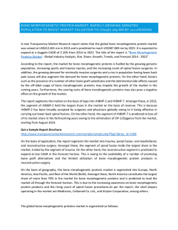
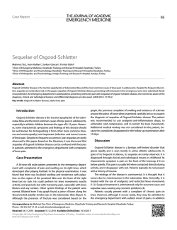
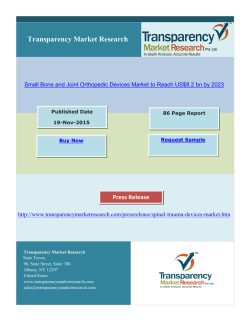
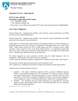
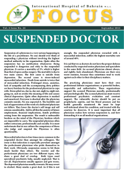
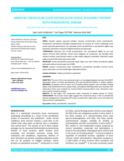
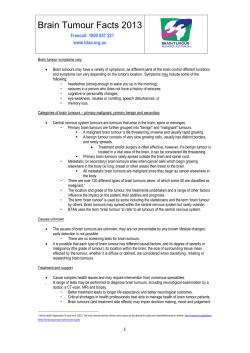
4|Page
Recurrent giant cell tumour of distal Tibia: Case report
and review of the literature
SANDESH MADI , SANDEEP
NAIK , SHARATH RAO
1
1
1
VIJAYAN ,
1
MONAPPA
1
Department of Orthopaedics, Kasturba Medical college, Manipal Univer-
sity, Manipal, Karnataka, India
Received
ABSTRACT
22 November 2014
Accepted 18 January 2015
Introduction
First described by Jaffe et al in 1940, Giant Cell Tumours [GCT] consititue 20% of benign bone tumours [1].
Less than 4% of these involve the foot and ankle region,
but the exact prevalence in the distal Tibia is not known.
Age group most commonly affected is from 20-40 years
with a slight female preponderance. Typically, it is described as an expansile lytic lesion in the epiphyseometaphyseal region in a skeletally mature bone. Clinically,
patient presents as a dull aching or a vague pain around
the affected joint and sometimes trauma brings notice to
the existence of this lesion. Swelling and joint stiffness
can also be the presenting complaints. Pathological fractures are seen in 12% of patients at the time of presentation [2]. We have briefly reviewed various treatment modalities for the management of this tumour in distal end
Tibia.
Case Report
Background: Giant Cell Tumour [GCT] is a familiar
benign but locally aggressive bone tumour especially
around the knee joint. Less than 4% of these tumours are
known to affect the ankle joints. But, its biological behavior at this rare location is quite unpredictable. Moreover, restoring the ankle joint functionality following
tumour resection is a challenging task
Case report: We report a case of Giant Cell Tumour of
distal end of left Tibia in a 53 year old male patient. Initially the condition was treated by curettage and bone
grafting. But, due to recurrence of the condition within 6
months, he was treated with extended curettage and bone
cementation. At One year follow up there is no recurrence and reasonably good function around the ankle
joint is maintained.
Conclusions: Primary GCTs have been traditionally
treated with curettage of the lesion followed by bone
grafts/bone cement. Recurrent cases often require aggressive management. In our case, despite recurrence, extended curettage of the lesion was done and the defect
was packed with bone cement. This added adjuvant
treatment offered good stability and allowed early mobilization of the ankle joint. This case substantiates the use
of bone cement in the treatment of recurrent GCT of distal Tibia whenever the articular integrity is intact with
reasonably good functional outcomes. However, a periodic follow-up is still recommended to watch-out for late
re-recurrences.
KEY WORDS:
Giant cell tumour
Bone cement
Distal tibia
Recurrence
Curettage
A 53 year old street vendor came to outpatients depart-
There was no history of constitutional symptoms. On ex-
ment with history of pain and swelling around the left
amination, diffuse swelling at the anteromedial border of
ankle. He related pain to a trivial trauma suffered 2
the left ankle with normal ankle movements and intact
months back. Pain was dull aching and aggravated with
neurovascular status was noted. X-rays showed expansile
prolonged standing and walking.
lytic lesion in the distal end of tibia (Figure 1). Plain and
contrast MRI revealed a well-defined lytic lesion measur-
Correspondence to: Dr Sandesh Madi
Email: [email protected]
ing 7x3.4x4.5cms having narrow zone of transition in the
distal tibial epi metaphyseal region. Areas of hemorrhage
ANNALS OF MEDICAL AND BIOMEDICAL SCIENCES. 2015; 1(1): 4-7
5|Page
and necrosis within the lesion were also noted, but there
was no evidence of periosteal reaction, sclerosis or matrix
calcification. Cortical thinning with minimal soft tissue
extension along antero-inferior aspect was also observed.
These features were suggestive of GCT of distal tibia.
Through an antero-lateral incision entire lesion was curetted out. The defect was filled with cortico-cancellous bone
graft harvested from ipsilateral iliac crest (Figure 2). Specimen sent for histopathological evaluation confirmed as
GCT (Netherlands grade II). Post-operative days were
uneventful.
Unfortunately, patient returned back within 6 months with
Figure 3. Recurrence within 6 months (articular surface
intact).
complaints of dull aching pain around the same ankle.
Repeat x-rays and MRI confirmed the recurrence of the
lesion (Figure 3). As the articular surface still appeared
intact, we planned to use bone cement as fillers. Lesion
was approached through the previous surgical incision.
Extended curettage was done till only a rim of subchondral bone was left intact (Figure 4). Thorough debridement and pulsatile lavage followed by high speed burring
was also employed this time. The defect was filled with
bone cement spacer. Wound was closed over a suction
drain. Repeat histo-pathological evaluation reported it as
Figure 4. Intra-operative. Extended curettage of the cavity.
GCT of same grade (Netherlands grade II). Post-operative,
the patient was given below knee slab support for 6 weeks
and non-weight bearing with the help of crutches was encouraged. After 6 weeks, slab was discarded and active
ankle mobilization was initiated. Weight bearing was allowed as tolerated only at 3rd month follow-up. He was
followed up periodically at 3 weeks, 6 weeks, 3 months, 6
months and 1 year (Figure 5). His Revised Musculoskeletal Tumor Society Rating at last follow up was 27/30
(90%).
Figure 1. X-rays antero-posterior and lateral view of ankle at 1st presentation.
Figure 2. X-rays following autologous bone grafting
ANNALS OF MEDICAL AND BIOMEDICAL SCIENCES. 2015; 1(1): 4-7
Figure 5. Last follow-up at one year after recurrence. .
Bone cement filling up the cavity.
6|Page
Discussion
mary tumour as a part of the disease progression or due to
The changing trends in the management of bone tumours
a pathological fracture. Moreover, with each recurrence
are from limb sacrifice to limb salvage and currently
these tumours tend to get aggressive and invariably affect
preservation of limb functionality. Amputation surgeries
the adjacent joint. Attempts at aggressive tumour resection
have become only of historical significance. Small cavi-
may control the condition but, at the expense of joint mo-
tary lesions have been traditionally treated by intralesional
bility. Such aggressive lesions can be treated with extend-
curettage with adjuvant chemical cauterization using phe-
ed tumour resection followed by ankle arthrodesis or re-
nol, hydrogen peroxide, and zinc chloride. Saleh.A et al in
cently, endoprosthetic replacement.
their retrospective study of 31 cases of distal tibia GCTs
Ankle Arthrodesis following tumour excision can be
have suggested that extended curettage of the lesion alone
achieved by vascularized autografts, non-vascularized
is sufficient in local control of the disease, even in recur-
autografts, allografts, or pasteurized autografts. Despite
rent conditions [3]. Cribb et al have described a case of
good functional outcomes, limitations for this procedure
GCT of distal end of Tibia managed with curettage fol-
include stiff ankle, a long period of recovery, infection,
lowed by high speed burring and the ankle was stabilized
and non-union. Saglik et al have described the use of
using an Ilizarov frame. Despite a prolonged course of
Fibular autografts to achieve arthrodesis in 2 patients with
rehabilitation, excellent functional recovery was noted [4].
aggressive GCT of distal Tibia [9]. Economopoulos et al
A large bone defect needs to be filled with either auto-
achieved arthrodesis using a custom made porous tanta-
grafts / allografts. Blackley et al reported an overall 12%
lum spacer after tumour resection in distal Tibia and sug-
recurrence in the treatment of GCTs of long bones with
gested that Tantalum to be a feasible structural substrate
curettage and bone grafting. Of 59 cases studied, 3 were in
and a suitable substitute to bone grafts [10].
distal tibia [5]. However, there are concerns regarding
Similar to mega-prosthesis in the treatment of aggressive
donor site morbidity (autograft), risk of disease transmis-
GCTs around the knee, custom–made ankle endoprosthe-
sion (allograft), and difficulty in visualizing (in x-rays)
sis have been introduced. V A Singh et al treated 4 cases
recurrence with grafts occupying the cavity.
(1 primary and 3 recurrent) by endoprosthetic reconstruc-
Alternative to bone grafts, bone cement is quite popular as
tion. One developed deep infection and one had talar col-
void fillers following tumour resection. According to
lapse [11]. In another series by Shekkeris et al, 2 out of 6
Thomas JL et al use of polymethylmethacrylate in large
cases developed deep infection [12]. Although ankle en-
osseous defects in foot and ankle following tumour resec-
doprosthesis looks a viable option, concerns regarding
tion provides stability and allows early weight bearing [6].
long term outcomes, deep infection, implant loosening and
The rationale behind the use of bone cement is that the
cost of implant need to be addressed.
exothermic reaction during polymerization of cement aids
in killing residual tumour cells without damaging the native host tissue. Similar to our case, Monish Bami et al
have reported a case with good results with the use of
bone cement, albeit in primary GCT of distal tibia [7].
Pan et al described a case with defect of anterior and posterior cortex in distal tibia due to recurrence. A double
layer of polypropylene mesh was used for the containment
of cement and the lesion healed uneventfully [8].
Due to proximity of these lesions around the joints, it
Conclusion
GCTs affecting the distal tibia are rare to encounter. Bony
defects can be filled with autografts. However, when there
is recurrence, it is still amenable to do extended curettage
and use bone cement as void fillers to achieve local control of the disease. This modality of treatment offers good
stability and early ankle mobilization is possible. Nevertheless, a periodic follow-up is still warranted to watch–
out for late re-recurrences.
becomes a daunting task to address both the tumour lesion
as well as joint functionality. As these tumours predominantly arise in the epiphyseo-metaphyseal region, there
Conflict of Interest
We declare that we have no conflict of interest.
may be breach in the adjacent articular surface by the pri-
ANNALS OF MEDICAL AND BIOMEDICAL SCIENCES. 2015; 1(1): 4-7
7|Page
References
8
1
2
3
4
5
6
7
Jaffee HL, Lichtenstein L, Portis RB. Giant cell
tumour of bone. Its pathologic appearance, grading, supposed variants and treatment. Arch
Pathol. 1940;30:993–1013.
Turcotte RE. Giant cell tumor of bone. Orthop
Clin North Am. 2006;37(1):35-51.
AlSulaimani SA, Turcotte RE. Iterative Curettage is Associated with Local Control in Giant
Cell Tumors Involving the Distal Tibia. Clin Orthop Relat Res. 2013;471(8):2668-74.
Cribb GL, Cool P, Hill SO, Mangham DC. Distal
tibial giant cell tumour treated with curettage and
stabilization with an Ilizarov frame. Foot Ankle
Surg. 2009;15(1):28-32.
Blackley HR, Wunder JS, Davis AM,White LM,
Kandel R, Bell RS. Treatment of Giant-Cell Tumors of Long Bones with Curettage and BoneGrafting. J Bone Joint Surg Am. 1999;81(6):81120.
Thomas JL, Kenneth AJ. Use of polymethylmethacrylate in large osseous defects in the foot
and ankle following tumor excision. J Foot Ankle
Surg. 1999;38(3):208-13.
Bami M, Nayak AR, Shreepad K, Kulkarni A,
and Gupta R. Giant cell tumor of lower end of
tibia. Case Rep Orthop. 2013;2013:429615.8.
ANNALS OF MEDICAL AND BIOMEDICAL SCIENCES. 2015; 1(1): 4-7
Pan, K. L., and W. H. Chan. Curettage and Cementation in Giant Cell Tumour of the Distal
Tibia Using Polypropylene Mesh for Containment: A case report. Malays Orthop J.
2010;4:51–3.
9 Saglik Y, Yildiz Y, Atalar H, and Gunay C. The
use of fibular autograft and ankle arthrodesis for
aggressive giant cell tumor in the distal tibia: a
case report. Foot Ankle Int. 2008;29(4):438-41.
10 Economopoulos K, Barker L, Beauchamp C, and
Claridge R. Case report: reconstruction of the
distal tibia with porous tantalum spacer after resection for giant cell tumor. Clin Orthop Relat
Res. 2010;468(6):1697-701.
11 Ajit SV, Nasirudin N,Bernatt M. Endoprosthetic
reconstruction for giant cell tumors of the distal
tibia: A short term review. Asia Pac J Clin Oncol.
2013;9(2):182-9.
12 Shekkeris, AS, Hanna SA, Sewell MD, Spiegelberg BGI, Aston WJS, et al. Endoprosthetic reconstruction of the distal tibia and ankle joint after resection of primary bone tumours. J Bone
Joint Surg Br. 2009;91(10):1378-82.
Background: Giant Cell Tumour [GCT] is a familiar benign but locally aggressive bone tumour especially around the knee joint. Less than 4% of these tumours are known to affect the ankle joints. But, its biological behavior at this rare location is quite unpredictable. Moreover, restoring the ankle joint functionality following tumour resection is a challenging task Case report: We report a case of Giant Cell Tumour of distal end of left Tibia in a 53 year old male patient. Initially the condition was treated by curettage and bone grafting. But, due to recurrence of the condition within 6 months, he was treated with extended curettage and bone cementation. At One year follow up there is no recurrence and reasonably good function around the ankle joint is maintained. Conclusions: Primary GCTs have been traditionally treated with curettage of the lesion followed by bone grafts/bone cement. Recurrent cases often require aggressive management. In our case, despite recurrence, extended curettage of the lesion was done and the defect was packed with bone cement. This added adjuvant treatment offered good stability and allowed early mobilization of the ankle joint. This case substantiates the use of bone cement in the treatment of recurrent GCT of distal Tibia whenever the articular integrity is intact with reasonably good functional outcomes. However, a periodic follow-up is still recommended to watch-out for late re-recurrences.
© Copyright 2026 Paperzz