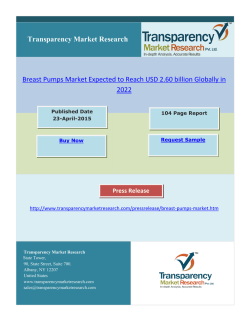
37 | P a g e
Association between age at presentation and pathological
features of breast cancer and its effect on survival; a
comparative study done in Sri Lanka
ABSTRACT
HARSHINI PEIRIS , LAKMINI MUDDUWA , NEIL THAL1
AGALA , KAMANI JAYATILAKA
3
2
4
1
Allied Health Sciences Degree Programme, Faculty of Medicine, University of
Ruhuna, Galle, Sri Lanka; 2Department of Pathology, Faculty of Medicine, University
of Ruhuna, Galle, Sri Lanka; 3Family Health Bureau, Ministry of Health, Colombo,
Sri Lanka; 4Department of Biochemistry, Faculty of Medicine, University of Ruhuna,
Galle, Sri Lanka
Received
30 June 2015
Accepted 22 August 2015
Introduction
Breast cancer developing at a young age has been reported
to have a more aggressive biological behavior compared
to the disease in older patients. Breast cancers in younger
women are found to have less well differentiated (higher
grade) tumors with higher proliferating fraction and higher
chance of vascular invasion than those occurring in older
patients [1]. The age at presentation is recognized by some
studies as an independent prognostic factor and as a significant predictor of long term survival of breast cancer
patients [2]. However, there are studies which have given
contradictory results [3, 4]. Yoshida M et al have shown
that the age at presentation is not an independent prognostic factor for Japanese women with breast cancer [3].
Objective: The young age at presentation of breast cancer (BC) is an independent prognostic factor of poor
survival. Therefore the aim of this study was to determine the association between age at presentation and the
clinicopathological features of BC and to assess the impact of this association on the breast cancer specific survival (BCSS) of different age groups.
Methods: This retrospective study included all BC patients who had sought the services of our unit from May
2006 to December 2012. Data were collected through
follow up visits, clinic and laboratory records. BCSS was
calculated from the date of BC diagnosis to the last follow up date or the event: death due to BC. Analysis was
done using Pearson chi-square, Kaplan-Meier and Coxregression models.
Results: A total of 944 subjects were grouped according
to the age at presentation; ≤ 35 years (7%), 36-60 years
(70%) and >60 years (23%). The prevalence of duct carcinoma in situ, tumour size, lymph node stage (LNS),
lympho-vascular invasion and Nottingham prognostic
index decreased as the age at presentation increased
(p<0.05). Multivariate analysis revealed that LNS is the
single factor affecting the BCSS of all groups. BCSS of
each group was influenced by one or two additional factors (tumour size for ≤35 years; progesterone receptor
and Her2 status for 36-60 years group and estrogen receptor status for >60 years).
Conclusions: Poor prognostic features are prevalent
among the ≤35 years group. LNS is the single most important independent predictor of BCSS irrespective of the
age at presentation.
Apart from the age at diagnosis, tumour size, lymph node
status, histological grade and pathological stage are other
important prognostic factors for survival of breast cancer
patients [5].
Correspondence to: Mrs Harshini Peiris
Email: [email protected]
KEY WORDS:
Age at presentation
Breast cancer specific survival
Clinicopathological features
Prognostic factors of breast cancer
Hormone receptors
In making therapeutic decisions, age is considered as one
factor together with other prognostic information of the
patients. In general, chemotherapy is recommended for
ANNALS OF MEDICAL AND BIOMEDICAL SCIENCES. 2015; 1(2):37-45
38 | P a g e
patients who are less than 35 years at the time of diagnosis
stained for estrogen receptor (ER), progesterone receptor
of the disease [6]. The age, less than 35 years is consid-
(PR) and human epidermal growth factor receptor 2
ered a high risk factor in planning adjuvant treatment
(Her2) expressions were retrieved from the archives of the
[6,7].
department for evaluation. Primary monoclonal mouse
Older women with breast cancer are claimed to have a
antihuman estrogen receptor α clone 1D5 (Dako-M7047),
more indolent course. The specific tumour types and pat-
monoclonal mouse antihuman progesterone receptor
terns of metastasis associated with a more favorable prog-
(Dako- M3569) and polyclonal rabbit antihuman c-erbB-2
nosis are found more frequently among elderly [8].The
oncoprotein (Dako-A0485) have been used with the sec-
aim of our study was to document the breast cancer spe-
ondary antibody (Dako Real EnVisionTM) for immuno-
cific survival (BCSS) rate for a Sri Lankan cohort of pa-
histochemical (IHC) staining of all breast cancers to as-
tients as there is no published research data on the survival
sess the ER, PR and Her2 expression. Scoring of ER and
of breast cancer population in Sri Lanka and to determine
PR expressions were done using Allred Score and Her 2
the association between the age at presentation and the
expression was assessed using UK recommendations for
clinicopathological features of females with breast cancer.
all breast cancers [10, 11]. IHC assessment too was done
The study assessed the impact of this association on the
by a single investigator eliminating inter-observer varia-
BCSS of different age groups.
tion. The complete absence of the staining for ER, PR and
a score of 0 or +1 for Her 2 were considered the criterion
Materials and methods
for categorizing as triple negative breast cancer (TNBC)
Study population
for this analysis.
This was a retrospective study. This study included female
patients who had histopathologically confirmed to have
Follow up and outcomes
breast cancer. The patients who had sought the services of
After enrolling, the study subjects were followed up for
our unit from May 2006 to December 2012 were included
recurrence or death at six months intervals. The study
in the study. There were 1068 breast cancer patients. Only
ended on 31st December 2013. The actual minimum fol-
944 patients gave consent to participate in the study. The
low up period was 12 months. More than 50% of patients
study was approved by the Ethical Review Committee of
were followed up beyond four years from the date of di-
our institution.
agnosis (88% for 24 months, 67.7% for 36 months, 51%
for 48 months and 38.9% for five or more years). Breast
Data collection
cancer specific survival time was defined as the time
The histopathological findings of the breast tumors were
elapsed from the date of diagnosis of breast cancer to the
retrieved from the laboratory records available in the la-
last follow up date or the date of death. Patients who died
boratory. They were recorded according to the modified
of breast cancer or who died with breast cancer (progres-
version of the minimum data set of National Health Ser-
sion/metastasis) were included [12]. Deaths from other
vice Breast Screening Programme (NHSBSP) guidelines
causes or from unknown causes were censored to the date
[9]. Nottingham grading for all breast cancers were done
of death. The cause of death of the patient was obtained
by a single investigator (principal investigator) using the
from the death certificate issued by the Department of
hematoxylin and eosin (H&E) stained slides to eliminate
Registrar General. Those who were lost to follow up or
inter-observer variation. Nottingham Prognostic Index
alive at the last follow up date were censored.
(NPI) was calculated for all breast cancers using the formula; NPI = 0.2 × tumour size (cm) + lymph node stage
Statistical Analysis
(1, 2 or 3) + histological grade (1, 2 or 3) [9].
The subjects were divided into three age groups; ≤ 35
years, 36-60 years and >60 years. The age at the date of
Laboratory methods
diagnosis of breast cancer was taken as the age at presen-
Slides with sections that had been immunohistochemically
tation of the disease. The Pearson chi-square test and the
ANNALS OF MEDICAL AND BIOMEDICAL SCIENCES. 2015; 1(2):37-45
39 | P a g e
chi-square test for trends were used to determine the asso-
There was a statistically significant trend of decreasing
ciation between age groups and the histopathological fac-
prevalence of T3 tumours with increasing age (p=0.040, χ2
tors. Kaplan-Meier model was used to estimate the BCSS;
trend=0.010) (Table 1).
in this case the log-rank test was used to compare the different groups. In multivariate analysis, Cox-regression
Nottingham Grade
model was used to estimate the predictors of survival us-
Nottingham grading of invasive breast cancers was done
ing the backward factor retention method. p < 0.05 was
only for 790 subjects out of the total 944 breast cancers
considered significant in all analysis.
due to the unavailability of well-preserved archival tissue
blocks to prepare H&E slides to replace the faded ones.
Results
There was a statistically significant difference between the
There were 64 (7%) females aged 35 years and younger,
age groups where the young patients had more high grade
661 (70%) aged 36-60 years and 219 (23%) were older-
tumors compared to the other age groups while only 3%
than 60 years. The mean age of the younger age group
of the young patients had Grade 1 tumors (p=0.043).
was 32.47 (SD±3.62), while it was 49.67 (SD ±6.49) and
However there was no significant trend of increase or de-
67.59 (SD±6.16) in the 36 to 60 years and <60 years
groups respectively.
In situ carcinoma
The prevalence of associated ductal carcinoma in situ
(DCIS) in the breast tumour was compared among the age
groups. Out of the total 944, this information was not
available for 18 patients. The prevalence of DCIS was
47% in the ≤35years age group while 38% and 23% in
the36-60 years and >60 years groups respectively. The
youngest age group had the highest prevalence of associated DCIS and there was a statistically significant trend of
crease in the grade with increasing age at presentation (χ 2
trend=0.102) (Table 1).
Tumour stage
The prevalence of patients with stage I and II breast cancer in the entire study population was 58% while the prevalence of III and IV disease was 42%. There were no stage
IV patients in the youngest age group. The majority of the
young patients were stage II or III (44%). The prevalence
of stage IV tumors was highest in the elderly group (3%).
However there was no statistically significant difference
between the age groups (p=0.109) with regard to the stage
at presentation (Table 1).
decreasing prevalence of DCIS with increasing age
(p<0.001; χ2trend<0.001). However there was no statistically significant difference with regard to the grade or
growth pattern of the associated DCIS. There was no statistically significant difference between the age groups
with regard to the presence of lobular carcinoma in situ
Lymph-node metastasis (LNM)
The axillary clearance had been done only for 912 subjects and 27 had not had axillary clearance. Out of the 912
patients, the number of involved lymph nodes was available only for 897 subjects. There was a progressive reduc-
(LCIS) or the presence of Paget’s disease.
tion in the prevalence of LNM with increasing age
Within the entire group of subjects, invasive duct carci-
(p=0.012, χ2trend=0.004). The prevalence of LNM was
noma of no special type (93.7%) was the most common.
least among the females who were >60 years old (Table
Invasive tubular, lobular, mucinous, papillary and medul-
1).
lary like comprised of 0.1%, 3.4%, 1.4%, 1.0% and 0.3%
respectively.
Presence of lympho-vascular invasion (LVI)
For the present study, no attempt was made to differenti-
Tumour characteristics
ate between blood vessel and lymphatic invasion. The
Tumour size
highest prevalence of LVI was seen in the youngest age
In all age groups, T2 (20-50mm) tumour was the most
group (44%). The lowest prevalence; 21% was seen in the
common. However T3 (>50mm) tumour was more preva-
oldest age group. There was a statistically significant trend
lent in the youngest age group (13%) while T1 (≤20mm)
of decrease in the prevalence of LVI with increasing age
tumour was more prevalent in the oldest group (40%).
at presentation (p=0.002, χ2trend=0.001) (Table 1).
ANNALS OF MEDICAL AND BIOMEDICAL SCIENCES. 2015; 1(2):37-45
40 | P a g e
Nottingham Prognostic Index (NPI)
poor prognosis. There was a statistically significant trend
Patients with NPI of ≤3.4 and >5.4 are considered to have
of decreasing NPI with increasing age at presentation
good and poor prognosis respectively according to the
(p=0.011, χ2trend=0.001) (Table 1).
NHSBSP guidelines [9]. The age group ≤35 years had the
highest prevalence (52%) of NPI >5.4, which indicated
Table 1. Comparison of tumour characteristics among the age groups.
Tumour characteristics
Age (years)
≤35
(n=64)
36-60
(n=661)
p
χ2 trend
>60
(n=219)
Tumour size
T1 (≤20mm)
T2 (21-50mm)
T3 (>50mm)
Missing
20 (33%)
33 (54%)
8 (13%)
3
194 (31%)
377 (61%)
51 (8%)
39
79 (40%)
113 (56%)
8 (4%)
19
0.040
0.010
Grade 1
Grade 2
Grade 3
Missing
2 (3%)
21 (36%)
35(60%)
6
73 (13%)
248 (45%)
228 (42%)
112
21 (11.5%)
86 (47%)
76 (41.5%)
36
0.043
0.102
I
II
III
IV
Missing
7 (12%)
27 (44%)
27(44%)
0 (0%)
3
97 (15%)
265 (42%)
263 (42%)
8 (1%)
28
44 (22%)
78 (39%)
74 (37%)
6 (3%)
17
0.109
0.158
Presence
Absence
Missing
39 (64%)
22 (36%)
3
363(57%)
273 (43%)
25
93 (46%)
107 (54%)
19
0.012
0.003
0 (No positive LNs)
1(1-3 positive LNs)
2 (4-9 positive LNs)
3(>9 positive LNs)
Missing
Lympho-vascular invasion
Presence
Absence
Missing
Nottingham Prognostic Index
≤3.40
3.41-5.40
>5.40
Missing
22 (36%)
16 (26%)
15 (25%)
8 (13%)
3
273 (43%)
163 (26%)
128 (20%)
72 (11%)
25
107 (54%)
44 (22%)
30 (15%)
19 (9%)
19
0.147
0.007
28 (44%)
36 (56%)
0
187 (29%)
465 (71%)
9
45 (21%)
166 (79%)
8
0.002
0.001
5 (9%)
22 (39%)
29 (52%)
8
73 (14%)
279 (53%)
174 (33%)
135
30 (18%)
92 (55%)
44 (27%)
53
0.011
0.001
Nottingham grade
Tumour stage
Lymph-node metastasis
Lymph node stage
n, number; p, significance
Hormone receptors and Her2 expression
trend of decrease in the prevalence of ER negative tumors,
Out of the total 944 breast cancers, IHC assessment was
with increasing age
done only on 805 subjects with ER, 795 subjects with PR
trend=0.001) (Table 2). The prevalence of PR negative
and 804 subjects with Her2 due to the unavailability of the
tumors was highest in the young age group compared to
well preserved archival tissue blocks for the replacement
the others (p=0.005). However PR expression did not have
of the faded slides. There was a statistically significant
a decreasing / increasing trend across the three age groups.
at
presentation (p=0.002,
χ2
ANNALS OF MEDICAL AND BIOMEDICAL SCIENCES. 2015; 1(2):37-45
41 | P a g e
Table 2. Comparison of ER, PR and Her 2 expression among the age groups.
Age (years)
≤35
(n=64)
36-60
(n=661)
p
χ2 trend
>60
(n=219)
ER
Positive
Negative
Missing
12 (21%)
46 (79%)
6
222 (39%)
341 (61%)
98
86 (47%)
98 (53%)
35
0.002
0.001
Positive
Negative
Missing
13 (22%)
45 (78%)
6
248 (44%)
312 (56%)
101
73 (41%)
105 (59%)
41
0.005
0.186
Positive
Negative
Missing
16 (28%)
42 (78%)
6
142 (25%)
422 (75%)
97
49 (27%)
133 (73%)
37
0.848
0.869
PR
Her2
Triple negative
Yes 27 (47%)
182 (32%)
60 (33%)
0.093
No 31 (53%)
380 (68%)
121 (67%)
Missing
6
99
38
n, number; p, significance; ER, estrogen receptor; PR, progesterone receptor; Her2, human epidermal growth factor receptor 2
0.236
Her2 expression did not show a statistically significant
died due to other reasons were censored to the date of
difference between the age groups (p= 0.848). The majori-
death. Out of the censored population 35.5% (270/760)
ty of patients in the youngest age group was ER negative
were followed up for more than five years from the date of
(79%), PR negative (78%) and Her2 negative (78%).
diagnosis of the disease (≤35 years-47%, 36-60 years-36%
Therefore the youngest age group had the highest preva-
and >60 years-32%). The median survival time was
lence of TNBC (47%) (Table 2). The study group was
120.367 months (SE 25.651; 95% CI 70.090-170.644).
divided into two age groups as ≤35 years and >35 years,
The Kaplan-Meier model was used to estimate the five-
to compare the receptor status. A statistically significant
year BCSS. The five-year BCSS rate of the whole cohort
difference was observed between those two age groups
was 78.8%. The five year BCSS varied according to the
2
with regard to their ER status (p=0.002, χ trend=0.002),
2
PR status (p=0.002, χ trend=0.002) and triple negative
2
status (p=0.030, χ trend=0.030). However, Her2 expression did not show a statistically significant difference
(p=0.739).
Clinical management
Mastectomy with axillary clearance was the main type of
surgical management for all the three age groups. The
97% of ≤35 years age group, 95% of 36-60 years age
group and 83% of >60 years age group patients had received adjuvant chemotherapy. Radiotherapy had been
given to 80% of ≤35 years, 75% of 36-60 years and 66%
of >60 years age patients. Hormone therapy had been given to 48% of ≤35 years, 65% of 36-60 years and 68% of
>60 years age patients.
Survival analysis
Breast cancer specific survival
There were 164 deaths due to breast cancer and 20 deaths
due to causes other than breast cancer, and the rest (760)
were censored to the last follow up date. Those who had
ANNALS OF MEDICAL AND BIOMEDICAL SCIENCES. 2015; 1(2):37-45
age group; 63%, 80% and 79% respectively for the ≤35
years, 36-60 years and >60 years age groups (Figure 1).
Figure 1. Breast Cancer Specific Survival patterns of the
three age groups. Kaplan-Meier survival analysis of patients with breast cancer stratified into three age groups
(35 or below, 36-60 and above 60): patients age 35 or below had a poor survival compared to the age groups 3660years and above 60years (p=0.02).
42 | P a g e
The BCSS curves were compared using the log-rank test
all three age groups. All three age groups had an addition-
which indicated that there was a survival benefit of being
al prognostic factor which independently affected the sur-
more than 35 years of age at presentation (p=0.02).
vival of each group (Table 4).
Histopathological and IHC factors which were found to
have a significant association with the age groups were
considered for the univariate analysis using the KaplanMeier estimator and the log-rank test for the estimation
and comparison of survival curves. According to the findings of the univariate analysis, tumour size, LNS, NPI,
Table 4. Multivariate analysis of factors predicting BCSS
of the age groups.
Factor
HR
95%CI
Age group ≤35
Lymph node stage
<0.001
pathological stage and Her2 status were significantly as-
0 (No positive LNs)
1 (ref)
sociated with the survival of the youngest age group (≤35
1(1-3 positive LNs)
1.05
0.09-12.10
years) (Table 3). The tumour size, Nottingham grade,
2 (4-9 positive LNs)
4.80
0.87-26.38
LNS, LVI, NPI, pathological stage, PR and Her2 status
3(>9 positive LNs)
34.87
5.41-224.93
were significantly associated with the survival of the age
Tumour size
group 36-60 years (Table 3).
The LNS, pathological stage, TNBC, ER and PR status of
p
T1 (≤20mm)
1 (ref)
T2 (21-50mm)
2.13
0.24-19.07
T3 (>50mm)
21.32
2.07-219.04
Her2
a
-
0.008
the tumour were significantly associated with survival of
the age group >60 years (Table 3).
0.515
Age group 36-60
Table 3. Results of univariate analysis of pathological
features.
Log-rank test p value
Factor
Age
group
≤35 years
Age group 3660 years
Age
group
>60 years
Lymph node stage
<0.001
0 (No positive LNs)
1 (ref)
1(1-3 positive LNs)
2.60
1.31-5.17
2 (4-9 positive LNs)
4.71
2.44-9.11
3(>9 positive LNs)
8.94
4.53-17.64
Presence of associ-
PR
ated DCIS
0.428
0.329
0.690
Presence
1 (ref)
0.001
Tumour size
0.026
0.005
0.290
Absence
2.25
Nottingham grade
0.507
0.005
0.220
Absence
1 (ref)
Presence of LVI
0.437
<0.001
0.217
Presence
1.62
1.00-2.63
Tumour size
a
-
0.382
1.36-3.72
Her2
Lymph node me-
0.051
tastasis
0.062
<0.001
0.001
Nottingham grade
a
-
0.295
Lymph node stage
<0.001
<0.001
0.001
Presence of LVI
a
-
0.388
Pathological stage
0.001
<0.001
<0.001
Age group >60
NPI
0.012
<0.001
0.077
ER
0.397
0.104
<0.001
PR
0.210
0.002
Her2
0.007
<0.001
TNBC
0.862
0.859
Lymph node stage
0.002
0 (No positive LNs)
1 (ref)
0.005
1(1-3 positive LNs)
2.64
0.92-7.55
0.301
2 (4-9 positive LNs)
4.04
1.46-11.17
0.001
3(>9 positive LNs)
7.04
2.46-20.15
DCIS, ductal carcinoma in situ; LVI, lympho-vascular invasion; NPI, Nottingham
ER
Prognostic Index; ER, estrogen receptor; PR, progesterone receptor; Her2, human
Presence
1 (ref)
epidermal growth factor receptor 2; TNBC, triple negative breast cancer.
Absence
4.41
1.66-11.66
PR
a
-
0.551
Multivariate analysis was performed for all the age groups
TNBC
a
-
0.855
separately to find out the factors which independently
HR, hazard ratio; CI, confidence interval; p, significance; LNs, lymph nodes; ref –
affect the survival of each age group. It was the lymph
reference group; Her2, human epidermal growth factor receptor 2; a- removed from
node stage which independently affected the survival of
0.003
the final model; PR, progesterone receptor; LVI, lympho-vascular invasion; ER,
estrogen receptor; TNBC, triple negative breast cancer.
ANNALS OF MEDICAL AND BIOMEDICAL SCIENCES. 2015; 1(2):37-45
43 | P a g e
Table 5. Characteristic features of breast cancer in young
results favoring it [8, 17]. In the clinical setting, we have
patients (≤35 years).
often seen young females have breast cancers with poor
prognostic features and not surviving long. Therefore we
large tumours (2-5cm />5cm)
designed the present study to determine the survival of
high Nottingham grade (Grade 2 / Grade 3)
different age groups of breast cancer patients and assess
lymph node metastasis
whether the well-established pathological prognostic pa-
lympho-vascular invasion
rameters vary according to the age. The prevalence of
high NPI value
associated DCIS was found to decrease with increasing
age in the present study. Therefore the youngest age group
Discussion
of this can be expected to have a higher chance of devel-
One of the aims of our study was to document the BCSS
oping local recurrences [18]. With increasing age, the
rate as there are no published data on BCSS for Sri
prevalence of T3 tumors (>5cm), LNS and LVI became
Lankan breast cancer patient population. Cancer Institute
less. The NPI also became less with the increase in age at
data has been published in the past on overall survival but
presentation. Therefore the pathological factors which
not on the BCSS. In the present cohort of breast cancer
predict poor prognosis became less prevalent with increas-
patients, the five year BCSS was 78.8% and the overall
ing age predicting a better prognosis as the age increased.
survival was 76%. A study on global surveillance of can-
Although the pathological stage depends on the tumour
cer survival (1995-2009) based on 279 population based
size and lymph node stage, it did not show a significant
registries from 67 countries revealed that for women diag-
difference between the age groups. Similarly Nottingham
nosed during 2005-2009, age standardized five year net
grade did not have an increasing or decreasing trend with
survival from breast cancer was 80% or higher in 34 coun-
the age at presentation. Except for ER expression, other
tries around the world including Canada, Germany, US,
IHC markers did not show a trend with increasing age. ER
UK and Japan. However, breast cancer survival is less
expressing tumors became more prevalent with increasing
than 70% in Malaysia (68%) and India (60%) [13]. Ac-
age. But PR did not have the same effect. Therefore,
cording to the same study overall survival was less than
whether patients became more responsive to hormone
60% in Mongolia (57%) and South Africa (53%) [13].
therapy with increasing age is uncertain as expression of
The BCSS of our cohort is very much close to 80% and
PR is needed for the ER to have its effect [19]. There was
much better than the neighboring Asian countries. The
no association found between the Her2 expression and the
relatively better BCSS in the present Sri Lankan study
age at presentation (Table 3). A few pathological factors
cohort may be multifactorial. Since there is no national
were identified to have an association with the different
breast cancer screening programme established in Sri
age groups without any significant trend with increase or
Lanka, a significant proportion of patients present with
decrease in age. TNBCs, PR negative tumors and high
stage III or IV disease according to the present study
Nottingham grade tumors were more common in the age
(Stage I and II-58%, Stage III and IV-42%). However
group of ≤35 years than the rest. All three are considered
patients have the access to best care at government hospi-
poor prognostic features. A study done in India stated that
tals free of charge and in private sectors which may have
tumors with high grade, high lymph node involvement and
improved the survival. There are many reports on the as-
negative hormone receptor expression occur more com-
sociation between age at presentation and the prognosis of
monly in young breast cancer patients [14]. Our results on
breast cancer patients [1, 3, 4, 8]. Although the definition
the biological nature of breast cancer in young patients are
of ‘young patient with breast cancer’ varies from publica-
very much similar to the mentioned Asian countries.
tion to publication, many agree that younger patients have
Clinicopathological profile of breast cancer in the age
a poor prognosis [1, 2, 4, 14, 15]. For the present study we
group ≤35 years according to the present study supports
defined the young age group as females who are ≤35 years
the poor prognostic behavior of breast cancer patients who
[4, 15, 16]. Age as an independent prognostic factor has
are ≤35 years (Table 5). The cumulative effect of these
been disputed in some studies while others have found
statistically significant trends and associations were re-
ANNALS OF MEDICAL AND BIOMEDICAL SCIENCES. 2015; 1(2):37-45
44 | P a g e
flected in the survival curves/patterns and the five year
ed or are on treatment. The majority have undergone sim-
BCSS rates of the three age groups. The age group ≤35
ple mastectomy with level I or II axillary clearance and
year, had the worst survival. Although the said pathologi-
one or more forms of adjuvant therapy. Even though these
cal factors indicate a better survival in the oldest age
patients had been treated or on treatment, the patients who
group, the survival curves of the middle and oldest age
were ≤35 years had a poor survival compared to the others
groups were similar and overlapped each other. This is
most probably due to the high degree of aggressiveness of
reiterated in the BCSS rates of 36 to 60 years and
the tumors in the youngest age group, which is substanti-
>60years groups which were 80% and 79% respectively.
ated by the association of poor prognostic pathological
Although the elderly patients have better prognostic fea-
features. This retrospective study is the first study on the
tures compared to the other two groups, they too have
BCSS in Sri Lanka according to the accessible literature.
more of high grade (Grade 2and 3 in 88.5%) and larger
Therefore we do not have published data in Sri Lanka to
tumors (T2 and T3 in 60%) within the group. These fea-
compare. Since this is a retrospective study, follow up
tures are indications for adjuvant chemotherapy. The ma-
period of individual patients varied depending on the date
jority (83%) of the elderly patients had received chemo-
of diagnosis. Therefore patients who developed breast
therapy. Therefore their survival may have deviated from
cancer in 2006, 2007 and 2008 could be followed up be-
the better survival predicted in the analysis of pathological
yond five years (38.9%) while the others could be fol-
features and superimposed on the survival curve of the 36-
lowed up for less than five years. However those who
60 years age group. A study done in USA, reported that
could be followed up beyond five years were almost
older patients (>65 years) with breast cancer usually are
equally distributed within the three groups. Therefore the
undertreated because of co-morbid conditions (40.9%), or
effect is similar on all three groups and the power of sta-
for refusal of treatment (31.8%), or favorable tumour pa-
tistical analysis is less affected. Since the length of follow
thology (13.8%) or unexplainable causes (13.6%) [20]. In
up period is not too long, the present cohort of patients is
the present study too, some of those factors may have had
homogeneous in terms of treatment modalities. These pa-
an effect. The poor tolerance of chemotherapy in elderly
tients have been diagnosed and managed at a single unit
and the biological behavior of high grade larger tumors
(the tertiary care hospital). Immunohistochemical assess-
would have contributed to the deviation in the survival.
ment also was done in a single laboratory. Therefore there
The univariate analysis revealed that the pathological
is a consistency among the study subjects of the cohort in
stage and the LNS affected the survival of all three age
terms of diagnosis, management and prognostication.
groups and presence of DCIS affected none. The survival
On the basis of observed outcomes, this study has re-
of patients of 36-60 years age was affected by tumour
vealed that being ≥35 years of age at presentation gives a
size, Nottingham grade, LVI, PR and Her2 status but not
survival benefit as breast cancers with poor prognostic
by the expression of ER. All these factors were used for
features are seen mostly among the ≤35 years age group
the multivariate analysis to identify the factors with an
compared to the others. Lymph node stage is the single
independent effect on the survival. The LNS was the sin-
independent predictor of survival irrespective of the age at
gle most important pathological factor affecting the sur-
presentation. However having T3 tumors in ≤35 years age
vival of all age groups in the present study. The LNS has
group, expression of PR and Her2 in 36-60 years age
been recognized as the most significant prognostic indica-
group and expression of ER in >60 years age group inde-
tor for patients with early-stage breast cancer in the past
pendently affect the survival of breast cancer patients.
[8, 21]. It is being reiterated in our study too. In our study,
the presence of T3 tumors affected the survival of ≤35
Conflict of Interest
years age group in addition to the LNS. The expression of
We declare that we have no conflict of interest.
PR and Her2 (36-60 years) and ER (>60 years) also had
an independent effect on the survival. This retrospective
study enrolled breast cancer patients who have been treatANNALS OF MEDICAL AND BIOMEDICAL SCIENCES. 2015; 1(2):37-45
45 | P a g e
Acknowledgement
The authors wish to acknowledge the staff of the Oncology Unit of the Teaching Hospital, Karapitiya, Galle, Sri
Lanka for permitting retrieval of clinic data, providing
facilities to retrieve clinic files and follow-up of the patients. Department of Pathology, Faculty of Medicine,
University of Ruhuna, Galle, Sri Lanka is acknowledged
for the technical assistance. Financial support for the research was provided by the University Grants Commission-Research grants and University of Ruhuna, research
grants, Sri Lanka.
References
1
2
3
4
5
6
7
8
9
Zavango G, Meggiolaro F, Pluchinotta A, Bozza
F, Favretti F, Marconato R, et al. Influence of age
and menopausal status on pathologic and biologic features of breast cancer. The Breast 2000; 9:
320-328.
Jayasinghe UW, Taylor R, Boyages J. Is age at
diagnosis an independent prognostic factor for
survival following breast cancer? ANZ J Surg
2005; 75: 762-767.
Yoshida M, Shimizu C, Fukutomi T, Tsuda H.
Prognostic factors in young Japanese women
with breast cancer: Prognostic value of age at diagnosis. Jpn J Clin Oncol 2011; 41(2): 180-189.
Vostakolaei FA, Broeders MJM, Rostami N,
Dijck JAAM. Age at diagnosis and breast cancer
survival in Iran. Int J Breast Cancer 2012;
doi:10.1155/2012/517976.
Schwartz AM, Henson DE, Chen D, Rajamarthandan S. Histologic grade remains a prognostic
factor for breast cancer regardless of the number
of positive lymph nodes and tumour size. Arch
Pathol Lab Med 2014; 1 (38):10481052;doi:10.5858/arpa.2013-0435-OA.
Sharma M, Abraham J. Breast Cancer. In: Abraham J, Gulley JL, Allegra C.J., editors. Bethesda
Handbook of Clinical Oncology. 3rd ed. Lippincott Williams and Wilkins. 2010; 151-173.
Kataja V, Castiglione M. Primary breast cancer:
ESMO clinical recommendations for diagnosis,
treatment and follow-up. Ann Oncol 2009;20
(4):10-14.
Fisher CJ, Egan MK, Smith P, Wicks K, Millis
RR, Fentiman IS. Histopathology of breast cancer in relation to age. Br J Cancer 1997; 75(4):
593-596.
NHSBSP Guidelines for Pathology Reporting in
Breast Cancer Screening. NHSBSP publication
No58,.2005.
ANNALS OF MEDICAL AND BIOMEDICAL SCIENCES. 2015; 1(2):37-45
10 Hammed MEH, Hayes DF, Dowsett M, Allred
DC, Hagerty KL, Badve S, et al. American Society of Clinical Oncology/College of American
Pathologists Guideline Recommendations for
Immunoistochemical Testing of Estrogen and
Progesterone Receptors in Breast Cancer. Arch
Pathol Lab Med 2010; 134: 907-922.
11 Ellis IO, Bartlett J, Dowsett M. Best practice No
176: Updated recommendations for HER2 testing
in the UK. J Clin Pathol 2004; 57: 233-237.
12 Rakha EA. Pitfalls in outcome prediction of
breast cancer. J Clin Pathol 2013; 66: 458-464.
13 Allemani C, Weir HK, Carreira H, Harewood R,
Spika D, Wang XS, et al. Global surveillance of
cancer survival 1995-2009: analysis of individual
data for 25676887 patients from 279 populationbased registries in 67 countries (CONCORD-2).
Lancet 2015; 385: 977-1010.
14 Ghosh S, Sarkar S, Simhareddy S, Kotne S, Rao
PBA, Turlapati, SPV. Clinico-Morphological
profile and receptor status in breast cancer patients in a South Indian Institution. Asian Pac J
Cancer Prev 2014; 15(18): 7839-7842.
15 Saghir NSEI, Seoud M, Khalil MK, Charafeddine M, Salem ZK, Geara, FB, et al. Effects
of young age at presentation on survival in breast
cancer. BMC Cancer 2006; 6: 194.
16 Filleron T, MD FD, Karmar A, Spielmann M,
Levy C, Fumoleau, P, et al. Prognostic factors of
young women (≤35 years) with node positive
breast cancer: possible influence on posttherapeutic follow-up. Bull Cancer 2013; 100:
E22-E29.
17 Colzani E, Liljegren A, Johansson ALV, Adolfsson J, Hellborg H, Hall PFL, et al. Prognosis of
patients with breast cancer: Cause of death and
effects of time since diagnosis, age and tumour
characteristics. J Clin Oncol 2011; 29: 40144021.
18 Lakhani SR, Ellis IO, Schnitt SJ, Tan PH, Viver
MJ van de. WHO classification of tumours of the
breast. 4th ed. International Agency for Research
on Cancer, Lyon 2012; 92-93.
19 Leake R. Prediction of hormone sensitivity- the
receptor years and onwards. Endocr Relat Cancer
1997; 4(3): 289-296.
20 Velanovich V, Gabel M, Walker EM, Doyle TJ,
O’Bryan RM, Szymanski W, et al. Causes for the
under treatment of elderly breast cancer patients:
tailoring treatments to individual patients. J Am
Coll Surg 2002; 194: 8-13.
21 Cianfrocca M, Goldstein LJ. Prognostic and predictive factors in early-stage breast cancer. The
Oncologist 2004; 9: 606-616.
HARSHINI PEIRIS, LAKMINI MUDDUWA, NEIL THALAGALA, KAMANI JAYATILAKA, AMBS Objective: The young age at presentation of breast cancer (BC) is an independent prognostic factor of poor survival. Therefore the aim of this study was to determine the association between age at presentation and the clinicopathological features of BC and to assess the impact of this association on the breast cancer specific survival (BCSS) of different age groups. Methods: This retrospective study included all BC patients who had sought the services of our unit from May 2006 to December 2012. Data were collected through follow up visits, clinic and laboratory records. BCSS was calculated from the date of BC diagnosis to the last follow up date or the event: death due to BC. Analysis was done using Pearson chi-square, Kaplan-Meier and Coxregression models. Results: A total of 944 subjects were grouped according to the age at presentation; ≤ 35 years (7%), 36-60 years (70%) and >60 years (23%). The prevalence of duct carcinoma in situ, tumour size, lymph node stage (LNS), lympho-vascular invasion and Nottingham prognostic index decreased as the age at presentation increased (p<0.05). Multivariate analysis revealed that LNS is the single factor affecting the BCSS of all groups. BCSS of each group was influenced by one or two additional factors (tumour size for ≤35 years; progesterone receptor and Her2 status for 36-60 years group and estrogen receptor status for >60 years). Conclusions: Poor prognostic features are prevalent among the ≤35 years group. LNS is the single most important independent predictor of BCSS irrespective of the age at presentation. KEY WORDS: Age at presentation Breast cancer specific survival Clinicopathological features Prognostic factors of breast cancer Hormone recept
© Copyright 2026 Paperzz