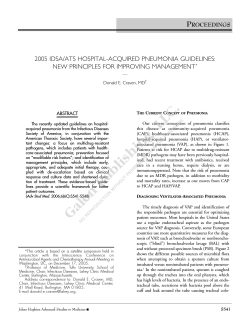
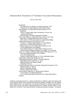
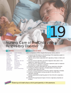
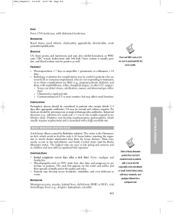
4|P age
Normal CT Scan in a Patient with Pneumonia: a Case Report
ABSTRACT
SAEED REZA JAMALI MOGHADAM , YOSOF MOJTAHEDI , SEYEDAHMAD SEYEDALINAGHI , REZVAN
KAKAVAND GHALEH-NOEI
1
1
2
3
1
Ziaeian Hospital, Tehran University of Medical Sciences, Tehran, Iran;
2
Iranian Research Center for HIV/AIDS (IRCHA), Iranian Institute for
Reduction of High-risk Behaviors, Tehran University of Medical Sciences,
Tehran, Iran; 3 Department of Virology, School of Public Health, Tehran
University of Medical Sciences, Tehran, Iran
Received
16 March 2015
Accepted 29 May 2015
A 68-year-old female was hospitalized with a primary
complaint of dyspnea in Ziaeian Hospital affiliated to
Tehran University of Medical Sciences in April 2014.
She had malaise and non-productive cough along with a
loss of appetite during the last two days. She was also
suffering slight tachypenea. The patient had normal chest
x-ray and Computed Tomography (CT) scan on the admission time. Initially there was no response to the first
empiric treatment, the scan was repeated after 2 days and
subsequently we observed abnormal signs in the scan
suggestive to pneumonia.
KEY WORDS:
Introduction
Pneumonia is a common lung infection that mainly affects the alveoli [1-3]. Overall approximately 450 million
Pneumonia
Diagnosis
Chest X-Ray
CT scan
Treatment
people are infected with pneumonia each year worldwide
and results in the death for about four million people [4,
5]. It occurs in all age groups but mortality rates are high
among children particularly in the newborn period and in
the elderly over 75 years of age [4]. In the United States
each year 5.6 million people are infected with community
-acquired pneumonia [6]. Its symptoms can be expressed
in different ways and includes cough, chest pain, fever and
difficulty breathing [1]. Although most of the people regain their health between one to three weeks, pneumonia
can be a serious threat to all lives [5]. It is usually caused
by viral, bacterial infections or other microorganisms such
as fungi, certain drugs and autoimmune diseases [1, 7].
Most of community-acquired pneumonia is caused by the
gram-positive bacterium Streptococcus pneumoniae or
pneumococcus (20%-60%) [1, 5]. This bacterium is the
most common cause of pneumonia in adults [4, 5].
Macrolids and antibiotics known as Beta-lactam such as
penicillins are used for the treatment of pneumonia [2, 5].
The second common agent of community-acquired pneumonia is Heamophilus influenzae that generally occurs in
patient with chronic lung disease, elderly people and those
with alcoholism [2, 5]. On the other hand, atypical pneumonia is caused by Mycoplasma pneumoniae, Chlamydia
pneumoniae, and Legionella pneumophila [3, 5]. A number of viruses cause pneumonia including Influenza virus,
Respiratory Syndrome Virus (RSV), Severe Acute Respiratory Syndrome (SARS), Parainfluenzae virus, Adeno
virus, and Herpes virus [1, 3, 5]. Based on patient characteristics and disease severity according to the kind of acquired pneumonia and etiologic agents are usually applied
to a choice of diagnostic methods [8]. For diagnosis the
types of pneumonia, physical exam, radiographic imaging
and laboratory studies are used [1, 2]. Pneumonia can be
diagnosed by Chest X-Ray (CXR), Computed Tomogra-
Correspondence to: Miss Rezvan Kakavand Ghaleh-Noei
Email: [email protected]
phy (CT) scan, culture of the sputum and blood, gram
stain sputum, and serology. Urinary antigen is alternative
ANNALS OF BRITISH MEDICAL SCIENCES. 2015; 1(1): 4-6
5|P age
or complementary method to detect S. pneumoniae and
The treatment was changed to Meropenem, Vancomycin
Legionella. Polymerase chain reaction (PCR), enzyme
and Ciprofloxacin. The general condition of the patient
immunoassay (EIA), and immunofluorescence are rapid
became better within 72 hours after changing of the anti-
tests for identifying Chlamydophila pneumoniae, Myco-
biotics. The antibiotics continued for two weeks and final-
plasma pneumoniae and some other respiratory tract vi-
ly the patient discharged in good condition.
ruses [9-12]. We report a patient with pneumonia who had
normal CXR and CT scan on the admission time. In the
Figure 1. The first CT scan showed no sign of pneumonia
on the admission time.
second CT scan, abnormal signs suggestive for pneumonia
were appeared.
Case Report
A 68-year-old female referred to Ziaeian Hospital affiliated to Tehran University of Medical Sciences with malaise
and non-productive cough along with loss of appetite during the last two days. She had no fever and in the physical
examination showed general illness with mild tachypenea.
The vital signs included heart rate: 80bpm/min, blood
pressure: 120/80 mmHg, respiratory rate: 32 / min and
oxygen saturation 92% on room air. Mild coarse crackles
were heard especially in the base of both lungs. The cardi-
Figure 2. The second CT scan showed air space consolidation in the mid and lower lobe of the right lung.
ac examination revealed no murmur or any tachycardia.
Laboratory studies showed Complete Blood Count (CBC),
Erythrocyte Sedimentation Rate (ESR), electrolytes,
Blood Urea Nitrogen (BUN), Creatinine and Arterial
Blood Gas (ABG) in normal limits but hemoglobin revealed mild anemia and C - reactive protein (CRP) was
positive. In the CXR and CT scan no sign of pneumonia
was shown on admission (Figure 1). Echocardiography
was performed for the patient and did not show any cardiac problems.
Empiric treatment was initiated with Oseltamvir, Ceftriaxon and Azithromycin. Forty eight hours after the treatment, the symptoms of the patient such as productive
Discussion
cough and dyspnea were exacerbated and subsequently the
CXR can be used to confirm pneumonia in patients with
CXR and CT scan were repeated. In the new CXR re-
suspected pneumonia. In the past, chest-x rays were the
vealed blunted right costo-pherenic angle and base her
golden standard for pneumonia but now, there is signifi-
right lung showed opacity. Also in the new CT scan, pleu-
cant evidence that pneumonia can be identified using CT
ral effusion was seen with air space consolidation (air
scans [13]. In fact, CXR has less sensitivity for detecting
bronchogram view) in the mid and lower lobe of the right
pneumonia at initial presentation e.g. in a study conducted
lung that was compatible with pneumonia (Figure 2).
by Brandon C and colleagues found infiltrates consistent
Bronchoscopy and Bronchoalveolar lavage (BAL) were
with pneumonia in cases who had negative CXR. Conse-
performed that showed pus in the right bronchus and cul-
quently, it can be said that CT scan may be premier for
ture of the secretion showed normal flora. In addition,
diagnosis patient with pneumonia especially in ICU or the
culture was negative for fungi, tuberculosis and influenza.
emergency department [13]. In a rare report; we diagnosed
Also, cytology of secretion was negative for malignancy.
a patient with pneumonia in which her CT scan was nor-
ANNALS OF BRITISH MEDICAL SCIENCES. 2015; 1(1): 4-6
6|P age
mal on admission. Due to no response to the first treatment, the scan was repeated after two days and we observed abnormal signs suggestive to pneumonia.
Although many antibiotics are available for treating
pneumonia, it is occasionally difficult to choose the most
References
1
2
3
suitable drug [1, 3-5]. Treatment depends on the cause of
pneumonia, symptoms, age and total health of the patient
[5]. Patients suffering from pneumonia need an antibiotic
4
that is effective against the cause of disease [1, 3, 4]. In
some cases the cause of pneumonia is unknown, so ”em-
5
piric therapy” is used; antibiotics were chosen by the physician based on factors such as age, health and severity of
6
the disease [3, 5]. In order to choose the appropriate antibiotic, the physician should first know how severe the
7
pneumonia is and if the cause is known or not [1, 2, 5].
Subsequently, the physician should prescribe appropriate
8
antibiotics according to the type of acquired pneumonia
[2, 5]; however, individuals have different responses depending on age, health and other factors. Antibiotic therapy is continued for at least five days and if a patient has
fever or other symptoms, the duration of treatment will be
9
become longer [2, 5]. At present, for S. pneumoniae exact
duration of treatment is 7 to 10 days and for Mycoplasma
pneumoniae, and Chlamydia pneumoniae between 10 and
10
14 days is recommended [2, 3, 5]. Oseltamivir (Tamiflu)
and Zanamivir (Relenza) are used for influenza A and B
that during the primary 48 hours of the onset of symptoms
11
may be effective to reduce disease severity and its duration [3, 5].
We reported the patient with pneumonia who had normal
CT scan on the initial evaluation. Treatment was begun
12
with Oseltamivir, Ceftriaxon and Azithromycin. Despite
taking the antiviral and antibiotic, the symptoms of the
patient were exacerbated. By changing the regimen, the
symptoms were improved.
13
McLuckie A. Respiratory disease and its management. New York; 2009. p. 51.
Leach R. Acute and Critical Care Medicine at a
Glance (2nd ed.), Wiley-Blackwell; 2009
Musher DM, Thorner AR. Community-acquired
pneumonia. N Engl J Med. 2014; 371(17):161928.
Ruuskanen O, Lahti E, Jennings LC, Murdoch
DR. Viral pneumonia. Lancet. 2011; 377 (9773):
1264-75.
Kabra SK, Lodha R, Pandey RM. Antibiotics for
community-acquired pneumonia in children.
Cochrane Library 2010; CD004874.
Anevlavis S, Bouros D. Community acquired
bacterial pneumonia. Expert Opin Pharmacother.
2010; 11 (3): 361-74.
Pommerville JC. Alcamo's Fundamentals of Microbiology (9th ed.). Sudbury MA: Jones & Bartlett; 2010, p. 323.
Baron EJ, Miller JM, Weinstein MP, Richter SS,
Gilligan PH, Thomson RB Jr, et al. A guide to
utilization of the microbiology laboratory for diagnosis of infectious diseases: recommendations
by the Infectious Diseases Society of America
(IDSA) and the American Society for Microbiology (ASM). Clin Infect Dis. 2013; 57:e22.
Jartti T, Jartti L, Peltola V, Waris M, Ruuskanen
O. Identification of respiratory viruses in asymptomatic subjects: asymptomatic respiratory viral
infections. Pediatr Infect Dis J. 2008; 27:1103.
Caliendo AM. Multiplex PCR and emerging
technologies for the detection of respiratory
pathogens. Clin Infect Dis. 2011; 52 (Suppl
4):S326.
Cho MC, Kim H, An D, Lee M, Noh SA, Kim
MN, et al. Comparison of sputum and nasopharyngeal swab specimens for molecular diagnosis
of Mycoplasma pneumoniae, Chlamydophila
pneumoniae, and Legionella pneumophila. Ann
Lab Med. 2012; 32:133.
Singh V, Aneja S. Pneumonia management in the
developing world. Paediatric respiratory reviews.
2011; 12 (1): 52-9.
Maughan BC, Asselin N, Carey JL, Sucov A,
Valente JH. False-Negative Chest Radiographs in
Emergency Department Diagnosis of Pneumonia.
R I Med J. 2014; 97(8):20-3.
Conflict of Interest
We declare that we have no conflict of interest.
ANNALS OF BRITISH MEDICAL SCIENCES. 2015; 1(1): 4-6
SAEED REZA JAMALI MOGHADAM, YOSOF MOJTAHEDI, SEYEDAHMAD SEYEDALINAGHI, REZVAN KAKAVAND GHALEH-NOEI, ABMS A 68-year-old female was hospitalized with a primary complaint of dyspnea in Ziaeian Hospital affiliated to Tehran University of Medical Sciences in April 2014. She had malaise and non-productive cough along with a loss of appetite during the last two days. She was also suffering slight tachypenea. The patient had normal chest x-ray and Computed Tomography (CT) scan on the admission time. Initially there was no response to the first empiric treatment, the scan was repeated after 2 days and subsequently we observed abnormal signs in the scan suggestive to pneumonia. KEY WORDS: Pneumonia Diagnosis Chest X-Ray CT scan Treatment
© Copyright 2026 Paperzz