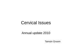
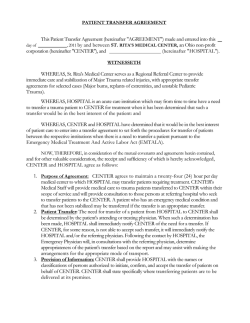
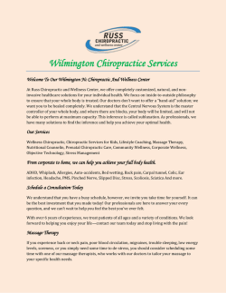
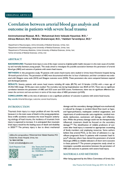
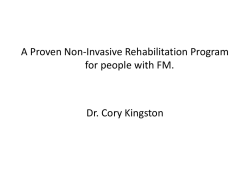
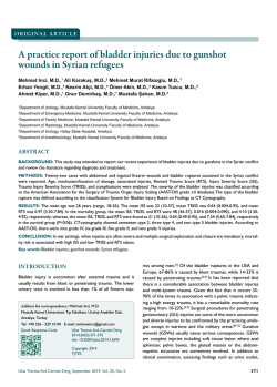
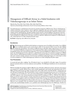
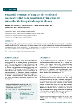
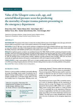
CA S E R EP O RT
Complete cervical tracheal transection caused
by blunt neck trauma: Case report
Jin Hui Paik, M.D.,1 Jeong-seok Choi, M.D.,2 Seung Baik Han, M.D.,1
Hyun Min Jung, M.D.,1 Ji Hye Kim, M.D.1
1
Department of Emergency Medicine, College of Medicine, Inha University, Incheon, South Korea;
2
Department of Otorhinolaryngology-Head and Neck Surgery, College of Medicine, Inha University, Incheon, South Korea
ABSTRACT
This study aimed to report the survival of a rare case of complete tracheal transection followed by blunt neck trauma. A 66-year-old
man was presented in the emergency room after a motorcycle accident in which a rope was wrapped around his neck. Although alert,
he was in respiratory distress. A computed tomographic scan showed transection of the cervical trachea. Emergency neck exploration
revealed that the tracheal laceration had been cut from the tracheal anterior third ring to the posterior first ring and the anterior
esophageal wall had ruptured. Laryngectomy, tracheostomy, and esophagopharyngeal anastomosis were performed. Prompt airway
management and immediate neck exploration is important for survival in these cases.
Key words: Blunt injuries; rupture; trachea.
INTRODUCTION
Traumatic tracheal injury after blunt neck trauma is rare;
however, most patients with complete tracheal transection
usually die at the scene due to loss of airway.[1] The few, who
survive and arrive at a hospital, pose a diagnostic and therapeutic challenge to the trauma team. These patients may suffer fatal outcomes when misdiagnosed or long-term complications, if treated improperly.[2] Early diagnosis and treatment
of tracheal injuries lead to the best outcome. Nevertheless,
emergency physicians have limited experience in these cases
and have reached no consensus on its management.[3]
In this paper, our experience with one survivor of complete
cervical tracheal transection caused by blunt neck trauma
Address for correspondence: Ji Hye Kim, M.D.
Department of Emergency Medicine, College of Medicine, Inha University, 7-206, Sinheung-dong 3 ga, Jung-Gu, Incheon, South Korea.
400-711 Incheon - South Korea
Tel: +82 32 890 2310 E-mail: [email protected]
Qucik Response Code
Ulus Travma Acil Cerrahi Derg
2014;20(6):459-462
doi: 10.5505/tjtes.2014.32744
Copyright 2014
TJTES
Ulus Travma Acil Cerrahi Derg, November 2014, Vol. 20, No. 6
combined with esophageal rupture and review of the relevant
medical literature were reported.
CASE REPORT
A 66-year-old man was presented in the emergency room
after a motorcycle accident in which a rope was wrapped
around his neck. Although alert, he was in respiratory distress. His motor and sensory neurologic signs were normal.
He had neck fluctuation on breathing and initial pulse oximetry saturation (SpO2) was 93%. The vital signs included a
blood pressure of 134/80 mmHg, a heart rate of 102 beats/
min, a respiratory rate of 22 breaths/min, and an axillary
temperature of 36.2°C. Significant crepitus was noted on his
neck area. Massive subcutaneous emphysema was present on
a lateral cervical spine radiograph and pneumomediastinum
was seen on a supine chest radiograph (Fig. 1). Fiber optic
bronchoscopy guided endotracheal intubation was planned;
however, 10 minutes after the patient’s arrival in the emergency room, he showed agitation, cyanotic change, and a
decrease in SpO2 at 81%. Despite aggressive and effective
ambu-bagging with manual airway maintenance, SpO2 did not
increase. Therefore, urgent orotracheal intubation was done.
There was large amount of blood in the mouth and the right
epiglottic area was swollen. His SpO2 increased to 98% after
orotracheal intubation. Computed tomographic scan (CT) of
the neck and chest revealed transection of the cervical trachea above the thoracic inlet, pneumomediastinum, and sub459
Paik et al. Complete cervical tracheal transection caused by blunt neck trauma
(a)
(b)
Figure 1. (a) Lateral cervical spine radiograph showing massive
subcutaneous emphysema. (b) Supine chest radiograph showing
pneumomediastinum (arrows).
(a)
(b)
Approximately 90 minutes after arrival in the emergency
room, neck exploration was performed by an otorhinolaryngology surgeon. Apron incision was done and subplastymal
flap was made from hyoid bone level to substernal notch level.
The tracheal laceration had been cut from the tracheal anterior third ring to the posterior first ring (Fig. 3). Urgently,
endotracheal tube insertion was done at the lower part of
lacerated trachea. Exposured larynx showed frangibility with
esophageal rupture. Thus, total laryngectomy was performed,
and closed thoracostomy was carried out for right pneumothorax. Esophagopharyngeal anastomosis was conducted by
thoracic surgeon for esophageal rupture, which was pedunculated on the anterior wall; the posterior wall was intact. The
patient was managed postoperatively with pain control and antibiotics. As an abscess formation was found at the left of the
tracheostomy site, an iatrogenic fistula formation was made for
the pharyngocutaneous fistula. Fistula repair was performed
four times during the following one year and fistula recurrence
was not reported up to the following next year. The patient is
now in tracheostomy state and able to feed orally.
DISCUSSION
(c)
(d)
Figure 2. CT scan showing transection of the cervical trachea
above the thoracic inlet. (a) Axial CT image of the neck showing
over-distension of the endotracheal tube cuff (black arrows). (b)
Coronal CT image of the neck showing over-distension of the endotracheal tube cuff (black arrows). (c) Axial CT image of the chest
showing the endotracheal tube tip in the upper anterior chest wall,
suprasternal notch level (black arrow) and thoracic trachea (white
arrow). (d) Sagittal CT image of the chest showing the endotracheal tube tip in the upper anterior chest wall, suprasternal notch
level (black arrow) and thoracic trachea (white arrow).
cutaneous emphysema in the chest wall and lower neck (Fig.
2). Routine blood samples taken on admission showed a leukocytosis of 12.450/microliters, hemoglobin concentration of
14.4 g/dl, hematocrit of 41.7%, and C-reactive protein of 0.04
mg/dl. The results of the blood chemistry were as follows:
serum glucose 170 mg/dL, BUN 20.7 mg/dL, creatinine 0.86
mg/dL, albumin 3.7 g/dL, creatine phosphokinase 320 IU/L,
and LDH 244 IU/L. The serum electrolytes and blood gas
parameters were as follows: sodium 138 mEq/L, potassium
3.6 mEq/L, chloride 104 mEq/L, pH 7.37, paCO2 46.3 mmHg,
paO2 36.4 mmHg, and bicarbonate 26.3 mmol/L in room air.
460
The incidence of traumatic tracheal injuries seems to be
roughly 0.5% to 2% among individuals sustaining blunt trauma, including blunt trauma to the neck.[4] Main types of blunt
injuries are high-impact blunt compression injuries such as
motor vehicles accidents (59%), followed by crush injuries
(27%).[5] In blunt injuries, intrathoracic trachea and main stem
bronchi injury account for 62%, cervical trachea 23%, and lobar bronchi 15%.5 Most common presentations are respiratory distress, dyspnea, poor gas exchange, and hemoptysis.[5]
Cyanosis and serious respiratory embarrassment is present
in 30% of the cases. Another common symptom is hoarseness or dysphonia, occurring in 46% of the patients.[4] The
most common signs of airway injury reported in most series
are subcutaneous emphysema (35%-85%), pneumothorax
Figure 3. The tracheal laceration had been cut from the tracheal
anterior third ring to the posterior first ring. Orally intubated endotracheal tube tip was placed in out of trachea (black arrow). Endotracheal tube was inserted at lower part of completely transected
trachea (white arrow).
Ulus Travma Acil Cerrahi Derg, November 2014, Vol. 20, No. 6
Paik et al. Complete cervical tracheal transection caused by blunt neck trauma
(20%-50%), and hemoptysis (14%-25%); however, the lack of
specificity and the occult nature of the injury frequently result in a delayed diagnosis.[6]
Deep cervical emphysema and pneumomediastinum are
seen in 60% of the patients with tracheobronchial injuries.
[4,7,8]
Blunt trauma is often associated with multiple injuries
involving not only the chest, but also the abdomen, head, and
orthopedic structures.[4,9] Cervical trauma of the airway frequently involves the esophagus, recurrent laryngeal nerves,
cervical spine and spinal cord, larynx, and carotid arteries and
jugular veins.[4] Combined transection of the cervical trachea
and esophagus, as a result of blunt trauma, is a rare injury
accounting for less than 1% of trauma cases seen in major
centers.[10]
Many tracheobronchial injuries are not diagnosed immediately (25-68%). Delayed diagnosis is more commonly seen with
left-sided injuries.[5,8] Physicians need to maintain a high index of suspicion related to nonspecific signs such as dyspnea,
cough, subcutaneous emphysema, and hemoptysis.[11] The
mechanism of injury, vocal changes, and rapidly expanding
subcutaneous emphysema in the neck are important clues.
[11]
Clinical examination is followed by radiologic imaging,
angiography, computed tomography, and tracheo-bronchioesophagoscopy.[12]
Accurate interpretation of the chest radiograph is essential
in the early diagnosis of occult upper-airway injury.[13] The
preponderant findings on chest radiograph include subcutaneous emphysema, pneumomediastinum, pneumothorax, and
air surrounding the mainstem bronchi.[13] The best diagnostic
investigation is bronchoscopy; flexible bronchoscopy should
be carried out first to determine the location and extent of
the injury.[5,14-16] However, the bronchoscopic procedure is
precarious and characterized by abundant bleeding in the respiratory tract and a rapid drop in arterial oxygen saturation,
which are all factors precluding diagnostic confirmation.[6]
Patients who show respiratory distress, are unstable hemodynamically, or who have clinical suspicion of an airway
injury should be intubated immediately with in-line cervical
stabilization, preferably with the guidance of a flexible bronchoscope, as described previously.[4,11] Airway manipulation
can exacerbate a tracheal injury, hence careful orotracheal
intubation is considered to be the only option available before transport.[16]
[12]
Debridement and primary repair is the treatment of choice
and good postoperative recovery has been reported.[5] Surgical repair is indicated when a transmural tear longer than 1
cm causes a pneumothorax unrelieved by tube thoracostomy.
Complete transection of the trachea should be managed
by careful suturing and being cautious to avoid damage to
the recurrent laryngeal nerves. While small lacerations have
been successfully managed conservatively, primary repair of
a tracheobronchial rupture is the treatment of choice.[8] For
most cervical tracheal injuries, urgent tracheostomy is the
best method for airway control.[16] Patients with suspected
tracheal injuries, who are clinically stable and oxygenating at
greater than 90% SpO2, should not be intubated and treated
only with high flow oxygen. This recommendation may have
to be urgently modified in patients presenting with hypovolemic shock or altered mental status from brain injuries.[13,16] In
this case, patient was unstable and unable to maintain airway.
Urgent orotracheal intubation was done. Even though the
endotracheal tube was placed out of trachea, ventilation and
oxygenation was maintained by positive ventilation. As the
case stands above, if the patient is unstable, it is our belief
that oral intubation should not be delayed and urgent orotracheal intubation must be considered.
The prognosis of the patient is largely dependent on an early
diagnosis and good interdisciplinary management.[12] Prompt
airway management and immediate neck exploration is important for survival in these cases.
Conflict of interest: None declared.
A CT scan can be performed if diagnosis is uncertain on plain
films.[7,11] Preoperative CT can be useful in assessing associated laryngeal injuries or other unsuspected chest injuries that
should be dealt with at the time of surgical exploration. CT is
contraindicated in hemodynamically unstable trauma patients
or patients with unstable airways.[4] Helical CT with 3D reconstruction should be considered a suitable ‘screening’ test
in a trauma patient suspected of tracheal rupture and may
help the clinician in the decision to perform a bronchoscopy
on the patient.[6]
The primary initial goals are stabilization of the airway, reversal of shock, relief of the pneumothorax, and determination
of the extent and location of injury.[4,8] The management of
tracheobronchial injuries, depending on the severity, consists of emergency operation in cases of tracheal rupture,
hemorrhage or esophageal injury, or of selective operation.
Ulus Travma Acil Cerrahi Derg, November 2014, Vol. 20, No. 6
REFERENCES
1. Dertsiz L, Arici G, Arslan G, Demircan A. Acute tracheobronchial injuries: early and late term outcomes. Ulus Travma Acil Cerrahi Derg
2007;13:128-34.
2. Hamid UI, Jones JM. Combined tracheoesophageal transection after
blunt neck trauma. J Emerg Trauma Shock 2013;6:117-22. CrossRef
3. McCrystal DJ, Bond C. Cricotracheal separation: a review and a case with
bilateral recovery of recurrent laryngeal nerve function. J Laryngol Otol
2006;120:497-501. CrossRef
4. Karmy-Jones R, Wood DE. Traumatic injury to the trachea and bronchus. Thorac Surg Clin 2007;17:35-46. CrossRef
5. Wong EH, Knight S. Tracheobronchial injuries from blunt trauma.
ANZ J Surg 2006;76:414-5. CrossRef
6. Le Guen M, Beigelman C, Bouhemad B, Wenjïe Y, Marmion F, Rouby
JJ. Chest computed tomography with multiplanar reformatted images
for diagnosing traumatic bronchial rupture: a case report. Crit Care
2007;11:R94. CrossRef
461
Paik et al. Complete cervical tracheal transection caused by blunt neck trauma
7. Hahn B. Tracheobronchial rupture. J Emerg Med 2007;33:193-4. CrossRef
8. Roxburgh JC. Rupture of the tracheobronchial tree. Thorax 1987;42:681-8.
9. Rossbach MM, Johnson SB, Gomez MA, Sako EY, Miller OL, Calhoon
JH. Management of major tracheobronchial injuries: a 28-year experience. Ann Thorac Surg 1998;65:182-6. CrossRef
10. Hamid UI, McGuigan JA, Jones JM. Transection of the aerodigestive
tract after blunt neck trauma. Ann Thorac Surg 2011;92:1896-8. CrossRef
11. Hsiao SH, Chen BS, Lee TM, Hsu SY, Lai YY. Delayed diagnosis of
complete tracheal transection after blunt neck trauma. Tzu Chi Med J
2009;21:77-80. CrossRef
12. Veit JA, Metternich F. Management of traumatic tracheal injuries: presentation of a rare case and review of the literature. [Article in German]
Laryngorhinootologie 2008;87:270-3. [Abstract] CrossRef
13. Cassada DC, Munyikwa MP, Moniz MP, Dieter RA Jr, Schuchmann GF,
Enderson BL. Acute injuries of the trachea and major bronchi: importance of early diagnosis. Ann Thorac Surg 2000;69:1563-7. CrossRef
14. Baumgartner FJ, Ayres B, Theuer C. Danger of false intubation after traumatic tracheal transection. Ann Thorac Surg 1997;63:227-8. CrossRef
15. Kaptanoglu M, Dogan K, Nadir A, Gonlugur U, Akkurt I, Seyfikli Z, et
al. Tracheobronchial rupture: a considerable risk for young teenagers. Int
J Pediatr Otorhinolaryngol 2002;62:123-8. CrossRef
16.Norwood SH, McAuley CE, Vallina VL, Berne JD, Moore WL.
Complete cervical tracheal transection from blunt trauma. J Trauma
2001;51:568-71. CrossRef
OLGU SUNUMU - ÖZET
Künt boyun travmasının neden olduğu komplet servikal trakeal transeksiyon: Olgu sunumu
Dr. Jin Hui Paik,1 Dr. Jeong-seok Choi,2 Dr. Seung Baik Han,1 Dr. Hyun Min Jung,1 Dr. Ji Hye Kim1
1
2
Inha Üniversitesi Tıp Fakültesi, Acil Tıp Anabilim Dalı, Incheon, Güney Kore;
Inha Üniversitesi Tıp Fakültesi, Kulak Burun Boğaz-Baş ve Boyun Cerrahisi Anabilim Dalı, Incheon, Güney Kore
Bu yazıda, künt boyun travması sonrası seyrek görülen ve sağ kalan bir komplet trakeal transeksiyon olgusu sunuldu. Altmış altı yaşında erkek hasta
bir motosiklet kazası sonrası, boynunu bir halata kaptırmış vaziyette acil servise getirildi. Bilinci açık olmasına rağmen solunum sıkıntısı çekiyordu.
Bilgisayarlı tomografi taraması servikal trakea transeksiyonunu gösteriyordu. Acil boyun eksplorasyonu trakea laserasyonunun trakea anteriorda 3.
halkadan posteriorda 1, halkaya uzandığını ve ön özofagus duvarının yırtılmış olduğunu gösteriyordu. Larengektomi, trakeostomi ve özofagofarengeal anastomoz yapıldı. Bu olguların sağkalımı için acilen havayolunun açılması ve hemen boyun eksplorasyonu yapılması önemlidir.
Anahtar sözcükler: Künt travmalar; rüptür; trakea.
Ulus Travma Acil Cerrahi Derg 2014;20(6):459-462
462
doi: 10.5505/tjtes.2014.32744
Ulus Travma Acil Cerrahi Derg, November 2014, Vol. 20, No. 6
© Copyright 2026 Paperzz