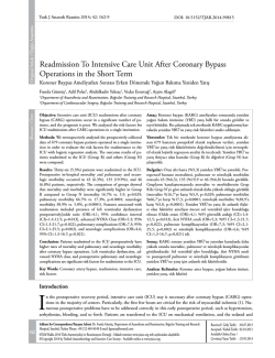
Dicle Tıp Dergisi / Dicle Medical Journal 2014; 41 (3): 577-580
doi: 10.5798/diclemedj.0921.2014.03.0477
CASE REPORT / OLGU SUNUMU
A new possible acquired risk factor for pulmonary embolism: Colonoscopy
procedure
Pulmoner emboli için kazanılmış olası yeni bir risk faktörü: Kolonoskopi işlemi
Talat Kılıç1, Hilal Ermiş1, Omar Kaya2, Hakan Alan3
ABSTRACT
ÖZET
Knowing the risk factors of PE contributes to the promptness of diagnosis and treatment. Although most patients
with pulmonary embolism (PE) have acquired or hereditary risk factors, the cause of PE is unknown in one-quarter of patients. We describe a 66-year-old man who presented with chest pain, dyspnea, and hemoptysis one day
after he had a colonoscopy performed due to chronic constipation complaints. The computed tomography pulmonary angiography revealed thrombus in the left peripheral
pulmonary artery. He did not have any risk factors for PE,
and a hypercoagulable workup was normal. Because of
his development of PE one day after his colonoscopy and
the absence of any risk factors, the colonoscopy could
have caused the PE. PE may be caused by increased intra-abdominal pressure during this procedure. This is the
first case of PE following a colonoscopy in the literature.
We recommend that patients with idiopathic PE should be
questioned aspect of invasive procedures such as colonoscopy.
Pulmoner emboli (PE) için risk faktörünün bilinmesi, bu
hastalığın erken tanı ve tedavisinin düzenlenmesine
önemli katkı sağlamaktadır. Pek çok hastada akkiz veya
kalıtsal risk faktörü bulunmasına rağmen, bu hastaların ¼
de herhangi bir risk faktörüne rastlanılmamaktadır. Kronik
kabızlık nedeniyle kolonoskopi uygulanmış 66 yaşında
erkek hasta, işlemden bir gün sonra dispne, göğüs ağrısı ve hemoptizi şikâyetleri gelişmesi üzerine kliniğimize
başvurmuştu. Çekilen bilgisayarlı tomografi pulmoner anjiyografide sol pulmoner arter periferik dalında trombus
saptanması üzerine hasta PE olarak değerlendirildi. Hastada herhangi bir kalıtsal ya da akkiz risk faktörü yoktu. İşlemden hemen sonra gelişmesi, bilinen bir risk faktörünün
olmaması nedeniyle PE’nin kolonoskopiye bağlı gelişmiş
olabileceği düşünüldü. İşlem sırasında artmış intraabdominal basınca bağlı olarak PE gelişmiş olabileceğini düşünüyoruz. Olgumuz kolonoskopi işlemini takiben gelişmiş ilk PE olgusudur. Nedeni bilinmeyen PE hastalarının
kolonoskopi gibi invaziv işlemler açısından sorgulanmasının uygun olacağını düşünüyoruz.
Key words: Pulmonary embolism; risk factor; colonoscopy
INTRODUCTION
Colonoscopy is expansively used for diagnosis of
colorectal pathology and carries low risk complication. Hemorrhage and perforation of the colon have
been reported as the most common complications.
Extra-colonic or visceral damages, including pneumothorax pneumomediastinu, and other are much
less common [1].
1
Anahtar kelimeler: Pulmoner emboli; risk faktörü, kolonoskopi
Despite the advances in the diagnosis and management of venous pulmonary thromboembolism,
pulmonary embolism (PE) remains an important
cause of morbidity and mortality [2]. Although the
mortality of untreated patients is approximately 2530%, mortality is reduced to 2-8% in treating patients [3,4] .
In clinical practice, the physicians encounter
many patients with PE. In addition, as the all other
İnonu University Faculty of Medicine, Turgut Özal Medical Center, Department of Pulmonary Medicine, Malatya, Turkey
2
Yeşilyurt State Hospital, Department of Pulmonar Medicine, Malatya, Turkey
3
Malatya State Hospital, Department of Gastroenterology, Malatya, Turkey
Yazışma Adresi /Correspondence: Talat Kılıç,
Inonu University Faculty of Medicine, Dept. Pulmonary Medicine, Malatya, Turkey Email:[email protected]
Geliş Tarihi / Received: 04.08.2014, Kabul Tarihi / Accepted: 18.08.2014
Copyright © Dicle Tıp Dergisi 2014, Her hakkı saklıdır / All rights reserved
578
T. Kılıç et al. Colonoscopy and pulmonary embolism
disease, knowing the risk factors of PE or the formation of emboli as a complication of some processes
(such as colonoscopy) contributes to a prompt diagnosis and treatment. Therefore, the physicians
should be aware of all possible risk factors for PE.
Although PE can occur in patients without any
determinable predisposing factors, one or more of
these factors, which are acquired or hereditary, are
usually recognized [3-6]. The proportion of patients
with idiopathic or unprovoked PE was about 25 %
[3,5,7]. In this article, a certain invasive procedure,
colonoscopy was proposed as possible causes of PE.
be detected. After three months of anticoagulation
therapy, treatment was discontinued. At the time of
discontinuation of treatment, PAP was 20 mmHg.
At the 2-year follow-up, he does not have any health
problems. The patient’s informed consent was obtained.
CASE REPORT
A 66-year-old white male who presented with chest
pain, dyspnea, and hemoptysis was admitted to the
emergency department of our hospital. The patient
was referred to the department of pulmonology. All
vital signs were normal. Body mass index was 26.
The D-dimer assay using latex agglutination was
reported up to 2860μg/L (reference range: 0-5). Hemogram and biochemical parameters were normal.
The computed tomography pulmonary angiography was performed, and a consistent filling defect
by a thrombus was observed in the left peripheral
pulmonary artery (Figure 1). The echocardiogram
showed normal right cavities, and pulmonary artery
pressure (PAP) was 45 mmHg. No evidence of deep
vein thrombosis was found in the subsequent duplex
ultrasound examination of his lower extremities at
that time. The patient was considered as having a
PE. The patient was initially hospitalized and treated with therapeutic dose of weight-based subcutaneous low molecular weight heparin (enoxaparin)
while it was converted to oral vitamin K antagonist
(warfarin sodium) therapy. After treatment, the patient’s clinic situation dramatically improved. The
patient had no acquired risk factors of PE. Hematological studies that included anti-phospholipid antibody, lupus anticoagulant, anti-thrombin III, protein
C, protein S, anti-cardiolipin antibody, homocysteine, APC-resistant factor V, PT 20210, and factor V
Leyden were all normal. In addition, there was no
history of smoking. However, a colonoscopy was
performed due to the dyspeptic and chronic constipation complaints one day before PE diagnosis, and
it was evaluated as normal. In addition, the abdominal CT was normal. Thus, No occult cancer could
Dicle Tıp Derg / Dicle Med J Figure 1. Computed tomography pulmonary angiography
shows the filling defect on peripheral branches of the left
pulmonary artery
DISCUSSION
Currently, colonoscopy is a crucial method for diagnosis and treatment of any colorectal disease. In
the majority of cases, it is a safe, tolerated, and easily obtainable procedure. Therefore, using colonoscopy has shown a gradual increase. Generally, any
complications after colonoscopy are rare [1]. So far,
in the English and Turkish literature, there is no reported any PE case after colonoscopy.
On the other hand, in the diagnosis of diseases,
it is very important to know the risk factors. Many
acquired and hereditary risk factors play a role in
the etiology of PE. The factors causing intravascular coagulation were detected by Virchow in 1856 as
follows: “1- Vascular endothelial damage 2-Hypercoagulable 3-Staz.” Of the 75% of the PE patients,
acquired and/or hereditary factors causing one of
these three conditions are detected [3-6]. The occult
cancer and thrombophilia risks are higher in patients
with undetected risk factors [3,4]. However, no reason is detected at the 25% of the PE patients, and
they were considered as idiopathic [7,8]. Previously
www.diclemedj.org
Cilt / Vol 41, No 3, 577-580
T. Kılıç et al. Colonoscopy and pulmonary embolism
unrecognized cancer, present in 7-12 % of patients
with idiopathic PE, can usually be identified by a
combination of careful clinical evaluation, routine
blood tests, and chest radiography, but the current
consensus is the recommendation that it is not suitable to proceed with tests such as ultrasound, computed tomography, or endoscopy [3,9,10].
In our case, there were no acquired (including the long travel) and hereditary risk factors for
PE. In addition, the body mass index of this patient
was normal. He has no history/family history of
thrombophilia, and smoking. He has not any occult
cancer. However, a colonoscopy was performed because of dyspeptic and chronic constipation one day
before PE formation, and it was assessed as normal.
The anticoagulant treatment was discontinued at
the end of the third month of treatment. The genetic
risk factors including protein C/S, anti-thrombin III,
and activated protein C resistance were examined
in the second week after the cessation of anticoagulant therapy. These factors were normal. Because of
the development of PE one day after the colonoscopy and the absence of any risk factors of PE in
this patient, we have considered that the colonoscopy procedure could have caused the PE. It is not
clear that which mechanism of the colonoscopy
procedure could have caused the PE. The increased
intra-abdominal pressure during a colonoscopy procedure may enhance the venous stasis of the intraabdominal vein, and thus PE could have occurred.
Previously, it has been shown that increased intraabdominal pressure may cause PE via increasing
venous stasis in patients undergoing a laparoscopic
surgery procedure [11-14]. In one of these cases, PE
was detected in a 47-year-old female patient following a laparoscopic ovariectomy [11]. This was
a case with massif PE. The current case was submassif PE because PAB was 40 mmHg. Similar to
our patient, for this case, there was no evidence of
deep vein thrombosis in the subsequent duplex ultrasound examination of her lower extremities at the
time of diagnosis. In another study, it was shown
that PE occurred in two patients after a laparoscopic
surgery; one patient underwent umbilical hernia repair and the other cholecystectomy [13]. Also, deep
venous thrombosis and PE as a complication of
laparoscopic surgery have been reported in multiple
case reports and individual series [13,14]. Increased
Dicle Tıp Derg / Dicle Med J 579
intra-abdominal pressure with other mechanism
such as increased ventilator pressure, and the vasodilator effects of hypercarbia with anesthesia during the laparoscopic procedure might have caused
PE in all of these cases [14]. Really, laparoscopic
operation and colonoscopy are different procedure.
Nevertheless, as during laparoscopy, it is likely to
increase the intra-abdominal pressure during colonoscopy and may be damaged veins in this area.
Thus, PE could be occurred.
To the best of our knowledge, no cases of PE
following a colonoscopy procedure have been reported to date. This is the first such case. Although
the mechanism of PE formation in this patient is
not clear, increased intra-abdominal pressure could
have been the cause.
In conclusion, it should be kept in mind that a
colonoscopy procedure is a possible risk factor for
patients with idiopathic PE. However, whether or
not the difference in the incidence of PE before and
after colonoscopy should be investigated in future
studies. On the other hand, if the colonoscopy as a
risk factor of PE is shown by a series of cases, prophylaxis may be considered before this procedure.
Conflict of interest: No conflict of interest
REFERENCES
1. Mangualde J, Cremers MI, Vieira AM, et al. The American Society for Gastrointestinal Endoscopy (ASGE)
Guideline:Complications of colonoscopy. Gastrointest Endosc 2011;74:745-752.
2. Tapson VF. Acute Pulmonary embolism; Review N Engl J
Med 2008;358:1037-1052.
3. Torbicki A, Perrier A, Konstantinides S, et al. Guidelines on
diagnosis and management of acute pulmonary embolism.
European Heart Journal 2008;29:2276-2315.
4. Den Exter PL, Van der Hulle T, Lankeit M, et al. Long-term
clinical course of acute pulmonary embolism. Review.
Blood Rev. 2013;27:185-192.
5. White RH. The epidemiyology of venous thromboembolism.
Circulation 2003;107:14-18.
6. Karakaş MS, Özbek SC, Er A, et al. Right heart thrombus entrapped in patent foramen ovale with pulmonary embolism
in a patient with primary hypercoagulable state. Dicle Tıp
Dergisi. 2012;39:440-444.
7. Heit JA, O’Fallon WM, Petterson TM, et al. Relative impact of risk factors for deep vein thrombosis and Pulmonary embolism: a population-based study. Arch Intern Med
2002;162:1245-1248.
www.diclemedj.org
Cilt / Vol 41, No 3, 577-580
580
T. Kılıç et al. Colonoscopy and pulmonary embolism
8. Tsai AW, Cushman M, Rosamond WD, et al. Cardivascular
risk faktors and venous thromboembolism insidance: the
longitudinal investigation of thromboembolism etioloyg.
Arch Intern Med 2002;162:1182-1189.
9. British Thoracic Society Standards of Care Committee Pulmonary Embolism Guideline Development Group.British
Thoracic Society guidelines for the management of suspected acute pulmonary embolism. Thorax 2003;58:470484.
10. Uresandi F, Blanquer J, Conget F, et al. Guidelines for the
diagnosis, treatment, and follow up of Pulmonary Embolism. Arch Bronconeumol 2004;40:580-594.
Dicle Tıp Derg / Dicle Med J 11. Chu CS, Lee TL, Cheng KH, et al. Acute pulmonary embolism following laparascopic ovariectomy: A case report.
Kaohsiung J Med Sci2006;22:452-456.
12. Holzheimer RG. Laparoscopic procedures as a risk factor
of deep venous thrombosis, superficial ascending thrombophlebitis and pulmonary embolism-case report and Review
of the literature. Eur J Med Res 2004; 9:417-422.
13. Tsutsumi N, Tomikawa M, Konishi K, et al. Pulmonary
embolism following laparoscopic surgery: a report of two
cases. Hepatogastroenterology 2002;49:719-720.
14. Inderbitzin DT, Opitz I, Giger U, et al. Incidence of clinical
pulmonary embolism after laparoscopic surgery. Br J Surg.
2007;94:599-603.
www.diclemedj.org
Cilt / Vol 41, No 3, 577-580
© Copyright 2026 Paperzz