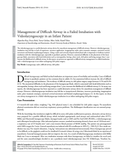
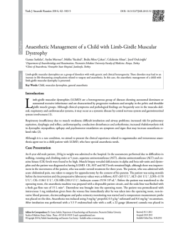
Case Report
Turk J Anaesth Reanim 2014; 42: 220-2
DOI: 10.5152/TJAR.2014.16362
Surgical Excision of Postintubation Granuloma Under Jet
Ventilation
Demet Altun1, Eren Yılmaz2, Bora Başaran2, Emre Çamcı1
1
Abstract
2
Department of Anaesthesiology and Reanimation, İstanbul University İstanbul Faculty of Medicine, İstanbul, Turkey
Department of Ear Nose and Throat, İstanbul University İstanbul Faculty of Medicine, İstanbul, Turkey
Following the use of an endotracheal or tracheostomy tube, circumferential lesions, stenosis, or granulomatous lesions at the cuff level or
tip of the tube may be observed on the tracheal wall. This injury mainly occurs due to excessive pressure of the cuff on the tracheal wall
and may be prevented by a high-volume, low-pressure cuff and a carefully monitored tracheostomy tube. Although there is an overall
improvement in the design of high-volume cuffs, hyperinflation of these cuffs may still contribute to tracheal injuries. If the size of the
granuloma is limited, the lesion is treated by excision (microlaryngeal surgery) under general anaesthesia. Using jet ventilation during the
operation minimizes the trauma caused by intubation and reduces the risk of oedema and the risk of barotrauma, as it provides ventilation
over a possible stenosis. In addition to providing better visualization of the surgical field and superior surgeon comfort, jet ventilation also
increases the success of the operation. In this case report, we aimed to present a successful anaesthesia technique performed by jet ventilation
in a patient with a postintubation granuloma, which was excised by microlaryngeal surgery without the need for reintubation.
Key Words: Intubation, granuloma, jet ventilation, anaesthesia
Introduction
I
n general, laryngeal granulomas are benign lesions located in the 1/3 posterior aspect of the vocal cords and may either be
unilateral or bilateral. They are typically associated with three etiological factors including endotracheal intubation, improper use of voice, and gastroesophageal reflux. It is known that endotracheal intubation may cause significant laryngeal
and tracheal injury and laryngeal granuloma is one of the late complications related to this injury. Laryngeal granuloma may
be encountered even in the event of intubations lasted shorter than 24 hours. Pressure necrosis, which results from traumatic
effect in the arytenoid cartilage due to high positive pressure caused by intubation tube cuff, and characteristic features of female
larynx are the predisposing factors (1, 2). Symptoms appear in the form of hoarseness (88%), sore throat (35%), dyspnoea
(18%), and neck swelling and the treatment is excision of granuloma by microlaryngeal surgery under general anaesthesia (3, 4).
While ventilation is provided during microlaryngeal surgery, small-diameter endotracheal tubes are preferred to visualize
surgical area as clearly as possible. Nevertheless, it is sometimes observed that even the tubes in that diameter hinder the view
and make the surgery difficult. Moreover, in certain cases, laryngotracheal stenosis may be at such a level that could make
endotracheal intubation impossible. In this case, jet ventilation option through a thin cannula should be considered. Jet
ventilation was first used in 1967 by Saunders during routine bronchoscopy and then, as of 1980, supraglottic, infraglottic,
and transtracheal options have been developed to be used in microlaryngeal surgery. Today, jet ventilation is being used in
trachea and larynx surgeries, bronchoscopy, pneumonectomy, and broncho-pleural fistulae (5).
In the present case report, a microlaryngeal surgery performed by jet ventilation was reported in a patient, who was admitted
to Ear, Nose and Throat Clinic with hoarseness and dyspnoea and in whom endotracheal granuloma due to endotracheal
intubation during a previous appendectomy was detected.
Case Presentation
A 30-year-old female patient was admitted to the Ear, Nose and Throat Clinic with cough, shortness of breath, and hoarseness lasting for 4 months. Her personal history revealed appendectomy a year ago. Evaluation of the patient’s records showed
220
Address for Correspondence: Dr. Demet Altun, Department of Anaesthesiology and Reanimation, İstanbul University İstanbul Faculty of Medicine,
İstanbul, Turkey Phone: +90 212 414 20 00-31742 E-mail: [email protected]
©Copyright 2014 by Turkish Anaesthesiology and Intensive Care Society - Available online at www.jtaics.org
Received: 28.06.2013
Accepted: 23.08.2013
Available Online Date: 29.05.2014
Altun et al. Jet Ventilation
that the surgery had lasted for 105 minutes, intubation had
been performed by an endotracheal tube No. 7.5, and intubation and anaesthesia had been completed without any
complication.
Bronchoscopy demonstrated bilateral granulomatous configuration in the vocal cords that caused narrowing in the posterior 1/3 of the lumen and was considered as post-intubation
granuloma (Figure 1). After obtaining consent of the patient,
microlaryngeal surgery was planned under general anaesthesia. The patient underwent standard monitoring and fluid replacement was initiated through an intravenous line. Anaesthesia induction of the patient, of whom the body weight was
67 kg, was performed using 30 µg remifentanil and 200 mg
propofol; 14 mg mivacurium was administered to facilitate
laryngoscopy and intubation. Jet ventilation catheter of 40
cm in length (Acutronic Medical Systems AG, Hirzel, Switzerland) was inserted under the guidance of a laryngoscope
and the catheter was fixed in the way to be located below
the glottis. Jet ventilation settings were as follows: a driving
pressure (DP) of 1.3 bar, an inspiration time of 55%, an FiO2
level of 0.8, and a frequency of 130 min-1.
Maintenance of anaesthesia was performed by infusing propofol at a dose of 6-10 mg kg h-1 and remifentanil at a dose
of 0.05-0.25 µg kg minute-1 as total intravenous anaesthesia.
In addition, the patient received 8 mg dexamethasone, 50
mg ranitidine and 10 mg metoclopramide. During the procedure that lasted for 25 minutes, adequate oxygenation was
provided by jet ventilation without serious reduction in saturation. Based on the detection of 95% saturation on the 10th
minute of the procedure, DP was increased to 1.5 bar. The
procedure continued by maintaining the oxygen saturation
between 97% and 98%. The granuloma was excised under
precise surgical view provided by a thin catheter (Figure 2).
No other complication was developed. Anaesthetic agents
were discontinued at the end of procedure and jet catheter
was removed. Respiration of the patient was supported via
a mask and the patient started spontaneous breathing in 2-3
minutes. After providing adequate spontaneous breathing,
the patient was transferred to the recovery room. Thus, the
procedure was completed without intubation in any phase
from induction to recovery.
Discussion
In addition to trauma in the posterior larynx caused by endotracheal tube, other factors (abnormal position of the patient’s head, use of extremely large tube inappropriate for the
patient’s larynx, mislocation or overpressure of tube cuff, and
extubation trauma) that enhance trauma contribute to the
development of granuloma (6). This possible complication
may be prevented by precise attention paid in airway manipulations.
Blanc and Tremblay (7) classified the endotracheal intubation-related complications in three groups as location of in-
Granulation
Granulation
Figure 1. Bronchoscopic image of postintubation granuloma
Jet ventilation catheter
Figure 2. Bronchoscopic image of jet ventilation catheter
tubation tube, duration of intubation, and extubation. They
determined the predisposing factors for the development of
laryngeal granulomas as female gender, obesity, short neck,
and hereditary airway anomalies. Female gender and short
neck were among these risk factors in the present patient.
In a similar study, Barton et al. (8) determined that laryngeal
granulomas, which they evaluated as due to traumatic intubation, are related to the trauma and ischemia in the tracheal
wall due to high pressure caused by large-diameter tube or
cuff. In the present case, the previous intubation period of
the patient was limited to a 105-minute surgery period suggesting that the granulomatous configuration was related to
the pressure caused by the cuff rather than duration. As a
general rule, it is possible to lower the likelihood of complications such as intubation-related granuloma by using the
possible smallest-diameter tube, paying attention to cuff pressure, avoiding extreme extension or flexion of the neck, and
providing adequate muscle relaxation and anaesthesia depth.
Monitoring the cuff pressure via a manometer is important,
as the pressure is likely to increase within the process with the
use of nitrous oxide even though the initial inflation is within
the normal ranges (9).
The treatment of laryngeal granulomas is the excision via microlaryngeal surgery. While ventilation is enabled during microlaryngeal surgery, small-diameter endotracheal tubes are
preferred to visualize the surgical area as clearly as possible.
It is sometimes observed that even the tubes in that diameter
make the surgical view difficult. In this case, jet ventilation
option via a thin cannula may be considered. Manipulations
become easier with this technique, which is performed by a
thin catheter, as it allows precise visualization of anatomic
configurations and completely exposes the surgical field for
221
Turk J Anaesth Reanim 2014; 42: 220-2
the surgeon. Moreover, it is possible to lower the risk of
oedema by eliminating intubation-related trauma via jet ventilation. Jet ventilation was primarily developed to be used
during routine bronchoscopy. Thereafter, this technology has
been adjusted to provide ventilation via one of the supraglottic, infraglottic, and transtracheal routes to be used in microlaryngeal surgery (10).
Studies have determined that infraglottic technique is more
effective than supraglottic technique as it provides ventilation
below the level of vocal cords and causes minimal movement
in the cords (11). Blowing of blood and tissue particles to
the upwards and outside with expiratory airflow appears to
be another advantage of the technique (12). Khan et al. (13)
successfully used the infraglottic ventilation in the treatment
of laryngeal lesions and in taking biopsy from vocal cords and
observed no related complications such as hypoxia, hypercapnia, or barotrauma. In the present case as well, infraglottic
jet ventilation was performed and the granuloma was excised
under precise surgical view by means of the thin catheter.
Performing supraglottic technique as the other option was
reported in a 50-case series that underwent microlaryngeal
surgery for tracheal stenosis (14). In that series, successful airway management was provided and need for intubation due
to hypoxia was reported in only one case.
Conclusion
In the present case report, which presented a patient who
underwent surgical intervention for intubation-related granuloma, safe airway management and gas exchange were provided via infraglottic jet ventilation technique without need
for re-intubation and the patient was transferred to the recovery room at the end of intervention after a successful recovery
without need for re-intubation. Thus, the risks for intubation
and possible granuloma development were eliminated in the
patient, who previously developed granuloma despite very
short intubation period. In conclusion, jet ventilation allows
effective gas exchange without causing high airway pressure
or hemodynamic problem. Moreover, jet ventilation provides
precise surgical view and working conditions and makes it
possible to eliminate the risk in patients at high-risk for intubation-related granuloma.
Informed Consent: Written informed consent was obtained from
patient who participated in this case.
Peer-review: Externally peer-reviewed.
222
Author Contributions: Concept - D.A.; Design - D.A., E.Ç.; Supervision - E.Ç.; Funding - D.A., E.Ç.; Materials - D.A.; Data Collection and/
or Processing - D.A., E.Y., B.B.; Analysis and/or Interpretation - D.A.,
E.Y.; Literature Review - D.A.; Writer - D.A., E.Ç.; Critical Review - E.Ç.
Conflict of Interest: No conflict of interest was declared by the authors.
Financial Disclosure: The authors declared that this study has received no financial support.
References
1. Barton R. Observation on the pathogenesis of laryngeal granuloma due to endotracheal anesthesia. N Engl J Med 1958; 248:
1097-9. [CrossRef ]
2. Lewis F, Scholbohm R, Thomas A. Prevention of complications
from prolonged tracheal intubation. Am J Surg 1978; 135:
452-7. [CrossRef ]
3. Dubick M, Wright B. Comparison of laryngeal pathology
following long-term oral and nasal endotracheal intubations.
Anesth Analg 1978; 57: 663-8. [CrossRef ]
4. De Lima Pontes PA, De Biase NG, Gadelha EC. Clinical evolution of laryngeal granulomas: treatment and prognosis. 1999;
109: 289-94.
5. Hu A, Weissbrod PA, Maronian NC, Hsia J, Davies JM, Sivarajan GK, et al. Hunsaker Mon-Jet tube ventilation: A 15year experience. Laryngoscope 2012; 122: 2234-9. [CrossRef ]
6. Drosnes DL, Zwillenberg DA. Laryngeal granulomatous polyp
after short-term intubation of a child. Ann Otol Rhinol Laryngol 1990; 99: 183-6.
7. Blanc V, Tremblay N. The complications of tracheal intubation: A new classification with a review of the literature. Anesth
Analg 1974; 53: 202-13. [CrossRef ]
8. Barton R. Medicolegal aspects of intubation granuloma. JAMA
1958; 166: 1821-3. [CrossRef ]
9. Henderson J. Airway management in the adult. In: Miller RD,
Eriksson LI, Wiener-Kronish JP, Young WL (eds). Miller’s Anesthesia. 7th edition. San Francisco: Churchill Livingstone;
2009: 1573-611.
10. Yilmazer C, Şener M, Yilmaz I. Bilateral giant posterior laryngeal
granulomas with dyspnea: a rare complication of endotracheal
intubation. Anesth Analg 2005; 101: 1881-2. [CrossRef]
11. Davies MJ, Hillel DA ,Maronian NC. Posner KL. The Hunsaker Mon-Jet tube with jet ventilation is effective for microlaryngeal surgery. Can J Anaesth 2009; 56: 284-90. [CrossRef ]
12. Orloff LA, Parhizkar N, Ortiz E. The Hunsaker Mon-Jet ventilation tube for microlaryngeal surgery:optimal laryngeal exposure. Ear Nose Throat J 2002; 81: 390-4.
13. Khan I, Shakeel M, Nagaraja R, Ram B, Thomas AD. A Hunsaker Mon-Jet tube trapped in the larynx. J Laryngo Otol
2011; 125: 1204-5. [CrossRef ]
14. Biro P. Jet ventilation for surgical interventions in the upper
airway. Anesthesiol Clin 2010; 28: 397-409. [CrossRef ]
© Copyright 2026 Paperzz