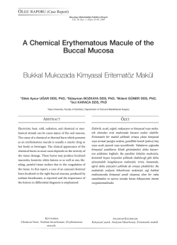
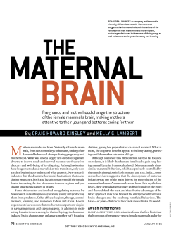
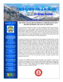
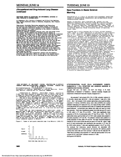
EXPERIMENTAL STUDY
Captopril protects against burn-induced
cardiopulmonary injury in rats
Esra Sağlam, M.D.,1 Ahmet Özer Şehirli, PhD.,2 Emine Nur Özdamar, M.D.,3 Gazi Contuk, PhD.,4
Şule Çetinel, M.D.,4 Derya Özsavcı, PhD.,5 Selami Süleymanoğlu, M.D.,6 Göksel Şener, PhD.2
1
Maltepe University, School of Medicine, Department of Pharmacology and Clinical Pharmacology, Istanbul
2
Department of Pharmacology, Marmara University Faculty of Pharmacy, Istanbul
3
Department of Medical Pharmacology, Marmara University Faculty of Medicine, Istanbul
4
Department of Histology & Embryology, Marmara University Faculty of Medicine, Istanbul
5
Department of Biochemistry, Marmara University Faculty of Pharmacy, Istanbul
6
Department of Cardiology, Gulhane Military Medical Academy, Istanbul
ABSTRACT
BACKGROUND: This study was designed to determine the possible protective effect of captopril treatment against oxidative damage in heart and lung tissues induced by burn injury.
METHODS: Under ether anesthesia, the shaved dorsum of Wistar albino rats was exposed to 90°C water bath for 10 seconds.
Captopril was administered intraperitoneally (10 mg/kg) after the burn injury and repeated twice daily. In the sham group, the dorsum
was dipped in a 25°C water bath for 10 seconds. At the end of the 24 hours, echocardiographic recordings were performed, then
animals were decapitated and heart and lung tissue samples were taken for the determination of tumor necrosis factor-α (TNF-α) as
a pro-inflammatory cytokine, malondialdehyde and glutathione levels and myeloperoxidase, caspase-3, and Na+,K+-ATPase activity in
addition to the histological analysis.
RESULTS: Burn injury caused significant alterations in left ventricular function. In heart and lung tissues, TNF-α and malondialdehyde
levels and myeloperoxidase and caspase-3 activities were found to be increased, while glutathione levels and Na+, K+-ATPase activity
were decreased due to burn injury. Captopril treatment significantly elevated the reduced glutathione level and Na+, K+-ATPase activity,
and decreased cytokine and malondialdehyde levels and myeloperoxidase and caspase-3 activities.
CONCLUSION: Captopril prevents burn-induced damage in heart and lung tissues and protects against oxidative organ damage.
Key words: Captopril; cytokine; lipid peroxidation; myeloperoxidase; thermal trauma.
INTRODUCTION
Thermal trauma produces profound systemic changes such
as oligemic shock, anemia, renal failure, and metabolic disturbances. It causes direct tissue damage as well as inflammatory
reactions.[1] Thermal trauma causes a progressive decrease in
cardiac output and cardiac stroke volume, release of vasoconAddress for correspondence: Esra Saglam, M.D.
Alemdağ Cad., Site Yolu, No: 27, Ümraniye, İstanbul, Turkey
Tel: +90 216 - 633 06 33 E-mail: [email protected]
Qucik Response Code
Ulus Travma Acil Cerrahi Derg
2014;20(3):151-160
doi: 10.5505/tjtes.2014.96493
Copyright 2014
TJTES
Ulus Travma Acil Cerrahi Derg, May 2014, Vol. 20, No. 3
strictor mediators such as vasopressin, altered baroreceptor
and chemoreceptor reflexes, and reabsorption of fluid.[2] A
number of experimental and clinical studies have shown that,
despite aggressive fluid resuscitation and maintenance of pulmonary capillary wedge pressure, stroke work and ejection
fraction (EF) often decrease after thermal trauma.[3,4] Several
studies have indicated that decreased cardiac contractility is
one of the important mechanisms for decreased cardiac output and cardiac dysfunction after severe thermal trauma.[5,6]
Angiotensin converting enzyme (ACE) catalyzes the conversion of angiotensin I (Ang I) to angiotensin II (Ang II). ACE also
cleaves the terminal dipeptide of vasodilating hormone bradykinin to inactivate this hormone. Hyperactivity of the renin–
angiotensin system (RAS), an endocrine system with critical
roles in cardiovascular function, has been implicated in the etiology of high blood pressure, obesity and metabolic syndrome.
[7]
Therefore, inhibition of ACE is generally used as one of the
151
Saglam et al. Captopril protects against burn-induced cardiopulmonary injury in rats
methods in the treatment of hypertension and chronic inflammation.[8,9] Pharmacologically, it is known that ACE inhibitors
containing a sulfhydryl or a thiol radical, such as the orally active compound captopril, have an antioxidant activity.[10,11]
In the light of these findings, we hypothesized that captopril
would provide protection against burn-induced cardiopulmonary damage. We aimed to investigate whether and to
what extent captopril reduces this damage, by determining
the presence of oxidative tissue injury using biochemical and
histological parameters.
MATERIALS AND METHODS
Animals
Wistar albino rats of both sexes, weighing 200-250 g, were
obtained from Marmara University School of Medicine Animal House. The rats were kept at a constant temperature
(22±1ºC) with 12-hour (h) light and dark cycles, fed with
standard rat chow, and fasted for 12 h before the experiments, but were allowed free access to water. All experimental protocols were approved by the Marmara University Animal Care and Use Committee.
Thermal Injury and Experimental Design
This is a randomized controlled trial. All groups were elected
in exactly the same way. Twenty-four animals were divided
into three groups of eight rats each: One group served as
controls. Two groups underwent burn injury and were treated with saline or captopril. Under brief ether anesthesia, the
dorsum of the rats was shaved and exposed to 90° water
bath for 10 seconds (s), which resulted in a second-degree
burn involving 30% of the total body surface area.[12] To rule
out the effects of the anesthesia, the same protocol was applied in the control group, except that the dorsa were dipped
in a 25°C water bath for 10 s. After sham or burn injury, rats
were resuscitated with physiological saline solution (10 ml/
kg subcutaneous). ACE inhibitor captopril (10 mg/kg, intraperitoneal (i.p.); Sigma-Aldrich; St. Louis, MO, USA) or saline was given intraperitoneally to rats immediately after the
burn injury, and the injections were repeated twice a day.
Two groups (controls and the first burn group) received saline and the second burn group received i.p. captopril (10
mg/kg in saline) administered immediately after the burn, and
the injections were repeated twice a day. Intraperitoneal administration of captopril could potentially have a systemic effect.[13] In both the saline- and captopril-treated burn groups,
rats were decapitated at 24 h following burn injury. At the
end of the experimental period, transthoracic echocardiography was performed to assess the cardiac function of the
rats. Then, the animals were decapitated in order to evaluate
the presence of oxidant injury in the heart, and lung tissue
samples were taken and stored at -80°C for the determination of pro-inflammatory cytokine [tumor necrosis factor-α
(TNF-α)], malondialdehyde (MDA) and glutathione (GSH)
152
levels and myeloperoxidase (MPO) and Na+, K+-ATPase and
caspase-3 activities. For histological analysis, samples of the
tissues were fixed in 10% (vol/vol) buffered p-formaldehyde
and prepared for routine paraffin embedding. Tissue sections
(6 µm) were stained with hematoxylin and eosin (H&E) and
examined under a light microscope (Olympus-BH-2). An experienced histologist who was unaware of the treatment conditions performed the histological assessments.
Echocardiography
Echocardiographic imaging and calculations were done according to the guidelines published by the American Society
of Echocardiography[14] using a 12 MHz linear transducer and
5-8 MHz sector transducer (Vivid 3, General Electric Medical
Systems Ultrasound, Tirat Carmel, Israel). Under ketamine
(50 mg/kg, i.p.) anesthesia, measurements were made from
M-mode and two-dimensional images obtained in the parasternal long- and short axes at the level of the papillary muscles after observation of at least six cardiac cycles. Interventricular septal thickness (IVS), left ventricular diameter (LVD)
and left ventricular posterior wall thickness (LVPW) were
measured during systole (s) and diastole (d). EF, fractional
shortening (FS) and left ventricular mass (LVM) and relative
wall thickness (RWT) were calculated from the M-mode images using the following formulas: % EF = [(LVDd)3 - (LVDs)3/
(LVDd)3 x 100], % FS = [LVDd-LVDs/LVDd x 100], LVM=
[1.04 x ((LVDd+LVPWd+IVSd)3 - (LVDd)3) x 0.8 + 0.14], and
RWT = [2 x (LVPWd/LVDd)].
Determination of Tissue TNF-α Levels:
The levels of TNF-α in the tissues were measured by enzyme-linked immunosorbent assay (ELISA). Heart and lung
tissues (100 mg) were homogenized in an ice-cold bath. After centrifugation, the supernatant was collected and total
protein content was determined. Tissue TNF-α levels were
measured with rat TNF-α ELISA kits (Invitrogen; Taastrup,
Denmark) according to the manufacturer’s instructions. Absorbance of standards and samples was determined on a plate
reader at 405 nm. Results are expressed as pg/100 mg tissue.
Malondialdehyde and Glutathione Assays
Tissue samples were homogenized with ice-cold 150 mM KCl
for the determination of MDA and GSH levels. The MDA
levels were assayed for products of lipid peroxidation by
monitoring thiobarbituric acid reactive substance formation
as described previously.[15] Lipid peroxidation was expressed
in terms of MDA equivalents using an extinction coefficient of
1.56x10⁵ M-1 cm-1, and results are expressed as nmol MDA/g
tissue. GSH measurements were performed using a modification of the Ellman procedure.[16] Briefly, after centrifugation at
3000 rev/minute (min) for 10 min, 0.5 ml of supernatant was
added to 2 ml of 0.3 mol/L Na2HPO4.2H2O solution. A 0.2
ml solution of dithiobisnitrobenzoate (0.4 mg/ml 1% sodium
citrate) was added, and the absorbance at 412 nm was measured immediately after mixing. GSH levels were calculated
Ulus Travma Acil Cerrahi Derg, May 2014, Vol. 20, No. 3
Saglam et al. Captopril protects against burn-induced cardiopulmonary injury in rats
using an extinction coefficient of 1.36x10⁴ M-1 cm-1. Results
are expressed in µmol GSH/g tissue.
Myeloperoxidase Activity
Myeloperoxidase (MPO) is an enzyme found predominantly
in the azurophilic granules of polymorphonuclear leukocytes
(PMN). Tissue MPO activity is frequently utilized to estimate
tissue PMN accumulation in inflamed tissues, and correlates
significantly with the number of PMN determined histochemically in tissues. MPO activity was measured in tissues
in a procedure similar to that documented by Hillegass et
al.[17] Tissue samples were homogenized in 50 mM potassium
phosphate buffer (PB, pH 6.0), and centrifuged at 41,400 g
(10 min); pellets were suspended in 50 mM PB containing
0.5% hexadecyltrimethylammonium bromide (HETAB). After
three freeze and thaw cycles, with sonication between cycles,
the samples were centrifuged at 41,400 g for 10 min. Aliquots
(0.3 ml) were added to 2.3 ml of reaction mixture containing
50 mM PB, o-dianisidine, and 20 mM H2O2 solution. One unit
of enzyme activity was defined as the amount of MPO present that caused a change in absorbance measured at 460 nm
for 3 min. MPO activity was expressed as U/g tissue.
Na+, K+-ATPase Activity
Since the activity of Na+, K+-ATPase, a membrane-bound enzyme required for cellular transport, is very sensitive to free
radical reactions and lipid peroxidation, reductions in this activity can indicate membrane damage indirectly. Measurement
of Na+, K+-ATPase activity is based on the measurement of
inorganic phosphate released by ATP hydrolysis during incubation of homogenates with an appropriate medium containing 3 mM ATP as a substrate. The total ATPase activity was
determined in the presence of 100 mM NaCl, 5 mM KCl, 6
mM MgCl2, 0.1 mM EDTA, and 30 mM Tris HCl (pH 7.4),
while the Mg2+-ATPase activity was determined in the presence of 1mM ouabain. The difference between the total and
the Mg2+-ATPase activities was taken as a measure of the Na+,
K+-ATPase activity.[18] The reaction was initiated with the addition of the homogenate (0.1 ml), and a 5-min preincubation period at 37ºC was allowed. Following the addition of
Na2ATP and a 10-min re-incubation period, the reaction was
terminated by the addition of ice-cold 6% perchloric acid. The
mixture was then centrifuged at 3500 g, and Pi in the supernatant fraction was determined by the method of Fiske and Subarrow.[19] The specific activity of the enzyme was expressed as
µmol Pi mg-1 protein h-1. The protein concentration of the
supernatant was measured by the Lowry method.[20]
Caspase-3 Activity
Caspase-3 activity was measured using ApoTargetCaspase-3/
CPP32 Colorimetric Protease Assay (Invitrogen; Taastrup,
Denmark) kit according to the manufacturer’s instructions.
Briefly, brain and lung tissue samples were homogenized and
treated for 10 min with iced lysis buffer supplied by the manufacturer. The cell lysates were subjected to three freeze and
thaw cycles and 2x10 s sonication to fully disrupt the cells
and disperse cell debris. The cell lysate was then centrifuged
at 20,000 g for 5 min and the supernatant transferred to new
Eppendorf tubes. Following protein determination, cytosol
extracts were diluted in 50µL lysis buffer and 50 µL reaction
buffer (10mM dithiothreitol). Samples were added to wells
and the microplate was equilibrated at 37°C for 10 min. The
reaction was initiated by adding 5 µl of DEVD-pNA (AspGlu-Val-Asp p-nitroanilide) substrate (200 µM final concentration), and the reaction was carried out at 37°C for 2 h in
the dark. The colorimetric release of p-nitroaniline (pNA)
from the Ac-DEVD-pNA substrate was recorded at 405 nm
using a microplate reader. Experiments were performed in
triplicate. Results are presented as mean±SD of six separate
experiments and expressed as fold-increase over pretreatment level (untreated samples).
Histopathological Analysis
For light microscopic investigations, heart and lung specimens
Table 1. Transthoracic echocardiography measurements in the experimental groups
Parameter
Control group
Burn group
Saline-treated Saline-treatedCaptopril-treated
Interventricular septal thickness (mm)
2.36±0.18
3.01±0.11**2.45±0.08+
Left ventricular posterior wall thickness (mm)
1.73±0.12
2.55±0.11*1.77±0.11+
Relative wall thickness
0.66±0.03
0.98±0.10*0.71±0.08+
Left ventricular diameter in systole (mm)
2.63±0.27
3.96±0.15**2.98±0.21+
Left ventricular diameter in diastole (mm)
4.09±0.16
5.18±0.14**4.32±0.24+
Ejection fraction (%)
79.03±3.67
63.60±2.73*77.78±3.44+
Fractional shortening (%)
42.27±3.54
26.61±2.24**39.71±3.21+
Heart/body weight ratio (mg/g)
2.21±0.12
2.73±0.08**2.29±0.09+
Data are the mean±SEM of six animals. *p<0.05, **p<0.01 compared to saline-treated control group; +p<0.05 compared with the saline-treated burn group.
Ulus Travma Acil Cerrahi Derg, May 2014, Vol. 20, No. 3
153
Saglam et al. Captopril protects against burn-induced cardiopulmonary injury in rats
were fixed in 10% buffered formalin for 48 h, dehydrated in
an ascending alcohol series, and embedded in paraffin wax.
Approximately 5-μm-thick sections were stained with H&E
for general morphology. Histological assessments were made
with a photomicroscope (Olympus BX 51; Tokyo) by an experienced histologist who was unaware of the experimental
groups.
Statistics
Statistical analysis was carried out using GraphPad Prism 3.0
(GraphPad Software; San Diego, CA, USA). All data were
expressed as means±SEM. Groups of data were compared
with an analysis of variance (ANOVA) followed by Tukey’s
multiple comparison tests. Values of p<0.05 were regarded
as significant.
RESULTS
Table 1 summarizes the transthoracic echocardiography measurements of the experimental groups. In the saline-treated
burn group, LVPW, LV end-diastolic and end-systolic dimensions and RWT, and heart/body weight ratio were increased
significantly compared to the control group (p<0.05-0.01),
while percent FS and EF were decreased significantly. On
(a)
3000
Saline-treated
the other hand, in the captopril-treated burn group, echocardiographic measurements were significantly different than
those of the saline-treated burn group (p<0.05), but similar
to those of the control group.
TNF-α levels in the heart and lung tissues of the saline-treated burn group were significantly increased, while TNF-α levels were found to be decreased in the captopril- treated burn
group (p<0.001; Figs. 1a, b).
Burn injury caused significant decreases in the GSH levels of the heart and lung tissues of the saline-treated burn
group (p<0.01-0.001), but this antioxidant was restored in
the captopril-treated burn group (p<0.05-0.01; Figs. 2a, b).
On the other hand, MDA levels in both tissues were significantly increased following burn injury (p<0.01-0.001), while
treatment with captopril reversed burn-induced elevations in
MDA back to the control levels (p<0.05-0.01; Figs. 3a, b).
In the saline-treated burn group, MPO activities in both the
heart and lung tissues were increased significantly (p<0.001),
and treatment with captopril prevented these alterations
(p<0.01-0.001; Figs. 4a, b).
Na+, K+-ATPase activities measured in the cardiac and pulmo-
(a)
Saline-treated
2
Captopril-treated
TNF-a
(pg/100 mg tissue)
(b)
+++
1000
***
Burn
4
3
2000
+++
++
2
***
1
1000
C
0
Burn
Figure 1. Tumor necrosis factor-α (TNF-α) levels in the (a) heart
and (b) lung tissues of control (C) and saline- or captopril-treated
burn groups. Each group consists of 8 animals. ***: p<0.001: compared to saline-treated control group; +++: p<0.001: compared to
saline-treated burn group.
154
**
C
Burn
(b)
3000
1
0
C
4000
0
GSH (μmol/g)
***
2000
0
+
GSH (μmol/g)
TNF-a
(pg/100 mg tissue)
Captopril-treated
C
Burn
Figure 2. Glutathione (GSH) levels in the (a) heart and (b) lung
tissues of control (C) and saline- or captopril-treated burn groups.
Each group consists of 8 animals. **: p<0.01, ***: p<0.001: compared to saline-treated control group; + p<0.05, ++: p<0.01: compared to saline-treated burn group.
Ulus Travma Acil Cerrahi Derg, May 2014, Vol. 20, No. 3
Saglam et al. Captopril protects against burn-induced cardiopulmonary injury in rats
(a) 100
Saline-treated
MPO (U/g)
+
40
20
0
C
Burn
(b) 100
(b) 40
++
C
Burn
**
80
30
+
60
40
MPO (U/g)
MDA (nmol/g)
Captopril-treated
10
20
***
20
+++
10
20
0
***
30
***
60
0
Saline-treated
Captopril-treated
80
MDA (nmol/g)
(a) 40
C
0
Burn
C
Burn
Figure 3. Malondialdehyde (MDA) levels in the (a) heart and (b)
lung tissues of control (C) and saline- or captopril-treated burn
groups. Each group consists of 8 animals. **: p<0.01, ***: p<0.001:
compared to saline-treated control group; +: p<0.05: compared to
saline-treated burn group.
Figure 4. Myeloperoxidase (MPO) activity in the (a) heart and (b)
lung tissues of control (C) and saline- or captopril-treated burn
groups. Each group consists of 8 animals. ***: p<0.001: compared
to saline-treated control group; ++: p<0.01, +++: p<0.001: compared to saline-treated burn group.
nary tissues were reduced in the saline-treated burn group
(p<0.001, Figs. 5a, b). In the captopril-treated burned rats,
the measured Na+, K+-ATPase activities in the studied tissues
were increased (p<0.05-0.001).
DISCUSSION
Caspase-3 activities in both the heart and lung tissues were
increased significantly (p<0.001), while treatment with captopril reduced these increases (p<0.01-0.001; Figs. 6a, b).
Light microscopic evaluation of the cardiac muscle cells in the
control group revealed regular nuclei with central alignment
(Fig. 7a). In the burn group, the capillaries present in the connective tissue of muscle fibers showed congestion in addition
to vacuolization in the cytoplasm of most of the cardiomyocytes (Fig. 7b). On the other hand, in the captopril-treated
burn group, vasocongestion was reduced and regular cellular
morphology was settled (Fig. 7c).
In lung tissues, the control group demonstrated a welldesignated morphology (Fig. 8a), whereas severe interstitial
edema with bronchiolar hemorrhage and stretched alveolar
walls were observed in the burn group (Fig. 8b), which was
ameliorated by captopril treatment, revealing the reversal of
degeneration in the interstitium (Fig. 8c).
Ulus Travma Acil Cerrahi Derg, May 2014, Vol. 20, No. 3
The aim of the present work was to explore the effect of
captopril, a known ACE inhibitor, on the cardiopulmonary
inflammatory process associated with burn in rats.
Thermal trauma is a stressful condition challenging whole
body homeostatic mechanisms, accompanied by both local
and distant effects leading to intense inflammation, tissue
damage and infection. As local production of proinflammatory cytokines will activate non-specific host immunity, tissue
injury or infection, cytokines are approached as being important components in the postburn pathophysiological process.
[21-23]
Accordingly, after thermal trauma, a great many cytokines are induced rapidly,[12,24-27] and ACE mediates inflammation to elicit T cell stimulation. On the other hand, the
ACE inhibitor captopril inhibits conclusive immune functions
and attenuates inflammation.[28,29] Our data above demonstrate that burn-induced cardiac and lung injury involve oxidant formation and inflammation, while captopril treatment
greatly attenuated these responses. The ability of captopril
to protect against thermal trauma was observed with significant decreases in tissue MDA and TNF-α levels and MPO
and caspase-3 activities, which were found to be increased
155
Saglam et al. Captopril protects against burn-induced cardiopulmonary injury in rats
(a)
4
Saline-treated
(a)
4
***
3
2
***
1
0
(b)
+
Caspase-3 activity
(fold increase)
Na+-K+ATPase
(μmol/mg protein/h)
Captopril-treated
2
C
Burn
(b)
2.5
Captopril-treated
+++
0
C
Saline-treated
Burn
4
2.0
1.5
Caspase-3 activity
(fold increase)
Na+-K+ATPase
(μmol/mg protein/h)
+++
***
1.0
***
++
2
0.5
0
C
0
Burn
C
Burn
Figure 5. Na+-K+ ATPase activity in the (a) heart and (b) lung tissues of control (C) and saline- or captopril-treated burn groups.
Each group consists of 8 animals. ***: p<0.001: compared to saline-treated control group; +: p<0.05, +++: p<0.001: compared to
saline-treated burn group.
Figure 6. Caspase-3 activity in the (a) heart and (b) lung tissues
of control (C) and saline- or captopril-treated burn groups. Each
group consists of 8 animals. ***: p<0.001: compared to saline-treated control group; ++: p<0.01, +++: p<0.001: compared to salinetreated burn group.
following thermal trauma. Furthermore, captopril treatment
in the burned rats caused significant increases in both GSH
levels and Na+, K+-ATPase activities, which were found to be
depleted in the burn group.
mRNA expressions attenuated LV remodeling. Captopril has
been shown to express immune-regulating, antioxidant and
anti-inflammatory properties.[34] In agreement with these
studies, our results showed that depressing cytokine expression is associated with improved cardiac function.
TNF-α is a cytokine excreted by macrophages and monocytes
in response to different stimuli, and concentrations were
found to be high in patients with heart disease, in association
with noticeable activation of the RAS. Levine et al.[30-32] found
increased concentrations of TNF-α in heart disease patients
with high renin concentrations. In addition, they demonstrated that increased serum TNF-α, which plays an essential role
in the inflammatory processes, was also decreased with captopril treatment. In our study, following burn injury, increased
cytokine TNF-α was associated with impaired cardiac function
since in the saline treated-burn group, LVPW, LV end-diastolic
and end-systolic dimensions and RWT, and IVS thickness were
significantly higher as compared to control rats (p<0.05-0.01),
while FS and EF were found to be significantly decreased. Youn
et al.[33] studied both ACE inhibition and angiotensin receptor
antagonism in a rat model of acute infarction in which transforming growth factor (TGF) beta-1 mRNA expression was
also increased. Their results demonstrated that both captopril
and losartan suppressing the acute induction of TGF-beta1
156
Major burn injuries can lead to life-threatening end-organ
dysfunction including cardiac dysfunction, which is a major
contributor to mortality.[35,36] Despite prompt and adequate
fluid replacement following burn injury, alterations in cardiovascular function such as reduced cardiac output, myocardial inflammation and an intrinsic defect in myocardial performance can cause multiple organ dysfunction syndrome.
Numerous experimental studies have been performed to
identify the molecular mechanisms involved in burn-related
cardiac dysfunction with the end goal of creating novel therapeutic interventions and agents to reduce the incidence of
life-threatening complications.[37,38] In our study, by improving
cardiac function, captopril protected cardiac and lung tissues
since oxidative injury observed through MDA and GSH levels and MPO, Na+, K+-ATPase and caspase activities were reversed in both tissues. One of the causative agents accountable for the development of burn shock and distant organ
damage in animal models of burn injury are oxygen radicals.[27]
Ulus Travma Acil Cerrahi Derg, May 2014, Vol. 20, No. 3
Saglam et al. Captopril protects against burn-induced cardiopulmonary injury in rats
Figure 7. (a) Control group: regular alignment of muscle cells with centrally located nuclei (**). (b) Burn group: capillary vasocongestion among
the connective tissue of the muscle cells (arrow), vacuolization in the cytoplasm of the cardiac muscle cells and degeneration of some gapjunctions (arrowhead). (c) Burn and captopril treatment: the capillary vasocongestion was reduced, and vacuolization in the cytoplasm of the
cells was decreased (**), H&E staining, x200, insets x400.
Figure 8. (a) Control group demonstrated a regular alveolar structure (arrow) with no distention in its walls. (b) Burn group, severe congestion
(**) and interstitial edema, which led to a decrease in alveolar space (arrow) in most areas of the tissue. In some regions, the alveoli united
with each other resulting in large distended alveolar spaces (arrowhead-inset). (c) Burn and captopril group: reduced interstitial congestion and
edema (**) and expanded alveolar areas and no congestion present.
The distant organs involved in the thermal damage during experimental skin burn are thought to be the lungs, heart, liver,
kidney, and gastrointestinal mucosa. In all cases of thermal
trauma, neutrophils diffuse these organs, being the potent
source of reactive oxygen metabolites.[39] As is known, accumulation and activation of neutrophils induce tissue damage
and release of different cytotoxic proteins into the extracellular fluid. MPO activity, an indirect marker of neutrophil infiltration,[40] has been shown to be significantly increased in
various tissues of burned rats.[12,41] Similarly, in the present
study, increased MPO activity in both heart and lung tissues
suggests that neutrophil accumulation in these tissues assist
in organ injury distant from the original wound. On the other hand, in the captopril- treated burn groups, MPO activities were reduced. The effects of captopril on MPO activity
were studied previously in different experimental models of
inflammation. Leor et al.[42] studied the effects of captopril
experimentally, and proposed that captopril did not decrease
neutrophil-induced myocardial injury following coronary occlusion and reperfusion. Nonetheless, Van Antwerpen et
al.[43] in their in vitro study showed that captopril, by inhibiting
the MPO/HOCl system, was able to significantly reduce the
oxidative modification of low-density lipoprotein in a dosedependent manner. Furthermore, Li et al.[44] demonstrated
Ulus Travma Acil Cerrahi Derg, May 2014, Vol. 20, No. 3
that pathological injury of the lung in rats with intense acute
pancreatitis was decreased with captopril treatment.
Malondialdehyde (MDA), an end product of lipid peroxidation, is major evidence of lipid peroxidation, and in this study,
its levels were found to be significantly advanced due to burn
in both heart and lung tissues. Results from animal and human
studies suggested that there is a relationship between tissue
MDA levels and the degree of burn complications, including
shock and remote organ damage.[25,41,45,46] Our results showed
that captopril treatment in the burned rats caused significant
reduction in MDA.
Esterified GSH is a significant component of the cellular antioxidant system. The GSH molecule acts as an electron acceptor for hydrogen peroxide by forming an oxidized thiol and
then undergoing degradation by glutathione reductase.[47] In a
previous study, GSH levels have been reported to be decreased
in the lung, liver and kidney of rats after burn.[12,25,26,28,45] In this
study, lessening effects of captopril on MDA concurred with
preservation of tissue GSH level, which is an important antioxidant. Moreover, Gurer et al.,[48] studying GSH, suggested
that captopril has a promising beneficial role in augmenting the
reducing capacity of the cells by increasing GSH.
157
Saglam et al. Captopril protects against burn-induced cardiopulmonary injury in rats
Apoptosis is an orchestrated form of cell ‘death by suicide’.
It is principal in both the development and normal continuation of tissue function. Apoptotic nerve cell death is related
to the pathogenesis of several devastating neurodegenerative
diseases. Recently, apoptosis was found to be important in
the pathophysiological changes occurring after major burns.
After thermal injuries, nearly every organ or tissue seems
affected and manifests this peculiar cellular mechanism characterizing a “systemic apoptotic response”.[49] Currently, ROS
may mediate apoptosis through the starting of poly (ADPribose) polymerase (PARP), advances in mitochondrial permeability with the release of cytochrome C, and activation of
caspases.[50,51] ROS-triggered release of lysosomal ingredients
can also lead to apoptosis. Hydrogen peroxide can freely diffuse across cell membranes, including the lysosomal membrane. Inside the lysosome, hydrogen peroxide can react with
iron to form the potent hydroxide radical, at least causing
damage to the lysosomal membrane and leakage of contents.
The translocation of the protease cathepsin D from the lysosome to the cytosol yields to the induction of apoptosis.[52]
Since captopril has recently been found to inhibit Fas-induced
apoptosis in human-activated T cells[53] and lung epithelial
cells,[54] we hypothesized that suppression of apoptosis could
be one of the mechanisms bringing about the efficacy of ACE
inhibitors. In the present study, we examined the apoptosis
measuring caspase-3 activity in the heart and lung tissues, and
found that thermal trauma increased heart and lung apoptosis. On the other hand, captopril treatment effectively lessened caspase-3 activity and protected tissues, as also confirmed histologically.
Na+, K+-ATPase is a considerable membrane protein that carries out the coupled extrusion and uptake of Na+ and K+
ions across the plasma membranes. As the activity of Na+,
K+-ATPase, a membrane-bound enzyme required for cellular transport, is very sensitive to free radical reactions and
lipid peroxidation, decrease in this activity can show membrane damage indirectly. Na+, K+-ATPase activities measured
in the cardiac and pulmonary tissues were reduced in the
saline-treated rats (p<0.001), indicating damaged transport
function in these tissues (Fig. 5a, 5b). In the captopril-treated
burned rats, the measured Na+, K+-ATPase activities in the
studied tissues were increased compared to those of the control rats (p<0.05-0.001). Ottlecz et al.[55] demonstrated that
stimulation of retinal Na+, K+-ATPase activity in diabetes is
most likely one of the mechanisms through which captopril
can improve retinal complications. Our study has shown that
Na+, K+-ATPase activities were significantly reduced in both
cardiac and lung tissues of burned rats, while captopril treatment significantly protected the enzyme activity.
In conclusion, the findings of the present study demonstrated
that captopril treatment protected multiorgan damage in
thermal trauma through inhibition of pro-inflammatory and
oxidative pathways. Furthermore, captopril treatment provided regular cellular morphology in lung and heart tissues,
158
which caused a concomitant decrease in lipid peroxidation
and increase in tissue antioxidant defense. Therefore, these
results suggest an anti-inflammatory, cardiopulmonary protective effect of captopril, with a reduction in the circulating
proinflammatory markers and an increase in those antiinflammatory cytokines; these effects result in a benefit on the
cardiopulmonary inflammatory process associated with burninduced thermal trauma. Thus, captopril treatment merits
consideration as a potential therapeutic agent for restoring
organ damage following thermal trauma.
Conflict of interest: None declared.
REFERENCES
1. Chandran PK, Kuttan R. Effect of Calendula officinalis Flower Extract
on Acute Phase Proteins, Antioxidant Defense Mechanism and Granuloma Formation During Thermal Burns. J Clin Biochem Nutr 2008;43:5864.
2. Parihar A, Parihar MS, Milner S, Bhat S. Oxidative stress and anti-oxidative mobilization in burn injury. Burns 2008;34:6-17.
3. Horton JW, Garcia NM, White DJ, Keffer J. Postburn cardiac contractile
function and biochemical markers of postburn cardiac injury. J Am Coll
Surg 1995;181:289-98.
4. Mueller M, Sartorelli K, DeMeules JE, Gamelli RL. Effects of fluid resuscitation on cardiac dysfunction following thermal injury. J Surg Res
1988;44:745-53.
5. Adams HR, Baxter CR, Izenberg SD. Decreased contractility and compliance of the left ventricle as complications of thermal trauma. Am Heart
J 1984;108:1477-87.
6. Gao S, Chen ZW, Zheng H, Chen XL. Ligustrazine attenuates acute
myocardium injury after thermal trauma. Burns 2007;33:321-7.
7. de Kloet AD, Krause EG, Woods SC. The renin angiotensin system and
the metabolic syndrome. Physiol Behav 2010;100:525-34.
8. Bhuyan BJ, Mugesh G. Synthesis, characterization and antioxidant activity of angiotensin converting enzyme inhibitors. Org Biomol Chem
2011;9:1356-65.
9. Kubota M, Shimizu M, Sakai H, Yasuda Y, Ohno T, Kochi T, et al.
Renin-angiotensin system inhibitors suppress azoxymethane-induced
colonic preneoplastic lesions in C57BL/KsJ-db/db obese mice. Biochem
Biophys Res Commun 2011;410:108-13.
10. Molteni A, Ward WF, Ts’ao CH, Taylor J, Small W Jr, Brizio-Molteni
L, et al. Cytostatic properties of some angiotensin I converting enzyme
inhibitors and of angiotensin II type I receptor antagonists. Curr Pharm
Des 2003;9:751-61.
11. Bagchi D, Prasad R, Das DK. Direct scavenging of free radicals by captopril, an angiotensin converting enzyme inhibitor. Biochem Biophys Res
Commun 1989;158:52-7.
12. Sener G, Sehirli AO, Satiroğlu H, Keyer-Uysal M, C Yeğen B. Melatonin
improves oxidative organ damage in a rat model of thermal injury. Burns
2002;28:419-25.
13. Ripley EB, Gehr TW, Kish CW, Sica DA. Hormonal, blood pressure,
and peritoneal transport response to short-term ACE inhibition. Perit
Dial Int 1994;14:378-83.
14. Schiller NB, Shah PM, Crawford M, DeMaria A, Devereux R, Feigenbaum H, et al. Recommendations for quantitation of the left ventricle by
two-dimensional echocardiography. American Society of Echocardiography Committee on Standards, Subcommittee on Quantitation of Two-
Ulus Travma Acil Cerrahi Derg, May 2014, Vol. 20, No. 3
Saglam et al. Captopril protects against burn-induced cardiopulmonary injury in rats
Dimensional Echocardiograms. J Am Soc Echocardiogr 1989;2:358-67.
15. Buege JA, Aust SD. Microsomal lipid peroxidation. Methods Enzymol
1978;52:302-10.
16. Beutler E. Glutathione in red blood cell metabolism. A manual of biochemical methods. E Beutler (editor). New York: Grune&Stratton; 1975.
p. 112-4.
17. Hillegass LM, Griswold DE, Brickson B, Albrightson-Winslow C. Assessment of myeloperoxidase activity in whole rat kidney. J Pharmacol
Methods 1990;24:285-95.
18. Reading HW, Isbir T. The role of cation-activated ATPases in transmitter release from the rat iris. Q J Exp Physiol Cogn Med Sci 1980;65:10516.
19. Fiske CH, Subbarow Y. The colorimetric determination of phosphorus. J
Biol Chem 1925;66:375-400.
20.LOWRY OH, ROSEBROUGH NJ, FARR AL, RANDALL RJ.
Protein measurement with the Folin phenol reagent. J Biol Chem
1951;193:265-75.
21. Ravage ZB, Gomez HF, Czermak BJ, Watkins SA, Till GO. Mediators of
microvascular injury in dermal burn wounds. Inflammation 1998;22:61929.
22. Yeh FL, Lin WL, Shen HD. Changes in circulating levels of an anti-inflammatory cytokine interleukin 10 in burned patients. Burns
2000;26:454-9.
23. Summer GJ, Romero-Sandoval EA, Bogen O, Dina OA, Khasar SG,
Levine JD. Proinflammatory cytokines mediating burn-injury pain. Pain
2008;135:98-107.
24. Sener G, Sehirli AO, Gedik N, Dülger GA. Rosiglitazone, a PPARgamma ligand, protects against burn-induced oxidative injury of remote
organs. Burns 2007;33:587-93.
25. Sehirli O, Sener E, Sener G, Cetinel S, Erzik C, Yeğen BC. Ghrelin
improves burn-induced multiple organ injury by depressing neutrophil
infiltration and the release of pro-inflammatory cytokines. Peptides
2008;29:1231-40.
26. Wang G, Tian J, Tang H, Zhu S, Huan J, Ge S, et al. The role of Kupffer
cells on the postburn production of TNFalpha, IL-1beta and IL-6 in severely scalded rats. [Article in Chinese] Zhonghua Shao Shang Za Zhi
2002;18:282-4. [Abstract]
27. Demiralay E, Saglam IY, Ozdamar EN, Sehirli AO, Sener G, Saglam E.
nNOS expression in the brain of rats after burn and the effect of the ACE
inhibitor captopril. Burns 2012. pii: S0305-4179(12)00328-2.
28. Constantinescu CS, Ventura E, Hilliard B, Rostami A. Effects of the
angiotensin converting enzyme inhibitor captopril on experimental
autoimmune encephalomyelitis. Immunopharmacol Immunotoxicol
1995;17:471-91.
29. Jerng JS, Hsu YC, Wu HD, Pan HZ, Wang HC, Shun CT, et al. Role of
the renin-angiotensin system in ventilator-induced lung injury: an in vivo
study in a rat model. Thorax 2007;62:527-35.
30. Dutka DP, Elborn JS, Delamere F, Shale DJ, Morris GK. Tumour necrosis
factor alpha in severe congestive cardiac failure. Br Heart J 1993;70:1413.
31. Levine B, Kalman J, Mayer L, Fillit HM, Packer M. Elevated circulating
levels of tumor necrosis factor in severe chronic heart failure. N Engl J
Med 1990;323:236-41.
32. Miguel-Carrasco JL, Zambrano S, Blanca AJ, Mate A, Vázquez CM.
Captopril reduces cardiac inflammatory markers in spontaneously hypertensive rats by inactivation of NF-kB. J Inflamm (Lond) 2010;7:21.
33. Youn TJ, Kim HS, Oh BH. Ventricular remodeling and transforming
growth factor-beta 1 mRNA expression after nontransmural myocardial
infarction in rats: effects of angiotensin converting enzyme inhibition and
Ulus Travma Acil Cerrahi Derg, May 2014, Vol. 20, No. 3
angiotensin II type 1 receptor blockade. Basic Res Cardiol 1999;94:24653.
34. Albuquerque D, Nihei J, Cardillo F, Singh R. The ACE inhibitors enalapril and captopril modulate cytokine responses in Balb/c and C57Bl/6
normal mice and increase CD4(+)CD103(+)CD25(negative) splenic T
cell numbers. Cell Immunol 2010;260:92-7.
35. Lang CH, Frost RA, Vary TC. Thermal injury impairs cardiac protein
synthesis and is associated with alterations in translation initiation. Am J
Physiol Regul Integr Comp Physiol 2004;286:R740-50.
36. Lupia E, Spatola T, Cuccurullo A, Bosco O, Mariano F, Pucci A, et al.
Thrombopoietin modulates cardiac contractility in vitro and contributes
to myocardial depressing activity of septic shock serum. Basic Res Cardiol
2010;105:609-20.
37. Zang QS, Maass DL, Wigginton JG, Barber RC, Martinez B, Idris AH, et al. Burn serum causes a CD14-dependent mitochondrial
damage in primary cardiomyocytes. Am J Physiol Heart Circ Physiol
2010;298:H1951-8.
38. Xiao R, Lei ZY, Dang YM, Huang YS. Prompt myocardial damage contributes to hepatic, renal, and intestinal injuries soon after a severe burn
in rats. J Trauma 2011;71:663-72.
39. Marciniak A, Szpringer E, Lutnicki K, Beltowski J. Influence of early excission of burn wound on the lipid peroxidation in selected rat tissues.
Bull Vet Pulawy 2002;46:255-65.
40. Bradley PP, Priebat DA, Christensen RD, Rothstein G. Measurement of
cutaneous inflammation: estimation of neutrophil content with an enzyme marker. J Invest Dermatol 1982;78:206-9.
41. Işeri SO, Gedik IE, Erzik C, Uslu B, Arbak S, Gedik N, et al. Oxytocin
ameliorates skin damage and oxidant gastric injury in rats with thermal
trauma. Burns 2008;34:361-9.
42. Leor J, Varda-Bloom N, Hasdai D, Ovadia Z, Battler A. Failure of captopril to attenuate myocardial damage, neutrophil accumulation, and mortality following coronary artery occlusion and reperfusion in rat. Angiology 1994;45:717-24.
43. Van Antwerpen P, Legssyer I, Zouaoui Boudjeltia K, Babar S, Moreau
P, Moguilevsky N, et al. Captopril inhibits the oxidative modification of
apolipoprotein B-100 caused by myeloperoxydase in a comparative in vitro assay of angiotensin converting enzyme inhibitors. Eur J Pharmacol
2006;537:31-6.
44. Li SL, Chen X, Zhang XW, Wu T, Ji ZZ. Protective effects of captopril
against lung injury in rats with severe acute pancreatitis. [Article in Chinese] Nan Fang Yi Ke Da Xue Xue Bao 2010;30:2742-5. [Abstract]
45. Sener G, Kabasakal L, Cetinel S, Contuk G, Gedik N, Yeğen BC. Leukotriene receptor blocker montelukast protects against burn-induced oxidative injury of the skin and remote organs. Burns 2005;31:587-96.
46. Sabry A, Wafa I, El-Din AB, El-Hadidy AM, Hassan M. Early markers
of renal injury in predicting outcome in thermal burn patients. Saudi J
Kidney Dis Transpl 2009;20:632-8.
47. Ballatori N, Krance SM, Notenboom S, Shi S, Tieu K, Hammond CL.
Glutathione dysregulation and the etiology and progression of human
diseases. Biol Chem 2009;390:191-214.
48. Gurer H, Neal R, Yang P, Oztezcan S, Ercal N. Captopril as an antioxidant in lead-exposed Fischer 344 rats. Hum Exp Toxicol 1999;18:27-32.
49. Gravante G, Delogu D, Sconocchia G. “Systemic apoptotic response” after thermal burns. Apoptosis 2007;12:259-70.
50. Shupp JW, Nasabzadeh TJ, Rosenthal DS, Jordan MH, Fidler P, Jeng JC.
A review of the local pathophysiologic bases of burn wound progression.
J Burn Care Res 2010;31:849-73.
51. Gibran NS, Heimbach DM. Current status of burn wound pathophysiology. Clin Plast Surg 2000;27:11-22.
159
Saglam et al. Captopril protects against burn-induced cardiopulmonary injury in rats
52. Roberg K, Ollinger K. Oxidative stress causes relocation of the lysosomal
enzyme cathepsin D with ensuing apoptosis in neonatal rat cardiomyocytes. Am J Pathol 1998;152:1151-6.
54. Uhal BD, Gidea C, Bargout R, Bifero A, Ibarra-Sunga O, Papp M, et al.
Captopril inhibits apoptosis in human lung epithelial cells: a potential
antifibrotic mechanism. Am J Physiol 1998;275:L1013-7.
53. Déas O, Dumont C, Mollereau B, Métivier D, Pasquier C, Bernard-Pomier G, et al. Thiol-mediated inhibition of FAS and CD2 apoptotic signaling in activated human peripheral T cells. Int Immunol 1997;9:117-25.
55. Ottlecz A, Bensaoula T. Captopril ameliorates the decreased Na+,K(+)ATPase activity in the retina of streptozotocin-induced diabetic rats. Invest Ophthalmol Vis Sci 1996;37:1633-41.
DENEYSEL ÇALIŞMA - ÖZET
OLGU SUNUMU
Sıçanlarda yanıkla uyarılan kardiyopulmoner hasara karşı kaptoprilin koruyucu etkisi
Dr. Esra Sağlam,1 Ahmet Özer Şehirli,2 Emine Nur Özdamar,3 Gazi Contuk,4 Şule Çetinel,4
Derya Özsavcı,5 Selami Süleymanoğlu,6 Göksel Şener2
Maltepe Üniversitesi Tıp Fakültesi, Farmakoloji ve Klinik Farmakoloji Anabilim Dalı, İstanbul
Marmara Üniversitesi Eczacılık Fakültesi, Farmakoloji Anabilim Dalı, İstanbul
3
Marmara Üniversitesi Tıp Fakültesi, Tıbbi Farmakoloji Anabilim Dalı, İstanbul
4
Marmara Üniversitesi Tıp Fakültesi, Histoloji ve Embriyoloji Anabilim Dalı, İstanbul
5
Marmara Üniversitesi Eczacılık Fakültesi, Biokimya Anabilim Dalı, İstanbul
6
Gülhane Askeri Tıp Akademisi, Kardiyoloji Anabilim Dalı, İstanbul
1
2
AMAÇ: Bu çalışma, kalp ve akciğer dokularında yanıkla uyarılan oksidatif hasara karşı kaptopril tedavisinin olası koruyucu etkisini saptamak için
tasarlandı.
GEREÇ VE YÖNTEM: Eter anestezisi altında sırt derileri traş edilen Wistar albino türü sıçanlar 10 saniye süreyle 90°C suya tutuldu. Yanık hasarından sonra ve 12 saat sonra olmak üzere 10 mg/kg intraperitoneal kaptopril uygulandı. Kontrol grubunda ise sıçanların sırtı 10 saniye süreyle 25°C
suya tutuldu. Yanık hasarından 24 saat sonra ekokardiyografik kayıtları alınan sıçanlar dekapite edildi ve kalp ve akciğer doku örnekleri çıkartılarak
tümör nekroz faktör-α (TNF-α), malondialdehit ve glutatyon düzeyleri, miyeloperoksidaz, kaspaz-3 ve Na+, K+-ATPaz aktiviteleri tayini ile histolojik
analizler yapıldı.
BULGULAR: Yanık sol ventrikül fonksiyonlarında önemli değişikliklere neden oldu. Kalp ve akciğer dokularında yanık hasarına bağlı olarak glutatyon
düzeyleri ve Na+, K+-ATPaz aktivitelerinin azaldığı ve TNF-α ve malondialdehit düzeyleri ile miyeloperoksidaz ve kaspaz-3 aktivitelerinin arttığı bulundu. Kaptopril tedavisinin, azalmış glutatyon düzeyi ve Na+, K+-ATPaz aktivitesini anlamlı düzeyde yükselttiği ve sitokin ve malondialdehit düzeyleri
ve miyeloperoksidaz ve kaspaz-3 aktivitelerini ise anlamlı düzeyde azalttığı bulundu.
TARTIŞMA: Kaptopril kalp ve akciğer dokularında yanıkla uyarılan hasarı önler ve oksidatif organ hasarına karşı korur.
Anahtar sözcükler: Kaptopril; lipid peroksidasyonu; miyeloperoksidaz; sitokin; yanık.
Ulus Travma Acil Cerrahi Derg 2014;20(3):151-160
160
doi: 10.5505/tjtes.2014.96493
Ulus Travma Acil Cerrahi Derg, May 2014, Vol. 20, No. 3
© Copyright 2026 Paperzz