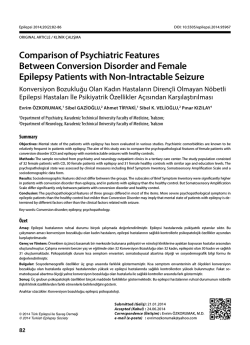
Epilepsi 2014;20(2):61-66
DOI: 10.5505/epilepsi.2014.19870
EXPERIMENTAL STUDY / DENEYSEL ÇALIŞMA
Presentation to Affirmative Effect of Doxcycyline in
Pentylene Tetrazole Induced Seizures Models
Doksisiklinin Pentilentetrazol İndükte Nöbetler
Üzerine Olan Olumlu Etkisinin İncelenmesi
Oytun ERBAŞ,1 Ayşegül ÖZER ÇELİK,2 Bilge PİRİ ÇINAR,3 Volkan SOLMAZ,4# Dürdane AKSOY4
Department of Physiology, Gaziosmanpasa University Faculty of Medicine, Tokat;
Department of Neurology, Trabzon Numune State Hospital, Trabzon;
3
Department of Neurology, Giresun State Hospital, Giresun;
4
Department of Neurology, Gaziosmanpasa University Faculty of Medicine, Tokat
1
2
Summary
Objectives: There were some experimental and clinical evidence that inflammation in the brain is likely to predispose epileptogenesis. Also,
it is thought to be that oxidative stress can play a role in epilepsy. Both laboratory and clinical studies have demonstrated the antiinflammatory-antioxidant properties of doxycycline. We aimed to highlight the anticonvulsant action of doxycycline based on clinical, laboratory and
EEG findings in animal models.
Methods: 36 rats were randomly divided in two groups. Group A for EEG recordings and Group B for behavioral assesment. 35 mg/kg pentylene tetrazole (PTZ) used for EEG recording and 70 mg/kg PTZ used for behavioral evaulations. For behavioral evaluations we assessed first
myoclonic jerk time (FMJ) and Racine convulsion scores (RCS).
Results: The groups were evaluated according to EEG records and severety of seizures. It was found to be doxcycyline is effective both on
the time of the first myoclonic jerk’ (FMJ) latency and Racine convulsion scale (RCS) scores. Besides, doxcycyline significantly decreased to
the percentage of spikes on electroencephalography (EEG) records. Also In current study, it was detected that Malone dialdehit (MDA) levels
decreased and superoxide dismutase (SOD) activity increased in doxcycyline-treated groups.
Conclusion: In our study, we evaluated that the anticonvulsant effects of doxycycline showed a dose dependent protective effect against
PTZ-induced seizures in rats by its anti-inflammatory and neuroprotective properties.
Key words: Doxcycyline; malonedialdehit; pentylenetetrazole; superoxide dismutase.
Özet
Amaç: Daha önce yapılan çalışmalarda enflamasyonun epilepsi patogeneziyle olan ilişkisi gösterilmiştir, buna göre oksidatif stresin de epilepsi patogeneziyle ilişkili olabileceği düşünülebilir. Literatürde doksisiklinin hem antienflamatuvar hem de antioksidan etkilerini inceleyen
çalışmalar bulunmaktadır. Bu çalışmanın amacı doksisiklinin muhtemel antikonvulzan etkilerini elektroensefelografi kayıtları (EEG), laboratuvar bulguları ve davranışsal bulgular eşliğinde incelemektir.
Gereç ve Yöntem: Otuz altı sıçan rastgele iki gruba ayrıldı. Grup A EEG kayıtları, Group B ise davranışsal değişikliklerin incelenmesi için
belirlendi. 35 mg/kg pentilentetrazol (PTZ) EEG kayıtları için, 70 mg/kg PTZ davranışsal değişiklikler oluşturmak için kullanıldı. Davranışsal
değişikliklerin değerlendirilmesinde ilk miyoklonik jerk zamanı ve Racine konvulziyon skalası kullanıldı, EEG kayıtlarının değerlendirilmesinde
ise spike yüzdeleri kullanıldı.
Bulgular: Yapılan istatistiksel analiz sonucunda doksisiklin verilen gruplarda spike yüzdeleri, ilk miyoklonik jerk zamanları ve Racine konvulziyon skoları açısından doksisiklinin olumlu etkileri tespit edildi. Ayrıca çalışmamızda doksisiklin verilen grupta malonedialdehit seviyeleri
düşmüşken, süperoksitdismutaz seviyelerinin artmış olduğu görüldü.
Sonuç: Sunulan çalışmada doksisiklinin PTZ indükte konvulziyonlar üzerine olumlu etkileri olduğu görülmüştür, bu etkisini de antienflamatuvar ve nöroprotektif özellikler yoluyla olabileceğini düşünmekteyiz.
Anahtar sözcükler: Doksisiklin; malonedialdehit; pentilentetrazol; superoksit dismutaz.
Current affiliation: Turhal State Hospital, Department of Neurology, Tokat.
#
© 2014 Türk Epilepsi ile Savaş Derneği
© 2014 Turkish Epilepsy Society
Submitted (Geliş): 18.03.2014
Accepted (Kabul) :28.08.2014
Correspondence (İletişim) : Volkan SOLMAZ, M.D.
e-mail (e-posta) :[email protected]
61
Epilepsi 2014;20(2):61-66
Introduction
International League Against Epilepsy (ILAE) defined an
epileptic seizure as transient occurance of signs and/or
symptoms due to abnormal excessive or synchronous
neuronal activity in the brain. Epilepsy is a disorder of the
brain characterized by an enduring predisposition to generate epileptic seizures and by the neurobiologic, cognitive,
psychological, and social consequences of this condition.[1]
Epileptogenesis refers to a process in which an initial braindamaging insult triggers a cascade of molecular and cellular
changes that eventually lead to the occurence of spontaneous seizures.[2] Brain inflammation has gained recognition
as a crucial contributor to epileptogenesis. Brain inflammation seems to be an intrinsic feature of the diseased
hyperexcitable brain tissue where spontaneous and recurrent seizures originate. Following a convulsant challenge or
epileptogenic brain injury, cytokines, prostaglandines, and
inflammatory molecules together with their receptors are
induced in neurons and activated glial cells, as well as in
endothelial cells of blood-brain barrier, in the brain region
of focal injury. Brain inflammation contributes significantly
to determine seizure threshold in susceptible brain regions,
thus playing a role in seizure precipitation and their recurrence.[3] Brain inflammation induced by an epileptogenic
injury has a rapid onset (<30 min), it can persist for several
days thus outlasting the initial precipitating event, and it
is inefficiently opposed by endogenous anti-inflammatory
mechanisms.[4]
Doxycycline is a long-acting second generation tetracyclineclass antibiotic. It can pass to CSF well and exerts its effect in
1-3 hours when it is given orally and in 30 minutes when it
is administered intravenously. Its action mechanism is based
on the characteristics that tetracyclines inhibit protein synthesis by acting ribosome levels, 16S rRNA.[5] Both some laboratory and clinical studies have demonstrated anti-oxidant
properties of doxycycline. Furthermore, it promotes neuronal survival, inhibits microglial activation and reduces reactive gliosis in some animal models.[6,7] According to these
information, the aim of our study was to investigate doxycycline’s anti-convulsant effects with EEG records, biochemical
findings and clinical (behavioral) assesment.
Materials and Methods
Animal and Laboratory
The experimental procedures employed in present study
62
were approved by Ege University Animal Ethics Comittee.
All experiments were carried out according to the Guide for
the Care and Use of Laboratory Animals, as confirmed by
National Institues of Health (U.S.) 36 male (18 of them for
EEG recording and 18 of them are for behavioral studies)
Sprague–Dawley rats, weighing 200–250 g each were utilized for this study. The rats were kept on a 12 hour –12 hour
light–dark cycle (light from 07.00 to 19.00), in quiet rooms,
with 22–24 °C ambient temperature. They were fed by standard laboratory food and tap water ad libitum.
Experimental procedures
36 rats were randomly divided in two groups: Group A for
EEG recordings and Group B for behavioral assesment.
In Group A; Rats were deeply anesthetized. Then, a small
hole was opened with a drill stereotaxically. The electrodes
(Polyamide-coated stainless steel wires, 0.1 mm diameter
and electrical resistance <1Ω/10 mm) were implanted on
dura over left frontal cortex (2.0 mm lateral to the midline,
1.5 mm anterior to the bregma) and the reference electrode
was implanted over cerebellum (1.5 mm posterior to the
lambda, on midline)[8,9] for EEG recording. Then, electrodes
were fixed by using dental acrylic (Dental acrylic is a mixture of numerous alloys using for dental restoration). Rats
were deeply anesthezied by ketamine (80 mg/kg) and xylazine (4 mg/kg) intraperitoneally (i.p.) After 10 days from
the electrode replacement were fixed, 24 rats were divided
randomly into 3 groups (n=6): Group A1, A2, A3.
Group A1 was administered saline i.p, Group A2 was administered 100 mg/kg doxycycline (Tetradox, Actavis) i.p and,
Group A3 was administered 200 mg/kg doxycycline i.p. The
drugs were administered 30 minutes prior to pentylentetrazol (PTZ) (35 mg/kg, i.p.) injection. All groups were administered 35 mg/kg PTZ and EEG was recorded. EEG recordings
were taken in awake rats in a special container after 5 minutes from PTZ administeration.
The duration of EEG recording was 60 minutes (Figure
1).[10,11] The signals were amplified 10,000 times and filtered
with a range of 1–60 Hz. EEG records were taken by using
the Biopac MP 150 amplifier system and spike percentage
was evaluated. Two clinical neurophysiologists scored the
EEG data for spike percentage. We defined “spike percentage” as the percentage of 1-second bins with at least one
spike-wave in them.[12] We affirmed the electrode location
histologically following euthanization.
Presentation to Affirmative Effect of Doxcycyline in Pentylene Tetrazole Induced Seizures Models
(a)
(b)
(c)
Fig. 1. EEG recording (a) PTZ (35 mg/kg) and saline, (b) PTZ
(35 mg/kg) and 100 mg/kg doxycycline group; (c) PTZ
(35 mg/kg) and 200 mg/kg doxycycline group.
Then the groups were rearranged with different 18 rats
(Group B) and these rats were then divided into 3 groups
(n=6): Group B1 was administered saline i.p, Group B2 was
administered 100 mg/kg doxycycline (Tetradox, Actavis) i.p
and, Group B3 was administered 200 mg/kg doxycycline
i.p. The drugs were administered 30 minutes prior to PTZ
(70 mg/kg, i.p.) injection. Racine’s Seizure Scale (RCS) score
and latency times of ‘first myoclonic jerk’ (FMJ) was used
to evaluate the seizures (for only PTZ 70 mg/kg) as follows:
0 = no seizure; 1 = twitching of vibrassae and pinnae; 2 =
motor arrest with more pronounced twitching; 3 = motor
arrest with generalized myclonic jerks; 4 = tonic clonic seizure while the animal remained on its feed; 5 = tonic–clonic
seizure with loss of the righting reflex; 6 = lethal seizure.
Rats were observed for latency times of FMJ as previously
described.[13] The onset times were recorded as seconds. Almost all animals showing tonic generalized extension were
died. The observation period for PTZ-induced seizures were
limited with 30 minutes duration.[13] After this duration, the
animals were euthanized and brain tissues were removed
for biochemical analysis.
Measurement of brain lipid peroxidation (MDA)
Lipid peroxidation was determined in brain tissue samples
by measuring malondialdehyde (MDA) levels as thiobarbituric acid reactiıve substances (TBARS).[14] Briefly, trichloroacetic acid and TBARS reagent were added to the tissue
samples, then mixed and incubated at 100 °C for 60 min.
After cooling on ice, the samples were centrifuged at 3000
rpm for 20 min and the absorbance of the supernatant was
read at 535 nm. MDA levels were calculated from the standard calibration curve using tetraethoxypropane and expressed as nmol/gr protein.
Determination of brain SOD activity
Total SOD activity was determined according to the method
of Sun et al. The principle of the method is the inhibition of
nitrobluetetrazolium (NBT) reduction by the xanthine-xanthine oxidase system as a superoxide generator. One unit of
SOD was defined as the enzyme amount causing 50% inhibition in the NBT reduction rate. SOD activity was given as
units per milligram protein (U/mg protein).
Statistical analysis
Results were expressed as a mean±standard error of mean
(SEM). Data analyses were performed by utilizing SPSS version 15.0 for Windows. The RCS score, FMJ time were evaluated by one-way analysis of variance (ANOVA). Post-hoc
Bonferonni test was utilized to identify differences between
the experimental groups. The value of p<0.05 was accepted
as statistically significant.
Results
Evaluation of brain lipid peroxidation (MDA)
In 100 mg/kg and 200 mg/kg doxycycline-treated groups,
brain MDA levels (42.4±4.7 nmol/gr, 36.1±7.7 nmol/gr, respectively) significanlty decreased when compared with
saline-given group (93.7±4.8 nmol/gr, p<0.0001) (Figure 2).
Evaluation of brain SOD activity
In 100 mg/kg and 200 mg/kg doxycycline-treated groups,
brain SOD levels (0.087±0.012 U/mg protein, 0. 11±0.018 U/
mg protein, respectively) significanlty increased when compared with saline-given group (0.038±0.007 U/mg protein,
p<0.001).
100
Brain MDA levels
50
0
B1 group (PTZ
[70 mg/kg]
and saline)
B2 group (PTZ
[70 mg/kg]
and 100 mg/kg
doxycycline)
B3 group (PTZ
[70 mg/kg]
and 200 mg/kg
doxycycline)
Fig. 2. Brain MDA levels in saline-given and doxcycylinetreated groups.
63
Epilepsi 2014;20(2):61-66
5.6
Saline-treated
group
4.8
100 mg/kg
Doxycyclinetreated group
Mean RCS score
3.8
200 mg/kg
Doxycyclinetreated group
Fig. 3. Mean Racine’s Seizure Scale (RCS) scores in saline-given and doxcycyline-treated groups.
Evaluation of the seizures
The Racine seizure scale score and FMJ latency time were
measured in saline and doxcycyline-treated groups. RCS
score was 5.6±0.2; FMJ latency time is 72.2±5.4 sec in saline-given group. 100 mg/kg and 200 mg/kg doxycycline
treatment decreased RCS scores (4.8±0.2 and 3.8±0.2,
p<0.01, p<0.000) and increased FMJ latency time (89.8±4.3,
115.5±5.6, p<0.05, p<0.000) when compared with salinegiven group (Figure 3 and Table 1).
Evaluation of EEG records
Percentage of spikes were calculated as %73.3±4.7 in salinegiven group. 100 mg/kg and 200 mg/kg doxycycline treatment significantly decreased spike percentage (%55.5±7.3
and %37.8±8, p<0.05, p<0.01) when compared with salinegiven group (Figure 1).
Discussion
Doxycycline is one of the second generation tetracyclines
that are known to prevent neuronal and oligodendroglial
cell death in some in vitro models.[15] In previous studies it
is presented that doxycycline has neuroprotective effects.
[6]
Though, the mechanism of epileptogenesis is not understood completely, the seizures have been reported to occur among patients with chronic inflammatory diseases.[16]
Proinflammatory cytokines are induced rapidly in rodent
brains during epileptic activity.[17] Seizures increase steadystate levels of mitochondrial O2−, a central mediator of
oxidative stres.[18] The cytotoxic mechanism by which reactive oxygene species (ROS) induce neuronal damage may
involve direct oxidative attack on cellular macromolecules
and initiation or propagation of free radical chain reaction,
ultimately leading to macromolecular damage.[19] Proteins,
lipids and DNA are sensitive targets of ROS. As seizure induced inflammation is associated with free radical production and oxidative stress, there is also an increase in lipid
peroxidation level.[20] Lipids are the major target of oxidative
damage that occurs during seizures. Polyunsaturated fatty
acids present in phospholipids of biological membranes
are highly susceptible to oxidation by ROS.[21] Oxidation of
fatty acids alters the structure of the cell membrane, cause
changes in fluidity and permeability. Malondialdehyde is
one of the products of lipid peroxidation and is therefore a
good indicator of the rate of lipid peroxidation.[22]
The presence of lipid peroxidation following seizures has
been demonstrated with MDA levels in our study. In doxycycline-treated group, MDA levels significanlty decreased
and SOD levels significantly increased in a dose dependent
manner when compared with saline-given group.
Recent studies demonstrated that doxycycline has protective effects on ischemic and degenerative brain diseases.
[23,24]
Hydrogen peroxide (H2O2) is formed whenever O2− is
generated because of its rapid conversion to H2O2 by superoxide dismutase (SOD) enzyme.[25] H2O2 has moreless
effect than superoxide group and it is counteracted for
cell been converting by such as catalase, peroxidase and
glutation. Causes of increases to effect of SOD contribute to
render superoxide groups harmless.[26,27] Researchers have
observed contradictory results in the levels of SOD, either in
acute or chronic models.[28-30] SOD is one of the major antioxidants. In current study, SOD levels increased in doxycycline treated when compared with saline-given. This proves
doxycycline’s anti-oxidant effect on PTZ induced seizure
Table 1. First Myoclonic Jerk’ (FMJ) latency time of groups
Groups
FMJ latency time (sec)
PTZ (70 mg/kg) and saline (B1 Group)
PTZ (70 mg/kg) and 100 mg/kg doxycycline (B2 Group)
PTZ (70 mg/kg) and 200 mg/kg doxycycline (B3 Group)
Data were expressed as mean±SEM. *p<0.05, #p<0.0001, **p<0.01 (different from saline-treated group).
64
72.2±5.4
89.8±4.3*
115.5±5.6#
Presentation to Affirmative Effect of Doxcycyline in Pentylene Tetrazole Induced Seizures Models
model of rats. In our study, we also detected both 100 and
200 mg/kg doxycycline’s affirmative effects on seizures by
EEG recordings, it decreases spike percentage and seizure
severity.
In conclusion, following the treatment with doxycycline, latency time for FMJ significantly increased; RCS scores and
spike percentage decreased in a dose-dependent manner.
This results demonstrate that doxycycline preserves from
PTZ-induced seizures and decreases oxidant stress which
occurs because of seizures.
References
1. Fisher RS, van Emde Boas W, Blume W, Elger C, Genton P, Lee P,
et al. Epileptic seizures and epilepsy: definitions proposed by
the International League Against Epilepsy (ILAE) and the International Bureau for Epilepsy (IBE). Epilepsia 2005;46(4):470-2.
2. Pitkänen A, Lukasiuk K. Molecular and cellular basis of epileptogenesis in symptomatic epilepsy. Epilepsy Behav 2009;14
Suppl 1:16-25.
3. Vezzani A, French J, Bartfai T, Baram TZ. The role of inflammation in epilepsy. Nat Rev Neurol 2011;7(1):31-40.
4. Vezzani A, Friedman A, Dingledine RJ. The role of inflammation
in epileptogenesis. Neuropharmacology 2013;69:16-24.
5. Kim HS, Suh YH. Minocycline and neurodegenerative diseases.
Behav Brain Res 2009;196(2):168-79.
6. Nogueira CR, Damasceno FM, de Aquino-Neto MR, de Andrade
GM, Fontenele JB, de Medeiros TA, et al. Doxycycline protects
against pilocarpine-induced convulsions in rats, through its
antioxidant effect and modulation of brain amino acids. Pharmacol Biochem Behav 2011;98(4):525-32.
7. Jantzie LL, Cheung PY, Todd KG. Doxycycline reduces cleaved
caspase-3 and microglial activation in an animal model
of neonatal hypoxia-ischemia. J Cereb Blood Flow Metab
2005;25(3):314-24.
8. Schwierin B, Achermann P, Deboer T, Oleksenko A, Borbély
AA, Tobler I. Regional differences in the dynamics of the cortical EEG in the rat after sleep deprivation. Clin Neurophysiol
1999;110(5):869-75.
9. Paxinos G, Watson C. The rat brain in stereotaxic coordinates.
3rd ed. New York: Eisevier; 1991.
10.Souza MA, Mota BC, Gerbatin RR, Rodrigues FS, Castro M,
Fighera MR, et al. Antioxidant activity elicited by low dose of
caffeine attenuates pentylenetetrazol-induced seizures and
oxidative damage in rats. Neurochem Int 2013;62(6):821-30.
es. Epilepsia 2005;46(12):1937-42.
13. Kaputlu I, Uzbay T. L-NAME inhibits pentylenetetrazole and
strychnine-induced seizures in mice. Brain Res 1997;753(1):98101.
14. Demougeot C, Marie C, Beley A. Importance of iron location in
iron-induced hydroxyl radical production by brain slices. Life
Sci 2000;67(4):399-410.
15.Domercq M, Matute C. Neuroprotection by tetracyclines.
Trends Pharmacol Sci 2004;25(12):609-12.
16. Rao RS, Medhi B, Saikia UN, Arora SK, Toor JS, Khanduja KL, et
al. Experimentally induced various inflammatory models and
seizure: understanding the role of cytokine in rat. Eur Neuropsychopharmacol 2008;18(10):760-7.
17. Vezzani A, Aronica E, Mazarati A, Pittman QJ. Epilepsy and brain
inflammation. Exp Neurol 2013;244:11-21.
18. Patel M, Li QY. Age dependence of seizure-induced oxidative
stress. Neuroscience 2003;118(2):431-7.
19. Shin EJ, Jeong JH, Chung YH, Kim WK, Ko KH, Bach JH, et al.
Role of oxidative stress in epileptic seizures. Neurochem Int
2011;59(2):122-37.
20. Girish C, Koner BC, Jayanthi S, Ramachandra Rao K, Rajesh B,
Pradhan SC. Hepatoprotective activity of picroliv, curcumin and
ellagic acid compared to silymarin on paracetamol induced liver toxicity in mice. Fundam Clin Pharmacol 2009;23(6):735-45.
21.Rowley S, Patel M. Mitochondrial involvement and oxidative stress in temporal lobe epilepsy. Free Radic Biol Med
2013;62:121-31.
22. Rumià J, Marmol F, Sanchez J, Giménez-Crouseilles J, Carreño
M, Bargalló N, et al. Oxidative stress markers in the neocortex of
drug-resistant epilepsy patients submitted to epilepsy surgery.
Epilepsy Res 2013;107(1-2):75-81.
23. Lee H, Park JW, Kim SP, Lo EH, Lee SR. Doxycycline inhibits matrix metalloproteinase-9 and laminin degradation after transient global cerebral ischemia. Neurobiol Dis 2009;34(2):18998.
24. Cho Y, Son HJ, Kim EM, Choi JH, Kim ST, Ji IJ, et al. Doxycycline is neuroprotective against nigral dopaminergic degeneration by a dual mechanism involving MMP-3. Neurotox Res
2009;16(4):361-71.
25. Rumià J, Marmol F, Sanchez J, Giménez-Crouseilles J, Carreño
M, Bargalló N, et al. Oxidative stress markers in the neocortex of
drug-resistant epilepsy patients submitted to epilepsy surgery.
Epilepsy Res 2013;107(1-2):75-81.
26.Matés JM. Effects of antioxidant enzymes in the molecular
control of reactive oxygen species toxicology. Toxicology
2000;153(1-3):83-104.
11. Erbas O, Yılmaz M, Korkmaz HA, Bora S, Evren V, Peker G. Oxytocin inhibits pentylentetrazol-induced seizures in the rat. Peptides 2013;40:141-4.
27. Tejada S, Sureda A, Roca C, Gamundí A, Esteban S. Antioxidant
response and oxidative damage in brain cortex after high dose
of pilocarpine. Brain Res Bull 2007;71(4):372-5.
12. Aeby A, Poznanski N, Verheulpen D, Wetzburger C, Van Bogaert
P. Levetiracetam efficacy in epileptic syndromes with continuous spikes and waves during slow sleep: experience in 12 cas-
28. Rauca C, Wiswedel I, Zerbe R, Keilhoff G, Krug M. The role of
superoxide dismutase and alpha-tocopherol in the development of seizures and kindling induced by pentylenetetrazol
65
Epilepsi 2014;20(2):61-66
- influence of the radical scavenger alpha-phenyl-N-tert-butyl
nitrone. Brain Res 2004;1009(1-2):203-12.
are modified in the hippocampus of epileptic rats. Epilepsy Res
2001;46(2):121-8.
29. Bellissimo MI, Amado D, Abdalla DS, Ferreira EC, Cavalheiro EA,
Naffah-Mazzacoratti MG.et al. Superoxide dismutase, glutathione peroxidase activities and the hydroperoxide concentration
30. Devi PU, Manocha A, Vohora D. Seizures, antiepileptics, antioxidants and oxidative stress: an insight for researchers. Expert
Opin Pharmacother 2008;9(18):3169-77.
66
© Copyright 2026 Paperzz