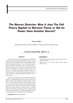
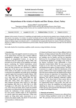
ORJİNAL
Türk Biyokimya Dergisi [Turkish Journal of Biochemistry–Turk J Biochem] 2014; 39 (2) ; 132–139
doi: 10.5505/tjb.2014.32932
Research Article [Araştırma Makalesi]
Yayın tarihi 30 Haziran, 2014 © TurkJBiochem.com
[Published online 30 June, 2014]
1976
[Bir üniversite hastanesine başvuran sağlıklı insanlarda iskemi modifiye
albuminin referans değerleri]
1. ÖRNEK
Can Duman1,
Coşkun Bakar2,
Ertan Eşsizoglu1,
Funda Kırtay Tütüncüler1,
Elif Demircan1
1Çanakkale Onsekiz Mart Üniversitesi Tıp
Fakültesi Biyokimya Anabilim Dalı, 2Halk Sağlığı
Anabilim Dalı, Çanakkale, Türkiye
ABSTRACT
Objectives: The Ischemia Modified Albumin (IMA) test is a myocardial ischemia marker
which may have potential use for the diagnosis of Acute Coronary Syndrome. The aim of this
study was determine reference values of IMA test on the healthy people over the age of 15
who’s admitted to Çanakkale Onsekiz Mart University.
Methods: This study was performed on 582 people over 15 years old and healthy in appearance who had presented at the Çanakkale Onsekiz Mart University Faculty of Medicine Research and Application Hospital. The year 2011 population of Turkey was taken as the reference
in the distribution of the study group according to age group and gender. A survey which
queried the demographic features and health problems was administered to the participants
of the study and 9 cc venous blood was drawn. IMA levels were measured by using the spectrophotometric method defined by Bar-Or. The mean, standard deviation, median, minimum,
maximum, and 2.5 % - 97.5 % interval were used in order to detect the IMA reference values.
Results: A total of 274 males (47.1 %) and 308 women (52.9 %) were studied. The mean IMA
value of the examined group was 0.339 ± 0.093, and the median was 0.343 (Min-Max: 0.0530.631), while the 2.5 % - 97.5 % percentil values were 0.145 - 0.530.
Conclusion: Studies that indicate the reference levels of the IMA for the diagnosis of ischemic conditions are insufficient. Our study is expected to contribute to closing this gap.
Key Words: Ischemia, Ischemia-modified albumin, Reference range
Conflict of Interest: The authors do not have a conflict of interest regarding the subjects
related to this manuscript.
Yazışma Adresi
[Correspondence Address]
Professor Can Duman
Çanakkale Onsekiz Mart University,
School of Medicine, Department of Biochemistry,
Çanakkale, Turkey.
Phone. +90 286 2180018
Fax. +90 286 2183806
E-mail. [email protected]
Registered: 18 April 2013; Accepted: 4 November 2013
[Kayıt Tarihi: 18 Nisan 2013; Kabul Tarihi: 4 Kasım 2013]
http://www.TurkJBiochem.com
ÖZET
Amaç: İskemi modifiye albumin (IMA) akut koroner sendromun tanısında potansiyel kullanımı olabilecek bir miyokardiyal iskemi belirtecidir. Bu çalışmada amacımız Çanakkale
Onsekiz Mart Üniversitesi’ne başvuran 15 yaşın üzerindeki sağlıklı insanlarda IMA testinin
referans değerlerini belirlemektir.
Gereç ve Yöntemler: Bu çalışma Çanakkale Onsekiz Mart Üniversitesi Tıp Fakültesi Araştırma ve Uygulama Hastanesine başvuran 15 yaş ve üzeri ve görünürde sağlıklı 582 kişi üzerinde gerçekleştirilmiştir. Çalışma grubunun yaş ve cinsiyet dağılımı belirlenirken 2011 yılı
Türkiye nüfusu referans olarak alınmıştır. Katılımcılara demografik özelliklerinin ve sağlık
problemlerinin sorgulandığı bir anket formu uygulandı ve 9 ml venöz kan alınmıştır. IMA
düzeyleri Bar-Or tarafından tanımlanan spektrofotometrik metot kullanılarak ölçülmüştür.
IMA referans değerlerini saptamak için ortalama, standart sapma, ortanca, minimum, maksimum ve % 2,5 - % 97,5 persentil değeri kullanılmıştır.
Bulgular: Toplamda 274 erkek (% 47,1) ve 308 kadın (% 52,9) değerlendirildi. Çalışılan
grupta ortalama IMA değeri 0,339 ± 0,093, ortanca değeri 0,343 (Min-Maks: 0,053 - 0,631),
% 2,5 - % 97,5 persentil değeri 0,145-0,530’dir.
Sonuçlar: İskemik durumların tanısında IMA’nın referans değerlerini vurgulayan çalışmalar
yetersizdir. Bizim çalışmamızın bu açığı kapatmaya katkıda bulunacağını beklemekteyiz.
Anahtar Kelimeler: İskemi, İskemi Modifiye Albumin, Referans aralık
Çıkar Çatışması: Yazarların makale konusu dâhilinde herhangi bir çıkar çatışmaları bulunmamaktadır.
132
DER
AD
RNN
MYYA
İM
EE
Kİ
1976
K BİİYYO
RRK
O
TTÜÜ
RK BİYO
TÜ
YA DERN
İM
E
DERGİSİ
Ğİ
K
RG
GİİSSİ
ER
DE
D
İ
Ğİİ
Ğ
The reference values of ischemia modified albumin levels
in healthy people admitted to a university hospital
ISSN 1303–829X (electronic) 0250–4685 (printed)
2. ÖRNEK
Introduction
Procedure and measures of data collection
Numerous studies have been done on the use of Ischemia Modified Albumin (IMA) in the diagnosis of acute
coronary syndrome [1-3]. Some of these studies also
define the decision values regarding the presence or
absence of acute coronary syndrome in patients who
come to the emergency service with acute chest pain
[4, 5]. On the other hand, there are also studies stating
that IMA levels increase in pathologies other than acute coronary syndromes and they may be a diagnostic
marker in these pathologies as well [6-9]. In fact, some
of these studies also suggested IMA as a marker for
oxidative stress [10, 11]. However, the number of studies defining the normal population is inadequate. There
is especially no study that identifies the Turkish population. In order to define the critical value of IMA that
will be able to identify pathologic situations, the normal values in different age groups and genders should
first be defined. Determining the normal IMA values
of the population in different age groups and genders
will help to identify the critical values in pathological
conditions.
The study was conducted at the Çanakkale Onsekiz
Mart University Application and Research Hospital by
the Biochemistry Department. Research assistants of
the Department of Biochemistry interviewed the patients who had presented at the hospital’s outpatients for
examination. After the patients were informed verbally
and in writing, volunteers who had presented to the hospital outpatients over the age of 15, who were healthy in
appearance, had no respiratory system and cardiac problems or chronic diseases such as hypertension participated in the study. The volunteers participating in the study
were asked to fill a questionnaire containing demographic information and personal health history.
A total of 9 cc of venous blood was taken from IMA participants for determination of the reference value.
The aim of this study was to determine the reference values of the IMA test in people over the age of 15 who
had no respiratory system and cardiac problems, chronic
diseases such as hypertension, or any other health issue.
The results obtained are expected to help in determining
the critical value in pathological conditions such as the
coronary syndrome in the long term.
Material and Methods
Study population and sample
This study was conducted at Çanakkale 18 Mart University Faculty of Medicine, Research and Application
Hospital between May and July 2012. A total of 61070
people presented at the hospital outpatients in the same
period. The gender distribution was 58.8% female and
41.2% male (35919 and 25151 respectively). We planned
to reach a total of 600 subjects over the age of 15 who
had presented at the hospital outpatients, were healthy in
appearance, and had no respiratory system and cardiac
problems or chronic diseases such as hypertension. The
2011 population of Turkey was taken as the reference for
the distribution of the study groups according to the age
group and gender. According to the Turkey Statistical
Institute Address-Based Population Registration System,
the population aged 15 and over in 2011 is 55,837,694.
The gender distribution is 50.1 % female and 49.9 %
male. In order to determine the distribution according to
the age groups, we divided the population by age group
(15-19, 20-24, 25-29, … 90-+) and stratified the subjects.
Table 1 provides the distribution of reference, targeted
and reached population according to the age groups. In
the end, we identified 582 volunteers meeting the inclusion criteria of the study [12].
Turk J Biochem, 2014; 39 (2) ; 132–139
Laboratory analysis
The blood samples were transferred to plain tubes containing no preservatives. Clot formation was allowed
for 30 minutes and the blood samples were centrifuged
before separating the serum. Then, the obtained serum
samples were subjected to measurement immediately.
ACB was analyzed according to the method described by
Bar-Or et al. In this method, 200 μL of serum is added to
the water solution of 50 μL 0.1 % (w/v) cobalt chloride
(CoCl2•6H2O; Sigma-Aldrich Inc., St Louis, MO, USA).
It is mixed gently and left to sit for 10 minutes to allow
for sufficient cobalt–albumin binding to occur. Then, 50
μL of dithiothreitol (1.5mg•mL−1H2O; Sigma-Aldrich
Inc.) is added as a colorizing agent. After waiting for
2 minutes, 1.0 mL of 0.9% NaCl is added to stop the
cobalt–albumin binding process. Next, absorbance is
measured using a spectrophotometer at 470 nm. A blank
sample without dithiothreitol is used as the blind. The
results are reported in absorbance units (ABSU) [13].
Statistical evaluation
The data of the study were transferred to the SPSS 19.0
package program and the analysis was performed in this
program. In order to detect the IMA reference values,
mean, standard deviation, mean, minimum, maximum,
and 2.5 % - 97.5 % interval were used. These values
were calculated in five age groups over the age of 15 for
both genders. The conformance of the IMA value with
the normal distribution of the male, female and combined groups was evaluated with the Kolmogorov-Smirnov test and each of the three distributions was found to
conform to the normal distribution (Figure 1).
Ethical approval
Consent was obtained from the Çanakkale Onsekiz University Clinical Studies Ethical Committee for this study
(Decision Number: 2012-2, Date: 11.01.2012).
Results
A total of 582 subjects consisting of 274 males (47.1 %)
133
Duman et al.
Turk J Biochem, 2014; 39 (2) ; 132–139
134
Duman et al.
1118310
688840
284594
79428
75 – 79
80 – 84
85 – 89
90 - +
100.0
0.1
0.5
1.2
2.0
2.6
3.3
4.6
6.2
6.8
8.6
8.5
10.1
11.6
11.3
11.1
11.3
%
27838215
20159
94160
260355
497023
649739
876489
1231274
1716102
1909912
2405435
2430841
2837182
3285387
3210343
3173618
3240196
n
Male
50.1
0.1
0.3
0.9
1.8
2.3
3.1
4.4
6.2
6.9
8.6
8.7
10.2
11.8
11.5
11.4
11.6
%
27999479
59269
190434
428485
621287
801629
991686
1335213
1738313
1882524
2380649
2339933
2795560
3210247
3095890
3050973
3077387
N
Female
* Turkey Statistical Institute 2011 Address-Based Population Registration System Data
55837694
1451368
70 – 74
Toplam
1868175
4786084
45 – 49
65 – 69
4770774
40 – 44
2566487
5632742
35 – 39
60 – 64
6495634
30 – 34
3454415
6306233
25 – 29
55 – 59
6224591
20 – 24
3792436
6317583
15 – 19
50 – 54
N
Total
Groups
Age
REFERENCE POPULATION *
49.9
0.2
0.7
1.5
2.2
2.9
3.5
4.8
6.2
6.7
8.5
8.4
10.0
11.5
11.1
10.9
11.0
%
600
1
3
7
12
16
20
28
37
41
51
51
61
70
68
67
68
n
%
100.0
0.1
0.5
1.2
2.2
2.6
3.3
4.6
6.2
6.8
8.6
8.5
10.1
11.6
11.3
11.1
11.3
Total
299
0
1
3
5
7
9
13
18
21
26
26
30
35
34
34
35
n
%
49.9
0.1
0.3
0.9
1.8
2.3
3.1
4.4
6.2
6.9
8.6
8.7
10.2
11.8
11.5
11.4
11.6
Male
SAMPLE
Table 1. Reference, Sample and Distribution of Reached Population According to the Age Groups and Gender, 2012, Canakkale, Turkey
301
1
2
5
7
9
11
14
19
20
26
25
30
35
33
33
33
n
50.1
0.2
0.7
1.5
2.2
2.9
3.5
4.8
6.2
6.7
8.5
8.4
10.0
11.5
11.1
10.9
11.0
%
Female
3.8
2.6
2.6
1.0
0.2
0.2
22
15
15
6
1
1
100.0
8.2
48
582
8.1
10.1
59
47
9.5
55
8.4
10.8
63
49
10.3
60
8.1
47
9.3
6.9
40
54
%
n
Total
274
0
0
6
12
8
11
27
22
26
24
33
26
22
20
23
14
n
9.5
8.0
7.3
8.4
5.1
%
47.1
0.0
0.0
2.2
4.4
2.9
4.0
9.9
8.0
9.5
8.8
12.0
Male
ACHIEVED
308
1
1
0
3
7
11
21
25
23
35
22
37
38
34
24
26
n
52.9
0.3
0.3
0.0
1.0
2.3
3.6
6.8
8.1
7.5
11.4
7.1
12.0
12.3
11.0
7.8
8.4
%
Female
Figure 1. Distribution of IMA Values According to Gender, 2012, Canakkal
15
Figure 1. Distribution of IMA Values According to Gender, 2012, Canakkale, Turkey
ues According to Gender, 2012, Canakkale, Turkey
and 308 women (52.9 %) were examined in this study.
The mean age of the subjects was 43.1±16.5 (male 45.5
± 17.1, female 40.9 ± 15.7) years. The 2011 population
based on the Turkey Statistical Institute Address-Based
Population Registration System was taken as the reference population. The distribution of the reference population, targeted and reached population according to the
age groups is seen in Table 1.
The mean IMA value of the examined group was 0.339 ±
0.093, the median 0.343 (Min-Max: 0.053 - 0.631), while
the 2.5 % - 97.5 % interval values were 0.145-0.530 (Table 2). IMA value reference intervals of the age groups
according to gender have been provided in Table 3, and
15 the distributions of 50 %, 2.5 % and 97.5 % values of the
Turk J Biochem, 2014; 39 (2) ; 132–139
IMA values can be observed in Figure 2. There was no
statistically significant difference the values according
to age group. IMA values were observed to comply with
the normal distribution in males and females and the total study group.
Coefficients of skewness and kurtosis of the examined
group was found to be 0.101, 0.202 respectively. Skewness and kurtosis, respectively, 0.139, 0.277 were determined as in woman. In man, skewness and kurtosis was
found to be 0.147 and 0.293 respectively.
Discussion
IMA is an early indicator of cardiac ischemia that develops before necrosis. However, a change in IMA levels
135
Duman et al.
Table 2. Distribution of IMA Values According to the Age Groups, 2012, Canakkale, Turkey
Age
Groups
15 – 19
20 – 24
25 – 29
30 – 34
35 – 39
40 – 44
45 – 49
50 – 54
55 – 59
60 – 64
65 – 69
70 – 74
75 – 79
80 – 84
85 – 89
90 - +
Total
Total
Mean±SD
0.330±0.098
0.319±0.085
0.349±0.087
0.340±0.093
0.353±0.102
0.322±0.099
0.352±0.101
0.326±0.100
0.350±0.080
0.338±0.081
0.333±0.070
0.310±0.086
0.361±0.107
0.348±0.070
0.285*
0.456*
0.339±0.093
Median(Min-Max)
2.5-97.5% Interval
0.355(0.062-0.475)
0.064-0.474
0.329(0.079-0.517)
0.092-0.500
0.342(0.167-0.613)
0.183-0.581
0.349(0.053-0.585)
0.088-0.565
0.357(0.100-0.631)
0.107-0.623
0.312(0.099-0.576)
0.105-0.550
0.364(0.112-0.609)
0.128-0.600
0.330(0.072-0.561)
0.092-0.544
0.359(0.204-0.532)
0.207-0.523
0.331(0.147-0.565)
0.155-0.540
0.322(0.202-0.467)
0.202-0.467
0.322(0.140-0.415)
0.140-0.415
0.370(0.126-0.572)
0.126-0.572
0.350(0.259-0.447)
0.259-0.447
---
---
---
---
0.343 (0.053-0.631)
0.145-0.530
*: Value of a single person
Figure 2. Distribution of IMA 2.5%, 50%, 97.5% Values According to the Age Groups, 2012, Canakkale, Turkey
Turk J Biochem, 2014; 39 (2) ; 132–139
136
Duman et al.
Turk J Biochem, 2014; 39 (2) ; 132–139
137
Duman et al.
0.348±0.070
---
---
0.331±0.099
80 – 84
85 – 89
90 - +
Toplam
* : Mann-Whitney U Test
**: Independent Sample T Test
***: Value of a single person
0.363±0.111
75 – 79
0.321±0.078
50 – 54
0.321±0.106
0.359±0.125
45 – 49
70 – 74
0.305±0.111
40 – 44
0.357±0.080
0.362±0.118
35 – 39
65 – 69
0.332±0.082
30 – 34
0.315±0.084
0.333±0.099
25 – 29
60 – 64
0.274±0.088
20 – 24
0.360±0.082
0.325±0.090
15 – 19
55 – 59
Mean±SD
Age Groups
0.331(0.079-0.631)
---
---
0.350(0.259-0.447)
0.369(0.126-0.572)
0.377(0.140-0.415)
0.362(0.202-0.467)
0.320(0.147-0.454)
0.369(0.204-0.532)
0.323(0.164-0.440)
0.375(0.144-0.369)
0.387(0.099-0.576)
0.364(0.100-0.631)
0.318(0.209-0.585)
0.335(0.167-0.613)
0.277(0.079-0.417)
0.352(0.168-0.467)
Median(Min-Max)
Male
0.138-0.577
---
---
0.259-0.447
0.126-0.572
0.140-0.415
0.202-0.467
0.147-0.454
0.204-0.532
0.164-0.440
0.144-0.609
0.099-0.576
0.100-0.631
0.209-0.585
0.167-0.613
0.079-0.417
0.168-0.467
2.5-97.5% Interval
Table 3. Distribution of IMA Values According to the Age Groups and Gender, 2012, Canakkale, Turkey
0.346±0.086
0.456***
0.285***
---
0.354±0.109
0.299±0.062
0.309±0.052
0.366±0.070
0.341±0.079
0.332±0.121
0.348±0.082
0.347±0.074
0.347±0.090
0.344±0.100
0.359±0.079
0.361±0.057
0.332±0.103
Mean±SD
0.350 (0.053-0.565)
---
---
---
0.370(0.238-0.455)
0.290(0.194-0.403)
0.300(0.232-0.419)
0.335(0.236-0.565)
0.354(0.221-0.489)
0.337(0.072-0.561)
0.350(0.112-0.496)
0.334(0.206-0.476)
0.350(0.113-0.520)
0.354(0.053-0.548)
0.356(0.217-0.529)
0.366(0.222-0.517)
0.355(0.062-0.475)
Median(Min-Max)
Female
0.170-0.517
---
---
---
0.238-0.455
0.194-0.403
0.232-0.419
0.236-0.565
0.221-0.489
0.072-0.561
0.112-0.496
0.206-0.476
0.113-0.520
0.053-0.548
0.217-0.529
0.222-0.517
0.062-0.475
2.5-97.5% Interval
0.058**
---
---
---
0.945
0.397
0.076
0.059
0.371
0.645
0.787
0.081
0.655
0.345
0.220
0.001
0.769
p*
does not only indicate cardiac pathology as IMA levels
are also elevated in individuals with skeletal muscle ischemia [14, 15].
The study by Troxler et al. [16] revealed that skeletal
muscle ischemia increased IMA levels in major arterial
surgical procedures. Similar results were also reported
by Montagnana et al. [17] during orthopedic surgical
procedures.
There are no reference studies in the literature on IMA
where normal reference ranges can be found. The majority of existing studies are in the form of comparison
of ischemic situations with the controls without the relevant ischemic condition. In addition, the separation
values for controls and patients in different diseases are
very different. Türedi et al., in their study on patients
with cardiac arrest, determined the mean IMA value of
the ABSU type as 0.16 for the control group and 0.25 in
the cardiac arrest group with an unfavorable prognosis
[18]. Gündüz et al. found mean IMA values as 0.280 in
patients with brain infarction and 0.172 for the control
group [19]. However, in a study that evaluated IMA as
a marker of oxidative stress in patients with beta thalessemia, IMA values were 0.725 in the thalessemia group
and 0.554 in the control group [10]. An attempt has been
made to estimate the IMA cut-off value in ischemic
events from these studies. Chen et al defined the ABSU
type mean IMA value in healthy people as a high value,
0.855, and the cut-off value for myocardial infarction as
0.906 [4]. The reference ranges of healthy individuals
are required to find the pathological diagnosis values
for IMA. It is not possible to say that the comparisons
made with healthy groups in the studies are sufficient to
provide these reference values. Shen et al found a value
of 0.55 in subjects with Acute Coronary Syndrome and
recommended a value of 0.34 for the control group [5].
The mean IMA value of 582 subjects over 15 years of
age who presented at the Çanakkale Onsekiz Mart University Outpatients in our study was 0.339 ± 0.093, the
median 0.343 (Min-Max: 0.053 - 0.631), while the 2.5 %
- 97.5 % interval values were 0.145-0.530.
Evaluation of the data we obtained in our study without
considering age groups and gender provided results in
support of Shen et al. [5]. However, the normal definition of a variable requires the taken sample to represent
the universe in size and nature. A sample similar to the
gender and age groups of the reference population that
can be considered as the universe should therefore be
selected. Although our study was conducted in the Çanakkale Province, we tried to use the Turkish population
as the reference population in distribution when determining the distribution of sample size according to age
and gender. Deviations in some age groups and in males
are due to conduct of the study on patients who presented at the university hospital. A total of 61070 patients
presented at the hospital outpatients during May-July
2012, when the study data was collected, of which 58.8
Turk J Biochem, 2014; 39 (2) ; 132–139
% (35919) were female and 41.2 % (25151) were male.
This difference was naturally reflected in our study group. It was therefore quite hard to find a male patient who
met the study inclusion criteria in some groups. Such
studies that provide the normal IMA values are rare in
the literature. Information about normal IMA values is
usually obtained from the control groups consisting of
small samples in studies that try to determine ischemic
conditions. This is not a suitable method to find the reference ranges. Despite the problems with the age groups
in our study, a sample that may close the gap in the literature has been studied. However, we need more studies
that can represent the current population. The reference
intervals, IMA level assessment of myocardial ischemia,
distinguishing be used for diagnosis and follow-up. However, positive and negative predictive value that’s the
data to be obtained from the patient, the possibility to
remain outside the reference range is not known. New
studies are needed using the reference range values on
patients diagnosed with myocardial infarction.
These studies will provide guidance in determining the
pathological thresholds of IMA in ischemic conditions.
The differences between the cut-off values currently
obtained from the different studies in the literature can
thus be decreased.
Conflıct of Interest
There is no conflict of interest.
References
[1] Dekker MS, Mosterd A, van’t Hof AW, Hoes AW. Novel biochemical markers in suspected acute coronary syndrome: systematic review and critical appraisal. Heart 2010; 96:1001-10.
[2] Kim JS, Hwang HJ, Ko YG, Kim JS, Choi D, et al. Ischemia-modified albumin: is it a reliable diagnostics and prognostic marker
for myocardial ischemia in real clinical practice? Cardiology
2010; 116:123-9.
[3] Peacock F, Morris DL, Anwaruddin S, Christenson RH, Collinson PO, et al. Meta-analysis of ischemia-modified albumin
to rule out acute coronary syndromes in the emergency department. Am Heart J 2006; 152:253-62.
[4] Chen ZX, Wang Q, Zheng L, Bao J. Clinical value of an improved ischemia-modified albumin assay in the diagnosis of early
acute myocardial infarction. Nan Fang Yi Ke Da Xue Xue Bao
2009; 29:1378-80.
[5] Shen XL, Xing C, Han LL, Lin L, Lin LF, et al. Diagnostic value
of ischemia-modified albumin in patients with acute coronary
syndrome. Zhonghua Xin Xue Guan Bing Za Zhi 2007; 35:814817.
[6] Kanko M, Yavuz S, Duman C, Hosten T, Oner E, et al.. Ischemia-modified albumin use as a prognostic factor in coronary
bypass surgery. J Cardiothorac Surg 2012; 5:7-3.
[7] Fidan E, Mentese A, Kavgaci H, Orem A, Fidan S, et al. Increased ischemia-modified albumin levels in patients with gastric
cancer. Neoplasma 2012; 59:393-7.
[8] Mehmetoglu I, Yerlikaya FH, Kurban S, Erdem SS, et al. Oxidative stress markers in hemodialysis and peritoneal dialysis patients, including coenzyme Q10 and ischemia-modified albumin.
Int J Artif Organs 2012; 35:226-32.
138
Duman et al.
[9] Valle Goettlieb MG, da Cruz JB, Duarte MM, Moresco RN, Wiehe M, et al. Associations among metabolic syndrome, ischemia,
inflammatory, oxidatives and lipid biomarkers. J. Clin Endocrinol Metab 2010; 95:586-91.
[10] Awadallah SM, Atoum MF, Nimer NA, Saleh SA. Ischemia
modified albumin: an oxidative stress marker in β-thalassemia
major. Clin Chim Acta 2012; 18:907-10.
[11] Karahan SC, Sonmez M, Saglam F, Mentese A, Erkut N, et al.
Can ischemia-modified albumin be a valuable indicator of tissue
ischemia in polycythemia vera? Hematology 2010; 15:151-6.
[12] Turkish Statistical Institute. Address Based Population
Registration System Results 2011, Turkish Statistical Institute,
Printing Division, 2012, Ankara.
[13] Bar-Or D, Lau E, Winkler JV. A Novel Assay For CobaltAlbumin Binding and Its Potential as a Marker For Myocardial
Ischemia-A Preliminary Report. J Emerg Med 2000; 19:311-5.
[14] Montagnana M, Lippi G, Regis D, Fava C, Viola G, et al. Evaluation of cardiac involvement following major orthopedic surgery.
Clin Chem Lab Med 2006; 44:1340-1346.
[15] Refaai MA, Wright RW, Parvin CA, Gronowski AM, Scott
MG, et al. Ischemia-modified albumin increases after skeletal
muscle ischemia during arthroscopic knee surgery. Clin Chim
Acta 2006; 366: 264-8.
[16] Troxler M, Thompson D, Homer-Vanniasinkam S. Ischaemic Skeletal Muscle Increases Serum Ischaemia Modified Albumin. Eur J VascEndovasc Sur 2006; 31:164-9.
[17] Montagnana M, Lippi G, Volpe A, Salvagno GL, Biasi D,
et al. Evaluation of cardiac laboratory markers in patients with
systemic sclerosis. Clin Biochem 2006; 39:913-7.
[18] Turedi S, Gunduz A, Mentese A, Dasdibi B, Karahan SC, et al.
Investigation of the possibility of using ischemia-modified albumin as a novel and early prognostic marker in cardiac arrest patients after cardiopulmonary resuscitation. Resuscitation, 2009;
80: 994-999.
[19] Gunduz A, Turedi S, Mentese A, Altunayoglu V, Turan I,
et al. Ischemia-modified albumin levels in cerebrovascular accidents. Am J Emerg Med 2008; 26: 874-8.
Turk J Biochem, 2014; 39 (2) ; 132–139
139
Duman et al.
© Copyright 2026 Paperzz