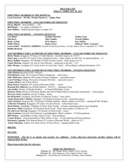
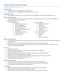
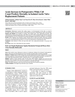
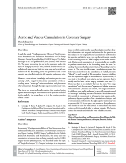
EXPERIMENTAL STUDY
The efficacy of a hemostatic agent in anticoagulant
drug-induced rat bleeding model
Ertan Sönmez, M.D.,1 Umut Yücel Çavuş, M.D.,3 Cemil Civelek, M.D.,1 Ali Dur, M.D.,1
Eda Karayel, M.D.,1 Bedia Gülen, M.D.,1 Ömer Uysal, M.D.,2 Göktürk İpek, M.D.4
1
Department of Emergency Medicine, Bezmialem Vakıf University Faculty of Medicine, Istanbul;
2
Department of Biostatistics, Bezmialem Vakıf University Faculty of Medicine, Istanbul;
3
Department of Emergency Medicine, Dışkapı Yıldırım Beyazıt Training and Research Hospital, Ankara;
4
Department of Cardiology, Siyami Ersek Training and Research Hospital, Istanbul
ABSTRACT
BACKGROUND: Bleeding is a major problem in warfarin pretreated patients who need emergency surgical procedures. APH is a
hemostatic agent with ultra-hydrophilic and particulate properties. This study aimed to evaluate the in vivo hemostatic effect of APH
in rats pretreated with warfarin.
METHODS: Forty-eight Wistar rats were divided into two main groups: one group was pretreated with warfarin and the other
group was not. These two groups were further divided into three subgroups according to the administration of APH, wheat meal, or
saline, for a total of six subgroups. Standard full thickness tissue defects were performed on the backs of the rats. Saline, wheat meal,
or APH were administered to the bleeding defect site in both main groups. Hemostasis time and amount of bleeding were calculated.
RESULTS: The bleeding time in rats administered APH was significantly shorter than those administered wheat meal and saline.
Consequently, the amount of bleeding was significantly less in the APH groups than in the control groups.
CONCLUSION: APH has an effective hemostatic property in rats pretreated or non-pretreated with anticoagulants. Hemostatic
agents can be useful for incidences of external bleedings, which are increasing because of anticoagulation.
Key words: Hemostatic; rat; warfarin.
INTRODUCTION
Warfarin is an anticoagulant that reduces the effects of vitamin K dependent factors, and its effects are increased by
many drugs.[1] Interruption of therapy for surgical intervention increases the risk of thromboembolism, although continuation of therapy increases the risk of bleeding in patients
taking warfarin. The most preferred method for stabilization in major surgical interventions is discontinuation of oral
warfarin therapy and temporarily taking up to parenteral
LMWH. After surgery, warfarin is reintroduced. If warfarin
Address for correspondence: Ertan Sönmez, M.D.
Bezmialem Vakıf Üniversitesi,
Adnan Menderes Bulvarı (Vatan Cad.), 34093 Fatih, İstanbul, Turkey
Tel: +90 212 - 523 22 88 E-mail: [email protected]
Qucik Response Code
Ulus Travma Acil Cerr Derg
2014;20(2):79-85
doi: 10.5505/tjtes.2014.54938
Copyright 2014
TJTES
Ulus Travma Acil Cerr Derg, March 2014, Vol. 20, No. 2
treatment is stopped before surgery, the risk of thrombosis
and the consequences of thrombosis need to be carefully
considered. The reduction of this risk with bridge therapy
with treatment dose LMWH or unfractionated heparin in
the pre-operative and post-operative periods should also be
well-evaluated.[2]
An alternative option is the administration of intravenous
vitamin K the day prior to surgical intervention. However,
this method is used principally in the management of warfarin
overdose, and studies about its usage in such cases are retrospective and heterogenous.[3] Although these methods are
not definitive solutions, they are principally used in elective
major surgery.
In some minor surgical procedures such as joint injections,[4]
cataracts,[5] and certain endoscopic procedures (including
mucosal biopsy),[6] continuation of warfarin therapy is considered. However, the generally-preferred method in plastic
surgery, dermatology, and minor or invasive surgical interventions in emergency medicine is to take measures to stop
bleeding without changing the anticoagulant therapy.
79
Sönmez et al. The efficacy of a hemostatic agent in anticoagulant drug-induced rat bleeding model
External bleeding episodes due to warfarin are not limited to
tissue defects secondary to any surgical procedure or trauma. Warfarin can cause various bleeding episodes that are
clinically insignificant, but cause discomfort for patients, such
as gingival bleeding after tooth brushing, epistaxis, and skin
bleeding after shaving. Patients should be informed on such
possible bleedings before starting these drugs.[3] These kinds
of bleeding episodes are not life-threating, but hemostasis
can often be bothering. Likewise, excessive bleeding in some
patients during minor surgery and long episode of leakage
in the postoperative period can lead to increased anxiety in
surgeons and patients, respectively.
anticoagulant therapy. This study aimed to determine the efficacy and reliability of this agent in hemostasis in rats pretreated with warfarin.
MATERIALS AND METHODS
Experimental Model
Forty-eight adult male Wistar albino rats weighing between
250-300 g were randomly selected from our animal research
center. The rats were randomized into six experimental
groups of eight rats each, with sample sizes for attaining 0.9
power at 95% significance level. They were kept at a constant temperature (22±1°C) under a 12h light/dark cycle.
The animals were supplied with standard laboratory pellet
diet and water ad libitum. All experimental procedures were
elaborately evaluated and approved by the Bezmialem Local
Research Ethics Committee.
Long-term use of surgical tampons for bleeding control can
cause discomfort in patients. Some clinicians use hemostatic
agents as an alternative supportive therapy in cases requiring emergency surgery and traumatic bleeding. Since hemostasis can generally be provided with simple methods, and
hemostatic agents bring extra costs, clinicians hold themselves back from prescribing such agents. Several hemostatic
agents with different compositions are currently available that
achieve hemostasis in different ways. Some provide primary
hemostasis, whereas some stimulate fibrin formation or inhibit fibrinolysis.[7] Absorbable hemospheres produced by
microporous polysaccharide hemospheres (MPH) technology
were first used as a topical bleeding stopper. They were used
to control bleeding wounds including traumatic wounds, cuts,
and military wounds.[8]
Experimental Design
Rats were randomly divided into two main groups as seen
Table 1. The back regions of all animals in both groups were
shaved and cleaned with povidone-iodine. The first group
(n=24) did not receive any treatment. The second group
(n=24) were treated with warfarin (2 mg/kg) dissolved in
isotonic solution through oral feeding catheter daily for four
days. International Normalized Ratio (INR) was assessed with
INR monitoring systems (INRatio®; Hemosense, Calif) prior
to and on the fifth day of warfarin treatment. INR value above
2 was accepted as adequate anticoagulation. All groups was
further divided into three subgroups-A, B and C (eight rats
per group) and animals were numbered from 1 to 8 as shown
in Table 1. In order to administer to the sites of tissue defects of subgroups, 4 mL saline were prepared by injector
for subgroup A; 3 mg APH in powdery form (HaemoCer™,
Germany) were prepared for subgroup B; and wheat meal,
which was similar in appearance and measurement of particle size to APH by light microscopy, was prepared for subgroup C on numbered paper sheets (Figure 1). Tare weights
of numbered papers were determined on a precision scale,
and then agents were weighed. On the fifth day, the rats were
anesthetized with intraperitoneal 30 mg/kg ketamine hydro-
APH is a hemostatic agent with ultra-hydrophilic structure.
When APH comes in contact with blood, it dries the blood
by accelerating the concentration of platelets, red blood
cells, and coagulation proteins at the bleeding site and consequently forms a mechanic barrier by turning into gel form.
It is hydrolyzed by histamine and degraded to amylase and
glucoamylase and then completely resorbed. Clinically, MPHs
have produced very successful results in endoscopic nasal
sinus surgery,[9] dermatologic surgery,[10,11] and laparoscopic
surgery, causing no serious complications intra- and postoperatively.[12] Formerly, the hemostatic agent APH has not
been used against bleeding of tissue defect in patients under
Table 1. Study groups
Groups
Subgroups*
n
Daily
Method of drug Duration of drug
(mg/kg) administrationadministration
Without warfarin pretreatment
A
B 8–
8
–
–
–
–
–
C 8–
With warfarin pretreatment
A
8
2
Orally
4 days
B
8
2
Orally
4 days
C
8
2
Orally
4 days
–
–
*: Agents applied to bleeding area. A: 0.9% saline; B: APH; C: Wheat meal.
80
Ulus Travma Acil Cerr Derg, March 2014, Vol. 20, No. 2
Sönmez et al. The efficacy of a hemostatic agent in anticoagulant drug-induced rat bleeding model
Bleeding time
The agents were administered after creation of full thickness
tissue defect and observation of bleeding within one minute.
It was observed that APH showed an effective hemostatic
efficiency by forming a thick layer of gel very quickly, as seen
Figure 2a, b.
Figure 1. APH and wheat meal appear macroscopically similar on
the blotting paper. Particle size on the light microscope are close
to each other.
chloride (Ketalar; Eczacibasi, Istanbul, Turkey) and 5 mg/kg
xylazine (Rompun, 2% solution; Bayer, Germany). The middle
of a piece of cardboard was cut out in the shape of an equilateral triangle of size 2x2x2 cm to make a pattern. Equilateral
triangles were drawn on the back of rats using this pattern.
Then full thickness tissue defects were performed by cutting
skin with scissors.
Evaluation
We waited to see precise bleeding after incision for one minute in all rats. Afterwards, the prepared materials were administered to the defect sites on the numbered animals by
a laboratory technician who did not know the material on
numbered papers. Then the defect sites were gently covered
with sterile sponge, which were wetted with 20 mL of 0.9%
saline, for one minute. The wet sponges were removed and
bleeding time was monitored. The bleeding time was defined
as the time between the removal of the sponge and hemostasis, and was monitored with a chronometer. The amount
of bleeding was calculated using the weights of the numbered
blotting papers that absorbed the blood in the defect site.
Tare weights of these numbered papers were determined on
a precision scale. Bleeding time and amount of bleeding were
blindly measured by a laboratory technician.
Data Analysis
Statistical analyses were conducted using IBM SPSS for Windows, version 19.0. Hemostasis time and amount of bleeding
of the three subgroups were compared using one way ANOVA with Tukey HSD posthoc comparisons. The mean and
standard deviation were calculated for each group. All data
were expressed as means and 95% confidence intervals and p
value of 0.005 or less was considered statistically significant.
RESULTS
The rats were pretreated with warfarin for four days. INR
values were measured before and on the fifth day of warfarin
treatment. The INR values found were above 2, as seen in
Table 2.
Ulus Travma Acil Cerr Derg, March 2014, Vol. 20, No. 2
In groups that were not pretreated with warfarin, the bleeding
time was 1.20 minutes (95% CI 1.04-1.36) in the saline administered subgroup, 0.58 minutes (95% CI 0.44-0.73) in the wheat
meal administered subgroup, and 0.15 minute (95% CI 0.120.18) in the APH administered subgroup. APH reduced the
duration of bleeding in the non-pretreated group by 87.50%
compared to saline-administered group, and 74.13% compared
to wheat meal administered group (p<0.001) (See Table 3).
In groups pretreated with warfarin, the bleeding time was
3.61 minutes (95% CI 2.82-4.40) in the saline administered
subgroup, 2.40 minutes (95% CI 1,86–2,94) in the wheat meal
administered subgroup, and 0.38 minute (95% CI 0.16-0.60) in
the APH administered subgroup. APH reduced the duration
of bleeding in pretreated group by 89.48% compared to the
saline administered group, and 84.1% compared to the wheat
meal administered group (p<0.001) (Table 3).
Amount of Bleeding
APH reduced both the bleeding time and amount. In the
groups not pretreated with warfarin, the amount of bleeding
was 0.11 mL (95% CI 0.10-0.13) in the saline administered
subgroup, 0.06 mL (95% CI 0.05-0.07) in the wheat meal administered subgroup, and 0.04 mL (95% CI 0.03-0.05) in the
APH subgroup. APH reduced the amount of bleeding in nonpretreated group by 63.63% compared to saline administered
group, and 33.33% compared to wheat meal administered
group (p<0.001) (Table 3).
Table 2. INR values
Without
Under warfarin
pretreatment pretreatment*
ABC
A B C
1
0.91.11.2
5.6 7.5 2.7
2
1.21.21.1
2.7 4.5 2.1
3
1.21.31.2
2.4 3.2 4.5
4
0.81.10.8
2.1 5.2 7.5
5
1.11.11.2
5.6 2.4 2.2
6
1.21.11.3
2.1 7.5 3.9
7
1.21.01.3
4.8 4.8 2.2
8
1.11.31.2
2.4 2.1 2.4
Groups; A: 0.9% saline; B: APH; C: Wheat meal. *: INR values on the fifth day
following daily application of warfarin (2 mg/kg) in isotonic solution through oral
feeding catheter for four days.
81
Sönmez et al. The efficacy of a hemostatic agent in anticoagulant drug-induced rat bleeding model
(a)
(b)
Figure 2. (a) Triangular-shaped full-thickness cut was made by surgical scissors, and bleeding became clear in one minute. (b) APH applied to the tissue defect after bleeding clarified. We cushioned gently with a wet sponge for one minute.
Hemostasis was provided in 20 seconds after the sponge moved away.
In groups pretreated with warfarin, the amount of bleeding
was 0.34 mL (95% CI 0.23-0.45) in the saline administered
subgroup, 0.25 mL (95% CI 0.19-0.31) in the wheat meal
administered subgroup, and 0.05 mL (95% CI 0.03-0.07) in
the APH subgroup. APH reduced the amount of bleeding in
pretreated group by 85.30% compared to saline administered
Table 3. Duration and amount of bleeding for each agent applied, relative statistical meanings and percentages
95% Confidence interval for mean
Group
Mean
Std. Lower
Upper
ANOVA
APH vs. %
(ni=8) deviationboundbound
Duration of
A
1.20
0.19
1.04
1.36
F=99.11;
–
bleeding without
p=0.001
B vs. A
warfarin
B 0.15 0.03 0.12 0.18(tukeyHSD: B vs. C
pretreatment
all groups)
C vs. A
(min)
C0.58 0.17 0.440.73
Amount of
A
0.11
0.02
0.10
0.13
F=60.91;
–
bleeding p=0.001B vs. A
without
B 0.04 0.01 0.03 0.05(tukeyHSD: B vs. C
warfarin
all groups)
pretreatment
(ml)
C0.06 0.01 0.050.07
C vs. A
Duration of
A
3.61
0.94
2.82
4.40
F=46.61;
–
bleeding p=0.001B vs. A
with
B 0.38 0.27 0.16 0.60(tukeyHSD: B vs. C
warfarin
all groups)
pretreatment
(min)
C2.40 0.64 1.862.94
C vs. A
Amount of
A
0.34
0.13
0.23
0.45
F=22.64;
–
bleedingp=0.001
with
B
0.05
0.02
0.03
0.07
(tukeyHSD: B vs. A
warfarin
B vs AC)
B vs. C
pretreatment
(ml)
C0.25 0.07 0.190.31
C vs. A
82
Controls
decrease
–
87.50
74.13
51.66
–
63.63
33.33
45.45
–
89.48
84.17
33.52
–
85.30
80.00
26.48
Ulus Travma Acil Cerr Derg, March 2014, Vol. 20, No. 2
Sönmez et al. The efficacy of a hemostatic agent in anticoagulant drug-induced rat bleeding model
group, and 80.00% compared to wheat meal administered
group (p<0.001) (Table 3).
DISCUSSION
In this study, we evaluated the in vivo effect of APH, a hemostatic agent of plant origin, on the bleeding of skin defects in
rats pretreated and non-pretreated with warfarin. The results
of the study showed that APH was an effective hemostatic,
reducing the bleeding time and amount in rats on and not on
anticoagulant therapy.
Warfarin has been used successfully in cases requiring longtime anticoagulation, such as atrial fibrillation, history of
stroke, history of deep venous thrombosis or pulmonary
embolism, valvular heart disease, presence of a mechanical
heart valve, and underlying hypercoagulopathy.[13] If warfarin
therapy is stopped, it takes about four days to reduce INR to
1.5 in order to perform the operation safely,[14] and if warfarin therapy is restarted, it takes about three days to reach
2.0 of INR.[15] Our study was performed on rats with INR
over 2.0 (Table 2). If warfarin therapy is stopped for four days
before surgery and is started as soon as possible following
surgery, it means that INR values will be below therapeutic
dosages for two days before surgery and two days after surgery, which means that patients would have minimal protection against thromboembolism.[13] This risk exists even in the
absence of anticoagulation therapy one day before and one
day after surgery.[16] Regardless of the approach to perioperative anticoagulation used, patients need to have a normal or
nearly-normal state of coagulation during surgery, so the risk
of thromboembolism is unavoidable. Since stopping anticoagulation can lead to life-threatening complications, therapy
should not stop abruptly.
Approximately 1.5-3.7% of patients on warfarin therapy undergo a cutaneous surgery. According to the studies, although
warfarin therapy is stopped for patients in 80% of cutaneous
surgeries, heparin is not administered as bridging therapy for
90% of these patients.[16] It is widely accepted that the risk
of bleeding is low in patients taking warfarin during and after
minor cutaneous surgery, and bleeding is easy to control. Alcalay[17] reviewed the intraoperative and postoperative bleedings for 16 patients on warfarin therapy. There were 14 cases
with lesions of the head and neck region, 11 cases with Mohs’
excisions, and 5 cases with excisional surgery. The measured
INR values of patients with bleedings were 3.0 in 4 patients
and above 2.0 in 6 patients a week prior to surgery. A group
of patients on warfarin who underwent surgery on the same
days served as the control group. There was no significant
difference between test and control group in terms of intraoperative and postoperative bleedings.
Billingsley and Maloney[16] studied the relation of preoperative
warfarin use to intraoperative bleedings, need for dressing
changes, and other postoperative procedures. In this study,
Ulus Travma Acil Cerr Derg, March 2014, Vol. 20, No. 2
332 patients underwent cutaneous Mohs’ surgery and were
examined; 3.6% of these patients had taken warfarin within
two days before surgery. Intraoperative bleedings which took
longer than three minutes to control were related to warfarin use. Although intraoperative bleedings were severe in 5
of the 12 patients on warfarin, no serious problem requiring early dressing change, frequent visits or having surgery
again, were reported in the postoperative period. There was
no statistical difference between patients who were or were
not on warfarin therapy. According to these results, warfarin use can cause intraoperative complications, but cause no
complications in the postoperative period. Bordeaux et al.[18]
supported that warfarin use significantly increased the risk of
bleeding (p<0.001). In their prospective study, Syed et al.[19]
reported that there was more bleeding in patients on warfarin therapy when compared with the controls in cutaneous
surgery. In our study, warfarin use in the control group increased the bleeding time from 1.20 minutes to 3.61 minutes
and increased the amount of bleeding from 0.11 mg to 0.34;
although these values were found to be highly significant, they
did not cause life-threatening problems.
On the other hand, there exist studies on complications of
serious bleeding. In their prospective study of 102 patients,
Kargi et al.[20] investigated the effect of warfarin in minor surgeries and showed that warfarin use was a risk factor for
persistent bleeding, hematoma, or graft/flap infection. All of
these studies underline that although warfarin prolongs the
bleeding time, stopping warfarin therapy causes life-threatening risks; physicians approach these patients carefully, but
should not withhold the drug. However, prolonged bleedingtime can cause stress both for the surgeon and the patient. In
such a situation, the surgeon has to make a choice: he would
either accept a theoretically-higher likelihood of bleeding
and the patient would be anticoagulated, or take the risk of
thromboembolism by stopping the anticoagulant therapy. The
decision is often postponed and deferred to the caretakers.
Caretakers often make a decision without previously being
informed in detail on bleeding complications or seriousness
of the complications. Studies have shown that many surgical
procedures can be safely performed without stopping anticoagulation therapy. Surgeons can choose to operate without
discontinuing anticoagulant therapy and can avoid potentially
life-threatening thromboembolic complications.[13]
The paradox of the surgeon is to sustain blood flow in the
whole body while avoiding problems caused by this flow at
the surgery site. For this very reason, hemostatic properties
of adjuvant therapy methods without hindering normal blood
flow are of the utmost importance.
The control of apparent bleeding is performed primarily using mechanical means such as manual pressure, ligature, or
application of a tourniquet. However, these methods can
sometimes be labor-intensive and time-consuming. Bleeding
83
Sönmez et al. The efficacy of a hemostatic agent in anticoagulant drug-induced rat bleeding model
vessels can be interfered with electro-cauterization or laser
cauterization. However, these methods constitute a necrotic
tissue, burning the vessels with the surrounding tissue, and
can cause a delay in wound healing by incurring an infection.
[10]
Conventional methods are also less effective in controlling bleeding from complex lesions and where access to the
bleeding site is difficult. In such situations, use of topical hemostatic agents is recommended.[8]
Currently, hemostatic agents are used in a wide-range of surgical interventions and have been reported by many authors
as effective in controlling bleeding. For example, the direct
application of absorbable hemospheres on superficial cerebral hemorrhage helped to stop bleeding and reduced the
use of electrocautery, and thus shortened the surgical time in
neurosurgery.[22] In a controlled study comparing the bleeding
time in incisions, it was shown that MPH significantly reduced
the time to hemostasis. For many incisions of 5x1 mm examined in the study, no relevance of the incision method to the
bleeding time was found.[8] In cases of prolonged puncturesite bleeding in patients receiving hemodialysis, MPH technology was thought to reduce the normal average bleeding time
of 35 minutes to 5 minutes.[23] In our study, bleeding time in
rats non-pretreated with warfarin was 1.20 minutes in the
saline administered subgroup, 0.58 minutes in the wheat meal
administered subgroup, and 0.15 minutes in the APH administered subgroup. There was a significant statistical difference
between APH and the other subgroups as to bleeding time
(p<0.001). MPH technology provides fast and effective hemostasis in endoscopic nasal sinus surgeries,[9] dermatologic surgeries,[11] and laparoscopic surgeries[12] with no serious sideeffects or postoperative complications. With the increase of
minimally-invasive surgical procedures with risks of bleeding,
the importance of topical hemostats has also increased.[8]
Hemostatics provide quite successful results in patients not
receiving anticoagulants, but there are only a few studies
on their effects on patients on warfarin therapy. The Syvek
Patch, a polysaccharide, can be used in patients on anticoagulant therapy for the control of bleedings at vascular access site punctures, percutaneous catheter or tube sites,
and surgical debridement sites.[24] King et al.[25] observed 10
hypothermic and coagulopathic trauma patients with severe
visceral injuries who failed in conventional treatments. The
authors showed that the application of MRDH (Modified
Rapid Deployment Hemostat) stopped bleeding in these patients. In our study, bleeding time in the control group on
warfarin therapy was 3.61 minutes in the saline administered
subgroup, 2.40 minutes in the wheat meal administered subgroup and 0.38 minutes in the APH administered subgroup.
The hemostatic effect of APH was apparent when compared
with those of the control groups. Although no life-threatening problem exists in cases of prolonged bleeding-time due to
anticoagulants, it is evident that APH effectively reduces the
bleeding time and also the stress of the surgeons and patients
in elective and emergency surgery.
84
Conclusion
APH is an effective hemostatic agent in rats pretreated with
anticoagulants. This agent could be an effective hemostatic
in patients in plastic surgery and dermatology clinics and in
patients with tissue defect applying to emergency centers,
and could also raise the self-confidence of the surgeon. The
hemostatic effect of APH should be investigated further in
larger and more severe tissue injuries.
Acknowledgements
All the authors of this article have no financial or personal
relationship with any of the companies mentioned in the text.
REFERENCES
1. Phillips S, Barr A, Wilson E, Rockall TA, Stebbing JF. Two cases of retroperitoneal haematoma caused by interaction between antibiotics and
warfarin. Emerg Med J 2006;23:e8.
2. Eijgenraam P, ten Cate H, Ten Cate-Hoek A. Safety and efficacy of bridging with low molecular weight heparins: a systematic review and partial
meta-analysis. Curr Pharm Des 2013;19:4014-23.
3. Shetty HG, Backhouse G, Bentley DP, Routledge PA. Effective reversal
of warfarin-induced excessive anticoagulation with low dose vitamin K1.
Thromb Haemost 1992;67:13-5.
4. Thumboo J, O’Duffy JD. A prospective study of the safety of joint and
soft tissue aspirations and injections in patients taking warfarin sodium.
Arthritis Rheum 1998;41:736-9.
5. Dunn AS, Turpie AG. Perioperative management of patients receiving oral
anticoagulants: a systematic review. Arch Intern Med 2003;163:901-8.
6. Eisen GM, Baron TH, Dominitz JA, Faigel DO, Goldstein JL, Johanson
JF, Mallery JS, et al. Guideline on the management of anticoagulation
and antiplatelet therapy for endoscopic procedures. Gastrointest Endosc
2002;55:775-9.
7. Levy JH. Hemostatic agents. Transfusion 2004;44(12 Suppl):58S-62S.
8. Seyednejad H, Imani M, Jamieson T, Seifalian AM. Topical haemostatic
agents. Br J Surg 2008;95:1197-225.
9. Antisdel JL, West-Denning JL, Sindwani R. Effect of microporous
polysaccharide hemospheres (MPH) on bleeding after endoscopic sinus
surgery: randomized controlled study. Otolaryngol Head Neck Surg
2009;141:353-7.
10. Tan SR, Tope WD. Effectiveness of microporous polysaccharide hemospheres for achieving hemostasis in mohs micrographic surgery. Dermatol
Surg 2004;30:908-14.
11. Ho J, Hruza G. Hydrophilic polymers with potassium salt and microporous polysaccharides for use as hemostatic agents. Dermatol Surg
2007;33:1430-3.
12. Humphreys MR, Castle EP, Andrews PE, Gettman MT, Ereth MH. Microporous polysaccharide hemospheres for management of laparoscopic
trocar injury to the spleen. Am J Surg 2008;195:99-103.
13. Adjusted-dose warfarin versus low-intensity, fixed-dose warfarin plus aspirin for high-risk patients with atrial fibrillation: Stroke Prevention in
Atrial Fibrillation III randomised clinical trial. Lancet 1996;348:633-8.
14. White RH, McKittrick T, Hutchinson R, Twitchell J. Temporary discontinuation of warfarin therapy: changes in the international normalized
ratio. Ann Intern Med 1995;122:40-2.
15. Harrison L, Johnston M, Massicotte MP, Crowther M, Moffat K, Hirsh
J. Comparison of 5-mg and 10-mg loading doses in initiation of warfarin
Ulus Travma Acil Cerr Derg, March 2014, Vol. 20, No. 2
Sönmez et al. The efficacy of a hemostatic agent in anticoagulant drug-induced rat bleeding model
therapy. Ann Intern Med 1997;126:133-6.
16. Billingsley EM, Maloney ME. Intraoperative and postoperative bleeding
problems in patients taking warfarin, aspirin, and nonsteroidal antiinflammatory agents. A prospective study. Dermatol Surg 1997;23:381-5.
17. Alcalay J. Cutaneous surgery in patients receiving warfarin therapy. Dermatol Surg 2001;27:756-8.
18. Bordeaux JS, Martires KJ, Goldberg D, Pattee SF, Fu P, Maloney ME.
Prospective evaluation of dermatologic surgery complications including
patients on multiple antiplatelet and anticoagulant medications. J Am
Acad Dermatol 2011;65:576-83.
19. Syed S, Adams BB, Liao W, Pipitone M, Gloster H. A prospective assessment of bleeding and international normalized ratio in warfarinanticoagulated patients having cutaneous surgery. J Am Acad Dermatol
2004;51:955-7.
20. Kargi E, Babuccu O, Hosnuter M, Babuccu B, Altinyazar C. Complications of minor cutaneous surgery in patients under anticoagulant treat-
ment. Aesthetic Plast Surg 2002;26:483-5.
21. Zwischenberger JB, Brunston RL Jr, Swann JR, Conti VR. Comparison
of two topical collagen-based hemostatic sponges during cardiothoracic
procedures. J Invest Surg 1999;12:101-6.
22. Galarza M, Porcar OP, Gazzeri R, Martínez-Lage JF. Microporous Polysaccharide Hemospheres (MPH) for cerebral hemostasis: a preliminary
report. World Neurosurg 2011;75:491-4.
23. Ahmed Z, Lee J, Elivera H, Shah A, Ranganna KM. Use of microporous
polysaccharide particles in prolonged vascular access bleeding after hemodialysis. Presented to the American Society of Nephrology, 1 November
2002; http://www.medaforinc.com/research/index.html [Accessed 12
September 2007].
24. Hirsch JA, Reddy SA, Capasso WE, Linfante I. Non-invasive hemostatic
closure devices: “patches and pads”. Tech Vasc Interv Radiol 2003;6:92-5.
25. King DR, Cohn SM, Proctor KG; Miami Clinical Trials Group. Modified rapid deployment hemostat bandage terminates bleeding in coagulopathic patients with severe visceral injuries. J Trauma 2004;57:756-9.
DENEYSEL ÇALIŞMA - ÖZET
OLGU SUNUMU
Hemostatik bir ajanın antikoagülan ilaç alan sıçan kanama modelindeki etkinliği
Dr. Ertan Sönmez,1 Dr. Umut Yücel Çavuş,3 Dr. Cemil Civelek,1 Dr. Ali Dur,1
Dr. Eda Karayel,1 Dr. Bedia Gülen,1 Dr. Ömer Uysal,2 Dr. Göktürk İpek4
Bezmialem Vakıf Üniversitesi Tıp Fakültesi, Acil Tıp Anabilim Dalı, İstanbul;
Bezmialem Vakıf Üniversitesi Tıp Fakültesi, Biyoistatistik Anabilim Dalı, İstanbul;
Dışkapı Yıldırım Beyazıt Eğitim ve Araştırma Hastanesi, Acil Tıp Kliniği, Ankara;
4
Siyami Ersek Eğitim ve Araştırma Hastanesi, Kardiyoloji Kliniği, İstanbul
1
2
3
AMAÇ: Varfarin ile tedavi edilen hastalarda özellikle acil cerrahi işlem gerektiğinde kanama önemli bir problemdir. APH ultra-hidrofilik ve toz halinde
bir kanama durdurucu ajandır. Bu çalışmada, APH’nin varfarin alan sıçanlardaki kanama durdurucu etkisi araştırıldı.
GEREÇ VE YÖNTEM: Kırk sekiz Wistar sıçan iki ana guruba ayrıldı. Bir gurup varfarin ile tedavi edilirken diğerine verilmedi. Bu iki gurup daha sonra
üç altguruba bölündü. Birine APH, birine buğday unu ve birine de serum fizyolojik uygulanmak üzere toplam altı gurup yapıldı. Sıçanların sırtında
standart tam kat doku defekti oluşturuldu. Her iki ana guruba da serum fizyolojik, buğday unu veya APH uygulandı. Kanama zamanı ve kanama
miktarları hesaplandı.
BULGULAR: APH uygulananlarda, serum fizyolojik veya buğday unu uygulananlara göre kanama zamanı önemli derecede kısa ve kanama miktarları
oldukça az bulundu.
TARTIŞMA: APH antikoagülan tedavi alan veya almayan sıçanlarda etkili bir kanama durdurucudur. Kanama durdurucu ajanlar antikoagülasyona bağlı
artmış dış kanamalarda kullanılabilir.
Anahtar sözcükler: Hemostatik; sıçan; varfarin.
Ulus Travma Acil Cerr Derg 2014;20(2):79-85
doi: 10.5505/tjtes.2014.54938
Ulus Travma Acil Cerr Derg, March 2014, Vol. 20, No. 2
85
© Copyright 2026 Paperzz