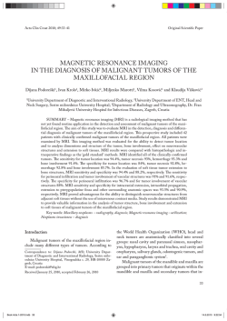
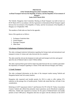
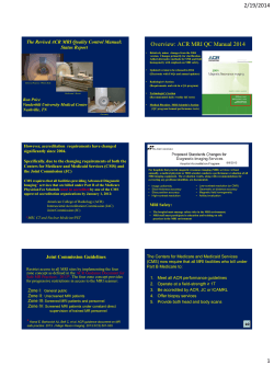
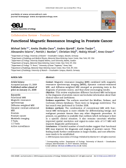
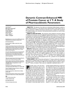
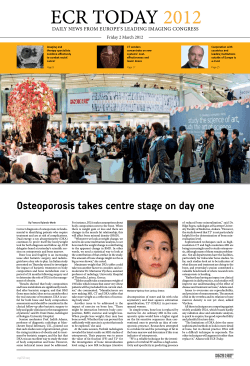
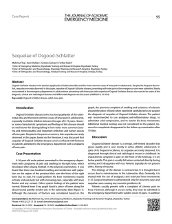
Journal of Contemporary Medicine 2014; 4(Supp): CR 29-31
Acu et al.
Case Report / Olgu Sunusu
Bilateral Kalkeneal Apofiziti Bulunan Bir Olgunun MRG Bulguları
MRI findings of a Case With Bilateral Calcaneal Apophysitis
Berat Acu1, Taylan Kara2, Safiye Topaloglu Asci3, Aysegul Altunkas3
1
Eskişehir Osmangazi
Üniversitesi Tıp Fakültesi
Radyoloji Anabilim Dalı.
2
Mersin Üniversitesi Tıp
Fakültesi Radyoloji
Anabilim Dalı
3
Tokat Devlet Hastanesi.
Corresponding Author:
Uzm. Dr. Ayşegül Altunkaş
Tokat Devlet Hastanesi
E-mail:
[email protected]
Tel: 90 555 8313895
Başvuru Tarihi/Received :
13-12-2013
Düzeltme Tarihi/Revised:
25-01-2014
Kabul Tarihi/Accepted:
10-02-2014
ÖZET
Kalkaneal apofizit (KA), kalkaneal epifizin ossifikasyonu öncesi dönemdeki
topuk ağrısının en sık nedenlerinden biridir. Bu sunumda bilateral
medial kalkaneal ağrıyla gelen 9 yaşındaki erkek hastada saptanan kalkaneal
apofizitin MRG bulgularının vurgulanması amaçlandı. Bilateral topuk ağrısı şikayeti
olan 9 yaşında erkek hasta MR görüntüleme amacıyla bölümümüze yönlendirildi.
Çekilen bilateral ayak MR’ında her iki aşil tendonu yapışma yeri olan tuber
kalkaneide ödemi ve subkondral erozyonu düşündüren T1 ağırlıklı görüntülerde
azalmış sinyal intensitesi, T2 ağırlıklı yağ baskılı görüntülerde ise artmış sinyal
intensitesi saptandı. Bu bulgular ışığında tanı olarak bilateral kalkaneal apofizit
düşünüldü. Sever hastalığı veya KA aşil tendonunun traksiyonu nedeniyle
tekrarlayan mikrotravmaya bağlı olduğu düşünülen kalkaneal apofizin
inflamasyonudur. Sıklıkla 8-15 yaşları arasında, büyüme dönemi veya öncesinde
gözlenir. Hastaların çoğunda yeni bir spora başlamanın ertesinde görülür. Kızarıklık,
şişlik, cilt değişiklikleri gibi lokal anormallikler gözlenmez.
Kalkaneal apofize komşu metafizer bölgede gözlenen kemik erozyonu
kalkaneal apofizitin MR bulgusudur. Benzer sinyal değişiklikleri sekonder apofizyel
ossifikasyon merkezlerinde de görülebilir.
Anahtar Kelimeler: Kalkaneal apofizit, Sever Hastalığı, manyetik
rezonans görüntüleme, MRG
ABSTRACT
Calcaneal apophysitis (CA) is the most common cause of heel pain in patients
before the ossification of calcaneal epiphysis. We report magnetic resonance imaging
(MRI) findings of a case with bilateral CA. A 9 year-old male patient with bilateral
foot pain was referred to our department for MRI. On bilateral foot MRI, bilateral
decreased signal intensity on T1-weighted images, increased signal intensity on fat
suppressed T2-weighted images that represents edema, subchondral erosions located
tuber calcanei, insertion of Achilles tendon were detected. The diagnosis was
bilateral calcaneal apophysitis. Sever's disease or CA is an inflammation of the
calcaneal apophysis believed to be caused by repetitive microtrauma from the
traction of the Achilles tendon on the unossified apophysis. It frequently occurs
before or during the peak growth spurt and often shortly after a child begins a new
sport or season. It is usually seen among 8-15 age group. Patients typically have no
features such as swelling, skin changes, erythema, or other local abnormalities.
Osseous erosion at metaphysis of the neighboring to calcaneal apophysis is
the finding of CA on MRI. Similar signal difference may be seen at the secondary
apophysial ossification centers.
Key Words: Calcaneal apophysitis, Sever’s disease, magnetic resonance
imaging, MRI
CR-29
Çağdaş Tıp Dergisi 2014; 4(Supp): CR 29-31
Acu ve ark.
Introduction
Calcaneal apophysitis (CA) is the most
common cause of heel pain in patients before
the ossification of calcaneal epiphysis (1,2). CA
(also known as Sever’s disease) is an overuse
syndrome thought to be caused by repetitive
micro trauma due to increased traction of the
calcaneo-achilles apophysis (3). Calcaneal
apophysitis is reported as a self limiting
condition (4), usually presenting between the
ages of 8–15 years (1,5), but has been observed
in children as young as six. The classical case is
usually a preadolescent boy with chronic heel
pain. Pain is increased with activity. We report
magnetic resonance imaging (MRI) findings of
a case with bilateral CA.
Figure 2 Sagittal fat-suppressed fast spin-echo T2-weighted
image of right ankle shows diffuse hypointense signal intensity of
the calcaneal epiphysis and milimetric subchondral bone erosions
at the tuber calcanei,the insertion of Achilles tendon
Case Report
A 9 year-old male patient was presented
with bilateral foot pain at the medial calcaneal
region. He was referred to our department for
bilateral foot MRI exam. On bilateral foot MRI,
bilateral decreased signal intensity in T1weighted images, increased signal intensity in
fat suppressed T2-weighted and proton density
images that represents edema, subchondral
erosions located tuber calcanei, insertion of
Achilles tendon were detected. The diagnosis
was bilateral CA.
The treatment of the patient was regulated as
activity modification and oral parasetamol for
three weeks. (Figure 1-4) The patient returned
to sport activities after two months.
Figure 1 Sagittal T1 weighted image of right ankle shows diffuse
hypointense signal
Figure 3 Sagittal T1 weighted image of left ankle shows diffuse
hypointense signal intensity
Figure 4 Sagittal fat-suppressed fast spin-echo T2-weighted
image of left ankle shows hyperintens signal intensity related to
bone marrow edema and subchondral bone erosions at the tuber
calcanei,the insertion of Achilles tendon.
CR-30
Journal of Contemporary Medicine 2014; 4(Supp): CR 29-31
Discussion
Sever's disease or CA is a common but
frequently undiagnosed source of heel pain in
young athletes. It is an inflammation of the
calcaneal apophysis believed to be caused by
repetitive microtrauma from the traction of the
Achilles tendon on the unossified apophysis.
The pain related to this inflammation is though
to cease after fusion of the calcaneus (3).
However, no studies have yet reported the
incidence or prevalence of this condition in the
general population. It frequently occurs before
or during the peak growth spurt and often
shortly after a child begins a new sport or
season (1).
It is usually seen among 8-15 age group.
Patients typically have no features such as
swelling, skin changes, erythema, or other local
abnormalities (2). The patient that we report
was 9 year old as in the literature age group.
Sever's disease is usually a self limited
disease, most of them usually resolves in two
weeks to two months after the conservative
treatment such as ice application, rest, heel lifts,
stretching and strengthening exercises, and
nonsteroidal anti-inflammatory drugs in more
severe cases (6).
In a child with heel pain, the differential
diagnosis may include Achilles tendonitis,
retrocalcaneal
bursitis,
calcaneal
stress
fractures, calcaneal cysts, osteomyelitis, and
plantar fasciitis (7).
Plain
radiographs
do
not
reveal
characteristic features of
Sever disease.
Ossification irregularities with sclerosis and
fragmentation within the apophysis are normal
features of a developing calcaneus. The lateral
calcaneal view and axial (Harris) view are taken
only when the history is not classic, when the
patient has night pain, swelling, exquisite
tenderness, or when a diagnosis other than
Sever disease is suspected (7). An MRI can
show areas of bone edema and hemorrhage, or
a bone bruise, within the metaphyseal bone of
the calcaneus (5). Our case had same MRI
findings like edema, subchondral erosions
located tuber calcanei.
Acu et al.
Osseous erosion at metaphysis of the
neighboring to calcaneal apophysis is the
finding of CA on MRI. Similar signal
difference may be seen at the secondary
apophysial ossification centers.
Secondary ossification center of calcaneus
typically appears in girls by age 6, and in boys
by age 8 (8).
After the treatment disappearing of
metaphysial and apophyseal findings on MRI
was reported in the literature (5).
Severe’s disease should be kept in mind as a
usual diagnosis among the late child-adolescent
age.
References
1. Hendrix CL. Calcaneal apophysitis (Sever
disease). Clin Podiatr Med Surg. 2005;22:55-62.
2. Micheli LJ, Ireland LM. Prevention and
management of calcaneal apophysitis in children; an
overuse syndrome. J Pediatr Orthop 1987;7:34–38.
3. Sever JW: Apophysitis of the os calcis. NY
Med J 1912, 95:1025.
4. Orava S, Virtanen K: Osteochondroses in
athletes. Br J Sports Med 1982,16:161–168.
5. Ogden JA, Ganey TM, Hill JD, Jaakkola JI:
Sever’s Injury: a stress fracture of the immature
calcaneal metaphysis. J Pediatr Orthop 2004,
24:488–492
6. Madden CC, Mellion MB. Sever's disease and
other causes of heel pain in adolescents. Am Fam
Physician. 1996;54(6):1995-2000.
7. Morrissy RT and Weinstein SL: Lovell and
Winter’s Pediatric Orthopedics Philadelphia:
LippincottWilliams &Wilkins; 52001, 1206.
8. Ross SE, Caffey J. Ossification of the
calcaneal apophysis in healthy children. Stanford
Med Bull 1957;15:224–226.
© Copyright 2026 Paperzz