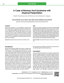
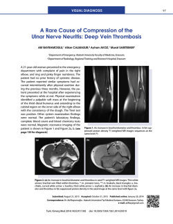
Turkish Journal of Medical Sciences
Turk J Med Sci
(2014) 44: 484-489
© TÜBİTAK
doi:10.3906/sag-1302-145
http://journals.tubitak.gov.tr/medical/
Research Article
An anatomical study on the facial nerve trunk in fetus cadavers
1
1
2,
2
2
Ahmet KALAYCIOĞLU , Gülay YEGİNOĞLU , Canan ERTEMOĞLU ÖKSÜZ *, Özlem UZUN , Şahi Nur KALKIŞIM
1
Department of Anatomy, Faculty of Medicine, Karadeniz Technical University, Trabzon, Turkey
2
Vocational School of Health Science, Karadeniz Technical University, Trabzon, Turkey
Received: 28.02.2013
Accepted: 13.06.2013
Published Online: 31.03.2014
Printed: 30.04.2014
Background/aim: Facial nerve paralysis is the most worrying parotid surgery complication. Little attention has been paid to the facial
nerve trunk, especially in children. Pathological variations, tumors, and anatomic variations of the facial nerve may cause parotid and
facial nerve surgery difficulties. This study describes the facial nerve trunk.
Materials and methods: We measured the facial nerve trunk from 8 female and 8 male fetus cadavers aged 21.0 to 35.5 gestational
weeks. The locations and positions of the trunks were described. The length of the facial nerve trunk (K) was measured and bifurcation
and trifurcation of the trunk was examined. Values were analyzed separately for right and left sides and for male and female fetuses.
Results: The most common facial nerve trunk type was bifurcation (81.25%), followed by trifurcation (18.75%). K was 11.59 ± 2.80 mm
in the total fetuses. There was no significant difference between K measurements.
Conclusion: Facial nerve injury during parotid surgery is a main cause of pediatric facial paralysis. There are few previous descriptions
of the facial trunk in children. K must be accurately known in any surgical procedure planned for the area. The main furcation of the
facial nerve should also receive special attention.
Key words: Facial nerve, fetus, morphometry, anatomy
1. Introduction
Facial nerve paralysis is a common complication of parotid
surgery and one that causes particular anxiety. The facial
nerve is a particularly important structure encountered
during surgical extraction of parotid gland tumors, not
least since these approximate the nerve very closely.
The facial nerve emerges from the facial canal via the
stylomastoid foramen. It then passes anteriorly to enter
the parenchyma of the parotid gland. After crossing the
styloid process, retromandibular vein, and external carotid
artery, it divides into 5 terminal branches behind the neck
of the mandible (1–3). The segment of the facial nerve
between its point of emergence from the skull through
the stylomastoid foramen and its furcation into upper and
lower branches at the parotid area is known as the main
trunk of the facial nerve (2,4).
Holt referred to the difficult problem of dissecting the
facial nerve trunk through the surrounding soft tissues (5).
The dissection of this segment becomes more difficult and
risky when the normal anatomy is deformed by a tumor, a
scar from previous surgery, or other pathological processes
(4). In infants, the facial nerve trunk is located in a more
*Correspondence: [email protected]
484
superficial plane and is liable to injury during surgical
incision and trauma to the retromandibular area (6). The
processus mastoideus is not yet developed in infants, and
the facial nerve arising from the stylomastoid foramen
is very close to the surface. The facial nerve trunk may
therefore be vulnerable when using the forceps during
difficult births (7). Accurate knowledge of the anatomy of
the facial nerve trunk is therefore essential for performing
various surgical procedures on the mastoid process and
parotid gland, for approaches to the cranial base, and for
surgery aimed at facial nerve repair (4,8).
Preservation of the facial nerve during parotid gland
surgery depends upon its being exposed and located
without suffering damage. An accurate knowledge of the
anatomy of the nerve and considerable perioperative care
are essential if trauma is to be avoided. The surgeon must
be acquainted with a range of techniques, since anatomical
variations may make any specific approach difficult (9,10).
The facial nerve trunk being dissected and manipulated
between the exit from the cranial base through the
stylomastoid foramen and its furcation is a crucial stage
in a number of otological, plastic, and neurosurgical
KALAYCIOĞLU et al. / Turk J Med Sci
procedures (4). Understanding the anatomy of the facial
nerve trunk is essential for performing any surgical
procedure in the region.
This study was intended to describe the furcation types
of the facial nerve trunk and to measure K values in infants
and premature babies, whose facial trunks can be easily
injured since these lie close to the surface. We also aimed
to compare the morphometric measurements obtained.
2. Materials and methods
The study was performed at the Karadeniz Technical
University (KTU) Department of Anatomy, Trabzon,
Turkey. Sixteen fetus cadavers (8 females, 8 males) aged
between 21.0 and 35.5 gestational weeks (gw) by foot
length with no visible external abnormalities and preserved
in 10% formaldehyde were used (11).
Measurements were performed in 32 sides of faces
(males: 8 right, 8 left; females: 8 right, 8 left). Fetuses with
no external pathologies or abnormalities were obtained
from the Trabzon Maternity and Children’s Hospital
between 1992 and 1998. Written consent from the families
and the approval of the Ethics Committee of the KTU
Faculty of Medicine were obtained prior to the start of the
study.
Dissections were performed with cadavers in a
semilateral position. A skin incision was made starting
in front of the ear and extending downward behind the
lobulus auriculae and further down to the neck along
the anterior border of the sternocleidomastoid muscle.
The facial skin and subcutaneous tissue were removed
and the platysma muscle was divided and retracted. The
sternocleidomastoid muscle was identified and retracted
posterolaterally.
The entire trunk of the facial nerve through the
dense soft tissue was identified and traced back to the
stylomastoid foramen. The facial nerve trunk was identified
at its emergence from the stylomastoid foramen and was
followed caudally to its furcation. In every dissection,
the entire trunk of the facial nerve was exposed from its
emergence at the stylomastoid foramen and distally as far
as the main furcation (Figures 1 and 2). K was measured
between its emergence from the skull through the
stylomastoid foramen and its furcation in the parotid area.
The main trunk of the facial nerve was then traced forward
to its furcation and examined in order to determine biand trifurcation of the facial nerve trunk. K measurement
values were recorded as male/female and right/left, and
averages were calculated. Following dissections, the facial
nerves were photographed and patterns were drawn.
Measurements were performed using digital calipers,
sensitive to 0.05 mm. Because the facial nerve trunk has
a small arc, it could not be measured directly with the
digital calipers. Measurement was performed 5 times with
surgical silk thread, and these threads were measured 5
times with digital calipers. K was thus measured 25 times.
Means and standard deviations were calculated using SPSS
10.0. Normally distributed data were analyzed using the
independent t-test; data that were not normally distributed
were analyzed using the Mann–Whitney U test (12). P <
0.05 was considered statistically significant.
Figure 1. Photograph showing pattern of bifurcation of the facial
nerve trunk. K: Facial nerve trunk, K1: upper division of the facial
nerve trunk (truncus temporofacialis), K2: lower division of the
facial nerve trunk (truncus cervicofacialis). *: Bifurcation point
of the facial nerve trunk. ì : The stylomastoid foramen.
Figure 2. Pattern of bifurcation of the facial nerve trunk. K:
Facial nerve trunk, K1: upper division of the facial nerve trunk
(truncus temporofacialis), K2: lower division of the facial nerve
trunk (truncus cervicofacialis). *: Bifurcation point of the facial
nerve trunk. ì : Stylomastoid foramen.
485
KALAYCIOĞLU et al. / Turk J Med Sci
3. Results
Measurements were performed on 32 sides of faces (males: 8
right, 8 left; females: 8 right, 8 left) of 16 fetuses (8 males, 8
females) calculated at 21.0 to 35.5 gw according to foot length.
Fetuses had a mean age of 29.80 ± 4.29 gw, with 30.78
± 3.59 gw for females and 28.33 ± 5.16 gw for males. No
difference was determined between fetus ages by sex (P =
0.30). Male and female fetus groups were similar in terms
of intrauterine age.
Average K in all fetuses irrespective of sex was 11.59 ±
2.80 mm. K values measured in this and earlier studies are
presented in Table 1.
Average K was 12.38 ± 3.05 mm for female fetuses
and 10.80 ± 2.37 mm for males. There was no difference
between female and male fetus K values (P = 0.11, Table
2). K was 11.70 ± 2.93 mm for the right and 11.48 ±
2.76 mm for the left in all fetuses, irrespective of sex. No
difference was determined between right and left K values
(P = 0.83, Table 2). Furthermore, K values were similar in
comparisons of female fetus right and left sides, male fetus
right and left sides, and right and left sides of different
sexes (P = 0.75, P = 0.79, P = 0.38, P = 0.24, respectively,
Table 3).
Examination of facial nerve trunk furcation types in all
fetuses, irrespective of sex, revealed that 81.25% of fetuses
were of the bifurcation type (2 subtrunks: upper and
lower) and 18.75 % of the trifurcation type (3 subtrunks:
upper, lower, and middle). These were also analyzed
according to sex and side (Table 4). Bifurcation type was
the most common in all individuals. The bifurcation and
trifurcation types of the facial nerve trunks examined in
this and previous studies are presented in Table 5.
Table 1. Length of the facial nerve trunk according to various authors.
Author
Length (mm)
Dargent and Duroux, 1946 (14)
13
Kempe, 1970 (21)
14–26
Proctor, 1984 (22)
15
May, 1986 (23)
20
Holt, 1996 (5)
21
Ekinci, 1999 (15)
9
Salame et al., 2002 (4)
16.44
Cannon et al., 2004 (16)
9.38
Kwak et al., 2004 (1)
13
Present study
11.59
Table 2. Measurement of K performed for facial nerve means, standard deviations, and statistical comparison of right and left sides,
females, and males in total subjects.
Measurement n = 16 (Mean ± SD)
Right sides
Left sides
P1
Female
Male
P2
K
11.70 ± 2.93
11.48 ± 2.76
0.840
12.38 ± 3.05
10.80 ± 2.37
0.110
P 1: for total right vs. total left; P 2: for total female vs. total male.
Table 3. Measurement of K performed for facial nerve means, standard deviations, and statistical comparison of right side and left
sides in females and males.
Right sides n = 8 (mean ± SD)
Left sides n = 8 (mean ± SD)
P-values
Female n = 8 (mean ± SD)
12.42 ± 3.20
12.34 ± 3.10
P 3 = 0.753
Male n = 8 (mean ± SD)
10.98 ± 2.63
10.62 ± 2.23
P 4 = 0.793
P-values
P 5 = 0.382
P 6 = 0.248
P 3: for female right vs. left; P 4: for male right vs. left; P 5: female right vs. male right; P 6: female left vs. male left.
486
KALAYCIOĞLU et al. / Turk J Med Sci
Table 4. Incidence of furcation types of the facial nerve trunk.
Type
Total %
Female %
Male %
Total
Right
Left
Total
Right
Left
Total
Right
Left
Bifurcation
81.25
37.50
43.75
50.00
25.00
25.00
31.25
12.50
18.75
Trifurcation
18.75
12.50
6.25
0
0
0
18.75
12.50
6.25
Table 5. Bifurcation and trifurcation of the facial nerve trunk according to various authors.
Bifurcation
Trifurcation
%
%
Davis et al., 1956 (18)
100
-
Park and Lee, 1977 (19)
95.6
4.4
Katz and Catalano, 1987 (24)
100
-
Kopuz et al., 1994 (20)
82
18
Ekinci, 1999 (15)
81.4
18.6
Salame et al., 2002 (4)
97.8
2.2
Tsai and Hsu, 2002 (25)
100
-
Kwak et al., 2004 (1)
86.7
13.3
Present study
81.3
18.8
Author
4. Discussion
The facial nerve trunk is the part of the facial nerve between
where it emerges from the skull through the stylomastoid
foramen and the point of furcation at the parotid area
(4). This segment of the facial nerve is located in an area
of complex anatomy that must be fully understood to
prevent iatrogenic damage to the facial nerve. Therefore,
understanding the anatomy of the facial nerve trunk is
essential for performing any surgical procedure in this
region.
Parotidectomy is the standard operation for parotid
tumors. The main trunk of the facial nerve is frequently
dissected and exposed during parotidectomy and other
surgical procedures. Injury to the facial nerve causes
considerable fear and anxiety. Such injury during parotid
surgery is also one of the most widespread causes of facial
paralysis in children (6). The key to safe parotidectomy
therefore lies in correct identification and safeguarding of
the facial nerve trunk (13).
Our scan of the literature showed that the facial
nerve trunk between the stylomastoid foramen and the
main furcation of the nerve has attracted little attention
to date. Salame et al. emphasized the importance of the
length of the facial nerve trunk, since a segment needs to
be sufficiently long to permit anastomosis with the fewest
possible manipulations, and neither too tense nor too
loose (4).
In a study published in 1946, Dargent and Duroux
examined 27 specimens and determined a K value of 1.3
cm (14). Ekinci investigated the facial nerves on 27 sides
of 14 cadavers aged 0 to 5 and determined a mean K value
of 0.9 cm (0.6–1.2 cm) (15).
In a study published in 2002, Salame et al. examined
the facial nerve trunk in 46 specimens from its emergence
through the stylomastoid foramen to its furcation and
reported a K value of 16.44 ± 3.20 mm (12.20–18.68 mm)
(4). Kwak et al. investigated the facial nerve trunk in 30
subjects, with K measured at 13.0 ± 2.8 mm (1). These 2
studies examined adult cadavers.
In a study published in 2004, Cannon et al. investigated
the facial nerve trunk in 79 patients, reporting a K value of
9.38 mm (16).
We also measured K between where the facial nerve
emerges from the skull through the stylomastoid foramen
and its furcation at the parotid area. Our groups were
identical in measurements made, irrespective of sex,
among all fetuses. Our results were in agreement with those
of other studies. All measured values were compatible with
those of previous research (Tables 1–3).
487
KALAYCIOĞLU et al. / Turk J Med Sci
Comparison of female and male fetuses, right and left
sides, female fetus right and left sides, male fetus right and
left sides, and right and left sides of different sexes revealed
similar K values. No statistically significant difference was
determined among any of these pairs (P = 0.11, P = 0.83, P
= 0.75, P = 0.79, P = 0.38, and P = 0.24, respectively).
After emerging through the stylomastoid foramen, the
main trunk of the facial nerve passes through the parotid
gland, where it branches to form the parotid plexus. The
most common branching pattern is a bifurcation of the
trunk (17).
A study published in 1956 investigated 350 specimens
and reported on the bifurcation of the main facial nerve
trunk (18). Park and Lee examined 111 facial nerves and
determined a pattern of trifurcation of the main facial
nerve trunk in 4.4% of cases (19). Kopuz et al. investigated
the facial nerve in the parotid gland in 50 specimens and
reported a trifurcation of the main facial nerve trunk
in 9 (18%) cases (20). Salame et al. identified 1 case of
trifurcation out of 46 cases (4). Kwak et al. reported 4
cases (13.3%) of trifurcation of the facial nerve trunk (1).
These studies all examined adult subjects. In 1999, Ekinci
examined the facial nerves on 27 sides of 14 cadavers aged
0 to 5 and reported bifurcation in 22 cases and trifurcation
in 5 (15).
We examined the facial nerve trunk by bilaterally
dissecting the sides and investigated trunk bifurcation
and trifurcation. We observed bifurcation in 26 out of
32 dissections (81.25%) and trifurcation in the other 6
(18.75%). In addition, in the trifurcation cases, the middle
divisions originated from somewhere between the upper
and lower divisions and connected in various patterns
with the other 2 main divisions.
In conclusion, understanding the anatomy of the
facial nerve trunk is essential for performing any surgical
procedure in the region. We are firmly of the opinion that
understanding the morphometric measurements of the
facial nerve trunk will shed light on all kinds of surgical
and clinical evaluation concerning the facial nerve and
prevent injury of the facial nerve, especially in children.
The implications of a more detailed facial nerve
anatomy are applicable to complex approaches to facial
nerve surgery. Therefore, a detailed knowledge of all
aspects of the anatomy of the facial nerve trunk is essential
for careful dissection and preservation of the facial nerve.
References
1.
Kwak HH, Park HD, Youn KH, Hu KS, Koh KS, Han SH,
Kim HJ. Branching patterns of the facial nerve and its
communication with the auriculotemporal nerve. Surg Radiol
Anat 2004; 26: 494–500.
2. Pather N, Osman M. Landmarks of the facial nerve:
implications for parotidectomy. Surg Radiol Anat 2006; 28:
170–175.
3.
Keskinöz İ. [The stylomastoid artery and the rarely seen
variations in parotid surgery]. Fırat Tıp Dergisi 2010; 15: 99–
100 (in Turkish with English abstract).
4. Salame K, Ouaknine GER, Arensburg B, Rochkind S.
Microsurgical anatomy of the facial nerve trunk. Clin Anat
2002; 15: 93–99.
10. Şekercioğlu N. [Problems and solutions involving the facial
nerve in parotid gland surgery]. Türkiye Klinikleri J Surg Med
Sci 2007; 3: 37–41 (in Turkish with English abstract).
11. Mercer BM, Sklar S, Shariatmadar A, Gillieson MS, D’Alton
ME. Fetal foot length as a predictor of gestational age. Am J
Obstet Gynecol 1987; 156: 350–355.
12. Hayran M, Özdemir O. Bilgisayar İstatistik ve Tıp. 2nd ed.
Ankara, Turkey: Hekimler Yayın Birliği; 1996 (in Turkish).
13. Wong DSY. Surface landmarks of the facial nerve trunk: a
prospective measurement study. ANZ J Surg 2001; 71: 753–
756.
14. Dargent M, Duroux P. Donnees anatomiques concernant la
morphologie et certains rapports du facial intra-parotidean.
Presse Med 1946; 54: 523–524 (in French).
5.
Holt JJ. The stylomastoid area: anatomic-histologic study and
surgical approach. Laryngoscope 1996; 106: 396–400.
15. Ekinci N. A study on the branching pattern of the facial nerve
of children. Acta Anat Nippon 1999; 74: 447–450.
6.
Farrior JB, Santini H. Facial nerve identification in children.
Otolaryngol Head Neck Surg 1985; 93: 173–176.
7.
Snell RS. Clinical Anatomy for Medical Students. 5th ed.
Boston, MA, USA: Little, Brown and Company; 1995.
16. Cannon CR, Replogle WH, Schenk MP. Facial nerve in
parotidectomy: a topographical analysis. Laryngoscope 2004;
114: 2034–2037.
8.
Aslan A. [Anatomy and physiology of the facial nerve]. Türkiye
Klinikleri J E.N.T-Special Topics 2008; 1: 1–7 (in Turkish with
English abstract).
9.
De Ru JA, Van Benthem PPG, Bleys RLAW, Lubsen H, Hordijk
G. Landmarks for parotid gland surgery. J Laryngol Otol 2001;
115: 122–125.
488
17. Gardetto A, Kovacs P, Piegger J, Rainer C, Meirer R, PizaKatzer H. Direct coaptation of extensive facial nerve defects
after removal of the superficial part of the parotid gland: an
anatomic study. Head Neck 2002; 24: 1047–1053.
18. Davis RA, Anson BJ, Budinger JM, Kurth LRE. Surgical
anatomy of the facial nerve and parotid gland based upon a
study of 350 cervicofacial halves. Surg Gynecol Obstet 1956;
102: 385–412.
KALAYCIOĞLU et al. / Turk J Med Sci
19. Park IY, Lee ME. A morphological study of the parotid gland
and the peripheral branches of the facial nerve in Koreans.
Yonsei Med J 1977; 18: 45–51.
23. May M. Anatomy of the facial nerve for clinician. In: May M,
editor. The Facial Nerve. New York, NY, USA: Thieme Inc.;
1986. pp. 21–62.
20. Kopuz C, Turgut S, Yavuz S, Ilgi S. Distribution of facial nerve
in parotid gland: analysis of 50 cases. Okajimas Folia Anat Jpn
1994; 70: 295–300.
24. Katz AD, Catalano P. The clinical significance of the various
anastomotic branches of the facial nerve. Arch Otolaryngol
Head Neck Surg 1987; 113: 959–962.
21. Kempe LG. Operative Neurosurgery. Vol. 2. Berlin, Germany:
Springer-Verlag; 1970.
25. Tsai SC, Hsu H. Parotid neoplasms: diagnosis, treatment, and
intraparotid facial nerve anatomy. J Laryngol Otol 2002; 116:
359–65.
22. Proctor B. The extratemporal facial nerve. Otolaryngol Head
Neck Surg 1984; 92: 537–545.
489
© Copyright 2026 Paperzz