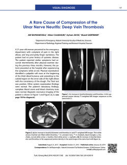
CA S E R EP O RT
Swallowed a needle stuck in heart
Mustafa Yolcu, M.D.,1 Ahmey Aydın, M.D.,2 Ali Fuat Korkmaz, M.D.,1
Özgür Dağ, M.D.,2 Emrah İpek, M.D.,1 Bilgehan Erkut, M.D.2
1
Department of Cardiology, Erzurum Region Training and Research Hospital, Erzurum;
2
Department of Cardiovascular Surgery, Erzurum Region Training and Research Hospital, Erzurum
ABSTRACT
Cardiac tamponade (CT) is a clinical entity characterized by hemodynamic insufficiency resulting from increased intrapericardial pressure due to accumulation of contents such as serous fluid, blood, and pus. CT is a treatable cause of cardiogenic shock, which can be
fatal unless diagnosed promptly. Dyspnea, chest pain, hypotension, tachycardia, pulsus paradoxus, raised jugular venous pressure, muffled
heart sounds, decreased electrocardiographic voltage, and enlarged cardiac silhouette on chest X-ray are the major clinical signs in
CT. Idiopathic or viral pericardititis, iatrogenic trauma during percutaneous coronary interventions or coronary artery bypass grafting, external trauma, malignancies, acute or chronic kidney disease, collagen vascular diseases, tuberculosis, radiation on the chest wall,
hypothyroidism and aortic dissection are the etiologic factors. Herein, we present a case of surgically treated CT, which was diagnosed
in the third day of ingestion of a sewing needle.
Key words: Cardiac tamponade; esophagus; needle.
INTRODUCTION
Cardiac tamponade (CT) is a clinical entity characterized by
hemodynamic insufficiency resulting from increased intrapericardial pressure due to accumulation of contents such as serous fluid, blood, and pus.[1] Idiopathic or viral pericardititis,
iatrogenic trauma during percutaneous coronary interventions or coronary artery bypass grafting (CABG), external
trauma, malignancies, acute or chronic kidney disease, collagen
vascular diseases, tuberculosis, radiation on the chest wall, hypothyroidism, and aortic dissection are the etiologic factors.[2]
Herein, we present a case of surgically treated CT, which was
diagnosed in the third day of ingestion of a sewing needle.
CASE REPORT
A 35-year-old male patient was admitted to our emergency
clinic with the complaints of abdominal pain, progressive dysAddress for correspondence: Mustafa Yolcu, M.D.
Erzurum Bölge Eğitim ve Araştırma Hastanesi, Kardiyoloji Kliniği,
Çat Yolu, Erzurum, Turkey
Tel: +90 442 - 232 55 55 E-mail: [email protected]
Qucik Response Code
Ulus Travma Acil Cerrahi Derg
2014;20(4):308-310
doi: 10.5505/tjtes.2014.30049
Copyright 2014
TJTES
308
pnea and palpitations started after ingestion of a meal 3 days
ago. In his physical examination, the arterial blood pressure
was 80/40 mmHg, and he was tachycardic and tachypneic.
Cardiac auscultation revealed muffled heart sounds and
there were not any murmurs or pathologic sounds. There
were jugular venous distension and hepatomegaly. The electrocardiography (ECG) revealed sinus tachycardia and low
QRS voltage. In chest X-ray, the cardiothoracic ratio was
increased, and there was a metallic needle density over the
diaphragm (Fig. 1a).
In transthoracic echocardiography, a pericardial effusion with
3 cm width was detected surrounding the entire heart resulting in diastolic collapse of the right ventricle. The patient
was then examined under scopy, and a sewing needle with
4 cm length was shown to have a synchronized motion with
the heart. A thorax computer tomography was taken, and
the needle was demonstrated to have one tip in the distal
esophagus and the other tip in the pericardium next to the
left atrium (Fig. 1b).
An emergency surgery was performed. After median sternotomy, the pericardium was opened vertically, and cardiac
decompression was achieved. The fibrin and hematoma on
the heart was cleaned. When the heart was exposed and
lifted, a 4-cm long sewing needle which had its 2 cm portion
in the pericardium was extracted (Fig. 1c and d). The thorax
was closed after bleeding control. Then the patient was discharged in the 3rd day without any complications.
Ulus Travma Acil Cerrahi Derg, July 2014, Vol. 20, No. 4
Yolcu et al. Swallowed a needle stuck in heart
(a)
(b)
(c)
(d)
Figure 1. (a) Appearance of the needle on chest. (b) Computer tomography image of the needle. (c) The needle during
operation. (d) The needle after operation.
DISCUSSION
CT causes hemodynamic insufficiency due to compression
of heart chambers by a pericardial fluid leading increased
pericardial pressure.[1] CT is a treatable cause of cardiogenic shock which can be fatal unless diagnosed promptly.[1]
Dyspnea, chest pain, hypotension, tachycardia, pulsus paradoxus, raised jugular venous pressure, muffled heart sounds,
decreased ECG voltage, and enlarged cardiac silhouette on
chest X-ray are the major clinical signs in CT.[3] The percutaneous pericardiosynthesis, balloon pericardiotomy or
surgical drainage are the treatment options depending on
the clinical presentation.[3-5] Depending on the rate of effusion formation, CT can have rapid or slow progression. This
situation shows that tamponade is related mostly to the rate
of accumulation of fluid in the pericardium rather than the
quantity of effusion.
The mean right atrial filling pressure formed by normal systemic venous return is 6-8 mmHg.[6] During CT the rapid accumulation of fluid in non-elastic pericardial cavity prevents
the filling of the right atrium.[6] The intrapericardial pressure
first overcomes the right atrial pressure then the right ventricular pressure was overcome.[6] The cardiac output falls
depending on increased intrapericardial pressure over right
Ulus Travma Acil Cerrahi Derg, July 2014, Vol. 20, No. 4
heart pressures, which then causes clinical CT.[6] Depending
on the rate of effusion formation, CT can have rapid or slow
progression. This situation shows that tamponade is related
mostly to the rate of accumulation of fluid in the pericardium
rather than the quantity of effusion.
The right heart catheterization is diagnostic, but now-a-days
transthorasic echocardiography takes the role since it is noninvasive, rapid, and reliable.[7] The main echocardiographic
findings are compression of right heart chambers, dilatation
of the inferior vena cava without respiratory variation of its
diameter and increased respiratory variation of intracardiac
Doppler velocities.[1,2] Besides its diagnostic importance,
echocardiography is a useful guide during pericardiosynthesis.[1,2] Echo-guided pericardiosynthesis is a reliable, rapid and
well-tolerated procedure. A study in Mayo Clinic revealed
that it has 97% success and 4.7% total complication rate.[4]
However, Gumrukcuoglu et al. reported that it has 97% success and 10% total complication rate.[1]
In 1826, Larrey defined drainage procedure for pericardial
effusion through sub-xyphoidal region.[8] Emergency pericardiosynthesis can relieve CT, but surgical drainage may
be necessary, especially in tamponades after trauma, CABG
and percutaneous coronary interventions.[8] Surgical sub309
Yolcu et al. Swallowed a needle stuck in heart
xyphoidal window is a simple and reliable procedure and has
lesser mortality, complication, and recurrence rates.[8] In foreign body or trauma induced tamponades, as in our case, a
sternotomy is necessary most of the time in order to evaluate and treat the cardiac injury properly. Surgical approach
should be considered, especially in purulent, recurrent, and
malignant effusions and if a biopsy is needed for the diagnosis.[1] In a study in Mayo Clinic, 88 patients who experienced CT due to catheter-based procedures and underwent
pericardiosynthesis with guidance of echocardiography were
studied retrospectively and cardiac surgery after pericardiosynthesis was shown to be 18%.[4]
Olivotti et al.[8] reported an ingested metallic wire, which
caused CT passing into the pericardium through esophagus.
A few CT cases after ingestion of a metallic wire or needle which then passed through diaphragm, was previously
reported also. In a few case reports, it was stated that a
Kirschner wire left its location and reached the heart via
arterial route leading CT. Gevaert et al. reported a case in
which a 3-cm long sharp piece of crab caused CT passing
through the diaphragm.
Herein, we present a CT case of a sewing needle, which was
ingested and passed into the pericardium through the esophagus and lead tamponade by disruption of the right atrium.
As a result, pericardial effusion and tamponade have a lot of
reasons. It must be kept in mind that ingested metallic wires
or needles can pass into the pericardium through esophagus
or diaphragm and lead tamponade.
Conflict of interest: None declared.
REFERENCES
1. Gumrukcuoglu HA, Odabasi D, Akdag S, Ekim H. Management of
Cardiac Tamponade: A Comperative Study between Echo-Guided Pericardiocentesis and Surgery-A Report of 100 Patients. Cardiol Res Pract
2011;2011:197838.
2. Sagristà-Sauleda J, Mercé AS, Soler-Soler J. Diagnosis and management
of pericardial effusion. World J Cardiol 2011;3:135-43. CrossRef
3. Seferović PM, Ristić AD, Imazio M, Maksimović R, Simeunović D, Trinchero R, et al. Management strategies in pericardial emergencies. Herz
2006;31:891-900. CrossRef
4. Phadke G, Whaley-Connell A, Dalal P, Markley J, Rich A. Acute Cardiac
Tamponade: An Unusual Cause of Acute Renal Failure. Cardiorenal Med
2012;2:83-86. CrossRef
5. Spodick DH. Acute cardiac tamponade. N Engl J Med 2003;349:684-90.
6. Cuculi F, Newton JD, Banning AP, Prendergast BD. Resistant pericardial
tamponade. Circulation 2011;123:566-7. CrossRef
7. Tsang TS, Freeman WK, Barnes ME, Reeder GS, Packer DL, Seward
JB. Rescue echocardiographically guided pericardiocentesis for cardiac
perforation complicating catheter-based procedures. The Mayo Clinic
experience. J Am Coll Cardiol 1998;32:1345-50. CrossRef
8. Olivotti L, Succio G, Moshiri S, Nicolino A, Gravano M, Serafini G, et
al. Cardiac tamponade caused by a swallowed metallic wire. J Am Coll
Cardiol 2010;56:e27. CrossRef
OLGU SUNUMU - ÖZET
Yutulup kalbe saplanmış iğne
Dr. Mustafa Yolcu,1 Dr. Ahmey Aydın,2 Dr. Ali Fuat Korkmaz,1 Dr. Özgür Dağ,2 Dr. Emrah İpek,1 Dr. Bilgehan Erkut2
1
2
Erzurum Bölge Eğitim ve Araştırma Hastanesi, Kardiyoloji Kliniği, Erzurum;
Erzurum Bölge Eğitim ve Araştırma Hastanesi, Kardiyovasküler Cerrahi Kliniği, Erzurum
Kardiyak tamponat (KT) seröz sıvı, kan ve püğ gibi içeriklerin birikiminin yol açtığı intraperikardiyal basınç artışı sonucu hemodinamik yetersizlikle
karakterize klinik durumdur. KT acil olarak tedavi edilmediğinde ölümcül olabilen kardiyojenik şokun tedavi edilebilir bir nedenidir. Nefes darlığı,
göğüs ağrısı, hipotansiyon, taşikardi, pulsus paradoksus, artmış juguler venöz basınç, derinden gelen kalp sesi, azalmış elektrokardiyografik voltaj
ve akciğer grafisinde genişlemiş kardiyak silüet KT’nin majör bulgularıdır. İdiyopatik ya da viral perikardit, koroner baypas ya da perkütan koroner
girişim sırasında oluşabilen iyatrojenik travma, eksternal travma, kanserler, akut ya da kronik böbrek hastalığı, kollojen doku hastalıkları, tüberküloz,
göğüs duvarına radyasyon, hipotiroidi ve aort diseksiyonu etiyolojik faktörlerdir. Biz burada dikiş iğnesinin yutulmasının üçüncü gününde tanı konulan
ve cerrahi olarak tedavi edilen KT olgusunu sunduk.
Anahtar sözcükler: İğne; kardiyak tamponat; özofagus.
Ulus Travma Acil Cerrahi Derg 2014;20(4):308-310
310
doi: 10.5505/tjtes.2014.30049
Ulus Travma Acil Cerrahi Derg, July 2014, Vol. 20, No. 4
© Copyright 2026 Paperzz