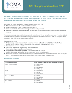
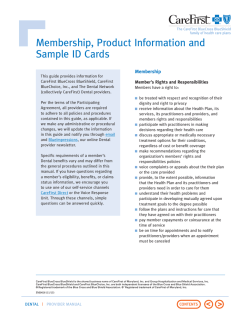
Acta Clin Croat 2011; 50:553-561 Review CONTACT ALLERGY IN THE MOUTH: DIVERSITY OF CLINICAL PRESENTATIONS AND DIAGNOSIS OF COMMON ALLERGENS RELEVANT TO DENTAL PRACTICE Andrijana Bakula, Liborija Lugović-Mihić, Mirna Šitum, Juraj Turčin and Ana Šinković University Department of Dermatovenereology, Sestre milosrdnice University Hospital Center, Zagreb, Croatia SUMMARY – Delayed-type hypersensitivity reaction or type IV allergic reaction can cause different oral manifestations. They can be localized or diffusely visible on oral mucosa and usually appear 24-72 hours after antigen input. The antigens that cause this type of reaction are mostly external, such as contact allergens (particularly metals) and drugs. It has been shown that the most common oral manifestations are cheilitis, gingivitis, stomatitis, perioral dermatitis, burning mouth syndrome, lichenoid reaction and orofacial granulomatosis. The most important part of diagnosis is the use of patch testing that indicates contact allergic reaction to an allergen. The results of patch testing have shown that the most common proven allergens are gold, nickel, mercury, palladium, cobalt, acrylate, etc. Although connection between specific clinical manifestations and positive patch test results was not always found, patch testing is necessary to prove contact hypersensitivity. Therefore, in patients with oral symptoms, allergic hypersensitivity to dental components has to be considered. Key words: Contact allergy; Mouth; Oral; Allergen; Patch test The Pathogenesis of Delayed-Type Hypersensitivity Reactions Delayed-type hypersensitivity reactions (type IV allergic reactions) are allergic immune reactions manifesting primarily through T cells (cellular immunity). Cellular immunity and delayed hypersensitivity are often considered to be the same reactions, where hypersensitivity is actually a reaction so strong that it causes tissue damage1. Delayed hypersensitivity can only occur in patients previously in contact with a specific antigen and thus having become sensitized1. Studies have found that the introduced antigen stimulates sensitized CD4+ T Correspondence to: Liborija Lugović-Mihić, MD, PhD, University Department of Dermatovenereology, Sestre milosrdnice University Hospital Center, Vinogradska c. 29, HR-10000 Zagreb, Croatia E-mail: [email protected] Received October 20, 2010, accepted November 11, 2011 Acta Clin Croat, Vol. 50, No. 2, 2011 cells to the secretion of different cytokines. These cytokines, TNF-α and TNF-β, induce the expression of adhesion molecules (E-selectin, ICAM-1, VCAM-1) on dermal endothelial cells of the blood vessels. This enables extravasations of different cells infiltrating surrounding tissues (in the beginning, these are mainly neutrophils, and later monocytes and macrophages). Macrophage accumulation is enhanced by cytokines, which are secreted by sensitized CD4- cells in contact with the antigen (IL-3, GM-CSF), and macrophage activation is stimulated by IFNγ. Lymphocyte cytokines cause an increased permeability of local capillaries, which contributes to edema. Enzymes from the macrophages contribute to tissue damage and necrosis. These damages can occur by the activity of lymphotoxins (TNFβ), excreted from sensitized CD4+ cells1. Cytotoxic CD8+ T cells may also participate in the delayed type of hypersensitivity reactions. 553 Andrijana Bakula et al. Contact allergy in the mouth: Common allergens relevant to dental practice Contact allergy is an important type of delayed hypersensitivity, which can develop after the skin or mucosa contact with certain substances. These are mainly substances having small molecular mass (picric acid, dinitrochlorobenzene, different herbal ingredients, cosmetics, some drugs, metals, and other substances), which exhibit behavior of haptens. After absorption into the epidermis, the substance is bound to proteins (carriers) and becomes immunogenic; then hypersensitivity occurs, manifested as erythema and edema of the skin, sometimes followed by vesicles that can occupy greater parts of the skin1. Studies have shown that patients who are more prone to such contact allergic reactions also suffer from atopy, which should be taken into consideration when testing such patients2. From the pathogenetic perspective, it has been proven that complexes of antigens and carriers enter Langerhans cells, which constitute the prevailing part of the presenting cells in the epidermis. Afterwards, Langerhans cells reach regional lymphatic nodes, where CD4+ T cells present the antigen together with their own MHC-II molecules, thus stimulating memory CD4+ T cells1. After repeated contact with the same antigen, Langerhans cells present it to memory CD4+ lymphocytes in the dermis, which are then activated. Activated lymphocytes secrete different cytokines. Thus, IFNγ causes ICAM-1 and MHC-II expression with epidermal keratinocytes and capillary endothelium cells, and they also stimulate keratinocytes to the secretion of cytokines that cause inflammatory reaction (IL-1, IL-6, GM-CSF). Nonspecific CD4+ T cells are also attracted and they are connecting to keratinocytes over ICAM-1 and MHC-II molecules. Afterwards, in this area, macrophages are also gathered through the action of lymphocyte cytokines (IFNγ, IL-3, TNFβ). The reaction is most visible after 48-72 hours, and after that period it gradually diminishes due to the contribution of PGE secreted by macrophages, keratinocytes and IL-101. There are also other forms of type IV allergic reactions, for example tuberculin reaction (Mantoux test) caused by different infections (bacteria, viruses, parasites, fungi), particularly in chronic infectious diseases, when granulomatous inflammation develops (such as tuberculosis and leprosy), some autoimmune 554 diseases (experimental allergic encephalitis), during transplant rejection and in tumor disease1. The Possible Oral Type IV Allergic Reactions Delayed-type hypersensitivity reaction or type IV allergic reaction can cause different oral manifestations. They manifest 24-72 hours after the antigen has been introduced and they can be localized or diffusely visible on oral mucosa3. Numerous oral manifestations result from this type of allergy. The antigens that cause it are most often external, such as metals and drugs3. Medicamentous allergic stomatitis is a type IV allergic reaction to drugs. Pathologic changes are diffusely visible on oral mucosa, usually affecting huge areas from 20 to 40 mm. They often begin as inflammation with edema, followed by huge confluent erosions with pseudo-membranes. They can manifest in the form of bullae, then they are similar to pemphigus or lichenoid reactions, thus being clinically hardly distinguishable from oral lichen ruber3. Fixed drug reaction (eruptio fixa) is a localized or fixed allergic reaction, specific to allergic reaction to drugs. It occurs on oral mucosa in the form of localized, sharply demarcated erosion, with thick pseudomembranes. Most often, they are solitary or, at the most affecting two regions in different parts of oral mucosa. Localized lesions are to be expected on the hard palate mucosa, dorsum of the tongue, or on the labia3. They always appear on the same spot, after repeated contact with the antigen. Stomatitis (cheilitis) venenata is a contact allergic reaction caused by different chemical and cosmetic substances, which cause inflammation of the labia and inflammation of the entire oral mucosa. They are manifested as inflammation with a highly pronounced edema, followed by small erosions, 0.5 mm in diameter, and usually appear in multiple forms. With chronic local irritation on the labia, exfoliative cheilitis with strong exudation can be expected3. Contact allergic stomatitis rarely occurs, but cases of contact allergy to different materials in the mouth are possible4-6. Here we primarily consider nickel sulfate, mercury-based products, gold, and others. Sometimes, reaction to cobalt chloride is found, which Acta Clin Croat, Vol. 50, No. 4, 2011 Andrijana Bakula et al. Contact allergy in the mouth: Common allergens relevant to dental practice currently is the most common allergen in children. Metals are often used in dental prosthetics, but oral manifestation of contact allergy is nevertheless rare4. When the reaction is caused by prosthetic material, we speak of prosthetic allergic stomatitis. This form rarely occurs as an expression of contact allergy to acrylate, denture furnish, metal denture alloys, and cobalt-chromium pastes for denture fixation. Thereby, pathologic changes occur on the locations where the prosthesis base comes in contact with the surrounding tissue, not only on the mucosa that is covered by the prosthesis base. Lesions of this stomatitis are found in the form of erythema, edema, vesicles, bullae, erosions and ulcerations3. Allergic reactions on oral mucosa can occur as a consequence of contact with composites, ethereal oils, silicon and polyester impression materials. Oral mucosa lesions are localized in the area of contact with problematic substance, and they can be polymorphic3. Diagnosis of the mentioned clinical forms of delayed type of allergic reactions is confirmed by laboratory diagnostic methods, and by attestation of cellular immunity and delayed-type hypersensitivity to contact allergens, which is most often done by epicutaneous or patch test5,6. Therapy is most often conducted by local administration of corticosteroid preparations, and if needed, by systemic administration of corticosteroids3. Granulomatous stomatitis and cheilitis is a rare disease, which is pathogenetically a type IV allergic reaction that involves connective tissue of oral mucosa. In the beginning, it is manifested as an acute edema of labia. Often only the lower lip is affected, or the edema is only unilaterally visible. Initially, lips are swollen but later they become firm and fibrous, with granules having an uneven appearance or can be spotted only by palpation. The color of the lips is violet-red, with signs and marks of peelings. When lesions are found only on the lips, it is called Miescher disease3. If granulomatous inflammatory lesions are present in other regions of the oral cavity, and also on the tongue, gingiva and buccal mucosa, then it is called Melkerson Rosenthal syndrome. Besides granulomatous inflammation of the mentioned areas of oral mucosa, fissured tongue may be present, as well as hyperplastic parodontitis profunda Acta Clin Croat, Vol. 50, No. 4, 2011 and facial nerve paresis, which may be transitory3. Diagnosis of these problems requires internist examination of the patient because of the possibility that granulomatous inflammation has also involved other organs. Also, due to similar clinical appearance, sarcoidosis should be excluded. Therapy includes systemic and intralesional administration of corticosteroids to reduce edema and granulomatous inflammation3. Geographic tongue or benign migratory glossitis is a disorder of unknown cause and pathogenesis, although genetics has been proposed as the causative factor. It is a common injury of oral mucosa in children. The disease is mainly asymptomatic, but in some cases burning sensations are reported. Some cases of geographic tongue are associated with psoriasis and atopy (genetically caused proclivity to allergic reactions with high values of IgE), which should be taken in consideration on making the diagnosis4. Reiter syndrome is the occurrence of arthritis, urethritis and conjunctivitis as an immune reaction to the infection found somewhere else in the body (Shigella, Salmonella, gonococci, Mycoplasma, Chlamydia, Yerssinia and Campylobacter)3. Stomatitis is present in 50% of patients. Oral lesions start as exulcerating red maculae on buccal mucosa, lips, tongue and gingiva, surrounded by white uneven margins3. The disease occurs with immunodeficiency, with lowered number of leukocytes (for example, in HIV infection). It is treated orally by local application of corticosteroids and antiseptics, systemic administration of corticosteroids, non-steroid anti-inflammatory drugs and immunosuppressants (methotrexate or azathioprine)3. Allergologic Skin Testing When suspecting allergy, thorough history and clinical examination should be taken, after which dermatologist can determine the testing that is necessary to perform. Allergologic testing is most often performed on the skin, where very small quantities of standardized solutions of purified allergens are applied in or upon the skin, and then local allergic reaction on the skin is observed and measured. Skin testing can be done by different methods, depending on the allergy type suspected. Prick method (prick test) and scratching of the skin (scratch test) are 555 Andrijana Bakula et al. Contact allergy in the mouth: Common allergens relevant to dental practice Fig. 1. Allergic stomatitis caused by acrylic resins23. used to prove and confirm early-type hypersensitivity, whereas testing of contact with the skin (patch or epicutaneous test) will prove type IV or delayed-type allergy reaction. Patch (epicutaneous) test is used to determine and identify type IV allergic reaction and contact hypersensitivity to different chemicals5,6. It is performed by applying allergic preparations onto patches, which are stuck to the skin of the back. The result of this test is read twice, at 48 h and 72 h. The procedure is painless and can be employed even in older schoolchildren. It is important to note that the patient should not be taking anti-allergic drugs and should be informed of that during examination. Epicutaneous test is performed by a patch with al- lergens from the standard series (standardized allergens) or targeted professional antigens. Allergens are applied onto the skin of the back in the interscapular area or exceptionally onto the volar side of the forearm. The allergen is applied onto healthy, previously disinfected (by benzine, ether or alcohol) skin in a non-toxic and prescribed (according to European standards) dose. About 0.02-0.03 g of allergen in petrolatum or other base is put onto filter paper (dimensions 1/1 cm). The patch is then covered with cellophane measuring 2x2 cm and afterwards all this is covered by 5x5 cm leucoplast tape. Reaction is measured at 48 h and 72 h, when positive reaction is observed in the place of each particular substance (erythema, light edema, or even formation of small vesicles, and others). This test is significant in the diagnosis of contact allergic dermatitis or stomatitis, and it is conducted for all kinds of dermatitis as a delayed response. In Croatia, the standard series contains 29 allergens. These are allergens found in daily life and at workplaces, for example, chromium, nickel, cobalt, mercury, ursol, formaldehyde, epoxy resins, charcoal tars, Peru balsam, mercapto compounds, thiuram compounds, paraben mix, thimerosal, and others. Targeted testing is designated exclusively by the specialist and thus applied to allergens according to profession, e.g., according to the sample of the material brought along. Allergens are made from samples taken from the suspected sources from the workplace. For example, in Croatia, there is commercial produc- Fig. 2. Cheilitis angularis . 24 Fig. 3. Allergic stomatitis caused by metal base removable denture25. 556 Acta Clin Croat, Vol. 50, No. 4, 2011 Andrijana Bakula et al. Contact allergy in the mouth: Common allergens relevant to dental practice tion of allergens for hairdressers. In Europe, there are several series of antigens for different kinds of occupations. When allergy to dental material is suspected, dermatologist conducts epicutaneous testing for certain substances, in cooperation with the dentist. Discussion During dental treatment, different materials are applied, which sometimes can do harm to patients and workers, thus it is necessary to act with caution during their use. Recently, the number of papers about contact allergy of oral mucosa to different foods, oral hygienic products and materials used in dentistry has increased7. However, there is no individual or specific clinical study of contact allergy to dental materials, thus this area has not been well investigated. It can be said that the spectrum of oral symptoms with such manifestations is very wide. Patients with contact allergy without clinically visible manifestations (lichenoid tissue lesions or oral ulcerations) can feel burning sensations or paresthesia. In these patients pain can occur, along with oral changes. Considering the fact that clinical manifestations sometimes do not correspond to current symptoms, therapy for such patients may pose a diagnostic and therapeutic challenge8. Based on researches and different papers about oral contact allergy and diseases, it has been shown that these patients are predominantly middle-aged women, particularly 50-60 years old. Comparing the incidence of these diseases in this age with other age and sex groups, it has been made clear that oral disease is truly more frequently present in middle-aged women, or they simply visit physicians more often and undergo patch testing more often. Clinical manifestations of contact allergic dermatitis to dental materials can be different. In 2006, Khamaysi et al. conducted a research in Israel of allergens in dental practice related to contact reactions7. Patch testing of 134 patients aged 20-80 showed cheilitis and perioral dermatitis to be the most common oral manifestations (25.6%), followed by burning mouth syndrome (15.7%), lichenoid reaction (14%) and orofacial granulomatosis (10.7%). The most common contact allergens were gold sodium thiosulfate (14%), nickel sulfate (13.2%), mercury (9.9%), pallaActa Clin Croat, Vol. 50, No. 4, 2011 dium-chloride (7.4%), cobalt-chloride (5%) and 2-hydroxyethyl methacrylate (5.8%)7. It can easily be noticed that positive reactions to metals are common or frequent in all clinical examinations, however, a specific link between a certain clinical examination and a particular antigen could not be found. Several studies have shown connection between oral lichen planus and allergy to mercury, but results vary among different studies. According to Khamaysi et al., allergy to mercury is not an important factor contributing to the pathogenesis of oral lichenoid reactions. Although they found that 35% of patients with lichenoid reaction were positive on mercury test, only one patient was truly reacting to mercury7. On the other hand, testing was useful with positive reaction to mercury in some patients with orofacial granulomatosis, where removal of amalgam fillings resulted in quick recovery. Although orofacial granulomatosis was assumed to be a reactive process with the occurrence of contact hypersensitivity to food components, contact allergic reactions to gold in gold dental crowns and reactions to mercury in fillings were actually demonstrated and confirmed7,9-11. There are controversies with regard to the role of contact allergies in the pathogenesis of burning mouth syndrome. In 42% of patients with burning mouth syndrome, positive reactions were demonstrated on testing to gold, nickel, mercury and palladium, however, there was no correlation between positive patch tests and exposure to allergens7. Cheilitis is a common clinical problem often connected to licking of the lips, although in many cases the cause is actually unknown. Freeman and Stephenes describe 75 cases of resistant cheilitis, 25% of which were diagnosed as contact dermatitis12. The most common allergens connected to cheilitis were discovered in drugs, lipsticks, suntan creams and toothpastes. While dental patients exhibit different symptoms, mainly on oral mucosa, dental staff (dentists as well as assistants) commonly suffer from dermatitis localized on hands. While the most common cause of contact allergies are metals (such as amalgam and gold) and glues, in dental workers the causes are metals, rubber, antimicrobial drugs, preservatives and methacrylates7. Studies in dental workers showed the incidence of skin diseases to range from 30% to 50%, and in the 1990s it was tripled13,14. In their study of contact al557 Andrijana Bakula et al. Contact allergy in the mouth: Common allergens relevant to dental practice lergy, Khamaysi et al. report on 14.9% of dental workers having dermatitis on their hands7. Examination of dental workers suspected to have skin disease revealed that all of them suffered from contact dermatitis on their hands. Only 39% of these patients tested positive, mainly for metals like nickel, gold, palladium and cobalt7. Khamaysi et al. showed dental workers with hand dermatitis to have a relatively high incidence of allergies to metals7. It is possible that dental instruments are not the only source of activating skin lesions, but they were the key factor for dermatitis to occur on the hands. Lee et al. also found similar re- Table 1. Most common allergens for specific oral diseases8 Disease 558 Allergen Patients with positive reaction (%) Burning mouth syndrome Potassium dicyanoaurate Nickel sulfate hexahydrate Gold sodium thiosulfate Palladium chloride Aroma mixtures 16.4 12.3 10.9 9.3 8.3 Lichenoid reaction of tissue Potassium dicyanoaurate Aroma mixtures Gold sodium thiosulfate Nickel sulfate hexahydrate Peru balsam Cheilitis Aroma mixtures Gold sodium thiosulfate Dodecyl gallate Cain III mixture Benzoic acid Stomatitis Mercury Peru balsam Gold sodium thiosulfate Nickel sulfate hexahydrate Dodecyl gallate 14.3 12.5 11.5 11.5 9.5 Gingivitis Potassium dicyanoaurate Nickel sulfate hexahydrate Palladium chloride Beryllium sulfate tetrahydrate Gold sodium thiosulfate 34.8 33.3 29.2 20.8 17.4 Orofacial granulomatosis Nickel sulfate hexahydrate Benzoyl peroxide Dodecyl gallate Gold sodium thiosulfate 15.4 7.7 7.7 7.7 Perioral dermatitis Cobalt-chloride Gold sodium thiosulfate Balsam of Peru Nickel sulfate hexahydrate 60 25 20 20 Recurrent stomatitis aphthosa Vanillin 28 17.1 15.1 13.8 11.9 13 6.8 6.1 5.6 4 33.3 Acta Clin Croat, Vol. 50, No. 4, 2011 Andrijana Bakula et al. Contact allergy in the mouth: Common allergens relevant to dental practice sults in 49 dental technicians in Korea15. On the other hand, information from Europe and USA suggests that the most common cause of allergies are gloves and dental restorative plastic material, where gloves cause both early and delayed type of allergic reactions16,17. Although a relatively large number of patients tested positive for gold and nickel, there was no connection of either of these allergens with the skin or mucosa lesions, so the activation most probably originated from some other sources7. Half of the patients mentioned in the study by Khamaysi et al. had positive reaction to palladium chloride at the same time, and one-third of them to cobalt-chloride, probably due to allergy to nickel, and not due to the activation by dental products7. Significant reactions to mercury are found in dental workers, unlike reaction to gold and nickel, where the activation originates from dental products. While many studies investigated allergies to metals in dental restorations or orthodontic devices, reactions to aromas, preservatives and dental acrylates were rarely examined. Torgerson et al. assessed the incidence of contact allergies to aromas, preservatives, dental acrylates, drugs and metals in 331 patients with burning mouth syndrome, lichenoid reaction of tissue, cheilitis, stomatitis, gingivitis, orofacial granulomatosis, perioral dermatitis and recurrent stomatitis aphthosa8. Positive patch test results were recorded in 44.7% of patients, while 27% of patients had two or more positive reactions. Allergens that induced the highest proportion of positive reactions were potassium dicyanoaurate, nickel sulfate and argentum potassium thiosulfate. Out of 341 positive reactions to patch test, 221 were clinically relevant. Positive and relevant allergic reactions to metals, aromas and preservatives showed that contact allergy might have some influence on oral disease8. Other studies dealing with testing for oral antigens in patients with oral or perioral symptoms showed that 64% of patients had positive patch test reaction18,19. However, considering differences in the patient selection criteria, it is impossible to compare the results of allergen testing and test protocols and to conduct data analysis of different studies8. Many studies pointed to the symptoms of allergy to metal in association with the use of dental restorations or orthodontic devices. The majority of these results Acta Clin Croat, Vol. 50, No. 4, 2011 confirmed positive patch test for metals in patients with lichenoid reaction of tissue and lichen planus. The common positive reactions to metals in patients with burning mouth syndrome, stomatitis, gingivitis or perioral dermatitis have been confirmed8. Besides allergy to metals, according to the study results, the second most common allergens are aromas and preservatives8. The aroma inducing the most pronounced allergic reactions was a mixture of aromas (positive reaction in 9.8%) consisting of 8 components, including eugenol and cinnamic aldehyde. This mixture is used as aroma in food industry, skin care products and toothpastes. It should be noted that balsam of Peru, which can be found in toothpastes, mouthwashes, lipsticks and food, was the second most interacting aroma (positive reaction in 7.2%)8. However, this allergen is rarely included in patch test. The preservative with the greatest number of positive reactions was dodecyl gallate (4.2%)8. It is used for prolongation of expiry date in the oil based food, such as salad dressing, peanut butter, soups and cookies. In another study, dodecyl gallate was also examined and the result was 2% of positive reactions18. The second most interactive preservative was benzoic acid (positive reactions in 3.2%). Kanerva et al. found a similar percentage of positive reactions to benzoic acid (4.3%)20. The high percentage of positive reactions to aromas and preservatives indicates that the oral antigen set should also include other allergens besides metals8. Considering corticosteroids, the percentage of positive reactions is low. Corticosteroids are rarely included in testing, however, it is sometimes important to include them in order to obtain useful information for future therapy (for example, in patients resistant to therapy)8. In spite of the low percentage of positive reactions to acrylates (31%), positive reactions to at least some of the acrylates were found in some patients. Thus, Torgerson et al. report on 31% of positive reactions, with at least one positive reaction to some of the acrylates, whereby 2-hydroxyethyl-methacrylate caused the highest proportion of positive reactions (5.2%)8. Contact allergy caused by green mint oil is very rare and it is only exceptionally used on testing21. There are only few studies on contact cheilitis caused by green mint, which is an inclusive part of toothpastes, and 559 Andrijana Bakula et al. Contact allergy in the mouth: Common allergens relevant to dental practice cases of contact dermatitis after its application onto the skin for pain relief have been described. Thus, dentists sometimes prescribe Gengigel water as mouthwash as a pain relieving substance21. Gengigel water used as mouthwash is a substance made of hyaluronic acid used for the prevention of gingivitis, stomatitis and periodontitis, which may contain green mint oil. In such cases, avoiding Gengigel water as a mouthwash as well as toothpastes and chewing gums that contain green mint oil leads to significant improvement 21. Clinically, evaluation of patch test relevance is probably the most demanding aspect of patch test interpretation. The absence of reaction after avoiding contact allergens may be perceived as the best indicator of the test relevance. However, the number of contact allergens in daily life and their huge chemical complexity is a challenge, particularly with oral allergens8. In patients with a broad spectrum of oral diseases, the results of patch test for contact allergens are often positive for metals, aromas and preservatives. Although allergic contact reaction to metals is common in patients with oral lichenoid lesions, it may occur in other oral diseases as well8,22. Besides that, the incidence of positive reactions to aromas and preservatives emphasizes the need of using all encompassing allergic sets on testing patients with oral disease. The results cited above point to a conclusion that patch testing in patients with oral diseases and symptoms is controversial, however, it is recommended when necessary8. Conclusion It has been shown that the most common oral manifestations of contact allergy are cheilitis, gingivitis, stomatitis, perioral dermatitis, burning mouth syndrome, lichenoid reaction and orofacial granulomatosis. The most important diagnostic procedure is patch test, which indicates contact allergic reaction to some antigens. According to patch test results, the most common confirmed antigens are substances containing gold, nickel, palladium, cobalt, acrylates, etc. Although there is no confirmed connection between specific clinical manifestations and positive patch test reactions in all cases, this test is crucial to confirm contact hypersensitivity. That is why patients with oral problems and dental workers with hand eczema must 560 undergo testing for allergic hypersensitivity to dental material and products. References 1. Andreis I, et al. Imunologija, 6th edn. Zagreb: Medicinska naklada, 2004. 2. Lugović L, Lipozenčić J. Contact hypersensitivity in atopic dermatitis. Arh Hig Rada Toksikol 1997;48:287-96. 3. Cekić-Arambašin A, et al. Oralna medicina. Zagreb: Školska knjiga, 2005. 4. Waroquier D, Evrard L, Nelis M, Parent D. Allergic contact stomatitis presenting as geographical tongue with pruritus. Contact Dermatitis 2009;60:106. 5. Przybylla B, Rueff F. Contact dermatitis. In: Braun-Falco O, Plewig G, Wolf HH, Landthaler M, editors. Dermatology, 3rd edn. Berlin: Springer-Verlag, 2009:377-401. 6. Lugović-Mihić L, Šitum M, Šešerko A. Alergijske reakcije na stomatološke materijale. Sonda 2009;19:33-5. 7. Khamaysi Z, Bergman R, Weltfriend S. Positive patch test reactions to allergens of the dental series and the relation to the clinical presentations. Contact Dermatitis 2006;55:216-8. 8. Torgerson RR, Davis MD, Bruce AJ, Farmer SA, Rogers RS 3rd. Contact allergy in oral disease. J Am Acad Dermatol 2007;57:315-21. 9. Armstrong DK, Biagioni P, Lamey PJ, Burrows D. Contact hypersensitivity in patients with orofacial granulomatosis. Am J Contact Dermatitis 1977;8:35-8. 10. Guttman-Yassky E, Weltfriend S, Bergman R. Resolution of orofacial granulomatosis with amalgam removal. J Eur Acad Dermatol Venereol 2003;17:344-7. 11. Lazarov A, Kidron D, Tulchinsky Z, Minkow B. Contact orofacial granulomatosis caused by delayed hypersensitivity to gold and mercury. J Am Acad Dermatol 2003;49:1117-20. 12. Freeman S, Stephenes R. Cheilitis: analysis of 75 cases referred to a contact dermatitis clinic. Am J Contact Dermatitis 1999;10:198-200. 13. Kanerva L, Lahtinen A, Toikkanen J, et al. Increase in occupational skin diseases of dental personnel. Contact Dermatitis 1999;40:104-8. 14. Kanerva L, Alanko K, Estlander T, et al. Statistics on occupational contact dermatitis from (meth)acrylates in dental personnel. Contact Dermatitis 2000;42:175-6. 15. Lee JY, Yoo JM, Cho BK, Kim HO. Contact dermatitis in Korean dental technicians. Contact Dermatitis 2001;45:13-6. 16. Alanko K, Susitaival P, Jolanki R, Kanerva L. Occupational skin diseases among dental nurses. Contact Dermatitis 2004;50:77-82. 17. Hamann CP, Rodgers PA, Sullivan KM. Occu- Acta Clin Croat, Vol. 50, No. 4, 2011 Andrijana Bakula et al. Contact allergy in the mouth: Common allergens relevant to dental practice pational allergens in dentistry. Curr Opin Allergy Clin Immunol 2004;4:403-9. oil in a patient with oral lichen planus. Contact Dermatitis 2004;51:314-5. 18. Shah M, Lewis FM, Gawkrodger DJ. Contact allergy in patients with oral symptoms: a study of 47 patients. Am J Contact Dermatitis 1996;7:146-51. 22. PERŠIĆ S, LUGOVIĆ-MIHIĆ L, BUDIMIR J, ŠITUM M, BULAT V, KROLO I. Oral lesions in patients with lichen planus. Acta Clin Croat 2008;47:91-6. 20. Kanerva L, Rantanen T, Aalto-Korte K, et al. A multicenter study of patch test reactions with dental screening series. Am J Contact Dermatitis 2001;12:83-7. 24. Treating Perleche. Available at: http://treatingperleche.blog. com/ 19. Wray D, Rees SR, Gibson J, Forsyth A. The role of allergy in oral mucosal diseases. Q J Med 2000;93:507-11. 21. Clayton R, Orton D. Contact allergy to spearmint 23. Laskaris G. Atlas oralnih bolesti. Jastrebarsko: Naklada Slap, 2005. 25. Intelligent Dental. Available at: www.intelligentdental. com/2009/08/17/dentures-and-sore-gums/ Sažetak KONTAKTNA ALERGIJA U USTIMA: RAZNOLIKE KLINIČKE MANIFESTACIJE I DIJAGNOSTIKA NAJČEŠĆIH ALERGENA VAŽNIH ZA STOMATOLOŠKU PRAKSU A. Bakula, L. Lugović-Mihić, M. Šitum, J. Turčin i A. Šinković Kasna reakcija preosjetljivosti ili IV. tip alergijske reakcije može izazvati različite oralne manifestacije. One mogu biti lokalizirane ili vidljive difuzno na oralnoj sluznici, a obično se javljaju 24-72 sata nakon unošenja antigena. Pritom najčešće dolazi do reakcija na vanjske antigene kao što su kontaktni alergeni (osobito metali i lijekovi). Pokazalo se da su najčešće oralne manifestacije gingivitis, heilitis, perioralni dermatitis, sindrom pekućih usta, lihenoidna reakcija i orofacijalna granulomatoza. U dijagnostici je najvažnije provođenje epikutanog (patch) testa kojim se dokazuje kontaktna alergijska reakcija na neki alergen. Rezultati epikutanog testa su pokazali da su najčešći dokazani alergeni pripravci zlata, zatim nikla, žive, paladija, kobalta, akrilati i dr. Iako nije uvijek pronađena veza specifičnih kliničkih smetnja i pozitivnih reakcija u epikutanom testu, on je ključan za dokazivanje kontaktne preosjetljivosti. Stoga se kod bolesnika s oralnim problemima i stomatoloških djelatnika s ekcemom na rukama mora razmotriti alergijska preosjetljivost na stomatološke materijale i pripravke. Ključne riječi: Kontaktna alergija; Usta; Oralni; Alergen; Epikutani test Acta Clin Croat, Vol. 50, No. 4, 2011 561
© Copyright 2026 Paperzz