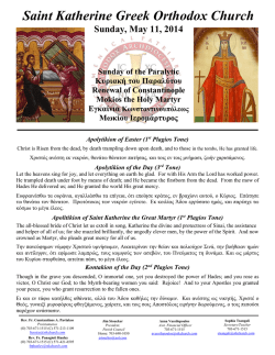
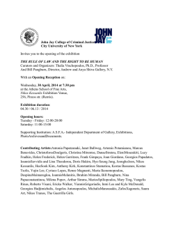
38
258
Hellenic Journal of Surgery 2011; 83: 5
Boerhaave’s Syndrome or Spontaneous Perforation of the
Oesophagus
Review Article
A. Marinis, S. Rizos
Received 03/06/2011 Accepted 11/07/2011
Abstract
Spontaneous perforation of the oesophagus, also
called Boerhaave’s syndrome, is a barogenic rupture
caused by a sudden rise in intraluminal pressure,
usually in the distal oesophagus. Early diagnosis of
spontaneous oesophageal perforation is critical to
the survival of the patient. Sudden and very intense
chest or epigastric pain after an abrupt increase of
oesophageal intraluminal pressure is the most characteristic clinical symptom and should raise suspicion for Boerhaave’s syndrome. Plain radiographs
demonstrate the presence of free air in the mediastinum, subcutaneous emphysema or air subdiaphragmatically, as well as pleural effusions. Upper
gastrointestinal series reveal leakage of the contrast
and confirms the location of the perforation. Similarly, chest computed tomography demonstrates the
presence of an air-fluid level, pneumomediastinum
and pleural effusions and, more importantly, the
complications related to oesophageal perforation.
Oesophageal perforation is associated with a high
mortality rate, suggested to be approximately 2%
for every hour after initial presentation. More specifically, diagnosis and intervention within the first
12 hours results in 80-90% survival, which falls to
70-75% between 12-24 hours; after 24 hours, survival decreases to less than 50%, and after 48 hours to
less than 10%. Operative treatment of Boerhaave’s
syndrome mainly includes a complete debridement
and lavage of infected and necrotic tissues with wide
drainage of the mediastinum and pleural cavities
and either primary oesophageal repair and reinforcement (if diagnosed early) or T-tube diversion
or resection (if diagnosis is delayed). Early confirmation of the diagnosis is very important because
it allows earlier support of the critically ill patient
and prompt operative intervention when necessary.
Eventful outcome of patients is related to these
A. Marinis (Corresponding author), S. Rizos
- First Department of Surgery, Tzaneion General Hospital Piraeus
Greece
e-mail: [email protected]
early events in conjunction with the good general
condition of the patient. Otherwise, the patient is
virtually condemned to die..
Key words:
Oesophageal perforation, Boerhaave’s syndrome, Spontaneous,
Post-emetic, Barogenic.
Introduction
Spontaneous perforation of the oesophagus (SPE),
also called Boerhaave’s syndrome, is the perforation of the oesophagus after forceful vomiting and is
considered as a surgical emergency. This syndrome
was first described by the Dutch surgeon Hermann
Boerhaave in 1724, who observed a transmural
rupture of the distal oesophagus in a post-mortem
examination of Baron John von Wassenaeur [1].
Though rare, this type of oesophageal perforation
is life-threatening due to the development of mediastinitis and severe sepsis. Patients usually present
with non-specific symptoms, such as epigastric pain
and vomiting; this leads to a delay in diagnosis and
appropriate treatment.
Aetiology - Pathophysiology
Boerhaave’s syndrome is a barogenic rupture
caused by a sudden rise in intraluminal pressure,
usually in the distal 3-6 cm of the oesophagus. It is
described more commonly in males than females at
a ratio of 4:1 with a median age of 64 years [2]. This
syndrome generally occurs in the absence of preexisting oesophageal pathology and is commonly
associated with large meals and heavy consumption
of alcohol. Pathophysiologically, the combination of
a sudden rise in intra-abdominal pressure caused by
vomiting or retching and neuromuscular incoordination provokes the failure of the cricopharyngeal
muscle to relax, leading to a dramatic increase in
the intraluminal oesophageal pressure. However,
cases of SPE resulting from trauma, weightlifting,
parturition, defaecation, the Heimlich manoeuvre
and status epilepticus have also been reported [3].
A transmural rupture is usually observed in the
lower third of the oesophagus (90%) and less fre-
259
Hellenic Journal of Surgery 2011; 83: 5
quently in the mid (8%) and upper (2%) thirds. The
left lateral side of the lower oesophageal wall is the
usual site of perforation due to anatomical weakness of this point. The length of the tear varies from
0,6-8,9 cm (average 2,2 cm) with the mucosal injury
being longer than the muscular tear [4]. With the
progress of time, the negative intrathoracic pressure
progressively draws more air, fluids and foods from
the oesophageal lumen into the mediastinum and
the pleural cavities, and a chemical and septic pneumomediastinitis develops.
Clinical presentation
The classic clinical presentation of Boerhaave’s syndrome is the Mackler’s triad, consisting of vomiting
or retching, severe lower chest pain and cervical
subcutaneous emphysema. In a large series, however, this classic triad was present only in the 14%
of the patients [5]. The most important clinical feature is a sudden, often “dramatic” in intensity, chest
pain following an episode of raised intra-abdominal
pressure, most commonly after forceful vomiting.
Pain is located retrosternally and is usually exacerbated by movement and respiration. Typically,
haematemesis is absent, which distinguishes it from
the Mallory-Weiss tear, and swallowing precipitates
cough and pleuritic pain because of communication
of the oesophagus with the pleural cavity. Clinical
examination reveals a patient with tachypnoea and
orthopnoea, tachycardia, fever, diaphoresis and
hypotension, with bilateral pleural effusions. Mediastinal emphysema can be audible on auscultation
or visible on chest x-rays and precedes subcutaneous emphysema, which is seen in 28-60% of patients
at initial presentation. Another rare presentation
is Anderson’s triad, referring to subcutaneous emphysema, rapid respirations and abdominal rigidity.
Abdominal pain and tenderness is found in about
60% of patients, leading to a negative laparotomy
in 9% of cases [6].
Differential diagnosis, errors and prognosis
Diagnostic errors are actually very frequent with the
most common misdiagnosis being the perforated ulcer, followed by myocardial infarction, pulmonary
embolism, dissecting aneurysm and pancreatitis [7].
The diagnostic error is generally over 50%, with a
delay of more than 12 hours in the majority of cases,
with only 5% diagnosed initially at presentation
and about 35% correctly diagnosed pre-mortem [8].
SPE is associated with a high mortality rate, suggested to be approximately 2% for every hour after initial presentation. More specifically, diagnosis
and intervention within the first 12 hours results in
Boerhaave’s Syndrome or Spontaneous Perforation of the Oesophagus
80-90% survival. This rate falls to 70-75% between
12-24 hours; after 24 hours survival, it decreases to
less than 50% and after 48 hours to less than 10%
[9 –11].
Diagnostic work-up
Diagnostic workup includes chest x-rays, a watersoluble contrast swallow, chest computed tomography (CT) and upper gastrointestinal (GI) endoscopy.
Radiology
• A plain chest x-ray usually shows pneumomediastinum in 10-20% of cases (Naclerio’s V-sign), subcutaneous emphysema or air subdiaphragmatically
and pleural effusions (Fig. 1), commonly left-sided,
which develop immediately from the oesophageal
leakage, or at a later phase sympathetically from adjacent mediastinitis.
Fig. 1 Chest radiograph showing a large left pleural effusion (from
our archives).
• Oesophagogram is performed using barium for
a suspected thoracic perforation and Gastrografin®
for an abdominal perforation. Barium is inert in the
chest but causes peritonitis in the abdomen, whereas aspirated Gastrografin® can cause life-threatening pneumonitis. Usually the contrast media is extravasated into the pleural cavity (Fig. 2), showing
the location of perforation and facilitating the decision concerning the surgical approach (abdominal
vs. thoracic). However, contrast studies are associated with a negative false rate of 27-66% [12].
• Chest CT demonstrates an air-fluid level at the
site of perforation, pleural effusions, pneumomediastinum and subcutaneous emphysema, highly
suggesting the site of perforation (Fig. 3). This diagnostic modality, however, is commonly used postoperatively in order to assess the development
of complications (pneumonitis, mediastinitis, empyema, abscesses) and to control the adequacy of
therapeutic interventions (resolving mediastinitis,
39
40
260
Boerhaave’s Syndrome or Spontaneous Perforation of the Oesophagus
adequate drainage of the mediastinum and pleural
cavities, etc).
Hellenic Journal of Surgery 2011; 83: 5
modynamic monitoring (central venous and arterial
catheters), inotropic agents, and monitoring of urine
output. Management can be either non-operative or
surgical, depending on time delay in presentation
and diagnosis, extent of perforation and the overall
general condition of the patient.
Non-operative management
Fig. 2
Swallow with a non-ionic water-soluble contrast agent (Iopamiro®) showing leakage from the lower thoracic esophagus and
the development of an air-fluid level (from our archives).
A contained perforation diagnosed early, with limited soiling and without any distal GI obstruction, in
a patient without signs or symptoms of mediastinitis
or sepsis and without other medical co-morbidities,
and a delayed diagnosis which is well tolerated, are
the usual indications for non-operative treatment
[13, 14]. The latter involves aggressive resuscitation
and close vital sign monitoring in an intensive care
unit (ICU) environment, along with measures such
as: nil per os (NPO), intravenous anti-secretory
agents (H2-receptor antagonists or proton pump
inhibitors), broad-spectrum antibiotics and antifungal agents, naso-gastric decompression through
a nasogastric tube safely placed during endoscopy
and adequate chest drainage of pleural effusions.
Successful attempts to seal the oesophageal leak
with covered, self-expandable stents endoscopically
placed have been described in the literature [15].
Operative management
Fig. 3
Chest computed tomography demonstrating the leakage of
the contrast into the lower mediastinum, the concomitant presence
of free air and bilateral pleural effusions (from our archives).
Endoscopy
Upper GI endoscopy with flexible endoscopes is
currently considered to have a higher diagnostic
sensitivity than contrast radiology in Boerhaave’s
syndrome, allowing accurate assessment of the location of perforation and mucosal extent of the tear,
as well as safe placement of a nasojejunal feeding
tube for enteral feeding.
Finally, preoperative investigations can include
the oral ingestion of dyes, such as methylene blue,
as well as thoracocentesis, which reveal frank gastric contents on aspiration or a pleural fluid with a
low pH (less than 6.0) and a high amylase level.
Management
Resuscitation of the patient with spontaneous oesophageal perforation includes airway control and
proper oxygenation, large-bore intravenous catheters and aggressive fluid resuscitation, invasive hae-
The primary goal of operative intervention is to
manage the perforation site in order to prevent
ongoing spillage of oesophageal contents, to completely debride the infected and necrotic tissues
with concomitant wide drainage of the mediastinum
and pleural cavities and, finally, to restore oesophageal integrity and gastrointestinal continuity. Interestingly, thorough debridement and irrigation are
more important manoeuvres in terms of survival
than the type of reconstruction [16]. In general, primary repair and reinforcement usually follow if diagnosis is established early and t-tube diversion and
resection if diagnosis is delayed beyond 24 hours.
- Primary repair and reinforcement. Primary repair,
first employed by Barrett in 1947 [17], consists of
a precise two-layer closure of the perforation with
2/0 or 3/0 interrupted absorbable sutures, followed
by reinforcement of the suture line with adjacent
healthy tissue (pleura, lung, diaphragm, intercostal muscle, gastric fundus, omentum) or even
absorbable mesh [18]. However, primary repair is
associated with a high leakage rate of up to 23%
if treatment is delayed beyond 24 hours, rising to
50% in the presence of sepsis [19, 20].
- T-tube diversion. Stable patients presenting late
in the course of the disease with an established
261
Hellenic Journal of Surgery 2011; 83: 5
oesophago-pleural communication and localized
sepsis are not usually treated by primary repair,
but can be managed instead by diverting the oesophageal contents via a T-tube in a controlled
manner [21]. The tube is left in place for 3-6 weeks
until a fistula tract has developed.
- Oesophagectomy. In delayed diagnosis with concomitant sepsis resection of the severely damaged
oesophagus with immediate (if contamination is
minimal) or delayed reconstruction is the method
of choice. This operative approach is associated
with a high morbidity and mortality rate and reserved for critically ill septic patients. Techniques
that simply exclude and divert the injured organ
proximally and distally, leaving in situ the oesophagus, have been abandoned and are now historical.
Conclusions
Early diagnosis of spontaneous oesophageal perforation is critical to the survival of the patient. Sudden and very intense chest or epigastric pain after an
abrupt increase of oesophageal intraluminal pressure is the most characteristic clinical symptom and
should raise suspicion for Boerhaave’s syndrome.
Early confirmation of the diagnosis allows earlier support of the critically ill patient and prompt
operative intervention when necessary. Eventful
outcome of patients is related to these early events
in conjunction with the good general condition of
the patient. Otherwise, the patient is virtually condemned to die.
Conflict of interest
The authors declare that they have no conflict of
interest.
References
1. Derbes VJ, Mitchell RE. Hermann Boerhaave’s Atrocis, nec
Descripti Prius. Morbi Historia: the first translation of the classic
case report of rupture of the esophagus, with annotations. Bull
Med Libr Assoc. 1955;43:217.
2. Brauer RB, Liebermann-Meffert D, et al. Boerhaave’s syndrome: analysis of the literature and report of 18 new cases. Dis
Esophagus 1997;10:64-8.
3. Mackler S. Spontaneous rupture of the oesophagus; an experimental and clinical study. Surg Gynecol Obst 1952;93:345-56.
4. Kossick PR. Spontaneous rupture of the oesophagus. S Afr
Med J 1973;47:1807-9.
5. Griffin SM, Lamb PJ, Shenfine J et al. Spontaneous rupture of
the esophagus. Br J Surg 2008; 95:1115-20.
6. Shenfine J, Dresner SM, Vishwanath Y, et al. Management of
spontaneous rupture of the oesophagus. Br J Surg 2000;87:362-73.
7. Symbas PN, Hatcher CR, Harlaftis N: Spontaneous rupture of the esophagus. Ann Surg 1978;187:634–9.
8. Abbott OA, Mansour KA, Logan WA Jr, et al. Atraumatic
so-called «spontaneous» rupture of the esophagus. A review of
47 personal cases with comments on a new method of surgical
therapy. J Thorac Cardiovasc Surg 1970;59:67-83.
Boerhaave’s Syndrome or Spontaneous Perforation of the Oesophagus
9. Brewer LA, Carter R, Mulder GA, Stiles QR. Options in
the management of perforations of the esophagus. Am J Surg
1986;152:62-9.
10. Henderson JA, Peloquin AJ. Boerhaave revisited: spontaneous esophageal perforation as a diagnostic masquerader. Am J
Med 1989;86:559-67.
11. Richardson JD, Martin LF, Borzotta AP, Polk HC Jr. Unifying concepts in treatment of esophageal leaks. Am J Surg
1985;149:157-62.
12. Buecker A, Wein BB, Neueberg JM, et al. Esophageal perforation: comparison of use of aqueous and barium-containing
contrast media. Radiology 1997; 202: 683-6.
13. Ivey TD, Simonowitz DA, Dillard DH, Miller DW Jr. Boerhaave syndrome. Successful conservative managementin three
patients with late presentations. Am J Surg 1981;141:531-3.
14. Cameron JL, Kieffer RF, Hendrix TR, et al. Selective nonoperative management of contained intrathoracic esophageal
disruptions. Ann Thorac Surg 1979;27:404-8.
15. Yuasa N, Hattori T, Kobayashi Y, et al. Treatment of spontaneous esophageal rupture with a covered self-expanding metal
stent. Gastrointest Endosc 1999;49:777-80.
16. Altorjay A, Kiss J, Voros A, et al. The role of esophagectomy
in the management of esophageal perforations. Ann Thorac Surg
1998; 65:1433-6.
17. Barrett N. Report of a case of spontaneous perforation of
the oesophagus successfully treated by operation. Br J Surg 1947;
47: 216-8.
18. Bardaxoglou E, Manganas D, Meunier B, et al. New approach to surgical management of early esophageal thoracic perforation: primary suture repair reinforced with absorbable mesh
and fibrin glue. World J Surg. 1997 ;21:618-21.
19. Wright CD, Mathisen DJ, Wain JC, et al. Reinforced primary repair of thoracic esophageal perforation. Ann Thorac
Surg 1995;60:245-9.
20. Lawrence DR, Ohri SK, Moxon RE, et al. Primary esophageal
repair for Boerhaave’s syndrome. Ann Thorac Surg 1999;67:818-20.
21. Mansour KA, Wenger RK. T-tube management of late
esophageal perforations. Surg Gynecol Obstet 1992; 175:571-2.
41
42
262
Hellenic Journal of Surgery 2011; 83: 5
Boerhaave’s Syndrome or Spontaneous Perforation of the Oesophagus
Σύνδρομο Boerhaave ή Αυτόματη Ρήξη του Οισοφάγου
Άρθρο Ανασκόπησης
Αθανάσιος Μαρίνης, Σπύρος Ρίζος
Περίληψη
Ως αυτόματη ρήξη του οισοφάγου, επίσης γνωστή ως
σύνδρομο Boerhaave, θεωρείται η βαρο-τραυματική
ρήξη που συμβαίνει μετά από αιφνίδια αύξηση
της ενδοαυλικής πίεσης συνήθως στο περιφερικό
τμήμα του οισοφάγου. Η πρώιμη διάγνωση της
αυτόματης ρήξης του οισοφάγου αποτελεί τη πιο
κρίσιμη παράμετρο που καθορίζει την επιβίωση του
ασθενή. Η εμφάνιση αιφνίδιου και ιδιαίτερα έντονου
θωρακικού ή επιγαστρικού άλγους μετά από βίαιη
αύξηση της ενδο-οισοφαγικής πίεσης είναι το πιο
χαρακτηριστικό σύμπτωμα και θα πρέπει να θέτει
την υπόνοια του συνδρόμου Boerhaave. Οι απλές
ακτινογραφίες αναδεικνύουν τη παρουσία ελεύθερου
αέρα στο μεσοθωράκιο, υποδόριο εμφύσημα ή
ακόμα κι αέρα υποδιαφραγματικά, καθώς και
πλευριτικές συλλογές. Η διάβαση του ανώτερου
πεπτικού αναδεικνύει τη διαφυγή του σκιαγραφικού
από τον οισοφάγο καθώς και την εντόπιση του
πιθανού σημείου ρήξης. Παρομοίως, η αξονική
τομογραφία θώρακος επιβεβαιώνει τη παρουσία
υδραερικού επιπέδου, πνευμομεσοθωρακίου και
πλευριτικών συλλογών, αλλά εξίσου σημαντική είναι
και η συμβολή της στη διάγνωση των επιπλοκών
απότοκων της ρήξης του οισοφάγου. Η ρήξη του
οισοφάγου σχετίζεται με υψηλή θνητότητα, η οποία
αυξάνεται κατά περίπου 2% για κάθε ώρα από την
έναρξη των συμπτωμάτων. Ειδικότερα, η πρώιμη
διάγνωση κι έγκαιρη παρέμβαση εντός των πρώτων
12 ωρών συνδέεται με επιβίωση 80-90%, η οποία
όμως ελαττώνεται στο 70-75% όταν η παρέμβαση
γίνει στο διάστημα 12-24 ωρών. Δυστυχώς η
επιβίωση ελαττώνεται κάτω από 50% μετά τις 24
ώρες, ενώ μετά από 48 ώρες πέφτει κάτω από 10%.
Η χειρουργική θεραπεία του συνδρόμου Boerhaave περιλαμβάνει κυρίως τον επαρκή καθαρισμό
κι έκπλυση των επιμολυσμένων και νεκρωτικών
ιστών, με ταυτόχρονη ευρεία παροχέτευση του
μεσοθωρακίου και των υπεζωκοτικών κοιλοτήτων,
καθώς και τη πρωτογενή συρραφή με ενίσχυση της
- Ά Χειρουργική Κλινική, Γενικό Νοσοκομείο Πειραιά «Τζάνειο»
γραμμής συρραφής (όταν η διάγνωση γίνεται νωρίς) ή
την εκτροπή με σωλήνα Τ και την οισοφαγεκτομή (σε
καθυστερημένη διάγνωση). Η πρώιμη επιβεβαίωση
της διάγνωσης ακολουθείται από ταχύτερη
υποστήριξη του βαρέως πάσχοντος ασθενή και
από άμεση χειρουργική παρέμβαση όταν κρίνεται
σκόπιμη. Η καλή πρόγνωση του ασθενή σχετίζεται
με αυτά τα πρώιμα γεγονότα, σε συνδυασμό με τη
καλή γενική κατάσταση του ασθενή. Ειδάλλως, ο
ασθενής είναι σχεδόν καταδικασμένος να πεθάνει.
Λέξεις κλειδιά
Διάτρηση οισοφάγου, Σύνδρομο Boerhaave, Αυτόματη διάτρηση,
Μεθεμετική διάτρηση, Βαρότραυμα.
© Copyright 2026 Paperzz