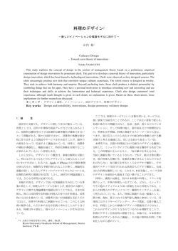
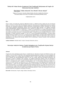
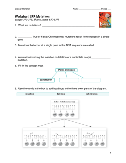
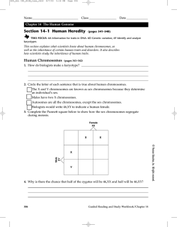
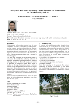
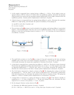
292
J Nippon Med Sch 2001; 68(4)
―Photogravure―
Philadelphia Chromosome-negative, bcr abl-positive
Chronic Myelogenous Leukemia
Koiti Inokuchi and Kazuo Dan
Third Department of Internal Medicine, Nippon Medical School
Fig. 1
Chronic myelogenous leukemia(CML)
is characterized by the presence of the abnormal chromosome 22, which
is termed the Philadelphia chromosome(Ph). The Ph is derived from a translocation between chromosomes 9
and 22, (9;
t 22)
(q 34; q 11). The c-abl oncogene on the long arm of chromosome 9 fused to the bcr gene on the
long arm of chromosome 22 and leads to the formation of a chimeric bcr abl gene located on the Ph . The
chimeric bcr abl gene produces 8.5-kb bcr abl transcript and chimeric fusion protein, p 210 BCR ABL. The p
210 BCR ABL protein plays a central role in the leukemogenesis of CML.
The case shown in the Figures was a rare Ph-negative CML case having typical clinical findings of CML. In
these Ph-negative CML cases, in addition to cytogenetic analysis, molecular biological analyses such as fluorescence in situ hybridization(FISH)and spectral karyotyping(SKY)FISH are used as potential tools for diagnosis.
FISH and SKY FISH are used as an adjunct to standard G-banding karyotyping(Fig. 1)to achieve a higher
sensitivity and specificity. FISH and SKY FISH provided additional cytogenetic information including the clarification of complex chromosomal rearrangements . The present SKY FISH revealed a translocation between
chromosomes 2 and 9(Fig. 2),and FISH revealed a chimeric bcr abl gene on chromosome 22(Fig. 3). From
these findings, the present case was finally diagnosed as a Ph-negative and bcr abl-positive CML.
Fig. 1 Chromosomal analysis of bone marrow cells by the conventional trypsin-Giemsa banding technique. The
cells were processed by a direct method. The karyotype was determined by both direct microscopic analysis and
photography. Karyotyping of a metaphase cell showed 46, XX, add
(2)
(q 33)
, del
(3)
(p 21 p 24)
, del
(7)
(q 34)
, add
(9)
(q 34)
, but failed to detect Ph. Aberrant chromosomes are indicated by arrows.
Journal Website(http: www.nms.ac.jp jnms )
32
J Nippon Med Sch 2001; 68(4)
293
Fig. 2
Fig. 3
Fig. 2 SKY FISH of the classification-colored metaphase chromosomes. Chromosomes were simultaneously hybridized with a combination of 24 labeled chromosome painting probes. Multicolor SKY revealed a translocation
of the chromosome 2 material to the long arm of chromosome 9; this was not detected by G-banding analysis.
Unbalanced translocation of der(9)is indicated by arrowhead. SKY failed to detect any abnormality of chromosome 22. The combination of Giemsa banding and SKY allowed us to refine the karyotype to 46, XX, add(2)
(q 33)
, del
(3)
(p 21 p 24)
, del
(7)
(q 34)
, der 9t(2; 9)
(q 33; q 34)
.
Fig. 3 FISH analysis of BM cells in metaphase using a Vysis LSI BCR-ABL translocation DNA probe. A yellow
signal indicates by the arrow head, composed of a red signal of bcr and a green signal of abl showing bcr abl
fusion gene was detected in all 20 cells analyzed.
解説:慢性骨髄性白血病(CML)はフィラデルフィア染色体(Ph)の核型異常を特徴とする.Ph は 9 番と 22
番染色体の相互転座により形成される.これにより bcr 遺伝子と c-abl 遺伝子が融合し,異常な bcr abl キメラ遺
伝子ができ,その遺伝子産物が CML を発症させる.ところが本例のような Ph 陰性症例では通常の染色体分析の
他に分子生物学的手法を応用した染色体解析(FISH,SKY-FISH)が診断に有用である.これらを用いた解析の
結果本症例は Ph 陰性,bcr abl 遺伝子陽性 CML と診断された.
References
1.Verdman T, Vignon C, Schrock E, Rowley JD, Ried T: Hidden chromosome abnormalities in haematological
malignancies detected by multicolour spectral karyotyping. Nature Genetics 1997; 13: 406―410.
2.Inokuchi K, Scinohara T, Futaki M, Hanawa H, Tanosaki S, Yamaguchi H, Nomura T, Dan K: Establishment of
a cell line with variant BCR ABL breakpoint expressing P180 BCR ABL from late-appearing Philadelphiapositive acute biphenotypic leukemia. Genes, Chromosomes & Cancer 1998; 23: 227―238.
3.Inokuchi K, Hamaguchi H, Taniwaki M, Yamaguchi H, Tanosaki S, Dan K: Establishment of a cell line with
AML 1-MTG 8, p 53 and p 73 abnormalities from acute myelogenous leukemia. Genes, Chromosomes & Cancer(in press)
33
© Copyright 2026 Paperzz