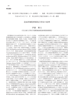
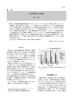
ISSN 1881-901X INCLUDED IN THIS ISSUE Full Articles 運動障害の鑑別診断における神経化学的バイオマーカー 傍腫瘍性運動障害 Abstracts セロトニンとパーキンソン病:運動機能,気分,精神疾患との関連 他 7 本収載 日本語版 Vol.3 No.3 January 2010 e-mail: [email protected] 日本語版 Vol.3 No.3 January 2010 監修: 水野 美邦 順天堂大学医学部附属 順天堂越谷病院院長 編集委員: 宇川 義一 (五十音順) 福島県立医科大学医学部 髙橋 良輔 京都大学医学研究科臨床神経学教授 神経内科学講座教授 野元 正弘 愛媛大学大学院医学系研究科 病態治療内科教授 梶 龍兒 徳島大学医学部神経内科教授 近藤 智善 公立大学法人和歌山県立医科大学 服部 信孝 順天堂大学医学部脳神経内科教授 神経内科教授 山本 光利 香川県立中央病院神経内科主任部長 Full Articles 運動障害の鑑別診断における神経化学的バイオマーカー 2 傍腫瘍性運動障害 17 Abstracts セロトニンとパーキンソン病: 運動機能,気分,精神疾患との関連 パーキンソン病における衝動行動と強迫行動 27 34 パーキンソン病と多系統萎縮症における 進行性核上性麻痺における小脳病変: 臨床病理学的研究 胃筋電図所見の違い 36 早期パーキンソン病における抑うつ症状の経過 38 28 パーキンソン病患者の衝動性障害・強迫性障害に関する 質問票の妥当性検討 30 パーキンソン病におけるレボドパ空腸内注入:非運動症状 およびQOL への効果に関する予備的多施設共同試験 代謝比率で評価したパーキンソン病患者の大脳皮質の代謝 低下は主に認知機能低下を反映する:[18F]FDG-PET 表紙:代謝指数(MI,黄色)と代替代謝指数(MI*,オレンジ色)の 計算に使用したマスク処理画像(mask) (Movement Disorders, 2009, Vol. 24 No. 10, page 1504) 40 32 Selected from Movement Disorders Vol. 24 No. 9-12, 2009 運動障害の鑑別診断における神経化学的 バイオマーカー Neurochemical Biomarkers in the Differential Diagnosis of Movement Disorders *, ** Brit Mollenhauer, MD and Claudia Trenkwalder, MD * Paracelsus-Elena Klinik, Kassel, Germany Department of Neurology and Clinical Neurophysiology, Georg August University, Goettingen, Germany * * 近年,アルツハイマー病やクロイツフェルト - ヤコブ病 経変性疾患では CSF および血中の蛋白質に関する研究 な ど の 神 経 変 性 認 知 症 の 診 断 に 際 し, 脳 脊 髄 液 においてバイオマーカーによる鑑別診断の有用性が検 (cerebrospinal fluid; CSF)中の神経蛋白質の神経化学 討されてきた。パーキンソン病,Lewy 小体型認知症, 的解析が行われるようになってきた。CSF は中枢神経 多系統萎縮症,進行性核上性麻痺,大脳皮質基底核変 系を取り囲むように存在し,CSF 蛋白質の中には,血 性症の臨床診断は,現在でも主に国際分類基準で定義 液由来の蛋白質よりも意義のある脳特異的蛋白質も認 された臨床症状に頼っている。本論文では,これらの運 められている。CSF 中の特異的蛋白質濃度は,中枢神 動障害における CSF バイオマーカーをレビューすると 経系疾患のきわめて有望なバイオマーカーとなりうる。 ともに,神経変性疾患の神経化学的な生前診断に関して 我々は,さらに採取が容易な血中バイオマーカーを確立 最近発表された報告(最近の CSF 中α—シヌクレイン する必要がある。認知症合併の有無にかかわらず,神 に関する知見も含む)について考察する。 Movement Disorders Vol. 24, No. 10, 2009, pp. 1411–1426 Key Word 運動障害,バイオマーカー,脳脊髄液 科学界は国際臨床分類基準の導入を通じ,神経変性疾 最近,National Institute of Neurological Disorders and 患の分類に少なからず努力してきた。特異的で神経防御・ Stroke-Alzheimer Disease and Related Disorders(NINCDS- 神経保護効果の期待できる新薬の登場に伴い,神経変性 ADRDA)の AD 作業部会による基準が改訂され,AD の 疾患の正確かつ早期の鑑別診断はとりわけ重要となって バイオマーカーに関しては次のように記載された。 「AD いる。しかし,神経画像検査などの専門的検査(technical のバイオマーカーに関するエビデンスも増えつつあり, investigation)に関するコンセンサスが得られ,多系統萎 これらを新たな AD 診断研究基準に加えることが容認さ 縮症(multiple system atrophy; MSA)の補助的診断基準 れる」3。この研究基準の最新版では,専門的検査(画像 の 1 つとして受け入れられるようになったのは,つい最 検査や CSF 蛋白質検査など)を補助的手段と位置づけて 近のことである。 いる。最近開発された病態特異的アミロイド蛋白質の in 理想的なバイオマーカーとは何かを考えた場合,その条 vivo 画像検査(Pittsburg compound B による)4 も研究基 件として,アルツハイマー病(Alzheimer’ s disease; AD)の 準に取り入れられた。CSF を用いる診断技術はエビデン 「本質的な神経病理学的特徴を検出できること,神経病理 スに基づいており,比較的安価で,広く利用可能である 学的な確定症例で妥当性が確認されていること〔 (AD を にもかかわらず,検討対象となるまでに 15 年を要した 5,6。 検出するための)診断の感度> 80%,他の認知症との鑑 別の特異度> 80%〕 ,信頼性が高く,再現性があり,非侵 襲的で,容易に実施でき,安価であること,少なくとも 2 つの独立した試験で検討されていること」が挙げられる 1,2。 2 CSF バイオマーカー 神経変性疾患では,脳周囲の体液である CSF は腰椎穿 B. Mollenhauer and C. Trenkwalder 刺で採取できる。CSF 組成が脳の生化学的変化をある程 度反映していることは明らかである。 (c)死後ではなく生前のヒトへの腰椎穿刺で得た CSF の 研究,とした。 一般に疾患特異的バイオマーカーは,その疾患の生化 すべての研究に共通する限界として,感度と特異度が 学的特徴〔例えばパーキンソン病(Parkinson’ s disease; 低い,独立したコホートによって結果の再現性が検証さ PD)ではドパミン作動性経路〕 ,神経病理学的特徴およ れていない,疾患特異的な蛋白質もしくは蛋白質パター び / または神経生理学的特徴(例えばシヌクレイノパチー ンの測定方法はごく新しいものである, などが挙げられる。 ではα—シヌクレイン)を反映する。代用マーカーと呼ば れる別のタイプのバイオマーカーは,疾患に特異的な生 化学的,神経病理学的および / または神経生理学的特徴 とは独立した様々な発現パターンを反映する。 神経変性疾患に関連する CSF 蛋白質 タウ蛋白質 CSF における蛋白質動態 リン蛋白質タウ(68 kDa)は微小管の安定化に重要な 役割を果たし,自然状態では折りたたまれていない微小 血液 - 脳(厳密には血液 -CSF)関門を形態学的に捉え 管関連蛋白質である 11。AD における神経原線維変化は, た「漏出(leakage) 」モデルとは対照的に,これを分子流 対をなすらせん状の蛋白フィラメントで,神経細胞の細 速と CSF 流量のより動的なプロセスとして捉える最近の 胞骨格に認められる 12,13。これらの蛋白フィラメントは, 理論により,少なくとも CSF 蛋白質濃度の調節に関する 微小管に関連する不溶性かつ安定性の低分子タウ蛋白ポ 我々の理解は深まった 7。CSF 蛋白質濃度はその起源(血 リマーである 14。微小管への親和性は,潜在する 79 部位 液または脳)に依存し, 血液由来蛋白質濃度は, 脊髄に沿っ の異なるリン酸化によって調節されている 15,16。フィラメ た CSF への受動拡散によって脳室から腰椎腔(lumbar ント状沈着物を伴うヒト疾患の場合,タウ蛋白質は過剰 space)に至るまでの間,持続的に上昇していく 。CSF にリン酸化されているが,これはフィラメント集合に先 が流れるための駆動エネルギーは,動脈血液系と静脈血 立つ初期変化であると考えられる 13,17。 7,8 液系の圧力差で生じる 9。血液 - 脳関門が障害された場合 AD 患者では CSF 総タウ蛋白質(以下,CSF タウ蛋白 には,主として CSF 流量が CSF 中の血液由来蛋白質濃 質とする)濃度が上昇するが,その濃度は 300 ~ 900 pg/ 度を調節することになる。CSF 流量が低下すると,それ mL と研究によって異なっている 18,19。ただし,タウオパ らの蛋白質の分子流速が増加する 8,10。 チー以外の神経変性疾患でも,複数の単独例で CSF タウ 蛋白質濃度の上昇が認められている 20,21。一方,タウオ 方 法 Table 1 に,PD,Lewy 小体型認知症(dementia with パチーであっても正常な CSF タウ蛋白質が検出される場 合もある 22,23。これらの結果から,CSF タウ蛋白質はタウ 関連疾患に特異的な所見ではないと考えられる。 Lewy bodies; DLB) ,MSA,進行性核上性麻痺(progressive クロイツフェルト - ヤコブ病(Creutzfeldt-Jakob disease; supranuclear palsy; PSP) ,大脳皮質基底核変性症 CJD) 患者のCSFタウ蛋白質濃度は通常1,300 pg/mLを超え, (corticobasal degeneration; CBD)を対象とした CSF バイ 広範な神経細胞喪失による細胞内分画を反映している 24,25。 オマーカーに関する既発表研究の概要を示す。この一覧 さらに,脳卒中のような他の急性神経疾患でも CSF タウ蛋 表はすべての研究を網羅したものではなく,以下の用語 白質濃度の上昇を示すことから,神経細胞喪失のマーカー で検索した PubMed 論文である(2008 年 5 月まで) 。単 としての役割を担っていることが示唆される 26。 一疾患名としての「パーキンソン病(Parkinson disease) 」 CSF タウ蛋白質濃度の測定は,PD ならびに認知症を伴 「Lewy 小体型認知症(dementia with Lewy bodies) 」 「多系 う PD(Parkinson’ s disease with dementia; PDD)と AD と 統萎縮症(multiple system atrophy) 」 「進行性核上性麻痺 の鑑別に役立つ可能性があるが 27,対照群と比べると PD (progressive supranuclear palsy) 」 「大脳皮質基底核変性症 (PDD および PDD の CSF タウ蛋白質の差はわずかである 28 」 , な ら び に「 脳 脊 髄 液 (corticobasal degeneration) 73 例,PD 23 例,非認知症性神経疾患の対照 41 例;カッ (cerebrospinal fluid) 」 。適格基準は, (a)バイオマーカー トオフ値 250 pg/mL での PDD に対する感度 42%,特異度 に焦点を絞った研究, (b)対象患者が 7 名を超える研究, 88%) 。DLB の場合には,CSF タウ蛋白質濃度は AD より 3 運動障害の鑑別診断における神経化学的バイオマーカー CSF DIAGNOSTICS IN MOVEMENT DISORDERS 1413 Table 1 パーキンソン病(PD) ,Lewy 小体型認知症(DLB) ,多系統萎縮症(MSA) ,進行性核上性麻痺(PSP) , TABLE 1. CSF biomarker in Parkinson disease (PD), dementia with Lewy Bodies (DLB), multiple system atrophy (MSA), 大脳皮質基底核変性症(CBD)の CSF バイオマーカー progressive supranuclear palsy (PSP) or corticobasal degeneration (CBD) Marker 4 CSF DIAGNOSTICSPD IN MOVEMENT DISORDERS MSA DLB PSP 1413 CBD 129,130 129 8-hydroxy-20 -deoxyguanosine (8-OHDG), 8-hydroxyguanosine (8-OHG) in Parkinson disease (PD), dementia with Lewy Bodies (DLB), multiple system atrophy (MSA), TABLE 1. CSF biomarker Acetyl choline 126–128 progressive supranuclear palsy (PSP) or corticobasal degeneration (CBD) Angiotensin-converting enzyme (ACE) 131,132 133 131 Acetylcholinesterase (AChE) and butyrylcholinesterase 134–139 140 141–143 Marker PD DLB MSA PSP CBD (BChE) 0 129,130 129 8-hydroxy-2 Amino acids -deoxyguanosine and metabolites (8-OHDG), 144–158 159,160 156,158,161 8-hydroxyguanosine (8-OHG) Annexin V 162 Acetyl choline 126–128 Bcl-2 protein 163 Angiotensin-converting (ACE) 131,132 133 131 Biogene amine and theirenzyme metabolites 30,132,135,139, 119,120 (30,101,140,164, Acetylcholinesterase (AChE) and butyrylcholinesterase 134–139 140 141–143 155,164–193 165,184,194,195 (BChE) C4d complement protein 196 Amino and acidscGMP and metabolites 144–158 159,160 156,158,161 cAMP 197–201 Annexin V 162 Carnitine 202 Bcl-2 protein 163 Ceruloplasmin 203 Biogene amine Aand 30,132,135,139, 119,120 (30,101,140,164, Chromogranin andtheir B metabolites 204 155,164–193 165,184,194,195 Coenzyme Q-10 205 C4d complement protein 196 Delta-sleep-inducing peptide (DSIP), phosphorylated 206 cAMP and cGMP 197–201 Delta-sleep-inducing peptide (p-DSIP) Carnitine 202 DJ-1 (PARK7) 207 Ceruloplasmin 203 Dipeptidyl-aminopeptidase II (DAP II) 208 Chromogranin 204 Ferroxidase A and B 209 Coenzyme Q-10 205 Ferritin 210,211 210 Delta-sleep-inducing 206 Glial fibrillary acidic peptide protein (DSIP), (GFAP) phosphorylated 30,116 30,116 Delta-sleep-inducing peptide (p-DSIP) Glutathione peroxidase (GPx) DJ-1 (PARK7) 207 4-hydroxynonenal (HNE)-conjugated GPx 212 Dipeptidyl-aminopeptidase II (DAP II)hormone (GHRH) 208 Growth hormone (GH), GH-releasing 213 Ferroxidase 209 Growth-associated protein 43 (GAP-43) 214 Ferritin 210,211 210 Harman and norharman 215 Glial fibrillary protein (GFAP) 30,116 30,116 Heart-fatty acidacidic binding protein (H-FABP) 93 91–93 Glutathione peroxidase (GPx) Homocarnosine 151 4-hydroxynonenal (HNE)-conjugated GPx 212 Homocystein 216 Growth hormone (GH), 213 Hydroxyindoleacetic acidGH-releasing (HYDA) hormone (GHRH) 140 Growth-associated protein 43 (GAP-43) 214 Interleukins 217,218 219 Harman and norharman 215 Isoprostanes 220 Heart-fatty acid binding protein (H-FABP) 93 91–93 Lysosomal enzymes 221 Homocarnosine 151 Metals 222–228 229 230 Homocystein 216 Methionine-enkephalin 172,231 Hydroxyindoleacetic acid (HYDA) 140 Methyl-6,7-dihydroxy-1,2,3,4-tetrahydroisoquinolines 187,188,232 Interleukins 217,218 219 (2-MDTIQ); (1-MDTIQ, salsolinol); Isoprostanes 220 N-methyl-norsalsolinol Lysosomal enzymes 221 Myelin basic protein (MBP) 30 30,31 Metals 222–228 229 230 Neopterin and biopterin 157,233,234 Methionine-enkephalin 172,231 Neural thread protein 235 236 Methyl-6,7-dihydroxy-1,2,3,4-tetrahydroisoquinolines 187,188,232 Neurofilaments heavy and light chain (NFL, NFH) 31,115,116 237 101,115,116, 115,116,239 (2-MDTIQ); (1-MDTIQ, salsolinol); 195,238,239 N-methyl-norsalsolinol Neuronspecific enolase (NSE) 30 30,101 Myelin basic protein (MBP) 30 165, 30,31 Neuropeptides 135,139, 165,249 249,250 Neopterin and biopterin 157,233,234 172, 185, 197, Neural thread protein 236 231,235 240–248 Neurofilaments 31,115,116 237 101,115,116, 115,116,239 Neuroserpins heavy and light chain (NFL, NFH) 133 195,238,239 Nitrite, nitrate, nitric oxide metabolites 199,210, 251–253 159 210 Neuronspecific enolase (NSE) 30 30,101 Orexin A/hypocretin-1 110 110,254 101,255 110 110 Neuropeptides 135,139, 165,249 249,250 Oxidative stress indices 227 165, 172,208 185, 197, Post-proline cleaving enzyme 231, 240–248 Proteins 14-3-3 256–259 Neuroserpins 133 Reelin 260 260 Nitrite, nitrate, nitric oxide metabolites 199,210,30251–253 159 210 1414 protein B. MOLLENHAUER AND C. TRENKWALDER S100B 20,261 30,101 Orexin A/hypocretin-1 110 110,254 101,255 110 110 Secretogranin II 204 Oxidative stressofindices 227 Soluble forms intercellular adhesion molecule-1 133 Post-proline cleaving enzyme 2081. (Continued ) (sICAM-1), vascular cell adhesion molecule-1 TABLE Proteins 14-3-3and platelet endothelial cell adhesion 256–259 (sVCAM-1) Reelin 260 260 molecule-1 (sPECAM-1) Marker protein PD DLB MSA PSP CBD S100B 30 20,261 30,101 Superoxide dismutase activity (SOD) (Cu, Zn, MN) 227,262 212,263 Secretogranin 204 Tau isoforms II 264 264 264 264 Soluble forms of intercellular adhesion molecule-1 133 Tau protein (total) 27,28,31,154, 20,33,39,54,93, 30,101 22,32,271 22,32,56, (sICAM-1), vascular cell adhesion molecule-1 214,235,265 219,237,261, 271,272 (sVCAM-1) and platelet endothelial cell adhesion 264,266–270 molecule-1 (sPECAM-1) Tau protein phosphorylated 27 39,237,261,264, 271 Movement Disorders, Vol. 24, No.56,271 10, 2009 Superoxide dismutase activity (SOD) (Cu, Zn, MN) 227,262 212,263 267,270,273–275 Thyroid-stimulating hormone (TSH) response to 213 thyrotropin-releasing hormone (TRH) TNF-alpha 276 Transglutaminase 277 Urate 278 Movement Disorders, Vol. 24, No. 10, 2009 Vitamins alpha-tocopherol (vitamin E); 154,279,280 194 Thiamine (vitamin B1) Xanthine 278 a-synuclein (total and oligomeric) 74,77–79, 78,281 281 86,281 1414 B. MOLLENHAUER AND C. TRENKWALDER TABLE 1. Marker 1414 (Continued ) PD AND C. TRENKWALDER DLB B. MOLLENHAUER Tau isoforms Tau protein (total) Tau protein phosphorylated Marker 264 264 27,28,31,154, 20,33,39,54,93, Table 1 214,235,265 219,237,261, TABLE 1. (つづき) (Continued ) 264,266–270 27 39,237,261,264, PD DLB 267,270,273–275 264 264 27,28,31,154, 20,33,39,54,93, 214,235,265 219,237,261, 276 264,266–270 277 27 39,237,261,264, 278 267,270,273–275 154,279,280 B. Mollenhauer PSP and C. Trenkwalder MSA CBD 30,101 264 22,32,271 264 22,32,56, 271,272 MSA 271 PSP 56,271 CBD Tau isoforms 264 264 Thyroid-stimulating hormone (TSH) response to 213 Tauthyrotropin-releasing protein (total) 30,101 22,32,271 22,32,56, hormone (TRH) 271,272 TNF-alpha Transglutaminase Tau 271 56,271 Urateprotein phosphorylated Vitamins alpha-tocopherol (vitamin E); 194 Thyroid-stimulating (TSH) response to 213 Thiamine (vitaminhormone B1) thyrotropin-releasing hormone (TRH) Xanthine 278 TNF-alpha (total and oligomeric) 276 a-synuclein 74,77–79, 78,281 281 Transglutaminase 277 86,281 Urate 278 b 2-microglobulin 282 Vitamins alpha-tocopherol (vitamin E); 154,279,280 194 b-Amyloid 1-40 20,269 Thiamine1-40* (vitamin B1) b-Amyloid (oxidized) 58,59,266 58,59,266 Xanthine 278 b-Amyloid 1-42 28,54,55,214, 20,33,39,54,219, 55 55,271 56,289 a-synuclein (total and oligomeric) 74,77–79, 78,281 281 265,283 237,270,275 86,281 b-Amyloid peptide pattern 57 57 57 57 57 b 2-microglobulin 282 死後ではなく生前のヒトへの腰椎穿刺で得た CSFthan を対象とし,バイオマーカーに焦点を当てた対象患者 8 例以上の研究のみを掲載した(齧 b-Amyloid 1-40with a biomarker focus, and more Only studies 7 patients were included, as20,269 were studies based only on intra vitam, not post mortem, b-Amyloid 1-40* (oxidized) 58,59,266 gained CSFのみを扱った研究は除外) by lumbar puncture in humans from rodents were 58,59,266 excluded). 歯類の CSF 。 (studies on CSF only b-Amyloid 1-42 28,54,55,214, 20,33,39,54,219, 55 55,271 56,289 265,283 237,270,275 b-Amyloid peptide pattern 57 57 57の方がより小規模である 57 5737,38。 的な共通点であるが,DLB も低く,PD および PDD よりも高い 20(DLB 71 例,AD tau levels in MSA patients (MSA-P and MSA-C) were towards AD by p-tau protein 181 than with total tau shown to be elevated compared to PD, possibly due to protein (DLB n 5 18; AD n 5 23). gained CSF29 by lumbar puncture in humans (studies on CSF only from rodents were excluded). で「DLBaxonal がほぼ確実(probable 分類基準 度は,総タウ蛋白質よりも p タウ蛋白質 181 の方が高い more widespread degeneration inDLB) MSA」と判定 relative 20 5 15; MSA-C n 5 14; PD n 5 35; to PD30 (MSA-P n タウ蛋白質濃度が低い傾向にある Amyloid-b-Peptides 。 18 例,AD 23 例) 。 された患者は,CSF としている 39(DLB 31 sensitivity 95%, specificity 77%) (MSA-P n 5 19; Different of protein Amyloid-b peptides tau levels in MSA patients (MSA-P and MSA-C) were towards AD lengths by p-tau 181 (Ab) than with totalarise tau 最近の研究では,MSA 患者〔パーキンソン型多系統萎縮 PD n 5to31; 76%, specificity 97%). In due CBD, through39enzymatic the23). 120 kDa transmemshown be sensitivity elevated compared to PD, possibly to (DLB n 5cleavage 18; AD of n5 protein 症(parkinsonian variantelevated of multipleCSF system atrophy; MSA-P) アミロイドβペプチド there widespread were slightly tauin protein levels brane amyloid precursor protein (APP) by three differmore axonal degeneration MSA relative 30 compared to controls and PSP patients as reported by および小脳型多系統萎縮症(cerebellar variant of multiple kDa の膜貫通性アミロイド前駆体蛋白質(amyloid ent120secretases, resulting in either carboxyterminally n 5 15; MSA-C n 5 14; PD n 5 35; to PD (MSA-P Amyloid-b-Peptides 22 32 40 31 n 5 12); (CBD n 5 10; PSP (CBD Urakami et al. cleavage truncated or –elongated Ab peptides. sensitivity 95%, specificity 77%) (MSA-P n 5 system atrophy; MSA-C) 〕の CSF タウ蛋白質濃度が PD19; 患 precursor protein; APP)が 3 つの異なるセクレターゼで酵素 Different lengths of Amyloid-b (Ab) This peptides arise nPD5n 27; PSPsensitivity n 5 30; 76%, sensitivity 81, 5 97%). and specificity is divided into an amyloidogenic processing conducted 5 31; specificity In CBD, through enzymatic長さの異なる複数のアミロイドβ(Aβ) cleavage of the 120 kDa transmem者よりも高いことが示されたが,これは PD に比べ MSA で 的に切断されると, 41,42 80% a cut-off of 180 pg/mL); waslevels also by b-secretase (called BACE-1) and b-secretase, there atwere slightly elevated CSFthistaufinding protein brane amyloid precursor protein (APP) by three differ33 30 shown in an independent smallas sample (MSA-P は軸索変性がより広範囲なせいかもしれない ペプチドが産生される。これらはカルボキシル末端側が and a nonamyloidogenic by metalloprocompared to controls and but PSPvery patients reportedset15 by ent secretases, resulting43inprocessing either carboxyterminally 22 (CBD n 5 3; PSP n 5 6). 3132 40 The emerging vary teases called a-secretase. 40 peptides 例, MSA-C 14 35 例;感度 特異度 n 5 95%, 10; PSP n 577%) 12);(MSA-P (CBD Urakami et 例, al. PD(CBD 。この切 短縮もしくは延長された Aβペプチドである truncated or –elongated Ab peptides. This cleavage As hyperphosphorylation of tau protein promotes its in their proneness to aggregate, depending on their 5 27; PSP n 5 30;76%,特異度 sensitivity 81, 5 。Urakami and specificity is divided into an amyloidogenicと呼ばれる)とγセクレ processing conducted 97%) ら 22 19n 例,PD 31 例;感度 断は,βセクレターゼ(BACE-1 44 aggregation through the facilitation process neurofilength and the degree post-translational oxidation. 41,42 80% at a cut-off of 180 pg/mL); this findingofwas also by b-secretase (called ofBACE-1) and b-secretase, 41,42 が報告しているように,CBD では,CSF タウ蛋白質濃度 33 ターゼによるアミロイド産生プロセシング brillary intangles, it is obvious that quantification Amyloid-plaques depositedprocessing in the brain ofと,αセク patients shown an independent but verythe small sample setof and a nonamyloidogenic by metallopro40,45 phosphorylated tau protein in CSF might serve as an mainly consist of carboxytermiwith AD and DLB が対照および レターゼと呼ばれるメタロプロテアーゼによる非アミロ (CBD n 5 3;PSP PSP患者に比べてわずかに高かった(CBD n 5 6). teases called a-secretase.43 The emerging peptides vary even more specific marker for AD apart from neuronal nally-elongated formstoof Ab peptides such as Ab 32 43aggregate, As hyperphosphorylation of tau protein promotes its in their proneness depending on 1-42. their 10 例,PSP3412 例) (CBD 27 例,PSP 30 例;カットオフ に分けられる。切断後に生じる イド産生プロセシング cell loss. For CSF tau protein phosphorylated at threoStudies in transgenic mice have shown that the lack of 44 aggregation through the facilitation process of neurofilength and the degree of post-translational oxidation. 46 値 180181 pg/mL で感度 81%,特異度 80%) 。この所見は, ペプチドは,その長さと翻訳後酸化程度によって凝集傾 nine (p-tau protein 181), Blennow et al. showed, for APP leads to impaired neuronal function. brillary tangles, it is obvious that the quantification of Amyloid-plaques deposited in the brain of patients 33 to 44 the first time in 1995, elevated AD compared In AD 1992, Seubert et al. demonstrated existence of 非常に小規模ではあるが,独立したサンプルセット 。AD および DLBconsist 患者の脳内に沈着するア 向が異なる phosphorylated tau protein in levels CSF in might serve as で an mainly ofthecarboxytermiwith and DLB40,45 27 5 healthy controls and other neurodegenerative diseases. This observation extracellular Ab, including CSF. 40,45 of Ab peptides such as even more specific 3marker for6 例) AD。apart from neuronal nally-elongated forms Ab 1-42. も確認された(CBD 例,PSP は,主として Aβ 1-42 などの Aβ ミロイドプラーク 34epitope phosphorylations of CSF tau protein Different offered new opportunities for developing diagnostic cell loss. For CSF tau protein phosphorylated at threoStudies in transgenic mice have shown that the lack of 23,27,35,36 タウ蛋白質のリン酸化が過剰に亢進すると,神経原線 ペプチドのカルボキシル末端延長型からなる。トランス have been(p-tau analyzed for their value. tests for AD. Initially,neuronal Motter et al. reported signifi46 nine 181 protein 181),diagnostic Blennow et al. showed, for APP leads to impaired function. respect to DLB, the neuropathological overlap cantly decreased levels CSF Ab 1-42theinの欠損が神経 37 patients 維変化の促進プロセスを通してその凝集が加速されるこ ジェニックマウスを用いた研究では,APP theWith first time in 1995, elevated levels in AD compared to In 1992, Seubert et al.ofdemonstrated existence of 27 with AD involves the phosphorylation of tau protein, with AD, compared to 32 neurological disease controls 5 healthy controls and other neurodegenerative diseases. This46。 observation extracellular Ab, including CSF. とから,CSF 中のリン酸化タウ蛋白質の定量化は,神経 機能障害を引き起こすことが示されている 37,38 6 Some of CSF have but to a epitope lesser extent. Since then, there have and 20 nondemented controls. Different phosphorylations CSFstudies tau protein offered new opportunities for などの細胞外にも developing diagnostic 細胞喪失とは別に,AD のより特異的なマーカーとして用 1992 年に,Seubert らはand CSF Aβが revealed a better specificity for the discrimination 23,27,35,36 been several independent multicenter studies replihave been analyzed for34 their diagnostic value. tests for AD. Initially, Motter et al. reported signifi5 。1995 年に Blennow らは,スレ いられる可能性がある 。この観察所見は,AD の 存在することを明らかにした With respect to DLB, the neuropathological overlap cantly decreased levels of CSF Ab 1-42 in 37 patients with AD involves the phosphorylation of tau protein, オニン 181 でリン酸化された CSF タウ蛋白質(p タウ蛋 with AD, compared to 32 neurological disease controls 診断検査法を開発する新たなきっかけとなった。まず 37,38 but 181)濃度は,AD to Disorders, a lesserVol.extent. then, there have and 20 らは,神経疾患の対照群 nondemented controls.6 Since Movement 24, No. 10, 2009Some CSF studies have 白質 患者では健常対照および他の神経 Motter 32 例ならびに非認知症 revealed a better specificity for the discrimination been several independent and multicenter studies repli27 67 例, 非認知症性神経疾患の対照 例) 。興味深いことに, 一部のas39 CSF Only studies with a biomarker focus,41and more than 7 patients were included, were研究では,DLB studies based onlyと onAD intraとの鑑別における特異 vitam, not post mortem, 変性疾患患者よりも高いことを初めて示した 。CSF タ の対照群 20 例に比べ,AD 患者 37 例では CSF の Aβ ウ蛋白質の異なるエピトープのリン酸化については,そ 1-42 濃度が有意に低いことを報告した 6。その後,この成 23,27,35,36 。 の診断的価値が解析されている Movement Disorders, Vol. 24, No. 10, 2009 績を再現するいくつかの独立した多施設共同研究が報告 タウ蛋白質のリン酸化は,DLB と AD との神経病理学 されている 19。その中には神経病理学的変化との相関を 5 運動障害の鑑別診断における神経化学的バイオマーカー 示す研究もあったが 47,その研究は最適とは言えない死 ト法でも確認されている 57(PD 11 例,PSP 20 例,CBD 後採取 CSF で実施されていた(下記参照) 。脳内の Aβ 12 例,MSA 18 例,DLB 20 例,神経疾患の対照 19 例) 。 プラークが Aβ 1-42 の「巣窟(sink) 」として働き,脳と CSF 間の可溶性 Aβ 1-42 の輸送を妨げるとの理論 6,48 は, Aβ 1-40OX AD 患者で CSF 中の Aβ 1-42 が少ない理由として広く受 け入れられている。 最近,SDS-PAGE/ イムノブロット法により,新規の A βペプチドが同定された。このペプチドは,すでに知ら CSF 中の Aβ 1-42 は従来の ELISA(Enzyme-Linked Immunosorbent Assay)法,免疫沈降質量分析法,ウエス タン/イムノブロット法で測定することができる 。A 6,49,50 れている断片 Aβ 1-40 によく似た生理化学的性質をもつ ことが示されたが,易動度は非定型的であった。その後, このペプチドはαヘリックス構造を有する,Aβ 1-40 の βのドデシル硫酸ナトリウム(sodium dodecylsulfate; 酸化型(Aβ 1-40OX)と同定された 58,59。DLB 患者(21 例) SDS)- ポリアクリルアミドゲル電気泳動(polyacrylamide では,PDD 患者(21 例)や非認知症対照群(23 例)に gel electrophoresis ; PAGE)/ イムノブロット法は Wiltfang 比べ Aβ 1-40OX が特異的に上昇することが示されている。 ら 50 によって導入された方法で,Aβペプチドと他の AD 患者では Aβ 1-40OX がやや上昇しているが,DLB と APP 代謝物を電気泳動により分離し,ウエスタン / イム の重複はわずかであるため,DLB では Aβ 1-40OX が独自 ノブロットで超高感度に検出することができる。この方 の病態生理学的代謝を受けているとの議論も成り立つと 法では,8 M 尿素処理を加えることによって 1 つのアミ 考えられる。ただし,これについては独立した研究グルー ノ酸のみが異なる CSF Aβペプチドも分離可能である。 プによるさらなる解明と結果の再現が必要である。Aβ 本 法 で 検 討した結 果,Aβペ プ チド 1-42,1-40,1-39, 1-40OX をマーカーとする診断感度はそれぞれ 81%および 1-38,1-37 が CSF およびその他の細胞外体液中に恒常的 71%であった。 に存在することが明らかになった 。検出限界は,Aβペ 51 プチド 1-40 が 600 pg/mL,Aβペプチド 1-42 が 1 pg/mL CSF および血清中のα—シヌクレイン であった 。アッセイ間およびアッセイ内変動は 10%未 α—シヌクレインは 140 個のアミノ酸からなり,主とし 満であった 。この方法には従来の ELISA 法を上回る価 てシナプス前に存在し,PD と DLB の病理学的特徴であ 値のあることは明らかであるが,複数の独立した研究グ る Lewy 小体や,MSA のグリア細胞質封入体に認められ ループあるいはより多くのサンプルによって結果の妥当 る細胞内凝集体の主要成分である 60,61。約 16 kDa のα— 性が確認されていないため,本法をハイスループット解 シヌクレインをコードする SNCA 遺伝子において,遺伝 析(大量高速解析)に用いるのは適当ではない。 性のパーキンソニズムと認知障害を引き起こす 3 つ(本 52 53 一般に Aβペプチドは翻訳後修飾(特にメチオニン 35 の酸化)を非常に受けやすいため,解析前試料の取り扱 64 いはきわめて重要である(下記参照) 。 認されている 65,66。発症年齢や臨床症状の重症度,とりわ 大部分の研究では,PD 患者の CSF Aβ 1-42 は対照群 と比べ正常ないし軽度低下していると報告されているが, ,新たに SNCA 遺伝子の重複(multiplication)現象が確 け 認 知 機 能 の 低 下は,遺 伝 子 量(duplication ないし triplication)に依存すると考えられる。 (PD 15 例, DLB 患者の CSF Aβ 1-42 は通常低下している 54 α—シヌクレインのホメオスタシスに関する生体内動態 DLB 11 例,対照 19 例) 。PD 患者の CSF Aβ 1-42 濃度は, は様々な代謝イベントによって変化し,この動態に応じて より長期の罹病期間および認知障害の併発に伴って低下 シヌクレイノパチーが促進される 223。α—シヌクレインの する傾向がある (PD 73 例,PDD 23 例,神経疾患の対 動態を変化させる代謝イベントには,合成速度の上昇 68, 照 41 例;カットオフ値 350 pg/mL での PDD に対する感 protofibril(原線維)を生成する変異体の沈着増加 69,セ 。 度 85%,特異度 41%)55(PD 48 例,健常対照 32 例) リン 129 の持続的リン酸化 70,C 末端の短縮,分解の低 このような Aβ 1-42 濃度の低下は,MSA55(MSA 36 例) 下 71 などがある。最近,α—シヌクレインの分解経路が検 に加え, 一部の研究では PSP と CBD でも認められ 55(PSP 討され,プロテオソームあるいはリソソーム活性の関与 15 例) (PSP 18 例,CBD 9 例,神経疾患の対照 43 例; が示された 71-73。細胞内α—シヌクレインが減少する別の AD と比較した場合の感度と特異度は PSP では 43%と 機序として,細胞間腔への放出が考えられる 74。完全長 92%,CBD では 64%と 91%) ,ウエスタン / イムノブロッ のα—シヌクレインは,血漿や調製細胞培養液などの生 28 56 6 論文執筆時)のミスセンス変異が発見されたのに続き 62- B. Mollenhauer and C. Trenkwalder 体試料中において検出されている 73,74。したがって,我々 上昇していることを見出した 242(PD 34 例,非神経疾患 のグループや他の研究者は,細胞外α—シヌクレインの の対照 27 例;感度 51%,特異度 85%) 。 定量化がシヌクレイノパチーの有望なバイオマーカーに なりうると判断している 。 抗体ならびに全α—シヌクレインおよびオリゴマーα— シヌクレインの ELISA 法については,特異性に関してさ 75-78 ヒト CSF 中にα—シヌクレインが存在することは,こ れまでに 2 つの研究で報告されている。1 つ目の研究では, らなる検証が必要であり, 血液および CSF を様々なコホー トで検討する独立した研究も必要である。 α—シヌクレインに対する同一抗体が濃縮と検出に使用さ れたため,非特異的反応である可能性が残されている 79。 心臓由来脂肪酸結合蛋白質 2 つ目の研究では,剖検時に CSF が採取されたため,自 当初,心臓由来脂肪酸結合蛋白質(heart-type fatty acid- 己融解が結果に影響した可能性がある 。CSF 中にα— binding protein; H-FABP)は,CJD の CSF バイオマーカー シヌクレインが存在することの最終的な証明は,アフィ 候補として 2 次元ゲル電気泳動法で同定された 87。脂肪 ニティ濃縮と質量分析によって示された 78。 酸結合蛋白質は,脂肪酸の取り込み,輸送,代謝に関与す 74 腰椎穿刺で採取した CSF に関する複数の独立した横断 る 14 ~ 16 kDa の細胞質蛋白質である。心筋から最初に精 的研究では,PD および DLB 患者の CSF α—シヌクレイン 製された H-FABP は 88,心虚血と脳傷害の生化学マーカー は,対照患者や AD 患者に比べ有意に少ないことがサンド としてすでに評価されている 89。H-FABP は,脳での発現 イッチ ELISA 法による測定で示された (PD 8 例,DLB も含め,広汎な組織分布を示すことが確認されている 90。 55 例,神経疾患の対照 13 例;これとは独立したサンプル CJD 症例では CSF 中 H-FABP 濃度が上昇していること サブセットは PD 5 例,DLB 6 例,健常対照 4 例) (PD が独立した複数の研究グループによって示されているが, 33 例,健常対照 9 例と神経疾患の対照 29 例からなる対照 これは神経変性に起因する可能性が最も高い 87,91。血清 被験者 38 例;受信者動作特性曲線下面積 0.874) 。これら H-FABP 濃度の上昇はすでに DLB と PD で示されており 92 とは別の ELISA 法による研究では,同様の結果は認めら (PD 63 例,DLB 17 例,AD 23 例,神経疾患の対照 10 例; 78 77 。 れなかった (PD 15 例,DLB 15 例,健常対照 55 例) 80 93 (PD DLB と AD を比較した場合の感度 47%, 特異度 91%) 現時点では CSF 中のα—シヌクレインが減少する機序 45 例,PDD 25 例,神経疾患の対照 51 例;DLB と AD を は明らかにされていないが,第 1 にα—シヌクレインの細 比較した場合の感度 71%,特異度 69%,PDD と対照を比 胞内凝集による細胞間腔への放出低下,第 2 に SNCA 遺 較した場合の感度,特異度 64%) ,その原因として心臓交 伝子転写の変化 ,mRNA 発現の変化 ,あるいは蛋白 感神経組織の損傷が考えられる。血清 H-FABP 濃度と 123I- 質プロセシング 70 の変化,第 3 に CSF への血漿α—シヌ メタヨードベンジルグアニン(metaiodobenzyl guanine; クレインの透過低下を伴う CSF 流量増加,第 4 に CSF か MIBG)心筋シンチグラフィーの相関は,血清濃度が交感 らのα—シヌクレインのクリアランス率の上昇 ,第 5 に 神経性組織の損傷を反映することを示している 94。 81 82 83 未知の因子あるいは複数の機序の協同による可能性が考 えられる。 S100B 蛋白質 末梢血漿中のα—シヌクレインに関する研究では,一 S100 蛋白質は,細胞質に存在する 21 kDa の二量体カル 貫した結果が得られていない。Lee らは,市販の ELISA シウム結合蛋白質ファミリーの一員で,星状膠細胞とシュ キットを用いて,PD および MSA では年齢を一致させた 。様々 ワン細胞に認められる 95(Bloomfield96 の総説参照) 健常対照と比べ血漿α—シヌクレイン濃度が上昇してい な機序で細胞間腔に放出される S100B 蛋白質のβアイソ ることを報告した 84(PD 105 例,MSA 38 例,健常対照 マー 97 は,急性神経疾患 98 のみならず神経変性疾患 99,100 51 例) 。一方, Li らはウエスタンブロット法による測定で, においても CSF 中での増加が示されている。S100B 蛋白 PD 患者では健常対照に比べ血漿α—シヌクレイン濃度が 質は一部の DLB 患者でわずかに増加することが示されて (PD 27 例, 健常対照 11 例) 。 低下していることを報告した 85 , いるが 20(DLB 71 例,非認知症性神経疾患の対照 41 例) この不一致は,ELISA 法ではオリゴマーとその他の交差 MSA と PD30(MSA-C 14 例,MSA-P 15 例,PD 35 例; 反応分子も併せて定量されたためと考えられている。一 PD と MSA の鑑別に関する感度 79%,特異度 45%) ,あ 方,El-Agnaf らはオリゴマーα—シヌクレイン特異的 るいは MSA-P と MSA-C101(MSA-P 19 例,MSA-C 26 例) ELISA 法を開発し,PD 患者では対照に比べ血漿濃度が との間で差は認められていない。 7 運動障害の鑑別診断における神経化学的バイオマーカー ヒポクレチン -1(オレキシン -A) アミン代謝物 ヒポクレチン -1 は,外側視床下部ニューロンの小さな 生化学的研究によるとソマトスタチンはドパミン放出 サブセットで産生される 33 アミノ酸からなる神経ペプチ の生化学的機構,ひいては PD の総体的症状に関与して ドで,脳のほぼ全域に広く投射し,睡眠と覚醒の調節に いる可能性があり 118,これを受けて PD,DLB,MSA 患 。ノックアウト 関与している(Zeitzer ら 102 の総説参照) 者の CSF におけるモノアミンとその代謝物が研究されて マウスのデータでは,ヒポクレチン -1 の神経伝達障害と いる。いくつかの研究で,PD および DLB(AD および対 。ナル 照と比較)におけるホモバニリン酸(homovanillic acid; コレプシー患者ではヒポクレチン -1 ニューロン数が減少 HVA)の減少が報告されているが 119(PD 56 例,DLB 14 している 104。臨床的には,ナルコレプシー患者における , 例,対照 34 例)120(AD 58 例,DLB 24 例;死後診断) このニューロンの減少は CSF 中のヒポクレチン -1 濃度の 5- ヒドロキシインドール酢酸(5-hydroxyindole acetic acid; 低下を伴っており,特に脱力発作を併発する患者で著し 5-HIAA)と 3- メトキシ -4- ヒドロキシフェニルエチレン ナルコレプシー様症状との関連が示されている い 。このような減少は,神経変性など 103 ,他の神経 105,106 107 グリコール(3-methoxy-4-hydroxyphenyl-ethylene glycol; 学的状態(例えば腫瘍および多発性硬化症)による二次 MHPG)の CSF 濃度は正常ないしわずかに低下すること 性ナルコレプシーでも認められる 106。PD では,変性変 が示されている。 化と Lewy 小体病変は視床下部領域に及ぶ 108,109。 腰椎穿刺による CSF 中のヒポクレチン -1 濃度は,ほと イソプロスタン んどの PD, DLB, MSA, CBD 症例では正常であるが 107(PD イソプロスタンは,フリーラジカル由来の脂質過酸化 7 例およびその他,健常対照 48 例) (PD 62 例,DLB によって生成される安定なプロスタグランジンアイソ 13 例,CBD 7 例) (MSA-P 6 例,MSA-C 6 例,年齢を マーで,酸化的ストレスを反映すると考えられている 121。 一致させた対照) ,PSP 患者 16 例を対象としたある研究 イソプロスタンの代謝物は尿,血漿および CSF 中に認め 。その研究で られ,ヒト体液中のこれらの定量化は,神経変性疾患に 111 111 では,PSP では低下することが示された 110 はヒポクレチン -1 濃度は罹病期間と逆相関していたが, おける生体内酸化ストレスを評価する上で信頼性の高い これは PSP では特定領域に神経変性が広く分布している 基本手段になるとの仮説も提唱されている 122。AD にお ことによると考えられた 110。 いてイソプロスタンは前臨床期や経過観察期も含め広範 に研究されているが 123,124,神経変性性シヌクレイノパ チーに関するデータは未だにわずかしかない 125。 ニューロフィラメント 細胞骨格蛋白質の一員であるニューロフィラメント (neurofilament; NF)はニューロンにのみ発現し,細胞の PD,DLB,MSA,PSP,CBD で実施された CSF 蛋白 質バイオマーカーの研究の一覧を Table 1 に示す。 安定性の維持と,細胞質内粒子や細胞小器官の輸送の円 滑化を担っている。NF は,60 kDa の NF-light(NF-L) , 100 kDa の NF-medium(NF-M) ,110 kDa の NF-heavy (NF-H)の 3 つのサブユニットからなり,糖化,リン酸化 (phosphorylated neurofilament; NFp)などの翻訳後修飾を 複数のバイオマーカーの併用が診断精度の向上に有用 受ける。細胞質内における NF の異常な蓄積や再分布は, であることが,最近の研究で示されている 284,285。今後も, AD や MSA(主として NFp)112,113 および DLB や PD など, 少量の試料で様々なマーカーを同時に測定できる基本手 多くのヒト神経変性疾患で検出されている(Liu ら 114 の 段が増すにつれ,このような併用はますます一般的とな 総説参照) 。 るであろう 286。 CSF 中の NF-L(一部の研究では NFp も)は,PD や他 DLB では,ELISA 法で測定した CSF タウ蛋白質と の神経疾患の対照に比べ MSA と PSP で増加が認めら Aβ 1-42/Aβ 1-37 比の併用は,AD との鑑別において感度 れ (PD 35 例,MSA 36 例,PSP 14 例) (PD 19 例, 115 116 100%, 特異度 92%であった 287。血清マーカーと CSF マー PSP 12 例,MSA 10 例)31(PD 31 例,MSA-P 19 例,感 カーの併用に関しては,血清 H-FABP と CSF タウ蛋白質 度 76 ~ 94%,特異度 83 ~ 97%) ,DLB でも対照に比べ の比は DLB と AD との鑑別において感度 91%,特異度 増加していた 8 複数の CSF バイオマーカーの 併用 (DLB 18 例,非神経疾患の対照 26 例) 。 117 66%であった 285。 B. Mollenhauer and C. Trenkwalder 運動障害における CSF 蛋白質バイオマーカー(具体的 サンドイッチ ELISA など,新たな方法も開発されており, には,Aβ 1-42,NF-L,NF-H,タウ蛋白質,グリア線維 今後,血液によるバイオマーカー検査が発展する可能性 性酸性蛋白質,ニューロン特異的エノラーゼ,S100B 蛋 PD の血液トランスクリプトームマー がある 295,296。さらに, 白質,ミエリン塩基性蛋白質,脳関連蛋白質)に関する カーの妥当性確認の見通しは明るい 297。単核血液細胞は 最近のある総説では,鑑別診断を補助する上で,複数の 血液と脳コンパートメントとの間を行き来して脳の転写 マーカーの併用が有用であることが明らかにされた 。 287 さらに,神経画像検査,神経化学的検査,嗅覚機能検査 など,CSF によらないバイオマーカーと CSF バイオマー カーとの併用も今後重要となる可能性がある 。 の異常状態を末梢に伝えるが 298,これは血液トランスク リプトームに反映されることが示されている。 現時点では個々の患者でカットオフ値を決定できない ため,CSF 解析は運動障害の臨床診断を単に補強する程 164 度である。当分の間,運動障害の CSF および血液バイオ CSF バイオマーカー研究にお ける交絡因子 マーカーについて,エビデンスに基づく研究はあまり出 てこないであろう。また,特に早期,さらには前臨床段 階における診断能力および予後判定能力に関し,大部分 一般に,解析前試料の取り扱いや生物学的交絡因子な のマーカーは未だに疾患非特異的であり,さらに一部の どのいくつかの要因がバイオマーカーの研究結果に影響 マーカーではさらなる妥当性確認が必要である。CSF 中 を与える。不適切な試料処理,遠心分離ステップ,保存 のα—シヌクレインのような有望なバイオマーカーについ 状態(- 80℃以下) , 保存容器(ポリプロピレンを除く)は, ては,臨床分類が同等の独立した大規模サンプルサブ しばしば翻訳後修飾,変性および/または蛋白質喪失の セットでの妥当性確認が必要であり,他の疾患特異的マー 原因となるが,プロテオミクスやその他のバイオマーカー カーについても最適なコホート(剖検で疾患が証明済み) 研究では,凍結解凍サイクルの反復にも考慮する必要が および健常対照で検討する必要がある。 ある 。標準操作手順を決めておくことが望ましい 288 。 289 謝 辞 また死後採取 CSF は,血液や死後自己融解物を混入して いる可能性があるため(死後経過時間による) , バイオマー 本研究は,Dr. W. Jackstaedt Fellowship des Stifterverbandes カー研究には推奨されない。バイオマーカー研究では試 fuer die Deutsche Wissenschaft,米国パーキンソン病協会 料の小さいサブグループを対象とする独立した検証が必 (APDA),Michael J. Fox Foundation の支援を受けた(B.M. に 要であり,マーカーと疾患との誤った関連性が検出され 対して)。 ることを避けるためには異なる研究グループによる実施 が望ましい 290。 これらの交絡因子はすべてに当てはまるわけではなく, 具体的な試料処理や保存条件はマーカー候補ごとに異な 著者の役割 B.M. は本総説の目的とデザインの策定,表の項目の選択, 原稿作成を担当した。C.T. はアイデアの提供と原稿の校閲を 担当した。 ると考えられる。 今後のバイオマーカーの展望 質量分析に基づく新たな方法論は,今後,神経変性疾 患や運動障害の新たなバイオマーカーを見出す上で役立 。 つであろう 290,291(Zetterberg288 の総説参照) 腰椎穿刺のリスクは低いが 292,将来の目標は,末梢血 で神経変性疾患の信頼性の高いバイオマーカーを見つけ ることである。AD のバイオマーカーの領域では,AD は 他の神経変性疾患よりも開発が 10 年先行しているが,血 液バイオマーカーに関してはまだ高い成功率は得られて いない 293,294。ただし,フィルターを用いるアレイ方式の 9 W. Jackstaedt Fellowship des Stifterverbandes fuer die Deutsche Wissenschaft, the American Parkinson Disease Association and Michael J. Fox Foundation (to B.M.). nto account for The definition ptimal.289 Addibe contaminated epending on the ot recommended dation in a small ed in biomarker nt group to avoid d diseases.290 versally applicaconditions may candidate. 88 ARKERS n the basis of new biomarkers ement disorders erberg288]. puncture,292 the r for neurodegeThe biomarker f biomarker dediseases, has not omarkers,293,294 d, arrayed sandfacilitate bloode.295,296 Furthermic markers for hown that monoblood and brain of transcription can be reflected movement disoranalysis as cutr the individual evidence-based for movement disease-nonspeespecially as far nostic and progsing biomarkers ed in independparative clinical pecific markers 10 19. Hulstaert F, Blennow K, Ivanoiu A, et al. Improved discrimination of AD patients using beta-amyloid(1-42) and tau levels in CSF. Neurology 1999;52:1555–1562. 20. Mollenhauer B, Cepek L, Bibl M, et al. Tau protein, Abeta42 Author Roles: B.M. developed the idea and the design of and S-100B protein in cerebrospinal fluid of patients with dementia with Lewy bodies. Dement Geriatr Cogn Disord 2005; the review, did the selection of the items in the table and 19:164–170. wrote the manuscript. C.T. provided ideas and reviewed the 21. Paraskevas GP, Kapaki E, Liappas I, et al. The diagnostic value manuscript. 運動障害の鑑別診断における神経化学的バイオマーカー of cerebrospinal fluid tau protein in dementing and nondementing neuropsychiatric disorders. J Geriatr Psychiatry Neurol 2005;18:163–173. 22. Urakami K, Mori M, Wada K, et al. A comparison of tau proREFERENCES REFERENCES tein in cerebrospinal fluid between corticobasal degeneration 1. Trojanowski JQ, Growdon JH. A new consensus report on and progressive supranuclear palsy. Neurosci Lett 1999;259: biomarkers for the early antemortem diagnosis of Alzheimer 127–129. disease: current status, relevance to drug discovery, and recom23. Itoh N, Arai H, Urakami K, et al. Large-scale, multicenter study mendations for future research. J Neuropathol Exp Neurol 1998; of cerebrospinal fluid tau protein phosphorylated at serine 199 57:643–644. for the antemortem diagnosis of Alzheimer’s disease. Ann Neu2. Wiltfang J, Lewczuk P, Riederer P, et al. Consensus paper of rol 2001;50:150–156. the WFSBP Task Force on Biological Markers of Dementia: the 24. Otto M, Wiltfang J, Cepek L, et al. Tau protein and 14-3-3 prorole of CSF and blood analysis in the early and differential tein in the differential diagnosis of Creutzfeldt-Jakob disease. diagnosis of dementia. World J Biol Psychiatry 2005;6:69–84. Neurology 2002;58:192–197. 3. Dubois B, Feldman HH, Jacova C, et al. Research criteria for 25. Mollenhauer B, Serafin S, Zerr I, et al. Diagnostic problems the diagnosis of Alzheimer’s disease: revising the NINCDSduring late course in Creutzfeldt-Jakob disease. J Neurol 2003; ADRDA criteria. Lancet Neurol 2007;6:734–746. 250:629–630. 4. Klunk WE, Engler H, Nordberg A, et al. Imaging brain amyloid 26. Hesse C, Rosengren L, Andreasen N, et al. Transient increase in Alzheimer’s disease with Pittsburgh Compound-B. Ann Neuin total tau but not phospho-tau in human cerebrospinal fluid rol 2004;55:306–319. after acute stroke. Neurosci Lett 2001;297:187–190. 5. Seubert P, Vigo-Pelfrey C, Esch F, et al. Isolation and quantifi27. Blennow K, Wallin A, Agren H, Spenger C, Siegfried J, cation of soluble Alzheimer’s beta-peptide from biological fluVanmechelen E. Tau protein in cerebrospinal fluid: a biochemiids. Nature 1992;359:325–327. cal marker for axonal degeneration in Alzheimer disease? Mol 6. Motter R, Vigo-Pelfrey C, Kholodenko D, et al. Reduction of Chem Neuropathol 1995;26:231–245. beta-amyloid peptide42 in the cerebrospinal fluid of patients 28. Mollenhauer B, Trenkwalder C, von Ahsen N, et al. Betawith Alzheimer’s disease. Ann Neurol 1995;38:643–648. amlyoid 1-42 and tau-protein in cerebrospinal fluid of patients 7. Reiber H. Flow rate of cerebrospinal fluid (CSF)--a concept with Parkinson’s disease dementia. Dement Geriatr Cogn Disord common to normal blood-CSF barrier function and to dysfunc2006;22:200–208. tion in neurological diseases. J Neurol Sci 1994;122:189–203. 29. McKeith IG, Galasko D, Kosaka K, et al. Consensus guidelines 8. Reiber H. Dynamics of brain-derived proteins in cerebrospinal for the clinical and pathologic diagnosis of dementia with Lewy fluid. Clin Chim Acta 2001;310:173–186. bodies (DLB): report of the consortium on DLB international 9. Reiber H. Liquorraeume, liquorbildung und liquorfluss. In: workshop. Neurology 1996;47:1113–1124. Wildemann B, Reiber H, Oschmann P, editors. Neurlogische 30. Abdo WF, De Jong D, Hendriks JC, et al. Cerebrospinal fluid Labordiagnostik. Thieme Verlag; 2006. analysis differentiates multiple system atrophy from Parkinson’s 10. Reiber H, Peter JB. Cerebrospinal fluid analysis: disease-related disease. Mov Disord 2004;19:571–579. data patterns and evaluation programs. J Neurol Sci 2001;184: 31. Abdo WF, Bloem BR, Van Geel WJ, Esselink RA, Verbeek 101–122. MM. CSF neurofilament light chain and tau differentiate multi11. Cleveland DW, Spiegelman BM, Kirschner MW. Conservation ple system atrophy from Parkinson’s disease. Neurobiol Aging of microtubule associated proteins. Isolation and characteriza2007;28:742–747. tion of tau and the high molecular weight microtubule associ32. Urakami K, Wada K, Arai H, et al. Diagnostic significance of ated protein from chicken brain and from mouse fibroblasts and tau protein in cerebrospinal fluid from patients with corticobasal comparison to the corresponding mammalian brain proteins. degeneration or progressive supranuclear palsy. J Neurol Sci J Biol Chem 1979;254:12670–12678. 2001;183:95–98. 12. Braak H, Braak E, Grundke-Iqbal I, Iqbal K. Occurrence of 33. Arai H, Morikawa Y, Higuchi M, et al. Cerebrospinal fluid neuropil threads in the senile human brain and in Alzheimer’s tau levels in neurodegenerative diseases with distinct tau-related disease: a third location of paired helical filaments outside of pathology. Biochem Biophys Res Commun 1997;236:262–264. neurofibrillary tangles and neuritic plaques. Neurosci Lett 1986; 34. Hampel H, Goernitz A, Buerger K. Advances in the developCSF DIAGNOSTICS IN MOVEMENT 1419 65:351–355. ment ofDISORDERS biomarkers for Alzheimer’s disease: from CSF total tau 13. Grundke-Iqbal I, Iqbal K, Quinlan M, Tung YC, Zaidi MS, and Abeta(1-42) proteins to phosphorylated tau protein. Brain Wisniewski HM. Microtubule-associated protein tau. A compoRes Bull 2003;61:243–253. action, intracellularpaired trafficking neurodegeneration. 35. Buerger K, Zinkowski R, Teipel SJ, et al. Differential diagnosis nent of Alzheimer helicaland filaments. J Biol ChemBiochem 1986; J 1997;323 (Part 3):577–591. of Alzheimer disease with cerebrospinal fluid levels of tau pro261:6084–6089. tein phosphorylated at threonine 231. Arch Neurol 2002;59: 16.Lee Buee L, Goedert BussiereM, T, Trojanowski Buee-Scherrer Delacourte A, Hof PR. 14. VM, JQ.V,Neurodegenerative tauoCSFandDIAGNOSTICS IN MOVEMENT DISORDERS 1419 1267–1272. Tau protein phosphorylation role in neurodegenerpathies. Annuisoforms, Rev Neurosci 2001;24:1121–1159. ative disorders. ResRL. BrainRegulated Res Rev 2000;33:95–130. 36. Hu YY, He SS, Wang XC, et al. Elevated levels of phosphoryl15. Billingsley ML, Brain Kincaid phosphorylation and ated neurofilament proteins in cerebrospinal fluid of Alzheimer 17.dephosphorylation Lee VM, Trojanowski JQ. Neurodegenerative tauopathies: of tau protein: effects on microtubule interdisease K, patients. Neurosci Lett 2002;320:156–160. humanintracellular disease andtrafficking transgenicand mouse models. NeuronBiochem 1999;24: action, neurodegeneration. 35. Buerger Zinkowski R, Teipel SJ, et al. Differential diagnosis 37.ofArima K, Hirai S, Sunohara N, et al. Cellular co-localization J 507–510. 1997;323 (Part 3):577–591. Alzheimer disease with cerebrospinal fluid levels of tau pro-of phosphorylated tau-at and NACP/alpha-synuclein-epitopes 18.Buee Andreasen N, Minthon L, ClarbergV, A, Delacourte et al. Sensitivity, tein phosphorylated threonine 231. Arch Neurol 2002;59:in 16. L, Bussiere T, Buee-Scherrer A, Hof speciPR. lewy bodies in sporadic Parkinson’s disease and in dementia ficity, and isoforms, stability of CSF-tau in AD community-based 1267–1272. Tau protein phosphorylation and in rolea in neurodegenerwith Lewy bodies. Brain 1999;843:53–61. patient sample.Brain Neurology 1999;53:1488–1494. ative disorders. Res Brain Res Rev 2000;33:95–130. 36. Hu YY, He SS, Wang XC,Res et al. Elevated levels of phosphoryl38.ated Merdes AR, Hansen LA, Jeste DV, et al. Influence of Alzhei19.Lee Hulstaert Blennow K, JQ. Ivanoiu A, et al. Improvedtauopathies: discriminaneurofilament proteins in cerebrospinal fluid of Alzheimer 17. VM, F,Trojanowski Neurodegenerative mer pathology clinicalLett diagnostic accuracy in dementia with tion ofdisease AD patients using beta-amyloid(1-42) and tau1999;24: levels in disease patients. on Neurosci 2002;320:156–160. human and transgenic mouse models. Neuron Lewy K, bodies. 2003;60:1586–1590. CSF. Neurology 1999;52:1555–1562. 507–510. 37. Arima HiraiNeurology S, Sunohara N, et al. Cellular co-localization of 39.phosphorylated Parnetti L, Tiraboschi P, Lanari A, et al. Cerebrospinal Fluid 20.Andreasen Mollenhauer CepekL,L,Clarberg Bibl M, A, et et al. al. Tau protein, Abeta42 tau- and NACP/alpha-synuclein-epitopes in 18. N, B, Minthon Sensitivity, speciBiomarkers Disease with Dementia Demenand S-100B proteinofinCSF-tau cerebrospinal patients with delewy bodies in Parkinson’s sporadic Parkinson’s disease and inand dementia ficity, and stability in ADfluid in aofcommunity-based tia with Bodies. 2008;64:850–855. mentiasample. with Lewy bodies.1999;53:1488–1494. Dement Geriatr Cogn Disord 2005; with LewyLewy bodies. BrainBiol Res Psychiatry 1999;843:53–61. patient Neurology 19:164–170. 40.Merdes GlennerAR, GG,Hansen Wong LA, CW,Jeste Quaranta V, al. Eanes ED. The amyloid 38. DV, et Influence of Alzhei19. Hulstaert F, Blennow K, Ivanoiu A, et al. Improved discriminadeposits in Alzheimer’s their nature in and pathogenesis. 21.tion Paraskevas GP, Kapaki Liappas I, et al. The mer pathology on clinical disease: diagnostic accuracy dementia with of AD patients usingE,beta-amyloid(1-42) anddiagnostic tau levelsvalue in Appl bodies. Pathol 1984;2:357–369. of cerebrospinal fluid tau protein in dementing and nondementLewy Neurology 2003;60:1586–1590. CSF. Neurology 1999;52:1555–1562. ing neuropsychiatric disorders. Neurol 41.Parnetti Vassar L, R. Tiraboschi BACE1: the enzyme in Alzheimer’s 39. P, beta-secretase Lanari A, et al. Cerebrospinal Fluid 20. Mollenhauer B, Cepek L, Bibl M, JetGeriatr al. Tau Psychiatry protein, Abeta42 2005;18:163–173. disease. J Mol Neurosci 2004;23:105–114. Biomarkers in Parkinson’s Disease with Dementia and Demenand S-100B protein in cerebrospinal fluid of patients with de22.mentia Urakami K, Lewy Mori bodies. M, Wada K, et al. A comparison of tau pro42.tiaTakasugi N, Tomita Hayashi I, et2008;64:850–855. al. The role of presenilin with Lewy Bodies. T, Biol Psychiatry with Dement Geriatr Cogn Disord 2005; tein in cerebrospinal fluid between corticobasal degeneration cofactors in Wong the gamma-secretase complex. Nature 19:164–170. 40. Glenner GG, CW, Quaranta V, Eanes ED. The 2003;422: amyloid and progressive supranuclear palsy. Lett 1999;259: 438–441. deposits in Alzheimer’s disease: their nature and pathogenesis. 21. Paraskevas GP, Kapaki E, Liappas I, et Neurosci al. The diagnostic value 43.Appl Parkin ET, 1984;2:357–369. Trew A, Christie G, et al. Structure-activity relationPathol of127–129. cerebrospinal fluid tau protein in dementing and nondement23.ing Itohneuropsychiatric N, Arai H, Urakami K, et al.J Large-scale, multicenter study ship ofR. hydroxamate-based inhibitorsenzyme on theinsecretases that disorders. Geriatr Psychiatry Neurol 41. Vassar BACE1: the beta-secretase Alzheimer’s of cerebrospinal fluid tau protein phosphorylated at serine 199 cleave Jthe protein, angiotensin converting 2005;18:163–173. disease. Molamyloid Neurosciprecursor 2004;23:105–114. for the antemortem disease. Neuenzyme, N, CD23, and T, pro-tumor 22. Urakami K, Mori M,diagnosis Wada K, ofet Alzheimer’s al. A comparison of Ann tau pro42. Takasugi Tomita Hayashinecrosis I, et al. factor-alpha. The role of Biochemispresenilin rol 2001;50:150–156. try 2002;41:4972–4981. tein in cerebrospinal fluid between corticobasal degeneration cofactors in the gamma-secretase complex. Nature 2003;422: 24.and Ottoprogressive M, Wiltfangsupranuclear J, Cepek L, palsy. et al. Tau proteinLett and 14-3-3 pro44.438–441. Guntert A, Dobeli H, Bohrmann B. High sensitivity analysis of Neurosci 1999;259: tein in the differential diagnosis of Creutzfeldt-Jakob disease. amyloid-beta composition in amyloid deposits from 127–129. 43. Parkin ET, Trewpeptide A, Christie G, et al. Structure-activity relationNeurology human PS2APP mouse inhibitors brain. Neuroscience 2006;143:461– 23. Itoh N, Arai 2002;58:192–197. H, Urakami K, et al. Large-scale, multicenter study ship of and hydroxamate-based on the secretases that 475. the amyloid precursor protein, angiotensin converting 25.ofMollenhauer Serafin Zerr phosphorylated I, et al. Diagnostic problems cerebrospinalB,fluid tau S, protein at serine 199 cleave during late course diagnosis in Creutzfeldt-Jakob disease. J Neurol 2003; 45.enzyme, Jendroska K, Kashiwagi M, necrosis Sassoonfactor-alpha. J, Daniel SE. Amyloid for the antemortem of Alzheimer’s disease. Ann NeuCD23, and pro-tumor Biochemis250:629–630. beta-peptide and its relationship with dementia in Lewy body rol 2001;50:150–156. try 2002;41:4972–4981. disease.A,J Neural Suppl 1997;51:137–144. 26.Otto Hesse Rosengren L, Andreasen N, protein et al. Transient increase 24. M, C, Wiltfang J, Cepek L, et al. Tau and 14-3-3 pro44. Guntert Dobeli Transm H, Bohrmann B. High sensitivity analysis of in total but not phospho-tau human cerebrospinal fluid 46.amyloid-beta Perez RG, Zheng Van der Ploeg Koo deposits EH. Thefrom betatein in thetaudifferential diagnosis ofin Creutzfeldt-Jakob disease. peptideH, composition in LH, amyloid after acute2002;58:192–197. stroke. Neurosci Lett 2001;297:187–190. amyloid Alzheimer’s disease2006;143:461– enhances neuNeurology human andprecursor PS2APPprotein mouse ofbrain. Neuroscience 38. Merdes AR, Han mer pathology on Lewy bodies. Neu 39. Parnetti L, Tirab Biomarkers in Pa tia with Lewy Bo 40. Glenner GG, Wo deposits in Alzhe Appl Pathol 1984 41. Vassar R. BACE disease. J Mol Ne 42. Takasugi N, Tom cofactors in the 438–441. 43. Parkin ET, Trew ship of hydroxa cleave the amyl enzyme, CD23, a try 2002;41:4972 44. Guntert A, Dobel amyloid-beta pep human and PS2A 475. 45. Jendroska K, Ka beta-peptide and disease. J Neural 46. Perez RG, Zheng amyloid precurso ron viability and 17:9407–9414. 47. Strozyk D, Blenn levels correlate w based autopsy stu 48. Fagan AM, Min between in vivo Abeta42 in huma 49. Portelius E, Zette disease-specific b nal fluid. Neurosc 50. Wiltfang J, Smirn phoretic separati beta) peptides 1527–532. 51. Lewczuk P, Essel tion of amyloid b 25:3336–3343. 52. Wiltfang J, Esse amyloid peptide ease and its gen 2001;276:42645– 53. Wiltfang J, Essel disease-specific p peptides 1-37/38/ ease and in patie chem 2002;81:48 54. Kanemaru K, Ka loid beta42 and bodies. Neurology he diagnostic value ng and nondementPsychiatry Neurol mparison of tau proobasal degeneration sci Lett 1999;259: e, multicenter study ylated at serine 199 disease. Ann Neu- ein and 14-3-3 profeldt-Jakob disease. Diagnostic problems ase. J Neurol 2003; Transient increase cerebrospinal fluid 87–190. er C, Siegfried J, l fluid: a biochemieimer disease? Mol en N, et al. Betanal fluid of patients Geriatr Cogn Disord onsensus guidelines ementia with Lewy DLB international Cerebrospinal fluid hy from Parkinson’s elink RA, Verbeek differentiate multie. Neurobiol Aging ostic significance of ts with corticobasal alsy. J Neurol Sci Cerebrospinal fluid distinct tau-related 997;236:262–264. ces in the developfrom CSF total tau tau protein. Brain system atrophy: a new alpha-synuclein disease? Lancet 1998; 40. Glenner GG, Wong CW, Quaranta V, Eanes ED. The amyloid 352:547–548. deposits in Alzheimer’s disease: their nature and pathogenesis. 62. Polymeropoulos MH, Lavedan C, Leroy E, et al. Mutation in Appl Pathol 1984;2:357–369. the alpha-synuclein gene identified in families with Parkinson’s 41. Vassar R. BACE1: the beta-secretase enzyme in Alzheimer’s disease. Science 1997;276:2045–2047. disease. J Mol Neurosci 2004;23:105–114. 63. Kruger R, Kuhn W, Muller T, et al. Ala30Pro mutation in the 42. Takasugi N, Tomita T, Hayashi I, et al. The role of presenilin gene encoding alpha-synuclein in Parkinson’s disease. Nat cofactors in the gamma-secretase complex. Nature 2003;422: Genet 1998;18:106–108. 438–441. B. Mollenhauer C. Trenkwalder 64. Zarranz JJ, Alegre J, Gomez-Esteban JC, etand al. The new muta43. Parkin ET, Trew A, Christie G, et al. Structure-activity relationtion, E46K, of alpha-synuclein causes Parkinson and Lewy ship of hydroxamate-based inhibitors on the secretases that body dementia. Ann Neurol 2004;55:164–173. cleave the amyloid precursor protein, angiotensin converting enzyme, CD23, and pro-tumor necrosis factor-alpha. Biochemis65. Chartier-Harlin M-C, Kachergus J, Roumier C, et al. Alpha-syntry 2002;41:4972–4981. uclein locus duplication as a cause of familial Parkinson’s disease. The Lancet 2004;364:1167–1169. 44. Guntert A, Dobeli H, Bohrmann B. High sensitivity analysis of 66. Singleton AB, Farrer M, Johnson J, et al. alpha-Synuclein locus amyloid-beta peptide composition in amyloid deposits from human and PS2APP mouse brain. Neuroscience 2006;143:461– triplication causes Parkinson’s disease. Science 2003;302(5646): 475. 841. 67. Cookson MR. The biochemistry of Parkinson’s disease. Annu 45. Jendroska K, Kashiwagi M, Sassoon J, Daniel SE. Amyloid Rev Biochem 2005;74:29–52. beta-peptide and its relationship with dementia in Lewy body disease. J Neural Transm Suppl 1997;51:137–144. 68. Outeiro TF, Lindquist S. Yeast cells provide insight into alpha46. Perez RG, Zheng H, Van der Ploeg LH, Koo EH. The betasynuclein biology and pathobiology. Science 2003;302:1772–1775. amyloid precursor protein of Alzheimer’s disease enhances neu69. Conway KA, Lee SJ, Rochet JC, Ding TT, Williamson RE, ron viability and modulates neuronal polarity. J Neurosci 1997; Lansbury PT, Jr. Acceleration of oligomerization, not fibrilliza17:9407–9414. tion, is a shared property of both alpha-synuclein mutations 47. Strozyk D, Blennow K, White LR, Launer LJ. CSF Abeta 42 linked to early-onset Parkinson’s disease: implications for levels correlate with amyloid-neuropathology in a populationpathogenesis and therapy. Proc Natl Acad Sci USA 2000;97: 571–576. based autopsy study. Neurology 2003;60:652–656. 70. Anderson JP, Walker DE, Goldstein JM, et al. Phosphorylation 48. Fagan AM, Mintun MA, Mach RH, et al. Inverse relation between in vivo amyloid imaging load and cerebrospinal fluid of Ser 129 is the dominant pathological modification of alpha Abeta42 in humans. Ann Neurol 2006;59:512–519. synuclein in familial and sporadic Lewy body disease. J Biol Chem 2006;281:29739–29752. 49. Portelius E, Zetterberg H, Andreasson U, et al. An Alzheimer’s 71. Cuervo AM, Stefanis L, Fredenburg R, Lansbury PT, Sulzer D. disease-specific beta-amyloid fragment signature in cerebrospiImpaired degradation of mutant alpha-synuclein by chaperonenal fluid. Neurosci Lett 2006;409:215–219. mediated autophagy. Science 2004;305:1292–1295. 50. Wiltfang J, Smirnov A, Schnierstein B, et al. Improved electro72. Shin Y, Klucken J, Patterson C, Hyman BT, McLean PJ. The phoretic separation and immunoblotting of beta-amyloid (A co-chaperone carboxyl terminus of hsp70-interacting protein beta) peptides 1-40, 1-42, and 1-43. Electrophoresis 1997;18: (CHIP) mediates {alpha}-synuclein degradation decisions 527–532. between proteasomal and lysosomal pathways. J Biol Chem 51. Lewczuk P, Esselmann H, Bibl M, et al. Electrophoretic separa2005;280:23727–23734. tion of amyloid beta peptides in plasma. Electrophoresis 2004; 73. Lee HJ, Patel S, Lee SJ. Intravesicular localization and exocyto25:3336–3343. sis of alpha-synuclein and its aggregates. J Neurosci 2005;25: 52. Wiltfang J, Esselmann H, Cupers P, et al. Elevation of beta6016–6024. amyloid peptide 2-42 in sporadic and familial Alzheimer’s disease and its generation in PS1 knockout cells. J Biol Chem 74. El-Agnaf OM, Salem SA, Paleologou KE, et al. Alpha-synu1420 B. MOLLENHAUER AND C. TRENKWALDER 2001;276:42645–42657. clein implicated in Parkinson’s disease is present in extracellular biological fluids, including human plasma. FASEB J 2003; 53. Wiltfang J, Esselmann H, Bibl M, et al. Highly conserved and 17:1945–1947. disease-specific patterns of carboxyterminally truncated Abeta 55.peptides Holmberg B, Johnels Blennow K, Rosengren L. Cerebrospi75. van Geel WJ, Abdo WF, Melis R, Williams S, Bloem BR, Ver1-37/38/39 in B, addition to 1-40/42 in Alzheimer’s disnal fluid Abeta42 reduced in multiple system atrophy but norbeek MM. A more efficient enzyme-linked immunosorbent ease and in patientsis with chronic neuroinflammation. J Neuromal in Parkinson’s disease and progressive supranuclear palsy. assay for measurement of alpha-synuclein in cerebrospinal fluid. chem 2002;81:481–496. 1420 Mov Disord J Neurosci Methods 2008;168:182–185. 54. Kanemaru K, 2003;18:186–190. Kameda N, YamanouchiB.H.MOLLENHAUER Decreased CSF amy- AND C. TRENKWALDER Movement Disorders, 24, No.K, 10,Jensen 2009 PH. Determination of alpha56.loid Noguchi Yoshita Ono K, Iwasa 76. Fjorback AW,Vol. Varming beta42M,and normalM,tauMatsumoto levels in Y, dementia with LewyK, Yamada M. Decreased b-amyloid peptide42 in cerebrospinal synuclein concentration in human plasma using ELISA. Scand J bodies. Neurology 2000;54:1875–1876. fluid of patients withB,progressive supranuclear and corti55. Holmberg B, Johnels Blennow K, Rosengren palsy L. CerebrospiClinGeel LabWJ, Invest 2007;67:431–435. 75. van Abdo WF, Melis R, Williams S, Bloem BR, Vercobasal degeneration. J Neurol Sci 2005;237:61–65. nal fluid Abeta42 is reduced in multiple system atrophy but norMM. moreSA,efficient immunosorbent 77.beek Tokuda T, A Salem Allsop enzyme-linked D, et al. Decreased alpha-synuin Parkinson’s disease and progressive palsy. assay alpha-synuclein in cerebrospinal fluid. cleinfor in measurement cerebrospinaloffluid of aged individuals and subjects 57.mal Mollenhauer B, Bibl M, Esselmann H, et supranuclear al. Tauopathies and Mov Disord 2003;18:186–190. J with Neurosci Methodsdisease. 2008;168:182–185. synucleinopathies: Do cerebrospinal fluid beta-amyloid peptides Parkinson’s Biochem Biophys Res Commun 2006; Movement Disorders, Vol. 24, No. 10, 2007; 2009 reflect disease-specific JY,Neural Transm 56. Noguchi M, Yoshita M,pathogenesis? Matsumoto Ono K, Iwasa K, 349:162–166. 76. Fjorback AW, Varming K, Jensen PH. Determination of alpha114:919–927. Yamada M. Decreased b-amyloid peptide42 in cerebrospinal concentration plasma Scand Jof 78.synuclein Mollenhauer B, CulleninV,human Kahn I, et al. using DirectELISA. quantification with progressive palsy andofcorti58.fluid BiblofM,patients Mollenhauer B, Lewczuksupranuclear P, et al. Validation amyClin Invest 2007;67:431–435. CSFLab alpha-synuclein by ELISA and first cross-sectional study cobasal degeneration. 2005;237:61–65. in patients with neurodegeneration. ExpDecreased Neurol 2008;213:315– loid-beta peptides JinNeurol CSF Sci diagnosis of neurodegenerative 77. Tokuda T, Salem SA, Allsop D, et al. alpha-synudementias. Mol Psychiatry 2007;12:671–680. clein 57. Mollenhauer B, Bibl M, Esselmann H, et al. Tauopathies and 325. in cerebrospinal fluid of aged individuals and subjects Do cerebrospinal peptides 59.synucleinopathies: Bibl M, Mollenhauer B, Esselmann fluid H, etbeta-amyloid al. CSF amyloid-betaParkinson’s disease. Biochem Reslength Commun 2006; 79.with Borghi R, Marchese R, Negro A, Biophys et al. Full alpha-synureflect disease-specific pathogenesis? J Neural peptides in Alzheimer’s disease, dementia withTransm Lewy 2007; bodies 349:162–166. clein is present in cerebrospinal fluid from Parkinson’s disease 114:919–927. and Parkinson’s disease dementia. Brain 2006;129 (Part 5): and normal B, subjects. 78. Mollenhauer CullenNeurosci V, KahnLett I, et2000;287:65–67. al. Direct quantification of 58. Bibl M, Mollenhauer B, Lewczuk P, et al. Validation of amy1177–1187. alpha-synuclein by Andreasen ELISA andN,first cross-sectional 80.CSF Ohrfelt A, Grognet P, et al. Cerebrospinalstudy fluid inalpha-synuclein patients with neurodegeneration. Exp Neurol 2008;213:315– in CSF neurodegenerative 60.loid-beta Spillantinipeptides MG, Schmidt ML,diagnosis Lee VM, of Trojanowski JQ, Jakes in neurodegenerative disorders-A marker of dementias. Mol 2007;12:671–680. R, Goedert M.Psychiatry Alpha-synuclein in Lewy bodies. Nature 1997; 325. synapse loss? Neurosci Lett 2008;450:332–335. 388:839–840. 59. Bibl M, Mollenhauer B, Esselmann H, et al. CSF amyloid-beta79. R, Marchese I,R,Klucken Negro A, et al. FullM, length 81.Borghi Cantuti-Castelvetri J, Ingelsson et al. alpha-synuAlpha-synuAlzheimer’s disease, dementia with WW. LewyMultiplebodies clein present in cerebrospinal Parkinson’s disease cleinisand chaperones in dementiafluid withfrom Lewy bodies. J Neuropa61.peptides Gai WP,in Power JH, Blumbergs PC, Blessing and Parkinson’s dementia. Braindisease? 2006;129 (Part1998; 5): and subjects. Neurosci Lett 2000;287:65–67. system atrophy: disease a new alpha-synuclein Lancet tholnormal Exp Neurol 2005;64:1058–1066. 1177–1187. 352:547–548. 80. A, Humbert Grognet P, Andreasen al. Cerebrospinal 82.Ohrfelt Beyer K, J, Ferrer A, etN,al.etLow alpha-synucleinfluid 126 60. MG, Schmidt ML, Lee Trojanowski JQ, Jakesin 62.Spillantini Polymeropoulos MH, Lavedan C,VM, Leroy E, et al. Mutation alpha-synuclein neurodegenerative disorders-A marker disof mRNA levels inindementia with Lewy bodies and Alzheimer R,theGoedert M. Alpha-synuclein in Lewy bodies.with Nature 1997; synapse loss? Neurosci Lett 2008;450:332–335. ease. Neuroreport 2006;17:1327–1330. alpha-synuclein gene identified in families Parkinson’s 388:839–840. disease. Science 1997;276:2045–2047. 81. I, Klucken J, Ingelsson et al. plexus: Alpha-synu83.Cantuti-Castelvetri Chodobski A, Szmydynger-Chodobska J. M, Choroid target clein and chaperones dementia with Lewy bodies. J Neuropa61. WP, R,Power Multiple63.Gai Kruger KuhnJH, W, Blumbergs Muller T, etPC, al. Blessing Ala30ProWW. mutation in the for polypeptides andinsite of their synthesis. Microsc Res Tech system atrophy: aalpha-synuclein new alpha-synuclein disease? Lancet 1998; gene encoding in Parkinson’s disease. Nat thol Exp Neurol 2005;64:1058–1066. 2001;52:65–82. 352:547–548. Genet 1998;18:106–108. 82. K, Humbert J, Ferrer A, etOY, al. Joo LowIS,alpha-synuclein 126 84.Beyer Lee PH, Lee G, Park HJ, Bang Huh K. The plasma 62. MH,J, Lavedan C, Leroy Mutation in mRNA levels in dementia Lewy bodies and Alzheimer alpha-synuclein levels in with patients with Parkinson’s diseasedisand 64.Polymeropoulos Zarranz JJ, Alegre Gomez-Esteban JC,E,etetal.al.The new mutaease. Neuroreport 2006;17:1327–1330. the alpha-synuclein gene identified causes in families with Parkinson’s tion, E46K, of alpha-synuclein Parkinson and Lewy multiple system atrophy. J Neural Transm 2006;113:1435–1439. disease. Science 1997;276:2045–2047. body dementia. Ann Neurol 2004;55:164–173. 83. A, SS, Szmydynger-Chodobska Choroid plexus: targetis 85.Chodobski Li QX, Mok Laughton KM, et al. J.Plasma alpha-synuclein 63. R, Kuhn M-C, W, Muller T, et J,al.Roumier Ala30Pro in the for polypeptides and sitewith of their synthesis.disease. MicroscExp Res Neurol Tech decreased in subjects Parkinson’s 65.Kruger Chartier-Harlin Kachergus C, mutation et al. Alpha-syngene alpha-synuclein in of Parkinson’s disease. Nat 2001;52:65–82. 2007;204:583–588. ucleinencoding locus duplication as a cause familial Parkinson’s disGenet ease. 1998;18:106–108. The Lancet 2004;364:1167–1169. 84. PH, LeeOM, G, Park HJ,SA, Bang OY, Joo IS, K. The plasmaof 86.Lee El-Agnaf Salem Paleologou KE,Huh et al. Detection alpha-synuclein levels in patients with protein Parkinson’s diseaseplasma and 64. JJ, AB, Alegre J, Gomez-Esteban al. The new muta66.Zarranz Singleton Farrer M, Johnson J, etJC,al.etalpha-Synuclein locus oligomeric forms of alpha-synuclein in human tion, E46K, causes of alpha-synuclein causes Science Parkinson and Lewy multiple system atrophy. J Neural 2006;113:1435–1439. as a potential biomarker for Transm Parkinson’s disease. FASEB J triplication Parkinson’s disease. 2003;302(5646): body 841.dementia. Ann Neurol 2004;55:164–173. 85. Li2006;20:419–425. QX, Mok SS, Laughton KM, et al. Plasma alpha-synuclein is with Parkinson’s Exp Neurol 65. Kachergus J, Roumier C, et al.disease. Alpha-syn87.decreased GuillaumeinE,subjects Zimmermann C, Burkhard disease. PR, Hochstrasser DF, 67.Chartier-Harlin Cookson MR. M-C, The biochemistry of Parkinson’s Annu 2007;204:583–588. uclein locus duplication as a cause of familial Parkinson’s disRev Biochem 2005;74:29–52. Sanchez JC. A potential cerebrospinal fluid and plasmatic The TF, Lancet 2004;364:1167–1169. marker for theSalem diagnosis Creutzfeldt-Jakob Proteo86. El-Agnaf OM, SA, of Paleologou KE, et al.disease. Detection of 68.ease. Outeiro Lindquist S. Yeast cells provide insight into alpha66. Singleton Farrer Johnson J, et al. alpha-Synuclein locus mics 2003;3:1495–1499. oligomeric forms of alpha-synuclein protein in human plasma synucleinAB, biology andM, pathobiology. Science 2003;302:1772–1775. a potential biomarker for EH, Parkinson’s disease. J causes disease. Science 88.asHeuckeroth RO, Birkenmeier Levin MS, GordonFASEB JI. Analy69.triplication Conway KA, LeeParkinson’s SJ, Rochet JC, Ding TT, 2003;302(5646): Williamson RE, 2006;20:419–425. 841. sis of the tissue-specific expression, developmental regulation, Lansbury PT, Jr. Acceleration of oligomerization, not fibrillizaand linkage rodent gene heart DF, fatty 87. Guillaume E, relationships ZimmermannofC,a Burkhard PR,encoding Hochstrasser 67. Cookson The biochemistry of Parkinson’s disease. Annu tion, is MR. a shared property of both alpha-synuclein mutations Rev Biochem 2005;74:29–52. acid binding J Biolcerebrospinal Chem 1987;262:9709–9717. Sanchez JC. protein. A potential fluid and plasmatic linked to early-onset Parkinson’s disease: implications for the Hermens diagnosis WT, of Creutzfeldt-Jakob disease. Proteo89.marker Pelsersfor MM, Glatz JF. Fatty acid-binding propathogenesis and therapy. Proccells Natl Acad insight Sci USA 68. Outeiro TF, Lindquist S. Yeast provide into2000;97: alphamics teins2003;3:1495–1499. as plasma markers of tissue injury. Clin Chim Acta 2005; 571–576.biology and pathobiology. Science 2003;302:1772–1775. synuclein 352:15–35.RO, Birkenmeier EH, Levin MS, Gordon JI. Analy88. Heuckeroth 70.Conway Anderson JP, Lee Walker Goldstein JM, TT, et al.Williamson Phosphorylation 69. KA, SJ, DE, Rochet JC, Ding RE, thol Exp Neurol 2 82. Beyer K, Humbe mRNA levels in d ease. Neuroreport 83. Chodobski A, Szm for polypeptides 2001;52:65–82. 84. Lee PH, Lee G, P alpha-synuclein l multiple system at 85. Li QX, Mok SS, decreased in sub 2007;204:583–58 86. El-Agnaf OM, S oligomeric forms as a potential b 2006;20:419–425 87. Guillaume E, Zim Sanchez JC. A marker for the d mics 2003;3:1495 88. Heuckeroth RO, B sis of the tissueand linkage relati acid binding prote 89. Pelsers MM, Her teins as plasma m 352:15–35. 90. Veerkamp JH, Zi nervous tissue. J 151–137. 91. Steinacker P, Mo binding protein a nerative diseases. 92. Wada-Isoe K, Im K. Serum heart-fa Lewy body diseas 93. Mollenhauer B, S fatty acid-binding candidates for de 2007;4:366–375. 94. Arimoto T, Take damage relates t patients with hear 95. Kligman D, Hilt Sci 1988;13:437– 11 nson’s disease. Nat t al. The new mutaarkinson and Lewy 73. C, et al. Alpha-synilial Parkinson’s dis- lpha-Synuclein locus nce 2003;302(5646): son’s disease. Annu de insight into alpha2003;302:1772–1775. TT, Williamson RE, zation, not fibrilliza-synuclein mutations e: implications for d Sci USA 2000;97: t al. Phosphorylation odification of alpha body disease. J Biol nsbury PT, Sulzer D. uclein by chaperone2–1295. BT, McLean PJ. The 0-interacting protein gradation decisions ways. J Biol Chem lization and exocytoJ Neurosci 2005;25: , et al. Alpha-synupresent in extracelluma. FASEB J 2003; 12 and other hypersomnias. Arch Neurol 2002;59:1553–1562. 2001;52:65–82. 107. Ripley B, Overeem S, Fujiki N, et al. CSF hypocretin/orexin 84. Lee PH, Lee G, Park HJ, Bang OY, Joo IS, Huh K. The plasma levels in narcolepsy and other neurological conditions. Neurolalpha-synuclein levels in patients with Parkinson’s disease and ogy 2001;57:2253–2258. multiple system atrophy. J Neural Transm 2006;113:1435–1439. 108. Jellinger KA. The pathology of Parkinson’s disease. Adv Neurol 85. Li QX, Mok SS, Laughton KM, et al. Plasma alpha-synuclein is 2001;86:55–72. decreased in subjects with Parkinson’s disease. Exp Neurol 2007;204:583–588. 109. Langston JW, Forno LS. The hypothalamus in Parkinson disease. Ann Neurol 1978;3:129–133. 86. El-Agnaf OM, Salem SA, Paleologou KE, et al. Detection of 運動障害の鑑別診断における神経化学的バイオマーカー 110. Yasui K, Inoue Y, Kanbayashi T, Nomura T, Kusumi M, Nakaoligomeric forms of alpha-synuclein protein in human plasma shima K. CSF orexin levels of Parkinson’s disease, dementia as a potential biomarker for Parkinson’s disease. FASEB J 2006;20:419–425. with Lewy bodies, progressive supranuclear palsy and corticobasal degeneration. J Neurol Sci 2006;250:120–123. 87. Guillaume E, Zimmermann C, Burkhard PR, Hochstrasser DF, 111. Abdo WF, Bloem BR, Kremer HP, Lammers GJ, Verbeek MM, Sanchez JC. A potential cerebrospinal fluid and plasmatic Overeem S. CSF hypocretin-1 levels are normal in multiple-sysmarker for the diagnosis of Creutzfeldt-Jakob disease. Proteotem atrophy. Parkinsonism Relat Disord 2008;14:342–344. mics 2003;3:1495–1499. 88. Heuckeroth RO, Birkenmeier EH, Levin MS, Gordon JI. Analy112. Sternberger NH, Sternberger LA, Ulrich J. Aberrant neurofilasis of the tissue-specific expression, developmental regulation, ment phosphorylation in Alzheimer disease. Proc Natl Acad Sci and linkage relationships of a rodent gene encoding heart fatty USA 1985;82:4274–4276. acid binding protein. J Biol Chem 1987;262:9709–9717. 113. Yazawa I, Giasson BI, Sasaki R, et al. Mouse model of multiple 89. Pelsers MM, Hermens WT, Glatz JF. Fatty acid-binding prosystem atrophy alpha-synuclein expression in oligodendrocytes teins as plasma markers of tissue injury. Clin Chim Acta 2005; causes glial and neuronal degeneration. Neuron 2005;45:847– 352:15–35. 859. 114. Liu Q, Xie F, Siedlak SL, et al. Neurofilament proteins in 90. Veerkamp JH, Zimmerman AW. Fatty acid-binding proteins of neurodegenerative diseases. Cell Mol Life Sci 2004;61:3057– nervous tissue. J Mol Neurosci 2001;16:133–142; discussion 151–137. 3075. 91. Steinacker P, Mollenhauer B, Bibl M, et al. Heart fatty acid 115. Holmberg B, Johnels B, Ingvarsson P, Eriksson B, Rosengren binding protein as a potential diagnostic marker for neurodegeL. CSF-neurofilament and levodopa tests combined with discriminant analysis may contribute to the differential diagnosis nerative diseases. Neurosci Lett 2004;370:36–39. of Parkinsonian syndromes. Parkinsonism Relat Disord 2001;8: 92. Wada-Isoe K, Imamura K, Kitamaya M, Kowa H, Nakashima 23–31. K. Serum heart-fatty acid binding protein levels in patients with Lewy body disease. J Neurol Sci 2008;266:20–24. 116. Holmberg B, Rosengren L, Karlsson JE, Johnels B. Increased DISORDERS 1421 cerebrospinal fluid levels of neurofilament protein in progressive 93. Mollenhauer B, Steinacker P, Bahn CSF E, et DIAGNOSTICS al. Serum heart-typeIN MOVEMENT supranuclear palsy and multiple-system atrophy compared with fatty acid-binding protein and cerebrospinal fluid tau: marker Parkinson’s disease. Mov Disord 1998;13:70–77. candidates for dementia with Lewy bodies. Neurodegener Dis 96.2007;4:366–375. Bloomfield SM, McKinney J, Smith L, Brisman J. Reliability of 117. de Jong D, Jansen RW, Pijnenburg YA, et al. CSF neurofilaS100B in severity central injury. ment proteins in the differential diagnosis of dementia. J Neurol 94. Arimoto T, predicting Takeishi Y, Niizekiof T, et al.nervous Ongoingsystem myocardial Neurocrit Care to 2007;6:121–138. Neurosurg Psychiatry 2007;78:936–938. damage relates cardiac sympathetic nervous disintegrity in IN MOVEMENT DISORDERS 1421 97.patients Shashoua Hesse GW, BW.DIAGNOSTICS Proteins of the brain 118. Chesselet MF, Reisine TD. Somatostatin regulates dopamine withVE, heart failure. AnnMoore NuclCSF Med 2005;19:535–540. extracellular fluid: forprotein releasefamily. of S-100 protein. J Neurelease in rat striatal slices and cat caudate nuclei. J Neurosci 95. Kligman D, Hilt DC.evidence The S100 Trends Biochem rochem 1984;42:1536–1541. 1983;3:232–236. Sci 1988;13:437–443. 98.Bloomfield Berger RP,SM, Pierce MC, Wisniewski et al.J.Neuron-specific 119.deKanemaru Yamanouchi H. Assessment 96. McKinney J, Smith L,SR, Brisman Reliability of 117. Jong D, K, Jansen RW, Pijnenburg YA, etofal.CSF CSFhomovanillic neurofilaenolaseinand S100B in cerebrospinal fluid after severe acid proteins levels in distinguishes dementia withof dementia. Lewy bodies from S100B predicting severity of central nervous systemtraumatic injury. ment the differential diagnosis J Neurol brain injury infants and children. Pediatrics 2002;109:E31. Alzheimer’s disease. 2007;78:936–938. J Neurol 2002;249:1125–1126. Neurocrit Carein2007;6:121–138. Neurosurg Psychiatry 99.Shashoua Maeck L,VE, Meller J, Otto Stiens G, Proteins Wiltfang ofJ, the Stoppe 120.Chesselet Weiner MF, RC,TD. Cullum CM, et al.regulates Alzheimer’s disease 97. Hesse GW, M, Moore BW. brainG. 118. MF,Risser Reisine Somatostatin dopamine Abeta peptide 1-42, Tau protein and ofS-100B protein level and itsinLewy body variant: a clinical analysis of Jpostmortem extracellular fluid: evidence for release S-100 protein. J Neu-in release rat striatal slices and cat caudate nuclei. Neurosci cerebrospinal fluid of three patients with primary progressive verified cases. Am J Psychiatry 1996;153:1269–1273. rochem 1984;42:1536–1541. 1983;3:232–236. aphasia. Neurosci 121.Kanemaru Awad JA,K,Morrow JD, Takahashi K, Roberts LJ,homovanillic 2nd. Identifi98. Berger RP, Pierce Lett MC, 2002;333:33–36. Wisniewski SR, et al. Neuron-specific 119. Yamanouchi H. Assessment of CSF 100.enolase Sussmuth Tumani H, Ecker D,fluid Ludolph Amyotrophic cation of non-cyclooxygenase-derived (F2-isoprosandSD, S100B in cerebrospinal after AC. severe traumatic acid levels distinguishes dementia withprostanoid Lewy bodies from lateral sclerosis: disease stage related changes of tau protein tane) metabolites human 2002;249:1125–1126. urine and plasma. J Biol Chem brain injury in infants and children. Pediatrics 2002;109:E31. Alzheimer’s disease.inJ Neurol and S100 beta inJ,cerebrospinal fluidG,and creatineJ,kinase se1993;268:4161–4169. 99. Maeck L, Meller Otto M, Stiens Wiltfang Stoppein G. 120. Weiner MF, Risser RC, Cullum CM, et al. Alzheimer’s disease rum. Neurosci Lett 2003;353:57–60. 122.and Montine TJ, body Markesbery Morrow JD, Roberts LJ, 2nd. Abeta peptide 1-42, Tau protein and S-100B protein level in its Lewy variant:WR, a clinical analysis of postmortem 101.cerebrospinal Abdo WF, van BP,with Kremer HP, progressive Bloem BR, Cerebrospinal fluid F2-isoprostane levels are increased in fluiddeofWarrenburg three patients primary verified cases. Am J Psychiatry 1996;153:1269–1273. VerbeekNeurosci MM. CSF biomarker profiles do not differentiate Alzheimer’s disease. Neurol 1998;44:410–413. aphasia. Lett 2002;333:33–36. 121. Awad JA, Morrow JD,Ann Takahashi K, Roberts LJ, 2nd. Identifibetween the andEcker parkinsonian phenotypes of multiple 123.cation de Leon Mosconi L, Li J, et al. Longitudinal CSF isopros100. Sussmuth SD,cerebellar Tumani H, D, Ludolph AC. Amyotrophic of MJ, non-cyclooxygenase-derived prostanoid (F2-isoprossystemsclerosis: atrophy. Parkinsonism Disord 2007;13:480–482. tane and MRI atrophy in the urine progression to AD. J Neurol 2007; lateral disease stage Relat related changes of tau protein tane) metabolites in human and plasma. Biol Chem 102.and Zeitzer S, Mignot fluid E. The neurobiology of hypocre254:1666–1675. S100JM, betaNishino in cerebrospinal and creatine kinase in se1993;268:4161–4169. tins Neurosci (orexins),Lett narcolepsy and related therapeutic interventions. 124.Montine RingmanTJ,JM, Younkin WR, SG, Morrow Pratico D, al. Biochemical rum. 2003;353:57–60. 122. Markesbery JD, etRoberts LJ, 2nd. Trends Pharmacol 2006;27:368–374. markers in persons preclinical familial Alzheimer disease. 101. Abdo WF, van de Sci Warrenburg BP, Kremer HP, Bloem BR, Cerebrospinal fluid with F2-isoprostane levels are increased in 103.Verbeek ChemelliMM. RM, CSF Williebiomarker JT, Sinton CM, do et al. Neurology 2008;71:85–92. profiles not Narcolepsy differentiatein Alzheimer’s disease. Ann Neurol 1998;44:410–413. orexin knockout mice:and molecular genetics of sleepofregulation. 125.deConnolly J, Mosconi Siderowf L, A,LiClark Mu D, Pratico F2 isobetween the cerebellar parkinsonian phenotypes multiple 123. Leon MJ, J, et CM, al. Longitudinal CSFD.isoprosCell 1999;98:437–451. prostane levels in plasma urine dotonot increased system atrophy. Parkinsonism Relat Disord 2007;13:480–482. tane and MRI atrophy in the and progression AD.support J Neurol 2007; 104.Zeitzer Thannickal TC, Moore RY, Nienhuis R, et al. Reduced number lipid peroxidation in cognitively impaired Parkinson disease 102. JM, Nishino S, Mignot E. The neurobiology of hypocre254:1666–1675. of hypocretin neurons inand human narcolepsy. Neuron 2000;27: patients. Cogn Behav Neurol tins (orexins), narcolepsy related therapeutic interventions. 124. Ringman JM, Younkin SG, 2008;21:83–86. Pratico D, et al. Biochemical 469–474. 126.markers Welch MJ, Markham CH, Jenden DJ. Acetylcholine anddisease. choline Trends Pharmacol Sci 2006;27:368–374. in persons with preclinical familial Alzheimer 105.Chemelli Nishino S, Ripley B, Overeem S, etCM, al. Low in cerebrospinal fluid of patients with Parkinson’s disease and 103. RM, Willie JT, Sinton et al.cerebrospinal Narcolepsy fluid in Neurology 2008;71:85–92. hypocretin (Orexin) altered energy in human Huntington’s chorea. A, J Neurol Neurosurg orexin knockout mice:and molecular geneticshomeostasis of sleep regulation. 125. Connolly J, Siderowf Clark CM, Mu D, Psychiatry Pratico D. 1976;39: F2 isonarcolepsy. Ann Neurol 2001;50:381–388. 367–374.levels in plasma and urine do not support increased Cell 1999;98:437–451. prostane 106.Thannickal Mignot E, TC, Lammers Ripley B, etR, al.etThe of cerebrospi127.lipid Manyam BV, Giacobini E, Colliver JA. Cerebrospinal fluid 104. MooreGJ, RY, Nienhuis al. role Reduced number peroxidation in cognitively impaired Parkinson disease fluid hypocretin in the diagnosis of narcolepsy choline Cogn levels Behav are decreased in Parkinson’s disease. Ann Neurol ofnalhypocretin neuronsmeasurement in human narcolepsy. Neuron 2000;27: patients. Neurol 2008;21:83–86. and other hypersomnias. Arch Neurol 2002;59:1553–1562. 1990;27:683–685. 469–474. 126. Welch MJ, Markham CH, Jenden DJ. Acetylcholine and choline 107.Nishino Ripley S, B, Ripley Overeem S, Fujiki S,N,etetal.al.Low CSFcerebrospinal hypocretin/orexin 128.inYamada H, Otsuka K, Kawashima Yoshidaand M. 105. B, Overeem fluid cerebrospinal fluidM, of Fujimoto patients with Parkinson’sK,disease levels in narcolepsy andaltered other neurological conditions. NeurolDeterminationchorea. of acetylcholine concentration in cerebrospinal hypocretin (Orexin) and energy homeostasis in human Huntington’s J Neurol Neurosurg Psychiatry 1976;39: ogy 2001;57:2253–2258. fluid of patients with neurologic diseases. Acta Neurol Scand narcolepsy. Ann Neurol 2001;50:381–388. 367–374. 108.Mignot Jellinger The pathology ofB, Parkinson’s Adv Neurol 1996;93:76–78. 106. E, KA. Lammers GJ, Ripley et al. The disease. role of cerebrospi127. Manyam BV, Giacobini E, Colliver JA. Cerebrospinal fluid 2001;86:55–72. 129.choline Kikuchilevels A, Takeda A, Onodera H, et al. disease. SystemicAnn increase nal fluid hypocretin measurement in the diagnosis of narcolepsy are decreased in Parkinson’s Neurolof 109.and Langston JW, Forno LS. TheNeurol hypothalamus in Parkinson disoxidative nucleic acid damage in Parkinson’s disease and multiother hypersomnias. Arch 2002;59:1553–1562. 1990;27:683–685. ease. Ann Neurol 1978;3:129–133. ple system atrophy.M, Neurobiol 107. Ripley B, Overeem S, Fujiki N, et al. CSF hypocretin/orexin 128. Yamada H, Otsuka FujimotoDis K, 2002;9:244–248. Kawashima K, Yoshida M. 110.levels YasuiinK,narcolepsy Inoue Y, Kanbayashi T, Nomura T, Kusumi M, Naka130.Determination Abe T, Isobe ofC,acetylcholine Murata T, Sato C, Tohgi in H. cerebrospinal Alteration of and other neurological conditions. Neurolconcentration shima K. CSF orexin levels of Parkinson’s disease, dementia 8-hydroxyguanosine in theActa cerebrospinal fluid ogy 2001;57:2253–2258. fluid of patients with concentrations neurologic diseases. Neurol Scand with Lewy progressive supranuclear palsy Adv and Neurol corticoand serum from patients with Parkinson’s disease. Neurosci Lett 108. Jellinger KA.bodies, The pathology of Parkinson’s disease. 1996;93:76–78. basal degeneration. J Neurol Sci 2006;250:120–123. 2003;336:105–108. 2001;86:55–72. 129. Kikuchi A, Takeda A, Onodera H, et al. Systemic increase of 111.Langston Abdo WF, Bloem HP, Lammers inGJ,Parkinson Verbeek MM, 131.oxidative Zubenkonucleic GS, Volicer L, Direnfeld LK, Freeman 109. JW, FornoBR, LS.Kremer The hypothalamus disacid damage in Parkinson’s diseaseM, andLanglais multiOvereem S. CSF 1978;3:129–133. hypocretin-1 levels are normal in multiple-sysPJ,system Nixonatrophy. RA. Cerebrospinal fluid levels of angiotensin-conease. Ann Neurol ple Neurobiol Dis 2002;9:244–248. tem atrophy. Relat verting enzyme Alzheimer’s disease 110. Yasui K, InoueParkinsonism Y, Kanbayashi T, Disord Nomura2008;14:342–344. T, Kusumi M, Naka130. Abe T, Isobe C, in Murata T, Sato disease, C, TohgiParkinson’s H. Alteration of and progressive supranuclear palsy.inBrain Res 1985;328:215– 112.shima Sternberger NH, Sternberger LA, Ulrich J. Aberrant neurofilaK. CSF orexin levels of Parkinson’s disease, dementia 8-hydroxyguanosine concentrations the cerebrospinal fluid 221. mentLewy phosphorylation in Alzheimer disease. palsy Proc Natl Sci with bodies, progressive supranuclear and Acad corticoand serum from patients with Parkinson’s disease. Neurosci Lett USAdegeneration. 1985;82:4274–4276. 132.2003;336:105–108. Zubenko GS, Marquis JK, Volicer L, Direnfeld LK, Langlais basal J Neurol Sci 2006;250:120–123. 113.Abdo Yazawa GiassonBR, BI,Kremer Sasaki R, al. MouseGJ, model of multiple PJ, NixonGS, RA.Volicer Cerebrospinal fluid levels of angiotensin-convert111. WF,I, Bloem HP,etLammers Verbeek MM, 131. Zubenko L, Direnfeld LK, Freeman M, Langlais ingNixon enzyme, dopamine metabolites in system atrophy alpha-synuclein expression in oligodendrocytes Overeem S. CSF hypocretin-1 levels are normal in multiple-sysPJ, RA.acetylcholinesterase, Cerebrospinal fluidand levels of angiotensin-condementia associated with Alzheimer’s disease and Parkinson’s causes glial Parkinsonism and neuronalRelat degeneration. Neuron 2005;45:847– tem atrophy. Disord 2008;14:342–344. verting enzyme in Alzheimer’s disease, Parkinson’s disease disease: a correlative study.palsy. Biol Brain Psychiatry 1986;21:1365– 859. and progressive supranuclear Res 1985;328:215– 112. Sternberger NH, Sternberger LA, Ulrich J. Aberrant neurofila1381. 114.ment Liu phosphorylation Q, Xie F, Siedlak SL, et al.disease. Neurofilament 221. in Alzheimer Proc Natlproteins Acad Sciin neurodegenerative diseases. Cell Mol Life Sci 2004;61:3057– 133.Zubenko Nielsen HM, Londos E, SM. Soluble USA 1985;82:4274–4276. 132. GS, Marquis JK,Minthon VolicerL,L,Janciauskiene Direnfeld LK, Langlais adhesion molecules and angiotensin-converting enzyme in 3075. I, Giasson BI, Sasaki R, et al. Mouse model of multiple 113. Yazawa PJ, Nixon RA. Cerebrospinal fluid levels of angiotensin-convertdementia. Dis 2007;26:27–35. 115.system Holmberg B, Johnels B, Ingvarsson P, Eriksson B, Rosengren ing enzyme,Neurobiol acetylcholinesterase, and dopamine metabolites in atrophy alpha-synuclein expression in oligodendrocytes L. CSF-neurofilament anddegeneration. levodopa tests combined with dis134.dementia Ruberg M, Rieger F, Villageois A, Bonnet Y. Aceassociated with Alzheimer’s diseaseAM, andAgid Parkinson’s causes glial and neuronal Neuron 2005;45:847– criminant analysis may contribute to the differential diagnosis tylcholinesterase and butyrylcholinesterase in frontal cortex and disease: a correlative study. Biol Psychiatry 1986;21:1365– 859. 1990;27:683–685 128. Yamada H, Otsu Determination o fluid of patients 1996;93:76–78. 129. Kikuchi A, Tak oxidative nucleic ple system atrop 130. Abe T, Isobe C 8-hydroxyguano and serum from 2003;336:105–10 131. Zubenko GS, V PJ, Nixon RA. verting enzyme and progressive 221. 132. Zubenko GS, M PJ, Nixon RA. C ing enzyme, ace dementia associ disease: a corr 1381. 133. Nielsen HM, Lo adhesion molec dementia. Neuro 134. Ruberg M, Rieg tylcholinesterase cerebrospinal flu Parkinson’s dise 135. Jolkkonen J, So immunoreactivit kinson’s disease surg Psychiatry us in Parkinson dis- T, Kusumi M, Naka’s disease, dementia ar palsy and cortico120–123. rs GJ, Verbeek MM, rmal in multiple-sys08;14:342–344. . Aberrant neurofila. Proc Natl Acad Sci use model of multiple in oligodendrocytes Neuron 2005;45:847– ofilament proteins in e Sci 2004;61:3057– iksson B, Rosengren combined with disdifferential diagnosis Relat Disord 2001;8: Johnels B. Increased protein in progressive ophy compared with 0–77. 148. de Jong PJ, Lakke JP, Teelken AW. CSF GABA levels in oxidative nucleic acid damage in Parkinson’s disease and multiParkinson’s disease. Adv Neurol 1984;40:427–430. ple system atrophy. Neurobiol Dis 2002;9:244–248. 149. Manyam BV, Tremblay RD. Free and conjugated GABA in 130. Abe T, Isobe C, Murata T, Sato C, Tohgi H. Alteration of human cerebrospinal fluid: effect of degenerative neurologic 8-hydroxyguanosine concentrations in the cerebrospinal fluid diseases and isoniazid. Brain Res 1984;307:217–223. and serum from patients with Parkinson’s disease. Neurosci Lett 2003;336:105–108. 150. Araki K, Takino T, Ida S, Kuriyama K. Alteration of amino acids in cerebrospinal fluid from patients with Parkinson’s dis131. Zubenko GS, Volicer L, Direnfeld LK, Freeman M, Langlais ease and spinocerebellar degeneration. Acta Neurol Scand 1986; PJ, Nixon RA. Cerebrospinal fluid levels of angiotensin-conB. Mollenhauer and C. Trenkwalder 73:105–110. verting enzyme in Alzheimer’s disease, Parkinson’s disease and progressive supranuclear palsy. Brain Res 1985;328:215– 151. Bonnet AM, Tell G, Schechter PJ, et al. Cerebrospinal fluid 221. GABA and homocarnosine concentrations in patients with Friedreich’s ataxia, Parkinson’s disease, and Huntington’s cho132. Zubenko GS, Marquis JK, Volicer L, Direnfeld LK, Langlais rea. Mov Disord 1987;2:117–123. PJ, Nixon RA. Cerebrospinal fluid levels of angiotensin-converting enzyme, acetylcholinesterase, and dopamine metabolites in 152. Jimenez-Jimenez FJ, Molina JA, Vargas C, et al. Neurotransdementia associated with Alzheimer’s disease and Parkinson’s mitter amino acids in cerebrospinal fluid of patients with Pardisease: a correlative study. Biol Psychiatry 1986;21:1365– kinson’s disease. J Neurol Sci 1996;141:39–44. 1381. 153. Mally J, Szalai G, Stone TW. Changes in the concentration of 133. Nielsen HM, Londos E, Minthon L, Janciauskiene SM. Soluble amino acids in serum and cerebrospinal fluid of patients with adhesion molecules and angiotensin-converting enzyme in Parkinson’s disease. J Neurol Sci 1997;151:159–162. 1422 C. TRENKWALDER dementia. Neurobiol Dis 2007;26:27–35.B. MOLLENHAUER AND 154. Molina JA, Jimenez-Jimenez FJ, Gomez P, et al. Decreased cere134. Ruberg M, Rieger F, Villageois A, Bonnet AM, Agid Y. Acebrospinal fluid levels of neutral and basic amino acids in patients tylcholinesterase and butyrylcholinesterase in frontal cortex and with Parkinson’s disease. J Neurol Sci 1997;150:123–127. 136.cerebrospinal Sirvio J, Soininen Kutvonen Hyttinen JM,patients Helkala EL, 155. Tohgi H, Abe T, Takahashi S, Nozaki Y, Kikuchi T. Concenfluid ofHS, demented and R, non-demented with Riekkinen disease. PJ. Acetyltrations of tyrosine, L-dihydroxyphenylalanine, dopamine, and Parkinson’s Brainand Resbutyrylcholinesterase 1986;362:83–91. activity in the cerebrospinal fluid of H, patients with T, Parkinson’s disease. J Neu3-O-methyldopa in the cerebrospinal fluid of Parkinson’s dis135. Jolkkonen J, Soininen Halonen et al. Somatostatin-like 1422immunoreactivity rol Sci 1987;81:273–279. ease. Neurosci Lett 1991;127:212–214. in the cerebrospinal B. fluidMOLLENHAUER of patients with Par- AND C. TRENKWALDER Movement Disorders, Vol. 24, No. 2009 JJ, Bergmans PL, Scheltens P, 137.kinson’s Yoshinaga J, Sasaki T, Ideshita Hikiji A, Kuwaki Acetyl156. Kuiper MA, Teerlink T, 10, Visser disease and its relation H, to dementia. J NeurolT.Neurocholinesterase activity in CSF in senile dementia of Alzheimer Wolters EC. L-glutamate, L-arginine and L-citrulline levels in surg Psychiatry 1986;49:1374–1377. type, J, vascular dementia, and Parkinson’s disease. Rinsho Shincerebrospinal fluid of Parkinson’s disease, multiple T. system atro136. Sirvio Soininen HS, Kutvonen R, Hyttinen JM, Helkala EL, 155. Tohgi H, Abe T, Takahashi S, Nozaki Y, Kikuchi Concenkeigaku 1989;29:376–378. phy, and disease patients. J Neural Transm 2000; Riekkinen PJ. Acetyl- and butyrylcholinesterase activity in the trations of Alzheimer’s tyrosine, L-dihydroxyphenylalanine, dopamine, and 107:183–189. in the cerebrospinal fluid of Parkinson’s dis138.cerebrospinal Konings CH,fluid Kuiper MA, Mulder C, Calliauwdisease. J, Wolters EC. of patients with Parkinson’s J Neu3-O-methyldopa CSF acetylcholinesterase in Parkinson disease: decreased 157.ease. Widner B, Leblhuber F, Fuchs D. Increased neopterin producrol Sci 1987;81:273–279. Neurosci Lett 1991;127:212–214. Movement Disorders, Vol. 24, No. 10, 2009 enzyme activity immunoreactivity in Kuwaki demented patients. tion and degradation advancedPL, Parkinson’s 137. Yoshinaga J, Sasakiand T, Ideshita H, Hikiji A, T. Acetyl156. Kuiper MA,tryptophan Teerlink T, Visser JJ, inBergmans Scheltens disP, Clin Chim Acta 1995;235:101–105. ease. J Neural Transm 2002;109:181–189. cholinesterase activity in CSF in senile dementia of Alzheimer Wolters EC. L-glutamate, L-arginine and L-citrulline levels in 139.type, Hartikainen Reinikainen KJ, Soininendisease. H, Sirvio J, Soikkeli 158.cerebrospinal Konings CH,fluid Kuiper MA, Teerlink T, Mulder Scheltens vascular P,dementia, and Parkinson’s Rinsho Shinof Parkinson’s disease, multipleC,system atro-P, R, Riekkinen PJ. Neurochemical markers in the cerebrospinal Wolters EC. Normal disease cerebrospinal fluid glutathione concentrakeigaku 1989;29:376–378. phy, and Alzheimer’s patients. J Neural Transm 2000; fluid of CH, patients withMA, Alzheimer’s Parkinson’s tions in Parkinson’s disease, Alzheimer’s disease and multiple 107:183–189. 138. Konings Kuiper Mulder C,disease, Calliauw J, Woltersdisease EC. and amyotrophic lateral sclerosis and normal controls. J Neural system B, atrophy. J Neurol Sci 1999;168:112–115. CSF acetylcholinesterase in Parkinson disease: decreased 157. Widner Leblhuber F, Fuchs D. Increased neopterin producTransm activity Park Disand Dement Sect 1992;4:53–68. 159.tion Molina Leza JC, degradation Ortiz S, et al.inCerebrospinal fluid and plasma enzyme immunoreactivity in demented patients. and JA, tryptophan advanced Parkinson’s dis140.Clin Polinsky RT, Burns RS, Harvey-White J, Kopin IJ. concentrations of nitric2002;109:181–189. oxide metabolites are increased in demenChim RJ, ActaBrown 1995;235:101–105. ease. J Neural Transm Low lumbarP,CSF levels ofKJ, homovanillic acidSirvio and 5-hydroxyintia with CH, LewyKuiper bodies.MA, Neurosci Lett 2002;333:151–153. 139. Hartikainen Reinikainen Soininen H, J, Soikkeli 158. Konings Teerlink T, Mulder C, Scheltens P, acid multiple systemmarkers atrophyinwith fail160.Wolters Molina EC. JA, Normal Gomez P, Vargas C, etfluid al. glutathione Neurotransmitter amino R,doleacetic Riekkinen PJ.inNeurochemical the autonomic cerebrospinal cerebrospinal concentraure. of J Neurol Psychiatrydisease, 1988;51:914–919. acid in in Parkinson’s cerebrospinaldisease, fluid ofAlzheimer’s patients withdisease dementia Lewy fluid patientsNeurosurg with Alzheimer’s Parkinson’s disease tions andwith multiple 141.and Konagaya M, Konagaya Y, Iidaand M.normal [CSF acetylcholinesterase bodies.atrophy. J NeuralJ Neurol TransmSci 2005;112:557–563. amyotrophic lateral sclerosis controls. J Neural system 1999;168:112–115. activityPark in central neurological diseases involving cholinergic 161.Molina Tosca JA, P, Canevari L, DiS,Paolo et al. Glutamate andplasma GABA Transm Dis Dement Sect 1992;4:53–68. 159. Leza JC, Ortiz et al. E, Cerebrospinal fluid and systems].RJ, Rinsho Shinkeigaku levels in CSFof from affected are by increased dementia in and olivo140. Polinsky Brown RT, Burns 1992;32:266–271. RS, Harvey-White J, Kopin IJ. concentrations nitric patients oxide metabolites demen142.Low Ruberg M, Villageois AM, Pillon B, Rieger F, Agid atrophy. ActaLett Neurol Scand 1992;85:430–435. lumbar CSF levelsA, of Bonnet homovanillic acid and 5-hydroxyintiaponto-cerebellar with Lewy bodies. Neurosci 2002;333:151–153. Y. Acetylcholinesterase butyrylcholinesterase activity in the 162.Molina VermesJA, I, Steur C, Haanen C. Decreased doleacetic acid in multipleand system atrophy with autonomic fail160. GomezEN, P, Reutelingsperger Vargas C, et al. Neurotransmitter amino cerebrospinal fluid of patients with neurodegenerative diseases concentration of annexin V patients in parkinsonian cerebrospinal fluid: ure. J Neurol Neurosurg Psychiatry 1988;51:914–919. acid in cerebrospinal fluid of with dementia with Lewy involving M, cholinergic Neurol Psychiatry speculation on Transm the underlying cause. Mov Disord 1999;14: 141. Konagaya Konagayasystems. Y, Iida JM. [CSF Neurosurg acetylcholinesterase bodies. J Neural 2005;112:557–563. 1987;50:538–543. 1008–1010. activity in central neurological diseases involving cholinergic 161. Tosca P, Canevari L, Di Paolo E, et al. Glutamate and GABA 143.systems]. Atack JR, Litvan I, Thal LJ,1992;32:266–271. May C, Rapoport SI, Chase TN. 163.levels Mogi inM,CSF Harada Kondo affected T, et al. by bcl-2 protein and is increased Rinsho Shinkeigaku fromM,patients dementia olivoCerebrospinal fluid acetylcholinesterase in progressive supranuin the brain from parkinsonian patients. Lett 1996; 142. Ruberg M, Villageois A, Bonnet AM, Pillon B, Rieger F, Agid ponto-cerebellar atrophy. Acta Neurol Scand Neurosci 1992;85:430–435. palsy: reduced activity relative to normalactivity subjects and 215:137–139. Y.clear Acetylcholinesterase and butyrylcholinesterase in the 162. Vermes I, Steur EN, Reutelingsperger C, Haanen C. Decreased lack of inhibition bypatients oral physostigmine. J Neurol Neurosurg 164.concentration Goldstein DS,ofHolmes O, et al. Biomarkers detect cerebrospinal fluid of with neurodegenerative diseases annexinC,V Bentho in parkinsonian cerebrospinalto fluid: Psychiatrycholinergic 1991;54:832–835. central dopamine distinguish Parkinson disease involving systems. J Neurol Neurosurg Psychiatry speculation on the deficiency underlyingandcause. Mov Disord 1999;14: 144.1987;50:538–543. Abbott RJ, Pye IF, Nahorski SR. CSF and plasma GABA levels from multiple system atrophy. Parkinsonism Relat Disord 2008; 1008–1010. in Parkinson’s Neurol Psychiatry 1982;45: 14:600–607. 143. Atack JR, Litvandisease. I, ThalJ LJ, MayNeurosurg C, Rapoport SI, Chase TN. 163. Mogi M, Harada M, Kondo T, et al. bcl-2 protein is increased 253–256. 165.inMartignoni Blandini F, Petraglia F, Pacchetti Cerebrospinal fluid acetylcholinesterase in progressive supranuthe brain E, from parkinsonian patients. Neurosci C, LettBono 1996;G, 145.clear Kuroda H, Ogawa Yamawaki Y, to et al. Cerebrospinal fluid Nappi G. Cerebrospinal fluid norepinephrine, 3-methoxy-4palsy: reduced N,activity relative normal subjects and 215:137–139. GABA levels in various andJ psychiatric diseases. hydroxyphenylglycol neuropeptide levels in Parkinson’s lack of inhibition by oral neurological physostigmine. Neurol Neurosurg 164. Goldstein DS, Holmes and C, Bentho O, et al.YBiomarkers to detect J Neurol Neurosurg Psychiatry 1982;45:257–260. disease,dopamine multiple deficiency system atrophy and dementia of the Alzheimer Psychiatry 1991;54:832–835. central and distinguish Parkinson disease 146.Abbott Manyam Low CSF gamma-aminobutyric acid levels type.multiple J Neuralsystem Transm Park Dis Dement Sect 1992;4:191–205. 144. RJ, BV. Pye IF, Nahorski SR. CSF and plasma GABA levelsin from atrophy. Parkinsonism Relat Disord 2008; of Neurosurg levodopa Psychiatry and carbidopa. Arch 166.14:600–607. Papeschi R, Molina-Negro P, Sourkes TL, Erba G. The conceninParkinson’s Parkinson’sDisease. disease. Effect J Neurol 1982;45: Neurol 1982;39:391–392. tration of homovanillic andPetraglia 5-hydroxyindoleacetic acids in ven253–256. 165. Martignoni E, Blandini F, F, Pacchetti C, Bono G, 147.Kuroda FerraroH, TN, Manyam BV, Hare TA. Further characterization tricularG.andCerebrospinal lumbar CSF. Studies in patients with 3-methoxy-4extrapyramidal 145. Ogawa N, Yamawaki Y, et al. Cerebrospinal fluidof Nappi fluid norepinephrine, in vitro conditions appropriate for and GABA determination disorders, epilepsy, and andneuropeptide other diseases. Neurology 1972;22: GABA levels in various neurological psychiatric diseases.in hydroxyphenylglycol Y levels in Parkinson’s impact of acid 1982;45:257–260. deproteinization and freeze/thaw. 1151–1159. J human Neurol CSF: Neurosurg Psychiatry disease, multiple system atrophy and dementia of the Alzheimer J Neurochem 167.type. Casati C, Agnoli A,Park JoriDis A, Dement Dolfini Sect E. On the relationship 146. Manyam BV. 1983;41:1057–1064. Low CSF gamma-aminobutyric acid levels in J Neural Transm 1992;4:191–205. 148.Parkinson’s de Jong PJ, Lakke Effect JP, Teelken AW. CSF GABA levels betweenR,L-DOPA therapyP,and CSF TL, monoamine Disease. of levodopa and carbidopa. Archin 166. Papeschi Molina-Negro Sourkes Erba G. metabolites The concen-in Parkinson’s disease. Adv Neurol 1984;40:427–430. Parkinson’s disease. Z and Neurol 1973;204:149–154. acids in venNeurol 1982;39:391–392. tration of homovanillic 5-hydroxyindoleacetic 149.Ferraro Manyam Tremblay RD. TA. Free Further and conjugated GABAofin 168.tricular Watsonand E, lumbar Wilk S.CSF. Assessment cerebrospinal fluid levels of 147. TN,BV, Manyam BV, Hare characterization Studies inofpatients with extrapyramidal fluid: effect degenerative neurologic dopamine epilepsy, metabolites gas chromatography. Psychopharmainhuman vitro cerebrospinal conditions appropriate for ofGABA determination in disorders, andbyother diseases. Neurology 1972;22: diseases and impact isoniazid. Res 1984;307:217–223. cologia 1975;42:57–56. human CSF: of Brain acid deproteinization and freeze/thaw. 1151–1159. 150.J Araki K, Takino T, Ida S, Kuriyama K. Alteration of amino 169.Casati Chase C, TN,Agnoli Eng N,A,Gordon EK.Dolfini Biochemical the diagnoNeurochem 1983;41:1057–1064. 167. Jori A, E. Onaids the inrelationship cerebrospinal from AW. patients with Parkinson’s sis of Parkinson’s disease.and AnnCSF Clinmonoamine Lab Sci 1976;6:4–10. 148. deacids Jongin PJ, Lakke JP,fluid Teelken CSF GABA levels disin between L-DOPA therapy metabolites in ease and spinocerebellar degeneration. Acta Neurol Scand 1986; 170.Parkinson’s Extein I, Van Woert M, Roth1973;204:149–154. RH, Bowers MB, Jr. 14C-homoParkinson’s disease. Adv Neurol 1984;40:427–430. disease. Z Neurol 73:105–110. vanillicE,acid in S. theAssessment cerebrospinal fluid of parkinsonian patients 149. Manyam BV, Tremblay RD. Free and conjugated GABA in 168. Watson Wilk of cerebrospinal fluid levels of 151.human Bonnetcerebrospinal AM, Tell G,fluid: Schechter al. Cerebrospinal fluid after intravenous 14C-L-dopa. Psychiatry 1976;11:227–232. effect PJ, of et degenerative neurologic dopamine metabolites by gas Biol chromatography. PsychopharmaGABA and andisoniazid. homocarnosine in patients with 171.cologia Davidson DL, Yates CM, Mawdsley C, Pullar IA, Wilson H. diseases Brain Resconcentrations 1984;307:217–223. 1975;42:57–56. Friedreich’s ataxia, disease, Huntington’s choCSF TN, studies relationship between dopamine 5-hy150. Araki K, Takino T, Parkinson’s Ida S, Kuriyama K. and Alteration of amino 169. Chase EngonN,the Gordon EK. Biochemical aids in theand diagnorea. Mov Disord 1987;2:117–123. Parkinsonism other disorders. acids in cerebrospinal fluid from patients with Parkinson’s dissisdroxytryptamine of Parkinson’s in disease. Ann Clinand Lab Sci movement 1976;6:4–10. 152.ease Jimenez-Jimenez FJ, Molina JA, Vargas et al.Scand NeurotransJ Neurol Neurosurg 1977;40:1136–1141. and spinocerebellar degeneration. Acta C, Neurol 1986; 170. Extein I, Van Woert Psychiatry M, Roth RH, Bowers MB, Jr. 14C-homomitter amino acids in cerebrospinal fluid of patients with Par172.vanillic Pezzoliacid G, in Panerai AE, Di Giulio A, Passerini 73:105–110. the cerebrospinal fluidA,ofLongo parkinsonian patientsD, kinson’s disease. Sci 1996;141:39–44. Carenzi A. Methionine-enkephalin, substance P, and homovanil151. Bonnet AM, Tell JG,Neurol Schechter PJ, et al. Cerebrospinal fluid after intravenous 14C-L-dopa. Biol Psychiatry 1976;11:227–232. 153.GABA Mally J,and Szalai G, Stone TW. Changes in the lic acid in CSF CM, of parkinsonian patients. homocarnosine concentrations in concentration patients withof 171. Davidson DL,theYates Mawdsley C, Pullar Neurology IA, Wilson1984; H. amino acidsataxia, in serum and cerebrospinal of patientschowith 34:516–519. Friedreich’s Parkinson’s disease, andfluid Huntington’s CSF studies on the relationship between dopamine and 5-hyParkinson’s disease. J Neurol Sci 1997;151:159–162. 173.droxytryptamine Direnfeld LK, Albert ML, Volicer L, Langlais PJ, disorders. Marquis J, rea. Mov Disord 1987;2:117–123. in Parkinsonism and other movement 154.Jimenez-Jimenez Molina JA, Jimenez-Jimenez FJ, Gomez Decreased cereE. Parkinson’s disease.1977;40:1136–1141. The possible relationship of lat152. FJ, Molina JA, Vargas P,C,etetal.al. NeurotransJ Kaplan Neurol Neurosurg Psychiatry brospinal fluid levels neutral and basic acidswith in patients erality G, to Panerai dementiaAE, andDineurochemical findings. Arch Neurol mitter amino acids in of cerebrospinal fluid amino of patients Par172. Pezzoli Giulio A, Longo A, Passerini D, with Parkinson’s Neurol Sci 1997;150:123–127. 1984;41:935–941. kinson’s disease. Jdisease. Neurol JSci 1996;141:39–44. Carenzi A. Methionine-enkephalin, substance P, and homovanil153. Mally J, Szalai G, Stone TW. Changes in the concentration of lic acid in the CSF of parkinsonian patients. Neurology 1984; amino acids in serum and cerebrospinal fluid of patients with 34:516–519. Parkinson’s disease. J Neurol Sci 1997;151:159–162. 173. Direnfeld LK, Albert ML, Volicer L, Langlais PJ, Marquis J, 154. Molina JA, Jimenez-Jimenez FJ, Gomez P, et al. Decreased cereKaplan E. Parkinson’s disease. The possible relationship of latMovement Disorders, Vol. 24,ofNo. 10, 2009 brospinal fluid levels neutral and basic amino acids in patients erality to dementia and neurochemical findings. Arch Neurol with Parkinson’s disease. J Neurol Sci 1997;150:123–127. 1984;41:935–941. 168. 169. 170. 171. 172. 173. 13 between L-DOP Parkinson’s dise Watson E, Wilk dopamine metab cologia 1975;42 Chase TN, Eng sis of Parkinson Extein I, Van W vanillic acid in after intravenous Davidson DL, Y CSF studies on droxytryptamine J Neurol Neuros Pezzoli G, Pan Carenzi A. Meth lic acid in the 34:516–519. Direnfeld LK, A Kaplan E. Parki erality to deme 1984;41:935–94 with Parkinson’s disa Neurol Scand 1986; sis of Parkinson’s disease. Ann Clin Lab Sci 1976;6:4–10. 170. Extein I, Van Woert M, Roth RH, Bowers MB, Jr. 14C-homovanillic acid in the cerebrospinal fluid of parkinsonian patients . Cerebrospinal fluid after intravenous 14C-L-dopa. Biol Psychiatry 1976;11:227–232. ns in patients with 171. Davidson DL, Yates CM, Mawdsley C, Pullar IA, Wilson H. nd Huntington’s choCSF studies on the relationship between dopamine and 5-hydroxytryptamine in Parkinsonism and other movement disorders. CSF DIAGNOSTICS IN MOVEMENT DISORDERS 1423 C, et al. NeurotransJ Neurol Neurosurg Psychiatry 1977;40:1136–1141. 運動障害の鑑別診断における神経化学的バイオマーカー of patients with Par172. Pezzoli G, Panerai AE, Di Giulio A, Longo A, Passerini D, –44. Carenzi A. Methionine-enkephalin, substance P, and homovanil174.licVolicer Direnfeld Freedman patients. M, AlbertNeurology ML, Langlias 192. Kuhn W, Muller T, Gerlach M, et al. Depression in Parkinson’s the concentration of acid inL,the CSF ofLK, parkinsonian 1984;PJ, Bird ED. Serotonin and 5-hydroxyindoleacetic acid in CSF. Difdisease: biogenic amines in CSF of ‘‘de novo’’ patients. J Neufluid of patients with 34:516–519. ference in Parkinson’s disease and L, dementia the Marquis Alzheimer’s ral Transm 1996;103:1441–1445. :159–162. 173. Direnfeld LK, Albert ML, Volicer Langlaisof PJ, J, DIAGNOSTICS IN MOVEMENT 1423 type. Arch Neurol 1985;42:127–129. 193. Zhou DISORDERS G, Shoji H, Yamada S, Matsuishi T. Decreased betaet al. Decreased cereKaplan E. Parkinson’s disease. TheCSF possible relationship of lat175.erality Gibson Logue M, JH. CSFfindings. monoamine phenylethylamine in CSF in Parkinson’s disease. J Neurol Neumino acids in patients to CJ, dementia andGrowdon neurochemical Archmetabolite Neurol levels in Alzheimer’s and Parkinson’s disease. Arch Neurol rosurg Psychiatry 1997;63:754–758. 150:123–127. 1984;41:935–941. 1985;42:489–492. 174. Volicer L, Direnfeld LK, Freedman M, Albert ML, Langlias PJ, 192. W,MI, Muller T, SN. Gerlach M, etamine al. Depression in Parkinson’s 194.Kuhn Botez Young Biogenic metabolites and thiamine ED. Serotonin and D, 5-hydroxyindoleacetic acid in CSF. Difdisease: biogenic amines of ‘‘de novo’’ patients. in cerebrospinal fluid in CSF heredo-degenerative ataxias.J NeuCan J 176.Bird Kurlan R, Goldblatt Zaczek R, et al. Cerebrospinal fluid ference in Parkinson’s disease and dementia of the Alzheimer’s ralNeurol Transm homovanillic acid and parkinsonism in Huntington’s disease. Sci1996;103:1441–1445. 2001;28:134–140. type. Neurol 1985;42:127–129. AnnArch Neurol 1988;24:282–284. 193. Shoji S, Matsuishi T. Decreased 195.Zhou AbdoG,WF, vanH,deYamada Warrenburg BP, Munneke M, et al.betaCSF 175. JH. CSF monoamine metaboliteS, phenylethylamine in CSFmultiple-system in Parkinson’s disease. Neurol Neuanalysis differentiates atrophy Jfrom idiopathic 177.Gibson Nishi CJ, K, Logue Kondo M, T, Growdon Narabayashi H, Takubo H, Muramoto levels Alzheimer’s and Parkinson’s disease.inArch Neurol rosurg Psychiatry 1997;63:754–758. late-onset cerebellar ataxia. Neurology 2006;67:474–479. Arakiin H. Unresponsiveness to L-DOPA parkinsonian 1985;42:489–492. patients: a study of homovanillic acid concentration in the cere194. MI, Young SN. I, Biogenic amine metabolites thiamineK. 196.Botez Yamada T, Moroo Koguchi Y, Asahina M, and Hirayama brospinal J Neurol Sci 1989;92:65–70. inIncreased cerebrospinal fluid in ofheredo-degenerative ataxias. CancereJ 176. Kurlan R, fluid. Goldblatt D, Zaczek R, et al. Cerebrospinal fluid concentration C4d complement protein in the and parkinsonism Huntington’s disease.M. Neurol Sci 2001;28:134–140. brospinal fluids in progressive supranuclear palsy. Acta Neurol 178.homovanillic Fukuda H, acid Nakamura S, Hara K, in Udaka F, Kameyama Ann Neurol ScandWF, 1994;89:42–46. [Study on 1988;24:282–284. the concentration of 5-hydroxyindoleacetic acid (5195. Abdo van de Warrenburg BP, Munneke M, et al. CSF HIAA) the lumbar cerebrospinal fluid (CSF) in neurological idiopathic 177. Nishi K, in Kondo T, Narabayashi H, Takubo H, Muramoto S, 197.analysis Volicerdifferentiates L, Beal MF,multiple-system Direnfeld LK, atrophy Marquisfrom JK, Albert ML. diseases]. Shinkeigaku to 1989;29:1192–1194. late-onset cerebellar ataxia. Neurology 2006;67:474–479. Araki H. Rinsho Unresponsiveness L-DOPA in parkinsonian CSF cyclic nucleotides and somatostatin in Parkinson’s disease. a study of FC, homovanillic concentration in themetabocereNeurology 179.patients: Chia LG, Cheng Kuo JS. acid Monoamines and their 196. Yamada T, 1986;36:89–92. Moroo I, Koguchi Y, Asahina M, Hirayama K. brospinal fluid. J and Neurol Sci 1989;92:65–70. lites in plasma lumbar cerebrospinal fluid of Chinese patients concentration C4d complement in the cere198.Increased Covickovic-Sternic N, ofKostic VS, Djuricicprotein BM, Bumbasirevicwith Parkinson’s disease. NeurolK,SciUdaka 1993;116:125–134. brospinal fluids in progressive Acta Neurol 178. Fukuda H, Nakamura S, J Hara F, Kameyama M. Beslac L, Nikolic M, Mrsuljasupranuclear BB. Cyclic palsy. nucleotides in cereScand 1994;89:42–46. on the concentration of 5-hydroxyindoleacetic acid study (5180.[Study Cerebrospinal fluid homovanillic acid in the DATATOP brospinal fluid of drug-free Parkinson patients. Eur Neurol HIAA) in the lumbar cerebrospinal (CSF) in neurological 1987;27:24–28. on Parkinson’s disease. Parkinsonfluid Study Group. Arch Neurol 197. Volicer L, Beal MF, Direnfeld LK, Marquis JK, Albert ML. diseases]. Rinsho Shinkeigaku 1989;29:1192–1194. 1995;52:237–245. cyclic nucleotides in Parkinson’s disease. 199.CSF Ikeda M, Sato I, Yuasaand T, somatostatin Miyatake T, Murota S. Nitrite, nitrate Neurology 1986;36:89–92. 179. LG,E,Cheng FC, Kuo JS. Monoamines and their metaboand cGMP in the cerebrospinal fluid in degenerative neurologic 181.Chia Eldrup Mogensen P, Jacobsen J, Pakkenberg H, Christensen lites plasma lumbarconcentrations cerebrospinal fluid of Chinese patients diseases. J Neural N, Transm 1995;100:263–267. 198. Covickovic-Sternic KosticGen VS,Sect Djuricic BM, BumbasirevicNJ.inCSF andandplasma of free norepinephrine, with Parkinson’s disease. J Neurol Sci 1993;116:125–134. L, JA, Nikolic M, Mrsulja BB. Cyclic nucleotides in ceredopamine, 3,4-dihydroxyphenylacetic acid (DOPAC), 3,4-dihy200.Beslac Navarro Jimenez-Jimenez FJ, Molina JA, et al. Cerebrospi180. Cerebrospinal fluid homovanillic acidepinephrine in the DATATOP study droxyphenylalanine (DOPA), and in Parkinson’s brospinal drug-free3’5’ Parkinson patients.levels Eur inNeurol nal fluid fluid cyclicofguanosine monophosphate ParkinActa Neurol 1995;92:116–121. 1987;27:24–28. ondisease. Parkinson’s disease.Scand Parkinson Study Group. Arch Neurol son’s disease. J Neurol Sci 1998;155:92–94. 199. M, Sato I, Yuasa Miyatake Murota of S. neurotransmitter Nitrite, nitrate 182.1995;52:237–245. Chia LG, Cheng LJ, Chuo LJ, Cheng FC, Cu JS. Studies of 201.Ikeda Cramer H, Warter JM,T,Renaud B. T, Analysis and cGMP in and the cerebrospinal fluid in degenerative 181. Eldrup E, Mogensen P, electrophysiology Jacobsen J, Pakkenberg H, Christensen dementia, depression, and cerebrospinal fluid metabolites adenosine 3’,5’-monophosphate in neurologic the CSF of diseases. Neural Transm Gen motor Sect 1995;100:263–267. monoamine in patients with Parkinson’s disease. NJ. CSF and metabolites plasma concentrations of free norepinephrine, patientsJwith extrapyramidal disorders. Adv Neurol 1984; J Neurol Sci 1995;133:73–78. dopamine, 3,4-dihydroxyphenylacetic acid (DOPAC), 3,4-dihy40:431–435. 200. Navarro JA, Jimenez-Jimenez FJ, Molina JA, et al. Cerebrospi(DOPA), and epinephrine in Parkinson’s fluid cyclic guanosine 3’5’JC, monophosphate in Parkin202.nalJimenez-Jimenez FJ, Rubio Molina JA, etlevels al. Cerebrospinal 183.droxyphenylalanine LeWitt PA, Galloway MP, Matson W, et al. Markers of dopadisease. Acta NeurolinScand 1995;92:116–121. son’s J Neurol mine metabolism Parkinson’s disease. The Parkinson Study fluiddisease. carnitine levelsSciin1998;155:92–94. patients with Parkinson’s disease. Group. 182. Chia LG,Neurology Cheng LJ,1992;42:2111–2117. Chuo LJ, Cheng FC, Cu JS. Studies of J Neurol 1997;145:183–185. 201. Cramer H, Sci Warter JM, Renaud B. Analysis of neurotransmitter depression,A, electrophysiology and cerebrospinal 184.dementia, Gonzalez-Quevedo Garcia JC, Fernandez R, Fernandezfluid CarandDeMaggio adenosine AJ, 3’,5’-monophosphate in the CSF of is 203.metabolites Loeffler DA, Juneau PL, et al. Ceruloplasmin monoamine metabolites in patients with Parkinson’s disease. patients withinextrapyramidal motorindisorders. Adv disease Neurol 1984; increased cerebrospinal fluid Alzheimer’s but not taya L. Monoamine metabolites in normal human cerebrospinal J Neurol Sciin1995;133:73–78. 40:431–435. fluid and degenerative diseases of the central nervous system. Parkinson’s disease. Alzheimer Dis Assoc Disord 1994;8:190– Bol Estud Biol 1993;41:13–19. 202. Jimenez-Jimenez FJ, Rubio JC, Molina JA, et al. Cerebrospinal 183. LeWitt PA, Med Galloway MP, Matson W, et al. Markers of dopa197. metabolism in Parkinson’s disease. Parkinson Study 185.mine Strittmatter M, Hamann GF, Strubel D, The Cramer H, Schimrigk levels in patients with Parkinson’s disease. 204.fluid Edercarnitine U, Leitner B, Kirchmair R, et al. Levels and proteolytic Group. Neurology 1992;42:2111–2117. J Neurol Sci 1997;145:183–185. processing of chromogranin A and B and secretogranin II in K. Somatostatin-like immunoreactivity, its molecular forms and 184. Gonzalez-Quevedo A, Garcia in JC,aged Fernandez R, Fernandez cerebrospinal fluid in AJ, neurological diseases. J Neural Transm monoaminergic metabolites and demented patientsCarwith 203. Loeffler DA, DeMaggio Juneau PL, et al. Ceruloplasmin is Parkinson’s disease--effect of L-Dopa. Neural cerebrospinal Transm 1996; increased in cerebrospinal fluid in Alzheimer’s disease but not taya L. Monoamine metabolites in normalJ human 1998;105:39–51. 103:591–602. fluid and in degenerative diseases of the central nervous system. disease.T,Alzheimer Dis Assoc 205.Parkinson’s Isobe C, Murata Sato C, Terayama Y. Disord Increase1994;8:190– of oxidized/ Estud Med 1993;41:13–19. 197. total coenzyme Q-10 ratio in cerebrospinal fluid in patients with 186.Bol Kanemaru K, Biol Mitani K, Yamanouchi H. Cerebrospinal fluid 185. Strittmatter M, acid Hamann D, Cramer H, Schimrigk Parkinson’s disease. J Clin Neurosci 2007;14:340–343. homovanillic levelsGF, areStrubel not reduced in early corticobasal 204. Eder U, Leitner B, Kirchmair R, et al. Levels and proteolytic Neurosci Lett 1998;245:121–122. of chromogranin B and secretogranin II in K.degeneration. Somatostatin-like immunoreactivity, its molecular forms and 206.processing Ernst A, Cramer H, StrubelAD,and Kuntzmann F, Schoenenberger cerebrospinal fluid of in DSIPneurological diseases. J Neural Transm metabolites and demented patients with GA. Comparison (delta sleep-inducing peptide) and P187.monoaminergic Moser A, Scholz J, NobbeinF,aged Vieregge P, Bohme V, Bamberg Parkinson’s disease--effect of L-Dopa. J inNeural Transm 1996; 1998;105:39–51. DSIP-like (phosphorylated) immunoreactivity in cerebrospinal H. Presence of N-methyl-norsalsolinol the CSF: correlations 103:591–602. fluidC, of Murata patientsT,with dementia Y. of Increase Alzheimer multiwith dopamine metabolites of patients with Parkinson’s disease. 205. Isobe Satosenile C, Terayama of type, oxidized/ infarct syndrome, hydrocephalus and ParkinJ Neurol Sci total coenzyme Q-10 communicating ratio in cerebrospinal fluid in patients with 186. Kanemaru K, 1995;131:183–189. Mitani K, Yamanouchi H. Cerebrospinal fluid son’s disease. J Neurol Parkinson’s disease. J Clin1987;235:16–21. Neurosci 2007;14:340–343. acid levels are not reduced in early corticobasalL, 188.homovanillic Krygowska-Wajs A, Szczudlik A, Antkiewicz-Michaluk degeneration. 1998;245:121–122. 207.Ernst Waragai M, WeiH,J,Strubel Fujita M, al. Increased level of DJ-1 in Romanska I,Neurosci Vetulani Lett J. Salsolinol, 3-O-methyl-dopa and homo206. A, Cramer D, et Kuntzmann F, Schoenenberger the Comparison cerebrospinal sporadic Parkinson’s disease. vanillic in J,the cerebrospinal fluidP, of Parkinson patients. GA. of fluids DSIP- of (delta sleep-inducing peptide) and BioP187. Moser A, acid Scholz Nobbe F, Vieregge Bohme V, Bamberg chem Biophys Res Communimmunoreactivity 2006;345:967–972. Neurochir Pol 1997;31:875–885. DSIP-like (phosphorylated) in cerebrospinal H.Neurol Presence of N-methyl-norsalsolinol in the CSF: correlations of patients with senile dementia of Alzheimer type, multidopamine metabolites of patientsBD, withCurzon Parkinson’s disease. 208.fluid Hagihara M, Mihara R, Togari A, Nagatsu T. Dipeptidyl-amino189.with Godwin-Austen RB. Kantamaneni G: Comparison infarct syndrome, communicating hydrocephalus andinParkinJ Neurol Scifrom 1995;131:183–189. peptidase II in human cerebrospinal fluid: changes patients of benefit L-dopa in Parkinsonism with increase of amine son’s J Neurol 1987;235:16–21. withdisease. Parkinson’s disease. Biochem Med Metab Biol 1987;37: metabolites in theA,CSF. J Neurol A, Neurosurg Psychiatry 1971;34: 188. Krygowska-Wajs Szczudlik Antkiewicz-Michaluk L, 360–365. 219–223. I, Vetulani J. Salsolinol, 3-O-methyl-dopa and homo207. Waragai M, Wei J, Fujita M, et al. Increased level of DJ-1 in Romanska cerebrospinal Parkinson’s disease. BioacidDowson in the JH, cerebrospinal 209.theBoll MC, Sotelo J,fluids OteroofE, sporadic Alcaraz-Zubeldia M, Rios C. Reduced 190.vanillic Pullar IA, Ahmed R, fluid ChowofR,Parkinson Gillinghampatients. FJ. Biochem Biophysactivity Res Commun 2006;345:967–972. Neurol Neurochir Pol 1997;31:875–885. ferroxidase in the cerebrospinal fluid from patients with chemical investigations in Parkinsonism. A study of the metaboParkinson’s disease.R, Neurosci 1999;265:155–158. lites of the biogenic amines in the lumbar CSF. Neurol 208. Hagihara M, Mihara Togari Lett A, Nagatsu T. Dipeptidyl-amino189. Godwin-Austen RB. Kantamaneni BD, Curzon G: Confin Comparison II inMulder humanC,cerebrospinal fluid: changes patients of1972;34:143–148. benefit from L-dopa in Parkinsonism with increase of amine 210.peptidase Kuiper MA, van Kamp GJ, Scheltens P, in Wolters EC. with Parkinson’sfluid disease. Biochem Metab with Biol Parkinson’s 1987;37: the CSF. J Neurol NeurosurgG.Psychiatry 1971;34: Cerebrospinal ferritin levels Med of patients 191.metabolites Cheng FC,inKuo JS, Chia LG, Dryhurst Elevated 5-S-cystei360–365. 219–223. disease, Alzheimer’s disease, and multiple system atrophy. nyldopamine/homovanillic acid ratio and reduced homovanillic 1424 MOLLENHAUER 209. Boll MC, Sotelo J, Otero E, Alcaraz-Zubeldia M, Rios C. Reduced J Neural Transm Park Dis Dement SectB.1994;7:109–114. acid IA, in cerebrospinal fluid: possible for andFJ. potential 190. Pullar Dowson JH, Ahmed R, Chowmarkers R, Gillingham Bioinsights investigations into the pathoetiology of Parkinson’s J Neural in A, theVidailhet cerebrospinal from patients with chemical in Parkinsonism. A studydisease. of the metabo211.ferroxidase Dexter DT,activity Carayon M, et fluid al. Decreased ferritin levTransm Parkinson’s Neuroscidisease. Lett 1999;265:155–158. lites of the1996;103:433–446. biogenic amines in the lumbar CSF. Confin Neurol els in braindisease. in Parkinson’s J Neurochem 1990;55:16–20. 1972;34:143–148. 210. MA,K,Mulder C, vanK,Kamp GJ, Scheltens P, Wolters EC. 212.Kuiper Aoyama Matsubara Kobayashi S. Aging and oxidative Cerebrospinal fluid ferritin levels ofpalsy. patients Parkinson’s stress in progressive supranuclear Eur with J Neurol 2006;13: 191. Cheng FC, Kuo JS, Chia LG, Dryhurst G. Elevated 5-S-cysteidisease, 89–92. Alzheimer’s disease, and multiple system atrophy. nyldopamine/homovanillic acid ratio and reduced homovanillic DIAGNOSTICS IN MOVEMENT 1423 Park Dis Dement Sect acid inDISORDERS cerebrospinal fluid: possible markers for and potential 213.J Neural Gomez Transm JM, Aguilar M, Navarro MA,1994;7:109–114. Ortola J, Soler J. SecreMovement Vol. 24,ferritin No. 10,lev2009 insights into the pathoetiology of Parkinson’s disease. J Neural 211. Dexter DT,growth Carayonhormone A, Vidailhet etDisorders, al. Decreased tion of and M, thyroid-stimulating hormone in Transm 1996;103:433–446. elspatients in brainwith in Parkinson’s disease. J Neurochem 1990;55:16–20. dementia. Clin Investig 1994;72:489–493. bert ML, Langlias PJ, 192. Kuhn W, Muller T, Gerlach M, et al. Depression in Parkinson’s 214. Sjogren M, Minthon L, Davidsson P, et al. CSF levels of tau, etic acid in CSF. Difdisease: biogenic amines in CSF of ‘‘de novo’’ patients. J Neubeta-amyloid(1-42) and GAP-43 in frontotemporal dementia, a of the Alzheimer’s ral Transm 1996;103:1441–1445. other types of dementia and normal aging. J Neural Transm 2000;107:563–579. 193. Zhou G, Shoji H, Yamada S, Matsuishi T. Decreased betaMovement Disorders, Vol. 24, No. 10, 2009 monoamine metabolite phenylethylamine in CSF in Parkinson’s disease. J Neurol Neu215. Kuhn W, Muller T, Grosse H, Rommelspacher H. Elevated levisease. Arch Neurol14 rosurg Psychiatry 1997;63:754–758. els of harman and norharman in cerebrospinal fluid of parkinsonian patients. J Neural Transm 1996;103:1435–1440. 194. Botez MI, Young SN. Biogenic amine metabolites and thiamine in cerebrospinal fluid in heredo-degenerative ataxias. Can J Cerebrospinal fluid 216. Isobe C, Murata T, Sato C, Terayama Y. Increase of total hoHuntington’s disease. mocysteine concentration in cerebrospinal fluid in patients with Neurol Sci 2001;28:134–140. Alzheimer’s disease and Parkinson’s disease. Life Sci 2005;77: 195. Abdo WF, van de Warrenburg BP, Munneke M, et al. CSF 1836–1843. analysis differentiates multiple-system atrophy from idiopathic bo H, Muramoto S, late-onset cerebellar ataxia. Neurology 2006;67:474–479. A in parkinsonian 217. Blum-Degen D, Muller T, Kuhn W, Gerlach M, Przuntek H, AND C. TRENKWALD 234. Williams A, B Aging and CSF 1246. 235. Kahle PJ, Jakow tau and neuron Neurology 2000 236. Yamada T, Ch Concentration from progressiv Jpn J Psychiatry 237. de Jong D, Jan nal fluid amylo between Alzhei A Biol Sci Med 238. Rosengren LE, C. Patients wit degenerative d protein in CSF. MOLLENHAUER Aging and oxidative ur J Neurol 2006;13: ola J, Soler J. Secremulating hormone in 72:489–493. l. CSF levels of tau, otemporal dementia, ng. J Neural Transm cher H. Elevated levnal fluid of parkinso435–1440. Increase of total hofluid in patients with se. Life Sci 2005;77: ach M, Przuntek H, kin-6 are elevated in de novo Parkinson’s –20. uhn W. Interleukin-6 relate to severity of 98;98:142–144. 89–92. 1246. 1424 B.Ortola MOLLENHAUER 213. Gomez JM, Aguilar M, Navarro MA, J, Soler J. Secre-AND C. 235.TRENKWALDER Kahle PJ, Jakowec M, Teipel SJ, et al. Combined assessment of tion of growth hormone and thyroid-stimulating hormone in patients with dementia. Clin Investig 1994;72:489–493. 214.Aoyama Sjogren K, M, Matsubara Minthon L,K,Davidsson et Aging al. CSFand levels of tau, 212. KobayashiP, S. oxidative beta-amyloid(1-42) and GAP-43palsy. in frontotemporal dementia, stress in progressive supranuclear Eur J Neurol 2006;13: other types of dementia and normal aging. J Neural Transm 89–92. 2000;107:563–579. 213. Gomez JM, Aguilar M, Navarro MA, Ortola J, Soler J. Secre215.tion Kuhn Mullerhormone T, Grosse H, thyroid-stimulating Rommelspacher H. Elevated of W, growth and hormone levin els of harman and norharman in cerebrospinal fluid of parkinsopatients with dementia. Clin Investig 1994;72:489–493. nian patients. J Neural 1996;103:1435–1440. 214. Sjogren M, Minthon L, Transm Davidsson P, et al. CSF levels of tau, 216.beta-amyloid(1-42) Isobe C, Murata T,and SatoGAP-43 C, Terayama Y. Increase ofdementia, total hoin frontotemporal mocysteine concentration in cerebrospinal in patients with other types of dementia and normal aging.fluid J Neural Transm 2000;107:563–579. Alzheimer’s disease and Parkinson’s disease. Life Sci 2005;77: 1836–1843. 215. Kuhn W, Muller T, Grosse H, Rommelspacher H. Elevated lev217.els Blum-Degen D, Muller T, Kuhn W, Gerlachfluid M, ofPrzuntek of harman and norharman in cerebrospinal parkinso-H, Riederer P. Interleukin-1 beta 1996;103:1435–1440. and interleukin-6 are elevated in nian patients. J Neural Transm the cerebrospinal andIncrease de novoofParkinson’s 216. Isobe C, Murata T,fluid SatoofC,Alzheimer’s Terayama Y. total hodisease patients. NeurosciinLett 1995;202:17–20. mocysteine concentration cerebrospinal fluid in patients with 218.Alzheimer’s Muller T, Blum-Degen D, Przuntek disease. H, KuhnLife W.Sci Interleukin-6 disease and Parkinson’s 2005;77: levels in cerebrospinal fluid inversely correlate to severity of 1836–1843. Parkinson’s disease. Acta 1998;98:142–144. 217. Blum-Degen D, Muller T,Neurol Kuhn Scand W, Gerlach M, Przuntek H, 219.Riederer Gomez-Tortosa E, Gonzalo S, et al.areCerebrospinal P. Interleukin-1 beta I, andFanjul interleukin-6 elevated in the cerebrospinal fluid of Alzheimer’s and decompared novo Parkinson’s fluid markers in dementia with lewy bodies with Alzheimerpatients. disease. Neurosci Arch Neurol disease Lett2003;60:1218–1222. 1995;202:17–20. 220.Muller Montine Quinn JF,D,Zhang J, et H, al. Kuhn Isoprostanes and related 218. T, KS, Blum-Degen Przuntek W. Interleukin-6 products of lipid peroxidation in neurodegenerative diseases. levels in cerebrospinal fluid inversely correlate to severity of Chem Physdisease. Lipids 2004;128:117–124. Parkinson’s Acta Neurol Scand 1998;98:142–144. 219. E, Gonzalo I, FanjulE,S,et et Cerebrospinal 221.Gomez-Tortosa Balducci C, Pierguidi L, Persichetti al. al. Lysosomal hydrolasesmarkers in cerebrospinal fluid from Parkinson’s disfluid in dementia with lewysubjects bodies with compared with Alzease. Mov Disord 2007;22:1481–1484. heimer disease. Arch Neurol 2003;60:1218–1222. 222.Montine AlimontiKS, A, Quinn Bocca JF, B, Pino F, Forte G,and Sancesario 220. ZhangA,J,Ruggieri et al. Isoprostanes related G. Elemental profile of cerebrospinal fluid in patients diseases. with Parproducts of lipid peroxidation in neurodegenerative Chem Physdisease. Lipids J2004;128:117–124. kinson’s Trace Elem Med Biol 2007;21:234–241. 223.Balducci Qureshi C, GA, QureshiL,AA, Memon E,SA, Parvez SH. Impact 221. Pierguidi Persichetti et al. Lysosomal hydro-of selenium, iron, copper andfrom zincsubjects in on/off Parkinson’s patients lases in cerebrospinal fluid with Parkinson’s dison L-dopa therapy. J Neural Transm Suppl 2006:229–236. ease. Mov Disord 2007;22:1481–1484. 224.Alimonti Forte G, A, Bocca B, B, Senofonte et al. Trace and major elements 222. Bocca Pino A,O,Ruggieri F, Forte G, Sancesario G.in Elemental profile of cerebrospinal patients with Parwhole blood, serum, cerebrospinalfluid fluidinand urine of patients with Parkinson’s disease. Neural 2004;111:1031– kinson’s disease. J Trace ElemJ Med BiolTransm 2007;21:234–241. 1040. GA, Qureshi AA, Memon SA, Parvez SH. Impact of 223. Qureshi 225.selenium, Jimenez-Jimenez FJ, Molina JA, in Aguilar et al. Cerebrospiiron, copper and zinc on/offMV, Parkinson’s patients nalL-dopa fluid levels of Jtransition metals Suppl in patients with Parkinson’s on therapy. Neural Transm 2006:229–236. disease. J Neural 1998;105:497–505. 224. Forte G, Bocca B, Transm Senofonte O, et al. Trace and major elements 226.inGazzaniga GC, serum, Ferrarocerebrospinal B, Camerlingo M, and Casto L, Viscardi M, whole blood, fluid urine of patients Mamoli A. A case control study ofTransm CSF copper, iron and with Parkinson’s disease. J Neural 2004;111:1031– manganese in Parkinson disease. Ital J Neurol Sci 1992;13:239– 1040. 243. 225. Jimenez-Jimenez FJ, Molina JA, Aguilar MV, et al. Cerebrospilevels of transition metals in patients Parkinson’s 227.nal Ilicfluid T, Jovanovic M, Jovicic A, Tomovic M.with Oxidative stress and Parkinson’s disease. 1998;105:497–505. Vojnosanit Pregl 1998;55:463–468. disease. J Neural Transm 228.Gazzaniga Kjellin KG. CSFB,iron in patients dis226. GC,The Ferraro Camerlingo M, with Castoneurological L, Viscardi M, eases. Acta Neurol 1967;43:299–313. Mamoli A. A case Scand control study of CSF copper, iron and Parkinson Ital J Neurol 1992;13:239– 229.manganese Bostrom F,inHansson O,disease. Gerhardsson L, et al.Sci CSF Mg and Ca 243. as diagnostic markers for dementia with Lewy bodies. NeurobiolT,Aging 2008. M, Jovicic A, Tomovic M. Oxidative stress 227. Ilic Jovanovic Parkinson’s disease. Vojnosanit Pregl 1998;55:463–468. 230.and Polinsky RJ, Holmes KV, Brown RT, Weise V. CSF acetylcholinesterase are reduced multiple atrophy diswith 228. Kjellin KG. levels The CSF iron in in patients withsystem neurological autonomic failure. Scand Neurology 1989;39:40–44. eases. Acta Neurol 1967;43:299–313. 231.Bostrom Sandyk F, R, Hansson Snider SR. CSF Met-enkephalin levelsMgin and Parkin229. O, Gerhardsson L, et al. CSF Ca disease.markers Neurology asson’s diagnostic for 1985;35:776–777. dementia with Lewy bodies. NeuroAging 232.biol Moser A, 2008. Kompf D. Presence of methyl-6,7-dihydroxy-1,2,3,4tetrahydroisoquinolines, the neurotoxin isoquino230. Polinsky RJ, Holmes KV,derivatives Brown RT,ofWeise V. CSF acetylcholine, in parkinsonian Life Scisystem 1992;50:1885–1891. linesterase levels are lumbar reducedCSF. in multiple atrophy with failure. Neurology 1989;39:40–44. 233.autonomic Fujishiro K, Hagihara M, Takahashi A, Nagatsu T. Concentrations ofR,neopterin and CSF biopterin in the cerebrospinal fluid of 231. Sandyk Snider SR. Met-enkephalin levels in Parkinpatients withNeurology Parkinson’s disease. Biochem Med Metab Biol son’s disease. 1985;35:776–777. 1990;44:97–100. 232. Moser A, Kompf D. Presence of methyl-6,7-dihydroxy-1,2,3,4tetrahydroisoquinolines, derivatives of the neurotoxin isoquinoline, in parkinsonian lumbar CSF. Life Sci 1992;50:1885–1891. 233. Fujishiro K, Hagihara M, Takahashi A, Nagatsu T. ConcentraAND C. TRENKWALDER tions of neopterin and biopterin in the cerebrospinal fluid of Movement Disorders, Vol. 24, No. 10, 2009 Biochem Med Metab Biol patients with Parkinson’s disease. 1990;44:97–100. 234. Williams A, Ballenger J, Levine R, Lovenberg W, Calne D. Aging and CSF hydroxylase cofactor. Neurology 1980;30:1244– 1246. 235. Kahle PJ, Jakowec M, Teipel SJ, et al. Combined assessment of Movement Disorders, Vol. 24, No. 10, 2009 in Alzheimer’s disease CSF. tau and neuronal thread protein Neurology 2000;54:1498–1504. 236. Yamada T, Chong JK, Asahina M, Koguchi Y, Hirayama K. Concentration of neural thread protein in cerebrospinal fluid from progressive supranuclear palsy and Parkinson’s disease. Jpn J Psychiatry Neurol 1993;47:631–635. 237. de Jong D, Jansen RW, Kremer BP, Verbeek MM. Cerebrospinal fluid amyloid beta42/phosphorylated tau ratio discriminates between Alzheimer’s disease and vascular dementia. J Gerontol A Biol Sci Med Sci 2006;61:755–758. 238. Rosengren LE, Karlsson JE, Karlsson JO, Persson LI, Wikkelso C. Patients with amyotrophic lateral sclerosis and other neurodegenerative diseases have increased levels of neurofilament protein in CSF. J Neurochem 1996;67:2013–2018. 239. Brettschneider J, Petzold A, Sussmuth SD, et al. Neurofilament heavy-chain NfH(SMI35) in cerebrospinal fluid supports the differential diagnosis of Parkinsonian syndromes. Mov Disord 2006;21:2224–2227. 240. Jolkkonen J, Hartikainen P, Soikkeli R, Bissette G, Nemeroff C, Riekkinen P. A correlation study of CSF neuropeptides in Alz- tau and neuronal thread protein in Alzheimer’s disease CSF. Neurology 2000;54:1498–1504. 236.Williams Yamada A, T, Chong JK,J,Asahina Koguchi Y,W, Hirayama 234. Ballenger Levine M, R, Lovenberg Calne D.K. Aging and CSF hydroxylase cofactor. Neurology 1980;30:1244– Concentration of neural thread protein in cerebrospinal fluid 1246. from progressive supranuclear palsy and Parkinson’s disease. Jpn J PJ, Psychiatry 1993;47:631–635. 235. Kahle JakowecNeurol M, Teipel SJ, et al. Combined assessment of B. Mollenhauer andMM. C. disease Trenkwalder 237.tau de and Jong neuronal D, Jansenthread RW, Kremer BP, Verbeek Cerebrospiprotein in Alzheimer’s CSF. nal fluid amyloid beta42/phosphorylated tau ratio discriminates Neurology 2000;54:1498–1504. betweenT, Alzheimer’s andM, vascular dementia. J Gerontol 236. Yamada Chong JK,disease Asahina Koguchi Y, Hirayama K. A Biol Sci Med 2006;61:755–758. Concentration of Sci neural thread protein in cerebrospinal fluid 238.from Rosengren LE, Karlsson JE, Karlsson JO, Parkinson’s Persson LI, Wikkelso progressive supranuclear palsy and disease. C. JPatients withNeurol amyotrophic lateral sclerosis and other neuroJpn Psychiatry 1993;47:631–635. diseases increased levelsMM. of neurofilament 237. dedegenerative Jong D, Jansen RW, have Kremer BP, Verbeek Cerebrospiprotein CSF. J beta42/phosphorylated Neurochem 1996;67:2013–2018. nal fluid in amyloid tau ratio discriminates 239.between Brettschneider J, Petzold A, and Sussmuth SD,dementia. et al. Neurofilament Alzheimer’s disease vascular J Gerontol NfH(SMI35) in cerebrospinal fluid supports the difAheavy-chain Biol Sci Med Sci 2006;61:755–758. ferential diagnosis of JE, Parkinsonian syndromes. Mov Disord 238. Rosengren LE, Karlsson Karlsson JO, Persson LI, Wikkelso C.2006;21:2224–2227. Patients with amyotrophic lateral sclerosis and other neuro240.degenerative Jolkkonen J, diseases Hartikainen P, Soikkeli Bissette Nemeroff C, have increasedR,levels of G, neurofilament Riekkinen P. AJ correlation of CSF neuropeptides in Alzprotein in CSF. Neurochemstudy 1996;67:2013–2018. heimer’s and J,Parkinson’s 1991;19:97– 239. Brettschneider Petzold A, disease. SussmuthNeuropeptides SD, et al. Neurofilament 102. heavy-chain NfH(SMI35) in cerebrospinal fluid supports the dif241.ferential Dupont E, Christensen SE, Hansen syndromes. AP, de FineMov Olivarius diagnosis of Parkinsonian DisordB, Orskov H. Low cerebrospinal fluid somatostatin in Parkinson 2006;21:2224–2227. disease: an irreversible P,abnormality. Neurology 1982;32:312– 240. Jolkkonen J, Hartikainen Soikkeli R, Bissette G, Nemeroff C, 314. Riekkinen P. A correlation study of CSF neuropeptides in AlzParkinson’s disease. MF, Neuropeptides 242.heimer’s Beal MF,and Growdon JH, Mazurek Martin JB. 1991;19:97– CSF somatostatin-like immunoreactivity in dementia. Neurology 1986;36: 102. 294–297. 241. Dupont E, Christensen SE, Hansen AP, de Fine Olivarius B, somatostatin in Parkinson 243.Orskov Poewe H. W, Low Benkecerebrospinal T, Karamat E,fluid Schelosky L, Wagner M, Sperk disease: irreversible abnormality. Neurology 1982;32:312– G. CSF an somatostatin-like immunoreactivity in dementia of Parkinson’s disease. J Neurol Neurosurg Psychiatry 1990;53:1105– 314. 1106.MF, Growdon JH, Mazurek MF, Martin JB. CSF somato242. Beal in dementia. 244.statin-like Espino A,immunoreactivity Calopa M, Ambrosio S, OrtolaNeurology J, Peres J,1986;36: Navarro 294–297. MA. CSF somatostatin increase in patients with early parkinsonian syndrome. Transm Park Dis L, Dement Sect 243. Poewe W, Benke JT,Neural Karamat E, Schelosky Wagner M, 1995;9: Sperk G.189–196. CSF somatostatin-like immunoreactivity in dementia of Pardisease. J FJ, Neurol Neurosurg Psychiatry 1990;53:1105– 245.kinson’s Jimenez-Jimenez Molina JA, Vargas C, et al. Normal cere1106. brospinal fluid levels of insulin in patients with Parkinson’s disease. J A, Neural Transm 2000;107:445–449. 244. Espino Calopa M, Ambrosio S, Ortola J, Peres J, Navarro CSF somatostatin increase in patients with early parkinso246.MA. Sundquist J, Forsling ML, Olsson JE, Akerlund M. Cerebrospinian J Neural Transm Park Dis Dement disorders Sect 1995;9: nal syndrome. fluid arginine vasopressin in degenerative and 189–196. other neurological diseases. J Neurol Neurosurg Psychiatry 1983;46:14–17. FJ, Molina JA, Vargas C, et al. Normal cere245. Jimenez-Jimenez 247.brospinal Nappi G,fluid Petraglia F, insulin Martignoni E, Facchinetti F, Bonodis-G, levels of in patients with Parkinson’s ease. J Neural Transm 2000;107:445–449. Genazzani AR. beta-Endorphin cerebrospinal fluid decrease in untreated J,parkinsonian patients. 1985;35:1371–1374. 246. Sundquist Forsling ML, OlssonNeurology JE, Akerlund M. Cerebrospi248.nal Jolkkonen JT, Soininen HS, Riekkinen PJ. beta-Endorphin-like fluid arginine vasopressin in degenerative disorders and immunoreactivity cerebrospinal fluid of patients with Alzheiother neurological indiseases. J Neurol Neurosurg Psychiatry 1983;46:14–17. mer’s disease and Parkinson’s disease. J Neurol Sci 1987;77: 153–159. 247. Nappi G, Petraglia F, Martignoni E, Facchinetti F, Bono G, 249.Genazzani Nutt JG, AR. Mroxbeta-Endorphin EA, Leeman cerebrospinal SE, Williams fluid AC, decrease Engel WK, in Chase TN. Substance P in human cerebrospinal fluid: reductions untreated parkinsonian patients. Neurology 1985;35:1371–1374. in peripheral andRiekkinen autonomicPJ. dysfunction. Neurology 248. Jolkkonen JT, neuropathy Soininen HS, beta-Endorphin-like 1980;30:1280–1285. immunoreactivity in cerebrospinal fluid of patients with Alzhei250.mer’s Litvandisease I, Berrettini WH, Atack disease. JR, Chase TN. CSF and and Parkinson’s J Neurol Scigalanin 1987;77: neuropeptide Y immunoreactivity in progressive supranuclear 153–159. palsy.JG, Acta Neurol 1992;86:204–206. 249. Nutt Mrox EA,Scand Leeman SE, Williams AC, Engel WK, TN.GA, Substance in human cerebrospinal fluid: reductions 251.Chase Qureshi Baig S,PBednar I, Sodersten P, Forsberg G, Siden Increasedneuropathy cerebrospinal concentration of nitrite in ParinA.peripheral andfluid autonomic dysfunction. Neurology kinson’s disease. Neuroreport 1995;6:1642–1644. 1980;30:1280–1285. 250. Berrettini Atack JR,N, Chase TN. CSF 252.Litvan ShuklaI, R, Rajani WH, M, Srivastava Barthwal MK,galanin Dikshitand M. IN neuropeptide Y immunoreactivity inCSF progressive supranuclear Nitrite and malondialdehyde content in DIAGNOSTICS cerebrospinal fluid of palsy. Actawith Neurol Scand 1992;86:204–206. patients Parkinson’s disease. Int J Neurosci 2006;116: 1391–1402. 251. Qureshi GA, Baig S, Bednar I, Sodersten P, Forsberg G, Siden 253.A.Molina JA, Jimenez-Jimenez Navarro JA, of et nitrite al. CerebrospiIncreased cerebrospinal fluidFJ,concentration in Parnal fluiddisease. nitrate levels in patients with Parkinson’s disease. Acta kinson’s Neuroreport 1995;6:1642–1644. NeurolR, Scand 1996;93:123–126. 252. Shukla Rajani M, Srivastava N, Barthwal MK, Dikshit M. 254.Nitrite Baumann Dauvilliers Y,content MignotinE,cerebrospinal Bassetti CL.fluid Normal and CR, malondialdehyde of CSF hypocretin-1 (orexin disease. A) levels Lewy patients with Parkinson’s Int inJ dementia Neurosci with 2006;116: bodies associated with excessive daytime sleepiness. Eur Neurol 1391–1402. 2004;52:73–76. 255. Martinez-Rodriguez JE, Seppi K, Cardozo A, et al. Cerebrospinal fluid hypocretin-1 levels in multiple system atrophy. Mov Disord 2007;22:1822–1824. 256. Van Everbroeck B, Dobbeleir I, De Waele M, De Deyn P, Martin JJ, Cras P. Differential diagnosis of 201 possible Creutzfeldt-Jakob disease patients. J Neurol 2004;251:298– 304. 257. Zerr I, Pocchiari M, Collins S, et al. Analysis of EEG and CSF 14-3-3 proteins as aids to the diagnosis of Creutzfeldt-Jakob disease. Neurology 2000;55:811–815. 258. Burkhard PR, Sanchez JC, Landis T, Hochstrasser DF. CSF detection of the 14-3-3 protein in unselected patients with dementia. Neurology 2001;56:1528–1533. 259. Tschampa HJ, Neumann M, Zerr I, et al. Patients with Alzheimer’s disease and dementia with Lewy bodies mistaken for Creutzfeldt-Jakob disease. J Neurol Neurosurg Psychiatry 2001; 71:33–39. 260. Botella-Lopez A, Burgaya F, Gavin R, et al. Reelin expression and glycosylation patterns are altered in Alzheimer’s disease. Proc Natl Acad Sci USA 2006;103:5573–5578. 261. Mollenhauer B, Bibl M, Trenkwalder C, et al. Follow-up investigations in cerebrospinal fluid of patients with dementia with Lewy bodies and Alzheimer’s disease. J Neural Transm 2005; MOVEMENT DISOR 272. Mitani K, Furi protein in cor 44–46. 273. Hampel H, Bue phosphorylated heimer disease: Gen Psychiatry 274. Vanderstichele H performance and TAU181P assay and dementia w 1472–1480. 275. Wada-Isoe K, K tic markers for and MIBG card 33–37. 276. Mogi M, Hara Nagatsu T. Tum both in the brai nian patients. Ne 277. Vermes I, Steur centration of ce kinson’s diseas 15 1252–1254. 278. Tohgi H, Abe T thine concentrat vascular dement mentia, and Pa Dement Sect 19 279. Jimenez-Jimenez fluid hypocretin-1 levels in multiple systemof atrophy. performance clinical utility ofstudy. the INNOTEST PHOSPHOand MIBG and cardiac scintigraphy J Neurol Sci 2007;260: 257.nal Zerr I, Pocchiari M, Collins S, et al. Analysis EEG andMov CSF Disord TAU181P 33–37. assay for discrimination between Alzheimer’s disease 14-3-32007;22:1822–1824. proteins as aids to the diagnosis of Creutzfeldt-Jakob dementia with Lewy bodies. Clin Chem Lab Med 256. Van Everbroeck B, 2000;55:811–815. Dobbeleir I, De Waele M, De Deyn P, disease. Neurology 276.and Mogi M, Harada M, Riederer P, Narabayashi H, 2006;44: Fujita K, 1472–1480. JJ, PR, CrasSanchez P. Differential diagnosis of 201 possible Nagatsu T. Tumor necrosis factor-alpha (TNF-alpha) increases 258.Martin Burkhard JC, Landis T, Hochstrasser DF. CSF Creutzfeldt-Jakob diseaseprotein patients. J Neurol patients 2004;251:298– 275. Wada-Isoe Kitayama Nakaso K, Nakashima K. Diagnosboth in theK,brain and inM,the cerebrospinal fluid from parkinsodetection of the 14-3-3 in unselected with de304. ticnian markers forNeurosci diagnosing with Lewy bodies: CSF patients. Lett dementia 1994;165:208–210. mentia. Neurology 2001;56:1528–1533. MIBGI, cardiac scintigraphy J NeurolC.Sci 2007;260: 257. I, Pocchiari M, CollinsM,S,Zerr et al.I, Analysis of EEG andAlzheiCSF 277.and Vermes Steur EN, Jirikowskistudy. GF, Haanen Elevated con259.Zerr Tschampa HJ, Neumann et al. Patients with 33–37. 14-3-3 as aids to the with diagnosis Creutzfeldt-Jakob centration of cerebrospinal fluid tissue transglutaminase in Parmer’s proteins disease and dementia Lewy ofbodies mistaken for 運動障害の鑑別診断における神経化学的バイオマーカー disease. Neurology disease. 2000;55:811–815. 276. Mogi M, Harada Riedererapoptosis. P, Narabayashi H, Fujita K, kinson’s disease M, indicating Mov Disord 2004;19: Creutzfeldt-Jakob J Neurol Neurosurg Psychiatry 2001; Nagatsu T. Tumor necrosis factor-alpha (TNF-alpha) increases 258. Burkhard 1252–1254. 71:33–39.PR, Sanchez JC, Landis T, Hochstrasser DF. CSF in H, the Abe brainT,and in the cerebrospinal of theA, 14-3-3 protein in unselected with de278.both Tohgi Takahashi S, Kikuchi T.fluid Thefrom urateparkinsoand xan260.detection Botella-Lopez Burgaya F, Gavin R, et al. patients Reelin expression nian NeurosciinLett mentia. Neurology 2001;56:1528–1533. thinepatients. concentrations the1994;165:208–210. cerebrospinal fluid in patients with and glycosylation patterns are altered in Alzheimer’s disease. 277. Vermes Steur EN,ofJirikowski GF, Haanen C. Elevated con259. Tschampa Neumann Zerr I, et al. Patients with AlzheivascularI, dementia the Binswanger type, Alzheimer type deProc Natl HJ, Acad Sci USAM, 2006;103:5573–5578. centration of cerebrospinal fluid tissue transglutaminase in Pardisease B, and dementia with Lewy mistakeninvesfor mentia, and Parkinson’s disease. J Neural Transm Park Dis 261.mer’s Mollenhauer Bibl M, Trenkwalder C, etbodies al. Follow-up kinson’s disease indicating apoptosis. Mov Disord 2004;19: Creutzfeldt-Jakob disease. J fluid Neurol Psychiatry 2001; Dement Sect 1993;6:119–126. tigations in cerebrospinal of Neurosurg patients with dementia with 71:33–39. 279.1252–1254. Jimenez-Jimenez FJ, Molina JA, Hernanz A, et al. CerebrospiLewy bodies and Alzheimer’s disease. J Neural Transm 2005; 278. Tohgi H, Abe S, patients Kikuchiwith T. The urate anddisease. xan260. Botella-Lopez nal fluid levelsT,ofTakahashi thiamine in Parkinson’s 112:933–948. A, Burgaya F, Gavin R, et al. Reelin expression thine concentrations in the cerebrospinal fluid in patients with patternsM,areBorggreve altered inF, Alzheimer’s disease. Neurosci Lett 1999;271:33–36. 262.and De glycosylation Deyn PP, Hiramatsu et al. Superoxide disdementia Binswanger Alzheimer deProc Natlactivity Acad Sci 2006;103:5573–5578. 280.vascular Buhmann C, Arlt of S, the Kontush A, et al.type, Plasma and CSFtype markers mutase in USA cerebrospinal fluid of patients with dementia mentia, and Parkinson’s Transmdisease Park Dis 261. Mollenhauer B, Bibl M, Trenkwalder C, etAlzheimer al. Follow-up of oxidative stress are disease. increasedJ inNeural Parkinson’s and and some other neurological disorders. DisinvesAssoc Dement Sectby1993;6:119–126. tigations in cerebrospinal fluid of patients with dementia with influenced antiparkinsonian medication. Neurobiol Dis 2004; Disord 1998;12:26–32. 279. Jimenez-Jimenez FJ, Molina JA, Hernanz A, et al. Cerebrospibodies and Alzheimer’s disease.Y,J etNeural Transmof2005; 15:160–170. 263.Lewy Aoyama K, Matsubara K, Fujikawa al. Nitration manfluid levelsB,ofTrenkwalder thiamine in patients with disease. 112:933–948. 281.nal Mollenhauer C, Cullen V, Parkinson’s et al. A platform for ganese superoxide dismutase in cerebrospinal fluids is a marker Neurosci Lett 1999;271:33–36. 262. De PP, Hiramatsu M, oxidative Borggrevestress F, et inal.neurodegenerative Superoxide disthe quantification of alpha-synuclein in cerebrospinal fluid: eviforDeyn peroxynitrite-mediated 280. Buhmann S, Kontush A, et Plasma and CSF Neurodemarkers mutase activity in cerebrospinal fluid of patients with dementia dence forC,a Arlt potential biomarker foral.synucleinopathies. diseases. Ann Neurol 2000;47:524–527. ofgenerative oxidativeDiseases stress are increased Parkinson’s someB,other neurological Alzheimer Dis forms Associn 2007;4 (Suppl in 1):1–350 (P463).disease and 264.and Borroni Gardoni F, Parnettidisorders. L, et al. Pattern of Tau antiparkinsonian Dis 2004; Disord 1998;12:26–32. 282.influenced Mogi M, by Harada M, Kojimamedication. K, et al. Neurobiol Beta 2-microglobulin CSF is altered in progressive supranuclear palsy. Neurobiol 15:160–170. 263. Aoyama K, Matsubara K, Fujikawa Y, et al. Nitration of mandecrease in cerebrospinal fluid from parkinsonian patients. NeuAging 2007;30:34–40. 281. Mollenhauer B, Trenkwalder C, Cullen V, et al. A platform for superoxide cerebrospinal fluidsJellinger is a marker rosci Lett 1989;104:241–246. 265.ganese Lins H, Wichart dismutase I, Bancherin C, Wallesch CW, KA, quantification of alpha-synuclein fluid: evifor peroxynitrite-mediated oxidative stress inbeta neurodegenerative Rosler N. Immunoreactivities of amyloid peptide((1-42)) 283.the Verbeek MM, Abdo WF, De Jong inD,cerebrospinal Horstink MW, Kremer dence for a potential biomarker for Neurodediseases. BP, Bloem BR. Cerebrospinal fluidsynucleinopathies. Abeta42 levels in multiple and totalAnn tauNeurol protein2000;47:524–527. in lumbar cerebrospinal fluid of patients generative DiseasesMov 2007;4 (Suppl2004;19:238–240; 1):1–350 (P463). author reply 264. Borroni B, Gardoni F, Parnetti L, et al. Pattern of Tau forms in system atrophy. Disord with normal pressure hydrocephalus. J Neural Transm 2004; 282. Mogi M, Harada M, Kojima K, et al. Beta 2-microglobulin CSF is altered in progressive supranuclear palsy. Neurobiol 240–231. 111:273–280. cerebrospinal from parkinsonian patients. Neu2007;30:34–40. 266.Aging Bibl M, Mollenhauer B, Esselmann H, et al. CSF diagnosis of 284.decrease HanssoninO, Zetterberg fluid H, Buchhave P, Londos E, Blennow rosci Lett 1989;104:241–246. 265. Lins H, Wichart I, Bancher C, Wallesch CW,bodies. Jellinger KA, Alzheimer’s disease and dementia with Lewy J Neural K, Minthon L. Association between CSF biomarkers and inRosler Immunoreactivities of amyloid beta peptide((1-42)) 283. Verbeek Abdo WF, De Jong D, Horstink MW, cognitive Kremer cipient MM, Alzheimer’s disease in patients with mild TransmN.2006;113:1771–1778. BP, Bloem BR.a Cerebrospinal fluidLancet Abeta42Neurol levels 2006;5:228– in multiple total L, tauLanari proteinA,inAmici lumbar cerebrospinal fluid of patients impairment: follow-up study. 267.and Parnetti S, Gallai V, Vanmechelen E, Hulsystem with hydrocephalus. Neural Transm 2004; 234. atrophy. Mov Disord 2004;19:238–240; author reply staertnormal F. CSFpressure phosphorylated tau is aJ possible marker for dis111:273–280. criminating Alzheimer’s disease from dementia with Lewy 285.240–231. Mollenhauer B, Steinacker P, Bahn E, et al. Serum Heart-Type 266. Bibl M, Mollenhauer Esselmann H,Study et al.Group. CSF diagnosis 284. Hansson O, ZetterbergProtein H, Buchhave P, Londos E,Fluid Blennow Fatty Acid-Binding and Cerebrospinal Tau: bodies. Phospho-TauB,International Neurol of Sci Alzheimer’s disease and dementia with Lewy bodies. J Neural K,Marker Minthon L. Association between CSF biomarkers and inCandidates for Dementia with Lewy Bodies. Neurode2001;22:77–78. cipient Alzheimer’s disease in patients with mild cognitive 2006;113:1771–1778. generative Diseases 2007;4:366–375. 268.Transm Tschampa HJ, Schulz-Schaeffer W, Wiltfang J, et al. Decreased follow-up particle-based study. Lancet flow Neurol 2006;5:228– 267. Parnetti L, Lanari A, Amici S, Gallai Vanmechelen E, HulCSF amyloid beta42 and normal tauV,levels in dementia with 286.impairment: Vignali DA. aMultiplexed cytometric assays. 234. staert CSF phosphorylated tau is a possible marker for disJ Immunol Methods 2000;243:243–255. LewyF.bodies. Neurology 2001;56:576. Lewy 285. B, Steinacker P, Bahn E, et al. Serum Heart-TypeL. 269.criminating MollenhauerAlzheimer’s B, Bibl M, disease Wiltfangfrom J, etdementia al. Total with tau protein, 287.Mollenhauer Constantinescu R, Zetterberg H, Holmberg B, Rosengren Fatty Protein Cerebrospinalfluid: Fluid bodies. Phospho-Tau International Study Group. Neurol Sci phosphorylated tau (181p) protein, beta-amyloid(1-42), and LevelsAcid-Binding of brain related proteinsand in cerebrospinal An Tau: aid in Marker Candidates for Dementia with Lewydisorders. Bodies. ParkinsonNeurode2001;22:77–78. beta-amyloid(1-40) in cerebrospinal fluid of patients with dethe differential diagnosis of parkinsonian generative 2007;4:366–375. 268. Tschampa HJ, Lewy Schulz-Schaeffer Wiltfang et al. Decreased mentia with bodies. ClinW,Chem Lab J,Med 2006;44:192– ism Relat Diseases Disord 2008;15:205–212. CSF 286. DA.H, Multiplexed flow assays. 195.amyloid beta42 and normal tau levels in dementia with 288.Vignali Zetterberg Ruetschi U,particle-based Portelius E, et al. cytometric Clinical proteomics J in Immunol Methods 2000;243:243–255. bodies. Neurology 270.Lewy Engelborghs S, Maertens2001;56:576. K, Vloeberghs E, et al. Neuropsychoneurodegenerative disorders. Acta Neurol Scand 2008;118:1– 269. Mollenhauer B, Bibl M,correlates Wiltfang ofJ, CSF et al.biomarkers Total tauinprotein, 287. Constantinescu R, Zetterberg H, Holmberg B, Rosengren L. logical and behavioural demen11. phosphorylated tau International (181p) protein, beta-amyloid(1-42), and of brain related proteins in cerebrospinal fluid:Competence An aid in tia. Neurochemistry 2006;48:286–295. 289.Levels Lewczuk P, Kornhuber J, Wiltfang J. The German 1426 B. disorders. MOLLENHAUER in cerebrospinal fluid degeneration of patients with the parkinsonian 271.beta-amyloid(1-40) Urakami K, Nakashima K. Corticobasal and deproNetdifferential Dementias:diagnosis Standardofoperating procedures for Parkinsonthe neuro- AND C. TRENKWALD mentia with Lewy bodies. Clin Chem Labmarker. Med 2006;44:192– ism Relat Disord 2008;15:205–212. gressive supranuclear palsy--biochemical Rinsho Shinchemical dementia diagnostics. J Neural Transm 2006;113: 195. 288. Zetterberg H, Ruetschi U, Portelius E, et al. Clinical proteomics keigaku 2002;42:1162–1164. 1075–1080. 270. Engelborghs S, Maertens K, Vloeberghs E, et al. Neuropsychoneurodegenerative disorders. Neurol Scand 2008;118:1– 290.inMollenhauer B, Krastins B, Acta Trenkwalder C, Schlossmacher 294. Mehta PD, Pirtt logical and behavioural correlates of CSF biomarkers in demen11. MG, Sarracino DA. Proteome analysis of cerebrospinal fluid Amyloid beta pr tia. Neurochemistry International 2006;48:286–295. 289. Lewczuk Kornhuber J,AWiltfang Themarker German Competencein by mass P,spectrometry: platformJ.for development spinal fluid and DIAGNOSTICS IN MOVEMENT DISORDERS 271. Urakami K, Nakashima K. Corticobasal degeneration and1425 proNet Dementias: Standard operating procedures for (Suppl the neurosynucleinopathies. Movement Disorders 2006;21 15): Neurosci Lett 20 Movement Vol. 24, No. 10, 2009 gressive supranuclear palsy--biochemical marker. Rinsho Shinchemical J Disorders, Neural Transm 2006;113: P780. dementia diagnostics. 295. Huang RP. Cyto keigaku 2002;42:1162–1164. 291.1075–1080. Lewczuk P, Esselmann H, Groemer TW, et al. Amyloid beta 264:215–231. A, et al. Cerebrospi272. Mitani K, Furiya Y, Uchihara T, et al. Increased CSF tau peptides in cerebrospinal fluid as profiled with surface enhanced 296. Ray S, Britschgi inson’s disease. Acta protein in corticobasal degeneration. J Neurol 1998;245: laser desorption/ionization time-of-flight mass spectrometry: tion of clinical A 44–46. evidence of novel biomarkers in Alzheimer’s disease. Biol Psyproteins. Nat Me Bassetti CL. Normal 273. Hampel H, Buerger K, Zinkowski R, et al. Measurement of chiatry 2004;55:524–530. 297. Hennecke G, Sch Movement Vol.and 24, No. 10, 2009 ementia with Lewy phosphorylated tau epitopes in the differential diagnosis of Alz292. Peskind ER, Riekse R, Quinn JF, Disorders, et al. Safety acceptability developing tool fo eepiness. Eur Neurol heimer disease: a comparative cerebrospinal fluid study. Arch of the research lumbar puncture. Alzheimer Dis Assoc Disord 298. Tabchy A, Hous 1426 B. MOLLENHAUER AND C. TRENKWALDER Gen Psychiatry 2004;61:95–102. 2005;19:220–225. report card from A, et al. Cerebrospi274. Vanderstichele H, De Vreese K, Blennow K, et al. Analytical 293. Irizarry MC. Biomarkers of Alzheimer disease in plasma. Neuprogression in H ystem atrophy. Mov performance and clinical utility of the INNOTEST PHOSPHOroRx 2004;1:226–234. 14:649–650. assayB,forKrastins discrimination between Alzheimer’s disease 290.TAU181P Mollenhauer B, Trenkwalder C, Schlossmacher 294. Mehta PD, Pirttila T, Patrick BA, Barshatzky M, Mehta SP. and dementia withDA. Lewy bodies. Clin Chem Med 2006;44: ele M, De Deyn P, MG, Sarracino Proteome analysis of Lab cerebrospinal fluid Amyloid beta protein 1-40 and 1-42 levels in matched cerebro1472–1480. is of 201 possible by mass spectrometry: A platform for marker development in spinal fluid and plasma from patients with Alzheimer disease. urol 2004;251:298– 275. Wada-Isoe K, Kitayama M, Nakaso K, Nakashima Diagnossynucleinopathies. Movement Disorders 2006;21K.(Suppl 15): Neurosci Lett 2001;304:102–106. ticP780. markers for diagnosing dementia with Lewy bodies: CSF 295. Huang RP. Cytokine protein arrays. Methods Mol Biol 2004; MIBG P, cardiac scintigraphy study. TW, J Neurol 2007;260: sis of EEG and CSF 291.and Lewczuk Esselmann H, Groemer et al.Sci Amyloid beta 264:215–231. 33–37. of Creutzfeldt-Jakob peptides in cerebrospinal fluid as profiled with surface enhanced 296. Ray S, Britschgi M, Herbert C, et al. Classification and prediclaser M, desorption/ionization time-of-flight mass H,spectrometry: tion of clinical Alzheimer’s diagnosis based on plasma signaling 276. Mogi Harada M, Riederer P, Narabayashi Fujita K, evidenceT.ofTumor novel necrosis biomarkers in Alzheimer’s disease. increases Biol Psyproteins. Nat Med 2007;13:1359–1362. Nagatsu factor-alpha (TNF-alpha) ochstrasser DF. CSF chiatry 2004;55:524–530. 297. Hennecke G, Scherzer CR. RNA biomarkers of Parkinson’s disease: both in the brain and in the cerebrospinal fluid from parkinsoed patients with de292.nian Peskind ER, Neurosci Riekse R,Lett Quinn JF, et al. Safety and acceptability developing tool for new therapies. Biomarkers Med 2008;2:41–53. patients. 1994;165:208–210. of the research lumbar puncture. Alzheimer DisElevated Assoc Disord 298. Tabchy A, Housman D. Huntington’s disease: A transcriptional 277. Vermes I, Steur EN, Jirikowski GF, Haanen C. conPatients with Alzhei2005;19:220–225. report card from the peripheral blood: can it measure disease centration of cerebrospinal fluid tissue transglutaminase in Parbodies mistaken for 293.kinson’s Irizarry MC. Biomarkers Alzheimer Mov diseaseDisord in plasma. Neuprogression in Huntington’s disease? Eur J Hum Genet 2006; disease indicatingof apoptosis. 2004;19: urg Psychiatry 2001; roRx 2004;1:226–234. 14:649–650. 1252–1254. 278. Tohgi H, Abe T, Takahashi S, Kikuchi T. The urate and xanal. Reelin expression thine concentrations in the cerebrospinal fluid in patients with Alzheimer’s disease. vascular dementia of the Binswanger type, Alzheimer type de578. mentia, and Parkinson’s disease. J Neural Transm Park Dis al. Follow-up invesDement Sect 1993;6:119–126. with dementia with 279. Jimenez-Jimenez FJ, Molina JA, Hernanz A, et al. CerebrospiNeural Transm 2005;16 nal fluid levels of thiamine in patients with Parkinson’s disease. Neurosci Lett 1999;271:33–36. t al. Superoxide dis280. Buhmann C, Arlt S, Kontush A, et al. Plasma and CSF markers tients with dementia of oxidative stress are increased in Parkinson’s disease and lzheimer Dis Assoc influenced by antiparkinsonian medication. Neurobiol Dis 2004; 15:160–170. al. Nitration of man- 傍腫瘍性運動障害 Paraneoplastic Movement Disorders * Robin Grant, MB, ChB, MD, FRCP and Francesc Graus, MD * Edinburgh Centre for Neuro-Oncology, Western General Hospital, Edinburgh, United Kingdom 傍腫瘍性運動障害は癌の転移によるのではなく,自己免 する臨床的な特徴としては,発症が急であること,重症 疫性機序で起こる,稀に見られる合併症である。傍腫 であること,進行が早いこと,治療に抵抗すること,そ 瘍性運動障害でよくみられるのは,小脳症候群,オプソ して傍腫瘍性以外の病因では考えられないほど広汎な クローヌス・ミオクローヌス症候群,大脳基底核障害, 神経学的症状を呈することにある。典型的な症状を示す stiff person 症候群,神経性筋強直症である。通常,こ 患者や,傍腫瘍性抗体を有して疑わしい症状を呈する れらの症候群は癌と診断される前に発現し,単一あるい 患者では,癌を検索すべきである。最初の検索で腫瘍 は複数の血清抗体を伴うことが多い。原因となる多くの がみつからなかった場合には,時間を置いて再度スク 抗 体 が 認 識 さ れ つ つ あ る(Hu,Yo,Ri,CV2, リーニングするのが望ましい。本症に最も良く伴う癌は, amphiphysin,Ma,Ta,Tr,NMDA,mGluR1,PCA2, 肺小細胞癌,乳癌,婦人科領域の癌,精巣癌,リンパ腫, ANNA-3,VGCCA) 。抗体が存在する場合は,基礎疾患 胸腺腫である。早期発見と早期治療によって神経学的 として癌がある可能性が高く,抗体の種類はある特定の 改善が得られる場合があり,癌の予後も改善する可能 腫瘍と密接に結びついている。傍腫瘍性の病因を示唆 性がある。予後は, 腫瘍の種類と治療反応性に依存する。 Movement Disorders Vol. 24, No. 12, 2009, pp. 1715–1724 Key Word 傍腫瘍性,小脳性,オプソクローヌス,ミオクローヌス,舞踏病,パーキンソン,stiff person 傍腫瘍性神経症候群は癌の転移によらない,自己免疫 性の合併症で,発症率は全癌患者の 1%未満と稀である。 中枢神経系(central nervous system; CNS)自己抗体が同 定された患者の一部では,運動障害が認められる。 る抗神経抗体は,細胞内に標的があり,おそらく病因で はないと考えられる。 アポトーシスを起こした腫瘍細胞から放出される腫瘍 抗原は末梢リンパ節を灌流する T リンパ球に提示され, CNS 傍腫瘍性症候群では,神経抗原に対する抗体が血 これが複雑な免疫応答のきっかけとなり,その結果 CNS 清および脳脊髄液(cerebrospinal fluid; CSF)中に高レベ 内で類似抗原を発現しているニューロンが攻撃されると ルに検出されることが多い(ただし常に認められるわけ 考えられる 2。 このような機序の例外として, 稀ではあるが, ではない) 。どの抗体が検出されたかにより,基礎疾患と P/Q 型電位依存性カルシウムチャネル抗体(基礎疾患が しての癌がどのような癌であるかを予測でき,またどの 肺小細胞癌の場合) ,あるいは mGluR1 グルタミン酸受容 部位の癌かを推定するのに大きな助けとなる場合がある。 体抗体(基礎疾患がホジキン病の場合)が関与する純粋 しかしどのような神経症候群を呈するかは特異的でない 型小脳変性症例がある 3,4。また,基礎疾患に卵巣奇形腫 ことが多い 1。癌神経抗原(onconeural antigen)は,CNS がある場合には,N- メチル -D- アスパラギン酸受容体 または末梢神経系ニューロンに発現している抗原と同一 (N-methyl-D-aspartate receptor; NMDAR)抗体が脳炎を引 ないしは関連性があるが,正確な免疫学的・病理学的発 き起こす直接の病因となることがあり,これは治療に反 生機序は未だ不明である。CNS 傍腫瘍性症候群に関連す 応する可能性がある 5。 17 傍腫瘍性運動障害 傍腫瘍性神経症候群の診断に関 するエビデンス も明らかでない場合には,PET スキャンが腫瘍の原発部 約 70%の症例では,癌と診断される前に神経学的所見 ド療法,免疫グロブリン静注(intravenous injection of 位の特定に役立つであろう 7。早期に傍腫瘍性と診断され た場合には,すべての患者に対症療法〔例えばステロイ immunoglobulin; ivIg) ,血漿交換〕を行うべきである 8。 が認められている。傍腫瘍性症候群の診断基準は国際的 に受け入れられ, 合意に達している(Figure 1 参照)6。 「典 傍腫瘍性小脳変性症 型的」および「非典型的」な症状も定義されている。典 型的な症候群は,脳脊髄炎,辺縁系脳炎,亜急性小脳変 臨床像 性症,オプソクローヌス・ミオクローヌス,亜急性感覚 性末梢神経障害(subacute sensory neuronopathy)と慢性 傍腫瘍性小脳変性症は最も頻度の高い CNS 傍腫瘍性 消化管偽閉塞症,Lambert-Eaton 筋無力症候群(Lambert- 症候群の 1 つである。患者は亜急性小脳性運動失調症を Eaton Myasthenic Syndrome; LEMS) ,皮膚筋炎である。傍 呈し,数週間ないし数ヵ月かけて進行して最終的に 6 ヵ 腫瘍性神経症候群に典型的な,あるいは疑わしい症状が 月以内に安定化するが,患者には重度の身体能力障害が 認められた場合には,癌の検索を急ぐべきである(Figure 残る。小脳性運動失調症の 40%は非対称性で,しばしば 1) 。癌がみつかった場合には,血清抗体の同定結果の如 構音障害,上肢の協調障害,眼振を伴う。通常,眼振は 何にかかわらず,傍腫瘍性神経症候群であると確信して 下向眼振であるが,滑動性追従眼球運動の消失やオプソ よい。非典型的な症候群であっても,腫瘍が特定されて クローヌスがみられることもある 9,10。 抗体が存在すれば, その診断は「確実(definite) 」である。 小脳性運動失調症の症状には次の 4 パターンがある。 傍腫瘍性神経症候群が疑われるが,癌がみつからない場 a. 小脳失調以外に,診察で比較的びまん性の脳脊髄炎 合には,臨床的ジレンマが生じる。典型的な症候群を伴 の徴候(例えば,痙攣,気分の変化,人格変化,記 う患者の血清で「性質が十分に明らかにされている(well 憶障害,眼振,舞踏病)を伴うか,辺縁系脳炎または characterised) 」抗体が同定されれば,病因が傍腫瘍性で 感覚性ニューロパチーの併存を認める。血清および あることの裏付けとなる(Table 1) 。 CSF 中に抗 Hu 抗体が認められる場合が多く,通常, 腫瘍のスクリーニング検査として胸部・腹部 CT スキャ 基礎疾患として肺癌が存在する 9-11。 ンを行うが,必要に応じて骨盤超音波検査とマンモグラ b. 女性に発症する純粋型小脳症候群では一般に抗 Yo 抗 フィーも行う。スクリーニング結果が陰性で,他の診断 体がみられ,基礎疾患として乳癌あるいは婦人科領 神経症候群 典型的 腫瘍あり 癌神経抗体 の有無 確実 非典型的 腫瘍なし 癌神経抗体 なし 腫瘍なし 癌神経抗体 あり 癌の危険性高 性質が十分に 明らかにされている 癌神経抗体あり 可能性あり 確実 腫瘍あり 癌神経抗体 なし 性質がある程度 明らかにされている 癌神経抗体あり 可能性あり 癌治療後の改善 もしくは 癌神経抗体あり 可能性あり 確実 Figure 1 傍腫瘍性神経症候群の診断根拠のフローチャート(Graus F, et al: J Neurol Neurosurg Psychiat 2004, 75: 1135-1140) 18 41.5 128 Ma2/Ta Amphiphysin AchR Other Membrane NMDAR Synaptic Membrane Nuclear Cytoplasmic Cytoplasmic Synaptic Nuclear Cytoplasmic Cyotplasmic Nuclear Membrane 65, 67 Site Nuclear VGCCA GAD 65 VGKCA Various 290 66 CV2/CRMP-5 ZIC4 and ANNA-3 PCA-2 34, 52, 62 PCA-1 (anti-Yo) unknown 55, 80 ANNA-2 (anti-Ri) Tr 34–40 Antibody ANNA-1 (anti-Hu) Staining pattern Postsynaptic Neuromuscular junction Neuropile None Cerebellar Purkinje cells, dots in molecular layer CNS nuclei Cerebellar Purkinje cells/other neurones Central presynaptic terminals Neuropile Central presynaptic terminals CNS nuclei Oligodendrocyte, neurons Cerebellar Purkinje cell and axons CNS nuclei CNS nuclei 1 peripheral neurones Myasthenia Oro-facial dyskinesias,c Muscle rigidity, dystonia PCD SPS, PCD, extrapyramidal Neuromyotonia PCD PCD PCD SPS, spinal myoclonusb PSP, Parkinsonism PCD, chorea PCD, chorea OMS, PCD Thymoma, Renal SCLC, Thymoma None (90%) SCLC None (50%) Ovary/testicular Teratoma (50%) None (50%) Thymoma Associated cancer Lung (86%) Other (12%) None (2%) Lung (46%) Breast (32%) Other (13%) None (<9%) Ovary/breast (90%) Other (8%) None (2%) Lung (60%) Thymoma (13%) Extrathoracic (23%) None (4%) Testis (53%) Lung (21%) Other (14%) None (12%) Breast (mainly) Lung, ovary None (20%) Hodgkin None (2%) Lung (90%) Lung (90%) Movement disorder PCD, OMS,a epilepsy partialis continuans,b chorea Nil Encephalitis LEMS PEM, BSE PLE PEM, PSN Nil Nil PEM, PSN PLE PEM, PSN Nil BSE PEM, PLE, PSN Associated paraneoplastic syndromes 例外的な患者。オプソクローヌス・ミオクローヌス運動失調症(opsoclonus myoclonus OMA)および肺小細胞癌(small Occasional patients. Most patients with opsoclonus myoclonus ataxia (OMA) and small cellataxia; lung cancer (SCLC) are seronegative. cell lung cancer; SCLC)患者の大部分は血清抗体 b Only a few case reports described. 陰性である。 c b Dyskinesias are part of a more complex encephalopathic syndrome. わずか数例の症例報告のみ。 ANNA, antinuclear neuronal antibody; CNS, central nervous system; PCD, paraneoplastic cerebellar degeneration; OMS, opsoclonus myoclonus syndrome; PEM, paraneoplastic encephac ジスキネジアは,より複雑な脳症症候群の一部である。 lomyelitis; PLE, paraneoplastic limbic encephalitis; PSN, paraneoplastic sensory neuropathy; Tr, anti-Purkinje cell antibody-Tr; BSE, brain stem encephalitis; PCA, anti-Purkinje cell antibody; CV2, crossveinless-2; CRMP5, collapsing response mediator protein-5; PSP, progressive supranuclear palsy; SPS, stiff person syndrome; ZIC4, zinc finger protein; GAD, glutamic ANNA =神経細胞抗核抗体,CNS =中枢神経系,PCD =傍腫瘍性小脳変性症,OMS =オプソクローヌス・ミオクローヌス症候群,PEM =傍腫瘍性脳脊髄炎,PLE =傍腫瘍性 acid decarboxylase; VGKC, voltage-gated potassium channel; SCLC, small cell lung carcinoma; VGCC, voltage-gated calcium channel; LEMS, Lambert eaton myasthenic syndrome; 辺縁系脳炎,PSN =傍腫瘍性感覚性ニューロパチー,Tr -Tr,BSE =脳幹脳炎,PCA = crossveinless-2,CRMP5 = NMDA GluR, N-methyl-D-aspartate type glutamate receptor;=抗プルキンエ細胞抗体 NMDAGluR1, N-Methyl-D-aspartate glutamate receptor =抗プルキンエ細胞抗体,CV2 type 1; AchR, acetylcholine receptor; Ma/Ta, a family of proteins collapsing in response protein-5,PSP =進行性核上性麻痺,SPS = stiff person 症候群,ZIC4 =ジンクフィンガー蛋白質,GAD =グルタミン酸脱炭酸酵素,VGKC =電位依存 expressed neuronsmediator and testes. 性カリウムチャネル,SCLC =肺小細胞癌,VGCC =電位依存性カルシウムチャネル,LEMS = Lambert-Eaton 筋無力症候群,NMDA GluR = N- メチル -D- アスパラギン酸型グル タミン酸受容体,NMDA GluR1 = N- メチル -D- アスパラギン酸型グルタミン酸受容体 1 型,AchR =アセチルコリン受容体,Ma/Ta =ニューロンと精巣に発現する蛋白質ファミリー aa Associated with PNS and autoimmune syndromes Partially characterized antibodies Well characterised Molecular Wt (kD) TABLE 1. Paraneoplastic antibodies and their clinical accompaniments Table 1 傍腫瘍性抗体とその臨床的関連性 R. Grant and F. Graus PARANEOPLASTIC DISORDER 1717 Movement Disorders, Vol. 24, No. 12, 2009 19 傍腫瘍性運動障害 域の癌を伴う。患者の 90%は車椅子生活となる。 生じることがある。多系統萎縮症や遺伝性成人発症型運 c. 純粋型小脳症候群(症例の 93%)はホジキンリンパ 動失調症は,数ヵ月ないし数年という,はるかに緩徐な 腫が確認された患者にみられ,男性のほうが女性より 経過をたどるのが一般的である。 も多い(3:1) 。小脳症候群が腫瘍診断に先行するの MRI は通常早期には正常で,症候群が進展する後期に はわずか 30%である 。血清検査で抗体が認められ なると小脳萎縮がみられる。症例の 80%で CSF に異常が ない場合もあるが,CSF 中に抗 Tr 抗体が検出される みられ,約 1/3 に蛋白質の軽度増加やオリゴクローナル こともある 12。ホジキンリンパ腫では,稀に腫瘍が治 バンドが認められる 10,14。胸部,腹部,骨盤部 CT による 癒して何年も経過したのちに mGluR1 に対する自己抗 予備的検索で腫瘍がみつからなかった場合には,傍腫瘍 体(autoantibody against mGluR1; mGluR1-Ab)が認め 性抗体の同定が診断に役立つ。血清抗体が陽性であれば, られる症例もある 。 運動失調症の病因が傍腫瘍性であることが裏付けられ, 10 9 d. LEMS を随伴する小脳症候群(あるいは,LEMS の臨 床所見がなく,電位依存性カルシウムチャネル抗体 より詳細な評価あるいは経過観察すべき,原発の可能性 が高い腫瘍を指摘できる。 を伴う小脳症候群) 。通常これらの症例では,基礎疾 管理および予後 患として肺小細胞癌がみられる。 通常,患者は数週間ないし数ヵ月間かけて神経学的に悪 病因 化し,重度の身体能力障害となり安定化する。治療による 病理学的所見は小脳プルキンエ細胞のびまん性の消失 神経学的回復の確率ならびに全体的な予後は,抗体と癌の で,周囲の星状グリア細胞の反応性増殖と血管周囲への 種類に左右される。傍腫瘍性小脳性運動失調症を全体と リンパ球浸潤(cuffing)を伴う。脳脊髄炎の一部として して見ると,患者の 14%が改善し,32%が症状が安定し 本症候群が認められる場合には,さらに広汎な CNS の炎 てしまい,54%が増悪する。改善した患者は,いずれも基 症性変化が報告されている 。傍腫瘍性小脳変性症には 礎疾患としての癌に対する抗腫瘍療法によりもとの癌が完 数多くの様々な抗体が関係している(Table 1) 。傍 全寛解に到達した。抗腫瘍療法(免疫抑制療法を併用し 腫瘍性小脳性運動失調症患者 137 例を対象とした研究で てもしなくても)を受けた患者の生存期間は有意に延長し は,38%が抗 Yo 抗体を有し,36%が抗 Hu 抗体,14%が た〔ハザード比(hazard ratio; HR)0.3,95%信頼区間 抗 Tr 抗体,12%が抗 Ri 抗体,4%が抗 mGluR1 抗体を有 (confidence interval; CI)0.1 ~ 0.6,p = 0.004〕9。抗 Yo 抗 13 9,14-16 していた。肺癌を合併した小脳性運動失調症患者では, 体あるいは抗Hu抗体関連の小脳性運動失調症に対しては, 39 例中 16 例(41%)で P/Q 型電位依存性カルシウムチャ 抗腫瘍療法や免疫抑制療法は一般に有効ではない 9,15,16,22。 ネル抗体が上昇していたが,この 16 例のうち LEMS の ただし,稀に奏効例も報告されている 23。抗 Hu 抗体関連 臨床所見を示したのは 7 例のみであった 3。抗 Hu 抗体は 小脳性運動失調症を呈する肺小細胞癌における生存期間 39 例中 9 例(23%)で認められたが,抗 CV2/CRMP5 抗 の中央値は約 7 ヵ月である。抗 Ri 抗体関連小脳性運動失 体,あるいは ANNA-3,PCA-2,amphiphysin などの抗体 調症の症例では,50%が治療により神経学的に改善し,生 陽性者は少なかった 存期間の中央値は 69 ヵ月であった 9。抗 Tr 症候群では, 。同一患者にこれらの抗体のい 3,17-21 くつかが存在する場合もしばしばみられる。 15%で小脳症状の寛解が報告されているが,寛解するのは 通常,比較的若い患者である。小脳失調の症候群と抗体 診断 成人発症の亜急性小脳性運動失調症の鑑別診断は多岐 が存在しても,リンパ腫がみつからないか,腫瘍が良好に 治療できた場合には,小脳症候は消失しうる 12。 にわたる。クロイツフェルト - ヤコブ病や一部の感染性 オプソクローヌス・ミオクロー ヌス症候群 疾患が類似の病態を示す場合がある。アルコールに関連 した小脳性運動失調症や,Wernicke Korsakoff 症候群, ビタミン B12 欠乏症,葉酸欠乏症などの欠乏症は亜急性 の運動失調の経過をとりうる。甲状腺機能低下症や橋本 甲状腺炎などの内分泌疾患も,亜急性の小脳失調徴候を 20 臨床像 オプソクローヌス・ミオクローヌス症候群(Opsoclonus- R. Grant and F. Graus Myoclonus Syndrome; OMS)は,不随意かつ非律動性で, て出現する場合がある。結合組織病,炎症性疾患,連鎖 全方向に振幅の大きな共同性サッケードが認められる無 球菌感染後疾患は傍腫瘍性 OMS に類似した症状を呈し, 秩序な眼球運動と,ミオクローヌスを伴う稀な症候群で いずれの疾患でも精神医学的所見を随伴する可能性があ ある。ミオクローヌスは持続時間が短く,自発的で,通 る。リチウム,アミトリプチリン過量投与,シクロスポリ 常刺激感受性でもある筋収縮(jerk)で,四肢や口蓋・顔 ン毒性,タリウムなどの薬剤によるものを含め,中毒性な 面・喉頭あるいは呼吸筋に認められる。通常,本症候群 いし代謝性脳症も OMS を引き起こすことがある。少数例 は亜急性に発症し,速やかに進行する 24。 ではあるが,糖尿病患者の高浸透圧性昏睡でも OMS をき OMS の原因は発症時年齢により異なる。小児では, たすとの報告がある。他の原因が除外されれば,傍腫瘍 OMS は 6 ヵ月~ 3 歳 の 女 児 の 発 症 頻 度 が 最も高 い。 性 OMS の可能性は非常に高くなる。小児では,全例に対 OMS の発症前に明らかな感染エピソードがなければ,傍 し基礎疾患としての神経堤腫瘍の検索が必要である。抗 腫瘍性 OMS を疑うべきである。50%を超える症例で神 体検査の結果は陰性であることが多い。成人では,画像 経芽細胞腫が確認されているが,その症状は感染後 OMS 検査によって肺小細胞癌,乳癌,または稀ではあるがそ や特発性 OMS と区別できない。自然に寛解する場合もあ の他の癌(腎癌,胃癌,甲状腺癌,膵癌,卵巣癌,リン るが, 再発も多い 。70 ~ 80%の小児に神経学的後遺症, パ腫,胸腺腫,成人神経芽細胞腫,肉腫)が検出される 認知あるいは行動関連の後遺症が長期にわたって残る。 場合がある 28-36。ただし,成人症例では癌の初回検索結果 成人では,ほとんどの症例で OMS が腫瘍の診断に先行 はほとんどが陰性である。患者が 50 歳を超えており,認 する。器質的疾患または感染性の原因を除外すると, 知もしくは行動所見を呈する場合には,傍腫瘍性抗体の OMS は特発性か傍腫瘍性のいずれかである 。年齢中央 血清検査が特に有用となる。さらに,広範な傍腫瘍性脳 値は,ある比較研究では傍腫瘍性群が 66 歳,特発性群 脊髄炎の一部としてオプソクローヌス・ミオクローヌス運 が 40 歳で,傍腫瘍性群には 50 歳未満の患者はいなかっ 動失調症(opsoclonus myoclonus ataxia; OMA)が存在する た 。傍腫瘍性群ではほぼ例外なく脳症がみられ,一部 場合には,抗 Ri 抗体がしばしば同定される 36。抗 Ri 抗 の症例は重度かつ致命的であった。ほとんどの症例で 体は,肺癌,乳癌,子宮頸癌,膀胱癌に伴う事が多い 36。 25,26 27 28 CSF および MRI 脳スキャン所見は完全に正常であるが, 少数例で脳幹と小脳に非特異的変化が認められている 28。 管理および予後 発症は亜急性で,きわめて重症で,患者の 30%は 1 ヵ 病因 月以内に車椅子生活となることが多いが,オプソクロー 傍腫瘍性 OMS 症例の半数では,脳幹または小脳に病 ヌスは自然寛解する場合もあれば,再発と寛解を繰り返 理学的変化は認められていない。ただし,小脳萎縮と血 す経過をとる場合もある。小児では,症例の 70 ~ 80% 管周囲へのリンパ球浸潤(cuffing)を示す症例が報告さ で著しい神経障害,認知障害,行動障害が長期に残り, れている これらは腫瘍を治療しても改善しない 25,26。ただし,神経 。オプソクローヌスは脳幹のオムニポーズ 29,30 ニューロンの障害が原因であると考えられている 31。 芽 細 胞 腫 に 伴う OMS では,副 腎 皮 質 刺 激 ホ ル モン OMS の抗体は小児ではめったに検出されないが,一部の (adrenocorticotropic hormone; ACTH)や高用量デキサメ 患児はプルキンエ細胞の細胞質と結合する IgM および タゾンパルス療法 37,ivIg または血漿交換 38,39 による治 IgG 抗体をもっている。腫瘍周囲の著明なリンパ球浸潤 療で,良好な臨床的軽快が得られたとの報告が多数ある。 から,腫瘍抗原に対する T 細胞依存性応答とそれに続く オプソクローヌスが消失しても,正式な検査を行うと, B 細胞活性化が裏付けられる 追従眼球運動の異常や,程度は低いものの衝動性眼球運 。特異的自己抗原は同 32-34 定されていない。 動が持続して認められる 26。傍腫瘍性 OMS の成人では, 特に特発性の場合に自然軽快や免疫療法への反応がみら 診断 れることがある 27。抗 CD20 モノクローナル抗体のリツ 小児では,軽度のウイルス性脳炎に罹患した後に OMS キシマブは,補助療法として有用な場合がある 40。成人 が生じる場合がある 24,35。成人では,中脳出血,視床出血, 患者の生存期間は,基礎疾患の腫瘍の反応性とその腫瘍 もしくは腫瘍(例えば, 非ホジキンリンパ腫, 腎癌)に伴っ の予後に左右される。 21 傍腫瘍性運動障害 単独ミオクローヌス(オプソクローヌスを伴わな い) 診断 舞踏病,片側バリスム,ジストニアを引き起こす他の 単独ミオクローヌス(オプソクローヌスを伴わない)は, 原因,特に血管障害,感染症(HIV など) ,全身性エリテ 傍腫瘍性症候群の所見の 1 つとして生じることがある。 マトーデス(systemic lupus erythematosus; SLE) ,代謝性 抗 amphiphysin 抗体をもつ患者の 6%は,臀部,脊柱,四 疾患,薬剤(ラモトリジン,メタドン,リチウムなど)を 肢または横隔膜にミオクローヌスを有し,振戦,ジスト 除外しなければならない 45。舞踏病,片側バリスム,ジ ニア,舞踏病,筋強剛など,その他の運動障害を併発す ストニアを引き起こす血管性または構造的な病変は,通 ることもある 。Amphiphysin 関連のミオクローヌスで, 常,画像検査により除外できる。MRI スキャンは正常ま 基礎疾患の腫瘍として最も頻度が高いのは,肺癌(64%) , たは軽微な大脳基底核異常を示す。ただし,占拠性病変 乳癌(32%) ,黒色腫(4%)である。傍腫瘍性の固有脊 がみられる症例も報告されている 43。場合によっては, 髄性ミオクローヌス(propriospinal myoclonus)が甲状腺 MRI 所見から腫瘍の疑いが浮上することもある。そのた 乳頭癌に伴い発症した事が報告されている。いずれの抗 め,一部の症例では生検も行われている 46。CSF は正常 体も同定されていない 42。 または蛋白質増加を伴う軽度の白血球増多を示し,オリ 41 ゴクローナルバンドが認められる場合もある 47。 傍腫瘍性大脳基底核障害 抗体陽性の頻度が最も高いのは,抗 CRMP5/CV2 抗体 である。この抗体が同定されたとき,基礎疾患の腫瘍と 傍腫瘍性大脳基底核障害は稀であるが,一般に次の型 してよくみられるのが,肺癌(81%) ,腎癌,胸腺腫,リ に分けられる。すなわち,初発症状が運動障害で後によ ンパ腫である 46,48。抗 CRMP5 抗体陽性患者 121 例を対 り広汎な CNS 傍腫瘍性症候群となる運動過多型と,しば 象とした研究では,大部分の患者で多巣性の神経学的徴 しば発熱・意識レベルの低下・痙攣・その他の所見を伴 候が認められた 20。舞踏病は患者の 11%で認められたが, う急性で重症の運動減少型である。 最も多い所見は末梢ニューロパチー(47%) ,自律神経 ニューロパチー(31%) ,小脳性運動失調症(26%) ,亜 臨床像 急性認知症(25%) ,神経筋接合部障害(12%) ,脳神経 傍腫瘍性の舞踏病,片側バリスム,ジスキネジア,ジ ニューロパチー(17%)であった。傍腫瘍性舞踏病と抗 ストニアが報告されている。正確な発症率を示すことは CRMP5/CV2 抗体は,辺縁系脳炎 44 と視神経炎 18 に併存 できないが,発症はきわめて稀である。通常,亜急性に する形で認められている。抗 NMDAR 抗体は,卵巣また 生じ,重度で,急速に進行し,しばしば薬剤抵抗性である。 は精巣奇形腫を有し口腔顔面ジスキネジアとジストニア 傍腫瘍性神経症候群で抗 CRMP5/CV2 抗体陽性の症例で を伴う脳炎症例の一部で同定されている 49。 は,通常,舞踏病が初発症状であると同時に最も顕著な 症状でもある(69%) 。非対称性または一側性である場合 管理および予後 が多いが(31%) ,進行すると 87.5%の患者でより広汎な 舞踏病,ジストニア,ジスキネジアは,通常の治療に CNS 障害がみられるようになる 。傍腫瘍性大脳基底核 は抵抗性の場合が多い。早期に診断された場合には,メ 運動障害を呈する全例で,脳脊髄炎関連の自己抗体がみ チルプレドニゾロン,ivIg あるいは血漿交換による治療 られる。また,精神障害や行動障害 ,辺縁系脳炎 , が妥当であろう。腫瘍に対する化学療法またはメチルプ 視神経炎 レドニゾロン静注で,大脳基底核の症状も改善したと思 18 43 45 44 を伴う場合もある。 われる症例も存在する 44,49,50。 病因 非定型パーキンソン病症状 病理学的所見としては,他の傍腫瘍性神経症候群と同 様に,血管周囲炎とミクログリア活性化が認められてい る。片側バリスムを呈した一例では,MRI で異常が認め られた大脳基底核の生検で,限局性の脳炎性変化が明ら かにされた 。 46 22 臨床像 傍腫瘍性の原因によるパーキンソニズムはいかなる CNS 傍腫瘍性脳脊髄炎でも生じうるが,非常に稀である R. Grant and F. Graus Stiff person 症候群 (Table 1 参照) 。脳幹病変は患者の 53%で認められる 51。 垂直性注視麻痺はよくみられ(60%) ,時に完全外眼筋麻 痺を伴う。一部の患者に運動機能低下,重度筋強剛ある いは蠟屈症を伴う緊張病様所見や,閉眼,発語減少の傾 向が認められた 臨床像 Stiff person 症候群(stiff person syndrome; SPS)は,体 。本症候群は,頭痛,発熱,不安な 幹の筋強剛と間欠的かつ有痛性の攣縮を特徴とする稀な どのウイルス性感染症の前駆症状に似た症状の後に発症 疾患である。自己免疫性,傍腫瘍性,特発性の 3 つの病 することが多く,自律神経不安定症,低換気,高体温を 型が報告されている。SPS は男性よりも女性に多い (30%: 続 発 する場 合もあり,筋 強 剛,しかめ 顔,腹 部 収 縮 70%) 。SPS の類型(variant)には,stiff limb 症候群, (abdominal contraction) ,間欠性ジストニアを伴う 。経 jerking stiff person 症候群,筋強剛とミオクローヌスを伴 過観察中に急速に進行し,原因不明の発熱,末梢ニュー う進行性脳炎(progressive encephalitis with rigidity and ロパチー,CSF 異常を示した後,潜在性の B 細胞リンパ myoclonus; PERM)がある 59。体幹の屈曲を妨げる腰部 腫が検出された亜急性核上性麻痺の症例が報告されてい 脊柱過前彎がみられる場合がある。症状は変動し,睡眠 る 。この男性患者は重度神経障害に伴った合併症のた 中は改善するが,ストレッチング,情動あるいは感覚性 め死亡した。肺がん患者に合併した,パーキンソニズム 刺激によって誘発される。転倒や骨折の原因となりうる や多系統萎縮症の症例も報告されている 攣縮やミオクローヌス反射を併発する場合がある。疼痛 51-53 52 54 。 55-57 を伴うこともある。経過は緩徐であるが確実に進行する。 病因 SPS 症例の 50 ~ 90%では,グルタミン酸脱炭酸酵素 運動減少型の大脳基底核障害の病因は,他の脳脊髄炎 (glutamic acid decarboxylase; GAD)に対する自己抗体が の場合と同様に考えられており, ,大脳基底核の変性が 陽性である。抗 GAD 抗体陽性 SPS と器官特異的自己免 いっそう強調されているだけである 55-56。 疫性疾患(例えば,原発性インスリン依存性糖尿病)と の合併がよく見られるが,他の自己免疫的な障害がみら 診断 れない腎癌症例も報告されている 60。 傍腫瘍性の運動減少型大脳基底核障害の鑑別診断で SPS 全体としてみると,基礎疾患の癌と合併するのは, は,癌の直接的影響,水頭症,感染症,血管性パーキン 症例の約 5%である。傍腫瘍性でない症例と傍腫瘍性の ソニズムまたは進行性核上性麻痺を考慮する 。精神状 症例とを臨床的に鑑別する信頼性の高い方法はないが, 態の変化,筋強剛,自律神経失調症の三徴候は,セロト 通常,傍腫瘍性 SPS は抗 amphiphysin 抗体を伴う 41。あ ニン症候群,致死性緊張病あるいは抗精神病薬による悪 る研究では,患者 13 例中 4 例(31%)が腫瘍と合併して 性症候群に類似すると考えられる。MRI では,傍腫瘍性 おり,この 4 例のうち 3 例が抗 amphiphysin 抗体をもっ 脳脊髄炎の最大 3/4 の症例に非特異的な大脳基底核の変 ていた 61。傍腫瘍性 SPS の発症は特徴的に速やかであり, 化を認める。 一肢のみが侵され,診断が困難な場合もある。本症では, 58 傍腫瘍性の病因と判断する糸口となるのは,亜急性の 脳脊髄ニューロパチーの一部として、一側または両側の 発症,数週間または数ヵ月間で急速に進行する経過,な 上肢のみが侵される場合や,感覚性ニューロパチーを伴 らびに症例の 86%でみられる CSF の炎症性変化である 20。 う場合も比較的多いと考えられる。抗 amphiphysin 抗体は, 傍腫瘍性自己抗体検査は,もし陽性であれば,原因を確 筋強剛を伴う進行性脳脊髄炎でも報告されている 62。この 定し,腫瘍原発巣の部位を推定することができる。運動 疾患は,筋強剛,異常姿勢,有痛性の筋攣縮およびミオ 減少型の症状はしばしば抗 Ma2 抗体関連脳炎患者に特徴 クローヌスを特徴とし,脳幹および脊髄の炎症を伴う 62。 的な所見となりうる。若年男性に抗 Ma2 抗体が存在する 場合には,精巣腫瘍の検索を行うべきである 53。 病因 いくつ か の 研 究 で 感 覚 運 動 皮 質のγ - アミノ酪 酸 管理および予後 (gamma amino butyric acid; GABA)の低下が見出され, Ma2 関連脳炎患者の約 30%は腫瘍に対する治療,ある いは免疫療法に反応する 。 51 責任抗原が同定されているかどうかにかかわらず,本症 候群には抑制性 GABA 経路が障害されていることが示唆 23 傍腫瘍性運動障害 されている 63。このように運動系での中枢性抑制が減弱 縮・線維束性収縮症候群(cramp fasciculation syndrome) することで,体幹と四肢の筋肉に有痛性攣縮が生じる。 あるいは CNS 所見も認められる Morvan 症候群をきたし 血管周囲炎や,前庭神経核と脊髄のニューロン消失が認 うる自己免疫性の病態である。これらの 3 つの病態はい められる場合もある 。本症候群の徴候を示すラットの ずれも胸腺腫と合併することがあり,傍腫瘍性のことも CNS では IgG 結合が証明された 。抗 GAD IgG を傍脊椎 ありうる 69。神経性筋強直症は末梢神経興奮性亢進症の 部に投与すると,腓腹筋に持続的な収縮が誘発された 65。 最も一般的な病型で,遺伝性の場合も後天性の場合もあ amphiphysin に対する抗体を有する SPS をラットに移すこ る 70。後天性の病型は,自己免疫性の場合(糖尿病や慢 とができた。すなわち,ヒトの SPS に類似した攣縮を伴 性炎症性脱髄性ニューロパチーなどの後天性ニューロパ う用量依存性の硬直を誘導できた 。この知見は,上記 チーに伴う) ,薬剤 / 毒素誘発性の場合,あるいは胸腺腫 抗体の直接的な病因的役割を裏付けている 。 やその他の腫瘍に関連する傍腫瘍性の場合がある。本疾 41 64 64 66 患ではミオキミア〔 「虫を入れた袋(bag of worms) 」と形 診断 容される,筋の持続的でうねるような運動〕 ,強直を伴う 筋電図検査(electromyography; EMG)で随意運動時の 有痛性攣縮,手足攣縮,筋収縮後の弛緩障害(偽性筋強 ような持続活動電位が認められ,体幹筋で最も顕著であ 直症)が認められる。運動により増悪する遠位筋の硬直, れば,臨床的に SPS が支持される。持続時間 50 ~ 60 ミ 体重減少,脊柱後彎,構音障害あるいは嚥下障害がみら リ秒の群化放電が,5 ~ 6 Hz で数多く見られ,それらが れる場合もある。運動は睡眠中も持続し,全身麻酔下で 攣縮により中断されている。SPS の 80%では CSF 中にオ も軽快しない 70。Morvan 症候群では,神経性筋強直症に リゴクローナルバンドが認められる。MRI スキャンは正 加え,幻覚,錯乱,不眠などの精神症状が認められる。 常所見を示す場合が多い。抗 amphiphysin 抗体は,傍腫 これらの病態の症例の 40%に抗電位依存性カリウム 瘍性と推定される症例を選び出すのに役立つが,この抗 チャネル(voltage gated potassium channel; VGKC)抗体 体は本病態に特異的ではない。抗 amphiphysin 抗体が血 が認められる。これらの抗体を有する症例のうち,25% 清中に検出された場合でも,実際に SPS を呈するのは女 もの患者に基礎疾患として腫瘍が認められている。胸腺 性 で は 39 %, 男 性 で は 12 % に す ぎ な い 。 抗 腫であることが多いが,肺癌,腎癌,リンパ腫あるいは amphiphysin 抗体は主として乳癌に伴うが,肺癌や腎癌, 形質細胞腫の場合もある 71-74。傍腫瘍性の神経性筋強直 さらに胸腺腫での報告もある。女性では潜在性の乳癌を, 症は,しばしば自律神経障害(50%は多汗症を有する) 喫煙者では肺癌を検索するのが望ましい。 や感覚症状(33%)を随伴する。 管理および予後 病因 41 ベンゾジアゼピンあるいは GABA 作動性の抑制作用を 抗体は筋からのカリウム流出を減少させ,静止膜電位 増強する薬剤(例えば,バクロフェン,チザニジン,ク を低下させて神経性筋強直性の放電を生じる 71,75。臨床 ロニジン)は攣縮を軽減するが,基礎にある病態には影 的な神経性筋強直症をマウスへ受動的に伝達することが, 響を及ぼさない。ステロイド,免疫グロブリン G 静注 神経性筋強直症患者から精製された IgG を注入すること (intravenous injection of immunoglobulin G; ivIgG) ,血漿 で達成され,臨床的な病態が認められている 75。 交換,リツキシマブは,本病態を治療する目的で使用さ れている 62,63,65,66。腎癌患者に対する ivIg と腎摘出術,乳 癌治療,胸腺腫に対する胸腺摘出術については,臨床的 な有効性が報告されている 。 67,68 診断 標準的な運動神経伝導検査の刺激後にしばしば後放電 がみられ,針 EMG では持続性の運動単位活動電位の発 火が認められる。最もよくみられる異常放電は,線維束 傍腫瘍性末梢神経興奮性亢進症 臨床像 末梢神経興奮性亢進症は,神経性筋強直症,有痛性攣 24 性収縮,二重放電または多重放電,ミオキミア性放電で ある。抗 VGKC 抗体の血清検査は行うべきである。抗 VGKC 抗体は傍腫瘍性の病因に特異的なわけではない。 抗 VGKC 抗体は,本症候群の自己免疫型では 1/3 を超え 5. Dalmau J, Tüzün E, Wu HY, et al. Paraneoplastic anti-N-methylD-aspartate receptor encephalitis associated with ovarian teratoma. Ann Neurol 2007;61:25–36. 6. Graus F, Delattre JY, Antoine JC, et al. Recommended diagnostic criteria for paraneoplastic neurological syndromes. J Neurol Neurosurg Psychiatry 2004;75:1135–1140. 7. Linke R, Schroeder M, Helmberger T, Voltz R. Antibody positive paraneoplastic neurological syndromes: value of CT and R. Grant and F. Graus PET for tumor diagnosis. Neurology 2004;63:282–286 8. Vedeler CA, Antoine JC, Giometto B, et al. Management of paraneoplastic neurological syndromes: report of an EFNS Task Force. Eur J Neurol 2006;13:682–690. る患者で認められる。ただし, 基礎疾患に胸腺腫があると, 9. Shams’ili S, Grefkens J, de Leeuw B, et al. Paraneoplastic cere75 これらの抗体をもつ患者は 80%に達する 。腰椎穿刺で bellar degeneration associated with antineuronal antibodies: analysis of 50 patients. Brain 2003;126:1409–1418. オリゴクローナルバンドが認められる場合もある。胸部 / 10. Hammack JE, Kotanides H, Rosenblum MK, Posner JB. Paraneoplastic cerebellar degeneration: clinical and immunological find腹部の CT/MRI スキャンによって症例の 25%で腫瘍,特 ings in 21 patients with Hodgkin’s disease. Neurology 1992;42: 1938–1943. に胸腺腫,肺癌,腎癌,リンパ腫 / 形質細胞腫が同定さ 11. Nakao YK, Motomura M, Fukudome T, et al. Seronegative Lambert-Eaton myasthenic syndrome: study of 110 Japanese patients. れる。抗 Hu 抗体陽性の肺小細胞癌患者の一例で,傍腫 Neurology 2002;59:1773–1775. 12. Bernal F, Shams’ili S, Rojas I, et al. Anti-Tr antibodies as 瘍性末梢神経興奮性亢進症(paraneoplastic peripheral markers of paraneoplastic cerebellar degeneration and Hodgkin’s nerve hyperexcitability; PPNH)が認められ,基礎疾患とし disease. Neurology 2003;60:230–234. 13. Mason WP, Graus F, Lang B, et al. Small cell lung cancer, paraてホジキンリンパ腫や形質細胞腫を有し抗体陰性の患者 neoplastic cerebellar degeneration and Lambert-Eaton Myasthenic Syndrome. Brain 1997;120:1279–1300. でも PPNH が報告されている 73,75,76。 14. Petersen K, Rosenblum MK, Kotanides H, Posner JB. Paraneoplastic cerebelaar degeneration: a clinical analysis of 55 anti-Yo antibody positive patients. Neurology 1992;42:1931–1937. 15. Lucchinetti CF, Kimmel DW, Lennon VA. Paraneoplastic and 管理および予後 oncologic profiles of patients seropositive for type 1 antineuronal PPNH/ 筋強直症の症状は,フェニトイン,カルバマゼ nuclear auto-antibodies. Neurology 1998;50:652–657. 16. Graus F, Keime-Guibert F, Rene R, et al. Anti-Hu associated ピン,ガバペンチン,ダントロレンナトリウムで改善可 paraneoplastic encephalomyelitis: analysis of 200 patients. Brain 2001;124:1138–1148. 能である。また,基礎疾患の腫瘍に対する治療,免疫グ 17. Chan KH, Vernino S, Lennon VA. ANNA-3 antineuroanl nuclear antibody: marker of lung cancer related autoimmunity. Ann Neuロブリン・血漿交換・ステロイドおよびアザチオプリンに rol 2001;50:301–311. 18. Vernino S, Tuite P, Adler CH, et al. Paraneoplastic chorea assoよる免疫療法でも,筋強直症は軽快する可能性がある 77。 ciated with CRMP-5 neuronal autoantibody and lung carcinoma. 胸腺腫の予後は良好である 78。胸腺腫の大部分はステー Ann Neurol 2002;51:625–630. 19. de la Sayette V, Bertran F, Honnorat J, Schaeffer S, Iglesias S, ジⅠないしステージⅠ Ⅰであり,患者の 80%は 20 年間生存 Defer G. Paraneoplastic cerebellar syndrome and optic neuritis with anti-cv2 antibodies. Arch Neurol 1998;55:405–408. 79 する 。基礎疾患に癌のある症例の生存期間は,その腫 PARANEOPLASTIC20.DISORDER 1723 Yu Z, Kryzer TJ, Griesmann GE, Kim K, Benarroch EE, Lennon VA. CRMP-5 neuronal autoantibody: marker of lung cancer and 瘍が治療可能かどうかに左右される。 thymoma-related autoimmunity Ann Neurol 2001;49:146–154. 21. Honnorat J, Antoine JC, Derrington E, Aguera M, Belin MF. neoplastic syndromes, has advised on other important literaAntibodies to a paraneoplastic neurological syndromes. J Neurol ture, and advised on design and content of the article in subNeurosurg Psychiatry 1996;61:270–276. 著者の役割 sequent drafts. PARANEOPLASTIC22. DISORDER 1723 Uchya M, Graus F, Rene R, Delattre JY. Intravenous immunoR. Grant は,本論文の構想の立案,現在入手可能な文献の globulin treatment in paraneoplastic neurological syndromes with antineuronal autoantibodies. J Neurol Neurosurg Psychiatry 1996; 検索,本総説の初稿の作成を担当した。F. Graus は傍腫瘍性 60:388–392. 21. Honnorat J, Antoine JC, Derrington E, Aguera M, Belin MF. neoplastic syndromes, REFERENCES has advised on other important litera症候群の幅広い論文執筆経験があり,他の重要文献について 23.Antibodies Counsell C, M, Grantneurological R. Reversalsyndromes. of subacuteJ paraneoto McLeod a paraneoplastic Neurol ture, and advised on design and content of the article in subplastic cerebellar with intravenous immunoglobulin. Neurosurg Psychiatrysyndrome 1996;61:270–276. 1. Pittock SJ, Kryzer TJ, Lennon VA. Paraneoplastic antibodies sequent drafts. アドバイスするとともに,第 2 稿以降では論文のデザインと NeurolM, 1994;44:1184–1185. 22. Uchya Graus F, Rene R, Delattre JY. Intravenous immunocoexist and predict cancer, not neurological syndrome. Ann Neu24.globulin Wong A. An update on opsoclonus.neurological Curr Opin syndromes Neurol 2007;20: treatment in paraneoplastic with 内容について助言した。 rol 2004;56:715–719. 25–31. antineuronal autoantibodies. J Neurol Neurosurg Psychiatry 1996; 2. Roberts WK, Darnell RB. Neuroimmunology of paraneoplastic 25.60:388–392. Rudnick E, Khakoo Y, Antunes NL, et al. Opsoclonus-Myocloneurological degenerations. Curr Opin Immunol 2004;16:616–622. REFERENCES REFERENCES nus-Ataxia syndromeM,inGrant neuroblastoma: outcome and 23. Counsell C, McLeod R. Reversal clinical of subacute paraneo3. Graus F, Lang B, Pozo-Rosich P, Saiz A, Casamitjana R, Vinantineuronal antibodies-a report the children’s cancer group plastic cerebellar syndrome withfrom intravenous immunoglobulin. cent A.SJ,P/QKryzer type calcium channel in paraneoplastic 1. Pittock TJ, Lennon VA.antibodies Paraneoplastic antibodies study. 1994;44:1184–1185. Med Pediatr Oncol 2001;36:612–622. Neurol cerebellar degeneration lung cancer.syndrome. NeurologyAnn 2002;59: coexist and predict cancer,with not neurological Neu26.Wong Mitchell WG,update Davalos-Gonzalez Y, Curr Brumm VL, et al. 2007;20: Opsoclo24. A. An on opsoclonus. Opin Neurol 764–766. rol 2004;56:715–719. nus-ataxia caused by childhood neuroblastoma: developmental 25–31. Coesmans M,Darnell SillevisRB. Smitt PA, Linden DJ, et Mechanisms 2.4.Roberts WK, Neuroimmunology of al. paraneoplastic and neurological sequelae. Pediatrics 2002;109:86–98. 25. Rudnick E, Khakoo Y, Antunes NL, et al. Opsoclonus-Myoclounderlying cerebellar motor deficits to mGluR1-autoantibodneurological degenerations. Curr Opin due Immunol 2004;16:616–622. 27.nus-Ataxia Bataller L,syndrome Graus F, inSaiz A, Vilchez JJ. Clinical outcome neuroblastoma: clinical outcome andin ies. Ann NeurolB,2003;53:325–336. 3. Graus F, Lang Pozo-Rosich P, Saiz A, Casamitjana R, Vinadult onset antibodies-a idiopathic orreport paraneoplastic opsoclonus-myoclonus. antineuronal from the children’s cancer group 5.cent Dalmau J, Tüzün Wu HY, et al. antibodies Paraneoplastic anti-N-methylA. P/Q type E, calcium channel in paraneoplastic Brain Med 2001;124:437–443. study. Pediatr Oncol 2001;36:612–622. D-aspartate receptor encephalitis with ovarian teracerebellar degeneration with lung associated cancer. Neurology 2002;59: 28.Mitchell Ohara S,WG, Iijima N, Hayashida K,Y,Oide T, Katai Autopsy case 26. Davalos-Gonzalez Brumm VL, S. et al. Opsoclotoma. Ann Neurol 2007;61:25–36. 764–766. of opsoclonus-myoclonus-ataxia and cerebellar cognitive affecnus-ataxia caused by childhood neuroblastoma: developmental Graus F, Delattre JY, Smitt Antoine et al. DJ, Recommended diagnos4.6.Coesmans M, Sillevis PA,JC, Linden et al. Mechanisms tiveneurological syndrome associated with small2002;109:86–98. cell carcinoma of the lung. and sequelae. Pediatrics tic criteriacerebellar for paraneoplastic neurological syndromes. J Neurol underlying motor deficits due to mGluR1-autoantibodMov Disord 2007;22:1320–1324. 27. Bataller L, Graus F, Saiz A, Vilchez JJ. Clinical outcome in Neurosurg Psychiatry 2004;75:1135–1140. ies. Ann Neurol 2003;53:325–336. 29.adult Baetsonset J, Pals P, Bergmans B, et al. Opsoclonus-myoclonus synidiopathic or paraneoplastic opsoclonus-myoclonus. Linke R, Schroeder M, HY, Helmberger T, Voltz R. anti-N-methylAntibody posi5.7.Dalmau J, Tüzün E, Wu et al. Paraneoplastic drome: a colinicopathological confrontation. Acta Neurol Belg Brain 2001;124:437–443. tive paraneoplastic syndromes:with value of CTteraand D-aspartate receptor neurological encephalitis associated ovarian 2006;106:142–146. 28. Ohara S, Iijima N, Hayashida K, Oide T, Katai S. Autopsy case PET Ann for tumor diagnosis. Neurology 2004;63:282–286 toma. Neurol 2007;61:25–36. 30.ofGiordana MT, Soffietti R, Schiffer Paraneoplastic opsoclonus: opsoclonus-myoclonus-ataxia andD.cerebellar cognitive affecVedeler CA, Antoine JC, Giometto al. Management of par6.8.Graus F, Delattre JY, Antoine JC, etB, al.etRecommended diagnosa neuropathologic study with of two cases. Neuropathol tive syndrome associated small cellClin carcinoma of the1989;8: lung. aneoplastic syndromes: report of an EFNS Task tic criteria forneurological paraneoplastic neurological syndromes. J Neurol 295–300. Mov Disord 2007;22:1320–1324. Force. EurPsychiatry J Neurol 2006;13:682–690. Neurosurg 2004;75:1135–1140. 31.Baets RidleyJ, A, Kennard C, Scholtz JA, Summers 29. Pals P, Bergmans B, etCL, al.Büttner-Ennever Opsoclonus-myoclonus synShams’ili S, Grefkens de Leeuw B, al. Paraneoplastic cere7.9.Linke R, Schroeder M, J,Helmberger T, et Voltz R. Antibody posiB, Turnbull A. Omnipause neurons in two cases opsoclonus drome: a colinicopathological confrontation. Acta of Neurol Belg bellar degeneration neurological associated with antineuronal antibodies: tive paraneoplastic syndromes: value of CT analand associated with oat cell carcinoma of the lung. Brain 1987;110: 2006;106:142–146. ysis for of 50 patients. BrainNeurology 2003;126:1409–1418. PET tumor diagnosis. 2004;63:282–286 1699–1709. 30. Giordana MT, Soffietti R, Schiffer D. Paraneoplastic opsoclonus: 10. Hammack Kotanides H, Rosenblum Posner JB. Paraneo8. Vedeler CA,JE, Antoine JC, Giometto B, et MK, al. Management of par32.aPranzatelli MR, Tate ED, A, et al. Neuropathol Screening for1989;8: autoanneuropathologic study ofWheeler two cases. Clin plastic cerebellar degeneration: clinical and of immunological findaneoplastic neurological syndromes: report an EFNS Task tibodies in children with opsoclonus myoclonus ataxia. Pediatr 295–300. ings inEur 21J patients with Hodgkin’s disease. Neurology 1992;42: Force. Neurol 2006;13:682–690. NeurolA,2002;27:384–387. 31. Ridley Kennard C, Scholtz CL, Büttner-Ennever JA, Summers 1938–1943. 9. Shams’ili S, Grefkens J, de Leeuw B, et al. Paraneoplastic cere33.B,Bataller L, A. Rosenfeld MR,neurons Graus in F, two Vilchez NK, Turnbull Omnipause casesJJ,of Cheung opsoclonus 11.bellar Nakaodegeneration YK, Motomura M, Fukudome T, et al. Seronegative Lamassociated with antineuronal antibodies: analDalmau J.with Autoantigen diversity of in the the lung. opsoclonus-myoclonus associated oat cell carcinoma Brain 1987;110: bert-Eaton myasthenic study of 110 Japanese patients. ysis of 50 patients. Brainsyndrome: 2003;126:1409–1418. syndrome. Ann Neurol 2003;53:347–353. 1699–1709. NeurologyJE, 2002;59:1773–1775. 10. Hammack Kotanides H, Rosenblum MK, Posner JB. Paraneo34.Pranzatelli Caviness JN, PA, LaytonA,DD, TJ. for Theautoanmove32. MR,Forsyth Tate ED, Wheeler et al.McPhee Screening 12.plastic Bernalcerebellar F, Shams’ili S, Rojasclinical I, et al. antibodies degeneration: andAnti-Tr immunological find-as ment disorder of adult opsoclonus. Mov Disord 1995;10:22–27. tibodies in children with opsoclonus myoclonus ataxia. Pediatr markers paraneoplastic cerebellardisease. degeneration and Hodgkin’s ings in 21ofpatients with Hodgkin’s Neurology 1992;42: 35.Neurol Martino D, Giovannoni G. Autoaggressive immune mediated 2002;27:384–387. disease. Neurology 2003;60:230–234. 1938–1943. movement 33. Bataller L, disorders. Rosenfeld Adv MR,Neurol Graus2005;96:320–335. F, Vilchez JJ, Cheung NK, 13.Nakao MasonYK, WP,Motomura Graus F, Lang B, et al. Small cellSeronegative lung cancer,Lampara11. M, Fukudome T, et al. 36.Dalmau Pittock SJ, Lucchinetti CF, LennoninVA. nuclear auJ. Autoantigen diversity the Antineuronal opsoclonus-myoclonus neoplastic myasthenic cerebellar syndrome: degeneration Lambert-Eaton Myasbert-Eaton studyand of 110 Japanese patients. toantibodyAnn typeNeurol 2: paraneoplastic accompaniments. Ann Neurol syndrome. 2003;53:347–353. thenic Syndrome. Brain 1997;120:1279–1300. Neurology 2002;59:1773–1775. 28. 29. 30. 31. 32. 33. 34. 35. 36. 37. 38. 39. 40. 41. 42. 43. 25 adult onset idiop Brain 2001;124:4 Ohara S, Iijima N of opsoclonus-my tive syndrome as Mov Disord 2007 Baets J, Pals P, B drome: a colinic 2006;106:142–146 Giordana MT, So a neuropathologic 295–300. Ridley A, Kennar B, Turnbull A. O associated with o 1699–1709. Pranzatelli MR, T tibodies in childr Neurol 2002;27:3 Bataller L, Rose Dalmau J. Autoa syndrome. Ann N Caviness JN, For ment disorder of Martino D, Giov movement disorde Pittock SJ, Lucch toantibody type 2 2003;53:580–587. Ertle F, Behnisch blastoma related high dose dexame 365–608. Yiu VW, Kovith pheresis as an eff drome. Pediatr Ne Armstrong MB, opsoclonus-myocl Pediatr Neurol 20 Bell J, Moran C, neuroblatoma and 2008;50:370–371. Pittock SJ, Lucch munity: paraneop 96–107. Attarian H, Apple with papillary thy Muehlschlegel S, Fernandez HH. P tz R. Antibody posis: value of CT and 3:282–286 Management of parof an EFNS Task Paraneoplastic cereonal antibodies: anal18. , Posner JB. Paraneoimmunological findNeurology 1992;42: l. Seronegative Lam10 Japanese patients. nti-Tr antibodies as ration and Hodgkin’s ell lung cancer, paraambert-Eaton Myas0. Posner JB. Paraneonalysis of 55 anti-Yo 42:1931–1937. . Paraneoplastic and r type 1 antineuronal 652–657. . Anti-Hu associated f 200 patients. Brain antineuroanl nuclear immunity. Ann Neu- oplastic chorea assoand lung carcinoma. haeffer S, Iglesias S, me and optic neuritis 55:405–408. enarroch EE, Lennon r of lung cancer and 2001;49:146–154. 26 29. Baets J, Pals P, Bergmans B, et al. Opsoclonus-myoclonus synintratubular germ cell neoplasm of the testes. Mov Disord 2007; drome: a colinicopathological confrontation. Acta Neurol Belg 22:728–731. 2006;106:142–146. 53. Pruss H, Voltz R, Flath B, et al. Anti-Ta associated paraneoplas30. Giordana MT, Soffietti R, Schiffer D. Paraneoplastic opsoclonus: tic encephalitis with occult testicular intratubular germ cell neoa neuropathologic study of two cases. Clin Neuropathol 1989;8: plasia. J Neurol Neurosurg Psychiatry 2007;78:651–652. 295–300. 54. Tan JH, Goh BC, Tambyah PA, Wilder-Smith E. Paraneoplastic 31. Ridley A, Kennard C, Scholtz CL, Büttner-Ennever JA, Summers progressive supranuclear palsy in a patient with B cell lymB, Turnbull A. Omnipause neurons in two cases of opsoclonus phoma. Parkinsonism Relat Disord 2005;11:187–191. 傍腫瘍性運動障害 associated with oat cell carcinoma of the lung. Brain 1987;110: 55. Fahn S, Brin MF, Dwork AJ, Weiner WJ, Goetz CG, Rajput AH. 1699–1709. Case 1, 1996: rapidly progressive Parkinsonism, incontinence, 32. Pranzatelli MR, Tate ED, Wheeler A, et al. Screening for autoanimpotency, and levodopa-induced moaning in a patient with multibodies in children with opsoclonus myoclonus ataxia. Pediatr tiple myeloma. Mov Disord 1996;11:298–310. Neurol 2002;27:384–387. 56. Golbe LI, Miller DC, Duvoisin RC. Paraneoplastic degeneration 33. Bataller L, Rosenfeld MR, Graus F, Vilchez JJ, Cheung NK, of the substantia nigra with dystonia and Parkinsonism. Mov DisDalmau J. Autoantigen diversity in the opsoclonus-myoclonus ord 1989;4:147–152. syndrome. Ann Neurol 2003;53:347–353. 57. Schluter E, Domm S, Erdag S. A case of rapidly progressive 34. Caviness JN, Forsyth PA, Layton DD, McPhee TJ. The movemultiple system degeneration: morphological findings and pathoment disorder of adult opsoclonus. Mov Disord 1995;10:22–27. genic implications. Acta Neuropathol 2002;103:395–300. 35. Martino D, Giovannoni G. Autoaggressive immune mediated 58. Josephs KA, Ishizawa T, Tsuboi Y, Cookson N, Dickson DW. A movement disorders. Adv Neurol 2005;96:320–335. clinicopathological study of vascular progressive supranuclear 36. Pittock SJ, Lucchinetti CF, Lennon VA. Antineuronal nuclear aupalsy: a multi-infarct disorder presenting as progressive supranutoantibody type 2: paraneoplastic accompaniments. Ann Neurol clear palsy. Arch Neurol 2002;59:1597–1601. 2003;53:580–587. 59. Meinck HM, Thompson PD. Stiff man syndrome and related 37. Ertle F, Behnisch W, Ali Al Mulla N, et al. Treatment of neuroconditions. Mov Disord 2002;17:853–866. 1724 R. GRANT F. GRAUS blastoma related opsoclonus-myoclonus-ataxia syndrome with AND 60. McHugh JC, Murray B, Renganathan R, Connolly S, Lynch T. high dose dexamethasone pulses. Pediatr Blood Cancer 2007;45: GAD antibody positive stiff person syndrome in a patient with 365–608. renal cell carcinoma. Mov Disord 2007;22:1343–1346. presenting with obsessive-compulsive disorder. 61. Grimaldi LM, Martino G, Braghi S, Quattrini A, Furlan R, Bosi 38. Yiu VW, Kovithavongs T, McGonigle and LF, behavioural Ferreira P. PlasmaMov Disord E, Comi G. Heterogeneity of autoantibodies in stiff man synpheresis as an2005;20:1523–1527. effective treatment for opsoclonus-myoclonus syn44. drome. Kinirons P, Fulton A,2001;24:72–74. Keoghan M, Brennan P, Farrell M. Paradrome. Ann Neurol 1993;34:57–64. Pediatr Neurol neoplastic limbic encephalitis chorea associated with 62. Ishii A, Hayashi A, Ohkoshi N, Matsuno S, Shoji S. Progressive 39. Armstrong MB, Robertson PL,(PLE) CastleandVP. Delayed, recurrent CRMP-5 neuronal antibody. Neurology 2003;61:1623–1624. encephalomyelitis with rigidity associated with anti-amphiphysin opsoclonus-myoclonus syndrome responding to plasmapheresis. 45. Pediatr MartinoNeurol D, Giovannoni G. Anti-basal Ganglia antibodies and antibodies. J Neurol Neurosurg Psychiatry 2004;75:661–662. 2005;33:365–367. theirJ,relevance movement disorders. Curr Opin 63. Levy LM, Levy-Reis I, Fijii M, Dalakis MC. Brain gamma ami40. Bell Moran C,toBlatt J. Response to rituximab in Neurol a child 2004; with 17:425–432. and opsoclonus myoclonus. Pediatr Blood Cancer nobutyric acid changes in Stiff Person Syndrome. Arch Neurol neuroblatoma 46. 2008;50:370–371. Kujawa KA, Niemi VR, Tomasi MA, Mayer NW, Cochran E, 2005;62:970–974. Goetz CG. Ballistic–choreic movements the presentingautoimfeature 64. Sommer C, Weishaupt A, Brinkhoff J, et al. Paraneoplastic stiff41. Pittock SJ, Lucchinetti CF, Parisi JE, et al.asAmphiphysin of renal paraneoplastic cancer. Arch Neurol 2001;58:1133–1135. person syndrome: passive transfer to rats by means of IgG antimunity: accompaniments. Ann Neurol 2005;58: 47. 96–107. Stich O, Jarius S, Kleer B, Rasiah C, Voltz R, Rauer S. Specific bodies to amphiphysin. Lancet 2005;365:1406–1411. antibodyH,index in theG, cerebrospinal fluid from patientsmyoclonus with cen65. Manto MU, Laute MA, Aguera M, Rogemond V, Pandolfo M, Hon42. Attarian Applebee von Lepel A. Paraneoplastic 1724 R. GRANT tral and peripheral paraneoplastic neurological syndromes. J Neu-AND F. GRAUS norat J. Effects of anti-glutamic acid decarboxylase antibodies assowith papillary thyroid carcinoma. Eur Neurol 2007;58:182–183. roimmunol 2007;183:220–224. ciated with neurological diseases. Ann Neuol 2007;61:544–551. 43. Muehlschlegel S, Okun MS, Foote KD, Coco D, Yachnis AT, 48. Fernandez Samii A, HH. Dahlen DD, Spencechorea AM, with Maronian NC, Kraus EE, 66. Dalakas MC. Advances in the pathogenesis and treatment of Paraneoplastic leukoencephalopathy Lennon VA. movementand disorder in a patient with patients LM, with Martino Stiff Person Syndrome. Curr Neurol Neurosci Rep presenting withParaneoplastic obsessive-compulsive behavioural disorder. 61. Grimaldi G, Braghi S, Quattrini A, Furlan R, Bosi Movement Disorders, Vol. 24, No. 12, non-Hodgkin’s lymphoma and CRMP-5 autoantibody. Mov DisMov Disord 2005;20:1523–1527. E,2008;8:45–55. Comi G. Heterogeneity of 2009 autoantibodies in stiff man synord 2003;18:1156–1158. 67.drome. Essalmi L,Neurol Meaux-Ruault N, Hafsaoui C, Gil H, Curlier E, 44. Kinirons P, Fulton A, Keoghan M, Brennan P, Farrell M. ParaAnn 1993;34:57–64. 49.neoplastic Sansing LH, Tuzun E, Ko MW, Baccon Lynchassociated DR, Dalmau Dupond JL. Stiff withS,thymoma. Efficacy of limbic encephalitis (PLE) and J, chorea withJ. 62. Ishii A, Hayashi A, person Ohkoshisyndrome N, Matsuno Shoji S. Progressive Disorders, Vol. 24,receptor No. 12, 2009 A patientneuronal with encephalitis associated with NMDA antithymectomy. Revwith Medrigidity Interne associated 2007;28:627–630. CRMP-5 antibody.Movement Neurology 2003;61:1623–1624. encephalomyelitis with anti-amphiphysin bodies. Nat Clin Pract Neurol 2007;3:291–296. 68.antibodies. Wessig C,J Neurol Klein R, Schneider MF, Toyka KV, Naumann M, 45. Martino D, Giovannoni G. Anti-basal Ganglia antibodies and Neurosurg Psychiatry 2004;75:661–662. 50.their Croteau D, Owainati A, Dalmau J, Rogers Response canSommer Neuropathology andDalakis binding MC. studies in anti-amphiphyrelevance to movement disorders. CurrLR. Opin Neurol to 2004; 63. Levy LM,C. Levy-Reis I, Fijii M, Brain gamma amicer therapy in a patient with a paraneoplastic choreiform disorsin associated person Neurology 2003;61:195–198. 17:425–432. nobutyric acid stiff changes in syndrome. Stiff Person Syndrome. Arch Neurol der. Neurology 2001;57:719–722. 69.2005;62:970–974. Newsom-Davis J. Neuromyotonia. Rev Neurol 2004;160:S85–S89. 46. Kujawa KA, Niemi VR, Tomasi MA, Mayer NW, Cochran E, 51.Goetz Dalmau Graus F, Villarejo A, et al. as Clinical analysis feature of anti70.Sommer Gutmann Gutmann A, L. Brinkhoff MyokymiaJ,and Neuromyotonia. NeuCG.J,Ballistic–choreic movements the presenting 64. C,L,Weishaupt et al. Paraneoplastic Jstiffencephalitis. Brain 2004;127:1831–1844. rol 2004;251:138–142. ofMa2-associated renal cancer. Arch Neurol 2001;58:1133–1135. person syndrome: passive transfer to rats by means of IgG anti52.Stich Matsumoto L, S, Yamamoto T, Higashihara al. Severe hypo71.bodies Hart IK, Maddison P,Lancet Newsom-Davis J, et al. Autoantibodies 47. O, Jarius Kleer B, Rasiah C, VoltzM, R, et Rauer S. Specific to amphiphysin. 2005;365:1406–1411. kinesia caused anti-Ma2 encephalitis associated with bilateral detected expressed potassium channels are implicated neuantibody index inbythe cerebrospinal fluid from patients with cen65. Manto MU,toLaute MA, Aguera M, Rogemond V, Pandolfo M,inHonintratubular germ paraneoplastic cell neoplasm neurological of the testes.syndromes. Mov Disord 2007; romyotonia. Neurol 1997;41:238–246. tral and peripheral J Neunorat J. EffectsAnn of anti-glutamic acid decarboxylase antibodies asso22:728–731. 72.ciated Hart with IK, Maddison Newsom-Davis J, 2007;61:544–551. Vincent A, Mills KR. roimmunol 2007;183:220–224. neurologicalP,diseases. Ann Neuol 53.Samii Pruss A, H, Voltz FlathSpence B, et al.AM, Anti-Ta associated Phenotypic of peripheral nerve hyperexcitability. 48. DahlenR,DD, Maronian NC, paraneoplasKraus EE, 66. Dalakas MC.variants Advances in the pathogenesis and treatmentBrain of tic encephalitis with occultmovement testicular intratubular cellwith neo2002;125;1887–1895. Lennon VA. Paraneoplastic disorder in agerm patient patients with Stiff Person Syndrome. Curr Neurol Neurosci Rep plasia. J Neurol Neurosurgand Psychiatry 73.2008;8:45–55. Caress JB, Abend WK, Preston DC, Logigian EL. A case of non-Hodgkin’s lymphoma CRMP-52007;78:651–652. autoantibody. Mov Dis54.ord Tan2003;18:1156–1158. JH, Goh BC, Tambyah PA, Wilder-Smith E. Paraneoplastic Hodgkins producing neuromyotonia. 67. Essalmi L, lymphoma Meaux-Ruault N, Hafsaoui C, Gil Neurology H, Curlier1997; E, progressive supranuclear palsyBaccon in a patient with cell lym49:258–259. 49. Sansing LH, Tuzun E, Ko MW, J, Lynch DR,BDalmau J. Dupond JL. Stiff person syndrome with thymoma. Efficacy of Relatassociated Disord 2005;11:187–191. 74.thymectomy. Zifko U, Drlcek U, Machacek E, et al. Syndrome of continuous Aphoma. patient Parkinsonism with encephalitis with NMDA receptor antiRev Med Interne 2007;28:627–630. 55.bodies. Fahn S,Nat Brin MF, Dwork AJ,2007;3:291–296. Weiner WJ, Goetz CG, Rajput AH. muscle C, fiber activity and plasmacytoma withKV, IgMNaumann paraproteineClin Pract Neurol 68. Wessig Klein R, Schneider MF, Toyka M, Case 1,D,1996: rapidly progressive Parkinsonism, incontinence, mia. Neurology 1994;44:560–561. 50. Croteau Owainati A, Dalmau J, Rogers LR. Response to canSommer C. Neuropathology and binding studies in anti-amphiphyimpotency, anda levodopa-induced moaning in achoreiform patient with mul75.sin Hart IK, Waters C, Vincent A, et Neurology al. Autoantibodies detected to cer therapy in patient with a paraneoplastic disorassociated stiff person syndrome. 2003;61:195–198. tipleNeurology myeloma.2001;57:719–722. Mov Disord 1996;11:298–310. expressed potassium channels are implicated in neuromyotonia. der. 69. Newsom-Davis J. Neuromyotonia. Rev Neurol 2004;160:S85–S89. 56.Dalmau Golbe LI, MillerF,DC, Duvoisin RC.al.Paraneoplastic degeneration Ann Neurol 1997;41:238–246. 51. J, Graus Villarejo A, et Clinical analysis of anti70. Gutmann L, Gutmann L. Myokymia and Neuromyotonia. J Neuof the substantia nigra with Brain dystonia and Parkinsonism. Mov Dis76.rol ToepferM, Schroeder M, Unger JM, Lochmüller H, Pongratz D, Ma2-associated encephalitis. 2004;127:1831–1844. 2004;251:138–142. ord 1989;4:147–152. Müller-Felber W. Neuromyotonia, myoclonus, neuropa52. Matsumoto L, Yamamoto T, Higashihara M, et al. Severe hypo71. Hart IK, Maddison P, Newsom-Davis J, et al. sensory Autoantibodies 57.kinesia Schluter E, Domm S, Erdag S. A case of rapidly thyand cerebellar symptoms in achannels patient with antibodies to caused by anti-Ma2 encephalitis associated withprogressive bilateral detected to expressed potassium are implicated in neuroneumultiple system findings and 2007; pathonal neucleoprotein antibodies (anti-Hu). Clin Neurol Neurosurg intratubular germ degeneration: cell neoplasm morphological of the testes. Mov Disord romyotonia. Ann Neurol 1997;41:238–246. genic implications. Acta Neuropathol 2002;103:395–300. 1999;101:207–209. 22:728–731. 72. Hart IK, Maddison P, Newsom-Davis J, Vincent A, Mills KR. 58.Pruss Josephs KA, Ishizawa Y, Cookson N, Dickson DW. A 77.Phenotypic Alessi G, De ReuckofJ,peripheral De Bleeker J, Vancayzeele S. Successful 53. H, Voltz R, Flath T, B, Tsuboi et al. Anti-Ta associated paraneoplasvariants nerve hyperexcitability. Brain study testicular of vascular progressive supranuclear immunoglobulin treatment in a patient with neuromyotonia. Clin ticclinicopathological encephalitis with occult intratubular germ cell neo2002;125;1887–1895. palsy:JaNeurol multi-infarct disorder presenting as progressive supranuNeurolJB, Neurosurg plasia. Neurosurg Psychiatry 2007;78:651–652. 73. Caress Abend 2000;102;173–175. WK, Preston DC, Logigian EL. A case of clear Neurol 2002;59:1597–1601. 78.Hodgkins Tormoehlen LM, Pascuzzi Thymoma, myasthenia gravis 54. Tan JH,palsy. Goh Arch BC, Tambyah PA, Wilder-Smith E. Paraneoplastic lymphoma producingRM. neuromyotonia. Neurology 1997; 59.progressive Meinck HM, Thompsonpalsy PD. Stiff syndrome and other paraneoplastic syndromes. Haematol Oncol Clin North supranuclear in a man patient with B and cell related lym49:258–259. conditions. Mov Disord 2002;17:853–866. Am 2008;22:509–526. phoma. Parkinsonism Relat Disord 2005;11:187–191. 74. Zifko U, Drlcek U, Machacek E, et al. Syndrome of continuous 60.Fahn McHugh JC,MF, Murray B,AJ, Renganathan Connolly S, Lynch 79.muscle Blumberg Port JL,and Weksler B, et al. Thymoma: a multivariate 55. S, Brin Dwork Weiner WJ,R,Goetz CG, Rajput AH.T. fiberD,activity plasmacytoma with IgM paraproteineGAD1,antibody positiveprogressive stiff personParkinsonism, syndrome in incontinence, a patient with analysis of factors predicting survival. Ann Thorac Surg 1995;60: Case 1996: rapidly mia. Neurology 1994;44:560–561. renal cell carcinoma. Mov Disordmoaning 2007;22:1343–1346. 908–913. impotency, and levodopa-induced in a patient with mul75. Hart IK, Waters C, Vincent A, et al. Autoantibodies detected to tiple myeloma. Mov Disord 1996;11:298–310. expressed potassium channels are implicated in neuromyotonia. 56. Golbe LI, Miller DC, Duvoisin RC. Paraneoplastic degeneration Ann Neurol 1997;41:238–246. of the substantia nigra with dystonia and Parkinsonism. Mov Dis76. ToepferM, Schroeder M, Unger JM, Lochmüller H, Pongratz D, ord 1989;4:147–152. Müller-Felber W. Neuromyotonia, myoclonus, sensory neuropa57. Schluter E, Domm S, Erdag S. A case of rapidly progressive thyand cerebellar symptoms in a patient with antibodies to neuromultiple system degeneration: morphological findings and pathonal neucleoprotein antibodies (anti-Hu). Clin Neurol Neurosurg genic implications. Acta Neuropathol 2002;103:395–300. 1999;101:207–209. 58. Josephs KA, Ishizawa T, Tsuboi Y, Cookson N, Dickson DW. A 77. Alessi G, De Reuck J, De Bleeker J, Vancayzeele S. Successful clinicopathological study of vascular progressive supranuclear immunoglobulin treatment in a patient with neuromyotonia. Clin palsy: a multi-infarct disorder presenting as progressive supranuNeurol Neurosurg 2000;102;173–175. clear palsy. Arch Neurol 2002;59:1597–1601. 78. Tormoehlen LM, Pascuzzi RM. Thymoma, myasthenia gravis 59. Meinck HM, Thompson PD. Stiff man syndrome and related and other paraneoplastic syndromes. Haematol Oncol Clin North conditions. Mov Disord 2002;17:853–866. Am 2008;22:509–526. 60. McHugh JC, Murray B, Renganathan R, Connolly S, Lynch T. 79. Blumberg D, Port JL, Weksler B, et al. Thymoma: a multivariate romyotonia. Ann 72. Hart IK, Maddiso Phenotypic varian 2002;125;1887–18 73. Caress JB, Aben Hodgkins lympho 49:258–259. 74. Zifko U, Drlcek muscle fiber activ mia. Neurology 1 75. Hart IK, Waters expressed potassi Ann Neurol 1997 76. ToepferM, Schroe Müller-Felber W. thyand cerebellar nal neucleoprotei 1999;101:207–209 77. Alessi G, De Reu immunoglobulin t Neurol Neurosurg 78. Tormoehlen LM, and other paraneo Am 2008;22:509– 79. Blumberg D, Port analysis of factors 908–913. セロトニンとパーキンソン病: 運動機能,気分,精神疾患との関連 Serotonin and Parkinson’s Disease: On Movement, Mood, and Madness * Susan H. Fox, MRCP, PhD, Rosalind Chuang, MD, and Jonathan M. Brotchie, PhD * Movement Disorders Clinic, McL 7–421, University of Toronto, Toronto Western Hospital, Toronto, Ontario, Canada 近年,パーキンソン病(Parkinson’ s disease; PD)に することや,他の神経伝達物質系(ドパミン,GABA, おいてセロトニン(5-hydroxytryptamine; 5-HT)が果 グルタミン酸など)の調節に果たす 5-HT の潜在的機能 たしうる多面的役割に対する理解が深まってきた。PD への理解が深まったことで,PD の運動症状と非運動症 に関する初期の病理学的研究では,脳組織における 状への効果が期待できるセロトニン作動薬の開発に大き 5-HT の非選択的な減少が証明されたが,ジスキネジア な力が注がれるようになった。ただし,未解決の問題も や気分障害などの併存疾患との相互関係についてはほ いくつか残されている。今後は,5-HT 神経伝達の変化 とんど明らかにされなかった。また,セロトニン作動薬 とあらゆる臨床症状との相互関係に焦点を当て,病理学 による治療はレボドパ(L—ドパ)に比べて有効性が低い 的ならびに in vivo イメージングの両面から研究する必 との知見もあり,最近までこの分野はほとんど無視され 要がある。 てきた。脳内には 5-HT 受容体サブタイプが多数存在 Movement Disorders Vol. 24, No. 9, 2009, pp. 1255–1266 SEROTONIN AND PARKINSON’S DISEASE 1257 Key Word パーキンソン病,セロトニン,5-HT,ジスキネジア,抑うつ,不安,精神病,便秘 Table 1 臨床で利用可能なドパミン受容体アゴニストの 受容体に対する相対的親和性 TABLE 1. Relative affinity of clinically available dopamine 5-HT receptor agonists for 5-HT receptors Dopamine agonist Non ergoline Ergoline Ropinirole Pramipexole Apomorphine Cabergoline Pergolide Bromocriptine Lisuride 5-HT1A 5-HT1B 5-HT1D 5-HT2A 5-HT2B 5-HT2C 1 1 1 1 11 11 111 0/1 0/1 1 1 1 1 1 1 1 1 11 1 11 111 0/1 0/1 1 11 1 1 11 0/1 0/1 1 11 11 1 22 0/1 0/1 1 1 1 1 11 1 5 agonist; 2 5 antagonist; 0 5 no activity; 0/1 5 low +=低活性,+~++++=+が多いほど効力が高い(Reference activity; 1 toET 1111 1258 S.H. FOX AL. 5 increased potency (adapted from ref. 31). 31 より引用) +=アゴニスト,-=アンタゴニスト,0 =活性なし,0/ Table 2 PD の運動症状とレボドパ誘発性ジスキネジアに関するセロトニン作動薬の評価 TABLE 2. Serotonergic drugs evaluated for motor symptoms and levodopa-induced dyskinesia in PD tive affinity for 5-HT1A receptors31 (Table 1). To date, 5-HT Drugs in the Treatment of Effective on the most well-defined clinical effect relatedEffective to thison5levodopa-induced Parkinsonian Tremor action of HT binding property the 5-HT2B-agonist Drug 5-HTissubtype PD motor symptoms dyskinesia One motor feature of PD that Comments may be mediated in some ergoline DAs that has been linked to the potenpart Clinical observations Reduces PD tremor Yesby 5-HT is tremor. Mirtazapine also binds to non 5-HTsuggest Mirtazapine 5-HT1A agonist; 5-HT2, tially serious but5-HT rare problem of restrictive cardiac receptors including acetylcholine and 3 antagonist tremor in PD is less responsive to dopaminergic drugs noradrenaline valulopathy.32,33 Lisuride is a 5-HT2B antagonist and, than rigidity and bradykinesia. A PET study in Reduces tremor; no34 Yes Practical issues with regulatory monitoring Clozapine 5-HT2A/2C receptor as such, has not been reported to cause this problem. advanced patients with PD showed a 27% reduction in antagonist worsening of PD 5-HT2B receptors and mg) midbrain receptor on cardiac At lowvalves, doses (25–50 No No studies have been preformed using Quetiapine 5-HTare 2A/2C located raphe 5-HT 1A binding potential compared 35 seen adverse effects higher doses of quetiapine (>50mg/d) their stimulationantagonist results in fibroblast no mitogenesis. with healthy controls, a trial change that correlated with No worsening Possible Single in 10 patients with PD Buspirone 5-HT1A agonist 48 Pleuropulmonary and retroperitoneal fibrosis also Early tremor but not withSarizotan bradykinesia or rigidity. Potential have to worsen Non significant also has dopamine D2 antagonist Sarizotan 5-HT 1A agonist and possibly been reported to be caused by 5-HT2B,parkinsonism binding. Largewas placebo loss compared of 5-HTtotransporter binding alsoeffect noted in the placebo (development has now stopped) 5-HT2A, receptor binding activity.36,37 thalamus in drug naı̈ve patients with PD with tremor Unknown Possible Preliminary reports to date; on going study Pimavanserin 5-HT2A inverse agonist Clinical experience would suggest that ergoline compared with those without; however, after 17 DAs, particularly lisuride, induce more psychiatric months follow-up, this difference was not significant.49 side-effects than nonergoline DAs, although this has Mirtazapine, an antidepressant with multiple mechanot untreated been shown in randomized clinical trialsof(RCTs). the MPTP-lesioned primate model PD, 5in serotonin-rich than5-HT in dopamine-rich 5-HT2 nisms of actions, grafts including 1A agonist andgrafts— Such findings may be related to greater 5-HT 54 binding PostHT an correlated with the dopaminerandeffect 5-HTthat actions, candegree reduceofparkinsonian 1A receptors are upregulated in the putamen. 3 antagonist 64 50 of ergoline versus nonergoline (Table 1). either Howmortem studies in patients with DAs PD have shown gic degeneration. (Table 2). In addition, the atypical antipsytremors 55studies have reported impulse control disorever, some Otherclozapine, potential which areas binds whereto5-HT agonists may no change or increased 5-HT1A receptors in the neoreceptors, chotic 5-HT 1A2A/2C ders, incompared particularwith pathological gambling, in 56patients Thus, receptor cortex age-matched controls. reduce LID include postsynaptic 5-HT1A The mechanism of action or also suppresses tremor.51 27 進行性核上性麻痺における小脳病変:臨床病理学的研究 Cerebellar Involvement in Progressive Supranuclear Palsy: A Clinicopathological Study * Masato Kanazawa, MD, Takayoshi Shimohata, MD, PhD, Yasuko Toyoshima, MD, PhD, Mari Tada, MD, Akiyoshi Kakita, MD, PhD, Takashi Morita, MD, PhD, Tetsutaro Ozawa, MD, PhD, Hitoshi Takahashi, MD, PhD, and Masatoyo Nishizawa, MD, PhD * Department of Neurology, Brain Research Institute, Niigata University, Niigata, Japan 進行性核上性麻痺(progressive supranuclear palsy; 質徴候を呈する残りの 4 例は,分類不能 PSP と判断さ PSP)の多様な臨床型についてはこれまでも議論がなさ れた。このうち,3 例は小脳性運動失調症が初発および れ, 現 在 は, 古 典 的 な Richardson 症 候 群 主要症状であった。特記すべき点として,プルキンエ細 (Richardson’ s syndrome; RS)と PSP- パーキンソニ 胞のタウ陽性封入体は,小脳性運動失調症のない患者 ズム(progressive supranuclear palsy-Parkinsonism; よりも,小脳性運動失調症のある患者で観察される頻度 PSP-P)に分類されている。本研究では,PSP の臨床 が有意に高かった。小脳性運動失調症のあるすべての 病理学的な多様性を検討するため,病理学的に証明さ 患者は,小脳性運動失調症のない患者に比べ,グリオー れた連続 22 例の日本人 PSP 患者をレトロスペクティ シスを伴う神経細胞脱落が多く,小脳歯状核内のコイル ブに解析した。臨床像は疾患経過の初期段階と経過中 体(coiled body)の密度も高かった。本研究により, の任意の時点で検討した。グリオーシスを伴う神経細胞 日本人患者を対象として,PSP の幅広い臨床病理学的 脱落とタウ病変の病理学的重症度も評価した。臨床像 所見を明示した。小脳性運動失調症は PSP 診断の除外 に 基 づ き,10 例 の 患 者 は RS に 分 類 さ れ,8 例 は 基準の一つに含まれているが,本研究により,PSP 患 PSP-P に分類された。小脳性運動失調症または大脳皮 者は小脳性運動失調症を呈することが実証された。 Movement Disorders Vol. 24, No. 9, 2009, pp. 1312–1318 Key Word 進行性核上性麻痺,脊髄小脳失調症,パーキンソン病,パーキンソン性障害 PSP: A DISTINCT PHENOTYPE WITH CEREBELLAR ATAXIA 1315 2 発症から 年以内の PSPwithin 患者の臨床像 TABLE 2.Table Clinical features of2 PSP patients 2 yr of disease onset Clinical feature RS PSP-P Unclassifiable PSP Number Male(%) Age at onset (yr) Age at death (yr) Disease duration (yr) Supranuclear vertical gaze palsy Gait disturbance Falls Postural instability Cognitive decline Asymmetric onset L-dopa responsiveness Tremor Cerebellar ataxia Cerebral cortical signs 10 80.0 69.3 6 8.0 74.4 6 8.4 5.2 6 2.3a 6/10 (60.0%) 9/10 (90.0%) 10/10 (100%) 6/9 (66.7%) 5/9 (55.6%) 1/10 (10.0%) 0/5 (0%) 0/10 (0%) 0/10 (0%) 0/10 (0%) 8 50.0 64.0 6 9.4 74.9 6 9.9 10.8 6 2.6 1/6 (25.0%) 7/7 (100%) 2/7 (28.6%) 1/6 (16.7%) 1/8 (12.5%) 6/8 (75.0%) 5/6 (83.3%) 5/8 (62.5%) 0/6 (0%) 0/8 (0%) 4 75.0 69.0 6 4.2 73.8 6 3.6 4.8 6 1.0a 2/4 (50.0%) 3/4 (75.0%) 3/4 (75.0%) 2/4 (50.0%) 1/4 (25.0%) 2/4 (50.0%) NA 0/4 (0%) 3/4 (75.0%) 2/4 (50.0%) a aP < 0.01 vs. patients with PSP-P. RS, Richardson’s syndrome; PSP-P, progressive supranuclear palsyPSP-P 患者との比較で p < 0.01。RS = Richardson 症候群,PSP-P =進行性核上性麻痺 - パーキンソニ parkinsonism; NA, not administered. ズム,NA =投与せず 28 pathological severities of the cerebellar dentate nucleus and cerebellar cortex between 3 patients with cerebellar ataxia (Patients 1–3) and the remaining patients without cerebellar ataxia. The disease durations for the patients with and without cerebellar ataxia were 4.3 6 0.6 years and 7.6 6 3.7 years, respectively (P 5 0.14). All 3 patients with cerebellar ataxia revealed more severe neuronal loss with gliosis (Fig. 1A, Table 4) and higher densities of CBs (Fig. 1B, Table 4) in PSP-P patients. Two of the 3 patients with cerebellar ataxia showed a small number of TAs, while 3 of the 19 patients without cerebellar ataxia showed mild pathology of TAs in the cerebellar dentate nucleus (P 5 0.12). In the cerebellar sections of patients with cerebellar ataxia, we found granular immuno-reactivity for AT8 antibody in the cytoplasm of a small proportion of the Purkinje cells (Fig. 1C,D). By Gallyas–Braak silver staining, we confirmed argyrophilic profiles (Fig. parkinsonism; NA, not administered. pathological severities of the cerebellar dentate nucleus and cerebellar cortex between 3 patients with cerebelM. KANAZAWA ET AL. lar ataxia (Patients 1–3) and the remaining patients without cerebellar ataxia. The disease durations for the patients with and without cerebellar ataxia were 4.3 6 0.6 years and 7.6 6 3.7 years, respectively (P 5 0.14). All 3 patients with cerebellar ataxia revealed more severe neuronal loss with gliosis (Fig. 1A, Table 4) and higher densities of CBs (Fig. 1B, Table 4) in the cerebellar dentate nucleus compared with RS and PSP-P patients. Two of the 3 patients with cerebellar ataxia showed a small number of TAs, while 3 of the 19 patients without cerebellar ataxia showed mild pathology of TAs in the cerebellar dentate nucleus (P 5 Abstract 0.12). In the cerebellar sections of patients with cerebellar ataxia, we found granular immuno-reactivity for AT8 antibody in the cytoplasm of a small proportion of the Purkinje cells (Fig. 1C,D). By Gallyas–Braak silver staining, we confirmed argyrophilic profiles (Fig. 1E), with variable intensity, of the tau-labeled granular TABLE 3. Clinical of patients with unclassifiable PSP within 2 yr of disease onset Table 3 分類不能 PSPfeatures 患者の発症 2 年以内および疾患経過中の任意の時点における臨床像 and at any time during the disease course Patient 1 2 3 4 First 2 yr Sex F M M Age at onset (yr) 64 72 73 Supranuclear vertical gaze palsy 2 1 1 Gait disturbance 1 1 1 Falls 1 1 1 Postural instability 1 1 2 Cognitive decline 2 2 1 Asymmetric onset 1 2 1 L-dopa responsiveness NA NA NA Tremor 2 2 2 Cerebellar ataxia T>L T<L T>L Cerebral cortical signs 2 2 1 Entirenucleus disease(A, course 1. Histopathological features of the cerebellar dentate B) and cortex (C–E) of PSP patients with cerebellar ataxia. Neuronal Disease durationimage (yr) of a tau-immunostained 5section showing two 4 labeled Purkinje4 nd gliosis (A), and scattered coiled bodies (B) are seen. Low-power Supranuclear vertical gaze granular, palsy 1 (D), and argyrophilic 1 (E) profiles in1 C: arrows). High-power magnifications of Purkinje cells (arrows) demonstrating tau-positive Gait disturbance 1 100 lm in A and 1 B, 500 lm in C,1 toplasm. A: hematoxylin–eosin stain; B–D: AT8 immunostain; E: Gallyas–Braak silver stain. Bars 5 Falls issue, which is available at www.interscience.wiley.com.] 1 1 1 5 lm in D and E. [Color figure can be viewed in the online Postural instability 1 1 1 decline 1 2 1 sions. The Purkinje cells with the inclusionsCognitive were gorized as having PSP-P. These patients showed no L-dopa responsiveness NA NA NA nk. Interestingly, we saw such inclusions inTremor all 3 gender differences and 2 a long disease2duration com-2 Cerebellar ataxiapared with the RS patients, T 5 L which is T< L T>L nts with cerebellar ataxia, whereas we encounconsistent with Cerebral cortical signs 2 23 1 only 5 out of the 19 patients without cerebellar the findings reported in a previous study. It was difficult=男性,F toM,differentiate theseT, patients from the ataxia. patients a (P 5 0.036). NA, not administrated; male; and F, female; truncal ataxia; L, limb NA =投与せず,M =女性,T =体幹運動失調症,L =四肢運動失調症 with PD early in the disease course, although we could e finally compared the pathological severities of eventually differentiate them because the PSP-P recentral gyrus between the 2 patients with cerebral patients also developed supranuclear vertical gaze cal signs and 19 patients without them. The disease palsy several years after the disease onset, which is ion for the patients with cerebral cortical signs was consistent with previous descriptions.5 Supranuclear 6 1.4 years, vs. 7.4 6 3.7 years for the patients out cerebral cortical signs (P 5 0.38). Both patients cerebral cortical signs revealed superficial spongiond moderate neuronal loss with gliosis (P 5 0.071), Table 4 4.RSCerebellar 患者,PSP-P 患者,小脳性運動失調症を TABLE dentate nucleus in RS, PSP-P, and higher densities of CBs (P 5 0.071) in the precen伴う PSP 患者の小脳歯状核 PSP patients with cerebellar ataxia gyrus, whereas only 4 of 19 patients without cereSevere Moderate Mild Absent cortical signs revealed such severe pathology (Fig. Neuronal loss with gliosis B). We also evaluated the density of TAs in the preRS (N 5 10) 3 6 1 0 al gyrus and found that both patients with cerebral PSP-P (N 5 8) 4 2 2 0 cal signs had very high TA density, whereas 7 of PSP-Cbll (N 5 3) 3 0 0 0 Densities of coiled bodies 9 patients without cerebral cortical signs had very RS (N 5 10) 0 1 4 5 TA density (P 5 0.17). DISCUSSION Clinicopathological Features of PSP-P ur study demonstrates that approximately one–third e PSP patients in our Japanese cohort were cate- PSP-P PSP-Cbll (N 5 8) (N 5 3) 0 1 0 1 3 1 M 67 2 2 2 2 2 2 NA 2 2 1 6 1 2 1 2 2 NA 2 2 1 Movement Disorders, Vol. 24, No. 9, 2009 5 0 The differences in pathological severities of neuronal loss with RS 患者,PSP-P 患者,小脳性運動失調症を伴う PSP 患者におけ gliosis and densities of coiled bodies evaluated using the semiquantiるグリオーシスを伴う神経細胞脱落とコイル体(coiled tative four-point scale among RS, PSP-P, and PSP patientsbody)の密 with cerebellar ataxia are shown. 度による病理学的重症度の差を,4 段階の半定量的尺度を用いて評 PSP-Cbll, PSP patients with cerebellar ataxia. 価した。 PSP-Cbll =小脳性運動失調症を伴う PSP 患者 ent Disorders, Vol. 24, No. 9, 2009 29 パーキンソン病患者の衝動性障害・強迫性障害に関する 質問票の妥当性検討 Validation of the Questionnaire for Impulsive-Compulsive Disorders in Parkinson’s Disease *, **, ***, **** Daniel Weintraub, MD, Staci Hoops, BA, Judy A. Shea, PhD, Kelly E. Lyons, PhD, Rajesh Pahwa, MD, Erika D. Driver-Dunckley, MD, Charles H. Adler, MD, PhD, Marc N. Potenza, MD, PhD, Janis Miyasaki, MD, MEd, FRCPC, Andrew D. Siderowf, MD, MSCE, John E. Duda, MD, Howard I. Hurtig, MD, Amy Colcher, MD, Stacy S. Horn, DO, Matthew B. Stern, MD, and Valerie Voon, MD * Department of Psychiatry, University of Pennsylvania, Philadelphia, Pennsylvania, USA Department of Neurology, University of Pennsylvania, Philadelphia, Pennsylvania, USA *** Parkinson’s Disease Research, Education and Clinical Center (PADRECC), Philadelphia Veterans Affairs Medical Center, Philadelphia, Pennsylvania, USA **** Mental Illness Research, Education and Clinical Center (MIRECC), Philadelphia Veterans Affairs Medical Center, Philadelphia, Pennsylvania, USA ** 現在のところ,パーキンソン病(Parkinson’ s disease; 障害または行動の各項目で高かった〔受信者動作特性曲 PD)患者の衝動制御障害(impulse control disorder; 線下面積(receiver operating characteristic area ICD)に関する包括的な評価法は存在しない。本研究の under the curve; ROC AUC) :賭博= 0.95,性行動= 目的は,PD 患者の ICD およびその他の強迫行動に関す 0.97,買い物= 0.87,摂食= 0.88,punding = 0.78, る自己記入式スクリーニング用質問票をデザインし,そ 趣味への耽溺= 0.93,徘徊= 0.79〕 。事後解析の結果, の精神測定特性を評価することである。Questionnaire QUIP-S の ICD セクションでも同様の特性が認められた for Impulsive-Compulsive Disorders in Parkinson’ s (ROC AUC:賭博= 0.95,性行動= 0.96,買い物= Disease(QUIP)は次の 3 つのセクションで構成される。 0.87, 摂食= 0.88) 。障害/行動を複合的に検討すると, セクション 1 では 4 つの ICD(賭博,性行動,買い物, 何らかの障害を伴う患者を検出する QUIP と QUIP-S の 摂食) ,セクション 2 ではその他の強迫行動(punding, 感度はそれぞれ 96%および 94%であった。QUIP のス 趣味への耽溺,徘徊) ,セクション 3 では医薬品の強迫 コアは,PD 患者に生じる様々な ICD およびその他の強 的使用について評価する。本質問票の妥当性を検討 迫行動の自己評価式スクリーニング手段として妥当であ す る た め,4 つ の 運 動 障 害 セ ン タ ー で 便 宜 的 標 本 り,簡易版も完全版と同様の高い効果をもつと考えられ (convenience sample)の PD 患者 157 例に QUIP に る。スクリーニング結果が陽性であれば,続いて包括的 回答してもらい,その後,研修を受け QUIP の結果を知 な臨床面接を行い,症状の範囲と重症度,臨床管理の必 らされていない評価担当者が診断面接を行った。次いで 要性を判定すべきである。 同質問票 の 簡 易版(shortened version of QUIP; ※ ※ QUIP-S) についても検討した。QUIP の弁別的妥当性は, 日本語版注釈:研究者の利便性を図るために無作為化を行わ ず,アクセス可能な対象者から構成された標本。 Movement Disorders Vol. 24, No. 10, 2009, pp. 1461–1467 Key Word パーキンソン病,衝動制御障害,ドパミン調節異常症候群(dopamine dysregulation syndrome), punding,病的賭博 30 5%), and 3.2% of the population having both pundSimilarly, as some patients were diagnosed with and hobbyism. more than one ICD or other compulsive behavior but he median completion time for the QUIP was 5 did not endorse questions on the QUIP for each diagutes. nosed disorder or behavior, we assessed the validity of a positive response for any ICD or other compulsive uestionnaire for Impulsive-Compulsive Disorders Abstract behavior (using the aforementioned cutoff points) to in Parkinson’s Disease identify an individual with any ‡1 ICD or other compul1464 D. WEINTRAUB ET AL. D Section sive behavior (compulsive medication use excepted). This2 analysis had AUC 5 0.85 (sensitivity 5 0.96, he optimal cutoff point (i.e., point of maximum Table QUIP ICD セクションの障害別の妥当性検討 TABLE 2. Validation ofPPV the QUIP ICD section specificity 5 0.73, 5 0.62, and NPVby5disorder 0.98). mbined sensitivity and specificity) for each ICD Cutoff pointsa : (1) gambling: affirmative answers to ‡2 questions; Shortened Version of the QUIP sexual behavior: ‡1 questions; (3) buying: Gambling ‡1 ques-(N 5 11) Sex (N 5 14) Buying (N 5 10) Eating (N 5 7) s; and (4) eating: ‡2 questions (Table 2). These 1 2 3 4 Item 5 Selection 1 2 3 4 5 1 2 3 4 5 1 2 3 4 5 off points provided at least 80% sensitivity and 91 91 82 64 46 evaluate 100 86a shortened 71 50 version 43 80of the 60 ICD 40 section, 30 30 86 86 57 43 29 To cificity for each ICD. Sensitivity Specificity 94 97 99 99 we 100started 89 with 93 the98introductory 98 99 question 89 97 for99each 99ICD100 83 89 94 98 99 PPV 53 71 90 88 100 47 55 77 70 86 33 55 67 75 100 19 26 31 50 67 her Compulsive Behaviors NPV Sections 99 99 99 97 and 96added 100 questions 99 97 only 95 if they 95 increased 98 97 the 96 sensitiv95 95 99 99 98 97 97 AUC (95% CI) 0.95 (0.85–1.05) ity of the instrument. 0.97 (0.95–1.00) 0.87 (0.72–1.02) 0.88 (0.72–1.04) As a result, the abbreviated ICD or other compulsive behaviors, each introductory section has two questions for each disorder (8 total a aFive stion by itself provided optimal sensitivity and questions per ICD. 各 ICD につき質問は 5 項目。 questions; Table Thearea optimal cutoff PPV, positive predictive value; NPV, negative predictive value;4). AUC, under the curve. point for each cificity (Table 3). The discriminant validity of the PPV =陽性適中率,NPV =陰性適中率,AUC =曲線下面積 ICD was ‡1 affirmative answer to any question, which gle question for hobbyism was similar to that for led to similar AUCs, sensitivities, and as (11.5%), and 3.2% of the population having both pundSimilarly, specificities as some patients were diagnosed with ICD section (sensitivity 5 0.96, specificity 5 the full ICD section. ing and hobbyism. more than one ICD or other compulsive behavior but 0), while the questions for punding and walkabout QUIP-S, created onquestions the results median completion time forThe the complete QUIP was 5 did not based endorse on the QUIP for each diagsimilarly high specificity The but lower sensitivity. mentioned earlier, consists of two questions for each of minutes. nosed disorder or behavior, we assessed the validity of Affirmative answers to the compulsive medication use the four ICDs, the three introductory questions for a positive response for any ICD or other compulsive stions were uncommon, ranging from 0 tofor6.4%. The Questionnaire Impulsive-Compulsive Disorders other compulsive behaviors, and the two questions for behavior (using the aforementioned cutoff points) to patient who met criteria for compulsiveinmedication Parkinson’s Disease identify an individual with any ‡1 ICD or other compulendorsed both the introductory question and the Table3.3 その他の強迫行動の妥当性検討 TABLE Validation of other compulsive behaviors ICD Section sive behavior (compulsive medication use excepted). st commonly-endorsed of the other four questions. This analysis had AUC 5 0.85 (sensitivity 5 0.96, The optimal cutoff point (i.e., point of maximum Introductory questions specificity 5 0.73, PPV 5 0.62, and NPV 5 0.98). combined sensitivity and specificity) for each Hobbyism ICD mbining Disorders/Behaviors Punding Walkabout was: (1) gambling: affirmative answers to ‡2 questions; (N 5 23) (N 5 16) (N 5 5) As some patients were diagnosed with more than one Shortened Version of the QUIP (2) sexual behavior: ‡1 questions; (3) buying: ‡1 quesD but did not endorse questions on the QUIP for all Sensitivity 96 63 60 tions; (4) of eating: ‡2 questions (Table 2). These Specificity 90 93 97 gnosed ICDs, we assessed the and validity a positive Item Selection PPV 61 50 43 cutoff points provided at least 80% sensitivity and ponse for any ICD (using the aforementioned cutoff NPV 99 96 99 To evaluate a shortened version of the ICD section, specificity nts) to identify an individual withfor anyeach ‡1 ICD. ICD (as AUC (95% CI) 0.93 0.78 0.79 we started with the introductory question for each ICD (0.87–0.98) (0.63–0.92) (0.52–1.05) osed to examining each individual ICD). This analyand added questions only if they increased the sensitivOther Compulsive Behaviors Sections had AUC 5 0.88 (sensitivity 5 0.97, specificity 5 PPV, positive predictive value; negative predictiveAs value; PPV =陽性適中率,NPV =陰性適中率,AUC =曲線下面積 ityNPV, of the instrument. a result, the abbreviated ICD For0.99). other compulsive behaviors, each introductory AUC, area under the curve. 9, PPV 5 0.53, and NPV 5 section has two questions for each disorder (8 total question by itself provided optimal sensitivity and questions; Table 4). The optimal cutoff point for each specificity (Table 3). The discriminant validity of the ICD was ‡1 affirmative answer to any question, which single question for hobbyism was similar to that for ment Disorders, Vol. 24, No. 10, 2009 led to similar AUCs, sensitivities, and specificities as the ICD section (sensitivity 5 0.96, specificity 5 the full ICD section. 0.90), while the questions for punding and walkabout The complete QUIP-S, created based on the results had similarly high specificity but lower sensitivity. ICD QUESTIONNAIRE FOR PARKINSON’S DISEASE 1465 mentioned earlier, consists of two questions for each of Affirmative answers to the compulsive medication use the four ICDs, the three introductory questions for questions were uncommon, ranging from 0 to 6.4%. The other compulsive behaviors, and the two questions for Table 4 QUIP-S ICD セクションの障害別の妥当性検討 sole patient who met criteria for compulsive medication TABLE 4. Validation of QUIP-S ICD section by disorder use endorsed both the introductory question and the most commonly-endorsed of the other four questions. Gamblingb (N 5 11) a Cutoff points TABLE 3. Validation of other compulsive behaviors Sexc (N 5 14) Introductory questions e Buyingd (N 5 10) Eating (N 5 7) Combining Disorders/Behaviors Hobbyism Punding Walkabout 2 2 1 (N 5 23) 2 (N 5 16) 1 (N 5 5) 2 As some patients were1 diagnosed with more than1 one Sensitivity 91 73 100 64 80 40 86 ICD but did not endorse questions on the QUIP for all Sensitivity 96 63 60 43 Specificity 95 99 90 96 91 99 85 Specificity 90 93 97 96 diagnosed ICDs, we assessed the validity of a positive PPV 59 89 48 60 38 80 21 PPV 61 50 43 40 response for any ICD (using the aforementioned cutoff NPV 99 98 100 96 99 96 99 NPV 99 96 99 98 AUC (95% 0.95 (0.84–1.05) 0.87 0.93 (0.72–1.02) 0.88 (0.72–1.04) points) toCI) identify an individual with any ‡1 ICD0.96 (as(0.93–0.99) AUC (95% CI) 0.78 0.79 (0.87–0.98) (0.63–0.92) (0.52–1.05) opposed to examining each individual ICD). This analya a per ICD. 各Two ICDquestions につき質問は 2 項目。 b sis had AUC 5 4. 0.88 (sensitivity 5 0.97, specificity 5 Questions #14。 and b PPV, positive predictive value; NPV, negative predictive value; 質問 1 および c Questions #1 0.53, and 2. and NPV 5 0.99). AUC, area under the curve. 0.79, PPV 5 c d 質問 1 および 2。 Questions #1 and 5. e Questions #15。 and 3. 質問 1 および PPV, positive3。 predictive value; NPV, negative predictive value; AUC, area under the curve. e 質問 1 および PPV =陽性適中率,NPV =陰性適中率,AUC =曲線下面積 Movement Disorders, Vol. 24, No. 10, 2009 d compulsive medication use endorsed by the sole patient who met diagnostic criteria (for a total of 13 questions). A single positive response to any disorder’s/behavior’s question is a positive screen for that disorder or behavior. Combining Disorders/Behaviors As we did for the QUIP, we assessed the validity of sexual behavior, and buying, has been used in PD.4,17 However, the MIDI does not cover compulsive eating or other compulsive behaviors, and thresholding of scores to identify cases has varied across studies. Different rating scales (e.g., the South Oaks Gambling Screen25), questionnaires (Punding Questionnaire20), and diagnostic criteria (DSM-IV diagnostic criteria for pathological gambling, McElroy criteria 26 31 代謝比率で評価したパーキンソン病患者の大脳皮質の代謝 低下は主に認知機能低下を反映する: [18F]FDG-PET Cortical Hypometabolism Assessed by a Metabolic Ratio in Parkinson’s Disease Primarily Reflects Cognitive Deterioration—[18F]FDG-PET * Inga Liepelt, PhD, Matthias Reimold, MD, Walter Maetzler, MD, Jana Godau, MD, Gerald Reischl, PhD, Alexandra Gaenslen, MD, Heinz Herbst, MD, and Daniela Berg, MD * Hertie Institute for Clinical Brain Research, Department of Neurodegenerative Diseases, University of Tuebingen, Tuebingen, Germany 認 知 機 能 が 低 下し た パ ー キ ン ソン 病(Parkinson’ 的な FDG 取り込 み の平 均値)を計 算し,認 知機 能 Disease;PD)患者では, [ F]フルオロデオキシグル (Minimental State Examination; MMSE) ,運動能力 コース陽電子放射断層撮影(fluorodeoxyglucose- (Unified Parkinson’ s Disease Rating Scale; UPDRS positron emission tomography; FDG-PET)により大 Part Ⅲ ) ,行動(Neuropsychiatric Inventory)の各評 脳皮質の局所的な代謝低下が認められる。本研究の目 価スコアと比較した。ステップワイズ線形回帰分析の結 的は,PD 患者の総体的な代謝低下程度を単一指標で把 果,MI との有意な関連が認められたのは MMSE スコア 握できる強力な手法を開発することと,代謝低下と最も 。 DSM- Ⅳ の基準に照ら のみであった( p < 0.001) 高い相関を示す臨床的特徴を明らかにすることである。 して診断した認知症に対する MI の推計学的な感度およ PD] 患者 22 例(10 例は認知症を伴う)と対照被験者 び特異度は高く,それぞれ 91%および 100%であった。 7 例に FDG-PET を施行した。患者ごとに代謝指数 今回のデータを総合すると,PD 患者における大脳の代 18 (metabolic index; MI) (典型的な病変部における相対 謝低下は主に認知障害と関連することが示された。 Movement Disorders Vol. 24, No. 10, 2009, pp. 1504–1511 Key Word パーキンソン病,PET,認知症 CORTICAL HYPOMETABOLISM IN PARKINSON’S DISEASE Spearman-rank correlation coefficients (q)および MI*) show that this index is able to differentiate b Table 2 PD TABLE 患者(222.例)の臨床的特徴および代謝指数(MI between the clinical characteristics and the metabolic indices PDND and PDD patients with very high accurac 間の Spearman 順位相関係数(ρ) (MI and MI*) for patients with Parkinson’s disease (n 5 22) Brain regions typically showing decrease MI Age (years) Disease duration (years) Levodopa (mg/day) Unified Parkinson’s Disease Rating Scale Motor score Minimental State Examination score Neuropsychiatric Inventory-total score Neuropsychiartric Inventory-dysphoria score Neuropsychiatric Inventory-hallucination score 32 MI* q Level of significance q Level of significance 20.19 20.15 P 5 0.41 P 5 0.52 20.16 20.21 P 5 0.49 P 5 0.36 20.07 20.41 P 5 0.75 P 5 0.06 20.006 20.42 P 5 0.98 P 5 0.06 0.75 P < 0.001 0.75 P < 0.001 20.30 P 5 0.17 20.38 P 5 0.08 0.19 P 5 0.40 0.13 P 5 0.57 20.64 P 5 0.001 20.60 P 5 0.003 Post Hoc Analysis: Diagnostic Accuracy of [18F]FDG-PET for PDD ROC analysis revealed an area under the curve (AUC) of 0.95 (95% confidence interval [CI]: 0.85–1.00) for differentiation of PDD (n 5 10) and PD uptake in PD appear in the nominator of thi while the remaining gray matter appears denominator. Therefore, absolute quantificat FDG uptake including blood sampling is not ne However, this definition leaves some room to what actually qualifies a region ‘‘typically s decreased FDG uptake.’’ If a conservative st threshold is applied to identify differences in uptake between PDND patients and controls regions will appear in the denominator althou do show disease related hypometabolism ( error). The effect might be that the calculated ra marily reflects hypometabolism in selected are (i.e. those with the highest effect size), rather t overall extent of metabolic deterioration whi also include disease-related hypermetabolism, e to compensatory increase in neuronal activ avoid this problem and to equally account for hyper- and hypometabolism, we calculated the tive index MI* with a threshold of t > 0, whic most liberal threshold possible but associated increased possibility of a type I error. Both indi culated in this article correlated closely, so t aforementioned effects did not compromise our The MI correlated more closely with the MM measurement of dementia severity than with th Abstract 28 26 20 8 28 26 用したマスク処理画像(mask) 。これらのマスク処理画像は,認知症を伴わない PD 患者と対照被験者とのボクセル毎の群間比較で作成した(黄色:ボクセル毎で 22 p < 0.05,オレンジ色:t > 0) 。 22 30 A Figure 1 代謝指数(MI,黄色)と代替代謝指数(MI*,オレンジ色)の計算に使 24 24 rho = 0.77 18 0.85 0.9 0.95 1 1.05 1.1 A B 1.15 1.2 20 rho = 0.77 18 0.85 1.25 rho = -0.66 8 0.9 0.95 1 1.05 1.1 B 1.15 1.2 1.25 rho = -0.66 28 NPI - Hallucination Minimental State Examination NPI - Hallucination 30 A Minimental State Examination Minimental State Examination 30 6 26 4 24 222 20 rho = 0.77 0 18 0.85 0.85 0.9 0.9 B 0.95 0.95 11 1.05 1.05 1.1 1.1 1.15 1.15 Metabolic Index (MI) 1.2 1.2 6 4 2 0 1.25 1.25 0.85 0.9 0.95 1 1.05 1.1 1.15 1.2 1.25 Metabolic Index (MI) rho = -0.66 Figure 2 代謝指数(MI)と Minimental State Examination(MMSE)スコア(上図)および Neuropsychiatric Inventory(NPI)の幻 覚関連項目スコア(下図)との Spearman 順位相関解析。黒点は認知症を伴う PD 患者,白点は認知症のない PD 患者,白十字は対 照被験者を示す。 6 NPI - Hallucination 8 4 33 2 0 パーキンソン病における衝動行動と強迫行動 Impulsive and Compulsive Behaviors in Parkinson’s Disease * Andrew H. Evans, FRACP, Antonio P. Strafella, MD, Daniel Weintraub, MD, and Mark Stacy, MD * Department of Neurology, Royal Melbourne Hospital, and Department of Medicine, University of Melbourne, Parkville, Australia 抗パーキンソン病療法は,複雑で抑制の利かない一連 dysregulation syndrome; DDS)もこれに含まれる。よ の精神運動障害など,様々な非運動症状の主要原因とな くみられる ICD には,病的賭博,性欲過剰,強迫摂食, りうる。これらの非運動症状はいずれも,反復的で,報 強迫買い物症などがある。本総説では,これらの障害の 酬や動機に基づく性質を有する。これらの行動は異常な 現象学,疫学,ならびに同定・評価方法に焦点を合わせ または過剰なドパミン受容体刺激に関連し,衝動制御障 て検討する。ドパミン作動薬に関連する強迫行動の管理 害(impulse control disorder; ICD) ,反復常同行動 については,現時点におけるこれらの障害の神経生物学 (punding) ,ド パ ミン 調 節 異 常 症 候 群(dopamine 的基質に関する理解を踏まえて考察する。 Movement Disorders Vol. 24, No. 11, 2009, pp. 1561–1570 Key Word パーキンソン病,衝動制御障害(impulse control disorder; ICD),ドパミン調節異常症候群(dopamine dysregulation syndrome; DDS),反復常同行動(punding),中脳辺縁系,報酬,腹側線条体 34 Abstract Figure 1 腹側被蓋野(ventral tegmental area; VTA)から生じ,側 坐核(nucleus accumbens; NA)と前頭前皮質(prefrontal cortex; PFC)に投射する中脳辺縁系ドパミン作動系。扁桃体(amygdala; A) と海馬(hippocampus; HC)は NA に投射を送る。 [カラーの図は www.interscience.wiley.com のオンライン版で閲覧可能] 。 IMPULSIVE AND COMPULSIVE BEHAVIORS IN PD 1565 Table 1 DDS,ICD,反復常同行動(punding)の発現との関連が報告されている薬剤,疾患,個人因子 TABLE 1. Medication, disease, and individual factors reported to be relevant to the emergence of DDS, ICDs, and punding ICDs Medication factors Dopamine agonist use Longer agonist therapy Agonist dose Total medication dose Disease factors Early PD onset Disease duration Cognitive dysfunction Individual factors Impulsivity Male gender Marital status Prior substance use Depression Medication-induced mania Family history DDS PG HS Punding No Yes Probable Probable Probable Yes Yes Probable Probable Probable Yes Yes 9.5 years74 Possible Yes 7.8 years34 Yes 9.6 years Yes Longer Yes No Yes Yes Yes Yes Possible Yes Yes Yes Yes Yes No Yes No Yes Modifies phenomenology Yes Yes Yes abusers have consistently reported long-lasting reductions in the level of striatal dopamine D2 receptors. Chronic drug consumption appears to reduce striatal dopamine activity and also leads to a dose-related disruption of metabolism in the frontal regions particularly the orbitofrontal and anterior cingulate cortex86— brain regions implicated in motivation, drive and inhibitory control respectively. Cocaine abusers also display decreased dopamine cell activity, as evidenced by reduced dopamine release in response to a pharmaco- Assoc. with poorer disease related QoL sufficient to drive compulsive dopaminergic drug use in PD patients. Aberrant Learning and Impaired Behavioral Inhibition The Iowa Gambling task emphasizes the learning of reward and punishment associations in order to guide ongoing decision-making. The task is sensitive to dysfunction of the ventromedial cortex in which individu- 35 パーキンソン病と多系統萎縮症における胃筋電図所見の違い Gastric Myoelectrical Differences between Parkinson’s Disease and Multiple System Atrophy * Yumi Sakakibara, MD, Masato Asahina, MD, PhD, Atsuya Suzuki, MD, PhD, and Takamichi Hattori, MD, PhD * Department of Neurology, Graduate School of Medicine, Chiba University, Chiba, Japan 600 胃筋電図(electrogastrogram; EGG)を,パーキンソ frequency range; NFR%) ,高周波数帯域(ratio of ン病(Parkinson’ s disease; PD)患者 17 例,多系統 high frequency range; HFR%)の割合を求めた。PD 萎縮症(multiple system atrophy; MSA)患者 17 例, 患者では不規則徐波,HFR%高値,ICDF 高値を認めた 健常対照被験者 8 例で 24 時間にわたって記録し,両 µV 0 のに対し,MSA 患者では規則的徐波と ICDF 低値を認 疾患の EGG 所見の違いを検討した。スペクトル解析の めた。対照群では食後に DF と NFR%が上昇したが, -200 ため,全記録から 8 つの EGG セグメント(食前 3,食 両患者群では DF と NFR%の食後の上昇程度は対照群 後 3,睡眠時 2)を選択し,優位周波数(dominant A よりも有意に小さかった。PD 患者では,胃ペースメー frequency; DF) ,DF の 不 安 定 性 係 数(instability カー障害を示す 0 1 EGG2上の律動不整がみられた。MSA 3 4 5 coefficient of dominant frequency; ICDF) ,総パワー 患者では徐波リズムの変動性が少ない(ICDF 低値の) 値に占める低周波数帯域(ratio of low frequency 400 規則的徐波が認められたが,これは胃自律神経機能に起 range; LFR%) ,標準周波数帯域(ratio of normal 200 因する可能性がある。 400 200 -400 -600 600 µV 0 Movement Disorders Vol. 24, No. 11, 2009, pp. 1579–1586 -200 -400 Key Word 胃筋電活動,胃筋電図,パーキンソン病,多系統萎縮症,自律神経系 B -600 0 µV 600 600 400 400 200 200 µV 0 -200 2 3 4 5 1 2 3 4 5 0 -200 -400 -600 1 -400 A 0 1 2 3 4 5 C -600 0 600 400 200 µV 0 -200 -400 B -600 0 600 400 36 200 µV 0 -200 1 2 3 4 5 Figure 1 (A)健常対照被験者, (B)多系統萎縮症(MSA) 患者, (C)パーキンソン病(PD)患者において空腹時に記 録した胃筋電図(未加工) 。 Abstract A B 4.0 * 14 2.0 9 cpm % 0 Control MSA Figure 2 対照被験者,多系統萎縮症(MSA)患者,パー キンソン病(PD)患者における(A)優位周波数, (B) 優位周波数の不安定性係数, (C)低周波数帯域の割合 (LFR%) , (D)標準周波数帯域の割合(NFR%) , (E) 高周波数帯域の割合(HFR%) 。8 つのセグメントの平 均値。*p < 0.05,**p < 0.01。 * ** 4 PD Control MSA C PD D E * 40 65 20 % 45 % 0 25 40 20 % Control Control MSA PD MSA PD Control 3.4 3.2 3.0 2.8 2.6 Á Á Á 15 10 5 * 0 t s e- *p<0.05 le ep ea br r r h h p st ne ne lee nc nc fa ak e-lu t-lu -din -din y-s e t r rl pr pos pre os -b ea p st kf epr po as PD 50 40 30 20 80 70 60 50 40 t compared with the controls. p<0.05, Á p<0.01 between the MSA and PD groups. Figure 3 対照被験者,多系統萎縮症(MSA)患者,パー キンソン病(PD)患者における(A)優位周波数(DF) , (B)優位周波数の不安定性係数(ICDF) , (C)低周波数 帯 域 の 割 合(LFR %) , (D)標 準 周 波 数 帯 域 の 割 合 (NFR%) , (E)高周波数帯域の割合(HFR%)の 24 時 間変化。* 対照群との比較で p < 0.05。MSA 群と PD 群 との比較で† p < 0.05,‡ p < 0.01。 E High-frequency range (%) ICDF (%) Á* 20 Normal-frequency range (%) D 25 la MSA 10 2.4 B Control C 3.6 Low-frequency range (%) Dominant frequency (cycle/min) A 0 MSA PD 40 30 20 10 0 t t h h er ner ep ep as as nc inn nc le n kf sle akf lu t-lu d di ly-s ea ree t r r s e r p la e-b -b pr pos po ea st pr po te 37 早期パーキンソン病における抑うつ症状の経過 The Course of Depressive Symptoms in Early Parkinson’s Disease * Bernard Ravina, MD, MSCE, Jordan Elm, MA, Richard Camicioli, MD, Peter G. Como, PhD, Laura Marsh, MD, Joseph Jankovic, MD, and Daniel Weintraub, MD * Department of Neurology, University of Rochester School of Medicine and Dentistry, Rochester, New York, USA パーキンソン病(Parkinson’ s disease; PD)患者にみ と判定され,GDS-15 スコアの中央値は軽度の抑うつ られる抑うつ症状の経過についてはほとんど知られてい を示す 6 であった。患者の 47%は,6 ヵ月以内に臨床 ない。本研究では,未治療の早期 PD 患者 413 名を 的に有意な抑うつ症状が寛解した。軽度の抑うつ症状 12 ~ 18 ヵ月間経過観察した 2 つの臨床試験のデータ がある患者は,それまで抑うつ症状がなかった患者に比 を用いて, 臨床的に有意な抑うつ症状の経過を検討した。 べると,中等度~重度の抑うつ症状(GDS-15 スコア 抑うつ症状は, 15項目の老年期うつ病評価尺度 (15-item ≧ 10)を発現する傾向があった(相対リスク= 6.16) 。 geriatric depression scale; GDS-15)を用いて評価し 抑うつ症状の重症度が高いほど,年齢が高いほど,そし た。GDS-15 スコアが 5 以上の場合,臨床的に有意な て PD 罹病期間が長いほど,症状消失の可能性は低下す 抑うつ症状ありと判定される。本研究では,抑うつ症状 ると予測された(ハザード比 0.83 ~ 0.92) 。軽度の抑 が消失するまでの時間と,患者の人口統計学的変数, うつ症状は様々な経過をたどり,時間の経過とともに寛 PD 重症度,薬剤使用との関連性を,時間依存性 Cox モ 解することもあれば,さらに持続的かつ重度の症状が発 デルを用いて検討した。患者 413 名中 114 名 (27.6%) 現する場合もある。重症度の高い抑うつ症状は,遷延 が試験期間中のスクリーニングによって抑うつ症状あり 性の経過をたどることの前触れかもしれない。 Movement Disorders Vol. 24, No. 9, 2009, pp. 1306–1311 Key Word パーキンソン病,抑うつ,臨床経過,予後 1.0 0.9 Proportion Remaining Depressed 0.8 0.7 0.6 0.5 0.4 0.3 0.2 0.1 0.0 0 3 6 9 12 15 18 21 Time from First Observed GDS>=5 (mo) Figure 1 ベースライン時の抑うつ症例および経過観察中の新規 発症例における寛解までの期間に関する Kaplan-Meier プロット 38 Abstract Baseline and Incident Cases N=114 77 61 28 Percent of subjects (at each time point) 100% 90% 80% 70% Mod/severe GDS >9 Mild GDS 5-9 60% 50% 40% 30% 20% Not Depressed GDS <5 10% 0% 0 6 12 18 Time (months) after screening positive Figure 2 ベースライン時の抑うつ症例および経過観察中の新規 発症例における GDS-15 スコアの経時的変化 DEPRESSIVE SYMPTOMS IN EARLY PD Table 2 抑うつが寛解(GDS-15 に回復)するまで TABLE 2. Cox Regression model< of5time to depression remission (returnCox to GDS , 5) の時間に関する 回帰分析 study in early PD, 27% of the cohort screened p for depression using the Beck Depression Inven Only 15.9% of those who were depressed at b 95% Hazard ratio remained depressed at the final visit. Subjec Hazard confidence P remain depressed had higher baseline BDI Variable ratio limits value Another study examining a range of PD s Age, yr 0.91 0.86 0.96 0.001 showed that after 1 year, 63% of subjects with Gender (reference is 1.11 0.55 2.26 0.77 depression were likely to be in remission, bu Female) Duration of PD 0.92 0.86 0.98 0.01 11% of subjects who initially had major dep Diagnosis, mo were in remission.13 Consistent with prior stud Baseline GDS 0.83 0.70 0.98 0.03 found that the majority of cases of depression i Baseline UPDRS total 1.02 0.97 1.06 0.52 Baseline Hoehn and Yahr 2.47 1.07 5.73 0.03 PD were in remission by 6 to 9 months, b Baseline RBANS total 1.03 1.01 1.06 0.01 greater depression severity was associated with score (reverse coded) a chance of remission. 1.3 0.51 3.22 0.60 Symptomatic therapy use 0.13 0.37 0.04 0.0002 Started antidepressantsa We found a more than sixfold increase in more severe depressive symptoms among tho 本モデルでは試験,治療群,表中のすべての変数について補正 Model adjusts for study, treatment group, and all variables shown had minor symptoms. Similarly, findings in a c ※ in table. RBANS was reverse coded so that higher score is worse. した。アーバンス(RBANS)神経心理テスト は,スコアが高いほ For all other scales a higher score is worse. nity setting and general medical patients sugge ど状態が不良となるように数値を逆転させた。他の尺度はいずれも a Time varying covariates. Overall model R2 5 0.4. minor or sub-threshold depression may predict スコアが高いほど状態が不良であることを示す。 14 Chronic illness has als quent major depression. a 2 時間依存性共変量。モデル全体では R = 0.4。 associated with an increased risk for conversio sub-threshold depression to major depression.15 model including both baseline and incident cases of many of the community-based studies have h depression, only initiation of antidepressants remained low-up periods of one year, some have followe a statistically significant predictor and were associated jects for more than 10 years. Studies with long with a reduced likelihood of remission (data not low-up will help to clarify the evolution of dep ※ shown).Repeatable Battery for the Assessment of Neuropsychological Status の略称。高次脳機能を評価する symptoms during the course of PD and help 日本語版注釈:米国の Randolph が開発した treatment decisions. Our findings clearly indicat 心理検査。12 種類のテストにより, 「即時記憶」 「短期記憶」 「視空間・構成」 「言語」 「注意(集中)力」の 5 項目が評価できる。 ever, that careful monitoring of depressive sym DISCUSSION in PD is warranted. This study assessed the course of clinically signifiWe modeled remission of depressive sympt cant depressive symptoms in early PD subjects foltwo ways to maximize the 39 value of our dat lowed for 12 to 18 months. We used data from two model including only baseline cases allowed clinical trials that were designed to evaluate progresinclude important covariates that were not meas sion of PD, controlling for treatment assignment and all visits, such as cognition (RBANS) and HY other covariates. Most subjects who screened positive The model using both baseline and incident cas パーキンソン病におけるレボドパ空腸内注入:非運動症状 および QOL への効果に関する予備的多施設共同試験 Intrajejunal Levodopa Infusion in Parkinson’s Disease: A Pilot Multicenter Study of Effects on Nonmotor Symptoms and Quality of Life * Holger Honig, MD, Angelo Antonini, MD, Pablo Martinez-Martin, MD, Ian Forgacs, FRCP, Guy C. Faye, FRCP, Thomas Fox, MD, Karen Fox, MD, Francesca Mancini, MD, Margherita Canesi, MD, Per Odin, MD, PhD, and K. Ray Chaudhuri, MD, FRCP, DSc * Department of Neurology, Central Hospital, Bremerhaven, Germany 進行期パーキンソン病(Parkinson’ s disease; PD)患 睡眠 / 疲労,注意 / 記憶,消化器系,泌尿器系,その他 者では,内服治療からレボドパ(L—ドパ)/ カルビドパ・ (疼痛および流涎を含む) 〕と,NMSS の総合スコア ゲル剤の持続注入に切り替えると,運動合併症が減少 (NMSST)で統計学的に有意な有益効果が認められ,運 する。しかし,非 運 動症 状(nonmotor symptom; 動症状〔 “best on”状態における UPDRS Part Ⅲ (運動 NMS)に対する効果は不明である。今回のプロスペク (合併症) 〕とジスキネジア /motor 能力)および Part Ⅳ ティブな非盲検観察研究では,Nonmotor Symptoms fluctuation の改善も並行して認められた。さらに, Scale(NMSS)および Unified Parkinson’ s Disease Parkinson’ s Disease Sleep Scale(PDDS)と PDQ-8 Rating Scale〔UPDRS Part Ⅲ (運動能力)および Part (QOL)でも有意な改善が認められた。PDQ-8 スコア (合併症) 〕 に よ る 標 準 的 評 価 と,Parkinson’ s Ⅳ の改善は NMSST の変化ときわめて有意に相関したの Disease Questionnaire(PDQ-8)による生活の質 に対し,UPDRS の変化との相関は中程度の強さであっ (quality of life; QOL)の評価に基づき,PD の NMS に た。本研究により,L—ドパによる持続性ドパミン刺激は, 対する L—ドパ / カルビドパ・ゲル剤空腸内注入の効果 motor fluctuation とジスキネジアの軽減に加え,PD 患 について報告する。進行期 PD 患者 22 例(平均年齢 者の NMS ならびに健康関連 QOL に対し,有益な効果 58.6 歳,罹病期間 15.3 年)を 6 ヵ月間追跡調査した。 を発揮することが初めて実証された。 NMSS を構成する 9 つの領域のうち 6 つ〔心血管系, Movement Disorders Vol. 24, No. 10, 2009, pp. 1468–1474 LEVODOPA INFUSION AND NONMOTOR PD SYMPTOMS 1471 Key Word パーキンソン病,非運動症状,QOL,十二指腸,注入 Table 1 使用評価尺度のスコアと変化程度 TABLE 1. Scores of the applied measures and magnitude of the change Baseline score UPDRS 3-Motor UPDRS 4-Complications UPDRS-Dyskinesia scorea PD Sleep Scaleb PDQ-8 NMSS-Total score Cardiovascular Sleep/Fatigue Mood/Cognition Perception/Hallucinations Attention/Memory Gastrointestinal Urinary Sexual Miscellaneous 19.1 10.5 5.6 86.0 44.2 89.9 2.9 18.1 15.3 2.9 7.3 10.0 11.4 7.9 14.1 (14.0) (2.9) (2.3) (13.2) (18.4) (56.5) (3.6) (15.8) (15.3) (5.2) (8.1) (9.3) (10.7) (8.2) (8.0) Follow-up score 11.6 4.5 1.9 114.5 20.7 39.4 0.5 6.8 7.8 1.3 4.0 3.8 4.8 3.9 6.4 (7.2) (2.2) (1.5) (16.2) (12.0) (33.9) (1.3) (7.7) (11.2) (3.4) (5.6) (5.4) (6.1) (5.7) (6.1) P* Mean D in score Relative change (%) 0.002 0.0000 0.0001 0.002 0.0003 0.0001 0.0004 0.0001 0.02 NS 0.1 NS 0.002 0.0003 0.002 0.03 NS 0.0004 27.54 25.91 23.7 28.51 223.4 250.55 22.41 211.32 27.50 21.54 23.27 26.23 26.64 23.91 27.73 239 256 267 33 253 256 281 263 249 254 245 262 258 250 255 * *Wilcoxon test: Mean (SD). Bonferroni correction: 0.003; NS, no significant; D, difference baseline-follow-up; UPDRS, Unified Parkinson’s Wilcoxon 検定:平均(SD) 。Bonferroni 補正:0.003。NS =有意性なし,Δ=ベースライン時と追跡調査時の差,UPDRS = Unified disease rating scale. Parkinson’ s Disease Rating Scale a UPDRS-Dyskinesia, items 32 to 35 of the UPDRS; PD Sleep Scale, Parkinson’s Disease Sleep Scale; PDQ-8, Parkinson’s disease questiona UPDRS(ジスキネジア) ,UPDRS の項目 32 ~ 35。PD Sleep Scale = Parkinson’ s Disease Sleep Scale,PDQ-8 = Parkinson’ s Disease naire-8 items; NMSS, nonmotor symptoms scale. b (nPDSS 5 15). Increase in= PDNonmotor sleep scale score means Questionnaire-8 項目,NMSS Symptoms Scaleimprovement. In all other used scales increased score means worsening. (nPDSS = 15) 。PD Sleep Scale スコアの増加は改善を意味する。使用した他のすべての尺度では,スコアの増加は悪化を意味する。 b 40 gastrointestinal, urinary, and miscellaneous) and NMSST. The remaining three categories (mood/cognition, perception/hallucinations, sexual function) showed a trend for improvement. The change in NMSST score showed a moderate strongly associated with improvement in NMSST scores (rS 5 0.61; P 5 0.003) and moderately with changes in UPDRS-3 motor (rS 5 0.42; P 5 0.05) and UPDRS-4 complications (rS 5 0.47; P 5 0.03). The strongest correlations between changes in NMSS
© Copyright 2026 Paperzz