ῌEFmno§ 2009
76
CT 21 4 30 CT CT !
"#$%!&'
"#$%(&)*+'(*+')*!+,-#$ CT ,.
/ 01'/0!213,.-
-.&
234' 56789:5$;9<=>?9
$%4'0%'@A/ 5B678CDED9:;F<G=>&/
HIJK9(*+'
)*--L9M@A
NG/ ED9CD:?O
)*
-PQ@AR7STUVWXYSBZ[\#$=>&' (C]=>6R
D^ EFD^!G_`\a 4R%!'b"'
&2)/ #\ CT
I cHI 10 JK 25 dL
2<G
'!"' wRx
c~
/ 'R"
6/ Me 19 &/ NOHI 10 JK 75.3 /
W%'3)/ 84)/ R@A_ 5)Ref
NfPg 4 g& ' hij)kQR!ST$
¡
-UldEFmVn-#$W3/ o\
"VSMR"' XyC$rz
o\$XY
!2
1)
4"pZ"3-Z
&' "/ q1$HIJK91r[
\s;t5uv9>?WY! "w]xyrz
{^
R|w#$}R_`&' a~
b_`mc_3/ =
CT A¢!£-/ CT 2~
-
&$
/ ¤¥U¦f
§L6=96S6) / ¨
©R"
ªWM CT !«-$¬[\"' f1
pC
- CT ~
'
/ G$#®k
)$"`VSM&56789:5
w_\wP&' de
&7, 8)' / ¯q'/ °'/
c CT AWSR"' ='±'R=>6R CT D^!~-(k
Q'z/ CT !
-#$/ ld(k
f X ghi
2@Aj@k)
&lW3/ fmnopC
R$"
VSMq"#$m,r&' _w CT
s
K$"6t$/ ua3!c$
"t56Mv&'
Q'z${
~&'
K- CT ~
'VSM²W
3R"' CT f X ghi
5R
}!`3\&/ #\
c2
/ <G/ wR
W3¥U³u!mw[-%!'&' ##
xyC/ XyC/ f1pC/ ld
z{R&' <G
/,$[oµ44¶^m·Z¸-'
/ c CT 2!/ ´6
$ \ w 110 R | 9 | (8.2) f X
ghi@AR3 CT @A!BZw$"}
II. HRCT
CT !,.-
¤£¥¦§» (¼160῍0023) ¨©*ª© 6῍7῍1
TEL: 03῍3342῍6111
FAX: 03῍5381῍6651
E-mail: [email protected]
10 EFmno«¬
Vol. 19
No. 2
2009.
&w|-Z
R"#$% (secondary lobulus) $"(&
4"¬'
$% (lobulus) $0% (acinus) [R
g&/ . 6C¹
_º_
Rm&' Miller C¹w"#$%
9)
/ CT !.¡-
^¢/ . £)/,
CT UVD´µ
77
10) Miller 1 11 2῍*3245
6724589:
῎῍ ῍
;< /=$>245"?@A@;<1 B>
!"#$%&'()῍*+,-. /0
῎C<DE? Miller )῍
3 F
24 FGH2῍?$ IJD
῍*KL Miller ?+,-.)῍M*N
:M*?OA $.῍*PQRE$>ST
@
CT UVWX:PKDP=YZ[\.
Reid11, 12) )῍DE Reid ῍
!
"B>45]^B>_
.245`ab
cdefg-.h)῍+,- Reid
45]^ij
45k245`a
?῍lmno$Dpeqrd(nC<
!
D`a-s- ῍t*uvrd
(nC<@M*wx-.11) /0 21 Wyz@
{|}K 1~2 mm *`a:? 324
5DEM*-. Reid )῍ 3~
5 F2῍ /32451 ?$? Miller )῍*
\?DEP?E
Miller )῍=
\? 0.5 3.0 cm *$$DEM*s- Reid )
0 1.
0 2.
Miller )῍
῍ !"D#$.¢£?)῍DE
¤[[B¥2῍
\DE>
¤[[B¥῍
\DE
¦§ 9 B>¨ῌ
῍=
\ 10 mm *DE
CT UVDD\ )῍
;<: / 0.5 mm1 *)῍
!"9: / 0.5 mm1 DE
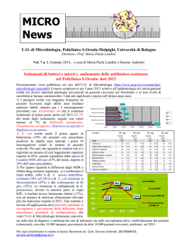
/0 31 `W CT / ¡ HRCT1 D
Reid )῍
©45]^DE>ª;uvrd(nbcde`a /«¬ ®1 ¡;peqrd(nbcd
e`a /«¬ ¯1 ? uvrd(nbcde`a*-
°324
5DE ±`a²³0DE A B o;`peqrd(nbcde`aDE ¦§
10 B>¨ῌ
¶῏·¸¹º»¼
Vol. 19
No. 2
2009. 11
A³´Kµ¶K·K¸¹º
78
Streptococcus pneumoniae (23.8!24.6j), Haemophilus influenzae (6 ! 18.5j) R # @ >
Staphylococcus aureus (1.4!3.4j), Klebsiella pneumoniae (1.3!1.4j), Moraxella catarrhalis (1.7!
2.2jG &CR13, 14) Streptococcus pneumoniae 6 E6kh6G Klebsiella pneumoniae 6 El
mnopq6G A06D/E"
6F&/'7) h0
6-r> B-GH1ih#/sI
0JtK)-Lu06 MNO S2, S6, S10 -vF@6
| 3.
=Z
¯|
==Z}%"> *a/*#"9"A°
"> HRCT 0.1 mm 11 <±*²/
R B-wx%#1)<T/4$
`+P HRCT Q7-< y0zL{
i|`}06D/~)
%J>+15, 16) <=
RS-rBGHA]
T%6:)N
-06 EA06 06'U"
G 6 E06G -V">+
0.1 mm HRCT "1M X 3W|+\]J
0.1 mm J+ -=Z*
&XNE?
10) HRCT &PB-RSYN5"
0.5!1 mm 0.05!0.15 mm HRCT 61)<ZR<T@[-|
"
#$% &'(
=
) HRCT * #
+,-./0-
1
/!%*"#/ 2
\]h0606 6-
>`^f #@>_`B-.C->'^
f+
ΐῑΐ῎ ῌῒῑΐ῎῍
3-
4$
%5' 6&'7(8+%5
1.
- #"9"*:)*;-< HRCT 06:au<= `
"<=-
+,>#"?@,A/
-bc@+1ih-d/e6&0@(
-
8 fg/ /1ihf
CT B-.C/D@> 01* E2F
hi@ S;-e@>vkj/
34AG 501* 6H01* ^/6k@fl0m+,
7
& 2F34A-I
nop9 r<|ῌ Kohn %J>
1/KL87- Y M4
ῌ Lambert &qrP
-2F@>N"OA 0
@>fst/uRv@>R R
1*9P@+6&Q:/ #RS
v?. %J+ ¡?.
;&-< =J+01*<T/%
w]> Ex02yG '7/w]
5 - 2 F U V W X % Y " +
z{`Rv/ ¢&rA0
Reid =Z[>%1)\]< 6
76H01* ?.^
CT B-£X¤¥¦Wo§XD@ #6H
OA
&%5- -_`"abcd
¨6/©qªOA
E| 4G £X¤¥
Y"+ Miller =Ze@%f<
¦Wo§X{#"}« CT
~-<J>"+4)e¬@ 0
-@O®L6H
4)
III. ῐ῏ῑΐ῎
/©qªOA L6HC
g6ABh06 1ihN
CT ~ /©q
12 \(»¼
Vol. 19
No. 2
2009.
CT RM¹
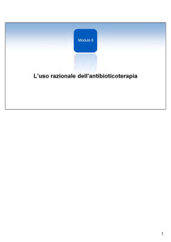
4.
©ª«¬®><
; CT' ¯SC ;
J air bronchogram A
' _°J
t`±u 4V
'
5.
CT 79
h
; HRCT t¯<,²³´u' 56
t)u f' ,-J454
=$4V t`±u A
' )0J)µ t`±¶
·¸u '
!"#"#$%&"#' (
CT R9:;<=>?@:#4V
)* +,-.,-
,-.)J45"
t
#/01*%2"34' 5678
5u' 56.oJ8o"rs^Jkf
9:;<=>?@:56
89 CT ,-45:;`<
(air bronchogram) A
'
,=$V {\;*3qJk
4BCD
>^' ?@4)A
E%F4 bulging fissure sign kB8J air bronchogram ' k
' GHI#789:;<=>?@
B8JC
DE# *
:J GJ
KL"M
fF;*%G ,-.)J454
N>O (Swiss cheese appear-
f56%2'
ance) %4' N>O
!
)0J
4(#H
PQM"#JRS$T"UV WX
" HI56.J@44045
#YZ%[%&4'
yK0J
`4#' ]
῎῍῏ῑῌ
JyL0#' 5656QJ
2.
56\]YZJ"^_' 56
`a56'()bcde*fV Q
Y C(
MJNz4OIf'
ῑῐῒ
34a5678ghi
3.
+G
' `",j)-JQ
PQHP*vJYVJV
kV #lm)%^_4' nQ
PQ`R%4*f' S]vw
I#56`a56f *56
f(# io`J?@4T
.oJp/"/0%123qJkrsJ
.Uf' 56.V
$TkV t,-45u %4' 56
R%^_4' R*WIJX
YZJ,-%2#vw"
Gf#!Pf' J Y¡¢#
(xyz#( a56{
%v
78QJ|}6ghQ3%7
[¥\J¦]%,j t^
##(~'
§¨4'
PQ GZ`£¤
abcºdV»¼
Vol. 19
u PQ
No. 2
2009. 13
o³vw´Pxµy¶P·z{|P}¸¹~º
80
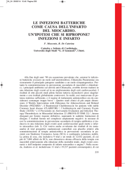
6.
« CT ¬l® ¯m°n ±o
0&'( !Oi8'9pq67
(
7.
>cdeRf>C
« HRCT ¯m° rs)t
Bsu$52 L²T$2&'(
Fqrstu)CGHvchRcw34Q
CT C6%01)
6
u)C]^,I4x; CT JK
yL0z{|w}
>cdeRf>C~
o CT GMSRghij
! "#$ %&
'(
RC klmnRC-.x;NO%
)*+,)-.'(/
70<o0GM8
b
!"01#234$5
90 FPklmnRC
2%%67( &8'9
834QC6 -.%
()*
):+,; feeding ves-
sel sign -./00<
1.
"=$>?@A (pneumatocele) B,C
Mycoplasma pneumoniae ? lipoprotein D 1 E : F G H : F I check
Q(BR80B,
STUVW
valve JKLM;&NOP2NO
XY BRZ[SVW\]
I
?8^_O`;
SVW\]?8^_;6 :Fa
IV ;
34QC (atypical pneumonia) b-RST
9)*So7S+,/0C8]
UVW,56X0 Y/Z7[%; \
:F):dGHw8C
RU89]^,_L'(0 B,`a^:
BReo7:FC8%; >cdeRf>
;bC<=
Mycoplasma pneumo-
C(,af/0ghg
niae C >cdeRf>C Chlamydophila
f/g %Oi'( 7(%
pneumoniae C SRghijRC >?/
9af/g,O`¡¢'(
@0C8];A7( Legionella
£¤¥jk¦
17) f/
pneumophila C klmnRC b-RSTU
g,? lipoprotein >S§i¨$l
VW,56X
\RU89]^,_L'
Toll-like receptors (TLRs) 8©;/
(0 -./0BC8D;o7MpE
hg
18ª20)
14 EFBC:;s»¼
Vol. 19
No. 2
2009.
b c8C8]; CT [v
H 8.
)*+,-.)
HRCT D° ¦ G ± ²
@F MNO"PMNO8J !
S DS³F MN8&
&bc-d +C^<_>`aC
1 DS³´ µ¶F
H 9.
81
w-;xyz-
66 ·T CT DUMF 23
DS³F != DS³´ µ
¶F 1 V¸ 8 Vb¹W
b13, 14) -.{)*+,-.)8
X CT 9|/0Q )*+,-.)}1
CT ~88'qw-;xyz- COPD QR
2$3485& 67
08
!"
!#$
%&'(
)*+,-.)/01 2
345 678
!
!8
CT 9
EF 4 G41
DH 7F : I J 6 K 9 L 1 8) DH 9F &bc-de+C^<_>`a
7, 8, 21) MNO PMNOQR
Cj\]<& !:S 23"
88n1)*+,-.);q
!=)*+,
!UVW
-.)'>?@
!X#$IYZ$
8) qqw-;xyz- COPD QR
7K9 L1
!S
!$&
Reid 92:;<=>?@AB>C D
7, 8, 21)
DH 8F TJ
2$A&~8]Bqe&q
)*+,-.)V:Q
!"
!'([\] +C^<_>`aC
@b #$CDE
" 25n <CA
k! 33n1
5 K 8 L & b c - d 7 K 8 L 1 3.
7, 8, 21) 1 2eMN8&
Legionella pneumophila FGfG4
\]fMN$gJh$&\]
2i)*+,-.)AB>C
2$A& 2K5 mm -
Ht +t$@ I8V
k!e"#$%lm
b8JK& )wy¡>¢8VL£
82n, 73n &o'pq(1
¤M¥
)wy¡>¢UVW II {¦NO
22)
§ Ds*F q$¨& 67
08
j <CA
2.
Chlamydophila pneumoniae Dr Chlamydia pneumoniaeF $JQ)s*t
!+'( # yz©<CQ
Rª«P¬j di#use alveolar damage (DAD) $&
+,8Uuv%)*+,-.)
Q1$&
5.2K11.2n w-;xyz- 3.4K6.5n
J ®1R¯23) -MN8&&bcX+YºZ*[
»¼
Vol. 19
No. 2
2009. 15
°±6²³´7µ8¶´·9:¸´¹º;»
82
4)
5)
6)
) 10.
234567
$ CT (1£*+ £¤K
FG6b (2¥*+ 3
¦
§>-$¨ (2¥© ª«* ¬&
!+ 4.5C® ! (2
¥© V ¯*+
7)
8)
!"#$
9)
%#&!' () 10*+ ,-"./012345
67. 35 8.9: 24 8 (69;) !
10)
7<=7$1 9;$%>-#
?&!'+ @
.A.BC.DE#1 .FG6
,!- 8 8 (22;) 11)
.HI.J 2 KLMN
7O- 1 $%>-+
12)
Vῌ ῍ ῐ ῏ ῎
7P CT .K.QR1STU
1'+ VWX XY#Z
[\
]^_!` 7.abcUdefghi
13)
"#$j$%k9+ ,-l7. CT BC
/0m
?1n_oF p/0b,_!
`qrhsK1t'+
14)
ῒ
ῑ
1) uvLXwuvLxy8mGz{6z
|}~w 2007. } 7qGz{6
z p. 2ῌ35, uvLXw !
2) "#
1997. 7.qroi$ CT .
Kh'.% X&Xw
57: 258ῌ264.
3) Fine, M. J., M. A. Smith, C. A. Carson, et al. 1996.
Prognosis and outcomes of patients with community-acquired pneumonia. A meta-analysis.
16 p.¼<X
Vol. 19
No. 2
2009.
15)
16)
JAMA 275: 134ῌ141.
Hasley, P. B., M. N. Albaum, Y. H. Li, et al. 1996.
Do pulmonary radiographic findings at presentation predict mortality in patients with community-acquired pneumonia? Arch. Intern.
Med. 156: 2206ῌ2212.
Rello, J., R. Rodriguez, P. Jubert, et al. 1996.
Severe community-acquired pneuimonia in the
elderly: Epidemiology and prognosis. Clin. Infect. Dis. 23: 723ῌ728.
Niederman, M. S., L. A. Mandell, A. Anzueto, et
al. 2001. Guidelines for the management of
adults with community-acquired pneumonia.
Diagnosis, assessment of severity, antimicrobial therapy, and prevention. Am. J. Respir.
Crit. Care Med. 163: 1730ῌ1754.
Reittner, P., S. Ward, L. Heyneman, et al. 2003.
Pneumonia: High-resolution CT findings in 114
patients. Eur. Radiol. 13: 515ῌ521.
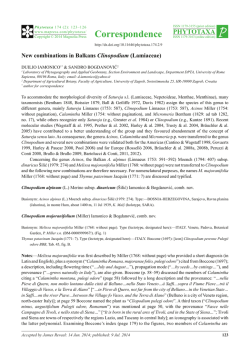
Nambu, A., A. Saito, T. Araki, et al. 2005. Chlamydia pneumonia: Comparison with findings of
Mycoplasma pneumoniae and Streptococcus
pneumonia and thin-section CT. Radiology 238:
979ῌ986.
Miller, W. S. 1947. The lung. Springfield, III:
Thomas. p. 162ῌ204.
Webb, W. R.. 2006. Thin-section CT of the secondary pulmonary lobule: Anatomy and the
image. The 2004 Fleischner lecture. Radiology
239: 322ῌ338.
Reid, L., G. Simon. 1958. The peripheral pattern
in the normal bronchogram and its relation to
peripheral pulmonary anatomy. Thorax 13:
103ῌ109.
Reid, L. 1958. The secondary pulmonary lobule
in the adult human lung, with special reference
to its appearance in bronchograms. Thorax 13:
110ῌ115.
Saito, A., S. Kohno, T. Matsushima, et al. 2006.
Prospective multicenter study of the causative
organisms of community-acquired pneumonia
in adults in Japan. J. Infect. Chemother. 12: 63ῌ
69.
Ishida, T., T. Hashimoto, M. Arita, et al. 2004. A
3-year prospective study of aurinary antigendetection test for Streptococcus pneumoniae in
community-acquired pneumonia: Utility and
clinical impact on the reported etiology. J. Infect. chemother. 10: 359ῌ363.
'() 1990. g,K*K
7 uv 9: 263ῌ267.
': ( + A 1998. ,
- ../* #¡\!t0¢.g,
K*K
7. 1 uvLXw
36: 444ῌ447.
CT 17)
18)
19)
20)
Low, I. E., S. M. Zimkus. 1973. Reduced nicotinamide adenine dinucleotide oxidase activity and H2O2 formation of Mycoplasma pneumoniae. J. Bacteriol. 116: 346ῌ354.
Chu, H. W., S. Jeyaseelan, J. G. Rino, et al. 2005.
TLR2 signaling is critical for Mycoplasma pneumoniae-induced airway mucin expression. J.
Immunol. 174: 5713ῌ5719.
Shimizu, T., Y. Kida, K. Kuwano, 2007. Triacylated lipoproteins derived from Mycoplasma
pneumoniae activate nuclear factor-kappaB
through toll-like receptors 1 and 2. Immunology 121: 473ῌ483.
Wu, Q., R. J. Martin, S. Lafasto, et al. 2008.
Toll-like receptor 2 down-regulation in estab-
21)
22)
23)
83
lished mouse allergic lungs contributes to decreased mycoplasma clearance. Am. J. Respir.
Crit. Care Med. 177: 720ῌ729.
Tanaka, N., T. Matsumoto, T. Kuramitsu, et al.
1996. High resolution CT findings in community-acquired pneumonia. J. Comput. Assist. Tomogr. 20: 600ῌ608.
Lee, I., T. S. Kim, H. K. Yoon. 2006. Mycoplasma
pneumoniae pneumonia: CT features in 16 patients. Eur. Radiol. 16: 719ῌ725.
Sakai, F., H. Tokuda, H. Goto, et al. 2007. Computed tomographic features of Legionella pneumophila pneumonia in 38 cases. J. Comput. Assist. Tomogr. 31: 125ῌ131.
Understanding of Community-acquired Pneumonia
on the basis of chest computed tomography imaging
Hirotsugu Ohkubo, Yuki Togashi, Yuta Kono, Yasuhiro Setoguchi
First Department of Internal Medicine, Tokyo Medical University,
6ῌ7ῌ1 Nishi-shinjuku, Shinjuku-ku, Tokyo 160ῌ0023, Japan
Computed tomographic (CT) scans are now working in many general hospitals in Japan. Most of the
Japanese clinicians can use CT scans as one of the useful tool of the examination of community-acquired
pneumonia. Chest CT imaging plays an important role in the accurate interpretation of pulmonary
anatomy and pathology. Mycoplasma pneumoniae pneumonia and Chlamydophila pneumoniae pneumonia
demonstrate centrilobular or acinar shadows, and thickening of bronchivascular bundles on CT imaging.
Characteristic findings of Legionella pneumophila pneumonia on CT are sharply demarcated peribronchovascular foci of consolidation intermingled with ground glass opacity. It is possible to estimate the
pathogenic bacteria, if there are characteristic features on CT imaging on the basis of clinical information.
Vol. 19
No. 2
2009. 17
© Copyright 2026 Paperzz