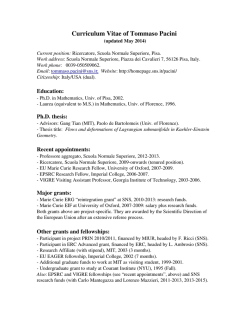
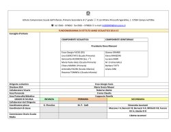
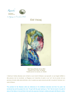
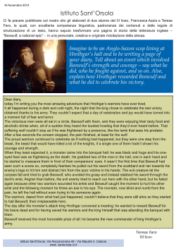
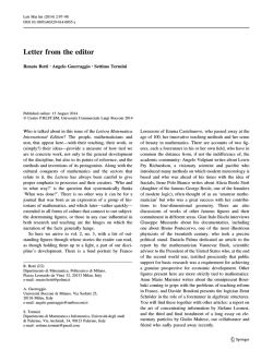
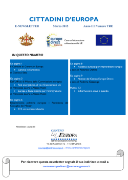
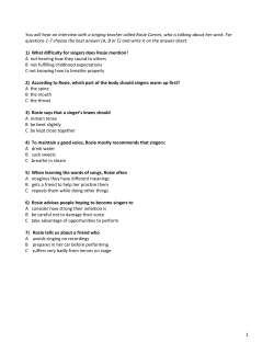
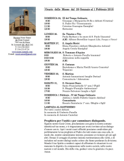
BrainResearchBulletin,Vol. 38, No. 2, pp. 161-165, 1995
Copyright © 1995ElsevierScienceLtd
Printedin the USA.All rightsreserved
0361-9230/95 $9.50 + .00
Pergamon
0361-9230(95)00083-6
Filippo Pacini: A Determined Observer
MARINA BENTIVOGLIO.1 AND PAOLO PACINI1-
*Institute of Anatomy and Histology, University of Verona, Italy
?~Department of Anatomy and Histology, University of Florence, Italy
[Received 7 November 1994; Accepted 27 March 1995]
ABSTRACT: The life of Filippo Pacini (1812-1883) and his major
scientific achievements are outlined. Pacini drew attention to
the corpuscles named after him in 1831, when he was a medical
student, and had to straggle for many years to convince the scientific community of the reliability and importance of his findings. In 1849 Pacini became professor of anatomy at the University of Florence. Creative scientist, innovative teacher, well
aware that the use of the microscope represented a revolutionary approach, Pecini pursued histological studies until his death.
He also first discovered in 1854 (30 years before Robert Koch)
the causative agent of cholera, and firmly believed that the disease was contagious. Strong-willed, modest, and poor, Pacini
received from his colleagues more recognition in the obituaries
then during his life.
KEY WORDS: Pacinian corpuscles, History of neuroscience, Infectious diseases.
INTRODUCTION
Pacinian corpuscles are primary mechanoreceptors that play a
role in the sensation of touch. These relatively large receptors
(whose long axis is about 2500 #m in humans [1]) can even be
seen with the naked eye, and their description dates back at least
to the 18th century. Filippo Pacini, unaware of previous reports,
rediscovered the corpuscles that were named after him, and struggled to bring his findings to the attention of the scientific community. Pacinian corpuscles were the first specialized end organs
in the skin to be identified, although their functional role in sensation was identified only much later [8].
This article is aimed at providing a brief overview of Pacini's
contribution to science and to neuroscience in particular.
digital nerves [21]. These observations seem to have been then
forgotten for many decades.
The lamellar corpuscles were again identified by John Shekleton (1795-1824). As reported recently [20], the corpuscles
were dissected out, together with many other pieces of dissection,
while Shekleton was the curator of the Museum of the Royal
College of Surgeons in Ireland between 1820 and 1824. Shekleton's findings were published by Smith in 1848 [19], when
Pacini's reports had already appeared.
PACINI'S BIOGRAPHICAL SKETCH
The following data on Pacini's life and studies on the lamellar
corpuscles are derived from Coturri [5,6] and one of Pacini's
students, Aurelio Bianchi, who collected for the National Library
of Florence Pacini's manuscripts after his death [2], Filippo Pacini (Fig. 1) was born in Pistoia, in Tuscany, on May 25, 1812.
He was of humble origin (his father was a cobbler) and received
a strict religious education. His family wanted him to become a
bishop and committed him to religious studies. Pacini, however,
soon abandoned the ecclesiastical career. In 1830 he was awarded
a scholarship and admitted to the famous "Scuola Medica Pistoiese," founded in 1666 by the Ospedale del Ceppo in Pistoia.
At this medical school Pacini attended the basic courses with
excellent results, becoming a very experienced dissector.
In 1849 Pacini obtained the chair of General and Topographic
Anatomy at the University of Florence, which he held until his
death. Pacini never married, and for a long time took care of his
two sisters, both seriously ill. He spent all his money in assisting
his sisters and supporting his own studies. Pacini died, very poor,
on July 9, 1883.
THE REDISCOVERY OF PACINIAN CORPUSCLES
THE FIRST DESCRIPTIONS OF PACINIAN
CORPUSCLES
In 1831, attending as an "intern student" the dissections in
the anatomy course of the second year of medical school, Pacini
observed some small ovoidal bodies attached to the digital
branches of the median and ulnar nerves, hardly visible to the
naked eye. "What are these?" he asked to his professors, and
was very unsatisfied by the answer: "Small globules of fat tissue." Pacini did not find any mention of the corpuscles in his
anatomy textbooks. Very determined and hard-headed, he used
all his savings to buy a small microscope with a wooden tube
and acromatic lenses, providing a 30 times magnification, and
began to investigate on his own the morphology of the small
Johannes Gottlieb Lehmann [13, cited in ref. 1] is credited for
the description of Pacinian corpuscles in his Doctoral thesis in
1741. It is, however, commonly recognized [4,8] that Pacinian
corpuscles were first observed in the same year by the German
professor of Anatomy and Botany, D. Abraham Vater (16841751). Without providing any structural description, Vater made
a drawing of the lamellar corpuscles (the original specimens were
preserved in the Anatomical Museum of GOttingen) and classified them as Papillae Nerveae, attached in large quantities to the
Requests for reprints should be addressed to Dr. M. Bentivoglio, Institute of Anatomy and Histology, Medical Faculty, Strada Le Grazie, 37134
Verona, Italy. E-mail: [email protected]
161
162
BENTIVOGLIO AND PACINI
In the meantime, also French scientists (Andral, Camus, and
Lacroix) had observed the corpuscles in 1833 and provided a
description in an anatomy textbook in 1836 [7]. Pacini was not
aware of this report, and published in extenso his findings in 1840
I 151, providing a drawing of the ovoid bodies formed by concentric lamellae, in which an "elementary nerve fiber" was
coursing through a "funiculus" to the "central cylinder" of the
corpuscle (Fig. 2).
In 1844 Pacini presented his work again at a congress in
Lucca, describing the peculiar histological technique employed
to visualize the corpuscles, which had been treated for 36 h with
diluted nitric acid and ethanol. Pacini could thus demonstrate the
" c o m p l e t e " structure of the corpuscles. He reported that the interlamellar spaces contained some fluid and that the lamellae
were connected by a ligament (Legamentum intercapsulare) to
the distal pole of the corpuscles. He also suggested that the corpuscles could represent "terminal ganglia related to the animal's
bioelectrical activity."
In the same year (1844), the famous German histologist Jakob
Henle (1809-1885) and his assistant, Rudolph Albrecht von K61liker ( 1 8 1 7 - ! 905), published a detailed study on the corpuscles,
which they named after Pacini [ l 1]. Henle and KOlliker described
that the lamellae consisted of cells and two different layers of
FIG. 1. Portrait of Filippo Pacini ca. 1870.
corpuscles. Convinced that the hand corpuscles were related to
nerve fibers, Pacini defined them as "tactile ganglia." In 1833
he found the same corpuscles between the dorsal surface and the
large vessels of the pancreas.
In October 1835 Pacini submitted a scientific communication
to the Societh Medico-Fisica Fiorentina (Medico-Physical Society of Florence). In this manuscript [15] he reported the identification of the small corpuscles connected with the digital nerves
and also distributed in different splanchnic regions: "small ellipsoidal globose corpuscles, just like a cyst with an inner pulpy
and white substance, moistened by a limpid fluid; their larger
diameter is crossed by a little streak which is whiter than the
remaining part of the corpuscle." The original report was read
by the secretary of the Medico-Physical Society during the session held on November 22, 1835; the reaction of the audience
was very cold, and Pacini's findings did not raise any interest.
However, his research activity did not seem to suffer much from
such indifference, and Pacini insisted in presenting further observations to a scientific congress held in Pisa in 1839. On that
occasion, the congress President appointed an ad hot: committee
to examine Pacini's data. The secretary of the committee (Francesco Puccinotti, professor of Internal Medicine at the University
of Pisa) finally declared that the corpuscles did indeed exist, but
it was unclear whether they were neural structures (as proposed
by Pacini) or "tendinous-aponeurotic expansions."
C O R P U S C O L O D E L PACINI
Riproduzione & m ~
del vero del 414~no autogmf~ di Filippo
Paclni
(.,~ee. Pac~,i, Vol. XX, e. 9).
FIG. 2. Pacini's first drawing of the lamellar corpuscles, made in 1840.
Handwritten labels: Funiculus, elementary nerve fiber, central extremity
of the corpuscles, central small cylinder, peripheral extremity of the corpuscle.
FILIPPO PACINI
fibrillar tissue, the outer layer being transverse and the inner longitudinal. Henle and K611iker also found blood vessels around
and within the corpuscles and described the myelinated nerve
fiber, which could divide into terminal branches, ending within
them; they suggested that Pacinian corpuscles were "electric organs," comparable with those of some "electric" fish. Thus,
Pacinian corpuscles had finally been christened, and their connection to a nerve fiber had been established.
In 1863 Pacini commissioned a set of anatomical models of
the corpuscles from the Florentinian wax modeller Remigio Lei.
Such models were prepared according to the artistic, and almost
secret, traditions of the Wax Laboratory of the University Zoological Museum "La Specola," which possesses a remarkable
and large collection of human and animal anatomy wax models.
A plaster cast was added to the clay model. After its solidification, a large series of differently coloured wax layers were accurately settled down. Muscle fibers, blood and lymphatic vessels, as well as nerve fibers, completed the work. These unique
wax models still display in all their striking perfection in the
Anatomical Museum of the University of Florence (Fig. 3).
PACINIAN CORPUSCLES AFTER PACINI'S STUDIES
Mayer [14] performed preliminary physiological studies on
the corpuscles. In 1876 Key and Retzius [11] presented in their
monograph a large collection of drawings of the different types
of Pacinian corpuscles throughout the human body. Detailed surveys of the older literature on Pacinian corpuscles and the first
163
hypotheses on their function can be found in refs. [1] and [4].
Bell et al. [1] also provided an updated review of the structure
and function of Pacinian corpuscles. It has now been definitely
established that these encapsulated nerve endings are widely distributed in the human body, where they are numerous in the skin
and concentrated in the fingers, and have been found in many
other structures (such as in articular capsules, on tendons and
within muscles, near peripheral nerves, and in the urinary tract
and bladder, as well as genital organs of both sexes). Pacinian
corpuscles have also been described in many higher vertebrate
species and are very numerous in the mesentery of the cat.
Light and electron microscopic studies (reviewed in ref. [1])
have elucidated that the Pacinian corpuscles are innervated by a
myelinated axon, whose terminal segment is unmyelinated and
has a bead-shaped ending. The neurite, that can divide in two or
more endings, is surrounded by nonneuronal cytoplasmic layers
arranged in concentric lamellae. The inner and outer cores, separated by an intermediate growth zone, form the inner capsule of
the corpuscle. The lamellae of the inner core are very closely
packed. The lamellae of the outer core are instead separated by
relatively wide spaces, filled with fluid containing macrophages,
collagen fibrils, and vascular capillaries. The inner capsule is
enclosed by a few tightly packed lamellae and by connective
tissue, which form the external capsule of the corpuscle. Each
lamella consists of a single layer of tightly fitting cells of epithelial type.
It has now been established that cutaneous Pacinian corpuscles are quickly adapting mechanoreceptor responding to high
FIG. 3. Wax model of Pacini's organs commissioned by Pacini himself. The hand is "natural size," and two
different magnifications of Pacinian corpuscles are shown.
164
BENTIVOGLIO AND PACINI
frequency vibrations. The accessory structure, which includes the
inner core, intermediate growth zone, outer core, and external
capsule (making up 99.9% of the size of the corpuscles) does not
play a role in the signal transduction but is involved in the regulation of the ionic environment surrounding the afferent axon
and in the mechanical filtering of the stimulus [ 1IPACINI AS INNOVATIVE TEACHER AND
PASSIONATE MICROSCOPIST
During his university career Pacini introduced (first in Italy)
the course of histology and the systematic study of microscopic
anatomy in Medical Schools, and was giving practical demonstrations at the microscope during his lectures [6]. His microscope (donated by the philanthrope Niccol6 Puccini) was unique
in the Faculty. A large collection of letters (kept in the Library
of the town hall of Pistoia) to the Dean and Rector of the University and especially to the Grand Duke of Tuscany, Leopold
II, documents that Pacini had been fighting for years to obtain
microscopes for his students. The Grand Duke initially did not
pay much attention to Pacini's requests and received him in a
great hurry, impatient to go hunting. However, persuaded by Pacini's insistence, the Grand Duke finally donated one microscope
to the University of Florence in the academic year 1844-1845,
recommending to Pacini not to waste too much time with the
microscope during his courses [6].
At the end of 1844 Pacini investigated microscopically the
retina, and described the membrana limitans interna 117]. In his
subsequent studies he proposed new histological methods and
technical modifications of the microscope. Pacini also favoured
the introduction of mathematical models in biological studies,
and proposed a new method for artificial respiration 121.
PACINI AND THE DISCOVERY OF THE
VIBRIO CHOLERA
During a cholera epidemic in Florence in the summer of 1854,
Pacini carried out detailed investigations on the etiology of the
disease. He based his studies on the histological examination of
the intestinal mucosa of cholera patients, performing the autopsy
himself a few hours after their death. During these studies, Pacini
first discovered a bacillus, which he described as a Vibrio with a
comma-like shape. Pacini pursued his investigations on cholera
for about 20 years and published several articles (reviewed in ref.
[10]). He provided a thorough description of the destruction of
intestinal mucosa during cholera and ascribed this devastating
damage to the bacillus he had observed. Pacini clearly stated that
this Vibrio was the "specific" causative agent of the disease. In
addition, Pacini insisted that cholera was contagious [3]. The
principle of a contagium vivum [9] had already been pointed out
by Girolamo Fracastoro (1478-1553), a Veronese fellow student
of Nicholas Copernicus ( 1473-1543) at the University of Padua.
However, the idea that cholera was contagious was still under
dispute, and Pacini's warnings for severe prevention rules were
contradicted by the illustrious clinician Maurizio Bufalini ( 17871875), very influential in Italy at that time.
Pacini's data were ignored by the scientific community. The
ctiological agent of cholera was rediscovered by Robert Koch
11843-1910) in May 1884 (30 years after Pacini's description
and 1 year after his death) and was named Vibrio cholera Koch.
Thus, Pacini never knew that his observations had been confirmed.
Some of the microscope slides prepared by Pacini for his studies on cholera are still kept in the Department of Anatomy and
Histology of the University of Florence (Fig. 4).
LATE RECOGNITION
As reported by Bianchi [2], many scientists honoured Pacini
on the occasion of a celebration that was held in Pistoia in 1885,
and among those:
Camillo Golgi
Filippo P a c i n i . . . was one of the first to make anatomical studies using the new avenues opened by the m i c r o s c o p e ; . . , was
one of the first to understand the new horizons provided to anatomy by comparative studies and . . . was one of the first to
enrich the microscopic technique with procedures that represent
a precious help to researchers, having understood the importance
of techniques for the minute investigations.
FIG. 4. One of the slides prepared by Pacini from the intestinal mucosa of cholera patients.
FILIPPO PACINI
165
Gustav Retzius
Filippo Pacini is one of those men who belong not only to their
h o m e land but to the entire world.
Wilhelm Waldeyer
All my admiration to an investigator who, far away from the
largest scientific centers, animated by a pure love for science,
obtained such important results with the simplest tools.
Wilhelm Krause
I have now been teaching histology at the University of GiSttingen for 25 years. W h e n I have to start dealing with nerve terminals I begin with Pacinian corpuscles. And every semester I
recall that Pacini, still as a student, with a small nonacromatic
microscope and a wooden tube, like those sold at fairs, discovered the corpuscles that are now n a m e d after him, which represent the starting point for all that we know nowadays on sensory
nerve terminals: you see, gentlemen, it is not the instrument but
the observer who makes the discoveries.
REFERENCES
1. Bell, J.; Bolanowski, S.; Holmes, M. H. The structure and function
of Pacinian corpuscles: A review. Progr. Neurobiol. 42:79-128;
1994.
2. Bianchi, A. Relazione e catalogo dei manoscritti di Filippo Pacini,
Florence-Rome: Tipografia Bencini; 1889.
3. Castaldi, L. Filippo Pacini nel quarantesimo anniversario della sua
morte. Rivista di Storia delle Scienze Mediche e Naturali 14:182212; 1923.
4. Cauna, N.; Mannan, G. The structure of human digital Pacinian corpuscles (Corpuscula lamellosa) and its functional significance. J.
Anat. 92:1-25; 1958.
5. Coturri, E. Contributo alia storia della divulgazione della scoperta
dei corpuscoli del Pacini. Atti e Memorie dell'Accademia di Storia
Sanitaria 23:1-7; 1957.
6. Coturri, E. Un grande anatomico pistoiese dell'Ottocento: Filippo
Pacini. Studi Storici Pistoiesi 3:35-52; 1978.
7. Cruvelhier, J. Anatomie descriptive. Paris; 1836.
8. Finger, S. Origin of neuroscience. New York & Oxford: Oxford
University Press; 1994.
9. Fracastoro, G. De sympathia et antipathia rerum liber unus: De contagione et contagiosis morbis et curatione libri tre. Venice: Eredi
Juntae Florentini; 1546.
10. Franceschini, P. Filippo Pacini e il colera. Phisis 13:324-332; 1971.
l 1. Henle, F.; K/Slliker, A. Ueber die Pacinischen Korperchen an den
Nerven des Menschen und der Sangethiere. Zurich; 1844.
12. Key, A.; Retzius, G. Studien in der Anatomie des Nervensystems
und des Bindegewebes. Stockholm: Samson & Wallin; 1876.
13. Lehmann, J. C. Dissertatio inauguralis medica de consensu partium
corporis humani occasione spasmi singularis in manu ejusque digitis
ex hernia observati; exposito simul nervorum brachialium et crurallure coalitu peculiari atque papillarum nervearum in digitis dispositione. Doctorate Thesis, Vitembergae: Typis Scholmachianis;
1741.
14. Mayer, A. F. Die Pacinischen Korperchen: Eine physiologische Abbandlung. Bonn:C. Georgi; 1844.
15. Pacini, F. Sopra un particolare genere di piccoli corpi globosi scoperti nel corpo umano da Filippo Pacini, Alunno interno degli Spedali riuniti di Pistoia. Letter to the Accademia Medico-Fisica di Firenze; 1835.
16. Pacini, F. Nuovi organi scoperti nel corpo umano. Pistoia: Tipografia
Cino; 1840.
17. Pacini, F. Nuove ricerche microscopiche sulla tessitura intima della
retina. Bologna; 1844.
18. Pacini, F. Osservazioni microscopiche e deduzioni patologiche sul
Cholera asiatico. Florence: Tipografia Bencini; 1854.
19. Smith, R. W. Treatise on the pathology, diagnosis and treatment of
neuroma. Dublin: Hodges and Smith; 1848.
20. Thompson, A. J.; Martin, E. A. The Pacinian corpuscle and John
Shekleton. Neurology, 35:1747; 1985.
21. Vater, A. Dissertatio de consensu patrium corporis humani. Hailer,
Disputationum Anatomicarum selectarnm, Voi. II, Gottingen,
1741:953-972.
© Copyright 2026 Paperzz