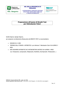
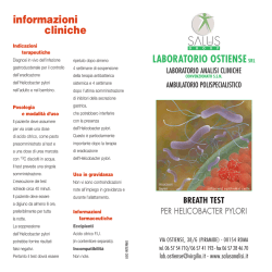
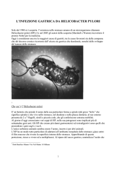
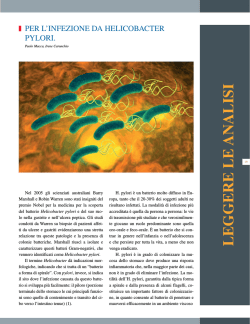
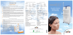
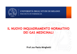
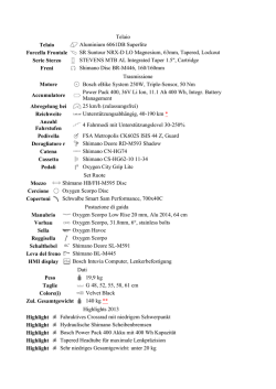
FEMS Microbiology Letters 168 (1998) 9^15
The e¡ect of oxygen on the growth and cell morphology of
Helicobacter pylori
Gianfranco Donelli a , Paola Matarrese a , Carla Fiorentini a , Benedetto Dainelli b ,
Tea Taraborelli b , Emanuela Di Campli b , Soraya Di Bartolomeo b ,
Luigina Cellini b *
b
a
Laboratorio di Ultrastrutture, Istituto Superiore di Sanitaé, Viale Regina Elena, Rome, Italy
Dipartimento di Scienze Biomediche, Laboratorio di Batteriologia Medica, Facoltaé di Medicina, Universitaé `G. D'Annunzio',
Via dei Vestini 31, Chieti, Italy
Received 4 May 1998; received in revised form 31 August 1998; accepted 31 August 1998
Abstract
The in vitro effect of progressive oxygen decrease on the growth and morphology of Helicobacter pylori was studied. H.
pylori ATCC 43504 was used for the experiments. The strain inoculated in Brucella broth plus fetal calf serum was incubated
under a controlled atmosphere with oxygen concentration from 5 to 0%. CFU ml31 and bacterial morphology were detected at
the time of spreading and at 24 h, 72 h, 7 days and 14 days. A detailed ultrastructural investigation of the bacterial cells, grown
in different experimental conditions, was performed by scanning electron microscopy. Oxygen deprivation produced a rapid
reduction of CFU ml31 . In particular, a significant reduction of viable bacteria was recorded at 72 h of incubation in the
presence of 1% oxygen and anaerobiosis, and 0 CFU ml31 was found after 7 days of incubation at the above mentioned oxygen
concentrations. The coccoid phenotype was already prevalent after 24 h of incubation with a progressive tendency to aggregate
in clusters. These clusters were progressively larger, depending on the reduction of oxygen concentration, since the aggregation
phenomenon can be the expression of a hypothesized mechanism of protection among bacterial cells. z 1998 Federation of
European Microbiological Societies. Published by Elsevier Science B.V. All rights reserved.
Keywords : Helicobacter pylori; Oxygen; Morphology; Viable but not culturable state
1. Introduction
Helicobacter pylori is now de¢ned as the main
etiological agent of active gastritis and peptic ulcer
[1^4] and the persistence of this germ in the stomach
increases the risk of gastric cancer [5,6].
* Corresponding author. Tel.: +39 (871) 355 5279;
Fax: +39 (871) 355 5282; E-mail: [email protected]
While H. pylori infection is the most common
gastrointestinal bacterial disease worldwide, knowledge of its mechanism of transmission is still poor
[7^12].
H. pylori, as well as Vibrio-like bacteria, shows
two di¡erent morphologic aspects: the typical spiral
form and the coccoid form, the latter observed in
vivo [13], and induced in vitro under stress conditions [14^17]. The coccoid forms are not culturable
in vitro and, for a long time, investigators have dis-
0378-1097 / 98 / $19.00 ß 1998 Federation of European Microbiological Societies. Published by Elsevier Science B.V. All rights reserved.
PII: S 0 3 7 8 - 1 0 9 7 ( 9 8 ) 0 0 4 0 5 - 4
FEMSLE 8408 23-10-98
10
G. Donelli et al. / FEMS Microbiology Letters 168 (1998) 9^15
cussed their viability and their role in germ transmissibility and in infection relapse [13,17^23]. This
morphology seems to be related to a speci¢c form
of `dormancy' occurring in non-sporulating bacteria
[15,16,24].
According to our data [25], coccoid forms represent the morphological expression of a viable but not
culturable (VBNC) state, corresponding to a temporary adaptation to an unsuitable environment.
A focal point in the study of VBNC H. pylori is
represented by the singling out of the di¡erent factors which can induce these morphological forms.
Moreover, clear di¡erences in density, DNA contents and secreted proteins among coccoid forms
[26] are the consequence of various cultural conditions such as nutrient deprivation, antibiotic addition, unfavorable atmosphere.
The aim of this study was to evaluate, in vitro, the
e¡ect of oxygen deprivation on the growth of
H. pylori and on the morphology of the bacterial
cells.
2. Materials and methods
2.1. Bacterial strain and growth conditions
H. pylori ATCC 43504 was the standard control
strain used for this work.
Microorganisms, stored at 370³C by the method
of Drumm and Shermann [27], were defrosted to
room temperature, and rapidly plated in chocolate
agar plus 1% of IsoVitaleX (CA) (Becton Dickinson,
Cockeysville, MD, USA) at 37³C for 3 days in a
microaerophilic atmosphere (90% N2 , 5% O2 , 5%
CO2 , CampyPak Jar, Oxoid Ltd, Basingstoke, UK).
2.2. Determination of conversion rate under di¡erent
oxygen concentrations
Bacteria harvested from almost con£uent CA
plates were inoculated in Brucella broth (BB, Biolife
Italiana, Milan, Italy), supplemented with 2% fetal
calf serum (FCS, Seromed, Biochrom KG, Leonorstiftung, Berlin, Germany), and incubated overnight
at 37³C in a microaerophilic atmosphere with gentle
agitation.
At the start of the experiment, the broth culture
was divided into tubes of 14 ml each. Groups of four
tubes were incubated in separate jars under the following condition of gas mixture: (a) 90% N2 , 5% O2 ,
5% CO2 (standard condition), (b) 91% N2 , 4% O2 ,
5% CO2 , (c) 92% N2 , 3% O2 , 5% CO2 , (d) 93% N2 ,
2% O2 , 5% CO2 , (e) 94% N2 , 1% O2 , 5% CO2 , (f)
95% N2 , 0% O2 , 5% CO2 (anaerobiosis).
As control, experiments with either 10% CO2 , 85%
N2 and 5% O2 (A) or 10% CO2 , 90% N2 and 0% O2
(B) were also performed.
Number of viable bacteria (CFU ml31 ) and bacterial morphology were determined at time 0 and after
24 h, 72 h, 7 days, and 14 days.
2.3. Viable count
The number of viable bacteria was determined by
plating 100 Wl of 10-fold serial dilutions in BB plus
2% FCS broth cultures, on CA plates in controlled
atmosphere (standard condition) at 37³C for 3 days.
For tubes incubated in atmospheres tending to
anaerobiosis (2^0% of oxygen) and, for each mixture, after 7 days of incubation, the viable count
was also performed by plating on CA in a controlled
atmosphere at 37³C for 3 days, both 100 Wl of 10fold concentrated broth culture and the same aliquot
of concentrated broth culture after passage in fresh
broth (1 ml of broth culture plus 9 ml of BB+2%
FCS and incubated overnight in standard microaerophilic environment at 37³C with gentle agitation).
This latter procedure was to evaluate the reactivation of bacteria either in coccoid form or aggregated
in clusters.
2.4. Bacterial morphology
Bacterial features were studied in Gram modi¢ed
stain with a Zeiss standard microscope at magni¢cation 1000U. The morphologies, indicated as bacillary (B), or coccoid (C alone, or grouped in CL
clusters) were counted as follows: for each broth
studied, three slides were read and, for each slide,
four ¢elds were counted. Slides were read by two
`blind' microbiologists.
2.5. Electron microscopic observations
Ten milliliters of each broth culture were centri-
FEMSLE 8408 23-10-98
G. Donelli et al. / FEMS Microbiology Letters 168 (1998) 9^15
fuged for 20 min at 8000Ug and were prepared for
scanning electron microscopy (SEM) by ¢xing them
with 2.5% glutaraldehyde in 0.1 M cacodylate bu¡er
(pH 7.4) at 4³C for 30 min. After several washes in
cold cacodylate, samples were resuspended in 40 Wl
of the same bu¡er. The solutions were dropped on
13-mm polylysine-covered coverslips at room temperature. Then, samples were post¢xed in 1% OsO4
for 30 min, dehydrated through graded ethanols,
critical point dried in CO2 and gold coated by sputting.
The samples were examined with a Cambridge 360
scanning electron microscope.
2.6. Statistical analysis
All experiment data are shown as mean þ S.D. of
triplicate determinations and the results are representative of two separate experiments.
Student's t-test (`Statistic' program for Macintosh)
for correlated samples was used. A P value of less
than 0.01 was considered signi¢cant.
11
3. Results and discussion
The growth of H. pylori ATCC 43504 cultures at
di¡erent percentages of oxygen is shown in Fig. 1.
Similar results were obtained with 5% and 4% oxygen concentration. Only at these oxygen tensions a
signi¢cant increase of bacterial growth, compared to
the starting concentration, was recorded (P 6 0.001).
A peak of 5U108 CFU ml31 was observed at 72 h of
incubation and a signi¢cant reduction of CFU ml31
until values of 106 CFU ml31 was reached after
14 days (P 6 0.001).
The variations of the morphological aspect of the
germ were similar in the above considered environmental conditions (Fig. 2) with prevalence of the
coccoid morphology after 24 h of incubation. Moreover, after 7 days of incubation, coccoid H. pylori
tended to aggregate in clusters (Fig. 2a,b). These
clusters were also observed in conditions c and d
(Fig. 2c,d) in which constant values of CFU ml31
were recorded (Fig. 1), with a reduced culturability
in 2% oxygen (condition d). More stringent oxygen
Fig. 1. Growth of H. pylori in cultures under di¡erent oxygen concentrations (a, 5% O2 ; b, 4% O2 ; c, 3% O2 ; d, 2% O2 ; e, 1% O2 ;
f, 0% O2 ). Values are plotted as mean þ S.D.
FEMSLE 8408 23-10-98
12
G. Donelli et al. / FEMS Microbiology Letters 168 (1998) 9^15
Fig. 2. Percentage of H. pylori forms detected under di¡erent oxygen concentrations (a, 5% O2 ; b, 4% O2 ; c, 3% O2 ; d, 2% O2 ; e, 1%
O2 ; f, 0% O2 ). Each value represents an average of triplicate determinations and the results are representative of two separate experiments. The standard deviations range from 30.3 to +0.3. B, bacillary forms ; C, coccoid forms ; CL, clusters.
deprivation (1% and anaerobiosis, conditions e and
f) revealed a reduction of 10-fold of CFU ml31 at 24
h of incubation and after 7 days of incubation the
loss of culturability (0 CFU ml31 ) was signi¢cant
compared to the previous readings (P 6 0.001).
These latter two condition showed morphologi-
FEMSLE 8408 23-10-98
G. Donelli et al. / FEMS Microbiology Letters 168 (1998) 9^15
13
Fig. 3. Scanning electron micrographs of H. pylori in di¡erent oxygen conditions and growth times. A: 4^5% oxygen concentration ;
B: 2^3% oxygen concentration ; C: 1% oxygen and anaerobiosis. Top line (Aa, Ba, Ca): 3 days of bacterial growth; middle line (Ab, Bb,
Cb): 7 days of bacterial growth; lower line (Ac, Bc, Cc): 14 days of bacterial growth. Magni¢cation : 2100U.
cally (Fig. 2e,f) a large presence of clusters starting
from 24 h of incubation. In particular, at more stringent conditions, in which the percentage of oxygen
was from 2% to 0%, an unusual morphology of H.
pylori at scanning electron microscopy was detected.
Long and undivided rods were observed after 7 days
with a tendency to become ¢lamentous (data not
shown).
The general view of the changes observed in the
morphology of H. pylori, grown in di¡erent oxygen
conditions, is presented in Fig. 3. Panel A represents
H. pylori cells grown at 4^5% oxygen concentration;
panel B at 2^3% oxygen concentration; panel C
bacteria grown at 1% oxygen and anaerobic condition.
The micrographs on the upper (Aa, Ba, Ca), the
middle (Ab, Bb, Cb) and the lower lines (Ca, Cb, Cc)
show bacteria cultivated for 3, 7 and 14 days, respectively.
The most relevant aspect arising from the comparative analysis of the cells grown in di¡erent environmental conditions is represented by the increase in
the number of clusters as a function of the time of
bacterial culture. This phenomenon was more evident with the oxygen deprivation. In particular, in
1^0% oxygen, the presence of clusters (Fig. 2e,f) was
earlier than in other oxygen conditions. Regarding
the size of clusters, a maximum was reached when
the incubation time was prolonged to 14 days and
for the oxygen concentration of 1^0%.
FEMSLE 8408 23-10-98
14
G. Donelli et al. / FEMS Microbiology Letters 168 (1998) 9^15
Fig. 4. Growth of H. pylori in cultures detected in standard conditions (A) and in anerobiosis (B). Values are plotted as mean þ S.D.
Fig. 4 shows bacterial growth in both anaerobiosis
and microaerophilic atmosphere obtained using 85%
N2 , 5% O2 and 10% CO2 . In this microenvironment
with a percentage of CO2 higher (10%) than that
used for the above reported experiments (5%),
CFU ml31 decreased more rapidly and no viable
bacteria were recorded after 14 days of incubation.
In conclusion, our results show that the presence
of 5^4% oxygen in the gas mixture reveals a good
growth of H. pylori, with an increase of 100-fold of
the starting concentration after 72 h of incubation.
The e¡ect of oxygen decrease (3^2%) shows, after
each control time, a relatively constant value of
CFU ml31 . In fact, at 14 days of incubation bacterial
growth was still observed at the above mentioned
oxygen concentration. Further oxygen deprivation
produced a rapid reduction of CFU ml31 . In particular, a signi¢cant reduction of viable bacteria (about
two steps of concentration) was recorded at 72 h of
incubation in the presence of 1% oxygen and anaerobiosis, and 0 CFU ml31 was found after 7 days of
incubation at the above mentioned oxygen concentrations.
Five instead of 10% CO2 enhanced the tolerance
to the presence of toxic catabolic products and to the
starvation.
Particularly interesting are the morphological
changes observed at the ultrastructural level. Coccoid forms are more rapidly induced at low oxygen
concentration with a tendency to aggregate with each
other to form progressively larger clusters in accordance with recent data on adhesive properties of coccoid versus spiral forms of H. pylori [20].
Moreover, the presence of aggregate cocci can be
interpreted as the modi¢cation of the behavioral answers of the germs which, in unfavorable conditions,
tends to overcome the toxicity of the medium by
their clustering.
Krieg and Ho¡man [28] studied the e¡ect of a
high density of bacterial population on its own
self-protection against the toxicity created in the medium because of increased oxygen; the more the
broth culture is concentrated, the higher the protection of cells. An analogous mechanism could be effective in our opposite experimental conditions of
oxygen deprivation in which the presence of clusters,
more evident after 7 days of incubation, can express
the tendency of cells to protect each other.
FEMSLE 8408 23-10-98
G. Donelli et al. / FEMS Microbiology Letters 168 (1998) 9^15
Acknowledgments
This
work
was
supported
by
Grant
CU.9704058.CT04 from the Consiglio Nazionale
delle Ricerche and of the Ministero dell'Universitaé
e della Ricerca Scienti¢ca e Tecnologica (60%).
[15]
[16]
[17]
References
[18]
[1] Goodwin, C.S., Mendall, M.M. and North¢eld, T.C. (1997)
Helicobacter pylori infection. Lancet 349, 265^269.
[2] Labigne, A. and de Reuse, H. (1996) Determinants of Helicobacter pylori pathogenicity. Infect. Agents Dis. 5, 191^202.
[3] Lee, A., Fox, J.G. and Hazell, S. (1993) Pathogenicity of
Helicobacter pylori : a perspective. Infect. Immun. 61, 1601^
1610.
[4] Wyatt, J.I. and Dixon, M.F. (1988) Chronic gastritis ^ a pathogenetic approach. J. Pathol. 154, 113^124.
[5] Asaka, M., Kimura, T., Kato, M., Kudo, M., Miki, K.,
Ogoshi, K., Kato, T., Tatsuta, M. and Graham, D.J. (1994)
Possible role of Helicobacter infection in early gastric cancer
development. Cancer 73, 2691^2694.
[6] Forman, D. (1996) Helicobacter pylori and gastric cancer.
Scand. J. Gastroenterol. 251, 48^51.
[7] Hill, M. (1997) The microbiology of Helicobacter pylori. Biomed. Pharmacother. 51, 161^163.
[8] Mendall, M.A. (1997) Transmission of Helicobacter pylori.
Semin. Gastrointest. Dis. 8, 113^123.
[9] Mendall, M.A. and North¢eld, T.C. (1995) Transmission of
Helicobacter pylori infection. Gut 37, 1^3.
[10] Namavar, F., Roosendaal, R., Kuipers, E.J., de Groot, P.,
van der Bije, M.W., Pena, A.S. and de Graa¡, J. (1995) Presence of Helicobacter pylori in the oral cavity, oesophagus,
stomach and faeces of patients with gastritis. Eur. J. Clin.
Microbiol. Infect. Dis. 14, 234^237.
[11] Sahay, P., West, A.P., Hawkey, P.M. and Axon, A.T.R.
(1995) Isolation of Helicobacter pylori from faeces. J. Infect.
30, 262^263.
[12] Sipponen, P. (1997) Helicobacter pylori gastritis ^ epidemiology. J. Gastroenterol. 32, 273^277.
[13] Chan, W.J., Hui, P.K., Leung, K.M., Chow, J., Kwok, F. and
Ng, C.S. (1994) Coccoid forms of Helicobacter pylori in the
human stomach. Am. J. Clin. Pathol. 102, 503^507.
[14] Bena|ëssa, M., Babin, P., Quellard, N., Pezennec, L., Cenatiempo, Y. and Faucheére, J.L. (1996) Changes in Helicobacter
[19]
[20]
[21]
[22]
[23]
[24]
[25]
[26]
[27]
[28]
15
pylori ultrastructure and antigens during conversion from the
bacillary to the coccoid form. Infect. Immun. 64, 2331^2335.
Cellini, L. (1996) Coccoid forms of Helicobacter pylori. J. Infect. Dis. 173, 1288.
Cellini, L., Allocati, N., Di Campli, E. and Dainelli, B. (1994)
Helicobacter pylori : a ¢ckle germ. Microbiol. Immunol. 38,
25^30.
Kusters, J.G., Gerrits, M.M., Van Strijp, J.A.G. and Vandenbroucke-Grauls, C.M.J.E. (1997) Coccoid forms of Helicobacter pylori are the morphologic manifestation of cell death.
Infect. Immun. 65, 3672^3679.
Berry, V., Jennings, K. and Woodnutt, G. (1995) Bactericidal
and morphological e¡ects of amoxicillin on Helicobacter pylori. Antimicrob. Agents Chemother. 39, 1859^1861.
Bode, G., Mauch, F. and Malfertheiner, P. (1993) The coccoid
forms of Helicobacter pylori. Criteria for their viability. Epidemiol. Infect. 111, 483^490.
Cole, S.P., Cirillo, D., Kagno¡, M.F., Guiney, D.G. and Eckmann, L. (1997) Coccoid and spiral Helicobacter pylori di¡er
in their abilities to adhere to gastric epithelial cells and induce
interleukin-8 secretion. Infect. Immun. 65, 843^846.
Hua, J. and Ho, B. (1996) Is the coccoid form of Helicobacter
pylori viable? Microbios 87, 103^112.
Narikawa, S., Kawai, S., Aoshima, H., Kawamata, O., Kawaguchi, R., Hikiji, K., Kato, M., Iino, S. and Mizushima, Y.
(1997) Comparison of the nucleic acids of helical and coccoid
forms of Helicobacter pylori. Clin. Diagn. Lab. Immunol. 4,
285^290.
Soërberg, M., Nilsson, M., Hanberger, H. and Nilsson, L.E.
(1996) Morphologic conversion of Helicobacter pylori from
bacillary to coccoid form. Eur. J. Clin. Microbiol. Infect.
Dis. 15, 216^219.
Cellini, L., Allocati, N., Angelucci, D., Iezzi, T., Di Campli,
E., Marzio, L. and Dainelli, B. (1994) Coccoid Helicobacter
pylori not culturable in vitro reverts in mice. Microbiol. Immunol. 38, 843^850.
Cellini, L., Robu¡o, I., Di Campli, E., Di Bartolomeo, S.,
Taraborelli, T. and Dainelli, B. (1998) Recovery of Helicobacter pylori ATCC43504 from a viable but not culturable
state: regrowth or resuscitation ? APMIS 106, 571^579.
Enroth, H., Wreiber, K. and Engstrand, L. (1996) Comparisons of density, DNA content and secreted proteins in wellde¢ned coccoid forms of Helicobacter pylori. Gut 39 (Suppl.
2), A55^A56.
Drumm, B. and Sherman, P. (1989) Long-term storage of
Campylobacter pylori. J. Clin. Microbiol. 27, 1655^1656.
Krieg, N.R. and Ho¡man, P.S. (1986) Microaerophilic and
oxygen toxicity. Annu. Rev. Microbiol. 40, 107^130.
FEMSLE 8408 23-10-98
© Copyright 2026 Paperzz