Studies on the diagnosis of endometrial cancer in
women with postmenopausal bleeding
Helena Catharina van Doorn
Cover design: Elly Kooiman
Printed by: Optima Grafische Communicatie, Rotterdam
ISBN: 90-393-4412-4
The studies were supported by a grant from the Healthcare Insurance Board, Amstelveen.
CVZ/VAZ doelmatigheidsproject: 01135
© H.C. van Doorn 2006
No part of this publication may be reproduced, stored in a retrieval system or transmitted
in any form or by any means, electronic, mechanical, photocopying, recording or otherwise,
without the prior permission of the author.
Studies on the diagnosis of endometrial cancer in
women with postmenopausal bleeding
Studies naar de diagnostiek van endometriumcarcinoom bij vrouwen
met postmenopauzaal bloedverlies.
(met een samenvatting in het Nederlands)
Proefschrift
ter verkrijging van de graad van
doctor aan de Universiteit Utrecht op gezag van
de rector magnificus, prof. dr. W. H. van Gispen
ingevolge het besluit van het college voor promoties
in het openbaar te verdedigen op
woensdag 13 december 2006
des middags te 12.45 uur
door
Helena Catharina van Doorn
geboren op 13 juni 1964
te Losser, Nederland
Promotoren:
Prof. Dr. A. P. M. Heintz
Prof. Dr. C. W. Burger
Co-promotoren:
Dr. B. W. J. Mol
Dr. B. C. Opmeer
CONTENTS
Chapter 1
Introduction
Chapter 2
Adherence of Dutch gynecologist to the guideline “Investigation of
7
21
abnormal postmenopausal bleeding”
Chapter 3
The value of cervical cytology in diagnosing endometrial carcinoma in
31
women with postmenopausal bleeding
Chapter 4
An insufficient office endometrial sample necessitates further
39
endometrium sampling in women presenting with postmenopausal
bleeding
Chapter 5
Accuracy of transvaginal sonography in diabetic or obese women with
47
postmenopausal bleeding
Chapter 6
The relation between age, time since menopause, and endometrial
59
cancer in women with postmenopausal bleeding
Chapter 7
Improving the existing diagnostic strategy by accounting for patient
69
characteristics in the diagnostic workup for postmenopausal bleeding
Chapter 8
Preoperative selection of patients with low-stage endometrial cancer
83
at high risk of pelvic lymph node metastases
Chapter 9
General discussion
91
References
103
Summary
113
Samenvatting
119
Abbreviations
127
Co-authors and their affiliations
129
Dankwoord
131
Curriculum Vitae
133
Studies on the diagnosis of endometria
cancer in women with postmenopausal
bleeding. Studies naar de diagnostiek va
endometriumcarcinoom bij vrouwen m
postmenopauzaal bloedverlies.
bloedverlies Studies
on the diagnosis of endometrial cancer
in women with postmenopausal bleedin
Studies naar de diagnostiek
van
Chapter
1 endom
triumcarcinoom bij vrouwen
met postm
Introduction
nopauzaal bloedverlies. Studies on the
Studies on the diagnosis of endometria
cancer in women with postmenopausal
bleeding. Studies naar de diagnostiek va
endometriumcarcinoom bij vrouwen m
postmenopauzaal bloedverlies. Studies
on the diagnosis of endometrial cancer
in women with postmenopausal bleedin
Studies naar de diagnostiek van endom
triumcarcinoom bij vrouwen met postm
nopauzaal bloedverlies. Studies on the
Studies on the diagnosis of endometria
cancer in women with postmenopausal
bleeding. Studies naar de diagnostiek va
Introduction
Abnormal postmenopausal bleeding is a common symptom. It is the present in 80-95% of
patients with endometrial cancer. Abnormal postmenopausal bleeding (PMB) can be defined
as uterine bleeding occurring at least one year after the last menstrual period, and the menopause as the last cyclic bleeding caused by endogenous hormones. Abnormal PMB should
be distinguished from cyclic uterine bleeding during sequential hormone treatment, which
can be considered as normal. The frequency of spontaneous bleeding after menopause is
greater immediately after 12 months of amenorrhea and declines with time.1;2 On the other
hand the probability that PMB is caused by endometrial cancer increases with time after
menopause and increasing age;1 the peak incidence of endometrial adenocarcinoma occurs
between 65 and 69 years of age. As a consequence, the probability of uterine malignancy in
women presenting with postmenopausal bleeding increases with age.1 In postmenopausal
women with abnormal bleeding, approximately 10% - 20% have an endometrial carcinoma
or atypical hyperplasia.1;3-7 The incidence of endometrial cancer is rising; in The Netherlands
approximately 1200-1300 cases were reported in 1989 / 1990; substantially lower than the
incidence provided for 2002 / 2003, being 1600 cases. (www.ikcnet.nl)
DESCRIPTION OF THE GUIDELINE ABNORMAL POSTMENOPAUSAL BLEEDING.
It is well established that women with PMB require further evaluation to exclude carcinoma
or precancerous lesions. Endometrial cancer and endometrial hyperplasia with atypia, are
amongst the most frequent, serious pathologic conditions that present with abnormal postmenopausal bleeding. To date, however, no universal algorithm exists for the evaluation of
women with PMB.8-10 The diagnostic work-up for women presenting with postmenopausal
bleeding used in The Netherlands is described in two guidelines. The first is a transmural
guideline that is issued jointly by the Dutch General Practitioners (Nederlandse Huisartsen
Genootschap (NHG)) and the Dutch Society of Obstetrics and Gynaecology (“Nederlandse
Vereniging voor Obstetrie en Gynaecologie” (NVOG)), with agreement on diagnostic tests
and allocation of tasks.11 The second is the guideline for Dutch gynecologists issued by
the NVOG.12;13 Both guidelines are concerned with the diagnostic work-up in women with
abnormal postmenopausal bleeding. The guideline focuses on the detection of malignant or
premalignant disease in women experiencing a first episode of postmenopausal bleeding,
and when postmenopausal bleeding persists, or recurs within six months, the guideline aims
to ensure detection of other (benign) intrauterine abnormalities.
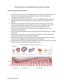
The algorithm of the NVOG is represented in Figure 1.1. In short a gynecological examination, including cervical cytology (Figure 1.2), is performed, followed by transvaginal
sonography using high frequency (5-7.5 MHz) transducers (Figure 1.3). Endometrial thickness
(ET) is measured as a double layer measurement at the thickest part of the endometrium in
the longitudinal plane.14;15 If the ET is 4 mm or less, the patient is reassured, and instructed
9
guidelines No. 4; February 1997.
With permission from the Dutch Society for Gynaecology.
Figure 1.1
“Guidelines for management of patients with abnormal vaginal blood loss in the post menopause” from NVOG
guidelines No. 4; February 1997.
With permission from the Dutch Society for Obstetrics and Gynaecology.
postmenopausal bleeding
gynecological exam, cervical smear,
transvaginal sonography
endometrial thickness > 4 mm, or
not obtained
endometrial thickness < 5 mm
Chapter 1
histological assessment
(pre)
malignancy
no (pre)
malignancy
10
treatment
wait and see policy
recurrent or persistent
bleeding
histological assessment;
hysteroscopy to be
considered
bleeding ceased
1
to contact her practitioner if new bleeding should occur. In case of abnormal findings in
the cervical cytology, or in case the ET is more than 4 mm or not assessable, histology is
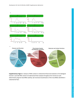
indicated. In the 1997 version of the guideline office endometrial sampling techniques are
advocated, specifically the Vabracurette and the Pipelle endometrium sampler (Figure 1.4).12
These techniques combine high sensitivity for endometrial cancer in women complaining of
Introduction
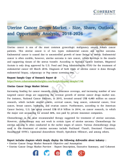
Figure 1.2
Cervical cytology in women with postmenopausal bleeding.
1.2A en 1.2B Normal endometrial cells
1.2.A
1.2B
Atypical endometrial cells
1.2B
1.2B
Figure 1.2C and 1.2D
Figure 1.2C and 1.2D
Atypical endometrial cells
3
1.2.C
3
1.2D
Figure 1.2E
Endometrial
carcinoma cells in cervical smear
1.2.C
1.2D
Figure 1.2E
Endometrial carcinoma cells in cervical smear
4
5
1.2E
1.2E
All pictures kindly provided by Dr. Patricia Ewing, pathologist Erasmus Medical Centre, Rotterdam.
All pictures kindly provided by Dr. Patricia Ewing, pathologist Erasmus Medical Centre, Rotterdam.
6
11
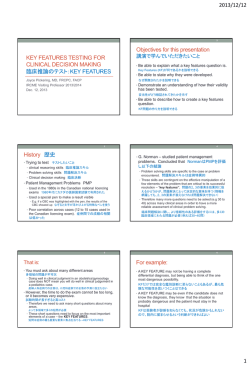
Figure 1.3
Transvaginal sonography in women with postmenopausal bleeding
Chapter 1
Figure 1.3
Transvaginal sonography in women with postmenopausal bleeding
12
1.3A
Double endometrium thickness in axial plane (14.5 mm).
1.3A
Double endometrium thickness in axial plane (14.5 mm).
7
1.3B
Same view as 1.3A, endometrium lining marked with a yellow dotted line. Anterior myometrium (distance
2) measures 15.8 mm, posterior myometrium (distance 1) measures 13.7 mm. Hysterectomy confirmed the
presence
of a grade I, stage IB endometrioïd carcinoma of the endometrium.
1.3B
Same view as 1.3.A, endometrium lining marked with a yellow dotted line. Anterior myometrium (distance 2)
measures 15.8 mm, posterior myometrium (distance 1) measures 13.7 mm. Hysterectomy confirmed the
presence of a grade I, stage IB endometrioïd carcinoma of the endometrium.
Introduction
1.3C
1.3C
In another patient the entire uterine cavity was filled with an irregular mass. The distinction between the
mass
and the uterine wall is unclear, particularly at the posterior wall, suggesting deep myometrial infiltration.
1.3C
Ventrally
the remaining myometrium measures 5.6 mm (distance 1), dorsally the remaining myometrium
In another patient the entire uterine cavity was filled with an irregular mass. The distinction between the mass
measures 0.7mm (distance 2). Hysterectomy confirmed the presence of a grade III, stage IIB endometrioïd
and the uterine wall is unclear, particularly at the posterior wall, suggesting deep myometrial infiltration.
carcinoma of the endometrium, with extension to the cervical canal (not shown here).
Ventrally the remaining myometrium measures 5.6 mm (distance 1), dorsally the remaining myometrium
measures 0.7mm (distance 2). Hysterectomy confirmed the presence of a grade III, stage IIB endometrioïd
postmenopausal
bleeding, with good patient tolerance, and diagnostic accuracy with cost
carcinoma of the endometrium, with extension to the cervical canal (not shown here).
effectiveness.16
9
In women with recurrent bleeding hysteroscopy is advocated. Hysteroscopy is superior
to any office sampling technique, and to dilatation and curettage (D&C) for detecting intrauterine abnormalities.17 Hysteroscopy also provides an opportunity to obtain a (directed)
sample, or to perform a resection if required. In experienced hands office hysteroscopy is
successful in obtaining a diagnosis in 80% of patients with postmenopausal bleeding.7;17-20
The diagnostic accuracy of hysteroscopy is high for endometrial cancer, but only moderate
for hyperplasia.21
GUIDELINE DEVELOPMENT AND REVISION
In The Netherlands, a steering committee under the leadership of Prof. Dr H.A.M. Brölmann,
developed a guideline that was accepted by the NVOG in November 1996, and publisher
in February 1997.12 Since a NVOG - guideline is valid for a period of five years, a revision
was presented and published in 2003.13 (http://www.nvog.nl/files/leidraad_opst_richtlijnen_web,01-2004.doc) Two different types of changes were made, a small change to the
13
Figure 1.3.D E F
A woman presenting with postmenopausal bleeding, diagnosed with bladder cancer
Chapter 1
Figure 1.3.D E F
A woman presenting with postmenopausal bleeding, diagnosed with bladder cancer
14
1.3D
The double endometrium thickness in the axial plane measures 6.2 mm.
1.3D
The double endometrium thickness in the axial plane measures 6.2 mm.
10
1.3E
In the bladder an irregular structure of 7.4 * 6.3 mm is visualized.
1.3E
In the bladder an irregular structure of 7.4 * 6.3 mm is visualized.
Introduction
1.3F
Cystoscopic evaluation, showing a
small urotheelcell carcinoma of the
bladder.
Pictures kindly provided by Dr. Anneke Steensma, gynaecologist, Eramsus Medical Centre, Rotterdam, The
Netherlands
1.3F
Cystoscopic evaluation, showing a small urotheelcell carcinoma of the bladder.
recommended
approach for women with abnormal postmenopausal bleeding, and secondly
Pictures kindly provided by Dr. Anneke Steensma, gynaecologist, Eramsus Medical Centre, Rotterdam, The
aNetherlands
change concerning the outline of the guideline, this is done to improve standardization,
and composition of the guidelines. In the 2003 version of the guideline of the Dutch Society
of Obstetrics and Gynaecology the flow chart remained unaltered, although for women
with an ET > 5 mm office endometrial sampling could now be offered with Saline Infusion
Sonography (SIS) to better detect intrauterine abnormalities.13 When a center does not have
SIS or office hysteroscopy available, the guideline proposes further diagnostic testing only in
women with recurrent or persistent bleeding.12 Saline infusion sonohysterography consists
of sonographic imaging of the uterus and uterocervical cavity, using real-time sonography
during injection of sterile saline into the uterus. The 12
benefits include minimal and brief
discomfort, and a better understanding of intrauterine pathology. In a systematic review
the success rate of SIS in postmenopausal women was reported 87% (95% CI 83 to 90%).22
Nevertheless, the accuracy of SIS in diagnosing intrauterine pathology such as submucous
fibroids and polyps is comparable to that of outpatient hysteroscopy,23;24 and SIS depicted
more abnormalities at a lower cost per abnormality.25
To improve standardization of approach, and content of the guidelines, the AGREE instrument has been introduced (appraisal of guidelines for research & evaluation in Europe).
(http://www.agreecollaboration.org) One of the components of the AGREE instrument is
the applicability of the guideline in clinical practice. This requires clearly defined review
criteria that are derived from the key recommendations in the guideline. In the first guideline
(1997),12 key recommendations were not given, but from the revised version (2003) the following conclusions and recommendations can be drawn.13
- After a first episode of abnormal postmenopausal bleeding an expectant policy is justified
in women with double endometrial thickness not more than 4 mm.
15
Devices to obtain an office endometrial sample.
Figure 1.4
Devices to obtain an office endometrial sample.
Chapter 1
Photo kindly provided by Gynotec, Malden, The Netherlands
16
Photo kindly provided by Gynotec, Malden, The Netherlands
- In women with an ET > 4 mm, office endometrial sampling is an accurate method for detecting endometrial adenocarcinoma. During the same session, using the same catheter a
saline infusion sonography (SIS) can be performed.
- Persistent or recurrent bleeding after menopause necessitates a diagnostic hysteroscopy,
with tissue sampling.
JUSTIFICATION OF THE GUIDELINE
The Dutch guideline is a description of minimal care to be provided by a gynecologist to a
13
woman with postmenopausal bleeding. In some circumstances deviation from the guideline
may be justified, for example because of patient wishes, or the guideline might not be applicable as a result of local policies or circumstances (i.e. at the level of care institutions).
In the guidelines the a-priori chance of a particular woman with postmenopausal bleeding
having a (pre) malignancy is not taken into account. Both endometrial thickness and the risk
of endometrial carcinoma are found to be associated with various other individual risk indicators, including age, time since menopause, obesity, hypertension, diabetes mellitus, parity
and smoking.26-29 Measurement of the endometrial thickness has been demonstrated to be
useful for ruling out endometrial hyperplasia or carcinoma, although the a-posteriori risk is
still approximately 2.5%, and there is still debate about accuracy as well as the appropriate
Introduction
cut-off level.30;31 A recent study demonstrated that time since menopause, in combination
with endometrial thickness, can be useful for determining the pre-test probability, and to
define when a negligible risk of cancer renders endometrial sampling unnecessary.32 The
a-priori risk and a-posteriori risk in an individual patient are not taken into account in the
guideline, and could usefully be included in clinical protocols; in women with a high-risk
profile immediate endometrial sampling may prove to be a cost-effective strategy. Some
doctors and patients might find a 2,5% a-posteriori risk for endometrial cancer unacceptable
and request further testing, even when the ET is < 5 mm.19
AIMS OF THE THESIS
The aim of this thesis is to evaluate the diagnostic work-up in abnormal postmenopausal
bleeding. First, we aim to evaluate whether the NVOG guideline is followed by gynecologists.
Second, we want to evaluate whether the work-up can be improved by the use of a multivariable diagnostic approach incorporating data of medical history, physical examination,
sonography, endometrium sampling and cytology.
OUTLINE OF THE THESIS
Chapter 2 evaluates the adherence to guideline of the Society of Dutch Obstetrics and
Gynaecology. Chapters 3 assesses the potential value of cervical cytology in relation to the
detection of endometrial cancer. Chapter 4 assesses the clinical consequences of an insufficient office endometrial sample.
The chapters 5, 6 and 7 focus on the prediction of endometrial cancer in women with
postmenopausal bleeding in relation to patient characteristics. The accuracy of the TVS in
the detection of endometrial cancer is investigated in relation to body mass and to the presence of diabetes and hypertension. The relationship between age, postmenopausal age and
endometrial cancer is also investigated, and a multivariable model is constructed. In chapter
8, myometrium infiltration by endometrial cancer is studied by TVS, and compared to definitive pathology of the hysterectomy specimen.
DESCRIPTION OF DATABASE
Patients who presented with postmenopausal bleeding were registered prospectively in a
multi-center study between January 2001 and June 2003. Recruitment of patients was performed in one university hospital and seven teaching hospitals. Each hospital is represented
17
by one or two members in the DUPOMEB study group. (Dutch study in postmenopausal
bleeding) (Table 1.1). In November 2005 we started to retrieve the hospital charts to obtain
follow-up information on the patients. For women diagnosed with endometrial cancer or
pre-malignancy, data regarding treatment, disease stage and survival were obtained. For all
other women, not diagnosed with a malignancy, we determined whether recurrent bleeding
had occurred.
Papers forthcoming from this database are written on behalf of the DUPOMEB study
group.
For each chapter subsets of patients were drawn from the database. Chapter 2 was based
on the principal report that was written for the Healthcare Insurance Board, Amstelveen, The
Netherlands.33 This organization provided a grant (number 01135). This chapter concerns the
first 837 women that were included in the study till January 2003. Chapter 3 concerns the
contribution of the cervical smear in the detection of endometrial cancer in women with
postmenopausal bleeding. As hormone therapy might induce shedding of the endometrium,
and therefore increase the number of endometrial cells in the cervical smear, the composition
of the cervical smear differs in women using HRT as compared to those who do not. Therefore,
women using hormone replacement therapy (HRT) were not included in the analysis of the
Chapter 1
contribution of cervical cytology in postmenopausal bleeding. In chapter 4 we evaluated
the final diagnosis in patients in whom the office endometrial samples were considered to
18
Table 1.1 DUPOMEB members and institutions
be not- diagnostic. All patients originally in the database were included, and women with an
Participating hospitals
Number of
patients included
DUPOMEB members (gynecologists)
Albert Schweitzer Hospital, Dordrecht
111
G. Sjarlot Kooi MD PhD
Diakonessenhuis, Utrecht
57
Maurice V. A. M. Kroeks MD PhD
Gelre Hospital, Apeldoorn
48
Peter H. M. van de Weijer MD PhD
Meander Medical Centre, Amersfoort
108
M. Jitze Duk MD, PhD
Mesos Medical Centre, Utrecht
31
Annette Bouwmeester, MD
Rijnstate Hospital, Arnhem
252
Paul H. L. J. Dijkhuizen MD, PhD
TweeSteden Hospital, Tilburg
192
Roy F. M. P. Kruitwagen MD, PhD
Annette F. ter Haar MD
University Medical Centre, Utrecht
122
Peter M. Heintz MD PhD
Helena C. van Doorn, MD (principal
investigator)
Other institutions
Other members
Department of Clinical Epidemiology and Biostatistics, Academic
Medical Centre, Amsterdam
Brent C. Opmeer PhD, epidemiologist
University Medical Centre, Utrecht
Arianne Witteveen, nurse, data
extraction and data management
Academic Medical Centre, Amsterdam and Máxima Medical
Centre, Veldhoven
Ben W. J. Mol MD, PhD, gynecologist,
epidemiologist
Erasmus Medical Centre, Rotterdam
Curt W. Burger, MD, PhD, gynecologist
Introduction
ET ≥ 5 mm, or in whom the ET could not be measured, and where a subsequent sample was
non-diagnostic were evaluated. For this analysis long term follow-up data were obtained to
include all histological testing that was done from inclusion in the study, and to determine
whether recurrent bleeding had occurred.
In chapter 5 the accuracy of endometrial thickness measurement in the diagnosis of endometrial cancer was assessed in relation to particular patient characteristics. Women with
postmenopausal bleeding during HRT usage have a smaller chance of significant endometrial
pathology than those women with postmenopausal bleeding not on HRT. HRT also influences
the sonographic findings in women; tamoxifen has well described effects, and unopposed
estrogens will increase the endometrial thickness.34;35 For these reasons, women on HRT were
not included in this study. Chapter 5 was based on a preliminary database; with patients
included from January 2001 - January 2003.
Table 1.2 Patient characteristics in relation to diagnoses
Malignancy
N = 84
Pre-malignancy
N = 10
Others @
N = 827
Age (years)(mean (SD) range)
69.0 (10.4; 49-90)
66.3 (10.3; 55 – 88)
61.2 (9.5; 37 – 93)
Nulliparous (%)
16 (19.0%)
2 (20%)
91 (11.0%)
Body Mass Index (kg/m2)
29.9 (16.6 - 57.2)
(n = 66)
34.8 (21.7 – 70.1)
(n = 9)
27.7 (16.9 – 57.4)
n = (703)
The presence of
Number (%)
Number (%)
Number (%)
Diabetes
- diet controlled
- oral medication
- insulin dependent
3 (3.6)
9 (10.7)
5 (6.0)
1 (10.0)
2 (20.0))
15 (1.8)
37 (4.5)
28 (3.4)
Hypertension
- no prescription
- medically treated
8 (9.5)
24 (28.5)
3 (30.0)
42 (5.1)
174 (21.0)
81 (96.4)
none
654 (79.1)
39 (0.4)
7 (0.8)
70 (8.5)
37 (4.5)
17 (2.1)
39 (4.7)
1 (10.0)
37 (4.5)
Hormone treatment
- Estrogens only
- Progestogens only
- Estrogens and progestogens
- Tibolon
- Tamoxifen
- Other prescriptions
1 (1.2)
1 (1.2)
1 (1.2)
Thyroid disease
- present
6 (7.1)
Malignancy in history
- Breast cancer
- Other malignancy
3 (3.6)
4 (4.8)
Occurrence
- entered study with first episode
72 (85.7)
45 (5.4)
32 (3.8)
9 (90.0)
689 (83.3)
@ This group included women diagnosed with vulva, cervix, bowel, and metastatic breast cancer, women
without diagnoses, due to patient refusal, death, or contraindications for surgery
19
Table 1.3 Diagnostic procedures in women presenting with abnormal postmenopausal bleeding
Malignancy
N = 84
Pre-malignancy
N = 10
Others @
N = 827
TVS
- missing
- ET < 5mm
- ET ≥ 5mm
11 (13.1)
3 (3.6)
70 (83.3)
2 (20.0)
0
8 (80.0)
91 (11.0)
382 (46.2)
354 (42.8)
Cervical smear
- Pap classification
- CISOE
66 (78.6)
61 (72.6)
7 (70.0)
6 (60.0)
744 (90.0)
619 (74.8)
Office endometrium sampling
- not performed
- not successful
- not- diagnostic
- adequate sample (histology)
17 (20.2)
21 (25.0)
3 (3.6)
43 (51.2)
4
1
1
4
(40.0)
(10.0)
(10.0)
(40.0)
370 (44.7)
153 (18.5)
87 (10.5)
217 (32.8)
Hysteroscopy
36 (42.9)
10 (100)
234 (28.3)
Dilatation and curettage
49 (58.3)
9 (90.0)
239 (28.9)
@ This group included women diagnosed with vulva, cervix, bowel, and metastatic breast cancer, women
without diagnoses, due to patient refusal, death, or contraindications for surgery
In chapter 6 we evaluated the relation between current age and time since menopause on
one hand, and the risk of endometrial cancer on the other. Only women presenting with a
first episode of uterine bleeding were included.
In chapter 7 we evaluated whether the efficiency of the current diagnostic work-up can be
improved by combining patient characteristics with transvaginal measurement of endometrial thickness. In accordance with the arguments listed under chapter 5 we excluded patients
using any hormonal treatment.
Chapter 8 includes data obtained in eleven hospitals, mainly in the region of Utrecht that
participated in a study on the diagnostic accuracy of preoperative transvaginal sonography
(TVS) for assessing myometrium infiltration in patients with endometrial cancer.
Overall, data were collected on 921 patients with abnormal postmenopausal bleeding. A
summary of basic information concerning the whole population is given in Table 1.2. Patients
are placed in three groups on the basis of diagnosis within 6 months after inclusion in the
study; women with a malignancy of the endometrium, women with any type of endometrial
hyperplasia with atypia, and in the last group all other patients. This group includes women
diagnosed with vulva, cervix, and bowel, or breast cancer, women without diagnoses, cases
where there was patient refusal, death or a contraindication for surgery. In Table 1.3 diagnostic assessment is summarized for each of the aforementioned groups.
Studies on the diagnosis of endometria
cancer in women with postmenopausal
bleeding. Studies naar de diagnostiek va
endometriumcarcinoom bij vrouwen m
postmenopauzaal bloedverlies.
bloedverlies Studies
on the diagnosis of endometrial cancer
in women with postmenopausal bleedin
Studies naar de diagnostiek
van
Chapter
2 endom
triumcarcinoom bij vrouwen
met postm
Adherence of Dutch
gynecologist
to the on the
nopauzaal bloedverlies.
Studies
guideline “Investigation
of abnormal
Studies on the diagnosis
of endometria
postmenopausal
bleeding”
cancer in women with
postmenopausal
bleeding. Studies naar de diagnostiek va
endometriumcarcinoom bij vrouwen m
postmenopauzaal bloedverlies. Studies
on the diagnosis of endometrial cancer
in women with postmenopausal bleedin
Studies naar de diagnostiek van endom
triumcarcinoom bij vrouwen met postm
nopauzaal bloedverlies. Studies on the
Studies on the diagnosis of endometria
cancer in women with postmenopausal
bleeding. Studies naar de diagnostiek va
Adherence of Dutch gynecologist to the guideline “Investigation of abnormal postmenopausal bleeding”
By DUPOMEB (Dutch study on postmenopausal bleeding)
Ned Tijdschr Geneeskd. 2005;149:2676-82.
SUMMARY
Aim: The guidelines of the Dutch Society for Obstetrics and Gynaecology recommend that
patients with postmenopausal blood loss be examined by transvaginal sonography followed
Chapter 2
by histological studies of the endometrium whenever the thickness of the endometrium
22
exceeds 4 mm. We investigated whether these guidelines are followed in clinical practice.
Design: Prospective cohort study
Method: In 8 hospitals a total of 837 patients with abnormal postmenopausal blood loss was
studied. The guideline for postmenopausal bleeding was known at the start of the study in
all hospitals. All patients were evaluated to determine whether the diagnostic approach used
conformed to the guideline.
Results: In total 98% of the women underwent sonography. The thickness of the endometrium was 4 mm or less in 43%, more than 4 mm in 46% and not evaluated in 9% of the
cases. Underdiagnosis occurred when no examination whatsoever was carried out or when
histological studies were not performed when the endometrial thickness was more than 4
mm. It appeared that this was the case for 52 (6%) of the patients, 3 patients refused further
examination and one patient had co-morbidity so that subsequent examinations were not
performed. In 108 cases (13%) tissue samples for histological examination were obtained although this was not necessary according to the guideline. In 86 (10%) cases curettage and/or
hysteroscopy was performed whereas endometrial aspiration would have been sufficient.
Conclusion: Implementation of the guideline for the diagnosis of abnormal postmenopausal
bleeding was fairly good. There were only a few cases of underdiagnosis; in contrast, unnecessary (histological) examination or unnecessary invasive diagnostic procedures were
seen more often. The efficiency of diagnostic management can be enhanced by performing
subsequent histological studies only when a patient has a thickened endometrium and by
relying on endometrial aspiration in such cases.
Comparison with the guideline
INTRODUCTION
Abnormal postmenopausal bleeding is quite common.1 The incidence decreases with the
duration of the postmenopausal period.2 Diagnosis of abnormal postmenopausal blood loss
is the subject of both a guideline of the Dutch Society for Obstetrics and Gynecology (NVOG)
and a transmural agreement (2002) between general practitioners and gynecologists.11-13 The
NVOG guideline was drawn up in 199712 and revised in 2003.13
The NVOG guideline is shown schematically in Figure 2.1. After anamnesis and physical
examination, a transvaginal sonogram is performed whereby the thickness of the double
layer of the endometrium is measured. At an endometrial thickness of 4 mm or less, the
chance of malignant abnormalities is so small that a “wait-and-see” approach is considered
sufficient. When the endometrial thickness is more than 4 mm, histological examination of
the endometrium is indicated in order to exclude carcinoma or atypical hyperplasia of the
endometrium. Aspiration of the endometrium in the case of postmenopausal bleeding has
a sensitivity of 99% for demonstration of a malignancy.16 The risk of complications of this
technique is low, the method is inexpensive and the burden to the patient is small. When
aspiration is not possible or does not yield representative material, more invasive examination techniques, such as curettage – if necessary with hysteroscopy, must be applied.23;36
The NVOG guidelines thus form a guide for daily practice. However, for implementation
more is needed than just passing out the guidelines.37;38 The first step is to determine whether
the guidelines are applied. We carried out a prospective cohort study to determine whether
the diagnostic management of women with postmenopausal blood loss conformed to the
NVOG guideline of 1997.
MATERIAL AND METHODS
The study was carried out by the Departments of Gynecology of eight Dutch hospitals. The
guideline for postmenopausal blood loss was known in all hospitals at the start of the study
and was followed everywhere, according to those involved. Between January 2001 and January 2003 all women with abnormal postmenopausal bleeding were included in the study.
Data on diagnostic management was registered. For evaluation purposes, the following
patient groups were defined:
1. Patients who did not undergo sonography
2. Patients for whom the endometrium could not be evaluated at sonography
3. Patients with an endometrial thickness of 4 mm or less
- without subsequent histological examination
- with subsequent histological examination
4. Patients with an endometrial thickness of more than 4 mm
23
guidelines No. 4; February 1997.{1997 162 /id}
With permission from the Dutch Society for Gynaecology.
Figure 2.1
“Guidelines for management of patients with abnormal vaginal blood loss in the post menopause” from NVOG
guidelines No. 4; February 1997.
With permission from the Dutch Society for Obstetrics and Gynaecology.
postmenopausal bleeding
gynecological exam, cervical smear,
transvaginal sonography
endometrial thickness > 4 mm, or
not obtained
endometrial thickness < 5 mm
Chapter 2
histological assessment
(pre)
malignancy
no (pre)
malignancy
24
wait and see policy
treatment
recurrent or persistent
bleeding
histological assessment;
hysteroscopy to be
considered
bleeding ceased
14
- without subsequent histological examination
- with subsequent histological examination
A distinction was made between histological samples acquired by means of aspiration and
samples acquired by curettage and/or hysteroscopy. An hysteroscopy that did not yield
histological samples because of atrophy is not considered as non-adherence to the guide-
Comparison with the guideline
line.23 Subsequently it was determined whether the physicians conformed to the guideline
and if not, whether there was an explanation for not following the guideline. Procedures
were considered “unnecessary diagnostic procedures” when supplementary tissue studies
were carried out without reason in patients with an endometrium ≤ 4 mm. Also when further
studies were carried out, after a representative endometrial aspiration that did not show
(pre)malignancy. “Underdiagnosis” was defined as no examination at all or no histological
studies when the endometrium was more than 4 mm thick.
RESULTS
The study included 845 women, eight of whom were excluded due to a prior hysterectomy.
The average age was 62 years (range 37-92), the average duration of the postmenopausal
period was 11.4 years (range 1-50). For 705 (85%) of the women it was the first episode of
abnormal vaginal blood loss; 732 (88%) of the women were multiparae. Twenty percent of
the women used a hormone preparation. The approach of the gynecologist is compared with
the guideline (Table 2.1).
Patients who did not undergo sonography
Of the 19 (2%) women who did not undergo sonography there was an obvious reason in four
cases. Fourteen of the other 15 patients underwent histological examination directly, four by
curettage. For further details, see Table 2.1.
Patients for whom the endometrium could not be evaluated at sonography
For 74 of the 818 women (9%) the thickness of the endometrium could not be determined.
In 50 of the 74 cases an attempt was made to perform endometrial aspiration. In 14 cases
an endometrial aspiration was not attempted but an appointment for hysteroscopy and/or
curettage was made. For the remaining ten cases neither aspiration nor hysteroscopy or
curettage was requested. Ultimately it was not clear why 12 patients, including five after
unsuccessful aspiration, did not undergo a supplementary examination.
Patients with an endometrial thickness of 4 mm or less
In 361 (44%) women who underwent sonography the endometrial thickness was 4 mm or
less. For 136 (38%) patients subsequent histological examinations were performed, see also
Table 2.1. In 19 cases, the patient suffered a recurrent episode of postmenopausal blood loss;
nine patients underwent subsequent examination because of an abnormal Pap smear, two
due to use of tamoxifen and one for therapeutic reasons after very severe bleeding. Finally
98 patients underwent endometrial diagnostic examination without a clear reason while the
25
Chapter 2
n=587 (70%)
Diagnostic management according
to guidelines
Other explanation for blood loss:
hematuria, cervix carcinoma;
lichen sclerosis (previously normal
sonogram) (n=4)
cervical polyp (n=1)
n=56 (7%)
Underdiagnosis
Not according to the guidelines
Endometrial aspiration (n=10)
Unnecessary diagnostic
procedures
n=108 (13%)
Hysteroscopy and/or curettage
when aspiration would have been
sufficient (n=4)
Unnecessarily invasive diagnostic
procedures
n=86 (10%)
Further examination refused
(n=1)
Hysteroscopy contraindicated
(n=1)
No reason for not performing
further diagnostic procedures
(n=5)
No good reason for not
performing histology (n=7)
Hysterectomy for adnex pathology
(n=2), cervix carcinoma (n=1)
Hysteroscopy indicated (n=8)
No attempt made to perform
aspiration (n=10)
Curettage and/or hysteroscopy
(n=14)
Histological diagnosis from
aspiration (n=27)
Attempted aspiration; aspiration
Hysteroscopy and/or curettage
not successful or not representative (n=13)
Hysteroscopy not successful,
(n=23)
subsequent hysterectomy (n=1)
and local excision of vulvar
carcinoma (n=1)
Hysterectomy after diagnosing
endometrial cancer in cervical
biopsy (n=1)
Successful aspiration, histological
diagnosis (n=27) obtained
Hysteroscopy and/or curettage
when aspiration would have been
sufficient (n=6)
Sonography not possible (n=7) (1%) or endometrial thickness could not be evaluated (n=67) (8%), indication for subsequent histological assessment of endometrium
Direct histology (n=14)
No subsequent examination (n=5)
Sonography not performed (n=19)(2%)
Diagnostic result
Table 2.1 Diagnosis of 837 women presenting with abnormal postmenopausal blood loss
26
n=587 (70%)
Diagnostic management according
to guidelines
n=56 (7%)
Underdiagnosis
Not according to the guidelines
Hysteroscopy and or curettage for
abnormal cervix cytology (n=7)
Aspiration for abnormal cervix
cytology (n=2)
Hysteroscopy and or curettage
for 2nd episode of blood loss
(n=7)Hysteroscopy and or
curettage after recurrent blood loss
within 6 months (n=7)
Hysteroscopy for tamoxifen use
(n=1)
Therapeutic curettage (n=1)
Subsequent histology (n=136)
Aspiration instead of
hysteroscopy because of 2nd
episode of blood loss (n=12)
Aspiration instead of
hysteroscopy because of use of
tamoxifen (n=1)
Hysteroscopy because of
malignancy (n=8);
Hysteroscopy and/or curettage
after recurrent blood loss within 6
months (n=4);
Hysteroscopy for use of tamoxifen
(n=1)
Hysteroscopy and curettage (n=84)
Atrophy at hysteroscopy (n=9)
Malignancy elsewhere (n=3)
2nd attempt successful (n=1)
Hysterectomy (n=1)
Successful aspiration, histological
diagnosis (n=198) obtained
Aspiration not successful (n=64) or
not representative (n=49)
No endometrial aspiration (n=72)
Refused examination (n=1)
Colposcopy without assessment
of endometrium (n=1)
No reason for not obtaining
histology (n=10)
Refused further examination
(n=1)
No reason for not performing
histology (n=15)
Endometrial thickness more than 4 mm (N=383)(46%),histological assessment of endometrium indicated
No histology (n=225)
No histology (n=225)
Endometrial thickness 4 mm or less (N=361) (43%); wait-and-see policy indicated
Diagnostic result
aspiration and
and/or curettage
aspiration (n=81)
and/or curettage
Hysteroscopy and/or curettage
despite benign aspiration result
(n = 17/168 benign results)
Endometrium
Hysteroscopy
(n= 10)
Endometrium
Hysteroscopy
(n=7)
Unnecessary diagnostic
procedures
n=108 (13%)
Hysteroscopy and curettage
(n=52)
Atrophy at hysteroscopy (n=7)
Unnecessarily invasive diagnostic
procedures
n=86 (10%)
Comparison with the guideline
27
sonogram was reassuring. Neither atypical hyperplasia nor endometrial cancer was found in
any of these patients.
In the group of patients with an endometrial thickness of 4 mm or less serious pathology
was found in three cases. One patient who used tamoxifen underwent sonography (endometrial thickness 3 mm) as well as endometrial aspiration at the first consultation. In another
case a patient underwent hysteroscopy and curettage 5 months after her first consultation
for recurrent bleeding; a malignancy was diagnosed. Finally one patient had an endometrium
thickness of 4 mm but severe atypia of the endometrium was indicated by the cervical smear
and subsequently confirmed by curettage.
Patients with an endometrial thickness of more than 4 mm
Of the women who underwent sonography, 383 (47%) had an endometrial thickness of
more than 4 mm. In 72 (19%) cases endometrial aspiration was not performed a priori (see
Table 2.1). One patient underwent hysterectomy since a probable diagnosis of endometrial
carcinoma was based on cervix cytology. In ten cases no reason could be found for omitting
histological examination.
Endometrial aspiration was performed in 311 cases. This yielded an outcome in 198 (64%)
Chapter 2
cases. Additional examination of 17 patients was carried out despite a favorable result of the
28
aspiration without a clear reason.
Of the 113 patients for whom the endometrial aspiration was not successful (N=64) or
resulted in an outcome “not representative” (N=49), subsequent hysteroscopy and/or curettage followed in 55 and 29 cases, respectively, while 29 patients did not undergo any further
histological examination. In three of these latter patients a malignancy had already been
demonstrated. For one 92-year-old patient endometrial carcinoma was diagnosed on the
basis of cervix cytology but because of her poor condition further treatment was not considered feasible. In addition one patient had a metastasized breast cancer and one a carcinoma
of the bladder. In 15 of the 29 cases it is not clear why further histological examination was
not carried out.
In summary: in 38 cases histological examination was indicated but was not carried out
without a clear reason, in 13 cases aspiration was performed while hysteroscopy and curettage were indicated, three patients refused further examination and one could not be
examined further because of co-morbidity. In one case neither sonography nor histological
examination was carried out.
In total 108 patients underwent unnecessary histological examination, usually aspiration
but sometimes hysteroscopy and/or curettage or both. In addition 86 underwent a curettage
and/or hysteroscopy when endometrial aspiration would have been sufficient.
Comparison with the guideline
DISCUSSION
We found that for two-thirds of the women who presented with abnormal vaginal blood
loss, diagnostic management conformed to the guideline. Deviation from the guideline
consisted predominantly of unnecessary diagnostic procedures or unnecessarily invasive
diagnostic procedures and to a lesser degree underdiagnosis. For approximately one-third
of the patients with an endometrial thickness of 4 mm or less, histological examination
was performed without an indication anyway. These extra examinations did not lead to the
detection of more (pre)malignancies than would have been the case if the guideline had
been followed strictly. In addition histological examination was often carried out in an unnecessarily invasive manner. For patients with an endometrial thickness of more than 4 mm a
curettage and/or hysteroscopy was often carried out while an endometrial aspiration would
have been sufficient. On the basis of our study it would appear possible to conform more
strictly to the NVOG guideline for abnormal postmenopausal blood loss without decreasing
the accuracy.
The guideline was followed fairly well, possibly because the guideline is relatively simple
and can easily be carried out in clinical practice. Transvaginal sonography can, at present, be
carried out in almost every office directly, just like the endometrial aspiration. For some patients with postmenopausal bleeding the presence of other risk-enhancing factors suggests
that the chance of endometrial carcinoma is increased and it is possible that the gynecologist
in these cases will decide to deviate from the guideline and will proceed sooner to an invasive
examination: for instance, women with a high risk due to their age, use of tamoxifen, obesity
or diabetes mellitus.39 For users of tamoxifen with postmenopausal blood loss, the reliability
of the sonogram is decreased,40 while the reliability of endometrial aspiration is not clear for
this group of patients. In our study we did not investigate the degree to which these factors
influenced the management of diagnosis.
Deviation from the guideline could also be explained by the principle of shared decisionmaking whereby the patient herself is involved in making decisions about medical interventions.41 Although the chance of abnormal postmenopausal blood loss in a patient with an
endometrial thickness of 4 mm or less is in general estimated to be very small, the two metaanalyses carried out in this field contradict one another somewhat.30;42 The decision to accept
the small chance of endometrial carcinoma with the wait-and-see policy or to undergo more
invasive diagnostic procedures and accept the inherent risks can turn out differently for individual patients and physicians, whereas the guideline are based on a general consideration
which applies for everyone. An investigation of the preference of women for a certain policy
in case of postmenopausal blood loss has to our knowledge not been carried out.
The guideline for postmenopausal blood loss focus entirely on the exclusion of malignancy
whereas postmenopausal blood loss can also be caused by polyps, or other benign disorders.1;43 For the demonstration and eventual removal of polyps hysteroscopy is superior. In
29
some of the participating clinics there is a triage system and the patient who presents with
postmenopausal blood loss can undergo an outpatient hysteroscopy with biopsies at her
first consultation. This too will influence the frequency of hysteroscopy.
Sometimes for the evaluation of implementation of a guideline it is worthwhile to develop
quality indicators.44;45 Depending upon the nature of the care and the relevant guideline, the
indicators can vary in detail or complexity. The guideline for diagnosis of postmenopausal
blood loss is simple and deviations from the guideline can be described fairly easily as deviations from the proposed diagnostic management. The deviations found in our study could
be useful for definition of valid indicators for measurement of the quality of care during
diagnosis of postmenopausal blood loss.
The next step to improve the implementation of these guideline and therefore also the
care provided is to identify success and failure factors that can enhance or hinder use of the
guideline.46 These factors can vary in nature (for example, the quality of the guideline, knowledge and motivation of the physician, organization of care, regulations, patient factors) and
can be investigated by means of structured interviews with physicians and patients.
Although the guideline for diagnosis of postmenopausal blood loss was followed fairly
well, it appears from this study that unnecessary diagnostic procedures or unnecessarily inChapter 2
vasive diagnostic procedures are performed. In addition to the uncertainty about the true ac-
30
curateness of the proposed diagnostic management, individual considerations of physicians
and patients may possibly play a role in the decision to perform subsequent examinations in
order to exclude eventual malignant abnormalities with certainty.
Ned Tijdschr Geneeskd. 2005;149:2649-52.
Ned Tijdschr Geneeskd. 2006;150:586.
Ned Tijdschr Geneeskd. 2006;150:586; author reply 586-7.
Studies on the diagnosis of endometria
cancer in women with postmenopausal
bleeding. Studies naar de diagnostiek va
endometriumcarcinoom bij vrouwen m
postmenopauzaal bloedverlies.
bloedverlies Studies
on the diagnosis of endometrial cancer
in women with postmenopausal bleedin
Studies naar de diagnostiek
van
Chapter
3 endom
triumcarcinoom bij vrouwen
met postm
The value of cervical
cytologyStudies
in diagnosingon the
nopauzaal bloedverlies.
endometrial carcinoma
in women with
Studies on the diagnosis
of endometria
postmenopausal
bleeding
cancer in women with
postmenopausal
bleeding. Studies naar de diagnostiek va
endometriumcarcinoom bij vrouwen m
postmenopauzaal bloedverlies. Studies
on the diagnosis of endometrial cancer
in women with postmenopausal bleedin
Studies naar de diagnostiek van endom
triumcarcinoom bij vrouwen met postm
nopauzaal bloedverlies. Studies on the
Studies on the diagnosis of endometria
cancer in women with postmenopausal
bleeding. Studies naar de diagnostiek va
The value of cervical cytology in diagnosing endometrial carcinoma in women with
postmenopausal bleeding
Helena C. van Doorn, Brent C. Opmeer, Curt W. Burger, G. Sjarlot Kooi, Roy F. P. M. Kruitwagen,
Ben W. J. Mol
For DUPOMEB: Dutch Study in Postmenopausal Bleeding.
Submitted
Chapter 3
SUMMARY
32
Background: The potential value of the cervical smear in the diagnosis of endometrial cancer
is unclear. The aim of the present study was to assess the accuracy of the cervical smear in
the diagnosis of endometrial cancer in women with postmenopausal bleeding, with a special
focus on the diagnostic accuracy of the presence of normal endometrial cells.
Methods: Women presenting with abnormal postmenopausal bleeding, not using HRT, were
prospectively included in eight hospitals in The Netherlands. Cervical cytology was coded according to both the Papanicolaou classification, as well as the Dutch coding system (CISOE-A).
The latter system classifies squamous cells, endometrial cells and other cell types separately.
For both classification systems, likelihood ratios for the presence of (pre) malignancy of the
endometrium were calculated.
Results: We included 543 women with postmenopausal bleeding. There were 56 women
(11.7%) with endometrial carcinoma and six women with atypical hyperplasia. A Pap III
increased the probability of (pre) malignancy (LR 3.5), whereas Pap IV and Pap V virtually
proved the presence of carcinoma. The CISOE-A classification showed similar results. The
presence of normal endometrial cells did not increase the probability of endometrial (pre)
malignancy.
Conclusions: Addition of the results of the cervical smear to endometrial thickness could
detect incidental endometrial cancers that are missed by TVS (< 5 mm). In women with
postmenopausal bleeding the presence of normal endometrial cells is not predictive for
endometrial cancer.
Cervical cytology in postmenopausal bleeding
INTRODUCTION
The work-up for postmenopausal bleeding has changed over the last decade. Until recently
dilatation and curettage (D&C) was the procedure of first choice, but transvaginal sonography and office biopsy techniques have replaced the need for D&C.16;30;47 Some authors
recommend hysteroscopy or saline infusion sonography (SIS) in all women presenting with
postmenopausal bleeding, since intrauterine abnormalities can easily be missed by D&C and
office sampling techniques.17;22;48 Although there is controversy over the usefulness of SIS
or hysteroscopy, there is general consensus that transvaginal sonography and subsequent
histologic testing for women with ET ≥ 5 mm should be performed.9;12
Cervical cytology is also recommended in the work-up for postmenopausal bleeding.9;12;49;50
In a systematic review the conventional Papanicolaou (Pap) test was reported to be accurate
in the diagnosis of cervical (pre) malignancy.51 In postmenopausal bleeding, cytology is used
to detect cervical neoplasm as a cause of postmenopausal bleeding. The contribution of the
cervical smear in the diagnosis of endometrial cancer is less clear. Second, cytology might
be useful in the diagnosis of endometrial cancer when cervical pathology has been ruled
out.52;53 Third, presence of endometrial cells in a cervical smear of a postmenopausal woman
is considered to be indicative for endometrial pathology.54-56 The purpose of this study was
to evaluate whether cervical cytology is useful for the diagnosis of endometrial cancer in
women with postmenopausal bleeding.
MATERIALS AND METHODS
The study was performed in a university hospital and seven teaching hospitals in The Netherlands. Between January 2001 and June 2003, consecutive patients who presented with
abnormal postmenopausal bleeding were registered. Women who had had a hysterectomy,
women using any kind of hormonal treatment in the preceding 24 months, and women who
were diagnosed with another malignancy were not included in the study.
A cervical smear was made either by the referring general practitioner, or by the gynecologist, as customary to the local protocols. Cervical cytology specimens were submitted to the
cytology laboratory either as conventional slides or as liquid based cytology. Cervical cytology was coded according to both the Papanicolaou classification (Pap- score), and the Dutch
national coding system (CISOE-A).57;58 In the Dutch CISOE-A classification system the smears
are examined for five different dimensions to indicate the composition and morphology of
the smear. These dimensions are composition (C), inflammation (I), squamous epithelium
(S), other findings and endometrium (O) and endocervical columnar epithelium (E). These
CISOE-A dimensions are scored varying from 1 to 9, and the score is than merged into a five
33
digit score. In addition to this, the CISOE-A system gives a judgment on the adequacy of the
smear (-A) which is graded as adequate, suboptimal, or inadequate.
In the current study, evaluation of the women was performed according to the guideline
of the Dutch Society of Obstetrics and Gynaecology.12;39 In short, a gynecological examination, including cervical cytology, was performed, followed by transvaginal sonography
using high frequency (5-7.5 MHz) transducers. Endometrial thickness (ET) was measured
as a double layer measurement at its thickest part in the longitudinal plane. In case the
ET was 4 mm or less, and cervical cytology was normal, the patient was reassured, and
instructed to contact the doctor if new bleeding should occur. In case of abnormal findings
in the cervical cytology, or when the ET was more than 4 mm, histology was obtained,
using an office endometrial sampling technique, during hysteroscopy, or with dilatation
and curettage.
Malignancy and pre-malignancy were considered absent when women with an endometrial thickness of 4 mm or less had an uneventful follow-up, or when histology specimens
showed atrophy, benign polyps, hyperplasia without atypia, or proliferation. We recently
reviewed the patient charts to confirm the negative follow-up status.
Atypical hyperplasia and endometrial malignancy in the histology specimen were categoChapter 3
rized as “endometrial (pre) malignancy”, since these findings both warrant further treatment.
34
The pathologist was unaware of the final diagnosis while assessing the smear. With respect
to final outcome, we distinguished women with (pre) malignancy and women without (pre)
malignancy.
Analysis
We tabulated the results of the cervical smear against the final disease status. We made
separate tables for the conventional Papanicolaou classification and for the CISOE-A score.
For each of the categories likelihood ratios (LR) and their 95% confidence intervals (95% CI)
were calculated. The LR of a test result is calculated as the probability of that test result in
women with (pre) malignancy divided by the probability of that test result in women without
(pre) malignancy. A LR can vary between 0 and infinity. A LR of 1 expresses no discriminatory
capacity at all. The higher the LR, the more likely the presence of malignancy. On the other
hand, the closer the LR is near 0, the less likely is the presence of malignancy.
RESULTS
During the study period 921 women presented with postmenopausal bleeding. We excluded
eight women with a previous hysterectomy, and 175 women using hormonal treatment.
Ten women had other malignancies; i.e. bladder cancer (n = 4), cervical cancer (n = 2), vulva
cancer, borderline tumor of the ovary, metastatic breast and colon cancer. Of the remaining
Cervical cytology in postmenopausal bleeding
728 women a cervical smear was obtained in 648, of which in 543 both the Papanicolaou
classification and the CISOE-A results were available. Among these 543 women, there were 56
women (11.7%) with endometrial carcinoma and six women with atypical hyperplasia.
The mean age of the patients was 62.8 years (SD 9.9, range 37 - 91 years). The smear was
reported to be “adequate” in 450 women (83%). One patient had a Pap V (CISOE C3, I9, S1, O7,
E2), and an endometrial thickness of 12 mm. Office endometrial sampling was technically
unsuccessful. Due to severe co-morbidity her endometrium was not further assessed, and
she was treated with high dose progestin. As the final disease status could not be verified,
this patient was excluded from further analyses.
Table 3.1 shows the findings at the Papanicolaou classification. A normal Pap score (Pap I)
decreased the probability of (pre) malignancy (LR 0.52, 95% CI 0.40-0.68). A Pap score Pap II
did not affect the probability of (pre) malignancy (LR 0.87, 95% CI 0.27-2.8). A Pap score Pap
III increased the probability of (pre) malignancy (LR 2.6, 95% CI 0.39-2.9), whereas all patients
with a Pap score IV or V had endometrial cancer.
Table 3.2 shows the results of the CISOE classification. As for the Papanicolaou classification
a normal smear decreased the probability of endometrial cancer (LR 0.50, 95% CI 0.38-0.67).
Similarly, an otherwise unremarkable smear, that contained normal endometrial cells, decreased the probability of (pre) malignancy of the endometrium (LR 0.62 (95% CI 0.15-2.6).
However, presence of dysplastic endometrial cells had an increased likelihood ratio, and
the likelihood of endometrial cancer increased further when the dyskaryosis became more
severe.
There were 31 patients with dysplastic squamous cells, but this was not associated with the
presence of endometrial (pre) malignancy. Three patients had dysplastic endocervical cells,
two of which had (pre-) malignancy of the endometrium. In the 11 patients with multiple
types of dysplastic cells at the CISOE classification, five of the six patients with endometrial
carcinoma had at least dysplastic endometrial cells.
When a Papanicolaou classification III or higher was considered abnormal, the sensitivity
of the smear was 46% (95%CI 34 to 60%) for a specificity of 97% (95% CI 95 to 99%). Similar
Table 3.1 Papanicolaou classification of cervical smear in relation to final diagnosis
Carcinoma
Atypical
hyperplasia
Benign
(N=56)
(N=6)
(N=481)
Likelihood ratio
(95% CI)
Non diagnostic (Pap 0)
0
0
6
0
Normal (Pap I)
24
5
431
0.52 (0.40 to 0.68)
Very mild dyskaryosis (Pap II)
3
1
29
1.1 (0.39 to 2.9)
Mild or moderate dyskaryosis (Pap III)
5
0
15
2.6 (0.39 to 2.9)
Carcinoma in situ (Pap IV)
9
0
0
∞
Carcinoma (Pap V)
15
0
0
∞
35
Table 3.2 CISOE-A classification of cervical smear in relation to final diagnosis
CISOE
Normal smear, no endometrial cells
present
normal
Chapter 3
Normal endometrial cells present
36
Carcinoma
Atypical
hyperplasia
Benign
Likelihood ratio
(95% CI)
(N = 56)
(N=6)
(N=481)
22
5
415
0.50 (0.38-0.67)
2
0
25
0.62 (0.15-2.6)
Mild dysplastic endometrium cells
O3 – O4
2
0
6
2.6 (0.53-13)
Moderate dysplastic endometrium
cells
O5
1
0
1
7.8 (0.49-122)
Severe dysplastic endometrium cells
O6
4
0
1
31 (3.5-273)
Adenocarcinoma of endometrium
cells
O7 – O8
14
0
0
∞
Abnormal endocervical cells
E3 – E5
0
1
1
7.8 (0.49-122)
Severe atypical endocervical cells
E6
1
0
0
∞
Mild atypia and dyskaryosis of
sqaumous cells
S2 – S4
4
0
26
1.2 (0.43-3.3)
Severe dyskaryosis of sqaumous
cells
S6
0
0
1
∞
Multiple abnormalities, maximal
atypia
S2-3 or
E3
1#
0
2#
3.9 (0.36-42)
Multiple abnormalities, not more
than mild dysplasia
S4, O4,
E4
1*
0
3**
2.6 (0.27-24)
Multiple abnormalities, not more
than severe dysplasia
S6, O6,
E6
4***
0
0
∞
O3 = atypical repair reaction, O4 = mild atypical endometrium, O5 = moderate atypical endometrium, O6 =
severe atypical endometrium, O7 = endometrial adenocarcinoma, O8 = metastasis malignant tumour (not
endometrium), E3 = some atypical endocervical cells, E4 = mildly atypical endocervical cells, E5 = moderate
atypical endocervical cells, E6 = severe atypical endocervical cells, E7 = carcinoma in situ of endocervix, E9 =
adenocarcinoma of endocervix S2 = abnormal squamous metaplasia, S3 = atypical squamous metaplasia, S4
= mild squamous dyskaryosis, S5 = moderate squamous dyskaryosis, S6 = severe squamous dyskaryosis, S7
= carcinoma in situ of squamous epithelium, S9 = invasive squamous carcinoma
None of the patient with multiple abnormalities, maximal atypia, had abnormal endometrial cells
The patient with multiple abnormalities, not more than mild dysplasia and endometrial cancer had mild
dysplastic endometrial cells in her smear.
**
Two out of three patients with multiple abnormalities, not more than mild dysplasia without endometrial
cancer had mild dysplastic endometrial cells in their smear.
***
Three out of four patients with multiple abnormalities, not more than severe dysplasia and endometrial
cancer had severe dysplastic endometrial cells in their smear
#
*
sensitivity and specificity were obtained when abnormal endometrial or endocervical cells
were considered as a positive test result.
Cervical cytology in postmenopausal bleeding
There were 228 patients with endometrial thickness of 4 mm or less, two of which had
endometrial cancer. One of these two patients was detected from an abnormal Pap smear
(Pap IV, CISOE O6 E6-A2), whereas the other woman had a normal smear (Pap I, C4 I6 S1 O1
E1-A1).
When TVS and endocervical smear were combined, and either an ET ≥ 5 mm, or an abnormal smear was considered to be abnormal, the sensitivity increased from 96.7% to 98,4%, for
a decrease in specificity from 47.0% to 41.2%, as compared to TVS only.
DISCUSSION
We reported on the accuracy of cervical smears in the diagnosis of endometrial cancer. The
sensitivity of cervical smear was around 50%, for a specificity of 97%. One of the two cases
of endometrial cancers that were missed by sonography (ET < 5 mm) was identified by the
smear. The presence of normal endometrial cells did not increase the risk of endometrial (pre)
malignancy. Dysplastic endometrial cells were found to be highly associated with adenocarcinoma of the endometrium.
For women with dysplastic endometrial cells in the smear histologic testing is mandatory,
since two in three will be found with (pre) malignancy of the endometrium. In women with
adenocarcinoma cells in the cervical smear and significant co-morbidity, in whom office
sampling techniques fail, treatment might be justified without further invasive testing. For
women without such restrictions (hysteroscopic) directed biopsies or D&C should however
be performed to obtain an adequate diagnosis.
In this study almost half of endometrial cancer patients had abnormal Pap smear results
(> Pap II). In 25% of the malignancies, adenocarcinoma cells were present (Pap V), which is
within the range of 15-50% reported in the literature.52;53;59-66
We compared the Pap classification and the CISOE-A classification. A hypothetical advantage of the CISOE-A classification is that women with abnormal squamous cells can be differentiated from women with other abnormal cells. However, as there was only one woman with
a Pap III or higher due to dysplastic squamous cells, this advantage was absent in women
with postmenopausal bleeding.
The second advantage of the CISOE-A classification is that both the composition of the
smear and the morphology of the different cell types are scored. Therefore, the CISOE-A system provides the practitioner with information whether or not the endometrial cells have a
normal aspect. In previous studies normal endometrial cells, histiocytes, psammoma bodies
and atypical glandular cells of undetermined significance (AGCUS) have been related to the
presence of endometrial cancer.54-56;67-70 A thorough analysis of the studies on the contribution of cervical cytology in endometrial cancer is hampered somewhat by the heterogeneity
of the study population and patient characteristics like symptoms (bleeding), menopausal
37
status, and the use of hormone replacement therapy. In addition to this, the (histological)
follow-up of patients with abnormal cytology results was diverse within studies; particularly
in studies that addresses typical finding in cervical smears, definitive (histologic) diagnosis
often were incomplete. In a review on the significance of benign endometrial cells in cervicovaginal smears it was concluded that the frequency of endometrial adenocarcinoma
following the finding of normal endometrial cells in a postmenopausal patient with vaginal
bleeding is definitely higher than in the asymptomatic patient in the same setting.71
In our study only women presenting with postmenopausal bleeding were included. In the
studied group the LR of normal endometrial cells for the presence of endometrial cancer was
0.62 (95% CI 0.15 to 2.0). This is in agreement with the results of Gomez-Fernandez et al, who
found that in women diagnosed with endometrial cancer or atypical hyperplasia, the prevalence of benign endometrial cells was comparable to the prevalence in women without these
conditions. 69 Endometrial cells are shedded by the endometrium throughout the menstrual
cycle, but more frequently in the first 12 days.72 In postmenopausal women not using HRT
such a cycle is absent, but the endometrium continuously remodels, with subsequent shedding of endometrial cells, which might be found in the cervical smear.73 Benign pathology
can also lead to the presence of endometrial cells in the cervical smear. This is reported for
Chapter 3
endometrial polyps, leiomyoma, and other causes of vaginal bleeding.71
38
In conclusion, addition of the results of the cervical smear to endometrial thickness could
detect incidental endometrial cancers that are missed by TVS (ET < 5 mm). In women with
postmenopausal bleeding the presence of normal endometrial cells is not predictive for
endometrial cancer.
Studies on the diagnosis of endometria
cancer in women with postmenopausal
bleeding. Studies naar de diagnostiek va
endometriumcarcinoom bij vrouwen m
postmenopauzaal bloedverlies.
bloedverlies Studies
on the diagnosis of endometrial cancer
in women with postmenopausal bleedin
Studies naar de diagnostiek
van
Chapter
4 endom
triumcarcinoom bij vrouwen
met
postm
An insufficient offi
ce
endometrial
sample on the
nopauzaal bloedverlies.
Studies
necessitates further
endometrium sampling
Studies on the diagnosis
of endometria
in women presenting
with postmenopausal
cancer in women with
postmenopausal
bleeding
bleeding. Studies naar de diagnostiek va
endometriumcarcinoom bij vrouwen m
postmenopauzaal bloedverlies. Studies
on the diagnosis of endometrial cancer
in women with postmenopausal bleedin
Studies naar de diagnostiek van endom
triumcarcinoom bij vrouwen met postm
nopauzaal bloedverlies. Studies on the
Studies on the diagnosis of endometria
cancer in women with postmenopausal
bleeding. Studies naar de diagnostiek va
An insufficient office endometrial sample necessitates further endometrium sampling
in women presenting with postmenopausal bleeding
Helena C. van Doorn, Brent C. Opmeer, Curt W. Burger, A. Peter M. Heintz, M. Jitze Duk, Ben
W. J. Mol.
For DUPOMEB: Dutch Study in Postmenopausal Bleeding.
Submitted
Chapter 4
SUMMARY
40
Objective: Guidelines for postmenopausal women presenting with abnormal vaginal bleeding advocate endometrial sampling in women with a double endometrial thickness (ET) of 5
mm or more at transvaginal sonography. The amount of tissue obtained by office endometrial
sampling varies considerably, and might be not sufficient for pathological characterization.
We assessed whether or not further histologic assessment can be omitted after an office
sampling with a non-diagnostic histology specimen.
Methods: We used data from a prospective cohort study of women who presented with
abnormal postmenopausal bleeding in eight hospitals in The Netherlands. This study was
limited to women with an ET of 5 mm more, or an ET that could not be measured and subsequently a non-diagnostic office endometrial sample. Further evaluation of the endometrium
was performed with hysteroscopy and/or curettage.
Results: Among 913 women with postmenopausal bleeding ET was either unknown or 5 mm
or more in 516 of them. An endometrial office biopsy was performed in 403 of these women.
Sampling was not possible in 93 women, whereas in the remaining 310 women the procedure
was technically adequate. In 244 of these women a diagnosis was obtained, whereas in 66
women the quantity of tissue obtained was not sufficient for pathological characterization.
Further investigation revealed an endometrial malignancy in three of these women, and
hyperplasia with atypia in one.
Conclusion: In women with postmenopausal bleeding and a non-reassuring TVS, a technically well performed, but non-diagnostic office endometrial sample does not rule out
endometrial cancer, and necessitates further endometrial sampling.
Insufficient office endometrial sample
INTRODUCTION
Guidelines considering the management of abnormal postmenopausal vaginal bleeding advocate measurement of the double endometrial thickness (ET) with transvaginal sonography.
In women with an ET of 5 mm or more, and women in whom the ET can not be measured, endometrial sampling to rule out atypical hyperplasia or endometrial cancer is warranted.12;30;74
Dijkhuizen et al. performed a meta-analyses on the accuracy of endometrial sampling in the
diagnosis of endometrial cancer, and found that in women with postmenopausal bleeding
the sensitivity and specificity for office endometrium sampling devices were 99.6% and 95%
respectively.16 Only one study reported on the diagnosis of atypical hyperplasia in postmenopausal bleeding, and reported for the Pipelle office endometrium sampler a sensitivity and
specificity of 88% and 98%, respectively.75 Despite these reassuring features, the amount of
tissue obtained by office sampling varies considerably, and is sometimes “insufficient” for
a reliable histological diagnosis. “Insufficient” might refer to scant tissue, a tissue sample
without endometrium present, and a sample with suspicion for malignancy, in which the
pathologist cannot make a definitive diagnosis given the material present. In case the material is classified as insufficient for histological diagnosis, the clinician is in doubt whether or
not to proceed with more invasive testing, e.g., (office) hysteroscopy with directed biopsies,
or dilatation and curettage (D&C), or simply to rely on the negative biopsy. In case of a notdiagnostic sample the guideline of the Dutch Society for Obstetrics and Gynaecologists, for
example, leaves further testing to the discretion of the gynecologist.12
Studies that provide information on endometrial malignancies in women with insufficient
office samples concern one or two patients with (pre-) malignancies after biopsies with insufficient samples in small series.76-79 The aim of our study was to evaluate the clinical outcome
in patients who underwent an office endometrial biopsy that revealed insufficient material
for a definite diagnosis.
MATERIALS AND METHODS
We performed a prospective study among women who presented with abnormal postmenopausal bleeding in eight hospitals in The Netherlands. Postmenopausal bleeding is considered abnormal when bleeding occurs after cessation of menstrual periods for 12 months
or more, and when bleeding in postmenopausal women on hormone replacement therapy
is not explained by hormonal withdrawal. All women underwent a work up for postmenopausal bleeding according to the guideline of the Dutch Society for Obstetrics and Gynaecology.12 This work up started with transvaginal ultrasound measurement of the ET with a high
frequency (5-7.5 MHz) transducer. Endometrial thickness was measured as a double layer
measurement at its thickest part in the longitudinal plane. When the endometrial layers were
41
separated by intrauterine fluid, both layers were measured and the sum recorded. When it
was not possible to measure the ET in a reliable way, this was also recorded. In case the ET was
≤ 4 mm the patient was reassured, and instructed to contact the physician if new bleeding
should occur.
When the endometrial thickness was 5 mm or more, or not measurable, endometrial biopsy
was performed using an office sampling technique. Recurrent bleeding within 6 months after
presentation was followed by hysteroscopy and curettage.
The present study evaluated two groups of patients. In the first group office sampling could
not be performed due to technical reasons (”technical failure”), such as inability to introduce
a vaginal speculum, to visualize the cervix, or to pass the cervical os due to stenosis or pain.
In the second group the quantity of tissue obtained was not sufficient for diagnosis, although
the physician was certain that the uterine cavity had been sampled thoroughly.
In case office biopsy resulted in an inconclusive diagnosis, subsequent endometrial assessment was performed with hysteroscopy, with D&C, or both. Among patients with inconclusive office biopsies, we determined the proportion of women diagnosed with endometrial
cancer, or atypical hyperplasia. Hospital charts of women with either technical failure or a
non-diagnostic office endometrial sample were reviewed in November 2005 to include all
Chapter 4
histological testing that was done from inclusion into the study, and to determine whether
recurrent bleeding had occurred.
42
RESULTS
We included 921 consecutive women with postmenopausal bleeding. Eight women with a
hysterectomy in their medical history were excluded. Hundred-seventy-six women reported
hormone therapy (19.3%) in the preceding 24 months. The median age of the women was 62.0
years (range 36.6-92.5). Figure 4.1 shows an overview of the diagnostic process and the results.
The ET was 5 mm or more in 431 women, whereas in another 85 patients it could not be
measured. Endometrial office biopsy was performed in 403 of these 516 patients (78%). In
244 women a reliable diagnosis was obtained by office endometrial sampling; diagnosing
atypical hyperplasia, endometrial cancer, and benign findings in respectively seven, 36 and
201 patients. In 93 patients sampling failed for technical reasons, whereas in 66 patients the
amount of tissue was insufficient for accurate pathologic diagnosis. Among the 93 patients
with “technical failure” of the office endometrial sampling, 22 (24%) ultimately showed (pre)
malignancy of the endometrium. All but two (pre) malignancies were discovered by D&C
combined with hysteroscopy. In one patient endometrial cancer was diagnosed by cervical
biopsy; in the second patient the cervical smear showed endometrial cancer cells, the latter
woman was treated with high dose progestin since surgery (even a diagnostic procedure)
was contraindicated. Further diagnostic procedures are described in Figure 4.1.
Insufficient office endometrial sample
43
Figure
4.1 4.1
Figure
Flow chart of diagnostic work-up and final diagnosis in 913 women with postmenopausal bleeding
Flow chart of diagnostic work-up and final diagnosis in 913 women with postmenopausal bleeding
postmenopausal bleeding
(n = 913)
Gynecologic exam including cervical cytology and measurement of
endometrial thickness (ET) by transvaginal sonography (TVS)
No TVS performed
(n = 20, 2.2%)
Not performed
(n = 113)
ET > 4mm, or not measurable
(n = 431, 47.2%, n = 85, 9.3%)
ET < 5mm
(n = 377, 41.3%)
Office endometrial sample
indicated
(n = 516, 56.6%)
Patient reassured
Sampling failure
(n = 93)
non diagnostic sample
(n = 66)
diagnostic sample
(n = 244)
Hysteroscopy and curettage (D&C)
D&C + hysteroscopy (n = 83)
hysteroscopy only (n = 3)
D&C only (n = 2)
hysterectomy (n = 3)
2nd TVS, ET < 5mm (n = 4)
diagnostic cervical smear /
biopsy (n = 4)
no further procedures (n = 13)
D&C + hysteroscopy (n = 73)
hysteroscopy only (n = 4)
D&C only (n = 8)
hysterectomy (n = 2)
benign hystology(n = 74)
(pre-) malignancy (n = 19)
cervical neoplasia (n = 3)
ovarian mass (n = 2)
no diagnosis (n = 15)
benign hystology (n = 65)
(pre-) malignancy (n = 22)
vulvar cancer (n = 1)
diagnostic cervical smear /
biopsy (n = 2)
no further procedures (n = 4)
no diagnosis (n = 15)
D&C + hysteroscopy (n = 43)
hysteroscopy only (n = 3)
D&C only (n = 2)
hysterectomy (n = 1)
2nd TVS, ET < 5mm (n = 4)
succesfull 2nd sampling (n =
1)
no further procedures (n = 12)
benign hystology (n = 50)
(pre-) malignancy (n = 4)
other malignancy (n = 2 )
ovarian mass (n = 2)
no diagnosis (n = 10)
benign hystology(n= 201)
premalignancy (n = 7)
malignancy (n = 36)
15
Table 4.1 Characteristics of patients finally diagnosed with a (pre) malignancy in the uterus
age
Menopausal
age
BMI
69
49
73
51
32
68
53
22
64
52
32
PAP smear of the
cervix
ET (mm)
time to curettage
(months)
Pap II
10
3
21
7
Pap I
not conclusive
1
Pap I
7
1
All patients presented within 3 month after experiencing a first episode of postmenopausal bleeding. Hormone
treatment or a history of malignancy was not present.
In five of 66 patients with “insufficient samples” the gynecologist did not obtain material for
the pathologist. In the other 61 patients the pathologist could not establish a diagnosis, and
reported it as “inconclusive diagnosis”. Most samples (n = 48, 72.7%) were taken by consultant
gynecologists. Subsequent testing was done in 48 patients by hysteroscopy (n = 3), D&C (n =
2), or both (n = 43). In four of the 66 patients malignancies were discovered by D&C combined
with hysteroscopy. Hysteroscopy in these four patients was suspect for malignancy in two,
Chapter 4
showed an intrauterine polyp in one, and in the last patient the uterine cavity appeared
44
disordered. Patient data are summarized in table 4.1. In addition in two of the 66 women
with non-diagnostic uterine samples non-gynecologic malignancies were diagnosed: i.e.,
bladder carcinoma, and extensive abdominal metastatic breast cancer. Figure 4.1 shows the
further diagnostic work-up. In three cases a hysteroscopy was performed without sampling
of the endometrium, since the uterine cavity appeared atrophic. One patient had a successful
second office endometrial sample taken. Four times curettage again resulted in insufficient
samples.
The subgroup of 15 women, who initially had a non- diagnostic sample and without subsequent histological analyses of the endometrium was followed for a median of 35 months
(range 6 – 50). One patient died of cardiac failure, and one patient underwent a hysterectomy
because of pelvic floor relaxation. Recurrent bleeding, necessitating further analyses did not
occur in any of these 15 patients.
DISCUSSION
In our study, four (6%) out of 66 women with an insufficient tissue specimen at office endometrial sampling were subsequently diagnosed with endometrial carcinoma (n = 3) or atypical
hyperplasia (n = 1). This finding implicates that woman with an insufficient sample and an ET
of 5 mm or more should not be reassured. Although the chance of atypical hyperplasia and
Insufficient office endometrial sample
Histology
curettage
Final stage and treatment
Atypical
hyperplasia
Initially provera 10mg continuously. Hysterectomy after 18 months for persistent
bleeding. No (pre) malignancy in specimen.
Malignancy
Deceased through cardiac failure prior to surgery
Malignancy
Endometrial office sampling suspect, but not conclusive of endometrial cancer. Total
hysterectomy with adjuvant radiotherapy. Stage IC grade 3 endometrioid endometrial
cancer.
Malignancy
Final diagnosis Stage IIIc endometrioid adenocarcinoom of the ovary with extension to
the uterus.
endometrial carcinoma is low as compared to other women with ET of 5 mm or more, these
diagnoses cannot be ruled out in case office endometrial sampling yields a non-diagnostic
sample.
Several techniques of office endometrial sampling have been reported in the literature. Histology can be obtained through the Accurette,77;80 Endorette,76 Explora, 80;81 Novak curette, 7;82
Pipelle, 19;36;75;78-80;82-91 Tis-U-Trap,90 Vabra, 77;89;92;93 Vaculok- aspiration syringe,94 and Vakutage.95
A distinction can be made between technically unsuccessful procedures, i.e., when insertion of the device is not possible, and procedures that are not diagnostic due to an insufficient tissue sample despite adequate access into the cavum uteri. Technically unsuccessful
procedures were accounted for in about a quarter of the procedures. This percentage is
comparable with some previous reports for office endometrial sampling techniques, with
unsuccessful procedures in one-fifth of the procedures,78;79 although others report 4 to 16%
unsuccessful procedures.77;91;96 The large number of gynecologists and gynecologic residents
(89 in eight hospitals) that participated in this study, might explain our high technical failure
rate, although the three carcinomas and one atypical hyperplasia were encountered in biopsies taken by consultants.
In our study 16% of the attempts resulted in insufficient tissue samples. In the studies that
report on outcome of office endometrial sampling the percentage of insufficient samples
varies between 0 and 76% of the samples taken.19;77-79;82;84;88;96 According to the literature the
percentage of adequate tissue samples is positively related to the endometrial thickness,
as well as hormone replacement therapy.84;92 We can not confirm that sampling was more
successful in women with an ET of 5 mm or more (60%) as compared to ET of 4 mm or less
(55%). In women with insufficient office samples subsequent D&C sampling was reported
inadequate in 13 – 86%,78;79;86;96 higher than our result (6 %), but three patients with atrophic
appearing endometrium had no sample taken, this might influence the percentage of insufficient samples at curettage. Two studies report on Pipelle sampling in patients already
diagnosed with endometrial cancer, prior to hysterectomy, in an outpatient setting,82 or
45
under anesthesia, prior to the hysterectomy.87 Stovall et al. retrieved sufficient samples in
all patients, whereas Guido et al. reported on insufficient samples in two of 65 patients, with
endometrial carcinoma confirmed in the hysterectomy specimen.82;87
Studies that provided information on definitive diagnosis (i.e., malignancies and atypical
hyperplasia) in women with insufficient office samples concern one or two patients with these
diagnoses,77-79;96or no such pathology in small patient series.83;97 Critchley et al. diagnosed two
cases of endometrial cancer by Tao brush cytology sampling that were missed by a Pipelle
endometrium biopsy taken simultaneously in a study of 200 women with postmenopausal
bleeding.19 Our results seem to be compatible with the results of Farrell et al., who reported
on 141 women with initially insufficient Pipelle samples.36 Follow–up was obtained by hysteroscopy and directed endometrial biopsy (22%), transvaginal sonography of the endometrial
thickness and subsequent histology in cases with an ET of 5 mm or more (45%), or a waitand-see policy (33%).36 Finally, a total of 4.3% of the women were diagnosed with carcinoma
or atypical hyperplasia of the endometrium. Feldman and co-workers analyzed 263 patients
with initially benign diagnosis or insufficient sampling (n = 62) in post- or perimenopausal
vaginal bleeding.85 During 23 to 29 months of follow-up in four (1.9%) a malignancy was
diagnosed; including two women with insufficient tissue at initial analysis.
Chapter 4
In postmenopausal women that present with vaginal bleeding and an ET of 5 mm or more,
46
a non-diagnostic endometrial office sample does not rule out endometrial cancer or atypical
hyperplasia. In these women subsequent testing is warranted.
Studies on the diagnosis of endometria
cancer in women with postmenopausal
bleeding. Studies naar de diagnostiek va
endometriumcarcinoom bij vrouwen m
postmenopauzaal bloedverlies.
bloedverlies Studies
on the diagnosis of endometrial cancer
in women with postmenopausal bleedin
Studies naar de diagnostiek
van
Chapter
5 endom
triumcarcinoom bij vrouwen
met postm
Accuracy of transvaginal
sonography
in diabeticon the
nopauzaal bloedverlies.
Studies
or obese women with
postmenopausal
Studies on the diagnosis
of endometria
bleeding
cancer in women with postmenopausal
bleeding. Studies naar de diagnostiek va
endometriumcarcinoom bij vrouwen m
postmenopauzaal bloedverlies. Studies
on the diagnosis of endometrial cancer
in women with postmenopausal bleedin
Studies naar de diagnostiek van endom
triumcarcinoom bij vrouwen met postm
nopauzaal bloedverlies. Studies on the
Studies on the diagnosis of endometria
cancer in women with postmenopausal
bleeding. Studies naar de diagnostiek va
Accuracy of Transvaginal Sonography in Diabetic or Obese Women with Postmenopausal Bleeding
Lena C. van Doorn, F. Paul H. L. J. Dijkhuizen, Roy F. M. P. Kruitwagen, A. Peter M. Heintz, G.
Sjarlot Kooi, Ben W. J. Mol.
Obstet Gynecol 2004;104:571-578.
SUMMARY
Chapter 5
Objective: To assess the accuracy of endometrial thickness measurement in the diagnosis
48
of endometrial cancer in patients with obesity, diabetes and hypertension, and to evaluate if
patient characteristics influence endometrial thickness irrespective of the final diagnosis.
Methods: This was a prospective study of women not using hormone replacement therapy
who presented with postmenopausal bleeding in eight hospitals in The Netherlands. All
women underwent transvaginal sonography, and, in case the endometrial thickness (double
layer) was > 4 mm, subsequent endometrial sampling. The performance of endometrial
thickness measurement in the diagnosis of atypical hyperplasia and endometrial cancer was
evaluated in subgroups of patients with diabetes, hypertension and obesity, using Receiver
Operating Characteristic (ROC) analysis.
Results: Overall, we included 594 consecutive women, of whom 62 (10%) had endometrial
carcinoma and 6 (1%) had atypical hyperplasia. In these women, transvaginal sonography
had an area under the ROC-curve (AUC) of 0.87 (standard error (se) 0.03).
In the absence of (pre)malignancy, women with diabetes or obesity were found to have
thicker endometrium than women without these risk factors, whereas in women with a
(pre)malignancy this difference was not present. The area under the ROC-curve decreased to
0.74 (se 0.05) and 0.75 (se 0.07) in diabetic women and obese women, respectively.
Presence or absence of hypertension had no impact on the accuracy of transvaginal sonography.
Conclusions: In view of the decreased diagnostic accuracy in diabetic women and obese
women, the clinical value of transvaginal endometrial thickness measurement in these
women is questionable.
Accuracy of transvaginal sonography
INTRODUCTION
Transvaginal sonography has been proposed to be the test of first choice in postmenopausal
women with vaginal bleeding due to its almost perfect accuracy,30 although others report
this accuracy to be lower.31 Due to the fact that the probability of malignancy is strongly
reduced in case of an endometrial thickness of ≤ 4 mm, expectant management may be
justified in women with such test results. In case the endometrial thickness is 5 mm or more,
endometrial sampling is advised in order to exclude the possibility of endometrial cancer.9;98
Cost-effectiveness analysis showed that a diagnostic strategy starting with transvaginal
sonography followed by endometrial biopsy in case of an increased endometrial thickness
was the most cost effective strategy when the prevalence of endometrial carcinoma was less
than 15%.99
Smith-Bindman et al. performed a meta-analysis summarizing the available evidence
on the accuracy of transvaginal sonography in the detection of endometrial cancer.30 The
authors concluded that transvaginal sonography of the endometrium identifies women that
are unlikely to have significant endometrial disease, and therefore would not benefit from
endometrial sampling. Tabor et al. also performed a meta analysis on the subject.42 In contrast
to the conclusion of Smith-Bindman, Tabor et al. concluded that transvaginal sonography
did not reduce the need for invasive diagnostic testing in women with postmenopausal
bleeding. The discrepancy between the conclusion of Smith-Bindman and Tabor might be
caused by their different meta-analytic approach; whereas Tabor et al. used real distributions
of endometrial thickness in diseased and non-diseased patients, Smith-Bindman was likely
to use cut-off points at which sensitivity and specificity were optimized, the latter approach
leading to inflated estimates of test accuracy.
An issue that was not addressed in both meta-analyses, or in many other studies on the
subject, was that test characteristics of endometrial thickness measurement for the detection
of endometrial cancer might be affected by particular patient characteristics. It is known that
in asymptomatic postmenopausal women endometrial thickness is associated with certain
risk factors for endometrial cancer, such as obesity, diabetes, hypertension, as well as current
use of hormone replacement therapy (HRT), age, parity and smoking.26-29 It is likely that the
diagnostic accuracy of transvaginal sonography to detect endometrial cancer is different in
obese, and non-obese women, in diabetic, and non-diabetic women, in hypertensive, and
normotensive women, and in HRT-users versus non HRT users. Indeed, Smith-Bindman et
al. reported a better accuracy of endometrium thickness in the detection of endometrial
carcinoma among women who were not using HRT, as compared to HRT-users. Furthermore,
these characteristics affect the prior probability for a woman with postmenopausal bleeding
to have endometrial cancer.100 We are not aware of any studies that assessed the association
between patient characteristics like diabetes, obesity, and hypertension, and the accuracy of
transvaginal sonography.
49
The first aim of the present prospective study among consecutive patients with postmenopausal bleeding was therefore to assess the impact of different patient characteristics
obesity, hypertension and diabetes on endometrial thickness measured by transvaginal
sonography in relation to the presence or absence of endometrium cancer. The second aim
was to compare the accuracy of the endometrial thickness measurement in the diagnosis of
endometrial cancer in patients with obesity, diabetes and hypertension.
MATERIALS AND METHODS
The study was performed in one university hospital and seven teaching hospitals in The
Netherlands. Between January 2002 and January 2003, consecutive patients who presented
with postmenopausal bleeding were registered prospectively. The study was limited to
women not using HRT. For each patient, a case record form was completed containing body
mass index (BMI), relevant medical history and co-morbidity. Previously diagnosed diabetes
and hypertension, as well as medication use were recorded as stated by the patients.
Women were evaluated according to the guideline of the Dutch Society of Obstetrics
Chapter 5
and Gynaecology.12 Transvaginal sonography was performed by the gynecologist in the
50
outpatient office, or by specially trained sonographists, as customary in the hospital were
the patient was evaluated. All women underwent a transvaginal ultrasound scan with high
frequency (5-7.5 MHz) transducers from different manufacturers. Endometrial thickness was
measured as a double layer measurement at its thickest part in the longitudinal plane. In
case it was not possible to measure the endometrial thickness in a reliable way, this was also
recorded. When the endometrial layers were separated by intrauterine fluid, both layers were
measured and the sum recorded. In case the endometrial thickness was 4 mm or less, the
patient was reassured, and instructed to contact the doctor if new bleeding should occur.
In case the endometrial thickness was 5 mm or more, biopsy was performed using an office
endometrial sampling device, during hysteroscopy, or with dilatation and curettage.
Definite disease state was determined as follows: ”no abnormality” was diagnosed in women
with an endometrial thickness 4 mm or less with an uneventful follow-up, or in women whose
specimens showed atrophy, benign polyps, simple hyperplasia, or proliferative endometrium
on histology. Hyperplasia with atypia and malignant findings in the histology specimen were
combined in the analyses in the diagnostic group “(pre)malignancy”, since both diagnostic
categories warrant further treatment.
Analysis
The aim of the analysis was to assess the accuracy of endometrial thickness as measured at
transvaginal sonography in the diagnosis of atypical hyperplasia and endometrial cancer in
different subgroups of patients with postmenopausal bleeding. First, a receiver-operating-
Accuracy of transvaginal sonography
characteristic-(ROC) curve was constructed, illustrating the capacity of endometrial thickness to discriminate between patients with and without endometrial (pre)malignancy.
Subsequently, the area under the ROC-curve was calculated. The area under the ROC-curve
expresses the performance of a diagnostic test taking values in the range between 0.5 and
1. An area under the ROC-curve of 0.5 implies that the diagnostic test under study has a
discriminative capacity that does not exceed chance, whereas an area under the ROC-curve
of 1 implies that the discriminative capacity of the test under study is perfect. Subsequently,
the diagnostic accuracy of endometrial thickness measurements was associated with
patient characteristics. To do so, we constructed scatter plots for the endometrial thickness
in hypertensive versus normotensive women, for diabetic versus non-diabetic women and
for obese versus non-obese women, both in patients with and without a (pre)malignancy
of the endometrium. To assess the association between obesity and endometrial thickness,
we calculated a correlation coefficient between endometrial thickness and BMI, both in
women with a (pre)malignancy of the endometrium, and in women with normal findings,
separately.
We then compared the distribution of endometrial thickness in subgroups of patients using
the Kolmogorov-Smirnov test.101 In all comparisons, a P-value < 0.05 was considered to indicate statistically significant differences between these distributions. Such differences could
have impact on the optimal cut-off level and therefore could be of clinical significance.
Subsequently, ROC-analysis was performed in the subgroups of patients with and without
the specified characteristic, and likelihood ratios (LR) and their 95% confidence intervals (CI)
were calculated for various endometrial thickness measurements in patients with hypertension, obesity, diabetes, and patients without risk indicators. A likelihood ratio of a particular
test result is defined as the probability of that particular test result under condition of the
presence of disease (i.e. (pre)malignancy) as compared to the probability of that particular
test result under condition of the absence of disease (i.e. non-malignancy). A likelihood ratio
above 1 increases the probability of disease, whereas a likelihood ratio below 1 decreases
the probability of disease. A likelihood ratio of 1 indicates that a particular test result has no
diagnostic value.102
RESULTS
During the study period 688 patients were included. In 28 patients the results of transvaginal
sonography were missing. It was not possible to perform transvaginal sonography in 5 of
the remaining 660 patients, whereas in 68 patients the sonographist concluded that measurement of endometrial thickness by transvaginal sonography was not reliable at the first
attempt. In seven of these patients, however, a second attempt was successful. Thus, the
endometrial thickness as measured at sonography was available in 594 women.
51
Table 5.1 Subject characteristics
Characteristics
Range
Age (years)(mean ±SD)
62.8 ± 9.8
45 – 92
Body mass index (kg/m2) (mean ±SD)
28.1 ± 6.1
16.9 - 70.1
Endometrial thickness (median)
4.5
0 – 40
Years since menopause (median)
19
1 – 54
Table 5.1 presents subject characteristics. There were 66 (11%) nulliparous women. Diabetes
was present in 68 patients, of whom 10 were diet controlled, 30 were treated with oral drugs,
and 28 were insulin dependent. Hypertension requiring medical treatment was present in
129 patients, whereas 34 were hypertensive but had no medication. The BMI was known in
510 women. Among the included patients, 29% had a BMI of 30 or more (obese).
Among the 594 patients, 62 (10%) had endometrial cancer and six (1%) had atypical hyperplasia. Among the 94 patients who were excluded from the study due to missing or not
interpretable sonography measurements, 11 women had carcinoma and two had atypical
hyperplasia, resulting in a prevalence of 14%. In the remaining 594 patients in whom endometrial thickness as measured at sonography was available, a ROC-curve was constructed,
5.1
demonstrating the capacity of endometrial thickness to discriminate between patients with
urve illustrating the performance of the endometrial thickness measured by transvaginal sonography in the
and without (pre)malignant endometrium (Figure 5.1). The area under the ROC-curve was
52
0.86 (standard error (se) 0.03).
Figure 5.2A shows a scatter plot of endometrial thickness in diabetic and non-diabetic patients, stratified for endometrial cancer. Among the 68 patients with diabetes, 14 (21%) had
a (pre)malignancy of the endometrium. Among the 526 without diabetes, a (pre)malignancy
from the endometrium was present in 54 patients (10%) (P-value 0.002). In patients with a
Figure 5.1
ROC curve illustrating the performance of the endometrial thickness measured by transvaginal sonography in
the detection of a (pre)malignancy of the endometrium.
3 mm
1
5 mm
4 mm
0.9
0.8
0.7
Sensitivity
Chapter 5
n of a (pre)malignancy of the endometrium.
0.6
10 mm
0.5
0.4
0.3
0.2
0.1
0
20 mm
1 0.9 0.8 0.7 0.6 0.5 0.4 0.3 0.2 0.1 0
Specificity
cancer.
Figure 5.2B: ROC curve stratified for the presence of diabetes.
Accuracy of transvaginal sonography
Endometrial thickness at transvaginal sonography (mm)
Figure 5.2
Figure 5.2A: Scatterplot of endometrial thickness in diabetic and non-diabetic patients, stratified for
endometrial cancer.
Figure 5.2B: ROC curve stratified for the presence of diabetes.
Non-diabetic
patients
50
Diabetic
patients
1
0.9
0.8
40
0.7
Sensitivity
30
20
0.6
0.5
Diabetetic patients
Non-diabetic patients
0.4
0.3
0.2
10
0.1
0
no cancer cancer
no cancer
0
cancer
1 0.9 0.8 0.7 0.6 0.5 0.4 0.3 0.2 0.1 0
Specificity
A
B
Figure 5.3
(pre)malignancy
of the
endometrium,
mean endometrial
thickness
was
15.2 (SD
Figure 5.3A Scatterplot
of endometrial
thicknessthe
in hypertensive
and non-hypertensive
patients,
stratified
for 8.2) and
13.7
mm (SDcancer.
7.0) in diabetic and non-diabetic patients, respectively (P-value 0.67). The mean
endometrial
endometrial
thickness in patients without a (pre)malignancy with and without diabetes was
Figure 5.3B ROC curve stratified for the presence of hypertension.
8.6 (SD 7.3) and 5.2 mm (SD 4.6), respectively (P-value 0.01).
A
Normotensive
patients
Hypertensive
patients
1
50
0.9
0.8
40
17
0.7
30
Sensitivity
Endometrial thickness at transvaginal sonography (mm)
Figure 5.3
Figure 5.3A Scatterplot of endometrial thickness in hypertensive and non-hypertensive patients, stratified for
endometrial cancer.
Figure 5.3B ROC curve stratified for the presence of hypertension.
20
0.6
0.5
0.4
0.3
0.2
10
Hypertensive patients
Normotensive patients
0.1
0
no cancer cancer
no cancer
0
cancer
1 0.9 0.8 0.7 0.6 0.5 0.4 0.3 0.2 0.1 0
Specificity
B
53
cancer.
Figure 5.4B ROC curve stratified for the presence of obesity.
1
50
0.9
40
0.8
30
0.6
0.7
Sensitivity
Endometrial thickness at transvaginal sonography (mm)
Figure 5.4
Figure 5.4A Scatterplot of endometrial thickness in obese and non-obese patients, stratified for endometrial
cancer.
Figure 5.4B ROC curve stratified for the presence of obesity.
Non-obese patients
Obese patients
20
0.5
0.4
Obese patients
Non-obese patients
0.3
0.2
10
0.1
0
no cancer cancer
no cancer
0
cancer
A
1 0.9 0.8 0.7 0.6 0.5 0.4 0.3 0.2 0.1 0
Specificity
B
Chapter 5
Figure 5.3A shows a scatter plot of endometrial thickness in hypertensive and normotensive
54
patients, stratified for (pre)malignancy of the endometrium. Among the 163 patients with
hypertension, 26 (16%) had a (pre)malignancy of the endometrium. Among the 431 without
hypertension, a (pre)malignancy of the endometrium was present in 42 patients (9.7%). In
patients with a (pre)malignancy of the endometrium, the mean endometrial thickness was
16.2 (SD 8.0) and 12.6 mm (SD 6.4) in hypertensive and normotensive patients, respectively
(P-value 0.24). The mean endometrial thickness in patients without a (pre)malignancy of the
endometrium with and without hypertension was 6.3 (SD 5.1) and 5.3 mm (SD 5.0), respectively (P-value 0.01).
Figure 5.4A shows a scatter plot of endometrial thickness in obese and non-obese patients,
stratified for presence of (pre)malignancy. The BMI was known in 510 patients. Among the
19 Among
148 patients with obesity, 26 (18%) had a (pre)malignancy of the endometrium.
the 362 women without obesity, a (pre)malignancy of the endometrium was present in 31
patients (8.6%). In patients with (pre)malignancy of the endometrium, the mean endometrial
thickness was 13.0 (SD 5.8) and 14.8 mm (SD 7.8) in obese and non-obese patients, respectively (P-value 0.99). The mean endometrial thickness in patients without (pre)malignancy
of the endometrium in obese and non-obese patients was 8.0 (SD 5.3) and 4.7 mm (SD 4.6),
respectively (P-value 0.001). There was a significant correlation between BMI and endometrial
thickness in non-cancer patients (0.29; P-value < 0.001), but this correlation was absent in
patients with cancer (0.03, P-value 0.80).
In view of the statistically significant differences between endometrial thickness in noncancer patients between diabetic and non-diabetic patients, between hypertensive and non-
Accuracy of transvaginal sonography
hypertensive patients and between obese and non-obese patients, we decided to construct
ROC-curves in these subcategories of patients. Figure 5.2B shows the ROC-curves stratified
for presence of diabetes. The area under the ROC-curve was 0.75 (se 0.07) for women with
diabetes and 0.88 (se 0.03) for women without diabetes. Figure 5.3B shows that the ROCcurves were virtually similar in women with and without hypertension, with areas under the
curve of 0.88 (se 0.04) and 0.87 (se 0.03), respectively. Figure 5.4B shows the ROC-curves for
obese and non-obese patients, with areas under the curve of 0.74 (se 0.05) and 0.90 (se 0.03)
respectively.
Table 5.2 Distribution of endometrial thickness (mm), measured by transvaginal sonography, in
women with diabetes, obesity or hypertension, and patients with neither of these risk indicators
Endometrial thickness at
transvaginal sonography
(pre) malignancy
No (pre)
malignancy
LR
95% CI
Patients with diabetes
n = 14
n = 54
ET < 4 mm
1
15
0.26
(0.0 - 1.8)
4 ≤ ET < 7 mm
1
11
0.35
(0.1 - 2.5)
7 ≤ ET < 10 mm
2
8
0.96
(0.3 - 3.7)
10 ≤ ET < 15 mm
3
11
1.1
(0.3 - 3.3)
ET ≥ 15 mm
7
9
3.0
(1.3 - 7.9)
Obese patients
(BMI > 30 kg/m2)
n = 26
n = 122
ET < 4 mm
1
28
0.17
(0.0 - 1.2)
4 ≤ ET < 7 mm
2
25
0.38
(0.1 - 1.5)
7 ≤ ET < 10 mm
6
27
1.04
(0.5 - 2.4)
10 ≤ ET < 15 mm
7
26
1.3
(0.6 - 2.8)
ET ≥ 15 mm
10
16
2.9
(1.5 - 5.7)
Patients with hypertension
n = 26
n = 137
ET < 4 mm
1
48
0.11
(0.0 - 0.7)
4 ≤ ET < 7 mm
1
35
0.14
(0.0 - 1.1)
7 ≤ ET < 10 mm
3
26
0.61
(0.2 - 1.9)
10 ≤ ET < 15 mm
9
15
3.2
(1.6 - 6.1)
ET ≥ 15 mm
12
13
4.7
(2.5 - 9.0)
Patients without risk
indicators
n = 26
n = 301
ET < 4 mm
1
174
0.07
(0.0 - 0.3)
4 ≤ ET < 7 mm
3
54
0.64
(0.2 – 2.0)
7 ≤ ET < 10 mm
5
42
1.4
(0.6 - 3.6)
10 ≤ ET < 15 mm
10
22
5.3
(2.9 – 12)
ET ≥ 15 mm
7
9
9.0
(3.5 – 15)
Note: ET = Endometrial Thickness, LR = likelihood ratio; CI = confidence interval
55
The distribution of endometrial thickness in women with diabetes, obesity or hypertension,
and patients with neither of these risk indicators is summarized in Table 5.2. In patients with
diabetes and in obese patients, the LR was only increased in case the endometrial thickness
exceeded 15 mm. In hypertensive patients, the LR was increased in case the endometrial
thickness exceeded 10 mm, as was the accuracy in women without any risk indicators. However, in women with hypertension the LR of an endometrial thickness between 10 mm and
15 mm was 3.2, whereas the LR for a similar test result in women without any risk indicators
was 5.3.
Since diabetes and obesity are correlated to each other, the above data do not demonstrate whether diabetes and obesity are independent factors in the decrease of diagnostic
accuracy. To evaluate this potential interaction between diabetes and obesity, we performed
subgroup analysis. There were 27 patients with diabetes who had a BMI below 30. Three of
these 27 patients had cancer or atypical hyperplasia, resulting in a prevalence of 11%. There
were 117 patients with a BMI > 30 mg/kg2 who had no diabetes. Of these patients, 17 had
cancer or atypical hyperplasia, resulting in a prevalence of 15%. Nine of 31 obese diabetics
had cancer or atypical hyperplasia, resulting in a prevalence of 29%. The areas under the
ROC-curve were 0.88 (se 0.14) for non-obese patients with diabetes, 0.80 (se 0.06) for obese
Chapter 5
patients without diabetes, and 0.59 (se 0.11) for obese diabetics.
56
DISCUSSION
This study shows that the accuracy of sonographic transvaginal endometrial thickness
measurement in the diagnosis of endometrial cancer in women with obesity or diabetes is
decreased as compared to non-obese and non-diabetic patients. In women with hypertension the diagnostic performance of transvaginal endometrial thickness measurements was
not affected.
The guideline of the Dutch Society of Obstetrics and Gynaecology does, among many other
guidelines, recommend that endometrium sampling is not indicated in case transvaginal
sonography shows a double layer < 5 mm.12 Therefore, histology was obtained when the
endometrial thickness exceeded 4 mm. This might have led to verification bias, which occurs
when verification of the diagnosis depends on the test under study. Information on subsequent development of a malignancy in the women with reassuring results at first diagnoses
was not obtained, unless they had recurrent bleeding. Therefore, our study design may have
underestimated the presence of endometrial cancer in women with an endometrial thickness of 4 mm or less. It is important to realize that further assessment of the endometrium
was only dependent on the findings at sonography, and not on other risk indicators assessed
in the present study, such as obesity, diabetes or hypertension, thus limiting the impact of
verification bias on other findings.
Accuracy of transvaginal sonography
In the literature the accuracy of a diagnostic test is commonly reported in terms of sensitivity, specificity and likelihood ratios. When such parameters are used, the crucial underlying
assumption is that these indices remain constant for patients with different clinical characteristics.103;104 A diagnostic test should decrease the post-test risk of the presence of endometrial
cancer to a level of about 5%, also when used in women with a high pre-test change for
atypical hyperplasia or a malignancy of the endometrium. In women with a negative test, e.g.
endometrial thickness under a certain cut-off point further invasive diagnostic procedures
can be omitted. In the present study ROC-analysis showed that particular patient characteristics, i.e. the presence of obesity and diabetes, decreased the accuracy of transvaginal
endometrial thickness measurement in detecting endometrial cancer.
Two factors are important in understanding the decreased value of transvaginal ultrasound
in women with diabetes and obese women. First, our study confirms previous reports that
the incidence of malignancy is higher in women with postmenopausal vaginal bleeding and
obesity (18%) or diabetes (21%) as compared to women without one of these risk factors
(8.0%).105-107 In obese women with diabetes, the incidence was as high as 29%. Secondly, this
study shows that in the absence of malignancy, symptomatic women with obesity and/or
diabetes have thicker endometrium than women without these risk factors. In women diagnosed with a malignancy, endometrial thickness did not differ between patients with or
without risk factors. Thus, whereas the pre-test probability for malignancy was higher, the
potential of the test to reduce the post-test probabilities to below 5%, was very limited.
Previous reports on this topic are scanty. We are not aware of any studies that assessed the
endometrial thickness related to the absence or presence of diabetes in symptomatic women.
In a sample of 559 asymptomatic postmenopausal women with (33%) or without HRT, the
current use of HRT was the most important factor associated with endometrial thickness.26
Others found increased endometrial thickness in asymptomatic obese postmenopausal
women.27;29 Our findings are consistent with Bosch et al., who reported a significant positive
correlation between both weight (0.24, p < 0.01) and BMI (0.26, p < 0.01) and the endometrial
thickness in postmenopausal women with vaginal bleeding or endometrial cells on cervical cytology smear.105 From our study results, it is not all clear that diabetes and obesity are
independent factors that affect the diagnostic accuracy of transvaginal sonography, and a
synergistic effect can not be excluded. We found a clear decrease of the accuracy of transvaginal sonography in obese women with diabetes as compared to obese women without
diabetes, for a strong increase of the incidence of cancer, thus indicating an independent
effect. However, due to the relatively small number of patients with combined risk factors in
our cohort, definite conclusions on this topic cannot be drawn.
We found no relation between endometrial thickness and hypertension, both in asymptomatic and symptomatic women. To our knowledge, the relation between endometrial
thickness and hypertension has been examined in asymptomatic women only.28;29;108;109 After
correction for weight Serdar et al. found no relation between hypertension and endometrial
57
thickness.29 Pardo et al. showed that in women with an endometrial thickness exceeding 7
mm endometrial atrophy was present in 84% of the patients on nifedipine, as compared
to 41% of women not on antihypertensive drugs.109 They stated that a drug effect on the
endometrium caused a false positive test in women on nifedipine, comparable to the phenomenon described for tamoxifen.
In conclusion, it is debatable whether transvaginal measurement of endometrial thickness is of use in all postmenopausal women with vaginal bleeding. In obese women, and in
women with diabetes, it might be preferable to perform endometrial sampling irrespective
Chapter 5
of the findings at transvaginal sonography.
58
Studies on the diagnosis of endometria
cancer in women with postmenopausal
bleeding. Studies naar de diagnostiek va
endometriumcarcinoom bij vrouwen m
postmenopauzaal bloedverlies.
bloedverlies Studies
on the diagnosis of endometrial cancer
in women with postmenopausal bleedin
Studies naar de diagnostiek
van
Chapter
6 endom
triumcarcinoom bij vrouwen met postm
The relation
between on the
nopauzaal bloedverlies.
Studies
age, time since
menopause, and
endometrial cancer
in women with
postmenopausal
bleeding
Studies on the diagnosis of endometria
cancer in women with postmenopausal
bleeding. Studies naar de diagnostiek va
endometriumcarcinoom bij vrouwen m
postmenopauzaal bloedverlies. Studies
on the diagnosis of endometrial cancer
in women with postmenopausal bleedin
Studies naar de diagnostiek van endom
triumcarcinoom bij vrouwen met postm
nopauzaal bloedverlies. Studies on the
Studies on the diagnosis of endometria
cancer in women with postmenopausal
bleeding. Studies naar de diagnostiek va
The relation between age, time since menopause, and endometrial cancer in women
with postmenopausal bleeding
Helena C. van Doorn, Brent C. Opmeer, M. Jitze Duk, Roy F. M. P. Kruitwagen, F. Paul H. L. J.
Dijkhuizen, Ben W. Mol.
Submitted
Chapter 6
SUMMARY
60
Objective: To assess among women with postmenopausal bleeding the relationship of age
and time since menopause on one hand, and the presence of endometrial cancer or atypical
hyperplasia on the other hand.
Study design: In a multicenter prospective cohort study 614 women not using hormone
replacement therapy who presented with a first episode of postmenopausal bleeding were
included. All women underwent transvaginal sonography and, in case the endometrial
thickness was > 4 mm, endometrial sampling. Smoothed scatter plots (splines) were used to
assess the association between each of the continuous variables and (pre) malignancy of the
endometrium. Subsequently univariate and multivariate analysis were performed.
Results: Age was an independent predictor of (pre) malignancy of the endometrium.
In women younger than 55 years the risk increase was steeper than in women older than
55 years; the odds ratio was 1.9 (95%CI: 1.1; 3.3) for each year under 55, and 1.03 (95%CI
1.00; 1.06) for each year over 55. The risk of (pre) malignancy of the endometrium was 4.9%
in women less than 3 years postmenopausal, versus 19.7% in women more than 20 years
postmenopausal. However, in a multivariate analysis only age contributed to the prediction
of risk.
Conclusion: This study demonstrates that in postmenopausal women with vaginal bleeding
the risk of (pre) malignancy of the endometrium is low in women under 50, increases considerably until 55 and rises only modestly with further advancing age. Future studies should
explore whether these findings can be incorporated in the diagnostic work up of women
with postmenopausal bleeding.
Age, time since menopause and endometrial cancer
INTRODUCTION
The main objective in the diagnostic workup in postmenopausal women presenting with
uterine bleeding is to detect or rule out endometrial cancer or atypical hyperplasia, further
referred to as (pre) malignancy of the endometrium. As most cases are found to be benign,
the goal of clinical management is to achieve an accurate diagnosis without over-investigation.47 In current clinical practice, measurement of endometrial thickness with transvaginal
ultrasound (TVS) is used to determine whether or not to perform further endometrial sampling by office endometrial biopsy, hysteroscopy or dilatation and curettage.30 The use of TVS
has reduced risks and burden to patients, as well as costs. Measurement of the endometrial
thickness has been demonstrated to be useful for ruling out endometrial hyperplasia or carcinoma, although the a-posteriori risk is still approximately 2.5%,47 and there is still debate with
respect to the exact accuracy as well as the appropriate cut-off level.42
In this debate, the a priory chance of a particular woman with postmenopausal bleeding
for having a (pre) malignancy is not accounted for. Both endometrial thickness and the risk of
endometrial carcinoma are found to be associated with various other individual risk indicators, including age, time since menopause, obesity, hypertension, diabetes mellitus, parity
and smoking.1;2;26;32;39;110-112 These risk indicators might be used for further optimizing the work
up in postmenopausal bleeding. Although TVS can discriminate between women at high
risk and women at low risk for endometrial cancer, obtaining an endometrial sample in all
women with a high-risk profile may prove to be a cost-effective strategy.32;99
This study aims to evaluate the relation between age of the woman at presentation, and
time since menopause in women presenting with a first episode of uterine bleeding versus
the risk of endometrial cancer.
MATERIAL AND METHODS
Patients who presented with postmenopausal bleeding were registered prospectively in a
Dutch multicenter study. Recruitment of patients was performed in one university hospital
and seven teaching hospitals. Patients using hormone replacement therapy and patients
who had a repeated episode of postmenopausal bleeding were not included. The work up
was performed according to the guideline of the Dutch Society for Obstetrics and Gynaecology.12 All women underwent a transvaginal ultrasound scan with high frequency (5-7.5
MHz) transducers. Endometrial thickness was measured as a double layer measurement at its
thickest part in the longitudinal plane. In case the endometrial thickness was 4 mm or less the
patient was reassured, and instructed to contact the doctor if new bleeding should occur. In
case the endometrial thickness was 5 mm or more biopsy was performed by office endometrium sampling, during hysteroscopy, or with dilatation and curettage. In cases with recurrent
61
bleeding within 6 months after presentation, hysteroscopy and curettage were performed.
The study documented the diagnostic work up. Definite disease state was determined either
as “no abnormality” or “(pre) malignancy of the endometrium”. No abnormality” consisted of
women with an endometrial thickness of 4 mm or less, and women whose specimens showed
atrophy, benign polyps, simple hyperplasia, or proliferative endometrium on histology. “(Pre)
malignancy of the endometrium” represents women with either a pre-malignancy, defined
as any form of hyperplasia with atypia, or malignancy in the histology specimen, since both
diagnostic categories warrant further treatment.
The presence or absence of (pre) malignancy of the endometrium was analyzed in relation
to age, time since menopause, and age at menopause. “Age” was the age at which the women
suffered a first episode of postmenopausal bleeding. “Age at menopause” was defined as age
at the last occurrence of menstrual bleeding, followed by at least 12 months of amenorrhea
(retrospectively determined), and “time since menopause” was calculated as the difference
between age and age at menopause.
The sample was described with univariate statistics with respect to these parameters. In
order to appropriately model continuous variables (e.g. risk increase between 40 and 45
years of age is not necessarily the same as between 70 and 75 years), smoothed scatter plots
Chapter 6
(splines) were evaluated and models with different transformations were analyzed. Criteria
62
for evaluating the optimal model were clinical plausibility, as well as statistical significance
and model fit (log-likelihood; lack-of-fit test)113. Accuracy of the parameter estimates was
evaluated by repeating the analysis on different variations of the original sample (bootstrap
re-sampling).114
In order to visualize the clinical meaning of the statistical model the estimated risk was
graphically represented in a contour plot. Based on the predicted probability estimated by
the logistic regression model, contours reflected ranges of age and time since menopause
with similar risk for (pre) malignancy of the endometrium based on a-priori risk factors.
RESULTS
Between January 2001 and June 2003, 927 women with postmenopausal bleeding were
included in this study. Excluded were 10 women with hysterectomy in their medical history,
174 women currently using hormone replacement therapy and 110 women in whom the
current bleeding was not the first episode. For eight women in whom the exact date of postmenopausal bleeding was unknown, the date of the first visit was used as approximation for
the calculations. For 19 women data concerning age of menopause were incomplete, leaving
614 patients available for further analyses.
The mean age of these women was 62 years (SD± 10, range 37-91), mean age at menopause
was 50 years (SD ± 5, range 22-66) and mean time since menopause was 12 years (SD: ±11,
Age, time since menopause and endometrial cancer
range 1-53). Endometrial cancer was diagnosed in 63 women (10.3%) and hyperplasia with
atypia in 10 women (1.6%).
Age appeared to be highly associated with (pre) malignancy of the endometrium, as
reflected by increasing proportions of (pre) malignancy of the endometrium detected in
increasing age categories (Table 6.1). In women under 55 the probability of having (pre)
malignancy of the endometrium was 3.0% versus 18.9% in women over 70 years of age. A
similar pattern was found for the time since menopause, with a risk of (pre) malignancy of the
endometrium of 4.9% in women less than 3 years postmenopausal versus 19.7% in women
more than 20 years postmenopausal. Age at menopause showed only a modest association
with (pre) malignancy of the endometrium, with risks varying from 9.1 to 15.1% in patients
postmenopausal < 50 years of age and > 55 years of age, respectively.
Figure 6.1AB shows the results of the evaluation of the scale of continuous variables. The
spline analysis suggests a relatively steep increase per year for women younger than 55,
whereas for women above 55 the increase per year was more gentle. A similar pattern is seen
for time since menopause, where the risk increase is considerably for duration of menopause
< 7 years, to increase modestly when time since menopause is > 7 years.
In view of these spline functions we decided to redefine the continuous variables age and
time since menopause for the subsequent analyses. For age the discontinuity in the association with (pre) malignancy of the endometrium was represented by two new variables: the
years before the age of 55 and the years over 55. A similar transformation was applied to time
Table 6.1 Prevalence of endometrial cancer or atypical endometrial hyperplasia among women
with postmenopausal bleeding by age, time since menopause, and age at menopause
Risk group
Total number
Age
Time since menopause
Age at menopause
Number of
patients
Endometrial cancer or atypical
hyperplasia (number, (%))
614
73 (11.9%)
< 50 years
39
0 (0.0%)
50-54.9 years
126
5 (4.0%)
55-59.9 years
137
17 (12.4%)
60-69.9 years
169
24 (14.2%)
≥ 70 years
143
27 (18.9%)
< 3 years
163
8 (4.9%)
3-6.9 years
113
12 (10.6%)
7-9.9 years
53
9 (17.0%)
10-19.9 years
148
17 (11.5%)
≥ 20 years
137
27 (19.7%)
< 50 year
241
22 (9.1%)
50-54.9 years
300
40 (13.3%)
≥ 55 years
73
11 (15.1%)
63
Figure 6.1.A age; Figure 6.1.B time since menopause
Figure 6.1: Scale exploration of age and time since menopause across the risk for endometrial
carcinoma with smoothed scatter plots (splines)
Figure 6.1A age; Figure 6.1B time since menopause
100
-1
-2
0.1
-8
-4
0.1
0.1
1
-3
11
10
Risk Endometrial carcinoma (%)
risk endometrial carcinoma (%)
-4
-2
1010
-6
Risk Endometrial carcinoma (%)
risk endometrial carcinoma (%)
0
100
100
40
40
50
50
60
70
70
60
Age
age (years)
80
80
6.1A
-10
90
90
0
0
10
20
30
30
20
10
Time since menopause
time since
menopause (years)
40
40
50
50
6.1B
6.1.A
6.1.B
since menopause, with a twist at seven years. Model
fit was substantially improved by mod-
eling the discontinuity in these associations, and a spline function would not significantly
improve the discontinuous regression model of age (p = 0.87) or time since menopause (p
Chapter 6
= 0.17).
64
In the univariate analysis both age and time since menopause were highly associated with
the risk of (pre) malignancy of the endometrium: for each year increase in age over 55 years
of age, the risk of (pre) malignancy of the endometrium increased by an odds ratio (OR) of
1.03 (95% CI: 1.01; 1.04), whereas for each year under 55 risk decreased by and OR of 2.3
(95% CI: 1.3; 3.9) per year (Table 6.2). Time since menopause was found to have a comparable
association with the risk of (pre) malignancy of the endometrium, as risk increased by an OR
of 1.04 (95%CI 1.01; 1.06) for each year increase since menopause over 7 years, and decreased
by 1.2 (95%CI: 1.08; 1.4) for each year under 7 years. At multivariate analysis however, time
Table 6.2 Uni- and multivariate analysis of the variables age, time since menopause, and1age at
menopause in relation to the risk of endometrial cancer in postmenopausal bleeding
Variable
Univariate analysis
OR
95%CI
P-value
Per year < 55 years
2.25
1.28; 3.94
<0.01
Per year > 55 years
1.03
1.01; 1.04
<0.001
Per year < 7 years
1.24
1.08; 1.41
<0.01
Per year > 7 years
1.04
1.01; 1.06
0.01
1.07
1.01; 1.13
0.05
Age
Time since menopause
Age at menopause
Per year
* As age = age at Menopause + Time since Menopause, the third variable does not add new information to
the model
Age, time since menopause and endometrial cancer
since menopause did not significantly contribute to the prediction of risk in addition to age
and therefore was removed in the final model. (Table 6.2)
The estimated risk of (pre) malignancy of the endometrium in the final model increased by
an OR of 1.9 (95%CI: 1.1; 3.3) for each year fewer than 55 and 1.03 (95%CI 1.00; 1.06) for each
year over 55.
The lack-of fit test indicated good correspondence between the model and the observed
data. Repeated analyses after minor adjustments of the original sample (bootstrap validation)
suggested that the estimated risk associated with years below 55 of age may be somewhat
underestimated (Figure 6.2).
Figure 6.3 shows a plot in which the increased risk associated with age has been visualized.
The contour lines reflect the estimated risk based on the logistic regression model, whereas
the plotted distribution of patients indicates the empirical distribution of women with and
without (pre) malignancy of the endometrium for realistic combinations of age and time since
menopause. In this model, the vertical contour lines reflect that risk for (pre) malignancy of
the endometrium does not change with time since menopause.
DISCUSSION
In this study we have evaluated the risk of (pre) malignancy of the endometrium in women
with postmenopausal bleeding in relation to age, age at menopause, and time since menopause. Recently Bruchim et al.32 reported on the combination of time since menopause and
endometrial thickness in predicting endometrial (pre) malignancy. However, where they
assumed the association between endometrial cancer and age to be linear for all levels of
age, we found that the risk increase per year was not constant for all ages. The risk of (pre)
malignancy of the endometrium was also found to increase by time since menopause, with
Multivariate analysis
Final model
OR
95%CI
P-value
OR
95%CI
P-value
1.87
1.08; 3.23
0.02
1.91
1.12; 3.27
0.02
1.07
1.00; 1.14
0.05
1.03
1.00; 1.06
0.03
0.99
0.83; 1.18
0.91
0.97
0.91; 1.03
0.25
*
65
Figure 6.2
Association of probability of the presence of endometrial cancer in postmenopausal women with vaginal bleeding
Chapter 6
and age, estimated
by final model and bootstrap validation.
Figure
6.2
Association of probability of the presence of endometrial cancer in postmenopausal women with vaginal
bleeding and age, estimated by final model and bootstrap validation.
66
a comparable discontinuity in the association. Although in univariable analysis both age and
time since menopause were predictors of endometrial cancer, multivariable analysis showed
that only age was an independent predictor. Based on our findings, we do not agree with
the recommendation of Bruchim and co-workers to take into account time since menopause
in the work-up for postmenopausal bleeding.32 A plausible explanation for the observed
discontinuity in risk- increase could be that the risk of (pre) malignancy of the endometrium
is constantly increasing with age, but that in the first years after menopause bleeding may
occur more often due to temporary ovarian flare up, or other causes not related to malignancy. With prolonged duration of the menopause these causes of bleeding disappear, leaving endometrial atrophy and malignancy among the common causes of bleeding in elderly
women. According to this explanation, time since menopause should be a stronger predictor
3
than years under 55. Our results however, suggest that the incidence of such spontaneous
bleeding is age driven rather than due to the elapsed time since menopause. Reports on
incidence of spontaneous postmenopausal bleeding are rare and further research is recommended to empirically confirm this hypothetical explanation.1;2
We are not aware of previous studies that account for the relatively low risk of (pre) malignancy of the endometrium in the years under 55. The results of the stepwise evaluation of the
relation between age and time since menopause on one hand and (pre) malignancy of the
endometrium on the other indicate that assuming a strictly linear association for value of age
(or time in menopause) would not be appropriate.
Figure 6.3
Age, time since menopause and endometrial cancer
67
Distribution of estimated risk for endometrial carcinoma across age and time since menopause in women with
Figure 6.3
postmenopausal
vaginal risk
bleeding
by contour-plot.
Distribution
of estimated
for endometrial
carcinoma across age and time since menopause in women with
postmenopausal vaginal bleeding by contour-plot.
Open circles: patients without endometrial carcinoma or atypical hyperplasia. Closed circles: patients with
endometrial carcinoma or atypical hyperplasia
Open circles: patients without endometrial carcinoma or atypical hyperplasia. Closed circles: patients with
endometrial
carcinoma
or atypical hyperplasia
The incidence
of endometrial
carcinoma in postmenopausal women under 55 years of age
is low, and large samples are required in order to obtain accurate risk estimates. Despite the
considerable size of our study sample, statistical uncertainty -associated with small numbersstill yield wide confidence intervals for the estimated risk associated with age under 55. This
uncertainty also explains the results of bootstrap validation, as small differences between
random samples (e.g. one additional case with (pre) malignancy of the endometrium in the
group under 55) may considerably affect the associated risk estimates. Although the boot-
4
strap results are even more optimistic than our observed estimates, validation is needed to
confirm our findings and obtain more appropriate (or at least more confident) estimates for
these low-risk women.
A next step in the development of the work-up for postmenopausal bleeding is to evaluate whether age should be incorporated in the diagnostic algorithm. As the probability of
endometrial cancer in women with postmenopausal bleeding is rather low in women around
50, whereas it is high in women over 55, one could proceed to invasive diagnostic testing in
older women, whereas in the younger woman with postmenopausal bleeding the work-up
starts with transvaginal sonography. In such models, other clinical data, such as diabetes,
body mass index, or hypertension could also be incorporated.39 The importance of the present paper is that in such models age should be included, whereas age at menopause and
time since menopause should not.
In summary, this study demonstrates that in postmenopausal women with vaginal bleeding the risk of (pre) malignancy of the endometrium is low in women under 50, increases
considerably until 55 and increases only modestly with further increasing age. Time since
menopause contributes in predicting (pre) malignancy of the endometrium only in a model
Chapter 6
with age as a continuous predictor.
68
Studies on the diagnosis of endometria
cancer in women with postmenopausal
bleeding. Studies naar de diagnostiek va
endometriumcarcinoom bij vrouwen m
postmenopauzaal bloedverlies.
bloedverlies Studies
on the diagnosis of endometrial cancer
in women with postmenopausal bleedin
Studies naar de diagnostiek
van
Chapter
7 endom
triumcarcinoom bij vrouwen
met postm
Improving the existing
diagnostic
strategy by on the
nopauzaal bloedverlies.
Studies
accounting for patient
characteristics in the
Studies on the diagnosis
of endometria
diagnostic workup
for postmenopausal
cancer in women with
postmenopausal
bleeding
bleeding. Studies naar de diagnostiek va
endometriumcarcinoom bij vrouwen m
postmenopauzaal bloedverlies. Studies
on the diagnosis of endometrial cancer
in women with postmenopausal bleedin
Studies naar de diagnostiek van endom
triumcarcinoom bij vrouwen met postm
nopauzaal bloedverlies. Studies on the
Studies on the diagnosis of endometria
cancer in women with postmenopausal
bleeding. Studies naar de diagnostiek va
Improving the existing diagnostic strategy by accounting for patient characteristics in
the diagnostic workup for postmenopausal bleeding
Brent C. Opmeer, Helena C. van Doorn, A. Peter M. Heintz, Curt W. Burger, Patrick M. M.
Bossuyt, Ben W.J. Mol
Br J Obstet Gynaecol. Accepted.
SUMMARY
Objective: To evaluate whether the efficiency of the current diagnostic work-up following
postmenopausal bleeding could be improved by diagnostic strategies that take into account
patient characteristics in addition to the currently recommended transvaginal measurement
of endometrial thickness to determine for subsequent histological assessment.
Chapter 7
Design: Multicenter prospective cohort study.
70
Setting: One university hospital and seven teaching hospitals in the Netherlands
Sample: Consecutive women not using hormone replacement therapy who presented with
postmenopausal bleeding.
Methods: Five hundred and forty women underwent transvaginal sonography and, in case
endometrial thickness (double layer) was above 4 mm, subsequent endometrial sampling.
Presence of carcinoma was ruled out by absence of abnormalities in histological specimen or
by an uneventful follow-up of at least six months.
Main outcome measures: Probability of endometrium carcinoma estimated by multivariable logistic regression models. For each diagnostic strategy, we calculated diagnostic accuracy (area under ROC-curve (AUC)), negative predictive value (NPV) as well as the number
of diagnostic procedures.
Results: A strategy with transvaginal sonography alone with a fixed threshold incorrectly
classified 0.7% of the patients as non-malignant (NPV: 99.3% (98.5%-100%)), with 97% sensitivity and 56% specificity. A strategy integrating patient characteristics with transvaginal
sonography could result in less false reassurances (NPV: 99.6% (99.2%-100%)) with only
marginal decrease in diagnostic procedures, or a minor increase in false reassurances (NPV:
99.0% (98.3%-100%)) with a substantial reduction (15-20%) in procedures. AUCs associated
with these strategies could improve from 0.76 (0.73-0.79) for transvaginal sonography alone
to 0.90 (0.87-0.93) in the integrated strategy.
Conclusion: Taking into account patient characteristics could increase the efficiency of the
diagnostic workup for postmenopausal bleeding.
improving the diagnostic strategy
INTRODUCTION
The main objective in the diagnostic workup of postmenopausal women presenting with abnormal uterine bleeding is to detect or rule out endometrial cancer and atypical hyperplasia.
Since a malignancy is found in only one in ten women with postmenopausal bleeding, the
aim of the first assessment is to rule out disease without over-investigation.47
In current clinical practice, measurement of double layer endometrial thickness by
transvaginal sonography is used to determine whether further endometrial sampling is
required.12;30 By doing so, histological assessment of the endometrium is avoided in 40% of
the patients, thereby reducing costs as well as patient burden. The appropriate endometrial
thickness threshold for histological assessment is still under debate,42 and even a negative
test does not completely rule out carcinoma.47 In the Netherlands, the guideline recommends
endometrial sampling above an endometrium thickness of 5 mm or more.12
There is considerable variability in the likelihood of endometrial carcinoma across
women. This variability has been associated with individual patient characteristics including age, time since menopause, obesity, hypertension, diabetes mellitus, and reproductive
factors.26;39;100;110-112 However, guidelines currently used are mainly based on endometrial
thickness only, and do not systematically take these additional characteristics into account.12
Inclusion of these individual characteristics may allow for a more refined differentiation of
women with the same endometrial thickness. This could result in a more individualised and
possibly more accurate and efficient work-up strategy, in which a very high a priori chance of
endometrial carcinoma warrants further histological testing, whereas women with a very low
prior chance might be reassured even without TVS.
Associations between patient characteristics and the presence of endometrial cancer can
be investigated through the construction of a multivariable regression model.115 Potential
overlap in the diagnostic value of individual characteristics and test results can be accounted
for by combining multiple variables in a single model.
The aim of this study was to determine whether the use of patient characteristics and clinical history could improve the accuracy and efficiency of the diagnostic work-up of women
with postmenopausal bleeding. Based on a cohort study of women with postmenopausal
bleeding we have developed a multivariable diagnostic model, taking into account individual characteristics that have been previously reported to be associated with the presence
of endometrial carcinoma. With this model we explored whether patient characteristics
could be used for selective transvaginal ultrasound testing instead of performing ultrasound
investigation in all women, and whether a diagnostic strategy based on a combination of
transvaginal ultrasound and patient characteristics permits selective histological assessment
or could improve accuracy.
71
METHODS
Patients
Between January 2001 and June 2003, consecutive patients who presented with abnormal
postmenopausal bleeding were registered prospectively. Recruitment of patients was performed in one university hospital and seven teaching hospitals participating in this study.
The study documented the diagnostic work up of postmenopausal bleeding, which has
been described previously, and reflected the guideline of the Dutch Society for Obstetrics
and Gynaecology.12;39 In short, all women underwent transvaginal ultrasound. In case the endometrial thickness was 4 mm or less, the patient was reassured and instructed. Otherwise,
histology of the endometrium was obtained.
Diagnostic evaluation
Definite disease state was determined as follows: ”no abnormality” was diagnosed in women
with an endometrial thickness of 4 mm or less who remained asymptomatic during at least
6 months of follow-up, and in women whose histology specimens showed atrophy, benign
polyps, simple hyperplasia, or proliferative endometrium. Premalignancy, defined as any form
Chapter 7
of hyperplasia with atypia, and malignancy in the histology specimen were combined in the
analyses in the diagnostic group “endometrial malignancy”, since both diagnostic categories
72
Statistical analysis
warrant further treatment.
We built two statistical multivariable models to predict endometrial carcinoma. The first
model only includes characteristics form the patient history, the second model also includes
endometrial thickness as measured with TVS. The following patient characteristics were evaluated in these models: age, time since onset of menopause, body mass-index (BMI), diabetes,
parity, hypertension, use of anticoagulants, history of previous cancer, and dysfunction of
the thyroid gland. Age was defined as the age at which the first episode of postmenopausal
bleeding occurred. Previously diagnosed hypertension and diabetes, as well as medication
use were recorded as stated by the patient. Categorical variables with subdivisions (e.g. type
and management of diabetes) were dichotomised (diabetes: yes/no). We developed two
multivariable logistic regression models.
The first statistical model, based on patient characteristics only, is referred to as “patient
characteristics model”. The second model is an extension of the first model, in which patient
characteristics were combined with endometrial thickness, and is referred to as “patient characteristics and TVS model”. Since we reported previously that the accuracy of endometrial
thickness measurement was different in obese and non-obese women and in diabetic and
non-diabetic women,39 differences in diagnostic performance across subgroups were evaluated through interaction terms.116
improving the diagnostic strategy
The inclusion of potential individual characteristics in the logistic regression model was
based on a p-value criterion of 0.20 for a factor to enter in the model. Interactions of individual
characteristics with endometrial thickness were also evaluated, using a p-value criterion of
0.30 for a factor to stay in the model. Further statistical details are provided in the appendix.
Finally, based on these two statistical models we explored three different diagnostic strategies:
1. The “patient characteristics” rule, i.e. probability estimates based on patient characteristics, and invasive diagnostics in case the probability of (pre)malignancy exceeded 4%. In this
strategy rule TVS was not performed.
2. The “sequential” rule, i.e. probability estimates based on patient characteristics, with TVS
in case the probability for cancer exceeded 4%, and subsequent histological analyses when
the endometrial thickness exceeds 4 mm.
3. The “integrated” rule, i.e. TVS in all patients, with a probability estimate based on both
patient characteristics and TVS results, completed by endometrial sampling when the probability of cancer exceeded 4%.
These diagnostic strategies have been evaluated in terms of diagnostic accuracy (AUC,
negative predictive value) as well as efficiency (number of TVS procedures, number of subsequent procedures), and compared to current clinical practice, i.e. transvaginal ultrasound,
with histological assessment in women with endometrial thickness of 5 mm or more.
RESULTS
Between January 2001 and June 2003, 927 women with abnormal postmenopausal bleeding
were evaluated for eligibility in this study. Excluded were women with a history of hysterectomy (n = 10), women currently using hormone replacement therapy (n = 174), and women
in whom current bleeding was not the first episode (n = 110). For 19 women the age of
menopause was missing. For eight women, the exact date of the onset of postmenopausal
bleeding was unknown, and the first visit was used as approximation for this date. Transvaginal sonography could not be performed or could not be evaluated in 59 women, whereas in
15 women transvaginal sonography was not performed for unknown reasons, leaving 540
patients available for further analyses.
The mean age of these women was 62 years (SD ± 10, range 37 to 91), the mean age at
menopause was 50 years (SD ± 4.9, range 22 to 66), mean time since menopause was 12
years (SD: ±11, range 1 to 53), and the average BMI was 28 kg/m2 (SD ± 6.3, range 17 to 70).
Other patient and clinical history characteristics are summarised in Table 7.1. As almost 15%
of the sample had complete data except for BMI, values were imputed using linear regression
estimates of BMI on the other available individual characteristics. Endometrial cancer was
diagnosed in 56 women (10.3%) and hyperplasia with atypia in another nine (1.7%).
73
Table 7.1 Prevalence of endometrial carcinoma across patients with abnormal postmenopausal
bleeding in relation to patient characteristics
Patient characteristics
age (years)
time since menopause (years)
Chapter 7
BMI (kg/m2)
74
Diabetes
Parity
hypertension
anticoagulants
history of previous malignancy
thyroid dysfunction
Group
Number
(N=540)
Endometrial carcinoma or
atypical hyperplasia
N
%
< 55
150
5
3%
55-59
118
15
13%
60-69
152
21
14%
≥ 70
122
24
20%
<3
143
8
6%
3-9
145
18
12%
10-19
132
15
11%
≥ 20
120
24
20%
<24
109
10
7%
24-26
105
12
8%
27-30
124
13
16%
>31
112
20
18%
yes
62
15
24%
no
478
50
10%
nulliparous
57
10
18%
multiparous
479
55
11%
yes
135
22
16%
no
405
43
11%
yes
75
10
13%
no
465
55
12%
breast
18
2
11%
colon
5
1
20%
other
13
1
8%
no
504
61
12%
yes
18
4
22%
no
522
61
12%
Characteristics satisfying the criteria for inclusion in our statistical models were age, BMI,
diabetes, parity and use of anticoagulants (Table 7.2). Corrected for other characteristics
(but not taking into account endometrial thickness), the estimated probability of cancer was
1.01 - 1.07
0.94 - 2.9
0.69 - 6.8
1.2
1.04
2.3
2.7
1.7
1.2
1.6
2.2
0.91
time since menopause (per year under 7 years)
time since menopause (per year over 7 years)
overweight (BMI> 26 kg/m2)
Diabetes
Nulliparous
Anticoagulants
Hypertension
thyroid dysfunction
history of previous malignancy
0.31.-.2.7
0.56 - 2.4
0.79 - 3.5
1.4 - 5.2
1.3 - 4.0
1.03 - 1.08
0.86
0.19
0.08
0.71
0.18
0.00
0.00
0.00
0.01
0.00
0.01
p
0.58
2.1
2.2
2.0
1.03
1.3
OR
0.24 - 1.4
0.9 - 4.8
1.09 - 4.6
1.05 - 3.7
0.99 – 1.03
0.03 - 59
95% CI
0.15
0.09
0.03
0.02
0.08
0.02
p
patient characteristics model
0.45
2.2
2.4
0.68
1.01
1.4
OR
0.15 - 1.4
0.79 - 6.0
0.74 - 7.9
0.29 - 1.6
0.97 - 1.05
0.14 - 14.9
95% CI
0.07
0.10
0.08
0.31
0.63
0.02
p
patient characteristics and TVS
model*
fp: fractional polynomial transformation (see statistical details in appendix)
* Odds-ratios and confidence intervals adjusted for overfit (bias corrected statistics and lower/upper confidence limits)
† The models include age < 55 years with 55 as intercept: odds-ratios thus reflects a considerable decrease per year under 55 and a modest increase per year over 55
‡ Due to the spline transformation of endometrial thickness in the analysis to represent the nonlinearity in the association with endometrial cancer, there is no single
odds-ratio to indicate the strength of the association, but the odds-ratio is different for different values of endometrial thickness (in mm). In combination with the observed
interaction effects with diabetes and overweight, the probability of endometrial cancer for different values of endometrium thickness can be represented graphically by
different curves, overall, as well as for women with diabetes, with overweight, with neither or both (figure 7.3)
Endometrial thickness (fp) * overweight‡
Endometrial thickness (fp) * diabetes‡
TVS-endometrial thickness (fp) ‡
1.06 - 1.4
1.05
age (per year over 55 years) †
95% CI
2.2
age (per year under 55 years) †
1.26 - 3.9
OR
Effect
univariate analysis
Table 7.2 Results of the uni- and multivariable analysis
improving the diagnostic strategy
75
characteristics model” versus the estimated probability based on endometrial thickness measured by TVS.
Chapter 7
Figure 7.1
Estimated probability of endometrial cancer in women with postmenopausal bleeding based on the “patient
characteristics model” versus the estimated probability based on endometrial thickness measured by TVS.
76
Both probabilities are based on logistic regression models. The horizontal axis reflects probabilities associated
with each mm endometrial thickness. The vertical line represents the 4% probability threshold applied
to estimates in the “patient characteristics model” that is used in strategies 1 and 2 to decide whether to
perform further diagnostic procedures. Using this threshold, in 99% of the cases cancer is accurately ruled out
(negative predictive value), as reflected by one case located left of this threshold.
almost twice as high in diabetic (OR: 2.4 [95% CI 1.3 to 4.4]) and nulliparous women (OR: 1.8
Both probabilities are based on logistic regression models. The horizontal axis reflects probabilities associated
[95% CI 0.77 to 3.5]). Overweight increased the estimated cancer risk by 40% (OR: 1.40 [95%
with each mm endometrial thickness. The vertical line represents the 4% probability threshold applied to
CI 0.79 to 2.5]), whereas in women using anticoagulants this risk was almost halved (OR: 0.59
estimates
in the
model” that is used in strategies 1 and 2 to decide whether to perform
[95%
CI “patient
0.24 tocharacteristics
1.4]).
The firstprocedures.
statistical
model,
based on
patient
included
further diagnostic
Using
this threshold,
in 99%
of the characteristics,
cases cancer is accurately
ruledsix
out variables
(negative (Table
7.2). The area under the ROC-curve (AUC) representing the discriminatory capacity of the
predictive value), as reflected by one case located left of this threshold.
model based on a multivariable combination of these patient characteristics, was 0.76 [95%
CI 0.71 to 0.82]. We then constructed the second model by including endometrial thickness
26
as measured by TVS (Table 7.2). Transvaginal sonography performed differently across subgroups, indicating that endometrial thickness and overweight apparently depend on each
other (interaction). In this model, where endometrial thickness is accounted for, overweight
was associated with lower cancer risk (OR: 0.67 [95%CI 0.30 to 1.5]). The AUC of this “patient
characteristics and TVS model” was 0.90 (95%CI 0.87 to 0.93).
Figure 7.1 shows the relation between probabilities based on the “patient characteristics
model” and probabilities based on endometrial thickness as measured with transvaginal
sonography (with probabilities associated with each mm, instead of 4 mm or less vs. 5 mm or
Estimated probability of endometrium cancer in women with postmenopausal bleeding based on endometrial
thickness measured by TVS versus estimated probability based on “patient characteristics and TVS model”.
improving the diagnostic strategy
Figure 7.2
Estimated probability of endometrium cancer in women with postmenopausal bleeding based on endometrial
thickness measured by TVS versus estimated probability based on “patient characteristics and TVS model”.
The vertical line represents the currently recommended threshold to distinguish between women in whom
cancer can be safely ruled out, and those where further diagnostic assessment is warranted (between 4
and 5mm). Two cases located below this line represent false negatives, i.e. women that have been falsely
reassured. The horizontal line represents the 4% probability threshold applied to estimates in the “patient
characteristics and TVS model”, that is used in strategy 3 to decide whether to perform further diagnostic
procedures. While substantially more women have estimates below this threshold, still only one of these
The women
vertical would
line represents
thefalsely
currently
recommended threshold to distinguish between women in whom cancer
have been
reassured.
can be safely ruled out, and those where further diagnostic assessment is warranted (between 4 and 5mm). Two
more). As can be seen from the figure, there was one diagnosis of carcinoma in women with
cases located below this line represent false negatives, i.e. women that have been falsely reassured. The
an estimated probability below 4% (n = 101). There were 158 patients with estimated prob-
horizontal line represents the 4% probability threshold applied to estimates in the “patient characteristics and
abilities between 4% and 10%, among which 11 really had cancer (7.0%). The remaining 281
TVSpatients
model”, that
is used
in strategy 3above
to decide
whether
further
diagnostic
procedures.
While Figure 7.1
had
probabilities
10%,
andto53perform
of these
patients
had
cancer (19%).
also shows
substantially
areonly
obtained
when
using
onlyhave
TVSbeen
measuresubstantially
morethat
women
have estimatesdifferent
below thisestimates
threshold, still
one of these
women
would
ment, as compared to the “patient characteristics model”. Although endometrium thickness
falsely reassured.
below 5 mm is often recommended as a threshold to rule out endometrial carcinoma, two
cases of endometrial cancer were detected below this threshold, with an endometrial thickness of 2 mm and 4 mm, respectively.
27
Figure 7.2 shows the association between estimates based on TVS measurement, and es-
timates based on the “patient characteristics and TVS” model. Although almost all cases with
cancer are located in the at-risk range (above 4 mm), the above-mentioned two cases that
were falsely reassured with transvaginal sonography only (sensitivity 97%), had substantially
higher estimated probabilities in this statistical model in which also patient characteristics
are included. The horizontal line added to the plot represents the threshold (4%), which has
77
been used in the evaluation of the diagnostic strategies to rule out endometrial cancer. With
this threshold, the integrated rule based on the “patient characteristics and TVS model” would
correctly identify almost all cases with carcinoma, with an improved negative predictive value
(less women falsely classified as positive) as compared to a strategy based on TVS only.
Table 7.3 shows the consequences of the three evaluated diagnostic strategies, as compared
to the reference strategy based on only TVS (with a 4 mm or less vs. 5 mm or more threshold)
in terms of diagnostic performance and efficiency (AUC, NPV, number of transvaginal sonographies and number of invasive procedures). In strategy 1 (“patient characteristics” rule), TVS
is not performed, and further invasive procedures (histological analyses) are performed in
case the probability after the “patient characteristics” model is more than 4%. This strategy
could forego all ultrasound procedures, yet the number of invasive procedures would increase
by 60% (95%CI: 56 to 65%). In strategy 2 (“sequential rule”), TVS is performed only in case
the probability after the patient characteristics model is more than 4%. This strategy could
Figure
7.3 the number of ultrasound procedures by 19% [95%CI: 17 to 21%] and the number of
reduce
invasive procedures by 16% [95%CI: 11 to 21%], with a similar diagnostic accuracy.
Probability of endometrial cancer associated with endometrial thickness, estimated for different subgroups of
In strategy 3 (“integrated rule”, i.e. TVS in all patients, and a decision for invasive diagnosis
patients with diabetes, overweight, both or neither.
based on a probability based on the “patient characteristics and TVS” model), the number of
78
evaluation, the number of invasive procedures would decrease by only 5% [95%CI:-12 to 1%],
yet the number of women with a diagnosis of cancer incorrectly classified as “no carcinoma”
could be further reduced (from 2 to 1) as compared to the reference strategy.
Figure 7.3
Probability of endometrial cancer associated with endometrial thickness, estimated for different subgroups of
patients with diabetes, overweight, both or neither.
Probability of endometrial cancer
Chapter 7
transvaginal sonographies remains unchanged. Using a threshold of 4% for further diagnostic
Endometrial thickness (mm)
improving the diagnostic strategy
Table 7.3 Diagnostic accuracy and economic consequences of different diagnostic decision
strategies in women with postmenopausal bleeding
Decision strategy
AUC
NPV
median
(p 10-90)
TVS
median % change
(p 10-90)
Subsequent procedure
median % change
(p 10-90)
Reference strategy *
0.76 (0.73-0.79)
99.3 (98.5-100%)
540 (reference)
274 (reference)
1: Patient characteristics
rule§
0.76 (0.71-0.82)
99.0 (97.8-100%)
–100% (never TVS)
+60% (+56 - +65%)
2: Sequential rule ¶
0.76 (0.73-0.79)
99.0 (98.3-100%)
–19% (–17 - –21%)
–0.16 (–11 – 21%)
3: Integrated ruleº
0.90 (0.87-0.93)
99.6 (99.2-100%)
0% (always TVS)
-0.05 (-12 - +1%)
AUC area under the ROC curve; NPV negative predictive value; TVS transvaginal sonography;
percentual increase or decrease as compared to recommendations in current clinical practice guidelines
* Reference strategy; TVS only (current practice) fixed cut-off (>5mm) for sampling
§ patient characteristics rule; histological analysis if probability based on patient characteristics exceeds 4%
(no TVS)
¶ Sequential rule; decision for TVS based on probability for cancer calculated from patient characteristics, TVS
only performed when probability exceeds 4%, histological analysis if TVS > 4mm
ºintegrated rule; TVS in all patients, decision for histological analysis if probability for endometrial cancer
calculated from patient characteristics and TVS model exceeds 4%
DISCUSSION
In this study we have evaluated whether a probabilistic approach in women with postmenopausal bleeding improves the diagnostic efficiency. In addition to earlier findings demonstrating the probability of endometrial cancer to be increasing with age, especially in the years
under 55, overweight and diabetes, we found probabilities also to be higher in nulliparous
women, and lower in women bleeding while using anticoagulants. Although the time since
menopause was found to be associated with endometrial cancer, in the multivariate model
this association is accounted for by other patient characteristics. Our analysis showed that a
sequential strategy where transvaginal sonography is performed in patients selected with
a model based on patient characteristics only, could improve the efficiency of the current
workup, without compromising the diagnostic performance. Compared to the reference
strategy of TVS only, the efficiency gain is reflected in the simultaneous increase in AUC
(from 0.76 to 0.90) and reduction in number of diagnostic procedures (TVS and histological
analyses reduced by 19% and 16% respectively).
External validation in different samples is needed to demonstrate generalisability of our
statistical models and the estimated gain in efficiency by applying different diagnostic strategies. Furthermore, the presented diagnostic strategies are tentative rather than definitive,
and before these results can be translated into clinical practice guidelines, other diagnostic
strategies should be evaluated, in which different thresholds and/or different combinations
of the “patient characteristics model” and the “patient characteristics and TVS model” are
used, preferably based on validated models.
79
A diagnostic strategy leading to the optimal diagnostic performance needs to integrate
diagnostic accuracy, patient preferences and costs associated with TVS and histological
assessments. Thereby, appropriate thresholds to decide for expectant management or endometrial sampling depend on women’s willingness to accept a particular risk to avoid invasive
procedures.
Current guidelines for diagnostic management of postmenopausal bleeding recommend a
simple stratification into endometrial sampling in case the endometrial thickness is above 5
mm, versus reassurance in case of the endometrial thickness is 4 mm or less. These guidelines
pass over substantial additional differences between women that are not accounted for by
transvaginal sonography measurement only. An illustration of different probabilities of endometrial cancer in women with the same endometrial thickness, but with a different profile, is
provided in Table 7.4. Foregoing these individual differences can lead to sub optimal care, as in
the current approach further diagnostic interventions would not be performed in some women
with an individual profile indicating high probability of cancer. Simultaneously, unnecessary
endometrial assessment can be avoided in some women with a more favourable profile.
With the currently used threshold of 4 mm, incidental cases of endometrial cancer are
missed. According to current guidelines, the proportion of false negatives that is considered
Chapter 7
to be acceptable for expectant management is assumed to be around 1%. To our knowledge,
80
it is unknown whether this 1% criterion is consistent with patients’ preferences. We do not
know whether women would accept 1% of missed cases or an even higher threshold, in order
to avoid unnecessary diagnostic interventions. It might be that women desire certainty close
to 100% at any cost. It is also possible that preferences are split, indicating that both doctors
and women trade-off risks and discomforts differently, requiring individualised decision
making.117 Clinical practice showed considerable over-use of (predominantly office) histology
assessments.39 This over-use may reflect individual women’s preference for 100% certainty,
rather than the desire to avoid the risk and burden of invasive procedures.
Our calculations seem to support the conclusion that using additional patient characteristics
related to the likelihood of endometrial cancer could reduce the number of diagnostic procedures by 15-20% without a loss of sensitivity. Alternatively, the sensitivity can be increased
without a substantial increase in the total number of procedures (results not shown). Using
information a priori available in order to obtain more accurate estimates may improve decision making as compared to current practice. More differentiated and thus more accurate
estimates are obtained than based on the 4 versus 5 mm stratification, while reducing the
burden of testing to women and costs to society.
One possibility to implement this approach in future clinical practice would be to integrate
these diagnostic strategies in electronic health records (EHD), where patient characteristics
and clinical history are available or require to be entered anyway. Alternatively, estimates
could be generated by software used in ultrasound technology. Thirdly, one could provide
improving the diagnostic strategy
Table 7.4 Examples of different profiles associated with different probabilities according to the
“patient characteristics and TVS” model
individual profile
TVS
age
2
2
estimated probability of endometrial
carcinoma
Overweight
parity
Anticoagulants
diabetes
proportion
95% CI
50
No
58
Yes
multi
no
no
0.001
0.000 - 0.053
nulli
yes
no
0.015
2
60
0.003 - 0.083
Yes
nulli
no
yes
0.042
2
0.004 - 0.32
60
No
multi
no
no
0.015
0.004 - 0.051
3
54
No
multi
no
yes
0.061
0.012 - 0.29
4
59
No
multi
no
no
0.055
0.026 - 0.12
4
61
Yes
multi
no
yes
0.23
0.092 - 0.50
5
56
No
multi
no
no
0.087
0.046 - 0.16
5
60
Yes
multi
no
yes
0.11
0.037 - 0.30
6
50
Yes
multi
no
no
0.007
0.001 - 0.12
6
56
Yes
multi
no
no
0.12
0.068 - 0.21
8
45
Yes
multi
no
no
0.001
0.000 - 0.12
8
50
No
multi
no
no
0.012
0.002 - 0.13
8
63
Yes
multi
no
yes
0.23
0.12
9
76
Yes
nulli
yes
no
0.21
0.075 - 0.45
17
60
No
multi
no
yes
0.43
0.21
18
49
Yes
multi
no
no
0.027
0.002 - 0.39
- 0.42
- 0.68
This table illustrates that patients with the same endometrium thickness at TVS but with a different risk profile
may have substantially different estimated probabilities of endometrium carcinoma. For example, one women
with 8 mm endometrium, 45 years of age, with overweight but no diabetes and one or more children, has a
probability of 0.1% (0.001, or 1 in 1000) for a diagnosis of endometrium carcinoma, whereas another women
with 8 mm endometrium, 63 years of age, with both overweight and diabetes and one or more children, has a
probability of 23% (0.23, or almost 1 in 4). Based on differences in risk profile, women with endometrium < 4
mm or less can have higher estimated probabilities than women with endometrium > 5 mm.
easy to use probability scores, similar to those currently available in reproductive medicine.118
In that case, calculations required for applying these diagnostic strategies need to be simplified in order to be applicable in practice.
This study demonstrated that endometrial cancer is clearly associated with several patient
and clinical history characteristics, even in women with comparable thickness of the endometrium (Table 7.4). In addition, the increased likelihood associated with increased endometrial thickness is different for women with different profiles. Consequently, the currently
applied classification in women at-risk versus women where cancer can be ruled out based
on TVS alone is sub optimal as it neglects considerable variability in the likelihood of cancer
associated with different profiles. If future validation studies yield consistent findings, current
practice guidelines should be adjusted as to account for these differences, in order to provide
women presenting with postmenopausal bleeding with optimal care.
81
Appendix to chapter 7
STATISTICAL DETAILS
Methods
The assumption of linearity between predictor and disease state was evaluated for the
continuous variables age and BMI using both quartile analysis and smoothed piecewise
polynomials (splines).119 The models were internally validated for robustness by bootstrap
resampling114 and bias corrected estimates are reported to adjust for overfit. Estimates for
individual women were based on the average bootstrap estimates (bootstrap aggregating
or “bagging”).120
Results
As the assumption of linearity was not satisfied for age nor BMI, age was transformed using two separate segments (< 55 and ≥ 55 years of age), whereas BMI was dichotomised as
overweight (BMI > 25 kg/m2) or not. A spline was used to appropriately model the non-linear
Chapter 7
association between endometrial thickness and the probability of endometrial cancer.
82
Discussion
The validity of the logistic regression models was internally evaluated with bootstrap resampling. In these analyses, we observed that minimal changes in the prevalence of carcinoma
in low prevalent groups, in particular those less than 55 years of age, had substantial impact
on the model parameters (results not shown). Substantially larger samples would be required
to obtain more accurate and stable risk estimates. Model parameters have been adjusted for
overfit, yet results of the tentative diagnostic strategies may still be too optimistic.116;121
Studies on the diagnosis of endometria
cancer in women with postmenopausal
bleeding. Studies naar de diagnostiek va
endometriumcarcinoom bij vrouwen m
postmenopauzaal bloedverlies.
bloedverlies Studies
on the diagnosis of endometrial cancer
in women with postmenopausal bleedin
Studies naar de diagnostiek
van
Chapter
8 endom
triumcarcinoom bij vrouwen
metofpostm
Preoperative selection
patients Studies
with low-stageon the
nopauzaal bloedverlies.
endometrial cancer at
high risk of pelvic lymph
Studies on the diagnosis
of endometria
node metastases
cancer in women with postmenopausal
bleeding. Studies naar de diagnostiek va
endometriumcarcinoom bij vrouwen m
postmenopauzaal bloedverlies. Studies
on the diagnosis of endometrial cancer
in women with postmenopausal bleedin
Studies naar de diagnostiek van endom
triumcarcinoom bij vrouwen met postm
nopauzaal bloedverlies. Studies on the
Studies on the diagnosis of endometria
cancer in women with postmenopausal
bleeding. Studies naar de diagnostiek va
Preoperative selection of patients with low-stage endometrial cancer at high risk of
pelvic lymph node metastases
Helena C. van Doorn, Ate G. J. van der Zee, Petra H. M. Peeters, Maurice V. A. M. Kroeks, Marion
A. van Eijkeren
Int J Gynecol Cancer. 2002;12:144-8.
SUMMARY
Objective: To determine diagnostic accuracy of preoperative transvaginal sonography
Chapter 8
(TVS) to assess myometrial infiltration in patients with endometrial cancer. In addition, to
84
determine the possibility of preoperatively selecting low-stage endometrial cancer patients
at high risk of lymph node metastases.
Method: The depth of myometrial infiltration of endometrial cancer was assessed using TVS
before or after curettage. Infiltration was classified as superficial if less than half of the myometrium was involved. Else it was classified as deep infiltration. Results were compared with
the histology results of the definitive specimens. Patients were classified as high risk when
they satisfied two of the following three criteria: 60 years of age or older; deep myometrial
infiltration; and poorly or undifferentiated tumour.
Results: A total of 93 patients from 11 clinics were analysed. The mean age was 66.1 years (SD
± 11.4). The sonography and histology findings were in agreement in 69 of 93 patients. The
sensitivity, specificity, positive predictive value (PPV), and negative predictive value (NPV), of
‘deep infiltration’ by preoperative TVE were 79% (95% CI 0.65-0.93), 72% (95% CI 0.61-0.83),
61% (95% CI 0.46-0.75), and 86% (95% CI 0.76-0.96), respectively. Combining tumour grade
and myometrial infiltration in the hysterectomy specimen and age, 30 of 81 patients were
classified as high-risk patients. Sensitivity and PPV, specificity and NPV for preoperative diagnosis of high risk were 80% (95% CI 0.65-0.94), and 88% (95% CI 0.79-0.97) respectively.
Conclusion: Preoperative assessment of myometrial tumour infiltration using just TVS is
only moderately reliable in endometrial cancer patients. If the results of TVS, however, are
combined with the patient’s age and the degree of tumour differentiation in curettings, it is
possible to preoperatively select endometrial cancer patients with a high risk of pelvic lymph
node metastases with sufficient reliability.
Preoperative selection of high risk patients
INTRODUCTION
Endometrial cancer is diagnosed in 1400 women each year in the Netherlands.122 The majority
of these women have early-stage endometrial cancer without pelvic lymph node metastases.
However, since 20-30% is at high risk of such metastases, the policy in most Anglo-Saxon
countries is to conduct elective pelvic lymphadenectomy in this latter group.123;124 By pelvic
lymphadenectomy it is determined which patients are eligible for adjuvant radiotherapy.
Also, lymphadenectomy might be therapeutic. It is important to select a high-risk group
preoperatively and to identify patients with early stage endometrial cancer with a low risk
for metastases that do not need pelvic lymphadenectomy or radiotherapy. Patients with lowstage endometrial cancer who might benefit most from radiotherapy are characterised by
the presence of two of the following three criteria: (a) age ≥ 60 years; (b) Poorly or undifferentiated (Grade III) tumour; and (c) deep myometrial infiltration. These criteria are based upon
the recently published randomised Dutch study of the value of adjuvant pelvic radiotherapy
for low-stage endometrial cancer, the so-called PORTEC study.125
Transvaginal sonography (TVS) has been used during the past few years to preoperatively determine the myometrial infiltration of an endometrial cancer.14;15;126-134 The present
study was undertaken to evaluate preoperative TVS for myometrial invasion assessment in
daily practice in various university and non-university hospitals. In addition, we investigated
whether it was possible to preoperatively select patients at high risk of pelvic lymph node
metastases using the three above-mentioned risk factors.
PATIENTS AND METHODS
Between January 1998 and December 1998 gynaecologists of 11 hospitals were asked to judge
the myometrial infiltration depth of endometrial cancers, using TVS, before total extirpation
of the uterus. It was up to the gynaecologist to decide the moment when the assessment
would take place (before of after histological confirmation) and what equipment should be
used. Conform the FIGO staging of endometrial cancers, a distinction was made between
superficial (or none)(< 50%) and deep (≥ 50%) infiltration of the myometrium. After surgical
removal of the uterus the pathologist also classified the myometrial involvement. Data with
regard to the degree of tumour differentiation in the curettings and definitive pathology
specimens (uterus) and tumour staging were collected from the original histological reports.
Sensitivity, specificity, positive predictive value (PPV), negative predictive value (NPV), and
95% Confidence Intervals (95%CI) were calculated and compared.
85
RESULTS
A total of 93 patients were included in 11 clinics, 25 of whom in an academic centre. The
mean age was 66.1 years (SD ± 11.4); 64 patients were 60 years of age or older. The postoperatively diagnosed stage of the endometrial cancers could be retrieved for 81 of 93 patients:
60 had FIGO stage I tumours, 13 stage II, and 8 stage III. In 60 of the 93 patients, either no
residual tumour was found in the definitive (uterine) specimen (n = 2), or the infiltration was
superficial. Deep myometrial infiltration was found in the remaining 33 patients.
Assessment by sonography and histology of the depth of myometrial infiltration produced
comparable results in 69 of 93 patients. TVS overestimated the depth of infiltration in 17
patients and underestimated it in seven (Table 8.1). Sensitivity, specificity, PPV, and NPV of
the sonographic result ‘deep infiltration’ were 79% (95% CI 0.65-0.93), 72% (95% CI 0.61-0.83),
61% (95% CI 0.46 –0.75), and 86% (95% CI 0.76-0.96) respectively. TVS was conducted in 44
patients before the curettage. The sensitivity, specificity, PPV, and NPV in this group were
77% (95% CI 0.54-1.00), 74% (95% CI 0.59-0.76), 56% (95% CI 0.33-0.79), and 88% (95% CI
0.76-1.00), respectively. These values were 81% (95% CI 0.64-0.98), 68% (95% CI 0.50-1.01),
65%(95% CI 0.47-0.84), and 83%(95% CI 0.67-1.14), respectively, in the 49 patients who unChapter 8
derwent TVS after curettage.
Table 8.1 Determination of the myometrial infiltration: transvaginal sonography vs. histology
Sonography
Histology
86
Total
Deep myometrial infiltration
none or superficial
myometrial infiltration
deep myometrial
infiltration
26
17
43
no or superficial
myometrial infiltration
7
43
50
Total
33
60
93
Table 8.2 Degree of differentiation of the endometrial cancer: histology of curettage specimen vs.
histology of definitive specimen
Curettage
specimen
Definitive specimen
Well-differentiated
(Grade I)
Moderately
differentiated (Grade
II)
Poorly or
undifferentiated
(Grade III)
Total
No classification
2
5
1
8
Grade I
17*
6
Grade II
2
30
5
37
1
12
13
42
18
81
Grade III
Total
21
23
*With regard to two of the Grade I tumours in the curettage specimen, a tumour was no longer found in the
definitive specimen.
Preoperative selection of high risk patients
Table 8.3 Preoperative versus postoperative classification of high*- and low-risk endometrial
cancer
Postoperative high-risk
patient
Postoperative low-risk
patient
Total
Preoperative high-risk
patient
24
6
30
Preoperative low-risk
patient
6
45
51
Total
30
51
81
*High-risk patient: <60 years of age, Grade III tumour and deep infiltration OR >60 years of age, Grade III
tumour and / or deep infiltration.
Information with regard to the degree of tumour differentiation both in the curettings and
definitive specimens was collected for 81 patients. No degree of differentiation could be
established in the curettings of eight patients (Table 8.2). The degree of differentiation in the
two types of specimens was comparable in 61 patients. The definitive specimens of the older
patients (≥ 60 years old, n = 54) contained grade III tumours more often than those of the
younger patients (< 60 years) (24% vs. 18%). Deep myometrial infiltration was also seen more
often in the older age group (44% vs. 17%).
When the factors age, preoperative TVS-estimated depth of myometrial infiltration, and
degree of tumour differentiation in the curettings were combined, 30 of 81 patients were
preoperatively classified as high-risk patients (Table 8.3). This preoperative classification was
correct for 24 of the 30 patients. TVS overestimated the depth of infiltration in six patients (all
≥ 60 years old); i.e., they were incorrectly classified as high-risk patients. Six of the 51 women
classified preoperatively as ‘low-risk’ cases turned out to be high-risk patients after surgery.
The differentiation of the tumour in the definitive specimen of two of these six patients (55
and 73 years old) was considered grade III, while one of them was diagnosed as a ‘possible’
cancer and the other as a moderately differentiated (or grade II) tumour in the curettage
specimens. TVS did not recognize deep infiltration as such in the other four patients with
false "low risk" status. Therefore, sensitivity and PPV, specificity and NPV for the preoperative
selection of high-risk patients based on the three factors are 80% (95% CI 0.65-0.94) and 88%
(95% CI 0.79-0.97) respectively.
DISCUSSION
This study investigated whether preoperative TVS, conducted in a multicentre setting,
could sufficiently differentiate between superficial and deep myometrial infiltration of an
endometrial cancer. Our results were similar to those of earlier studies: sensitivity was 79%
and NPV 86% with a lower specificity (72%).14;15;126-134 In contrast with the other studies ours
was performed in several clinics as part of the routine work-up by sonographers or gynaecologist. Numbers in our study were too small to estimate age-specific rates, so we cannot
87
state whether TVS is equally trustworthy for different age groups. Theoretically, there is a
greater risk of judgement errors for women younger than 60 years of age because myomas
and adenomyosis occur more often at younger age and may incorrectly influence depth involvement. Overestimation of the infiltration can further be caused by a pyometrium, a small
uterus volume, an atrophic myometrium, or exophytic tumour growth. Furthermore, when
a tumour grows halfway into the myometrium, both the sonographer and the pathologist
experience difficulty determining the depth of infiltration.134-136 Two sonography techniques
were described in the literature recently that may support the diagnosis of the depth of
infiltration, namely Doppler and hydrosonography.137-140 However, since the predictive values
of these techniques are not yet known with regard to measuring the depth of myometrial
infiltration, it does not seem probable that they will play an important role in the near future.
Earlier studies compared the imaging techniques MRI and CT with TVS. Some of the authors
reported that MRI predicted the depth of infiltration better than TVS,127;134 while others found
the two methods similar.136 Also, MRI appears to be more reliable than a CT scan because of
its greater soft-tissue contrast.127 Nevertheless, because of the costs, logistics problems, and
time investment necessary to conduct a MRI, we find TVS a much more attractive choice for
the preoperative selection of high-risk patients.
Chapter 8
It is also possible to determine the depth of myometrial infiltration during surgery by
88
means of either macroscopic investigation of the uterus or intraoperative frozen section
diagnosis.141-144 The most important drawback of these two techniques, however, is that
preoperative selection of high-risk patients is not possible, so qualified surgery for pelvic
lymphadenectomy as well as surgery time is difficult to plan.
Our research was aimed at the sonographic assessment of the myometrial infiltration of
endometrial cancer. The inclusion of patients was independent of the (clinical) stage of
the tumour. The relative overrepresentation of patients treated in academic centres (26%)
explains the relatively large number of patients with tumour stage II or higher (26%).
We had data of 81/93 patients at our disposal for the retrospective part of the study, i.e.,
the preoperative selection of high-risk patients. The histology results of the curettage and
definitive specimens were comparable in 74% of the patients. The tumour was poorly differentiated in the definitive specimen more often than in the curettings, a fact already known
from the literature.131;142;144 When the factors age, preoperative TVS results, and histology
results (curettings) were combined, we correctly identified 24 of the 30 high-risk patients;
six were incorrectly classified as high-risk patients (specificity 88%, PPV 80%). These results
are better when compared to those of just using the TVS. The most important explanation
for this is that the three factors – age, depth of myometrial infiltration, and degree of tumour
differentiation – are not independent: women older than 60 years already have, by definition,
one of the risk factors (age). Moreover, older women have poorly differentiated tumours (24%
versus 18%) and/or tumours that grow deeply into the myometrium (44% versus 17%) more
Preoperative selection of high risk patients
often than younger women. Both of these histological risk factors were present in 19% of the
older women, but only in 11% of the younger women.
We conclude, therefore, that it is possible to preoperatively select patients at high risk of
pelvic lymph node metastasis using the factors age, transvaginal sonographic determination of myometrial infiltration, and degree of tumour differentiation in the curettings. Pelvic
lymphadenectomy is considered to be a worthwhile addition to the surgical treatment of
these women.
ACKNOWLEDGEMENT
The authors are very grateful to the gynecologists and sonographers of the following clinics: Eemland Ziekenhuis Amersfoort (Currently Meander Medisch Centrum); Leyenburg
Ziekenhuis The Hague (Currently HagaZiekenhuis); Albert Schweitzer Ziekenhuis Dordrecht;
Academisch Ziekenhuis Groningen (Currently Universitair Medisch Centrum Groningen);
Martini Ziekenhuis Groningen; St. Antonius Ziekenhuis Nieuwegein; St. Elisabeth Ziekenhuis
Tilburg; Diakonessenhuis Utrecht; Universitair Medisch Centrum Utrecht; Sophia Ziekenhuis
Zwolle; and Ziekenhuis De Weezenlanden Zwolle (Currently Isala Klinieken).
89
Studies on the diagnosis of endometria
cancer in women with postmenopausal
bleeding. Studies naar de diagnostiek va
endometriumcarcinoom bij vrouwen m
postmenopauzaal bloedverlies.
bloedverlies Studies
on the diagnosis of endometrial cancer
in women with postmenopausal bleedin
Studies naar de diagnostiek
van
Chapter
9 endom
triumcarcinoom bij vrouwen
met postm
General discussion
nopauzaal bloedverlies. Studies on the
Studies on the diagnosis of endometria
cancer in women with postmenopausal
bleeding. Studies naar de diagnostiek va
endometriumcarcinoom bij vrouwen m
postmenopauzaal bloedverlies. Studies
on the diagnosis of endometrial cancer
in women with postmenopausal bleedin
Studies naar de diagnostiek van endom
triumcarcinoom bij vrouwen met postm
nopauzaal bloedverlies. Studies on the
Studies on the diagnosis of endometria
cancer in women with postmenopausal
bleeding. Studies naar de diagnostiek va
General discussion
This thesis aims to evaluate the Dutch guideline for postmenopausal bleeding. We assessed
the clinical adherence to the guideline, considered two components in more detail, and
focused on the relation between several risk indicators and endometrial cancer in order to
build a model that would be helpful in clinical decision-making.
In this chapter the clinical implications of the findings are discussed, and questions for
further research are highlighted.
ADHERENCE TO THE GUIDELINE
For patients presenting with a first episode of abnormal postmenopausal bleeding the
guideline of the Dutch Society of Obstetrics and Gynaecology (“Nederlandse Vereniging
voor Obstetrie en Gynaecologie” (NVOG)) gives priority to the detection of malignancy or
atypical hyperplasia (Figure 1.1).12 Patient recruitment for our study started in January 2001
and ended in June 2003. Participating gynecologists were familiar with the guideline valid at
that time (the original version, published in 1997).12 A revision of the guideline was published
in October 2003.13 To evaluate a guideline the AGREE instrument (Appraisal of Guidelines for
Research & Evaluation in Europe) has been introduced. (Chapter 1) (http://www.agreecollaboration.org) To test the applicability of a guideline clearly defined review criteria are required,
and these criteria should derive from the key recommendations in the guideline. In the first
guideline (1997), key recommendations were not given, but from the revised version (2003)
three conclusions and recommendations are drawn (Chapter 1). Since the two versions of
the guideline differ only with respect to the recommendation in the revised version that a
Saline Infusion Sonography be performed in women with an ET ≥ 5 mm, we feel that the
applicability of the guidelines can be tested partly by our study.
Transvaginal sonography can reliably identify those postmenopausal women with vaginal
bleeding who are highly unlikely to have significant endometrial disease, so that endometrial
sampling may be unnecessary.9;30;98;145-147 Therefore, the first recommendation in the guideline
is that a “wait-and-see” policy is justified in women with an ET ≤ 4 mm. Our study showed
that Dutch gynecologists adhere to this recommendation in most cases, but nevertheless in
almost one-third (27%) of women with an ET ≤ 4 mm invasive diagnostic procedures were
also performed. This usually consisted of office endometrial sampling (22.4%), but hysteroscopy and D&C were also performed (4.7%) (Chapter 2). The second recommendation has
two parts. The first statement is: “In women with an ET > 4 mm, office endometrial sampling
is an accurate method to detect endometrial adenocarcinoma”. In our study 476 women
had no TVS result or an ET > 4 mm; in more than three-quarters of these women an office
endometrial sample was taken (78%). In 17% (n = 81) the gynecologist proceeded with hysteroscopy and curettage instead. In the patients that underwent office endometrial sampling
a sufficient sample with adequate histological diagnosis was obtained in 63%. Although we
93
can conclude that TVS and office endometrial sampling would be sufficient for almost half of
women presenting with an ET ≥ 5 mm, subsequent D&C and hysteroscopy procedures were
performed in 13% of these patients. In only 3.1% of patients with an ET ≥ 5 mm subsequent
testing was omitted. The second part of the guidelines’ recommendation is: “Consecutively,
with the same catheter, a saline infusion sonography (SIS) can be performed”. This cannot be
tested by our study since this advice was introduced in the revised version of the guideline
(2003). The third recommendation of the guideline (2003) is: “Persistent or recurrent bleeding within 6 months of presentation necessitates a diagnostic hysteroscopy, with tissue
sampling.” For women with known recurrent or persistent bleeding the gynecologists acted
accordingly. The design of our study however does not allow definitive conclusions, since
patients were not approached to ask whether recurrent bleeding had occurred and whether
they had consulted their general practitioner or gynecologist.
With respect to the guideline, we can conclude that most gynecologists were familiar with
the guideline and adhered to it in 70% of cases of abnormal postmenopausal bleeding. In
only 7% of patients there was underdiagnosis. Superfluous or unnecessary invasive histological testing was more common, occurring in 23% of patients. None of these extra tests
revealed either endometrial malignancy or atypical hyperplasia, so patients do not seem to
Chapter 9
benefit from it. They do pay, suffering more discomfort and higher costs. Also several medical
94
disciplines take the burden; the anesthetist, pathologist, and the gynecologist, since their
workload increases. Restricting to the guideline will therefore benefit patients, doctors and
reduce medical costs.
Since the guidelines are a recommendation rather than compulsory regulations, time will
have its effect on both form and content of the algorithm. This is reflected within the two versions of the guideline by a shift from primary detection of significant endometrial pathology
to the assessment of intrauterine pathology (often benign) by Saline Infusion Sonography.12;13
The desirability of such development will be discussed in the further research section of this
chapter.
COMPONENTS OF THE GUIDELINE
We evaluated two components of the guideline in more detail. First we evaluated the value
of cervical cytology since a cervical smear may reveal endometrial cancer in asymptomatic
women,54-56;67;68;70;148 and should always be obtained in women with an ET < 5 mm, to reduce
the chance of missing endometrial cancer or atypical hyperplasia (Chapter 3). Our study subscribes that statement. In women with endometrium thickness of 5 mm and over who have
poor clinical performance cervical cytology showing malignant endometrial cells might be
sufficient to allow the gynecologist to treat the patient medically. Further histological testing
General discussion
of the endometrium is however recommended, particularly in women with smears showing
atypical endometrial cells.
The presence of endometrial cells in cervical smears is claimed to be highly significant and
a predictor of endometrial cancer.71;148 To our knowledge our study is the first to report on the
presence of endometrial cells in cervical smears in a prospectively gathered, homogeneous
group of women experiencing postmenopausal bleeding, not using hormones. Under these
conditions the presence of normal endometrium cells does not increase the prevalence of
endometrial cancer or atypical hyperplasia. The CISOE-A classification provides the clinician
with information on both the composition of the smear (C) as well as the appearance of
the different cells in the smear (O). When, in the presence of postmenopausal bleeding, all
endometrial cells have a normal appearance (O1, O2) and the ET ≤ 4 mm further sampling of
the endometrium is not necessary.
The second component of the guideline evaluated in this thesis is the significance of an office
endometrial biopsy that renders insufficient material for a definite diagnosis, in women with
an ET ≥ 5 mm or of unknown thickness (Chapter 4). “Insufficient” might refer to scanty tissue,
a tissue sample without endometrium present, or a sample with suspicion for malignancy in
which the pathologist cannot make a definitive diagnosis. Where the material is classified as
insufficient for histological diagnosis the guideline of the Dutch Society for Obstetrics and
Gynaecologists leaves further testing to the discretion of the individual gynecologist. Studies that provide information on endometrial malignancies in women with insufficient office
samples concern one or two patients with (pre-) malignancies after biopsies with insufficient
samples in small series.76-79 We evaluated 66 patients that met the criteria of ET > 5 mm or
ET unknown, and subsequently an insufficient office sample. In 61 samples the pathologist
could not establish a diagnosis, and in the remaining five women the gynecologist did not
obtain material for histological evaluation. Subsequent testing was carried out in 48 women
(73%). This was by hysteroscopy (n = 3), D&C (n = 2), or both (n = 43). For the remaining
patients follow-up for a median of 35 months (range 6 – 50) did not reveal recurrent bleeding. Serious endometrial pathology was found in four of the 66 women (6.1%); endometrial
cancer in three and atypical hyperplasia in one. We recommend that the gynecologist should
be advised in the guideline that subsequent testing is mandatory in women with unknown
endometrial thickness, or an ET ≥ 5 mm, with an office endometrial sample that is classified
as insufficient for a reliable histological diagnosis.
RISK INDICATORS AND ENDOMETRIAL CANCER
Several risk indicators of developing endometrial cancer have been described in the
literature.32;100;106;107 Regardless of the a-priori chance of finding significant endometrium
95
pathology, all patients presenting with postmenopausal bleeding are evaluated in the same
way; gynecologic exam, along with cervical smear and TVS of the endometrium and adnexa.
(Chapter 1) Whether test performance particularly that of the TVS, is equivalent in all patients is debatable. We found that women with higher body mass index (BMI > 30kg / m2),
women with diabetes, and women with both risk indicators, were more likely to have thicker
endometrium at TVS, irrespective of final outcome (normal endometrium versus serious
endometrium pathology). However, it was reassuring that the false negative prediction rate
of TVS did not differ. Women with diabetes or a high BMI will not have more cancers at ET <
5 mm. (Chapter 5)
A cost- benefit analysis by Dijkhuizen et al,99 showed that in women presenting with postmenopausal bleeding with an a-priori risk of 15% it would be cost effective to perform
office endometrial sampling rather than TVS to detect serious endometrial pathology. TVS
however can also detect adnexal masses, bladder abnormalities (including malignancies)
and other significant pathology in the pelvis that might otherwise go unnoticed (Figure
1.3E).149-153 Therefore, one can debate whether the TVS can be omitted in women presenting
with a gynecologic complaint such as postmenopausal bleeding. In the Netherlands most
Chapter 9
gynecologists will perform TVS in their office, this is however not common practice world-
96
wide; e.g. in the United States of America and Australia TVS is performed by specially trained
gynecologists at a special hospital clinic. This means that many patients have to make several
visits to the clinic, unless they have access to dedicated “one-stop” clinics.19;145;154;155 Another
drawback is that although TVS will discern most high-risk patients, endometrial cancers in
women with thin endometrium layers may go undetected. These considerations made us
revise the diagnostic process for abnormal postmenopausal bleeding, by stratifying women
into high and low risk for the presence of endometrial cancer, using several risk- indicators as
well as diagnostic test results (i.e. TVS). The most important risk factor is age (Chapter 6).32 The
relationship between age and endometrial cancer is, however, not linear. We demonstrated
that in postmenopausal women with vaginal bleeding the risk of endometrial cancer and hyperplasia with atypia is low in women under 50, increases considerably until 55 and rises only
modestly with further advancing age. (Chapter 6) Time since menopause was found to have
a comparable association with the risk of serious endometrial pathology, with a kink at seven
years. These findings will not have significant implications for daily practice. However, in a
model with all relevant risk factors the appropriate mathematical specification of these factors can now be taken into account, as was done in chapter 7. Here a multivariate analysis was
performed including several risk- indicators: age, parity, time since onset of menopause, body
mass-index (BMI), the presence of diabetes, hypertension, dysfunction of the thyroid gland,
and the use of anticoagulants. Several (hypothetical) models were explored, in an attempt
to optimize selection of patients for histologic assessment. In the first model the requirement for histology was based on patient characteristics only, i.e. omitting TVS. In the second
General discussion
model patients were selected to undergo TVS based on their risk- indicators. The measured
ET further defined the need for histologic assessment of the endometrium. In the third model
patient characteristics were combined with TVS results to decide upon further histological
testing. This study demonstrated that endometrial cancer is clearly associated with several
patient and clinical history characteristics, even in women with comparable thickness of the
endometrium. In addition, the increased likelihood of malignancy associated with increased
endometrial thickness is different for women with different risk profiles. A strategy combining
patient characteristics with transvaginal sonography increased the sensitivity and specificity, while the number of TVS and histological analyses were reduced. Decision-making that
takes into account patient characteristics as well as TVS results increases the efficiency of the
diagnostic workup of postmenopausal bleeding. We should develop strategies to implement
this in our clinical practice (see further research).
TVS IN PRE-OPERATIVELY DISCERNING HIGH- FROM LOW-RISK ENDOMETRIAL
CANCER PATIENTS
Most of this thesis concerns the diagnostic process in women with postmenopausal bleeding. After diagnosing endometrial cancer the question arises whether TVS can be of use in
assessing stage and deciding on treatment (Figure 1.3). Several studies have reported on
the diagnostic accuracy of TVS for assessment of myometrium infiltration in patients with
endometrial cancer.128;156-160 Preoperative assessment of myometrium invasion, combined
with tumor grade as well as age, might help select high-risk endometrial cancer patients who
could benefit from pelvic lymphadenectomy. In a multicenter study including 93 patients we
concluded that preoperative assessment of myometrium tumor infiltration using just TVS
was only moderately reliable in endometrial cancer patients. If the results of TVS, however,
were combined with the patient’s age and the degree of tumor differentiation in the curetting, it was possible to reliably select endometrial cancer patients with a high risk of pelvic
lymph node metastases.
The standard surgical procedure for endometrial cancer is a total abdominal hysterectomy with bilateral salpingo-oophorectomy (TAH-BSO). The role of pelvic and para-aortic
lymphadenectomy or lymph node sampling has been widely debated. Determination of
nodal involvement has prognostic implications, and directs further therapy, i.e. adjuvant
radiotherapy. However, the addition of lymphadenectomy, especially if both pelvic and
para-aortic lymphadenectomy are performed, prolongs operation time and has side effects
such as leg edema (5%), lymph cysts (5-7%), increased rates of deep vein thrombosis (2%)
and small bowel obstruction (up to 5%), increased blood loss and higher transfusion rates
(5-10%). We are awaiting the results of the MRC-ASTEC trial, the first (and only) randomized
trial investigating the role of lymphadenectomy in clinical stage I endometrial cancer. First
97
results have recently been presented (H. Kitchener, oral presentation at the European Society
for Gynecologic Oncology, Istanbul, September 2005), and showed no benefit from lymphadenectomy. With the forthcoming PORTEC-III trial it is to be expected that pre-operative
identification of high-risk endometrial cancer patients, with subsequent lymph node dissection is not likely to be an issue in the Netherlands in the near future.
This thesis gives insight in the current approach of women presenting with abnormal postmenopausal bleeding and shows that the guideline is effective in detecting most endometrial cancers or atypical hyperplasia in patients presenting with abnormal postmenopausal
bleeding. Gynecologists should consider their actions with respect to over diagnosis, ask
themselves who benefits from the superfluous or unnecessary invasive diagnostic tests when
the most important goal is achieved, namely detection of (pre) malignancy. With respect to
future revisions of the guideline one can conclude that cervical cytology remains mandatory,
particularly to reveal cervical lesions, although a minority of endometrial cancers in women
with ET < 5 mm are detected though cervical smear. Gynecologist should be advised to
perform hysteroscopy and / or curettage after an inconclusive endometrial office sample.
Finally one can conclude that this thesis provides a basis to develop an approach that is
Chapter 9
based on patient characteristics as well as TVS results, resulting in less invasive procedures
with similar detection of (pre) malignancies.
98
FUTURE RESEARCH
In the preceding text a corner of the veil was raised. Further research should focus on the
long-term follow-up of treated and untreated women with postmenopausal bleeding.
Many questions still remain unanswered. A shift has been evident in the diagnostic aims for
women presenting with postmenopausal bleeding; from detection of endometrial cancer
and precursors, to concomitant assessment of the uterine cavity. Do women really benefit
from more (invasive) diagnostic procedures? Should we remove all (benign) intrauterine
lesions? To answer these questions four considerations may be of importance. One can assume that tiny cancers in a polyp are detected only when all polyps are removed. It is to be
expected that these cancers will manifest with recurrent bleeding, and, in accordance with
the guideline, hysteroscopy and histologic assessment of the endometrium are mandatory
under these circumstances. Whether the prognosis is significantly different when the tumors
are detected later in time remains unclear.161;162 The second is the assumption that women
who undergo polypectomy suffer fewer episodes of recurrent bleeding. Whether treating
intrauterine benign lesions prevents recurrent bleeding is at least doubtful.163 The tendency
to grow polyps can be a lifetime propensity, resulting in recurrent growth and bleeding.164;165
The third argument for treatment is the fear that benign lesions (polyps) undergo secondary
General discussion
transformation to malignancies. Again little data are available to support this statement. Last
but not least one wonders about patients’ wishes; are they reassured by TVS and (when indicated) office endometrial sampling? Would they prefer reassurance at higher costs and with
more discomfort?19 Further studies should focus on patient’s wishes and patient benefits, as
well as costs.
Studies concerning the applicability of Saline Infusion Sonography and office hysteroscopy in postmenopausal women in dedicated clinics show promising success rates.19;145;154
Whether similar results can be obtained in common gynecological practice is unclear: The
present study concerned a multicenter study and showed that the success rate of office
endometrial biopsies was 63.3%, and this only in those women in whom the procedure was
attempted (77%). In 17% of cases where office sampling would have been appropriate the
gynecologist proceeded directly with hysteroscopy or curettage. These numbers are in contrast with others that report successful office samples in more than 90% of postmenopausal
patients.16;83;87;91;95;166 Therefore, it might be too optimistic to expect that the 2003 guideline,
incorporating saline sonography in all women with an ET ≥ 5 mm, will be feasible in common
gynecological practice for women presenting with postmenopausal bleeding.
Another concern is the possible occurrence of acute complications (perforation, vaginal collapse, infection, etc) as well as possible upstaging of the tumor through unnecessary invasive
diagnostic procedures. Hysteroscopy and curettage may have effects on dissemination of
disease to the abdominal cavity and to the cervical canal.167-173 Although the significance
of positive washings in endometrial cancer is unclear,174-176 endocervical spread is of major
importance; necessitating adjuvant radiotherapy, with considerable side effects in this elderly population. (www.oncoline.nl) Similar concern of spread of endometrial cancer cells
to the peritoneal cavity also exists for saline infusion sonography.177 Studies are needed to
investigate all these aspects.
In the future, models should be developed that predict the presence of endometrial cancer in
women suffering form postmenopausal bleeding based on patient characteristics, as well as
TVS results. Based on our study we hope to explore an -easy to use- probability score, similar
to those currently available in reproductive medicine. Clinical applicability would however
depend on how easy the score was to calculate. Alternatively the score and decision-making rules could be generated automatically in an integrated electronic health record, where
patient characteristics and clinical history are already available or must be entered in any
case. After developing such a scoring systems integration in clinical practice must be tested
in a prospective, multicenter study.
99
Studies on the diagnosis of endometria
cancer in women with postmenopausal
bleeding. Studies naar
de diagnostiek va
References
endometriumcarcinoom
Summary bij vrouwen m
Samenvatting
postmenopauzaal bloedverlies.
bloedverlies
Studies
List of abbreviations cancer
on the diagnosis of endometrial
List of co-authors
in women with postmenopausal
bleedin
Dankwoord
Studies naar de diagnostiek
van endom
Curriculum vitae
triumcarcinoom bij vrouwen met postm
nopauzaal bloedverlies. Studies on the
Studies on the diagnosis of endometria
cancer in women with postmenopausal
bleeding. Studies naar de diagnostiek va
endometriumcarcinoom bij vrouwen m
postmenopauzaal bloedverlies. Studies
on the diagnosis of endometrial cancer
in women with postmenopausal bleedin
Studies naar de diagnostiek van endom
triumcarcinoom bij vrouwen met postm
nopauzaal bloedverlies. Studies on the
Studies on the diagnosis of endometria
cancer in women with postmenopausal
bleeding. Studies naar de diagnostiek va
References
(1) Gredmark T, Kvint S, Havel G, Mattsson LA. Histopathological findings in women with postmenopausal bleeding. Br J Obstet Gynaecol 1995; 102(2):133-136.
(2) Astrup K, Olivarius NF. Frequency of spontaneously occurring postmenopausal bleeding in the
general population. Acta Obstet Gynecol Scand 2004; 83(2):203-207.
(3) Lin HH, Wu MY, Shyu MK, Chen D, Tsai JL, Hsieh CY. Clinical study of 381 postmenopausal bleeding
patients. J Formos Med Assoc 1993; 92(3):241-244.
(4) Escoffery CT, Blake GO, Sargeant LA. Histopathological findings in women with postmenopausal
bleeding in Jamaica. West Indian Med J 2002; 51(4):232-235.
(5) Lidor A, Ismajovich B, Confino E, David MP. Histopathological findings in 226 women with postmenopausal uterine bleeding. Acta Obstet Gynecol Scand 1986; 65(1):41-43.
(6) Lee WH, Tan KH, Lee YW. The aetiology of postmenopausal bleeding--a study of 163 consecutive
cases in Singapore. Singapore Med J 1995; 36(2):164-168.
(7) Giusa-Chiferi MG, Goncalves WJ, Baracat EC, de Albuquerque Neto LC, Bortoletto CC, de Lima GR.
Transvaginal ultrasound, uterine biopsy and hysteroscopy for postmenopausal bleeding. Int J
Gynaecol Obstet 1996; 55(1):39-44.
(8) Bree RL, Carlos RC. US for postmenopausal bleeding: consensus development and patient-centered outcomes. Radiology 2002; 222(3):595-598.
(9) Goldstein RB, Bree RL, Benson CB, Benacerraf BR, Bloss JD, Carlos R et al. Evaluation of the woman
with postmenopausal bleeding: Society of Radiologists in Ultrasound-Sponsored Consensus
Conference statement. J Ultrasound Med 2001; 20(10):1025-1036.
(10) Davidson KG, Dubinsky TJ. Ultrasonographic evaluation of the endometrium in postmenopausal
vaginal bleeding. Radiol Clin North Am 2003; 41(4):769-780.
(11) Vaginaal bloedverlies in de postmenopauze. Landelijke Transmurale Afspraak nummer 2. Januari
2002. Utrecht. Current version available at http://www.nvog.nl/files/lta02_vaginaal_bloedverlies_postmenopauze.pdf.
(12) Abnormaal vaginaal bloedverlies in de postmenopauze. NVOG richtlijn nummer 4. Februari 1997.
Utrecht.
(13) Diagnostiek bij abnormaal vaginaal bloedverlies in de postmenopauze. NVOG richtlijn 4. Oktober
2003. Utrecht. Available at http://www.nvog.nl/files/rl04_abnormaal_bloedverlies_postmenopauze.pdf.
(14) Gordon AN, Fleischer AC, Reed GW. Depth of myometrial invasion in endometrial cancer: preoperative assessment by transvaginal ultrasonography. Gynecol Oncol 1990; 39(3):321-327.
(15) Karlsson B, Norstrom A, Granberg S, Wikland M. The use of endovaginal ultrasound to diagnose
invasion of endometrial carcinoma. Ultrasound Obstet Gynecol 1992; 2(1):35-39.
(16) Dijkhuizen FP, Mol BW, Brolmann HA, Heintz AP. The accuracy of endometrial sampling in the
diagnosis of patients with endometrial carcinoma and hyperplasia: a meta-analysis. Cancer 2000;
89(8):1765-1772.
(17) Tahir MM, Bigrigg MA, Browning JJ, Brookes ST, Smith PA. A randomised controlled trial comparing transvaginal ultrasound, outpatient hysteroscopy and endometrial biopsy with inpatient
hysteroscopy and curettage. Br J Obstet Gynaecol 1999; 106(12):1259-1264.
References
(18) Giorda G, Scarabelli C, Franceschi S, Campagnutta E. Feasibility and pain control in outpatient
hysteroscopy in postmenopausal women: a randomized trial. Acta Obstet Gynecol Scand 2000;
79(7):593-597.
(19) Critchley HO, Warner P, Lee AJ, Brechin S, Guise J, Graham B. Evaluation of abnormal uterine bleeding: comparison of three outpatient procedures within cohorts defined by age and menopausal
status. Health Technol Assess 2004; 8(34):iii-139.
(20) Emanuel MH, Verdel MJ, Wamsteker K, Lammes FB. A prospective comparison of transvaginal ultrasonography and diagnostic hysteroscopy in the evaluation of patients with abnormal uterine
bleeding: clinical implications. Am J Obstet Gynecol 1995; 172(2 Pt 1):547-552.
(21) Bronz L. Hysteroscopy in the assessment of postmenopausal bleeding. Contrib Gynecol Obstet
2000; 20:51-59.
(22) de Kroon CD, de Bock GH, Dieben SW, Jansen FW. Saline contrast hysterosonography in abnormal
uterine bleeding: a systematic review and meta-analysis. BJOG 2003; 110(10):938-947.
(23) Clark TJ, Voit D, Gupta JK, Hyde C, Song F, Khan KS. Accuracy of hysteroscopy in the diagnosis of
endometrial cancer and hyperplasia: a systematic quantitative review. JAMA 2002; 288(13):16101621.
(24) Guven MA, Bese T, Demirkiran F, Idil M, Mgoyi L. Hydrosonography in screening for intracavitary
pathology in infertile women. Int J Gynaecol Obstet 2004; 86(3):377-383.
(25) Carlos RC, Bree RL, Abrahamse PH, Fendrick AM. Cost-effectiveness of saline-assisted hyster-
104
(26)
(27)
(28)
(29)
(30)
(31)
(32)
(33)
(34)
(35)
osonography and office hysteroscopy in the evaluation of postmenopausal bleeding: a decision
analysis. Acad Radiol 2001; 8(9):835-844.
Gull B, Karlsson B, Milsom I, Granberg S. Factors associated with endometrial thickness and uterine
size in a random sample of postmenopausal women. Am J Obstet Gynecol 2001; 185(2):386-391.
Andolf E, Dahlander K, Aspenberg P. Ultrasonic thickness of the endometrium correlated to body
weight in asymptomatic postmenopausal women. Obstet Gynecol 1993; 82(6):936-940.
Alcazar JL. Endometrial sonographic findings in asymptomatic, hypertensive postmenopausal
women. J Clin Ultrasound 2000; 28(4):175-178.
Serdar S, I, Ozcelik B, Basbug M, Ozsahin O, Yilmazsoy A, Erez R. Effects of hypertension and obesity on endometrial thickness. Eur J Obstet Gynecol Reprod Biol 2003; 109(1):72-75.
Smith-Bindman R, Kerlikowske K, Feldstein VA, Subak L, Scheidler J, Segal M et al. Endovaginal
ultrasound to exclude endometrial cancer and other endometrial abnormalities. JAMA 1998;
280(17):1510-1517.
Runowicz CD. Can radiological procedures replace histologic examination in the evaluation of
abnormal vaginal bleeding? Obstet Gynecol 2002; 99(4):529-530.
Bruchim I, Biron-Shental T, Altaras MM, Fishman A, Beyth Y, Tepper R et al. Combination of endometrial thickness and time since menopause in predicting endometrial cancer in women with
postmenopausal bleeding. J Clin Ultrasound 2004; 32(5):219-224.
van Doorn LC, Mol BWJ. Richtlijn voor abnormaal postmenopauzaal bloedverlies; implementatie
in de dagelijkse praktijk. In het kader van het programma CVZ/VAZ doelmatigheidsprojecten
“Doelmatigheid Academische Ziekenhuizen”. 01135. 2004. Utrecht.
Gerber B, Krause A, Muller H, Reimer T, Kulz T, Makovitzky J et al. Effects of adjuvant tamoxifen on
the endometrium in postmenopausal women with breast cancer: a prospective long-term study
using transvaginal ultrasound. J Clin Oncol 2000; 18(20):3464-3470.
Mourits MJ, van der Zee AG, Willemse PH, Ten Hoor KA, Hollema H, de Vries EG. Discrepancy
between ultrasonography and hysteroscopy and histology of endometrium in postmenopausal
breast cancer patients using tamoxifen. Gynecol Oncol 1999; 73(1):21-26.
References
(36) Farrell T, Jones N, Owen P, Baird A. The significance of an ‘insufficient’ Pipelle sample in the investigation of post-menopausal bleeding. Acta Obstet Gynecol Scand 1999; 78(9):810-812.
(37) Cameron C, Naylor CD. No impact from active dissemination of the Ottawa Ankle Rules: further
evidence of the need for local implementation of practice guidelines. CMAJ 1999; 160(8):11651168.
(38) Bero LA, Grilli R, Grimshaw JM, Harvey E, Oxman AD, Thomson MA. Closing the gap between
research and practice: an overview of systematic reviews of interventions to promote the implementation of research findings. The Cochrane Effective Practice and Organization of Care Review
Group. BMJ 1998; 317(7156):465-468.
(39) van Doorn LC, Dijkhuizen FP, Kruitwagen RF, Heintz AP, Kooi GS, Mol BW. Accuracy of transvaginal
ultrasonography in diabetic or obese women with postmenopausal bleeding. Obstet Gynecol
2004; 104(3):571-578.
(40) Dijkhuizen FP, Brolmann HA, Oddens BJ, Roumen RM, Coebergh JW, Heintz AP. Transvaginal
ultrasonography and endometrial changes in postmenopausal breast cancer patients receiving
tamoxifen. Maturitas 1996; 25(1):45-50.
(41) Wensing M, Elwyn G. Methods for incorporating patients’ views in health care. BMJ 2003;
326(7394):877-879.
(42) Tabor A, Watt HC, Wald NJ. Endometrial thickness as a test for endometrial cancer in women with
postmenopausal vaginal bleeding. Obstet Gynecol 2002; 99(4):663-670.
(43) Sousa R, Silvestre M, Almeida e Sousa, Falcao F, Dias I, Silva T et al. Transvaginal ultrasonography
and hysteroscopy in postmenopausal bleeding: a prospective study. Acta Obstet Gynecol Scand
2001; 80(9):856-862.
(44) Lawrence M OF. Indicators of quality in health care. Eur J Gen Pract 1997; 3:103-108.
(45) Nelen W. Ontwikkeling van kwaliteitsindicatoren: een eerste stap bij implementatie van Nederlandse fertiliteitrichtlijnen. Tijdschrift voor Fertiliteitsonderzoek 2003; 17:90-95.
(46) Grol R, Dalhuijsen J, Thomas S, Veld C, Rutten G, Mokkink H. Attributes of clinical guidelines that
influence use of guidelines in general practice: observational study. BMJ 1998; 317(7162):858861.
(47) Gupta JK, Chien PF, Voit D, Clark TJ, Khan KS. Ultrasonographic endometrial thickness for diagnosing endometrial pathology in women with postmenopausal bleeding: a meta-analysis. Acta
Obstet Gynecol Scand 2002; 81(9):799-816.
(48) Gusberg SB, Milano C. Detection of endometrial cancer and its precursors. Cancer 1981; 47(5
Suppl):1173-1175.
(49) Smith RA, Cokkinides V, von Eschenbach AC, Levin B, Cohen C, Runowicz CD et al. American
Cancer Society guidelines for the early detection of cancer. CA Cancer J Clin 2002; 52(1):8-22.
(50) Bronz L, Dreher E, Almendral A, Studer A, Haller U. [Guideline for the diagnosis of postmenopausal
bleeding. PMPB Working Group of the SGGG]. Gynakol Geburtshilfliche Rundsch 2000; 40(2):7179.
(51) Nanda K, McCrory DC, Myers ER, Bastian LA, Hasselblad V, Hickey JD et al. Accuracy of the Papanicolaou test in screening for and follow-up of cervical cytologic abnormalities: a systematic
review. Ann Intern Med 2000; 132(10):810-819.
(52) Morimura Y, Nishiyama H, Hashimoto T, Takano Y, Yamada H, Yanagida K et al. Diagnosing endometrial carcinoma with cervical involvement by cervical cytology. Acta Cytol 2002; 46(2):284-290.
(53) Mitchell H, Giles G, Medley G. Accuracy and survival benefit of cytological prediction of endometrial carcinoma on routine cervical smears. Int J Gynecol Pathol 1993; 12(1):34-40.
105
References
106
(54) Ashfaq R, Sharma S, Dulley T, Saboorian MH, Siddiqui MT, Warner C. Clinical relevance of benign
endometrial cells in postmenopausal women. Diagn Cytopathol 2001; 25(4):235-238.
(55) Wu HH, Schuetz MJ, III, Cramer H. Significance of benign endometrial cells in Pap smears from
postmenopausal women. J Reprod Med 2001; 46(9):795-798.
(56) Zucker PK, Kasdon EJ. Normal endometrial cells in Papanicolaou smears: prevalence in women
with and without endometrial disease. Obstet Gynecol 2001; 97(5 Pt 1):798-799.
(57) Hanselaar AG. Criteria for organized cervical screening programs. Special emphasis on The Netherlands program. Acta Cytol 2002; 46(4):619-629.
(58) Bulk S, Van Kemenade FJ, Rozendaal L, Meijer CJ. The Dutch CISOE-A framework for cytology
reporting increases efficacy of screening upon standardisation since 1996. J Clin Pathol 2004;
57(4):388-393.
(59) Dubeshter B, Warshal DP, Angel C, Dvoretsky PM, Lin JY, Raubertas RF. Endometrial carcinoma: the
relevance of cervical cytology. Obstet Gynecol 1991; 77(3):458-462.
(60) Eddy GL, Wojtowycz MA, Piraino PS, Mazur MT. Papanicolaou smears by the Bethesda system
in endometrial malignancy: utility and prognostic importance. Obstet Gynecol 1997; 90(6):9991003.
(61) Fukuda K, Mori M, Uchiyama M, Iwai K, Iwasaka T, Sugimori H et al. Preoperative cervical cytology
in endometrial carcinoma and its clinicopathologic relevance. Gynecol Oncol 1999; 72(3):273277.
(62) Gu M, Shi W, Barakat RR, Thaler HT, Saigo PE. Pap smears in women with endometrial carcinoma.
Acta Cytol 2001; 45(4):555-560.
(63) Larson DM, Johnson KK, Reyes CN, Jr., Broste SK. Prognostic significance of malignant cervical
cytology in patients with endometrial cancer. Obstet Gynecol 1994; 84(3):399-403.
(64) Sasagawa M, Nishino K, Honma S, Kodama S, Takahashi T. Origin of adenocarcinoma cells observed on cervical cytology. Acta Cytol 2003; 47(3):410-414.
(65) Leminen A, Forss M, Lehtovirta P. Endometrial adenocarcinoma with clinical evidence of cervical
involvement: accuracy of diagnostic procedures, clinical course, and prognostic factors. Acta
Obstet Gynecol Scand 1995; 74(1):61-66.
(66) Zuna RE, Erroll M. Utility of the cervical cytologic smear in assessing endocervical involvement by
endometrial carcinoma. Acta Cytol 1996; 40(5):878-884.
(67) Nassar A, Fleisher SR, Nasuti JF. Value of histiocyte detection in Pap smears for predicting endometrial pathology. An institutional experience. Acta Cytol 2003; 47(5):762-767.
(68) Salomao DR, Hughes JH, Raab SS. Atypical glandular cells of undetermined significance favor
endometrial origin. Criteria for separating low grade endometrial adenocarcinoma from benign
endometrial lesions. Acta Cytol 2002; 46(3):458-464.
(69) Gomez-Fernandez CR, Ganjei-Azar P, Capote-Dishaw J, Averette HE, Nadji M. Reporting normal
endometrial cells in Pap smears: an outcome appraisal. Gynecol Oncol 1999; 74(3):381-384.
(70) Wen P, Abramovich CM, Wang N, Knop N, Mansbacher S, bdul-Karim FW. Significance of histiocytes on otherwise-normal cervical smears from postmenopausal women. A retrospective study
of 108 cases. Acta Cytol 2003; 47(2):135-140.
(71) Fadare O, Ghofrani M, Chacho MS, Parkash V. The significance of benign endometrial cells in
cervicovaginal smears. Adv Anat Pathol 2005; 12(5):274-287.
(72) Vooijs GP, van der GY, Vooijs MA. The presence of endometrial cells in cervical smears in relation to
the day of the menstrual cycle and the method of contraception. Acta Cytol 1987; 31(4):427-433.
(73) Ferenczy A, Bergeron C. Histology of the human endometrium: from birth to senescence. Ann N
Y Acad Sci 1991; 622:6-27.
References
(74) Karlsson B, Granberg S, Wikland M, Ylostalo P, Torvid K, Marsal K et al. Transvaginal ultrasonography of the endometrium in women with postmenopausal bleeding--a Nordic multicenter study.
Am J Obstet Gynecol 1995; 172(5):1488-1494.
(75) Gupta JK, Wilson S, Desai P, Hau C. How should we investigate women with postmenopausal
bleeding? Acta Obstet Gynecol Scand 1996; 75(5):475-479.
(76) Epstein E, Skoog L, Valentin L. Comparison of Endorette and dilatation and curettage for sampling
of the endometrium in women with postmenopausal bleeding. Acta Obstet Gynecol Scand 2001;
80(10):959-964.
(77) Goldberg GL, Tsalacopoulos G, Davey DA. A comparison of endometrial sampling with the Accurette and Vabra aspirator and uterine curettage. S Afr Med J 1982; 61(4):114-116.
(78) Batool T, Reginald PW, Hughes JH. Outpatient pipelle endometrial biopsy in the investigation of
postmenopausal bleeding. Br J Obstet Gynaecol 1994; 101(6):545-546.
(79) De Silva BY, Stewart K, Steven JD, Sathanandan M. Transvaginal ultrasound measurement of
endometrial thickness and endometrial pipelle sampling as an alternative diagnostic procedure
to hysteroscopy and dilatation and curettage in the management of post-menopausal bleeding.
J Obstet Gynaecol 1997; 17(4):399-402.
(80) Lipscomb GH, Lopatine SM, Stovall TG, Ling FW. A randomized comparison of the Pipelle, Accurette, and Explora endometrial sampling devices. Am J Obstet Gynecol 1994; 170(2):591-594.
(81) Kufahl J, Pedersen I, Sindberg EP, Helkjaer PE, Larsen LG, Jensen KL et al. Transvaginal ultrasound,
endometrial cytology sampled by Gynoscann and histology obtained by Uterine Explora Curette
compared to the histology of the uterine specimen. A prospective study in pre- and postmenopausal women undergoing elective hysterectomy. Acta Obstet Gynecol Scand 1997; 76(8):790796.
(82) Stovall TG, Photopulos GJ, Poston WM, Ling FW, Sandles LG. Pipelle endometrial sampling in
patients with known endometrial carcinoma. Obstet Gynecol 1991; 77(6):954-956.
(83) Kavak Z, Ceyhan N, Pekin S. Combination of vaginal ultrasonography and pipelle sampling in the
diagnosis of endometrial disease. Aust N Z J Obstet Gynaecol 1996; 36(1):63-66.
(84) Elsandabesee D, Greenwood P. The performance of Pipelle endometrial sampling in a dedicated
postmenopausal bleeding clinic. J Obstet Gynaecol 2005; 25(1):32-34.
(85) Feldman S, Shapter A, Welch WR, Berkowitz RS. Two-year follow-up of 263 patients with post/perimenopausal vaginal bleeding and negative initial biopsy. Gynecol Oncol 1994; 55(1):56-59.
(86) Goldchmit R, Katz Z, Blickstein I, Caspi B, Dgani R. The accuracy of endometrial Pipelle sampling
with and without sonographic measurement of endometrial thickness. Obstet Gynecol 1993;
82(5):727-730.
(87) Guido RS, Kanbour-Shakir A, Rulin MC, Christopherson WA. Pipelle endometrial sampling. Sensitivity in the detection of endometrial cancer. J Reprod Med 1995; 40(8):553-555.
(88) O’Connell LP, Fries MH, Zeringue E, Brehm W. Triage of abnormal postmenopausal bleeding: a
comparison of endometrial biopsy and transvaginal sonohysterography versus fractional curettage with hysteroscopy. Am J Obstet Gynecol 1998; 178(5):956-961.
(89) Kaunitz AM, Masciello A, Ostrowski M, Rovira EZ. Comparison of endometrial biopsy with the
endometrial Pipelle and Vabra aspirator. J Reprod Med 1988; 33(5):427-431.
(90) Koonings PP, Moyer DL, Grimes DA. A randomized clinical trial comparing Pipelle and Tis-u-trap
for endometrial biopsy. Obstet Gynecol 1990; 75(2):293-295.
(91) Mortakis AE, Mavrelos K. Transvaginal ultrasonography and hysteroscopy in the diagnosis of
endometrial abnormalities. J Am Assoc Gynecol Laparosc 1997; 4(4):449-452.
107
(92) Goldstein SR, Nachtigall M, Snyder JR, Nachtigall L. Endometrial assessment by vaginal ultrasonography before endometrial sampling in patients with postmenopausal bleeding. Am J
Obstet Gynecol 1990; 163(1 Pt 1):119-123.
(93) Stovall TG, Solomon SK, Ling FW. Endometrial sampling prior to hysterectomy. Obstet Gynecol
1989; 73(3 Pt 1):405-409.
(94) Dubinsky TJ, Parvey HR, Gormaz G, Curtis M, Maklad N. Transvaginal hysterosonography: comparison with biopsy in the evaluation of postmenopausal bleeding. J Ultrasound Med 1995;
14(12):887-893.
(95) Bibbo M, Kluskens L, Azizi F, Bartels PH, Wied GL, Yamauchi K et al. Accuracy of three sampling
technics for the diagnosis of endometrial cancer and hyperplasias. J Reprod Med 1982; 27(10):622626.
(96) Epstein E, Valentin L. Managing women with post-menopausal bleeding. Best Pract Res Clin
Obstet Gynaecol 2004; 18(1):125-143.
(97) Einerth Y. Vacuum curettage by the Vabrar method. A simple procedure for endometrial diagnosis.
Acta Obstet Gynecol Scand 1982; 61(4):373-376.
(98) Gull B, Karlsson B, Milsom I, Granberg S. Can ultrasound replace dilation and curettage? A longitu-
(99)
References
(100)
(101)
(102)
108
(103)
(104)
(105)
(106)
(107)
(108)
(109)
(110)
dinal evaluation of postmenopausal bleeding and transvaginal sonographic measurement of the
endometrium as predictors of endometrial cancer. Am J Obstet Gynecol 2003; 188(2):401-408.
Dijkhuizen FP, Mol BW, Brolmann HA, Heintz AP. Cost-effectiveness of the use of transvaginal
sonography in the evaluation of postmenopausal bleeding. Maturitas 2003; 45(4):275-282.
Weiderpass E, Persson I, Adami HO, Magnusson C, Lindgren A, Baron JA. Body size in different
periods of life, diabetes mellitus, hypertension, and risk of postmenopausal endometrial cancer
(Sweden). Cancer Causes Control 2000; 11(2):185-192.
Gibbons JD CS. Nonparametric statistical inference. 3rd ed. New York: 1992.
Sackett DL HRGGTP. Clinical epidemiology; a basic science for clinical medicine. Boston, Toronto,
London: Little, Brown and Company; 1991.
Feinstein AR. The architecture of clinical research. Philadelphia: WB Saunders company; 1985.
Philadelphia: WB Saunders company; 1985.
Kraemer HC. Evaluating Medical Tests. Newbury Park, London, New Delhi: SAGE Publications;
1992.
Van den BT, Vandendael A, Wranz PA, Lombard CJ. Endopap-versus Pipelle-sampling in the diagnosis of postmenopausal endometrial disease. Eur J Obstet Gynecol Reprod Biol 1996; 64(1):9194.
Thornberg SA CJ. Relationship between Quetelet’s index and cancer of breast and female genital
tract in 47,000 women followed for 25 years. Br J Cancer 1994; 69:358-361.
La VC, Parazzini F, Negri E, Fasoli M, Gentile A, Franceschi S. Anthropometric indicators of endometrial cancer risk. Eur J Cancer 1991; 27(4):487-490.
Bornstein J, Auslender R, Goldstein S, Kohan R, Stolar Z, Abramovici H. Increased endometrial
thickness in women with hypertension. Am J Obstet Gynecol 2000; 183(3):583-587.
Pardo J, Aschkenazi S, Kaplan B, Orvieto R, Nitke S, Ben-Refael Z. Abnormal sonographic endometrial findings in asymptomatic postmenopausal women: possible role of antihypertensive drugs.
Menopause 1998; 5(4):223-225.
McPherson CP, Sellers TA, Potter JD, Bostick RM, Folsom AR. Reproductive factors and risk of
endometrial cancer. The Iowa Women’s Health Study. Am J Epidemiol 1996; 143(12):1195-1202.
References
(111) Anderson KE, Anderson E, Mink PJ, Hong CP, Kushi LH, Sellers TA et al. Diabetes and endometrial
cancer in the Iowa women’s health study. Cancer Epidemiol Biomarkers Prev 2001; 10(6):611616.
(112) Xu WH, Xiang YB, Ruan ZX, Zheng W, Cheng JR, Dai Q et al. Menstrual and reproductive factors and
endometrial cancer risk: Results from a population-based case-control study in urban Shanghai.
Int J Cancer 2004; 108(4):613-619.
(113) Hosmer DW LS. Applied logistic regression. 3rd print ed ed. New York: Wiley; 1989.
(114) Efron B, Tibshirani R. An introduction to the bootstrap / Bradley Efron and Robert J. Tibshirani.
1993.
(115) Harrell FE, Jr., Lee KL, Mark DB. Multivariable prognostic models: issues in developing models,
evaluating assumptions and adequacy, and measuring and reducing errors. Stat Med 1996;
15(4):361-387.
(116) Steyerberg EW. Prognostic modeling for clinical decision making : theory and applications. Rotterdam: Proefschrift Erasmus Universiteit; 1996.
(117) Eddy DM, Council of Medical Specialty Societies Task Force on Practice Policies. A manual for
assessing health practices & designing practice policies : the explicit approach. Philadelphia, PA:
American College of Physicians; 1992.
(118) Steures P, van der Steeg JW, Mol BWJ, Eijkemans MJC, van der Veen F, Habbema JDF et al. Prediction of an ongoing pregnancy after intrauterine insemination. Fertility and Sterility 2004;
82(1):45-51.
(119) Harrell FE, Jr., Lee KL, Pollock BG. Regression models in clinical studies: determining relationships
between predictors and response. J Natl Cancer Inst 1988; 80(15):1198-1202.
(120) Breiman L. Bagging predictors. Machine Learning 1996; 24(2):123-140.
(121) Steyerberg EW, Harrell FE, Jr., Borsboom GJ, Eijkemans MJ, Vergouwe Y, Habbema JD. Internal
validation of predictive models: efficiency of some procedures for logistic regression analysis. J
Clin Epidemiol 2001; 54(8):774-781.
(122) Vereniging van Integrale kankercentra (Society of Integrated Cancer Centers). Incidence of cancer
in the Netherlands. Visser O editor. 2000.
(123) Creasman WT, Morrow CP, Bundy BN, Homesley HD, Graham JE, Heller PB. Surgical pathologic
spread patterns of endometrial cancer. A Gynecologic Oncology Group Study. Cancer 1987; 60(8
Suppl):2035-2041.
(124) Podratz KC, Mariani A, Webb MJ. Staging and therapeutic value of lymphadenectomy in endometrial cancer. Gynecol Oncol 1998; 70(2):163-164.
(125) Creutzberg CL, van Putten WL, Koper PC, Lybeert ML, Jobsen JJ, Warlam-Rodenhuis CC et al. Surgery and postoperative radiotherapy versus surgery alone for patients with stage-1 endometrial
carcinoma: multicentre randomised trial. PORTEC Study Group. Post Operative Radiation Therapy
in Endometrial Carcinoma. Lancet 2000; 355(9213):1404-1411.
(126) Artner A, Bosze P, Gonda G. The value of ultrasound in preoperative assessment of the myometrial
and cervical invasion in endometrial carcinoma. Gynecol Oncol 1994; 54(2):147-151.
(127) Kim SH, Kim HD, Song YS, Kang SB, Lee HP. Detection of deep myometrial invasion in endometrial
carcinoma: comparison of transvaginal ultrasound, CT, and MRI. J Comput Assist Tomogr 1995;
19(5):766-772.
(128) Olaya FJ, Dualde D, Garcia E, Vidal P, Labrador T, Martinez F et al. Transvaginal sonography in endometrial carcinoma: preoperative assessment of the depth of myometrial invasion in 50 cases.
Eur J Radiol 1998; 26(3):274-279.
109
References
110
(129) Osmers RG, Osmers M, Kuhn W. Prognostic value of transvaginal sonography in asymptomatic
endometrial cancers. Ultrasound Obstet Gynecol 1995; 6(2):103-107.
(130) Weber G, Merz E, Bahlmann F, Mitze M, Weikel W, Knapstein PG. Assessment of myometrial infiltration and preoperative staging by transvaginal ultrasound in patients with endometrial carcinoma.
Ultrasound Obstet Gynecol 1995; 6(5):362-367.
(131) Shipley CF, III, Smith ST, Dennis EJ, III, Nelson GH. Evaluation of pretreatment transvaginal ultrasonography in the management of patients with endometrial carcinoma. Am J Obstet Gynecol
1992; 167(2):406-411.
(132) Gabrielli S, Marabini A, Bevini M, Linsalata I, Falco P, Milano V et al. Transvaginal sonography vs.
hysteroscopy in the preoperative staging of endometrial carcinoma. Ultrasound Obstet Gynecol
1996; 7(6):443-446.
(133) Fishman A, Altaras M, Bernheim J, Cohen I, Beyth Y, Tepper R. The value of transvaginal sonography
in the preoperative assessment of myometrial invasion in high and low grade endometrial cancer
and in comparison to frozen section in grade 1 disease. Eur J Gynaecol Oncol 2000; 21(2):128130.
(134) Yamashita Y, Mizutani H, Torashima M, Takahashi M, Miyazaki K, Okamura H et al. Assessment of
myometrial invasion by endometrial carcinoma: transvaginal sonography vs contrast-enhanced
MR imaging. AJR Am J Roentgenol 1993; 161(3):595-599.
(135) Cacciatore B, Lehtovirta P, Wahlstrom T, Ylanen K, Ylostalo P. Contribution of vaginal scanning to
sonographic evaluation of endometrial cancer invasion. Acta Oncol 1989; 28(4):585-588.
(136) DelMaschio A, Vanzulli A, Sironi S, Spagnolo D, Belloni C, Garancini P et al. Estimating the depth
of myometrial involvement by endometrial carcinoma: efficacy of transvaginal sonography vs MR
imaging. AJR Am J Roentgenol 1993; 160(3):533-538.
(137) Hata K, Hata T, Kitao M. Intratumoral blood flow analysis in endometrial cancer: does it differ
among individual tumor characteristics? Gynecol Oncol 1996; 61(3):341-344.
(138) Develioglu OH, Bilgin T, Yalcin OT, Ozalp S, Ozan H. Adjunctive use of the uterine artery resistance
index in the preoperative prediction of myometrial invasion in endometrial carcinoma. Gynecol
Oncol 1999; 72(1):26-31.
(139) Cheng WF, Chen TM, Chen CA, Wu CC, Huang KT, Hsieh CY et al. Clinical application of intratumoral
blood flow study in patients with endometrial carcinoma. Cancer 1998; 82(10):1881-1886.
(140) Flam F, Almstrom H, Hellstrom AC, Moberger B. Value of uterine artery Doppler in endometrial
cancer. Acta Oncol 1995; 34(6):779-782.
(141) Doering DL, Barnhill DR, Weiser EB, Burke TW, Woodward JE, Park RC. Intraoperative evaluation
of depth of myometrial invasion in stage I endometrial adenocarcinoma. Obstet Gynecol 1989;
74(6):930-933.
(142) Noumoff JS, Menzin A, Mikuta J, Lusk EJ, Morgan M, LiVolsi VA. The ability to evaluate prognostic
variables on frozen section in hysterectomies performed for endometrial carcinoma. Gynecol
Oncol 1991; 42(3):202-208.
(143) Goff BA, Rice LW. Assessment of depth of myometrial invasion in endometrial adenocarcinoma.
Gynecol Oncol 1990; 38(1):46-48.
(144) Fanning J, Tsukada Y, Piver MS. Intraoperative frozen section diagnosis of depth of myometrial
invasion in endometrial adenocarcinoma. Gynecol Oncol 1990; 37(1):47-50.
(145) Panda JK. One-stop clinic for postmenopausal bleeding. J Reprod Med 2002; 47(9):761-766.
(146) Nasri MN, Shepherd JH, Setchell ME, Lowe DG, Chard T. Sonographic depiction of postmenopausal endometrium with transabdominal and transvaginal scanning. Ultrasound Obstet Gynecol
1991; 1(4):279-283.
References
(147) Smith-Bindman R, Weiss E, Feldstein V. How thick is too thick? When endometrial thickness should
prompt biopsy in postmenopausal women without vaginal bleeding. Ultrasound Obstet Gynecol
2004; 24(5):558-565.
(148) Gomez-Fernandez CR, Ganjei-Azar P, Behshid K, Averette HE, Nadji M. Normal endometrial cells
in Papanicolaou smears: prevalence in women with and without endometrial disease. Obstet
Gynecol 2000; 96(6):874-878.
(149) Tanaka H, Nakano S, Ishihara A, Toyoda N. Bladder carcinoma evaluated by transvaginal ultrasonography. Oncol Rep 2000; 7(1):75-77.
(150) Abu-Yousef MM, Narayana AS, Franken EA, Jr., Brown RC. Urinary bladder tumors studied by
cystosonography. Part I: Detection. Radiology 1984; 153(1):223-226.
(151) Abdel-Fattah M, Barrington JW, Youssef M, Mac Dermott JP. Prevalence of bladder tumors in
women referred with postmenopausal bleeding. Gynecol Oncol 2004; 94(1):167-169.
(152) Serafini G, Gandolfo N, Gandolfo N, Gazzo P, Martinoli C, Derchi LE. Transvaginal ultrasonography
of nongynecologic pelvic lesions. Abdom Imaging 2001; 26(5):540-549.
(153) Khan S, Patel AG, Jurkovic D. Incidental ultrasound diagnosis of pseudomyxoma peritonei in an
asymptomatic woman. Ultrasound Obstet Gynecol 2002; 19(4):410-412.
(154) Lotfallah H, Farag K, Hassan I, Watson R. One-stop hysteroscopy clinic for postmenopausal bleeding. J Reprod Med 2005; 50(2):101-107.
(155) Mohamed H, Nair P. One-stop clinic for postmenopausal bleeding at district general hospital:
does it have a role? J Obstet Gynaecol 2003; 23(2):182-184.
(156) Ruangvutilert P, Sutantawibul A, Sunsaneevithayakul P, Boriboonhirunsarn D, Chuenchom T.
Accuracy of transvaginal ultrasound for the evaluation of myometrial invasion in endometrial
carcinoma. J Med Assoc Thai 2004; 87(1):47-52.
(157) Kose G, Aka N, Api M. Preoperative assessment of myometrial invasion and cervical involvement
of endometrial cancer by transvaginal ultrasonography. Gynecol Obstet Invest 2003; 56(2):7076.
(158) Szantho A, Szabo I, Csapo ZS, Balega J, Demeter A, Papp Z. Assessment of myometrial and
cervical invasion of endometrial cancer by transvaginal sonography. Eur J Gynaecol Oncol 2001;
22(3):209-212.
(159) Arko D, Takac I. High frequency transvaginal ultrasonography in preoperative assessment of
myometrial invasion in endometrial cancer. J Ultrasound Med 2000; 19(9):639-643.
(160) Zarbo G, Caruso G, Caruso S, Mangano U, Zarbo R. Endometrial cancer: preoperative evaluation
of myometrial infiltration magnetic resonance imaging versus transvaginal ultrasonography. Eur
J Gynaecol Oncol 2000; 21(1):95-97.
(161) Gerber B, Krause A, Muller H, Reimer T, Kulz T, Kundt G et al. Ultrasonographic detection of asymptomatic endometrial cancer in postmenopausal patients offers no prognostic advantage over
symptomatic disease discovered by uterine bleeding. Eur J Cancer 2001; 37(1):64-71.
(162) Martin-Ondarza C, Gil-Moreno A, Torres-Cuesta L, Garcia A, Eyzaguirre F, az-Feijoo B et al. Endometrial cancer in polyps: a clinical study of 27 cases. Eur J Gynaecol Oncol 2005; 26(1):55-58.
(163) Goldenberg M, Nezhat C, Mashiach S, Seidman DS. A randomized, prospective study of endometrial resection to prevent recurrent endometrial polyps in women with breast cancer receiving
tamoxifen. J Am Assoc Gynecol Laparosc 1999; 6(3):285-288.
(164) Epstein E, Valentin L. Rebleeding and endometrial growth in women with postmenopausal
bleeding and endometrial thickness < 5 mm managed by dilatation and curettage or ultrasound
follow-up: a randomized controlled study. Ultrasound Obstet Gynecol 2001; 18(5):499-504.
111
(165) Twu NF, Chen SS. Five-year follow-up of patients with recurrent postmenopausal bleeding. Zhonghua Yi Xue Za Zhi (Taipei) 2000; 63(8):628-633.
(166) Stovall TG, Ling FW, Morgan PL. A prospective, randomized comparison of the Pipelle endometrial
sampling device with the Novak curette. Am J Obstet Gynecol 1991; 165(5 Pt 1):1287-1290.
(167) Revel A, Tsafrir A, Anteby SO, Shushan A. Does hysteroscopy produce intraperitoneal spread of
endometrial cancer cells? Obstet Gynecol Surv 2004; 59(4):280-284.
(168) Selvaggi L, Cormio G, Ceci O, Loverro G, Cazzolla A, Bettocchi S. Hysteroscopy does not increase
the risk of microscopic extrauterine spread in endometrial carcinoma. Int J Gynecol Cancer 2003;
13(2):223-227.
(169) Gucer F, Sayin C, Tamussino K. Association between initial diagnostic procedure and hysteros-
(170)
(171)
(172)
References
(173)
(174)
(175)
112
(176)
(177)
copy and abnormal peritoneal washing in patients with endometrial carcinoma. Cancer 2002;
96(2):123-124.
Kudela M, Pilka R. Is there a real risk in patients with endometrial carcinoma undergoing diagnostic hysteroscopy (HSC)? Eur J Gynaecol Oncol 2001; 22(5):342-344.
Kuzel D, Toth D, Kobilkova J, Dohnalova A. Peritoneal washing cytology on fluid hysteroscopy and
after curettage in women with endometrial carcinoma. Acta Cytol 2001; 45(6):931-935.
Obermair A, Geramou M, Gucer F, Denison U, Graf AH, Kapshammer E et al. Impact of hysteroscopy
on disease-free survival in clinically stage I endometrial cancer patients. Int J Gynecol Cancer
2000; 10(4):275-279.
Zerbe MJ, Zhang J, Bristow RE, Grumbine FC, Abularach S, Montz FJ. Retrograde seeding of
malignant cells during hysteroscopy in presumed early endometrial cancer. Gynecol Oncol 2000;
79(1):55-58.
Mariani A, Webb MJ, Keeney GL, Aletti G, Podratz KC. Endometrial cancer: predictors of peritoneal
failure. Gynecol Oncol 2003; 89(2):236-242.
Kadar N, Homesley HD, Malfetano JH. Positive peritoneal cytology is an adverse factor in endometrial carcinoma only if there is other evidence of extrauterine disease. Gynecol Oncol 1992;
46(2):145-149.
Obermair A, Geramou M, Tripcony L, Nicklin JL, Perrin L, Crandon AJ. Peritoneal cytology: impact
on disease-free survival in clinical stage I endometrioid adenocarcinoma of the uterus. Cancer
Lett 2001; 164(1):105-110.
Alcazar JL, Errasti T, Zornoza A. Saline infusion sonohysterography in endometrial cancer: assessment of malignant cells dissemination risk. Acta Obstet Gynecol Scand 2000; 79(4):321-322.
Summary
This thesis aims to evaluate the diagnostic work-up in postmenopausal women presenting
with abnormal vaginal bleeding. The Society of Dutch Obstetrics and Gynaecology composed
a guideline, which was described and discussed in Chapter 1 and Chapter 9. In Chapter 2 of
the thesis adherence to the guideline of the Society of Dutch Obstetrics and Gynaecology
was evaluated. The guideline advises the gynecologist to obtain a cervical smear in women
presenting with postmenopausal bleeding. The value of cervical cytology in relation to the
detection of endometrial cancer is evaluated in Chapter 3. The guideline does not instruct
the gynecologist on inconclusive or insufficient office endometrial samples, therefore we
reported on the clinical consequences of such insufficient office endometrial samples (Chapter 4).
The guideline concerns directions for a particular complaint, and does not discern between women with different characteristics. All women should undergo TVS, followed by
endometrial sampling when the ET is ≥ 5 mm, or not assessable. Chapter 5, 6 and 7 focused
on the prediction of endometrial cancer in women with postmenopausal bleeding in relation
to patient characteristics. The accuracy of the TVS in the detection of endometrial cancer
was investigated in relation to the presence of diabetes and body mass index. The relation
between age, time since menopause and endometrial cancer was studied in Chapter 6, and
finally we studied if a strategy using both patient characteristics and TVS results could optimize the diagnostic process (Chapter 7). In Chapter 8, myometrium infiltration in endometrial
cancer measured at TVS was compared to definitive pathology of the hysterectomy specimen.
Chapter 9 concerns the general discussion and recommendations for further research.
CHAPTER 1
Introduction
Chapter 1 addressed the clinical problem of abnormal postmenopausal bleeding. The
guideline of the Society of Dutch Gynaecologist and Obstetricians was summarized and
discussed. The aim and outline of this thesis were given. An overview of the Dutch Study of
Postmenopausal Bleeding study group was given. This study group collected data on more
than nine hundred women with postmenopausal bleeding in eight Dutch hospitals. Collected data were analyzed by members of the study group, resulting in several publications
in peer-reviewed journals.
Two tables provide demographic data on the whole group of patients. For each study the
selection from the database is given.
CHAPTER 2
Adherence of Dutch gynecologist to the guideline “Investigation of abnormal postmenopausal bleeding”
In this chapter the adherence of the Dutch Gynecologists to the guideline of the Society of
Dutch Obstetrics and Gynaecology was evaluated. The guideline was familiar in the eight
participating hospitals at the start of the study. For all 837 patients we evaluated whether the
diagnostic process was in accordance with the guideline. Transvaginal sonography was performed in 818 (98%) women; in 91% measurement of the endometrial thickness was reliable.
According to the Dutch guideline subsequent histologic assessment of the endometrium
is indicated in patients with an ET ≥ 5 mm, or when the ET can not be measured. We found
that in 7% of the patients a histological diagnosis was not obtained, where it ought to have
been. On the other hand, a histological diagnosis was obtained in 13% of the patients for
whom this was not necessary according to the guideline. In 10% of patients hysteroscopy
and/or curettage were performed, where a histological diagnosis by endometrial aspiration could have sufficed. We concluded that adherence to the guideline on the diagnostic
management of postmenopausal bleeding was fairly good. However, the efficiency of the
diagnostic process in women with postmenopausal bleeding might be increased by limiting
Summary
histological examination to women with increased endometrial thickness, and by relying on
endometrium aspiration in these women.
114
CHAPTER 3
The value of cervical cytology in diagnosing endometrial carcinoma in women with postmenopausal
bleeding
The contribution of the cervical smear to the diagnostic work-up of women with postmenopausal bleeding in relation to endometrial cancer was studied in this chapter. Cervical cytology was coded according to both the Papanicolaou classification and the Dutch national
coding system (CISOE-A). Since hormone use might influence the composition of the smear,
particularly increase the number of normal endometrial cells, women with current or recent
(< 24 month) hormone usage were excluded from the analyses. A cervical smear was obtained in 654 women, and in 543 of them both the Papanicolaou classification and the CISOE
results were available. Endometrial cancer or atypical hyperplasia was present in 64 patients
(11.7%). A Pap III increased the probability of (pre) malignancy (LR 3.5), whereas Pap IV and
Pap V virtually proved the presence of carcinoma. The CISOE-A classification showed similar
results.
This chapter also concerns the predictive value of normal endometrial cells in relation to
endometrial (pre) malignancy. In contrast to what is generally accepted we found no increased
risk for the presence of endometrial cancer or atypical hyperplasia when normal endometrial
Summary
cells were present. If, however, dysplastic endometrial cells were present, the prevalence
of endometrial cancer or atypical hyperplasia increased. This increase was most marked in
women with smears showing severe dysplasia of the endometrium cells, as compared to mild
or moderate dysplasia. We concluded that addition of the results of the cervical smear to
endometrial thickness could detect incidental endometrial cancers that are missed by TVS
(ET < 5 mm). In women with postmenopausal bleeding the presence of normal endometrial
cells is not predictive for endometrial cancer.
CHAPTER 4
An insufficient office endometrial sample necessitates further endometrium sampling in women
presenting with abnormal postmenopausal bleeding
This chapter assessed whether further histological assessment can be omitted after an office
sampling with a non-diagnostic histology specimen in women presenting with postmenopausal bleeding and an ET of ≥ 5 mm, or an ET that cannot be measured. After a non-diagnostic office endometrial sample further evaluation of the endometrium was performed with
hysteroscopy and/or curettage, or (uneventful) follow-up. Five hundred and sixteen women
from the original database either had an unknown endometrial thickness or an ET ≥ 5 mm.
Endometrial office biopsy was performed in 403 (78.1%) of these women. The sample was
non-diagnostic in 66 women (16.4%). Further investigation revealed endometrial carcinoma
in tree and atypical hyperplasia in one (6.1%). Therefore, in women with postmenopausal
bleeding and a non-reassuring TVS, a technically well-performed office endometrial sample
that is non-diagnostic does not rule out endometrial cancer, and necessitates further endometrial sampling.
CHAPTER 5
Accuracy of Transvaginal Sonography in Diabetic or Obese Women with Postmenopausal Bleeding.
This chapter concerned the accuracy of endometrial thickness measurement in the diagnosis
of endometrial cancer in patients with obesity, diabetes and hypertension, and evaluated
whether patient characteristics influenced endometrial thickness irrespective of the final
diagnosis. All women underwent transvaginal sonography, and, when the endometrial thickness (double layer) was ≥ 5 mm an endometrial sample was obtained. The performance of
endometrial thickness measurement in the diagnosis of atypical hyperplasia and endometrial cancer was evaluated in subgroups of patients with diabetes, hypertension and obesity,
using Receiver Operating Characteristic (ROC) analysis.
115
Overall, we included 594 women, of whom 10% had endometrial carcinoma and 1% had
atypical hyperplasia. In these women, transvaginal sonography had an area under the ROCcurve (AUC) of 0.87 (standard error (se) 0.03).
In the absence of (pre)malignancy, women with diabetes or obesity were found to have
higher mean endometrium thickness than women without these risk factors, whereas in
women with a (pre)malignancy this difference was not present. The area under the ROCcurve decreased to 0.74 (se 0.05) and 0.75 (se 0.07) in diabetic women and obese women,
respectively.
Presence or absence of hypertension had no impact on the accuracy of transvaginal sonography. In view of the decreased diagnostic accuracy in diabetic women and obese women,
the clinical value of transvaginal endometrial thickness measurement in these women is
questionable.
CHAPTER 6
Summary
The association of age and time since menopause with the presence of endometrial cancer and atypical
hyperplasia in women with postmenopausal bleeding.
116
In this chapter we assessed the relationship between the presence of endometrial cancer
on one hand, and age and time since menopause on the other in patients with postmenopausal bleeding. We studied 614 women, presenting with a first episode of postmenopausal
bleeding, none of whom were using hormone replacement therapy. All women underwent
transvaginal sonography, and, if the endometrial thickness was > 4 mm, endometrial sampling. Smoothed scatter plots (splines) were used to assess the association between each of
the continuous variables and endometrial cancer. Subsequently univariate and multivariate
analysis were performed. We found that age was an independent predictor of endometrial
cancer. We demonstrated that in postmenopausal women with vaginal bleeding the risk of
endometrial cancer is low in women under 50, increases considerably until 55 and rises only
modestly with further advancing age. The risk of endometrial cancer was 4.9% in women who
had been postmenopausal for less than 3 years compared to 19.7% in women who had been
postmenopausal for more than 20 years. In a multivariate analysis only age contributed to
the prediction of risk.
Summary
CHAPTER 7
Improving the existing diagnostic strategy by accounting for patient characteristics in the diagnostic
workup for postmenopausal bleeding
In this chapter we evaluated whether the efficiency of the current diagnostic work-up following postmenopausal bleeding could be improved by taking into account patient characteristics in addition to transvaginal measurement of endometrial thickness, as currently
recommended.
Women who presented with postmenopausal bleeding, and who were not using hormone replacement therapy, were included in the analyses. Five hundred and forty women
underwent transvaginal sonography and subsequent endometrial sampling if endometrial
thickness (double layer) was more than 4 mm.
We found that the probability of endometrial cancer increased with age, overweight and
diabetes. Probabilities also were higher in nulliparous women, and lower in women bleeding
while using anticoagulants. Our analysis showed that a sequential strategy where transvaginal
sonography is performed in patients selected with a model based on patient characteristics,
could improve the efficiency of the current workup, without compromising the diagnostic
performance. Compared to the reference strategy of TVS only, the efficiency gain is reflected
in the simultaneous increase in AUC (from 0.76 to 0.90) and reduction in number of diagnostic
procedures (TVS and histological analyses reduced by 19% and 16% respectively). We concluded that the use of patient characteristics, i.e. age, the use of anticoagulants, parity and
the presence or absence of diabetes, overweight, in combination with endometrial thickness
increased the efficiency of the diagnostic workup of postmenopausal bleeding.
CHAPTER 8
Preoperative selection of patients with low-stage endometrial cancer at high risk of pelvic lymph node
metastases
The diagnostic accuracy of preoperative transvaginal sonography (TVS) to assess myometrium
infiltration in patients with endometrial cancer was assessed in this chapter. In addition, the
possibility of preoperatively selecting low-stage endometrial cancer patients at high risk of
lymph node metastases was determined.
The depth of myometrium infiltration of endometrial cancer was assessed using TVS before
or after curettage, prior to hysterectomy. Infiltration was classified as superficial if less than
half of the myometrium was involved. Results were compared with the histological results of
the definitive specimens. Patients were classified as “high risk” when they satisfied two of the
following three criteria: 60 years of age or older; deep myometrium infiltration; and poorly or
undifferentiated tumor. A total of 93 patients were analyzed. The sonographic and histologic
117
findings were in agreement in almost three quarters of the patients. Combining tumor grade,
myometrium infiltration in the hysterectomy specimen, and age, 30 of 81 patients were classified as high-risk patients. Sensitivity and PPV, specificity and NPV for preoperative diagnosis
of high risk were 80% (95% CI 65 to 94%), and 88% (95% CI 79 to.97%) respectively.
From this study it can be concluded that preoperative assessment of myometrium tumor
infiltration using just TVS is only moderately reliable in endometrial cancer patients. If the
results of TVS, however, are combined with the patient’s age and the degree of tumor differentiation in endometrial samples, it is possible to preoperatively select endometrial cancer
patients with a high risk of pelvic lymph node metastases with sufficient reliability
CHAPTER 9
General discussion
This thesis gives insight in the current approach of women presenting with abnormal postmenopausal bleeding, and shows that the guideline is effective in detecting most endometrial cancers or atypical hyperplasia in patients presenting with abnormal postmenopausal
Summary
bleeding. Gynecologists should consider their actions with respect to over diagnosis, ask
118
themselves who benefits from the superfluous or unnecessary invasive diagnostic tests when
the most important goal is achieved, namely detection of (pre) malignancy. With respect to
future revisions of the guideline one can conclude that cervical cytology remains mandatory,
particularly to reveal cervical lesions, although a minority of endometrial cancers in women
with ET < 5 mm are detected though cervical smear. Gynecologist should be advised to
perform hysteroscopy and / or curettage after an inconclusive endometrial office sample.
Future studies should focus on ways to integrate TVS results and patient characteristics in
an algorithm, or clinical decision model, reducing the number of invasive procedures, without compromise. Furthermore, we should obtain information regarding recurrence rates of
postmenopausal bleeding and the necessity to treat endometrial polyps in postmenopausal
women.
Samenvatting
Dit proefschrift handelt over de diagnostiek bij abnormaal postmenopauzaal bloedverlies.
De Nederlandse Vereniging voor Obstetrie en Gynaecologie geeft in een richtlijn aan hoe
de gynaecoloog kan handelen indien deze klacht aan de orde is. Deze richtlijn wordt beschreven en bediscussieerd (hoofdstuk 1 en discussie) en de adherente aan de richtlijn wordt
geëvalueerd (hoofdstuk 2). In de richtlijn wordt aanbevolen cervixcytologie af te nemen. De
opbrengst van het uitstrijkje in de detectie van endometriumcarcinoom wordt bestudeerd
in hoofdstuk 3. De richtlijn laat in het midden hoe te handelen indien geen uitspraak gedaan
kan worden over het endometrium biopt, verkregen door middel van een aspiratie techniek,
met ander woorden als het endometrium aspiraat niet-conclusief is (hoofdstuk 4).
De richtlijn gaat uit van een bepaalde klacht (onverklaard postmenopauzaal bloedverlies)
en is voor iedereen met deze klacht gelijk; ze adviseert bij patiënten met postmenopauzaal
bloedverlies transvaginale echoscopie, gevolgd door histologisch onderzoek van het endometrium indien de endometriumdikte meer dan 4 mm is. Het onderscheidend vermogen van
endometriumdikte metingen bij patiënten met verschillende profielen (diabetes, obesitas en
hypertensie) is daarom bestudeerd in hoofdstuk 5. Vervolgens wordt bestudeerd welke relatie bestaat tussen leeftijd, menopauze en endometriumcarcinoom. Verschillende strategieën
voor de evaluatie van postmenopauzaal bloedverlies worden onderzocht; daarbij wordt de
standaard benadering vergeleken met drie strategieën (hoofdstuk 6 en 7). In hoofdstuk 8
wordt de preoperatieve selectie van hoog- risico endometriumcarcinoom patiënten bestudeerd.
Hoofdstuk 9 bevat de discussie en aanbevelingen voor verder onderzoek.
HOOFSTUK 1
Introductie
In dit hoofdstuk wordt de inhoud en ontwikkeling van richtlijn 4, getiteld “Abnormaal vaginaal
bloedverlies in de postmenopauze” van de Nederlandse Vereniging voor Obstetrie en Gynaecologie (NVOG) weergegeven (Figuur 10.1). De doelen en inhoud van het proefschrift worden
beschreven. Tevens wordt een overzicht gegeven van de “Dutch Postmenopausal Bleeding”
DUPOMEB studiegroep. Deze studie groep heeft zorggedragen voor het verzamelen van de
data van vrouwen met postmenopauzaal bloedverlies in 8 verschillende Nederlandse ziekenhuizen. De verzamelde gegevens worden door leden van de studiegroep bestudeerd en
verschillende publicaties zijn hieruit voortgekomen, of worden verwacht. Basale gegevens
van de patiënten die de studie populatie vormen, zijn in twee tabellen weergegeven. Voor
Vuistregels voor het beleid bij vaginaal bloedverlies in de postmenopauze, uit de richtlijn van de Nederlandse
Vereniging voor Obstetrie en Gynaecologie. (met toestemming overgenomen)
Figuur 10.1
Vuistregels voor het beleid bij vaginaal bloedverlies in de postmenopauze, uit de richtlijn van de Nederlandse
Vereniging voor Obstetrie en Gynaecologie. (met toestemming overgenomen)
postmenopauzaal bloedverlies
gynaecologisch onderzoek / cervixcytologie
transvaginale echografie
endometrium > 4 mm
of niet te meten
endometrium < 5 mm
histologisch onderzoek
Samenvatting
(pre) maligniteit
geen (pre) maligniteit
expectatief
therapie
bloedverlies
persisterend /recidiverend
120
bloedverlies gestopt
histologisch onderzoek,
hysteroscopie ter overweging
elke studie wordt aangegeven hoe selectie van patiënten uit het databestand tot stand is
gekomen.
HOOFDSTUK 2
29
Diagnostiek door gynaecologen bij vrouwen met abnormaal vaginaal bloedverlies in de postmenopauze;
vergelijking met de richtlijn.
In hoofdstuk 2 wordt nagegaan in hoeverre de richtlijn van de NVOG in de praktijk werd
toegepast in de acht participerende ziekenhuizen. De richtlijn over postmenopauzaal bloedverlies was bij het begin van de studie in alle klinieken bekend. We evalueerden voor alle 837
patiënten in hoeverre het gevolgde diagnostisch traject overeen kwam met de richtlijn.
Samenvatting
Transvaginale echoscopie (TVE) werd verricht bij 818 vrouwen (91%) en bij 91% was het
goed mogelijk een betrouwbare meting van het endometrium te verkrijgen. Volgens de
richtlijn is histologisch onderzoek geïndiceerd bij een endometriumdikte van 5 mm of meer.
Bij 7% van de vrouwen werd dit zonder reden nagelaten. Echter bij 13% van de patiënten
werd histologisch onderzoek verricht hoewel dit volgens de richtlijn niet nodig was. Bij 10%
van de patiënten werd een curettage en / of hysteroscopie verricht terwijl men met een
endometriumaspiratie had kunnen volstaan.
Geconcludeerd werd dat de implementatie van de richtlijn voor abnormaal postmenopauzaal bloedverlies redelijk goed was. De doelmatigheid van het diagnostisch traject bij
postmenopauzaal bloedverlies kan vergroot worden door aanvullend histologisch onderzoek alléén te doen bij patiënten met een verdikt endometrium en daarbij te vertrouwen op
endometriumaspiratie.
HOOFDSTUK 3
De waarde van het uitstrijkje bij het ontdekken van endometriumcarcinoom of atypische hyperplasie bij
vrouwen met abnormaal postmenopauzaal bloedverlies.
In het derde hoofdstuk wordt de waarde van het uitstrijkje bij vrouwen met postmenopauzaal
bloedverlies geëvalueerd. De uitstrijkjes zijn beoordeeld volgens zowel de Papanicoulaou
classificatie (Pap klasse) als het Nederlandse KOPAC-B systeem (in het Engels vertaald als
CISOE-A). Aangezien het gebruik van hormoonpreparaten van invloed is op de samenstelling
van het uitstrijkje, werden vrouwen met hormoongebruik in de voorafgaande 24 maanden
van analyse uitgesloten. Ook vrouwen met een andere neoplasie van gynaecologische herkomst (cervix, vulva, ovarium) werden van analyse uitgesloten.
Van 545 vrouwen waren zowel de Pap als de KOPAC-B classificatie beschikbaar. Van hen
hadden 64 vrouwen (11.7%) een endometriumcarcinoom, of atypische hyperplasie. Bij een
afwijkend uitstrijkje nam de kans op een (pre) maligniteit van het endometrium toe; bij een
Pap IV en Pap V was er bijna altijd sprake van een endometriumcarcinoom. Vergelijkbare
bevindingen werden gevonden voor de Pap classificatie en de KOPAC-B classificatie.
In dit hoofdstuk werd ook onderzocht of de aanwezigheid van normale endometriumcellen voorspellend was voor de aanwezigheid van endometriumcarcinoom of atypische hyperplasie. In tegenstelling tot wat algemeen wordt aangenomen vonden wij geen verhoogde
kans op een maligniteit als in het uitstrijkje normale endometriumcellen gezien werden.
Echter, indien endometriumcellen een abnormaal of dysplastisch aspect hadden dan nam
de kans op endometriumcarcinoom toe; de kans was daarbij gerelateerd aan de ernst van
de dysplasie.
121
Door de echoscopische beoordeling van het endometrium te combineren met de bevindingen van het uitstrijkje van de baarmoedermond wordt een sensitiviteit van 98.4% met een
specificiteit van 40% gevonden in het opsporen van (pre) maligniteit van het endometrium.
HOOFDSTUK 4
Niet diagnostisch endometrium aspiratie vereist verder onderzoek van het endometrium.
In dit hoofdstuk wordt de studie beschreven die onderzocht of nader histologisch onderzoek
achterwege gelaten kan worden indien het endometriumaspiraat niet conclusief is.
Bij 78% van de vrouwen met een niet te beoordelen of verdikt endometrium werd geprobeerd weefsel te verkrijgen door middel van een aspiratie techniek. Het biopt was bij 66 niet
te beoordelen. Na een niet conclusief aspiraat ondergingen bijna driekwart van de vrouwen
een hysteroscopie en of curettage. Van alle vrouwen werd nagegaan of in de twee jaar na
initiële presentatie een (pre) maligniteit van het endometrium werd vastgesteld. Bij drie
vrouwen werd binnen 7 maanden na presentatie een endometriumcarcinoom vastgesteld
Samenvatting
en bij één hyperplasie met atypie (6.1%). Daarom menen wij dat aanvullend histologisch
onderzoek noodzakelijk is bij vrouwen met een echografisch niet te beoordelen of verdikt
122
HOOFDSTUK 5
endometrium en vervolgens een niet conclusief aspiraat.
De accuratesse van transvaginale echoscopie bij vrouwen met abnormaal postmenopauzaal bloedverlies
en diabetes en of overgewicht.
In dit hoofdstuk wordt het onderzoek naar de accuratesse van de transvaginale echoscopische meting van de endometriumdikte bij het opsporen van endometriumcarcinoom bij
patiënten met hypertensie, obesitas en diabetes beschreven. Tevens werd onderzocht of de
genoemde karakteristieken, ongeacht de diagnose, effect hadden op de gemeten dikte van
het baarmoederslijmvlies. Alle vrouwen ondergingen een TVE en bij een gemeten endometriumdikte van 5 mm of meer werd aansluitend histologisch onderzoek verricht. De accuratesse
van endometriumdikte gemeten met TVE voor het opsporen van atypische hyperplasie en
endometriumcarcinoom werd geëvalueerd in de subgroepen van patiënten met diabetes,
hypertensie en obesitas. Daarbij werd gebruik gemaakt van Receiver Operating Characteristic (ROC) curven. Het oppervlak onder de ROC curve (AUC) voor de echoscopie bij alle 549
vrouwen was 0.87 (standaard fout (se) 0.03).
De aan- of afwezigheid van hypertensie had geen invloed op de accuratesse van de TVE.
Bij vrouwen met diabetes en of obesitas en een normaal endometrium bleek het endometrium gemiddeld dikker dan bij vrouwen zonder deze risicofactoren en een normaal endo-
Samenvatting
metrium. Bij vrouwen met een (pre) maligniteit van het endometrium werd geen verschil
gevonden in de endometriumdikte wanneer vrouwen met diabetes en obesitas werden
vergeleken met vrouwen zonder deze risicofactoren. Het oppervlak onder de ROC curve
daalde tot 0.74 (se 0.05) en 0.75 (se 0.07) bij vrouwen met diabetes respectievelijk obesitas.
Gezien de verminderde diagnostische accuratesse van de TVE bij vrouwen met diabetes en
vrouwen met obesitas is het de vraag of echoscopische beoordeling van het endometrium
bij deze vrouwen zinvol is.
HOOFDSTUK 6
De relatie tussen leeftijd en duur van de menopauze en de aanwezigheid van endometriumcarcinoom of
atypische hyperplasie bij vrouwen met abnormaal postmenopauzaal bloedverlies.
In het zesde hoofdstuk wordt het onderzoek beschreven dat bestudeerde hoe leeftijd,
menopauze leeftijd en duur van de menopauze zich verhielden tot de kans op een (pre)
maligniteit van het endometrium. We bestudeerden 614 vrouwen die zich presenteerden
met een eerste episode van postmenopauzaal bloedverlies. Vrouwen met hormoon gebruik
in de voorafgaande 24 maanden werden daarbij uitgesloten. Alle vrouwen ondergingen een
TVE en bij een gemeten dikte van het endometrium van meer dan 4 mm werd histologisch
onderzoek van het endometrium verricht. De relatie tussen leeftijd, menopauze leeftijd en
duur van de menopauze en (pre) maligniteit van het endometrium werd bestudeerd met
behulp van regressielijnen. Vervolgens werden enkelvoudige en multivariabele analyses
verricht. Leeftijd bleek een onafhankelijke voorspeller van (pre) maligniteit. Bij vrouwen met
postmenopauzaal bloedverlies was de kans op een (pre)maligniteit klein bij vrouwen jonger
dan 50 jaar, met een relatief sterke toename tot 55 jaar, en een langzamer verdere stijging
nadien. Een vergelijkbaar effect zien we wanneer we kijken naar de duur van de menopauze.
De kans op (pre) maligniteit was 4.9% bij vrouwen die korter dan 3 jaar in de overgang waren,
tegenover 19.7% bij vrouwen langer dan 20 jaar postmenopauzaal. In multivariate analyse
bleek echter dat alleen leeftijd gerelateerd was aan de kans op (pre) maligniteit.
HOOFDSTUK 7
Verbeteren van het diagnostisch traject door combinatie van patiënt kenmerken en de uitslag van
transvaginale echoscopie bij vrouwen met abnormaal postmenopauzaal bloedverlies.
In dit hoofdstuk wordt de studie beschreven waarin onderzocht werd of de efficiëntie van
de huidige strategie voor de benadering van vrouwen met postmenopauzaal bloedverlies
verbeterd kon worden. Daarbij werd de standaard benadering (Figuur 10.1) vergeleken met
drie andere strategieën.
123
1. Iedere patiënt ondergaat histologisch onderzoek.
2. De kans op een endometriumcarcinoom wordt berekend aan de hand van patiëntkenmerken. Boven een bepaalde kans (bijvoorbeeld 4%) op endometrium carcinoom wordt
echoscopisch onderzoek verricht. Boven een bepaalde grenswaarde (bijvoorbeeld 4 mm)
wordt histologisch onderzoek verricht.
3. De kans op een endometriumcarcinoom wordt berekend aan de hand van zowel de
uitslag van de echoscopie als van verschillende patiëntkenmerken. Indien de kans boven
een vastgesteld percentage ligt (bijvoorbeeld 4%) volgt nader histologisch onderzoek.
Bij 540 vrouwen werd een TVE verricht, al dan niet gevolgd door histologisch onderzoek van
het endometrium. Met deze standaard strategie werd bij 0.7% van de patiënten ten onrechte
aangenomen dat er geen (pre) maligniteit aanwezig was (negatief voorspellende waarde van
99.3% (95%-betrouwbaarheidsinterval (95%BI) 98.5 -100%), met 97% sensitiviteit en 56%
specificiteit.
De kans op endometrium carcinoom nam toe met toenemende leeftijd, overgewicht en
diabetes, en nulliparae hadden een grotere kans dan multiparae. De kans was kleiner bij
gebruik van antistolling. Onze analyse liet zien dat, wanneer gebruik gemaakt werd van
Samenvatting
het tweede model, de efficiëntie van het diagnostische traject toenam, zonder afbreuk te
124
doen aan de accuratesse. Vergelen met de standaard strategie waarbij iedereen een TVE
ondergaat, is dat zichtbaar in een toename van de AUC (van 0.76 – 0.90) bij een daling in het
aantal diagnostische procedures; 19% minder echo’s en 16% minder histologisch onderzoek.
We concludeerden dat het gebruik van patiëntkenmerken (leeftijd, pariteit, het gebruik van
antistolling, de aanwezigheid van diabetes en overgewicht), naast de bevindingen van de
TVE de efficiëntie van de diagnostiek bij postmenopauzaal bloedverlies kan verbeteren.
HOOFDSTUK 8
Preoperatieve selectie van laagstadium endometrium carcinoom patiënten met een hoog risico op
lymfeklier metastasen.
In dit hoofdstuk komt de accuratesse van de TVE bepaalde myometrium infiltratie bij endometriumcarcinoom aan de orde. Daarnaast werd de mogelijkheid om preoperatief patiënten
met een hoogrisico endometriumcarcinoom te selecteren bestudeerd. Hiertoe werden de
echoscopische bevindingen gecombineerd met de preoperatief bepaalde differentiatiegraad
van de tumor.
De infiltratiediepte van endometriumcarcinoom werd, hetzij voor, hetzij na, histologisch
onderzoek (overwegend curettage) van het endometrium bepaald. Ze werd geclassificeerd
als oppervlakkig indien minder dan de helft van het myometrium was aangedaan. De TVE
bevinding werd vergeleken met het uiteindelijke hysterectomie preparaat. Een patiënt
werd geclassificeerd als hoogrisico patiënt indien zij aan twee van de volgende drie criteria
Samenvatting
voldeed: leeftijd 60 jaar of ouder, infiltratie meer dan de helft van het myometrium, tumor
geclassificeerd als graad 3. In totaal werden 93 patiënten geanalyseerd. De echografische en
histologische bevindingen kwamen bij bijna driekwart van de patiënten overeen. Combinatie van differentiatiegraad, infiltratie diepte en leeftijd classificeerden 30 van de 81 patiënten
als hoogrisico. Sensitiviteit en positief voorspellende waarde, respectievelijk specificiteit en
negatief voorspellende waarde van de preoperatieve beoordeling van hoogrisico patiënt
waren 80% (95%BI 65-94%), respectievelijk 88% (95% BI 79-97%). We concludeerden dat
preoperatieve TVE beoordeling van de infiltratiediepte matig betrouwbaar was. Preoperatieve selectie van patiënten met een hoogrisico endometriumcarcinoom, door combinatie
van TVE bevindingen en de differentiatiegraad van de tumor in het curettage materiaal, was
redelijk betrouwbaar.
HOOFDSTUK 9
Discussie
Dit proefschrift geeft inzicht de huidige benadering van vrouwen met abnormaal postmenopauzaal bloedverlies. Ze toont aan dat de huidige richtlijn effectief is in de detectie van
de meeste endometrium carcinomen en hyperplasie met atypie van het endometrium bij
vrouwen met deze klacht. Gynaecologen moeten zich echter wel realiseren dat men neigt
naar over-diagnostiek, zowel onnodig als onnodig invasief diagnostisch onderzoek. Gynaecologen dienen zich af te vragen wie hierbij baat heeft, als het voornaamste doel, namelijk
het al dan niet vaststellen van (pre) maligniteit is bereikt.
In de toekomstige revisies van de richtlijn zal het advies cervixcytologie te verrichten
moeten blijven staan. Misschien niet om die sporadische tumor op te sporen met een
endometriumdikte van 4mm of minder, maar vooral om cervicale neoplasie uit te sluiten.
Gynaecologen moeten het advies krijgen om na een niet conclusief endometrium aspiraat
verder onderzoek te doen omdat de kans op ernstige pathologie dan toch nog 6% is.
Toekomstig onderzoek wordt voorgesteld; verder ontwikkelen en testen van een model
dat zowel patiënt kenmerken als de echografie bevindingen combineert en dat bepaalt of, en
zo ja welk nader onderzoek nodig is. Daarnaast zou onderzoek naar de wensen van de patiënt
aan de orde moeten komen; welk risico op het missen van een (pre) maligniteit is aanvaardbaar. Meer aandacht moet uitgaan naar het nut van watercontrast echo’s en hysteroscopie
bij vrouwen met een eerste episode van postmenopauzaal bloedverlies, het is immers amper
bekend wat recidief bloedverlies voorspeld en of men dat voorkomt door het verwijderen
van elke intracavitaire afwijking.
125
List of abbreviations
AGREE
Appraisal of Guidelines for Research & Evaluation in Europe
AUC
Area Under the Curve.
BMI
Body Mass Index
CISOE-A
Dutch coding system for judgement of cervical smear
D&C
Dilatation and Curettage
DUPOMEB
Dutch Study in Postmenopausal Bleeding
ET
Endometrium Thickness (Double layer)
HRT
Hormone Replacement Therapy
LR
Likelihood Ratio
NVOG
Nederlandse Vereniging voor Obstetrie en Gynaecologie, in English Dutch
Society of Obstetrics and Gynaecology
NPV
Negative Predictive Value
PPV
Positive Predictive Value
ROC
Receiver Operating Characteristic analysis.
SIS
Saline Infusion Sonography
TVS
Transvaginal Sonography
95% CI
95% Confidence Intervals
KEYWORDS
Postmenopausal bleeding, endometrial cancer, risk analyses, endometrial thickness, transvaginal sonography, office endometrial sampling, non-diagnostic sample, cervical cytology,
CISOE-A classification, guideline.
List of co-authors and their affiliations
Patrick M.M. Bossuyt MD PhD
Department of Clinical Epidemiology and Biostatistics.
Academic Medical Center, Amsterdam.
Curt W. Burger MD PhD
Department of Gynecological Oncology. Erasmus
Medical Center, University of Rotterdam.
M. Jitze Duk MD PhD
Department of Obstetrics and Gynaecology. Meander
Medical Centre, Amersfoort.
F. Paul H. L. J. Dijkhuizen MD PhD
Department Obstetrics and Gynecology. Hospital
Rijnstate, Arnhem.
Marion A. van Eijkeren MD PhD
Department of Oncological Gynaecology. University
Medical Centre Utrecht, Utrecht.
A. Peter M. Heintz MD PhD
Department of Obstetrics and Gynecology. University
Medical Center Utrecht, Utrecht.
G. Sjarlot Kooi MD PhD
Department Obstetrics and Gynecology. Albert
Schweitzer Hospital, Dordrecht.
Maurice V. A. M. Kroeks MD PhD
Department of Obstetrics and Gynaecology.
Diakonessenhuis, Utrecht.
Roy F. M. P. Kruitwagen MD PhD
Department Obstetrics and Gynecology. TweeSteden
Hospital, Tilburg.
Ben W. J. Mol MD PhD
Department of Obstetrics and Gynaecology. Academic
Medical Centre, Amsterdam.
Department of Obstetrics and Gynaecology, Máxima
Medical Centre, Veldhoven.
Brent C. Opmeer PhD
Department of Clinical Epidemiology and Biostatistics.
Academic Medical Centre, Amsterdam.
Petra H. M. Peeters MD PhD
Julius Centre for Patient Oriented Research. University
Medical Centre Utrecht, Utrecht.
Aad A. F. Planken MD
Department Obstetrics and Gynecology. Mesos Medical
Centre, Utrecht.
Peter H. M. van de Weijer MD PhD
Department Obstetrics and Gynecology. Gelre
Hospitals, Apeldoorn.
Ate G. J. van der Zee MD PhD
Department of Oncological Gynaecology. Academic
Hospital Groningen, Groningen.
Dankwoord
Alleen kom je niet ver. Ik ben dan ook aan heel veel mensen dank verschuldigd. Het huidige
onderzoek was niet tot stand gekomen als er niet een aanvraag voor een doelmatigheidsstudie werd gehonoreerd en een samenwerkingsverband werd gevonden met vele collega’s
in verschillende perifere ziekenhuizen. Zij zijn het die zeer trouw de gegevens hebben
verzameld van hun patiënten met postmenopauzaal bloedverlies. Deze collega’s en Ariane
Witteveen wil ik daarom bedanken voor hun niet aflatende steun. Ook de laatste jaren heb
ik nog regelmatig een beroep op jullie kunnen doen als details ontbraken of niet correct
leken.
Graag bedank ik mijn beide promotoren en co-promotoren.
Prof. Dr. A. P. M. Heintz, beste Peter, het was een waar genoegen om samen met jou te
opereren, het leek altijd zo gemakkelijk om ingewikkelde procedures te doen als we samen
aan tafel stonden. Pas later besef je de kracht van “de assistent”.
Geachte Prof. Dr. C. W. Burger, beste Curt, al vroeg in mijn carrière leerde ik je kennen, op
de oncologie - afdeling in het VU Ziekenhuis. Vanaf die periode dateert ook de eerste gezamenlijke publicatie en ik prijs me gelukkig dat jij nu ‘mijn baas’ bent. Je bent altijd bereid tot
hoor en wederhoor en probeert situaties zo te buigen dat de meeste partijen daar voordeel
uit halen. Het is prettig te weten dat onze kennis van het Latijns even grondig is en dat dit
onze samenwerking niet in de weg staat.
Dr. B. W. J. Mol, beste Ben Willem, samenwerken met jou laat zich niet gemakkelijk omschrijven. Aan het begin van het project heb je vast gedacht: “Waarom moet die eigenwijze
gynaecologe onderzoek doen, waartoe moet het leiden.” Tot onze schrik en verbazing werd
de subsidie aanvraag gehonoreerd en konden we starten. Ik wil je bedanken dat je altijd
overzicht hebt weten te houden en de kans hebt gezien om van relatief kleine dingen iets
groters te maken, met dit proefschrift als resultaat.
Dr. B. C. Opmeer, beste Brent, het is een voorrecht om samen te werken met iemand die
niet uit de medische wereld komt en om er dan achter te komen dat we inderdaad af en toe
een andere taal spreken. Gelukkig vonden we uiteindelijk toch altijd een modus om jouw
kennis en kunde van methodologie, epidemiologie en statistiek te combineren met een wat
meer praktische, medische instelling van mijn kant.
De leden van de commissie, Prof. B. C. J. M. Fauser, Prof. J. H. M. Borel-Rinkers, Prof. E. E. Voest,
Prof. P. J. van Diest, Prof. H. A. M. Brölmann, en Prof. L. F. A. G. Massuger dank ik allen hartelijk
voor hun bereidheid om het manuscript te beoordelen.
In de loop van de jaren heb ik in heel wat verschillende ziekenhuizen en daarbinnen op
verschillende afdelingen gewerkt. Het is niet mogelijk om een ieder te bedanken.
De gynaecologen en mede assistenten in Medisch Centrum Alkmaar, de Vrije Universiteit
in Amsterdam (nu Vrije Universiteit Medisch Centrum) en het Diakonessenhuis Utrecht hebben vooral bijgedragen in mijn vorming tot gynaecoloog. (Wat overigens niet zonder slag of
stoot gebeurde.) De collega’s in het Universitair Medisch Centrum Utrecht, het Universitair
Medisch Centrum Groningen en het Albert Schweitzer Ziekenhuis in Dordrecht wil ik danken
voor de prettige samenwerking, en verdere vorming als gynaecoloog - oncoloog.
I would like to thank Peter Grant and David Allen, gynaecologist oncologist and their colleagues in Mercy Women’s Hospital, Melbourne for the opportunity to work in their clinic.
Speaking for Rob, and little Nora as well, the time we spent in your beautiful country has been
unforgettable.
Prof. P. Lips, beste Paul de eerste onderzoekservaring, nog als co-assistent, deed ik op onder
jouw supervisie. Vooral je integere benadering van een jonge collega heeft veel indruk op mij
gemaakt. Ik hoop je voorbeeld te volgen.
Dankwoord
Mijn collega’s op de gynaecologie, maar ook de andere betrokkene bij de behandeling van
132
patiënten met gynaecologische maligniteiten in het Erasmus Medisch Centrum, bedank ik
voor de goede intercollegiale sfeer op de werkvloer. Voor de duidelijkheid; “collega’s” zijn natuurlijk ook de mensen op de verpleegafdelingen, op de ok’s, de poli’s en de secretariaten!
Ingrid en Sjarlot, wat fijn dat jullie mij als paranimf bij willen staan.
Lieve papa, in 2005 stierf je, te snel, te vroeg en een soort van onverwachts. De geboorte van
onze dochters heb je gelukkig mee kunnen maken, maar het afronden van het proefschrift
helaas niet. Ik weet hoe ontzettend trots u zou zijn geweest en in gedachte bent u bij ons.
Lieve mama, ik weet dat u en papa altijd achter ons hebben gestaan, waarbij het vooral
belangrijk was je best te doen en niet wat je bereikte. Ik ben trots op u, hoe u zich staande
weet te houden. Ik hoop dat we elkaar in de komende tijd weer wat meer zullen zien.
Ten slotte, zonder goed thuisfront was het niet mogelijk geweest. Lieve Rob, Noortje en
Josca, deze klus zit erop, we zullen meer tijd samen kunnen besteden.
Curriculum Vitae
Lena van Doorn werd geboren op 13 juni 1964 in Losser. In 1986 behaalde zij haar arts-examen aan de Vrije Universiteit te Amsterdam. Na een korte periode als junior houseofficier in
Telfort, Engeland, begon zij te werken als artsonderzoeker op het project ‘de ouder wordende
vrouw’ onder leiding van Professor P. Kenemans aan de Vrije Universiteit te Amsterdam.
Van 1992 tot 1998 volgde zij de opleiding gynaecologie in het cluster van de Vrije Universiteit: eerst in Alkmaar (opleider: Dr. J. B. Maathuis) en, na de academische stages (opleider:
Professor P. Kenemans), in Utrecht in het Diakonessenhuis (opleider: Dr. M. V. A. M. Kroeks)
Hierna volgde enkele waarnemingen. Eerst in Dordrecht en daarna in het Universitair
Medisch Centrum Groningen.
In 2000 begon zij als fellow gynaecologische oncologie in het Universitair Medisch Centrum in Utrecht, daar werd ook dit onderzoek gestart. In het kader van deze vervolgopleiding
werkte zij een half jaar in het Mercy Womens Hospital in Melbourne, Australië, samen met
Peter Grand en David Allen.
In augustus 2003 startte zij in haar huidige functie als gynaecologisch oncoloog in het
Erasmus Medisch Centrum te Rotterdam (afdelingshoofd: Prof. Th. J. M. Helmerhorst).
Zij woont samen met Rob Overdijk en hun twee dochters, Noortje en Josca.
© Copyright 2026 Paperzz