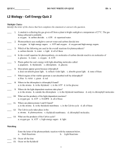
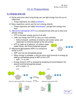
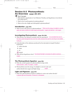
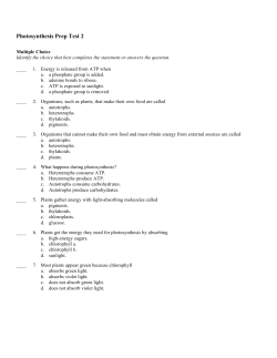
10 Photosynthesis Concept Outline 10.1 What is photosynthesis? The Chloroplast as a Photosynthetic Machine. The highly organized system of membranes in chloroplasts is essential to the functioning of photosynthesis. 10.2 Learning about photosynthesis: An experimental journey. The Role of Soil and Water. The added mass of a growing plant comes mostly from photosynthesis. In plants, water supplies the electrons used to reduce carbon dioxide. Discovery of the Light-Independent Reactions. Photosynthesis is a two-stage process. Only the first stage directly requires light. The Role of Light. The oxygen released during green plant photosynthesis comes from water, and carbon atoms from carbon dioxide are incorporated into organic molecules. The Role of Reducing Power. Electrons released from the splitting of water reduce NADP+; ATP and NADPH are then used to reduce CO2 and form simple sugars. 10.3 Pigments capture energy from sunlight. The Biophysics of Light. The energy in sunlight occurs in “packets” called photons, which are absorbed by pigments. Chlorophylls and Carotenoids. Photosynthetic pigments absorb light and harvest its energy. Organizing Pigments into Photosystems. A photosystem uses light energy to eject an energized electron. How Photosystems Convert Light to Chemical Energy. Some bacteria rely on a single photosystem to produce ATP. Plants use two photosystems in series to generate enough energy to reduce NADP+ and generate ATP. How the Two Photosystems of Plants Work Together. Photosystems II and I drive the synthesis of the ATP and NADPH needed to form organic molecules. 10.4 Cells use the energy and reducing power captured by the light reactions to make organic molecules. The Calvin Cycle. ATP and NADPH are used to build organic molecules, a process reversed in mitochondria. Reactions of the Calvin Cycle. Ribulose bisphosphate binds CO2 in the process of carbon fixation. Photorespiration. The enzyme that catalyzes carbon fixation also affects CO2 release. FIGURE 10.1 Capturing energy. These sunflower plants, growing vigorously in the August sun, are capturing light energy for conversion into chemical energy through photosynthesis. L ife on earth would be impossible without photosynthesis. Every oxygen atom in the air we breathe was once part of a water molecule, liberated by photosynthesis. The energy released by the burning of coal, firewood, gasoline, and natural gas, and by our bodies’ burning of all the food we eat—all, directly or indirectly, has been captured from sunlight by photosynthesis. It is vitally important that we understand photosynthesis. Research may enable us to improve crop yields and land use, important goals in an increasingly crowded world. In the previous chapter we described how cells extract chemical energy from food molecules and use that energy to power their activities. In this chapter, we will examine photosynthesis, the process by which organisms capture energy from sunlight and use it to build food molecules rich in chemical energy (figure 10.1). 183 10.1 What is photosynthesis? The Chloroplast as a Photosynthetic Machine Cuticle Epidermis Life is powered by sunshine. The energy used by most living cells comes ultimately from the sun, captured by plants, algae, and bacteria through the process of photosynthesis. The diversity of life is only possible because our planet is awash in energy streaming earthward from the sun. Each day, the radiant energy that reaches the earth equals about 1 million Hiroshima-sized atomic bombs. Photosynthesis captures about 1% of this huge supply of energy, using it to provide the energy that drives all life. The Photosynthetic Process: A Summary Mesophyll Vascular bundle Bundle sheath Stoma Photosynthesis occurs in many kinds of bacteria and algae, and in the leaves and sometimes the stems of green plants. Figure 10.2 describes the levels of organization in a plant leaf. Recall from chapter 5 that the cells of plant leaves contain organelles called chloroplasts that actually carry out the photosynthetic process. No other structure in a plant cell is able to carry out photosynthesis. Photosynthe- Outer membrane Chloroplasts Inner membrane Vacuole Granum Nucleus Stroma Cell wall Thylakoid FIGURE 10.2 Journey into a leaf. A plant leaf possesses a thick layer of cells (the mesophyll) rich in chloroplasts. The flattened thylakoids in the chloroplast are stacked into columns called grana (singular, granum). The light reactions take place on the thylakoid 184 Part III Energetics sis takes place in three stages: (1) capturing energy from sunlight; (2) using the energy to make ATP and reducing power in the form of a compound called NADPH; and (3) using the ATP and NADPH to power the synthesis of organic molecules from CO2 in the air (carbon fixation). The first two stages take place in the presence of light and are commonly called the light reactions. The third stage, the formation of organic molecules from atmospheric CO2, is called the Calvin cycle. As long as ATP and NADPH are available, the Calvin cycle may occur in the absence of light. The following simple equation summarizes the overall process of photosynthesis: 6 CO2 + 12 H2O + light —→ C6H12O6 + 6 H2O + 6 O2 carbon water glucose water oxygen dioxide Inside the Chloroplast The internal membranes of chloroplasts are organized into sacs called thylakoids, and often numerous thylakoids are stacked on one another in columns called grana. The thylakoid membranes house the photosynthetic pigments for capturing light energy and the machinery to make ATP. Surrounding the thylakoid membrane system is a semiliquid substance called stroma. The stroma houses the enzymes needed to assemble carbon molecules. In the mem- branes of thylakoids, photosynthetic pigments are clustered together to form a photosystem. Each pigment molecule within the photosystem is capable of capturing photons, which are packets of energy. A lattice of proteins holds the pigments in close contact with one another. When light of a proper wavelength strikes a pigment molecule in the photosystem, the resulting excitation passes from one chlorophyll molecule to another. The excited electron is not transferred physically—it is the energy that passes from one molecule to another. A crude analogy to this form of energy transfer is the initial “break” in a game of pool. If the cue ball squarely hits the point of the triangular array of 15 pool balls, the two balls at the far corners of the triangle fly off, but none of the central balls move. The energy passes through the central balls to the most distant ones. Eventually the energy arrives at a key chlorophyll molecule that is touching a membrane-bound protein. The energy is transferred as an excited electron to that protein, which passes it on to a series of other membrane proteins that put the energy to work making ATP and NADPH and building organic molecules. The photosystem thus acts as a large antenna, gathering the light harvested by many individual pigment molecules. The reactions of photosynthesis take place within thylakoid membranes within chloroplasts in leaf cells. Sunlight Stroma Thylakoid H2O Photosystem O2 Light reactions Thylakoid ADP Granum ATP NADPH Calvin cycle Stroma + NADP Organic molecules CO2 FIGURE 10.2 (continued) membrane and generate the ATP and NADPH that fuel the Calvin cycle. The fluid interior matrix of a chloroplast, the stroma, contains the enzymes that carry out the Calvin cycle. Chapter 10 Photosynthesis 185 10.2 Learning about photosynthesis: An experimental journey. The Role of Soil and Water The story of how we learned about photosynthesis is one of the most interesting in science and serves as a good introduction to this complex process. The story starts over 300 years ago, with a simple but carefully designed experiment by a Belgian doctor, Jan Baptista van Helmont (1577–1644). From the time of the Greeks, plants were thought to obtain their food from the soil, literally sucking it up with their roots; van Helmont thought of a simple way to test the idea. He planted a small willow tree in a pot of soil after weighing the tree and the soil. The tree grew in the pot for several years, during which time van Helmont added only water. At the end of five years, the tree was much larger: its weight had increased by 74.4 kilograms. However, all of this added mass could not have come from the soil, because the soil in the pot weighed only 57 grams less than it had five years earlier! With this experiment, van Helmont demonstrated that the substance of the plant was not produced only from the soil. He incorrectly concluded that mainly the water he had been adding accounted for the plant’s increased mass. A hundred years passed before the story became clearer. The key clue was provided by the English scientist Joseph Priestly, in his pioneering studies of the properties of air. On the 17th of August, 1771, Priestly “accidentally hit upon a method of restoring air that had been injured by the burning of candles.” He “put a [living] sprig of mint into air in which a wax candle had burnt out and found that, on the 27th of the same month, another candle could be burned in this same air.” Somehow, the vegetation seemed to have restored the air! Priestly found that while a mouse could not breathe candle-exhausted air, air “restored” by vegetation was not “at all inconvenient to a mouse.” The key clue was that living vegetation adds something to the air. How does vegetation “restore” air? Twenty-five years later, Dutch physician Jan Ingenhousz solved the puzzle. Working over several years, Ingenhousz reproduced and significantly extended Priestly’s results, demonstrating that air was restored only in the presence of sunlight, and only by a plant’s green leaves, not by its roots. He proposed that the green parts of the plant carry out a process (which we now call photosynthesis) that uses sunlight to split carbon dioxide (CO2) into carbon and oxygen. He suggested that the oxygen was released as O2 gas into the air, while the carbon atom combined with water to form carbohydrates. His proposal was a good guess, even though the later step was subsequently modified. Chemists later found that the proportions of carbon, oxygen, and hydrogen atoms in carbohydrates are indeed about one atom of carbon per molecule of water (as the term carbohydrate indicates). A Swiss botanist found in 1804 that water was a necessary reactant. By the end of that century the overall reaction for photosynthesis could be written as: 186 Part III Energetics CO2 + H2O + light energy —→ (CH2O) + O2 It turns out, however, that there’s more to it than that. When researchers began to examine the process in more detail in the last century, the role of light proved to be unexpectedly complex. Van Helmont showed that soil did not add mass to a growing plant. Priestly and Ingenhousz and others then worked out the basic chemical reaction. Discovery of the Light-Independent Reactions Ingenhousz’s early equation for photosynthesis includes one factor we have not discussed: light energy. What role does light play in photosynthesis? At the beginning of the previous century, the English plant physiologist F. F. Blackman began to address the question of the role of light in photosynthesis. In 1905, he came to the startling conclusion that photosynthesis is in fact a two-stage process, only one of which uses light directly. Blackman measured the effects of different light intensities, CO2 concentrations, and temperatures on photosynthesis. As long as light intensity was relatively low, he found photosynthesis could be accelerated by increasing the amount of light, but not by increasing the temperature or CO2 concentration (figure 10.3). At high light intensities, however, an increase in temperature or CO 2 concentration greatly accelerated photosynthesis. Blackman concluded that photosynthesis consists of an initial set of what he called “light” reactions, that are largely independent of temperature, and a second set of “dark” reactions, that seemed to be independent of light but limited by CO 2 . Do not be confused by Blackman’s labels—the so-called “dark” reactions occur in the light (in fact, they require the products of the light reactions); their name simply indicates that light is not directly involved in those reactions. Blackman found that increased temperature increases the rate of the dark carbon-reducing reactions, but only up to about 35°C. Higher temperatures caused the rate to fall off rapidly. Because 35°C is the temperature at which many plant enzymes begin to be denatured (the hydrogen bonds that hold an enzyme in its particular catalytic shape begin to be disrupted), Blackman concluded that enzymes must carry out the dark reactions. Blackman showed that capturing photosynthetic energy requires sunlight, while building organic molecules does not. The Role of Light The role of light in the so-called light and dark reactions was worked out in the 1930s by C. B. van Niel, then a graduate student at Stanford University studying photosynthesis in bacteria. One of the types of bacteria he was studying, the purple sulfur bacteria, does not release oxygen during photosynthesis; instead, they convert hydrogen sulfide (H2S) into globules of pure elemental sulfur that accumulate inside themselves. The process that van Niel observed was Temperature limited The striking parallel between this equation and Ingenhousz’s equation led van Niel to propose that the generalized process of photosynthesis is in fact CO2 + 2 H2A + light energy → (CH2O) + H2O + 2 A Lig ht lim ite d Excess CO2; 20°C (a) Rate of photosynthesis CO2 + 2 H2S + light energy → (CH2O) + H2O + 2 S Maximum rate Excess CO2; 35°C CO2 limited Insufficient CO2 (0.01%); 20°C 500 1000 1500 2000 2500 In this equation, the substance H2A serves Light intensity (foot-candles) (b) as an electron donor. In photosynthesis performed by green plants, H2A is water, FIGURE 10.3 while among purple sulfur bacteria, H2A is Discovery of the dark reactions. (a) Blackman measured photosynthesis rates under hydrogen sulfide. The product, A, comes differing light intensities, CO concentrations, and temperatures. (b) As this graph 2 from the splitting of H2A. Therefore, the shows, light is the limiting factor at low light intensities, while temperature and CO 2 O2 produced during green plant photosyn- concentration are the limiting factors at higher light intensities. thesis results from splitting water, not carbon dioxide. The Role of Reducing Power When isotopes came into common use in biology in the early 1950s, it became possible to test van Niel’s revoluIn his pioneering work on the light reactions, van Niel had tionary proposal. Investigators examined photosynthesis in further proposed that the reducing power (H+) generated by green plants supplied with 18O water; they found that the the splitting of water was used to convert CO2 into organic 18O label ended up in oxygen gas rather than in carbohymatter in a process he called carbon fixation. Was he right? drate, just as van Niel had predicted: In the 1950s Robin Hill demonstrated that van Niel was CO2 + 2 H218O + light energy —→ (CH2O) + H2O + 18O2 In algae and green plants, the carbohydrate typically produced by photosynthesis is the sugar glucose, which has six carbons. The complete balanced equation for photosynthesis in these organisms thus becomes 6 CO2 + 12 H2O + light energy —→ C6H12O6 + 6 O2 + 6 H2O. We now know that the first stage of photosynthesis, the light reactions, uses the energy of light to reduce NADP (an electron carrier molecule) to NADPH and to manufacture ATP. The NADPH and ATP from the first stage of photosynthesis are then used in the second stage, the Calvin cycle, to reduce the carbon in carbon dioxide and form a simple sugar whose carbon skeleton can be used to synthesize other organic molecules. Van Niel discovered that photosynthesis splits water molecules, incorporating the carbon atoms of carbon dioxide gas and the hydrogen atoms of water into organic molecules and leaving oxygen gas. indeed right, and that light energy could be used to generate reducing power. Chloroplasts isolated from leaf cells were able to reduce a dye and release oxygen in response to light. Later experiments showed that the electrons released from water were transferred to NADP+. Arnon and coworkers showed that illuminated chloroplasts deprived of CO2 accumulate ATP. If CO2 is then introduced, neither ATP nor NADPH accumulate, and the CO2 is assimilated into organic molecules. These experiments are important for three reasons. First, they firmly demonstrate that photosynthesis occurs only within chloroplasts. Second, they show that the light-dependent reactions use light energy to reduce NADP+ and to manufacture ATP. Thirdly, they confirm that the ATP and NADPH from this early stage of photosynthesis are then used in the later light-independent reactions to reduce carbon dioxide, forming simple sugars. Hill showed that plants can use light energy to generate reducing power. The incorporation of carbon dioxide into organic molecules in the light-independent reactions is called carbon fixation. Chapter 10 Photosynthesis 187 10.3 Pigments capture energy from sunlight. The Biophysics of Light Increasing energy Where is the energy in light? What is there in sunlight that a plant can use to Increasing wavelength reduce carbon dioxide? This is the 1 nm 10 nm 1000 nm 0.01 cm 1 cm 1m 100 m mystery of photosynthesis, the one fac- 0.001 nm tor fundamentally different from UV processes such as respiration. To anGamma rays X rays light Infrared Radio waves swer these questions, we will need to Visible light consider the physical nature of light itself. James Clerk Maxwell had theorized that light was an electromagnetic wave—that is, that light moved 430 nm 500 nm 560 nm 600 nm 650 nm 740 nm through the air as oscillating electric 400 nm and magnetic fields. Proof of this came FIGURE 10.4 in a curious experiment carried out in a The electromagnetic spectrum. Light is a form of electromagnetic energy conveniently laboratory in Germany in 1887. A thought of as a wave. The shorter the wavelength of light, the greater its energy. Visible young physicist, Heinrich Hertz, was light represents only a small part of the electromagnetic spectrum between 400 and 740 attempting to verify a highly mathe- nanometers. matical theory that predicted the existence of electromagnetic waves. To see whether such waves existed, Hertz designed a clever experiment. On one passage of the electric spark induced by the radio waves. side of a room he constructed a powerful spark generator Visible wavelengths of light were unable to remove the that consisted of two large, shiny metal spheres standing electrons because their photons did not have enough ennear each other on tall, slender rods. When a very high staergy to free the electrons from the metal surface at the ends tic electrical charge was built up on one sphere, sparks of the hoop. would jump across to the other sphere. After constructing this device, Hertz set out to investigate whether the sparking would create invisible electromagnetic The Energy in Photons waves, so-called radio waves, as predicted by the mathematiPhotons do not all possess the same amount of energy (figcal theory. On the other side of the room, he placed the ure 10.4). Instead, the energy content of a photon is inworld’s first radio receiver, a thin metal hoop on an insulatversely proportional to the wavelength of the light: shorting stand. There was a small gap at the bottom of the hoop, wavelength light contains photons of higher energy than so that the hoop did not quite form a complete circle. When long-wavelength light. X rays, which contain a great deal of Hertz turned on the spark generator across the room, he saw energy, have very short wavelengths—much shorter than visitiny sparks passing across the gap in the hoop! This was the ble light, making them ideal for high-resolution microscopes. first demonstration of radio waves. But Hertz noted another Hertz had noted that the strength of the photoelectric curious phenomenon. When UV light was shining across effect depends on the wavelength of light; short wavethe gap on the hoop, the sparks were produced more readily. lengths are much more effective than long ones in producThis unexpected facilitation, called the photoelectric effect, ing the photoelectric effect. Einstein’s theory of the photopuzzled investigators for many years. electric effect provides an explanation: sunlight contains The photoelectric effect was finally explained using a photons of many different energy levels, only some of concept proposed by Max Planck in 1901. Planck develwhich our eyes perceive as visible light. The highest energy oped an equation that predicted the blackbody radiation photons, at the short-wavelength end of the electromagcurve based upon the assumption that light and other forms netic spectrum (see figure 10.4), are gamma rays, with of radiation behaved as units of energy called photons. In wavelengths of less than 1 nanometer; the lowest energy 1905 Albert Einstein explained the photoelectric effect utiphotons, with wavelengths of up to thousands of meters, lizing the photon concept. Ultraviolet light has photons of are radio waves. Within the visible portion of the spectrum, sufficient energy that when they fell on the loop, electrons violet light has the shortest wavelength and the most enerwere ejected from the metal surface. The photons had getic photons, and red light has the longest wavelength and transferred their energy to the electrons, literally blasting the least energetic photons. them from the ends of the hoop and thus facilitating the 188 Part III Energetics Ultraviolet Light Chlorophyll b Chlorophyll a Relative light absorption The sunlight that reaches the earth’s surface contains a significant amount of ultraviolet (UV) light, which, because of its shorter wavelength, possesses considerably more energy than visible light. UV light is thought to have been an important source of energy on the primitive earth when life originated. Today’s atmosphere contains ozone (derived from oxygen gas), which absorbs most of the UV photons in sunlight, but a considerable amount of UV light still manages to penetrate the atmosphere. This UV light is a potent force in disrupting the bonds of DNA, causing mutations that can lead to skin cancer. As we will describe in a later chapter, loss of atmospheric ozone due to human activities threatens to cause an enormous jump in the incidence of human skin cancers throughout the world. Carotenoids Absorption Spectra and Pigments 400 450 500 550 600 650 700 How does a molecule “capture” the energy of light? A photon can be envisioned as a Wavelength (nm) very fast-moving packet of energy. When it FIGURE 10.5 strikes a molecule, its energy is either lost as The absorption spectrum of chlorophyll. The peaks represent wavelengths of heat or absorbed by the electrons of the mol- sunlight that the two common forms of photosynthetic pigment, chlorophyll a (solid ecule, boosting those electrons into higher line) and chlorophyll b (dashed line), strongly absorb. These pigments absorb energy levels. Whether or not the photon’s predominately violet-blue and red light in two narrow bands of the spectrum and energy is absorbed depends on how much reflect the green light in the middle of the spectrum. Carotenoids (not shown here) energy it carries (defined by its wavelength) absorb mostly blue and green light and reflect orange and yellow light. and on the chemical nature of the molecule it hits. As we saw in chapter 2, electrons occupy discrete energy levels in their orbits around atomic nuclei. To boost an electron into a different energy absorbed by the pigment in our eyes, we perceive them as level requires just the right amount of energy, just as reachgreen. ing the next rung on a ladder requires you to raise your Chlorophyll a is the main photosynthetic pigment and is foot just the right distance. A specific atom can, therefore, the only pigment that can act directly to convert light enabsorb only certain photons of light—namely, those that ergy to chemical energy. However, chlorophyll b, acting as correspond to the atom’s available electron energy levels. an accessory or secondary light-absorbing pigment, comAs a result, each molecule has a characteristic absorption plements and adds to the light absorption of chlorophyll a. spectrum, the range and efficiency of photons it is capable Chlorophyll b has an absorption spectrum shifted toward of absorbing. the green wavelengths. Therefore, chlorophyll b can absorb Molecules that are good absorbers of light in the visible photons chlorophyll a cannot. Chlorophyll b therefore range are called pigments. Organisms have evolved a varigreatly increases the proportion of the photons in sunlight ety of different pigments, but there are only two general that plants can harvest. An important group of accessory types used in green plant photosynthesis: carotenoids and pigments, the carotenoids, assist in photosynthesis by capchlorophylls. Chlorophylls absorb photons within narrow turing energy from light of wavelengths that are not effienergy ranges. Two kinds of chlorophyll in plants, chlorociently absorbed by either chlorophyll. phylls a and b, preferentially absorb violet-blue and red light (figure 10.5). Neither of these pigments absorbs phoIn photosynthesis, photons of light are absorbed by tons with wavelengths between about 500 and 600 pigments; the wavelength of light absorbed depends nanometers, and light of these wavelengths is, therefore, upon the specific pigment. reflected by plants. When these photons are subsequently Chapter 10 Photosynthesis 189 Chlorophylls and Carotenoids Chlorophylls absorb photons by means of an excitation process analogous to the photoelectric effect. These pigments contain a complex ring structure, called a porphyrin ring, with alternating single and double bonds. At the center of the ring is a magnesium atom. Photons absorbed by the pigment molecule excite electrons in the ring, which are then channeled away through the alternating carbon-bond system. Several small side groups attached to the outside of the ring alter the absorption properties of the molecule in different kinds of chlorophyll (figure 10.6). The precise absorption spectrum is also influenced by the local microenvironment created by the association of chlorophyll with specific proteins. Once Ingenhousz demonstrated that only the green parts of plants can “restore” air, researchers suspected chlorophyll was the primary pigment that plants employ to absorb light in photosynthesis. Experiments conducted in the 1800s clearly verified this suspicion. One such experiment, performed by T. W. Englemann in 1882 (figure 10.7), serves as a particularly elegant example, simple in design and clear in outcome. Englemann set out to characterize the action spectrum of photosynthesis, that is, the relative effectiveness of different wavelengths of light in promoting photosynthesis. He carried out the entire experiment utilizing a single slide mounted on a microscope. To obtain different wavelengths of light, he placed a prism under his microscope, splitting the light that illuminated the slide into a spectrum of colors. He then arranged a filament of green algal cells across the spectrum, so that different parts of the filament were illuminated with different wavelengths, and allowed the algae to carry out photosynthesis. To assess how fast photosynthesis was proceeding, Englemann chose to monitor the rate of oxygen production. Lacking a mass spectrometer and other modern instruments, he added aerotactic (oxygen-seeking) bacteria to the slide; he knew they would gather along the filament at locations where oxygen was being produced. He found that the bacteria accumulated in areas illuminated by red and violet light, the two colors most strongly absorbed by chlorophyll. All plants, algae, and cyanobacteria use chlorophyll a as their primary pigments. It is reasonable to ask why these photosynthetic organisms do not use a pigment like retinal (the pigment in our eyes), which has a broad absorption spectrum that covers the range of 500 to 600 nanometers. The most likely hypothesis involves photoefficiency. Although retinal absorbs a broad range of wavelengths, it does so with relatively low efficiency. Chlorophyll, in contrast, absorbs in only two narrow bands, but does so with high efficiency. Therefore, plants and most other photosynthetic organisms achieve far higher overall photon capture rates with chlorophyll than with other pigments. Chlorophyll a: R = —CH3 Chlorophyll b: R = —CHO Chlorophyll molecules embedded in a protein complex in the thylakoid membrane H2C CH H CH2CH3 CH3 N Porphyrin head N H N CH3 H CH3 O Thylakoid Hydrocarbon tail Granum Part III N Mg H Thylakoid membrane 190 R Energetics H CH2 H CH2 O CO2CH3 C O CH2 CH CCH3 CH2 CH2 CH2 CHCH3 CH2 CH2 CH2 CHCH3 CH2 CH2 CH2 CHCH3 CH3 FIGURE 10.6 Chlorophyll. Chlorophyll molecules consist of a porphyrin head and a hydrocarbon tail that anchors the pigment molecule to hydrophobic regions of proteins embedded within the membranes of thylakoids. The only difference between the two chlorophyll molecules is the substitution of a —CHO (aldehyde) group in chlorophyll b for a —CH3 (methyl) group in chlorophyll a. Absorbance Oxygen-seeking bacteria Filament of green alga T.W. Englemann revealed the action spectrum of photosynthesis in the filamentous alga Spirogyra in 1882. Englemann used the rate of oxygen production to measure the rate of photosynthesis. As his oxygen indicator, he chose bacteria that are attracted by oxygen. In place of the mirror and diaphragm usually used to illuminate objects under view in his microscope, he substituted a "microspectral apparatus," which, as its name implies, produced a tiny spectrum of colors that it projected upon the slide under the microscope. Then he arranged a filament of algal cells parallel to the spread of the spectrum. The oxygen-seeking bacteria congregated mostly in the areas where the violet and red wavelengths fell upon the algal filament. FIGURE 10.7 Constructing an action spectrum for photosynthesis. As you can see, the action spectrum for photosynthesis that Englemann revealed in his experiment parallels the absorption spectrum of chlorophyll (see figure 10.5). Oak leaf in summer Oak leaf in autumn FIGURE 10.8 Fall colors are produced by carotenoids and other accessory pigments. During the spring and summer, chlorophyll in leaves masks the presence of carotenoids and other accessory pigments. When cool fall temperatures cause leaves to cease manufacturing chlorophyll, the chlorophyll is no longer present to reflect green light, and the leaves reflect the orange and yellow light that carotenoids and other pigments do not absorb. Carotenoids consist of carbon rings linked to chains with alternating single and double bonds. They can absorb photons with a wide range of energies, although they are not always highly efficient in transferring this energy. Carotenoids assist in photosynthesis by capturing energy from light of wavelengths that are not efficiently absorbed by chlorophylls (figure 10.8; see figure 10.5). A typical carotenoid is β-carotene, whose two carbon rings are connected by a chain of 18 carbon atoms with alternating single and double bonds. Splitting a molecule of β-carotene into equal halves produces two molecules of vitamin A. Oxidation of vitamin A produces retinal, the pigment used in vertebrate vision. This explains why carrots, which are rich in β-carotene, enhance vision. A pigment is a molecule that absorbs light. The wavelengths absorbed by a particular pigment depend on the available energy levels to which light-excited electrons can be boosted in the pigment. Chapter 10 Photosynthesis 191 Organizing Pigments into Photosystems 1. Primary photoevent. A photon of light is captured by a pigment. The result of this primary photoevent is the excitation of an electron within the pigment. 2. Charge separation. This excitation energy is transferred to a specialized chlorophyll pigment termed a reaction center, which reacts by transferring an energetic electron to an acceptor molecule, thus initiating electron transport. 3. Electron transport. The excited electron is shuttled along a series of electron-carrier molecules embedded within the photosynthetic membrane. Several of them react by transporting protons across the membrane, generating a gradient of proton concentration. Its arrival at the pump induces the transport of a proton across the membrane. The electron is then passed to an acceptor. 4. Chemiosmosis. The protons that accumulate on one side of the membrane now flow back across the membrane through specific protein complexes where chemiosmotic synthesis of ATP takes place, just as it does in aerobic respiration. Discovery of Photosystems One way to study how pigments absorb light is to measure the dependence of the output of photosynthesis on the intensity of illumination—that is, how much photosynthesis is produced by how much light. When experiments of this sort are done on plants, they show that the output of photosynthesis increases linearly at low intensities but lessens at higher intensities, finally saturating at high-intensity light (figure 10.9). Saturation occurs because all of the light-absorbing capacity of the plant is in use; additional light doesn’t increase the output because there is nothing to absorb the added photons. It is tempting to think that at saturation, all of a plant’s pigment molecules are in use. In 1932 plant physiologists Robert Emerson and William Arnold set out to test this hypothesis in an organism where they could measure both the number of chlorophyll molecules and the output of photosynthesis. In their experiment, they measured the oxygen yield of photosynthesis when Chlorella (unicellular green algae) were exposed to very brief light flashes lasting 192 Part III Energetics Output ( O2 yield per flash) The light reactions of photosynthesis occur in membranes. In bacteria like those studied by van Niel, the plasma membrane itself is the photosynthetic membrane. In plants and algae, by contrast, photosynthesis is carried out by organelles that are the evolutionary descendants of photosynthetic bacteria, chloroplasts—the photosynthetic membranes exist within the chloroplasts. The light reactions take place in four stages: Saturation when all chlorophyll molecules are in use Saturation when all photosystems are in use Expected Observed Intensity of light flashes FIGURE 10.9 Emerson and Arnold’s experiment. When photosynthetic saturation is achieved, further increases in intensity cause no increase in output. only a few microseconds. Assuming the hypothesis of pigment saturation to be correct, they expected to find that as they increased the intensity of the flashes, the yield per flash would increase, until each chlorophyll molecule absorbed a photon, which would then be used in the light reactions, producing a molecule of O2. Unexpectedly, this is not what happened. Instead, saturation was achieved much earlier, with only one molecule of O2 per 2500 chlorophyll molecules! This led Emerson and Arnold to conclude that light is absorbed not by independent pigment molecules, but rather by clusters of chlorophyll and accessory pigment molecules which have come to be called photosystems. Light is absorbed by any one of the hundreds of pigment molecules in a photosystem, which transfer their excitation energy to one with a lower energy level than the others. This reaction center of the photosystem acts as an energy sink, trapping the excitation energy. It was the saturation of these reaction centers, not individual molecules, that was observed by Emerson and Arnold. Architecture of a Photosystem In chloroplasts and all but the most primitive bacteria, light is captured by such photosystems. Each photosystem is a network of chlorophyll a molecules, accessory pigments, and associated proteins held within a protein matrix on the surface of the photosynthetic membrane. Like a magnifying glass focusing light on a precise point, a photosystem channels the excitation energy gathered by any one of its pigment molecules to a specific molecule, the reaction center chlorophyll. This molecule then passes the energy out of the photosystem so it can be put to work driving the synthesis of ATP and organic molecules. A photosystem thus consists of two closely linked components: (1) an antenna complex of hundreds of pigment molecules that gather photons and feed the captured light energy to the reaction center; and (2) a reaction center, consisting of one or more chlorophyll a molecules in a matrix of protein, that passes the energy out of the photosystem. The Antenna Complex. The antenna complex captures photons from sunlight (figure 10.10). In chloroplasts, the antenna complex is a web of chlorophyll molecules linked together and held tightly on the thylakoid membrane by a matrix of proteins. Varying amounts of carotenoid accessory pigments may also be present. The protein matrix serves as a sort of scaffold, holding individual pigment molecules in orientations that are optimal for energy transfer. The excitation energy resulting from the absorption of a photon passes from one pigment molecule to an adjacent molecule on its way to the reaction center. After the transfer, the excited electron in each molecule returns to the low-energy level it had before the photon was absorbed. Consequently, it is energy, not the excited electrons themselves, that passes from one pigment molecule to the next. The antenna complex funnels the energy from many electrons to the reaction center. The Reaction Center. The reaction center is a transmembrane protein-pigment complex. In the reaction center of purple photosynthetic bacteria, which is simpler than in chloroplasts but better understood, a pair of chlorophyll a molecules acts as a trap for photon energy, passing an excited electron to an acceptor precisely positioned as its neighbor. Note that here the excited electron itself is transferred, not just the energy as we saw in pigment-pigment transfers. This allows the photon excitation to move away from the chlorophylls and is the key conversion of light to chemical energy. Figure 10.11 shows the transfer of energy from the reaction center to the primary electron acceptor. By energizing an electron of the reaction center chlorophyll, light creates a strong electron donor where none existed before. The chlorophyll transfers the energized electron to the primary acceptor, a molecule of quinone, reducing the quinone and converting it to a strong electron donor. A weak electron donor then donates a low-energy electron to the chlorophyll, restoring it to its original condition. In plant chloroplasts, water serves as the electron donor. Electron acceptor Reaction center chlorophyll Photon Electron donor Chlorophyll molecules Photosystem FIGURE 10.10 How the antenna complex works. When light of the proper wavelength strikes any pigment molecule within a photosystem, the light is absorbed by that pigment molecule. The excitation energy is then transferred from one molecule to another within the cluster of pigment molecules until it encounters the reaction center chlorophyll a. When excitation energy reaches the reaction center chlorophyll, electron transfer is initiated. Excited chlorophyll molecule Electron donor Chlorophyll oxidized Electron acceptor Acceptor reduced + – Donor Acceptor oxidized reduced + – FIGURE 10.11 Converting light to chemical energy. The reaction center chlorophyll donates a light-energized electron to the primary electron acceptor, reducing it. The oxidized chlorophyll then fills its electron “hole” by oxidizing a donor molecule. Photosystems contain pigments that capture photon energy from light. The pigments transfer the energy to reaction centers. There, the energy excites electrons, which are channeled away to do chemical work. Chapter 10 Photosynthesis 193 Bacteria Use a Single Photosystem Photosynthetic pigment arrays are thought to have evolved more than 3 billion years ago in bacteria similar to the sulfur bacteria studied by van Niel. 1. Electron is joined with a proton to make hydrogen. In these bacteria, the absorption of a photon of light at a peak absorption of 870 nanometers (near infrared, not visible to the human eye) by the photosystem results in the transmission of an energetic electron along an electron transport chain, eventually combining with a proton to form a hydrogen atom. In the sulfur bacteria, the proton is extracted from hydrogen sulfide, leaving elemental sulfur as a by-product. In bacteria that evolved later, as well as in plants and algae, the proton comes from water, producing oxygen as a by-product. 2. Electron is recycled to chlorophyll. The ejection of an electron from the bacterial reaction center leaves it short one electron. Before the photosystem of the sulfur bacteria can function again, an electron must be returned. These bacteria channel the electron back to the pigment through an electron transport system similar to the one described in chapter 9; the electron’s passage drives a proton pump that promotes the chemiosmotic synthesis of ATP. One molecule of ATP is produced for every three electrons that follow this path. Viewed overall (figure 10.12), the path of the electron is thus a circle. Chemists therefore call the electron transfer process leading to ATP formation cyclic photophosphorylation. Note, however, that the electron that left the P870 reaction center was a high-energy electron, boosted by the absorption of a photon of light, while the electron that returns has only as much energy as it had before the photon was absorbed. The difference in the energy of that electron is the photosynthetic payoff, the energy that drives the proton pump. For more than a billion years, cyclic photophosphorylation was the only form of photosynthetic light reaction that organisms used. However, its major limitation is that it is geared only toward energy production, not toward biosynthesis. Most photosynthetic organisms incorporate atmospheric carbon dioxide into carbohydrates. Because the carbohydrate molecules are more reduced (have more hydrogen atoms) than carbon dioxide, a source of reducing power (that is, hydrogens) must be provided. Cyclic photophosphorylation does not do this. The hydrogen atoms extracted from H2S are used as a source of protons, and are not available to join to carbon. Thus bacteria that are restricted to this process must scavenge hydrogens from other sources, an inefficient undertaking. 194 Part III Energetics Why Plants Use Two Photosystems After the sulfur bacteria appeared, other kinds of bacteria evolved an improved version of the photosystem that overcame the limitation of cyclic photophosphorylation in a neat and simple way: a second, more powerful photosystem using another arrangement of chlorophyll a was combined with the original. In this second photosystem, called photosystem II, molecules of chlorophyll a are arranged with a different geometry, so that more shorter wavelength, higher energy photons are absorbed than in the ancestral photosystem, which is called photosystem I. As in the ancestral photosystem, energy is transmitted from one pigment molecule to another within the antenna complex of these photosystems until it reaches the reaction center, a particular pigment molecule positioned near a strong membrane-bound electron acceptor. In photosystem II, the absorption peak (that is, the wavelength of light most strongly absorbed) of the pigments is approximately 680 nanometers; therefore, the reaction center pigment is called P680. The absorption peak of photosystem I pigments in plants is 700 nanometers, so its reaction center pigment is called P700. Working together, the two photosystems carry out a noncyclic electron transfer. When the rate of photosynthesis is measured using two light beams of different wavelengths (one red and the Excited reaction center Electron acceptor e– Fd Ferredoxin Energy of electrons How Photosystems Convert Light to Chemical Energy ADP e– Photon Reaction center b6-f complex ATP pC e– P870 Photosystem Plastocyanin b6-f complex FIGURE 10.12 The path of an electron in purple sulfur bacteria. When a light-energized electron is ejected from the photosystem reaction center (P870), it passes in a circle, eventually returning to the photosystem from which it was ejected. In sulfur bacteria, excited electrons ejected from the reaction center travel a circular path, driving a proton pump and then returning to their original photosystem. Plants employ two photosystems in series, which generates power to reduce NADP+ to NADPH with enough left over to make ATP. Relative rate of photosynthesis other far-red), the rate was greater than the sum of the rates using individual beams of red and far-red light (figure 10.13). This surprising result, called the enhancement effect, can be explained by a mechanism involving two photosystems acting in series (that is, one after the other), one of which absorbs preferentially in the red, the other in the far-red. The use of two photosystems solves the problem of obtaining reducing power in a simple and direct way, by harnessing the energy of two photosystems. The scheme shown in figure 10.14, called a Z diagram, illustrates the two electron-energizing steps, one catalyzed by each photosystem. The electrons originate from water, which holds onto its electrons very tightly (redox potential = +820 mV), and end up in NADPH, which holds its electrons much more loosely (redox potential = –320 mV). Far-red light on Off Red light on Ferredoxin e– Fd NADP reductase Plastoquinone e– Energy of electrons b6-f complex Plastocyanin Reaction center pC H+ e– Water-splitting enzyme e– NADP+ + H+ e– Q P680 Off FIGURE 10.13 The “enhancement effect.” The rate of photosynthesis when red and far-red light are provided together is greater than the sum of the rates when each wavelength is provided individually. This result baffled researchers in the 1950s. Today it provides the key evidence that photosynthesis is carried out by two photochemical systems with slightly different wavelength optima. Excited reaction center Reaction center Both lights on Time Excited reaction center Photon Off Proton gradient formed for ATP synthesis NADPH Photon P700 H2O 1 2H+ + ϪO 2 2 Photosystem II b6-f complex Photosystem I NADP reductase FIGURE 10.14 A Z diagram of photosystems I and II. Two photosystems work sequentially. First, a photon of light ejects a high-energy electron from photosystem II; that electron is used to pump a proton across the membrane, contributing chemiosmotically to the production of a molecule of ATP. The ejected electron then passes along a chain of cytochromes to photosystem I. When photosystem I absorbs a photon of light, it ejects a high-energy electron used to drive the formation of NADPH. Chapter 10 Photosynthesis 195 How the Two Photosystems of Plants Work Together Plants use the two photosystems discussed earlier in series, first one and then the other, to produce both ATP and NADPH. This two-stage process is called noncyclic photophosphorylation, because the path of the electrons is not a circle—the electrons ejected from the photosystems do not return to it, but rather end up in NADPH. The photosystems are replenished instead with electrons obtained by splitting water. Photosystem II acts first. High-energy electrons generated by photosystem II are used to synthesize ATP and then passed to photosystem I to drive the production of NADPH. For every pair of electrons obtained from water, one molecule of NADPH and slightly more than one molecule of ATP are produced. Stroma Photon Photon H+ + NADP+ Antenna complex Thylakoid membrane NADPH Fd Q pC H2O Plastoquinone Water-splitting enzyme 1 ϪO 2 2 Proton gradient Plastocyanin Ferredoxin H+ 2H+ Photosystem II b6-f complex Photosystem I NADP reductase Thylakoid space Photosystem II The reaction center of photosystem II, called P680, closely resembles the reac- FIGURE 10.15 tion center of purple bacteria. It con- The photosynthetic electron transport system. When a photon of light strikes a pigment sists of more than 10 transmembrane molecule in photosystem II, it excites an electron. This electron is coupled to a proton protein subunits. The light-harvesting stripped from water by an enzyme and is passed along a chain of membrane-bound cytochrome electron carriers (red arrow). When water is split, oxygen is released from the antenna complex consists of some 250 cell, and the hydrogen ions remain in the thylakoid space. At the proton pump molecules of chlorophyll a and acces- (b -f complex), the energy supplied by the photon is used to transport a proton across the 6 sory pigments bound to several protein membrane into the thylakoid. The concentration of hydrogen ions within the thylakoid chains. In photosystem II, the oxygen thus increases further. When photosystem I absorbs another photon of light, its pigment atoms of two water molecules bind to a passes a second high-energy electron to a reduction complex, which generates NADPH. cluster of manganese atoms which are embedded within an enzyme and bound to the reaction center. In a way that is poorly underport chain of mitochondria discussed in chapter 9. Arrival stood, this enzyme splits water, removing electrons one at a of the energetic electron causes the b6-f complex to pump a time to fill the holes left in the reaction center by departure proton into the thylakoid space. A small copper-containing of light-energized electrons. As soon as four electrons have protein called plastocyanin (symbolized pC) then carries the been removed from the two water molecules, O2 is released. electron to photosystem I. The Path to Photosystem I Making ATP: Chemiosmosis The primary electron acceptor for the light-energized electrons leaving photosystem II is a quinone molecule, as it was in the bacterial photosystem described earlier. The reduced quinone which results (plastoquinone, symbolized Q) is a strong electron donor; it passes the excited electron to a proton pump called the b6-f complex embedded within the thylakoid membrane (figure 10.15). This complex closely resembles the bc1 complex in the respiratory electron trans- Each thylakoid is a closed compartment into which protons are pumped from the stroma by the b6-f complex. The splitting of water also produces added protons that contribute to the gradient. The thylakoid membrane is impermeable to protons, so protons cross back out almost exclusively via the channels provided by ATP synthases. These channels protrude like knobs on the external surface of the thylakoid membrane. As protons pass out of 196 Part III Energetics the thylakoid through the ATP synthase channel, ADP is phosphorylated to ATP and released into the stroma, the fluid matrix inside the chloroplast (figure 10.16). The stroma contains the enzymes that catalyze the reactions of carbon fixation. H+ Photon H+ Making NADPH ATP H+ H+ Stroma Q Photosystem I The reaction center of photosystem I, called P700, is a transmembrane complex consisting of at least 13 protein subunits. Energy is fed to it by an antenna complex consisting of 130 chlorophyll a and accessory pigment molecules. Photosystem I accepts an electron from plastocyanin into the hole created by the exit of a light-energized electron. This arriving electron has by no means lost all of its light-excited energy; almost half remains. Thus, the absorption of a photon of light energy by photosystem I boosts the electron leaving the reaction center to a very high energy level. Unlike photosystem II and the bacterial photosystem, photosystem I does not rely on quinones as electron acceptors. Instead, it passes electrons to an iron-sulfur protein called ferredoxin (Fd). ADP H2O H+ Thylakoid 1 ϪO space 2 2 H+ H+ 2 H+ Photosystem II b6-f complex ATP synthase Chloroplast Plant cell FIGURE 10.16 Chemiosmosis in a chloroplast. The b6-f complex embedded in the thylakoid membrane pumps protons into the interior of the thylakoid. ATP is produced on the outside surface of the membrane (stroma side), as protons diffuse back out of the thylakoid through ATP synthase channels. Photosystem I passes electrons to ferredoxin on the stromal side of the membrane (outside the thylakoid). The reduced ferredoxin carries a very-highpotential electron. Two of them, from two molecules of reduced ferredoxin, are then donated to a molecule of NADP+ to form NADPH. The reaction is catalyzed by the membrane-bound enzyme NADP reductase. Because the reaction occurs on the stromal side of the membrane and involves the uptake of a proton in forming NADPH, it contributes further to the proton gradient established during photosynthetic electron transport. Making More ATP The passage of an electron from water to NADPH in the noncyclic photophosphorylation described previously generates one molecule of NADPH and slightly more than one molecule of ATP. However, as you will learn later in this chapter, building organic molecules takes more energy than that—it takes one-and-a-half ATP molecules per NADPH molecule to fix carbon. To produce the extra ATP, many plant species are capable of short-circuiting photosystem I, switching photosynthesis into a cyclic photophosphorylation mode, so that the light-excited electron leaving photosystem I is used to make ATP instead of NADPH. The energetic electron is simply passed back to the b6-f complex rather than passing on to NADP+. The b6-f complex pumps out a proton, adding to the proton gradient driving the chemiosmotic synthesis of ATP. The relative proportions of cyclic and noncyclic photophosphorylation in these plants determines the relative amounts of ATP and NADPH available for building organic molecules. The electrons that photosynthesis strips from water molecules provide the energy to form ATP and NADPH. The residual oxygen atoms of the water molecules combine to form oxygen gas. Chapter 10 Photosynthesis 197 10.4 Cells use the energy and reducing power captured by the light reactions to make organic molecules. The Calvin Cycle CO2 binds to RuBP in the key process called carbon fixation, forming two three-carbon molecules of phosphoglycerate (PGA) (figure 10.17). The enzyme that carries out this reaction, ribulose bisphosphate carboxylase/oxygenase (usually abbreviated rubisco) is a very large four-subunit enzyme present in the chloroplast stroma. This enzyme works very sluggishly, processing only about three molecules of RuBP per second (a typical enzyme processes about 1000 substrate molecules per second). Because it works so slowly, many molecules of rubisco are needed. In a typical leaf, over 50% of all the protein is rubisco. It is thought to be the most abundant protein on earth. Photosynthesis is a way of making organic molecules from carbon dioxide (CO2). These organic molecules contain many C—H bonds and are highly reduced compared with CO2. To build organic molecules, cells use raw materials provided by the light reactions: 1. Energy. ATP (provided by cyclic and noncyclic photophosphorylation) drives the endergonic reactions. 2. Reducing power. NADPH (provided by photosystem I) provides a source of hydrogens and the energetic electrons needed to bind them to carbon atoms. Much of the light energy captured in photosynthesis ends up invested in the energy-rich C—H bonds of sugars. Discovering the Calvin Cycle Nearly 100 years ago, Blackman concluded that, because of its temperature dependence, photosynthesis might involve enzyme-catalyzed reactions. These reactions form a cycle of enzyme-catalyzed steps similar to the Krebs cycle. This cycle of reactions is called the Calvin cycle, after its discoverer, Melvin Calvin of the University of California, Berkeley. Because the cycle begins when CO2 binds RuBP to form PGA, and PGA contains three carbon atoms, this process is also called C3 photosynthesis. Carbon Fixation The key step in the Calvin cycle—the event that makes the reduction of CO2 possible—is the attachment of CO2 to a very special organic molecule. Photosynthetic cells produce this molecule by reassembling the bonds of two intermediates in glycolysis, fructose 6-phosphate and glyceraldehyde 3-phosphate, to form the energy-rich five-carbon sugar, ribulose1,5-bisphosphate (RuBP), and a four-carbon sugar. H2C H2C O C O HC OH HC OH P HC C C O2 + H2O O P OH O O– 2 molecules of phosphoglycerate (PGA) Rubisco H2C O P Ribulose 1,5-bisphosphate (RuBP) H2C HC C O– 198 Part III Energetics O OH O P FIGURE 10.17 The key step in the Calvin cycle. Melvin Calvin and his coworkers at the University of California worked out the first step of what later became known as the Calvin cycle. They exposed photosynthesizing algae to radioactive carbon dioxide (14CO2). By following the fate of a radioactive carbon atom, they found that it first binds to a molecule of ribulose 1,5-bisphosphate (RuBP), then immediately splits, forming two molecules of phosphoglycerate (PGA). One of these PGAs contains the radioactive carbon atom. In 1948, workers isolated the enzyme responsible for this remarkable carbon-fixing reaction: rubisco. Sunlight Heat O2 Photosystem II NADP+ ADP Photosystem I ATP Electron transport system ATP H2O NAD+ NADPH Calvin cycle NADH CO2 Krebs cycle Glucose ATP Pyruvate Chloroplast Mitochondrion ATP FIGURE 10.18 Chloroplasts and mitochondria: Completing an energy cycle. Water and oxygen gas cycle between chloroplasts and mitochondria within a plant cell, as do glucose and CO2. Cells with chloroplasts require an outside source of CO2 and water and generate glucose and oxygen. Cells without chloroplasts, such as animal cells, require an outside source of glucose and oxygen and generate CO2 and water. The Energy Cycle The energy-capturing metabolisms of the chloroplasts studied in this chapter and the mitochondria studied in the previous chapter are intimately related. Photosynthesis uses the products of respiration as starting substrates, and respiration uses the products of photosynthesis as its starting substrates (figure 10.18). The Calvin cycle even uses part of the ancient glycolytic pathway, run in reverse, to produce glucose. And, the principal proteins involved in electron transport in plants are related to those in mitochondria, and in many cases are actually the same. Photosynthesis is but one aspect of plant biology, although it is an important one. In chapters 37 through 43, we will examine plants in more detail. We have treated photosynthesis here, in a section devoted to cell biology, because photosynthesis arose long before plants did, and all organisms depend directly or indirectly on photosynthesis for the energy that powers their lives. Chloroplasts put ATP and NADPH to work building carbon-based molecules, a process that essentially reverses the breakdown of such molecules that occurs in mitochondria. Taken together, chloroplasts and mitochondria carry out a cycle in which energy enters from the sun and leaves as heat and work. Chapter 10 Photosynthesis 199 THE CALVIN CYCLE 1 2 3 CO2 3 P P 3 RuBP (Starting material) 6 3-phosphoglycerate 6 3-phosphoglycerate 6 3 RuBP (Starting material) ATP 6 NADPH 3 P 6 P 1 ATP P Glyceraldehyde 3-phosphate 5 Glyceraldehyde 3-phosphate Glyceraldehyde 3-phosphate Glucose The Calvin cycle begins when a carbon atom from a CO2 molecule is added to a five-carbon molecule (the starting material). The resulting six-carbon molecule is unstable and immediately splits into three-carbon molecules. Then, through a series of reactions, energy from ATP and hydrogens from NADPH (the products of the light reactions) are added to the threecarbon molecules. The now-reduced three-carbon molecules either combine to make glucose or are used to make other molecules. Most of the reduced three-carbon molecules are used to regenerate the five-carbon starting material, thus completing the cycle. FIGURE 10.19 How the Calvin cycle works. Reactions of the Calvin Cycle In a series of reactions (figure 10.19), three molecules of CO2 are fixed by rubisco to produce six molecules of PGA (containing 6 × 3 = 18 carbon atoms in all, three from CO2 and 15 from RuBP). The 18 carbon atoms then undergo a cycle of reactions that regenerates the three molecules of RuBP used in the initial step (containing 3 × 5 = 15 carbon atoms). This leaves one molecule of glyceraldehyde 3phosphate (three carbon atoms) as the net gain. The net equation of the Calvin cycle is: 3 CO2 + 9 ATP + 6 NADPH + water —→ glyceraldehyde 3-phosphate + 8 Pi + 9 ADP + 6 NADP+ 200 Part III Energetics With three full turns of the cycle, three molecules of carbon dioxide enter, a molecule of glyceraldehyde 3phosphate (G3P) is produced, and three molecules of RuBP are regenerated (figure 10.20). We now know that light is required indirectly for different segments of the CO2 reduction reactions. Five of the Calvin cycle enzymes—including rubisco—are light activated; that is, they become functional or operate more efficiently in the presence of light. Light also promotes transport of three-carbon intermediates across chloroplast membranes that are required for Calvin cycle reactions. And finally, light promotes the influx of Mg++ into the chloroplast stroma, which further activates the enzyme rubisco. 3 molecules of Stroma of chloroplast Carbon dioxide (CO2) 6 molecules of Rubisco 3 molecules of 3-phosphoglycerate (3C) (PGA) Ribulose 1,5-bisphosphate (RuBP) (5C) 6 ATP Carbon fixation PGA kinase 3 ADP 6 ADP 6 molecules of Reforming RuBP 3 ATP 1,3-bisphosphoglycerate (3C) 6 NADPH 2 Pi G3P dehydrogenase Reverse of glycolysis 5 molecules of Glyceraldehyde 3-phosphate (3C) 6 NADP+ 6 Pi 6 molecules of Glyceraldehyde 3-phosphate (3C) (G3P) 1 molecule of Glyceraldehyde 3-phosphate (3C) (G3P) Glucose and other sugars FIGURE 10.20 The Calvin cycle. For every three molecules of CO2 that enter the cycle, one molecule of the three-carbon compound, glyceraldehyde 3phosphate (G3P), is produced. Notice that the process requires energy stored in ATP and NADPH, which are generated by the light reactions. This process occurs in the stroma of the chloroplast. Output of the Calvin Cycle The glyceraldehyde 3-phosphate that is the product of the Calvin cycle is a three-carbon sugar that is a key intermediate in glycolysis. Much of it is exported from the chloroplast to the cytoplasm of the cell, where the reversal of several reactions in glycolysis allows it to be converted to fructose 6-phosphate and glucose 1-phosphate, and from that to sucrose, a major transport sugar in plants (sucrose, common table sugar, is a disaccharide made of fructose and glucose). In times of intensive photosynthesis, glyceraldehyde 3phosphate levels in the stroma of the chloroplast rise. As a consequence, some glyceraldehyde 3-phosphate in the chloroplast is converted to glucose 1-phosphate, in an analogous set of reactions to those done in the cytoplasm, by reversing several reactions similar to those of glycolysis. The glucose 1-phosphate is then combined into an insoluble polymer, forming long chains of starch stored as bulky starch grains in chloroplasts. Plants incorporate carbon dioxide into sugars by means of a cycle of reactions called the Calvin cycle, which is driven by the ATP and NADPH produced in the light reactions which are consumed as CO2 is reduced to G3P. Chapter 10 Photosynthesis 201 Photorespiration Evolution does not necessarily result in optimum solutions. Rather, it favors workable solutions that can be derived from others that already exist. Photosynthesis is no exception. Rubisco, the enzyme that catalyzes the key carbonfixing reaction of photosynthesis, provides a decidedly suboptimal solution. This enzyme has a second enzyme activity that interferes with the Calvin cycle, oxidizing ribulose 1,5bisphosphate. In this process, called photorespiration, O2 is incorporated into ribulose 1,5-bisphosphate, which undergoes additional reactions that actually release CO 2 . Hence, photorespiration releases CO2—essentially undoing the Calvin cycle which reduces CO2 to carbohydrate. The carboxylation and oxidation of ribulose 1,5-bisphosphate are catalyzed at the same active site on rubisco, and compete with each other. Under normal conditions at 25°C, the rate of the carboxylation reaction is four times that of the oxidation reaction, meaning that 20% of photosynthetically fixed carbon is lost to photorespiration. This loss rises substantially as temperature increases, because the rate of the oxidation reaction increases with temperature far faster than the carboxylation reaction rate. Plants that fix carbon using only C3 photosynthesis (the Calvin cycle) are called C3 plants. In C3 photosynthesis, ribulose 1,5-bisphosphate is carboxylated to form a threecarbon compound via the activity of rubisco. Other plants use C4 photosynthesis, in which phosphoenolpyruvate, or PEP, is carboxylated to form a four-carbon compound using the enzyme PEP carboxylase. This enzyme has no oxidation activity, and thus no photorespiration. Furthermore, PEP carboxylase has a much greater affinity for CO2 than does rubisco. In the C 4 pathway, the four-carbon compound undergoes further modification, only to be decarboxylated. The CO2 which is released is then captured by rubisco and drawn into the Calvin cycle. Because an organic compound is donating the CO2, the effective concentration of CO2 relative to O2 is increased, and photorespiration is minimized. The loss of fixed carbon as a result of photorespiration is not trivial. C3 plants lose between 25 and 50% of their photosynthetically fixed carbon in this way. The rate depends largely upon the temperature. In tropical climates, especially those in which the temperature is often above 28°C, the problem is severe, and it has a major impact on tropical agriculture. The C4 Pathway Plants that adapted to these warmer environments have evolved two principal ways that use the C4 pathway to deal with this problem. In one approach, plants conduct C4 photosynthesis in the mesophyll cells and the Calvin cycle in the bundle sheath cells. This creates high local levels of CO2 to favor the carboxylation reaction of rubisco. These plants are called C4 plants and include corn, 202 Part III Energetics CO2 Phosphoenolpyruvate (PEP) PPi + AMP Oxaloacetate Mesophyll cell Pi + ATP Pyruvate Malate Pyruvate Malate CO2 Calvin cycle Bundlesheath cell Glucose FIGURE 10.21 Carbon fixation in C4 plants. This process is called the C4 pathway because the starting material, oxaloacetate, is a molecule containing four carbons. sugarcane, sorghum, and a number of other grasses. In the C 4 pathway, the three-carbon metabolite phosphoenolpyruvate is carboxylated to form the four-carbon molecule oxaloacetate, which is the first product of CO2 fixation (figure 10.21). In C4 plants, oxaloacetate is in turn converted into the intermediate malate, which is transported to an adjacent bundle-sheath cell. Inside the bundlesheath cell, malate is decarboxylated to produce pyruvate, releasing CO2. Because bundle-sheath cells are impermeable to CO2, the CO2 is retained within them in high concentrations. Pyruvate returns to the mesophyll cell, where two of the high-energy bonds in an ATP molecule are split to convert the pyruvate back into phosphoenolpyruvate, thus completing the cycle. The enzymes that carry out the Calvin cycle in a C4 plant are located within the bundle-sheath cells, where the increased CO2 concentration decreases photorespiration. Because each CO2 molecule is transported into the bundlesheath cells at a cost of two high-energy ATP bonds, and because six carbons must be fixed to form a molecule of glucose, 12 additional molecules of ATP are required to form a molecule of glucose. In C4 photosynthesis, the energetic cost of forming glucose is almost twice that of C3 photosynthesis: 30 molecules of ATP versus 18. Nevertheless, C4 photosynthesis is advantageous in a hot climate: photorespiration would otherwise remove more than half of the carbon fixed. CO2 Mesophyll cell Bundlesheath cell CO2 C4 pathway C4 pathway CO2 CO2 Calvin cycle Calvin cycle Glucose Glucose C4 plants Night Mesophyll cell Day CAM plants FIGURE 10.22 A comparison of C4 and CAM plants. Both C4 and CAM plants utilize the C4 and the C3 pathways. In C4 plants, the pathways are separated spatially: the C4 pathway takes place in the mesophyll cells and the C3 pathway in the bundle-sheath cells. In CAM plants, the two pathways are separated temporally: the C4 pathway is utilized at night and the C3 pathway during the day. The Crassulacean Acid Pathway A second strategy to decrease photorespiration in hot regions has been adopted by many succulent (water-storing) plants such as cacti, pineapples, and some members of about two dozen other plant groups. This mode of initial carbon fixation is called crassulacean acid metabolism (CAM), after the plant family Crassulaceae (the stonecrops or hens-and-chicks), in which it was first discovered. In these plants, the stomata (singular, stoma), specialized openings in the leaves of all plants through which CO2 enters and water vapor is lost, open during the night and close during the day. This pattern of stomatal opening and closing is the reverse of that in most plants. CAM plants open stomata at night and initially fix CO2 into organic compounds using the C4 pathway. These organic compounds accumulate throughout the night and are decarboxylated during the day to yield high levels of CO2. In the day, these high levels of CO2 drive the Calvin cycle and minimize photorespiration. Like C 4 plants, CAM plants use both C4 and C3 pathways. They differ from C4 plants in that they use the C4 pathway at night and the C3 pathway during the day within the same cells. In C4 plants, the two pathways take place in different cells (figure 10.22). Photorespiration results in decreased yields of photosynthesis. C4 and CAM plants circumvent this problem through modifications of leaf architecture and photosynthetic chemistry that locally increase CO2 concentrations. C4 plants isolate CO2 production spatially, CAM plants temporally. Chapter 10 Photosynthesis 203 Chapter 10 Summary http://www.mhhe.com/raven6e http://www.biocourse.com Questions Media Resources 10.1 What is photosynthesis? • Light is used by plants, algae, and some bacteria, in a process called photosynthesis, to convert atmospheric carbon (CO2) into carbohydrate. 1. Where do the oxygen atoms in the O2 produced during photosynthesis come from? • Art Activity: Chloroplast Structure • Chloroplast 10.2 Learning about photosynthesis: An experimental journey. • A series of simple experiments demonstrated that plants capture energy from light and use it to convert the carbon atoms of CO2 and the hydrogen atoms of water into organic molecules. 2. How did van Helmont determine that plants do not obtain their food from the soil? • Energy Conversion • Photosynthesis • Light Dependent Reactions • Light Independent Reactions • Light and Pigmentation 10.3 Pigments capture energy from sunlight. • Art Activity: 3. How is the energy of light • Light consists of energy packets called photons; the Electromagnetic captured by a pigment molecule? shorter the wavelength of light, the more its energy. Spectrum Why does light reflected by the When photons are absorbed by a pigment, electrons pigment chlorophyll appear in the pigment are boosted to a higher energy level. green? • Photosynthesis channels photon excitation energy 4. What is the function of the into a single pigment molecule. In bacteria, that reaction center chlorophyll? molecule then donates an electron to an electron What is the function of the transport chain, which drives a proton pump and primary electron acceptor? ultimately returns the electron to the pigment. 5. Explain how photosynthesis in • Plants employ two photosystems. Light is first the sulfur bacteria is a cyclic process. What is its energy yield absorbed by photosystem II and passed to in terms of ATP molecules photosystem I, driving a proton pump and bringing synthesized per electron? about the chemiosmotic synthesis of ATP. 6. How do the two photosystems • When the electron arrives at photosystem I, another in plants and algae work? Which photon of light is absorbed, and energized electrons stage generates ATP and which are channeled to a primary electron acceptor, which generates NADPH? + reduces NADP to NADPH. Use of NADPH rather than NADH allows plants and algae to keep the processes of photosynthesis and oxidative respiration separate from each other. 10.4 Cells use the energy and reducing power captured by the light reactions to make organic molecules. • The ATP and reducing power produced by the light reactions are used to fix carbon in a series of reactions called the Calvin cycle. • RuBP carboxylase, the enzyme that fixes carbon in the Calvin cycle, also carries out an oxidative reaction that uses the products of photosynthesis, a process called photorespiration. • Many tropical plants inhibit photorespiration by expending ATP to increase the intracellular concentration of CO2. This process, called the C4 pathway, nearly doubles the energetic cost of 204synthesizing Part III glucose. Energetics 7. In a C3 plant, where do the light reactions occur? Where does the Calvin cycle occur? 8. What is photorespiration? What advantage do C4 plants have over C3 plants with respect to photorespiration? What disadvantage do C4 plants have that limits their distribution primarily to warm regions of the earth? • The Calvin Cycle • Photorespiration
© Copyright 2026 Paperzz