ANATOMY OF VEIN ENDINGS IN HEDERA LEAVES
ASPECTS OF ONTOGENY AND INFLUENCE OF DRY
AND WET CONDITIONS
Promotor:
dr.M.T.M.Willemse,hoogleraarindeplantkunde.
Co-promotor: dr.ir.G.A.Pieters,universitairdocent.
J.F.C. MAGENDANS
ANATOMY OF VEIN E N D I N G S IN
H E D E R A LEAVES
Aspectsof ontogeny and influence ofdry and wet conditions
Proefschrift
ter verkrijging van degraad van
doctor inde landbouwwetenschappen,
opgezagvan derector magnificus,
dr. C. C. Oosterlee,
inhetopenbaar te verdedigen
opwoensdag 13 november 1985
desnamiddags tevieruur inde aula
vande Landbouwhogeschool te Wageningen
«.liiAiüJiiHi
Thisthesisisalsopublished as
Mededelingen Landbouwhogeschool 83-6(1983)
A G R I C U L T U R A L U N I V E R S I T Y W A G E N I N G E N P A P E R S 85-5(1985)
'•MOL
STELLINGEN
1.
Tussehdringende groei endespoelvorm zijn kenmerken dievoor procambiumcellen endelangecambiumcellen beide gelden.
Dit proefschrift.
2.
Demate vanpredifferentiatie vanxyleemmetbetrekking totde floeemdifferentiatie in hetprocambium vandenerfuiteinden vanhet blad, isafhankelijk van
devochtpotentiaal inenrondom hetblad.
Dit proefschrift.
3.
De differentiatie van primair xyleem moet functioneel als een vergroting van
deapoplastruimte indeprocambiumstreng worden beschouwd.
Dit proefschrift.
4.
Voor demorfogenese vandenerfuiteinden isdeapoplastische, acropetale transpiratiestroom metdedaarin voorkomende stimuli,het belangrijkste.
Dit proefschrift.
*
5.
Het maken vaneentheoretisch model voordemorfogenese vaneenbiologische
structuur isslechts zinvol alsditgepaard gaat metnauwkeurige waarnemingen
vandeontogenie.
MEINHARDT,H., 1978. Rev.Physiol.,Biochem.Pharmacol. 80:47-104
1979. Biologieinunserer Zeit 9:33-39
1984. In:Barlow,P.W.andD.J.Carr (eds.).Positional controls
inplant development. Cambridge Univ.Press.Cambridge,
London, NewYork, NewRochelle,Melbourne, Sydney
MITCHISON,G.J., 1980. Proc. R.Soc.Lond. B207:79-109
1981. Phil.Trans. R.Soc. Lond. B295: 461^171.
6.
Depresentatievananatomischegegevensvaneen'typical' kleinenerf, berustend
op enige, tamelijk willekeurig gekozen dwarse doorsneden van kleine nerven
uithetblad vanAmaranthus, is onbetrouwbaar.
FISHER, D. G. and R. F. EVERT, 1982. Amer. J. Bot. 69: 1375-1388.
7.
Deformulering: 'thephloem accompanies thexylem'metbetrekking totdenerfuiteinden vanhetblad,ismorfogenetisch onjuist.
ESAU,K.,
1969. Handbuch derPflanzenanatomie, 2Aufl. Bd5Teil2,Zimmermann, W.,P.Ozenda undH.D.Wulf. Berlin-Stuttgart
1977. Anatomy of seed plants. 2nd ed. John Wiley and Sons.
New York-Santa Barbara-London-Sydney-Toronto.
8.
De opvatting datdeplasmodesmata 'dieBahnen liefern für den Stofftransport
zur Ernährung derZellen,denn dieOrganisation derPflanzen mitihren rigiden
Zellwänden hatdieDifferenzierung voninterzellulären Räumen zu Transportbahnen nicht erlaubt' is onjuist.
KLEINIG, H. und P. SITTE, 1984. Zellbiologie. Ein Lehrbuch. Fisher. Stuttgart-New
York.
9.
Een homogeen, doordacht, didactisch gefundeerd onderwijsprogramma, leidend tot een optimaal verlopend leerproces, kan niet worden verwacht op basis
van democratische besluitvorming, politieke uitgangspunten ofvelewensen van
allemeer ofminder betrokkenen.
10.
Een vergaande verstrengeling van de morfologie, anatomie en cytologie in één
onderwijselement isstrijdig met de beoogde duidelijkheid van opzet van het onderwijs in ieder onderdeel, en draagt niets bij tot het inzicht in de morfologie
inbredezin.
11.
In het universitair onderwijs vormt het bezit van vak-didactische ervaring in
steedshogeremateeenhandicap voorde docent.
12.
Delangepraktijktijd vanstudentenalsdeelvanhetonderwijsaande Landbouwhogeschool ismeer inhet belangvan deonderwijsinstelling dan van de student.
*
13.
Planningvanfundamenteel onderzoek leidttot onderzoek datniet fundamenteel
is.
14.
Het pleidooi voor het stimuleren van de geboorten door het CDA (Weijers,
NRC, 26-06-1985) kan rationeel beschouwd slechts gebaseerd zijn op religieuze
opvattingen betreffende de voortplanting.
15.
Dat het Nederlandse publiek genoegen neemt met de hyperbetutteling in het
omroepbestel door de overheid, moet zijn oorsprong hebben in een zeldzame
tolerantiejegenselkanders roeping tot het bedrijven van zendingsactiviteit.
16.
Middels het op veel plaatsen overmatige aantal lantaarnpalen maakt het gemeentebestuur hetpubliek erophelderewijze opattent dat dekasnog overvloediggevuldis.
J. F. C. MAGENDANS
Anatomy ofveinendingsinHedera leaves
Wageningen, 13 november 1985
In herinneringaanmijn oud-docenten
indeAlgemene Plantkunde:
Prof.Dr.E. Reinders
Prof.Dr.C.A. Reinders-Gouwentak
Dr. J.deZeeuw
WOORD VOORAF
De inhoud van dit proefschrift vormt het resultaat van vijfjaar onderzoek
metnadruk opdelaatstejaren.Mijn promotor, professor dr. M.T.M.Willemse,
ben ik zeer erkentelijk voor het vertrouwen dat hij stelde bij de aanvang van
deze studieen voor dezorgvuldige aandacht bij de voorbereiding van de manuscripten. Evenzo dank ik dr. ir. G. A. Pieters voor gesprekken over relevante
onderwerpen uit de plantenfysiologie.
Van de overige medewerkers van de vakgroep Plantencytologie en -morfologievan wie ik hulp ontving, wil ik noemen de heer dr. ir. R. W. den Outer voor
zijn waardevolle opmerkingen na lezing van de manuscripten en Mw. M. G.
Boersma voor assistentie bij de submicroscopische microtechniek. De heer A.
B. Haasdijk, wiens incasseringsvermogen voor het verwerken van telkens weer
nieuwe tekenopdrachten en wijzigingen groot is, ben ik zeer erkentelijk. Ook
de heer P. A. van Snippenburg voor het tekenen van de grafieken en Mw. G.
G. van de Hoef-van Espelo voor het zorgvuldig typen van demanuscripten ben
ik dankbaar.
VI
CONTENTS
1. Introduction
IX
2. Anatomy of vein endings in Hedera leaves; influence of dry and wet
conditions
Article in: 1983, Meded. Landbouwhogeschool Wageningen, 83-6:
1-34
XIII
3. Anatomy ofveinendingsinHedera leaves;aspectsof ontogeny.
Article in: 1985, Agricultural University Wageningen Papers 85-5:
1-74
IXX
4. Summary
XXV
5. Summary in Dutch
XXIX
Curriculum vitae
VII
INTRODUCTION
Transport, rigidity and leaf shape are aspects belonging to the functions of
the venation of a leaf. The function of transport of the free vein endings, last
developed within an aréole, is suitable for studying the ontogeny of the vein
endings,alsoincaseofchanges oftypeofclimate. Leaf shapeandrigidity which
vary littlealongtheHedera stem,permit such a study.
Examinations of theinfluence of the climatic conditions on theanatomy of
the vascular tissue mostly concern thesecondary vascular tissue, especially the
wood (BAAS, 1973; CARLQUIST, 1975,1977). It isimportant, however, to study
the influence ofthe climate onprimary vascular tissue, especially in leaves,because changes ofclimate likely affect more directly theorigin and thedevelopmentofthisprimaryvascular tissue.Forexampleachangeoftherate oftranspiration through the stomata does affect the ontogeny of the vascular tissue in
the leaf. In submerged plant parts sometimes no xylem occurs in the smaller
vascular bundles; ifpresent, thexylem is relatively poor developed (ESAU and
KOSAKAI, 1975; ESAU, 1975).These observations suggest a reduction of the differentiation ofxylem under verywet circumstances.
Since the variability of the structure of vein endings in a leaf is enormous
(cp. STRAIN, 1933), at first theneed of finding some ordering principles in this
great variability arised. PRAY (1955) remarked 'that some newontogenetic factor, which previously had not been operative during the differentiation ofthe
earlierdevelopingportion ofthevenation,hasbecomeeffective'. Thesefree vein
endings originatemore independently of, andlater than theremaining venation
in theleaf development. Vein endings develop progressively from strands delimitingtheultimatearéoles(PRAY, 1955).Aninfluence oftheclimaticconditions,
notably therelative humidity, willprobably beexpressed at first inthedevelopment of thevein endings. Moreover from theend of the sixties interest arised
in the anatomy of the vein endings in connection with the study of the sugar
accumulation into thephloem ofthese veinlets ingreen leaf tissue (e.g. GEIGER
and CATALDO, 1969; GEIGER, MALONE and CATALDO, 1971; FELLOWS and
GEIGER, 1974; TURGEON, WEBB and EVERT, 1975; TURGEON and WEBB, 1976;
FISHER and EVERT, 1982).
When analyzing the structure of the vein endings the basic question arises
why during the initiation of the vascular tissue in bundles the differentiation
of the xylem turns out to be so strictly connected with the differentiation of
the phloem. Would thestudy ofthe ontogeny ofthe vein endings provide more
clarity in this matter? Perhaps this provides a contribution to the resolution
of the problem: what isa vascular bundle. Totake up theproblem three questions can be formulated: does the structure of the vein endings in green leaf
tissuediffer from that inwhiteleaftissuewithout chloroplasts,ortowhat extent
a structural response canbeperceived to different physiological conditions around thedeveloping veinendings;does thestructure ofthevein endings, differentiatedinleaftissueinverydryclimatediffer from theanatomy ofthoseveinlets
differentiated invery wetclimate,orcanthewater potential affect the anatomy
IX
of the vein endings; and finally the question: can a change in the anatomy of
the vein endings in response to different climatic conditions be explained on
the basis of the ontogeny, or at which moment in the developmental process
the change does appear. This way of approaching to the problem provides insight into the morphogenesis of the vein endings and not only this, but also
astohow far thisstructure isa response towetand dry conditions.
As experimental plant Hedera canariensis Willd. var. 'Gloire de Marengo'
isverysuited for trying toanswer thequestions above-mentioned. This chimeral
plant has variegated leaves. The type of variegation ischaracterized by several
shades of green centrally of the leaf and irregular areas of white marginally.
Besides totally white shoots did arise of which the whiteleaves have some small
chloroplasts in the epidermis only, especially in the guard cells. This plant is
able to grow and develop well in a conditioned growth cabinet in a cooled potometer with an aerated nutrient solution. In the cabinet the long and flexible
stems grow unlimited and regularly and can be led easily. These stems form
manynearlyidenticalleaves.Theleaveshavemany freeveinendingsinthemesophyll and these vein endings show a great number of tracheids at their distal
ends. The volume of these tracheids does vary experimentally. The leaves of
Hedera show a broad structural adaptability under several climatic conditions
(WATSON, 1942;WYLIE, 1943).This change of leaf anatomy under the influence
of change of climate can be studied by means of these long stems with many
leaves,also during the ontogeny. Finally it turns out to bepossible to elaborate
the vein endings in the Hedera leaf well microtechnically, and the growth of
theseveinendingscan beexpressed wellbymeans ofmathematical equations.
The regular occurrence of a distinct maximum of the number of tracheids
near the distal extremity of the phloem in the vein endings, that soon became
apparent, made one think also of the xylem differentiation being influenced by
thedifferentiation of thephloem, at least under sink conditions. For this reason
totally white leaf tissue and later totally white shoots have been used in these
examinations.
After gaining an insight into the structure of the vein endings of the Hedera
leafandthespecificinfluence oftherelativehumidityonthisstructure, thequestionoftheontogeny arised, alsowithaviewtoapossiblemoreaccurately directed,experimental approach to the problem.
Then the question still exists how the venation comes into being and what
the cause might be that there is a response to particular conditions. Models
are known concerning the venation, and these models can be compared with
the results of our examinations into thevenation of the Hederaleaf. Physiological stimuli influence the realization of the structure, but these stimuli are tied
down tothisstructure also.
Finallythemorphogenesis oftheveinendingsandtheroleofpossible differentiation stimuli isdiscussed. In this discussion the different theories of the development of vein endings (as MEINHARDT, 1979; MITCHISON, 1980, 1981) are involved. In their studies the anatomical structure of the developing tissue is not
involved in thetheoretical considerations.
X
REFERENCES
BAAS,P., 1973.Thewood anatomicalrangeinIlex (Aquifoliaceae)and itsecological andphylogeneticsignificance. Blumea21: 193-258.
CARLQUIST, S., 1975. Ecological strategies of xylem evolution. Univ. of Calif. Press. Berkeley, Los
Angeles, London.
CARLQUIST, S., 1977. Ecological factors in wood evolution: a floristic approach. Amer. J. Bot. 64:
887-896.
ESAU, K., 1975.Thephloem ofNelumbo nuciferaGaertn. Ann. Bot.39:901-913.
ESAU, K.and H. KOSAKAI, 1975.Laticifers in Nelumbo nucifera Gaertn.: distribution and structure.
Ann. Bot.39:713-719.
FELLOWS, R. J. and D. R. GEIGER, 1974. Structural and physiological changes in sugar beet leaves
during sinkto sourceconversion. PlantPhysiol.54:877-885.
FISHER, D. G. and R. F. EVERT, 1982. Studies on the leaf of Amaranthus retroflexus (Amaranthaceae):ultrastructure, plasmodesmatal frequency, and solute concentration in relation to phloem
loading. Planta 155: 377-387.
GEIGER, D. R. and D. A. CATALDO, 1969. Leaf structure and translocation in sugar beet. Plant
Physiol.44:45-54.
GEIGER,D. R.,J. MALONEandD.A.CATALDO, 1971. Structuralevidencefor atheory ofvein loading
oftranslocate.Amer. J. Bot.58:672-675.
MEINHARDT, H., 1979. Eine Theorie der Steuerung der räumlichen Zelldifferenzierung. Biologie
inunserer Zeit 9: 33- 39.
MITCHISON, G. J., 1980. A model for vein formation in higher plants. Proc. R. Soc. Lond. B207:
79-109.
MITCHISON, G. J., 1981.The polar transport of auxin and vein patterns in plants. Phil. Trans. R.
Soc. Lond. B295:461-471.
PRAY, T. R., 1955.Foliar venation ofangiosperms. II.Histogenesis of the venation of Liriodendron.
Amer. J. Bot.42: 18-27.
STRAIN, R. W., 1933. Astudy ofveinendings inleaves.American Midland Naturalist 14:367-375.
TURGEON, R., J. A. WEBB and R. F. EVERT, 1975. Ultrastructure of minor veins in Cucurbita pepo
leaves.Protoplasma 83":217-232.
TURGEON, R. and J. A. WEBB, 1976. Leaf development and phloem transport in Cucurbita pepo:
maturation oftheminor veins.Planta (Berl.) 129:265-269.
WATSON, R.W., 1942.Themechanism ofelongation inpalisadecells. NewPhytologist41:206-221.
WYLIE, R. B., 1943. Theleaforganization oîHedera helix. Proc.Iowa Acad. Sei.50: 199-207.
XI
MEDEDELINGEN L A N D B O U W H O G E S C H O O L
WAGENINGEN•NEDERLAND.83-6(1983)
ANATOMY OF VEIN E N D I N G S IN
HEDERA LEAVES;
I N F L U E N C E OF DRY AND WET
CONDITIONS
J.F.C.MAGENDANS
Department of Botany,
WageningenAgricultural University, The Netherlands
(received 6-VI-83)
LANDBOUWHOGESCHOOL-WAGENINGEN-1983
CIP-GEGEVENS
Magendans,J.F.C.
Anatomy ofveinendingsin Hedra leaves:influence ofdry and wet conditions
/J. F.C.Magendans.- Wageningen:Veenman.- 111. - (Mededelingen / LandbouwhogeschoolWageningen;83-6)
Metlit.opg.
ISBN90-6754-050-1
SISO583UDC581.4:582.892
Trefw.:plantenanatomie;klimop.
CONTENTS
ABSTRACT
1
INTRODUCTION
1
MATERIALSANDMETHODS
Plantmaterialandcultureconditions
Microtechnique
Statisticalmethods
3
3
4
4
RESULTS
5
Thecomposition ofveinendings
5
Anatomyoftheveinending
7
Analysisofveinendings
9
Classesofveinlengthintheveinendings
10
Thepositionofthetracheid maximum
11
Theamountsofphloemandxyleminveinendings
15
Distribution of phloem and xylem elements in a long not ramified vein
ending
15
Positionofthetracheidmaximumindetail
15
Thelocalizationofthetracheidmaximaindifferent typesofveinendings. 20
Externalinfluences onveinendingcomposition
24
The comparison of the distal extremities of the vein endings in dry and
wetclimate
26
DISCUSSIONANDCONCLUSION
31
ACKNOWLEDGEMENTS
33
REFERENCES
33
ABSTRACT
Invariegated and totallywhiteleavesoiHedera canariensisWilld.var. 'Gloire
de Marengo', the anatomical structure of the vein endings has been studied by
means of serial cross-sections through each analysed vein ending. Two groups
of observations were made: a comparison in structure between vein endings in
greenandinwhiteleaftissueandacomparison instructurebetweenveinendings
in white leaf tissue in a very dry (7.0 ± 2% r.h.) and in a very wet (97.0 ± 2%
r.h.) atmosphere. The elements of the phloem in the vein endings are sieveelements(se),intermediary cells(ic;companion cellsincluded) and vascular parenchymacells(vp).In thexylemtracheidswithspiral thickenings (tr),and vascular
parenchyma cells(vpx)have been found. In general onemay find four different
zones along a vein ending: lse(part of the vein ending with sieve elements), lic
(distad oflse,containing intermediary cells),lvp(distad oflic,containing vascular
parenchyma cells in a direct line with the distal end of the zone lic) and ltr (at
the ultimate extremity of the vein ending, containing only tracheids and sometimes vascular parenchyma cellsbelonging to the xylem).The percentage ofliving elements decreases fairly regularly in the direction of the distal extremity
of the vein ending, whereas the percentage of tracheids increases. The average
length of the extremities of the vein endings without sieve elements (lv— lse)is
independent of the total length of the vein ending (lv). It has been possible to
construct a model of a vein ending with a rather constant type of curve when
relatingthexylempart oftheveinendingwith thephloem part.Thiscurve shows
a distinct maximum, situated near the distal end of the zone lse. It was found
thatunderinfluence ofthepresenceofthezoneslicandlvpthistracheid maximum
shifts to a more distal position. Any part of a vein ending consisting of e.g.
the zones lic+ lvp+ ltr, lvp+ ltr or ltrmay also be found as lateral branches of
the vein ending. The point of branching may occur on any spot along the vein
ending; however, the zones licand lvpare continuous each and are connected
with a zone lse. The rather constant length of the extremities (lv—lse) of the
vein endings in a given climatic condition is significantly longer, however, in
a very dry atmosphere than in a very wet atmosphere. Models have been constructed of vein endings in very dry and in very wet atmosphere. The tracheary
volume of the ultimate endings formed in a dry climate, turned out to be 1.9
times as great as those produced under wet conditions. The possibility of the
distal extremity of the phloem being a sink of differentiating factors for the
xylemisdiscussed.
INTRODUCTION
In the leaves of Hedera canariensisWilld. var. 'Gloire de Marengo' (a clone,
Meded.Landbouwhogeschool Wageningen83-6 (1983)
1
propagated vegetatively; CHRISTENSEN, 1976) 'freely ending veinlets' (vein endingsor veinletsfor short) occur in the aréoles. Near the distal extremity of these
veinletsonetracheary element maybefound intransections oftheshortest veinlets and up to 26 tracheary elements occur in transections of the longer ones.
Leaves of Hedera show a broad structural adaptability under several climatic
conditions (WATSON, 1942; WYLIE, 1943a).Theleavesdonothavebundle sheath
extensions (WYLIE, 1943b; SHERIFFand MEIDNER, 1974),a xeromorphic characteristic.Thenumber oftracheidsforms animportant waterreservoiratthedistal
endsoftheveinlets,completely isolated from thetranspiringepidermis for some
time (WYLIE, 1943b). Especially under dry circumstances these 'storage tracheids' (PRAY, 1954) play an important role in loading into the terminal sieve
elements and during basipetal transport ofassimilateswithin sieveelements (see
PATE, LAYZELLand ATKINS, 1980).
This HEDERAisachimeralplantwithplastidvariegations.Thetypeofvariegation ischaracterized byseveral shadesofgreencentrally oftheleafand irregular
areas of white marginally. There isalso a difference in pattern between the two
surfaces of leaves; this is an important characteristic of true variegations that
are of geneticorigin (DERMEN, 1960). STEWART (1966)concluded from observationsofplastidvariegationsinEnglishIvy,thatuptofiveindependent histogenic
layerscould existintheleaves.TotallywhiteshootsareofGWWW composition
which means that itswhiteleaveshavesomechloroplasts intheepidermis only.
The anatomical structure of the minor veins and the vein endings has been
investigated several times (e.g. FISCHER, 1885; PRAY, 1954, 1955b; MORRETES,
1962; ESAU, 1967, 1972; ESAU and HOEFERT, 1971; and TURGEON, WEBB and
EVERT, 1975). ESAU (1967) and ESAU and HOEFERT (1971) found that the con-
ductingcellsinthephloem ofminorveinsaretypical angiosperm sieveelements.
Special interest for the types of vascular parenchyma cells in minor veins has
been given by ESAU (1973) in Mimosa pudica L. She clearly distinguished companion cells and parenchyma cells, the companion cells having denser protoplasts. But in the beet (ESAU, 1967) the companion cells in the minor veins of
the leaves cannot be singled out specifically because other cells in the vicinity
of the sieve elements may have the same appearance. The parenchyma cells
usually resemble the companion cells in density of cytoplasm. Many names for
theseparenchymatous elements inveinendings havebeen used. They constitute
a group of cells that intergrade in function and structure (ESAU, 1969). In this
article the parenchymatous elements will be indicated as intermediary cells (see
ESAU, 1969; TURGEON, WEBB and EVERT, 1975) when these cells are relatively
richer in cytoplasmic contents than are the other vascular parenchyma cells in
the phloem and when they are in contact with a sieveelement. The other parenchyma cellsin thephloem willbecalledvascular parenchyma cells.
The intermediary cells of the phloem are functionally 'transfer cells' in the
sense of GUNNING, PATE and BRIARTY, 1968 (ESAU, 1972). In the white leaves
of Hedera these intermediary cells could function as permanent sinks for the
translocated carbohydrates. The contents of theintermediary cellsindicate metabolically active protoplasts.
2
Meded.Landbouwhogeschool Wageningen83-6 (1983)
It iswell established that all important phytohormones can move within the
sieve tubes (cf. ZIEGLER, 1975).The differentiation of sieve tubes precedes that
of tracheary elements as in major and minor venation (PRAY, 1955a, b, c); as
in the stem referred by ESAU (1965); as in callus (ALONI, 1980). ALONI (1980)
proposed that phloem is formed in response to auxin, while xylem is formed
in response to auxin together with some added factor which reaches it from
the phloem. Sucrose may reach the differentiating tracheary elements from the
free space (ESCHRICH, 1980).
In submerged plant parts sometimes no xylem occurs in the smaller vascular
bundles; if present it is relatively poor developed (ESAU and KOSAKAI, 1975;
ESAU, 1975). These observations suggest a reduction of the differentiation of
xylem under verywet circumstances.
This anatomical study analyses the differences in vein endings under wet and
dry conditions.
MATERIALS AND METHODS
Plant material andculture conditions
The first group of observations was made on the veins of one variegated leaf
ofHedera canadensis Willd.var. 'Gloire de Marengo' (Fig. 1),grown in a green
houseunderconditionsnormally prevailinginsummertimewithadayly temperature range of 25+ 12°C and with a maximum light intensity of about 15,000
lux. The pieces of leaf tissue fixed for examination were chosen out of green
and white parts in such a way that more or less corresponding spots in the leaf
were used inrespect of the larger veinsof theleaf.All figures and tables concerningtheresultsofthis first group ofobservations havebeengiven the indication
'(green house conditions)' in the text belonging to them. The second group of
observations wasmade onleavesgrown inaconditioned growthcabinet (Weiss,
W. Germany). The first part of this second group of experiments was made
with one variegated leaf on aplant grown inan atmosphere of 7.0 + 2%relative
humidity (r.h.) (dry climate) and a light intensity of about 16,000 lux at plant
level(Fig. 16).From thisvariegated leaf onlywhitepartswerefixed.The second
part was done with one entirely white leaf in an atmosphere of 97.0 + 2% r.h.
(wet climate) and a light intensity of about 14,000 lux.The dark period of both
parts ofthesecond group wasfrom 20.30p.m. unto 08.00a.m. and the temperature was controlled at 31 °C + 1°C during the light period and 21 °C+ 0.5°C
in the dark period. The lamps used were Philips HPI/T 375 W mercury halide
and the air velocity in the cabinet 0.4-0.5 m/sec. In the culture chamber the
relative humidity wasmeasured bymeans ofthedry and wet bulbmethod makinguseofacalculation ruler based on the Mollier diagram relating air temperature, dew-point and water content of the air. Light intensity wasmeasured with
a Metrawatt lux meter (Metrux K, cos. corrected). The Hedera plant of the
Meded.Landbouwhogeschool Wageningen83-6 (1983)
3
second group ofobservations grewinapotometerconsisting ofa 'perspex' acrylatevessel,darkenedbyaplasticfoil.Thisvesselhaddoublewallsandwascooled
by means of a refrigerating system. A Hoagland nutrient solution modified by
STEINER (1968) was aerated and renewed after three days. Around the vessel
opening the stem was secured by means of a split rubber stop with lanoline
paste.
Measurements of transpiration have been done with the potometer method
determining the water absorption of the whole plant every 24 hours. Growth
curves have been made of the leaves. The examined leaves grown in a dry or
wet climate were fixed 6 respectively 12days after reaching their final laminar
length.
Microtechnique
For the first group of observations round (Fig. 1),for the second square leaf
tissue pieces (25 mm2) were punched out and immediately fixed in FAA. The
air in the tissue was extracted, the tissue was dehydrated with the TBA method
and embedded in paraplast (Lancer, Sherwood) paraffin wax. Transections of
7 um were made with a Leitz rotary microtome and stained with safranin and
fast green. The flattening out of the ribbons has been done carefully. Samples
which had been cut cross-wise entirely or nearly so along the whole length of
the vein endings were selected. As the vein endings may point at any direction,
this selection is indifferent. The vein length was estimated by multiplying the
number of sections by 7urn, and corrected by means of estimating microscopically the angle of obliqueness and calculating the real distance. In the second
group of observations the straight sides of the sections were used; calculating
of vein length was therefore possible by means of the number of sections and
the possible change of distance to the straight edge of the sections.All analysed
veins are in sequence of finding them in the slidesin that way. All observations
weremadewithaWildmicroscope usingoilimmersion and 1,500 x magnifying
optics. Determining of the surface areas of cross sections of tracheids has been
done with camera lucida drawings of the transections and after that using an
imageanalyser (MOP-30ofthe firm Kontron, W. Germany).
Statistical methods
Statistical analysis and tests of significance were by Wilcoxon's test. Significances were determined between green and white leaf tissue concerning vein
lengths in groups of veins with and without phloem, phloem lengths, numbers
of tracheids in the tracheid maxima and the total amount of transections of
tracheids in relation to the total amount of transections of sieveelements. Also
the Student's t-test was applied to determine possible significant differences betweenveinlengths,veinlengthsminusphloem lengthsand numbers of tracheids
inthetracheid maxima between dryand wet conditions.
Meded.Landbouwhogeschool Wageningen83-6 (1983)
RESULTS
Thecompositionofveinendings
In the used Hedera the irregular white leaf margins (Fig. 1)and the totally
white shoots contained small chloroplasts only in the epidermis, especially in
the guard cells.The central green parts of the leaf mostly contain 4-6 layers
ofcellsinthemiddleofthemesophyllwithlargechloroplasts.
Thefreeveinendingscanbedevidedintosixtypes(Fig.2).Intheveinendings
withphloem(i.e.vasculartissuewithoneormoresieveelements)themostdistal
endofthexylem(i.e.vasculartissuewithoneormoretrachearyelements)always
differentiates beyond the last sieveelements of theveinendings.The elements
oftheveinendingsarementionedinTable1.
FIG. 1. Hedera canadensis: variegated leafused for analysisof veinlets in three circular pieces of green tissue (dotted) and whitetissue(green house conditions).
I I I
1 i
not ramified
veinending
V
notramiramified,
ramified,
ramified,
ramified,
fied
phloemends phloemends phloem ends phloemends
veinending beforepoint atthepoint inoneof
inboth
ofbranching ofbranching thevein
veinendings
ofvein
ofvein
endings
endings
endings
FIG. 2.Sixtypesofveinendings.,
—= nophloem invein ending.
phloem,.
Symbols.'
= phloem,i.e.vascular tissuewith sieveelements (se)
= phloem withintermediary cells(ic)only
. . . . . = phloem with vascularparenchyma cells(vp)only
= xylem,i.e.vascular tissuewith tracheary elements (tr)
Meded.Landbouwhogeschool Wageningen83-6 (1983)
= xylem, + = phloem invein ending,
TABLE 1.
Phloem
Abbrev.
Xylem
sieveelements
tracheids with
spiral thickenings
intermediary cells
(companion cells
included)
vascular parenchyma
cells
vascular parenchyma
cells
Abbrev.
vpx
vp
adaxial
abaxial
FIG. 3. Transection of vein ending not far from the distal end of the vascular bundle. It shows
anamphivasal structure,ic = intermediarycell.se = sieveelement,tr = tracheidwithspiral thickenings,vp = vascular parenchyma cell,vpx = vascular parenchyma cellin the xylem.
6
Meded.Landbouwhogeschool Wageningen83-6 (1983)
Anatomyoftheveinending
The sieve elements of the veinlets are very small in cross sections (Fig. 3).
Their walls are mostly straight, somewhat rounded in the corners and slightly
thickenedshowingabrightcolourafter stainingwithfastgreen.Theseextremely
narrow elements mostly show nearly any cytoplasm. The sieve elements can
nearlyalwaysbefound inthemiddleofasmallgroupofmuchlargerparenchymacells(Fig.3).Thesieveelementsneverbecomeobliterated.Theintermediary
cellsaremuch larger than the adjoining sieveelements.Thesecellshavedense
cytoplasm and alargedistinct nucleus,mostly situated near thewall bordering
upon thesieveelement.Theone ortwointermediary cellsthat sometimesprotrude beyond thelast sieveelement of theveinlet, arein terminal contact with
thatsieveelement.
The vascular parenchyma cells do not have markedly dense cytoplasm and
they have a largevacuole (Table 2).In between and next to the trachearyelements vascular parenchyma cellscan bepresent also. The limitation between
vascular parenchyma cells in the xylem and those in the phloem isnot sharp.
Parenchyma cellsisolated from thecellsaround thecomplex of sieveelements
and intermediary cellsby tracheary elements will be regarded as belonging to
the xylem in thisarticle.The limitation to thecellsof thebundle sheath isunsharp,especiallyinthedistalpart oftheveinending.Thevascular parenchyma
cells in the xylem are usually as small as medium-sized tracheids and smaller
TABLE2.Detail of 14sections (16,distad- 29,proximad) of acomplex of4parenchyma cells being
in a direct line with the distal end of the phloem complex of the veinlet. The configuration of the
parenchyma cells is a square of 4 cells. The parenchyma cell with most cytoplasm (ic) adjoins the
distal sieveelement in section28.
For abbreviations see Table 1 and: central vac. = visible central vacuole (+ ); n = nucleus (cut
in the section);pi = section of the cell almost entirely filled up with cytoplasm; vp 1-3 = vascular
parenchyma cells 1-3 (whiteleaf tissue,dryclimate).
vp 1
ic
Section
no.
Cytoplasm
16
17
18
19
20
21
22
23
24
25
26
27
28
29
pl
Pi
Pl
Pl
Pl
Central
vac.
Cytoplasm
Central
vac.
Cytoplasm
+
Pl
n'
n
+
+
+
+
n
n
+
+
+
Pl
Meded.Landbouwhogeschool Wageningen83-6 (1983)
vp3
Central
vac.
Cytoplasm
Central
vac.
+
+
+
+
+
+
+
+
+
+
+
+
pl
Pl
n
n
n
n
pl
pl
vp2
n
n
n
n
+
+
+
+
+
+
+
+
zareatr
2se,ic,vp
120
80
yum 320
proximal end
distalend
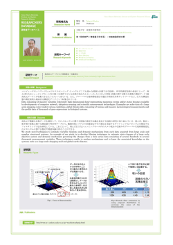
FIG. 4.Analysisofveinendingswith phloem inwhite leaftissue(growth cabinet).
A. Schematic drawings of sections of the vein endings at the place of the proximal ends (1), of
thedistalextremitiesofthephloem partswithsieveelements(2),ofthephloem partswith intermediary cells and vascular parenchyma cells only (3), of the phloem parts with vascular parenchyma
cellsonly(4)and ofthepartswith tracheids only (5).Cellsofthebundle sheath arenot given.
B. Diagram composed of valuescalculated as averages of 10vein endings in total, 3in dry climate
and 7inwetclimate.
Abscissa:average length in urn,reckoned from thetip,of: lengthsuntil thedistal extremity of vascular parenchyma cells, lengths until the distal extremity of intermediary cells and lengths until the
distal extremity of the sieveelements.The vein endings were 320urn long on an average. Ordinate:
averages of the (S transections area tr/number of se + ic + vp) values in the sections of 10 vein
endings at the place of the distal extremity of the sieve elements (117) of the intermediary cells
(148)and ofthevascular parenchyma (93).Near theproximal end thisvalueamounts to37.
For abbreviations seeTable 1 and: lse = length with sieveelements, etc., ltr = length with tracheids
only orwith vascular parenchyma cellsinaddition (vpx),1^= averagelength ofthe 10veinendings.
Calculation for each veinlet: I transections area tr means the sum of the section areas of the total
of tracheary elements per transection; number of se + ic + vp (denominator) means the sum of
thenumber of sieveelements, intermediary cellsand vascular parenchyma cellsin the same transection. Along the zone 1^.the denominator is variable; distad of the zone lseit is kept constant with
a valueasinthedistal extremity ofthezone1^,cp.Fig.13.
( r transections area tr/number of se + ic + vp)isabbreviated as(Xarea tr/Z se,ic,vp).
Meded.Landbouwhogeschool Wageningen83-6 (1983)
FIG. 5. Detail of a small connecting vein of white leaf tissue (dry climate; see also Fig. 15B) at
thedistalendofthezonel^andtheproximalextremityofthezonel;c.
Intheintermediarycellontherighthandside(ic)someplasmolysishastakenplaceattheproximal
extremity.Thevascularparenchymacells(vp)onlyhaveathinlayerofcytoplasmagainstthewalls
(notdrawn).
lic = zone with intermediary cell, \x = zone with sieve element, n = large nucleus in ic, se =
terminal sieveelement, from which thecontents arenot drawn; thewallsare thickened especially
inthecorners(theseterminalsieveelementsaresometimeswiderandtheyoftenhavemorecytoplasm
thantheothersieveelements),tr = trachearyelement.
thantheneighbouringcellsofthebundlesheath.Thesecellspossesslargevacuolesandlessdensecytoplasm.
Thetrachearyelementsaretracheidswithspiralthickeningsofthewalls.The
tracheids are very small and obliterated sometimes (protoxylem) or they are
largeruptoverylargeandintact(metaxylem).
Closetothedistalendoftheveinletsthevascular bundleoften becomesamphivasal, i.e. the tracheary elementscompletely surround thephloem (Fig.3).
Adjacent to and in a direct line with the distal end of the phloem with sieve
elements,usuallyanarrowingcomplexofparenchymacellsoccurstowardsthe
ultimate top of the veinlet (Fig. 4A).In this complex of parenchymatous elementsoften oneortwocellsarefound indirectcontactwiththedistalextremity
ofthedistal sieveelement (Fig. 5).Thesecellshavedensecytoplasm and large
nucleiandwillbecalledintermediary cellsalso(Table2,Fig.4A).Inthedirectionoftheultimatetopoftheveinletinadirectlinewiththedistalendofthese
intermediarycells,frequently someparenchymatouselementswillfollow.Inthe
majority of veinlets these vascular parenchyma cellshave distinctly lessdense
cytoplasm.Stillnearertotheultimatetopoftheveinletonlytracheidsandsometimesalsosomevascularparenchymacellsconstitutethetopoftheveinlet.
Analysisofveinendings
Theveinendingcanbedividedinfourzones(Fig.4B).
- Zone 1:lse = length of the vein ending along which sieve elements can be
found;
Meded.Landbouwhogeschool Wageningen83-6(1983)
9
- Zone 2: l ic = length of the vein ending with intermediary cells (no sieveelements);
- Zone 3:lvp = length of the vein ending with vascular parenchyma cells (no
sieveelementsand nointermediary cells);
- Zone 4: ltr = the most distal part of the vein ending in which only tracheids
and sometimes alsoa fewvascular parenchyma cellsamong them (vpx).
The vascular parenchyma cells of the xylem (vpx in zone ltr)abut on the tracheids and the cells of the bundle sheath. In general these cells are not in direct
contact with thecellsofthe phloem.
This division in zones is not always complete: the zones lse, licor lvpmay be
lacking. Finally the limits are not always sharp. The limit between the zones
lseand licis nearly always distinct. In one case this line could be drawn sharply
only after some difficulty because of the occurrence of an intermediary cell in
a direct line with a terminal sieve element and the fact that the general shape
and the thickness of the wall of the intermediary cell was similar to that of the
adjacent sieve element. Because this intermediary cell had among other things
much cytoplasm and a large nucleus with distinct nucleoli, the identification
could takeplacewithout doubt. Thelimitbetween thezoneslicand lvpwas determined by comparing the density of cytoplasm of an intermediary cell with the
density ofthemoredistal oriented vascular parenchyma cells.
In one variegated leaf (Fig. 1) 33 vein endings were analysed of which 16
vein endings differentiated in the green leaf tissue and 17vein endings differentiated in the white tissue. In both parts of the leaf 10 vein endings have been
analysed in which a zone lsewas present, i.e. the types 2, 5 and 6 according
to Fig.2.
Classesof veinlength intheveinendings
In Table 3 some results of vein length are given; the analysed vein endings
are arranged into groups of 100 urn difference in length each. In every group
the average values were determined of the total length of the vein endings (Q,
TABLE3.(Green house conditions)
Total
number
Length ofvein
endings,urn
Ç
*se
Ov-lse)
%ofi;
green
1
1
8
0
0-100
100-200
200-300
300-400
80
110
259
14
40
129
66
70
130
82,5
63,6
50,0
-
-
-
-
white
0
5
4
1
0-100
100-200
200-300
300-400
-
-
-
-
168
260
377
65
157
265
103
102
112
61,2
38,6
29,7
10
Meded.Landbouwhogeschool Wageningen83-6 (1983)
greentissue
^um
1
'v
'se
8
0
number
white tissue
/urn
400
400
300
300
0
5
4
1
number
200
200
_
100
n
100
i
i
D 100 200 300 400 length,
groups(yum
0
D 100 200 300 400 length,
groups(^um)
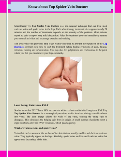
FIG. 6. Diagram of the mean values of the total length ( Q and of the length of zone lse ( Q per
group of vein endings. Each following group of vein endings is 100 um longer. The number of
veinendings ineachgroupisnoted above (variegated leaf,green-house conditions).
of thelengthsoflse( Q , ofthelengthsof (lv— lse)and of(lv —lse)asa percentage
ofÇ
From Table 3it becomes clear that the length of the extremities of the vein
endings (lv—1^)becomesproportionally smaller (in%of%)astheveinlet length
(1^)becomes longer.
In the diagram of Fig. 6B the mean values of the total length and the length
of zone lseare given. There is a tendency towards a rather constant value of
thelengthofthexylemtips(lv— lse)oftheveinendingsindependent ofthelength
of theveinlets in white leaf tissue. Fig. 7isa detailed representation of one vein
ending in white tissue. A tendency exists to a maximum of the calculated sum
ofareasoftransections oftracheids(Earea tr).Thismaximum coincides approximatelywith themaximum number of tracheids inonetransection. The position
of this tracheid maximum also coincides approximately with theposition of the
extremity ofthezonelse.
Itmaybenoticed thattheterminal sieveelementappearswider;thisphenomenon can be found frequently, but not in every veinlet. In this terminal sieve
element morecytoplasm isoften present too. It isalso evident that the tracheids
ofthedistal end of theveinlet arewider (metaxylem) than those ofthe proximal
end (more protoxylem).
Theposition of thetracheid maximum
The position of the tracheid maximum has been located for 33analysed vein
endingsofonevariegated leaf (Fig. 8).The numbersoftracheidsat the proximal
end of most analysed vein endings are approximately equal in both green and
whiteleaftissue;theshortveinendingsinDpossessasomewhat greater number
of tracheids.Themaximum number of tracheids in theveinendingswith a zone
lse(lse ^ 15um)isabout 20.
In white tissue this tracheid maximum is always situated in the vicinity of
the extremity of the zone lse(in 10out of 10analysed vein endings) and in green
tissue there seems to be more diversity: the tracheid maximum has been found
Meded.Landbouwhogeschool Wageningen83-6 (1983)
11
2 area tr
S arease
yum
yum
267
proximal end
distalend
FIG. 7. One vein ending in white leaf tissue of type 2 (see Fig. 2). Variegated leaf, green house
conditions. Abscissa: length of the vein ending lvand of the phloem with sieve elements (1^, with
sieve elements se^ etc.). Ordinate: calculated sum of areas of transections of tracheids (I area tr)
in each transverse section of the veinlet and for the transections of sieve elements (Z area se) in
Um2.Thenumber of tracheids isnoted belonging toeach transection.
to occur at theproximal side oftheextremity of 1^(1 x)and also at thedistal
side of the extremity of lse(4x). In the vein endings without a zone lse(types
1, 3and 4) the tracheid maximum is always close to the proximal end of the
veinletinwhiteleaftissue.Ingreentissuethistracheidmaximummaybefound
also further removed from the proximal end. The results in Fig. 8B and 8D
12
Meded.Landbouwhogeschool Wageningen83-6 (1983)
TABLE 4. The average length of the vein endings (Q, the average length of the phloem with sieve
elements ( Q , the average number of tracheids in the tracheid maxima and the average number
of sieve elements close to the proximal end of the vein endings per group of vein endings in green
and inwhiteleaftissue,with and without azone1^(variegated leaf,green houseconditions).
Leaf
tissue
green
green
white
white
Number of
veinlets
10
6
10
7
Types of
veinlets
1^,
um
2,5,6
1,3,4
2,5,6
1,3,4
1^,
um
Max.number
oftracheids
inmaxima
(average)
108,5
226,1
137,8
225,7
89,0
17,5
11,8
19,7
14,0
123,5
-
Number ofsieve
elements near
proximal end
(average)
1,6
1,9
trach.
number
A
1
20
green
type 2,5,6
10 ^ • " ^ . . - • • ' ^ ~
r = S C T
5
4
<--
», .._
^^"
---.
0
K
yum 25a5
B
61,9
\
^>. I
14,5 0 v
20
green
type 1,3,4 10
>um 212,5
C
112,0
20
white
type 2,5,6 10
> i m 225,7
>um 89,0
proximal end
I
distal end
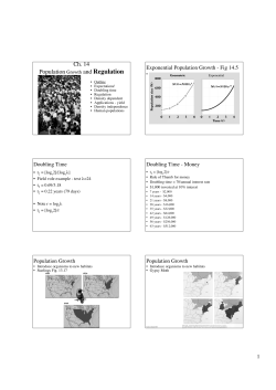
FIG. 8.Position ofthetracheid maximum inveinendingsingreen and inwhiteleaftissue (variegated
leaf,green house conditions).
Types of veinlets (see Fig. 2): 2, 5 and 6 (10 in A and 10in C) and types 1, 3 and 4 (6 in B and
7 in D). In each group of vein endings the average numbers have been determined of (a) the total
length (abscissa)oftheveinendings(1^),of(b)thenumber oftracheids(ordinate)closetothe proximalextremity,of(c)themaximum number oftracheids,of(d)theposition ofthetracheid maximum
along the veinlet, and of (e) the average sizes of the length of the phloem with sieve elements ( Q
belonging to the group of vein endings. The number at the top of the curves indicates the number
of vein endings in that group. The length of T^is expressed with special corresponding types of
lines.
Meded. Landbouwhogeschool Wageningen83-6 (1983)
13
correspond with those in Fig. 8A and 8C in this respect; and this creates the
impression that the vein endings without a zone lseare to be considered as the
extremities of the vein endings with a zone lsein the corresponding green and
whiteleaf tissue.
Table 4 shows that the average lengths of the vein endings with a zone lse
are equal in green (226.1 urn) and in white (225.7 um) tissue. The vein endings
without a zone lseare significantly shorter, however, than the veinlets with a
zone lse,Wilcoxon test, P = 0.025 in green and P = 0.001 in white tissue. The
number oftracheidsinthetracheid maxima issignificantly lowerinvein endings
without a zone lse, Wilcoxon test, P = 0.05 in green and P = 0.025 in white
tissue.Thecriticalvalues aregiven for one-tailed probability.
From the above mentioned data one may conclude that a further analysis
is desirable for a determination of the position of the tracheid maximum. For
anexperimental approach of thenature of thesestructures intheleafof Hedera,
the white leaf tissue seems to be most suited because its tracheid maximum
cumulative
relative
frequency (S)
1.0 _ D=green
A = white
0.9
a
A
JO
• D
0.8|0.7
• O
Q6
JO
Q5
IB
Q4 -
AO
0.3 -
An
transections
of veinlets,
total
0.2 - AO
Str^ sesections, sections.
number
number
green tissue
10veinlets
321
3708
225
white tissue
10veinlets
322
3879
358
0.1 - A O
0.0.
i
i
i
i
0 10 20 30 40 50
i
i
i
100
150
Str-sections
^se-sections
FIG. 9.Comparison of thetotal amount of thexylemwith thetotal amount ofthephloem by means
of the quotient E tr-sections/X se-sections per vein ending. 10vein endings with a zone 1^,in green
and in white leaf tissue were used for calculation. Ordinate: fraction smaller than or equal to the
valueofthe quotient.
Insert: total of transverse sections of the veinlets, total of cross-sections of the tracheids and the
total ofcross-sections of sieveelements (variegated leaf,green house conditions).
14
Meded.Landbouwhogeschool Wageningen83-6 (1983)
turned out to be more constantly in the vicinity of the extremity of the zone
'se-
Theamounts ofphloem andxylem invein endings
The total amount of phloem and xylem in the vein endings can be specified
by the length and the diameter from each of these tissues. An estimation of
these values may be achieved by scoring the total quantity of transections of
tracheids (I tr-sections) and of transections of the sieveelements (E se-sections)
in every transverse section of the vein ending (Fig. 9). The quotients Z tr-sections/Z' se-sections are not significantly higher in the green leaf tissue than in
the white tissue; this means that the difference between the given number of
tracheids in relation to the phloem part with sieve elements belonging to it in
thegreen and inthewhiteleaftissue,isnot significant.
Distribution ofphloem andxylem elements in alongnot ramified veinending
Thepercentage oftracheidsineverytransversesectionofaveinending, calculatedforthetotalnumber ofelementsineverysection,usuallyregularly increases
towards the distal extremity of the veinlet (Table 5,from 28 to 100%).The percentageoflivingelementsineverytransection decreasesregularly (Table 5, from
72 to 0%). Of 12vein endings these percentages have been calculated and they
areshown inTable 6.
The vein endings nos 5,6, 7,9, 11and 12 (Table 6),which showed a distinct
tracheid maximum in the veinlet, a very gradual decline of the percentage of
tracheids was found in all the six veinlets from the distal end to the proximal
end (asinTable 5).
The percentage of living elements per transection increases fairly regularly
in the same direction from 0 to 66.7 on an average in these six vein endings.
The tracheid maximum in these vein endings appears independent of the total
number of elements in the transections of the veinlet. Another structural cause
for the origin of this tracheid maximum (such as a greatly overlapping of the
extremities ofthetracheids at theend oftwoveinsegments)hasnot been found.
In order to investigate the cause of appearance of the tracheid maximum,
a long and not ramified vein ending (type 2, Fig. 2) was reexamined (Table 5).
Inthisveinendingthreetracheidmaximadoappearproperly,viz.in transections
10, 23and 33.These maxima do not clearly coincide in each case with a maximum ofthetotalnumber ofelements inthe sections.
Position of the tracheidmaximum indetail
It hasbeen shown (Figs4,7and 8)that theposition ofthetracheid maximum
is associated with the distal end of the zone lsein the vein ending, especially
inwhiteleaftissue.Itisofimportance tocomparethepositions ofthe appearing
tracheid maxima intheveinendinggiveninTable 5withcorresponding changes
in character and dimension of the phloem. This comparison can be made by
means ofthegraphs inthe Figs 10,11,12,13and 14.
In Fig. 10the three tracheid maxima are visible in the distal half of the vein
Meded.Landbouwhogeschool Wageningen83-6 (1983)
15
TABLE5.Surveyofthenumbers ofallelementsfound in thetransections no. 1(distalend)tono.
76(proximal end) of a not ramified vein ending, type 2(Fig. 2),with a zone \x and 588 umin
length.Thosevascularparenchyma cells(vp)situated inthedistalextremity oftheveinlet abaxial
of the xylem and in a direct line with the distal end of the zones 1^or ljC, have been put under
theheadingphloem.Thisveinendingisgrowninawetclimate.
Section
number
Phloem
se
1
2
3
4
5
6
7
8
9
10
11
12
13
14
15
16
17
18
19
20
21
22
23
24
25
26
27
28
29
30
31
32
33
34
35
36
37
38
39
40
41
42
43
44
45
16
2
2
2
2
3
3
3
3
3
3
3
3
4
4
5
4
4
4
4
4
4
4
3
3
ic
3
3
3
3
3
5
6
6
6
6
6
6
6
6
6
6
6
6
7
9
10
11
11
11
12
12
12
12
11
11
vp
1
1
3
5
5
5
6
6
6
7
7
7
7
7
4
3
3
6
5
7
5
6
7
6
6
9
8
8
7
8
9
11
9
3
4
11
13
13
10
8
6
8
10
Xylem
%living
%tr
oftotal
Totalnumber
ofelements
vpx
tr
elements
oftotal
3
3
3
4
10
16
17
16
16
18
17
16
14
15
13
13
13
13
15
17
17
15
18
17
15
13
12
13
12
12
14
16
21
20
17
18
18
14
13
12
12
13
13
13
12
0
0
25
20
23
27
26
27
33
31
32
38
42
38
41
48
43
43
42
39
43
52
42
45
50
55
57
59
60
60
55
53
49
56
60
54
55
67
70
73
71
68
66
66
69
100
100
75
80
77
73
74
73
67
69
68
62
58
62
59
52
57
57
58
61
57
48
58
55
50
45
43
41
40
40
45
47
51
44
40
46
45
33
30
27
29
32
34
34
31
3
3
4
5
13
22
23
22
24
26
25
26
24
24
22
25
23
23
26
28
30
31
31
31
30
29
28
32
30
30
31
34
41
45
42
39
40
43
44
44
41
40
38
38
39
1
1
1
1
1
1
2
2
1
1
1
2
3
4
1
2
1
1
1
1
1
1
1
1
2
3
3
3
3
3
3
3
3
3
3
3
3
Meded.Landbouwhogeschool Wageningen83-6(1983)
Section
number
46
47
48
49
50
51
52
53
54
55
56
57
58
59
60
61
62
63
64
65
66
67
68
69
70
71
72
73
74
75
76
Phloem
Xylem
se
ie
vp
vpx
tr
%living
— elements
oftotal
3
3
3
4
4
4
6
5
5
5
4
4
4
4
4
4
4
4
4
4
4
4
5
5
4
4
4
4
4
4
5
10
10
10
11
11
9
9
10
9
9
10
10
10
10
10
10
10
10
10
9
9
9
9
9
8
6
6
6
6
5
6
12
12
10
8
8
9
10
10
11
9
7
10
9
10
10
9
9
8
7
8
8
6
6
7
9
10
10
10
8
9
11
3
3
3
3
2
3
2
3
3
3
2
12
12
12
13
13
14
12
12
12
12
11
10
10
11
11
11
10
10
10
10
10
10
9
9
8
8
8
8
8
9
9
70
70
68
67
66
64
69
70
70
68
68
71
71
69
69
69
71
70
69
69
69
67
70
71
73
72
72
72
70
68
72
%tr
oftotal
30
30
32
33
34
36
31
30
30
32
32
29
29
31
31
31
29
30
31
31
31
33
30
29
27
28
28
28
30
32
28
Totalnumber
ofelements
40
40
38
39
38
39
39
40
40
38
34
35
34
36
36
35
34
33
32
32
32
30
30
31
30
29
29
29
27
28
32
ending. In this part of the veinlet the number of sieve elements decreases to
zeroand stillmoreindistaldirection azonewith oneintermediary cell(lic)follows.Closer to theextremity oftheveinlet thezonewithvascular parenchyma
cells(lvp)follows, being in a direct linewith thedistal end of the intermediary
cells. Finally a short zone with tracheids only (ltr) forms the ultimate end of
theveinlet.
In Fig. 11 the added section areas of tracheids (a better estimation of the
total quantity of the xylem) and sieve elements per transection, all along the
vein ending, have been indicated. The three tracheid maxima are still clearly
visible inthegraph; however, themaximum appearing at thedistal end of the
zonelicismoststrikingandreachesamuchhighervaluethantheothertwo.
In Fig. 12the number of tracheids in each transection has been plotted in
relation to the quantity of the phloem, expressed in the quotient of the total
number of tracheids in a transection divided by the total number of sieveelements,intermediarycellsandvascularparenchymacellsin thesametransection.
Meded.Landbouwhogeschool Wageningen83-6 (1983)
17
TABLE6.Percentages oftracheary elementswithregard to thetotal number ofelementsat theproximal end and at the distal extremity in 12veinlets of types 2,5 and 6 (with a zone \x, Fig. 2). The
type of vein ending is indicated in more detail according to the classification of Table 7. In two
veinlets the vascular parenchyma cells in a direct line with the distal end of the intermediary cells
or the sieve elements reach to the distal extremity (numbers 1and 10). lv = length of the veinlets
in um.
Veinlet
number
1
2
3
4
5
6
7
8
9
10
11
12
average
Typesof
veinlets
(Table7)
lv,um
s
t
n
m
n
q
p
t
t
t
s
t
143
240
285
286
295
304
325
332
345
363
403
588
20
28
33
31
30
38
31
29
29
26
28
30
45
56
42
35
43
35
23
38
34
46
36
30
4
1
1
4
2
1
1
1
1
2
1
3
325.8
29.4
38.6
1.8
Proximal end
Distal enc
Total
% trach.
number of elements
elements oftotal
Total
% trach.
number of elements
elements oftotal
Climate
25
100
100
100
100
100
100
100
100
50
100
100
type
dry
wet
wet
wet
dry
dry
wet
wet
wet
dry
dry
wet
89.6
These quotients could becalculated in thezoneslseand lic.The three tracheid
maximaabovementionedarefound againinthesamepositions,whilethethird
maximum again is the highest. In the zone lvpthe quotient of the sum of the
tracheids and the total number of vascular parenchyma cells has been determined ineach transection. These points in the graph show an indistinct path,
and no quotients can be calculated in the zone ltr. If one wishes to express a
relation between the numbers of tracheids and the quantity of the phloem in
thesezoneslvpand ltr,acomparison ofthenumber of tracheidsin thesezones
withthequantityofthephloemsomewhereatthedistalextremityofthephloem
isobvious.Ifthesequotientsarecalculatedbydividingthesumtotaloftracheids
per transection by the value of Z se, ic, vp at the distal extremity of the zone
lic(se = o,ic > 1),aratherregulardecreaseofthecurvefromthethirdmaximum
tozeroattheextremityofthe veinletisobtained.
In Fig. 13theadded section areas of tracheids havebeen plotted in relation
to thequantity of thephloem, asquotients again in thezones1^and lic.In the
zone lvpthe quotients have been determined of the sum of the section areas
of tracheids and the sum total of vascular parenchyma cells-per transection.
Thepointsinthegraphdeterminedinthiswaydonotshowadistinctcontinuation of thecurvein thezoneslseand lic.However, whencalculating thesequotientsinthezoneslvpand ltrusingtheconstant valueofZ se,ic,vpat thedistal
end of the zone lic(= 7; se = 0; ic = 1; vp = 6) as the denominator, one
18
Meded.Landbouwhogeschool Wageningen83-6 (1983)
FIGS. 10-14.Abscissa: length ofthenon ramified vein ending of Table 5in um; left: proximal and right: distal end. The zones 1^,lic,l vp
and ly-areindicated asin Fig.4.
numbertr
numberse
2°!
ig|
Sareatr — ~
FIG. 10.The number oftracheids(-•-»-•-) and
sieve elements (•••••) in each of the 76 transectionsofthe veinendingofTable5.
FIG. 11. The sum of the section areas of the
total of tracheary elements (Z area tr, ««»)
and the sum of the section areas of the total
of sieve elements (Z area se, • • • • ) in um per
transection of the vein ending all along the
veinlet.
FIG. 12. The quotients of the sum of the
number of tracheids and the sum of the
number of sieve elements, intermediary cells
and vascular parenchyma cells (Z tr/27 se, ic,
vp) per transection all along the zones lseand
lic(-«-•--•-).In the zone l vpthequotients of the
sum of the tracheary elements and the sum of
the number of vascular parenchyma cells (Z
tr/Z vp)have beendetermined per transection
( A A A ) . Moreover in the zones l vp and l tr the
quotients of Xtr on the one side and Z se, ic,
vp at the place of the distal extremity of the
zone lic on the other side, have been determined per transection (-o-O-O-).
1500
/jnf
1000
500
\
100
—•*""*
0
zum 5i 8
••*—••*"""*•
415
....„•••^••—*
279
125
0
FIG. 13.The quotients (Z area tr/Z se, ic, vp)
per transection all along the zones lse and ljc
(-•-•-•-). In the zone l vpthe quotients (Z area
t r / r vp)have been determined per transection
(AAA). Moreover in the zones lvpand l tr the
quotients of Z area tr on the one side and the
constant value of Z se, ic, vp at the place of
thedistal extremity ofthezone ljcon the other
side, have been determined per transection
(-0-0--0-).
FIG. 14. ThequotientsofZ area trand thesum
ofthenumber ofsieveelementsand intermediary cells (Z area tr/Z se, ic) (-•-•-»-) per transection allalongthezoneslseand ljc. However,
in the zones l vpand ltrthe quotients of Z area
tr per transection and the constant value of Z
se, icat the place of thedistal extremity of the
zonelie,havebeen determined.
Meded.Landbouwhogeschool Wageningen83-6 (1983)
19
can see the very regular continuation of the curve from the third maximum to
zero at the extreme point of the veinlet, just like the regular steep descent of
thecurveinFig. 11.Moreover itisstrikingthat onlythethirdtracheid maximum
still appears as a pronounced maximum and it arises exactly at the extremity
ofthezonelicofthis veinlet.
Finally in Fig. 14 the added section areas of tracheids have been plotted in
relation to thesum ofsieveelementsand intermediary cellsper transection. The
quantity of the phloem has now been characterized only by the total of Z se,
ic.Alsointhisgraph thezoneslvpandltrhavebeencalculated usingas denominator the constant value of Z se, ic at the distal end of the zone lic( = 1) in the
quotients. In this curve the most pronounced tracheid maximum also appears
at thedistal end of thezonelic.
In summary: the position of the already described tracheid maximum at the
distal end of the zone liccan be shown in the most expressive way when the
added section areas of the tracheids (and therefore also the total volume of the
tracheary elements), are given in relation to the quantity of thephloem in every
transection of the veinlet. The quantity of the phloem can be described best
by Z se,ic, vp, because of the fact that the difference between the intermediary
cells and the vascular parenchyma cells may be difficult to determine in some
parts of the veinlet. The procedure as represented in Fig. 13 will be followed
therefore in the next part of this article in case of doubt of the exact position
ofthetracheid maximum in the veinlet.
Thelocalization of thetracheidmaxima indifferent types of veinendings
After calculation on 20vein endings it turned out to be possible to represent
thequantityofxylemintheveinendingsinrelation tothequantityofthe phloem
in the shape of a more or less constant type of curve as in the Figs 4 and 13.
Now the question arises whether the position of the tracheid maximum can be
located moreexactlyinrelation to thezoneslse,lic,lvpand ltr.
Thepossible typesofveinendings(21types,a - u ) havebeenbrought together
in Table 7. The determining of the position of the tracheid maximum runs up
againstdifficulties when oneormorezonesarerelatively short, or when a broad
maximum appears within which more than one zone terminates. In these cases
thepositionofthemaximumhasbeenlocatedbyestimatingtheshortest distance
between the position of the maximum and the two adjacent limits of the zone.
The limit between the zones licand lvpwas determined by paying attention to
cytoplasmic density. Finally the distal limit of the zone with vascular parenchyma cells isnot sharp sometimes caused by the fact that the distinction between
vascularparenchymacellsandcellsofthebundlesheathisdifficult todetermine.
Based onthesetheoretical typesitcan beconcluded that:
- veinlet type b has not been found. This means that one does never find the
tracheidmaximum alongthat part oftheveinendingonlyconsisting ofxylem
(tracheids and possibly vascularparenchyma cells,vpx);
- veinlet types j , 1, oand rhave not been found. This indicates that the tracheid
maximum alongthat part oftheveinendinginwhich sieveelements do occur
20
Meded.Landbouwhogeschool Wageningen83-6 (1983)
TABLE 7. Theoretically possible types in dry and wet climate of free vein endings (a-u) in relation
to the position of the tracheid maximum (n). The position of the maximum is only determined
by the observed number of tracheids, or after calculation of the quotient I area ti/I se, ic, vp.
The zones 1^, licand l vp are always > 21 um in length, with the exception of the zone lsein one
of the vein endings of type k and one of the vein endings of type m (column no. 7), which are
onlyabout 15uminlength both.
dry
tr. max.M in
veinendings,
zones* 21Aim
wet
ramified
ramified
not ram.
not ram.
position of tracheid maximum determined by:
calculacalcula
calcula
calculanumber tionof humber tionof Inumber tionof number tionof dry
of
trach
of
trach
quotient
of
trach
quotient
quotient
total
oftrach.|quotient
wet
total
total
1
b|
r\
d
l777T^
W
»te
i,
m
"
10
Meded.Landbouwhogeschool Wageningen83-6 (1983)
21
not can be expected;
- veinlet type a has been found frequently (10x). In these cases at the origin
ofthisvein ending
1. sieveelements do occur at the proximal extremity, for example in the vein
oflowerorder(5 x);
2. intermediary cellsdo occur at theproximal extremity (2x);and
3. vascularparenchyma cellsdo occur at theproximal extremity (3x).
One may conclude that the veinlet type a can be considered entirely to be
azoneltr.
- the tracheid maximum does always occur near the distal extremity (= near
the limit of the zone in the direction of the extremity of the vein ending) of
the zones lse, licor lvp(also when respectively se, ic or vp are present at the
point ofbranching,viz.in typesa, cand g).Thisbecomes visible
1. in the end of zone lse,as in type a (5x),c (4x), g (1x), k (2x),m (9x),
p(1x),and s(2x),altogether 24x;
2. or in the end of zone lic, as in type a (2x), f (3x), h (2x), q (2x) and
t (10x ) , altogether 19 x ;
3. finally in the end of zone lvp, as becomes clear in type a (3x), i (1 x ) and
n (3x),altogether 7x.
It appears that the tracheid maximum has been found most often close to
the distal extremity of the zone lse, but the maximum also does occur near the
distal end of the zone licfrequently if this zone is present. In the presence of
a zone lvpthe tracheid maximum does appear near the end of this zone sometimes.
The conclusion is that a strong tendency exists of the tracheid maximum to
shift to the right (Fig. 15A) under the influence of an extension of the zone
lsebyazonelicor thezoneslic + lvpor sometimes byazonelvponly.
Any part of a vein ending consisting of the zones (lic + lvp + ltr), (lvp +
ltr) or ltrmay be found also as lateral vein branches. Thus the small branches
often represent the extremities of the longer free vein endings (cp. Fig. 8 and
the text belonging to it). The point of branching may occur at any place along
theveinending;however, thezoneslicand lvparecontinuous each and communicatewithazonelse.
veinending
A H-
—
—
'se
'ic
'vp
proximalend
distal end
FIG. 15A.Schematic drawing of a free veinending with zones1K, ljc,l vpand ltr; n : possible positions
ofthetracheid maxima, bs = bundle sheath.
B. Schematic drawing of a small vein connecting two veins of lower order. In the connecting vein
thesamezonesappear but atracheid maximum isabsent (seealso Fig.5).
22
Meded.Landbouwhogeschool Wageningen83-6 (1983)
TABLE 8.Average values of the number of tracheary elements in the tracheid maximum, expressed
in the average number of observed tracheids in a transection of a vein ending on the spot of the
tracheid maximum inthree different groups ofvein endings.Fy = average length ofthe vein endings
ina group.
Group Typesof
number veinlets
Dry climate
Wet climate
Number lv, um
of veinlets
Max.
number of
tracheids
(average)
Number 1^, um
of veinlets
Max.
number of
tracheids
(average)
1
onlywith zones
l v p andl t r
9
101.8
13.7
6
92.3
10.7
2
onlywith zones
l i c ,l v p andl t r
2
151.0
12.0
5
145.4
16.4
3
withzoneslse,
l, c ,l v p andl t r
16
347.7
19.3*
13
258.0
17.7*
total
27
251.1
16.9
24
193.1
15.7
*cp. Table 4: the number of tracheids in the maxima of 10 veinlets in white leaf tissue averages
19.7.
TABLE 9. Vein endings with a zone '.
dry and inwet climate.
Veinlet
number
1
2
3
4
5
6
7
8
9
10
11
12
13
14
15
16
, (type 2, 5, 6) arranged in order of increasing length (lv) in
Wet climate
Dry climate
l v ,um
lse. 1™
lv - 1 « . H m
lv,um
Ise.um
lv - 'se. I™
131
143
195
231
275
295
304
339
342
363
378
379
403
513
615
657
42
55
71
42
80
128
156
143
236
98
218
235
245
383
470
433
89
88
124
189
195
167
148
196
106
265
160
144
158
130
145
224
62
63
68
189
207
240
285
286
325
332
345
364
588
15
15
26
40
68
104
171
195
224
165
171
203
463
48
49
42
149
139
136
114
91
101
167
174
161
125
Meded.Landbouwhogeschool Wageningen83-6 (1983)
23
In Fig. 15Ba small vein has been drawn schematically being the connection
between two veinsoflower order. Fig. 5depictsa detail of thisconnecting vein,
viz. the marginal area near the distal extremity of zone lx and the proximal
end of zone lic. There is no tracheid maximum in this vein, the number of tracheids is6proximad (left) and 7distad. The intervening part contains 5-7 tracheids on an average. The zone lvpabuts on very large intermediary cells next
toabigsieveelement on one ofthesidesofthephloem ofalargevein.
External influencesonveinending composition
The influence of the moist percentage of the air in the growth cabinet might
cause a change of types of veins and/or a change of the number of tracheary
elements in the vein endings in respect of the phloem tissue. The transpiration
of the test object has been measured. When the relative humidity was low
(7.0+ 2%r.h.) the water absorption by the whole plant (32 leaves larger than
1 cm) appeared to be about 115 ml and in a wet atmosphere (97.0 ± 2% r.h)
the absorption by the plant (40 leaves larger than 1cm) amounted to about
55 ml during 24 hours. The average length of the 25 mature leaves in the dry
atmosphere wasabout 7.5cmand ofthe 30matureleavesinthewet atmosphere
about 6.5-7 cm.
It appears from Table 7that there isnot a clear difference in the distribution
of types of vein endings in a dry and a wet climate. All types found frequently
dooccurinbothclimatesandthetypesfound lessfrequently doappear regularly
spread over both climates. The influence of the moisture content of the air on
the number of tracheary elements may be considered with respect to the height
of thetracheid maximum (Table 8)and alsoregarding thelength of the terminal
part of the veinlet without sieve elements (i.e. the length of the zones lic + lvp
+ ltr;Table 9and Fig. 16)inproportion to thetotal length of theveinendings.
After some statistical calculation (t-test) on the data of which Table 8 is a
summary, it appeared that the total length of the vein endings as well as the
height of the tracheid maximum do not differ significantly in dry and in wet
climate (P = 0.05). The calculated dispersion showed rather homogeneous
groups of data. After statistical calculation (t-test) on the values (lv —lx) from
Table 9 in dry and in wet climate, the sum of the terminal zones (lic + lvp +
ltr) of the vein endings appeared to be shorter in wet climate (P = 0.05). This
means a decrease of differentiation of xylem in respect to the phloem part of
theveinendingwithinan aréole.(ThecriticalvaluesofParegivenfor one-tailed
probability).
In Fig. 16the average values of lv,l se and lic,calculated for groups of veinlets
of 100 urn difference in length each, have been put in a diagram. This diagram
confirms the diagram of Fig. 6; the lengths of the extremities (lv —lse) of the
vein endings are rather constant and highly independent of the length of the
vein ending lv. The shortest veinlets may be looked upon as exceptions (like
the short ones in Fig. 6) in which these extremities are shorter. In wet climate
theseextremities (lv —lse)thus appear tobeshorter than indry climate.
AsinFig.9thetotal amounts ofthexylem and thephloem inthevein endings
24
Meded.Landbouwhogeschool Wageningen83-6 (1983)
dry climate
wet climate
2 number
/im
/jm
700
700
600
600
500
500
400
400
300-
300
200
200
100
100-
0>
"0
100 200 300 400 500 600 700 Aim
0
3
0 number
1
___
100 200 300 400 500 600 700 yum
FIG. 16. Diagram with the data from Table 9. The vein endings have been divided into groups
of 100 um difference in length each (abscissa) and in every group of veinlets the average values
oflv,\x and lichavebeen determined (ordinate).
=
'v> ——— ~ Me ^—^^—
=
he-
The two drawn leaves,with a schematic major venation characteristic for Hedera leaves,have been
used for thisexperiment; on the left a variegated leaf with littlechlorenchyma (dotted), on the right
a white leaf without chlorenchyma. The small squares indicate the pieces of tissue that have been
usedin analysing.
TABLE 10. Comparison of the total amounts of the xylem and the phloem in the vein ending by
means of the quotient Q = I tr-sections/X se-sections per vein ending indry and wet climate. Vein
endings with a zonelse,arranged inorder ofincreasing length (lv).
Veinlet
number
Dry climate
Ttrsections,
number
I sesections,
number
Q
131
143
195
231
275
295
304
339
363
379
403
615
657
246
119
338
388
482
590
468
630
569
602
793
1061
1030
6
7
10
6
17
23
51
41
16
79
85
157
128
41.00
17.00
33.80
64.67
28.35
25.65
9.18
15.37
35.56
7.62
9.33
6.76
8.05
4330
7316
626
11.69
l v , (im
1
2
3
4
5
6
7
8
9
10
11
12
13
total
Wet climate
Meded.Landbouwhogeschool Wageningen83-6 (1983)
lv, Urn
Xtrsections,
number
Z sesections,
number
Q
62
63
68
189
207
240
285
286
325
332
345
364
588
37
57
67
393
431
467
765
575
500
641
642
662
1007
2
2
4
11
14
24
44
75
72
48
58
48
220
18.50
28.50
16.75
35.73
30.79
19.46
17.39
7.67
6.94
13.35
11.07
13.79
4.58
3354
6244
622
10.04
25
may again be approximated by determining the quotient Q = Z tr-sections/r
se-sectionsperveinending(Table 10).Itappearsfrom thistablethatQisdependent on the length of the veinlets;the longer the veinlet the lower Q willbe. This
isinconformity with data in Fig.4. It may be imagined that a veinlet lengthens
(the narrow part of the veinlet on the left lengthens out proximally), then the
length and usually width of the phloem increase (Fig. 7), while at the mean
time thesizeof thexylemdiminishes relatively. In spite of thelarger total length
of the vein endings in dry climate, the average value of Q seems to be higher
in dry climate (11.69). Because the extremities (lv - lse) of the veinlets in dry
climate are longer than in wet climate (Table 9), it could be expected that Q
was higher in dry climate than in wet climate. The difference is small because
ofan appearing difference inveinlength between both groups ofveinlets.
Thecomparisonofthedistalextremities of theveinendingsindryandwet climate.
The relation of the value of the quotient Q to the vein length isgiven in Fig.
17. In short vein endings Q is high, and Q is maximal in veinlets 100-200 um
in length (wet climate) and in veinlets 200-300 um in length (dry climate). In
very short veinendingstheaverage value ofQdrops rapidly, because the height
l v .yum
700
proximalend
600
500
400
300
200
100
0
distal end
FIG. 17. The value of Q in relation to the vein length in dry and in wet climate. The value of Q
(= 1 tr-sections/r se-sections per veinlet) has been noted as an average per group of vein endings;
these groups are 0-100 urn, 100-200 urn, etc. in length (Q. The numbers above the curves indicate
thenumbers ofveinletsinevery group.
26
Meded.Landbouwhogeschool Wageningen83-6 (1983)
dryclimate
20
FIG. 18. Vein endings with a zone lse: schematic drawings of calculated specimen in a dry (A) and
a wet climate (B) based on averages (curves S). The I area t r / r se, ic, vp values are indicated on
the left (left ordinate). The curves M have been drawn between three calculated points and zero.
Thecalculated points are averages of the height of tracheid maxima inthree groups of short veins,
shorter than or equalto acertain length (Table 12B).
The curves Q are the same as in Fig. 17(right ordinate). Below the abscissa the length (/im) and
the width (number of sieve elements) of the phloem are indicated at the proximal and at the distal
end. Further explanation inthe text.
Aim
Mm
Aim
of the tracheid maximum (number of tracheids) ismuch reduced. At the same
timeitisobviousthatinveinendingslongerthanabout200urnthetotalamount
ofthexyleminrespectofthetotalamountofthephloemishigherindryclimate
thaninwetclimate.Thisisinconformity withtheresult:theextremities(lv—1^)
arelongerindryclimatethaninwetclimate.
In Fig. 18the three curves Q, S and M have been drawn for dry and for
wetclimate. The curves Qare the same asin Fig. 17;thecause of shift of the
maximumofthecurveQtotherightinawetclimatewillbeevidentwhenconsidering the curves Qin connection with thecurves S.The curves Sdeal with all
vein endings with a zone lseand are constructed after the example of Fig. 4,
fordatagiveninTable 11.
Theaverageveinlength,Ty, didnotappear to besignificantly different indry
and in wet climate and this is not essential either for the purpose for which
Fig.18asasummaryofresultshasbeenmade.Itconcernsmainlythe differences
at the ultimate ends of the veinlets in a dry and a wet climate. The value of
E area tr/T se, ic, vp at the proximal end has been calculated only for three
vein endings in dry climate. The result is in agreement with the found values
ofQwhencomparingbothresultswiththoseattheproximalendinwetclimate.
The values of I area tr/r se, ic,vp in the tracheid maximum do not differ
significantly inbothclimates.Consequentlythevalueof200urn2hasbeentaken
for bothScurvesasanapproximation oftheaveragevalue.
Theaverage length of thepiecesofveinending from the tracheid maximum
to the ultimate end of the veinlet in a dry climate and a wet climate has been
usedfor thepositionfindingofthetracheidmaximuminthefigure.Thelengths
of the extremities (lv—lse)appeared significantly different in a dry and in wet
TABLE 11. Data required for the schematic drawings of vein endings with a zone \x in dry and
in wet climate (Fig. 18, the curves S). The numbers in brackets indicate the numbers of veinlets
that wereavailable for calculating theaverages.
i;, um(Table8)
xylem
I area t r / I se,ic,vp,proximal, um
I area tr/Z se,ic,vp,maximum, (im
I area tr/Z se,ic,vp,distal,um
average length between thespot of
thetracheid maximum and thedistal
extremity oftheveinlet, urn
phloem
(l v — Ige), u m (from Table 9)
number of se, proximal
number of se,distal,
along thelength, um
28
Dry climate
Wet climate
347.7(16)
258.0(13)
55
184
0
31
204
0
(3)
(4)
116.94(16)
158.00(16)
2.14(14)
1.00(16)
>58
(7)
(7)
88.70(10)
115.08(13)
3.10(10)
1.00(13)
34
Meded.Landbouwhogeschool Wageningen83-6 (1983)
TABLE 12A. Survey of all short vein endings. In the dry as well as wet climate a length of phloem
does occur in the longest specimen of at least about 20 um on an average and a size of the phloem
ofonly 1 sieveelement (seeFig. 18).
Veinlet
number
Dry climate
lv, Um
1
2
3
4
5
6
7
8
9
10
11
12
13
38
80
84
98
108
110
120
122
131
135
143
143
180
Wet climate
lse, um
—
42
55
-
tracheid
maximum
(number)
l v ,um
9
12
12
14
11
17
21
11
22
20
9
9
13
55
56
62
63
68
77
82
89
106
110
lse, um
_
15
15
26
-
tracheid
maximum
(number)
10
5
7
10
11
12
17
7
13
15
climate and the averages have been used to find the position of the distal end
ofthezonelseinFig.18.
Thedimensionofthephloem hasonlybeenindicated astheaveragenumber
ofsieveelementsatthedistalandattheproximalendofthephloem.Itisapparent from Fig. 18that thevolume of the xylem,beingin front of thedistalend
ofthephloem,must besmaller inwetclimate than indryclimate,for the area
below the curve Sto the abscissa is an approximation of the total volume of
this part of the xylem. Besides the short vein endings may be considered the
extremities of the long ones. Comparing the curves Sand Qit isevident that
in a wet climate the maximum of curve Q, reckoned from the distal end, is
reachedsoonerthaninadryclimate.InbothclimatesthevalueofI se-sections
TABLE 12B. The average number of tracheids in the tracheid maxima of all the veinlets shorter
than 657urn(Table 8), 180um, 110umetc.indry and inwetclimate.
dryclimate veinendings =S 657urn:average tracheid maximum = 16.9
veinendings ^ 180um:average tracheid maximum = 13.9
veinendings ^ 110um:average tracheid maximum = 1 2 . 5
veinendings < 80urn:averagetracheid maximum = 1 0 . 5
wetclimate veinendings =% 588urn:average tracheid maximum = 15.7
veinendings ^ 135um:average tracheidmaximum = 10.7
veinendings =% 100um:average tracheidmaximum = 9.9
veinendings ^ 70um:averagetracheid maximum = 8.6
Meded.Landbouwhogeschool Lagerungen 83-6 (1983)
29
is small yet in the class of veinlets 0-100 urn in length, and in dry climate this
value is also small in the class of veinlets 100-200 fim in length (see also Table
11). In the same classes of veinlets the value of Z tr-sections rises rapidly in
proximal direction especially in wet climate. Consequently the quotient Q increases in proximal direction much more in wet climate. Not until the number
of tracheid sections decreases and the number of sieve element sections rises,
thequotient Qwilldecline.Thisbend downwardsinthecurveinproximal direction takesplaceearlier inawet climate.
Curve M is given to make a more exact comparison possible between the
total tracheary volume, differentiated against and also protruding in front of
the 20 urn long extremity of the phloem (with one sieve element), in a dry as
well as in a wet climate. Data of Table 12were used to construct curve M. In
Table 12A one finds a survey of all the short vein endings and in Table 12B
the average numbers of tracheids in the tracheid maxima belonging to groups
of veinlets shorter than a certain length. Curves M were drawn between zero
and 3other points. The average values have been used of the tracheid maxima
belonging to all the veinlets shorter than or equal to a certain length between
38and 180um in dry climate and between 55and 135urn in wet climate (Table
12B).Plotting the three calculated points, a scale has been chosen (as ordinate)
with a maximum of 18.5,corresponding with the average number of tracheids
of the tracheid maximum in all the vein endings with a zone lsein dry and in
wet climate (Table 8: 19.3and 17.7respectively, and not significantly different).
This value 18.5 of the tracheid maxima has been chosen at the same height as
the maxima.of the curves S. The calculated averages of the tracheid maxima
ofthe 3groups of short veinletsinTable 12Bmay allbethought at the proximal
end with a small error. This means that the abscissa values of the three points
of the curves M correspond with the lengths of veinlets chosen to divide the
short vein endings into three groups. When connecting the three points with
zero the curve M arises that forms the upper limitation of an area that is an
approximation of the average tracheary volume at the extremity of the veinlet.
In Fig. 19one finds therelation between thenumber of tracheids inthe tracheid
maxima (abscissa) and the total area of the transections of the tracheids in the
maximum. The denominator in the quotients Z area ti/Z se, ic, vp varies in
theextremitiesoftheveinendings.Thecalculated averages atthedistal extremity of the zones lseand lic, and also in the zones lvpand ltr are for dry climate
6.75 (4 veinlets) and for wet climate 6.11 (9 veinlets). After correcting the left
scale with these averages, it is possible to estimate the tracheary volume in the
extremities oftheveinendingsbycalculating theshaded area.
Calculation with the image analyser gives a value for dry climate and one
for wet climate; the ratio of both values is 1.91. Calculation from the figure,
inwhichtheabscissavaluesarefound bydeterminingof, forinstance,7um-parts
of length multiplied by the ordinate values belonging to them, gives the values
of the tracheary volumes in um3: in dry climate 126,700 um3and in wet climate
66,600 um3.Theratio between thesetwovaluesis 1.90.
30
Meded.Landbouwhogeschool Wageningen83-6 (1983)
Sarea tr,,um
2000
1500
1000
500
24
26
number oftr
per tr max.
FIG. 19. Relation between the number of tracheitis in the tracheid maximum, and the total area
of transections of the tracheids in the maximum. 6 vein endings in dry climate (A) and 15 vein
endingsin wetclimate (O)- Straight linenot calculated.
DISCUSSION AND CONCLUSION
The term intermediary cell (Übergangszelle, FISCHER, 1885) has been used
in the sense of TURGEON, WEBBand EVERT (1975). Since the difference between
companion cells (direct ontogenetic relation with the sieve element) and other
parenchyma cellsrichincytoplasm next tothesieveelementsisdifficult to determine, and also because the function of both types of elements is probably the
same (a presumptive role in the exchange of photosynthates between the mesophyll and the sieve elements), it did make sense to use the name 'intermediary
cell'for both elements only.
The most general partition in zones of veinlets which end blindly within the
aréoles of Hedera leaves, reckoned from proximal end to distal extremity, is:
lse(part with sieveelements), lic(part with intermediary cells),lvp(part with vascular parenchyma cells)and ltr(part with tracheids and sometimes also vascular
parenchyma cells belonging to the xylem). The sometimes quoted example in
Hosta (PRAY, 1955b) of the phloem extending beyond the limits of the xylem
(e.g.ESAU, 1969),maybeinterpreted astheextending ofthezonelicorlvpbeyond
thelimitsof thexylem.
The vascular parenchyma cellsand the intermediary cellsintergrade in structure (ESAU, 1969);thereisalsoevidencethat theintermediary cellsand theterminal sieve elements intergrade in structure. In the light microscope examples of
enucleate terminal sieve elements have been found in true transections which
Meded.Landbouwhogeschool Wageningen83-6 (1983)
31
were wider and contained distinctly more cytoplasm than the more proximal
sieve elements. Thus the intergrading from parenchyma cells to sieve elements
may bemorecompleted insomeveinendings. Itmay bethat those intermediate
cellular forms show aspects of evolutionary trends indifferentiation of a parenchymacellinto asieveelement.
The relation of the quantity of xylem to the quantity of the phloem has been
expressed by means of the quotient Z area trjZ se, ic, vp. In this way it has
beenpossibletorepresent thequantity ofxylemalongtheveinendingsbymeans
of a more or lessconstant typeof curve with a distinct maximum (Fig. 4)corresponding with theactualvolume of tracheary elementsper unit ofphloem along
the veinlet. It is consistent to relate the xylem in the extremities of the veinlets
with the quantity of phloem in the distal end of the phloem tissue. The result
of this way of calculating was a descending curve like the diminishing of the
number of tracheids inthe distal end of the veinlet. Theoretically itis important
that the denominator of the quotient mentioned above, will remain larger than
one, which means that also in zones lic, lvpand ltra relation exists between the
volume of tracheary elements and the terminal phloem. The influence of the
characteristics of the phloem on the volume of the xylem is also evident from
the shifting of the tracheid maximum in the direction of the distal extremity
under influence of the presence of the zones licand lvp, suggesting an extension
of the phloem by the zones ljc and lvp. The tracheid maximum can always be
found at the distal extremities of the zones lse, licor lvp(Table 7, Fig. 15) and
this corresponds with the absence of veinlet types b, j , 1, o and r in Table 7.
Thepercentageofphloem elementsinthetransectionsdecreasesrather regularly
in distal direction along the veinlets; the percentage of tracheary elements increases (Table 5, the number of vascular parenchyma cells in the xylem, vpx,
is small). This means that the influence of the phloem on the volume of the
xylem increases in distal direction up to the distal ends of the zones lse, ljc or
lvpwhen present. The value lv—lseappeared to be constant in a certain climate.
It seems that an ontogenetic influence of the phloem on the differentiation of
the xylem at the extremities of the vein endings is a constant one, independent
ofthelengthsof theveinlets.
The system ofzoneslse,licand lvpisuninterrupted; that means that a probable
ontogenetic influence isabletoreach every smallveinlet,i.e. thezone complexes
(lie+ Wp + ltr)>Ovp + ltr)a n d ltrmay befound asseparate vein endings.
ALONI (1980)concluded from the spatial relation of phloem and xylem while
differentiating in callus tissues, that the xylem possibly is formed in response
to auxin together with some added factor which reaches it from the phloem.
This work on Hedera veinletsconfirms ALONI'S proposition; the differentiation
of xylem in vivo is not only exactly dependent on the dimension of the phloem
tissue, but also on the character of the phloem composition as sieve elements,
intermediary cells and vascular parenchyma cells. Especially the distal ends of
thephloem appear alsoassinksofdifferentiating factors, from whicha distributiontakesplaceofgrowth regulators,important for thedifferentiation ofxylem.
This leaching from the 'phloem' (i.e. sieve elements) into the free space (ESCH32
Meded.Landbouwhogeschool Wageningen83-6 (1983)
1980) can probably take place via the intermediary cells (zone lic) and
perhaps the vascular parenchyma cells (zone lvp). The quantity of xylem and
therefore probably also the amount of nutrients and growth regulators released
by the phloem turned out to be dependent on climatic circumstances. At least
a great increase of the relative humidity of the air and correlated with that a
sharp lowering of transpiration of the plant, are able to bring about a lowering
of thequantity of xylem per unit of phloem in the vein endings. The availability
of thenutrient solution to the roots wasunlimited, sothe limitation of the transpiration wasbecauseof thehighrelative humidity oftheair only.The temperature of the roots was kept constant at about 12.5°C. The distal extremities of
the veinlets (lv —lse) are significantly shorter in wet climate. It is shown (Fig.
18) that in veinlet tips (along 20 um of the zone lseand more distad) the value
of the tracheary volume in wet climate was about 0.53 times smaller than in
dry climate. This difference was smaller when longer vein endings were considered, but the quotient Q (Fig. 17)remained always higher in a dry climate. The
tracheid maximum (Table 8)isless high in wet climate, but anyhow this difference,compared with adryclimate,isnot significant on thebasisofthe available
data.
Finally itmay beasserted that hardening off theHedera plant means, among
other things, the differentiation of more xylem in the leaves. Therefore more
carbohydrates,i.e.moreassimilatory energyisusedfor theanatomical structure
oftheleaveindryclimate ascompared withwet climate.
RICH,
ACKNOWLEDGEMENTS
Thanks are due to Prof. Dr. M. T. M. Willemse for his critical advice and
for discussions, and to Dr. R. W. den Outer and Dr. G. A. Pieters (Dept. of
Plant Physiological Research) for reading the manuscript. I am grateful to Mr.
M.Keulsand Mr.M.A.J.vanMontfort (Dept.ofMathematics) for mathematical assistance and to Mr. A. B. Haasdijk, Mr. P. van Snippenburg and Mrs.
G. G. van deHoef-van Espelo for technical assistance.
REFERENCES
ALONI, R. Role of auxin and sucrose in the differentiation of sieve and tracheary elements in plant
tissuecultures.Planta 150(1980)255-263.
CHRISTENSEN, O. V. The growth rate and the variation in growth in relation to the stock plant
ofHedera canariensisWilld.'GloiredeMarengo'. Scientia Hort.4(1976)377-385.
DERMEN, H. Nature ofplant sports.Amer. Hort. Mag.39(1960) 123-173.
ESAU, K. Vascular differentiation inplants.New York, Holt, Rinehart and Winston (1965).
Meded.Landbouwhogeschool Wageningen83-6 (1983)
33
ESAU. K. Minor veins in Beta leaves: structure related to function. Proc. Amer. Philos. Soc. I l l
(1967)219-233.
— The phloem. In: Handbuch der Pflanzenanatomie (W. Zimmermann, P. Ozenda and H. D.
Wulff, eds):Histologie,vol.5,pt.2(1969).Berlin-Stuttgart, Borntraeger.
— Cytology of sieveelementsinminorveinsofsugar beetleaves.NewPhytoI.71(1972) 161-168.
— Comparative structure of companion cells and phloem parenchyma cells in Mimosa pudica L.
Ann. Bot.37(1973)625-632.
— Thephloem oiNelumbo nuciferaGaertn. Ann. Bot.39(1975)901-913.
— and HOEFERT, L.L.Composition and fine structure ofminor veinsin Tetragonia leaf.Protoplasma 72(1971)237-253.
— and KOSAKAI, H. Laticifers in Nelumbo nucifera Gaertn.: Distribution and structure. Ann. Bot.
39(1975)713-719.
ESCHRICH, W. Free space invertase, its possible role in phloem unloading. Ber. Deutsch. Bot. Ges.
Bd.93(1980)363-378.
FISCHER, A. Studien über die Siebröhren der Dicotylenblätter. Ber. Verh. Kön. Sachs. Ges. Wiss.
Leipzig, Math. -Phys.Cl.37(1885)245-290.
GUNNING, B. E. S., PATE, J. S. and BRIARTY, L. G. Specialized 'transfer cells' in minor veins of
leavesand theirpossible significance inphloem translocation. J. Cell Biol.37(1968)C7-C12.
MORRETES,B.L.de.Terminalphloeminvascularbundlesofleavesof Capsicumannuumand Phaseolusvulgaris. Amer. J. Bot.49(1962) 560-567.
PATE, J. S., LAYZELL, D. B. and ATKINS, C. A. Transport exchange of carbon, nitrogen and water
inthecontextofwholeplantgrowth andfunctioning - casehistoryofanodulated annuallegume.
Ber.Deutsch. Bot.Ges.Bd.93(1980)243-255.
PRAY, T. R. Foliar venation of angiosperms. I. Mature venation of Liriodendron. Amer. J. Bot.
41(1954)663-670.
— Foliar venation of angiosperms. II. Histogenesis of the venation of Liriodendron. Amer. J. Bot.
42(1955a) 18-27.
— Foliar venation of angiosperms. III. Pattern and histology of the venation of Hosta. Amer.
J.Bot.42(1955b)611-618.
— Foliar venation of angiosperms. IV. Histogenesis of the venation of Hosta. Amer. J. Bot. 42
(1955c)698-706.
SHERIFF, D. W. and MEIDNER, H. Water pathways in leaves of Hedera helix L. and Tradescantia
virginianaL.J. Exp.Bot.25(1974) 1147-1156.
STEINER, A. A. Soil-less culture. Proc. 6th colloquium Int. Potas Inst., Int. Potas Inst., Florence
(1968)324-341.
STEWART, R. N. The contribution of five independent histogenic layers to the stem and leaf of
Englishivy.Amer.J. Bot.53(1966)614-615.
TURGEON, R., WEBB,J. A. and EVERT, R. F.Ultrastructure ofminor veinsin Curcurbitapepo leaves.
Protoplasma 83(1975)217-232.
WATSON, R.W.Themechanism ofelongation inpalisadecells.New.Phytologist 41(1942)206-221.
WYLIE, R. B.Theleaforganization of Hederahelix. Proc. IowaAcad. Sei.50(1943a) 199-207.
— The role of the epidermis in foliar organization and its relations to the minor venation. Amer.
J.Bot.30(1943b)273-280.
ZIEGLER, H. Nature of transported substances. In: Encyclopedia of plant physiology, N.S., vol.
1 (1975).Zimmermann, M.H. and Milburn, J.A.,eds.Springer, Berlin-Heidelberg-New York.
34
Meded.Landbouwhogeschool Wageningen83-6 (1983)
AGRICULTURAL UNIVERSITY WAGENINGEN P A P E R S
85-5(1985)
ANATOMY OF VEIN E N D I N G S
I N H E D E R A LEAVES;
ASPECTS OF O N T O G E N Y
J.F.C. MAGENDANS
Department ofPlant Cytology and-morphology
Wageningen, Agricultural University, The Netherlands
in
Agricultural University Wageningen
TheNetherlands 1985
CIP
ISBN 90-6754-068-4
ISSN0169-345X
© Agricultural University,Wageningen, theNetherlands, 1985.
No part of this publication, apart from bibliographic data and brief quotations
embodied in critical reviews, may be reproduced, recorded or published in any
form including print, photocopy, microform, electronic or electromagnetic record without written permission from the publisher Agricultural University,
P.O. Box 1901,6700HBWageningen, the Netherlands.
Printed intheNetherlands byDrukkerij Veenmanb.v., Wageningen
CONTENTS
ABSTRACT
1
INTRODUCTION
1
MATERIALS AND METHODS
Plant material and cultureconditions
Microtechnique
Statisticalmethods
RESULTS
2
2
3
3
4
Measurements ofthegrowth ofleavesinlength and width
4
Comparison of growth in length of the total leaf with the growth of leaf
tissuenear theleafbase
6
Comparison ofgrowth inlength oftheleavesalong the shoot
8
Assessment of the final length ofaleafand determination ofthe percentageof final lamina length (F.L.L.)ofayoungerleaf
8
Thelength ofveinendings inleavesat different ages
10
Intrusive growth ofprocambiumcells
14
Conversion of thelength ofveinendingsinleavesat different ages . . . . 18
Origin ofthesegmentation oftheveinending
20
Number ofsegments oftheveinending
24
First differentiation oftracheary elements inveinendings
27
Interpretation of some transverse sections of a vein ending in young leaf
tissue
30
Analysis of differentiation of tracheids in vein endings in transverse sections
33
Aspects of division activity of cells in a procambium strand and stages
ofdifferentiation ofsieveelements
37
Analysis of differentiation of sieve elements by means of transections
oftheveinendings
42
Summary and comparison of observations inparadermal and transverse
sectionsofveinendings
46
Type of climate and the length of the distal extremities of the veinlets
with tracheary elements only
52
Determination oftheinterval ofleaf ageinwhich thetracheid maximum
arises
54
The position of the tracheid maximum in leaf tissue at 96% final lamina
length
58
The quantitative relation between the differentiating xylem and the differentiating phloem alongtheveinending
62
DISCUSSIONAND CONCLUSION
63
ACKNOWLEDGEMENTS
72
REFERENCES
72
ABSTRACT
In totally white leaves of Hedera canariensisWilld.var. 'Gloire de Marengo',
the anatomical structure of the vein endings in developing leaves in moderate
climate has been studied bymeans ofparadermal and serial transverse sections.
Near the leaf base a nearly equal growth rate of the leaf tissue in all directions
is found and, based on the identical growth curves of the tissue near the leaf
base and the whole leaf, the percentage final lamina length (F.L.L.) is used as
parameter for the ontogeny of the vein endings. The procambium cells of the
veinendings originate from youngmesophyll cellsin an acropetal sequence and
the young procambium cells show a manifest intrusive growth after obtaining
polarity. At about 40% F.L.L. the procambium of the vein ending has been
differentiated; measurements of the lengths of the young vein endings showed
an average increase which can be written as a linear equation. Geometrically
it follows that the length of every young veinlet can be converted to its length
at 100% F.L.L., and vice versa. The differentiation into the tracheids follows
that of the procambium from about 43% F.L.L.; at first rapidly and along a
narrow adaxial route of the procambium strand and after that slower and
broadwise. The differentiation into the sieveelements occurred not until about
87%F.L.L.,and tookplacebymeansofanewcontinuous and acropetal division
activity of the cells in the abaxial part of the procambium strand. The interval
of leaf age in which the tracheid maximum near the distal extremity of the
phloem length differentiates from the remaining procambium cells in that position,ranges from about 85%to 100%F.L.L.
Themorphogenesis oftheveinendingsisdiscussedinrelationtothe transpiration stream and the differentiation stimuli transported by it, possibly coming
from thedifferentiating phloem.
INTRODUCTION
IntheleafofHedera canariensisWilld.var. 'GloiredeMarengo'freely ending
veinlets (vein endings or veinlets for short) occur in the aréoles (MAGENDANS,
1983).Commonly onemay find four different zones alongaveinending:lse(part
of thevein ending with sieveelements),lic(distad oflse,containing intermediary
cells),lvp(distad oflic,containing vascular parenchyma cellsin a direct line with
thedistal end of thezonelic)and ltr(at theultimate extremity of thevein ending,
containing only tracheids and sometimes vascular parenchyma cells belonging
to the xylem). It has been possible to construct a model of a vein ending, represented as a rather constant type of curve when relating the xylem part of the
vein ending with the phloem part. This curve showed a distinct tracheid maximum;itwas found that theexact position ofthis tracheid maximum was depenAgric. Univ. WageningenPapers85-5 (1985)
1
dent ontheposition of the distal endofthe zones lse,licorlvp.
In theliterature, itisgenerally agreed u p o n the first phloem differentiating
before the first xylem intheprocambium strands (ESAU, 1965;HERBST, 1972;
J A I N , 1984; K U E H N E R T a n d LARSON, 1983; LERSTEN, 1965; O L S O N et al., 1969;
P R A Y , 1955a, 1962; SHININGER, 1979; SUNDBERG, 1983) until recently LARSON
(1984) a n d SIVARAMAKRISHNA a n d VIJAYARAGHAVAN (1983) found the reverse
situation inresp. very young leaves andin aerial roots.
M a n y authors consider auxin (I.A.A.)asthedifferentiation stimulus of xylem
(e.g. A L O N I a n d ZIMMERMANN, 1984; B R Ü C K a n d PAOLILLO, 1984b a n d M I N O C H A ,
1984), and also the apoplast ismentioned astransport route ofI.A.A. (JACOBS
a n d G I L B E R T , 1983; M O R R I Sa n d THOMAS, 1978; PERBALet al., 1982).
ALONI (1980) concluded from the spatial relation ofphloem a n d xylem while
differentiating in callus tissues, that thexylem possibly is formed in response
to auxin together with some added factor which reaches it from the phloem.
The close relation between thetracheid maximum a n dparticular positions in
the phloem also point outa nontogenetic relation between the phloem and the
xylem. Inorder toget more information about a possible ontogenetic relation
between thedifferentiation of the phloem andthexylem in theHedera leaf,
a close examination has been carried o u tofthe differentiation of the phloem
and thexylem in thevein endings intheyoung leaves. Besides a n exact time
scale ismade ofthe various processes of differentiation in relation tothe growth
rate ofthe leaf. Finally the morphogenesis ofthe vein endings a n dtheroleof
possible differentiation stimuli will bediscussed. Inthis discussion the different
theories of the development of vein endings (as M E I N H A R D T , 1979; M I T C H I S O N ,
1980, 1981)areinvolved; thereal structure of the developing tissue wasn o t
involved in these theoretical considerations.
MATERIALS ANDMETHODS
Plant material and culture conditions
The observations weremade ontheveins of whiteleavesintotally white shoots
of Hedera canadensis Willd.var. 'GloiredeMarengo'. Thisplant with variegated
leaves produced twoo r three totally white shoots that contained some small
chloroplasts inthe epidermis only, especially inthe guard cells. The plant grew
inapotometer (asinthepreceding examinations, MAGENDANS, 1983)ina conditioned growth cabinet (Weiss, W .Germany). The examined leaves grew inan
atmosphere of22.5 ± 1%relative humidity (r.h.) anda light intensity of about
17,000 luxatplant level. The dark period wasfrom 20.30 p.m.unto 08.00 a.m.
with 39 + 1%r.h. The temperature was controlled at 31°C ± 1°C duringthe
light period and 21°C ± 0.5°C inthedark period. Thelamps used were Philips
H P I / T 375W mercury halide a n d the airvelocity inthe cabinet 0.4-0.5 m/sec.
Measurements of light intensity andrelative humidity were taken corresponding
2
Agric Univ. WageningenPapers85-5 (1985)
MAGENDANS (1983). Growth curves of the leaves were made continually. Leaf
age was calculated and is indicated in this article as a percentage of the final
lengthofthelamina oftheleaf(%F.L.L.).Thelaminalengthhasbeen measured
from the point ofjunction of the major veins in the leaf base till the tip of the
lamina (fig. 3: L). Measurements of the part of the lamina near the leaf base
(figs 2,3)wasdifficult in very young leaves.It proved to be possible to measure
particular distances between observed points of intersection of larger veins by
means of a pocket-lens and the transmitted light of the lamps of the growth
cabinet.
Microtechnique
Thepreliminary observationsweremade onsquareleaftissuepieces(25mm 2 )
punched out of leaves near the leaf base (fig. 2B) at various ages and fixed in
F.A.A. Next the pieces of tissue were cleared in 5%NaOH during 3days and
an aqueous solution of chloral hydrate during 3 days and after that stained
in a solution of safranin in 50%alcohol (BERLIN, G. P.and J. P. MIKSCHE, 1976:
126, 127). After evaluating the results of observations on leaf tissue between
about 47% and 97% F.L.L. more exactly chosen fixations have been made, a
survey of theseisgivenintable 19.The procedure offixing, embeddingin paraffin wax, sectioning and staining isthe same asin MAGENDANS (1983).The measureofstainingwith safranin hasbeen regarded asameasure of the lignification
of the spiral thickenings in the tracheids. The length of the vein endings was
estimated also by making use of the straight sides of the sections. All analysed
veinlets are in sequence of finding them in the slides in this way; in the group
of 20veinlets at 96%F.L.L. a closer search has taken place because of shortage
ofmaterial.AllobservationsweremadewithaWildmicroscopeusingoilimmersion and l,500x magnifying optics. Camera lucida drawings have been made
by means of a Wild drawing tube. The electron micrographs of plate 1 were
prepared after fixation ofsmallsectionsofmature leavesin2.5%glutaraldehyde
in0.1Msodiumcacodylatebuffer (pH 7.2)for 3hrsand subsequent postfixation
in 1%osmium tetroxide in the same buffer for 5hrs at room temperature. The
material was dehydrated in a graded ethanol series and embedded in Epon 812
through propylene oxide. Ultrathin sections were stained with uranyl acetate
and lead citrate.
Statistical methods
Statistical analysis and many tests of significance were by Wilcoxon's test.
Also the t-test wasapplied to determine possible significant differences between
thelength ofzones(l v -l se )inmoderate and indry and wetclimate.
Agric. Univ. WageningenPapers85-5 (1985)
RESULTS
Measurements of thegrowth of leavesinlengthand width
Studyingtheontogeny ofveinendingsonemustbaseoneselfon measurements
ofthegrowth ofleaves.The veinendingsin theleafofHedera canariensisWilld.
var. 'Gloire de Marengo' develop in de aréoles in all directions (MAGENDANS,
1983). This means that in studying the ontogeny one must be acquainted also
with the growth of leaves in length as well as in width, if one wants to avoid
that the study will be confined to vein endings which develop in one certain
direction intheleaf only.
The course of length and width ofalamina isshown infig.1.
In the given growth period (abscissa) the length and width have increased
with the factors 3.15 and 3.11 resp.; the difference is 1.3%. In an other leaf a
largerdifference wasnoted (9.8%).Incaseofthesegrowthcurvesthe final length
of the leaf is known and consequently the percentage F.L.L. of the stages of
growth inwhich themeasurements werecarried out.
The research into the developing vein endings was confined, however, to a
small area near the leaf base (fig. 2B).The increase of length and width of the
leaftissueinthis small area isgiven infig.2.
The dimensions of 1,, 12,bx and b2 have been measured every two days. The
29 31
aug
4
12
sept
16
20
24
1983
FIG. 1. Increase of length and width of one lamina in mm during 24 days. Start and finish of the
measurements resp.at + 31% and 100%F.L.L.
Agric Univ. WageningenPapers85-5 (1985)
16
_J
20
I
24
1_
28
24
28 days
FIG. 2. A. Increase of length of l b 12,b| and b 2 in the leaf, indicating the growth of the leaf tissue
near the leaf base. Start and finish of the measurements resp. at + 24%F.L.L. (4days) and 100%
F.L.L. (26days).
B. Leaf with a schematic major venation characteristic for Hedera leaves with indications of the
measured lengths 1^ 12,b| and b 2 near the leaf base. The total lamina length was 18mm at 24%
F.L.L. and 74 mm'at 100% F.L.L. The small square (dotted) indicates approximately the piece
oftissuethat hasbeen used in analysing.
measuring-points could belocated bymeans ofspecial,identifiable, fixed points
in the venation of second and third order. The dimensions of 1,at 100% F.L.L.
Agric. Univ. WageningenPapers85-5 (1985)
TABLE 1. Ratios of the 11; 12, bL and b 2 (fig. 2B) values, measured at 100% F.L.L. and at resp.
24%,41%F.L.L.,etc.
%F.L.L.
24
41
51
72
89
li
4.36
4.00
4.00
4.17
2.50
2.18
2.35
2.36
2.00
1.88
1.95
1.89
1.44
1.33
1.38
1.36
1.15
1.11
1.11
1.12
h
b,
b2
OKIOO)) and at 24%F.L.L. (li(24))are in the ratio of 4.36 to 1.00. This ratio (4.36)
indicates the growth of the leaf from 24% to 100% F.L.L. near the leaf base
inthedirection of1, and is,combined with thedata ofotherdirections of growth
for various percentages F.L.L., shown in table 1.
The data of table 1are obtained by means of the graphs in fig. 2A for the
purpose ofeliminating smallerrors,appearingespeciallywhenmeasuring within
small leaves. The 12,b! and b2 values only show very small differences at the
various percentages F.L.L. The 1,values are somewhat larger, 6.1% on an averageoverthevariouspercentages F.L.L. (cp. ERICKSON, 1966and PIETERS, 1974).
The vein endings differentiate in all directions, independent of the direction of
the midvein. Measuring the length of the vein endings, a small error will be
made when one assumes an equal growth rate in all directions near the leaf
base. It must be considered, however, that the fixed piece of 5 x 5 mm leaf
tissue (fig. 2B, dotted square) is always entirely or for the most part included
in the measured part of the leaf base, that the studied vein endings never occur
near the margins of the fixed piece of leaf tissue, and that only those analysed
vein endings differentiating parallel to themidvein (and to1,) and also differentiating at the same distance from the midvein as 1,,willshow amaximum deviation ofgrowth inlength of 6.1%.
It may be concluded that the average deviation of the increase in length of
the analysed vein endings, based on the percentage of growth of the leaf tissue
near theleafbase,willonly amount toa fewper cents.The measuring of lengths
of vein endings in the following dissertation is based on an equal growth rate
inalldirections near theleaf base.
Comparison of growth inlength of the total lamina with thegrowth of leaf tissue
neartheleafbase
When comparing the growth curve of a leaf (fig. 3A) with the growth curves
ofthedimensions 1,,12, b, and b2 near theleafbase(fig. 3B),itbecomes apparent
that the growth of the entire lamina and the part near the leaf base elapse synchronous.
The tissue near the leafbaseattains maximum length (li,12) at about the same
moment as the total length (L, fig. 3B) of the lamina. This also applies to the
dimensions b t and b 2 . Any possible, measurable difference issmall;perhaps the
tissuenear theleafbasecompletesitsextension growth 1-2 days earlier.
6
Agric Univ. WageningenPapers 85-5 (1985)
20
24
28
«_•—
bi=b2
32
days
36
B
FIG. 3. A. Growth curves of a leaf with lamina length L (L(100) = 68 mm) and of a part of the
lamina near the leaf base (1|, 12, bj and b2).
B. Hederaleaf(cp.textfig.2B)withthemeasured part ofthelamina near thebase as indicated.
100 90
S*
80 ~
a /
°s
70
60 a /
ooo %F.I1.L
•••%FL.L.
50 40
30
?n
s°
i
I
13 15
1
17
1
19
i
21
1
23
i
l
25
27
29
i
31
i
33days
FIG. 4. Relation between growth of the lamina expressed in % F.L.L. and expressed in % F.^.L.
(1,in fig. 3B)and time(days)between ± 35%and 100%F.L.L.
Agric. Univ. WageningenPapers85-5 (1985)
The same comparison can be made with the percentage F.L.L. as parameter
(fig-4).
From this figure one may draw the conclusion that the growth of the entire
lamina, expressed in time as a percentage of the final length, almost entirely
corresponds with the growth of the tissue near the leaf base with the percentage
ofthefinallength ofthat part oftheleaftissueused asparameter. The measured
length 1, is the same as in fig. 3B and this part of the leaf tissue is the same
asthe analysed tissuefor thestudy of theanatomy of thevein endings.
One must conclude that the application of the relative time-scale for analysis
of the ontogeny of the vein endings in this part of leaf tissue, expressed in the
percentage F.L.L., corresponds with an equal relative time-scale expressed in
the percentage of growth of a part of tissue near the leaf base. In studying the
ontogeny of the veinendings thepercentage F.L.L. willbe applied as parameter
of thegrowth of leaf tissueinwhich thestudied veinendings havebeen found.
Comparison ofgrowth inlength of theleavesalongtheshoot
In fig. 5a number of growth curves of successive leaves along the shoot are
shown; the leaf length in mm has been measured every two days. The curves
are not identical: the final lamina length varies from 58 to 68 mm in this part
of the shoot. The laminae do approach to their final length gradually and to
make possible a uniform way of determining the final lamina length, this value
will be assessed by means of the angle of inclination (tga) of the growth curve.
The final lamina length is defined as the length at which tga becomes ^ 0 . 1 ;
i.e. the moment at which the growth curve does not rise more than 1mm in
a four days' period.
Assessment of thefinal length of a leaf and determination of the percentage of
final laminalength (F.L.L.) ofayounger leaf
In fig. 6 the relation is shown between the final lengths of the laminae of
Lamina length
n mm
15 19 23 27 31
1983
46
Time(data)
F I G . 5. G r o w t h curves of 9 successive leaves on a totally white stem. L a m i n a length is maximal
(F.L.L. = 1 0 0 % ) a t t g t x < 0.1. Determination of t g a at intervals of 1 0 - 2 0 , 2 0 - 4 0 m m etc. lamina
length. Leaf 10has been used for analysis of vein endings (Fix; at 37% F.L.L.).
Agric Univ. Wageningen Papers 85-5 (1985)
maximal
lamina length
mm
FIG. 6. Relation between the maximal lamina length (leaves 1-9, fig. 5)and the angle of inclination
of the growth curves (tgot) in intervals 20-25, 20-30 and 20-40 mm. The straight lines have been
calculated.
theleaves 1-9 (fig. 5)and the angles ofinclination of the growth curvesin intervals20-25, 20-30 and 20-40 mm. The straight lineshave been calculated with
the least squares method. By means of these lines it is possible to determine
approximately the final length of a still growing lamina. For instance leaf 10
in fig. 5: tga(20-25) = 1-25; the final length will be 68 mm in this case and the
percentage leaflength 37%F.L.L.
In fig. 7the progress of the magnitude of thecorrelation coefficient is shown
between the values of tgocat lamina lengths 10-20, 10-25, 20-30, 20-40 and
20-50 mm (ordinate) and the final lengths of the laminae of leaves 1-9 (fig.
correlation
coefficient
0 10 20 30 40 50 60 70 80 90 100 %F.LL.
0.0 6.3 12.6 18.9 25.2 31.5 378 44.1 50.4 56.7 63.0mm. av.lamina length (s.e:
3.8.)
FIG. 7. Correlation coefficients between the values of tg a at lamina lengths 10-20, 10-25, 20-30,
20-40and 20-50 mm and the final lengthofthelaminabelongingtoeachvalueoftga. The highest
values of lamina length in the interval have been noted in the graph. As parameter has been used
% F.L.L., based on the average final length of the laminae. Data of the shoot with 9 leaves of
fig. 5.
Agric. Univ. WageningenPapers85-5 (1985)
9
5) belonging to each value of tga (abscissa). The leaf age at which the angle
of inclination has been determined is indicated on the abscissa as percentage
F.L.L. of theaverage lamina length at 100%F.L.L. (63.0 + 3.8mm).The average lamina length has been noted in mm below the abscissa as well. It appears
from the graph that determining of the angle ofinclination of the growth curve
as a means of approximating the final length of a young lamina, is significant
above 37% F.L.L. The correlation coefficient with the angle of inclination of
the growth curve ishigher than 0.4in that case.In the following study the probable final lengths of young laminae has been determined in this way above 37%
F.L.L.
In still younger leaves it is better to base the calculation of the final lengths
of the laminae on the average lengths of the mature leaves in the last period
of growth. This last period of growth may be chosen as the period in which
10 leaves matured in the shoot, i.e. a period of 2 - 3 months in which changes
in growth of theplant as a result ofchanges in growth of the roots for instance,
will be small. Probably the mentioned values of the correlation coefficient between tga and the final lengths of the laminae (fig. 7) only apply to a growing
shoot of Hedera canariensis with undisturbed development. After removing a
leaf thecorrelation coefficients appeared to be lower than in fig. 7.The fixation
of leaf tissue has been carried out from shoots from which no leaveswere taken
away before.
Thelengthof veinendingsinleavesatdifferent ages
Square pieces of leaf tissue near the leaf base (fig. 2B) of different ages have
beenexamined bymeansofparadermal sections.Not ramified veinendings (fig.
12B) have been selected and the lengths have been measured (lv, tables 2-7).
The average length of the vein endings at each leaf age was noted in the graph
of fig. 8. The 6 located points are on a straight line approximately which was
to be expected in case of extension growth of this leaf tissue in all directions
to the same extent (fig. 2, table 1). All anatomical structures in this piece of
leaf tissue, consequently the vein endings of all dimensions and directions as
well,willbemade longer with about thesame factor.
The calculation of line 1through the 6 points in fig. 8 occurred by means
of theleast squares method. Theequation of thislineis:
W.F.L.L.)= 3.244 x (% F.L.L.) - 45.472 urn
(1)
GV(%F.L.L.) = averagelength oftheveinendingsinleaftissueofa particular
age(%F.L.L.)in urn).
Suppose a piece of the midvein of the leaf that is as long as the average length
of the vein endings in the adjoining, investigated leaf tissue. Then this piece
of midvein is,as procambium, already present when the leaf primordium is not
yet 1um long (cp. ESAU, 1965);that means at + 0% F.L.L. Suppose this piece
of midvein attains the length of 279 urn finally, corresponding with the average
10
Agric Univ. WageningenPapers85-5 (1985)
TABLE2-7. Resultsofobservations on20or21 (57%F.L.L.)not ramified veinendingsin paradermal
sectionsof6leavesofdifferent ages.lv = lengthoftheveinending;ltr = lengthinwhich (differentiating)tracheids arepresent. Further explanation inthe text.
TABLE2. Leaf at 38% F.L.L.
Veinlet
number
1
2
3
4
5
6
7
8
9
10
11
12
13
14
15
16
17
18
19
20
average
lv, um
37
49
63
66
66
77
79
82
82
87
87
87
89
91
94
105
117
126
135
157
88.8
ltr, um
0
0
0
0
0
0
0
0
0
0
0
0
0
0
0
0
0
0
0
28
1.4
Number of tr
Number of
segments
proximal
distal
0
0
0
0
0
0
0
0
0
0
0
0
0
0
0
0
0
0
0
1
0
0
0
0
0
0
0
0
0
0
0
0
0
0
0
0
0
0
0
0
1
1
2
2
2
2
3
3
3
3
3
3.
3
3
3
3
3-4
4
3
3-4
0.05
0
2.7
TABLE 3.
Leafat 48% F.L.L.
Veinlet
number
l v ,um
'tr. "m
Number of tr
distal
Over
um
Number of
segments
1
2
3
4
5
6
7
8
9
10
11
12
13
14
15
47
53
72
75
79
84
89
91
100
100
101
101
119
124
131
0
0
0
0
0
0
0
0
0
0
0
40
17
26
49
0
0
0
0
0
0
0
0
0
0
0
1
1
1
1
0
0
0
0
0
0
0
0
0
0
0
40
17
26
49
2
2
2
3
3
3
3
3
3
3
3
3
3
4
4
Agric. Univ. WageningenPapers85-5 (1985)
11
TABLE3. (continued)
Veinlet
number
lv,um
ltr,urn
16
17
18
19
20
131
135
135
145
150
74
0
26
0
0
average
103.1
11.6
Number oftr Over
distal
um
Number of
segments
67
0
26
0
0
0.35
TABLE4. Leafat 57%F.L.L.
ltr,um
Veinlet
number
lv,urn
1
2
3
4
5
6
7
8
9
10
11
12
13
14
15
16
17
18
19
20
21
56
68
84
84
107
122
128
129
133
138
145
150
150
154
175
177
177
180
182
212
213
53
51
59
63
54
122
84
105
114
98
140
108
126
133
161
117
147
168
173
206
171
average
141.1
116.8
Number of tr Over
distal
urn
2
2
2
2
2
24
51
59
63
54
122
59
56
114
14
9
37
37
10
87
51
98
23
170
37
52
.24
TABLE5. Leafat 66% F.L.L.
Veinlet
number
1
2
3
4
5
6
12
lv,um
ltr,|j.m
54
65
87
87
89
94
0
54
80
0
45
86
Number oftr Over
distal
urn
0
1
1
0
1
2
0
54
80
0
10
13
Agric Univ. WageningenPapers 85-5 (1985)
TABLE 5. (continued)
Veinlet
number
lv,urn
ltr,um
Number oftr Over
distal
um
7
8
9
10
11
12
13
14
15
16
17
18
19
20
105
117
133
140
145
145
156
164
178
192
206
213
252
466
75
110
123
140
140
105
124
142
154
140
196
175
213
456
1
2
1
1
1
1
1
2
3
1
3
1
1
1
average
154.4
127.9
1.25
28
31
30
31
45
14
53
56
42
35
35
17
10
35
TABLE6. Leafat82%F.L.L.
Veinlet
number
lv,um
ltr,um
1
2
3
4
5
6
7
8
9
10
11
12
13
14
15
16
17
18
19
20
105
131
133
142
147
152
157
157
175
189
199
224
234
236
241
339
353
353
406
437
105
131
133
135
147
152
124
157
166
189
199
224
234
236
241
339
353
353
399
437
average
225.5
222.7
Agric. Univ. WageningenPapers85-5 (1985)
Number oftr Number of
distal
segments
+4
2
2
3
1
2
2
2
2
1
2
2
2
3
2
2-3
4
2
3-4
4-5
5
4
4
5
+ 3.05
2.8
+3
1
5
3-4
2
2-3
+3
+4
+4
1
3^1
+3
1
3-4
>5
+5
13
TABLE 7. Leafat97°/F.L.L.
Veinlet
l v , urn
'tr.um
Number ofdifferentiating tr')
subdistal2)
distal3)
i
2
3
4
5
6
7
8
9
10
11
12
13
14
15
16
17
18
19
20
66
107
119
178
182
196
210
231
238
245
252
257
313
322
329
336
443
451
455
528
66
107
119
178
182
196
210
231
238
245
252
257
313
322
329
336
443
451
455
528
0
1
0
1
0
0
1
0
1
1
1
4
1
3
2
2
1
0
2
3
0
0
0
0
1
0
1
1
1
0
1
1
1
4
0
1
1
1
0
2
average
272.9
272.9
1.2
0.8
')i.e.trwithanucleus and cytoplasm;
)i.e.not indistal segment, but intheother segments;
3
)i.e.indistal segment.
2
length of the vein endings at 100% F.L.L. (lv(1oo), based on 121observations).
Line 2(fig. 8)represents thegrowth (lengthening)ofthissupposed pieceofmidvein near theleaf base. This piece ofmidvein is 139.5 urnlong at 50% F.L.L.;
however, îv(50) is only 117 umlong (formula 1).This means that thelength of
the veinendings does increase more rapidly than the supposed piece ofmidvein
and the surrounding mesophyll:intrusive growth (fig. 9and10).
Intrusivegrowth ofprocambium cells
In transverse sections ofthe leaf tissue one can find longitudinally sectioned
vein endings (fig.9). This vein ending, originated from the5thcell layer of the
plate meristem (cp.fig. 20D), intrudes at thedistal extremity between the 4th
and 5thcell layer. In fig. 10a median longitudinal section ofa vein ending is
shown that originated partly from theabaxial half ofcellsbelonging tothe 5th
cell layer. Theprocambium cells ofthe distal part (segment) originally had the
samelength astheadjacent cell(marked with *)inthe 5th celllayer.Itisevident
that theprocambium cells showed a strong extension growth in longitudinal
direction and meanwhile thecellshavebecome fusiform. The proximal extremi14
Agric Univ. WageningenPapers85-5 (1985)
s F.L.L.
FIG. 8.Relation between lvand ageof the leaf tissue(% F.L.L.);data (6average values) from tables
2-7. Straight line (1)calculated. Line 2: lengthening of a piece of midvein with a length at 100%
F.L.L. equal to lv(ioo)- L ' n e 3: growth of the longest veinlet from table 2 during 38-100% F.L.L.
period (formula 3). Line 4: growth of the smallest veinlet from table 2 during 38-100% F.L.L.
period (formula 3).
tiesoftheprocambium cellsofthisdistal segment intruded between the adjacent
fusiform procambium cells of the proximal segment and the distal extremities
intruded between the cells in the 5th and 6th cell layer of the plate meristem.
Consequent on this intrusive growth, the cells in the 5th and 6th cell layer in
front of the distal extremity of the veinlet have been somewhat pushed up, so
that wall foldings originated (big arrow). This vein ending ought to be + 64
um long considering the outlines of the cell (*) in the 5th cell layer. However,
the measured length after intrusive growth is ± 78 urn; the extension factor
Agric. Univ. WageningenPapers85-5 (1985)
15
/ /1
\ \o
o/° [oo/o °/o\ \
plo\
^V^KM->iTolo
Ç
r
5
ƒ
/—ï
^
^
/—K^~X^-<
? r~Cp§T
0
'
6
rNè^
^V~A—r~"^^^^\
I
) 'ƒ 7
50 /um
FIG. 9.Oblique longitudinal section ofvein ending (between arrows) inleaf tissue of38% F.L.L.
At the proximal end ofthe vein ending the rather tortuous boundary-line islocated by the position
of theprocambium cellsof theveinof lowerorder; theseprocambium cellsareoften almost perpendicular tothe cells ofthe vein ending. This veinlet arised entirely from the 5th layer ofthe plate
meristem and becomes wider byintrusive growth between the adjoining cells ofthe 4th and5th
layer. Other sections clearly show two segments inthis veinlet. 3 - 7 = layers ofplate meristem;
tr3 = differentiated tracheidsin small veinoflower order; i = intercellular space.
is 1.22in this case. It appears, both onthe ground ofcalculations basedon
observations inparadermal sections and from observations of longitudinally
sectioned vein endings in transverse sections, that the vein endings individually
extend more than theother tissues inthemesophyll bymeans of intrusive
growth. The extension factor ofthe vein endings can be calculated by means
of the straight line through the 6points infig.8,and the extension factorof
thesurrounding leaftissuecan becalculated bymeans ofline2(table8).
At the end of the growth period between 38%and 100%F.L.L. the extension
of the leaf tissue is2.63x and the lengthening ofthe vein endings 3.58x;the
ratiobetween thesenumbersis1.36,i.e. 36%ofthelengtheningconsistsof intrusive growth. Atthe end ofthe growth period between 38% and 57% F.L.L.
this ratio amounts to 1.19; this isonly 19%intrusive growth (cp. the ratio 1.22
infig.10). Therefore at 57% F.L.L. intrusive growth inthe young leaf tissue
still occurs.
16
Agric Univ. WageningenPapers85-5 (1985)
TABLE 8. Comparison of growth of the leaf tissue near the leaf base (cp. fig. 2) and the intrusive
growth of the vein endings. As in fig. 8 lv<ioo) = 279 um, the same length as an imaginary piece
of midvein or adjoining piece of leaf tissue at 100%F.L.L. (cp. fig. 8, line 2).The lengths of both
types of tissue at 38% F.L.L. and 57% F.L.L. have been calculated (formula 1and fig. 8). Then
therelation between theextension factors can be calculated.
leaf tissue (small square,
fig. 2B), um
38%
F.L.L.
57%
F.L.L.
100%
F.L.L.
57% F.L.L.
38% F.L.L.
100% F.L.L.
38% F.L.L.
106
159
279
1.501
2.63]
1.36x
1.19x
l v ,um
78
139
279
1.78J
3.58J
78/um
Fig. 10. Median longitudinal section of vein ending (between arrows) in leaf tissue of 57% F.L.L.
Thisveinlet consists of 2segments.The proximal segment contains one differentiated tracheid (tr3)
like the two tracheids (tr3) in transverse section in the small vein of lower order. The distal segment
contains one differentiating tracheid (tr2, with nucleus). The distal segment is much longer than
the abaxial part of the cell(*) in the 5th layer of the plate meristem from which this segment partly
arised.3-8 = layersofplatemeristem;double arrow = folding ofwallpartsinfront ofthe intrusive
apicalelongation of fusiform procambium cells;i = intercellular space.
Agric. Univ. WageningenPapers85-5 (1985)
17
Conversionof thelengthof veinendingsinleavesatdifferent ages
The straight line no. 1in fig. 8, based on the calculation of 121 examined
veinendings,hasbeendrawn againinfig. 11.Thecalculated pointofintersection
oftheabscissa is 14.0%F.L.L.Thecalculated valueoflv(38), based onthis straight
line,is78um;lv(100) = 279urn.From thesimilarity ofthetwodrawn right-angled
triangles, it follows that the proportion mentioned in fig. 11does apply to this
case.
Hencewemaywrite:
IVCXF.LX.) = [((%F.L.L.) - 14)/86] x Iv(]00)
or:
(2)
lv(100) = [86/((%F.L.L.) - 14)] x Iv(7oF.L.L.)
(3)
and: 38% < %F.L.L. < 100%(cp.fig.16)
By means of these equations between % F.L.L. and the average length of
the vein endings it is also possible to approximate the length of a vein ending
at 100%F.L.L. when for instance thevalue oflv(38)of thisvein ending is known.
Intable 2thelongestveinending is 157urn(lv(38) = 157um);thenagood approximation of lv(100)is 563 um (fig. 8).This vein ending really belongs to the longest
specimen (MAGENDANS, 1983: table 9). The shortest vein ending is lv(38) = 37
urn; then an approximation of lv(100)is 133urn.And this latter vein ending does
v,gm
300
279
: t-
)FLL.)-14] :86z lv(*RL.L):|v(100)
250
Tv=3.24x(%F.LL)-45,47
/
200 -
150 -
100
'v(100)
:279jjm
•
78
,--''
50
0
0
f^~
10 14 20
xT
*
30 38 40
an
v(38f78pm
50
60
70
80
90
100 %F.LL
on tne
FIG. 11. Calculating of \^ F.L.L.) d W(% F.L.L.)
base of similar right-angled triangles that
arise after drawing of respectively line L ( = line 1, fig. 8, equation 1)completely, and other lines
through thepoint 14%F.L.L. (abscissa) and any value of 1V(% F.L.L.)a s m fig- 8.
u
Agric Univ. WageningenPapers85-5 (1985)
not ramified
vein ending \
^ y ^
/
/
vein of lower order
bordering the aréole
aréole
ramified
vein ending
FIG. 12. A. Paradermal section of leaf tissue (82% F.L.L.). Strong intrusive apical elongation of
fusiform procambium cells. 1,2,3,4 = growingextremitiesofprocambium cells;2,3,4push against
thetracheid. In the adjoining adaxial section theright curved extremity ofthe tracheid isin contact
with tracheidsinthevein oflower order,n = nucleus,tr3 = differentiated tracheid.
B. Survey of part of an aréole with three vein endings. The uppermost vein ending is considered
asnot ramified becauseof theconnection with avein that isramified. Thedotted rectangle indicates
theportion ofwhich fig. 12Aisadetail.
Agric. Univ. WageningenPapers85-5 (1985)
19
not belong to the shortest specimen in mature leaf tissue (MAGENDANS, 1983:
table 12A).The causeliesin the fact that inleaf tissue at 38%F.L.L. very short
veinendingsstilldifferentiate that consist of only oneprocambium cell's length.
The equations above-mentioned are useful only when calculating the lengths
of vein endings and parts of them at various % F.L.L. of the Hedera leaf. All
other (pieces of) veins of lower order lengthen to the same extent as the other
leaf tissues, corresponding with the extension growth. However, intrusive
growth of the procambium cells does also occur in these veins of lower order
(fig. 12A).
In conclusion we may state that it is possible by means of the equations 2
and 3toconvert thelength ofveinendingsatdifferent ages,necessary to analyse
thedifferentiation processin theveinlets.
Originof thesegmentation of theveinending
In very young leaves (15% F.L.L. and 22% F.L.L., fig. 13) free ends of the
venation do occur in the mesophyll. These free ends are not identical to the
vein endings in the mesophyll of mature leaves. In young leaves these free ends
still differentiate acropetally; the examined specimen were at such a distance
from other veins that a distal contact with another vein was not probable to
occur. Some details of the process of differentiation are illustrated by means
of fig. 13. In the youngest leaf tissue (15% F.L.L.) mesophyll cells are found
around free ends of the venation, represented as squares. The dimensions of
these squares are in a distinct proportion to the average largest diameter of the
mesophyllcells,that surround afree end.Now letusconsider thedistal procam-
u/^
I?
—A
—
•)
—2
15% FL.L.
—1
—1
-1
22% FLL
38% FL.L.
nointercellularspaces nointercellularspaces intercellularspaces (i)in
in5 t hand6thi a y e r
in5 C ^and6 t h layer
5 t h and6 th layernear
ofmeristera
ofmeristem
distalendofveinending
only;notalongtheveinlet
andneartheproximalpart
FIG. 13.Schematic drawings of free endings of veins in young leaves (15%,22% and 38% F.L.L.)
as seen from the adaxial side. The procambium cells are dotted. Originating of the segments 1,
2 and 3 from the mesophyll cells. Open stomata do occur in the abaxial epidermis of leaves of
thethreeages.Furtherexplanation inthe text.
20
Agric Univ. WageningenPapers85-5 (1985)
bium cells of the free ends in this young leaf tissue, or those free ends that are
only slightly longer than an adjacent mesophyll cell (av. 17.82 um, table 9). In
this latter case one of these mesophyll cells divided in such a manner that the
new partition becomes an anticlinal cell wall (cp. fig. 20C-1 and D section 1)
and is about at right angles to the vein of lower order. The daughter cells, that
at first were no longer than the surrounding mesophyll cells, have already become longer at the moment of observation by intrusive growth in longitudinal
direction (table 9:av. 21.47 urn,and statistically significant, P < 0.005,Wilcoxon's test;cp.alsofig.10).
In somewhat older leaf tissue (22%F.L.L.) slightly larger mesophyll cells are
found (cp. PIETERS, 1974)than inleaftissueat 15%F.L.L. (average of20longest
diameters is21.31 um and isstatistically longer than at 15%F.L.L.; P < 0.001,
Wilcoxon's test). These larger mesophyll cells have been represented by means
of squares also, and to the same scale as in the leaf tissue at 15% F.L.L. In
this somewhat older leaf tissue free ends of the venation can be found also.
Now we consider the two most distal segments of these free ends. When we
assume that the procambium cells of segment 1 after dividing longitudinally
several times, have originated from the procambium cells of segment 1in leaf
tissue at 15% F.L.L. (segment 1(15)),then it will be understandable that these
procambium cells have grown longer in this somewhat older leaf tissue: both
intrusive growth and cellextension played a part (ïpd(22)> Ïpci(i5)> table9).
Tosegment2(1I)thesamedoesapplyastosegment1(15). Segment2(22)originated
from a somewhat larger mesophyll cell and consequently it was as long as that
TABLE 9. Data required for the schematic drawings of vein endings in young leaves (15%, 22%
and 38%F.L.L.) in fig. 13.Thenumbers inbrackets (2nd column) indicate thenumbers of measurementsthat wereavailable for calculating the averages
cel ( 1 5 > um
(average of largest diameter of mesophyll cells around distal end of free
endings at 15%F.L.L.; 5th and 6th layerofplate meristem)
Ïpci(i5),um
(average length of procambium cells of segment 1; distal cells at 15%
F.L.L.)
17,82
(20)
21.47
(9)
cëlï(22),um
îpci(22)."m
(segment l a t 22% F.L.L.)
Îpc2(22), "m
(segment 2at 22% F.L.L.)
21.31
27.81
(20)
(10)
25.90
(10)
cêl (38) , um
ïpcl(38). m
(segment l a t 38%F.L.L.)
Ïpc2(38>um
(segment 2at 38%F.L.L.)
iPc3(38).(im
(segment 3at 38%F.L.L.)
20.64
32.24
(23)
(10)
33.04
(10)
28.35
(10)
Agric. Univ. WageningenPapers85-5 (1985)
21
larger cell initially. However this segment 2(22)was already longer than the surroundingmesophyllcellsat themoment ofobservation byintrusivegrowth that
immediately occurred (table 9:1^2(22) > cën(22)),the difference isstatistically significant, P < 0.005,Wilcoxon's test).
In still older leaf tissue (38% F.L.L.) mesophyll cells do occur of which the
average dimensions are the same as in younger leaf tissue at 22%F.L.L. (table
9: 20.64 um on an average). In this leaf tissue free ends of the venation can
be found also and now we consider the three most distal segments of these free
ends. When we assume again that the procambium cells of segment 1(38), after
dividinglongitudinally severaltimes,haveoriginated from theprocambium cells
of segment 1(15), then itwillbeunderstandable that theseprocambium cells have
become much longer in this tissue at 38%F.L.L. This lengthening was brought
about by intrusive growth as well as cell extension. To segment 2(38) the same
does apply with respect to segment 2(22).When weassume that the procambium
cells of segment 2(38), after dividing longitudinally, have originated from those
of segment 2(22),then itwill beunderstandable that theseprocambium cellshave
grownlongeralsoconsequent onintrusivegrowth andcellextension. Meanwhile
it becomes more difficult, however, to determine the right border-line between
segment 1(38) and segment 2(38);this isbrought about by further dividing of procambium cells also, especially at the abaxial side of the procambium strand.
The limit of these segments has been determined by locating the middle of the
bounding linethat hasbecome wider and lessdistinct. More causes of the limits
ofthesegmentsbecomingblurred are:moreintrusivegrowth ofthe procambium
cells, local division of these cells transversely, an oblique end wall coupled with
a longitudinal division, and some remodelling of the procambium cell with an
(oblique) division afterwards. The cause of the fact that lpc2(38)is about as long
as
ïpci(38) m a y be found in the difficulty to determine exactly the limits of these
two segments.
To segment 3(38) the same does apply again as to segment 2(22) with regard
totheoriginfrom amesophyllcellatthedistalendofthedevelopingveinending.
This segment arised from a mesophyll cell which was larger than the one from
which segment 1(15)originated, but wasaslargeasthemesophyll cellfrom which
segment 2(22) originated. Therefore the procambium cells of segment 3(38) will
beaslongas,or slightlylonger than those ofsegment2(22)(table9,the difference
isnot significant statistically).
The size of the mesophyll cellsdoes not increase any more from 22%to 38%
F.L.L. This ispossible only because cell division still occurs very regularly and
thiscan beobserved also inthe leaftissue at 38%F.L.L.
The smallest veins mostly originate in the 5th and sometimes (also) in the
6th layer of cells of the plate meristem (fig. 10 and 20D). In these layers no
intercellular spacescanbefound yetat 15%and22%F.L.L.At 38%F.L.L. intercellular spacesdooccurinthe5thand 6th layerofcells,exclusivelyinthevicinity
of the distal ends of the vein endings (fig. 14, and represented schematically,
but about the correct average sizes in fig. 13). These intercellular spaces are
in contact with the already larger intercellular spaces in cell layers 7-12 and
22
Agric Univ. WageningenPapers85-5 (1985)
5 0 Aim
FIG. 14. Paradermal section of mesophyll cells in the vicinity of the distal extremity of a young
vein ending, 5th or 6th layer of plate meristem. Development of intercellular spaces (i). New cell
walls are formed by cell wall formation radiating from the intercellular spaces. 38% F.L.L. bs =
bundle sheath, de = distal end of veinlet (procambium), n = nucleus, p = procambium of larger
with the latter via the stomata with the surrounding air. Transpiration by way
of theapoplast oftheprocambium cellsin theyoung veinending and the future
cellsof thebundle sheath, and afterwards viatheintercellular spacesand stomata, is possible at 38% F.L.L. anyhow. At 97% F.L.L. the distribution of the
intercellular spaces in an aréole has been changed completely (fig. 15). Then
theveinendings areentirely surrounded bylargeintercellular spaces.
Finally wemay come to the conclusion that the procambium cellsof the vein
endings originate from young mesophyll cellsin an acropetal sequence and that
theyoungprocambium cells,after obtainingpolarity,lengthenextrawith regard
to the surrounding cells in the direction of the vein ending. The very specific
way of originating of the first intercellular spaces in the 5th or 6th layer of the
plate meristem makes one think of the possibility that the morphogenesis of
the vein endings isrelated to the first route along which the transpiration transport takes place.
Agric. Univ. WageningenPapers85-5 (1985)
23
100 Aim
FIG. 15. Position of intercellular spaces around and in the cell layers in which the vein ending is
situated inleaftissueat97%F.L.L.The intercellular spacesnear thedistalendoftheveinlet develop
earlier ( + 35%F.L.L.) than the others and are important for determining the position of the distal
limitoftheyoungveinending.Thepositionofintercellular spaceswithin theveinendingisindicated
infig.3, MAGENDANS, 1983.i = intercellular space,sp = spongy parenchyma, ve = vein ending.
Number ofsegments of theveinending
Is the number of 3 segments as represented in fig. 13,at 38% F.L.L., a real
image of the average number of segments of which completely differentiated
veinendingsat 100%F.L.L. arecomposed?Theappearance ofthedistinct intercellularspacesat thedistal end ofafree end ofthevenation (fig. 13)isan indication that the free end has reached its total length of differentiation. Soon the
limits of the segments can not be located exactly any longer in leaf tissue older
than 38% F.L.L. However, the number of segments of which a vein ending is
composed, mostly can bedetermined wellyet. In tables 2,3and 6,at resp.38%,
48% and 82% F.L.L. the number of segments has been given of the examined
vein endings in paradermal sections. The average number of segments is 2.7
(38%), 3.1 (48%) and 2.8 (82% F.L.L.). No statistically significant difference
exists between these numbers (Wilcoxon's test), this means that the number of
segments in the vein endings remains unaltered in leaf tissue at the age of 38%
F.L.L. and older. Soitappears that theveinendings inleaf tissue at 38%F.L.L.
do not increase in length any more owing to differentiation of mesophyll cells
into new procambium cells at the distal end. From the age of 38% F.L.L. the
vein endings have been differentiated completely in this respect. In fig. 16 the
straight line lv is the same as in fig. 8. In addition the average length of the
segments is represented by the straight line 1^ (the value of Iv divided by the
averagenumber ofsegments,2.87,ateachageoftheleaf)- Theaverage segment's
length at40%F.L.L. amounts to about 30um.
In table 10 data are shown concerning the free ends of the venation as are
24
Agric Univ. WageningenPapers85-5 (1985)
FIG. 16.Ivat different %F.L.L. (asin fig. 8, 11)and ïpc( = I v /2.87).The number 2.87isthe average
number of segments per veinlet, see tables 2, 3and 6: (2.7 + 3.1 + 2.8) x | . The part of lpçbelow
40% F.L.L. gives the average length of all segments present in the free ends at 15%, 22%and 30%
F.L.L. (table 10)and in the vein endings at 40%F.L.L. (îv(4o)/2.87), resp. 25.0, 27.1,28.5 and 29.4
Jim.
found in leaf tissue at 15%,22%and 30% F.L.L. In this very young leaf tissue
free ends of the venation do occur in the mesophyll (fig. 13);of these free ends
one cannot be sure new differentiating segments will appear at the distal end
or eventhey willramify also.Thesefree ends wewillcall free ends of the young
venation. In order to assess now the approximate leafs age at which the vein
endings have been differentiated, i.e. the number of segments has been fixed,
onecan measure the average length of the segments of thefree ends of the venation in very young leaf tissues. In leaf tissue in which this average segment's
length of the free ends equals that of the vein endings, and the average number
of segments corresponds also with that of the differentiated vein endings, the
prolongation of the free ends of the young venation has come to an end. Or
one can say that the average segment's length in very young leaf tissue (1^) at
a particular age of the leaf approaches to the value at which îv/ïpc = 2.87 (fig.
16, at about 40%F.L.L.). The value 2.87 (average of 2.7, 3.1 and 2.8, see tables
2, 3 and 6) remains constant when the leaf tissue grows older. The segment's
lengthof ± 30urnistoolongbelow40%F.L.L.for theleaftobeableto compose
vein endings with 2.87 segments on an average; unless the vein endings in
younger leaf tissue would have about the same length as in older leaf tissue,
and that isimpossible because of the growth of the leaf. In table 10the average
segment's lengths are given of the free ends with 1-3 segments at 15%,22%
Agric. Univ. WageningenPapers85-5 (1985)
25
TABLE 10. Average length of the 1-3 distal segments Ôpci-3) of free endings of the young venation at 15%,
22% and 30%(resp. lpci_3(i5>,etc.). F.L.L. For each percentage F.L.L. also the number of segments is noted.
In the last column the average length of the segments of a representative number of 10 vein endings at 38%
F.L.L. (from table 2)isgiven and alsothenumber ofsegments for each vein ending.
Ipcl—3(15>
um
31.0
21.0
24.5
25.5
29.5
19.0
24.0
24.0
25.0
26.0
av. 24.4 ±3.1
Number of
segments
lpcl-3(22)>
Number of
segments
V 1-3(30).
urn
Number of
segments
Ivl -4(38)
um
1
2
2
2
2
3
3
3
3
3
22.5
24.5
24.5
26.0
26.0
28.0
29.5
31.0
31.0
28.0
2
2
2
2
2
2
2
2
2
3
31.0
24.0
26.0
27.0
28.0
31.0
38.0
24.0
25.0
31.0
1
2
2
2
2
2
2
3
3
3
37.0
31.5
33.0
26.3
27.3
29.0
30.3
35.0
33.4
45.0
1
2
2
3
3
3
3
3
3.5
3
2.4
27.1 +2.7
2.1
28.1 ±4.2
2.2
32.5 ±5.5
2.65
noofsegm.
um
and 30% F.L.L. At 38% F.L.L. the average segment's length has been determined in vein endings with 1-4 segments (table 2). The number of segments
at 38% F.L.L., determined in this way, does not differ significantly from the
number of segments at 15%F.L.L.(Wilcoxon's test).When comparing, however, the length of the segments at 15% F.L.L. with the segment's length at 38%
F.L.L., the difference is significant: l ^ g , > Ipc(15)(Wilcoxon's test, P < 0.001).
Thecauseofthisdifference isbased on thefact that thedistal segments originated in larger mesophyll cells (fig. 13),and also because the older segments 1and
2grewmore in length already. The values of the average length of the segments
1-3 inthefree endsat 22%and 30%F.L.L. ( i p ^ ^ ) andïpcl_3(3o))are intermediate
(table 10).
The average segment's length of the vein endings at 38% F.L.L. is 32.5 urn
(table 10).The calculated value islower:lv(38)/2.87 = 77.8/2.87 = 27.1 urn. This
difference iscaused by the higher value noted forîv(38)(88.8 um in table 2), than
could beexpected, basingoneselfonthelinear equation betweenIvand %F.L.L.
(77.8 urn,fig.8).It ispossible that thebending-point intheline'l^at 40%F.L.L.
should becorrected into a smoothly continuous curve,inconsequence of which
the calculated point (29.4) at 40%F.L.L. must behigher. Still thepoint ofjunction at about 40%F.L.L. between the first, almost flat part of the line and the
ascending part of the line Ipc is clearly present. This means that the definitive
length of differentiation of the vein endings isestablished at about 40% F.L.L.,
corresponding with the first appearance of rather large intercellular spaces in
the 5th and 6th layers of cells of the plate meristem near the distal extremities
oftheveinendings(fig. 13,14).
In conclusion wemay state, based on the average number of segments (2.87)
26
Agric Univ. WageningenPapers85-5 (1985)
Number of
segments
and the value of the average segment's length in the free ends of the venation
(table 10),that the differentiation of the free ends has been completed at about
40%F.L.L. and that thefree endschanged into veinendingsat that moment.
Firstdifferentiation of tracheary elements inveinendings.
In tables 2 - 7 the length of the vein endings along which the differentiation
oftracheidsbecomesvisible,isindicatedbyltr,expressedinum.Theveinendings
areole
38% FLL lv-94/um
tr= 0 ,tjm
A
57% F L L I v . 1 3 8 / u m
ltr" 98AJm
, F.L.Ll v -241 /urn
l t r -241 /um
4 8 % F.LL.Iv -100 /um
l t r - 0 /urn
B
6 6 % F.L.L.I v -156 /urn
ltr -124 /um
D
9 7 % F.L.L l v -257 /urn
ltr =257 /um
FIG. 17.Not ramified vein endings (A-F) in paradermal sections. The veinlets are representative
of the length of the vein endings for each % F.L.L., and of the number of segments (3). The limits
of the segments are indicated with transverse lines.The differentiation of tracheids has taken place
up to the dotted lines. This differentiation of tracheids extends beyond the limits of the second
segments because of intrusive growth of the procambium cells. ltr = length of veinlet along which
differentiation of tracheidshastaken place.
Agric. Univ. WageningenPapers85-5 (1985)
27
in the young leaf at 38%F.L.L. (table 2) almost consist of procambium cells
only along thewholelength (fig. 17A).Onlyin the longest specimen (157\im)
the differentiation of one tracheid was beginning along a distance of 28 urn.
urn 0
250
9
jjm
200
10 number
150
150
100
100
50
50
0
0
50
100 150
2 number
0
urn
48*FLL
50
100
150 200 250 urn
57%FLL
A
1 number
urn
500
450
400
350
300
250
200
150
100
50
0
0
50
100
150
200
250 300
350
400
450 500 urn
66%FL.L
c
FIG. 18. Diagram with data from tables 3,4 and 5. The vein endings in young leaves at 48%(A),
57% (B) and 66% F.L.L. (C) have been divided into groups of 50 urn difference in length each
(abscissa) and in every group of veinlets the average values of lv (
) and l tr (
) have
been determined (ordinate).
28
Agric Univ. WageningenPapers85-5 (1985)
Thisdifferentiation took place attheproximal end, linkingupwith the differentiation of xylem in the vein of lower order. In 6 vein endings in the leaf tissue
at 48% F.L.L. (table 3) differentiation of tracheids took place in the proximal
part. In 5 specimen this differentiation concerned only one tracheid along 40
um on an average and in one specimen 2 tracheids were differentiating along
a distance of 26 um. In most vein endings no differentiation of tracheids took
place at all yet along the vein ending (fig. 17B). In fig. 18A it is shown that
differentiation of tracheids has taken place only in the group of 10vein endings
to which applies 100 urn ^ lv(48) ^ 150 um (lv(48) = 127.2 um in this group);
thisdifferentiation took place thelength of23.2umonan average.
Off the growth rate diagram of vein endings in fig. 19,based on the formulae
2and 3and onan averagegrowthcurve(fig. 28),onecanreadthat this situation
has been reached about 15days after the beginning of the growth of the leaf.
Well over 3 days later (66% F.L.L.) the diagram reads that this same group
of 10 vein endings has reached the length 153.0 urn < lv(66) < 229.4 um and
ïtr(66)= 1 5 5 umin thisgroup (table 5,fig.18C).
Inallveinendingsinleaftissueat 57%F.L.L.(table4,fig.17C) differentiation
into tracheids occurred. The total length along which differentiation into tracheidsisfound, now amounts to 83%ofïv(57)on an average.This differentiation
does occur continuously and the progress is acropetal without exception. The
maximum number of tracheids is 3 broadwise. This means that the first stage
ofxylemdifferentiation consistsofarather rapidproceeding ofa differentiation
processthatistransmitted continuously viaasmallnumber ofcellsattheadaxial
side of the procambium strand (fig. 21 and 22). In the specimen lv(57) = 1 8 2
um (table4)onecan seethat thewidth of xylem differentiation along a distance
of 170um is only one tracheid. At the distal end of the zone in which tracheids
differentiate, only 1-2 tracheids can be found always. In fig. 18B the relation
is given between Iv and ltr at 57% F.L.L. in the various groups of vein endings
that differ 50urnin length. Now it appears from this figure that the differentiation of tracheids in every group of vein endings has arrived up to about 24 um
from thedistalendoftheprocambium strand. Supposingthat thedistal segment
in the procambium strand differentiates at about 35%F.L.L. (fig. 13, 16), then
onemaycometotheconclusion that thefirst tracheid differentiates in thisdistal
segment about 4daysafter thedifferentiation ofprocambium (fig. 19).
In the group of vein endings lv(4g) = 100-150 um (fig. 18A)we find thatltr(48)
= 23.2 urn,i.e. 18.2%ofivinthisgroup of veinlets.In the group ofvein endings
lv(57) = 126.5-189.7 um (fig. 19) we find that Iv(57) = 155 urn and Ilr(57) = 129
urn, i.e. 83%ofIv(57)(table 4) and that occurs after about 1.5 day. In the group
lv(66) = 153.0-229.4 um (fig. 19) we can find that Iv(66) = 185 um and Itr(66) =
155 um, i.e. 84%(table 5),and that is almost 2days later again (fig. 19). Then
intheintervalbetween 48and 57%F.L.L. (± 38hrs)arapid first differentiation
into tracheids takes place, especially in longitudinal direction. Broadwise the
number of tracheids amounts to 3maximally at 57%F.L.L. In the interval between 57%and 66%F.L.L. (± 45hrs)amuch slowerdifferentiation of tracheids
proceeds in longitudinal direction (fig. 17C, D). However, more differentiation
Agric. Univ. WageningenPapers85-5 (1985)
29
100
382
765
r4 7
1529 1912
'\"500 ' V l 0 0 0 ~ ~ ' ~ 150Ó"-~.2~000--- . 2500
X
\p32
\763
-.12G5
' ^ , 1 8 9 7 ~~"-~.2529"~ ~~~ 316 2
Xl530
\
'33.I. . . . '','•'<''•>
' u l 10 30 50 70 90 110 130 150
0
" " - - . 2 2 9 4 ~~~--,3059
X2000
200
~ " - 3000
'-,,2529
.
250
300
350
^~-~~,3a34
"~~-^
''-3794
400
"~~-~^
*'---..5059
500
"'---.
600
FIG. 19.Growth ratediagram ofveinendingsinHederaleaves.Thediagram isbased onthe formulae
2 and 3 of growth of vein endings and on the average growth curve in fig. 28. This diagram has
been constructed to make possible comparisons between the diagrams A, Band C in fig. 18. The
number of segments of the vein endings is complete at 40% F.L.L. (fig. 16), from that time on
the formulae are valid. The strips between the broken lines with alternating thick and thin parts
in the horizontal lines indicate the growth of the groups of veinlets (13.1-50.0; 50.0-100.0;
100.0-150.0jim,etc.at 48% F.L.L., fig. 18A)from 40%to 100%F.L.L.
takes place broadwise, for the number of tracheids is already 6 at 77% F.L.L.
on the spot of the future tracheid maximum (MAGENDANS, 1983), see fig. 30A.
Inthegroupofveinendingslv(82) = 200-300 um(table6,fig.19)ltr(g2)hasreached
100% of lv(82) in each of the four veinlets (fig. 17E). This percentage amounts
to 99% on an average over all 20 veinlets and at the distal extremity already
5tracheids can be found. At 88%F.L.L. already 8tracheids on an average can
befound inonetransversesectionneartheplaceofthefuture tracheid maximum
(fig. 30B).
In the first stage of differentiation into tracheids between 43%(fig. 26) and
57% F.L.L. (fig. 18) 1-3 tracheids differentiate rapidly along a narrow adaxial
strip of the procambium (fig. 21,22) until not far from the distal extremity of
the procambium strand. In the second stage thedifferentiation slowly proceeds
acropetally but the number of tracheids especially increases broadwise until
about 70-80% F.L.L. After that the last stage of tracheid differentiation in the
veinendingsbegins,inwhichespeciallytheupbuilding ofthetracheid maximum
takes place. In table 7 it is shown that lv(97) = ltr(97) applies to all vein endings
(fig. 17F). Besides in this table the number of developing tracheids is given, as
visible in paradermal section. Those tracheids in which the spiral wall strengthening structures are clearly perceptible already, but in which a nucleus and
cytoplasm are still be found, will be considered as being in differentiation. It
is shown that the number of differentiating tracheids seems to be distributed
regularly along the vein ending; no clear maximum of differentiation activity
isperceptible (fig. 30C).
Interpretation ofsome transversesections ofaveinending in young leaf tissue
In fig. 20A a small part of an aréole is represented schematically with one
entirelystraightand not ramified veinending,occurringasayoung procambium
30
Agric Univ. WageningenPapers85-5 (1985)
Strand in leaf tissue at 38% F.L.L. This strand is represented in fig. 20B as a
series of procambium cells about 30 um wide on an average. This strand is 114
um long, which means that lv(100) = 408 um (formula 2).The value of (lv-lse)(100)
inthisclimateis 170.8 ± 30.1 um(table 19).
The95%prediction lower bound oflse(ioo)willbe:
408-[(ï^ü) ( 1 0 0 ) ]-t 0 9 5 (12)Vl/13+ 1 x SE =
408-170.8-1.9191 x 30.1 = 179.4um.
Then lse(38)willbe 50.1 um (formula 2);thismeans that in the transverse sections
9and 10(fig. 20B,D)phloem most probably will differentiate.
The tissues ofthemature Hedera leaf originate from aplatemeristem consistingof 10-13celllayers;theadaxialepidermisoriginates from thefirstcell layer.
The two layers of palisade cells originate from the 2nd and 3rd cell layers and
the cells of the bundle sheath originate from the 4th layer at the adaxial side
of a vein ending. Sometimes the cells of the bundle sheath originate from the
adaxial part ofcellsin the 5th layer (fig. 20D:sections 1-4).
The procambium strand infig.20originated from the 5th celllayer (fig. 20D:
sections 2, 3), as is the case normally; by way of exception the 6th cell layer
ß
transverse
sections
1-10
24/um/ 3 \ 2
vein ending
35jum
3
4
5
6 73/um
9
10
*
55AJm
largevein
'v (38) = ^4 AJm.3segments
X
—
X
X
o
o
O
o
FIG. 20A.Part ofan aréolewith one,not ramified vein ending (38%F.L.L.).
B. The straight schematic vein ending in A has been sectioned transversely (sections 1-10). Three
sectionshitthedistalsegment 3,fivesectionswent across segment 2and twosections across segment
1- lv(38) = '14 H m ; 'v(ioo) = 408 um; expectation value for lse(ioo) = 237 um and lse(38) = 66 um;
i.e. in that part of the veinlet, hit by the sections 7-10, differentiation of sieve elements may be
expected.
C. Sections 1-4 drawn schematically. Sections 1-3 in segment 3 and section 4 in segment 2. The
same symbols are used for the same procambium cells. The cells without symbols are cells of the
future bundle sheath.
Agric. Univ. WageningenPapers85-5 (1985)
31
1 X I I
M 3
/ '
0
°
JÀ
/vjj l u p i /„
4
,r YT°7\c
section1
SEGMENT 3
•\
) -^
35V
j
i
o \"J4
a
i5
IXy)
'o ,
n
—x—x~-\j
ƒ
1
/
\
6L
J
50 (jm
' >i
,
xV:
y"c
\
7Ï
d\ XM
~^"X— x
section7
SEGMENT2
_
^v
-Vis>
/c
y b
/
X
o VjH^>
section9
SEGMENT1
^-VH-
/
7
10
FIG. 20. D. Sections 1-10 in detail, adaxial side is at the top. Drawn layers of the plate meristem
are indicated 3-6. Layer 3 will differentiate into palisade parenchyma, cells of the 4th layer and
sometimes an adaxial part of the 5th layer differentiate into part of the bundle sheath adaxial of
theveinlet.Theoval to round structures inthecellsarenuclei,i = intercellular space.
mayalsocontributetothedifferentiation intoaveinending.Mostlytheprocambiumstrand doesnot originatefrom thecompleteanticlinal measureofthe5th
cell layer but from the abaxial half of the cells only (fig. 20D: sections 2,3).
The remaining cell layers 7-13 form the spongy parenchyma and the abaxial
epidermis.Theveinendinginfig.20iscomposed of 3segments.Thepositions
of the transverse sections 1,2and 3(fig. 20B)are in the distal segment 3,the
32
Agric Univ. WageningenPapers85-5 (1985)
sections4 - 8 are located in the middle segment 2and the sections 9and 10concern the distal part of segment 1. The bounding line between the segments 2
and 3isclearly perceptible by comparing the sections 3and 4, seethe positions
of longitudinal walls (fig. 20C-3, 4, at the right, below) and nuclei (fig. 20D,
sections 2, 3,4and 5).However, the bounding line between the segments 1and
2 has become indistinct in the transverse sections by vigorous intrusive growth
of the individual procambium cells and because dividing of procambium cells
took place,especially in the abaxial part of the procambium strand. The length
of segment 2can bededuced wellfrom the three relative long and wide procambium cells at the adaxial side of the procambium strand, of which the nuclei
are visible in section 6. The intrusive growth of the procambium cells is easily
perceptible insection 1 also (cp. fig. 10,thedistal extremity).
The initiation of the first tracheids takes place by direct differentiation of
adaxial cells of the procambium strand (fig. 21); the initiation of the first sieve
elementstakesplaceafter repeatedcelldivisionintheabaxialpartoftheprocambium strand (fig. 23)and isto beexpected in sections9and 10(fig. 20D).
Analysis ofdifferentiation of tracheidsinveinendingsintransversesections
The differentiation of tracheids in procambium strands in young leaf tissue
can be followed more closely when the differentiation process into a completely
differentiated tracheid can be divided into a number of anatomically defined
stages which are distinct from each other in a considerable measure. In regard
tothisdifferentiation processintheveinendingsoftheHedera leafthe classification intable 11appeared to be useful. Thisclassification isbased on differences
in the wall strengthening structure and cellcontents. It ispossible that the limit
between the stages tri and tr2 as perceived in longitudinal section, does not
exactly coincide with that in transverse section; but this difference must be very
small. The tr3 stage is clearly different from tr2, both in longitudinal and in
transverse section, provided that all sections through the element can be examined. Infigs21, 22and 23thevarious stageshavebeen represented.
Intable 12theresultsaregivenofobservations on 5veinendingsin transverse
sectionsoftheseveinletsinyoungleaftissue(38%F.L.L.).Intheshorter veinlets
(1-4) no differentiation of tracheids has taken place at all. Also the first stage
of differentiation (tri) has not been found yet in these veinlets, not even in the
proximal part. Only in the longest veinlet (5) a single tracheid in the last stage
of differentiation (tr3) is found in the proximal part the length of 14 um. For
themost part, however, this tracheid belongs tothevein oflower order inwhich
thedifferentiation into tracheidsdid makemoreprogress.Oneofthe extremities
of this tracheid bended into the vein ending (intrusive growth?). The value of
lv(38), that is to be expected, amounts to 77.8 urn (formula 1).This value of the
5 examined vein endings in table 12 is considerably higher (100.8 urn). When
correcting Itr3(38) = 2.8 urn of the 5 vein endings, corresponding with a lower
calculated value ofîv(3g), then Itr3(38)will have a lower value also and approaches
near totherealvalueofzero (fig.26).
Agric. Univ. WageningenPapers85-5 (1985)
33
TABLE 11. Three stages (tri, tr2 and tr3) of the differentiation process of a tracheid in the veinlet
procambium, as perceptible with the light microscope (1500x).The three stages are characterized
bydifferences inwallstrengtheningstructure andcellcontents.Themeasureofstainingwith safranin
has been regarded asameasureof thelignification ofthespiral thickenings in the tracheids.
Stagesof
Wall strengthening structure
differentiation
oftracheids
longitudinal section
transverse section
Cell
contents
Figure
tri
spiralinshape;thewind- wall a bit and evenly nucleus and
ings just becoming visi- thickened; no spiral cytoplasm
ble;colour green
windings visibleyet
21-3a
23-A
tr2
spiral inshape and clear- wall partly or entirely mostly with
ly perceptible; partly or lignified; if not lignified, nucleus and
entirely lignified; colour yet clearly thickened cytoplasm, or
faintly green and (local- spiral windings present
remnants of
ly)becoming faintly red
these present
21-3b,4
22-C
23-B, C
tr3
spiral windings thick- thickened spiral windened and lignified; col- ingsentirely lignified
our red
22-A, B, C
23-A, B, C
no nucleus,
no remnants
of cytoplasm
TABLE 12. Leaf tissue at 38%F.L.L. Analysis of 5not ramified vein endings in transections. Only
in the longest veinlet differentiation of 1tracheid occurred over a short distance: a tr3 in the vein
of lower order bended partly into the vein ending. l trl , 1^], etc. = length of veinlet tri, sel, etc.
elementsdifferentiated (Table 11and 18).
W(100) = 3.58 x 1 ,(38)(formula 3)and1 '(38) - 77.8 um (formula 1).
Veinlet
number
'v(38)
ltrl
'tr2
lv(100)
'se(IOO)
um
'sel
|im
lse3
um
1*3
um
'se2
urn
um
um
um
um
1
2
3
4
5
80
89
98
114
123
0
0
0
0
0
0
0
0
0
0
0
0
0
0
14
0
0
0
0
0
0
0
0
0
0
0
0
0
0
0
287
319
351
408
441
_
-
av.
100.8
0
0
0
0
0
361.2
-
2.8
In table 13 theresultsaregivenofobservations on 5veinendingsinlessyoung
leaftissue(46%F.L.L.).Intheshortestveinendingthelengthalongwhich differentiation of tracheids took place,isstill zero;thedifferentiation has not arrived
yet in the proximal part of this vein ending that originated perhaps later. In
thefour longer veinendings,whichareolderusually,differentiation of tracheids
has taken place already from theproximal end. The differentiation of tracheids
does occur irregular in the different vein endings, but in the individual veinlets
this differentiation, once caused to begin, proceeds rapidly; more rapid than
at which the differentiation of procambium took place (fig. 26). The value of
34
Agric Univ. WageningenPapers85-5 (1985)
TABLE 13. Astable 12.Leaftissueat46%F.L.L.Thedifferentiation ofthefirsttracheidsisacropetal
and continuous into the provascular tissue of the veinlet. This differentiation proceeds more rapid
than didthedifferentiation oftheprocambium (cp.table 12and fig.26).
W(ioo)= 2.69 x lv(46)(formula 3)andI v(46 ) = 103.8um(formula 1).
Veinlet
number
'v(46)
«tri
l.r2
l.r3
'sei
'se2
'se3
'v(lOO)
'se(lOO)
um
um
um
urn
um
um
um
um
um
1
2
3
4
5
73
122
132
150
153
0
42
114
0
107
0
42
0
95
107
0
0
0
0
31
0
0
0
0
0
0
0
0
0
0
0
0
0
0
0
196
328
355
403
411
_
-
av.
126.0
0
0
0
338.6
-
52.6
6.2
48.8
Iv(46), that is to be expected, amounts to 103.8 um (formula 1). This value of
the 5examined vein endings in table 13is considerably higher (126.0 um). The
found valuesofItrl(46) = 52.6urnandltr3(46) = 6.2 urncanbecorrected bylowering
these values, basing oneself on a fairly constant value of l v -l tr i and lv—ltr3 in
leaf tissue ofaparticular age (fig. 18,table 19).
In table 14theresultsaregiven ofobservations on 5veinendingsinleaf tissue
at 57% F.L.L. In all the veinlets differentiation of tracheids has taken place;
in the veinlets with the numbers 2 and 5 tracheids are found already, which
differentiated completely in the proximal part. The value of Iv(57) that is to be
expected, amounts to 139.4 urn (formula 1).This value of the 5examined vein
endings in table 14 is much lower (85.4 urn). Therefore the values of ltri(57) =
52.2 urn and Ïtr3(s7) = 22.8 um, given in the table, should be raised to obtain
a better general view by means of which comparison with other data becomes
possible (fig. 26). When correcting these data one can base oneself again on
the fairly constant value of lv-ltri in leaf tissue of a particular age (fig. 18, table
19).
In table 15 the results are given of observations on 5 vein endings in leaf
TABLE 14. As table 12. Leaf tissue at 57% F.L.L. The first, acropetal differentiation of tracheids
(tri) approaches thedistal extremity oftheprocambium strand insomeoftheyoung veinlets.
'v(ioo) = 2-00 x lv(57)(formula 3)andlv(57) = 139.4urn(formula 1).
Veinlet
number
1
2
3
4
5
av
lv(57)
«tri
ltd
l«r3
'sei
'se2
'se3
'v(lOO)
'se(lOO)
Um
urn
um
urn
um
um
um
um
um
55
59
62
125
126
0
0
59
86
116
29
0
21
86
94
0
36
0
0
78
0
0
0
0
0
0
0
0
0
0
0
0
0
0
0
110
118
124
250
252
_
-
46.0
22.8
0
0
0
170.8
-
85.4
52.2
Agric. Univ. WageningenPapers85-5 (1985)
35
tissueat77%F.L.L.Nowtheacropetaldifferentiation oftracheids approaches
the distal extremity in allvein endings.Also thecompletely differentiated tracheids(tr3)arefound closeto thedistalendsoftheveinletsalready.Thevalue
ofIv(77),that isto beexpected, amounts to 204.3um(formula 1)and thisvalue
does not differ much from thecalculated average of the 5vein endings(213.4
urn). Therefore the values ofltrl(77) = 205.6 urn and ïtr3(77) = 195.4 um, given
in the table, will be representative of this group of vein endings in leaf tissue
ofthisageandthisisapparentfromfig.26also.
In table 16the results are shown of observations on 5vein endings in leaf
tissueat 88%F.L.L.Inthissomewhatolderleaftissuethetotalnumber ofvein
endingsin thetransections, suitablefor analysis,issmaller already;inthefirst
instanceonly4useful (= sectionedtransversely atfull lengthnearly),notramified specimenwereavailable.Theacropetal differentiation oftracheids hasarrivedinthedistalextremityoftheveinendingswithall3 stagesofdevelopment
almost entirely. Thevalue ofIv(88),that isto beexpected, amounts to 240.0urn
TABLE 15. As table 12. Leaf tissue at 77% F.L.L. The acropetal differentiation of tracheids approaches the distal extremity of the procambium strands with all stages of development (tri, tr2
and tr3,cp.fig. 26).No differentiation ofsieveelementsisfound yet.
W(100) - 1.37 x lv (77)(formula 3) a n d v(77) - 204.3um (formula 1).
Veinlet
number
W(77)
Itrl
•tr2
ltr3
l*i
Ise2
um
urn
Urn
um
um
Um
lse3
Jim
'v(lOO)
(im
1
2
3
4
5
137
160
173
204
393
130
153
173
193
379
130
153
173
193
379
123
146
166
179
363
0
0
0
0
0
0
0
0
0
0
0
0
0
0
0
187
218
236
278
536
—
-
av.
213.4
205.6
205.6
195.4
0
0
0
291.0
-
lse(100)
um
TABLE 16. As table 12. Leaf tissue at 8 8 % F.L.L. Analysis of 4 n o t ramified and 1 ramified vein
ending in transections. T h e acropetal differentiation of tracheids h a s arrived in the distal extremity
of all young veinlets with all stages of development ( t r i , tr2 and t r 3 , cp. fig. 26). In the longest
veinlet two young sieve elements (sel) are found along 59 urn in the proximal p a r t , cp. fig. 26.
'v(lOO) — 1.16 x 1 »(88) (formula 3) and v(88) - 240.0 u m (formula 1).
Veinlet
number
'v(88)
Itrl
l,r2
ltr3
'sei
'se2
lse3
ly(lOO)
Ise(lOO)
um
urn
urn
urn
Um
um
um
urn
um
1
2
3
4
5
199
204
220
295
334
199
204
220
295
334
199
204
220
295
334
199
204
213
290
334
0
0
0
0
59
0
0
0
0
0
0
0
0
0
0
231
237
256
343
388
—
-
av.
250.4
250.4
250.4
248.0
11.8
0
0
291.0
-
36
Agric Univ. WageningenPapers85-5 (1985)
(formula 1) and this value does not differ much from the calculated average
of the 5veinendings (250.4 um).
In table 17 the results are shown of observations on 20 vein endings in leaf
tissue at 96% F.L.L. The acropetal differentiation of tracheids has arrived in
the distal extremity of the vein endings with all 3 stages of development. The
average length of these veinlets (ïv(96) = 206.9 um) is considerably lower than
the value that was to be expected (266.0 um, formula 1). Probably this larger
difference has been caused by a deficiency of suitable not ramified vein endings
in theavailable leaf material.
In conclusion we may say that it appears from the examination of the transverse sections of the vein endings that the differentiation of xylem in all three
stages of differentiation (tri, tr2 and tr3) proceeds entirely continuously and
acropetally. From about 40%F.L.L. this differentiation begins in the proximal
part of the procambium strand with stage 1 (tri) and this differentiation terminateslengthwise at about 90%F.L.L. when all 3stageshavearrived inthe distal
extremity oftheprocambium strand.Thistypeofdifferentiation proceeds rapidly along the procambium strand, more rapid than the differentiation process
oftheprocambium (fig. 26).
Aspects of divisionactivity of cellsinaprocambium strand andstages of differentiationofsieve elements
In a very young procambium strand, especially at the distal extremity, all
cells divide by means of new longitudinal partitions (fig. 20C, 21-1,resp. 38%
and46%F.L.L.).Soonthedivisionactivityshifts totheabaxialsideoftheyoung
procambium strand (fig. 21-2). In the vein ending of fig. 21-3 a tracheid in
the first stage of differentiation (tri) is shown in the distal part on the adaxial
side (fig. 3A), and dividing cells on the abaxial side. In the proximal part (fig.
21-3B) one tracheid is shown in the second stage of differentiation (tr2) and
dividing of cells on the abaxial side of the tracheid. In the vein ending of fig.
21-4differentiating tracheidsareshown intheproximal part ontheadaxial side,
and dividing of cells on the abaxial side. Consequently in the adaxial part of
a procambium strand only differentiation of tracheids can be found yet after
a very short period ofcelldivision took place;intheabaxialpart ofthe procambium strand thecelldivision activitycontinues meanwhile.
In leaf tissue at 57% F.L.L. no cell division can be found any more in the
adaxial part of the young vein ending, however, in the abaxial part cell division
continues. In fig. 22A it is shown that the procambium strand widens on the
abaxial side of the tracheid (tr3) about half the length of the vein ending. This
cell division activity seems to continue without interruption till the moment of
arrival of the phloem differentiation front, i.e. the last divisions in the abaxial
part of theprocambium are thepreparatory divisions before the differentiation
into sieve elements and associated cells. Generally only very few cell divisions
are found in themesophyll of this still young leaf tissue; however, cell divisions
are found rather frequently in the abaxial part of the procambium (and future
position of the phloem). Infigs22B,Csectionsare shown through the common
Agric. Univ. WageningenPapers85-5 (1985)
37
O M
O
_ ,ob
P.
cd I G» •>,
a ... 5*
2 u
cl
cd oo «S3
« o J3
« e
> o
bo N
Cd —
Is
O *
43 ..
—
+1
+-•
-° . c
Il g - -
2-
i
~sx
Cfl
o ^ t r^ m —
T t -tr r-- r*1 Os
Ss
o
o
CU
SS
13
c*O
EU
LH
c e
CA
cd
tu
o.
) ca . s
^O-HOty-,^00 —
c * 1 0 0 0 T t ( N 1 - m c N V O — - 0 ( N
^ r t H ^ - - ^ - . « M t N ( N C N t N N t ^ m n n
u
M
U .S
o . ru . 2 ca E
M r, CU
o > w
ir!
o — tu
^
•o
IS
'S'
o
=3
&
M
J2
T3 4 3
ca - o
c
ca b
o
&•
J
O
~
°
e
O
G
(Ici
ca
°-^
e
<N
<u
eo
•Ü
>
>
>>
Ui
1-
>
s-
>
>
u
>
w cu
cfi -ft
I G
O
i =3
ca <u „
>>
> > > is
S ca
E*
•3 o*
cu
Eid
0)
(i>
i 2
=>•
XE
H
-S2 +•»
H
• , W «
^
p
c
5! O "53
^
c >
ca c*_ Bß
C O e3 l e ed
O
G
ro
< >,
J3
B.3
u G
CU
43
C M
O
o
3D O
60
G
ES
O
c/>
r/l
T3
C
>-. cd0)
e
dj
<U
0)
-2
e
T)
CU (1>
•'-'
C
(11
ses
a
- E f f«
-j
<
38
u
a ae
> c_
o
•s S
^
o N
ca -'S ' S
(N r s
^ - i n ^ - r -
i
1
oo T t m ^o M
oo
x
Vi
O
_e s
>S2
ce<' „
—eo
e Sc
o.
«-• T3
< -
° ! 43
ca d
e ._ -o
» !T43
J O
U
m - « f N O o O - ( N " ! t O M J \ ^ ( N r n ^ O M N ^ t N ^
0 0 0 0 - — ( N > n o o o o o o o o o o — m T I vo <?> —' ^ r - s?
C
o
ca
_ - tu O
•* U G (N
ÖX)
CTÎ
(1J
Ä
T?
C
cd cd
'k.B '"5 Ji
— ' 5 JO 43
eu > 12
S u t -
w E
_? 3.
n r H ^ r J t M 3 0 - o | T t f > O \ ' t ( N f r i - - t > N \ 0 ( N ' t
O O O O — t N ^ û O O O O O Q O O O ' - r^iV-i^OO — \ o r -
«
1
«
•S S *> E
JB 42
•S E
u
>
G
- ^ r - t r o ^ t i n ^ o r - o o o o — ' ( N n i T j - i n ^ û i > - o o o \ o
Agric Univ. Wageningen Papers 85-5 (1985)
s^
3a
50jum
2tr2
3b
FlG. 21. Transections of4vein endings inyoung leaf tissue(46%F.L.L.). Bundle sheath not drawn.
Adaxial sideisat thetop ofthe figures, tri,2:seetable 11.
1. Vein ending, + 10um from distal extremity. Dividing of adaxial cells still happens in the distal
segment inthisyoung leaftissue.
2. Veinending, + 35umfrom distal extremity. Celldivision in theabaxial part of the procambium
strand.
3a,b. Transectionsofthe sameveinending(lv(46) = 160um).
a. + 50um from distalextremity; adaxial sidewith 1 tri, celldivision inabaxial part.
b. + 1 2 5 urnfrom distal extremity; adaxial sidewith 1 tr2,celldivision inabaxial part.
4. Vein ending, + 70um from distal extremity (lv(46) = ± 1 1 0 urn). On the adaxial side 2 tr2;
intheabaxial part celldivision (inthispart sieveelements willdifferentiate probably).
foot of resp. 3and 2vein endings. In these parts of the venation differentiation
of sieveelements did not occur yet. However, celldivision does take place regularly in the abaxial parts of the procambium which will certainly differentiate
intophloem comprising sieveelements.
In fig. 23sections are shown of a smaller (A) and of larger (B,C) connecting
veins in leaf tissue at 77% F.L.L. In fig. 23A cell division and differentiation
into a sieve element are visible in the abaxial part of the procambium strand.
The sieve element is in the first stage of differentiation and can be identified
by means of a not yet clearly perceptible thickening of the wall, that begins
in the corners of the element. Besides the identification is aided by the general
shapeand orientation oftheelement withregard to theadjacent cells. Moreover
this very young sieve element (sel, table 18) is at its proximal end in contact
Agric. Univ. WageningenPapers85-5 (1985)
39
FIG. 22. Transections of a vein ending (A) and small veins of lower order (B, C, see schematic
drawings underneath) in young leaf tissue (57% F.L.L.). Bundle sheath not drawn. Adaxial side
isat thetop ofthefigures. tr2,3:seetable 11.
A. Vein ending (1V(S7) = 133 um), + 70 um from distal extremity and probably distad of zone
lse.On theabaxial sidetheprocambium strand enlarges continuously bycell division.
B. Small vein, transection as indicated below. Cell division in abaxial part of procambium strand,
i.e.in thepart wherephloem wil differentiate.
C. Small vein, transection as indicated below. Cell division in abaxial part in which phloem will
differentiate. Many cellshavedensecytoplasm in theabaxial part.
with thedistal extremity ofa sieveelement inthe second stageof differentiation
(se2). The sieve element (sel) still contains a nucleus, as the contiguous older
sieveelement(se2).
In fig. 23B a larger connecting vein is shown with cell division and cells with
dense cytoplasm in the abaxial part. In adjacent sections the neighbouring cells
in the same abaxial part divided. This section is + 15 um distad from the first
differentiating sieve element (sel), the position of which is in a direct line with
the represented dividing cell, and the distal extremity of this sel is in contact
with thedividingcell.
In fig. 23C a still larger connecting vein is shown in which cell division takes
placeand inwhichcellswithdensecytoplasm and alsoacompletely differentiated sieve element (se3) are present in the abaxial part. In this larger vein the
phloem differentiation front has passed by already and still dividing cells are
found intheabaxialpart yet.No celldivision takesplaceinthexylemany more.
Just as the differentiation of tracheids the differentiation process into a sieve
element can be divided in a number of anatomically defined stages which are
40
Agric Univ. WageningenPapers85-5 (1985)
tr2
tr3
50 )jm
se3
FIG. 23.Transections of slightly larger connecting veins in young leaf tissue (77% F.L.L.). Bundle
sheath not drawn. Adaxial side is at the top of the figures, tri, 2, 3 and sel, 3: see resp. table 11
and 18.
A. Small connecting vein, transection as indicated below. Cell division in abaxial part and a sieve
element inthe first stageofdifferentiation (sel).
B. Larger connecting vein, transection as indicated below. Cell division in the abaxial part and
cells with dense cytoplasm. In adjacent sections the neighbouring cells in the same abaxial part
divided. This section is + 15urndistad from the first identifiable se1. This se1 isinterminal contact
with thedrawn dividingcell.
C. Largerconnectingvein.Intheabaxialpartcelldivisiontakesplaceandcellswithdense cytoplasm
arepresent and alsoacompletely differentiated sieveelement (se3).
distinct from each other in a considerable measure. The differentiation of the
procambium into phloem in a young Hedera leaf can be followed more closely
when using the classification in table 18. The thickening of the wall is a first
distinguishing mark of differentiation into a sieve element (ESAU, 1965; SINGH,
1980; ELEFTHERIOU and TSEKOS, 1982; THORSCH and ESAU, 1981). This wall
thickening of the sieve elements in Hedera leaves is well perceptible by means
ofthelightmicroscope. Moreover asgood marks of recognition inavery young
stage of development may also be considered the orientation of the small sieve
Agric. Univ. WageningenPapers85-5 (1985)
41
TABLE 18. Three stages (sel, se2 and se3) of the differentiation process of a sieve element in the
procambium of the vein ending and other veins, as perceptible with the light microscope (1500x).
The three stages are characterized by differences in wall thickness and the condition of the protoplasm.
Stagesof
differentiation
ofsieveelements
Wallthickness,
Contents ofsieveelement
viewed in transection
Figure
sel
thickening of wall
much cytoplasm, withnucleus
becomesvisible, first
in the corners
23-1
se2
thickened wall
clearlyvisible
cytoplasm entirely or for themost part present,protoplast often somewhatconstricted to
a central strand, sometimeswith nucleus
se3
thickened wall
clearly visible
almost nomyctoplasm visiblealongconsiderablepart ofelement, sometimesmyctoplasm
onlyasconstricted strand at theend oftheelement
23-3
plate1
element in respect of the larger surrounding cells, the mostly straight walls and
the proximal extremity of the sel primordium being in contact with the distal
end of an older sieveelement in adjacent sections. The second stage of differentiation can becorrectly identified bymeansof thestraight, somewhat thickened
wall and moreover by means of the different appearance of the protoplast. The
cytoplasm of the surrounding cellsbecomes denser of appearance; the quantity
of the myctoplasm of the sieveelement diminishes and appears as a constricted
strand sometimes. The third stage of differentiation is distinct because of the
very small quantity of myctoplasm, especially in relation to the surrounding
cellswithdense cytoplasm.
Analysis of differentiation of sieve elements by means of transections of the vein
endings
No differentiation of sieve elements takes place in vein endings in young leaf
tissue till the age of about 80% F.L.L. (tables 12, 13, 14 and 15). Only in the
longest veinlet of table 16 (lv(88) = 334 um) two sieve elements differentiated
in the most proximal part the length of 59 urn. These sieve elements are in the
first stage of differentiation (table 18). The value of Isd(88) = 12 urn seems to
be a tolerable estimate, because the value ofIv(88) = 250.4 urnof these 5veinlets
nearly corresponds with the value that is to be expected (240 um). In fig. 24
six transverse sections (A-F) are shown of the 4th vein ending in table 16(lv(88)
= 295 urn and lv(100) = 343 um). Diagrams of this veinlet are given below at
88% F.L.L. and 100%F.L.L. The positions of the 6 transections are indicated
inboth diagrams,in the lower diagram after conversion to 100%F.L.L. Besides
42
Agric Univ. WageningenPapers85-5 (1985)
PLATE 1 A - C . Electro micrographs ofone straight vein ending inawhite Hedera leaf, 100%F.L.L.
Transverse sections at different positions not far from thedistal extremity ofzone lse. The arrows
indicate abaxial side of the leaf. A. Onesieve element (se3), three intermediary cells, six vascular
parenchyma cellsandseven tracheids inonetransection ofthevein ending. Identification bymeans
of many other transections also (scale bar 5.15 um). B.One sieve element connected byso called
branched plasmodesmatawith intermediary cell(scalebar1.09um).C.Onesieveelement connected
bybranched plasmodesmata with both intermediary cells(scalebar0.39 urn),
ic = intermediary cell;se = sieveelement;tr = tracheid; vp = vascular parenchyma cell.
the calculated value of lse(ioo)a n d its standard deviation are given in the lower
diagram ((l v -l se ) 000) = 170.8+ 30.1urninthisclimate,table20).In transections
A and B, 7 tracheids are shown in the aggregate and moreover a number of
procambium cells that did not differentiate and does not contain much cytoplasm. Part of these procambium cells will differentiate into tracheids yet.In
transection C, 10tracheidsareshownintheaggregatewithanumber of procambium cellsthat didnotdifferentiate anddoesnotcontain much cytoplasm. This
transection corresponds approximately with thefuture position ofthe tracheid
Agric. Univ. WageningenPapers85-5 (1985)
43
2tr2
8tr3
7tr3
4tr3
4tr3
50Aim
343 j j m
z172.2i301[jm
FIG. 24. Transections ( A - F ) of part of the 4th vein ending in table 16 (88% F.L.L.). Veinlet not
ramified, bundle sheath not drawn. Adaxial side is above in the figures. Diagrams of this veinlet
are given below at 88% F.L.L. and 100% F.L.L. The positions of the 6 transections ( A - F ) are
indicated also, in the lower diagram after conversion to 100%F.L.L. Besides the calculated value
°f Ise(ioo)and itsstandard deviation isgiven for thisclimate.Seetext
= lse(ioo)-
maximum (MAGENDANS, 1983). In transection D the number of tracheids (7)
diminishesagain,whileintheabaxialpart smallercellsarefound whichespeciallycontain morecytoplasm (future distalextremity ofthezonelvporlicprobably).
In transection E the number of tracheids (4) went on decreasing, while in the
44
Agric Univ. WageningenPapers85-5 (1985)
abaxial part small cells originated which are acute-angled and contain dense
cytoplasm. These latter small cells are surrounded by larger ones which also
contain much cytoplasm relatively. One or perhaps both of these small angular
cellswilldifferentiate into a sieveelement. Therefore the position of transection
E will be at the place already where the zone lse differentiates. In the diagram
of fig. 24 (below) is shown that transection E has been made at a place that
is about as far as the distal extremity of zone lse can reach. The cells on both
sidesofthesmallangular cellswilldifferentiate intointermediary cells probably.
In section F the number of tracheids is only 4 also, whereas many cells with
much cytoplasm originated in the abaxial part after cell division (thin wall between pairs of cells). In this abaxial part with dividing cells the phloem will
differentiate. The distal limit of the zone lse can be located approximately in
vein endings in leaf tissue at 88%F.L.L. by paying attention to the distal limit
oftheabaxialzonecontainingcellswithmuchcytoplasm andcelldivision activity, to the occurrence of small angular cells among larger ones with much cytoplasm,andconsideringthelengthofthezonelse(88)thatistobeexpected, resulting
from thevalueof (lv—lse)(ioo)belonging to the prevailing typeofclimate. In transections proximad from F qualitative changes of significance do not occur any
more,corresponding with thedata in table 16(veinlet4).
In table 17,the results are given of observations on 20vein endings in nearly
mature leaf tissue (96% F.L.L.). In the shorter veinlets (nos 1-5, 8 and also
13)no identifiable sieve elements do occur yet in this leaf tissue. In the longer
vein endings the first sieve elements that can be identified while moving our
observations from distal to proximal positions, are sieve elements in the first
stage of differentiation (sel), more proximad somewhat older sieve elements
(se2)can be perceived and finally close to the proximal extremity of the longest
vein endings sieveelements in the third stage of differentiation (se3) are found.
Therefore the differentiation of the phloem does occur acropetally and continuously in theseveinendings and long after the first tracheary elements differentiated acropetally and continuously along theprocambium strand (fig. 26).The
distal extremity of the zone lsecan be located well, paying attention to the anatomical and cytological features in the abaxial part of the vein endings as mentioned above. These features have become much more distinct yet in this older
leaftissue.
In fig. 25the average values oflv(96),lsel(96),lse2(96)and lx3(?i)are given belonging
to the 20veinlets in table 17. It appears from the figure that the differentiation
of the phloem takes place in distal direction. This differentiation activity did
not arriveyetintheshorterveinlets,orhasjust arrived at theproximal extremity
oftheseveinlets.Inthelongerveinletsthisdifferentiation proceeded inthedirection of the distal extremity of zone lse already. At the same time an obvious
tendency towards an arrangement in particular zones is shown. It appears that
a zone with young sieve elements (sel) can be found most distad, behind (=
proximad) a zonewith older sieveelements (se2) does appear and thezone with
mature sieve elements (se3) is the last to enter into the vein ending. In some
vein endings the difference between sel and se2 is not distinct, or the zone sel
Agric. Univ. WageningenPapers85-5 (1985)
45
is as long as the zone se2.It can be seen also that the distal zone (lv-lsei)(96)
hasarather constant valueonanaverage;thisvaluedoesnotchange any more,
exceptforasmall fraction inconnection with thegrowth (extension) ofthe leaf
tissue.
In thenext columns oftable 17thetypeofeach veinendingisgiven, ramified
ornotramified (fig. 12B).The typeofbranching canbeindicated inmore detail:
the classification oftable 17corresponds with theclassification mentioned earlier (MAGENDANS, 1983,fig.2).Theinfluence ofwhether theveinletsare ramified
ornot,onthedifferentiation ofthephloemintheseveinendings,hasbeen tested.
The values (7)of (lv—lsei)(ioo)' n n ° t ramified vein endings are not statistically
different from thevalues(6)of(lv-lsei)(ioo)mramified veinlets(Wilcoxon's test).
It may be concluded that the distal limit of the zone lx in leaf tissue from
theageof88%F.L.L.may beidentified usingcytological features andconsidering thelength of thezone 1^that isto beexpected resulting from thevalue of
(lv-lse)(10o)belonging totheprevailing type ofclimate.Thedifferentiation ofthe
phloem does occur acropetally and continuously in thevein endings and long
after the first tracheary elements differentiated along the procambium strand
(fig. 26). Aswith thedifferentiation of xylem an obvious tendency towards an
arrangement inthree zones (sel, se2 andse3,fig.25)canbedistinguished with
thedifferentiating ofthephloem intoyoung veinlets.
Summary andcomparison of observations inparadermal andtransverse sections
of veinendings
In table 19theaverage values ofobservations inparadermal sections (tables
2-7) arecompared with those ofthetransverse sections (tables 12-17). Inthe
first column the various percentages of leaf length at which the observations
have been taken, are shown andin the second column the average lengths of
the vein endings that are to be expected at the various % F.L.L. (formula 1)
are given. Thevalues ofI v ( % F L L ) as found in paradermal sections (column 4),
vary little from thecalculated values (formula 1)andthis must beexpectedbecause formula 1 isbased onthe 121measurements oflvinparadermal sections.
Thevaluesofïtr(%FL L ) i n tables2 - 7havebeennoted down asvaluesof I t r l K F L L )
(column 5).Thisisallowed approximately; inparadermal section the difference
between thestagesofdifferentiation tri andtr2isnotexactlythesame, however,
as in transverse section. Probably thevalue ofltr(%FLL , inparadermal sections
corresponds forthemost part with ltri(%F.L.L)'D u t f° r a small part with ltr2(%F.L.L.)
and consequently thevalueofltrwillbetoolow.This isvisibleinfig.26because
the position ofthe curve through the6points (Itrl, paradermal) isfound atthe
right side of the curve through the 3 points with symbols A (l trb transverse)
that hasnotbeen drawn. Thevalues ofI v( o /FLL) asfound intransverse sections
(column 7),differ ingreater degree from those calculated bymeans of formula
1. Especially thevalue 85.4(57% F.L.L.) ismuch toolow;itisshown in table
14thatallthenotramified veinendings(5),thatcould beexaminedintransverse
sections, were rather short (compare lv(46) and 1V£77) in tables 13and 15).This
means that thevalues ofItrl(57) ( = 52.2urn) andltr3(57)( = 22.8urn)will betoo
46
Agric Univ. WageningenPapers85-5 (1985)
200
250
300 350
400 pm
FIG. 25. State of phloem differentiation at 96% F.L.L., diagram with the data from table 17. The
vein endings have been divided into groups of 50 um difference in length each (abscissa) and in
every group of veinlets the average values of lv(96),'sei(96)>'se2(96)a n d lSe3(96) n a v e been determined
(ordinate).
lv(96) ( - 'tr3(96))>
iSel(96) (and Ise2(96) if equal to
lxH96)),
'se2(96>
•se3(96)-
Agric. Univ. WageningenPapers85-5 (1985)
47
-o -a g ft
•g -a _• 2
- c 2 u '
-J J e- o
ce -
3 8-g*
C
_
s T3
0 0
\ m
\ \o 1
OO sO
• ° X)
- ,—,
<" ~ ^ -~
•lisi
B;^
g£ :
>ÇT3
s eo
^ <
00 (N
(N sO
0 0 SO
^r o
(N <N
Vc c >
J "
a o
J g S g.
l o so 1
"*• s o
P O
J3
o
cö •
2 o.
a>
1—l ffl —
1
ü
--
O M
I (N
o
1 w-i
Ë
o
o:
•21 » H>
w
.s U c«
•*->
1-1
« .2 « •- .
so
1 m
O SO
1
•n o
<N
Ö o « SR
O Ö 55 £>
(N
S
en *= > « •
a>
c«
cd
on
*-
ri
~»
is
1-1
i n «n
1 V"l
S B § 8
=i £s
—< —<
1i - hH >r,o
ü
(N
(L>
•S 1—" « C
° S H .
es
£ "• Sü
0
o
«U
11
I O I I O
- [t
c
. 3
« <N ctf
fi
'C fi > es
S fi «u -—
s- 1
„ T
\<
O
—
f ^ s O O f N ^ t s O S O
-J2
2 »0 =
W fi „ ,
«
ä Ë1S
£<2
48
so
J
c -o
a- - «- o c
u > o rt
/4gr/c [/n/v. WageningenPapers85-5 (1985)
low also.Thisdivergence, consequence of the small number of 5examined vein
endings, has been corrected as follows. The divergence at 57% F.L.L. abovementioned will be discussed as an example. It appears from fig. 18B that the
value(ly-ltr)isfairly constant inveinendingsofdifferent length. From the data,
on which fig. 18B is based, it can be calculated that (lv-ltr)(57) = 23.3 urn (21
veinlets). Looking at the two long vein endings 4 and 5 in table 14 it appears
that (lv-l,r)(57) = 125.5-101 urn = 24.5 um. This latter value does not differ
much from the value first mentioned, found in paradermal sections (23.3 urn).
Henceagoodestimateofthe valueItr,(57)is:ïv(57)(formula l)-(lv-ltr)(57) = 139-24
urn = 1 1 5 um, noted down in column 9. The values of other % F.L.L. have
beencorrected inthesame way.
The low value of lv(96) = 206.9 urn cannot be explained by pointing out to
the small number of examined vein endings. In this case the 20 vein endings
(table 17) could not be chosen at random as in the mature leaf (MAGENDANS,
1983). The cause of the low value of Iv(96)will probably be the limited quantity
offixedleaf tissue at 96%F.L.L.inwhich fewer long (and straight) vein endings
279
90 ! 100 %F.L.L.
FIG. 26.Comparison ofthe differentiation processoftracheidsand sieveelements alongtheprocambium strand of the vein ending. The straight line (ïv) is the same as in fig. 11 and 16. Between
thelinesl trl andl tr3onefindstheinterval ofleafagewithinwhich thefirstdifferentiation of tracheids
along the procambium strand comes about. Between the lines lsei and î se3 one finds the interval
of leaf age within which the first differentiation of sieve elements along the procambium strand
takes place. Both processes of differentiation are transmitted continuously and acropetally along
theprocambium strands with atimelapseof about 45%F.L.L. Seethe text.
... = l t r l , from tables 2-7; AAA = l t r i , from tables 13-17 (corrected); O O O = îtt3> fr°m tables
12-17 (corrected). • • = î sel , from tables 16, 17; = AAAÏse3> from table 17.
Agric. Univ. WageningenPapers85-5 (1985)
49
can be found within the same area. In younger leaves the number of suitable
veinendingswasmuch greater within 25square millimetres.
The values of îtr3 (column 10) have been determined only by means of the
transverse sections (tables 12-17). The corrections of these values have been
carried out inthesamewayaswithItr,(asabove,column 11).Onlyinthe longest
veinendingat46%F.L.L. differentiation of tracheids tillthe 3rd stagehas taken
place in the proximal part (table 13). After correction, however, i.e. bringing
into accordance with the value of Iv(46),itr3(46) is only 0 urn yet (column 11). In
fig. 27 diagrams of differentiation of tracheids (tr3) at 57% and 77% F.L.L.
are shown. The proceeding of thedifferentiation till the tr3 stage along the vein
ending can be clearly seen by means of the diagrams. This first differentiation
oftracheids always takesplacecontinuously and acropetally.
In fig. 26 the curve Itr3 forms the connection between the 4 observed points
based on the values of î tr3(%FLL) after correction (table 19, column 11). The
shaded zone between the curves Itrl and ltr3 now indicates the interval of leaf
age within which the first differentiation of tracheids takes place acropetally
along the young vein endings. It is clearly visible that this differentiation proceedsmuch more rapid alongtheprocambium strand than theincreasein length
oftheprocambium strand,resultingfor themostpartfrom theextension growth
oftheleaftissueinthisintervalofleafage.Besidestheprogressofthe differentiation tillthetr3stageseemsto overtake the progress of the first stageof differen-
57% F.L.L.
77%F.L.L
3
jjm
jjm
0
3
2
number
300
100-
200
50-
100
50
100
150 |jm
1
number
400
150
0
1
0
100 200 300 400 jjm
FIG. 27.Diagrams of the proceeding of differentiation of tracheids (tr3) (data from tables 14 and
15).The 5vein endings in thediagram (57%F.L.L.) have been divided into groups of 50um difference in length each and the 5 vein endings in the other diagram (77% F.L.L.) have been divided
into groups of 100 um difference in length (abscissa). In every group of veinlets the average values
ofï v andî t r 3have beendetermined (ordinate).
= w>— = w
50
Agric Univ. WageningenPapers85-5 (1985)
tiation (tri). Whereas this first stage of acropetal differentiation of tracheids
along the procambium strands of the vein endings islimited to the shaded zone
between the curves Itrl and Itr3, further differentiation of tracheids in breadth
of the procambium strand proceeds till beyond the leaf age of 96% F.L.L. In
vein endings inleaf tissue at 96%F.L.L. many tracheids in the tri and tr2 stage
ofdifferentiation willbefound aswellyet.
In table 16the value ofiseU88)(= 11.8 um)isshown and in table 17the values
of ïsei(96)( = 84.2 urn) and ïse3(96) ( = 16.0 um). The latter values derived from
table 17 concern the averages of the 13 vein endings with phloem (moreover
this group of vein endings has an average length (lv(100) = 259.1 um) that does
not differ much from the value of ïv(100), resulting from formula 1: 278.9 um).
The value of 'l^m is: 278.9-170.8 = 108.1 urn (formula 1and table 17). The
curve \xl in fig. 26 originated from the connection of the two calculated points
above-mentioned of L*,at 88%and 96% F.L.L. and the calculated point of the
distalextremityofthezone\x at 100%F.L.L.(108.1urn).Thecurvel ^ originates
from theconnection ofthecalculated point ofIse3at96%F.L.L.and the calculated point of the distal extremity of the zone 1^at 100%F.L.L. The shaded zone
between thecurvesïsel andlse3now indicates theinterval ofleaf agewithin which
thefirstdifferentiation ofsieveelementstakesplacecontinuously and acropetally along the vein endings. It is clearly visible that this differentiation proceeds
much more rapid also along the procambium strand than the increase in length
oftheprocambium strand.Theprogressofdifferentiation tillthese3stageseems
to overtake the progress of the first stage of differentiation (sel). Whereas this
first stage of acropetal differentiation of sieve elements along the young vein
endings is limited to the shaded zone between the curves lsei and l ^ , further
differentiation of sieveelements in breadth of the procambium strand proceeds
tillbeyond theleafage of 96% F.L.L..
In vein endings in leaf tissue at 96% F.L.L. sieve elements in the sel and se2
stageofdifferentiation canbefound aswellyet.Therapidityatwhichthe differentiation process moves acropetally along the vein ending isabout equal to the
rapidity at which the differentiation of the tracheids proceeds along the vein
endings.Thedifference intimebetween themoments at which thetwo processes
of differentiation enter the proximal end of the vein endings, is large, however,
in proportion to the total duration of leaf growth. The difference in time
amounts to about 45%F.L.L., i.e. about 10days, viz. from about the 15th day
till the 25th day of the average growth curve. In fig.28 the moment is shown
at which the tr3 stage of differentiation reaches the distal extremity of the vein
endingand also themoment atwhich thedifferentiation ofse3reachesthe distal
extremity ofthezonelse.
Finally we may come to the conclusion that the comparison of observations
in paradermal and transverse sections of vein endings is summarized best in
fig. 26. In this figure the growth of the procambium strand of the young vein
ending, the first differentiation of tracheids and about 10 days later the first
differentiation of sieve elements along the procambium strand are visualized.
Thelast twoprocesses ofdifferentiation aretransmitted continuously and acroAgric. Univ. WageningenPapers85-5 (1985)
51
length,
mm
60
50403020
10
10
0 2
3.3
75
16.7
32.5
14
18
22
26
30
53.3
71.7
883
96.7
100 0
34
38
« — i — • — i — • — . — ' — i — ' — i — • — • — ' — . — ' — i — " -
5.0
10.8 23.3 41.7
63.3
80.0
93.3
days
%F.L.L.
99.2
FIG. 28.Growth curve of a leaf of average leaf length (60 mm). The curve shows a normal course
(cp. fig. 5). The leaf lengths reached at the noted points of time (abscissa) have been turned into
% F.L.L. (noted below abscissa). On the 32nd day maximal leaf length was reached and on the
33rd day tg a = 0.1. From about 90% F.L. L. the differentiation of tr3 reaches thedistal extremity
of the vein endings (
) and from about 100% F.L.L. the differentiation of se3 reaches the
distal extremity ofthezone l se( ^ ^ ^ _ , below curve).
petally along theprocambium strands with atimelapse ofabout 45% F.L.L.
Typeofclimateandthelengthof the distalextremities oftheveinletswithtracheary
elements only
The prevailing type of climate in the growth cabinet was about intermediate
withregard totheverydryand theverywetclimate,adjusted duringtheprevious
series of observations (MAGENDANS, 1983). In the diagram of fig. 29 some data
of table 17(moderate climate) are shown, calculated for 100%F.L.L. The value
of (lv-lse)(ioo)is 170.8 + 30.1 um in this moderate climate (table 17, 13vein endings); in very dry climate this value is 158.0 ± 47.9 um and in very wet climate
115.1 + 46.1 urn (MAGENDANS, 1983, table 9: resp. 16 and 13 vein endings).
Whencomparing theseresultsstatistically thecontrast appearsasshownin table
20(t-test).The comparison of climate in table 20shows that the adjusted intermediate type of climate as appears from this analysis of ontogeny of the vein
endings, has been experienced by the Hedera plant as a very dry climate. The
value of (lv-lse)(ioo)a o e s n o t differ significantly from the one that originates in
52
Agric Univ. WageningenPapers 85-5 (1985)
TABLE 20. Statistical comparison between thevalues (l v -' S e)(ioo) m u m ofthree groups ofvein endings:dryandwetclimate (data of MAGENDANS, 1983)andinmoderate climate (data oftable 17).
158.0dry
158.0dry
n=16
0
170.8
moderate
115.1
wet
-12.8
+42.9
P > 0 . 1 0 (SE =15.9)
P < 0.01
(SE = 15.9)
t=—0.8 notsign. (onesided)
t=2.7sign.
170.8moderate
n=13
+12.8
115.1 wet
n = 13
-42.9
0
P < < 0.005
-55.7
+55.7
(SE = 16.7)
t = 3.3sign.
0
a very dry climate. However, the value of (lv—lse){100) in wetclimate does show
a significant difference.
Infig.29thediagram isshown of the values ofîv(100)andïse(ioo)ofthe 13vein
endingsat96%F.L.L. (table 17),divided into three groups of 100urn difference
in length each (abscissa). Thisdiagram hasbeen represented exactlyinthesame
way as fig. 16, MAGENDANS, 1983; the possible comparison through this also
shows that thevalues of (l v -l^ioo) arefairly constant in this moderate climate
and comparable in length with those in dry climate. The values of (lv—lse)aoo)
um
£-„„
bOOr
4
5
4
number
400
300
m.
200
f>rty^
100
0
0
100 200 300 400 500 urn
FIG. 29.Diagram with data from table 17.Thevein endings have been divided into groups of 100
^m difference in length each (abscissa) and in every group of veinlets theaverage values oflv(ioo)
andi s e (ioo) n a v e been determined (ordinate).
—lvnom,
v ( l 0 0 ) < ^ ^ _—
- 1.
'scdom-
The drawn leaf, with a schematic major venation characteristic for Hedera leaves, has been used
for this experiment; thesmall square indicates thepiece of tissue that hasbeen used in analysing.
This figure isentirely comparable withfig.16in MAGENDANS, 1983, concerned intwoother climatic
circumstances.
Agric. Univ. WageningenPapers85-5 (1985)
53
in thegroups ofshort veinlets till 200 urn, areoften lower ineach climate (cp.
alsofig.6, MAGENDANS, 1983).
Determination of theintervalofleafageinwhichthetracheidmaximum arises
The tracheid maximum invein endings ofHedera originates in43of50vein
endings near the distal extremities ofzones lseorlic;inthe remaining 7veinlets
near the end ofzone lvp (MAGENDANS, 1983,p.22). In order to determinethe
wayoforiginating ofthis tracheid maximum and toassess the leafage atwhich
this maximum originates, calculations have been made bymeans of the data
of three groups ofveinendings inleaf tissueat77%,88%and 96%F.L.L. (table
21). In each of these three groups only long vein endings have been used, in
which azone lseisalwayspresent (lv(ioo) ^ 200 um,l^oo) ^ 42 um, MAGENDANS,
1983andtable 17,i.e. concerning 21vein endings inthe aggregate). The values
of lje a t these three leaf ages have been calculated bymaking useofthe fairly
constant value of(l v -l se ) (MAGENDANS, 1983 andfig.29). This value amounts
to 170.8 ± 30.1 urnat 100%F.L.L. (table 17).Converting bymeans of formula
2 gives the value of(lv-lse)(y„ F.L.L.)- Then thevalue of1^that istobeexpected,
IS!lv(%F.LX.)-0v-lse)(%F.L.L.)-
TABLE 21. Calculation ofthe values tobeexpected of1^at 77%, 88%and96% F.L.L. (see tables
15, 16and 17). The calculation isbased onthefairly constant value of(lv-lSe) ' n v e in endings to
whichapplieslv(ioo)> 200urn
(cp.fig.29).Thevalue (\v-\x)
y F.L.L.)c a n t>ecalculated with formula
2,making useof (l v -l se ) (100) =
170.8 ± 30.1um(table 17).
% F.L.L.
Veinlet number
Ov(ioo) 5*200um;
from tables 15,
16and 17)
'v(%F.L.L.)
lv(100)
0v _ 'se)(%F.L.L.)
'se(%F.L.L.)
um
urn
um
Urn
77
77
77
77
2
3
4
5
160
173
204
393
218
236
278
536
125 +22
125 ±22
125 ±22
125 ±22
35
48
79
268
88
88
88
88
88
1
2
3
4
5
199
204
220
295
334
231
237
256
343
388
147 + 26
147 ±26
147 ±26
147 ±26
147 ± 26
52
57
73
148
187
96
96
96
96
96
96
96
96
12
14
15
16
17
18
19
20
204
233
251
269
292
316
362
374
214
244
263
282
306
331
380
392
163 ± 29
163 + 29
163 ±29
163 ± 29
163 ±29
163 + 29
163 ±29
163 ±29
41
70
88
106
129
153
199
211
54
Agric Univ. WageningenPapers85-5 (1985)
Inorder toexamine theformation ofthetracheidmaximum, wenow consider
only the distal parts of the vein endings with a length, chosen in such a way
that in this distal part of the veinlet the length of the distal part of the zone
lse willbe 50 urnat 100%F.L.L. Then the total length of the chosen distal parts
willbe(50 + 171um) = 221umat 100%F.L.L.Thislengthalmost corresponds
with the two shortest vein endings in table 21 (77% F.L.L., no. 2: lv(100) = 218
urnand 96%F.L.L., no. 12:lv(100) = 214urn).Now thelengthscanbe determined
of the distal part ofzonelx at the other leafages with formula 2:at 77% F.L.L.
= 37 urn, at 88%F.L.L. = 43 um and at 96% F.L.L. = 48 urn. Now for each
of these three leaf ages a model of the distal part of the vein endings can be
at the left
made (fig. 30). The abscissa has been divided in two parts: lse(% F.L.L.)>
of point zero, and (lv-lse),% F.L.L)
at the right of point zero. In the latter part
37 30
10
20
30
40
50
60
70
no
110
50
200
175
200
175
150
1.50
150
100 050 025
175
400
350
325
250
200
200
20
125 175 150
425 4.25 4 50
375
30
10
1.60
120
100 080 060 060
140
125 075
160 1.20
120 125
050 025 000 tr 3
90
100
120
1.40
1.40
1.40 140 1 20 080 tr 1,2
130
140 147 (im
440 500 460 360 3.60 380 380 3.80 4 20 5.00 5.60 6 60 6.00 560 5.60 480 4 20 320 160 080
48 40
30
20
10
110 110
130
1.10
100 100
1
1
1
1
1
0 "" ' 10
1
110
i
20
30
40
50
60
70
80
90
100
110
120
130
110
1.10
110
1.10
1.25
1.40
150
163
1.50 1.40
140
110 075 0.75
I
I
I
1
1
1
1
1
1
1
1
140
1
150 160 163 pm
1
030
1
tr 12
1—
1150'200 12.90 12.50 1340 1425 1500 1690 1750 17.90 1860 1760 1750 1780 1790 1710 1630 14 90 1330 1090 750 3.80
tr3
FIG. 30. Average numbers of differentiated tracheids (tr3,
) and differentiated tracheids plus
differentiating tracheids (tr3 and tri, 2, ) (ordinate and below abscissa), at indicated distances
(um) from the calculated distal extremity of the zone l se (point zero). The noted averages apply
to 4 (A), 5 (B) and 8 (C) vein endings at resp. 77% 88% and 96% F.L.L., and lv(100) » 200 urn
(table21).
A. (lv-lse)(77) = 125 um and the chosen value of l^ioo) = 50 um as proximal part of the model
means that 1^(77)= 37um.ïjc(77) = 28 + 27 um.
B- (lv~'se)(88) = '47 um and the chosen value of lse(ioo) = 50 um means that lse<88) = 43 urn. lj c(88)
= 33 ± 32 um.
C. (lv_lse)(96) = 163urn and the chosen value of l^ioo) = 50 um means that lse(96) = 48urn. ljc(96)
= 37 + 36urn.
:= future position of zone ~\x,!..
!= future position of zone ljc (A, B) and position
of not differentiated zonelj c (C),
|= position ofdifferentiating zonelse.
Agric. Univ. WageningenPapers85-5 (1985)
55
the value ofI ic(%FLL(hasbeenindicated alsoyet.Thevalueofïic(i0o)(MAGENDANS,
1983) amounts to 38.6 + 37.2 um in dry climate (16 vein endings), and we will
assume that this value in the moderate climate in which the data of table 21
and fig. 30 have been determined, does differ little from the value of lic(100) in
dry climate, as does the value of (l v -l se ) (table 20). By means of formula 2 the
valueofîic(%F L L ) can bedetermined again.
Now we consider the observations on the individual vein endings, to begin
with the group at 77%F.L.L. (fig. 30A).All observations in transverse sections
are now transferred to the accurate positions of the abscissa in the figure after
a precisecorrection oflength in the veinlet also and with thecalculated position
of the distal extremity of zone lse as fixed point (point zero). In this group of
vein endings this point zero cannot be perceived microscopically yet; this point
is determined by locating all transections within the exactly corrected length
of 125 urn from the distal extremity, in the zone (lv—lse)(77:i. The number of observed,entirelydifferentiated tracheids(tr3)and differentiating tracheids (tri,2)
in all transections is now converted for positions at every 10 urn and noted at
the correct position along this zone for each vein ending. Then these results
are summed up and averaged for all 4 vein endings at the corresponding positions with respect to point zero. The calculated averages are given in the figure
below the abscissa. It appears from the graph that there is no question of the
developing of a tracheid maximum yet. The activity of differentiation into tracheids in the procambium isabout the same along the entire, represented distal
part of the vein endings, but at this moment (77% F.L.L.) this activity is at
itsmaximum near the distal extremity of the zoneïic(77)(2.00tri,2 on an average
per transection). The number of tracheids that differentiated already (tr3) is
maximalintheproximal part offig.30A,inthezonelse,anddecreases uniformly
distad. The uniform increase of the number of tracheids per transection along
the vein ending, an increase that moves forward in distal direction along the
vein ending, may beconsidered asa second stage of the differentiation of xylem
in the veinlet (in succession of the first and linear stage). This second stage continues a long time and is combined later with a third stage in which an extra
activity of differentiation appears near the distal extremity of the procambium
strand as a first impulse to the formation of the tracheid maximum. In the leaf
tissue at 88%F.L.L. (fig. 30B)it seems as though the foremost limit of the progressing wave of activity of tracheid differentiation in distal direction, arrived
at thedistal extremity oftheprocambium strand, isstopped for lack of procambium cells in distal direction. From this moment the only way out for the progressing wave of activity of tracheid differentiation is formed by the remaining
procambium cellsbroadwise of the strand. Then thisisthecauseof the originating of a greater number of tracheids per transection in the distal part of the
procambium strand. Thisgreaternumber oftracheids,asafirstperceptibleindication ofextra activity of tracheid differentiation (stage 3of the tracheid differentiation in these vein endings), does not originate exactly in the position of
the future tracheid maximum, but more distad.
At 88% F.L.L. the acropetal wave of activity of phloem differentiation has
56
Agric Univ. WageningenPapers 85-5 (1985)
not arrived yetin theproximal part offig.30B.
At 96%F.L.L. (fig. 30C) the activity of tracheid differentation along the vein
endings does still differ little from the activity at 77% F.L.L. (compare the
number ofdifferentiating tracheids(tri,2)pertransection alongtheveinendings
in the threeleaf tissues).The maximum activitycan also befound at about halfway the zone (l v -l se ), as at 88% F.L.L. This greater activity brought about a
larger number of tracheids (tr3) in this position. Along the zones lse and lic the
activity ofdifferentiation seemsto decrease again (cp.fig.30A,B).
At 96% F.L.L. the acropetal wave of activity of phloem differentiation did
arrivealready at thedistal extremity ofzone lse(point zero).
Meanwhile the number of available procambium cells at the distal extremity
oftheprocambium strandhasbeenusedupalmostentirely;inmanyveinendings
this number has been reduced to zero, in others it is still varying from 1to 5
in the distal 50 um of the vein ending. Therefore the number of differentiating
tracheids (tri,2)must decrease sharply inthedistal extremity generally.
In the proximal part of the zone (lv-lse) more procambium cells are available
and in the zone lse the number of procambium cells still goes on increasing, in
connection with the acropetal progressing of abaxial cell division activity in the
procambium (fig. 22, 23). At 88% F.L.L. cell division activity does not take
placeanymoreinthezone(lv-lse)(fig.24);thedifferentiation oftracheids cannot
go beyond the number of available procambium cells,which number decreases
in distal direction. This does also apply to leaf tissue at 96% F.L.L., in which
still lessprocambium cells have been left over in the distal part. The distal limit
of zone lseis already characterized at 88% F.L.L. by a continual cell division
activity in the abaxial part of the veinlet, proximad of the distal limit of zone
lseand a short distance distad from this limit. Consequently in this part more
potentialities are created for tracheid differentiation and in older leaf tissue the
developing tracheid maximum will perhaps shift a bit to a more proximal position. At 96% F.L.L. the tracheid maximum has been formed partly already;
theexact position ofthismaximum isnot clearfrom fig. 30C, however.
Finally wemay come to the conclusion that the whole process of the forming
of a tracheid maximum in the vein endings of Hedera has some resemblance
to a flat wave of differentiation activity, rather rapidly proceeding along the
procambium and probably coming from the foremost part of the acropetally
differentiating phloem. This wave swells up far in front of the differentiating
phloem; in this stage 1of tracheid differentiation + 1-2 tracheids are formed
along the procambium strand (tables 3and 4).This wave reaches its maximum
activity before or at about 77% F.L.L. in the proximal half of fig. 30A; after
this moment the activity decreases again. At the distal extremity the moment
of maximal activity issomewhat later (about 88%F.L.L., fig. 30B). Meanwhile
thedifferentiation of tracheidscan only take placebroadwise ofthevein ending
(stage 2).At 96%F.L.L. the activity of tracheid differentiation almost stopped
atthedistalendoftheveinletforlackofprocambium cellsand from this moment
the rest of the slowly decreasing wave of differentiation activity of tracheids
can find a way out in proximal direction only, broadwise of the vein ending
Agric. Univ. WageningenPapers85-5 (1985)
57
(stage 3, fig. 30C) because of remaining procambium cells in that region yet.
At96%F.L.L.most tracheiddifferentiation takesplacedistadofthedistalextremitiesofzoneslseand licyet,probably because ofacropetally transported factors
from the differentiating phloem and the availability of procambium cellsin this
part ofthevein ending.
Theposition of thetracheidmaximum inleaf tissueat96%final lamina length
In order to obtain an impression of the position of the tracheid maximum
inleaftissueat96%F.L.L.data havebeengathered ofallthreelongest, analysed
vein endings (nos 18, 19and 20in table 17). In table 22 the estimated numbers
of procambium cellsin the xylem part of the vein endings that did not differentiate into tracheids yet (pc(xyi)), the numbers of differentiating tracheids (tri, 2),
the numbers of differentiated tracheids (tr3) and the numbers of sieveelements,
differentiating and differentiated (sel, 2, 3) of these vein endings can be found.
This table is divided into a part with observed numbers in the zones lse(on the
left of point 0 urn) and a part with data from the zones (l v -l se ) (on the right
of point 0 um). Therefore the observed numbers at the distal extremity of zone
lse can be found in a vertical row below the point 0 urn. The observations were
taken bymeans oftransverse sectionsoftheveinendings and after that convertingfor positionsatevery 10urntook place.Besidesaprecisecorrection oflength
occurred because some sections were not exactly transverse. The shown parts
of the veinlets on the left of point 0 um have been shortened till 160 urn for
each vein ending. When comparing these 3vein endings it isconspicuous that
only few procambium cells are found yet in the xylem of the veinlets 18 and
20, whereas the number of differentiating tracheids in the zones (l„-lse) is also
small and differentiating tracheids are entirely lacking in transections at about
50 urn from point 0 um for instance. On the other hand many still not differentiated procambium cells can be found in the xylem of vein ending 19, and the
number of differentiating tracheids in this veinlet is greater also. Apparently
vein ending 19 has developed less far yet than the other two. Obviously the
possibility of further differentiation of this vein ending is amply present: many
procambium cells have not been differentiated yet and the differentiation into
tracheids is not yet over. But the number of sieve elements in zone lse is also
greater than intheother twoveinendings;veinlet 19isinitiated asawider structure and therefore it has probably been differentiated less far than the other
two.
In order to form an idea of position and development of the tracheid maximum,thedata ofallthreeveinletscanbesummed upand after that the quotients
Itrl,2/Esel,2,3 and Itr3/Ssel,2,3 can be determined, in accordance with MAGENDANS, 1983:fig.12.In thiscase itmust be allowed that thevalue of Esel,2,3
issubstituted for the value of £(se,ic,vp). Now it appears that in a vein ending,
analysed before already (MAGENDANS, 1983:table 5), Ese/L(se,ic,vp) = 0.173
± 0.028 on an average (lse = 456 um, 60transections) and that this value does
vary little along theentire zone lse.In groups of 10transections, proximad from
the distal extremity ofzone lse,these 6values are:0.125 ± 0.024;0.181 ± 0.012;
58
Agric Univ. WageningenPapers 85-5 (1985)
u
S> ^
8 1> C l
^ f — co •—i
<U
c
Q
^
<D CS
cS
<N <N T t —•
m r-i •— —
OO (N Tf - H
en (N (N ~-
CS
C
> .o
"3 r*1
.3=!
ca
o
o -o o
o. G W3
>
^ H
es
O
f^T
.g
S
3.
O
•o
e '53
r-- —
CO (N ^ t —'
CS
r*~i
t-<
t—
O
•o
CO N ^ t rH
c*i (N — —
r- (N ^o ^
-^ (N m -^
r-
oo —
t
<;
\0 <N
Tf (N «O - ^
vo
^
t
-
T t tN
v*> r—« r n v i
n
-H
O
m - H (N >n
m — oo eN
G
CS
t/5
3
a
c
T3
B
<N
CS
l-i
e3
<N - H
S3
-o S -o o
135 *ö3 c o c
s
<N <N f-- r-H
e
01
£^
•oS
« « M 13
h a S o
e —, a u
C CS
es \S •— o J - Vi
^ G
—uH a
ju c: es ^ j « c
m
M
t
-
O
in
ö
3 -3 = .s G
(N
Ö
Co
ï :§-*.£
co 33
<N eN VI c i
es T3
(N (N -
OO U. oj ' S — ^2
S ' M S
I
S «
G
CD
.G
CS
I
•—'
»O -—' <N Vï
r n — oo m
(N <N -^ m
«ri — (N V)
m —
i < t~- m
(N ~ ^- Cl
^ « D in
m —i c~- m
(N ^ H ^
rn
^o
i n iri
r n ^H r -
Cs) —
f*")
^o
-^r «o
( N M
—•
— — ^
M
•a
a- s §g
3=
n
r^
S
3.
O
^O
73
S — o
•5 A\
CN
S
—
m
m
O
—
oo s?
.5 <r
•3 3 - " » 2 g
—
o o m
u u
.S G
ö « G 53
"• O
>> S
'a
s „«t o w(U e«
60 o
W
g -a a, M
-i E
co a>
•2 >>-
<N
•jjfS
HC 'S
o .Sco
•*• o2
G
G G
Q .
4->
4-1
CA
ïs
m
(N
ï-n
äS
-4->
on
u
11
^4gn'c. t/n('v. WageningenPapers85-5 (1985)
—
m
W
w
a
w
J3
Ö
w
59
Os
O
—
r-
m
—
(N — —
Ö
hS 1
60
-^
—• —
—
<N
(N r- —
M -
—
<N
—
C4
ÛO - i
Agric Univ. WageningenPapers85-5 (1985)
0.169 ± 0.046; 0.175 + 0.041; 0.178 ± 0.010; 0.212 ± 0.020. Calculation of
merely the position of the tracheid maximum by means of the quotient Ztr/
Esel,2,3(table 22)must therefore becomparable with thecalculation by means
of the quotient £tr/£(se,ic,vp). From fig. 31 this position, determined in this
way, becomes clear. In leaf tissue at 96% F.L.L. a definite tracheid maximum
(tr3) is developing already. This maximum originates in all three vein endings
at a short distance distad from thedistal extremity ofthezones\x. The tracheids
that are differentiating yet, can be found also in this part of the vein endings
mainly. It seems, however, that the differentiation on the spot of the tracheid
maximum stops owing to shortage of available procambium cells (veinlets 18
and 20,between 30and 70 um in thezone (l v -l se )). On either side of this arising
maximum differentiation oftracheids isstillpossible;distad aslong asthe stock
of procambium cells lasts and proximad also dependent on the still occurring
100
120 140 160 180 200
lengthsinpminveinending
FIG. 31. Thequotients of the sum of the number of differentiating tracheids and differentiated tracheids, and the sum of the total number of sieveelements in all 3 stages of development (L trl,2/£
se 1, 2, 3 (1) and ltr3/£se 1, 2, 3 (2)) per transverse section, calculated for 3 vein endings (table
22, 96% F.L.L.). Of zones lseonly 160 urn has been noted (l«,^) > 160 urn in the three veinlets).
The transections havebeen calculated 10urnapart and corrected for true transverse sections.
SflKKSSt = 1 ^ ; 1 ^ = 37urn
I = W ^ i s e U = 123um
: • ' : ' : . = lic;'ic(96) = 37urn(averageof 16veinlets,dryclimate, MAGENDANS, 1983).
Agric. Univ. WageningenPapers85-5 (1985)
61
division activity in theprocambium strand. Then the decline of thecurve 2(tr3)
between 10and 70urninzonelsewillbesmoothed out for thegreater part yet.
Therefore we may conclude that the tracheid maximum is formed at a short
distancedistad from theposition,and atabout thesametime,ofthelast division
activity in the phloem part of the procambium strand, as an introduction to
themost distal phloem differentiation (cp.fig.24).
Thequantitative relationbetween thedifferentiating xylem andthe differentiating
phloem alongtheveinending
It appeared that the tracheid differentiation after the distal extremity of the
procambium strand was reached (completion of stage 1),continued broadwise
alongtheprocambium strand (stage2,fig.30A).Inthedistalpartofthe procambium strand an extra differentiation of tracheids set in at 88%F.L.L. (fig. 30B)
between theprobableposition ofthedistalendofzonelseand thedistal extremity
of the vein ending. The thought of a wave of activity of tracheid differentiation
arises, that could not proceed on itsway along the vein ending for lack of procambium cells, but could find a way out broadwise of the veinlet because of
stillavailableprocambium cellsinthatdirection,especiallymoreproximad. The
transport of the wave of activity of tracheid differentiation probably continues
to be distal, but can find possibilities of realization only broadwise of the vein
endingnear theend oftheveinlet.Now itispossible toexamineinwhat position
along the vein ending this realization takes place, and one may think of the
possibility that places of phloem differentiation activity in the procambium
strand are the source of the wave of activity of tracheid differentiation. The
sum total of transections of differentiating sieve elements (Zse1,2) can serve as
a 'measure' for activity of phloem differentiation, both in the terminal part of
theacropetally differentiating phloem and in the other parts of thevein ending.
It appears from the data of table 23 that £se1,2 per veinlet increases sharply
when thelength of the veinendings increases. However, Etri,2 attains itsmaximum value in the group of vein endings with the length of 250-300 urn and
this value of Etri,2 remains about constant in longer vein endings. The ratio
£trl,2/Xsel,2 sections per veinlet shows a distinct maximum in the groups of
veinendings200-300 umlong;tothesegroupsbelongtheveinendingsin which
thetracheidmaximumiscomparativelylong(i.e.withregardto l v ). Consequently therefore no constant relation between Etrl,2 sections and Zsel,2 sections
isfound along the vein endings. The maximum value of Etri,2/Ese1,2 is found
at the place of the future tracheid maximum, and the moment at which this
maximum value manifests itself about coincides with the arrival of the distal
front oftheacropetally differentiating phloem inthis position.
Probably the origin ofthetracheid maximum istheresult ofextra differentiation of tracheids in that position by slowing down of the distal transport of
the differentiation factors and/or an increase of the concentration of these factors from the approaching, differentiating phloem (cp. fig. 24). This results in
thedifferentiating into tracheidsofevery oralmostevery available procambium
cellnear theterminal differentiating phloem (fig. 31),after which some tracheid
62
Agric Univ. WageningenPapers85-5 (1985)
TABLE 23. Survey of data of the 20 analysed vein endings in leaf tissue of 96% F.L.L. The vein
endings have been divided into groups of 50 urn difference in length each. £se1,2 means the sum
ofthenumber oftransections ofdifferentiating sieveelements,etc.
lv,um
Number îv(96)
of
Urn
veinlets
50-100 1
100-150 4
150-200 6
200-250 3
250-300 3
300-350 1
350-400 2
83
112
182
216
271
316
368
'sc1,2(96)
lse3(96)
um
um
0.0
0.0
22.2
40.7
88.0
169.0
203.0
0.0
0.0
0.0
2.3
0.0
70.0
65.5
Xsel,2
sections
per
veinlet']
tri,2
number
per
veinlet
Etrl,2 Etrl,2 sections/veinlet
sections Zsel,2 sections/veinlet
per
veinlet')
0.0
0.0
4.2
5.7
14.7
34.0
66.0
0.0
1.0
1.2
2.7
4.7
5.0
5.5
0.0
6.5
8.5
22.3
49.7
52.0
51.0
0.0
1.52)
2.0
3.9
3.4
1.5
0.8
) = calculated at regular intervals of 7(im
2
) = 6.5/4.2, cp. MAGENDANS, 1983: 18.
differentiation finally occurs yet proximad and distad of the distal extremity
of the phloem. Thus an extra big mass of tracheids is formed around the distal
extremity ofzone1^,partly dependent on theprevailing humidity (MAGENDANS,
1983).
DISCUSSION AND CONCLUSION
Themost importantfeatures of the differentiation processes ina veinending with
averagelength intheHedera leaf
A survey is given in table 24 of the characteristics and the duration of stages
of the differentiation processes in the developing vein endings. The data have
beenderived from thediagramsand tablesinthepreviousparts.Itisnot possible
to indicate the exact duration of the differentiation of one segment in the procambium strand, nor the exact duration of the differentiation into the zones
!«>11«lypa n d ltr can be determined. The mentioned % F.L.L. have been given
inclassesofleaflengths.
As isshown in previous chapters the biometrical results indicate a highly ordered process. During the developmental stages, expressed in % F.L.L., it is
possible to predict the mean length of the vein ending by the formula 1:Iv(%
F.L.L.)= 3.24 x (%F.L.L.)-45.47urn.Fromthisbiometricalanalysis, suggesting
a relative constancy in growth, and the ontogenetic development with the ordered sequential differentiation into the procambium strand, the composition
and growth oftheveinendingseemstobeahighlydetermined and programmed
process.Veinendingsseemtoshowagreaterindependenceintheir development
than thevenation oflower order.
Agric. Univ. WageningenPapers85-5 (1985)
63
I-.
acd
«
•
o
T!
c
J
J
fci<*-
H
4
^
U
73
u.
i
o \
r*~i
u.
TJ
•S
u
W A
O
^
r-
r/i
3
O
XI
c
cd
<-N|
o
&
«
T)
cd
O
cd -T3 <D O X>
.H s \ 2 g c o
g .2 TJ 'Z S c
•~ in .-s
("Ni
fci
O
s»
(D
HJ -a
^
Oo
c c
O
i^ u3 ni i-J
U <U Q
cd ^"j
<£H C H
'•3 -5 D.P-
H "i
•° -1
"Si
'S ^
cd ="
OX ..
" 0 \
r- — — t OO 1H u oo
i2 «
"""
=3 O
- O
O O
3
.S XI
• cd
ËÄ
Ie
c;T3 C
T3 "O C o"O
« 3 Ja
ID
cd ' S
w
C
o
—-s«
» M S
1
=
g .S D.
O
a
•o
•o
S
Bl
a •
•3
c*-
c*_
O
<~m
a
o
a
a
c
o
o
e
o
CD
M
c
e
te
fa
is
« 3 £3 o
•a
e.2
•a
O
•o
o
.i= §*.-=:
cd >?
CO
a
2
t/3
Z
<
13 |
cd w
—
00
.2 «
X> U
3 te
O
3
X
t
2
5 >,J
M o,
-a z.
S •%
<u Ô.
~
w
n <-*
cd 00
0\
h x
d
„ T3
s S?-
•Ö
ù Ë
<D
ss
-3 13
S- 3
te
'a
O,«
C
a —-o
'a x? §
o 'S i
O . 2 c«
s |
cd -w
•a o
s xi
i
1/3
E 2
s°
o s «
bs
'S -a 3
o s
o. ^
o c
cd - 5
•a .S
S3 G
C
» S
"
« ^^
>> ^ 2 •s Ë =
I'S«
o =3 S
o
S 3 13
» r< U ^
o E a jfc cd m S
3
42
*§
64
—
-a
•a »i
«
ci
CO
Agric Univ. WageningenPapers85-5 (1985)
3
^
The differentiation processes into a vein ending in the Hedera leaf show a
very close connection between the differentiation of the xylem and the phloem,
both in place and time, leading to the conclusion that the differentiation of the
xylem ishighlydependent on thedifferentiation ofthe phloem.
Veinendingsaresuitablestructures whenstudying thedifferentiation intoprocambiumandprimary vasculartissue
Vein endings show, also in the Hedera leaf, a greater independence in their
development than the venation of lower order. PRAY (1955a) remarked already
'that some new ontogenetic factor, which previously had not been operative
during the differentiation of the earlier developing portion of the venation, has
become effective'. The differentiation into procambium and vascular elements
can be studied simpler in free vein endings in aréoles of a young leaf than in
the stem and in the root. The advantages can be summarized by pointing out
thetwodimensionsoftheplatemeristem inwhich thesedifferentiation processes
take place, and besides the fact that these processes are confined to 1-2 cell
layers in the plate meristem only (cp. FOSTER, 1952; PRAY, 1955a,b). The cells
in the plate meristem are about equal in shape and size in these layers before
the differentiation into procambium. The measurements of the developing vein
endings are always limited and occur amply within the boundaries of an aréole.
Itisonly aquestion ofdifference intimebetween thevariousprocesses of differentiation in the procambium that lead to the forming of tracheids and sieve
elements.
The results summarized in table 20 have been based on different leaves. In
theory it ispossible that the observed differences are not founded on the influenceoftheclimate, but on differences in thevarious leavesonly.Thislast possibility seemsimprobable. The examined leavesalwayscame from the same plant
and were identical morphologically. The environment around the roots has always been kept exactly constant, with regard to composition, nutrient solution,
temperature, aeration and also regarding the room for growth of the young
roots.The resultsofthe third group ofveinendingsinmoderate climate exclude
apossibleinfluence ofasmallpart ofleaftissuewithchloroplasts (MAGENDANS,
1983: fig. 16,dry climate), because no statistical difference has been found between both groups of veinlets. The results of examination of the vein endings
of the heterophyllous plant Oenanthefistulosa L. (MAGENDANS, data not published) also show a (greater) difference of the value (lv-lse) of the vein endings
inleaftissueabove thewater-level and inleaf tissuethat hasgrown below waterlevel. The value of (l v -l se ) was much smaller in vein endings developed below
water-level. The value of (lv-lse) of the vein endings in the Hedera leaf in wet
climateisstatistically significantly lower than indryand moderate (dry)climate
and it seems evident to hold responsible the difference in relative humidity for
this.
This examination of the ontogeny of the vein endings in Hedera leaves only
concerns the longer veinlets: 120 urn < lv(100) < 650 urn approximately. The
ontogeny of theselonger veinletswill have taken placemore independently, less
Agric. Univ. WageningenPapers85-5 (1985)
65
influenced bythevein oflower order with which theproximal extremity ofthe
vein ending isincontact. When considering thelength of(lv-lse)(100) inthis type
ofclimate,only those 13veinendingshavebeen investigated that wereinpossession ofa zone lx (table 17).Thereason of this restriction is found inthe error
that canariseincaseofthelength ofzonelseintheveinendingsbecoming negative.Thisnegative valuecannot bedetermined andtherefore thevalue of(lv-lse)
cannot bedetermined also.
The observed intrusive growth of the young vein endings (figs 9, 10), that
continuesalongtime(table 8),hasbecomepossiblewithout much further pushingupofthewallsofneighbouring mesophyll cellsbecause ofthevigorous twodimensional enlargement oftheplate meristem andthedeveloping intercellular
spaces in this meristem. Theotherwise not scarce microscopical images of this
phenomenon of pushing up of cell walls still remain limited in extent for that
reason.
Ontogenetic models of leafgrowth, some theories
MEINHARDT (1979) starts from theprinciple ofa model with morphogenetic
gradientsinasystemofsignals.Atheory hasbeendeveloped bymeans of which
spatial inhomogeneous distributions canbeimagined tocomeinto beingbybiochemical interactions. In his model differentiated cells are added sequentially
at thetips ofabranching structure. Acompetitive interaction among cells near
toatipdecideswhichwillbeadded. Thischoiceisinfluenced bythedistribution
of a substance 'S',whose presence favours the competitive balance. 'S' isassumed to beproduced byallcells and removed by thevascular system. MEINHARDT(1978)suggests that auxin may play therole of substance'S'. MITCHISON
(1980) proposes against that, that itisnotclear howa tipthat acts asa source
for 'S'could generate awell defined strand, forthelocal gradients are presumably the reverse of those near a tip that is part of a draining vascular system.
MITCHISON(1980,1981)thenpresentsamodel of'purediffusion for meristematic tissues of the leaf and polar transport for stem segments. This model also
starts from the principle of the production of a signal substance 'S' in every
meristematic cellinaconstant quantity andthemodeljoins inthe observations
of SACHS (1969) who proposes a polar signal flow, improving the capacity of
thecellsforpolar transport ofthestimuliwhichcontrol vascular differentiation,
and leading to further differentiation, restricted to a defined strand. In 1975
SACHS suggests, however, that the trait that is first determined is the axis of
the transport ofthe differentiation-inducing stimuli, rather than itspolar direction. Thiswould account forthedifferentiation ofnetworksofveins.The model
of MITCHISON, for a growing leaf, based on diffusion, starts from the principle
of polarity acquired by cells 'de novo' at some stage in thematuration ofthe
leaf. Meristematic cells and perhaps early strands also, would have an axiality
only,defined bythedirection ofdiffusion facilitation.
These theories cannot bestated onthestructural results ofthepresent work
on Hedera. The results point to an other direction, namely a pure acropetal
transport of a signal flow (auxin) in theapoplast at first (cp.PERBAL, LEROUX
66
Agric Univ. WageningenPapers85-5 (1985)
and DRISS-ECOLE, 1982); only in a later developmental stage the principles of
thetheories of SACHSmay becomevalid,alsoinHedera, whenperhaps the polar
signalflow becomesmore dominant.
Humidity andthelengthof thedistalpart oftheveinendingswithoutsieveelements
LARSON (1984) observed a shifting of the moment of first differentiation of
xylem with regard to themoment of first differentiation into thephloem inthe
procambium of the midvein of a young leaflet of Gleditsia triacanthos L. At
± 3%F.L.L. thedifferentiation into phloem occurred first andat + 15% the
differentiation of thexylem was first; wewill speak of a shift from a negative
predifferentiation to a positive predifferentiation of thexylem in the procambium oftheleaf. Intheveinendings ofthe Hedera leaf apositive predifferentiation ofxylem takesplaceinagreat measure.Perhaps theshift of predifferentiation in the leaflets of Gleditsia is connected with the transpiration out of the
young leaf tissue; this transpiration becoming greater astheleaves grow older.
In very small leavesofHedera open stomata canbefound already andagreater
transpiration stream outof somewhat larger leaves iscoupled with an increase
of length of the predifferentiation of procambium and xylem and this seems
tohappen intheleavesof Gleditsia.
MAGENDANS (1983) observed a larger value of (L-Unoo)
maverv
dry climate
in a growth cabinet in vein endings of the Hedera leaf; in a very wet climate
thisvaluewassignificantly lower.Thisdifference resulted inanalmost two times
greater tracheary volume at thedistal ends ofthe veinlets indryclimate. Inthe
moderate climate inwhich theexamined vein endings ofthis article developed,
thevalue(lv-lse)(ioo)appeared about equal tothevalueindryclimate.
Of vein endings in leaves of Oenanthefistulosa L., a heterophyllous, partly
submerse water plant (MAGENDANS, data notpublished) thevalue of(lv—lse)a00)
of vein endings inleaves grown above water-level wasdifferent from the value
of (lv-lSe)(ioo)ofvein endings in submerse leaves.Thevalue of(L-l^ioo) m submerse leaves often appeared tobezero or negative (when substituting the total
length of the xylem for lv)and above water-level this value is always positive.
For example inthevein endingsinthe lodicules ofZea mays (PIZZOLATO, 1980)
with only a fewstomata, thevalue of(l v -l se ) also appears tobenegative sometimes.Aninfluence oftheacropetal transpiration stream alongthe first procambium cell, containing differentiation stimuli for thefurther development ofthe
procambium strand anddifferentiation intotracheids,onthelengthof predifferentiation ofxylem andthefinalvalue of(l v -l se )intheveinendings seems highly
probable.
During the examinations of the vein endings in the Hedera leaf the course
of the transpiration stream has been investigated also. This course has been
investigated ina totally white shoot with young leaves by means of colouring
with a solution ofeosin. In ayoung leaf ( ± 30%F.L.L.) thesolution of eosin
was found inthefree endsofthevenation ( = procambium) already after about
15min. Many stomata appeared tobeopen after microscopical examination.
Agric. Univ. WageningenPapers85-5 (1985)
67
Starting from the assumption that the transpiration stream can influence the
ontogeny of the vein endings, a theory can be devised concerning the ontogeny
of theveinending based on a transport stream.
Ontogeny of the procambium
The direction of the development of the procambium of the vein endings is
possibly determined by a 'transpiration sink' in the aréole (figs 13, 14). LEIST
(1976)mentions already three possible types of attraction centres, the marginal
meristem, a rest meristem in thearéoleand theinitiation of a sorus,in thedirection of which the development of procambium takes place, and he speaks of
apossiblefunction oftheprocambium asafeeder-line ofnutrients. SACHS(1975)
considers 'the early stage of vascular differentiation as an improvement in the
capacity of the cells for polar transport of the stimuli which control vascular
differentiation'. Theearly stage ofdifferentiation isconsidered as procambium.
The results of structural examinations in the Hedera leaf point out the existence
of such an apoplastic transport route for the transpiration stream in the young
leaf.This route includes the procambium strand with intercellular spaces at the
distal extremity (from 38% F.L.L.) and intercellular spaces in the cell layers
7-13of theplate meristem to thestomata that arepresent already and are functioning. Perhaps the development of the sequential, directed, series of procambium cellscomes into being because the procambium differentiation stimuli arrive in higher concentration via the transpiration stream along a preferential
route. At the, apoplastic, extremity of a young mesophyll cell, bordering on
a future vein of lower order that bounds the aréole, the supplied stimuli induce
thedevelopment ofaseriesofprocambiumcells,inaccordancewiththedirection
of the transpiration stream. The young mesophyll cell extends and by means
of the transpiration stream the polarization of the young mesophyll cell comes
about in the direction of the stream and this cell is the preferential route for
the differentiation stimuli after that. Apoplastic transport of these stimuli (cp.
PERBAL, LEROUX and DRISS-ECOLE, 1982), leading to higher concentrations at
theextremitiesofdirected cells,and perhapsalsoconsequent onthe evaporation
of water at these extremities, later leads to the development of procambium
andwiththat toafaster, shorter andmoredirected transport routefor the stimuli. The process of differentiation into procambium is probably the result of an
autocatalyticeffect (MITCHISON, 1981),to beconsidered asa directed self-steering, dependent on the apoplastic transport of water with, among other things,
dissolved differentiation stimuliinthedirection of the transpiration sink.
Thefirst differentiation into tracheidsintheveinendings
In contrast with many publications in which mention ismade of a preceding
differentiation of phloem, i.e. negative predifferentiation of xylem in the procambium strand, apositivepredifferentiation ofxylemisfound recentlyby LARSON (1984) in the leaflets of Gleditsia and by SIVARAMAKRISHNA and VIJAYARAGHAVAN(1983)inaerial roots of Ficus.Also thepresent resultsof the examination of the vein endings in the Hedera leaf show a positive predifferentiation
68
Agric Univ. WageningenPapers85-5 (1985)
of xylem inthe procambium. Primary cell walls form anapoplastic space that
is pervious to the t r a n s p i r a t i o n stream ( T A N T O N a n d C R O W D Y , 1972; BURBANO
et al., 1976; L Ä U C H L I , 1976; P I Z Z O L A T O et al., 1976). A n exception m a y be the
sieve element wall ofminor veins which is probably a primary wall, but very
different from that of the mesophyll tissue (LUCAS and FRANCESCHI, 1982). Comparing this with the results of PERBAL, LEROUX a n d D R I S S - ECOLE (1982) also,
w h o showed relative impervious walls inthephloem, wem a yconclude that the
transpiration stream canpass through theprimary walls of the procambium
cells ofthe developing vein endings, asappeared from the experiments withan
eosin solution in young Hedera leaves. The great length of predifferentiation
of tracheids inthe procambium ofthe vein endings inthe Hedera leaf can then
be considered asa great enlargement of the apoplast intheprocambium (=
trO). T h e very first thickening ofthe longitudinal walls of differentiating tracheids ( t r i ) can, together with theorientation of the longitudinal walls, probably
be considered asa first enlargement a n d 'stream-lining' of theapoplast a n d
offers with that an enlarged possibility for acceleration of the transpiration
stream inthedirection ofthe procambium. The differentiation ofthe adaxial
procambium cells into tracheids takes place almost directly; the differentiation
into sieve elements onthe contrary does occur much later a n d after many cell
divisions intheabaxial part ofthe procambium have appeared.
The positive predifferentiation ofxylem ispreceded again bythepositive predifferentiation ofprocambium consequent onthesame transport stream.T h e
quantity of positively predifferentiated procambium is determinative ofthe
quantity of xylem that ultimately is formed distad of the distal extremity of
the zone lse inthevein endings. These quantities of xylem in theHedera leaf
are roughly constant in a particular type of climate. BRÜCK a n d PAOLILLO
(1984a) suppose that auxin (I.A.A.) isthedifferentiation stimulus of procambium; DRISS-ECOLE etal. (1984) and PERBAL etal. (1982) suppose a rapid transport of auxin through the procambium, a n d the transport of this stimulus
t h r o u g h the apoplast ispossible (JACOBS a n d G I L B E R T , 1983; M O R R I Sa n d T H O M -
AS, 1978;PERBAL et al., 1982). Asdifferentiation stimulus of tracheids I.A.A.
is m e n t i o n e d as well ( A L O N I a n d ZIMMERMANN; 1984; B R Ü C K a n d PAOLILLO,
1984b; a n d MINOCHA, 1984). The concentration of I.A.A. in theapoplast increases probably intheyoung vein endings because part ofthe water evaporates
continuously viatranspiration. T h e many substances present in the apoplast
in the transpiration stream ( G I A Q U I N T A , 1980; M A D O R E and W E B B , 1981; W O L S -
WINKEL a n d AMMERLAAN, 1983) areatleast partly exchanged forthe same substances from thesieve elements ( F R I T Z et a l , 1983). SUNDBERG (1983) thinks
that theprocambium forms the first transport route in theembryo, but that
a separate, different way of regulation of the differentiation into phloema n d
xylem occurs; SIVARAMAKRISHNA a n d VIJAYARAGHAVAN (1983) also conclude
from their observations that the predifferentiation ofxylem inone aerial root
can be both positive and negative, thelength ofpredifferentiation being dependent onthetype of plant, that there must bea different way ofregulation of
the differentiation of the phloem a n dthexylem. According to these authors
Agric. Univ. WageningenPapers85-5 (1985)
69
these processes are independent of each other. ALONI (1980) concluded from
the spatial relation of phloem and xylem while differentiating in callus tissues,
that thexylem possibly isformed inresponseto auxin together with some added
factor which reaches it from the phloem. From the examinations into the vein
endings of the Hedera leaf (MAGENDANS, 1983)it appears that at least anatomically a very close tuning exists of the differentiation of phloem and xylem to
each other. That this tuning in roots and in embryo's seems to be less close,
does not necessarily mean that the differentiation stimuli in those meristems
are quite different and that the processes of differentiation are independent of
each other.
A young leaf in which no chloroplasts are formed, remains a sink for assimilates (cp. BLECHSCHMIDT-SCHNEIDER, 1984) in which an acropetal transport
takesplaceinthephloem,andinwhichsinkunloadingviathesymplast probably
occurspermanently (cp. GEIGER and FONDY, 1980;GIAQUINTA, 1983).Thisquality of the plant tissue also gives an explanation for the rather constant value
of(lv-lse)inveinendingswith azonelseinHedera leaves.
Thefirst differentiation intosieveelements intheveinendings
The differentiation into sieve elements occurs latest in the vein endings after
many cell divisions in the abaxial part, and remains always in contact with the
phloem ofveinsoflowerorder. Thefirstdistinguishingmark ofa differentiation
into a sieve element is a thickening of the walls (DUTE, 1983; NEUBERGER and
EVERT, 1976; SINGH, 1980; THORSCH and ESAU, 1981), at first in the corners
(ELEFTHERIOU and TSEKOS, 1982).This differentiation of sieve elements may be
understood as further specialization of the procambium cells, particularly of
the symplast (seO).This specialization of the symplast takes place abaxially and
perhaps the preceding cell divisions in the procambium of the differentiating
phloem are coupled with an important reduction of apoplast capacity as in the
maturephloem probably (cp. LUCASand FRANCESCHI, 1982;PERBALetal.,1982).
The transpiration stream is forced to take refuge in the adaxial part of the procambium strand which doesintensify the process of xylem differentiation adaxially. In a growing young leaf sink unloading probably takes place via the symplast of resp. se3,se2 and sei (GEIGER and FONDY, 1980), but perhaps also via
the apoplast in a less degree (HENDRIX, 1983). Whereas all nutrients and hormones that are indispensable for the growing plant occur in the phloem sap
(GIAQUINTA, 1980; ZIEGLER, 1975),verymany of these substances can be found
in the apoplast also, but in lower concentration (MADORE and WEBB, 1981, and
cp. WOLSWINKEL and AMMERLAAN, 1983).A transportation of these substances
togetherwiththetranspiration streaminacropetaldirectionviathe procambium
seemsunavoidable. Into plant parts with little transpiration this probably leads
to a smaller supply of these substances in acropetal direction and with that to
a shorter predifferentiation of procambium and xylem. This appears to occur
also in submerse leaves of Oenanthe (MAGENDANS, data unpublished) and in
verywetclimateinHedera leaves (MAGENDANS, 1983).Thelengthof differentiation of the phloem turned out to be independent as yet of an influence of the
70
Agric Univ. WageningenPapers 85-5 (1985)
humidity. Probably this differentiation ofthe phloem isdependent onthemorphogenesis ofan organ, considered initstotality,inamuch more direct sense.
In morphogenesis thecell walls andthedirections ofthe cell walls areinthe
first instance ofimportance because ofthe apoplastic transport that takes place
through it (LEIST, 1976).After that thehistogenesis, thefurther differentiation
of the tissues inan organ, canfollow inthedeveloping plant part (HAGEMANN,
1965). The procambium differentiation of thevein endings in the Hedera leaf
will have little or no influence on themorphogenesis of theleaf as a whole or
vice versa. But this differentiation does have influence on the histogenesis of
the leaf, on the ontogeny of the vascular tissues and on thefuture functioning
asasource leafwhendeveloping chloroplasts.
About theforming of thetracheidmaximum intheveinendingsof Hedera
Starting from thephenomenon ofagreat length ofpositive predifferentiation
of tracheids in the vein endings, the moment will come at which, in case of a
limitedlengthoftheprocambium strand andacontinuous,acropetal differentiation ofthephloem,thedifferentiation into thexylemreachesthedistal extremity
of theprocambium strand. After that theprocambium length that can differentiate into tracheids, becomes smaller and only differentiation of tracheids
broadwiseoftheprocambium strand ispossibleyetincaseofanincessant supply
of differentiation stimuli in distal direction with the transpiration stream, and
the approaching of the phloem differentiation front. Allprocambium cellsare
usedupduringthisdifferentiation andthelastdifferentiating willoccuratplaces
where procambium cells were available still, or possibly have been added by
cell division at orinthevicinity ofthe distal extremity ofzone lse.The tracheid
maximum isformed at a short distance distad from theposition, andat about
the same time,ofthelastdivision activityinthephloem part ofthe procambium
strand (fig.31andtable 23).This indicates that thelast differentiation activity
especially takes place near the position of the last differentiation into phloem
in thedistal extremity ofzone lse.Possibly thedifferentiation stimuli come from
the dividing phloem mother cells in the phloem differentiation front. Still the
differentiation into tracheids mainly takes place distad of the distal extremity
ofthezonelsebecause oftheacropetal transport inthe procambium.
Theinfluenceof theclimate inrelation tothe ontogeny
It appears from theforegoing that there isquestion ofacausal series ofprocesses intheupbuilding of the vein endings intheHedera leaf. This is possibly
an expression of a for the most part constantly elapsing, steered process by
meansofexternal stimuliinandalongtransport routes.Alsoinverydry circumstancestheleafreacts withingranted possibilities.Itappears from examinations
made before (MAGENDANS, 1983),that indryclimate more xylem differentiates
in the vein endings. This points out an ontogeny influenced by an aiming at
greater watercapacity oftheapoplast indryclimate.Inthiscase,the differentiation of xylem also began earlier perhaps, inconnection with a longer procambium strand inthezoneltr.
Agric. Univ. WageningenPapers85-5 (1985)
71
As conclusion about the developmental and differentiation processes, it may
be stated that the development of the vein endings in theHedera leaf asapartly
independent development, offers a suitable opportunity forexperimentalresearch into this development. Especially the important moments ofinitiation
ofvariousdifferentiation processesand thetypeofstimulileadingtothese differentiationsmight beinvestigated wellon thisvascular tissue.
ACKNOWLEDGEMENTS
Thanks are due toProf. Dr. M. T.M. Willemse forhis critical advice and
for discussions, and to Dr. Ir. R. W. den Outer and Dr. Ir. G. A. Pieters (Dept.
of Plant Physiological Research) for reading and correction of the manuscript.
I am grateful toMr. A. Otten (Dept. of Mathematics) for mathematical assistance,and toMrs.M.G. Boersmafor technicalassistance,toMr.A.B. Haasdijk
and Mr. P.A. van Snippenburg forthe drawings and toMrs. G.G.vande
Hoef-van Espelo for typing the manuscript.
REFERENCES
A L O N I , R., 1980. Role ofauxin a n d sucrose in the differentiation ofsieve and tracheary elements
in plant tissue cultures. Planta 150: 2 5 5 - 2 6 3 .
A L O N I , R.and M . H .ZIMMERMANN, 1984. Length, width, and pattern ofregenerative vessels along
strips of vascular tissue. Bot. G a z . 145: 5 0 - 5 4 .
BERLIN, G .P.and J.P. M I K S C H E , 1976. Botanical microtechnique and cytochemistry. Iowa State
Univ. Press. Ames, Iowa.
BLECHSCHMIDT-SCHNEIDER, S., 1984. Conversion of m a t u r e leaves into sinks o n the dicotyledonous
C 4 plants Amaranthus caudatus and Gomphrena globosa. C a n . J.Bot. 62: 1081-1089.
B R Ü C K , D . K. and D . J. PAOLILLO, Jr., 1984a. Replacement of leaf primordia with I A A in the induction of vascular differentiation in the stem ofColeus. N e w P h y t o l . 96: 3 5 3 - 3 7 0 .
B R Ü C K , D .K . a n dD .J. PAOLILLO, Jr., 1984b. A n a t o m y of nodes vs. internodes in Coleus: t h e
longitudinal course of xylem differentiation. A m e r . J.Bot. 71: 151—157.
B U R B A N O , J. L., T . D . P I Z Z O L A T O , P . R. M O R E Y a n d J. D . B E R L I N , 1976. A n a p p l i c a t i o n of t h e
Prussian blue technique t oa light microscope study of water m o v e m e n t in transpiring leaves
of cotton (Gossypium hirsutum L.).J.Exp. Bot. 27: 134-144.
DRISS-ECOLE, D . ,G . PERBAL et Y . LEROUX, 1984. Localisation p a ra u t o r a d i o g r a p h i e d e l'acide
[ 5 - 3 H ] indole-3-acétique d a n s lebourgeon terminal d u Lycopersicum esculentum. C a n . J. Bot.
62: 1149-1157.
D U T E , R.R., 1983. Phloem ofprimitive angiosperms. I. Sieve-element ontogeny in the petiole of
Liriodendron tulipifera L. (Magnoliaceae). Amer. J.Bot. 70: 6 4 - 7 3 .
ELEFTHERIOU, E. P. and 1. TSEKOS, 1982. Development ofp r o t o p h l o e m inroots ofAegilops comosa
var. thessalica. II. Sieve-element differentiation. P r o t o p l a s m a 113: 2 2 1 - 2 3 3 .
ERICKSON, R.O., 1966. Relative elemental rates and anisotropy of growth inarea: ac o m p u t e r p r o gramme. J.Exp. Bot. 17: 3 9 0 - 4 0 3 .
ESAU, K., 1965.Vascular differentiation in plants. N e w Y o r k , H o l t , Rinehart and Winston.
72
Agric Univ. Wageningen Papers 85-5 (1985)
FOSTER, A. S., 1952. Foliar venation in angiosperms from an ontogenetic standpoint. Amer. J. Bot.
39: 7 5 2 - 7 6 6 .
F R I T Z , E., R. F . EVERT a n d W . HEYSER, 1983. M i c r o a u t o r a d i o g r a p h i c studies of phloem loading
and transport in the leaf of Zea mays L. Planta 159: 1 9 3 - 2 0 6 .
GEIGER, D . R. and B. R. F O N D Y , 1980. Phloem loading and unloading: p a t h w a y s and mechanisms.
W h a t ' s N e w in Plant Physiology 1 1 : 2 5 - 2 8 .
GIAQUINTA, R. T., 1980. Translocation of sucrose and oligosaccharides. The Biochemistry of Plants
3:271-320.
GIAQUINTA, R. T., 1983.Phloem loading of sucrose. A n n . Rev. Plant Physiol. 34: 3 4 7 - 3 8 7 .
HAGEMANN, W., 1965. Vergleichende Untersuchungen zur Entwicklungsgeschichte des F a r n s prosses. II. Die Blattentwicklung in der G a t t u n g Adiantum L. Beitr. Biol. Pflanzen 41: 4 0 5 - 4 6 8 .
H E N D R I X , J. E., 1983. Phloem function: an integrated view. W h a t ' s New in Plant Physiology 14:
45-48.
HERBST, D . , 1972. Ontogeny of foliar venation in Euphorbia forbesii. Amer. J. Bot. 59: 8 4 3 - 8 5 0 .
JACOBS, M . and S. F . GILBERT, 1983. Basal localization of the presumptive auxin transport carrier
in pea stem cells. Science 220: 1297-1300.
JAIN, D . K., 1984.Development of primary a n d secondary vascular systems in stem of Adenocalymna
alliaceum Miers. Acta Botanica Indica 12: 2 6 - 3 4 .
K U E H N E R T , C. C. and P. R. LARSON, 1983. Development and organization of the primary vascular
system in the phase II leaf a n d bud ofOsmunda cinnamomea L. Bot. G a z . 144: 3 1 0 - 3 1 7 .
LARSON, P. R., 1984. Vascularization of developing leaves of Gleditsia triacanthos L. II. Leaflet
initiation a n d early vascularization. A m e r . J. Bot. 71: 1211-1220.
LÄUCHLI, A., 1976. Apoplasmic transport in tissues. In: Encyclopedia of plant physiology, N . S . ,
vol. 2B. Pirson, A. and Z i m m e r m a n n , M . H., eds. Springer, Berlin-Heidelberg-New York.
LEIST, N . , 1976. Ü b e r die Blattnervatur bei F a r n e n . Ber. Deutsch. Bot. G e s . 89: 2 2 5 - 2 4 2 .
LERSTEN, N . , 1965. Histogenesis of leaf venation in Trifolium wormskioldii (Leguminosae). Amer.
J. Bot. 52: OH-IIA.
LUCAS, W . J. and V. R. FRANCESCHI, 1982. Organization of the sieve-element walls of leaf m i n o r
veins. J. Ultrastruct. Res. 8 1 : 2 0 9 - 2 2 1 .
M A D O R E , M . and J. A. W E B B , 1981. Leaf free space analysis and vein loading in Cucurbita pepo.
C a n . J. Bot. 59: 2 5 5 0 - 2 5 5 7 .
M A G E N D A N S , J. F. C , 1983. A n a t o m y of vein endings in Hedera leaves; influence of dry and wet
conditions. Meded. Landbouwhogeschool Wageningen 83-6: 1-34.
M E I N H A R D T , H., 1978. Models for the ontogenetic development of higher organisms. Rev. Physiol.,
Biochem. P h a r m a c o l . 80: 4 7 - 1 0 4 .
M E I N H A R D T , H., 1979. Eine Theorie der Steuerung der räumlichen Zelldifferenzierung. Biologie
in unserer Zeit 9: 3 3 - 3 9 .
M I N O C H A , S. C , 1984. T h e role of benzyladenine in the differentiation of tracheary elements in
Jerusalem artichoke t u b e r explants cultured in vitro. J. Exp. Bot. 35: 1003-1015.
MITCHISON, G. J., 1980. A model for vein formation in higher plants. Proc. R. Soc. Lond. B 207:
79-109.
MITCHISON, G. J., 1981. T h e polar transport of auxin and vein patterns in plants. Phil. T r a n s . R.
Soc. L o n d . B 295: 4 6 1 - 4 7 1 .
M O R R I S , D . A. and A. G. THOMAS, 1978. A m i c r o a u t o r a d i o g r a p h i c study of auxin transport in
the stem of intact pea seedlings (Pisum sativum L.). J. Exp. Bot. 29: 147-157.
NEUBERGER, D . S. and R. F . EVERT, 1976. Structure and development of sieve cells in the primary
phloem of Pinus resinosa. P r o t o p l a s m a 87: 2 7 - 3 7 .
OLSON, K. C , T. W . TIBBITS and B. E. STRUCKMEYER, 1969. Leaf histogenesis in Lactuca saliva
with emphasis upon laticifer ontogeny. Amer. J. Bot. 56: 1212-1216.
PERBAL, G., Y . LEROUX et D . D R I S S - E C O L E , 1982. Mise en évidence de l'AIA-5- 3 H p a r a u t o r a d i o g r a phie d a n s le coléoptile de blé. Physiol. Plant. 54: 1 6 7 - 1 7 3 .
PIETERS, G. A., 1974. The growth of sun and shade leaves of Populus euramericana ' r o b u s t a ' in
relation to age, light intensity and temperature. Meded. Landbouwhogeschool Wageningen
74-11:1-107.
Agric. Univ. Wageningen Papers 85-5 (1985)
73
PizzOLATO,T. D., 1980.On the vascular anatomy and stomates of the lodicules oiZea mays. Can.
J. Bot.58: 1045-1055.
PIZZOLATO, T. D., J. L. BURBANO, J. D. BERLIN, P. R. MOREY and R. W. PEASE, 1976. An electron
microscope study of the path of water movement in transpiring leaves of cotton (Gossypium
hirsutum L.).J. Exp.Bot. 27: 145-161.
PRAY, T. R., 1955a. Foliar venation of angiosperms. II. Histogenesis of the venation of Liriodendron. Amer. J. Bot.42: 18-27.
PRAY, T. R., 1955b. Foliar venation of angiosperms. IV. Histogenesis of the venation of Hosta.
Amer. J. Bot.42:698-706.
PRAY, T. R., 1962. Ontogeny of the closed dichotomous venation of Regnellidium. Amer. J. Bot.
49:464-472.
SACHS,T., 1969.Polarity and theinduction oforganized vascular tissues.Ann. Bot. 33: 263-275.
SACHS,T., 1975. Thecontrol of thedifferentiation ofvascular networks.Ann. Bot.39: 197-204.
SHININGER,T. L., 1979.Thecontrol ofvasculardevelopment.Ann. Rev.Plant Physiol.30: 313-337.
SINGH, A. P., 1980. On the ultrastructure and differentiation of the phloem in sugarcane leaves.
Cytologia45: 1-31.
SIVARAMAKRISHNA, D. and P. VIJAYARAGHAVAN, 1983. Levels of maturation of protoxylem and
protophloem elements inaerial rootsof Cissusand Ficus.Can. J. Bot. 61:1140-1149.
SUNDBERG, M. D., 1983.Vascular development in the transition region of Populus deltoïdes Bartr.
exMarsh, seedlings.Amer. J. Bot.70:735-743.
TANTON, T. W. and S. H. CROWDY, 1972.Water pathways in higher plants. III. The transpiration
stream within leaves.J. Exp.Bot. 23: 619-625.
THORSCH,J.and K. ESAU, 1981.Ultrastructural studiesofprotophloem sieveelementsin Gossypium
hirsutum. J. Ultrastruct. Res.75: 339-351.
WOLSWINKEL, P. and A. AMMERLAAN, 1983. Phloem unloading in developing seeds of Vicia faba
L. The effect of several inhibitors on the release of sucrose and amino acids by the seed coat.
Planta 158:205-215.
ZIEGLER, H., 1975. Nature of transported substances. In: Encyclopedia of plant physiology, N.S.,
vol. 1.Pirson, A.and Zimmermann, M. H.,eds.Springer, Berlin-Heidelberg-New York.
74
Agric Univ. WageningenPapers85-5 (1985)
SUMMARY
This dissertation consists of two articles (MAGENDANS, 1983and 1985; abbr.
asM. 1983and M. 1985)inwhichtheanatomy and ontogenyoffreevein endings
inthemesophylloftheleafofHedera canariensisWilld.var.'GloiredeMarengo'
have been studied. The examinations have been carried out by means of serial
cross-sections and paradermal sections through individual vein endings grown
under different conditions and in developing leaves studying the ontogeny. All
observationsweremadewithaWildmicroscope usingoilimmersion after fixing
theleaftissueinF.A.A., embeddinginparaffin wax,cuttingwitharotary microtome and stainingthe sectionswith safranin and fast green. Measurements were
made by means of the camera lucida. The Hedera leaf appeared to be a good
test object because of the high number (up to 26) tracheids occurring distally
intheveinendingsinone transection.
The free vein endings can be divided into six types. The not ramified and
ramified types, contain phloem or not and the phloem may reach till the point
of ramification or further. In the ramified types the phloem can also be present
in one or in both free branchings (M. 1983:fig.2).The elements of the phloem
in the vein endings are sieve elements (se), intermediary cells (ic; companion
cells included) and vascular parenchyma cells (vp). In the xylem tracheids (tr)
with spiral thickenings and vascular parenchyma cells (vpx) have been found
(M. 1983: fig. 3). In general four different zones can be distinguished along a
vein ending: lsc (part of the vein ending with sieve elements, lic (distad of 1^,
containing intermediary cells),lvp(distad oflic,containing vascular parenchyma
cells in a direct line with the distal end of the zone lic) and ltr (at the ultimate
extremity of the vein ending, containing only tracheids and sometimes vascular
parenchyma cells belonging to the xylem) (M. 1983: fig. 4). The percentage of
living elements (se, ic, vp and vpx) decreases fairly regularly in the direction
of the distal extremity of the mature vein ending, whereas the percentage of
tracheids increases. The average length of the extremities of the vein endings
without sieveelements, (l v -l se ), turned out to be independent of the total length
oftheveinending (lv)and isabout constant inaparticular climate.
It is possible to construct a model of a vein ending, represented as a rather
constant type of curve when relating the xylem part of the vein ending with
thephloem part (M. 1983:fig.13).Thiscurveshowsadistinct maximum, situated near the distal end of the zone lse. It was found that under influence of the
presence of thezone with intermediary cells(lic)and itsdistal prolongation (lvp),
this tracheid maximum shifts to a more distal position (M. 1983:table 7). This
points to the influence of the diversity of characteristics of the phloem on the
volume of thexylem, suggesting an extension of thephloem with the mentioned
zones lic and lvp. Any part of a vein ending consisting of e.g. the zones lic +
lyp+ 't» Up+ ltror ltr may also be found as a lateral branch of the vein ending.
In other words: the short vein endings are as the tips of the longer ones. The
point of branching may occur on any spot along the vein ending; however, the
XXI
zoneslicand lvpareboth continuous and arealwaysconnected with azonelse.
A comparison in structure was made between vein endings in mature green
and white leaf tissue, and between vein endings in white leaf tissue in a very
dry (7.0 + 2% r.h.) and in a very wet (97.0 +2%r.h.) atmosphere. It appeared,
however, that the difference between the number of tracheids in relation to the
phloem part with sieve elements belonging to it, in the green and in the white
leaf tissue is not significant. But the rather constant length of the extremities
oftheveinendingswithout sieveelementsinmature leaves,(lv — lse)(ioo))issignificantly greater in a very dry atmosphere than in a very wet atmosphere. Models
have been calculated of vein endings in very dry and in very wet atmosphere
(M. 1983: fig. 18)and the tracheary volume of the ultimate endings formed in
a dry climate, turned out to be 1.9times as great as the tracheary volume produced under wet conditions. The quantity ofxylem and probably in connection
with that theamount ofnutrients and growth regulators released by the phloem
also, turned out tobedependent onclimaticconditions.At least agreat increase
of the relative humidity of the air and correlated with that a sharp decrease
of transpiration of the plant, are able to bring about a decrease of the quantity
ofxylemper unit ofphloem intheveinendings.
A biometrical foundation for studying the ontogeny of the vein endings in
the Hedera leaf has been found in the nearly equal growth rate of the tissue
inalldirectionsneartheleafbase(M. 1985:table 1).Basedonthenearly identical
growth curves of a small piece of tissue near the leaf base and the whole leaf
(M. 1985:fig.3),thepercentagefinallaminalength(F.L.L.)isusedas parameter
for the study of the ontogeny of the vein endings. The angles of inclination of
the growth curves have been used as a means of approximating the final length
of the young laminae, and the calculation of the percentage F.L.L. of a young
leafisbased on theestimate ofthe final length (M. 1985:figs5,6).
Comparison of the length of vein endings in leaves at different ages showed
a regular increase in length of these vein endings. The average length of the
veinendings(lv)doesincrease according to thelinear equation:
Ï
(%F.L.L.)= 3.24 x (%F.L.L.) -
45.47 um(M. 1985:fig.8)
From this equation (line 1in fig. 8) and from longitudinal sections of vein
endingsitappeared that theyoung procambium strands show a strong intrusive
growth (M. 1985: figs 9, 10). From this linear equation of the growth of lv, it
follows geometrically that the length of every young veinlet can be converted
to itslength at 100%F.L.L., and viceversa (M. 1985:fig.11).
In very young leaves (till about 35% F.L.L.) free ends of the venation do
occur in the mesophyll. The first, proximal procambium cells of a vein ending
originate from a young mesophyll cell(M. 1985:fig. 13,represented as squares)
adjacent to a veinlet of lower order after obtaining polarity due to its position.
This young mesophyll cell divides about perpendicularly to the direction of the
vein of lower order and the daughter cells grow strongly in this same acropetal
direction. The young procambium cells show a manifest intrusive growth. The
XXII
differentiation process is repeated at the distal end of the young procambium
strand in an adjacent meristematic mesophyll cell. Thus a procambium strand
is build up in segments. At about 40% F.L.L. the procambium strand of the
vein ending has entirely been differentiated; a conclusion based on the average
number of segments (2.87) and the value of the average segment's length in the
free endsofthevenation. At thesametimeintercellular spacesareformed exclusivelyinthevicinity of thedistal ends oftheveinendings (M. 1985:fig.14).
The acropetal differentiation into tracheids follows that of the procambium
from about 43%F.L.L. (M. 1985:fig.26). This differentiation may be divided
into three stages.The first stageconsists ofarather rapid proceeding ofa differentiation process(M. 1985:table 11)that istransmitted continuously viaa small
number of cells at the adaxial side of the procambium strand (M. 1985: figs
21, 22).In the second stage thedifferentiation more slowlyproceeds acropetally
but the number of tracheids especially increases broadwise until about 80%
F.L.L. After that the last stage of tracheid differentiation in the vein endings
begins,inwhichespecially the upbuilding ofthe tracheid maximum takesplace.
Theinterval ofleafageinwhich thistracheidmaximum near thedistal extremity
of thephloem differentiates from the remaining procambium cells,ranges from
about 85%to 100%F.L.L. (M. 1985:fig.30).
The differentiation into sieve elements in the young vein endings has been
studied by means of transections of the procambium strands exclusively. As
withthedifferentiation into tracheids, three stagesof thedifferentiation process
intoasieveelement (se1,se2and se3;M. 1985:table 18)havebeen distinguished.
By means of this approach the process of phloem differentiation in the vein
endings could be followed more closely. The differentiation into sieve elements
did not occur until about 87% F.L.L., and took place in continuity with the
phloem inthelowerorder veinlets(M. 1985:fig.26).Thephloem differentiation
does occur acropetally and continuously in the vein endings and long after the
first tracheary elementsdifferentiated alongtheprocambium strand.This differentiation into sieve elements takes place by means of new cell divisions in the
abaxial part of the procambium strand. As with the differentiation of xylem
an obvious tendency towards an arrangement in three zones (sel, se2 and se3;
M. 1985:fig.25)can be distinguished in the differentiating of the phloem along
theyoungveinlets.
In the moderate climate in which the ontogeny of the vein endings has been
studied, theaveragelength oftheextremities oftheveinendingsat 100%F.L.L.,
(lv-lse)(K)0), does not differ significantly from (lv—lse)<100)in dry climate (M. 1985:
table 20). In very wet climate, however, this value is significantly smaller (M.
1983:table 11).Thismeansthat thedifferentiation ofprocambium and tracheids
with regard to the phloem are influenced by differences in humidity of the climate.
In thediscussion (M. 1985:p.68)theinfluence of thedirection of the transpiration stream and transported stimuli is emphasized. The differentiation into
procambium and vascular elements takes place in the 5th and sometimes in the
6th celllayer of theplate meristem, probably because the transpiration streams
XXIII
in the young leaf prefer these middle layers. This preference in position may
come about because ofcell wall directions that are more suitable for apoplastic
transport in these layers,between developing palisade tissue and spongy parenchyma, tothedifferentiating stomata ('transpiration sinks').The differentiation
into the first proximal procambium cells of a vein ending may also originate
because ofapreferential transpiration stream inthose wallsofaparticular meristematiccell,adjacent to theveinof lower order, that have thecorrect direction
as a result of which an intrusive growth of these cells takes place in the same
direction. After this an adjacent meristematic cell, between the first proximal
procambium cell and a place of lower water potential, for example in the direction of a partly/totally functional stoma, will divide and show intrusive growth
and then this meristematic cell has become the second segment of the vein ending. The acropetal transpiration stream, containing differentiation stimuli (cp.
PERBAL, LEROUXand DRISS-ECOLE, 1982,inthecoleoptileof Triticum)passes along
the first procambium cells of the proximal segment and it seems highly probable
that this directed transpiration stream now affects the further development of the
procambium strand. Also the differentiation into tracheids, and the length of the
predifferentiation ofxylem(i.e.tracheidsdifferentiating from theprocambium cells
in front of the differentiating sieve elements) and the ultimate value
of (lv-lse) in the vein endings will be influenced by the acropetal transpiration
stream and thedifferentiation stimuli transported in it.
The great length of predifferentiation of tracheids in the procambium of the
vein endings in the Hedera leaf can be considered as a great enlargement of
the apoplast in the adaxial part of the procambium, while the continuing cell
division in the abaxial part of the procambium strand and the later differentiation of the phloem in this part of the procambium, may strongly reduce the
apoplast and therefore the apoplastic transport in the abaxial part (cp. LUCAS
and FRANCESCHI, 1982, and PERBAL, LEROUXand DRISS-ECOLE, 1982). The transpiration stream isthusforced toretreat intotheadaxialpartofthe procambium
strand, leading to thedifferentiation ofmore tracheidsin this part.
Alsotheupbuilding ofthetracheid maximum pointsto an influence of stimuli
transported in acropetal direction. The maximum value of differentiating tracheids in relation to the differentiating sieve elements is found at the place of
the future tracheid maximum (M. 1985: table 23). Also the moment at which
thismaximumvaluemanifests itself,about coincideswiththearrivalofthedistal
front oftheacropetally differentiating phloem at that place.Probably the origin
of the tracheid maximum is the result of extra differentiation of tracheids at
thatplacebyslowingdown oftheacropetal transport ofthedifferentiation stimuli and/or an increase of the concentration of these stimuli from the approaching,differentiating phloem.
Finally itmay be stated that the biometrical results indicate a highly ordered
process. A relative constancy in growth issuggested, and the ontogenetic development withtheordered sequential differentiation into theprocambium strand,
thecomposition and growth of the vein ending seemsto be a highly determined
and programmed process. The results mentioned in this dissertation indicate
XXIV
an influence of the relative humidity of the air on the differentiation length of
theprocambiumand thetracheiddifferentiation viathevelocityofthe transpiration stream.
The vein endings in the Hedera leaf seem to show a greater independence
in their development than the venation of lower order. Therefore the morphogenesisoftheseveinendingsoffers asuitableopportunity for more experimental
research into their development. The vein endings also offer an opportunity
for the study of the reaction to environmental influences. A reaction that is
restricted to limitsposed bytheprocambium cells.
REFERENCES
LUCAS, W. J. and V. R. FRANCESCHI, 1982. Organization of the sieve-element walls of leaf minor
veins.J. Ultrastruct. Res.81: 209-221.
MAGENDANS, J. F. C , 1983.Anatomy of vein endings in Hedera leaves; influence of dry and wet
conditions. Meded. Landbouwhogeschool Wageningen, 83-6: 1-34.
MAGENDANS,J. F.C , 1985.Anatomy ofveinendingsinHederaleaves;aspectsofontogeny. Agricultural University Wageningen Papers 85-5: 1-74.
PERBAL,G., Y. LEROUX etD. DRISS-ECOLE, 1982.Miseenévidencedel'AIA-5-3Hpar autoradiographicdans lecoléoptiledeblé.Physiol.Plant. 54: 167-173.
SAMENVATTING
Deze verhandeling omvat twee artikelen (MAGENDANS, 1983 en 1985; aangeduid met M. 1983 en M. 1985) waarin de anatomie en ontogenie van vrije
nerfuiteinden in het mesofyl van het blad van Hedera canariensis Willd. var.
'Gloire de Marengo' zijn bestudeerd. Het onderzoek is uitgevoerd met behulp
van dwarse en tangentiale doorsneden in serie door individuele nerfuiteinden,
gegroeid onderverschillendeomstandigheden, eninjongebladen vooreengedetailleerde studie van de ontstaanswijze. Allewaarnemingen werden verricht met
een Wild microscoop met gebruikmaking van olie immersie na fixatie van het
bladweefsel inF.A.A.,inbeddinginparaplast, snijdenmeteen rotatiemicrotoom
en kleuringvan decoupesmetsafranine enfast green.Metingen werden verricht
met behulp van objectmicrometer en tekenprisma. Het Hedera blad bleek een
goed proefobject te zijn vanwege het grote aantal (tot 26) tracheiden dat in de
nerfuiteinden distaal kan voorkomen inéén doorsnede.
De vrije nerfuiteinden kunnen worden verdeeld in zes typen. De onvertakte
en vertakte typen bevatten al of niet floeem, en het floeem kan reiken tot het
punt van vertakken of nog verder. In de vertakte typen kan het floeem ook
voorkomen in een of in beide vrije vertakkingen (M. 1983: fig. 2). De cellen
van het floeem in de nerfuiteinden zijn zeefelementen (se), intermediaire cellen
(ie;metinbegripvandebegeleidendecellen)envasculaireparenchymcellen (vp).
In hetxyleemzijn tracheiden (tr)met spiraalvormige wandverstevigingsstructuXXV
ren, en vasculaire parenchymcellen (vpx) aangetroffen (M. 1983: fig. 3). In het
algemeenkanmenvierverschillendezonesonderscheidenlangseen nerfuiteinde:
lse(deelvan het nerfuiteinde metzeefelementen), lic(distaad van lse,met intermediaire cellen), lvp(distaad van lic,met vasculaire parenchymcellen en in het verlengde van het distale uiteinde van de zone 1^)en ltr (aan de distale top van
het nerfuiteinde, met alleen tracheiden en soms vasculaire parenchymcellen die
tot het xyleem behoren) (M. 1983: fig. 4). Het percentage levende elementen
(se, ie, vp en vpx) neemt vrij regelmatig af in de richting van de distale top van
het volgroeide nerfuiteinde, terwijl het percentage tracheiden toeneemt. De gemiddelde lengte van de toppen van de nerfuiteinden zonder zeefelementen, (l v lse), bleek onafhankelijk te zijn van de totale lengte van het nerfuiteinde (lv),
enisongeveer constant ineenbepaald klimaat.
Het is mogelijk om een model te maken van een nerfuiteinde, voorgesteld
als een tamelijk constant type curve, wanneer men het xyleemgedeelte van het
nerfuiteinde in verband brengt met het floeemgedeelte (M. 1983: fig. 13). Deze
curve toont een duidelijk maximum, gelegen nabij het distale uiteinde van de
zone lse.Er isgebleken dat onder invloed van de aanwezigheid van de zone met
intermediaire cellen (lic) en de zone met vasculaire parenchymcellen (lvp) in het
verlengdedaarvan,dittracheidemaximum verschuift naareenmeerdistalepositie(M. 1983:tabel7).Ditwijstopdeinvloedvandeverscheidenheid van kenmerken van het floeem op het volume van het xyleem, en suggereert een verlenging
vanhetfloeem metdegenoemdezoneslicenlvp.Elkgedeeltevaneen nerfuiteinde
dat bijvoorbeeld bestaat uit de zones lic + lvp + ltr,lvp + ltr,of ltr kan ook aangetroffen worden als een zijvertakking van het nerfuiteinde. Met andere woorden: de korte nerfuiteinden zijn vergelijkbaar met de toppen van de langere.
Het vertakkingspunt kan op elke plaats voorkomen langs het nerfuiteinde; de
zones lk en lvp zijn echter beide ononderbroken en staan altijd in verbinding
meteenzonelse.
Een structuurvergelijking werd verricht tussen nerfuiteinden in volgroeid,
groen en wit bladweefsel, en tussen nerfuiteinden gegroeid in een zeer droog
(7.0 ± 2%r.h.) en in een zeer nat (97.0 + 2%r.h.) klimaat in wit bladweefsel.
Er bleek echter dat het verschil tussen het aantal tracheiden met betrekking tot
het floeemgedeelte met zeefelementen er in, in het groene en in het witte bladweefsel niet significant is. Maar de vrij constante lengte van de toppen van de
nerfuiteinden zonderzeefelementen involgroeidebladen,(lv-lse)(ioo)>isstatistisch
significant groterineenzeerdroogklimaatdanineenzeernatklimaat. Modellen
van nerfuiteinden in zeer droog en in zeer nat klimaat zijn berekend (M. 1983:
fig. 18)en het tracheaal volume van de toppen van de nerfuiteinden gevormd
in droog klimaat, bleek 1,9 x zo groot te zijn alsvan de nerfuiteinden gegroeid
in nat klimaat. De hoeveelheid xyleem en waarschijnlijk in verband daarmede
ook de hoeveelheid voedingsstoffen en groeiregulatoren afkomstig uit het
floeem, bleken afhankelijk te zijn van de klimaatsomstandigheden. Een grote
toename van de relatieve luchtvochtigheid en daarmede gecorreleerd een sterke
afname van de transpiratie van de plant, zijn althans in staat om een vermindering van de hoeveelheid xyleem per eenheid van floeem in de nerfuiteinden teXXVI
weegte brengen.
Eenbiometrischegrondslagvoordestudievandeontogenievande nerfuiteinden in het Hedera blad is gevonden in de vrijwel gelijke groeisnelheid van het
weefsel in alle richtingen nabij de bladvoet (M. 1985: tabel 1). Gebaseerd op
de bijna identieke groeicurves van een klein stukje weefsel nabij de bladvoet
en het gehele blad (M. 1985: fig. 3), wordt het percentage van de uiteindelijke
bladschijflengte (F.L.L.) gebruikt alsparameter voordestudievande ontogenie
van de nerfuiteinden. De hellingshoeken van de groeicurves zijn gebruikt als
middel omdeeindlengte vandejonge bladschijven bij benadering vasttestellen,
en de berekening van het percentage F.L.L. van eenjong blad wordt gebaseerd
opdeschatting van deeindlengte (M. 1985:fig.5en6).
Vergelijking van delengtevan nerfuiteinden in bladen van verschillende leeftijd toondeeenregelmatigelengtetoename vandezenerfuiteinden. Degemiddeldelengtevandenerfuiteinden (lv)neemt toevolgensdelineaire vergelijking:
W.F.L.L.)= 3,24 x (%F.L.L.) - 45,47um(M. 1985:fig.8)
Uit deze vergelijking (lijn 1in fig. 8) en uit longitudinale doorsneden van
nerfuiteinden bleekdat dejongeprocambiumstrengen eensterke tussendringende groei vertonen (M. 1985: fig. 9en 10). Uit deze lineaire vergelijking van de
groei van lv, volgt geometrisch dat de lengte van ieder jong nerfuiteinde kan
worden omgerekend naar zijn lengte bij 100% F.L.L. en omgekeerd (M. 1985:
fig-11).
In zeer jonge bladen (tot ongeveer 35% F.L.L.) komen vrije uiteinden van
de nervatuur voor in het mesofyl. De eerste, proximale procambiumcellen van
een nerfuiteinde ontstaan uit eenjonge mesofylcel (M. 1985: fig. 13, afgebeeld
alsvierkantjes), grenzend aan een kleine nerf van lagere orde, na het verkrijgen
van polariteit in verband met de positie van de cel. Dezejonge mesofylcel deelt
ongeveer loodrecht opderichtingvan dekleinenerfvanlagereorde ende dochtercellen groeien sterk uit in dezezelfde, acropetale, richting. Dejonge procambiumcellenvertonen daarbij eensterketussendringende groei.Dit differentiatieproceswordt herhaald aan het distale uiteinde van dejonge procambiumstreng
ineenaangrenzendemeristematischemesofylcel. Alduswordteen procambiumstreng opgebouwd uit segmenten. Bij ongeveer 40% F.L.L. is de procambiumstreng van het nerfuiteinde geheel gedifferentieerd; een conclusie gebaseerd op
hetgemiddeld aantal segmenten(2,87)endewaardevandegemiddelde segmentlengte in de vrije uiteinden van de nervatuur (M. 1985: fig. 16). Tezelfdertijd
wordenintercellulairen gevormd uitsluitend indenabijheid vandedistaleuiteinden van denerfuiteinden (M. 1985:fig.14).
De acropetale differentiatie tot tracheiden volgt die van het procambium
vanaf ongeveer 43% F.L.L. (M. 1985: fig. 26). Deze differentiatie kan in drie
stadia worden verdeeld. Het eerstestadium bestaat uiteentamelijk snelle voortgangvaneendifferentiatieproces (M. 1985:tabel 11)datzichcontinu voortplant
via een klein aantal cellen aan de adaxiale zijde van de procambiumstreng (M.
1985:fig.21en 22).In het tweede stadium zet het differentiatieproces zichlangzamer voort naar het distale uiteinde, maar het aantal tracheiden neemt vooral
XXVII
in de breedte toe tot ongeveer 80% F.L.L. (M. 1985: fig. 30A). Daarna begint
het laatste stadium vantracheidedifferentiatie indenerfuiteinden, waarin vooraldeopbouwvanhettracheidemaximum plaatsvindt.Het bladleeftijdsinterval,
waarbinnen dit tracheide maximum nabij het distale uiteinde van het floeem
differentieert uit de resterende procambiumcellen, bevindt zich ongeveer tussen
85%en 100%F.L.L. (M. 1985:fig.30B,C).
De differentiatie tot zeefelementen indejonge nerfuiteinden isuitsluitend bestudeerd met behulp van dwarse doorsneden door de procambiumstrengen.
Evenalsbij dedifferentiatie tot tracheiden, zijn erdrie stadia van het differentiatieproces tot een zeefelement (sel, se2en se3,M. 1985:tabel 18) onderscheiden.
Door deze benadering kon het proces van de floeemdifferentiatie in de nerfuiteinden nauwkeuriger worden gevolgd. De differentiatie tot zeefelementen trad
niet op tot ongeveer 87% F.L.L. en vond plaats in continuiteit met het floeem
indenerven van lagereorde (M. 1985:fig.26).Defloeemdifferentiatie geschiedt
acropetaal en continu in de nerfuiteinden en lang nadat de eerste trachéale elementen langsdeprocambiumstreng differentieerden. Dezedifferentiatie totzeefelementen vindt plaats door middel van nieuwe celdelingen in het abaxiale gedeelte van de procambiumstreng. Evenals bij de differentiatie tot het xyleem
kan er een duidelijke neiging tot een rangschikking in drie zones (sel, se2 en
se3,M. 1985:fig.25)worden onderscheiden bij hetdifferentieren van het floeem
langsdejonge nerfuiteinden.
In het gematigde klimaat, waarin de ontogenie van de nerfuiteinden is bestudeerd, verschilt de gemiddelde lengte van de toppen van de nerfuiteinden bij
100% F.L.L., (lv-lse)(ioo)> m e t significant van (L-L^i«» m droog klimaat (M.
1985: tabel 20). In zeer nat klimaat is deze waarde echter statistisch significant
lager (M. 1983: tabel 11). Dit betekent dat de differentiatie van procambium
en tracheiden met betrekking tot het floeem beinvloed worden door verschillen
invochtigheid van het klimaat.
In de discussie (M. 1985: p. 68) wordt de nadruk gelegd op de invloed van
de richting van de transpiratiestroom enmeegevoerde stimuli. De differentiatie
tot procambium en vasculaire elementen vindt plaats in de vijfde en soms in
de zesde cellaag van het plaatmeristeem, waarschijnlijk omdat de transpiratiestromeninhetjongeblad devoorkeur gevenaan dezemiddenlagen. De voorkeur
voor deze cellagen ontstaat wellicht vanwege gerichtheid van celwanden. Een
bepaalde wandgerichtheid is meer geschikt voor het apoplastisch transport in
deze cellagen, tussen het differentiërend palissadeweefsel en sponsparenchym,
naar de differentiërende stomata ('transpiratie sinks'). De differentiatie tot de
eerste,proximale procambiumcellen van een nerfuiteinde kan wellicht ook ontstaan vanwegeeenvoorkeurstranspiratiestroom indiewandenvaneen bepaalde
meristematische cel,grenzend aan de nerf van lagere orde, diedejuiste richting
hebben met als resultaat dat er een tussendringende groei van deze cellen in
dezelfde richtingplaats vindt. Hierna zal een aangrenzende meristematische cel,
gelegentussendeeerste,proximaleprocambiumcel eneenplaatswaareen lagere
waterpotentiaal heerst, bijvoorbeeld in de richting van een gedeeltelijk/totaal
functionerend huidmondje, delenentussendringende groeivertonenendezemeXXVIII
ristematische cel isdan het tweede segment van het nerfuiteinde geworden. De
acropetale transpiratiestroom, diedifferentiatie stimuli bevat (vergelijk PERBAL,
LEROUX en DRISS-ECOLE, 1982, in het coleoptiel van Triticum), stroomt langs
deeersteprocambiumcellen vanhetproximale segment enhet schijnt zeer waarschijnlijk dat deze gerichte transpiratiestroom nu de verdere ontwikkeling van
de procambiumstreng beinvloedt. Ook de differentiatie tot tracheiden, en de
lengte van de vóórdifferentiatie van xyleem (d.i. tracheiden differentiërend uit
procambiumcellen die meer distaal gelegen zijn dan het differentiërend floeem)
en deuiteindelijke waarde van (l v -l se ) inde nerfuiteinden, zullen beïnvloed worden door de acropetale transpiratiestroom en de differentiatie stimuli die met
detranspiratiestroom worden meegevoerd.
De grote mate van vóórdifferentiatie van tracheiden in het procambium van
de nerfuiteinden in het blad van Hedera kan beschouwd worden als een sterke
verruiming van de apoplast in het adaxiale deel van het procambium, terwijl
de voortgaande celdeling in het abaxiale gedeelte van de procambiumstreng en
de latere differentiatie van het floeem in dit procambiumgedeelte, de apoplastruimte wellicht sterk kunnen verkleinen. En daarmede wordt ook het apoplastisch transport gereduceerd inditabaxiaal gedeelte(vergelijk LUCASen FRANCESCHI, 1982 en PERBAL, LEROUX en DRISS-ECOLE, 1982). De transpiratiestroom
wordt aldus gedwongen om zich terug te trekken in het adaxiale deel van de
procambiumstreng, leidend tot de differentiatie van meer tracheiden in dit gedeelte.
Ook de opbouw van het tracheide maximum wijst op een invloed van stimuli
die in acropetale richting getransporteerd worden. De maximum waarde van
differentiërende tracheidenmetbetrekkingtot dedifferentiërende zeefelementen
wordt gevonden opdeplaatsvan het toekomstig tracheidemaximum (M. 1985:
tabel 23). Ook het moment waarop deze maximum waarde zich manifesteert,
valt ongeveer samen met de aankomst op die plaats van het distale front van
het acropetaal differentiërend floeem. Waarschijnlijk is het ontstaan van het
tracheide maximum het resultaat van extra differentiatie van tracheiden op die
plaats door vertraging van het acropetale transport van de differentiatie stimuli
en/of een toename van deconcentratie van dezestimuli uithet naderende, differentiërende floeem.
Tenslottekangesteldwordendatdebiometrische resultaten opeensterk geordendproceswijzen. Eenrelatieveconstantheid vandegroeiwordt gesuggereerd,
endeontogenetische ontwikkelingmethetgeordendeprocesvan opeenvolgende
differentiaties in de procambiumstreng, de samenstelling en groei van het nerfuiteinde, schijnt een sterk gedetermineerd en geprogrammeerd proces te zijn.
De resultaten vermeld in deze dissertatie wijzen op een invloed van de relatieve
vochtigheid van de lucht op de differentiatielengte van het procambium en de
tracheide differentiatie viadesnelheid van de transpiratiestroom.
De nerfuiteinden in het blad van Hedera schijnen een grotere onafhankelijkheid te tonen in hun ontwikkeling dan de nervatuur van lagere orde. Daarom
biedt de morfogenese van deze nerfuiteinden een geschikte gelegenheid voor
meer experimenteel onderzoek naar hun ontwikkeling. De nerfuiteinden bieden
XXIX
ook een gelegenheid voor het bestuderen van de reactie op milieu invloeden.
Een reactie die beperkt is binnen grenzen die gesteld worden door de procambiumcellen.
REFERENTIES
LUCAS, W. J. and V. R. FRANCESCHI, 1982. Organization of the sieve-element walls of leaf minor
veins.J. Ultrastruct. Res.81: 209-221.
MAGENDANS, J. F. C , 1983.Anatomy of vein endings in Hedera leaves; influence of dry and wet
conditions. Meded. Landbouwhogeschool Wageningen, 83-6: 1-34.
MAGENDANS,J. F.C , 1985.Anatomy ofveinendingsinHederaleaves;aspectsofontogeny. Agricultural University Wageningen Papers 85-5: 1-74.
PERBAL,G., Y. LEROUXetD. DRISS-ECOLE, 1982.Miseenévidencedel'AIA-5-3Hpar autoradiographicdans lecoléoptile deblé.Physiol. Plant. 54: 167-173.
C U R R I C U L U M VITAE
Johannes, Frederik, Christiaan Magendans werd op 11maart 1933geboren te
Amersfoort. Hij doorliep de H.B.S.-B-afdeling van het Wageningsch Lyceum
te Wageningen. Na het volbrengen van de militaire dienstplicht studeerde hij
aan deLandbouwhogeschool inderichtingTuinbouwplantenteelt. Tijdens deze
studie washij 6jaren verbonden aan het laboratorium voor Plantkunde alsassistent intijdelijke dienst. Na het behalen van het ingenieursdiploma in 1966met
als specialisaties de plantkunde (verzwaard) en de Phytopathologie, was hij 2
jaren als docent verbonden aan de Rijks Middelbare Tuinbouwschool te
Utrecht.
Sinds 1968ishij eerst alswetenschappelijk medewerker en nu als universitair
docentverbonden aan deafdeling Plantkunde,later devakgroep Plantencytologieen-morfologie, van de Landbouwhogeschool waarbij velejaren werden besteed aan het zeerveelaandacht vragende onderwijs.
XXX
© Copyright 2026 Paperzz