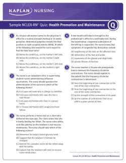
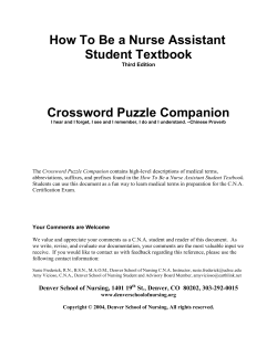
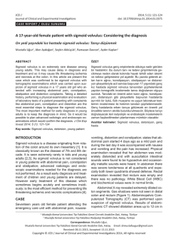
CHAPTER 5 Care of the Patient with a Gastrointestinal Disorder BARBARA LAURITSEN CHRISTENSEN Key Terms After reading this chapter, the student should be able to do the following: Anatomy and Physiology 1. List in sequence each of the component parts or segments of the alimentary canal and identify the accessory organs of digestion. 2. Discuss the function of each digestive and accessory organ. 5. 6. 7. 8. 9. 10. 11. 12. achalasia (�ak-�ah-LA ෆ-ze ෆ-�a, p. xxx) achlorhydria (�a-chl�or-HIෆ-dre ෆ-�a, p. xxx) ෆ-s�ıs, p. xxx) anastomosis (�a-n�as-t�o-MO � ෆ-�a, p. xxx) cachexia (k�a-KEK-se � ık � carcinoembryonic antigen (CEA) (k�ar-s�ın-o ෆ-em-bre ෆ-AN-� �AN-t�ı-j�en, p. xxx) dehiscence (d�e-HI�S-entz, � p. xxx) ෆ-je -� a , p. xxx) dysphagia (d�ıs-FA ෆ � p. xxx) evisceration (�e-v�ıs-er-A � ෆ-shun, � p. xxx) exacerbation (�eg-z�as-er-BA � ෆ-shun, � e-s� hematemesis (h�e-m�a-TEM� ıs, p. xxx) � intussusception (�ın-tus-s � us-S � EP-sh un, � p. xxx) ෆ-ke leukoplakia (lu ෆ-ko ෆ-PLA ෆ-�a, p. xxx) ෆ-m�en, p. xxx) lumen (LU � eh-n� melena (MEL� a, p. xxx) � occult blood (�o-KULT, p. xxx) � ı k, p. xxx) pathognomonic (p�ath-�og-no ෆ-MON-� � remission (r�e-MISH- un, � p. xxx) ෆ-�a, p. xxx) steatorrhea (st�e-�a-to ෆ-RE ෆ-m�a, p. xxx) stoma (STO � tenesmus (t�e-NEZ-m us, � p. xxx) � ෆ-lus, � p. xxx) volvulus (VOL-vu PR SA O M PE PL R E TY C O O F N E TE L N SE T V - N IE O R T FI N Medical-Surgical 3. Define the key terms as listed. 4. Discuss the laboratory and diagnostic examinations Be sure to check out the bonus material on the free CD-ROM, including selected audio pronunciations. AL Objectives and give the nursing interventions for patients with disorders of the gastrointestinal tract. Explain the etiology/pathophysiology, clinical manifestations, assessments, diagnostic tests, medical-surgical management, and nursing interventions for the patient with disorders of the mouth, esophagus, stomach, and intestines. Identify nursing interventions for preoperative and postoperative care of the patient who requires gastric surgery. Differentiate among irritable bowel syndrome, ulcerative colitis, and Crohn’s disease. Discuss the etiology/pathophysiology, clinical manifestations, assessment, diagnostic tests, medical management, and nursing interventions for the patient with acute abdominal inflammations (appendicitis, diverticulitis, and peritonitis). Discuss the etiology/pathophysiology, clinical manifestations, assessment, diagnostic tests, medical management, and nursing interventions for the patient with external hernias and hiatal hernia. Differentiate between mechanical and nonmechanical intestinal obstruction. Describe the etiology/pathophysiology, clinical manifestations, assessment, diagnostic tests, medical management, surgical procedures, and nursing interventions for the patient with cancer of the colon and rectum. Identify five nursing interventions for the patient with a stoma for fecal diversion. OVERVIEW OF ANATOMY AND PHYSIOLOGY DIGESTIVE SYSTEM Although it is understood that food is necessary for existence, not everyone understands (1) what happens to food once it is chewed and swallowed; (2) how food is prepared for its trip to each individual cell; and (3) the many changes that food undergoes, both chemically and physically. This chapter will review these changes and their effect on the body. The digestive tract, or alimentary canal, is a musculomembranous tube extending from the mouth to the anus and is approximately 30 feet long (Figure 5-1). It consists of the mouth, pharynx, esophagus, small intestine, large intestine, and anus. Peristalsis is the coordinated, rhythmic, serial contraction of smooth muscle that forces food through the digestive tract, bile through the bile duct, and urine through the ureter. During peristalsis the tract shortens to approximately 15 feet. 195 Copyright © 2006 Mosby Inc. All rights reserved. 196 CHAPTER 5 Care of the Patient with a Gastrointestinal Disorder Tongue Sublingual gland Pharynx Esophagus PR SA O M PE PL R E TY C O O F N E TE L N SE T V - N IE O R T FI N AL Submandibular gland Larynx Trachea Parotid gland Diaphragm Liver Gallbladder Cystic duct Hepatic bile duct Hepatic flexure Common bile duct Duodenum Pancreas Spleen Splenic flexure Stomach Transverse colon Duodenojejunal flexure Ascending colon Descending colon Region of ileocecal valve Cecum Vermiform appendix Ileum Sigmoid colon Rectum Anus FIGURE 5-1 Location of digestive organs. Accessory organs aid in the digestive process but are not considered part of the digestive tract. They release chemicals into the system through a series of ducts. The teeth, tongue, salivary glands, liver, gallbladder, and pancreas are considered accessory organs. These will be discussed in the chapter. Organs of the Digestive System and Their Functions (Box 5-1) Mouth. The mouth marks the entrance to the digestive system. The floor of the mouth contains a muscular appendage, the tongue. The tongue is involved in chewing, swallowing, and the formation of speech. Tiny eleva- tions, called papillae contain the taste buds. They differentiate between bitter, sweet, sour, and salty sensations. Digestion begins in the mouth. Here the teeth mechanically shred and grind the food and the enzymes begin the chemical breakdown of carbohydrates. Teeth. Each tooth is designed to carry out a specific task. Immediately to the center of the mouth lie the incisors, which are structured for biting and cutting. Posterior to the incisors are the canines, pointed teeth used for tearing and shredding food. The molars are to the rear of the jaw. These teeth have four cusps (points) and are used for mastication (to crush and grind food). Copyright © 2006 Mosby Inc. All rights reserved. Care of the Patient with a Gastrointestinal Disorder ORGANS OF THE ALIMENTARY CANAL • Mouth • Cecum • Pharynx (throat) • Colon • Esophagus (food pipe) Ascending colon • Stomach Transverse colon • Small intestine Descending colon Duodenum Sigmoid colon Jejunum • Rectum Ileum • Anal canal • Large intestine ACCESSORY ORGANS • Teeth and gums • Tongue • Liver • Gallbladder • Pancreas • Salivary glands Parotid Submandibular Sublingual PR SA O M PE PL R E TY C O O F N E TE L N SE T V - N IE O R T FI N Salivary Glands. There are three pairs of salivary glands (see Figure 5-1). They are the parotid glands, submandibular glands, and sublingual glands. They secrete a fluid called saliva, which is approximately 99% water with enzymes and mucus. Normally these glands secrete enough saliva to keep the mucous membranes Serosa Fundus Esophagus Cardiac sphincter Longitudinal muscle layer Circular muscle layer Duodenal bulb Oblique muscle layer overlying submucosa Body Lesser curvature Greater curvature Rugae Duodenum 197 of the mouth moist. Once food enters the mouth, the secretion increases to lubricate and dissolve the food and to begin the chemical process of digestion. The salivary glands secrete about 1000 to 1500 mL of saliva daily. The major enzyme is salivary amylase (ptyalin), which is responsible for the initiation of carbohydrate metabolism. Another enzyme, lysozyme, destroys bacteria, which protects the mucous membrane from infections and protects the teeth from decay. After food has been ingested, the salivary glands continue to secrete saliva, which cleanses the mouth. Esophagus. The esophagus is a muscular, collapsible tube that is approximately 10 inches long, extending from the mouth through the thoracic cavity and the esophageal hiatus to the stomach. No digestion takes place here. Peristalsis moves the bolus (food broken down and mixed with saliva, ready to pass to the stomach) through the esophagus to the stomach in 5 or 6 seconds. Stomach. The stomach is located in the left upper quadrant of the abdomen, directly inferior to the diaphragm (Figure 5-2). When the stomach is filled, it is the size of a football and holds approximately 1 L. The entrance to the stomach is the cardiac sphincter (so named because of the proximity to the heart); the exit is the pyloric sphincter. As food leaves the esophagus, it enters the stomach through the relaxed cardiac AL Box 5-1 Organs of the Digestive System CHAPTER 5 Pyloric Pylorus sphincter FIGURE 5-2 Stomach. Cut-away sections show muscle layers and interior mucosa thrown into folds called rugae. Copyright © 2006 Mosby Inc. All rights reserved. CHAPTER 5 Care of the Patient with a Gastrointestinal Disorder ileum. Most digestion takes place here—as much as 90%. The intestinal juices finish the metabolism of carbohydrates and proteins. Bile and pancreatic juices enter the duodenum. Bile from the liver breaks molecules into smaller droplets, which enables the digestive juices to complete their process. Pancreatic juices contain water, protein, inorganic salts, and enzymes. Pancreatic juices are essential in breaking down proteins into their amino acid components, in reducing dietary fats to glycerol and fatty acids, and in converting starch to simple sugars. The inner surface of the small intestine contains millions of tiny fingerlike projections called villi, which are clustered over the entire mucous surface. They are responsible for absorbing the products of digestion into the blood stream. They increase the absorption area of the small intestine 600 times. Inside each villus is a rich capillary bed, along with modified lymph capillaries called lacteals. The lacteals are responsible for the absorption of metabolized fats. Large Intestine. Once the small intestine has finished with its specific tasks, the ileocecal valve opens and re- PR SA O M PE PL R E TY C O O F N E TE L N SE T V - N IE O R T FI N sphincter. The sphincter then contracts, preventing reflux (splashing or return flow), which can be irritating. Once the bolus has entered the stomach, the muscular layers of the stomach churn and contract to mix and compress the contents with the gastric juices and water. The gastric juices are a group of secretions that are released by the gastric glands. Digestion of protein begins in the stomach. Hydrochloric acid softens the connective tissue of meats, kills bacteria, and activates pepsin (the chief enzyme of gastric juices that converts proteins into proteoses and peptones). Mucin is released to protect the stomach lining. Intrinsic factor (a substance secreted by the gastric mucosa) is produced to allow absorption of vitamin B12. After the stomach has completed its work, the food has been broken down into a viscous semiliquid substance called chyme. The chyme is sent through the pyloric sphincter into the duodenum for the next phase of digestion. Small Intestine. The small intestine is a 20-foot-long tube; it is 1 inch in diameter. It begins at the pyloric sphincter and ends at the ileocecal valve. It is divided into three major sections: duodenum, jejunum, and AL 198 Transverse colon Inferior vena cava Aorta Splenic vein Hepatic (right colic) flexure Ascending colon Right colic artery Mesentery Splenic (left colic) flexure Ilium Superior mesenteric artery Descending colon Inferior mesenteric artery and vein Sigmoid artery and vein Ileocecal valve Sigmoid colon Cecum Ileum Vermiform appendix Rectum Superior rectal artery and vein FIGURE 5-3 Divisions of the large intestine. Copyright © 2006 Mosby Inc. All rights reserved. Care of the Patient with a Gastrointestinal Disorder ACCESSORY ORGANS OF DIGESTION AL Liver The liver is the largest glandular organ in the body and one of the most complex. In the adult it weighs 3 pounds. It is located just inferior to the diaphragm, covering most of the upper right quadrant and extending into the left epigastrium. It is divided into two lobes. Approximately 1500 mL of blood is delivered to the liver every minute by the portal vein and the hepatic artery. The cells of the liver produce a product called bile, a yellow-brown or green-brown liquid. Bile is necessary for the metabolism of fats. The liver releases 500 to 1000 mL of bile per day. Bile travels to the gallbladder through hepatic ducts. The gallbladder is a sac about 3 to 4 inches long located on the right inferior surface of the liver. Here bile is stored until needed for fat digestion (Figure 5-4). Neck of gallbladder Right and left hepatic ducts Common hepatic duct Corpus (body) of gallbladder Cystic duct Common bile duct Accessory duct Pancreas Fundus of gallbladder Minor duodenal papilla Pancreatic duct Superior mesenteric artery and vein Sphincter muscles 199 K, needed for normal blood clotting, and the production of some of the B-complex vitamins. As the fecal material continues its journey, the remaining water and vitamins are absorbed into the blood stream by osmosis. Rectum. The last 8 inches of the intestine form the rectum, where the fecal material is expelled. PR SA O M PE PL R E TY C O O F N E TE L N SE T V - N IE O R T FI N leases the contents into the large intestine. The tube is larger in diameter (2 inches) but shorter (5 feet) than the small intestine. It is composed of the cecum; appendix; ascending, transverse, descending, and sigmoid colons; rectum; and anus (Figure 5-3). The process of digestion is completed here in the terminal portion of the digestive tract. Basically the large intestine has four major functions: (1) completion of absorption, (2) manufacture of certain vitamins, (3) formation of feces, and (4) expulsion of feces. Just inferior to the ileocecal valve is the cecum, a blind pouch approximately 2 to 3 inches long. Dangling from the cecum is a small wormlike, tubular structure: the vermiform appendix. To date, no function for the appendix is known. The open end of the cecum connects to the ascending colon; continues upward on the right side of the abdomen to the inferior area of the liver; becomes the transverse colon; and crosses to the left side of the abdomen, where it becomes the descending colon to the level of the iliac crest. The sigmoid colon begins here and continues toward the midline to the level of the third sacral vertebra. Bacteria in the large intestine change the chyme into fecal material by releasing the remaining nutrients. The bacteria are also responsible for the synthesis of vitamin CHAPTER 5 Major duodenal papilla Duodenum FIGURE 5-4 Gallbladder and bile ducts. Obstruction of the hepatic or common bile duct by stone or spasm occludes the exit of the bile from being ejected into the duodenum. Copyright © 2006 Mosby Inc. All rights reserved. CHAPTER 5 Care of the Patient with a Gastrointestinal Disorder In addition to producing bile, the liver has many other functions: managing blood coagulation; manufacturing cholesterol; manufacturing albumin to maintain normal blood volume; filtering out old red blood cells and bacteria; detoxifying poisons (alcohol, nicotine, drugs); converting ammonia to urea; providing the main source of body heat; storing glycogen for later use; activating vitamin D; and breaking down nitrogenous waste (from protein metabolism) to urea, which the kidneys can excrete as waste from the body. TUBE GASTRIC ANALYSIS Rationale The contents of the stomach are aspirated to determine the amount of acid produced by the parietal cells in the stomach. The analysis is done to determine the completeness of a vagotomy, confirm hypersecretion or achlorhydria (an abnormal condition characterized by the absence of hydrochloric acid in the gastric juice), estimate acid secretory capacity, or assay for intrinsic factor. PR SA O M PE PL R E TY C O O F N E TE L N SE T V - N IE O R T FI N Pancreas The pancreas is an elongated gland that lies posterior to the stomach (see Figure 5-4). It is an active organ that is involved in both endocrine and exocrine duties. In this chapter, discussion of the pancreas is limited to its exocrine activities. Each day the pancreas produces 1000 to 1500 mL of pancreatic juice to aid in digestion. This pancreatic juice contains the digestive enzymes protease (trypsin), lipase (steapsin), and amylase (amylopsin), which are important because of their ability to digest the three major components of chyme—proteins, fats, and carbohydrates. These enzymes are transported through an excretory duct to the duodenum. This pancreatic duct connects to the common bile duct from the liver and gallbladder and empties through a small orifice in the duodenum called the major duodenal papilla or papilla of Vater. In addition, the pancreas contains an alkaline substance, sodium bicarbonate, that has the ability to neutralize the hydrochloric acid in the gastric juices that enter the small intestine from the stomach. Nursing Interventions The patient should maintain nothing by mouth (NPO) status after midnight. Tell the patient to avoid smoking after midnight the night before the study. The nurse should explain to the patient the importance of rectally expelling all the barium after the examination. Stools will be light colored until all the barium is expelled up to 72 hours after the test. Eventual absorption of fecal water may cause a hardened barium impaction. Increasing fluid intake is usually effective. Milk of magnesia (60 mL) is commonly given after the examination unless contraindicated. AL 200 REGULATION OF FOOD INTAKE The hypothalamus, a portion of the brain, contains two centers that have an effect on eating. One center stimulates the individual to eat and the other signals the individual to stop eating. These centers work in conjunction with the rest of the brain to balance eating habits. However, many other factors also affect eating. For example, distention decreases appetite. Other controls in our bodies, lifestyle, eating habits, emotions, and genetic factors all influence intake of food and blend together to influence each individual’s body build. LABORATORY AND DIAGNOSTIC EXAMINATIONS UPPER GASTROINTESTINAL STUDY (UPPER GI SERIES, UGI) Rationale The upper GI study consists of a series of radiographs of the lower esophagus, stomach, and duodenum using barium sulfate as the contrast medium. A UGI series will detect any abnormal conditions of the upper GI tract, any tumors, or other ulcerative lesions. Nursing Interventions The patient should receive no anticholinergic medications for 24 hours before the test and should maintain NPO status after midnight so the gastric acid secretion will not be altered. The nurse should inform the patient that smoking is prohibited before the test because nicotine stimulates the flow of gastric secretions. The nurse or radiology personnel will insert a nasogastric tube into the stomach to aspirate gastric content. Specimens should be labeled properly and sent to the laboratory immediately. The nasogastric tube is removed as soon as specimens are collected. The patient may then eat if indicated. ESOPHAGOGASTRODUODENOSCOPY (EGD, UGI ENDOSCOPY, GASTROSCOPY) Rationale Endoscopy (endo, within, inward; scope, to look) enables direct visualization of the upper GI tract by means of a long, fiberoptic flexible scope. The esophagus, stomach (Figure 5-5), and duodenum are examined for tumors, varices, mucosal inflammations, hiatal hernias, polyps, ulcers, presence of Helicobacter pylori, strictures, and obstructions. Also, the endoscopist can remove polyps, coagulate sources of active GI bleeding, and perform sclerotherapy of esophageal varices through endoscopy. Areas of narrowing can be dilated by the endoscope itself or by passing a dilator through the scope. Camera equipment may be attached to the viewing lens, and the existing pathologic condition can be photographed. The Copyright © 2006 Mosby Inc. All rights reserved. Care of the Patient with a Gastrointestinal Disorder 201 BARIUM SWALLOW/ GASTROGRAFIN STUDIES Eyepiece Air PR SA O M PE PL R E TY C O O F N E TE L N SE T V - N IE O R T FI N Light Rationale This barium contrast study is a more thorough study of the esophagus than that provided by most UGI examinations. As in most barium contrast studies, defects in luminal filling and narrowing of the barium column indicate tumor, scarred stricture, or esophageal varices. With a barium swallow, anatomical abnormalities, such as hiatal hernia, are easily recognized. Left atrial dilation, aortic aneurysm, and paraesophageal tumors (such as bronchial or mediastinal tumors) may cause extrinsic compression of the barium column within the esophagus. A product called Gastrografin is now used in place of barium for patients in whom bleeding from the GI system may occur and surgery is being considered. Gastrografin is water soluble and rapidly absorbed, so it is preferable when a perforation is suspected. Gastrografin facilitates imaging through radiographs, but if the product escapes from the GI tract, unlike barium, it is absorbed by the surrounding tissue. Complications can occur if barium leaks from the GI tract. AL Focus Light CHAPTER 5 Observation port FIGURE 5-5 Fiberoptic endoscopy of the stomach. endoscope can also be used in obtaining tissue specimens for biopsy or culture to determine presence of H. pylori. Endoscopy enables evaluation of the esophagus, stomach, and duodenum; by using a longer fiberoptic scope, the upper small intestine can be evaluated. This is referred to as enteroscopy. Nursing Interventions The nurse should explain the procedure to the patient. The patient should maintain NPO status after midnight. The nurse must obtain the patient’s signature on a consent form and complete a preoperative checklist for the endoscopic examination. The patient is usually given a preprocedure intravenous sedative such as midazolam (Versed). Because the patient’s pharynx has been anesthetized (by spraying) with lidocaine HCl (Xylocaine), the nurse should not allow the patient to eat or drink until the gag reflex returns (usually about 2 to 4 hours). The nurse should assess for any signs and symptoms of perforation, including abdominal pain and tenderness, guarding, oral bleeding, melena, and hypovolemic shock. Nursing Interventions The patient should maintain NPO status after midnight. Food and fluid in the stomach will prevent the barium from accurately outlining the GI tract, and the radiographic results may be misleading. The nurse should explain to the patient the importance of rectally expelling all barium. Stools will be light colored until this occurs. Eventual absorption of fecal water may cause a hardened barium impaction. Increasing fluid intake is usually effective. Milk of magnesia (60 mL) is usually given after the examination unless contraindicated. ESOPHAGEAL FUNCTION STUDIES (BERNSTEIN TEST) Rationale The Bernstein test, an acid-perfusion test, is an attempt to reproduce the symptoms of gastroesophageal reflux. It aids in differentiating esophageal pain caused by esophageal reflux from that caused by angina pectoris. If the patient suffers pain with the instillation of hydrochloric acid into the esophagus, the test is positive and indicates reflux esophagitis. Nursing Interventions The nurse should avoid sedating the patient, because the patient’s participation is essential for swallowing the tubes, swallowing during acid clearance, and describing any discomfort during the instillation of hydrochloric acid. The patient is NPO for 8 hours before the examination, and any medications that may interfere with the production of acid, such as antacids and analgesics, are withheld. Copyright © 2006 Mosby Inc. All rights reserved. 202 CHAPTER 5 Care of the Patient with a Gastrointestinal Disorder EXAMINATION OF STOOL FOR OCCULT BLOOD BARIUM ENEMA STUDY (LOWER GI SERIES) Rationale Tumors of the large intestine grow into the lumen (the cavity or channel within a tube or tubular organ) and are subjected to repeated trauma by the fecal stream. Eventually the tumor ulcerates and bleeding occurs. Usually the bleeding is so slight that gross blood is not seen in the stool. If this occult blood (blood that is obscure or hidden from view) is detected in the stool, a benign or malignant GI tumor should be suspected. Tests for occult blood are also called guaiac, Hemoccult, and Hematest. Occult blood in the stool may occur also in ulceration and inflammation of the upper or lower GI system. Other causes include swallowing blood of oral or nasopharyngeal origin. Stool may be obtained by digital retrieval by the nurse or physician. However, the patient is usually asked to collect stool in an appropriate container. A specimen for occult blood must be obtained before barium studies are done. Rationale The barium enema (BE) study consists of a series of radiographs of the colon used to demonstrate the presence and location of polyps, tumors, and diverticula. Positional abnormalities (such as malrotation) can also be detected. Barium sulfate is more effective for visualizing mucosal detail. Therapeutically, the BE study may be used to reduce nonstrangulated ileocolic intussusception (infolding of one segment of the intestine into the lumen of another segment) in children. PR SA O M PE PL R E TY C O O F N E TE L N SE T V - N IE O R T FI N AL Nursing Interventions The nurse may administer cathartics such as magnesium citrate or other cathartics designated by institution policy the evening before the BE. A cleansing enema may also be administered the evening before or the morning of the BE if directed by physician’s order or hospital policy. Milk of magnesia (60 mL) may be ordered after the BE to stimulate evacuation of the barium. After the BE study, the patient should be assessed for complete evacuation of the barium. Retained barium may cause a hardened impaction. Stool will be light colored until all the barium has been expelled. Nursing Interventions The nurse should instruct the patient to keep the stool specimen free of urine or toilet paper, because either can contaminate the specimen and alter the test results. The nurse or patient should don gloves and use tongue blades to transfer the stool to the proper receptacle. Keep diet free of red meat for 24 to 48 hours before a guaiac test. SIGMOIDOSCOPY (LOWER GI ENDOSCOPY) Rationale Endoscopy of the lower GI tract allows visualization and, if indicated, access to obtain biopsy specimens of tumors, polyps, or ulcerations of the anus, rectum, and sigmoid colon. Because the lower GI tract is difficult to visualize radiographically, the direct visualization afforded through sigmoidoscopy is beneficial. Microscopic review of tissue specimens obtained using this procedure can provide the diagnoses of many lower bowel disorders. Nursing Interventions The nurse should explain the procedure to the patient. The patient should sign a consent form for the procedure. The nurse administers enemas as ordered on the evening before and/or the morning of the examination to ensure optimum visualization of the lower GI tract. After the examination, the nurse observes the patient for evidence of bowel perforation (abdominal pain, tenderness, distention, and bleeding). COLONOSCOPY Rationale With the development of the fiberoptic colonoscope, a high percentage of patients can have the entire colon—from anus to cecum—examined. Therefore, with colonoscopy, the detection of lesions in the proximal colon—which would otherwise be undetected by sigmoidoscopy—is possible. Benign and malignant neoplasms, mucosal inflammation or ulceration, and sites of active hemorrhage can also be visualized. Biopsy specimens can be obtained and small tumors removed through the scope with the use of cableactivated instruments. Actively bleeding vessels can be coagulated. Patients who have had cancer of the colon are at high risk for developing a subsequent colon cancer; patients who have a family history of colon cancer are at high risk. For these patients, colonoscopy allows early detection of any primary or secondary tumors. Nursing Interventions The patient signs a consent form. The nurse explains the procedure to the patient. The patient is instructed regarding dietary restrictions: usually a clear liquid diet is permitted 1 to 3 days before the procedure to decrease the residue in the bowel, and then NPO status is maintained for 8 hours before the procedure. The nurse administers a cathartic, enemas, and pre- Copyright © 2006 Mosby Inc. All rights reserved. Care of the Patient with a Gastrointestinal Disorder 1. Give patient one metoclopramide (Reglan) 10-mg tablet, as prescribed, orally 30 minutes before proceeding with step 2. 2. Administer the GoLYTELY solution* (prepared by pharmacy) per physician’s orders: a. 240 mL orally every 15 minutes or b. 30 mL/min via NG tube. Use a Travasorb enteral feeding container and a size 10 feeding tube. Administer until stools are clear yellow. c. Keep patient warm with heated blankets; they often become chilled after consuming copious amounts of GoLYTELY solution. d. Provide a bedside commode if patient is an older adult or weak. *Administer a minimum of 1 gallon of solution over a 2-hour period. When a patient is suspected of having a parasitic infection, the stool is examined for O&P. Usually at least three stool specimens are collected on subsequent days. Because culture results won’t be available for several days, they won’t influence initial treatment, but they will guide subsequent treatment if bacterial infection is present. Nursing Interventions If an enema must be administered to collect specimens, only normal saline or tap water should be used. Soapsuds or any other substance could affect the viability of the organisms collected. Stool samples for O&P are obtained before barium examinations. The patient is instructed not to mix urine with feces. The nurse dons gloves to collect the specimen. The specimen should be taken to the laboratory within 30 minutes of collection in specified container. OBSTRUCTION SERIES (FLAT PLATE OF THE ABDOMEN) PR SA O M PE PL R E TY C O O F N E TE L N SE T V - N IE O R T FI N medication as ordered to decrease the residue in the bowel. GoLYTELY (Box 5-2), an oral or nasogastric (NG) colonic lavage, is an osmotic electrolyte solution that is now commonly used as a cathartic. It is a polyethylene glycol solution. If taken orally, instruct the patient to drink the solution rapidly: 8 ounces (240 mL) every 15 minutes until enough solution has been consumed to make the colonic contents a light yellow liquid. Powdered lemonade may be added to make the oral solution more palatable. If it is given per lavage, it must be given rapidly. Taking the solution slowly will not clean the colon efficiently. Provide warm blankets during the procedure. Many patients experience hypothermia during the GoLYTELY procedure. Provide a commode at the bedside for older adults and frail patients. A preprocedure IV sedative such as midazolam is often given. After colonoscopy, the nurse checks for evidence of bowel perforation (abdominal pain, guarding, distention, tenderness, excessive rectal bleeding, or blood clots) and examines stools for gross blood. Assess for hypovolemic shock. 203 AL Box 5-2 GoLYTELY Bowel Preparation CHAPTER 5 STOOL CULTURE Rationale The obstruction series is a group of radiographic studies performed on the abdomen of patients who have suspected bowel obstruction, paralytic ileus, perforated viscus (any large interior organ in any of the great body cavities), or abdominal abscess. This series usually consists of at least two radiographic studies. The first is an erect abdominal radiographic study that should include visualization of the diaphragm. Radiographs are examined for evidence of free air under the diaphragm, which is pathognomonic (signs or symptoms specific to a disease condition) of a perforated viscus. This radiographic study is used also to detect air-fluid levels within the intestine. Nursing Interventions For adequate visualization, the nurse should ensure that this study is scheduled before any barium studies. DISORDERS OF THE MOUTH Rationale The feces (stool) can be examined for the presence of bacteria, ova, and parasites (a plant or animal that lives upon or within another living organism at whose expense it obtains some advantage). The physician may order a stool for culture of bacteria or for ova and parasites (O&P). Many bacteria (such as Escherichia coli) are indigenous in the bowel. Bacterial cultures are usually done to detect enteropathogens (such as Staphylococcus aureus, Salmonella, Shigella, E. coli 0157:H7, or Clostridium difficile). Common disorders of the mouth and esophagus that interfere with adequate nutrition include poor dental hygiene, infections, inflammation, and cancer. DENTAL PLAQUE AND CARIES Etiology/Pathophysiology Dental decay is an erosive process that results from the action of bacteria on carbohydrates in the mouth, which in turn produces acids that dissolve tooth enamel. Most Americans (95%) experience tooth decay Copyright © 2006 Mosby Inc. All rights reserved. CHAPTER 5 Care of the Patient with a Gastrointestinal Disorder at some time in their life. Dental decay can be caused by one of several factors, among which are the following: • The presence of dental plaque, a thin film on the teeth made of mucin and colloidal material found in saliva and often secondarily invaded by bacteria • The strength of acids and the ability of the saliva to neutralize them • The length of time the acids are in contact with the teeth • Susceptibility of the teeth to decay Medical Management Interventions include treatment of dental caries by removal of affected areas of the tooth and replacement with some form of dental material. Treatment of periodontal disease centers on removal of plaque from the teeth. If the disease has advanced, surgical interventions of the gingivae and alveolar bone may be necessary. NURSING DIAGNOSES CANDIDIASIS Etiology/Pathophysiology This condition is any infection caused by a species of Candida, usually C. albicans. Candida is a fungal organism normally present in the mucous membranes of the mouth, intestinal tract, and vagina and is also found on the skin of healthy people. This infection is also referred to as thrush and moniliasis. This disease appears more commonly in the newborn infant, who becomes infected while passing through the birth canal. In the older individual, candidiasis may be found in patients with leukemia, diabetes mellitus, or alcoholism, and in the person who has been taking antibiotics (chlortetracycline or tetracycline) or steroids for long periods or who is in a general weakened state. It is often seen in patients receiving chemotherapy and/or radiation therapy. PR SA O M PE PL R E TY C O O F N E TE L N SE T V - N IE O R T FI N Nursing Interventions and Patient Teaching Proper technique for brushing and flossing the teeth at least twice a day is the nurse’s primary focus of teaching for these patients. Plaque forms continuously and must be removed periodically through regular visits to the dentist. The patient must understand the importance of prevention through continual care. Because carbohydrates create an environment in which caries develops and plaque accumulates more easily, proper nutrition is included in patient teaching. When the patient is ill, the normal cleansing action of the mouth is impaired. Illnesses, drugs, and irradiation all interfere with the normal action of saliva. If the patient is unable to manage oral hygiene, the nurse must assume this responsibility. Nursing diagnoses and interventions for the patient with dental plaque and caries include but are not limited to the following: Prognosis The prevention and elimination of dental plaque and caries are directly related to oral hygiene, dental care, nutrition, and heredity. All but heredity are controllable characteristics. The prognosis is more favorable for people who brush, floss, regularly visit the dentist for removal of affected areas, eat low-carbohydrate foods, and drink fluoridated water. AL 204 Deficient knowledge, related to inability to prevent dental caries and periodontal disease Noncompliance, related to hygiene and dietary restrictions NURSING INTERVENTIONS Assess and observe the oral cavity for moisture, color, and cleanliness. Stress importance of meticulous oral hygiene. Explain need to see dentist at least yearly for examination. Brush teeth twice daily (bid) and as needed (prn) with toothpaste or powder, baking soda, or mouthwash. Rinse with water or mouthwash. Cleanse mouth with equal parts of hydrogen peroxide and water prn for halitosis. Teach oral hygiene to patient. Clinical Manifestations Candidiasis appears as pearly, bluish white “milkcurd” membranous lesions on the mucous membranes of the mouth, tongue, and larynx. One or more lesions may be on the mucosa, depending on the duration of the infection. If the patch or plaque is removed, painful bleeding can occur. Medical Management Treatment may include 1 to 4 mL of nystatin (Mycostatin) dropped in the infected infant’s mouth several times a day. For the adult, nystatin or amphotericin B (an oral suspension) or buccal tablets and half-strength hydrogen peroxide/saline mouth rinses may provide some relief. For treatment of adult Candida vaginal infection, nystatin vaginal tablets (100,000 units dissolved) inserted into the vagina twice a day is effective. Ketoconazole taken systemically appears to be equally effective. Nursing Interventions The nurse must use meticulous handwashing to prevent spread of infection. The infection may be spread in the nursery by carelessness of nursing personnel. Handwashing, care of feeding equipment, and cleanliness of the mother’s nipples are important to prevent spread. The nurse should cleanse the infant’s mouth of any foreign material, rinsing the mouth and lubricating the lips. The mouth should be inspected using a flashlight and tongue blade. Copyright © 2006 Mosby Inc. All rights reserved. Care of the Patient with a Gastrointestinal Disorder Prognosis If the host has a strong defense system and medical treatment is initiated early in the course of the disease, the prognosis is good. CARCINOMA OF THE ORAL CAVITY ready occurred in more than 60% of patients when the diagnosis is made because of the tongue’s abundant vascular and lymphatic drainage. Recent investigation has revealed a higher incidence of cancers of the mouth and throat among people who are heavy drinkers and smokers. Also, data show that the mortality for young men between the ages of 10 and 20 has doubled over the past 30 years as a result of the use of smokeless tobacco (snuff). This combination of high alcohol consumption and smoking or chewing tobacco causes an apparent breakdown in the body’s defense mechanism. Predisposing factors include exposure to the sun and wind, but more important is the progression of leukoplakia to an epidermoid lip cancer. Clinical Manifestations Leukoplakia (a white, firmly attached patch on the mouth or tongue mucosa) may appear on the lips and buccal mucosa. These nonsloughing lesions cannot be rubbed off by simple mechanical force. They can be benign or malignant. A small percentage develop into squamous cell carcinomas, and biopsy is recommended if the lesions persist for longer than 2 weeks. They occur most frequently between the ages of 50 and 70 years and appear more commonly in men. PR SA O M PE PL R E TY C O O F N E TE L N SE T V - N IE O R T FI N Etiology/Pathophysiology The lips, the oral cavity, the tongue, and the pharynx are prone to develop malignant lesions. The tonsils occasionally may be involved. The largest number of these tumors are squamous cell epitheliomas that grow rapidly and metastasize to adjacent structures more quickly than do most malignant tumors of the skin. In the United States, oral cancer accounts for 4% of the cancers in men and 2% in women. An estimated 28,260 new cases are expected in 2004. An estimated 7230 deaths from oral cavity and pharynx cancer are expected in 2004. Death rates have been decreasing since the 1970s, with rates declining faster in the 1990s. Tumors of the salivary glands occur primarily in the parotid gland and are usually benign. Tumors of the submaxillary gland have a high incidence of malignancy. These malignant tumors grow rapidly and may be accompanied by pain and impaired facial function. Kaposi’s sarcoma is a malignant skin tumor that occurs primarily on the legs of men between 50 and 70 years of age. Recently it has been seen with increased frequency as a nonsquamous tumor of the oral cavity in patients with acquired immunodeficiency syndrome (AIDS). The lesions are purple and nonulcerated. Irradiation is the treatment of choice. Cancer or neoplasm is characterized by the uncontrolled growth of anaplastic cells that tend to invade surrounding tissue and to metastasize to distant body sites. The tumor seen with cancer of the lip is usually called an epithelioma. It occurs most frequently as a chronic ulcer of the lower lip in men. The cure rate for cancer of the lips is high because the lesion is easily apparent to the patient and to others. Metastasis to regional lymph nodes has occurred in only 10% of people when diagnosed. In some instances a lesion may spread rapidly and involve the mandible and the floor of the mouth by direct extension. Occasionally the tumor may be a basal cell lesion that starts in the skin and spreads to the lip. Cancer of the anterior tongue and floor of the mouth may seem to occur together because their spread to adjacent tissues is so rapid. Metastasis to the neck has al- 205 AL For adults, instruct the patient to use a soft-bristled toothbrush and administer a topical anesthetic (lidocaine or benzocaine) to the mouth 1 hour before meals. Give soft or pureed foods and avoid hot, cold, spicy, fried, or citrus foods. CHAPTER 5 Assessment Collection of subjective data includes understanding that malignant lesions of the mouth are usually asymptomatic. The patient may feel only a roughened area with the tongue. As the disease progresses, the first complaints may be (1) difficulty in chewing, swallowing, or speaking; (2) edema, numbness, or loss of feeling in any part of the mouth; and (3) earache, facial pain, and toothache, which may become constant. Cancer of the lip is associated with discomfort and irritation caused by the presence of a nonhealing lesion that may be raised or ulcerated. Malignancy at the base of the tongue produces less obvious symptoms: slight dysphagia, sore throat, and salivation. Collection of objective data includes observing for premalignant lesions, including leukoplakia (white patches). Unusual bleeding in the mouth, some bloodtinged sputum, lumps or edema in the neck, and hoarseness may be observed. Diagnostic Tests Indirect laryngoscopy is an important diagnostic test for examination of the soft tissue. This procedure is especially important for men 40 years of age or older who have dysphagia and a history of smoking and alcohol ingestion. Radiographic evaluation of the mandibular structures is also an essential part of the head and neck examination to rule out the presence of cancer. Excisional biopsy is the most accurate method for a definitive diagnosis. Oral exfoliative cytology is a means of screening intraoral lesions. A scraping of the Copyright © 2006 Mosby Inc. All rights reserved. CHAPTER 5 Care of the Patient with a Gastrointestinal Disorder lesion provides cells for cytologic examination. The chance for a false-negative finding is about 26%. NURSING DIAGNOSES NURSING INTERVENTIONS Imbalanced nutrition, less than body requirements, related to oral pain or postoperative tissue loss (mucous membranes). Monitor the patient for changes in the character and quantity of mucus after radiation therapy. Provide meticulous oral hygiene. Observe for temporary or permanent loss of taste and the need for alternative routes for nutrition by monitoring daily weights. Provide alternative methods for communication if dysarthria (difficult, poorly articulated speech, resulting from interference in the control over muscles of speech) results from radiation treatment. Provide information to the patient and family to help with difficult decisions related to surgery, radiation, or chemotherapy. Be a support person to the patient and family. Disturbed body image and personal identity, related to disfiguring appearance of an oral lesion or reconstructive surgery PR SA O M PE PL R E TY C O O F N E TE L N SE T V - N IE O R T FI N Medical Management Treatment depends on the location and staging of the malignant tumor. Stage I oral cancers are treated by surgery or radiation. Stages II and III cancers require both surgery and radiation. Treatment for stage IV cancer is usually palliative. The survival rate for patients with oral cancers averages less than 50%. Small, accessible tumors can be excised surgically and include a glossectomy, removal of the tongue; hemiglossectomy, removal of part of the tongue; mandibulectomy, removal of the mandible; and total or supraglottic laryngectomy, removal of the entire larynx or the portion above the true vocal cords. Large tumors usually require more extensive and traumatic surgery. In a functional neck dissection of neck cancer with no growth in the lymph nodes, the lymph nodes are removed, but the jugular vein, sternocleidomastoid muscle, and spinal accessory nerve are preserved. In radical neck dissection, all these structures are removed and reconstructive surgery is necessary after tissue resection. These patients may have drains in the incision sites that are connected to suction to aid healing and reduce hematomas. A tracheostomy may also be performed, depending on the degree of tumor invasion. Because of the location of the surgery, complications can occur. These include airway obstruction, hemorrhage, tracheal aspiration, facial edema, fistula formation, and necrosis of the skin flaps. Neurologic complications can occur because of nerves being severed and manipulated during surgery. Radiation may be in the form of external radiation by use of roentgenograms or other radioactive substances or in the form of internal radiation by means of needles or seeds. The purpose of radiation therapy is to shrink the tumor. It can be given preoperatively or postoperatively, depending on the physician’s preference and the patient’s disease process. In more advanced cases, chemotherapy may be combined with radiation postoperatively to make the patient more comfortable. Other treatment options include laser excision. staff may be necessary for information and support during this potentially fatal disease. Nursing diagnoses and interventions for the patient with oral cancer include but are not limited to the following: AL 206 Nursing Interventions and Patient Teaching It is important that the nurse have a holistic approach to the patient. This includes being aware of the patient’s level of knowledge regarding the disease, the emotional response and coping abilities, and the spiritual needs. The nursing interventions must be individualized to the patient—beginning with the preoperative stage, continuing through the postoperative stage, and ending after the patient’s rehabilitation in the home environment. Family members, hospice members, close friends, social workers, and pastoral care Prevention centers on avoidance of predisposing factors: excess exposure to sun and wind on the lips, elimination of smoking or chewing tobacco, and maintenance of good oral and dental care. There is a high correlation between the incidence of cancer of the mouth and cirrhosis of the liver associated with alcohol intake. Early detection of oral cancer can help increase the patient’s chance of survival. Any person with a mouth lesion that does not heal within 2 to 3 weeks is urged to seek immediate medical care. Preoperative and postoperative care must be taught to the surgical patient, with full explanations regarding speech loss and alternate methods of nutritional intake. Explanation of tracheostomy care and other tubes the patient may be discharged with will relieve anxiety and encourage the patient’s control over the situation. Prognosis Staging and biologic characterization of the neoplasm provide prognostic information. The prognosis of carcinoma in the oral cavity is directly related to the size of the primary tumor, the involvement of regional nodes, and the presence or absence of metastasis. The Copyright © 2006 Mosby Inc. All rights reserved. Care of the Patient with a Gastrointestinal Disorder DISORDERS OF THE ESOPHAGUS GASTROESOPHAGEAL REFLUX DISEASE feeling of flatulence are other common complaints. Nocturnal cough, wheezing, or hoarseness all may occur with reflux, and it is estimated that greater than 80% of adult asthmatics may have reflux. The frequency and severity of reflux episodes usually determine the severity of the symptoms. Assessment Collection of subjective data includes heartburn, a substernal or retrosternal burning sensation that may radiate to the back or jaw (in some cases the pain may mimic angina); and regurgitation (not associated with nausea or eructation), in which a sour or bitter taste is perceived in the pharynx. Frequent eructation, flatulence, and dysphagia or odynophagia usually occurs only in severe cases. Collection of objective data may include nocturnal cough, wheezing, and hoarseness. Diagnostic Tests Mild cases of GERD are diagnosed from the classic symptoms, and treatment is initiated based on the presumptive diagnosis. More involved cases may require other screening tools. The gold standard for diagnosis is 24-hour pH monitoring, which accurately records the number, duration, and severity of reflux episodes and is considered to be 85% sensitive. The esophageal motility and Bernstein tests can be performed in conjunction with pH monitoring to evaluate lower esophageal sphincter competence and the response of the esophagus to acid infusion. The barium swallow with fluoroscopy is widely used to document the presence of hiatal hernia. Endoscopy is rarely necessary to establish diagnosis, but it is routinely performed to evaluate the presence and severity of esophagitis and to rule out malignancy. PR SA O M PE PL R E TY C O O F N E TE L N SE T V - N IE O R T FI N Etiology/Pathophysiology Gastroesophageal reflux disease (GERD) is a backward flow of stomach acid up into the esophagus. Symptoms typically include burning and pressure behind the sternum. Most cases are attributed to the inappropriate relaxation of the lower esophageal sphincter in response to an unknown stimulus. Reflux allows gastric contents to move back into the distal esophagus. Symptoms of GERD develop when the lower esophageal sphincter (LES) is weak or experiences prolonged or frequent transient relaxation, conditions that allow gastric acids and enzymes to flow into the esophagus. Reflux is much more common in the postprandial state (after meals); more than 60% of reflux sufferers have delayed gastric emptying. Gastroesophageal reflux disease occurs in all age groups and is estimated to affect up to 45% of the population to some degree, which translates to more than 60 million people. Clinical Manifestations The clinical manifestations of GERD are consistent in their nature, but they vary substantially in severity. The irritation of chronic reflux produces the primary symptom, which is heartburn (pyrosis). The pain is described as a substernal or retrosternal burning sensation that tends to radiate upward and may involve the neck, jaw, or back. The pain typically occurs 20 minutes to 2 hours after eating. An atypical pain pattern that closely mimics angina may also occur and needs to be carefully differentiated from true cardiac disease. The second major symptom of GERD is regurgitation, which is not associated with either eructation or nausea. The individual experiences a feeling of warm fluid moving up the throat. If it reaches the pharynx, a sour or bitter taste is perceived. Water brash, a reflux salivary hypersecretion that does not have a bitter taste, occurs less commonly. In severe cases, GERD can produce dysphagia or odynophagia (painful swallowing). Eructation and a 207 AL patient’s immunologic response and general condition also influence the prognosis and the choice of therapy. Carcinomas of the lip can be detected early by the person, the physician, or the dentist during examination, and the prognosis for cure is good. If the carcinoma is difficult to detect, as on the anterior tongue and floor of the mouth, it will be in the more advanced stage when detected and the prognosis will be bleak. The 5-year survival rate for cancer of the oral cavity and pharynx is 53% for whites and 34% for African Americans. CHAPTER 5 Medical Management In its simplest form, GERD produces mild symptoms that occur only infrequently (twice a week or less). In these cases, avoiding problem foods or beverages, stopping smoking, or losing weight if needed may solve the problem. Additional treatment with antacids or acid-blocking medications called H2 receptor antagonists—such as cimetidine (Tagamet), ranitidine (Zantac), famotidine (Pepcid), or nizatidine (Axid)— may also be used. More severe and frequent episodes of GERD can trigger asthma attacks, cause severe chest pain, result in bleeding, or promote a narrowing (stricture) or chronic irritation of the esophagus. In these cases, more powerful inhibitors of stomach acid production called proton pump inhibitors, such as omeprazole (Prilosec), esomeprazole (Nexium), pantoprazole (Protonix), rabeprazole (Aciphex), and lansoprazole (Prevacid), may be added to the treatment prescribed. Metoclopramide (Reglan) is used in moderate to severe cases of GERD. It is an Copyright © 2006 Mosby Inc. All rights reserved. CHAPTER 5 Care of the Patient with a Gastrointestinal Disorder example of a class of drugs called promotility agents that increase peristalsis without stimulating secretions. As a last resort, a surgical procedure called fundoplication is performed to strengthen the sphincter. The procedure involves wrapping a layer of the upper stomach wall around the sphincter and terminal esophagus to lessen the possibility of acid reflux (see Figure 5-16). If GERD is left untreated, serious pathologic (precancerous) changes in the esophageal lining may develop—a condition called Barrett’s esophagus. In Barrett’s esophagus there is replacement of the normal squamous epithelium of the esophagus with columnar epithelium. Because patients with Barrett’s esophagus are at higher risk for adenocarcinoma, they may need to be monitored on a regular basis (every 1 to 3 years) by endoscopy and biopsy. Prognosis If GERD is not successfully controlled, it can progress to serious and even life-threatening problems. Esophageal ulceration and hemorrhage may result from severe erosion, and chronic nighttime reflux is accompanied by a significant risk of aspiration. Adenocarcinoma can develop from the premalignant tissue (termed Barrett’s epithelium). Gradual or repeated scarring can permanently damage esophageal tissue and produce stricture. CARCINOMA OF THE ESOPHAGUS Etiology/Pathophysiology Carcinoma of the esophagus is a malignant epithelial neoplasm that has invaded the esophagus and has been diagnosed as the presence of a squamous cell carcinoma or an adenocarcinoma. Estimates of the percentage of esophageal cancer that are adenocarcinomas range from 30% to 70%, with the remainder being squamous cell. The incidence of squamous cell esophageal cancer is currently decreasing in the United States, whereas the incidence of adenocarcinoma of the distal esophagus is increasing (Lewis, 2004). Risk factors for esophageal cancer include alcohol intake and tobacco use and possibly long-standing achalasia (an abnormal condition characterized by the inability of a muscle to relax, particularly the cardiac sphincter of the stomach). Environmental carcinogens, nutritional deficiencies, chronic irritation, and mucosal damage have all been considered as causes of esophageal cancer. Another risk factor is Barrett’s esophagus. It is estimated that 1 of 200 cases of Barrett’s esophagus will progress to esophageal adenocarcinoma (Health Considerations box). Unfortunately, because of the location, esophageal cancer is usually at a late stage when discovered and treatment is aimed toward comfort and control rather than cure. Carcinoma of the bronchus, stomach, or breast may metastasize to the esophagus. The prevalent age group for esophageal cancer is 55 to 70 years. It occurs more commonly in men. PR SA O M PE PL R E TY C O O F N E TE L N SE T V - N IE O R T FI N Nursing Interventions and Patient Teaching Nursing intervention involves educating the patient about diet and lifestyle modifications that may alleviate symptoms of GERD: • Diet Eat four to six small meals daily. Follow a low-fat, adequate-protein diet. Reduce intake of chocolate, tea, and all foods and beverages that contain caffeine. Limit or eliminate alcohol intake. Eat slowly, and chew food thoroughly. Avoid evening snacking, and do not eat for 2 to 3 hours before bedtime. Remain upright for 1 to 2 hours after meals when possible, and never eat in bed. Avoid any food that directly produces heartburn. Reduce overall body weight if indicated. • Lifestyle Eliminate or drastically reduce smoking. Patients who smoke are encouraged to stop. Avoid constrictive clothing over the abdomen. Avoid activities that involve straining, heavy lifting, or working in a bent-over position. Elevate the head of the bed at least 6 to 8 inches for sleep, using wooden blocks or a thick foam wedge. Never sleep flat in bed. AL 208 Health Promotion Considerations Prevention or Early Detection of Esophageal Cancer • Patients with diagnosed GERD and hiatal hernia need to be counseled regarding regular follow-up evaluation. • Health considerations should focus on elimination of smoking and excessive alcohol intake. • Maintenance of good oral hygiene and dietary habits (intake of fresh fruits and vegetables) may be helpful. • Patients diagnosed with Barrett’s esophagus need to be monitored because this is considered a premalignant condition. Regular endoscopic screening with biopsy may be needed. • Patients are encouraged to seek medical attention for any esophageal problems, especially dysphagia. Clinical Manifestations The most common clinical symptom is progressive dysphagia (difficulty in swallowing) over a 6-month period, and may be expressed as a substernal feeling as if food is not passing. Assessment Collection of subjective data includes noting that initially the patient may have difficulty in swallowing when bulky foods are eaten; later it occurs with soft Copyright © 2006 Mosby Inc. All rights reserved. Care of the Patient with a Gastrointestinal Disorder Diagnostic Tests A barium swallow examination with fluoroscopy and endoscopy is used to detect esophageal cancer. A biopsy and cytologic examination provide a high degree of accuracy in the final diagnosis. Computed tomography (CT) and magnetic resonance imaging (MRI) also are used to assess the extent of the disease. 4. Gastrostomy: insertion of a catheter into the stomach and suture to the abdominal wall; performed when it is assumed that the patient will not be able to take food orally because of inoperable cancer of the esophagus interfering with swallowing Nursing Interventions and Patient Teaching Nursing diagnoses and interventions for the patient with esophageal carcinoma include but are not limited to the following: NURSING DIAGNOSES NURSING INTERVENTIONS Ineffective breathing pattern, related to incisional pain and proximity to the diaphragm Monitor respirations carefully because of proximity of incision to diaphragm and patient’s difficulty in carrying out breathing exercises. Monitor I&O and daily weights to determine adequate nutritional intake. Assess patient to determine which foods patient can and cannot swallow to select and prepare edible foods. Administer gastrostomy tube feedings, if present. Imbalanced nutrition: less than body requirements, related to dysphagia, decreased stomach capacity, anorexia PR SA O M PE PL R E TY C O O F N E TE L N SE T V - N IE O R T FI N Medical Management Tumor staging must be addressed to determine tumor size and patient management. In advanced cases, surgery is offered for palliative purposes to relieve dysphagia and restore continuity of the alimentary tract. An aggressive approach provides excellent palliation (therapy designed to relieve or reduce intensity of uncomfortable symptoms but not to produce a cure), increased longevity, and a chance for a cure. Standard resection seems to give as good a result as a radical procedure. Radiation therapy may be curative or palliative. Special problems associated with radiation therapy include the development of an esophagotracheal fistula (an abnormal passage between two internal organs). Aspiration from the fistula and edema from the radiation must be anticipated. Chemotherapeutic agents cisplatin (Platinol), paclitaxel (Taxol), and 5-FU in combination with radiation before and/or after surgery are currently used. If the tumor is in the upper third of the esophagus, radiation is indicated. A tumor in the lower third is usually resected surgically. Because of the extreme toxicity of these drugs, side effects of respiratory and liver dysfunction, nausea and vomiting, leukopenia, and sepsis can be anticipated. The following four types of surgical procedures can be performed: 1. Esophagogastrectomy: resection of a lower esophageal section with a proximal portion of the stomach, followed by anastomosis (a surgical joining of two ducts or blood vessels to allow flow from one to the other) of the remaining portions of esophagus and stomach 2. Esophagogastrostomy: resection of a portion of the esophagus with anastomosis to the stomach 3. Esophagoenterostomy: resection of the esophagus and anastomosis to a portion of the colon 209 AL foods and finally with liquids and even saliva. Another symptom is odynophagia (painful swallowing). Pain is a late symptom and indicates local extension of the malignancy. Collection of objective data includes the nurse observing the patient for regurgitation (a backward flowing or the casting up of undigested food), vomiting, hoarseness, chronic cough, and iron deficiency anemia. Weight loss may be directly related to the tumor or a side effect of treatment or the inability to swallow. CHAPTER 5 The nurse should discuss with the patient and family all aspects of care, including surgery, radiation, and chemotherapy if necessary. Psychological adjustment of the patient who cannot ingest food orally, whether temporary or permanent, is difficult. Step-by-step explanations of all diagnostic tests, medications, procedures, and the treatment plan will help decrease the patient’s anxiety. The nurse should support the patient with this serious diagnosis by allowing time for questions. Prognosis In carcinoma of the esophagus, the disease is usually well advanced by the time symptoms exist. The delay between the onset of early symptoms and the time when the patient seeks medical advice is often 12 to 18 months. High mortality rates among these patients are affected by the following issues: (1) the patient is generally older, (2) the tumor has usually invaded surrounding structures, (3) the malignancy tends to spread to nearby lymph nodes, and (4) the location of the esophagus in relation to the heart and lungs makes these organs accessible to the extension of the tumor. The esophagus, with its extensive lymphatic network, facilitates the rapid spread of malignant cells to varying local and distant sites. Carcinoma of the esophagus has a 5-year survival rate of 12%. The only prognostic variable is the stage of disease (an indication of the importance of early diagnosis). Copyright © 2006 Mosby Inc. All rights reserved. CHAPTER 5 Care of the Patient with a Gastrointestinal Disorder ACHALASIA Etiology/Pathophysiology Achalasia, also called cardiospasm, is an abnormal condition characterized by the inability of a muscle to relax, particularly the cardiac sphincter of the stomach. Although the cause is unknown, nerve degeneration, esophageal dilation, and hypertrophy are thought to contribute to the disruption of the normal neuromuscular activity of the esophagus. This results in decreased motility of the lower portion of the esophagus, absence of peristalsis, and dilation of the lower portion. Thus little or no food can enter the stomach, and in extreme cases, the dilated portion of the esophagus can hold as much as a liter or more of fluid. This disease may occur in people of any age, but it is more prevalent in those between 20 and 50 years. PREOPERATIVE NURSING INTERVENTIONS 1. Encourage improved nutritional status. a. High-protein, high-calorie diet if oral diet is possible. b. Total parenteral nutrition may be necessary for severe dysphagia or obstruction. c. Gastroscopy tube feedings if indicated. 2. Give meticulous oral hygiene; breath may be malodorous. 3. Give preoperative preparation appropriate for thoracic surgery. 4. Give prescribed antibiotics before esophageal resection or bypass, as ordered. POSTOPERATIVE NURSING INTERVENTIONS 1. Promote good pulmonary ventilation. 2. Maintain chest drainage system as prescribed. 3. Maintain gastric drainage system. a. Small amounts of blood may drain from nasogastric tube for 6 to 12 hours after surgery. b. Do not disturb nasogastric tube (to prevent traction on suture line). 4. Maintain nutrition. a. Start clear fluids at frequent intervals when oral intake is permitted. b. Introduce soft foods gradually to several small meals of bland foods. c. Have patient maintain semi-Fowler’s position for 2 hours after eating and while sleeping if heartburn (pyrosis) occurs. PR SA O M PE PL R E TY C O O F N E TE L N SE T V - N IE O R T FI N Clinical Manifestations The primary symptom of achalasia is dysphagia. The patient has a sensation of food sticking in the lower portion of the esophagus. As the condition progresses, the patient complains of regurgitation of food, which relieves prolonged distention of the esophagus. There may be some occurrence of substernal chest pain. Box 5-3 Nursing Interventions for the Person Experiencing Esophageal Surgery AL 210 Assessment The nurse should observe for loss of weight, poor skin turgor, and weakness. Diagnostic Tests Radiologic studies show esophageal dilation above the narrowing at the cardioesophageal junction. The diagnosis is confirmed by manometry, which shows the absence of primary peristalsis. Esophagoscopy is also used to confirm the diagnosis. Medical Management The conservative treatment of achalasia includes drug therapy and forceful dilation of the narrowed area of the esophagus. Anticholinergics, nitrates, and calcium channel blockers reduce pressure in the lower esophageal sphincter. Dilation is done by first emptying the esophagus. Then a dilator with a deflated balloon is passed down to the sphincter. The balloon is inflated and remains so for 1 minute; it may need to be reinflated once or twice. A cardiomyotomy is the preferred surgical approach. In this procedure the muscular layer is incised longitudinally down to but not through the mucosa. Two thirds of the incision is in the esophagus, and the remaining one third is in the stomach; this permits the mucosa to expand so that food can pass easily into the stomach. Nursing diagnoses and interventions for the patient with achalasia include but are not limited to the following: NURSING DIAGNOSES NURSING INTERVENTIONS Imbalanced nutrition: less than body requirements, related to difficulty swallowing both liquids and solids Encourage fluids with meals to increase lower esophageal sphincter pressure and push food into stomach. Monitor liquid diet for 24 hours after dilation procedure. Monitor for signs of esophageal perforation (chest pain, shock, dyspnea, fever) after dilation. Provide calm, nonstressful environment. Reinforce physician’s explanation of disease process. Encourage verbalization of fears; assist patient with identifying stressors and positive coping behaviors. Anxiety, related to continuous dilation process with threat of complications Nursing Interventions and Patient Teaching Nursing interventions for esophageal surgery are presented in Box 5-3. Copyright © 2006 Mosby Inc. All rights reserved. Care of the Patient with a Gastrointestinal Disorder Diagnostic Tests Testing the stools for occult blood, noting WBC differential increases related to certain bacteria, evaluating serum electrolytes, and observing for elevated hematocrit related to dehydration all aid in the diagnosis. Medical Management If medical treatment is required, antiemetic—such as prochlorperazine (Compazine) or trimethobenzamide (Tigan)—may be prescribed. Antacids and cimetidine (Tagamet) or ranitidine (Zantac) may be given in combination. Antibiotics are given if the cause is a bacterial agent. Intravenous fluids are used to correct fluid and electrolyte imbalances. Patients who experience GI bleeding from hemorrhagic gastritis require fluid and blood replacement and nasogastric lavage. Nursing Interventions and Patient Teaching The nurse records the patient’s I&O. Foods and fluids are withheld orally as prescribed until the signs and symptoms subside. The nurse should monitor the patient’s tolerance to oral feedings. The nurse will monitor the intravenous feedings as prescribed. Nursing diagnosis and interventions for the patient with gastritis include but are not limited to the following: PR SA O M PE PL R E TY C O O F N E TE L N SE T V - N IE O R T FI N Prognosis Esophageal motility is not restored by dilation procedure, but the open sphincter relieves the dysphagia in about 80% of cases, and the esophageal lumen is reduced in size. Surgical separation, in addition to bag dilation, permits normal peristalsis to return in approximately 10% of patients with achalasia. 211 AL Home care and follow-up care should be discussed in preparation for dismissal. A family member or support person should be included if possible, and patient should be an active participant in the planning. The following teaching should be included: • Explain need for high-calorie, high-protein diet, and provide printed material describing same. • Explain need to elevate head while sleeping and to avoid bending and stooping. • Discuss medications if prescribed: name, dosage, time of administration, purpose, and side effects. • Discuss methods of avoiding constipation by using high-fiber foods if tolerated and natural laxatives. • Explain importance of follow-up care with physician. • Discuss symptoms of recurrence or progression of disease and need to report these to physician. CHAPTER 5 DISORDERS OF THE STOMACH GASTRITIS (ACUTE) Etiology/Pathophysiology Gastritis is an inflammation of the lining of the stomach. Acute gastritis refers to a temporary inflammation associated with alcoholism, smoking, and stressful physical problems, such as burns; major surgery; food allergens; presence of viral, bacterial, or chemical toxins; chemotherapy; or radiation therapy. Changes in the mucosal lining interfere with acid and pepsin secretion. Acute gastritis is often a single occurrence that resolves when the offending agent is removed. Clinical Manifestations If the condition is acute, fever, epigastric pain, nausea, vomiting, headache, coating of the tongue, and loss of appetite may occur. If the condition results from ingestion of contaminated food, the intestines are usually affected and diarrhea may occur. Assessment Collection of subjective data involves observing for anorexia, nausea, discomfort after eating, and pain, although some patients with gastritis have no symptoms. Collection of objective data includes observing for vomiting, hematemesis, and melena caused by gastric bleeding. NURSING DIAGNOSES NURSING INTERVENTIONS Deficient fluid volume, related to vomiting, diarrhea, and blood loss Keep patient NPO or on restricted food and fluids as ordered, and advance as tolerated. Monitor laboratory data for fluid and electrolyte imbalance (potassium, sodium, and chloride). Maintain intravenous feedings. Record I&O. Patient education should include an explanation of (1) the effects of stress on the mucosal lining of the stomach; (2) how salicylates, nonsteroidal antiinflammatory medications, and particular foods may be irritating; and (3) how lifestyles that include alcohol and tobacco may be harmful. The nurse should be able to assist the patient in locating self-help groups in the community to deal with these stressful behaviors. Prognosis Because of the many classifications and causes of gastritis, prognosis is variable. Generally, prognosis is good in individuals who are willing to change their lifestyles and follow a medical regimen. Copyright © 2006 Mosby Inc. All rights reserved. CHAPTER 5 Care of the Patient with a Gastrointestinal Disorder PEPTIC ULCERS believed that the gastric mucosa of the body of the stomach undergoes a period of transient ischemia in association with hypotension, severe injury, extensive burns, and complicated surgery. The ischemia is due to decreased capillary blood flow or shunting of blood away from the GI tract so that blood flow bypasses the gastric mucosa. This occurs as a compensatory mechanism in hypotension or shock. The decrease in blood flow produces an imbalance between the destructive properties of hydrochloric acid and pepsin and protective factors of the stomach’s mucosal barrier, especially in the fundus portion, resulting in ulceration. Multiple superficial erosions result, and these may bleed. DUODENAL ULCERS Etiology/Pathophysiology The term duodenal ulcer is given to a group of disorders that may or may not be caused by hypersecretion. Excessive production or excessive release of gastrin or increased sensitivity to gastrin is found in 40% of people with these ulcers. In the other 60%, the amount of acid produced is normal but perhaps the buffering ability is lacking in the duodenum. Risk factors include H. pylori infection, NSAIDs, cigarette smoking, and coffee. The organism H. pylori is implicated in the development of duodenal ulcers. Ulceration occurs when the acid secretion exceeds the buffering factors. PR SA O M PE PL R E TY C O O F N E TE L N SE T V - N IE O R T FI N Peptic ulcers are ulcerations of the mucous membrane or deeper structures of the GI tract. They most commonly occur in the stomach and duodenum. The term peptic ulcer refers to ulcers that are the result of acid and pepsin imbalances. Peptic ulcer disease remains a major health problem and affects more men than women. The older adult reflects an increase in this disease, perhaps as a result of the use of nonsteroidal antiinflammatory drugs. Symptoms are common between the ages of 25 to 50, with the peak occurrence at age 40. Peptic ulcers require the presence of gastric acid and result from four major causes: (1) an excess of gastric acid (duodenal ulcers), (2) decrease in the natural ability of the GI mucosa to protect itself from acid and pepsin (gastric ulcers), (3) infection with spiral-shaped bacteria H. pylori, and (4) gastric injury from NSAIDs, aspirin, and corticosteroids. The understanding of the factors that contribute to ulcer formation is developing rapidly at the present time. The discovery of the bacterium H. pylori provides a new understanding of ulcer formation. H. pylori is thought to be a dominant factor in the promotion of peptic ulcer formation. H. pylori has been identified in more than 70% of gastric ulcer patients and 95% of those with duodenal ulcers. In Western cultures, half of all people older than age 50 harbor H. pylori, yet most do not develop peptic ulcer disease. Scientists still have to determine what triggers ulcers in those with H. pylori. A common belief is that people exhibiting certain traits such as tenseness or a striving for perfection or success are more likely to develop peptic ulcers. Conclusive evidence to support this belief is lacking. AL 212 GASTRIC ULCERS Etiology/Pathophysiology The most common site of a gastric ulcer is in the distal half of the stomach. The cause of gastric ulcer is not clear. There are relationships between factors such as diet, genetic predisposition, ingestion of excessive amounts of salicylates, NSAIDs, the use of tobacco, and H. pylori with increased incidence of gastric ulcers. Once the gastric mucosal barrier is damaged, acid secretion is stimulated. Without intervention, the cells die, erosion occurs, and ulcers develop. Gastric mucosal damage can occur in some individuals within 1 hour after the ingestion of acetylsalicylic acid. Reflux of duodenal contents (bile acids) also causes severe gastric mucosal damage. Gastric ulcers may occur on the surface of a gastric tumor because of interference with the blood supply. PHYSIOLOGIC STRESS ULCERS Physiologic stress ulcers are acute ulcers that develop following a major physiologic insult such as trauma or surgery. A stress ulcer is a form of erosive gastritis. It is Clinical Manifestations Both gastric and duodenal ulcers may have similar symptoms but differ in timing, degree, or factors that worsen or alleviate the symptoms. Pain is the characteristic symptom and is described as dull, burning, boring, or gnawing; it is located in the midline of the epigastric region. Assessment Collection of subjective data requires an awareness that, in gastric ulcer patients, the pain is closely associated with food intake and usually does not awaken the patient at night like the pain experienced by those with duodenal ulcers. Nausea, eructation, and distention are common complaints, termed dyspepsia. All these subjective symptoms intensify if the complications of perforation and obstruction are manifested. Collection of objective data includes observing for hemorrhage, a common complication with gastric ulcers; more gastric ulcers bleed than do duodenal ulcers. Duodenal ulcers are more apt to have chronic bleeding and are more prone to perforate than gastric ulcers. When GI bleeding occurs, one sign is vomiting blood (hematemesis) that has a “coffee grounds” appearance as a result of action of the gastric acid on the hemoglobin molecule. There may be presence of melena (tarlike, fetid-smelling stool containing Copyright © 2006 Mosby Inc. All rights reserved. Care of the Patient with a Gastrointestinal Disorder Diagnostic Tests Fiberoptic endoscopy can detect both gastric and duodenal ulcers. This is called esophagogastroduodenoscopy. Fiberoptic endoscopy is the procedure most often used. It is more reliable than barium contrast studies because of the maneuverability of fiberoptic scopes for viewing the entire esophagus and gastric and duodenal mucosa. This procedure also can be used to determine the degree of ulcer healing after treatment. During endoscopy, specimens can be obtained for identification of H. pylori. When gastric malignancy is a possibility, the endoscope can be used in obtaining tissue specimens for biopsy. The patient is sedated but remains conscious throughout the endoscopy procedure. Local anesthetics are used to anesthetize the throat, decrease the gag reflex, and minimize pain during the procedure. No liquids or food are allowed for 1 to 2 hours or until the patient can swallow. In September 1996 the U.S. Food and Drug Administration (FDA) approved a breath test to detect H. pylori. The test calls for the patient to drink a solution containing 13 carbon–enriched urea, a natural, nonradioactive substance. If H. pylori is present, it breaks down the compound and releases 13 carbon dioxide (13 CO2). Thirty minutes after drinking the solution, the patient exhales into a collection bag, which is sent to the manufacturer for analysis. A finding of 13 CO2 confirms H. pylori infection. The test may prove espe- 213 cially useful after a patient receives antibiotic therapy, to determine whether the H. pylori infection has been eradicated. A serum test for H. pylori antibodies and breath testing are reliable, noninvasive methods if the patient has mild or occasional symptoms. Another noninvasive test to confirm H. pylori infection includes serum or whole blood antibody test, in particular, immunoglobulin G (IgG). This test is approximately 90% to 95% sensitive for H. pylori infection. This test cannot distinguish active from recently treated disease. Radiologic studies (UGI) are not as specific for small lesions but are still commonly used. Hematest of feces for detecting occult blood in the intestinal tract may also be used for diagnosis. AL Medical Management The physician may order a nasogastric tube to be inserted to remove gastric content and blood. Surgery is indicated usually for complications: perforation, penetration, obstruction, or intractability (no longer responding to medical management). Scar tissue builds up with repeat episodes of ulceration and healing, causing obstruction, particularly at the pylorus. The patient may present with gastric dilation, vomiting, and distention. When fluid and electrolyte balance is achieved, surgical intervention is possible. The primary treatment for peptic ulcers is to reduce signs and symptoms by decreasing or neutralizing normal gastric acidity with drug therapy. The types of drugs most commonly used include the following (see the Medications table). • Antacids: neutralize or reduce the acidity of the stomach contents; these are Maalox, Gaviscon, Rolaids, Tums, Mylanta, and Riopan. • Histamine H2 receptor blockers: decrease acid secretions by blocking the histamine H2 receptors; these include cimetidine (Tagamet), ranitidine (Zantac), famotidine (Pepcid), and nizatidine (Axid). Do not give histamine receptor antagonists within 2 hours of antacids. • Proton pump inhibitor: antisecretory agent to inhibit secretion of gastrin by the parietal cells of the stomach; this includes omeprazole (Prilosec), lansoprazole (Prevacid), and pantoprazole (Pantoloc), and esomeprazole. • Mucosal healing agents: heal ulcers without antisecretory properties, possibly by adhering to the proteins in the ulcer base; this includes sucralfate (Carafate). • Antisecretory and cytoprotective: inhibits gastric acid secretion and protects gastric mucosa; this includes misoprostol (Cytotec). Cytotec is the only drug approved in the United States for the prevention of gastric ulcers induced by NSAIDs and aspirin. PR SA O M PE PL R E TY C O O F N E TE L N SE T V - N IE O R T FI N undigested blood) when the blood becomes black and tarry as it passes through the digestive tract. In extreme cases, bright red blood may be passed rectally. Both salicylates and alcohol aggravate bleeding in patients with a history of peptic ulcers. Bleeding from a gastric ulcer is more difficult to control than bleeding from a duodenal ulcer. Hemorrhage, with accompanying symptoms of shock, occurs when the ulcer erodes into a blood vessel. Surgical intervention is indicated if the patient remains unstable after receiving blood over several hours. Perforation occurs when the ulcer crater penetrates the entire thickness of the wall of the stomach or duodenum. The release of gastric acid, pancreatic enzymes, or bile causes signs and symptoms of pain, emesis, fever, hypotension, and hematemesis. Perforation is considered the most lethal complication of peptic ulcer. Gastric outlet obstruction is a complication of peptic ulcer disease that can occur at any time. Gastric outlet obstruction occurs more often in the patient whose ulcer is located close to the pylorus. Relief of symptoms may be achieved by constant NG aspiration of stomach contents. This allows edema and inflammation to subside and then permits normal flow of gastric contents through the pylorus. CHAPTER 5 Copyright © 2006 Mosby Inc. All rights reserved. 214 CHAPTER 5 Care of the Patient with a Gastrointestinal Disorder MEDICATIONS Gastrointestinal Disorders TRADE NAME ACTION SIDE EFFECTS NURSING IMPLICATIONS Antacids (aluminum, calcium, and magnesium salts and sodium bicarbonate are all used) Maalox, Mylanta, Titralac, Alternagel, others Neutralizes gastric acid; aluminum and calcium antacids also bind phosphates in renal failure patients Monitor serum electrolytes with long-term use; do not give antacid simultaneously with other medications because absorption of the other medication may be affected; best to separate administration by 2 hours. Antispasmodics (includes atropine, scopolamine, hyoscyamine, dicyclomine, clidinium, others) Donnatal Bentyl, others Anticholinergic agents that decrease GI motility by relaxing GI smooth muscle Bismuth subsalicylate Pepto-Bismol Antidiarrheal agent, also used in peptic ulcer disease due to Helicobacter pylori Aluminum— constipation, hypophosphatemia; calcium—constipation, rebound hyperacidity, hypercalcemia; magnesium— diarrhea, hypermagnesemia; sodium bicarbonate—sodium and water retention, alkalosis, rebound hyperacidity Dry mouth and skin, constipation, paralytic ileus, urinary retention, tachycardia, drowsiness, dizziness, confusion, altered vision Fecal impaction, tinnitus Cimetidine Tagamet H2 receptor antagonist; inhibits gastric acid secretion Confusion, headache, gynecomastia, bone marrow suppression (rare) Dimenhydrinate Dramamine, others Antiemetic agent; blocks central vomiting center Drowsiness, dry mouth, constipation Diphenoxylate/ atropine Lomotil Antidiarrheal agent (diphenoxylate— narcotic, atropine— anticholinergic) Drowsiness, sedation, constipation, dry mouth, urinary retention PR SA O M PE PL R E TY C O O F N E TE L N SE T V - N IE O R T FI N AL MEDICATION Copyright © 2006 Mosby Inc. All rights reserved. Avoid using other CNS depressants or alcohol concomitantly; avoid driving or other potentially hazardous tasks until accustomed to sedating effects. May turn stools dark gray-black; avoid use with aspirin; consult physician if diarrhea is accompanied by high fever or lasts more than 2 days. Increases serum levels and clinical effects of oral anticoagulants, theophylline, phenytoin, some benzodiazepines, and propranolol (these medications may require dosage reduction). Avoid use with other CNS depressants and alcohol; avoid driving or other hazardous activities until accustomed to sedating effects. Avoid use with other CNS depressants and alcohol; avoid driving or other hazardous activities until accustomed to sedating effects; do not use in infectious diarrhea. Care of the Patient with a Gastrointestinal Disorder CHAPTER 5 215 MEDICATIONS Gastrointestinal Disorders—cont’d TRADE NAME ACTION SIDE EFFECTS NURSING IMPLICATIONS Famotidine Pepcid H2 receptor antagonist; inhibits gastric acid secretion Headache, dizziness, constipation thrombocytopenia (rare) Kaolin-pectin Kaopectate Antidiarrheal agent Constipation Ketoconazole Nizoral Antifungal agent Gynecomastia, impotence, hepatotoxicity, abdominal pain Unlike cimetidine, does not affect serum levels of hepatically metabolized drugs (warfarin, phenytoin, theophylline). Shake well before using. Requires acid environment for absorption; do not use with antacids, H2 receptor blockers, or omeprazole; do not use with terfenadine, astemizole, or loratadine—has caused dysrhythmias and death; monitor liver function tests often; monitor serum levels and clinical effects of warfarin, cyclosporine, and theophylline. Sucralfate (Carafate) decreases absorption of Prevacid (take 30 min before Carafate); administer before meals. Assess patient routinely for epigastric or abdominal pain. May cause abnormal liver function tests. Monitor for dehydration; do not use in infectious diarrhea. Swallow tablets whole, give enema at bedtime, retain 6-8 hours. Absolutely contraindicated in pregnant women; women of childbearing age must use reliable contraception. PR SA O M PE PL R E TY C O O F N E TE L N SE T V - N IE O R T FI N AL MEDICATION Prevacid Binds to an enzyme in the presence of acid gastric pH, preventing the final transport of hydrogen ions into the gastric lumen Drowsiness, abdominal pain, diarrhea, nausea Loperamide Imodium Antidiarrheal agent Mesalamine Rowasa, Asachol GI antiinflammatory agent Misoprostol Cytotec Prostaglandin analogue that acts as gastric mucosal protectant, protects against NSAID-induced ulcers Drowsiness, dry mouth, constipation Abdominal cramps and gas, rash, headache, dizziness Diarrhea, nausea, vomiting, flatulence, uterine cramping Lansoprazole Continued Copyright © 2006 Mosby Inc. All rights reserved. 216 CHAPTER 5 Care of the Patient with a Gastrointestinal Disorder MEDICATIONS Gastrointestinal Disorders—cont’d MEDICATION TRADE NAME ACTION SIDE EFFECTS NURSING IMPLICATIONS Nizatidine Axid H2 receptor antagonist, inhibits gastric acid secretion Drowsiness, headache, dizziness, sweating, thrombocytopenia (rare) Nystatin Mycostatin, Nilstat, others Oral: nausea, vomiting, diarrhea; topical: local irritation Olsalazine Dipentum Antifungal agent, available as oral suspension and topical product GI antiinflammatory agent Does not affect serum levels of hepatically metabolized drugs (warfarin, phenytoin, theophylline). Long-term therapy may be needed to clear infection; use for entire course. Take with food; notify physician if severe diarrhea occurs. Omeprazole Prilosec Sucralfate Sulfasalazine AL PR SA O M PE PL R E TY C O O F N E TE L N SE T V - N IE O R T FI N Ranitidine Proton pump inhibitor, totally eradicates gastric acid production Diarrhea, abdominal pain and cramps, nausea, allergic reactions, arthralgia, rash, anaphylaxis Headache, dizziness, abdominal pain, nausea, vomiting, rare bone marrow suppression Zantac H2 receptor antagonist; inhibits gastric acid secretion Headache, abdominal discomfort, granulocytopenia and thrombocytopenia (both rare) Carafate Gastric mucosal protectant agent; adheres to site of ulcer Constipation, hypophosphatemia Azulfidine GI antiinflammatory agent Nausea, vomiting, abdominal pain, photosensitivity, rash, StevensJohnson syndrome (rare), renal failure, bone marrow suppression (rare), allergic reactions, anaphylaxis Copyright © 2006 Mosby Inc. All rights reserved. Inhibits hepatic metabolism of warfarin, phenytoin, benzodiazepines, and other drugs metabolized by liver; do not crush or chew capsule contents. Minimal effect on serum levels of hepatically metabolized drugs (phenytoin, warfarin, theophylline). Do not give with other drugs; coating action may interfere with the absorption of other drugs—separate by 2 hours. Ensure adequate hydration to prevent crystallization in kidneys; avoid exposure to sunlight; women on oral contraceptives need to use alternative methods because of decreased effectiveness of oral contraceptives, may increase effect; monitor CBC, renal function; take with meals. Care of the Patient with a Gastrointestinal Disorder A AL B C A B FIGURE 5-6 Types of gastric resections with anastomoses. A, Billroth I. B, Billroth II. 217 Surgery is usually indicated for complications. Surgical intervention has decreased drastically with more effective diagnosis and medical treatment. Approximately 20% of patients with ulcers require surgical intervention. Types of surgical procedures include the following: • Antrectomy: removal of the entire antrum, the gastric-producing portion of the lower stomach, to eliminate the main stimuli to acid production. • Gastroduodenostomy (Billroth I) (Figure 5-6, A): fundus of the stomach is directly anastomosed to the duodenum; used to remove ulcers or cancer located in the antrum of the stomach. • Gastrojejunostomy (Billroth II) (see Figure 5-6, B): duodenum is closed, and the fundus of the stomach is anastomosed into the jejunum; used to remove ulcers or cancer located in the body of the fundus. • Total gastrectomy: removal of the entire stomach; rarely used for patients with gastric cancer. • Vagotomy: removal of the vagal innervation to the fundus, decreasing acid produced by the parietal cells of the stomach. Vagotomy is usually done with a Billroth I or II procedure (Figure 5-7). • Pyloroplasty: surgical enlargement of the pylorus to provide drainage of the gastric contents. The decision as to which procedure to use is difficult; the choice depends on physician preference and results of diagnostic testing. Regardless of the procedure selected, postoperative complications can occur. Bleeding may occur up to 7 days after gastric surgery. Abdominal rigidity, abdominal pain, restless- PR SA O M PE PL R E TY C O O F N E TE L N SE T V - N IE O R T FI N Antibiotic therapy eradicates H. pylori. This includes metronidazole (Flagyl), tetracycline, amoxicillin, and clarithromycin (Biaxin). Treatment is typically combined in therapeutic regimen with other medications, such as bismuth or omeprazole (Prilosec). Another drug combination has entered the battle against H. pylori. It is a combination of bismuth, metronidazol (Flagyl), and tetracycline. Marketed under the brand name Helidac, the medication kit contains a 14-day supply of the three drugs, with each daily dose packaged on a blister card to improve patient compliance. Among patients whose H. pylori is treated with antibiotics, the peptic ulcer recurrence may be as low as 2%. Patients who do not receive antibiotics have a relapse rate of 75% to 90%. Dietary modification may be necessary so that foods and beverages irritating to the patient can be avoided or eliminated. There is considerable controversy over the actual therapeutic benefits derived from a bland diet, because the rationale is not supported by scientific evidence. Therefore it is recommended that smaller meals be taken more frequently throughout the day to decrease the degree of gastric motor activity. Smoking has an irritating effect on the mucosa, increases gastric motility, and delays mucosal healing. Smoking should be eliminated completely or severely reduced. The combination of adequate rest and cessation of smoking accelerates ulcer healing. Because caffeinated and decaffeinated coffee, tobacco, alcohol, and aspirin aggravate the mucosal lining of the stomach and duodenum, an effort should be made to change the lifestyle of the patient with ulcers. CHAPTER 5 FIGURE 5-7 Types of vagotomies. A, Truncal. B, Selective. C, Proximal or parietal cell. Copyright © 2006 Mosby Inc. All rights reserved. CHAPTER 5 Care of the Patient with a Gastrointestinal Disorder sult of rapid gastric emptying. Replacement therapy consists of oral iron in the ferrous form. Nursing Interventions and Patient Teaching Nasogastric (NG) tube insertion, irrigation, and GI suctioning are often performed while a patient is feeling ill and uncomfortable. In addition to being skilled and knowledgeable in performing these procedures, the nurse is responsible for allaying the patient’s fears and anxieties. Eliciting patient cooperation not only makes the procedures easier but also helps reduce patient discomfort. An understanding of the following points will enable the nurse to help patients through the experience of GI intubation: • For most patients, GI intubation is a new and frightening experience. • Inability to chew, taste, and swallow food and liquids may contribute to patient anxieties during GI intubation. • A patient with an NG or intestinal tube is usually on NPO status. Occasionally ice chips are allowed. • An NG tube is connected to either continuous or intermittent suctioning, usually at 100 mm Hg. • The presence of an NG or intestinal tube is a constant irritant to the nasopharynx and nares, requiring frequent care to the mouth and nose. • A patient with a GI tube may be afraid that moving will dislodge the tube. The nurse must implement frequent position changes to enhance tube functioning and prevent complications of immobility. An NG tube is inserted through the nose, pharynx, and esophagus into the stomach. Various tubes are available, depending on the purpose (Table 5-1). Nursing interventions depend on the stage of the ulcer disease. The emphasis on patient care should always be on prevention and early detection of pain in the epigastric region, hematemesis, melena, or tenderness and rigidity of the abdomen. (See Therapeutic Dialogue as well as Nursing Care Plan: The Patient with GI Bleeding.) PR SA O M PE PL R E TY C O O F N E TE L N SE T V - N IE O R T FI N ness, an elevated temperature, increase in pulse, decrease in blood pressure, and leukocytosis are all possible indications of postoperative bleeding. The amount and type of drainage from the incision must be noted. Surgical intervention may be necessary to correct the bleeding. Dumping syndrome is a rapid gastric emptying causing distention of the duodenum or jejunum produced by a bolus of hypertonic food. Increased intestinal motility and peristalsis and changes in blood glucose levels occur. Diaphoresis, nausea, vomiting, epigastric pain, explosive diarrhea, borborygmi (noises made from gas passing through the liquid of the small intestine), and dyspepsia may be reported by patients. Dumping syndrome can occur after gastric resection procedures. Approximately one third to one half of patients experience dumping syndrome after peptic ulcer surgery. Treatment includes eating six small meals daily that are high in protein and fat and low in carbohydrates, eating slowly, and avoiding fluids during meals. Treatment also includes (1) anticholinergic agents to decrease stomach motility, and (2) reclining for approximately 1 hour after meals. Several other complications after gastric surgery present serious health threats. Diarrhea is common and usually responds to conservative treatment of controlled diet and antidiarrheal agents. Diphenoxylate (Lomotil), Imodium, paregoric, or codeine is often used. Reflux esophagitis and nutritional deficits— including weight loss, malabsorption, anemia, and vitamin deficiency—can also be life threatening. Pernicious anemia is a serious potential complication in any patient who has had a total gastrectomy or extensive resections. This is caused by a deficiency of the intrinsic factor, produced exclusively by the stomach, that aids intestinal absorption of vitamin B12. It is recommended that all patients with a partial gastrectomy have a blood serum vitamin B12 level measured every 1 to 2 years so that replacement therapy can be instituted before anemia appears. The most common cause of anemia after gastric surgery is iron deficiency. Iron-deficiency anemia is caused by impaired absorption in the duodenum and proximal jejunum as a re- AL 218 Table 5-1 Purposes of Nasogastric Intubation PURPOSE DESCRIPTION TYPE OF TUBE Decompression Removal of secretions and gaseous substances from GI tract; prevention or relief of abdominal distention Instillation of liquid nutritional supplements or feedings into stomach for patients unable to swallow fluid Internal application of pressure by means of inflated balloon to prevent internal GI hemorrhage Irrigation of stomach in cases of active bleeding, poisoning, or gastric dilation Salem sump, Levin, Miller-Abbott Feeding (gavage) Compression Lavage Duo, Dubhoff, Levin Sengstaken-Blakemore Levin, Ewald, Salem sump Copyright © 2006 Mosby Inc. All rights reserved. Care of the Patient with a Gastrointestinal Disorder Nursing diagnoses and interventions for the specific stages of ulcer care include but are not limited to the following: NURSING INTERVENTIONS Monitor parenteral fluids with electrolyte additives as ordered. Measure intake and output. When bowel sounds return, administer clear liquids as ordered. Progress to small, frequent meals of soft food as ordered. Avoid milk because it may cause dumping syndrome. NURSING INTERVENTIONS Peptic ulcer disease Deficient knowledge, related to medications, diet, and signs and symptoms of bleeding, perforation, or gastric outlet obstruction It is necessary for the nurse to form a trusting relationship with the patient because of the severity of the condition and the long-term treatment of the patient with an ulcer. The family should be included with the patient for understanding and support, and the patient should be involved in goal setting if compliance is to be obtained (Home Health Considerations box). The patient should be aware that if severe and sudden pain occurs, medical attention should be sought PR SA O M PE PL R E TY C O O F N E TE L N SE T V - N IE O R T FI N Pain, related to gastric acid on ulceration of gastric or duodenal mucosa Provide verbal and written instructions on exact dosage and time intervals for medications and if medication is taken with or without food. Have dietitian provide instructions on therapeutic diet. Explain that repeat episodes are not uncommon; listen carefully for aggravating factors. Give prescribed histamine H2 receptor antagonists— cimetidine (Tagamet), ranitidine (Zantac), famotidine (Pepcid), or nizatidine (Axid)—with meals and at bedtime. Give prescribed antacid 1 and 3 hours after meals. Teach relaxation measures as appropriate. Instruct patient on side effects of antacid drugs (constipation or diarrhea) and importance of contacting physician if this occurs. Assess patient’s level of knowledge regarding food and other irritants to mucosal lining. Teach preventive measures, such as quitting smoking Explain need for small and frequent meals. Caution patient to avoid high-fiber foods, sugar, salt, caffeine, and alcohol, as well as milk. Remind to take fluids between meals, not with meals. Explain to eat slowly and chew food well. Discuss importance of adequate rest and exercise. Maintain NPO status. Connect NG tube to intermittent suction apparatus. Note color and amount of gastric output every 4 hours. Do not reposition tube. Maintain patency of tube by irrigation with measured amounts of saline only if ordered; Note: After gastrectomy, output will be minimal. Noncompliance, related to risk behaviors (use of tobacco/alcohol) and dietary patterns Imbalanced nutrition: less than body requirements, related to preoperative food and fluid restrictions 219 AL NURSING DIAGNOSES NURSING DIAGNOSES CHAPTER 5 Therapeutic Dialogue NURSE: You look like you are resting better, Mrs. Solos. How have you been feeling? (Reaffirming a relationship that was begun yesterday.) PATIENT: Hello, Mrs. Fuller. My stomach pain is much better. The medicine helped. NURSE: If you are comfortable, perhaps you and your husband have some questions about why you are here. (Trying to determine whether the patient is receptive to patient teaching. A knowledge deficit was suspected on admission.) PATIENT: I was scared when I started to vomit blood. It has happened before but not this much. Where does the blood come from? NURSE: You have a diagnosis of GI bleeding with questionable duodenal ulcer. That means you have bleeding in the gastrointestinal system, either in the stomach or in some part of the intestine. (The nurse begins with the admitting diagnosis and explains one thing at a time, making sure the patient verbalizes understanding before continuing.) Do you understand what I have said so far? PATIENT: Well, I understand where the bleeding is coming from, but why am I bleeding there? NURSE: We are not sure yet, Mrs. Solos, but you are scheduled for a procedure that will allow the doctor to actually look at the surface of the stomach and a portion of the intestine. It is called an endoscopy, and it will be done tomorrow morning. Did someone explain this to you? (The nurse answers the patient’s question openly and honestly and uses her answer to lead into further patient education.) Copyright © 2006 Mosby Inc. All rights reserved. 220 CHAPTER 5 Care of the Patient with a Gastrointestinal Disorder NURSING CARE PLAN The Patient with Gastrointestinal Bleeding Mr. D., a 33-year-old stockbroker, is admitted with pain in the epigastric region and copious hematemesis. He appears anxious; his skin is cool and clammy, and he is breathing rapidly. This patient has a history of recurrent episodes of vomiting blood that has a coffee-grounds appearance. He denies the presence of passing blood rectally but admits his stools have changed in consistency. NURSING DIAGNOSIS Risk for deficient fluid volume, related to hemorrhage, vomiting, and diarrhea Patient Goals/Expected Outcomes Nursing Interventions Evaluation Patient will have normal fluid balance as evidence by balanced I&O within 24 hours, including stable weight. Blood pressure, pulse, and respiratory rate will be within normal limits. Monitor IV and blood transfusion therapy as ordered. Patient will have normal tissue turgor within 24 hours. Document fluid losses for possible imbalance; urine output less than 30 mL/hr may indicate hypovolemia. Monitor for signs and symptoms of dehydration and fluid electrolyte imbalance (dry mucous membranes poor skin turgor, thirst, decreased urinary output, and changes in behavior) every 15 minutes until stable, then every 2 hours. Document characteristics of output. Test all emesis and fecal output for presence of blood as ordered. Prepare to assist with inserting an NG tube and connect to wall suction. Irrigate NG tube with saline as ordered to promote clotting; irrigation removes old blood from the stomach. Patient has urine output of 1500 mL for prior 24-hour period. Patient’s blood pressure, pulse, and respiratory rate are within patient’s pregastrointestinal bleeding baseline levels. Patient’s tissue turgor is normal. PR SA O M PE PL R E TY C O O F N E TE L N SE T V - N IE O R T FI N AL Accurately record I&O every hour until stable: emesis, urine, and stool. NURSING DIAGNOSIS Anxiety, related to hospitalization and illness Patient Goals/Expected Outcomes Nursing Interventions Evaluation Patient will demonstrate decrease in anxiety as evidenced by ability to sleep and/or rest at frequent intervals, verbalization of feelings, and blood pressure and pulse within normal limits. Assess physiologic components of anxiety: restlessness, increased pulse and respirations, diaphoresis, and elevated blood pressure at least every 8 hours. Provide concise explanations for all procedures; prepare patient for surgery if indicated. Develop rapport with patient and family members with each contact. Patient is sleeping 5 to 6 hours during the night and resting at intervals during the day. Patient verbalizes a feeling of less stress and anxiety. Therapeutic rapport with nurse, patient, and family members is noted. CRITICAL THINKING QUESTIONS 1. Mr. D. has a nasogastric tube connected to wall suction that is draining sanguineous fluid. He complains of severe fatigue and epigastric pain. He is pale and drawn, with a hemoglobin level of 5.1 g/dL. Mr. D. puts his light on and requests the nurse to assist him to the bathroom for a bowel movement. What are the appropriate interventions to ensure Mr. D.’s safety? 2. During assessment of Mr. D., what signs and symptoms would indicate deficient fluid volume? 3. Mr. D. verbalizes to the nurse that he fears he may die. He appears anxious and tremulous. What is the most therapeutic approach to help decrease his fears? Copyright © 2006 Mosby Inc. All rights reserved. Care of the Patient with a Gastrointestinal Disorder Prognosis for Peptic Ulcers Recurrence of an ulcer is possible and may happen within 2 years in about one third of all patients. Among patients whose H. pylori is treated with antibiotics, the peptic ulcer recurrence drops to 2%. Patients who do not receive antibiotics have a relapse of close to 75% to 90%. The likelihood of recurrence is lessened by eliminating foods that aggravate the condition. If symptoms recur, the prognosis is better in patients who resume antacid medications hourly and seek further medical treatment. CANCER OF THE STOMACH AL Etiology/Pathophysiology The incidence of gastric cancer has declined significantly in western Europe and the United States. In the 1940s gastric cancer was the most common malignant disease in the United States. The most common neoplasm or malignant growth in the stomach is adenocarcinoma. The primary location is in the pyloric area, but the incidence of proximal tumors appears to be rising (Figure 5-8). Because of the location, the tumor may metastasize to lymph nodes, liver, spleen, pancreas, or esophagus. Gastric cancer is more common in people 50 to 70 years of age. The cause is not known, but numerous factors are associated with the disease. These include history of polyps, pernicious anemia, hypochlorhydria (deficiency of hydrochloride in the stomach’s gastric juice), gastrectomy, chronic atrophic gastritis, and gastric ulcer. Because the stomach has prolonged contact with food, an association exists between cancer in this part v ature Typical location of carcinomas cur Pylorus ur e ss at • The patient who has recurrence of ulcer disease following initial healing must learn to live with a disease that is chronic. • The patient may be angry and frustrated, especially if the prescribed mode of therapy has been faithfully followed yet has failed to prevent the recurrence or extension of the disease process. • Unfortunately, many patients do not comply with the plan of care originally designed, and experience repeated exacerbation. • Changes in lifestyle are difficult for most people and may be met with resistance. • If the patient has been instructed to stop smoking or to avoid the use of alcohol, this request may be met with resistance. • The goal should be adhering to the prescribed therapeutic regimen, including drug therapy, nutritional management, cessation of smoking, and decreased use of alcohol and caffeine. • A patient with chronic ulcers needs to be aware of the complications that may result from the disease, the clinical manifestations indicating their presence, and what to do until the physician can be seen. er Peptic Ulcer Disease Le Home Health Considerations 221 turns. The nurse should help the patient to realize that repeat episodes of symptoms are not unusual and to seek medical care if they recur. PR SA O M PE PL R E TY C O O F N E TE L N SE T V - N IE O R T FI N immediately. Assistance should be given to the patient in describing signs and symptoms of weakness, anorexia, nausea, diarrhea, constipation, anxiety, or restlessness. When medications are prescribed, the patient must fully understand (1) the purpose of taking antibiotic therapy to eradicate H. pylori; (2) the importance of taking all prescribed medications such as H2 receptor antagonists, antiulcer drugs, prostaglandin E analog, and proton pump inhibitors as prescribed; (3) why the antacids are taken in large doses (30 mL) seven times daily (1 and 3 hours after a meal and at bedtime) or at the specific times ordered; and (4) the side effects that are known (diarrhea and constipation). Preventive teaching includes identifying risk behaviors in the patient’s lifestyle, such as the use of tobacco, caffeine, and alcohol. Dietary needs should emphasize six smaller meals daily and avoidance of any foods that cause noticeable stomach discomfort. If surgery is required, procedures should be explained thoroughly, including the reasons for them. The nurse should explain immediate postoperative care, including deep breathing; coughing; position changes; the need for frequently monitoring vital signs; intravenous tubing, NG tubing, catheters, and other drainage tubes; and the use of patient-controlled analgesia or other medications for pain relief. The ability of the patient to eat normally after healing will depend on the type of surgery and when peristalsis re- CHAPTER 5 Gr ea c ter ur v FIGURE 5-8 Typical sites of gastric cancer. Copyright © 2006 Mosby Inc. All rights reserved. CHAPTER 5 Care of the Patient with a Gastrointestinal Disorder of the body and diets high in salt, smoked and preserved foods (which contain nitrites and nitrates), and carbohydrates and low in fresh fruits and vegetables. Infection with H. pylori, especially at an early age, is considered a definite risk factor for gastric cancer. Clinical Manifestations The patient may be asymptomatic in early stages of the disease. With more advanced disease, the physical examination may reveal that the patient is pale and lethargic if anemia is present. When the appetite has been poor and weight loss has been considerate, the patient may appear cachectic. Nursing Interventions and Patient Teaching The nurse should provide further clarification of the disease and the surgical intervention to the patient and family. The preoperative preparation includes improving the patient’s nutritional status by monitoring total parenteral nutrition and providing supplemental diet feedings. Postoperative teaching is necessary to relieve anxiety and promote understanding of drainage tubes, feeding tubes, dressing changes, weakness, medications, and other routine care. Nursing diagnoses and interventions for the patient with cancer of the stomach include but are not limited to the following: PR SA O M PE PL R E TY C O O F N E TE L N SE T V - N IE O R T FI N Assessment Collection of subjective data may include complaints of vague epigastric discomfort or indigestion and postprandial (meal) fullness; 10% of patients have complaints of an ulcerlike pain that does not respond to therapy. Anorexia and weakness are also common. Collection of objective data may include weight loss, bleeding in the stools, hematemesis, and vomiting after drinking fluids or eating meals. Anemia is a common occurrence. It is caused by chronic blood loss as the lesion erodes through the mucosa or as a direct result of pernicious anemia, which develops when intrinsic factor is lost. The presence of ascites is a poor prognostic sign. through the disrupted wound). Dehiscence and evisceration may be caused by problems in suturing the wound or by poor tissue integrity. Some of the factors that may predispose a wound to dehiscence are excessive coughing, straining, malnutrition, obesity, and infection. Nursing interventions for the patient who has experienced dehiscence include instructing the patient to remain quiet and to avoid coughing or straining. The patient should be positioned to remove further stress on the wound. If evisceration occurs, the patient is kept on bed rest and the protruding viscera are loosely covered with a warm sterile saline dressing. The surgeon should be notified immediately because treatment consists of reapproximating the wound edges. Chemotherapy has greater response and longer survival rates than radiation. Radiation combined with chemotherapy has been more effective. These treatment modalities are often used with surgery. AL 222 Diagnostic Tests The tumor is diagnosed by radiographic barium studies (GI series). Endoscopic/gastroscopic examinations with biopsy remain the best diagnostic tool. Stool examination provides evidence of occult or gross bleeding. Carcinoembryonic antigen (CEA) and carbohydrate antigen (CA) 19-9 tumor markers are usually elevated in advanced gastric cancer. Serum tumor markers are correlated with the degree of invasion, liver metastasis, and cure rate (Ishigami et al., 2001). Laboratory studies of red blood cells (RBC), hemoglobin, and hematocrit and serum B12 levels assist in the determination of anemia and its severity. Medical Management The most therapeutic management of stomach cancer is surgical removal. Unfortunately, the purpose of the surgery may be an exploratory celiotomy to determine involvement or to make the patient more comfortable. The surgical intervention used in treating gastric cancer may be the same procedure used for peptic ulcer disease. A partial or total gastric resection is the choice for an extensive lesion. Surgery for advanced gastric cancer carries high morbidity (the condition of being diseased) and mortality rates. Wound healing may be disrupted by dehiscence (a partial or complete separation of the wound edges) or by evisceration (when the patient’s viscera protrude NURSING DIAGNOSES NURSING INTERVENTIONS Ineffective breathing pattern, related to pain, exploration of chest and abdominal cavities, and abdominal distention Place patient in semiFowler’s position to aid ventilation. Encourage and assist with gentle turning and repositioning. Encourage patient to turn, breathe deeply, and cough at least every 2 hours until patient is ambulating well; splint incision before patient coughs; encourage use of incentive spirometer; encourage ambulation. Monitor closely for elevated temperature, bleeding from incision, pallor, dyspnea, cyanosis, tachycardia, increased respirations, and chest pain. Monitor laboratory results and activity tolerance because of possible anemia. Change dressings using sterile technique. Risk for injury, related to aspiration, infection, hemorrhage, anastomotic leak into abdominal cavity, and anemia/vitamin deficiency Copyright © 2006 Mosby Inc. All rights reserved. Care of the Patient with a Gastrointestinal Disorder PR SA O M PE PL R E TY C O O F N E TE L N SE T V - N IE O R T FI N Prognosis The prognosis for patients with gastric cancer is poor. About 60% have clinical findings at the time of diagnosis, resulting in a low cure rate. Only 10% to 20% of patients develop disease confined to the stomach. The 5-year survival rate is 75% in patients with early stages of gastric cancer and less than 30% in those with advanced disease. DISORDERS OF THE INTESTINES INFECTIONS Etiology/Pathophysiology Intestinal infections are the invasion of the alimentary canal (both the small and large intestine) by pathogenic microorganisms that reproduce and multiply. The infectious agent can enter the body by several routes. The most common entry is through the mouth by contaminated food or water. Some intestinal infections occur as a result of person-to-person contact. Fecal-oral transmission occurs through poor handwashing after elimination. In active homosexual males, “gay bowel syndrome” is introduced by singlecell protozoal infections. Bacterial flora grow naturally in the intestinal tract and help the immune system combat infection. Their presence can be altered through long-term antibiotic therapy. The impaired immune response in some individuals delays the body’s attempt to destroy invading pathogens. Infectious diarrhea causes secretion of fluid into the intestinal lumen. Clostridia, Salmonella, Shigella, and Campylobacter bacteria are associated with intestinal infections. These bacteria produce toxic substances, and the mucosal cells respond by secreting water and electrolytes, causing an imbalance. The amount of fluid secreted exceeds the ability of the large intestine to reabsorb the fluid into the vascular system. 223 One strain of E. coli—serotype 0157:H7—often has a virulent course. Unlike other strains of E. coli, serotype 0157:H7 is not part of the normal flora of the human intestine. Found in the intestines of approximately 1% of food cattle, this strain can, even in small amounts, contaminate a large amount of meat, especially ground beef. It is transmitted in contaminated, undercooked meats such as hamburger, roast beef, ham, and turkey; in produce that has been rinsed with water contaminated by animal or human feces; or by a person who has been handling contaminated food. The bacterium has also been cultured in unpasteurized milk, cheese, and apple juice and can be found in lakes and pools that have been contaminated by fecal matter. Bloody diarrhea and severe cramping accompanied by diffuse abdominal tenderness develop between the second and fourth days. Antidiarrheals should not be given because these medications prevent the intestines from getting rid of the E. coli pathogen. Antimotility drugs such as diphenoxylate HCl with atropine (Lomotil) or antibiotic therapy are not recommended because they increase the likelihood of developing the dreaded complication of kidney pathology—hemolytic-uremic syndrome (HUS) (MacKenzie, 1999). Sigmoidoscopic or colonoscopic examination and stool specimens are used in the diagnosis of a specific type of inflammation or colitis called antibioticassociated pseudomembranous colitis (AAPMC). This type of colitis is a complication of treatment with a wide variety of antibiotics, including lincomycin, clindamycin, ampicillin, erythromycin, tetracycline, cephalosporins, and aminoglycosides. A C. difficile test is ordered on the stool specimen to aid in the diagnosis of AAPMC in both inpatients and outpatients. The identification of characteristic lesions of AAPMC is done on tissues obtained through endoscopic examination. Treatment with antibiotics (especially clindamycin, ampicillin, amoxicillin, and the cephalosporins) results in the inhibition of normal bacterial growth in the intestine. This inhibition of normal flora can lead to the overgrowth of other bacteria such as C. difficile. Under the right conditions, C. difficile produces two toxins, A and B. The literature states that both toxin A and toxin B are produced by C. difficile at the same time and that these two toxins cause the tissue damage seen in AAPMC disease. The incidence of C. difficile toxin found in the stool has ranged from 1% to 2% in a normal population to 10% in hospital inpatients and up to 85% to 90% in patients with proven AAPMC. The C. difficile test used alone is not conclusive but does aid in the diagnosis of AAPMC. Because the level of C. difficile antigens associated with the disease state may vary, a negative C. difficile test result alone may not rule out the possibility of C. difficile–associated colitis. The nurse must monitor signs and symptoms of the disease such as the dura- AL Because care encompasses so many areas, instruction should be (1) planned according to the patient’s needs and level of understanding, (2) given when the patient is free of pain and rested, and (3) communicated both verbally and in print. Surgery, chemotherapy, radiation therapy, continued nutritional needs, pain relief, and support groups for psychosocial needs should be explained. Weight loss will indicate the need for additional caloric intake and can be measured by monitoring weights compared with the patient’s normal weight before illness. Prevention of skin excoriation around the feeding tube is important. Hypermotility or diarrhea that follows radiation therapy can be treated with medication. Alternative methods of care for the debilitated patient and family may include referral for hospice care. CHAPTER 5 Copyright © 2006 Mosby Inc. All rights reserved. CHAPTER 5 Care of the Patient with a Gastrointestinal Disorder tion of antibiotic treatment and duration and severity of diarrhea. These observations, along with the presence of colitis or pseudomembranes, are all factors the physician must consider when diagnosing AAPMC disease. The physician will treat a mild case of antibioticrelated C. difficile–associated diarrhea by simply discontinuing the antibiotic and providing fluid and electrolyte replacement. In more severe cases the physician will need to both discontinue the antibiotic and start antimicrobial therapy; the drugs of choice are either metronidazole (Flagyl) or vancomycin. Current practice is to start with metronidazole and then use vancomycin if treatment fails. Nursing Interventions and Patient Teaching The nurse must do a thorough assessment to determine the seriousness of the intestinal infection. Determining the onset of the disease and the number of people exposed is important, because the majority of GI infections are communicable and represent a community health problem. The nurse must also assess for fluid imbalance. This assessment should include measurement of postural changes in blood pressure, skin turgor, mucous membrane hydration, and urine output. Nursing diagnoses and interventions for the patient with intestinal infections include but are not limited to the following: PR SA O M PE PL R E TY C O O F N E TE L N SE T V - N IE O R T FI N Clinical Manifestations Diarrhea is the most common manifestation of an intestinal infection. The fecal output has increased water content, and if the intestinal mucosa is directly invaded, the feces may contain blood and mucus. used to increase stool consistency. Bismuth subsalicylate (Pepto-Bismol) can effectively decrease intestinal secretions and decrease the diarrhea volume. These medications require large doses to be effective (30 to 60 mL every 30 minutes to 1 hour), and their use remains controversial. AL 224 Assessment Collection of subjective data includes noting complaints of diarrhea, rectal urgency, tenesmus (ineffective and painful straining with defecation), nausea, and abdominal cramping. Collection of objective data may include a fever greater than 102° F (38.8° C) and vomiting. History taking will provide useful information regarding number and consistency of bowel movements, recent use of antibiotics, recent travel, food intake, and exposure to noninfectious causes of diarrhea. Noninfectious diarrhea may be caused by heavy metal poisoning, shellfish allergy, and ingestion of toxic mushrooms and fish toxins. Diarrhea from noninfectious causes is usually characterized by a short incubation period (minutes to hours after exposure). Diagnostic Tests The key laboratory test for patients with intestinal infections is a stool culture. Another laboratory test that may be included is a blood chemistry study to monitor changes in the patient’s fluid and electrolyte status. Medical Management Usually the treatment of intestinal infections is conservative, letting the body limit the infection. Antibiotics may be given in cases of prolonged or severe diarrhea with a stool positive for leukocytes. If fluid and electrolyte replacement is necessary to offset the losses from diarrhea, the oral route is usually sufficient. The intravenous route is indicated if the patient cannot take sufficient fluids orally. The use of antidiarrheals and antispasmodic agents may actually increase the severity of the infection by prolonging the contact time of the microbe with the intestinal wall. Kaolin and pectin (Kaopectate) may be NURSING DIAGNOSES NURSING INTERVENTIONS Deficient fluid volume, related to excessive losses from diarrhea and vomiting If oral intake can be tolerated, apple juice, clear carbonated beverages, clear broth, plain gelatin, and water should be offered. If intravenous feedings are required to maintain intravascular volume, these fluids should have electrolytes added. Maintain accurate I&O. Monitor for decreasing episodes of diarrhea. Monitor blood pressure, tissue turgor, mucous membranes, and urinary output. Monitor weight loss if symptoms are severe. Imbalanced nutrition: less than body requirements, related to decreased intake and decreased absorption The nurse should instruct the patient to report the number, color, and consistency of bowel movements; abdominal cramping; and pain. The patient and family should understand the importance of handwashing after bowel movements to interrupt the fecal-oral route of transmission. Those family members responsible for food preparation should be made aware of the importance of proper methods of food preparation and storage to reduce the growth of infecting organisms. Prognosis Intestinal Infections. The body may be able to successfully defend against the infection without intervention. In severe cases, medications and fluid replacement assist the body and the cure rate is good. Copyright © 2006 Mosby Inc. All rights reserved. Care of the Patient with a Gastrointestinal Disorder IRRITABLE BOWEL SYNDROME Medical Management Diet and Bulking Agents. Increasing dietary fiber increases stool bulk and frequency of passage and also bloating. Adequate fiber is more reliably provided with bulking agents (e.g., Metamucil) than with diet unless the patient is a strict vegetarian. The bulking agents seem to be most effective in the treatment of constipation-predominant IBS, although they may alleviate mild diarrhea. If a patient consistently has exacerbation of symptoms after certain foods, those should be avoided. Medication. Anticholinergic drugs relieve abdominal cramps. Milk of magnesia may be prescribed if constipation does not respond to augmented fiber or if the patient cannot tolerate it. Mineral oil, in sufficient doses, is cheaper, “gasless,” and generally effective. Opioids can be quite effective in diarrhea-predominant IBS. Antianxiety drugs may help patients suffering from panic attacks associated with IBS. Antidepressants may be used sparingly for diarrhea-predominant IBS in patients with severe pain who have not responded to other measures. New drug therapies are in development. Drugs that affect serotonin receptors hold promise in the treatment of IBS. Patients with IBS often report higher levels of psychological distress, including anxiety, panic, and depression, which can amplify symptoms and affect treatment response. Psychological nonpharmacologic treatment may include counseling and cognitivebehavioral interventions such as hypnotherapy and progressive muscle relaxation techniques that are aimed at stress reduction. Studies have shown that a significant proportion of patients with IBS had fewer or less severe symptoms after stress-reduction treatment. Some patients have reported benefits derived from the use of complementary therapies such as acupuncture, Chinese herbal therapy, chiropractic techniques, and hatha yoga; patients with IBS are significantly more likely (11%) to use alternative remedies such as herbs than are patients with Crohn’s disease (4%) (Complementary & Alternative Therapies box). PR SA O M PE PL R E TY C O O F N E TE L N SE T V - N IE O R T FI N Etiology/Pathophysiology Irritable bowel syndrome (IBS) is a disorder with episodes of alterations in bowel function. The American Gastroenterological Association defines IBS as a combination of chronic and recurrent gastrointestinal symptoms—mainly intestinal pain and disturbed defecation or abdominal distention—that are not explained by structural or biochemical abnormalities; it is a dysfunction of the intestinal muscles. The syndrome is now thought to result from hypersensitivity of the bowel wall, which leads to disruption of the normal functioning of the intestinal muscles (Heitkemper & Jarrett, 2001). IBS is extremely common, occurring in about 20% of community populations. A small number of these people (5%) have severe symptoms that are difficult to manage. The cause of IBS may be a low pain threshold to intestinal distention caused by abnormal intestinal sensory neural circuitry. The patient with IBS may have associated psychological problems. In patients without psychological problems, the symptoms are attributed to spastic and uncoordinated muscle contractions of the colon, usually related to ingestion of excessively coarse or highly seasoned foods. However, there is also (1) a correlation of panic attacks in patients with IBS, and (2) an association of chronic low abdominal (pelvic) pain and a history of childhood sexual abuse. Clinical Manifestations Alterations of bowel function include abdominal pain relieved after a bowel movement; more frequent bowel movements with pain onset; a sense of incomplete evacuation; flatulence; and constipation, diarrhea, or both. Functional diarrhea is increased by stress; usually weight loss does not occur. The physical examination is generally normal, and nocturnal symptoms are rarely present. The symptoms of IBS are deceptive in nature and are frustrating to manage. Assessment Collection of subjective data for the patient with IBS includes complaints of abdominal distress, pain at onset of bowel movements, abdominal pain relieved by defecation, and feelings of incomplete emptying after defecation. Collection of objective data includes mucus in stools, visible abdominal distention, and frequent or unformed stools. 225 Diagnostic Tests The key to accurate diagnosis of IBS is a thorough history and physical examination. Emphasis should be on symptoms, health history (including psychosocial aspects such as physical or sexual abuse), family history, and drug and dietary history. Diagnosis of IBS occurs by exclusion. Patients who present with symptoms of intermittent or chronic abdominal pain and altered bowel motility are screened for pathology such as Crohn’s disease, ulcerative colitis, cancer of colon, diverticulitis, and infections such as Salmonella. When no pathology or structural abnormality is detected and when symptoms and signs include abdominal pain and altered motility, IBS is a probable diagnosis. AL Antibiotic-Associated Pseudomembranous Colitis. The prognosis of AAPMC is better when the disease is diagnosed early and the antibiotics are changed. This allows the normal growth of bacteria in the intestine to resume. CHAPTER 5 Copyright © 2006 Mosby Inc. All rights reserved. CHAPTER 5 Care of the Patient with a Gastrointestinal Disorder Complementary & Alternative Therapies Irritable Bowel Syndrome Nursing Interventions and Patient Teaching Most patients with IBS learn to cope with their symptoms sufficiently well to live with reasonable comfort. It is the nurse’s role to assist in identifying the 5% of patients with IBS who need management. Nurses skilled in history taking, listening skills, nutrition planning, and understanding the relationship of psychological effects on the body can assist the patient in setting goals to manage the disease. The nurse should emphasize the importance of keeping a daily log showing diet; number and type of stools; presence, severity, and duration of pain; side effects of medication; and life stressors that aggravate the disorder. This information will assist in the diagnosis and treatment of IBS. Nursing diagnoses and interventions for the patient with an irritable bowel include but are not limited to the following: PR SA O M PE PL R E TY C O O F N E TE L N SE T V - N IE O R T FI N • Traditional Chinese medicine has long been used to treat a variety of gastrointestinal complaints. Herbal formulas are generally administered and are chosen according to the specifics of the diagnostic patterns of traditional Chinese medicine. Most of the literature about such usage appears in either Chinese or Japanese journals or in textbooks of Chinese medicine, some of which are now available in English translations.* • Peppermint oil, an herbal extract, has also been studied for its use in irritable bowel syndrome. There was a significant improvement in irritable bowel syndrome manifestations with peppermint oil preparations, but the researchers cautioned that study design flaws make a fully positive conclusion difficult. • Another area of research that has good results in the treatment of irritable bowel syndrome is biofeedback. Also called “psychophysiologic self-regulation,” biofeedback is a relaxation training method that gives individuals a greater degree of awareness and control of physiologic function. Usually, computer-based biofeedback equipment is used, which gives immediate feedback to the patient as to progress in influencing certain parameters, such as muscle electrical activity and skin temperature. • Other similar interventions have used various psychotherapy, stress management, and relaxation exercises, often in combination. • Herbs that can cause Gl upset include milk thistle (silybun), goldenseal (Hydrastus canadensis), ginger (Zingiber officinale), kelp (Fucus vesiculosus), comfrey (Symphytum officinale), chaparral (Larrea divaricata), cayenne (capsicum), and alfalfa (Medicago sativa). • Some people have found that nausea and vomiting may be relieved with acupuncture or acupressure. Special wristbands that apply gentle pressure have been used to prevent motion sickness. • Some people have found that chiropractic adjustment has improved blood flow to digestive organs and improved digestion. • Anise has been used to decrease bloating and flatulence and as an antispasmodic. (Do not confuse with Chinese star anise.) • Comfrey is used to treat gastritis. • Fennel is used to treat mild, spastic disorders of the GI tract, feelings of fullness, and flatulence. • Queen Anne’s lace seeds are used for flatulence, colic, singultus, and dysentery. Although some studies have examined the use of such therapies in the treatment of IBS, clinical trial data are inadequate to determine their efficacy or to recommend any one as the sole therapy in the treatment of the syndrome. However, Bensoussan and colleagues (2001) found that both individualized and standardized Chinese herbal medicine significantly decreased the severity of IBS patients’ symptoms and improved their quality of life; patients whose herbal formulations had been individualized had sustained the improvement at a 3-month follow-up (Heitkemper & Jarrett, 2001). AL 226 *From Bensoussan, A., et al. Treatment of irritable syndrome with Chinese herbal medicine—a randomized controlled trial, JAMA, 280, 1585. Modified from Black, J.M., et al. (2005). Medical-surgical nursing. (7th ed.). W.B. Saunders. NURSING DIAGNOSES NURSING INTERVENTIONS Pain, related to diet consumed and bowel evacuation Deficient knowledge, related to the effect of fiber content on spastic bowel Logging the type of food for fiber content, consistency of stool, degree of pain. Patient teaching regarding the relationship of fiber to both constipation and diarrhea. Patient teaching regarding the use of bulking agents. IBS involves many personal feelings that the patient must recognize and be comfortable with before a plan of care can be established. Therefore it is important to establish a strong relationship with the patient before patient teaching begins. Patient teaching includes diet management and ways to control anxiety in daily living. The goal of patient teaching is to empower the patient to help control the disorder. Community resources for counseling should be provided to the patient if the nurse has observed a relationship of psychological problems to increased or decreased elimination accompanied by pain and discomfort. Copyright © 2006 Mosby Inc. All rights reserved. Care of the Patient with a Gastrointestinal Disorder CHRONIC INFLAMMATORY BOWEL DISEASE larities and differences exist (Table 5-2). Patients have been known to have features of both diseases, making a definite diagnosis difficult. ULCERATIVE COLITIS Etiology/Pathophysiology The incidence of ulcerative colitis is twice that of Crohn’s disease. The common enteric bacterium E. coli may play a role. Psychosomatic factors may cause, aggravate, or be a result of inflammatory bowel disease. The social isolation and frustration that accompany this chronic illness cause difficulties in effectively coping with daily life. Ulcerative colitis is confined to the mucosa and submucosa of the colon. The disease can affect segments of the entire colon, depending on the staging (phases or periods in the course of the disease). This disease usually starts on the left side of the colon and progresses to the right side. Tiny abscesses form, which grow and produce purulent drainage, sloughing of the mucosa, and subsequent ulceration. Capillaries become friable and bleed, causing the characteristic diarrhea containing pus and blood. Pseudopolyps are common in chronic ulcerative disease and may become cancerous. With healing and the natural formation of scar tissue, the colon may lose elasticity and absorptive capability. PR SA O M PE PL R E TY C O O F N E TE L N SE T V - N IE O R T FI N Ulcerative colitis and Crohn’s disease are chronic, episodic, inflammatory diseases. These disorders afflict young adults with education, careers, and the raising of families ahead of them. Ulcerative colitis and Crohn’s disease appear more often in women, in the Jewish population, and in the nonwhite population; there seems to be a familial tendency. The causes of ulcerative colitis and Crohn’s disease are unknown, although theories exist. These include both genetic and environmental factors, including viral infection, allergies to certain foods, immunological factors, and psychosomatic disorders. Inflammatory bowel diseases are characterized by exacerbations (increase in the severity of the disease or any of its symptoms) and remissions (a decrease in the severity of the disease or any of its symptoms). The two diseases require similar nursing interventions but different surgical interventions. Other simi- 227 AL Prognosis Approximately 95% of these patients are successfully managed. Compliance with a diet low in residue and nonstressful daily regimen contribute significantly to a good prognosis. CHAPTER 5 Table 5-2 Comparison of Ulcerative Colitis and Crohn’s Disease FACTOR Cause of disorder Area of involvement ULCERATIVE COLITIS CROHN’S DISEASE Unknown; possible cause is enteric bacterium E. coli Confined to mucosa or submucosa of the colon Unknown; possible cause is an altered immune state Can occur anywhere along the gastrointestinal tract Most common site is terminal ileum Transmural (pertaining to the entire thickness of the wall of an organ) May be continuous or interspersed between areas of normal tissue; may extend through all layers of the bowel No blood present Steatorrhea (fat in stool) 3 or 4 semisoft stools daily Malabsorption Right lower abdominal pain with mass present Area of inflammation Mostly mucosal Characteristics of inflammation Tends to be continuous, starting at the rectum and extending proximally; limited to the mucosal lining Blood present No fat 15 to 20 liquid stools daily Toxic megacolon Rectal bleeding Abdominal cramping Poor response to medical |herapy Removal of the colon cures the intestinal disease, but not extraintestinal symptoms, such as inflammation of joints and liver disease Increased risk Architectural changes consistent with chronic inflammation, including crypt abscesses Character of stools Major complication Major complaint Reason for surgery Response to surgery Cancer potential Biopsy findings Complications Indicated to remove diseased areas that don’t respond to aggressive medical therapy. Surgery doesn’t cure the disease Risk increases with age Architectural changes consistent with chronic inflammation; may show granulomas Copyright © 2006 Mosby Inc. All rights reserved. CHAPTER 5 Care of the Patient with a Gastrointestinal Disorder flammatory response and provides some antibacterial activity. It is effective in maintaining clinical remission and in treating mild to moderately severe attacks. • Nonsulfa drugs include olsalazine (Dipentum), given orally, and mesalamine (Rowasa), given by retention enema. • Corticosteroids are antiinflammatory drugs effective in relieving symptoms of moderate and severe colitis; they can be given systemically or topically. • Antidiarrheal agents are recommended over anticholinergic agents because anticholinergic drugs can mask obstruction or contribute to toxic colonic dilation. Loperamide (Imodium) may be used to treat cramping and diarrhea of chronic ulcerative colitis. Azathioprine (Imuran) is also beneficial. Diet Therapy. Diet therapy that excludes milk products and highly spiced foods has been effective in approximately 20% of patients. A high-protein, highcalorie diet is recommended for people who are nutritionally deficient. Total parenteral nutrition may be used for nutrition, fluid, and electrolyte replacement in severe cases. Stress Control. Ulcerative colitis is aggravated by stress. Identifying the factors that cause stress is the first step in controlling the disease. Working with the patient to find healthful coping mechanisms is part of the holistic approach in nursing interventions. Surgical Intervention. If an acute episode does not respond to treatment, if complications occur, or if the risk of cancer becomes greater because of the presence of chronic ulcerative colitis, surgical intervention is indicated (Box 5-4). Most surgeons prefer a conservative approach, removing only the diseased portion of the colon. The operations of choice may be a single-stage PR SA O M PE PL R E TY C O O F N E TE L N SE T V - N IE O R T FI N Clinical Manifestations Pathologic findings differ in particular individuals, but diarrhea is a predominant sign. It is not uncommon for a patient with ulcerative colitis to have as many as 15 to 20 liquid stools a day, containing blood, mucus, and pus. With severe diarrhea there may be losses of sodium, potassium, bicarbonate, and calcium ions. Abdominal cramps may occur before the bowel movement. The feeling of the urge to defecate lessens as scarring within the bowel progresses. This results in involuntary leakage of stool. Complications of ulcerative colitis include toxic megacolon (toxic dilation of the large bowel). This lifethreatening complication occurs in less than 5% of patients. The bowel becomes distended and so thin that perforation could happen at any time. Clinical manifestations of toxic megacolon include a temperature of 104° F (40° C) or more and abdominal distention. In those who have had chronic ulcerative colitis for 10 to 15 years, carcinoma of the colon occurs in 40% to 50% of cases with total colonic involvement. Surgical interventions for treatment of this complication are usually necessary. AL 228 Assessment Collection of subjective data for the patient with ulcerative colitis includes complaints of rectal bleeding and abdominal cramping. Lethargy, a sense of frustration, and loss of control result from painful abdominal cramping and the unpredictable bowel movements. Collection of objective data for the patient with ulcerative colitis includes weight loss, abdominal distention, fever, tachycardia, leukocytosis, and observation of frequency and characteristics of stools. Diagnostic Tests Barium studies of the intestine, sigmoidoscopy and colonoscopy with possible biopsy, and checking the stool for melena aid the physician in diagnosis. Additional studies include radiologic examination of the abdomen, determination of serum electrolytes and albumin levels and liver function, and other hematological studies. Medical Management The medical interventions chosen depend on the phase of the disease and the individual response to therapy. Common treatment modalities include medication, diet intervention, and stress reduction. Drug Therapy. The four major categories of drugs used are (1) those that affect the inflammatory response, (2) antibacterial drugs, (3) drugs that affect the immune system, and (4) antidiarrheal preparations. • Sulfasalazine (Azulfidine): a combination of sulfapyridine and 5-aminosalicylic acid (5-ASA). containing acetylsalicylic acid is the drug of choice for mild chronic ulcerative colitis. It affects the in- Box 5-4 Surgical Interventions Colon resection: Removal of a portion of the large intestine and anastomosis of the remaining segment Ileostomy: Surgical formation of an opening of the ileum onto the surface of the abdomen, through which fecal matter is emptied Ileoanal anastomosis: Removal of the colon and rectum but the anus is left intact along with the anal sphincter; anastomosis is formed between the lower end of the small intestine and the anus Proctocolectomy: Removal of anus, rectum, and colon; ileostomy is established for the removal of digestive tract wastes Kock pouch (Kock continent ileostomy): Surgical removal of the rectum and colon (proctocolectomy) with formation of a reservoir by suturing loops of adjacent ileum together to form a pouchlike structure, nipple valve, and stoma Copyright © 2006 Mosby Inc. All rights reserved. CHAPTER 5 229 AL Care of the Patient with a Gastrointestinal Disorder PR SA O M PE PL R E TY C O O F N E TE L N SE T V - N IE O R T FI N FIGURE 5-10 Ileostomy with absence of resected bowel. FIGURE 5-9 Kock pouch (Kock continent ileostomy). total proctocolectomy with construction of internal reservoir and valve (Kock pouch, or Kock continent ileostomy) (Figure 5-9), total proctocolectomy with ileoanal anastomosis (surgical joining of two areas to allow flow from one area to the other) with or without construction of an internal reservoir, and temporary ileostomy. In the case of a poor-risk patient, a subtotal colectomy may be performed with ileostomy (Figure 5-10). After the patient’s recovery (approximately 2 to 4 months), removal of the rectum or construction of an internal reservoir can be done. Today some patients view a permanent ileostomy as more forbidding than the disease itself. These surgical procedures are not without risk, and the patient may want to live with the disease and long-term risk of cancer rather than undergo the procedure. Nursing Interventions Areas of nursing intervention include a thorough assessment of the patient’s bowel elimination, knowledge level, support systems, coping abilities, nutritional status, pain, and ability to understand the disease process and treatment required. Patients need a complete understanding of the plan of care so that they can make informed choices. Prevention of future episodes is a goal for the ulcerative colitis patient. Preoperative care for these patients includes (1) selection of stoma site, (2) performing additional diagnostic tests if cancer is suspected, (3) allocation of time to accept that previous treatments were unsuccessful in curing the disease, and (4) preparation of the bowel for surgery. The bowel is prepared 2 or 3 days preoperatively. A bland to clear liquid diet is ordered, and a bowel prep of laxatives, GoLYTELY (an oral or NG colonic lavage/electrolyte solution), and enemas is ordered (see Box 5-2). Antibiotics, such an erythromycin and neomycin, are given to decrease the number of bacteria in the bowel. Postoperative nursing interventions depend on the type of procedure performed and the individual’s response. Areas of concern are bowel and urinary elimination; fluid and electrolyte balance; tissue perfusion; comfort/pain, nutrition; gas exchange; infection; and in the case of ostomy construction, assessment of the ileostomy and peristomal skin integrity. Nursing diagnoses and interventions for the patient with chronic inflammatory bowel disease include but are not limited to the following: NURSING DIAGNOSES NURSING INTERVENTIONS Imbalanced nutrition: less than body requirements, related to bowel hypermotility and decreased absorption Provide small frequent meals, which will help patients with poor appetite or intolerance to larger amounts. Eliminate foods that aggravate condition. Assist weakened patient with activities of daily living (bathing, oral hygiene, shaving, and other grooming needs). Offer choices to patient, when possible, to facilitate patient control. Powerlessness, related to loss of control of body function Nursing diagnoses for the surgical patient may be focused on risk for ineffective coping, situational low self-esteem, and disturbed body image. Nursing interventions include reinforcement of the physician’s Copyright © 2006 Mosby Inc. All rights reserved. CHAPTER 5 Care of the Patient with a Gastrointestinal Disorder Box 5-5 Postoperative Nursing Interventions PR SA O M PE PL R E TY C O O F N E TE L N SE T V - N IE O R T FI N 1. Monitor NG suction for patency until bowel function is resumed. Maintain correct wall suctioning. Accurately record color and amount of output. Irrigate NG tube as needed. Apply water-soluble lubricant to nares. Assess bowel sounds, being certain to turn off NG suction when auscultating bowel sounds. 2. Initiate ostomy care and teaching when bowel activity begins. Nurse should be sensitive to patient’s pain level and readiness for teaching of ostomy care. 3. Observe stoma (an artificial opening of an internal organ on the surface of the body) for color and size (should be erythematous and slightly edematous). Document assessment of stoma (example, “stoma pink and viable”). 4. Select pouch that has skin-protective barrier, accordion flange to ease pressure applied to new incisional site, adhesive backing, and pouch opening no more that 1⁄16 inch larger than the stoma. Stomas change in size over time and should be measured before new supplies are ordered. 5. Empty pouch when it is approximately one third full to prevent breaking seal, resulting in pouch leakage. 6. Explain that initial dark green liquid will change to yellow-brown as patient is allowed to eat. 7. Teach patient to care for the stoma; this includes having patient look at stoma and gradually assist with emptying, cleaning, and changing pouch; teach patient that normal grieving occurs after loss of rectal function. Be supportive of patient’s concerns. 8. Promote independence and self-care to decrease state of denial. 9. Instruct on follow-up home care, including changing pouch every 5 to 7 days. Using antacids, skin protective paste, and liquid skin barrier may be appropriate if skin excoriation is observed. 10. Patient may shower or bathe with or without pouch on. 11. Patient should avoid lifting objects heavier than 10 pounds until physician instructs differently. 12. A special diet is not necessary, but fluids should include 8 to 10 glasses of water a day, food should be chewed well, and certain gas-forming foods should be limited or avoided. 13. Sexual relationships can be resumed when physician feels it is not harmful to the surgical area. Counseling may be appropriate if patient has fear of resuming this activity. explanation of the surgical procedure and expected outcomes. Providing reading material and demonstrating the care of an ostomy pouch when the patient demonstrates readiness will reduce anxiety. A visitor from the United Ostomy Association can provide hope, as a recovered and productive role model. The nurse should not expect immediate patient acceptance of the stoma; acceptance will be gradual. The nurse should be supportive and should encourage the patient to verbalize fears. Box 5-5 lists postoperative nursing interventions. Peristomal Area Integrity. The nurse should assess the peristomal skin for impairment of integrity. Four primary factors contributing to loss of peristomal skin integrity are allergies, mechanical trauma, chemical reactions, and infection. Allergies to pouches, adhesives, skin barriers, powders and paste, or belts are evident at areas of contact. The skin may appear erythematous, eroded, weeping, and bleeding. Avoidance of the irritant by changing the type of pouch, tape, or adhesive may resolve the problem. Mechanical trauma caused by pressure, friction, or stripping of adhesives and skin barriers can be avoided by less frequent changes of the pouch, using adhesive tape sparingly, and wearing a belt only when the patient feels it is necessary. The skin must be protected when the pouch is removed. The most common chemical irritant is the stool from the stoma. The skin must be protected from these digestive enzymes by using skin barriers before applying the pouch. Skin barriers include adhesives (Stomahesive), powders (Stomahesive power), liquid skin barriers (Skin Prep), and caulking paste (Stomahesive paste). A common cause of infection of the peristomal skin is Candida albicans. People who have been on antibiotics for 5 or more days may be prone to this problem. Treatment is application of nystatin powder or cream, by physician order. A skin barrier should be applied over the medicated area to ensure adherence of the adhesive. AL 230 Patient Teaching The patient or significant other must be taught the appropriate care of the ileostomy or colostomy to foster independence. This includes pouch change, cleansing, irrigation, and skin care. A list of foods that are known to commonly cause problems of constipation, diarrhea, blockage, odors, and flatus is helpful. A list of resource people, phone numbers, where to obtain supplies, and what to ask for should be sent home with the patient. Prognosis The prognosis in patients with chronic ulcerative colitis is directly related to the number of years they have had the disease. This is due to the increased incidence of Copyright © 2006 Mosby Inc. All rights reserved. Care of the Patient with a Gastrointestinal Disorder CROHN’S DISEASE stress. Right-lower-quadrant abdominal pain is characteristic of the disease and may be accompanied in the same area by a tender mass of thickened intestines. Collection of objective data for the patient with Crohn’s disease includes complaints of diarrhea— three or four semisolid stools daily, containing mucus and pus but no blood. Steatorrhea (excess fat in the feces) may also be present if the ulceration extends high in the small intestine. Scar tissue from the inflammation narrows the lumen of the intestine and may cause strictures and obstruction, a frequent complication. Intestinal fistulas are a cardinal feature and may develop between segments of bowel. Cutaneous fistulas, common in the perianal area, and rectovaginal fistulas may occur. Fistulas communicating with the urinary tract may cause urinary tract infections. Poor absorption of bile salts by the ileum may cause stools to become watery. Fever and unexplained anemia may also occur. Diagnostic Tests A small bowel barium enema is preferred over an upper GI roentgenographic series with small bowel follow-through for detecting defining mucosal abnormalities such as cobblestoning of the mucosa, fistulas, and stricturing of the ileum. The most definitive test to differentiate Crohn’s disease from ulcerative colitis is colonoscopy with multiple biopsies of the colon and terminal ileum. The appearance of the mucosa in Crohn’s disease can range from normal to severely inflamed, and areas of inflammation may be continuous or interspersed with areas that appear normal. Granulomas in the biopsy specimen confirm the diagnosis of Crohn’s disease, but their absence does not rule it out. In contrast, biopsies from a patient with ulcerative colitis show chronic inflammatory changes with no granulomas Blood tests for anemia may also be ordered. PR SA O M PE PL R E TY C O O F N E TE L N SE T V - N IE O R T FI N Etiology/Pathophysiology Crohn’s disease, although not as prevalent as ulcerative colitis, is increasing in incidence. Crohn’s disease is characterized by inflammation of segments of the GI tract. It can affect any part of the GI tract, from the mouth to the anus. It was once thought to be a disease specific to the small intestine and was called regional enteritis. The cause of the disease is not known, but there seems to be a strong association between Crohn’s disease and altered immune mechanisms. It most often affects people 15 to 30 years of age. Only one segment of the bowel may be involved, or segments of healthy tissue may alternate with multiple segments of diseased tissue. In the early stages of Crohn’s disease, tiny ulcers form on various parts of the intestinal wall. Over time, horizontal rows of these ulcers fuse with vertical rows, causing the mucosa to take on a cobblestone appearance (Klonowski & Masoodi, 1999). The inflammation, fibrosis, scarring, and transmural (pertaining to the entire thickness of the wall of an organ) characteristics of Crohn’s disease primarily occur in the small intestine (jejunum and terminal ileum). Involvement of the esophagus, stomach, and duodenum is rare. In some patients the disease may involve the colon without any changes in the small intestine. Malabsorption is the major problem when the small intestine is involved. Megaloblastic (pernicious) anemia results from decreased absorption of vitamin B12 in the small intestine. Fluid and electrolyte disturbances with acid-base imbalances can occur, particularly with a depletion of sodium or potassium associated with diarrhea or with excessive small intestine drainage through fistulas that may be associated with the pathologic process. Clinical Manifestations The manifestations depend largely on the anatomical site of involvement, extent of the disease process, and presence or absence of complications. The onset of Crohn’s disease is usually insidious, with nonspecific complaints such as diarrhea, fatigue, abdominal pain, weight loss, and fever. As the disease progresses, there is increased weight loss, malnutrition, dehydration, electrolyte imbalance, anemia, and increased peristalsis. Assessment Collection of subjective data for the patient with Crohn’s disease includes noting the patient’s list of vague complaints, including weakness, loss of appetite, abdominal pain and cramps, intermittent lowgrade fever, sleeplessness caused by diarrhea, and 231 AL carcinoma when the colon is extensively involved over a length of time. The disease carries a higher mortality rate in patients who have the disease 15 to 20 years. CHAPTER 5 Medical Management Treatment of the patient must be individualized depending on the age of the patient, the location and severity of the disease, and the type of complications that may be present. Once Crohn’s has been diagnosed, the patient is started on drug therapy to try to get the disease in remission. Those with mild to moderate disease are usually put on antiinflammatory agents such as sulfasalazine or mesalamine (Asacol, Pentasa, Rowasa). When inflammation is severe, corticosteroids such as prednisone may be prescribed. Patients are weaned off steroids as soon as possible to avoid dependency and prevent long-term complications. Multivitamins and B12 injections are often recommended to correct deficiencies. If first-line therapy fails, treatment with more toxic, second-line drugs becomes necessary. These include immunosuppressive agents such as azathioprine (Imuran), cyclosporine (Neoral, Sandimmune), methotrex- Copyright © 2006 Mosby Inc. All rights reserved. CHAPTER 5 Care of the Patient with a Gastrointestinal Disorder tion of lymphocytes, the inflammatory process can be decreased. Particular problems with inadequate vitamin B12 absorption result when the terminal ileum is resected; lifelong replacement of vitamin B12 is then necessary. Complications of inflammation with fibrous scarring, obstruction, fistula formation in the small intestine, abscesses, and perforation are indications for surgical excision and anastomosis. If surgery is performed, resection is preferred because bypass has a greater failure rate. Two types of surgery used in Crohn’s disease are (1) segmental resection of diseased bowel with anastomosis of ileum with the remaining ascending or transverse colon and; (2) bypass of the diseased bowel by anastomosis of ileum to the colonic area free of disease, leaving the diseased bowel intact. Complications of malabsorption occur with both types of surgery. Surgery is performed only in selected instances for Crohn’s disease because of a high rate of recurrence. PR SA O M PE PL R E TY C O O F N E TE L N SE T V - N IE O R T FI N ate or MTX (Folex, Mexate, Rheumatrex), and IV immunoglobin. In August 1998 the FDA approved the use of infliximab (Remicade) for Crohn’s disease. It is a monoclonal antibody drug given as a single IV infusion except to those with fistulizing disease, in which the patient will need two additional infusions. Infliximab works by neutralizing tumor necrosis factor, a protein that causes much of the intestinal inflammation. Infliximab is the only medication specifically indicated for the treatment of Crohn’s disease (Klonowski & Masoodi, 1999). Diet intervention, stress reduction, and surgery are also used to manage Crohn’s disease. Diet. Bowel symptoms and diarrhea are minimized by excluding from the diet (1) lactose-containing foods in patients suspected of having lactose intolerance; (2) brassica vegetables (cauliflower, broccoli, asparagus, cabbage, and brussels sprouts); (3) caffeine, beer, monosodium glutamate, and sugarless (sorbitolcontaining) gum and mints; and (4) highly seasoned foods, concentrated fruit juices, carbonated beverages, and fatty foods. Diets high in protein (100 g/day) are recommended for patients with hypoproteinemia caused by mucosal loss, malabsorption, maldigestion, or malnutrition. Elemental diets have been shown to induce remission in 90% of patients with Crohn’s disease. Free elemental diets may help patients with diarrhea because they require minimal digestion and reduce stool volume. Such elemental dietary preparations include Criticare, Travasorb-HN, and Precision High Nitrogen. Total parenteral nutrition has been shown to be more effective in patients with Crohn’s disease than in those with ulcerative colitis. Medications. Corticosteroids continue to be the preferred medical treatment of active Crohn’s disease when there is small intestinal involvement. Sulfasalazine (Azulfidine) is effective in active Crohn’s disease, especially when there is colonic involvement. Antibiotics have been used for treating microabscess formation as a complication of Crohn’s disease, rather than in treating the actual disease process. Antidiarrheal agents (Lomotil and Imodium) and antispasmodics (Donnatal and Bentyl) have proven effective but are used with caution because of side effects. Enteric-coated fish oil capsules can help prevent relapses in patients with Crohn’s disease; however, their palatability has been low. Biologic drug therapies of Crohn’s disease include monoclonal antibodies to tumor necrosis factor-alpha (TNF-␣) (infliximab [Remicade]) and to a leukocyte adhesion molecule (natalizumab [Antegren]). Infliximab has been shown to reduce the degree of inflammation; however, not all patients with Crohn’s disease respond to infliximab. Natalizumab (Antegren), however, works by interrupting the movement of lymphocytes into the endothelial layer of the gut wall. By reducing the migra- AL 232 Nursing Interventions Nutrition, fluid balance, elimination, medications, psychological aspects, and sexuality must be considered in caring for the patient with Crohn’s disease. Total parenteral nutrition may be ordered in cases of severe disease and marked weight loss. Tube feedings that allow rapid absorption in the upper GI tract are begun, and then oral intake of a low-residue, highprotein, high-calorie diet is gradually introduced. Vitamin supplements are frequently necessary, and vitamin B12 is given when there is a marked loss of ileum. When anemia is present, iron dextran (DexFerrum) is given by Z-track injection because oral intake of iron is ineffective due to intestinal ulceration. Oral diets of 2500 mL per day to replace loss of fluids and electrolytes caused from diarrhea are not uncommon. Weight is monitored for losses or gains. The condition of the skin and all fluid I&O are monitored daily. A urinary output of at least 1500 mL per day is desired. When a person is hospitalized, a bedside commode or a bedpan must be accessible at all times because of the urgency and frequency of stools. Emptying the bedpan immediately and deodorizing the room maintain an aesthetic environment. The anal region may become excoriated from frequency of stools. The anal area should be examined regularly and kept clean using medicated wipes (Tucks) and sitz baths. These nursing interventions will promote comfort and hygiene for the patient. Instructions and information for the patient related to medications include the following: • Take sulfasalazine in equally divided doses. • Take medication with a full glass (240 mL) of water. Copyright © 2006 Mosby Inc. All rights reserved. Care of the Patient with a Gastrointestinal Disorder NURSING DIAGNOSES 233 first 5 years and 75% in 10 years. Prognosis depends on the extent of involvement, duration of illness, and success of medical interventions. No known therapy will maintain a patient with Crohn’s disease in remission. ACUTE ABDOMINAL INFLAMMATIONS APPENDICITIS AL Etiology/Pathophysiology Appendicitis is the inflammation of the vermiform appendix, usually acute, which if undiagnosed leads rapidly to perforation and peritonitis. Appendicitis is most apt to occur in teenagers and young adults and is more common in men. The vermiform appendix is a small tube in the right lower quadrant of the abdomen. The lumen of the proximal end is shared with that of the cecum, whereas the distal end is closed. The appendix fills and empties regularly in the same way as the cecum. However, the lumen is tiny and is easily obstructed. The most common causes of appendicitis are obstruction of the lumen by a fecalith (accumulated feces), foreign bodies, and tumor of the cecum or appendix. If it becomes obstructed and inflammation occurs, pathogenic bacteria (E. coli) begin to multiply in the appendix and infection develops with the formation of pus. If distention and infection are severe enough, the appendix may rupture, releasing its contents into the abdomen. The infection may be contained within an appendiceal abscess or may spread to the abdominal cavity, causing generalized peritonitis. PR SA O M PE PL R E TY C O O F N E TE L N SE T V - N IE O R T FI N • If gastric upset occurs, take medication after meals or with food. • Report side effects to physician (headache, photosensitivity, rash or peeling of skin, aching of joints, unusual bleeding or ecchymosis, jaundice, continuous nausea, vomiting). • Male infertility may be a side effect but is completely reversed on discontinuation of the medication. Most patients with Crohn’s disease require emotional support from nurses, physicians, aides, stomal therapists, and others. The support groups sponsored by the Crohn’s and Colitis Foundation of America (formerly the National Foundation of Ileitis and Colitis) have played a major part in helping these patients. Tranquilizers, antidepressants, and psychology or psychiatry services may be required when managing the disease. Current evidence suggests that Crohn’s disease is not caused by psychological stress but that psychiatric disturbances are the result of the nature of the symptoms and chronicity of the disease. Nursing diagnoses and interventions for patients with Crohn’s disease include but are not limited to the following: CHAPTER 5 Powerlessness, related to exacerbations and remissions Imbalanced nutrition: less than body requirements, related to bowel hypermotility and decreased absorption NURSING INTERVENTIONS Explore with the patient factors that aggravate the disease. Assist the patient in listing factors that can be controlled: diet, stressors, medication compliance, selfmonitoring of symptoms. Emphasize the importance of weighing daily, following special diets, and assessing energy levels. Clinical Manifestations Light palpation of the abdomen will elicit rebound tenderness. The abdomen musculature overlying the right lower quadrant may feel tense as a result of voluntary rigidity. The patient will often be lying on the back or side with knees flexed in an attempt to decrease muscular strain on the abdominal wall. Patient Teaching The patient must understand the effects of diarrhea and rapid emptying of the small intestine on the nutritional needs of the body. This will lead to acceptance of special diets and the ability to retain some personal control of the disease. The patient must also understand the relationship of emotional feelings to Crohn’s disease. Identifying resources for emotional support in the family and community and among health professionals will promote coping skills and mental hygiene. Assessment Collection of subjective data includes the most common complaint of constant pain in the right lower quadrant of the abdomen around McBurney’s point (exactly halfway between the umbilicus and the crest of the right ilium). The pain may be accompanied by nausea and anorexia. Collection of objective data includes vomiting, a lowgrade fever (99° to 102° F [37.2° to 38.8° C]), an elevated white blood cell (WBC) count, rebound tenderness, a rigid abdomen, and decreased or absent bowel sounds. Prognosis Crohn’s disease is a chronic disorder; it has a high rate of recurrence, especially in patients under 25 years of age. The rate of recurrence after surgery is 50% for the Diagnostic Tests A WBC count with differential will be ordered. Approximately 90% of people have a WBC level above 10,000/mm3, and approximately 75% have a neu- Copyright © 2006 Mosby Inc. All rights reserved. CHAPTER 5 Care of the Patient with a Gastrointestinal Disorder trophil count greater than 75%. An abdominal CT scan is helpful for diagnosis. When diagnosis is difficult, Hypaque contrast studies, ultrasound, and laparoscopy may be used. Medical Management Emergency surgical intervention is the treatment of choice for acute appendicitis (Safety Considerations box). It may be performed as an incidental procedure when a patient is having another abdominal surgical procedure. Because mortality correlates with perforation and perforation correlates with duration of symptoms, early diagnosis and appendectomy are essential for the lowest acceptable morbidity and mortality. Antibiotic therapy is given both prophylactically and when perforation is likely. Complications that can occur include infection, intraabdominal abscess, and mechanical small bowel obstruction. ! Safety Considerations Appendicitis NURSING DIAGNOSES NURSING INTERVENTIONS Deficient fluid volume, related to vomiting Monitor patient for signs of dehydration and fluid and electrolyte imbalance (poor skin turgor, flushed dry skin, coated tongue, oliguria, confusion, and abnormal sodium, potassium, and chloride levels). Provide support to patient and family through listening and explanation of tests and procedures; explain need to withhold medications. Monitor increase in amount of pain experienced, rebound tenderness, and abdominal rigidity. Take vital signs frequently (every 15 minutes). Pain, related to inflammation PR SA O M PE PL R E TY C O O F N E TE L N SE T V - N IE O R T FI N Nursing Interventions and Patient Teaching The nursing interventions of the patient include following general preoperative procedure. The nurse should explain diagnostic tests and possible surgical procedures to relieve anxiety. Other interventions include bed rest, NPO status, comfort measures for pain relief so that symptoms will not be masked by medication, and fluid and electrolyte replacement. The temperature, blood pressure, pulse, and respirations are monitored and documented every hour because of the threat of perforation with peritonitis. Usually no opioids are given during diagnosis to prevent masking of symptoms; sedatives may be given if necessary. In some cases an ice bag to relieve pain is given; no heat is applied because this may increase circulation to the appendix and lead to rupture. A cleansing enema is usually not ordered because of the danger of rupture. General postoperative care is performed. Nursing diagnoses and interventions for the patient with appendicitis include but are not limited to the following: AL 234 • The patient with abdominal pain is encouraged to see a health care provider and to avoid self-treatment, particularly the use of laxatives and enemas. • The increased peristalsis of laxatives and enemas may cause perforation of the appendix. • Until the patient is seen by a health care provider, nothing should be taken by mouth (NPO) to ensure that the stomach is empty in the event that surgery is needed. • An ice bag may be applied to the right lower quadrant to decrease the flow of blood to the area and impede the inflammatory process. • Heat is never used because it may cause the appendix to rupture. • Surgery is usually performed as soon as a diagnosis is made. Patient teaching may include the reason for intravenous fluids with gradual advancement of diet from clear liquids to general as peristalsis returns. If antibiotics or oral medications are continued postoperatively, the patient should understand the name, purpose, and side effects of each medication. If complications occur, necessitating an NG tube or drainage tubes, the nurse should ensure that the patient understands the reason for these interventions. Prognosis The rate of cure through surgical intervention is high in patients with appendicitis. The patient’s prognosis is altered if peritonitis complicates this diagnosis. DIVERTICULAR DISEASE OF THE COLON Etiology/Pathophysiology Diverticular disease has two clinical forms, diverticulosis and diverticulitis. Diverticulosis is the presence of pouchlike herniations through the muscular layer of the colon, particularly the sigmoid colon (Figure 5-11). Diverticulitis is the inflammation of one or more diverticula. Diverticulosis affects increasing numbers of people older than 50 years of age and may be the result of the modern, highly refined, low-residue diet. The penetration of fecal matter through the thin-walled diverticula Copyright © 2006 Mosby Inc. All rights reserved. Care of the Patient with a Gastrointestinal Disorder Feces in diverticulum Lumen FIGURE 5-11 Diverticulosis. Medical Management The treatment of diverticulosis depends on the cause. If muscle atrophy is responsible for the disease, a lowresidue diet, stool softeners, and bed rest are traditional interventions. When increased intracolonic pressure and muscle thickening are causes, a high-fiber diet of bran, fruits, and vegetables is recommended. Sulfa drugs have been used to treat uncomplicated signs of inflammation. Microperforation resulting in localized abscess is treated with a combination of antimicrobials effective against gram-negative, grampositive, and anaerobic organisms. Analgesics are given per patient-controlled analgesia (PCA) for pain. Patients with acute attacks of diverticulitis that do not respond to antibiotics and bed rest may require hospitalization with NG drainage, parenteral fluids, and intravenous antibiotics. Surgical treatment is advised if long-term problems do not respond to medical management and is mandatory if complications (e.g., hemorrhage, obstruction, abscesses, or perforation) occur. In elective surgery a thorough bowel preparation is most important. Laxatives, enemas, or intestinal lavage by GoLYTELY (see Box 5-2), as discussed, are given to cleanse the bowel, depending on the surgeon’s preference. Antibiotics are given orally and parenterally. In cases of perforation, abscess, peritonitis, or fistula, resection of the bowel with a temporary colostomy is needed. Either the one-stage procedure (resection of the affected bowel with anastomosis and no diverting colostomy) or the two-stage procedure (resection of the diseased bowel with diverting colostomy) is performed. The bowel diversion can be accomplished by Hartmann’s procedure (Figure 5-12), in which the descending colon is resected, the proximal end is brought to PR SA O M PE PL R E TY C O O F N E TE L N SE T V - N IE O R T FI N causes inflammation and abscess formation in the tissues surrounding the colon. With repeated inflammation, the lumen of the colon narrows and may become obstructed. When one or more diverticula become inflamed, diverticulitis results, which is a complication of diverticulosis. This inflammation can lead to perforation, abscess, peritonitis, obstruction, and hemorrhage. Clinical Manifestations When diverticula perforate and diverticulitis develops, the patient will complain of mild to severe pain in the lower-left quadrant of the abdomen and will have fever and an elevated white blood cell count and sedimentation rate. If the condition goes untreated, septicemia and septic shock can develop. This patient will be hypotensive and have a rapid pulse. Intestinal obstruction can occur, and the patient will experience abdominal distention, nausea, and vomiting. Assessment Collection of subjective data includes an awareness that the patient with diverticulosis may not display any problematic symptoms. Subjective complaints of constipation and diarrhea accompanied by pain in the lower-left quadrant are common to some. Other common symptoms include increased flatus and chronic constipation alternating with diarrhea, anorexia, and nausea. Collection of objective data may include abdominal distention, low-grade fever, vomiting, and blood in the stool. Diagnostic Tests A CT scan with oral contrast is the test of choice for diverticulitis. A CBC, urinalysis, and fecal occult blood test should be performed. A barium enema is used to determine narrowing or obstruction of the colonic lu- 235 men. Colonoscopy may be beneficial in diagnosing certain cases and is especially helpful in ruling out carcinoma. A patient with acute diverticulitis should not have a barium enema or colonoscopy because of the possibility of perforation and peritonitis. AL Diverticulum CHAPTER 5 FIGURE 5-12 Hartmann’s pouch. Copyright © 2006 Mosby Inc. All rights reserved. 236 CHAPTER 5 Care of the Patient with a Gastrointestinal Disorder Nursing Interventions and Patient Teaching Nurses should remember that when the distal loop is irrigated, irrigating solution and bowel contents will usually return from both the distal opening and rectum, so placement of the patient on the toilet or bedpan is important during the procedure. The return of bowel activity after closure may take several days. The patient will again have intravenous fluids and an NG tube for the first few days postoperatively. Nursing interventions include patient teaching of the disease process and surgery, if planned. The nutritional status must be assessed and discussion and reinforcement given as needed. The nurse should determine the nature of the pain the patient is having so that interventions of comfort measures or medication can be administered to provide relief. The patient and family should be included in the goals of the teaching plan. Nursing diagnoses and interventions for the patient with diverticular disease include but are not limited to the following: PR SA O M PE PL R E TY C O O F N E TE L N SE T V - N IE O R T FI N AL the abdominal wall surface, and the distal bowel is sealed off for later anastomosis. The second procedure is the double-barrel colostomy, in which the bowel is brought up through the abdominal surface, or loop colostomy (Figures 5-13 and 5-14). The bowel can be opened at the time of surgery or postoperatively. Removal of the affected bowel segment and reanastomosis of the bowel are done during the initial procedure. Closure of the temporary colostomy is the desired goal in the case of diverticular disease. Usually this is done from 6 weeks to 3 months after the initial surgical procedure. Again, the bowel must be prepared for closure by a liquid diet; laxatives; antibiotics; intestinal lavage as mentioned; and a cleansing colostomy irrigation of the proximal and, in the case of the loop or double-barrel colostomy, distal end of the stoma. NURSING DIAGNOSES NURSING INTERVENTIONS Deficient knowledge, related to disease process and treatment Instruct patient and significant others in disease process and signs and symptoms and acute diverticulitis attack. Instruct in dietary roughage (for prevention) or bland, low-residue diet (for inflammatory phase). Assess daily weights, calorie counts, I&O. Monitor serum protein and albumin. Imbalanced nutrition: less than body requirements, related to decreased oral intake FIGURE 5-13 Double-barrel transverse colostomy. When a colostomy is performed, the patient or significant other should be able to verbalize and demonstrate understanding of the ostomy care to the nurse. The teaching of colostomy care should not be rushed and must be done when the patient is free of pain and receptive to learning. A family member may be taught to help until the patient is able to assume self-care, keeping in mind that the ultimate goal is patient independence. A home care referral may be needed so that the teaching process can continue after discharge. FIGURE 5-14 Transverse loop colostomy with rod or butterfly. Prognosis With diverticulosis, the prognosis is good. Most patients have few symptoms except for occasional bleeding from the rectum. Diverticulitis has a good prognosis, with 30% of patients needing bowel resection of the affected part in acute cases to reduce mortality and morbidity. Copyright © 2006 Mosby Inc. All rights reserved. Care of the Patient with a Gastrointestinal Disorder Etiology/Pathophysiology Peritonitis is an inflammation of the abdominal peritoneum. This condition occurs after fecal matter seeps from the rupture site, causing bacterial contamination of the peritoneal cavity. Some examples may be diverticular abscess and rupture, acute appendicitis with rupture, and strangulated hernia. Chemical irritation can also cause peritonitis. Blood, bile, necrotic tissue, pancreatic enzymes, and foreign bodies are examples of these chemical irritants. Clinical Manifestations Generalized peritonitis is an extremely serious condition characterized by severe abdominal pain. The patient usually lies on the back with the knees flexed to relax the abdominal muscles; any movement is painful. The abdomen is usually tympanic and extremely tender to the touch. • Give oral hygiene to prevent drying of mucous membranes and cracking of lips from dehydration. • Monitor fluid and electrolyte replacement. • Encourage deep breathing exercises; patient tends to have shallow respirations as a result of abdominal pain or distention. • Use measures to reduce anxiety. • Use meticulous surgical asepsis for wound care. The nurse should instruct the patient of the importance of ambulation, coughing, deep breathing, and leg exercises. If the patient has a draining wound at discharge, surgical asepsis should be taught for dressing changes. A nutritious diet is encouraged. The patient should be instructed not to lift more than 10 pounds until instructed by the physician to do so. The importance of the patient’s keeping physician follow-up appointments is stressed. Prognosis The mortality rate of generalized peritonitis is 40% with the use of antibiotics and intensive support systems. Age, type of contamination, and ineffective tissue perfusion negatively affect the prognosis. PR SA O M PE PL R E TY C O O F N E TE L N SE T V - N IE O R T FI N Assessment Collection of subjective data includes observing for severe abdominal pain; any movement is painful. Nausea and vomiting occur, and as peristalsis ceases, constipation occurs with no passage of flatus. Chills, weakness, and abdominal tenderness (local and diffuse, often rebound) are also manifested. Collection of objective data includes noting a weak and rapid pulse, fever, and lowered blood pressure. Leukocytosis and marked dehydration occur, and the patient can collapse and die. Diagnostic Tests A fat plate of the abdomen is ordered to ascertain if free air is present under the diaphragm as a result of visceral perforation. A CBC with differential is ordered to determine the degree of leukocytosis present. A blood chemistry profile to determine renal perfusion and electrolyte balance is done. Medical Management Aggressive therapy includes correction of the contamination or removal of the chemical irritant by surgery, and parenteral antibiotics. Nasogastric intubation is ordered to prevent GI distention. Intravenous fluids and electrolytes will prevent or correct imbalances. The patient may be placed on total parenteral nutrition because of increased nutritional requirements. Early treatment to prevent severe shock from the loss of fluid into the peritoneal space is essential. Nursing Interventions and Patient Teaching Nursing interventions for the patient with peritonitis include the following: • Place patient on bed rest in semi-Fowler’s position to help localize purulent exudate in lower abdomen or pelvis. 237 AL PERITONITIS CHAPTER 5 HERNIAS EXTERNAL HERNIAS Etiology/Pathophysiology A hernia is a protrusion of a viscus through an abnormal opening or a weakened area in the wall of the cavity in which it is normally contained. Most hernias result from congenital or acquired weakness of the abdominal wall or postoperative defect, coupled with increased intraabdominal pressure from coughing, straining, or an enlarging lesion within the abdomen. The various types of hernias include ventral (or incisional), femoral, inguinal, and umbilical. Ventral, or incisional, hernia is due to weakness of the abdominal wall at the site of a previous incision. It is found most commonly in patients who are obese, who have had multiple surgical procedures in the same area, and who have inadequate wound healing because of poor nutrition or infection. A femoral, or inguinal, hernia is caused by a weakness in the lower abdominal wall opening through which the spermatic cord emerges in men and the round ligament emerges in women. A hernia may be reducible (can be returned to its original position by manipulation) or irreducible (or incarcerated: cannot be returned to its body cavity). When the hernia is irreducible or incarcerated, the intestinal flow may be obstructed. The hernia is strangulated when the blood supply and intestinal flow are occluded. Immediate surgical intervention is performed when a hernia strangulates, to prevent anaerobic infection in this affected area. Copyright © 2006 Mosby Inc. All rights reserved. CHAPTER 5 Care of the Patient with a Gastrointestinal Disorder Factors such as age, wound infection, malnutrition, obesity, increased intraabdominal pressure, or abdominal distention affect formation of hernias after surgical incisions. Fewer hernias occur with transverse incisions than with longitudinal incisions. Also, upper abdominal incisions are associated with fewer hernias than lower abdominal incisions. NURSING DIAGNOSES PR SA O M PE PL R E TY C O O F N E TE L N SE T V - N IE O R T FI N Assessment Collection of subjective data includes palpation of the hernia area, revealing the contents of the sac as soft and nodular (omentum) or smooth and fluctuant (bowel). At no time should the nurse attempt to reduce the sac in the ring because this can lead to complications such as rupture of the strangulated contents. Both subjective and objective signs and symptoms depend on where the hernia occurs. With an inguinal hernia, the patient may complain of pain, urgency, and the presence of a mass in the groin region. Collection of objective data includes visibility of a protruding mass or bulge around the umbilicus, in the inguinal area, or near an incision; this is the most common objective sign. If complications such as incarceration or strangulation follow, there may be bowel obstruction, vomiting, and abdominal distention. include NG suctioning, intravenous antibiotics, fluid and electrolyte replacement, and parenteral analgesics until peristalsis returns. Postoperatively the patient should be monitored for urinary retention; wound infection at the incision site; and, with inguinal hernia repair, scrotal edema. If scrotal edema is present, it may be decreased by elevating the scrotum on a rolled pad, applying an ice pack, and providing a supportive garment (jockstrap or briefs). The patient should deep breathe every 2 hours, but many physicians discourage coughing. The nurse should verify the postoperative orders. The patient should be taught support of the incision by splinting the area with pillow or pad. This support, along with analgesics, will help relieve pain. Nursing diagnoses and interventions for the patient with a hernia include but are not limited to the following: AL 238 Deficient knowledge, related to disease process Diagnostic Tests The diagnosis is aided by palpation of the weakened wall. Radiographs of the suspected area are diagnostic tests that may be ordered. Medical Management Hernias that cause no discomfort can be left unrepaired unless strangulation or obstruction follows. The patient should be taught to seek medical advice promptly if abdominal pain, distention, changing bowel habits, temperature elevation, nausea, or vomiting occurs. If the hernia can be reduced manually, a truss or firm pad placed over the patient’s hernia site and held in place with a belt prevents the hernia from protruding and holds the abdominal contents in place. Elective surgery for hernia repair may be done because of the inconvenience to the patient or constant risk of strangulation. A procedure to close the hernia defect by approximating adjacent muscles or using a synthetic mesh is done on either an inpatient or outpatient basis. Nursing Interventions and Patient Teaching The nursing interventions of the patient with an external hernia require observation of the hernia’s location and size; the patient may be limited in activity and the type of clothing worn. Tissue perfusion to the area should be observed. Open abdominal surgery may be necessary for the patient with a strangulated hernia. The patient should be prepared for a long hospitalization, which may Ineffective tissue perfusion, related to strangulation/ incarceration of hernia NURSING INTERVENTIONS Instruct patient to observe and report hernias that become irrreducible, begin to become edematous, or produce increased pain. Abdominal distention and change in bowel habits should be reported also. Explain reason to avoid prolonged standing, lifting, or straining. Instruct patient to support weakened area by use of truss or manually as needed (as when coughing). Monitor patient for increased pain, distention, changing bowel habits, abnormal bowel sounds, temperature elevation, nausea, and vomiting. Report changes in appearance and signs and symptoms to physician. Follow-up care includes teaching the patient to limit activities and avoid lifting heavy objects or straining with bowel movements for 5 to 6 weeks. Also the patient should immediately report to the physician any erythema or edema of the surgical area or increased pain or drainage. HIATAL HERNIA A hiatal hernia (esophageal hernia or diaphragmatic hernia) results from a weakness of the diaphragm. Hiatal hernia is a protrusion of the stomach and other abdominal viscera through an opening in the membrane Copyright © 2006 Mosby Inc. All rights reserved. CHAPTER 5 Care of the Patient with a Gastrointestinal Disorder A 239 B FIGURE 5-15 Hiatal hernia. A, Sliding hernia. B, Rolling hernia. AL matic patients is gastroesophageal reflux, and these patients complain of pyrosis (heartburn) after overeating. Complications of strangulation, infarction, or ulceration of the herniated stomach are serious and require surgical intervention. Factors contributing to the development of these hernias include obesity, trauma, and a general weakening of the supporting structures as a result of aging (Older Adult Considerations box). PR SA O M PE PL R E TY C O O F N E TE L N SE T V - N IE O R T FI N or tissue of the diaphragm (Figure 5-15). A hiatal hernia is the most common problem of the diaphragm that affects the alimentary tract. A hiatal hernia is an anatomical condition and not a disease. This condition occurs in about 40% of the population, and most people display few, if any, symptoms. The major difficulty in sympto- Older Adult Considerations Gastrointestinal Disorder • Loss of teeth and resultant use of dentures can interfere with chewing and lead to digestive complaints. • Dysphagia is commonly seen in the older adult population and may be caused by changes in the esophageal musculature or by neurologic conditions. • Hiatal hernias and esophageal diverticuli are significantly increased with aging because of changes in musculature of diaphragm and esophagus. • There is decreased secretion of hydrochloric acid (hypochlorhydria and achlorhydria) from the parietal cells of the stomach. This results in an increased incidence of pernicious anemia and gastritis in the older adult population. • Peptic ulcers are common, but often the symptoms are vague and go unrecognized until there is a bleeding episode. Medications such as aspirin, NSAIDs, and steroids that are taken for the chronic degenerative joint conditions common with aging should be used with caution because they can contribute to ulcer formation. • Frequency of diverticulosis and diverticulitis increases dramatically with aging and can contribute to malabsorption of nutrients. • Constipation is a problem for many older adults. Inactivity, changes in diet and fluid intake, and medications can contribute to this problem. Bowel elimination should be monitored and a bowel regimen established to prevent bowel impaction. Medical Management The physician may select one of the following procedures: • A posterior gastropexy, in which the stomach is returned to the abdomen and sutured in place. • Transabdominal or transthoracic fundoplication, in which the fundus is wrapped around the lower part of the esophagus and sutured in place (Figure 5-16). The use of laparoscopic techniques has reduced the overall morbidity associated with abdominal surgery. A thoracic or open abdominal approach may also be used. Esophagus Diaphragm Fundus of stomach FIGURE 5-16 Nissen fundoplication for hiatal hernia showing fundus of stomach wrapped around distal esophagus and sutured to itself. Copyright © 2006 Mosby Inc. All rights reserved. CHAPTER 5 Care of the Patient with a Gastrointestinal Disorder Nursing Interventions Nursing care of the patient after surgery is similar to that after gastric surgery or thoracic surgery, depending on the procedure performed. Prognosis The prognosis for hernias is good because surgical intervention is usually successful. This, of course, can be altered if the patient is a poor surgical risk or if other complications exist. INTESTINAL OBSTRUCTION PR SA O M PE PL R E TY C O O F N E TE L N SE T V - N IE O R T FI N Etiology/Pathophysiology Intestinal obstruction occurs when intestinal contents cannot pass through the GI tract; it requires prompt treatment (Figure 5-17). The obstruction may be partial or complete. The causes of intestinal obstruction may be classified as mechanical or nonmechanical. Mechanical Obstruction. Mechanical obstructions may be caused by an occlusion of the lumen of the intestinal tract. Most obstructions occur in the ileum, which is the narrowest segment of the small intestine. Mechanical obstructions account for 90% of all intestinal obstructions. Mechanical obstructions include adhesions (see Figure 5-17, A) or incarcerated hernias. Adhesions can develop after abdominal surgery. Other causes include impacted feces, tumor of the bowel, intussusception (prolapse of one segment of bowel into the lumen of another segment), volvulus (see Figure 5-17, B) (a twisting of bowel onto itself), or the strictures of inflammatory bowel disease. Residues from foods high in fiber, such as raw coconut or fruit pulp, can also obstruct the small bowel. Nonmechanical Obstruction. A nonmechanical obstruction may result from a neuromuscular or vascular disorder. Paralytic (adynamic) ileus is the most common form of nonmechanical obstruction. It occurs to some degree after any abdominal surgery. Other causes include inflammatory responses (e.g., acute pancreatitis, acute appendicitis), electrolyte abnormalities, and thoracic or lumbar spinal trauma from either fractures or surgical intervention. Vascular obstructions are rare and are due to an interference with the blood supply to a portion of the intestines. The most common causes are emboli and atherosclerosis of the mesenteric arteries. The celiac, inferior, and superior mesenteric arteries supply blood to the bowel. Emboli may originate from thrombi in patients with chronic atrial fibrillation, diseased heart valves, and prosthetic valves. When the small intestine becomes obstructed, the normal process of secretion and reabsorption of 6 to 8 L of electrolyte-rich fluid is interrupted. Large amounts of fluid, bacteria, and swallowed air build up in the bowel proximal to the obstruction. Water and salts shift from the circulatory system to the intestinal lumen, causing distention and further interference with absorption. As the fluid increases, so does the pressure in the lumen of the bowel. The increased pressure leads to an increase in capillary permeability and extravasation of fluids and electrolytes into the peritoneal cavity. Edema, congestion, and necrosis from impaired blood supply and possible rupture of the bowel may occur. The retention of fluid in the intestine and peritoneal cavity can lead to a severe reduction in circulating blood volume and result in hypotension and hypovolemic shock. AL 240 Clinical Manifestations The signs and symptoms of intestinal obstruction vary with the site and degree of obstruction. During partial or early phases of mechanical obstruction, auscultation of the abdomen will reveal loud, frequent, high-pitched sounds, but when smooth muscle atony (weak, lacking normal tone) occurs, bowel sounds will be absent. A B FIGURE 5-17 Intestinal obstructions. A, Adhesions. B, Volvulus. Copyright © 2006 Mosby Inc. All rights reserved. Care of the Patient with a Gastrointestinal Disorder Diagnostic Tests Abdominal x-rays are the most useful diagnostic aids. Upright and lateral x-rays show the presence of gas and fluid in the intestines. The presence of intraperitoneal air indicates perforation (it is sometimes referred to as free air under the diaphragm). Radiographic examination reveals the level of obstruction and its cause. The fluid and electrolyte balance can be monitored through laboratory test results. Elevated blood urea nitrogen (BUN) and decreased serum sodium, chloride, potassium, and magnesium are common. The patient’s hemoglobin and hematocrit levels may be increased because of hemoconcentration associated with the fluid volume deficit. Medical Management Treatment is directed toward decompression of the intestine by removal of gas and fluid, correction and maintenance of fluid and electrolyte balance, and relief or removal of the obstruction. Treatment includes the evacuation of intestinal contents by means of an intestinal tube. An NG or nasojejunal tube is inserted and connected to wall suction to decompress the intestine. Surgical repair is necessary to relieve mechanical obstructions caused by adhesions, volvulus, and strangulated hernias. Fluid and electrolyte balances are restored by carefully monitored intravenous infusion. Nonopioid analgesics are usually 241 prescribed to avoid the decrease in intestinal motility that often accompanies the administration of opioid analgesics. AL Nursing Interventions and Patient Teaching Unless surgery is indicated, the nursing intervention consists of careful monitoring of fluids and electrolytes, observation of the function of tubes used to decompress and relieve distention, and the administration of analgesics. For the patient with intestinal obstruction undergoing surgery, the preoperative preparation will include explanations of the procedure at a level the patient can understand. Emotional support for the patient will be important because the patient is experiencing not only the stressors of pain and vomiting but also the added stressor of emergency surgery. The postoperative nursing interventions are similar to those for any patient who has had abdominal surgery. The nurse should place the patient in Fowler’s position for greater diaphragm expansion and should encourage the patient to breathe through the nose and not swallow air, which would increase distention and discomfort. She should encourage deep breathing and coughing. Nasointestinal suctioning will be continued until bowel activity returns. The nurse should assess for bowel sounds and abdominal girth to help to determine the return of peristalsis. Some patients may require temporary bowel diversion via a double-barrel or loop colostomy to manage the obstruction. Nursing diagnoses and interventions for the patient with an intestinal obstruction include but are not limited to the following: PR SA O M PE PL R E TY C O O F N E TE L N SE T V - N IE O R T FI N Assessment Collection of subjective data should include information about the pattern of the patient’s pain, including onset, frequency, and characteristics. Nausea and the inability to pass flatus are common symptoms. Complaints of early intestinal obstruction of the small intestine include spasms of cramping abdominal pain as peristaltic activity increases proximal to the obstruction. As the obstruction progresses, the intestine becomes fatigued, and there may be periods of decreased or absent bowel sounds and complaints of increased abdominal pain. Any history of previous bowel disorders or abdominal surgeries and changes in bowel elimination should be noted. Collection of objective data begins with assessment of the patient’s abdomen. The abdominal surface is inspected for evidence of distention, hernias, scars indicating previous surgeries, or visible peristaltic waves. The increased peristaltic activity produces an increase in auscultated bowel sounds. Other objective data include vomiting, signs of dehydration caused by the fluid shift, abdominal tenderness and muscle guarding, and decreased blood pressure. Obstruction of the colon causes less severe pain than obstruction of the small intestine, marked abdominal distention, and constipation. The patient may continue to have bowel movements. The colon distal to the obstruction continues to empty. CHAPTER 5 NURSING DIAGNOSES NURSING INTERVENTIONS Acute pain, related to increased peristalsis Reposition patient frequently to help intestinal tube advance. Irrigate suction tubing with 30 mL sterile saline to keep tube patent. Explain purpose of all procedures. Provide comfort measures. Administer analgesics as ordered. Monitor for signs of dehydration, decreased blood pressure, change in laboratory values, and decreased urine output. Serum electrolyte levels should be monitored closely. Record and report frequency, amount, and nature of emesis. Strict intake and output record should be included. Deficient fluid volume, related to increased losses from vomiting and decrease in intestinal fluid absorption Copyright © 2006 Mosby Inc. All rights reserved. CHAPTER 5 Care of the Patient with a Gastrointestinal Disorder Follow-up teaching focuses on prevention and includes dietary management, prevention of constipation, and recognition of early symptoms of recurrence and the need to seek prompt medical care. For the patient with a temporary ostomy, follow-up care will be necessary as plans are made for closure of the stoma. Prognosis The prognosis depends on early detection of the obstruction and the type and cause of the obstruction, as well as the success of medical interventions. The prognosis is poorer in patients who develop complications such as hypovolemic shock. COLORECTAL CANCER Diagnostic Tests Early diagnosis of the tumor, including identifying the type of cells involved, is the most important factor in treating the disease. Digital examination can identify 15% of colorectal cancers. Proctosigmoidoscopy with biopsy can enable detection of 66% of these tumors. Colonoscopy is the procedure of choice if a questionable lesion is seen on barium enema visualization or sigmoidoscopy. Other procedures include endorectal ultrasonography and CT scan of the abdomen and pelvis to localize the lesions or determine its size. A baseline colonoscopy before age 50 should be performed on those who have a family history of colon cancer. The colonoscopy should be repeated every 4 years if one family member is affected or every 3 years if two first-degree relatives are affected. The fecal occult blood examination followed by proctosigmoidoscopy remains the most reliable tool for screening (Health Promotions Considerations box). Other laboratory and diagnostic studies include an upper GI series, radiological abdominal series, and barium enema. Hemoglobin, hematocrit, and electrolyte levels are examined, and the blood test for carcinoembryonic antigen (CEA) (an oncofetal glycoprotein found in colonic adenocarcinoma and other cancers and in nonmalignant conditions) is done. Active malignancy growth within the body can be assayed by CEA when cancer and metastasis are suspected. The CEA is a glycoprotein antigen in adenocarcinomas of the GI tract. Antibodies to this antigen are measured. Because the CEA level can be elevated in benign and malignant diseases, it is not considered a specific test for colorectal cancer. Its use is limited to determining the prognosis and monitoring the patient’s response to antineoplastic therapy. PR SA O M PE PL R E TY C O O F N E TE L N SE T V - N IE O R T FI N Etiology/Pathophysiology Malignant neoplasms that invade the epithelium and surrounding tissue of the colon and rectum are the third most prevalent internal cancers in the United States and the second leading cause of cancer deaths. In the colon, 45% of growths are seen in the sigmoid and rectal areas; 25% in the cecum and ascending colon; and the remaining 30% in the transverse splenic flexure, hepatic flexure, and descending colon. Cancer occurs with the same frequency in men and women, with the highest incidence in people 60 years and older. The cause of colorectal cancer remains unknown, but certain conditions appear to be more prone to malignant changes. These conditions are termed predisposing or risk factors. Particular diseases over time, including ulcerative colitis and diverticulosis, increase the risk of colorectal cancer. Neoplastic polyps or adenomas may undergo malignant change and become frank carcinomas. Recent research has isolated a gene that causes colon cancer in certain families. History taking and regular checkups are important preventive measures. Other factors implicated in colorectal cancer include lack of bulk in the diet, high fat intake, and high bacterial counts in the colon. It is theorized that carcinogens are formed from degraded bile salts, and the stool that remains in the large bowel for a longer period as a result of too little fiber to stimulate its passage may overexpose the bowel to these carcinogens. There is also a theory that the increased transit time for low-fiber foods to pass through the intestine is related to malignancy. These factors have encouraged diet changes; decreased animal fat and increased high dietary fiber found in fruits, vegetables, and bran may have a protective effect and act as a primary preventive measure. Cruciferous vegetables such as cauliflower, broccoli, brussels sprouts, and cabbage may help protect against the malignancy. Assessment Collection of subjective data includes complaints of a change in bowel habits alternating between constipation and diarrhea, excessive flatus, and cramps. Constipation is more likely produced by descending colon cancer, while ascending colon cancer may occur with no change in bowel habits. The other complaint may be rectal bleeding, with the color varying from dark to bright red, depending on the location of the neoplasm. Later stages of colon cancer may include subjective symptoms of abdominal pain, nausea, and cachexia (weakness and emaciation associated with general ill health and malnutrition). Collection of objective data includes observing for vomiting, weight loss, abdominal distention or ascites and test results that are compatible with the diagnosis. The most common clinical manifestations are chronic blood loss and anemia. AL 242 Clinical Manifestations Signs and symptoms of cancer of the colon vary with the location of the growth. During the early stages, most patients are asymptomatic. Copyright © 2006 Mosby Inc. All rights reserved. Care of the Patient with a Gastrointestinal Disorder CHAPTER 5 sponse vary regarding use of chemotherapy for colorectal cancer. Surgical interventions for treatment depend on the location of the tumor, presence of obstruction or perforation of the bowel, possible metastasis, the patient’s health status, and the surgeon’s preferences. When obstruction has not occurred, a portion of the bowel on either side of the tumor is removed and an end-to-end anastomosis is done between the divided ends. When obstruction of the bowel occurs, the commonly used procedures are as follows: • One-stage resection with anastomosis. • Two-stage resection with (1) resection by bringing the ends of the bowel to the surface and creating a temporary colostomy and mucus fistula or Hartmann’s pouch (see Figure 5-12); (2) a double-barrel colostomy (see Figure 5-13); or (3) a temporary loop colostomy (see Figure 5-14), for closure later. Surgical procedures for colorectal cancer include the following: • Right hemicolectomy: resection of ascending colon and hepatic flexure (Figure 5-18, A); ileum anastomosed to transverse colon. • Left hemicolectomy: resection of splenic flexure, descending colon, and sigmoid colon (see Figure 5-18, B); transverse colon anastomosed to rectum. • Anterior rectosigmoid resection: resection of part of descending colon, the sigmoid colon, and upper rectum (see Figure 5-18, C); descending colon anastomosed to remaining rectum. In carcinoma of the rectum, every effort is made by the surgeon to preserve the sphincter. An end-to-end anastomosis is often used. The use of EEA (end-to-end anastomosis) staplers has allowed lower and more secure anastomosis. The stapler is passed through the anus, where the colon is stapled to the rectum. This technique has made it possible to resect lesions as low Health Promotion Considerations Screening for Colorectal Cancer Current recommendations from the American Cancer Society for colorectal cancer screening are as follows: • Annual digital rectal examination beginning at age 50 years. • Starting at the age of 50 years, fecal testing for occult blood should be done every year. • Flexible sigmoidoscopy should be performed every 5 years. • Positive findings should be followed with colonoscopy or double-contrast barium enema. • Screening for high-risk patients should begin before age 50, usually beginning with colonoscopy. PR SA O M PE PL R E TY C O O F N E TE L N SE T V - N IE O R T FI N AL Medical Management Medical treatment includes radiation, chemotherapy, and surgery. Radiation therapy is often used before surgery to decrease the chance of cancer cell implantation at the time of resection. Radiation can both reduce the size of the tumor and decrease the rate of lymphatic involvement. There are few side effects from radiation before surgery, but there are complications. Postoperatively those patients at high risk for recurrence or people whose disease has progressed may receive radiation administered over 4 to 6 weeks. Chemotherapy is given (1) to patients with systemic disease that is incurable by surgery or radiation alone; (2) to patients in whom undetectable metastasis is suspected (e.g., when a patient has positive lymph node involvement at the time of surgery); or (3) for palliative therapy to reduce tumor size or relieve symptoms of the disease, such as obstruction or pain. Physician opinion and individual patient re- A 243 B C FIGURE 5-18 Bowel resection. A, Right hemicolectomy. B, Left hemicolectomy. C, Anterior rectosigmoid resection. Copyright © 2006 Mosby Inc. All rights reserved. CHAPTER 5 Care of the Patient with a Gastrointestinal Disorder as 5 cm from the anus. If the surgeon is unable to do an anastomosis, an abdominoperineal resection may be done. In the abdominoperineal resection, an abdominal incision is made and the proximal sigmoid is brought through the abdominal wall in a permanent colostomy. The distal sigmoid, rectum, and anus are removed through a perineal incision. The perineal wound may be closed around a drain or left open with packing to allow healing by granulation. Complications that can occur are delayed wound healing, hemorrhage, persistent perineal sinus tracts, infections, and urinary tract and sexual dysfunction (Figure 5-19). Nutritional status is important because of the threat of infection and postoperative healing process that may follow a compromised state as a result of constipation, diarrhea, nausea, vomiting, and possible obstruction. PR SA O M PE PL R E TY C O O F N E TE L N SE T V - N IE O R T FI N Nursing Interventions and Patient Teaching The assessment of bowel and urinary elimination, fluid and electrolyte balance, tissue perfusion, nutrition, pain, gas exchange, infection, and peristomal skin integrity was discussed previously. Preoperative Care. The patient will have some type of bowel preparation, which usually includes 2 or 3 days of liquid diets; a combination of laxatives, GoLYTELY, or enemas; and oral antibiotics to sterilize the bowel. The antibiotic of choice may be neomycin, kanamycin, or erythromycin; each suppresses anaerobic and aerobic organisms in the colon. Other aspects of preoperative care include instruction in turning, coughing, and deep breathing; wound splinting; and leg exercises. The patient should know that he will have intravenous lines, a Foley catheter, possibly an NG tube, and abdominal dressings after surgery. If a stoma is to be created, the enterostomal therapist should be notified so that the stoma site can be marked before surgery. The stoma should be placed at the best site for the patient. Postoperative Care. The patient should be assessed for stable vital signs and return of bowel sounds. The dressings should be checked for drainage or bleeding and changed as needed per the physician’s order. The NG tube and Foley catheter should be monitored for flow and amount and color of output. Accurate I&O records must be kept to maintain the fluid and electrolyte balance. Other postoperative care includes coughing, deep breathing, early ambulation, adequate nutrition, pain control, and meticulous wound and stoma care. Paralytic ileus, a common complication of abdominal surgery, produces the classic signs of increased abdominal girth, distention, nausea, and vomiting. Interventions include decompression of the bowel with an NG tube connected to wall suction, NPO status, and increased patient activity. Long-term complications of abdominal resection with permanent colostomy are urinary retention or incontinence, pelvic abscess, failure of perineal wound healing or wound infection, and sexual dysfunction. In addition to monitoring the stoma for color, size, location, and the condition of the peristomal skin, the nurse must watch for possible complications. Common stoma complications in the immediate postoperative period are necrosis and abscess. Necrosis results from a compromised blood flow to the stoma, causing the stoma to appear pale and dusky to black. Abscess caused by stoma placement too close to the wound, retention sutures, and drains must be assessed promptly. All complications must be reported promptly to the surgeon and documented in the medical record. Nursing diagnoses and interventions for the patient with cancer of the colon include but are not limited to the following: AL 244 NURSING DIAGNOSES NURSING INTERVENTIONS Imbalanced nutrition: less than body requirements, related to vomiting and/or anorexia, surgical intervention, and depression Maintain NPO status as ordered. Monitor parenteral fluids. Monitor patency and function of NG tube. Measure I&O. Monitor vital signs and serum electrolytes, hematocrit (Hct), and hemoglobin (Hgb). Provide high-protein, high-carbohydrate, highcalorie, low-residue diet as allowed and tolerated. FIGURE 5-19 Descending or sigmoid colostomy. Copyright © 2006 Mosby Inc. All rights reserved. Care of the Patient with a Gastrointestinal Disorder NURSING INTERVENTIONS Disturbed body image, related to loss of normal body function (colostomy) Allow time for grieving. Assist patient and significant other in accepting ostomy. Allow time for and encourage verbalization. Answer all questions, and explain treatment and procedure. Provide care in positive manner; always avoid facial expressions connoting distaste. Observe for signs of denial, grief, or anger. Provide privacy and safe environment. Encourage self-care and independence when patient demonstrates readiness. fears and providing information on penile prosthesis surgery and simple suggestions to both partners will help decrease anxiety concerning intercourse. Counseling may be necessary if the patient’s and partner’s perceptions of body image have been altered. Support groups are available to the cancer patient in most communities. Above all, the nurse’s silent communication of touch and eye contact can give the patient a message that he or she is accepted and valued. Prognosis The 5-year survival rate for patients with early localized colorectal cancer is 90% and 64% for cancer that has spread to adjacent organs and lymph nodes. Only distant metastases prevent the possibility of a cure. HEMORRHOIDS Etiology/Pathophysiology Hemorrhoids are varicosities (dilated veins) that may occur outside the anal sphincter as external hemorrhoids or inside the sphincter as internal hemorrhoids. This condition is one of the most common health problems seen in humans, with the greatest incidence from ages 20 to 50 years. Etiologic factors include straining at stool with increased intraabdominal and hemorrhoidal venous pressures. With repeated increased pressure and obstructed blood flow, permanent dilation occurs. Factors causing hemorrhoids are constipation, diarrhea, pregnancy, congestive heart failure, portal hypertension, and prolonged sitting and standing. PR SA O M PE PL R E TY C O O F N E TE L N SE T V - N IE O R T FI N The patient with a permanent end colostomy can be taught two forms of colostomy management: (1) emptying and cleansing the pouch as needed, and (2) managing colostomy irrigation. Factors to consider in planning patient teaching are past bowel habits, location of colostomy, age of the patient, general health of the patient, and the patient’s personal preference. Nerves that control the bladder may be damaged when a large amount of tissue is removed in the abdominoperineal resection. When the Foley catheter is removed after surgery, the patient may be unable to void or empty the bladder completely. If the problem does not resolve, the patient may need a Foley catheter and a urology consultation. When a large amount of tissue is removed, as in the abdominoperineal resection, and a cavity is left as sanctuary for bacteria, there is increased risk of infection. The drain site is monitored for increased pain, erythema, and purulent drainage, and body temperature is monitored for elevation. The perineal wound may be closed in one of three ways. The closed wound with a drain to suction has a high risk for abscess formation. The semiclosed wound is partially closed with either a Davol or Penrose drain that is left in place longer, with the drain shortened over time by the physician or nurse. The open wound (in which packing is used and later removed) may need irrigating, and sitz baths may be required to facilitate healing. Changes in exudate color and odor and temperature elevation should be reported to the physician. Sexual dysfunction of both men and women is related to removal of the rectum. Contributing factors may be partial to complete disruption of the nerve’s supply to the genital organs, psychological factors, or decreased activity associated with age. When a comfortable relationship exists between the nurse and the patient, the topic of sex can be introduced more effectively. Exploring the patient’s and the partner’s 245 AL NURSING DIAGNOSES CHAPTER 5 Clinical Manifestations The most common symptoms associated with enlarged, abnormal hemorrhoids are prolapse and bleeding. The bright red bleeding and prolapse usually occur at time of defecation. Assessment Collection of subjective data includes noting the patient’s complaints of constipation, pruritus, severe pain when dilated veins become thrombosed, and bleeding from the rectum that is not mixed with feces. Collection of objective data includes observing external hemorrhoids and palpating internal hemorrhoids on examination. Because bleeding and constipation are signs of cancer of the rectum, all patients with these symptoms should have a thorough examination to rule out cancer. Medical Management Conservative interventions include the use of bulk stool softeners—such as Metamucil, bran, and natural food fibers—to relieve straining. Topical creams with hydrocortisone relieve pruritus and inflammation, and analgesic ointments, such as dibucaine (Nupercainal), relieve pain. Sitz baths are usually given to relieve pain and edema and promote healing. Copyright © 2006 Mosby Inc. All rights reserved. CHAPTER 5 Care of the Patient with a Gastrointestinal Disorder dition, what they have been told about treatment, and what treatments have been done before surgery and why. The nurse should assess the patient with a prolapsed hemorrhoid for edema, thrombosis, and ischemia. Ischemic tissue will be dark red to necrotic (black). A low-bulk diet can produce chronic constipation, and this should be explained to the patient. For the surgical patient, vital signs should be taken frequently for the first 24 hours to rule out internal bleeding. Sitz baths are given several times daily. Early ambulation and a soft diet facilitate bowel elimination. The patient may have a great deal of anxiety concerning the first defecation, and this should be discussed. An analgesic may be given before the bowel movement to reduce discomfort. A stool softener such as docusate (Colace) is usually ordered for the first few postoperative days. Nursing diagnoses and interventions for the patient with hemorrhoids include but are not limited to the following: PR SA O M PE PL R E TY C O O F N E TE L N SE T V - N IE O R T FI N AL 246 FIGURE 5-20 Rubber band ligation of an internal hemorrhoid. Rubber-band ligation is a popular and easy method of treatment (Figure 5-20). Tight bands are applied with a special instrument in the physician’s office, causing constriction and necrosis. The destroyed tissue sloughs off in about 1 week, and discomfort is minimal. Sclerotherapy (a needle is inserted at the apex of the hemorrhoid column and a sclerosing agent is injected), cryotherapy (tissue destruction by freezing), infrared photocoagulation (destruction of tissue by creation of a small burn), laser excision, and operative hemorrhoidectomy are additional interventions. Hemorrhoidectomy, the surgical removal of hemorrhoids, can be performed if other interventions fail to relieve the distressing signs and symptoms. Surgery is indicated when there is prolapse, excessive pain or bleeding, or large hemorrhoids. In general, hemorrhoidectomy is reserved for patients with severe symptoms related to multiple thrombosed hemorrhoids or marked protrusion. Surgical removal may be done by cautery, clamp, or excision. After removal of the hemorrhoid, wounds can be left open or closed, although closed wounds are reported to heal faster. Although this surgery is not considered a major procedure, pain may be acute, requiring opioids and analgesic ointments. Complications of hemorrhoidectomy include hemorrhage, local infection, pain, urinary retention, and abscess. Nursing Interventions and Patient Teaching Rectal conditions can be embarrassing to the patient, and the nurse’s direct but concerned attitude can decrease this embarrassment. The nurse can assess the knowledge level by asking patients about their con- NURSING DIAGNOSES NURSING INTERVENTIONS Pain, related to edema, prolapse, and surgical intervention Instruct patient to wash anal area after defecation and pat dry. Sitz baths or local heat applied to site may be soothing. Use of local anesthetics (Nupercainal ointment or Tucks pads) may give relief. Reinforce need for highresidue diet. Instruct patient on manual reduction of external hemorrhoids. Apply ice packs to hemorrhoids if thrombosed to prevent edema and pain. Use a cushion for sitting postoperatively. Establish a supportive relationship with patient. Explain need for highresidue diet. Administer laxatives and oilretention enema as ordered. Give analgesic before first bowel movement and a sitz bath after for pain relief. Anxiety, related to previous experiences, fear of first bowel movement postoperatively, and lack of knowledge regarding diet The patient is advised to include in the diet bulkforming foods, such as fresh fruits, vegetables, and bran cereals, as well as 8 to 10 glasses of fluid a day unless contraindicated. If the patient is anemic, discussion of foods high in iron—such as red meats, liver, and dark green leafy vegetables—can be included. Sitz baths are recommended for 1 to 2 weeks postopera- Copyright © 2006 Mosby Inc. All rights reserved. Care of the Patient with a Gastrointestinal Disorder Prognosis There are several preferred methods of treatment for hemorrhoids. Both conservative modes of treatment and surgical intervention for hemorrhoids have good prognostic rates. ANAL FISSURE AND FISTULA Normally the contents of the bowel are moved by mass movements toward the rectum. The rectum then stores the stool until defecation occurs. Distention of the rectum initiates nerve signals that are transmitted to the spinal cord and then back to the descending colon, initiating peristaltic waves that force more feces into the rectum. The internal anal sphincter relaxes, and if the external sphincter is also relaxed, defecation results. Defecation occurs as a reflex response to the distention of the rectal musculature, but this reflex can be voluntary inhibited. Voluntary inhibition of defecation is learned in early childhood, and control typically lasts throughout life. Emptying of the rectum occurs when the external anal sphincter (under cortical control) relaxes, and the abdominal and pelvic muscles contract. Reflex defecation continues to occur even in the presence of most upper or lower motor neuron lesions, because the musculature of the bowel contains its own nerve centers that respond to distention through peristalsis. Reflex defecation therefore often persists or can be stimulated even when motor paralysis is present. Defecation occurs primarily in response to mass peristaltic movements that follow meals or whenever the rectum becomes distended. Any physical, mental, or social problem that disrupts any aspect of this complex learned behavior can result in incontinence. PR SA O M PE PL R E TY C O O F N E TE L N SE T V - N IE O R T FI N Anal fissure is a linear ulceration or laceration of the skin of the anus. Usually it is the result of trauma caused by hard stool that overstretches the anal lining. The fissure is aggravated by defecation, which initiates spasm of the anal sphincter; pain; and, at times, slight bleeding. If the lesion does not heal spontaneously, the tract is excised surgically. An anal fistula is an abnormal opening on the cutaneous surface near the anus. Usually this is from a local crypt abscess and also is common in Crohn’s disease. A perianal fistula may or may not communicate with the rectum. It results from rupture or drainage of an anal abscess. This chronic condition is treated by a fistulectomy (removal) or fistulotomy (opening of the fistula tract). The postoperative care required for repair of an anal fissure or fistula is similar to that for the patient who has had a hemorrhoidectomy. 247 AL tively. Moderate exercise and establishing a routine time for a daily bowel movement should be emphasized. The patient should also be instructed to report any signs of infection or delayed healing. CHAPTER 5 Prognosis The prognosis for anal fissures and fistulas is good. This favorable prognosis is found in patients treated with conservative measures as well as in those who have surgical intervention. FECAL INCONTINENCE Etiology/Pathophysiology Fecal incontinence is a complex problem that has a variety of causes. The external anal sphincter may be relaxed, the voluntary control of defecation may be interrupted in the central nervous system, or messages may not be transmitted to the brain because of a lesion within or external pressure on the spinal cord. The disorders that cause breakdown of conscious control of defecation include cortical clouding or lesions, spinal cord lesions or trauma, and trauma to the anal sphincter (e.g., from fistula, abscess, or surgery). Perineal relaxation and actual damage to the anal sphincter are often caused by injury from perineal surgery, childbirth, or anal intercourse. Relaxation of the sphincter usually increases with the general loss of muscle tone in aging. The normal changes that occur with aging are not of sufficient significance to cause incontinence, however, unless concurrent health problems predispose the patient to the disorder. Medical Management and Nursing Interventions Biofeedback training is the cornerstone of therapy for patients who have motility disorders or sphincter damage that causes fecal incontinence. The patient learns to tighten the external sphincter in response to manometric measurement of responses to rectal distention. This technique has demonstrated effectiveness with alert motivated patients. Bowel training is the major approach used with patients who have cognitive and neurologic problems resulting from stroke or other chronic diseases. If a person can sit on a toilet, it may be possible to achieve automatic defecation when a pattern of consistent timing, familiar surroundings, and controlled diet and fluid intake can be achieved. This approach allows many patients to defecate predictably and remain continent throughout the day. Surgical correction is possible for a small group of patients whose incontinence is related to structural problems of the rectum and anus. Patient Teaching Bowel training requires significant amounts of time and effort on the part of the nursing staff, family, and patient. The nurse teaches the family about the training program and how they can assist and support the effort. Incontinence is a major issue in home care and frequently is cited as the most common reason for older adults to be admitted to nursing homes. To plan the most effective approach, the nurse gathers specific information concerning the person’s gen- Copyright © 2006 Mosby Inc. All rights reserved. CHAPTER 5 Care of the Patient with a Gastrointestinal Disorder NURSING PROCESS for Patients with Gastrointestinal Disorders The role of the LPN/LVN in the nursing process is as stated: • Participate in planning for patients based on patient needs. • Review patient plan of care and recommend revisions as needed. • Review and follow defined prioritization for patient care. • Use clinical pathways/care maps/care plans to guide and review patient care. Assessment agnoses that should be considered for the patient with a gastrointestinal disorder include but are not limited to the following: • Activity intolerance • Anxiety • Disturbed body image • Constipation • Ineffective coping • Diarrhea • Fear • Risk for deficient fluid volume • Impaired home maintenance • Ineffective management of therapeutic regimen • Imbalanced nutrition: less than body requirements • Pain • Risk for impaired skin integrity • Disturbed sleep pattern • Social isolation • Ineffective tissue perfusion Expected Outcomes/Planning PR SA O M PE PL R E TY C O O F N E TE L N SE T V - N IE O R T FI N eral physical and cognitive condition, ability to contract the abdominal and perineal muscles on command, and awareness of the need or urge to defecate. Data are also collected about the nature and frequency of the incontinence problem, particularly its relationship to meals or other regular activities. The nurse teaches the family about the importance of a high-fiber diet and ensuring that the patient consumes at least 2500 mL of fluid daily. The need for a regular stool softener or bulk former is evaluated. When an optimal time for defecation has been established, usually after breakfast, a glycerin suppository may be inserted to stimulate defecation. Despite honest efforts by family members, staff, and patient, the fecal incontinence may remain uncontrolled. Efforts will then shift toward odor control, preventing skin impairment, and supporting the patient’s psychological integrity. Commercially available protective briefs are expensive, but they can substantially reduce the burden of care for the family and provide the patient with a sense of security and dignity. AL 248 As the nurse begins care of the patient admitted with a gastrointestinal disorder, a thorough, immediate, and accurate nursing assessment is an essential first step. The assessment should include the patient’s level of consciousness; vital signs; skin color; edema; appetite; weight loss; nausea; vomiting; and bowel habits, including color and consistency of stools. The abdomen should be assessed for distention, guarding, and peristalsis. The assessment should include a past history of smoking or alcohol use, medications, epigastric or abdominal pain, and acute or chronic stressors and coping-stress tolerance. Nursing Diagnoses Assessment provides the data from which the nurse identifies the patient’s problems, strengths, and potential complications, as well as learning needs of the patient. Once the diagnoses are defined, the nurse can assist in formulating a plan of care that meets the needs of the patient. Being able to prioritize the nursing interventions needed by the patient helps contribute to a patient’s well-being. Possible nursing di- When planning the care of the patient with a gastrointestinal disorder, the nurse should note the nursing diagnoses and assist in a plan of care based on the patient’s health care needs. By establishing the patient’s needs the nurse can then assist in the plan of care to include nursing interventions to assist in eliminating the problems. The patient must be included in the planning to promote compliance with the nursing interventions. The plan of care may be based on one or more of the following goals: Goal #1: Patient will have no evidence of excoriation around stomal area. Goal #2: Patient will begin to adjust to disturbed body image. Implementation Nursing interventions for the patient with a gastrointestinal disorder include a variety of interventions that may be simple to complex. Interventions include assessment, monitoring nutritional status, administering medications, promoting health, relieving pain, maintaining skin integrity, managing fluid and electrolyte imbalance, promoting normal bowel elimination patterns, preventing wound infection, health counseling to focus on elimination of smoking and excessive alcohol intake, and patient teaching for enterostomal therapy. Cultural considerations are a vital part of nursing interventions for the patient with a gastrointestinal disorder (Cultural & Ethnic Considerations box). Evaluation During and after the planned nursing interventions, the nurse should determine the outcomes of the nursing interventions. This is an ongoing process whereby the nurse is continuously endeavoring to establish the most effective plan of care. Copyright © 2006 Mosby Inc. All rights reserved. Care of the Patient with a Gastrointestinal Disorder Gastrointestinal Disorder • Inflammatory bowel disease (Crohn’s disease and ulcerative colitis) is more common among whites than African-Americans and Asian-Americans. • Inflammatory bowel disease is more common among Jewish people and those of middle European origin. • Colorectal cancer is higher in the United States and Canada than in Japan, Finland, or Africa. • Incidence of colorectal cancer is declining in the United States except among African-American men. Key Points • Treatment of esophageal disorders often involves providing the patient with a means of eating, in addition to treating the disorder. • Common causes of gastric disorders are alcohol, tobacco, aspirin, and antiinflammatory agents. • Duodenal ulcers are the most common type of peptic ulcer disease. • Surgical procedures are available as alternatives to the traditional ileostomy and colostomy. • A nursing goal for the patient with an ileostomy or colostomy is fostering patient independence in daily care when the patient demonstrates readiness. • Keeping the surgical area free of contamination is of primary importance after rectal surgery. • The approximate location of GI bleeding may be determined by the characteristics of the emesis or fecal material. • The nurse explains the purpose of any diagnostic procedure, how the procedure is performed, and the preparation necessary for the procedure and assists in the patient’s understanding of the results. • H. pylori has been identified in more than 70% of gastric ulcer patients and 95% of those with duodenal ulcers. • Individuals with inflammatory bowel disease have a greater risk of developing cancer of the bowel. • Early detection of cancer in the GI system facilitates early treatment and a better prognosis. • An NG tube is inserted to keep the stomach empty until peristalsis is resumed after a general anesthetic or any condition that interferes with peristalsis. • Effective postoperative care begins with patient teaching during the preoperative period. PR SA O M PE PL R E TY C O O F N E TE L N SE T V - N IE O R T FI N Evaluation involves determining whether the established goals have been met. The goals are evaluated by the nurse and patient to see whether the criteria for assessment have been met. Evaluation for Goal #1: There is no impairment of skin integrity around stoma. Evaluation for Goal #2: Patient is demonstrating adjustment to disturbed body image by expressing feelings about stoma and is beginning to assume some stoma and pouch care. 249 AL Cultural and Ethnic Considerations CHAPTER 5 • The digestive tract begins with the mouth, extends through the thoracic and abdominal cavities, and ends with the anus. • The major processes of digestion and absorption take place in the small intestine. • The large intestine is responsible for the preparation and evacuation of the waste products—feces. • Diet therapy has an important role in the treatment of GI disorders. Go to your free CD-ROM for an Audio Glossary, animations, video clips, and Review Questions for the NCLEX-PN® Examination. Be sure to visit the companion Evolve site at http://evolve.elsevier.com/Christensen/adult/ for WebLinks and additional online resources. CHAPTER CHALLENGE 1. Because the small intestine needs bile only a few times a day, bile is stored and concentrated in the: 1. 2. 3. 4. pancreas. gallbladder. liver. small intestine. gallbladder. mouth. small intestine. large intestine. 1. 2. 3. 4. cardiac sphincter. pyloric sphincter. lesser curvature. greater curvature. 4. The intrinsic factor is a gastric secretion necessary for the intestinal absorption of vitamin: 2. Although food is digested throughout the alimentary canal, up to 90% of digestion is accomplished in the: 1. 2. 3. 4. 3. The exit from the stomach is called the: 1. 2. 3. 4. B1. B12. C. K. Copyright © 2006 Mosby Inc. All rights reserved. CHAPTER 5 250 Care of the Patient with a Gastrointestinal Disorder CHAPTER CHALLENGE—cont’d 1. 2. 3. 4. Gallbladder Liver Pancreas Salivary gland 6. The digestive enzyme present in saliva is called: 1. 2. 3. 4. ptyalin. sucrase. lipase. trypsin. 7. In preparing the patient for endoscopic examination of the upper Gl tract, the patient’s pharynx is anesthetized with lidocaine (Xylocaine). Nursing interventions for postendoscopic examination include: allowing fluids up to 4 hours before examination. withholding anticholinergic medications. prohibiting smoking before the test. keeping patient NPO until gag reflex returns. 8. Mr. S., a 35-year-old patient, has been admitted with a diagnosis of peptic ulcers. The nurse recognizes which drugs as those most commonly used in these patients to decrease acid secretions? 1. 2. 3. 4. decrease the bulk of colon contents. reduce the bacteria content of the colon. soften the stool. prevent pneumonia. 10. Ms. B. is 78 years old and was admitted during the evening shift with a tentative diagnosis of cancer of the esophagus. The nurse in her initial assessment finds the patient’s major complaint is: 1. 2. 3. 4. 1. checking the blood pressure and pulse rates each shift. 2. frequently monitoring arterial blood levels. 3. observing vomitus for color, consistency, and volume. 4. checking the patient’s low-residue diet. 13. Mr. L., the staff nurse on the surgical floor, is aware of pulmonary complications that frequently follow upper abdominal incisions. These are most frequently related to: 1. aspiration. 2. pneumothorax if the chest cavity has been entered. 3. shallow respirations to minimize pain. 4. not forcing fluids. 14. Which of the following tests can distinguish between peptic ulcer disease and gastric malignancy? 1. 2. 3. 4. Maalox and Kayexalate Tagamet and Zantac erythromycin and Flagyl Dyazide and Carafate 9. Ms. P. is scheduled in the morning for a hemicolectomy for removal of a cancerous tumor of the ascending colon. The physician has ordered intestinal antibiotics for her preoperatively to: 1. 2. 3. 4. 12. In evaluating the care of Ms. K., a young executive admitted with bleeding peptic ulcer, the nurse focuses on nursing interventions. A nursing intervention associated with this type of patient is: PR SA O M PE PL R E TY C O O F N E TE L N SE T V - N IE O R T FI N 1. 2. 3. 4. 3. after gastroscopy, the patient may eat or drink immediately. 4. it is necessary to be an inpatient in the hospital. AL 5. Which organ manufactures heparin, prothrombin, and fibrinogen? dysphagia. malnutrition. pain. regurgitation of food. Radiographic GI series Breath test for H. pylori Serum test for H. pylori antibodies Endoscopy with biopsy 15. A recently approved medication for the treatment of Crohn’s disease, infliximab, (Remicade) is classified as which type of drug? 1. 2. 3. 4. Enzyme Antimetabolite Alkylating agent Monoclonal antibody 16. During assessment of the patient with esophageal achalasia, the nurse would expect the patient to report: 1. 2. 3. 4. a history of alcohol use. a sore throat and hoarseness. dysphagia, especially with liquids. relief of pyrosis with the use of antacids. 17. A nursing intervention that is most appropriate to decrease postoperative edema and pain in the male patient following an inguinal herniorrhaphy is: 11. Deficient knowledge is a commonly used nursing diagnosis when patients need information regarding their conditions and diagnostic tests. Before a gastroscopy, the nurse should inform the patient that: 1. fasting for 6 to 8 hours is necessary before the examination. 2. a general anesthetic will be used. 1. applying a truss to the hernial site. 2. allowing the patient to stand to void. 3. elevation of the scrotum with a support or small pillow. 4. supporting the incision during routine coughing and deep breathing. Copyright © 2006 Mosby Inc. All rights reserved. Care of the Patient with a Gastrointestinal Disorder 1. reduce the bacterial flora in the colon. 2. prevent additional formation of ammonia. 3. prevent postoperative formation of intestinal gas. 4. stimulate bowel bacteria to increase production of vitamin K. 19. In planning care for the patient with ulcerative colitis, the nurse recognizes that a major difference between ulcerative colitis and Crohn’s disease is that ulcerative colitis: 1. 2. 3. 4. Antiemetics Antimotility drugs Antilipidemic agents Beta blockers 21. Which of the following should a patient be taught after a hemorrhoidectomy? 1. Do not use the Valsalva maneuver. 2. Eat a low-fiber diet to rest the colon. 3. Administer oil-retention enema to empty the colon. 4. Use prescribed analgesic before a bowel movement. PR SA O M PE PL R E TY C O O F N E TE L N SE T V - N IE O R T FI N 1. causes more nutritional deficiencies than does Crohn’s disease. 2. causes more abdominal pain and cramping than does Crohn’s disease. 3. is curable with a colectomy, whereas Crohn’s disease often recurs after surgery. 4. is more highly associated with a familial relationship than is Crohn’s disease. 251 20. Which group of medications should be avoided in patients with E. coli 0157:H7? AL 18. The use of nonabsorbable antibiotics as preparation for bowel surgery is done primarily to: CHAPTER 5 Copyright © 2006 Mosby Inc. All rights reserved.
© Copyright 2026 Paperzz