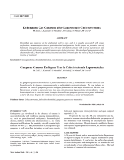
Abdominal Assessment 18 PART ONE STRUCTURE AND FUNCTION ABDOMINAL QUADRANTS ABDOMINAL WALL MUSCLES INTERNAL ANATOMY Solid Viscera Hollow Viscera Vascular Structures Equipment and Supplies Key Assessment Points PHYSICAL ASSESSMENT VALIDATION AND DOCUMENTATION OF FINDINGS Example of Subjective Data Example of Objective Data PART THREE ANALYSIS OF DATA PART TWO NURSING ASSESSMENT COLLECTING SUBJECTIVE DATA NURSING HISTORY Current Symptoms Past History Family History Lifestyle and Health Practices DIAGNOSTIC REASONING: POSSIBLE CONCLUSIONS Selected Nursing Diagnoses Selected Collaborative Problems Medical Problems DIAGNOSTIC REASONING: CASE STUDY Subjective Data Objective Data (YDOXDWLRQ &RS\ COLLECTING OBJECTIVE DATA Client Preparation 367 Structure and Function The abdomen is bordered superiorly by the costal margins, inferiorly by the symphysis pubis and inguinal canals, and laterally by the flanks (Fig. 18-1). To perform an adequate assessment of the abdomen, the nurse needs to understand the anatomic divisions known as the abdominal quadrants, the abdominal wall muscles, and the internal anatomy of the abdominal cavity. PA RT ONE xiphoid process of the sternum to the symphysis pubis (Fig. 18-2). The abdominal wall muscles protect the internal organs and allow normal compression during functional activities such as coughing, sneezing, urination, defecation, and childbirth. Internal Anatomy Abdominal Quadrants The abdomen is divided into four quadrants for purposes of physical examination. These are termed the right upper quadrant (RUQ), right lower quadrant (RLQ), left lower quadrant (LLQ), and left upper quadrant (LUQ). The quadrants are determined by an imaginary vertical line (midline) extending from the tip of the sternum (xiphoid), through the umbilicus to the symphysis pubis. This line is bisected perpendicularly by the lateral line, which runs through the umbilicus across the abdomen. Familiarization with the organs and structures in each quadrant is essential to accurate data collection, interpretation, and documentation of findings (Display 18-1). Another, older method divides the abdomen into nine regions. Three of these regions are still commonly used to describe abdominal findings—epigastric, umbilical, and hypogastric or suprapubic. Abdominal Wall Muscles (YDOXDWLRQ &RS\ The abdominal contents are enclosed externally by the abdominal wall musculature, which includes three layers of muscle extending from the back, around the flanks, to the front. The outermost layer is the external abdominal oblique; the middle layer is the internal abdominal oblique; and the innermost layer is the transverse abdominis. Connective tissue from these muscles extends forward to encase a vertical muscle of the anterior abdominal wall called the rectus abdominis. The fibers and connective tissue extensions of these muscles (aponeuroses) diverge in a characteristic plywood-like pattern (several thin layers arranged at right angles to each other), which provides strength to the abdominal wall. The joining of these muscle fibers and aponeuroses at the midline of the abdomen forms a white line called the linea alba, which extends vertically from the 368 A thin, shiny, serous membrane called the peritoneum lines the abdominal cavity (parietal peritoneum) and also provides a protective covering for most of the internal abdominal organs (visceral peritoneum). Within the abdominal cavity are structures of several different body systems—gastrointestinal, reproductive (female), lymphatic, and urinary. These structures are typically referred to as the abdominal viscera and can be divided into two types—solid viscera and hollow viscera. Solid viscera are those organs that maintain their shape consistently—the liver, pancreas, spleen, adrenal glands, kidneys, ovaries, and uterus. The hollow viscera consist of structures that change shape depending on their contents. These include the stomach, gallbladder, small intestine, colon, and bladder. Palpation of the abdominal viscera depends on location, structural consistency, and size. SOLID VISCERA The liver is the largest solid organ in the body. It is located below the diaphragm in the RUQ of the abdomen. It is composed of four lobes that fill most of the RUQ and extend to the left midclavicular line. In many people, the liver extends just below the right costal margin, where it may be palpated. If palpable, the liver has a soft consistency. The liver functions as an accessory digestive organ and has a variety of metabolic and regulatory functions as well (Fig. 18-3). The pancreas, located mostly behind the stomach, deep in the upper abdomen, is normally not palpable. It is a long gland, extending across the abdomen from the RUQ to the LUQ. The pancreas has two functions. It is an accessory organ of digestion and an endocrine gland. The spleen is approximately 7 cm wide and is located above the left kidney, just below the diaphragm at the level of the ninth, tenth, and eleventh ribs. It is posterior to the left midaxillary line and posterior and lateral to the stomach. CHAPTER 18 | ABDOMINAL ASSESSMENT 369 HOLLOW VISCERA Xyphoid process Right costal margin Right flank Umbilicus Femoral nerve Femoral artery Femoral vein Left costal margin Left flank Anterior superior iliac spine Inguinal canal Inguinal ligament Empty space Inguinal ligament (Poupart's) Symphysis pubis (YDOXDWLRQ &RS\ FIGURE 18-1. Landmarks of the abdomen. This soft, flat structure is normally not palpable. In some healthy clients, the lower tip can be felt below the left costal margin. When the spleen enlarges, the lower tip extends down and toward the midline. The spleen functions primarily to filter the blood of cellular debris, to digest microorganisms, and to return the breakdown products to the liver. The kidneys are located high and deep under the diaphragm. These glandular, bean-shaped organs, measuring approximately 10 × 5 × 2.5 cm, are considered posterior organs and approximate with the level of the T12 to L3 vertebrae. The tops of both kidneys are protected by the posterior rib cage. Kidney tenderness is best assessed at the costovertebral angle (Fig. 18-4). The right kidney is positioned slightly lower because of the position of the liver. Therefore, in some thin clients, the bottom portion of the right kidney may be palpated anteriorly. The primary function of the kidneys is filtration and elimination of metabolic waste products. However, the kidneys also play a role in blood pressure control and maintenance of water, salt, and electrolyte balance. In addition, they function as endocrine glands by secreting hormones. The pregnant uterus may be palpated above the level of the symphysis pubis in the midline. The ovaries are located in the RLQ and LLQ and are normally palpated only during a bimanual examination of the internal genitalia (see Chapter 20). The abdominal cavity begins with the stomach. It is a distensible, flasklike organ located in the LUQ, just below the diaphragm and in between the liver and spleen. The stomach is not usually palpable. The stomach’s main function is to store, churn, and digest food. The gallbladder, a muscular sac approximately 10 cm long, functions primarily to concentrate and store the bile needed to digest fat. It is located near the posterior surface of the liver lateral to the midclavicular line. It is not normally palpated because it is difficult to distinguish between the gallbladder and the liver. The small intestine is actually the longest portion of the digestive tract (approximately 7.0 m long) but is named for its small diameter (approximately 2.5 cm). Two major functions of the small intestine are digestion and absorption of nutrients through millions of mucosal projections lining its walls. The small intestine, which lies coiled in all four quadrants of the abdomen, is not normally palpated. The colon, or large intestine, has a wider diameter than the small intestine (approximately 6.0 cm) and is approximately 1.4 m long. It originates in the RLQ, where it attaches to the small intestine at the ileocecal valve. The colon is composed of three major sections: ascending, transverse, and descending. The ascending colon extends up along the right side of the abdomen. At the junction of the liver in the RUQ, it flexes at a right angle and becomes the transverse colon. The transverse colon runs across the upper abdomen. In the LUQ near the spleen, the colon forms another right angle and then extends downward along the left side of the abdomen as the descending colon. At this point, it curves in toward the midline to form the sigmoid colon in the LLQ. The sigmoid colon is often felt as a firm structure on palpation, whereas the cecum and ascending colon may feel softer. The transverse and descending colon may also be felt on palpation. The colon functions primarily to secrete large amounts of alkaline mucus to lubricate the intestine and neutralize acids formed by the intestinal bacteria. Water is also absorbed through the large intestine, leaving waste products to be eliminated in stool. The urinary bladder, a distensible muscular sac located behind the pubic bone in the midline of the abdomen, functions as a temporary receptacle for urine. A bladder filled with urine may be palpated in the abdomen above the symphysis pubis. VASCULAR STRUCTURES The abdominal organs are supplied with arterial blood by the abdominal aorta and its major branches. Pulsations of the aorta are frequently visible and palpable midline in the upper abdomen. The aorta branches into the right and left iliac arteries just below the umbilicus. Pulsations of the right and left iliac arteries may be felt in the RLQ and LLQ (Fig. 18-5). 370 HEALTH ASSESSMENT IN NURSING DISPLAY 18-1. Locating Abdominal Structures by Quadrants Abdominal assessment findings are commonly allocated to the quadrant in which they are discovered, or their location may be described according to the nine abdominal regions that some healthcare staff may still use as reference marks. Quadrants and contents are listed here with the illustrations of the quadrants and the nine abdominal regions. Right hypochondriac Left hypochondriac Epigastric RUQ LUQ Umbilical RLQ LLQ Right lumbar Hypogastric or suprapubic Right inguinal (YDOXDWLRQ &RS\ Abdominal quadrants (left) and abdominal regions (right). RIGHT UPPER QUADRANT (RUQ) LEFT UPPER QUADRANT (LUQ) Ascending and transverse colon Duodenum Gallbladder Hepatic flexure of colon Liver Pancreas (head) Pylorus (the small bowel—or ileum—traverses all quadrants) Right adrenal gland Right kidney (upper pole) Right ureter Left adrenal gland Left kidney (upper pole) Left ureter Pancreas (body and tail) Spleen Splenic flexure of colon Stomach Transverse ascending colon RIGHT LOWER QUADRANT (RLQ) Appendix Ascending colon Cecum Right kidney (lower pole) Right ovary and tube Right ureter Right spermatic cord LEFT LOWER QUADRANT (LLQ) Left kidney (lower pole) Left ovary and tube Left ureter Left spermatic cord Sigmoid colon MIDLINE Bladder Uterus Prostate gland Left lumbar Left inguinal CHAPTER 18 Linea alba | ABDOMINAL ASSESSMENT 371 Costovertebral angle Internal oblique Tendinous intersections 11th rib Rectus abdominis Transverse abdominis External oblique Aponeurosis of the external oblique FIGURE 18-2. Abdominal wall muscles. Diaphragm 12th rib Kidney FIGURE 18-4. Position of the kidneys. Stomach Liver Gallbladder Spleen Common bile duct (YDOXDWLRQ &RS\ Duodenum Transverse colon Pancreas and pancreatic duct Small intestine Ascending colon Xyphoid process Inferior vena cava Right kidney Right ureter Cecum Vermiform appendix Rectum Descending colon Sigmoid colon Pregnant uterus Full bladder Pancreas Left kidney Aorta Left ureter Sacral promontory Iliac artery and vein Anus FIGURE 18-3. Abdominal viscera. FIGURE 18-5. Abdominal and vascular structures (aorta and iliac artery and vein). Nursing Assessment Collecting Subjective Data Subjective data concerning the abdomen are collected as part of a client’s overall health history interview or as a focused history for a current abdominal complaint. The data focus on symptoms of particular abdominal organs and the function of the digestive system, along with aspects of nutrition, usual bowel habits, and lifestyle. The nurse aims to assure the client that all the questions are important tools for detecting and treating a possible disorder or disease. Keep in mind that the client may be uncomfortable discussing certain issues such as elimination. Asking questions in a matter-of-fact way helps to put the client at ease. In addition, a client experiencing abdominal symptoms may have difficulty describing the nature of the problem. Therefore, the nurse may need to facilitate client responses and quantitative answers by encouraging descriptive terms and examples (ie, pain as sharp or knifelike, headache as throbbing, or back pain as searing), rating scales, and accounts of effects on activities of daily living. PA RT TWO CURRENT SYMPTOMS Abdominal Pain Question Are you experiencing abdominal pain? Rationale Abdominal pain occurs when specific digestive organs or structures are affected by chemical or mechanical factors such as inflammation, infection, distention, stretching, pressure, obstruction, or trauma. Q How would you describe the pain? How bad is the pain (severity) on a scale of 1 to 10, with 10 being the worst? R The quality or character of the pain may suggest its origin (Display 18-2). The client’s perception of pain provides data on his or her response to, and tolerance of, pain. Sensitivity to pain varies greatly among individuals. Sensitivity to pain may diminish with aging. Therefore, elderly patients must be carefully assessed for acute abdominal conditions. Q How did (does) the pain begin? R The onset of pain is a diagnostic clue to its origin. For example, acute pancreatitis produces sudden onset of pain, whereas the pain of pancreatic cancer may be gradual or recurrent. CHARACTER: Describe the sign or symptom. How does (YDOXDWLRQ &RS\ it feel, look, sound, smell, and so forth? ONSET: When did it begin? LOCATION: Where is it? Does it radiate? DURATION: How long does it last? Does it recur? SEVERITY: How bad is it? PATTERN: What makes it better: What makes it worse? ASSOCIATED FACTORS: What other symptoms occur with it? Nursing History During the interview, you will ask the client many questions (Q), examples of which appear below along with the reason (R for rationale) for the question. 372 Q Where is the pain located? Does it move or has it changed from the original location? R Location helps determine the pain source and whether it is primary or referred (see Display 18-2). Q When does the pain occur (timing and relation to particular events, such as eating, exercise, bedtime)? R Timing and the relationship of particular events may be a clue to origin of pain (eg, the pain of a duodenal ulcer may awaken the client at night). Q What seems to bring on the pain (precipitating factors), make it worse (exacerbating factors), or make it better (alleviating factors)? R Various factors can precipitate or exacerbate abdominal pain, such as alcohol ingestion with pancreatitis or supine position with gastroesophageal reflux disease. Lifestyle and stress factors may be implicated in certain digestive disorders CHAPTER 18 | ABDOMINAL ASSESSMENT DISPLAY 18-2. Mechanisms and Sources of Abdominal Pain Abdominal pain may be formally described as visceral, parietal, or referred. KINDS OF PAIN • Visceral pain occurs when hollow abdominal organs, such as the intestines, become distended or • • contract forcefully or when the capsules of solid organs such as the liver and spleen are stretched. Poorly defined or localized and intermittently timed, this type of pain is often characterized as dull, aching, burning, cramping, or colicky. Parietal pain occurs when the parietal peritoneum becomes inflamed, as in appendicitis or peritonitis. This type of pain tends to localize more to the source and is characterized as a more severe and steady pain. Referred pain occurs at distant sites that are innervated at approximately the same levels as the disrupted abdominal organ. This type of pain travels, or refers, from the primary site and becomes highly localized at the distant site. The accompanying illustrations show common clinical patterns and referents of pain. Right upper quadrant or epigastric pain from the gallbladder, biliary tree and liver Appendix pain referred from periumbilical area to RLQ Referred from gallbladder Epigastric pain from the stomach, duodenum, or pancreas Flank pain from the kidney and/or radiating down the lower abdomen from the ureter Referred from pancreas or spleen Referred from pancreas or kidneys Kidney pain Small bowel pain Ureter pain Rectal Suprapubic pain from the rectum, colon, bladder or prostate Periumbilical pain from the small bowel, appendix, or proximal colon Patterns and referents of abdominal pain. (YDOXDWLRQ &RS\ CHARACTER OF ABDOMINAL PAIN AND IMPLICATIONS Dull, Aching Appendicitis Acute hepatitis Biliary colic Cholecystitis Cystitis Dyspepsia Glomerulonephritis Incarcerated or strangulated hernia Irritable bowel syndrome Hepatocellular cancer Pancreatitis Pancreatic cancer Perforated gastric or duodenal ulcer Peritonitis Peptic ulcer disease Prostatitis Prostatitis Urinary retention Burning, Gnawing Dyspepsia Peptic ulcer disease Cramping (“crampy”) Acute gastritis Acute mechanical obstruction Appendicitis Colitis Diverticulitis Gastroesophageal reflux disease (GERD) Colicky Colon cancer Pressure Benign prostatic hypertrophy Prostate cancer Sharp, Knifelike Splenic abscess Splenic rupture Renal colic Renal tumor Ureteral colic Vascular liver tumor Variable Stomach cancer 373 374 HEALTH ASSESSMENT IN NURSING such as peptic ulcer disease. Alleviating factors, such as using antacids or histamine blockers, may be a clue to origin. Significant appetite changes and food intake may adversely affect the client’s weight and put the client at additional risk. Q Is the pain associated with any of the following symp- Older clients may experience a decline in appetite from various factors, such as altered metabolism, decreased taste sensation, decreased mobility, and possibly depression. If appetite declines, the client’s risk for nutritional imbalance increases. toms: nausea, vomiting, diarrhea, constipation, gas, fever, weight loss, fatigue, or yellowing of the eyes or skin? R Associated signs and symptoms may provide diagnostic evidence to support or rule out a particular origin of pain. For example, epigastric pain accompanied by tarry stools suggests a gastric or duodenal ulcer. Indigestion Q Do you experience indigestion? Describe. Does anything in particular seem to cause or aggravate this condition? R Indigestion (pyrosis), often described as heartburn, may be an indication of acute or chronic gastric disorders, including hyperacidity, gastroesophageal reflux disease (GERD), peptic ulcer disease, and stomach cancer. Take time to determine the client’s exact symptoms because many clients call gaseousness, belching, bloating, and nausea indigestion. Certain factors (eg, food, drinks, alcohol, medications, stress) are known to increase gastric secretion and acidity and cause or aggravate indigestion. Bowel Elimination Q Have you experienced a change in bowel elimination patterns? Describe. R Changes in bowel patterns must be compared to usual patterns for the client. Normal frequency varies from two to three times per day to three times per week. Q Do you have constipation? Describe. Do you have any accompanying symptoms? R Constipation is usually defined as a decrease in the frequency of bowel movements or the passage of hard and possibly painful stools. Signs and symptoms that accompany constipation may be a clue as to the cause of constipation, such as bleeding with malignancies or pencil-shaped stools with intestinal obstruction. Nausea and Vomiting Q Have you experienced diarrhea? Describe. Do you have Q Do you experience nausea? Describe. Is it triggered by any accompanying symptoms? any particular activities, events, or other factors? R R Nausea may reflect gastric dysfunction and is also associated with many digestive disorders and diseases of the accessory organs, such as the liver and pancreas, as well as with renal failure and drug intolerance. Nausea may also be precipitated by dietary intolerance, psychological triggers, or menstruation. Nausea may also occur at particular times such as early in the day with some pregnant clients (“morning sickness”), after meals with gastric disorders, or between meals with changes in blood glucose levels. (YDOXDWLRQ &RS\ Q Have you been vomiting? Describe the vomitus. Is it associated with any particular trigger factors? R Vomiting is associated with impaired gastric motility or reflex mechanisms. Description of vomitus (emesis) is a clue to the source. For example, bright hematemesis is seen with bleeding esophageal varices and ulcers of the stomach or duodenum. Elderly or neuromuscular- or consciousnessimpaired clients are at risk for lung aspiration with vomiting. Q Have Diarrhea is defined as frequency of bowel movements producing unformed or liquid stools. It is important to compare these stools to the client’s usual bowel patterns. Bloody and mucoid stools are associated with inflammatory bowel diseases (eg, ulcerative colitis, Crohn’s disease); clay-colored, fatty stools may be from malabsorption syndromes. Associated symptoms or signs may suggest the disorder’s origin. For example, fever and chills may result from an infection, or weight loss and fatigue may result from a chronic intestinal disorder or a cancer. Older clients are especially at risk for potential complications with diarrhea, such as fluid volume deficit, dehydration, electrolyte, and acid–base imbalances, because they have a higher fat-to-lean muscle ratio. Q Have you experienced any yellowing of your skin or whites of your eyes, itchy skin, dark urine (yellow-brown or tea colored), or clay-colored stools? R These symptoms should be evaluated to rule out possible liver disease. you noticed a change in your appetite? Has this change affected how much you eat or your normal weight? PAST HISTORY R Q Have you ever had any of the following gastrointestinal Loss of appetite (anorexia) is a general complaint often associated with digestive disorders, chronic syndromes, cancers, and psychological disorders. Appetite changes should be carefully correlated with dietary history and weight monitoring. disorders: Ulcers, gastroesophageal reflux, inflammatory or obstructive bowel disease, pancreatitis, gallbladder or liver disease, diverticulosis, or appendicitis? CHAPTER 18 375 R Presenting the client with a list of the more common disorders may help the client identify any that he or she has or has had. as the stomach, pancreas, and liver. Alcohol-related disorders include gastritis, esophageal varices, pancreatitis, and liver cirrhosis. Q Have you had any urinary tract disease such as infec- Q What types of foods and how much food do you typi- tions, kidney disease or nephritis, or kidney stones? R cally consume each day? How much noncaffeinated fluid do you consume each day? How much caffeine do you think you consume each day (eg, in tea, coffee, chocolate, and soft drinks)? Urinary tract infections may become recurrent and chronic. Moreover, resistance to drugs used to treat infection must be evaluated. Chronic kidney infection may lead to permanent kidney damage. R Older clients are prone to urinary tract infections because the activity of protective bacteria in the urinary tract declines with age. Q How much and how often do you exercise? Describe Q Have you ever had viral hepatitis (type A, B, or C)? Have you ever been exposed to viral hepatitis? A baseline dietary and fluid survey helps determine nutritional and fluid adequacy and risk factors for altered nutrition, constipation, diarrhea, and diseases such as cancer. your activities during the day. R R Various populations (eg, school and health care personnel) are at increased risk for exposure to hepatitis viruses. Any type of viral hepatitis may cause liver damage. Regular exercise promotes peristalsis and thus regular bowel movements. In addition, exercise may help reduce risk factors for various diseases, such as cancer and hypertension (see Risk Factors—Gallbladder Cancer). Q Have you ever had abdominal surgery or other trauma Q What kind of stress do you have in your life? How does to the abdomen? it affect your eating or elimination habits? R R Q What prescription or over-the-counter medications do Q If you have a gastrointestinal disorder, how does it affect Prior abdominal surgery or trauma may cause abdominal adhesions, thereby predisposing the client to future complications or disorders. you take? R Medications may produce side effects that adversely affect the gastrointestinal tract. For example, aspirin, ibuprofen, and steroids may cause gastric bleeding. Chronic use of antacids or histamine-2 blockers may mask the symptoms of more serious stomach disorders. Overuse of laxatives may decrease intestinal tone and promote dependency. High iron intake may lead to chronic constipation. FAMILY HISTORY (YDOXDWLRQ &RS\ | ABDOMINAL ASSESSMENT Q Is there a history of any of the following diseases or disorders in your family: colon, stomach, pancreatic, liver, kidney, or bladder cancer; liver disease; gallbladder disease; kidney disease? R Family history of certain disorders increases the client’s risk for those disorders. Genetic testing can now identify the risk for certain cancers (colon, pancreatic, and prostate) and other diseases. Client awareness of family history can serve as a motivation for health screening and positive health promotion behaviors. LIFESTYLE AND HEALTH PRACTICES Q Do you drink alcohol? How much? How often? R Alcohol ingestion can affect the gastrointestinal tract through immediate and long-term effects on such organs Lifestyle and associated stress and psychological factors can affect gastrointestinal function through effects on secretion, tone, and motility. your lifestyle and how you feel about yourself? R Certain gastrointestinal disorders and their effects (eg, weight loss) or treatment (eg, drugs, surgery) may produce physiologic or anatomic effects that affect the client’s perception of self, body image, social interaction and intimacy, and life goals and expectations. Collecting Objective Data The abdominal examination is performed for a variety of different reasons: as part of a comprehensive health examination; to explore gastrointestinal complaints; to assess abdominal pain, tenderness, or masses; or to monitor the client postoperatively. Assessing the abdomen can be challenging, considering the number of organs of the digestive system and the need to distinguish the source of clinical signs and symptoms. The sequence for assessment of the abdomen differs from the typical order of assessment. Auscultate after you inspect so as not to alter the client’s pattern of bowel sounds. Percussion and then palpation follow auscultation. Adjust the bed level as necessary throughout the examination, and approach the client from the right side. Use tangential lighting, if available, for optimal visualization of the abdomen. The nurse needs to understand and anticipate various concerns of the client by listening and observing closely for 376 HEALTH ASSESSMENT IN NURSING RISK FACTORS Gallbladder Cancer OVERVIEW (YDOXDWLRQ &RS\ Of the several types of tumors that affect the gallbladder, about 80% are adenocarcinomas (ACS, 2000). The American Cancer Society reports that gallbladder cancer is the fifth most common gastrointestinal cancer and that between 5000 and 7000 new cases are diagnosed each year in the United States. Only 10% of patients survive 5 years due to late discovery after the cancer has advanced. Gallbladder cancer is described as “an age-dependent malignancy that is present mostly in women and that may be intimately associated with long-standing benign gall stone disease of the gall bladder“ (Vitetta, Sali, Little & Mrazeh, 2000). Women are affected two and one half times as often as men. Gallstones are the most common risk factor, especially when onset is at or before middle age or when there is one large stone. Many risk factors for gallbladder cancer are associated with gallstones, including high parity, obesity, and abnormalities of the biliary system promoting chronic inflammation. RISK FACTORS RISK REDUCTION TEACHING TIPS Age (over 70 years) Female Sex After menopause (Khan et al., 1999), Increased parity (Lowenfels et al., 1999) Ethnicity Native American (United States; North, Central, and South America) New Zealand Maori (Lowenfels et al., 1999) Health and Habit History Gallstones, especially early onset or large gallstone Chronic inflammation of gallbladder Porcelain gallbladder (calcium deposits) Gallbladder polyps Common bile duct abnormalities Typhoid carrier Obesity Diet high in carbohydrates and fats and low in fiber Cigarette smoking Environmental exposure to industrial chemicals used in rubber and metal manufacturing • • • • Stop smoking. Avoid obesity. Follow ACS diet and exercise suggestions. Diet consists of five servings of fruits and vegetables; six servings of bread, grains, and legumes; and fewer fatty foods. Exercise consists of 30 minutes most days. Avoid industrial chemical exposure. Schedule periodic medical examinations to assess gallbladder and general status, especially if a history of risk factors is identified. CULTURAL CONSIDERATIONS Gallbladder disease and cancer rates differ among ethnic groups. Native American populations have much higher rates than most world populations. Reports form the mid 1980s note that the risk for gallbladder cancer in African American women was 3 per 100,000; for white women, 11.5; and for Native American women 46.4 (Overfield, 1995, p. 110). The pattern for gallbladder disease has been noted to be similar, with 36% of Pima Indians being admitted to the hospital with gallbladder disease as compared to 6% of whites in Massachusetts (Comess, Bennet, & Burch, 1967; quoted in Overfield, 1995, p. 112). In addition to Native Americans in North Central and South America, the New Zealand Maori have a high rate as well (Lowenfels et al., 1999). CHAPTER 18 verbal and nonverbal cues. Commonly, clients feel anxious and modest during the examination, possibly from anticipated discomfort or fear that the examiner will find something seriously wrong. As a result, the client may tense the abdominal muscles, voluntarily guarding the area. (Tips for minimizing voluntary guarding appear in Display 18-3.) Explaining each aspect of the examination, answering the client’s questions, and draping the client’s genital area and breasts (in women) when these are not being examined all help to ease anxiety. Another potential factor to deal with is ticklishness. A ticklish client has trouble lying still and relaxing during the hands-on parts of the examination. Try to combat this using a controlled hands-on technique and by placing the client’s hand under your own for a few moments at the beginning of palpation. Finally, warm hands are essential for the abdominal examination. Cold hands cause the client to tense the abdominal muscles. Rubbing them together or holding them under warm water just before the hands-on examination may be helpful. | ABDOMINAL ASSESSMENT 377 the client’s knees to help relax the abdominal muscles. Drape the client with sheets so the abdomen is visible from the lower rib cage to the pubic area. Instruct the client to breathe through the mouth and to take slow, deep breaths; this promotes relaxation. Before touching the abdomen, ask the client about painful or tender areas. These areas should always be assessed at the end of the examination. Reassure the client that you will forewarn him or her when you will examine these areas. Approach the client with slow, gentle, and fluid movements. EQUIPMENT AND SUPPLIES • • • • Small pillow or rolled blanket Centimeter ruler Stethoscope (with a warm diaphragm and bell) Marking pen KEY ASSESSMENT POINTS • Observe and inspect abdominal skin and overall contour CLIENT PREPARATION Ask the client to empty the bladder before beginning the examination to eliminate bladder distention and interference with an accurate examination. Instruct the client to remove clothes and to put on a gown if desired. Help the client to lie supine with the arms folded across the chest or resting by the sides (Fig. 18-6). Raising arms above the head or folding them behind the head will tense the abdominal muscles. A flat pillow may be placed under the client’s head for comfort. Slightly flex the client’s legs by placing a pillow or rolled blanket under and symmetry. • Auscultate after inspection and before percussion, and, finally, palpate. • Assessment examination evaluates the following abdominal • structures in the abdominal quadrants: skin, stomach, bowel, spleen, liver, kidneys, aorta, and bladder. Common abnormal findings include abdominal edema, or swelling, signifying ascites; abdominal masses signifying abnormal growths or constipation; unusual pulsations, such as those seen with an aneurysm of the abdominal aorta; and pain associated with appendicitis. (text continues on page 31) (YDOXDWLRQ &RS\ DISPLAY 18-3. Palpating the Abdomen GUIDELINES 1. Avoid touching tender or painful areas until last, and reassure the client of your intentions. 2. Perform light palpation before deep palpation to detect tenderness and superficial masses. 3. Keep in mind that the normal abdomen may be tender, especially in the areas over the xiphoid process, liver, aorta, lower pole of the kidney, gas-filled cecum, sigmoid colon, and ovaries. 4. Overcome ticklishness and minimize voluntary guarding by asking the client to perform selfpalpation. Place your hands over the client’s. After a while, let your fingers glide slowly onto the abdomen while still resting mostly on the client’s fingers. The same can be done by using a warm stethoscope as a palpating instrument, again letting your fingers drift over the edge of the diaphragm and palpate without promoting a ticklish response. 5. Work with the client to promote relaxation and minimize voluntary guarding. Use the following techniques: Place a pillow under the client’s knees. Ask the client to take slow, deep breaths through the mouth. Apply light pressure over the client’s sternum with your left hand while palpating with the right. This encourages the client to relax the abdominal muscles during breathing against sternal resistance. • • • 378 HEALTH ASSESSMENT IN NURSING FIGURE 18-6. Two positions are appropriate for the abdominal assessment. The client may lie supine with hands resting on the center of the chest (left) or with arms resting comfortably at the sides (right). These positions best promote relaxation of the abdominal muscles. (Photos courtesy of M. B. Cunningham.) PHYSICAL ASSESSMENT ASSESSMENT PROCEDURE NORMAL FINDINGS ABNORMAL FINDINGS Observe the coloration of the skin. Abdominal skin may be paler than the general skin tone because this skin is so seldom exposed to the natural elements. Purple discoloration at the flanks (Grey Turner sign) indicates bleeding within the abdominal wall, possibly from trauma to the kidneys, pancreas, or duodenum or from pancreatitis. The yellow hue of jaundice may be more apparent on the abdomen. Pale, taut skin may be seen with ascites (significant abdominal swelling indicating fluid accumulation in the abdominal cavity). Redness may indicate inflammation. Bruises or areas of local discoloration are also abnormal. Note the vascularity of the abdominal skin. Scattered fine veins may be visible. Blood in the veins located above the umbilicus flows toward the head; blood in the veins located below the umbilicus flows toward the lower body. Dilated veins may be seen with cirrhosis of the liver, obstruction of the inferior vena cava, portal hypertension, or ascites. Dilated surface arterioles and capillaries with a central star (spider angioma) may be seen with liver disease or portal hypertension. ABDOMEN (YDOXDWLRQ &RS\ Inspect the Skin Dilated superficial capillaries without a pattern may be seen in older clients. They are more visible in sunlight. (continued ) CHAPTER 18 ASSESSMENT PROCEDURE | ABDOMINAL ASSESSMENT 379 NORMAL FINDINGS ABNORMAL FINDINGS Note any striae. Old, silvery, white striae or stretch marks from past pregnancies or weight gain are normal. Dark bluish-pink striae are associated with Cushing’s syndrome. Striae may also be caused by ascites, which stretches the skin. Ascites usually results from liver failure or liver disease. Inspect for scars. Ask about the source of a scar, and use a centimeter ruler to measure the scar’s length. Document the location by quadrant and reference lines, shape, length, and any specific characteristics (eg, 3-cm vertical scar in RLQ 4 cm below the umbilicus and 5 cm left of the midline). With experience, many examiners can estimate the length of a scar visually without a ruler. Pale, smooth, minimally raised old scars may be seen. Nonhealing scars, redness, inflammation. Deep, irregular scars may result from burns. Tip From the Experts Scarring should be an alert for possible internal adhesions. Keloids (excess scar tissue) result from trauma or surgery and are more common in blacks and Asians. Keloid beyond border of surgical scar. Abdomen is free of lesions or rashes. Flat or raised brown moles, however, are normal and may be apparent. Changes in moles including size, color, and border symmetry. Any bleeding moles or petechiae (reddish or purple lesions) may also be abnormal (see Chapter 9). Note the color of the umbilical area. Umbilical skin tones are similar to surrounding abdominal skin tones or even pinkish. Bluish or purple discoloration around the umbilicus (Cullen’s sign) indicates intra-abdominal bleeding. Observe umbilical location. Midline at lateral line A deviated umbilicus may be caused by pressure from a mass, enlarged organs, hernia, fluid, or scar tissue. Look for lesions and rashes. (YDOXDWLRQ &RS\ Inspect the Umbilicus (continued ) 380 HEALTH ASSESSMENT IN NURSING ASSESSMENT PROCEDURE Assess contour of umbilicus. NORMAL FINDINGS ABNORMAL FINDINGS Recessed (inverted) or protruding no more than 0.5 cm; round or conical An everted umbilicus is seen with abdominal distention (see Display 18-5). An enlarged, everted umbilicus suggests umbilical hernia (see Diplay 18-6). Inspect Contour, Symmetry, Movement To inspect abdominal contour, look across the abdomen at eye level from the client’s right side, from behind the client’s head, and from the foot of the bed. Abdomen is flat, rounded, or scaphoid (usually seen in thin adults). Abdomen should be evenly rounded. Flat Scaphoid (may be abnormal) Rounded Distended/protuberant (usually abnormal) View abdominal contour from the client’s side. Many abdomens are more or less flat; and many are round, scaphoid, or distended. (© B. Proud.) Tip From the Experts The major To assess abdominal symmetry, look at the client’s abdomen as he or she lies in a relaxed supine position. Abdomen is symmetric. To further assess the abdomen for herniation or diastasis recti, or to differentiate a mass within the abdominal wall from one below it, ask the client to raise the head. Abdomen does not bulge when client raises head. (YDOXDWLRQ &RS\ A generalized protuberant or distended abdomen may be due to air (gas) or fluid accumulation (see Display 18-5). Distention below the umbilicus may be due to a full bladder, uterine enlargement, or an ovarian tumor or cyst. Distention of the upper abdomen may be seen with masses of the pancreas or gastric dilation. causes of abdominal distention are sometimes referred to as the “6 Fs”: Fat, feces, fetus, fibroids, flatulence, and fluid (Display 18-5). A scaphoid (sunken) abdomen may be seen with severe weight loss or cachexia related to starvation or terminal illness. Asymmetry may be seen with organ enlargement, large masses, hernia, diastasis recti, or bowel obstruction. A hernia (protrusion of the bowel through the abdominal wall) is seen as a bulging in the abdominal wall. Diastasis recti appears as a bulging between a vertical midline separation of the abdominis rectus muscles. This condition is of little significance. An incisional hernia may occur when a defect develops in the abdominal muscles because of a surgical incision. A mass within the abdominal wall is more prominent when the head is raised, whereas a mass below the abdominal wall is obscured (Display 18-6). (continued ) CHAPTER 18 ASSESSMENT PROCEDURE | ABDOMINAL ASSESSMENT 381 NORMAL FINDINGS ABNORMAL FINDINGS Inspect abdominal movement when the client breathes (respiratory movements). Abdominal respiratory movement may be seen, especially in male clients. Diminished abdominal respiration or change to thoracic breathing in male clients may reflect peritoneal irritation. Observe aortic pulsations. A slight pulsation of the abdominal aorta, which is visible in the epigastrium, extends full length in thin people. Vigorous, wide, exaggerated pulsations may be seen with abdominal aortic aneurysm. Watch for peristaltic waves. Normally, peristaltic waves are not seen, although they may be visible in very thin people as slight ripples on the abdominal wall. Peristaltic waves are increased and progress in a ripple-like fashion from the LUQ to the RLQ with intestinal obstruction (especially small intestine). In addition, abdominal distention typically is present with intestinal wall obstruction. A series of intermittent, soft clicks and gurgles are heard at a rate of 5 to 30 per minute. Hyperactive bowel sounds that may be heard normally are the loud, prolonged gurgles characteristic of stomach growling. These hyperactive bowel sounds are called “borborygmi.” Hypoactive bowel sounds indicate diminished bowel motility. Common causes include abdominal surgery or late bowel obstruction. Hyperactive bowel sounds indicate increased bowel motility. Common causes include diarrhea, gastroenteritis, or early bowel obstruction. Decreased or absent bowel sounds signify the absence of bowel motility, which constitutes an emergency requiring immediate referral. Absent bowel sounds may be associated with peritonitis or paralytic ileus. High-pitched tinkling and rushes of high-pitched sounds with abdominal cramping usually indicate obstruction. Auscultate for Bowel Sounds Follow the guidelines for auscultating bowel sounds in Display 18-7. Note the intensity, pitch, and frequency of the sounds. Tip From the Experts Postoperatively, bowel sounds resume gradually depending on the type of surgery. The small intestine functions normally in the first few hours postoperatively; stomach emptying takes 24 to 48 hours to recover; and the colon requires 3 to 5 days to recover propulsive activity. Tip From the Experts The increasing (YDOXDWLRQ &RS\ pitch of bowel sounds is most diagnostic of obstruction because it signifies intestinal distention. (continued ) 382 HEALTH ASSESSMENT IN NURSING ASSESSMENT PROCEDURE NORMAL FINDINGS ABNORMAL FINDINGS Bruits are not normally heard over abdominal aorta or renal, iliac, or femoral arteries. However, bruits confined to systole may be normal in some clients depending on other differentiating factors. A bruit with both systolic and diastolic components occurs when blood flow in an artery is turbulent or obstructed. This usually indicates aneurysm or arterial stenosis. If the client has hypertension and you auscultate a renal artery bruit with both systolic and diastolic components, suspect renal artery stenosis as the cause. Venous hum is not normally heard over the epigastric and umbilical areas. Venous hums are rare. However, an accentuated venous hum heard in the epigastric or umbilical areas suggests increased collateral circulation between the portal and systemic venous systems, as in cirrhosis of the liver. Auscultate for Vascular Sounds and Friction Rubs Use the bell of the stethoscope to listen for bruits (lowpitched, murmurlike sound) over the abdominal aorta and renal, iliac, and femoral arteries. Tip From the Experts Auscultating for vascular sounds is especially important if the client has hypertension or if you suspect arterial insufficiency to the legs. Using the bell of the stethoscope, listen for a venous hum in the epigastric and umbilical areas. Hepatic friction rub Splenic friction rub Aorta Renal artery Venous hum Iliac artery (YDOXDWLRQ &RS\ Femoral artery Vascular sounds and friction rubs can best be heard over these areas. Auscultate for a friction rub over the liver and spleen by listening over the right and left lower rib cage with the diaphragm of the stethoscope. No friction rub over liver or spleen. Friction rubs are rare. If heard, they have a high-pitched, rough, grating sound produced when the large surface area of the liver or spleen rubs the peritoneum. They are heard in association with respiration. A friction rub heard over the lower right costal area is associated with hepatic abscess or metastases. A rub heard at the anterior axillary line in the lower left costal area is associated with splenic infarction, abscess, infection, or tumor. (continued ) CHAPTER 18 ASSESSMENT PROCEDURE | ABDOMINAL ASSESSMENT 383 NORMAL FINDINGS ABNORMAL FINDINGS Generalized tympany predominates over the abdomen because of air in the stomach and intestines. Normal dullness is heard over the liver and spleen. Dullness may also be elicited over a nonevacuated descending colon. Accentuated tympany or hyperresonance is heard over a gaseous distended abdomen. An enlarged area of dullness is heard over an enlarged liver or spleen. Abnormal dullness is heard over a distended bladder, large masses, or ascites. If you suspect ascites, perform the shifting dullness and fluid wave tests. These special techniques are described later. Percuss for Tone Lightly and systematically percuss all quadrants. Two sequences are illustrated below. Start Abdominal percussion sequences may proceed clockwise or up and down over the abdomen. Liver Spleen Bowel Stomach (gastric air bubble) Sigmoid Document normal findings after percussing the liver, bowel, sigmoid colon, stomach, and spleen. Percuss the Liver (YDOXDWLRQ &RS\ Percuss the span or height of the liver by determining its lower and upper borders. To assess the lower border, begin in the RLQ at the mid-clavicular line (MCL) and percuss upward. Note the change from tympany to dullness. Mark this point—it is the lower border of liver dullness. The lower border of liver dullness is located at the costal margin to 1 to 2 cm below. Tip From the Experts If you cannot find the lower border of the liver, keep in mind that the lower border of liver dullness may be difficult to estimate when obscured by intestinal gas. Begin liver percussion in the RLQ and percuss upward toward the chest. (© B. Proud.) (continued ) 384 HEALTH ASSESSMENT IN NURSING ASSESSMENT PROCEDURE NORMAL FINDINGS To assess the descent of the liver, ask the client to take a deep breath, then repeat the procedure. To assess the upper border, percuss over the upper right chest at the MCL and percuss downward, noting the change from lung resonance to liver dullness. Mark this point—it is the upper border of liver dullness. On deep inspiration, the lower border of liver dullness may descend from 1 to 4 cm below the costal margin. The upper border of liver dullness is located between the left fifth and seventh intercostal spaces. Measure the distance between the two marks—this is the span of the liver. The normal liver span at the MCL is 6 to 12 cm (greater in men and taller clients, less in shorter clients). Normally, liver size decreases after age 50. ABNORMAL FINDINGS Tip From the Experts The upper border of liver dullness may be difficult to estimate if obscured by pleural fluid or lung consolidation. Hepatomegaly, a liver span that exceeds normal limits (enlarged), is characteristic of liver tumors, cirrhosis, abscess, and vascular engorgement. Atrophy of the liver is indicated by a decreased span. A liver in a lower position than normal may be caused by emphysema, whereas a liver in a higher position than normal may be caused by an abdominal mass, ascites, or a paralyzed diaphragm. A liver in a lower or higher position should have a normal span (Display 18-8). (YDOXDWLRQ &RS\ 4–8 cm MSL 6–12 cm MCL The distance between the liver’s lower and upper border is denoted by a span of percussed dullness. (Left) Determine this by marking the distance from beginning to end of percussion and measuring with a ruler. (Right) Normal liver span. Repeat percussion of the liver at the midsternal line (MSL). The normal liver span at the MSL is 4 to 8 cm. (continued ) CHAPTER 18 ASSESSMENT PROCEDURE | ABDOMINAL ASSESSMENT 385 NORMAL FINDINGS ABNORMAL FINDINGS The sound produced by scratching becomes more intense over the liver. An enlarged liver may be roughly estimated (not accurately) when more intense sounds outline a liver span or borders outside the normal range. The spleen is an oval area of dullness approximately 7 cm wide near the left tenth rib, and slightly posterior to the MAL. Splenomegaly is characterized by an area of dullness greater than 7 cm wide. The enlargement may result from traumatic injury, portal hypertension, and mononucleosis. Normally, tympany (or resonance) is heard at the last left interspace. On inspiration, dullness at the last left interspace at the AAL suggests an enlarged spleen (see Display 18-8). Perform the Scratch Test If you cannot accurately percuss the liver borders, perform the scratch test. Auscultate over the liver and, starting in the RLQ, scratch lightly over the abdomen, progressing upward toward the liver. The scratch test. Percuss the Spleen Begin posterior to the left mid-axillary line (MAL), and percuss downward, noting the change from lung resonance to splenic dullness. Tip From the Experts Results of splenic percussion may be obscured by air in the stomach or bowel. A second method for detecting splenic enlargement is to percuss the last left interspace at the anterior axillary line (AAL) while the client takes a deep breath. Tip From the Experts Other sources of dullness (YDOXDWLRQ &RS\ (eg, full stomach or feces in the colon) must be ruled out before confirming splenomegaly. Percuss last interspace: normally tympanic (or resonant) Anterior axillary line Midaxillary line Dull tone over spleen (9th–11th ribs) Last left interspace at the anterior axillary line. (continued ) 386 HEALTH ASSESSMENT IN NURSING ASSESSMENT PROCEDURE NORMAL FINDINGS ABNORMAL FINDINGS Perform Blunt Percussion on the Liver and Kidneys To assess for tenderness in difficult-to-palpate structures, perform blunt (indirect fist) percussion. Percuss the liver by placing your left hand flat against the lower right rib cage. Use the ulnar side of your right fist to strike your left hand. Normally, no tenderness is elicited. Tenderness elicited over the liver may be associated with inflammation or infection (eg, hepatitis or cholecystitis). Perform blunt percussion on the kidneys at the costovertebral angles (CVA) over the twelfth rib. Normally, no tenderness or pain is elicited or reported by the client. The examiner senses only a dull thud. Tenderness or sharp pain elicited over the CVA suggests kidney infection (pyelonephritis), renal calculi, or hydronephrosis. Tip From the Experts This technique requires that the client is sitting with his or her back to you. Therefore, it may be best to incorporate blunt percussion of the kidneys with your thoracic assessment because the client will already be in this position. Performing blunt percussion over the kidney. (© B. Proud.) Perform Light Palpation (YDOXDWLRQ &RS\ Follow the guidelines for palpating the abdomen (see Display 18-3), and palpate lightly in all quadrants. Light palpation is used to identify areas of tenderness and muscular resistance. Using the fingertips, begin palpation in a nontender quadrant, and compress to a depth of 1 cm in a dipping motion. Then, gently lift the fingers and move to the next area. Nontender Performing light palpation. (© B. Proud.) (continued ) CHAPTER 18 ASSESSMENT PROCEDURE To minimize the client’s voluntary guarding (a tensing or rigidity of the abdominal muscles, usually involving the entire abdomen), see Display 18-3. Keep in mind that the rectus abdominis muscle relaxes on expiration. | ABDOMINAL ASSESSMENT 387 NORMAL FINDINGS ABNORMAL FINDINGS No guarding; abdomen is soft. Involuntary reflex guarding is serious and reflects peritoneal irritation. The abdomen is rigid and the rectus muscle fails to relax with palpation when the client exhales. It can involve all or part of the abdomen but is usually seen on the side (ie, right vs left, rather than upper or lower) because of nerve tract patterns. Right-sided guarding may be due to cholecystitis. Normal (mild) tenderness is possible over the xiphoid, aorta, cecum, sigmoid colon, and ovaries with deep palpation. Severe tenderness or pain may be related to trauma, peritonitis, infection, tumors, or enlarged or diseased organs. No palpable masses A mass detected in any quadrant may be due to a tumor, cyst, abscess, enlarged organ, aneurysm, or adhesions. Perform Deep Palpation Deeply palpate all quadrants to delineate abdominal organs and detect subtle masses. Using the palmar surface of the fingers, compress to a maximum depth, (5 to 6 cm). Perform bimanual palpation if you encounter resistance or to assess deeper structures. Performing deep bimanual palpation. (YDOXDWLRQ &RS\ Palpate for masses and their location, size (cm), shape, consistency, demarcation, pulsatility, tenderness, and mobility. Do not confuse a mass with a normally palpated organ or structure. Xyphoid process Pulsatile aorta Normal liver edge Right kidney, lower pole Cecum/ ascending colon Rectus muscles, lateral border Pregnant uterus Sacral promontory Sigmoid colon Full bladder Normally palpable structures in the abdomen. (continued ) 388 HEALTH ASSESSMENT IN NURSING ASSESSMENT PROCEDURE NORMAL FINDINGS ABNORMAL FINDINGS Umbilicus and surrounding area are free of swellings, bulges, or masses. A soft center of the umbilicus can be a potential for herniation. Palpation of a hard nodule in or around the umbilicus may indicate metastatic nodes from an occult gastrointestinal cancer. The normal aorta is approximately 2.5 to 3.0 cm wide with a moderately strong and regular pulse. Possibly, mild tenderness may be elicited. A wide, bounding pulse may be felt with an abdominal aortic aneurysm. A prominent, laterally pulsating mass above the umbilicus, with an accompanying audible bruit, strongly suggests an aortic aneurysm (see Display 18-8). Palpate the Umbilicus Palpate the umbilicus and surrounding area for swellings, bulges, or masses. Palpate the Aorta Use your thumb and first finger or use two hands and palpate deeply in the epigastrium, slightly to the left of midline. Assess the pulsation of the abdominal aorta. If the client is older than age 50 or has hypertension, assess the width of the aorta. Palpating the aorta. (© B. Proud.) Tip From the Experts Do not palpate a pul- (YDOXDWLRQ &RS\ sating midline mass; it may be a dissecting aneurysm that can rupture from the pressure of palpation. Also avoid deep palpation over tender organs as in the case of polycystic kidneys, Wilms’ tumor, transplantation, or suspected splenic trauma. (continued ) CHAPTER 18 ASSESSMENT PROCEDURE | ABDOMINAL ASSESSMENT 389 NORMAL FINDINGS ABNORMAL FINDINGS The liver is usually not palpable, although it may be felt in some thin clients. If the lower edge is felt, it should be firm, smooth, and even. Mild tenderness may be normal. A hard, firm liver may indicate cancer. Nodularity may occur with tumors, metastatic cancer, late cirrhosis, or syphilis. Tenderness may be from vascular engorgement (eg, congestive heart failure), acute hepatitis, or abscess. A liver more than 1 to 3 cm below the costal margin is considered enlarged (unless pressed down by the diaphragm). Enlargement may be due to hepatitis, liver tumors, cirrhosis, and vascular engorgement. The spleen is seldom palpable at the left costal margin; rarely, the tip is palpable in the presence of a low, flat diaphragm (eg, chronic obstructive lung disease) or with deep diaphragmatic descent on inspiration. A palpable spleen suggests enlargement (up to three times the normal size), which may result from trauma, mononucleosis, chronic blood disorders, and cancers. The splenic notch may be felt, which is an indication of splenic enlargement. Palpate the Liver Palpate to note consistency and tenderness. To palpate bimanually, stand at the client’s right side and place your left hand under the client’s back at the level of the eleventh to twelfth ribs. Lay your right hand parallel to the right costal margin (your fingertips should point toward the client’s head). Ask the client to inhale, then compress upward and inward with your fingers. To palpate by hooking, stand to the right of the client’s chest. Curl (hook) the fingers of both hands over the edge of the right costal margin. Ask the client to take a deep breath, and gently, but firmly, pull inward and upward with your fingers. Hooking technique for liver palpation. Palpate the Spleen (YDOXDWLRQ &RS\ Stand at the client’s right side, reach over the abdomen with your left arm and place your hand under the posterior lower ribs. Pull up gently. Place your right hand below the left costal margin with the fingers pointing toward the client’s head. Ask the client to inhale, and press inward and upward as you provide support with your other hand. Tip From the Experts Caution: To avoid traumatizing and possibly rupturing the organ, be gentle when palpating an enlarged spleen. Palpating the spleen. (continued ) 390 HEALTH ASSESSMENT IN NURSING ASSESSMENT PROCEDURE Alternatively, asking the client to turn onto the right side may facilitate splenic palpation by moving the spleen downward and forward. Document the size of the spleen in centimeters below the left costal margin. Also note consistency and tenderness. NORMAL FINDINGS ABNORMAL FINDINGS If the edge of the spleen can be palpated, it should be soft and nontender. The spleen feels soft with a rounded edge when it is enlarged from infection. It feels firm with a sharp edge when it is enlarged from chronic disease. Tenderness accompanied by peritoneal inflammation or capsular stretching is associated with splenic enlargement. The kidneys are normally not palpable. Sometimes, the lower pole of the right kidney may be palpable by the capture method because of its lower position. If palpated, it should feel firm, smooth, and rounded. The kidney may or may not be slightly tender. An enlarged kidney may be due to a cyst, tumor, or hydronephrosis. It can be differentiated from splenomegaly by its smooth rather than sharp edge, absence of a notch, and overlying tympany on percussion (see Display 18-8). Umbilicus Palpating the spleen with the client in side-lying position. Tip From the Experts Be sure to palpate with your fingers below the costal margin so you do not miss the lower edge of an enlarged spleen Palpate the Kidneys (YDOXDWLRQ &RS\ To palpate the right kidney, support the right posterior flank with your left hand, and place your right hand in the RUQ just below the costal margin at the MCL. To capture the kidney, ask the client to inhale. Then, compress your fingers deeply during peak inspiration. Ask the client to exhale and hold the breath briefly. Gradually release the pressure of your right hand. If you have captured the kidney, you will feel it slip beneath your fingers. To palpate the left kidney, reverse the procedure. A B C Palpating (A) the right kidney and (B, C ) the left kidney. (continued ) CHAPTER 18 ASSESSMENT PROCEDURE | ABDOMINAL ASSESSMENT 391 NORMAL FINDINGS ABNORMAL FINDINGS Normally not palpable A distended bladder is palpated as a smooth, round, and somewhat firm mass, extending as far as the umbilicus. It may be further validated by dull percussion tones. Palpate the Urinary Bladder Palpate for a distended bladder when the client’s history or other findings warrant (eg, dull percussion noted over the symphysis pubis). Begin at the symphysis pubis, and move upward and outward to estimate bladder borders. Kidneys Ureter Distended Empty bladder (YDOXDWLRQ &RS\ Palpating distended bladder (larger dotted line is area of distention). (continued ) 392 HEALTH ASSESSMENT IN NURSING ASSESSMENT PROCEDURE NORMAL FINDINGS ABNORMAL FINDINGS The borders between tympany and dullness remain relatively constant throughout position changes. When ascites is present and the client is supine, the fluid assumes a dependent position and produces a dull percussion tone around the flanks. Air rises to the top, and tympany is percussed around the umbilicus. When the client turns onto one side and ascites is present, the fluid assumes a dependent position and air rises to the top. There is a marked increase in the height of the dullness. This test is not always reliable, and definitive testing by ultrasound is necessary. SPECIAL ABDOMINAL TESTS Test for Shifting Dullness If you suspect that the client has ascites because of a distended abdomen or bulging flanks, perform this special percussion technique. The client should remain supine. Percuss the flanks from the bed upward toward the umbilicus. Note the change from dullness to tympany, and mark this point. Now, help the client turn onto his or her side. Percuss the abdomen from the bed upward. Mark the level where dullness changes to tympany. Tympany Level of dullness Dullness Percuss A Tympany Level of dullness with client on side Previous level of dullness supine (YDOXDWLRQ &RS\ B Dullness Percuss Percussing for level of dullness with (A) client supine and (B) lying on the side. (continued ) CHAPTER 18 ASSESSMENT PROCEDURE | ABDOMINAL ASSESSMENT 393 NORMAL FINDINGS ABNORMAL FINDINGS No fluid wave is transmitted. Movement of a fluid wave against the resting hand suggests large amounts of fluid are present (ascites). Because this test is not completely reliable, definitive testing by ultrasound is needed. No palpable mass or masses In the client with ascites, you can feel a freely movable mass moving upward (floats). It can be felt at the fingertips. A floating mass can be palpated for size. Ascites and the Fluid Wave Test A second special technique to detect ascites is the fluid wave test. The client should remain supine. You will need assistance with this test. Ask the client or an assistant to place the ulnar side of the hand and the lateral side of the forearm firmly along the midline of the abdomen. Firmly place the palmar surface of your fingers and hand against one side of the client’s abdomen. Use your other hand to tap the opposite side of the abdominal wall. Performing fluid wave test. Ballottement Technique for Masses (YDOXDWLRQ &RS\ Ballottement is a palpation technique performed to identify a mass or enlarged organ within an ascitic abdomen. Ballottement can be performed two different ways: Single-handed or bimanually. A P B R Performing ballottement with one hand (A) and bimanually (B). Single-Hand Method Using a tapping or bouncing motion of the fingerpads over the abdominal wall, feel for a floating mass. Bimanual Method Place one hand under the flank (receiving/feeling hand), and push the anterior abdominal wall with the other hand. (continued ) 394 HEALTH ASSESSMENT IN NURSING ASSESSMENT PROCEDURE NORMAL FINDINGS ABNORMAL FINDINGS No rebound tenderness The client has rebound tenderness when he or she perceives sharp, stabbing pain as the examiner releases pressure from the abdomen (Blumberg’s sign). It suggests peritoneal irritation (as from appendicitis). If the client feels pain at an area other than where you were assessing for rebound tenderness, consider that area as the source of the pain (see test for referred rebound tenderness, below). Tests for Appendicitis Rebound Tenderness and Rovsing’s Sign Abdominal pain and tenderness may indicate peritoneal irritation. To assess this possibility, test for rebound tenderness. Palpate deeply in the abdomen where the client has pain, and then suddenly release pressure. Listen and watch for the client’s expression of pain. Ask the client to describe which hurt more—the pressing in or the releasing—and where on the abdomen the pain occurred. Tip From the Experts The test for rebound tenderness should always be performed at the end of the examination because a positive response produces pain and muscle spasm that can interfere with the remaining examination. Assessing for rebound tenderness: (left) palpating deeply; (right) releasing pressure rapidly. Palpate deeply in the LLQ. No pain Pain in the RLQ during pressure in the LLQ is a positive Rovsing’s sign. It suggests acute appendicitis. No rebound pain Pain in the RLQ during pressure in the LLQ (referred rebound tenderness) suggests appendicitis. Referred Rebound Tenderness (YDOXDWLRQ &RS\ Palpate deeply in the LLQ and, quickly release pressure. Tip From the Experts Caution: Avoid continued palpation when test findings are positive for appendicitis because of the danger of rupturing the appendix. (continued ) CHAPTER 18 ASSESSMENT PROCEDURE | ABDOMINAL ASSESSMENT 395 NORMAL FINDINGS ABNORMAL FINDINGS No abdominal pain Pain in the RLQ is associated with irritation of the iliopsoas muscle due to an appendicitis (an inflamed appendix). No abdominal pain Pain in the RLQ indicates irritation of the obturator muscle due to appendicitis or a perforated appendix. The client feels no pain and no exaggerated sensation. Pain or an exaggerated sensation felt in the RLQ is a positive skin hypersensitivity test and may indicate appendicitis. No increase in pain Accentuated sharp pain that causes the client to hold his or her breath (inspiratory arrest) is a positive Murphy’s sign and is associated with acute cholecystitis (see Display 18-8). Psoas Sign Raise the client’s right leg from the hip, and place your hand on the lower thigh. Ask the client to try to keep the leg elevated as you apply pressure downward against the lower thigh. Testing for psoas sign. (© B. Proud.) Obturator Sign (YDOXDWLRQ &RS\ Support the client’s right knee and ankle. Flex the hip and knee, and rotate the leg internally and externally. Testing for obturator sign. (© B. Proud.) Hypersensitivity Test Stroke the abdomen with a sharp object (eg, broken cottontip applicator or tongue blade), or grasp a fold of skin with your thumb and index finger and quickly let go. Do this several times along the abdominal wall. Test for Cholecystitis To assess RUQ pain or tenderness, which may signal cholecystitis (inflammation of the gallbladder), press your fingertips under the liver border at the right costal margin and ask the client to inhale deeply. 396 HEALTH ASSESSMENT IN NURSING DISPLAY 18-4. Measuring Abdominal Girth GUIDELINES In clients with abdominal distention, abdominal girth (circumference) should be assessed periodically (eg, daily in hospital, or during a doctor’s office visit, or in home nursing visits) to evaluate the progress or treatment of distention. To facilitate accurate assessment and interpretation, certain guidelines are recommended: 1. Measure abdominal girth at the same time of day, ideally in the morning just after voiding, or at a designated time for bedridden clients or those with indwelling catheters. 2. The ideal position for the client is standing; otherwise, the client should be in the supine position. The client’s head may be slightly elevated (for orthopneic clients). The client should be in the same position for all measurements. 3. Use a disposable or easily cleaned tape measure. If a tape measure is not available, use a strip of cloth or gauze, then measure the gauze with a cloth tape measure or yardstick. 4. Place the tape measure behind the client and measure at the umbilicus. Use the umbilicus as a starting point when measuring abdominal girth, especially when distention is apparent. 5. Record the distance in designated units (inches or centimeters). 6. Take all future measurements from the same location. Marking the abdomen with a ballpoint pen can help you identify the measuring site. As a courtesy, the nurse needs to explain the purpose of the marking pen and ask the patient not to wash the mark off until it is no longer needed. Validation and Documentation of Findings (YDOXDWLRQ &RS\ Validate the abdominal assessment data that you have collected. This is necessary to verify that the data are reliable and accurate. Document the assessment data following the health care facility or agency policy. EXAMPLE OF SUBJECTIVE DATA A 44-year-old male client denies pain in abdomen, indigestion, nausea, vomiting, constipation, and diarrhea. He says that he has had no change in his usual bowel habits and denies yellowing of skin, itching, dark urine, or clay-colored stools. Client states he has never had ulcers, gastroesophageal reflux, inflammatory or obstructive bowel disease, pancreatitis, gallbladder or liver disease, diverticulosis, or appendicitis. He did have one urinary tract infection 3 years ago but has had no other problems since that time. He never had viral hepatitis and denies known exposure. Client denies abdominal surgery or trauma to the abdomen. He does not take prescribed or over-the-counter medications except for an occasional ibuprofen for headache. He denies any family history of colon, stomach, pancreatic, liver, kidney, or bladder cancer; liver disease; gallbladder disease; or kidney disease. Client tries to follow a low-fat, high-carbohydrate, moderated protein diet and drinks a lot of fluids daily. He has approximately two alcoholic drinks per week, runs 3 days a week, and bikes 2 days a week. He reports a moderate amount of stress from work but copes with it through exercise and spending time with his wife and children. EXAMPLE OF OBJECTIVE DATA Skin of abdomen is free of striae, scars, lesions, or rashes. Umbilicus is midline and recessed with no bulging. Abdomen is flat and symmetric with no bulges or lumps. No (text continues on page 39) CHAPTER 18 | ABDOMINAL ASSESSMENT 397 DISPLAY 18-5. Abdominal Distention ABNORMAL FINDINGS With the exception of pregnancy (which causes a generalized protuberant abdomen, protruberant umbilicus, a fetal heart beat that can be heard on auscultation, percussable tympany over the intestines, and dullness over the uterus), abdominal distention is usually considered an abnormal finding. However, fat, feces, masses, flatus, and fluid (ascites) are somewhat common and may sometimes be disclosed by percussion. Tympany Dullness Pregnancy. FAT Obesity accounts for most uniformly protuberant abdomens. The abdominal wall is thick and tympany is the percussion tone elicited. The umbilicus usually appears sunken. Fat. (YDOXDWLRQ &RS\ FECES Hard stools in the colon appear as a localized distention. Percussion over the area discloses dullness. Dullness over feces Feces. (continued ) 398 HEALTH ASSESSMENT IN NURSING DISPLAY 18-5. Abdominal Distention (Continued) FIBROIDS AND OTHER MASSES A large ovarian cyst or fibroid tumor appears as generalized distention in the lower abdomen. The mass displaces bowel, and, thus, the percussion tone over the distended area is dullness with tympany at the periphery. The umbilicus may be everted. Tympany Dullness Fibroids and masses. FLATUS The abdomen distended with gas may appear as a generalized protuberance (as shown), or it may appear more localized. Tympany is the percussion tone over the area. Tympany Flatus. (YDOXDWLRQ &RS\ ASCITIC FLUID Fluid in the abdomen causes generalized protuberance, bulging flanks, and an everted umbilicus. Percussion reveals dullness over fluid (bottom of abdomen and flanks) and tympany over intestines (top of abdomen). Tympany Dullness Bulging flank Fluid. CHAPTER 18 | ABDOMINAL ASSESSMENT 399 DISPLAY 18-6. Abdominal Bulges UMBILICAL HERNIA ABNORMAL FINDINGS An umbilical hernia results from the bowel protruding through a weakness in the umbilical ring. This condition occurs more frequently in infants, but it also occurs in adults. Umbilical hernia. EPIGASTRIC HERNIA An epigastric hernia occurs when bowel protrudes through a weakness in the linea alba. The small bulge appears midline between the xiphoid process and the umbilicus. It may be discovered only on palpation. Epigastric hernia. (YDOXDWLRQ &RS\ DIASTASIS RECTI Diastasis recti occurs when bowel protrudes through a separation between the two rectus abdominis muscles. It appears as a midline ridge. The bulge may appear only when client raises head or coughs. The condition is of little significance. Ridge Diastasis recti. (continued ) 400 HEALTH ASSESSMENT IN NURSING DISPLAY 18-6. Abdominal Bulges (Continued) INCISIONAL HERNIA An incisional hernia occurs when bowel protrudes through a defect or weakness resulting from a surgical incision. It appears as a bulge near a surgical scar on the abdomen. Incisional hernia. DISPLAY 18-7. Auscultating Bowel Sounds Always auscultate bowel sounds before touching the abdomen. This prevents alteration of bowel sounds. GUIDELINES 1. Use the diaphragm of the stethoscope, and make sure that it is warm before you place it on the client’s abdomen. 2. Apply light pressure or simply rest the stethoscope on a tender abdomen. 3. Begin in the RLQ and proceed clockwise, covering all quadrants. Tip From the Experts Bowel sounds may be more (YDOXDWLRQ &RS\ active over the ileocecal valve in the RLQ. 4. Confirm bowel sounds in each quadrant. Listen for up to 5 minutes (minimum of 1 minute per quadrant) to confirm the absence of bowel sounds. Tip From the Experts Bowel sounds normally occur every 5 to 15 seconds. An easy way to remember is to equate one bowel sound to one breath sound. Be sure the diaphragm of the stethoscope is warm before placing it on the abdomen. CHAPTER 18 | ABDOMINAL ASSESSMENT 401 DISPLAY 18-8. Enlarged Abdominal Organs and Other Abnormalities ENLARGED LIVER ABNORMAL FINDINGS An enlarged liver (hepatomegaly) is defined as a span greater than 12 cm at the midclavicular (MCL) and greater than 8 cm at the midsternal line (MSL). An enlarged nontender liver suggests cirrhosis. An enlarged tender liver suggests congestive heart failure, acute hepatitis, or abscess. Enlarged liver. ENLARGED NODULAR LIVER (YDOXDWLRQ &RS\ An enlarged firm, hard, nodular liver suggests cancer. Other causes may be late cirrhosis or syphillis. Enlarged nodular liver. (continued ) 402 HEALTH ASSESSMENT IN NURSING DISPLAY 18-8. Enlarged Abdominal Organs and Other Abnormalities (Continued) LIVER HIGHER THAN NORMAL A liver that is in a higher position than normal with a normal span may be caused by an abdominal mass, ascites, or a paralyzed diaphragm. Liver span normal Lower border high Liver higher than normal. LIVER LOWER THAN NORMAL A liver in a lower position than normal with a normal span may be caused by emphysema because the diaphragm is low. (YDOXDWLRQ &RS\ Upper border low Liver span normal Liver lower than normal. (continued ) CHAPTER 18 | ABDOMINAL ASSESSMENT 403 DISPLAY 18-8. Enlarged Abdominal Organs and Other Abnormalities (Continued) ENLARGED SPLEEN An enlarged spleen (splenomegaly) is defined by an area of dullness exceeding 7 cm. When enlarged, the spleen progresses downward and in toward the midline. Enlarged spleen. AORTIC ANEURYSM (YDOXDWLRQ &RS\ A prominent, laterally pulsating mass above the umbilicus strongly suggests an aortic aneurysm. It is accompanied by a bruit and a wide, bounding pulse. Aortic aneurysm. (continued ) 404 HEALTH ASSESSMENT IN NURSING DISPLAY 18-8. Enlarged Abdominal Organs and Other Abnormalities (Continued) ENLARGED KIDNEY An enlarged kidney may be due to a cyst, tumor, or hydronephrosis. It may be differentiated from an enlarged spleen by its smooth rather than sharp edge, the absence of a notch, and tympany on percussion. Enlarged kidney. ENLARGED GALLBLADDER (YDOXDWLRQ &RS\ An extremely tender, enlarged gallbladder suggests acute cholecystitis. A positive finding is Murphy’s sign (sharp pain that causes the client to hold the breath). Enlarged gallbladder. CHAPTER 18 (YDOXDWLRQ &RS\ bulges noted when client raises head. Slight respiratory movements and aortic pulsations noted. No peristaltic waves seen. Soft clicks and gurgles heard at a rate of 15 per minute. No bruits, venous hums, or friction rubs auscultated. Percussion reveals generalized tympany over all four quadrants, with dullness over the liver, spleen, and descending colon. Percussion of liver span reveals MCL = 8 cm and MSL = 6 cm. Percussion over spleen discloses a dull oval area approximately 7 cm wide near left tenth rib posterior to MAL. No tenderness elicited with blunt percussion over liver and kidneys. No tenderness or guarding | ABDOMINAL ASSESSMENT 405 in any quadrant with light palpation. Mild tenderness elicited over xiphoid, aorta, cecum, and sigmoid colon with deep palpation. No masses palpated. Umbilicus and surrounding area free of masses, swelling, and bulges. Aortic pulsation moderately strong, regular, and approximately 3.0 cm wide. Liver, spleen, kidneys, and urinary bladder not palpable. Test for shifting dullness reveals constant borders between tympany and dullness throughout position changes. No fluid wave transmitted during fluid wave test. No mass palpated during ballottement test. All test findings for appendicitis are negative as is test finding for cholecystitis. Analysis of Data After collecting assessment data, you will need to analyze the data using diagnostic reasoning skills. In Chapter 7 you can review the general steps of the diagnostic reasoning process. After that, in Diagnostic Reasoning: Possible Conclusions, you will see an overview of common conclusions that you may reach after abdominal assessment. Next, the case study presents an opportunity to analyze abdominal assessment data for a specific client. PA RT THREE • Risk for Urinary Infection related to urinary stasis and decreased fluid intake • Risk for Altered Nutrition: Less Than Body Requirements related to lack of dietary information or inadequate intake of nutrients secondary to values or religious beliefs or eating disorders. Nursing Diagnoses (Actual) • Altered Nutrition: Less Than Body Requirements re- Diagnostic Reasoning: Possible Conclusions • Listed below are some possible conclusions that may be drawn after assessment of the client’s abdomen. • SELECTED NURSING DIAGNOSES • After collecting subjective and objective data pertaining to the abdomen, you will need to identify abnormals and cluster the data to reveal any significant patterns or abnormalities. These data will then be used to make clinical judgments (nursing diagnoses: wellness, risk, or actual) about the status of the client’s abdomen. Following is a listing of selected nursing diagnoses that you may identify when analyzing data for this part of the assessment. Nursing Diagnoses (Wellness) Opportunity to enhance nutritional status Opportunity to enhance bowel elimination pattern Opportunity to enhance bladder elimination pattern Health-Seeking Behavior: Requests information on ways to improve nutritional status (YDOXDWLRQ &RS\ • • • • Nursing Diagnoses (Risk) • Risk for Fluid Volume Deficit related to excessive nausea and vomiting or diarrhea • Risk for Impaired Skin Integrity related to fluid volume • deficit secondary to decreased fluid intake, nausea, vomiting, diarrhea, fecal or urinary incontinence, or ostomy drainage Risk for Altered Oral Mucous Membranes related to fluid volume deficit secondary to nausea, vomiting, diarrhea, or gastrointestinal intubation 406 • • • • • • • • • • • • • lated to malabsorption, decreased appetite, frequent nausea, and vomiting Altered Nutrition: More Than Body Requirements related to intake that exceeds caloric needs Altered Sexuality Patterns related to fear of rejection by partner secondary to offensive odor and drainage from colostomy or ileostomy Grieving related to change in manner of bowel elimination Altered Body Image related to change in abdominal appearance secondary to presence of stoma Diarrhea related to malabsorption and chronic irritable bowel syndrome or medications Constipation related to decreased fluid intake, decreased dietary fiber, decreased physical activity, bedrest, or medications Perceived Constipation related to decrease in usual pattern and frequency of bowel elimination Bowel Incontinence related to muscular or neurologic dysfunction secondary to age, disease, or trauma Altered Health Maintenance related to chronic or inappropriate use of laxatives or enemas Self-Concept Disturbance related to obesity and difficulty losing weight Self-Concept Disturbance related to loss of bowel or bladder control Activity Intolerance related to fecal or urinary incontinence Anxiety related to fear of fecal or urinary incontinence Social Isolation related to anxiety and fear of fecal or urinary incontinence Pain: Abdominal (referred, distention, or surgical incision) Altered Urinary Elimination related to catheterization secondary to obstruction, trauma, infection, neurologic disorders, or surgical intervention CHAPTER 18 • Urinary Retention related to obstruction of part of the uri• • • • • • nary tract or malfunctioning of drainage devices (catheters) and need to learn bladder emptying techniques Altered Patterns of Urinary Elimination related to bladder infection Functional Incontinence related to age-related urgency and inability to reach toilet in time secondary to decreased bladder tone and inability to recognize “needto-void cues” Reflex Incontinence related to lack of knowledge of ways to trigger a more predictable voiding schedule Stress Incontinence related to knowledge deficit of pelvic floor muscle exercises Total Incontinence related to need for bladder retraining program Urge Incontinence related to need for knowledge of preventive measures secondary to infection, trauma, or neurogenic problems SELECTED COLLABORATIVE PROBLEMS After grouping the data, certain collaborative problems may emerge. Remember, collaborative problems differ from nursing diagnoses in that they cannot be prevented by nursing interventions. However, these physiologic complications of medical conditions can be detected and monitored by the nurse. In addition, the nurse can use physician- and nurse-prescribed interventions to minimize the complications of these problems. The nurse may also have to refer the client in such situations for further treatment of the problem. Following is a list of collaborative problems that may be identified when assessing the abdomen. These problems are worded as Potential Complications (or PC), followed by the problem. (YDOXDWLRQ &RS\ Diagnostic Reasoning: Case Study The case study presents assessment data for a specific client. It is followed by an analysis of the data, working out the seven key steps (see Chapter 7) to arrive at specific conclusions. Nikki Chen, a 32-year-old graduate student, comes into the clinic complaining of undifferentiated abdominal discomfort. She states that she has been “constipated for the last 4 days.” She appears nervous and fidgety and, when asked, confesses that she is very anxious about her upcoming final comprehensive examinations. “Sometimes I get so tense and upset I can’t calm down. I really would like some help to learn new ways to handle my stress and to be more healthy. I’m concerned that, if I don’t do something soon, I will have high blood pressure like my father does.” She indicates that her father took up smoking in his native China before coming to America, so she doesn’t • • • • • • • • • • • • • • • • • • • • • • • • • • • PC: PC: PC: PC: PC: PC: PC: PC: PC: PC: PC: PC: PC: PC: PC: PC: PC: PC: PC: PC: PC: PC: PC: PC: PC: PC: PC: | ABDOMINAL ASSESSMENT 407 Peritonitis Ileus Afferent loop syndrome Early dumping syndrome Late dumping syndrome Malabsorption syndrome Intestinal bleeding Renal calculi Abscess formation Bowel obstruction Toxic megacolon Mesenteric thrombosis Obstruction of bile flow Fistula formation Hyponatremia/hypernatremia Hypokalemia/hyperkalemia Hypoglycemia/hyperglycemia Hypocalcemia/hypercalcemia Metabolic acidosis Uremic syndrome Stomal changes Urinary obstruction Hypertension Gastroesophageal reflux disease Peptic ulcer disease Hepatic failure Pancreatitis MEDICAL PROBLEMS After grouping the data, it may become apparent that the client has signs and symptoms that may require medical diagnosis and treatment. Referral to a primary care provider is necessary. know if his high blood pressure is something she could inherit or if it’s the result of his smoking. “I never got involved with that bad habit!” she says. During the interview, she describes her dietary habits as terrible: She eats salty, high-fat junk food and doesn’t drink water, just “lots of regular sodas with caffeine.” She comments that her mother cooks healthful meals of rice and vegetables at home, but Nikki doesn’t get home very often any more. “Exercise? What graduate student has time for that?” An examination of the client’s abdomen reveals a moderately rounded, slightly firm, nontender abdomen with several small (quarter-sized), round, firm masses in the LLQ (sigmoid colon). Bowel sounds are active, moderate-pitched gurgles in all four quadrants. The abdomen is mostly tympanic upon percussion with scattered dullness in the LUQ. McBurney’s and Rovsing’s signs are both negative for rebound tenderness. A rectal examination reveals hard stool in the ampulla. 408 HEALTH ASSESSMENT IN NURSING 1 Identify abnormal data and strengths (in both subjective and objective data). • SUBJECTIVE DATA • • • • • • • • • • Complains of undifferentiated abdominal discomfort Reports constipation for the last 4 days Feels very anxious about upcoming final comprehensive examinations Gets tense and upset and cannot calm self Wants help learning new ways to handle stress and to be more healthy Voices concern that she will have high blood pressure like father who smokes Denies a smoking habit Describes diet as terrible: salty, high-fat junk food Doesn’t drink water, just lots of sugary sodas with caffeine Says mother prepares healthful meals, but client rarely eats at home Has no time for exercise OBJECTIVE DATA • • • • • • • Nervous and fidgety Moderately rounded, slightly firm, nontender abdomen Several small, quarter-sized, round, firm masses palpated in the sigmoid colon Bowel sound active with moderately pitched gurgles in all quadrants Abdomen mostly tympanic on percussion, scattered dullness in the LUQ Examination discloses no McBurney’s or Rovsing’s signs Rectal examination findings include hard stool in the ampulla 2 3 4 5 6 Cue Clusters Inferences Possible Nursing Diagnoses Defining Characteristics Confirm or Rule Out Nikki has diagnosed her own problem. The data strongly suggest constipation, probably as a result of poorly managed stress, lack of exercise, inadequate water intake. Colonic Constipation related to body tension, poor dietary habits, lack of exercise, and inadequate water intake Major: Decreased frequency, dry stool, abdominal distention Able to identify unhealthful behaviors and inadequate coping strategies, but does not verbalize that she knows how to manage her stressors. Ineffective Individual Coping related to increased life stress and lack of knowledge of appropriate management strategies Major: Verbalization of inability to cope A Complains of undifferentiated abdominal discomfort • Constipated for the last 4 days • Very anxious about examinations Minor: Abdominal discomfort Confirm because it meets the major and minor defining characteristics. • Gets tense and upset • Eats salty, high-fat junk food • Doesn’t drink water; does drink lots of sugary, caffeinated sodas • No time for exercise • Moderately rounded, slightly firm abdomen, not tender to palpation (YDOXDWLRQ &RS\ • Several small, round, firm masses in sigmoid colon • Abdomen tympanic on percussion • Negative McBurney’s and Rovsing’s signs • Hard stool in the ampulla B Anxious about final examinations • Gets tense and upset; unable to calm self • Wants to learn new ways to handle stress and to be healthier Minor: Reported difficulty with life stressors Confirm because it meets the major and minor defining characteristics. CHAPTER 18 | ABDOMINAL ASSESSMENT 409 2 3 4 5 6 Cue Clusters Inferences Possible Nursing Diagnoses Defining Characteristics Confirm or Rule Out • Describes dietary habits as terrible: high-fat junk food Client appears to be seeking help for her current problems of constipation and unmanaged stress, but she is also taking this opportunity to get more information about possible risk factors and ways to promote health. Altered Health Maintenance related to knowledge deficit and, possibly, lack of motivation to change unhealthful behaviors Major: Reports unhealthful practices Health-Seeking Behaviors Major: Expressed desire to seek information for health promotion • Doesn’t drink water, just “lots of sugary, caffeinated sodas” • No time for exercise Minor: None Confirm because it meets the major defining characteristic. More data needed to determine whether the etiology is knowledge deficit or lack of motivation Confirm Minor: None 7 Document conclusions. • The following diagnoses are appropriate for Ms. Chen at this time: • • • Colonic Constipation related to body tension, lack of exercise, and inadequate water intake Ineffective Individual Coping related to increased life stress and lack of knowledge of appropriate management strategies (YDOXDWLRQ &RS\ REFERENCES AND SELECTED READINGS Allison, O. C., Porter, M. E., & Briggs, G. C. (1994). Chronic constipation: Assessment and management in the elderly. Journal of the American Academy of Nursing Practice, 6(7), 311–317. Ambrose, M., & Drecker, H. M. (1996). Pancreatitis: Managing a flare-up. Nursing96, 26(4), 33–39. Breitfeller, J. (1999). Peritonitis. American Journal of Nursing, 99(4), 33. Goldsmith, C. (1998). Gastroesophageal reflux disease. American Journal of Nursing, 98(9), 44–45. Greenberg, L. (1994). Fast action for splenic rupture. American Journal of Nursing, 94(2), 51. Heslin, J. (1997). Peptic ulcer disease. Nursing97, 27(3), 34–39. Johnson, S. T. (2000). From incontinence to confidence. American Journal of Nursing, 100(2), 69–74, 76. Kamen, B. J. (1999). Combating upper G.I. bleeding. Nursing99, 29(7), 32hn1–hn6. Katz, S. K., Gordon, K. B., & Roenigk, H. H. (1996). The cutaneous manifestations of gastrointestinal disease. Primary Care: Gastroenterology, 23(3), 455–475. Kirton, C. (1997). Assessing bowel sounds. Nursing97, 27(3), 64. ———. (1997). Assessing for bladder distention. Nursing97, 27(5), 52–57. ———. (1996). Assessing for ascites. Nursing96, 26(4), 53. Lane-Reticker, A. (1993). Assessment of the injured abdomen. Topics in Emergency Medicine, 15(1), 1–7. Langan, J. C. (1998). Abdominal assessment in the home: From A to Zzz. Home Health Nurse, 16(1), 50–57. McConnell, E. (1994). Loosening the grip of intestinal obstructions. Nursing94, 24(3), 34–41. Muscari, M. E., & Milks, C. J. (1995). Assessing acute abdominal pain in adolescent females. Pediatric Nursing, 21(3), 215–220. Altered Health Maintenance related to knowledge deficit and possibly lack of motivation to change unhealthful behaviors Health-Seeking Behaviors Because there is no medical diagnosis, there are no collaborative problems at this time. Nebelkopf, H. (1998). Abdominal compartment syndrome. American Journal of Nursing, 99(11), 53–60. O’Hanlon-Nichols, T. (1998). Basic assessment series: Gastrointes tinal system. American Journal of Nursing, 98(4), 48–53. O’Toole, M. (1992). Advanced assessment of the abdomen and gastrointestinal problems. Nursing Clinics of North America, 25(4), 771–776. Stone, R. (1998). Acute abdominal pain. Lippincott’s Primary Ca re Practice, 2(4), 341–357. ———. (1996). Primary care diagnosis of acute abdominal pain. Nurse Practitioner, 21(12, Part 1), 19–20, 23–30, 35–39. Town, J. (1997). Bringing acute abdomen into focus. Nursing97, 27(5), 52–57. Wachtel, T. C. (1994). Critical care concepts in the management of abdominal trauma. Critical Care Nursing Quarterly, 17(2), 34–50. Wiener, S. L. (1993). Differential diagnosis of acute pain by body region. New York: McGraw-Hill. Wright, J. A. (1997). Seven abdominal signs every emergency nurse should know. Journal of Emergency Nursing, 23(5), 446–450. Risk Factors American Cancer Society (ACS). (2000). Gallbladder cancer: Prevention and risk factors. ACS Gallbladder Cancer Resource Center. Available: www3.cancer.org/cancerinfo/load_cont.asp?st=pr&ct=68& language=english. Accessed 12/20/2000. Comess, L., Bennet, P., & Burch, T. (1967). Clinical gallbladder disease in Pima Indians: Its high incidence in contrast to Framingham, Massachusetts. New England Journal of Medicine, 277(17), 894. Khan, Z. R., Neugut, A., Ahsan, H., & Chabot, J. (1999). Risk factors for biliary tract cancers. American Journal of Gastroenterology, 94(1), 149–152. Lowenfels, A. B., & Maisonneuve, P. (1999). Pancreatic biliary malignancy: Prevalence and risk factors. Annals of Oncology, 10(Suppl 4), 1–3. 410 HEALTH ASSESSMENT IN NURSING (YDOXDWLRQ &RS\ Lowenfels, A. B., Maisonneuve, P., Boyle, P., & Zatonski, W. (1999). Epidemiology of gallbladder cancer. Hepatogastroenterology, 46(27), 1529–1532. Overfield, T. (1995). Biological variation in health and illness: Race, age, and sex differences (2nd ed.). Boca Raton, FL: CRC Press. Sheth, S., Bedfore, A., & Chopra S. (2000). Primary gallbladder cancer: Recognition of risk factors and the role of prophylactic cholestectomy. American Journal of Gastroenterology, 95(6), 1402–1410. Vitetta, L., Sali, A., Little, P. M., & Mrazek, L. (2000). Gallstones and gallbladder carcinoma. Australian and New Zealand Journal of Surgery, 70(9), 667–673. For additional information on this book, be sure to visit http://connection.lww.com.
© Copyright 2026 Paperzz