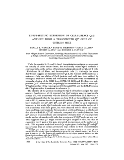
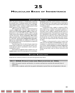
From www.bloodjournal.org by guest on December 22, 2014. For personal use only. RAPID COMMUNICATION Absence of the Human Retinoblastoma Gene Product in the Megakaryoblastic Crisis of Chronic Myelogenous Leukemia By Masayuki Towatari, Koichi Adachi, Hidefumi Kato, and Hidehiko Saito The human retinoblastoma gene (RBI product, which is involved in the control of cell cycle and tumor suppression, is constitutively expressed as a nuclear phosphoprotein in normal human cells. We examined leukemic cells from 22 patients with blast crisis of chronic myelogenous leukemia (CML) for alterations of the RB expression. Western blotting and flow cytometry with anti-RB-protein antibodies showed that all of five cases with megakaryoblastic crisis lacked the expression of the RB-encoded protein, whereas none of 17 cases with the other phenotypes such as myeloblastic or lymphoblastic crisis showed any abnormality. These findings suggest that megakaryoblastic transformationof CML might be lineage-specificallyassociated with loss of the RB protein. o 1991 by The American Society of Hematology. T monoclonal antibodies and molecular But the associations between the cell lineage of the blastic cells and the molecular events in the blastic transformation of CML have remained unclear. We studied 22 samples of blastic cells in CML blast crisis to evaluate whether alterations of the RB gene product correlate with the clonal evolution of each lineage of hematopoietic cells. HE PRODUCT of the human retinoblastoma susceptibility (RB) gene, one of tumor-suppressor genes, has recently been implicated in the control of cell cycle progression and normal cell g r ~ w t h . ”The ~ loss or inactivation of RB gene function contributes to the loss of growth regulation and leads to tumor formation. The RB gene was first identified through its association with an inherited predisposition to retinoblastomas.’ Structural abnormalities or altered expressions of the RB gene have subsequently been reported in other types of human tumors, including small cell lung carcinoma,6.7 breast cancer,’ bladder carcinoma: and osteosarcoma.1oDeletions of the RB gene or decreased expression of RB mRNA have also been observed in some cases of lymphoma/le~kemia.”~’~ These observations suggest that RB inactivation plays a significant role in the pathogenesis or progression of human cancers. Chronic myelogenous leukemia (CML) is a myeloproliferative disorder that has been shown to occur at the level of a pluripotential stem cell because of the presence of Philadelphia chromosome in all elements of hematopoietic cell lineages. The chronic phase of CML inevitably progresses to an acute phase (blastic transformation) after some period of time. CML is one of the most extensively studied leukemias at the molecular level, and activation of the c-ab1 proto-oncogene at the bcr-ab1 translocation is thought to be important in the pathogenesis of the chronic phase of CML. However, little is known about the subsequent genetic alterations or other molecular mechanisms responsible for progression to the blastic phase. The cellular origin and the stage of differentiation of the blastic cells at this clonal event have been investigated by using specific From the Division of Hematology/Oncology,The First Department of Intemal Medicine, Nagoya University School of Medicine; and Aichi Blood Disease Research Foundation, Showa-ku, Nagoya, Japan. Submitted July 29, 1991; accepted August 13, 1991. Supported by Grant-in-Aidfor Scientific Researchfrom the Ministry of Health and Welfarein Japan. Address reprint requests to Koichi Adachi, MD, Division of Hematology/Oncology,First Department of Intemal Medicine, 65, Tsurumalcho, Showa-ku, Nagoya 466, Japan. The publication costs of this article were defrayed in part by page charge payment. This article must therefore be hereby marked “advertisement” in accordance with 18 U.S.C. section I734 solely to indicate this fact. 0 1991 by The American Society of Hematology. 0006-4971191 /7809-0044$3.00/0 2178 MATERIALS AND METHODS Cell samples. Bone marrow aspirates or peripheral blood samples were collected, with the informed consents of 22 patients, at the time of diagnosis in the blastic phase of CML. The diagnosis was based on the standard clinical and hematologic criteria with Philadelphia chromosome. The leukemic blasts were separated by Ficoll-Conray (Pharmacia, Uppsala, Sweden) density gradient centrifugation, frozen in the medium with 10% dimethyl sulfoxide and 10% fetal calf serum by a programmed freezer, and cryopreserved in liquid nitrogen. All samples had greater than 70% blastic cells by morphologic examination and good viability of more than 80% by trypan blue dye exclusion test after being thawed and washed for use in this study. DNA and RNA analyses. Genomic DNAs extracted from leukemic samples were completely digested with each restriction enzyme, Hind 111, BamHI, or Egl 11. Agarose gel electrophoresis, Southern blotting, and hybridization analysis were performed according to the standard methods.” DNA probes of the RB gene (provided by Dr W. H. Lee, University of California at San Diego) were p0.9R which comprises exon 1 through part of exon 9 and p3.8R containing the rest of the 3’ exons. RNA isolation and Northern blotting were performed as described previo~sly.’~ Radiolabeling of the 4.5-kb RB cDNA probes and HLA-B7 probe from pDP00l plasmid for an internal control was performed using an oligo-random-labeling technique. Four sets of primers covering the binding regions to SV40 T antigen were used in this reverse transcription-polymerase chain reaction (PCR) analysis. They include primer la, 5’-CACACACTCCAGTTAGGACTGTTATGAACA-3’; primer lb, 5’-AATGTTGTCATTCAGAAGTTTGCTAAAATT-3�, primer 2a, 5’TAATGGAATCCATGCTTAAATCAGAAGAAG-3’;primer 2b, 5’-GACAAGCAGATTCAAGGTGATCAGTTGGTC-3�; primer 3a, 5’-ACCCAGAAGCCA�ITGAAATCTACCTCTCrT-3�; primer ’;primer 3b, 5’-AAGGTCTATATTCTTCACTTTGCATATGCATATGCC-3 4a, 5 �-AGTATGAACTCATGAGAGACAGGCATITGG-3’;and primer 4b, 5’-GTGAGGTA~GGTGACAAGGTAGGGGGCCT3’. After 30 cycles of the reactions at 94”C, 5 3 T , and 72°C for 1,2, and 2 minutes, respectively, in a Thermocycler (Perkin-Elmer Cetus, Emeryville, CA), the amplified products were analyzed by electrophoresis in agarose gels and ethidium bromide staining. Westem blot analysis. Leukemic cells (1 X lo’) were lysed with 400 PI of lysis buffer (50 mmol/L Tris-HC1 pH 8.0, 0.25 mol/L NaCl, 0.1% Nonidet P-40, 5 mmol/L EDTA, 1 mmol/L phenylBlood, Vol78, N o 9 (November 1). 1991: pp 2178-2181 From www.bloodjournal.org by guest on December 22, 2014. For personal use only. ABSENCE OF RB PRODUCT IN CML MEGAKARYOBLASTIC CRISIS 2179 methanesulfonyl fluoride, and 50 p,g/mL aprotinin). The cell lysates were clarified by centrifugation at 15.000 rpm for 10 minutes at 4°C. The total concentration of protein was determined using a commercial kit (Bio-Rad, Richmond, CA). and 50 p,g of protein from each sample was analyzed in 7.5% sodium dodecyl sulfatepolyacrylamide gel. and then the gels were transferred to nitrocellulose membranes. The gels were stained with Coomassie blue to identify protein integrity in each sample. After overnight blocking in phosphate-buffered saline (PBS) containing 0.1% Tween 20, 5% dried milk, and 0.1% sodium azide, the blots were incubated with a mouse monoclonal anti-RB-protein antibody, PMG3-245 (PharMingen, San Diego, CA) or a rabbit polyclonal antibody Rb(Ab-3) (Oncogene Science, Manhasset, N Y ) at the final concentration of 10 &mL. They were washed and incubated with biotinylated secondary immune reagents (rabbit antimouse IgG or donkey antirabbit IgG, respectively) and then reacted with streptavidin-alkaline phosphatase complex (Amersham, Buckinghamshire, UK). After washing thoroughly, nitro-blue tetrazolium and 5-bromo-4-chloro-3-indolyl phosphate were added to visualize the specific bands. Flow cyromerric ana!vsi.s. The cells ( 1 x 10")were fixed with 4% paraformaldehyde in PBS for 20 minutes. After centrifugation, they were resuspended in 0.1% Triton X-100 (Sigma, St Louis, MO) in PBS containing 0.1% bovine serum albumin for 5 minutes. The fixed cells were incubated with a monoclonal PMG3-245 antibody (10 p,g/mL) or nonspecific mouse IgG monoclonal antibody (MoAb) as a negative control. Binding of these antibodies was detected with fluorescein isothiocyanate conjugated goat antimouse IgG F(ab'), (Cappel Laboratories, West Chester, PA). After washes, the percentage of fluorescence-positive cells was A . 1 2 3 counted by flow cytometry using EPICS profile analyzer (Coulter Corp, Hialeah, FL). RESULTS We examined 20 leukemia samples in CML blast crisis that we had previously reported on the cell lineage'" and another two cases with the megakaryoblastic crisis. The phenotypic classification of surface antigens with a panel of lineage-spccific MoAbs showed that five cases expressed the megakaryocytic marker, platelet glycoprotein IIb-IIIa. The others were eight cases expressing myeloid differentiation antigens, six cases in lymphoblastic crisis with pre-B cell markers, and the remaining three cases showing no surface marker characteristic of any cellular lineage." Among these 22 samples, all of five samples from megakaryoblastic crisis were found to lack the 1 10-Kd RB protein by Western blotting with a monoclonal anti-RB-protein antibody, PMG 3-245, but none of 17 samples from the other phenotypes showed any abnormality (Fig IA). Western blotting using a polyclonal anti-RB protein antibody, Rb(Ab3), also presented the same result, indicating that RB proteins were lineage-specifically absent in all megakaryoblastic transformations of CML (data not shown). To exclude the possibility of RB protein degradation in the process of protein extraction, we mixed megakaryoblastic cells and RB-positive myeloid cells in the half and half ratio, then extracted the mixed protein, and confirmed that 4 5 7 8 - -.9 . ... -*- B 200 Fig 1. Western blot analysis showing loss of the RE protein in the megakaryoblastic crisis of CML. (A) Myeloblastic samples (lanes 1 through 3) and lymphoblastic samples (lanes 9 through 11) show the bands of the 110-Kd RB protein, but all samples from five patients with megakaryoblastic crisis (lanes 4 through 8) lack the expression of the RE gene product. (B) Lanes 1 through 3 of Coomassie blue-stained gel, which present the integrity of total protein, correspond to lanes 4 through 6 of the Western blot, respectively. Lane 4 is from RB-positive myeloblastic cells and lane 6 is from megakaryoblastic cells. Lane 5 shows the reduced RE protein extracted from the mixed sample of myeloblastic cells and megakaryoblastic cells in the half and half ratio. 6 97 68 43 = - 10 - 11 From www.bloodjournal.org by guest on December 22, 2014. For personal use only. 2180 TOWATARI ET AL Table 1. Characterization of Five Patients in the Megakaryoblastic Crisis of CML RE Protein Surface Antigen’ (GPllblllla) Patient Positive Positive Positive Positive Positive 1 Western Blotting Flow Cytornetryt RE Transcript RB Gene Alteration Undetectable Undetectable Undetectable Undetectable Undetectable 3.9 3.9 3.8 0.9 2.5 Normal Normal Normal Normal Normal None detected None detected None detected NE* NE �Positive percent reactivity: > 40%. tThe percentage of positive cells. *NE, not examined. there was decreased but detectable RB protein (Fig 1B). By India ink staining of blotted membranes, we also demonstrated that the pattern of other proteins than R B protein in megakaryoblastic samples was the same as that in the other phenotypic cells of CML blast crisis (data not shown). Compatible with the findings of Western blot analysis, flow cytometry with indirect immunofluorescence staining showed that only 0.9% to 3.9% of blastic cells in the cases of megakaryoblastic crisis reacted with the anti-RB-protein antibody in contrast to 48% to 6S% of blastic cells in myeloid or lymphoid crisis (Table 1). To examine the mechanism of the loss of R B gcnc product, we analyzed genomic DNA configuration and mRNA expression of the R B gene in each sample. Southern blot analysis of restriction fragments showed the normal R B gene structure without any gross deletion or rearrangement (data not shown). By Northern blotting using the full-length R B cDNA probe, no distinct abnormal size of R B transcript (4.7 kb) was identified in any sample of 22 cases. No decreased expression of the R B gene was also observed in comparison with HLA-B7 genc expression as an internal control for mRNA quantity (Fig 2). It has recently been reported that an aberrant R B protcin mutating in the binding region to adcnovirus-cncoded E I A proteins is unstablc’ and that the rcgions of R B protein for 1 2 3 4 5 6 binding to E l A or SV40 large T antigen are common sites for mutations.”’” Therefore, we used thc method of the cDNA synthesis-PCR to detect smaller alterations of RB mRNA, but each sample of megakaryoblastic crisis showed the normal amplified R B fragments for all four segments covering the binding regions to E I A and SV40 large T antigen (data not shown). DISCUSSION One of the simplest and most sensitive methods for detccting alterations of the R B gene expression is analysis o f the R B protein by using an immunoblotting assay with anti-RB-protein antibodies. We found that no R B protein in leukemic cells from patients with CML mcgakaryoblastic crisis was dctccted by both Western blotting and flow cytomctry despite intact R B genes and transcripts analyzed by Southern and Northern blotting. Some possible explanations can account for it: (1) A point mutation or a very small deletion at the 5�early portion of R B gene resulted in a new stop codon, a nonsense codon, or a frame shift by which detectable R B protein could not be produced. (2) Rapid protcin degradation or antigenic modulation of the RB gene product occurred just after translation by some genetic altcration or protein interaction in relation to megakaryoblastic transformation. 7 8 9 10 11 ) o m Fig 2. Northern blot analysis of leukemic samples from CML blast crisis. Ten micrograms of total RNA isolated from myeloblastic cells (lanes 1 through 3). megakaryoblastic cells (lanes 4 through E), and lymphoblastic cells (lanes 9 through 11) of CML blast crisis were electrophoresed, transferred t o nylon membranes, and hybridized with the ”P-labeled probe of the 4.5-kb RB cDNA (top panel). The same membranes were rehybridized as a control with the probe of HLA-B7 gene from pDPOOl plasmid (bottom panel). The number of each lane corresponds t o the same lane number of the Western blot in Fig 1A. ~ From www.bloodjournal.org by guest on December 22, 2014. For personal use only. 2181 ABSENCE OF RB PRODUCT IN CML MEGAKARYOBLASTIC CRISIS Recently, a group of investigators reported that no RB protein was detected by immunoprecipitation analysis in six small cell carcinoma cell lines, although normal RB transcripts were observed.’ In their subsequent report of the RB gene in the same cell lines,22 single base pair deletion resulting in a novel stop codon appeared within exon 20 or exon 23 in two cell lines. They speculated that structurally altered RB protein is less stable than normal RB protein. Further studies are also required at the sequence level to search for a possible aberration of the RB gene in the megakaryoblastic crisis. In any event, it is an interesting phenomenon that the RB protein is not detected only in megakaryoblastic transformation, in the fashion of all or none, for each cell lineage of CML blast crisis. These findings suggest that the clonal evolution of megakaryocytic lineage in CML blast crisis might be lineage-specificallyassociated with the absence of the RB gene product. ACKNOWLEDGMENT We thank Dr Tohru Marunouchi for critical reading of the manuscript, Drs Makoto Sawada and Isamu Sugiura for technical advice on Western blotting, Makoto Kondo for chromosome analysis, and Sayoko Sugiura for excellent technical assistance. REFERENCES 1. DeCaprio JA, Ludlow JW, Lynch D, Furukawa Y, Griffin J, Piwnica-Worms H, Huang CM, Livingston DM: The product of the retinoblastoma susceptibility gene has properties of a cell cycle regulatory element. Cell 58:1085,1989 2. Buchkovich K, D u e LA, Harlow E: The retinoblastoma protein is phosphorylated during specific phase of the cell cycle. Cell 58:1097, 1989 3. Chen PL, Scully P, Shew JY, Wang JYJ, Lee WH: Phosphorylation of the retinoblastoma gene product is modulated during the cell cycle and cellular differentiation. Cell 58:1193,1989 4. Mihara K, Cao XR, Yen A, Chandler S, Driscoll B, Murphree AL, T’Ang A, Fung YKT Cell cycle-dependent regulation of phosphorylation of the human retinoblastoma gene product. Science 246:1300,1989 5. Friend SH, Bernard R, Rogelj S, Weinberg RA, Rappaport JM, Albert DM, Dryja TP: A human DNA segment with properties of the gene that predisposes to retinoblastoma and osteosarcoma. Nature 323:643, 1986 6. Harbour JW, Lai SL, Whang-Peng J, Gazdar AF, Minna JD, Kaye FJ: Abnormalities in structure and expression of the human retinoblastoma gene in SCLC. Science 241:353,1988 7. Yokota J, Akiyama T, Fung YKT, Benedict WF, Namba Y, Hanaoka M, Wada M, Terasaki T, Shimosato Y, Sugimua T, Terada M: Altered expression of the retinoblastoma (RB) gene in small-cell carcinoma of the lung. Oncogene 3:471,1988 8. Lee EYHP, To H, Shew JY, Bookstein R, Scully P, Lee WH: Inactivation of the retinoblastoma susceptibility gene in human breast cancers. Science 241:218,1988 9. Horowitz JM, Yandell DW, Park SH, Canning S, Whyte P, Buchkovich K, Harlow E, Weinberg RA, Dryja TP: Point mutational inactivation of the retinoblastoma antioncogene. Science 243:937,1989 10. Friend SH, Horowitz JM, Gerber MR, Wang XF, Bogenmann E, Li FP, Weinberg RA: Deletions of a DNA sequence in retinoblastomas and mesenchymal tumors: Organization of the sequence and its encoded protein. Proc Natl Acad Sci USA 84:9059,1987 11. Cheng J, Scully P, Shew JY,Lee WH, Vila V, Haas M: Homozygous deletion of the retinoblastoma gene in an acute lymphoblastic leukemia (T) cell line. Blood 75730, 1990 12. Chen YC, Chen PJ, Yeh SH, Tien HF, Wang CH, Tang JL, Hong R L Deletion of the human retinoblastoma gene in primary leukemias. Blood 76:2060, 1990 13. Ginsberg AM, Raffeld M, Cossman J: Inactivation of the retinoblastoma gene in human lymphoid neoplasms. Blood 77:833, 1991 14. Furukawa Y, Ishida Y, Belvin M, DeCaprio JA, Griffin JD: Abnormalities in expression of the product of the retinoblastoma susceptibility gene in primary human leukemias. Blood 76:272a, 1990 (abstr) 15. Janossy G, Greaves MF, Revesz T, Lister TA, Roberts M, Durrant J, Kirk B, Catovsly D, Bread MEJ: Blast crisis of chronic myeloid leukaemia (CML): I1 Cell surface marker analysis of �lymphoid’and myeloid cases. Br J Haematol34:179,1976 16. Griffin JD, Todd RF 111, Ritz J, Nadler LM, Canellos GP, Rosenthal D, Gallivan M, Beveridge RP, Weinstein H, Karp D, Schlossman SF: Differentiation patterns in the blastic phase of chronic myeloid leukemia. Blood 61:85,1983 17. Bakhshi A, Minowada J, Arnold A, Cossman J, Jensen JP, Whang-Peng J, Waldmann TA, Korsmeyer SJ: Lymphoid blast crisis of chronic myelogenous leukemia represent stages in the development of B-cell precursors. N Engl J Med 309:826,1983 18. Adachi K, Okumura M, Tanimoto M, Morishima Y, Ohno R, Saito H: Analysis of immunophenotype, genotype, and lineage fidelity in blastic transformation of chronic myelogenous leukemia: A study of 20 cases. J Lab Clin Med 111:125,1988 19. Sambrook J, Fritsch EF, Maniatis T: Molecular cloning, in: A Laboratory Manual (ed 2). Cold Spring Harbor, NY, Cold Spring Harbor Laboratory, 1988 20. Hu Q, Dyson N, Harlow E: The regions of the retinoblastoma protein needed for binding to adenovirus E1A or SV40 large T antigen are common sites for mutations. EMBO J 9:1147, 1990 21. Huang S, Wang NP, Tseng BY, Lee WH, Lee EHHP: Two distinct and frequently mutated regions of retinoblastoma protein are required for binding to SV40 T antigen. EMBO J 9:1815, 1990 22. Mori N, Yokota J, Akiyama T, Sameshima Y, Okamoto A, Mizoguchi H, Toyoshima K, Sugimura T, Terada M: Variable mutations of the RB gene in small-cell lung carcinoma. Oncogene 51713, 1990 From www.bloodjournal.org by guest on December 22, 2014. For personal use only. 1991 78: 2178-2181 Absence of the human retinoblastoma gene product in the megakaryoblastic crisis of chronic myelogenous leukemia M Towatari, K Adachi, H Kato and H Saito Updated information and services can be found at: http://www.bloodjournal.org/content/78/9/2178.full.html Articles on similar topics can be found in the following Blood collections Information about reproducing this article in parts or in its entirety may be found online at: http://www.bloodjournal.org/site/misc/rights.xhtml#repub_requests Information about ordering reprints may be found online at: http://www.bloodjournal.org/site/misc/rights.xhtml#reprints Information about subscriptions and ASH membership may be found online at: http://www.bloodjournal.org/site/subscriptions/index.xhtml Blood (print ISSN 0006-4971, online ISSN 1528-0020), is published weekly by the American Society of Hematology, 2021 L St, NW, Suite 900, Washington DC 20036. Copyright 2011 by The American Society of Hematology; all rights reserved.
© Copyright 2026 Paperzz