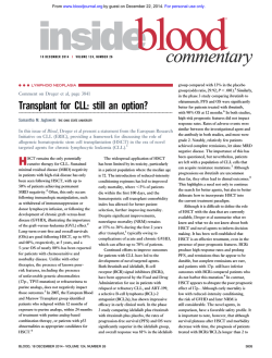
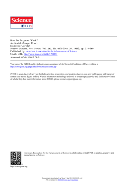
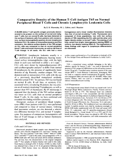
From www.bloodjournal.org by guest on December 29, 2014. For personal use only. The Hematopoietic Transcription Factor PU.l Is Downregulated in Human Multiple Myeloma Cell Lines By Monica Pettersson, Christer Sundstrom, Kenneth Nilsson, and Lars-Gunnar Larsson PU.l is a hematopoietictranscription factor belonging t o the Ets-family. It is identical t o the Spi-l oncogene, which is implicated in spleen focus-forming virus-induced murine erythroleukemias. PU.l seems t o be requiredfor early development of multiple hematopoietic lineages, but its expression in mature cells is preferentially observed in cells of the B-cell- andmonocyte/macrophage-differentiationlineage. It binds the so-called Pu box,an important tissue-specific regulatory DNA element present in a numberof genes expressed in these cell lineages. We have analyzedthe expression and activity of PU.1 during human B-cell development using a panel of B-cell lines representing different stages of maturation, from early precursors t o differentiated plasma cells. PU.l mRNA expression and PU.1 DNA binding activity, as measuredby Northern blot analysis and electrophoretic mobility shift assay, respectively, were evident in cell lines representing pro-B, pre-B, and mature B cells. We could also show Pu box-dependent transactivation of a reporter gene in transient transfections in these cell lines. In contrast, in a number of multiple myeloma cell lines, representingdifferentiated, plasma cell-like B cells, PU.l DNA binding activity, mRNAexpression, and Pu box-dependent transactivation were absent or detectable at a verylow level. In lymphoblastoid cell lines, which exemplify an intermediate stage of Bcell differentiation, a reduced expression andactivity were observed. Thefindings in the human multiple myeloma cell lines represent the firstexamples of B cells with downregulated PU.l expression and apparently contradict observations in the murine system in which PU.l is expressed and active in plasmacytoma cell lines. At present, it is unclear whether the lack of PU.l expression and activity in human multiple myeloma cell lines represents a malignancy-aosociated defect in these cells or exemplifies a normal develop mental regulation in terminally differentiated B cells. 0 7995 by The American Societyof Hematology. A ment of several lineages.’The PU.l binding site, the socalled Pu box, has been identified as an important tissuespecific regulatory element in the promoters or enhancers of a number of differentiation-associated genes in different hematopoietic cell Thus, PU.l has been implicated as an important tissue-specific factor regulating these genes in intimate cooperation with other transcription factors (such as NF-EMS,” or Sp12’) or other Ets-family members (such as Etsl,” Erg-3,” or GABPaZ3). We are interested in the role of PU. 1 during development of human B-lineage cells and its possible involvement in tumor formation in these cells. A panel of human B-cell lines representing different stages of development, from early precursors to differentiated plasma cells, was used for studies of PU.1 expression and activity. We found differences in PU.l mRNA expression, PU.l DNA binding, and transcriptional activity in cell lines at different maturation stages. In particular, we can show that PU.l activity and expression was shut off in a panel of multiple myeloma cell lines, representing differentiated B cells. The implications of these findings will be discussed. N IMPORTANT AREA of research on cellular differentiation is the identification of transcription factors regulating cell lineage- and differentiation stage-specific gene expression. Examples of such factors in the hematopoietic system are the Oct-2’ and GATA family proteins.’ Another transcription factor specific for hematopoietic cells is PU. 1, originally described as a lymphoid- and macrophagespecific f a ~ t o r .However, ~.~ by gene targeting, PU.l was recently shown to be required for the development of multiple hematopoietic lineages.’ PU. 1 belongs to the ets proto-oncogene family (including Etsl, Ets2, Erg, Elfl, GABPa,PEA3, Hi-l, E74A, Elkl, and SAP1) characterized by the wellconserved ETS domain that mediates specific DNA binding to the core sequence GGA.6,7The N-terminal part of the protein, which is only weakly or not at all homologous to other Ets-proteins, contains a glutamine-rich transactivation domain8 and is able to bind to the TATA-box-binding protein and the retinoblastoma protein Rb in vitro.’ PU.1 also contains a motif that mediates protein-protein interactions with the B-cell-specific transcription factor NF-EMS.” PU.1 is identical to the oncogene Spi-l implicated in the development of spleen focus-forming virus-induced murine Friend erythroleukemia. Spleen focus-forming virus was found to integrate in the Spi-l locus in 95% of the tumors, resulting in an elevated expression of Spi-l mRNA.” The observations that overexpression of Spi-1PU. 1 immortalizes primary erythroblasts12 and that antisense oligonucleotides inhibit the proliferation of Friend murine erythroleukemia cell linesI3 further indicated a role for Spi-1PU.1 in the growth control of immature erythroid cells. Also, the Etsfamily members v-Ets and Hi-l have been implicated in the development of erythroid tumors in chickens and mice, respe~tively.’,’~ The expression of PU. l in mature hematopoietic cells was reported to be confined to B cells, monocytes/macrophages, and mast cells, whereas it seems to be more broadly expressed at early stages of lymphoid and myeloid development.3.4.15-17 This would also agree with the gene-targeting data which suggested that itis required for the early developBlood, Vol 86, No 7 (October l ) , 1995: pp 2747-2753 MATERIALS AND METHODS Cell lines. All cell lines were maintained in RPM1 1640 (Flow Laboratories, Ayrshire, UK) supplemented with 7.5% fetal calf seFrom theLaboratory of TumorBiology, Department of Pathology, University of Uppsala, University Hospital, Uppsala, Sweden. Submitted December 13, 1994; accepted June 7, 1995. Supported by grants from the Swedish Cancer Society, T. & R. Stjderbergs and M. Bergvalls Foundations. Address reprint requests to Lars-Gunnar Larsson, PhD, Laboratory of TumorBiology, Department of Pathology, University of Uppsala, Univerisity Hospital, S-751 85 Uppsala, Sweden. The publication costs of this article were defrayed in part by page charge payment. This article must therefore be hereby marked “advertisement” in accordance with 18 U.S.C. section 1734 solely to indicate this fact. 0 1995 by The American Society of Hematology. oooS-4971/95/8607-0044$3.00/0 2747 From www.bloodjournal.org by guest on December 29, 2014. For personal use only. PETTERSSONET AL rum (GIBCO. Grand Island, NY). glutamine, 100 IUlmL penicillin, and S0 pglmL streptomycin. A panelof cell lines was selected representing different stages of B-cell development. KM3 is derived from a non-B. non-T acute lymphocytic leukemia and represents a lymphoid precursor cell line."' BJA-B, Raji, Daudi, and Ramos are B-lymphoma cell lines representing pre-B to mature B cells.'7 Karpas 422 and MN60 are B-lymphomaand leukemia cell lines, respecti~ely.'~.'~ representing mature B cells. U-255 and Corinna I1 are Epstein-Barr virus-immortalized Ig-secreting lymphoblastoid cell line^,'^.''' representing B cellsat the B-blast/immunoblast stage. Karpas 707, W63, U-1996. and U-266 are multiple myeloma cell line?." andrepresent plasmablasts-plasma cells. U-266-1970 andU-2661984 are early and late passages. respectively, of an IgE myeloma cell line, the phenotype of which has been described." Trcmsfection and chloramphenicol aceQltransferase (CAT)assay. Transfections were performed using the diethyl aminoethyl (DEAE)dextran method." The cells were fed 24 hours before transfection, and 20 X 10" cells were mixed with a solution containing 300 pg DEAE-dextran (Pharmacia, Sollentuna, Sweden) and 5 pg DNA. The mixture was incubated for 30 minutes atroom temperature, followed by glycerol treatment for 3 minutes. The cells were washed once in phosphate-buffered saline before fresh medium was added. Seventy-two hours postinfection, the cells were harvested, and a CAT assay was performed.25 Nuclear extracts and electrophoretic mohiliwshqt assay (EMSA). Nuclear extracts were prepared from40 X IO" cells using thelysolecithin procedure described by Zervitz and Akusjarvi." Ten micrograms of extract wasused for each binding reaction, and 1 pg poly(d1-dC)poly(dl-dC) (Pharmacia) and 10 pmol single-stranded oligonucleotide were included as nonspecific competitors. EMSA with the Pubox was performed in a buffer containing 10 mmol/L HEPES (pH 7.9). 10% glycerol, 20 mmol/L KCI, 4 mmol/L MgCI2, 10 mmollL EDTA, 0.25 mmol/L dithiotreitol, 4 mmol/L spermidine, and 1 0 0 p@mL bovine serum albumin. The Spl EMSA was performed in 10 mmol/L HEPES (pH 7.9). 10% glycerol, 50 mmol/L KCI, S mrnollL MgCI2. 0.6 mmol/L dithiotreitol, and 200 pmol/L ZnSO,. The extract was added to the buffer including the nonspecific competitor and was incubated for 20 minutes at room temperature. Pubox or the Spl oligonucleotide ( I to 2 fmol), end-labeled with Klenow DNA polymerase or T4DNA polynucleotidekinase, respectively, was added. and the mixture was further incubated for 20 minutes at room temperature. Samples were analyzed on a 4% polyacrylamide gel in 0.2SX TBE (0.022 mol/L Tris-borate and 0.5 mmol/L EDTA). The Pu box oligonucleotide sequence (S'TCGACTCTGAAAGAGGAACTCTCGAGCT) is derived from the SV40 control region.' The mutated Pu box sequence is S'TCGACTCGTCCAGAGGAACTCTCGAGCT. The Spl oligonucleotide (S'CCGGCCCCGCCCATCCCCGGCCCCGCCCATCC)contains a dimer of a binding site found in the immediate early gene 3 of herpes simplex virus. Antibody 1297 (kindly provided by Dr van Beveren, La Jolla Cancer Research Foundation, La Jolla, CA) is specific for the N-terminal region of PU. I , and antibody T-21 (Biotechnology, Santa CNZ, CA) recognizes the C-terminal part of the protein. Plasmid constructions. pCAT-Control plasmid (Promega, Madison, WI) withthe SV40 early promoter and enhancer sequences served as the control plasmid in the transfection experiments. An Xho I linker was cloned into the BgI I1 site of pCAT-Promoter plasmid (Promega), and the new plasmid was designated pPromXCAT. Four copies ofthe Pubox oligonucleotides (wild-type and mutated) were cloned into the oligonucleotide vector (OVEC) plasmid.'7 The resulting Xho I-Sal I fragment was then inserted into the Xho I site of pPromXCAT. Northwn analysis. Total R N A was extracted from exponentially growing cells by the LiCllurea method.2xRNA (IS pg) was denaturated in formamide and fractionated in an 1 % agarose gel containing B A € t 1 2 3 4 5 m 1 2 3 Fig 1. EMSA with thePu box sequence. The W 4 0 Pu box oligonucleotide was mixed with 10 p g of BJA-B nuclear extract, and the resulting complexes were separated on a native polyacrylamide gel. (AI Lane1,noextract; lane 2, with extract. The specificity of the complexes was analyzed by addition of a 500-fold molar excess of cold Pu box sequence (lane 31, a mutated Pu box (lane 4). and an oligonucleotide containing the Spl binding site (lane 5). The main Pu boxlprotein complexes formed are indicated as a and b. (B) Supershift performed with an antibody directed against the N-terminal part of the protein. Lane 1, no extract; lane 2, extract without antibody; lane 3, antibody added. formaldehyde (4 m U l 0 0 mL gel). After electrophoresis, the RNA was transferred to a nitrocellulose filter. The probes were "P-labeled by the random priming method (Amersham, Buckinghamshire, UK). Hybridization was performed at 42°C in a solution containing 50% formamide, I X Denhardt's solution, 2X SSC (2X SSC: 0.3 mol/L sodium chloride and 30 mmol/L sodium citrate), S mmol/L NaPO.,, 0.1% sodium dodecyl sulfate, and 200 pg/mL of salmon sperm DNA. Filters were washed in O S % sodium dodecyl sulfate and 2X SSC at 50°Cand exposed toKodakXAR film (Eastman Kodak, Rochester, NY). A murine PU.1 cDNA clone (unpublished, Dr L. Hellman, Department of Medical Immunology and Microbiology, University of Uppsala, Sweden) and a glyceraldehyde-3-phosphate dehydrogenase (GAPDH) cDNA clon$' were used as probes. RESULTS Absence of PU.1 DNA binding activity in multiple myeloma cell lines. To establish the conditions for studies of PU.1 DNA binding, weused the human BJA-B cell line known to express PU. 1.3 A "P-labeled oligonucleotide containing the Pu box sequence from the SV40 control region was mixed with nuclear extracts from BJA-B cells and analyzed by EMSA. The SV40 Pubox binds PU.1withhigh affinity but is a poor binding site for many other Ets-family proteins.""'43 Figure 1A shows that two major retarded complexes, a and b. were formed. These complexes were competed by an access unlabeled Pu box oligonucleotide but not by a mutated oligonucleotide or an oligonucleotide containing an Spl binding site, thus showing the specificity of the binding. The mutated oligonucleotide, previously shown to eliminate binding of PU. I ,3 contains 4 substitutions 4-7 From www.bloodjournal.org by guest on December 29, 2014. For personal use only. HUMAN PU.l CELLS EXPRESSION IN B Fig 2. PU.l DNA binding activity in B-cell nuclear extracts. EMSA using the Pu box oligonucleotide was performed with nuclear extracts prepared from the following B-cell lines: lane 1, no extract; lane 2, KM3; lane 3, BJA-B; lane 4, Daudi; lane 5, Raji; lane 6, Ramos; lane 7, Karpas 422; lane 8, MN60; lane 9, U-255; lane 10, Corinna II; lane 11, Karpas 707; lane 12, L363; lane 13, U-1996; lane 14, U-266-1970; and U-266-1984. lane 15, 2749 1 2 nucleotides 5' of the the GGA core binding site for Etsfamily proteins. Addition to the binding reaction of antibodies directed to the N-terminus of PU. 1 resulted in a supershift of the upper but not the lower Pu box binding complex, thus confirming the presence of PU.1 or an antigenically closely related protein in the former (Fig IB). Addition to the binding reaction of antibodies specific for the C-terminus of PU.1 prevented formation of both the upper and lower complexes (data not shown), indicating that the lower complex represents a degradation product of PU.1 containing the DNA binding domain but lacking the N-terminal antibody-binding epitope. PU.1 contains a so-called PEST sequence suggested to play a role in protein degradationAU To investigate the DNA binding activity of PU.1 in relation to B-cell differentiation, nuclear extracts were prepared from various human B-celllines representing different stages of B-cell differentiation. These included the pro-B-cell line KM3; the B-lymphoma cell lines BJA-B, Raji, Daudi, and Ramos representing pre-B to mature B cells; the Karpas 422 follicular lymphoma cell line and B-leukemia cell line MN60 representing mature B cells; the U-255 and Corinna I1 Igsecreting lymphoblastoid cell lines and, finally, the multiple myeloma cell lines Karpas 707, L363, U-1996, U-266-1970, and U-266-1984 representing plasmablasts-plasma cells. The extracts were analyzed by EMSA using the Pu box sequence as above. Figure 2 shows that a Pu box-binding complex comigrating with the upper PU.1 complex inBJA-B cells was clearly shown in the cell lines representing early and mature B cells (Fig 2, lanes 2-8). In contrast, three of the myeloma cell lines, U-1996, U-266-1970, and U-266-1 984, were completely negative; in the other two, L363 and Karpas 707, a very faint band could be detected (Fig 2, lanes 1115). Compared with early and mature B-cell lines, a reduced PU.1 binding activity was observed in the lymphoblastoid cell lines, in particular in Corinna I1 (Fig 2, lanes 9 and IO). Figure 3 shows that an oligonucleotide containing an Spl binding site showed specific Spl binding activity in the myeloma cell extracts as well as in BJA-B extracts, thus ruling out a general degradation of the myeloma extracts. Absence of Pu box-dependent transcriptional activio in 3 4 5 6 7 8 9 101112131415 multiple myeloma cell lines. We next investigated whether the observed binding of PU.1 to the Pu box in the various extracts correlated with transcriptional activity. Four copies of the Pu box oligonucleotide and a mutated version thereof were cloned upstream of the SV40 early promoter in a CAT gene construct. The plasmids were transfected into selected B-cell lines, and, 72 hours after transfection, the cells were harvested and assayed for CAT activity. A construct with the SV40 early promoter and enhancer sequences was 1 2 3 4 5 6 7 8 Fig 3. Spl DNA binding activity in B-cell nuclear extracts. An oligonucleotide containing two Spl binding sites was mixed with the following B-cell extracts and subjected to EMSA lane 1, no extract; lane 2 and 3, BJA-B; lane 4, Karpas 707; lane 5, U63;lane 6, U-1996; lane 7, U-266-1970; and lane 8, U-266-1984, In lane 3, a 500-fold molar excess of unlabeled S p l sequence is included in the mixture. From www.bloodjournal.org by guest on December 29, 2014. For personal use only. PETERSSON ET AL 2750 A C 0 O E 1 2 3 4 1 2 3 4 1 2 3 1 2 9 4 1 4 2 3 4 H G F E D 1 2 3 4 transfected in parallel and served as a positive control. In the BJA-B, Raji, U-255, and Karpas 422 cell lines, the presence of the Pu boxes led to an enhanced transcription from the CAT gene to various degrees, whereas mutations introduced in the Pu box reduced the activity to basal level (Fig 4). Densitometrical scanning of the autoradiogram and normalizing to the activity of pPromXCAT vector lacking Pu boxes showed that four copies of the Pu box stimulated transcription 28 times in BJA-B cells, 8 times in Raji cells, and 3 times in U-255 and Karpas 422 cells. Transfection of the myeloma cell lines L363 and U-266-1984 with the Pu box-containing plasmids did notlead to activation of the CAT gene, thus correlating with the lack of Pu box binding in these cells. A control CAT reporter plasmid containing SV40 promoter and enhancer sequences strongly stimulated transcription in the myeloma cell lines. We were also unable to detect any Pu box-dependent transcriptional activity in the Corinna I1 lymphoblastoid cell line, which has a reduced PU. 1 binding activity. The PU.1 gene is downregulated in multiple myeloma cell lines. The lack of PU. 1 activity in the myeloma cells could be because of a downregulation of the PU. I gene expression or, alternatively, because the protein is present in an inactive form. To investigate the expression of PU. 1 in the panel of B-cell lines used above, RNA was prepared, and a Northern blot analysis was performed using a mouse PU.1 cDNA as probe. Figure 5A shows that the 1.4-kb PU. I transcript was expressed in all B-cell lines with the exception of the myeloma cell lines, in which it was undetectable even after prolonged exposure. The lymphoblastoid cell line U-255 showed a reduced expression, and the level of PU. I mRNA was very low, but detectable, in Corinna 11. As a control, the same filter was rehybridized with a GAPDH probe, showing that this gene was expressed in all cell lines (Fig 5B). We conclude that the PU.1 gene is shut off in the myeloma 1 L 3 4 Fig 4. Pu box-dependent transcriptional activity in human B-cell lines. The various B-cell lines were transfected with 5 p g of the CAT reporter plasmids described below usingDEAE-dextran. Cells were harvested after 3 days, and transcriptional activity was analyzed by CATassay. (A) CATassay performed without extract (0) and with purified CAT (E). The following cell lines were used: B, MA-B; C, Raji; D, Karpas 422; E, U-255; F, Corinna 11; G , U-266-1984; H, L363. Reporter plasmids were used as follows: in lane 1 is pPromXCAT containing theW 4 0 early promoter butlacking Pu boxes. In lane 2, four copies of the Pu box are inserted upstream of theW 4 0 early promoter in pPromXCAT, and lane 3 contains, in a similar way, four copies of a mutated Pu box. Lane 4 is the pCAT-Control (Promega) with SV40 early promoter and enhancer. cell lines, thus explaining the lack ofPU.1 transcriptional activity and DNA binding in these cells. DISCUSSION PU.1is a transcription factor specific for hematopoietic cells that seems to be required for development of early A B 1 2 3 4 5 6 7 8 9 1 0 1 1 Fig 5. Northern blot analysis of PU.l expression in human B t e l l lines. Total RNA was prepared from 11 different B-cell lines and fractionated on a formaldehyde-containing agarose gel. After blotting onto nitrocellulose filter, hybridization was performed using "P-labeled probes. Lane 1, KM3; lane 2, RIA-B; lane 3, Daudi; lane 4, Raji; lane 5, Karpas 422; lane 6 MN60; lane 7, U-255; lane 8, Corinna II; lane 9, L363; lane 10, U-1996; lane 11, U-266-1984. (A) A murine PU.l cDNA clone was used as probe. (B) Hybridized probe was removed, and the filter was rehybridized with a GAPDH probe. From www.bloodjournal.org by guest on December 29, 2014. For personal use only. PU.1 EXPRESSION IN HUMAN B CELLS precursor cells. It has also been suggested to play a role in mature B cells and macrophages. We have undertaken a study to investigate the presence and activity of PU. 1 during human B-cell development using a panel of human B-cell lines representing the B-cell lineage from early precursors to differentiated plasma cells. Our results show major differences in the expression and activity of PU. l in cell lines representing different levels of B-cell development. PU. 1 mRNA expression and PU.l DNA binding activity was shown in the pro-B<ell line KM3 as well as in pre-B and mature B-cell lines. Pu box-dependent transcriptional activity of a CAT reporter gene was also shown in these cell lines in transient transfections. In contrast, in a panel of multiple myeloma cell lines representing differentiated B cells, no or only very low mRNA expression, DNA binding activity, and Pu box-dependent transactivation were detectable. A reduced PU.l activity and expression were observed in the lymphoblastoid cell lines U-255 and Corinna 11, representing an intermediate stage of differentiation. This reduction was most pronounced in Corinna 11, which seems further differentiated than many other EpsteinBarr virus-transformed lymphoblastoid cell lines in that it lacks CD37 expression (our unpublished observations). However, Corinna I1 is clearly less differentiated than the myeloma cell Our observations in human pro-B, pre-B and mature B cells are in good agreement with previous studies in murine B-cell lines.4,'6.19,22,45 However, the lack of PU. 1 expression and activity in human multiple myeloma cell lines is a unique finding and represents the first example of B cells with downregulated PU. 1 expression. Our results are apparently contradictory to results obtained in the murine system, in which PU.1 is expressed and active inmost plasmacytoma cell lines.4,i9.4S Our finding in human multiple myeloma cell lines may be interpreted in at least two different ways. (1) Because PU.1 seems generally expressed in other B cells, one interpretation is that the lack of PU. 1 is a malignancy-associated defect common to human multiple myeloma. If PU.1 is an important transcription factor involved in commitment along the B lineage, it is conceivable that loss of PU.l may result in problems for the cell to recognize itself as a B cell, and, thus, it may be unable to complete the differentiation program and develop into terminally differentiated, resting plasma cells. However, from Southern analysis (data not shown) and cytogenetic analysis of chromosome 11,46 we have found no evidence for chromosomal aberrations of the PU.1 loci in the myeloma cell lines. This does not exclude minor alterations such as small deletions or point mutations. (2) An alternative interpretation is that the shutoff of the PU.1 gene is a normal event occumng at a late stage of Bcell development corresponding to that of the myeloma cell lines. This interpretation is supported by the reduced expressionand activity in the U-255 and Corinna I1 cell lines representing an intermediate stage of differentiation. Data from homozygous gene-targeted PU.l mouse embryos suggest that PU.l is a very early hematopoietic transcription factor required for generation of B- and T-lymphocyte, monocyte, and granulocyte progenitors, thus acting already 2751 at a stage of a multipotent stem This interpretation would be in agreement with previous expression studies suggesting that PU.1 is expressed in early myeloidflymphoid cells committed to the erythroid, granulocytic, monocytic, mast cell, B- and T-lymphoid lineages. It is then shut off early in T-cell, erythroid, and granulocytic differentiation but is expressed at later stages of monocytic, mast cell, and B-cell differentiati~n.~~~"'"~ The present data may suggest that it is shut off late during B-cell differentiation, thus performing its function at preterminal stages of B-cell development. From this perspective, the differences in expression in human myelomas and murine plasmacytomas may be because of slight differences in maturation stage or a malignancy-associated inability to shut off PU.1 in the plasmacytomas or might represent a species difference. Studies of PU.l expression in normal human and mouse plasma cells are required to clarify these points. It may seem remarkable that PU.l is shut off in human myeloma cell lines that produce Ig at a high level, because PU. 1 has been suggested to be one important transcriptional activator of both the heavy- and light-chain Ig However, there are alternative pathways of Ig gene regulation mediated by, for instance, Oct-2 and NF-KB, acting on the heavy chain enhancer and onthe K-chain intron enhancer, respectively, that seem to increase in importance at late stages of B-cell differentiati~n.'~,~',~~ Therefore, such alternative pathways may be in operation in the human myeloma cell lines. Other genes suggested to be regulated by PU.l in B cells are the J-chain,' the mb-l and B29 antigen receptorassociated signaling proteins,'8320p2 i n t e g r i n ~ ,and ~ ~ the major histocompatibility complex class I1 antigens4The expression of these genes seems to be connected to B-cell activation, proliferation, and IgM secretion at preterminal stages of differentiation rather than to terminally differentiated plasma cells or myelomas. Therefore, one interesting possibility is that PU.l might be a transcription factor which integrates differentiation and cell growth at proliferative stages along the B-cell differentiation lineage. The oncogenic potential of PU.l/Spi-l in erythroleukemia and its ability to interact with Rb in vitro would point in this direction. Immunohistochemical studies in normal murine bone barrow also suggest the highest expression of PU.l in dividing cells." An attractive hypothesis is that PU.l is replaced byan alternative Ets-family or a non-Ets transcription factor such as Oct-2 or NF-KB,lacking proliferative or having an antiproliferative capacity, at terminal stages of B-cell differentiation in which high expression of the differentiation-associated genes is connected to irreversible growth-arrest. Examples of other Ets-family members expressed in hematopoietic cells are Etsl, Ets2, Erg, Fli-l, Elfl, GABPa,and/or S P ~ BThe . ~ EMSA and transactivation assays in the myelomas suggest that PU.l is not replaced byan Ets-family protein with capacity to bind the SV40 Pu box. However, other PU. 1 binding sites in relevant cellular target genes may have a broader Ets-family binding specificity that could result in a competition between different Ets-family proteins or a sequential role of these during B-cell development. Further studies of PU.1 and other Etsfamily proteins during normal B-cell development is re- From www.bloodjournal.org by guest on December 29, 2014. For personal use only. 2752 PETTERSSON ET AL quired toestablish whether our findings in multiple myeloma cell lines represent a malignancy-associated deregulationof PU.l in these cells or exemplify a normalphase of B-cell differentiation. ACKNOWLEDGMENT We thank Dr L. Hellman for providing the PU. 1 cDNA clone, Dr C. van Beveren for providing the antiserum against the N-terminal part of PU.l, and A. Kraft for skillful technical assistance. REFERENCES 1. Kemler I, Schaffner W: Octamer transcription factors and the cell type-specificity of immunoglobulin gene expression. FASEB J 4:1444, 1990 2. Orkin SH: GATA-binding transcription factors in hematopoietic cells. Blood 80575, 1992 3. Pettersson M, Schaffner W: A purine-rich DNA sequence motif present in SV40 and Lymphotropic papovavirus binds a lymphoidspecific factor and contributes to enhancer activity in lymphoid cells. Genes Dev 1:962, 1987 4. Klemsz MJ, McKercher SR, Celada A, Van Beveren C, Maki RA: The macrophage and B cell-specific transcription factor PU.l is related to the ets oncogene. Cell 6:113, 1990 5. Scott EW, Simon MC, Anastasi J, Singh H: Requirement of transcription factor PU.l in the development of multiple hematopoietic lineages. Science 265:1573, 1994 6. Karim FD, Urness LD, Thummel CS, Klemsz MJ, McKercher SR, Celada A, Van Beveren C, Maki RA, Gunther CV, Nye JA, Graves BJ: The ETS-domain: A new DNA-binding motif that recognizes a purine-rich core DNA sequence. Genes Dev 4: 1451, 1990 7. Wasylyk B, Hahn SL, Giovane A: The Ets family of transcription factors. Eur J Biochem 21 1.7, 1993 8. Shin MK, Koshland ME: Ets-related protein PU.1 regulates expression of the immunoglobulin J-chain gene through a novel Etsbinding element. Genes Dev 7:2006, 1993 9. Hagemeier C, Bannister AJ, Cook A, Kouzarides T: The activation domain of transcription factor PU.1 binds the retinoblastoma (RB) protien and the transcription factor TFIID in vitro:RB shows sequence similarity to TFIID and TFIIB. Proc Natl Acad Sci USA 90:1580, 1993 10. Pongubala JMR, Van Beveren C, Nagulapalli S, Klemsz MJ, McKercher SR, Maki RA, Atchison ML: Effect of PU.1 phosphorylation on interaction withNF-EM5and transcriptional activation. Science 259:1622, 1993 11. Moreau-Gachelin F, Tavitian A, Tambourin P: Spi-l is a putative oncogene in virally induced murine erythroleukaemias. Nature 331:277, 1988 12. Schuetze S, Stenberg PE, Kabat D: The Ets-related transcription factor PU. 1 immortalizes erythroblasts. Mol Cell Biol 135670, 1993 13. Delgado MD, Hallier M, Meneceur P, Tavitian A, MoreauGachelin F Inhibition of Friend cells proliferation by spi-l antisense oligodeoxynucleotides. Oncogene 9: 1723, 1994 14. Ben-David Y, Giddens EB, Letwin K, Bemstein A: Erythroleukemia induction by Friend murine leukemia virus: Insertional activation of a new member of the ets gene family, Fli-l, closely linked to c-ets-l . Genes Dev 5:908, 1991 15. Schuetze S, Paul R, Gliniak BC, Kabat D: Role of the PU. 1 transcription factor in controlling differentiation of Friend erythroleukemia cells. Mol Cell Biol 12:2967, 1992 16. Galson DL, Hensold JO, Bishop TR, Schalling M, D’Andrea AD, Jones C, Auron PE, HousmanDE: Mouse P-globin DNAbinding protien B1 is identical to a proto-oncogene, the transcription factor Spi-1PU.1, and is restricted in expression to hematopoietic cells and the testis. Mol Cell Biol 13:2929, 1993 17. Hromas R, Orazi A, Neiman RS, Maki R, Van Beveren C, Moore J, Klemsz M: Hematopoietic lineage- and stage-restricted oncogene family memberPU.1.Blood expression of theETS 82:2998, 1993 18. Hagman J, Grosschedl R: An inhibitory carboxyl-terminal domain in Ets-l and Ets-2 mediates differential binding of ETS family factors to promoter sequences of the mb-1 gene. Proc Natl Acad Sci USA 89:8889, 1992 19. Nelsen B, Tian G, Erman B, Gregoire J, Maki R, Graves B, Sen R: Regulation of lymphoid-specific immunoglobulin p heavy chain gene enhancer by ETS-domain proteins. Science 261:82, 1993 20. Omori SA, Wall R: Multiple motifs regulate the B-cell-specific promoter of the B29 gene. Proc Natl Acad Sci USA 90:11723, 1993 2 I . Pahl HL, Scheibe M,Zhang D-E, Chen H-M, Galson DL, Maki RA, Tenen DG: The proto-oncogene PU. 1 regulates expression of the myeloid-specific CD1 Ib promoter. J Biol Chem 2685014, 1993 22. Rivera RR, Stuiver MH, Steenbergen R, Murre C: Ets proteins: New factors that regulate immunoglobulin heavy-chain gene expression. Mol Cell Biol 13:7163, 1993 23. Battinger EP, Shelley CS, Farokhzad OC, Arnaout MA: The human p2 integrin CD18 promoter consists of two inverted ets cis elements. Mol Cell Biol 14:2604, 1994 24. Zhang D-E, Hetherington CJ, Chen H-M, Tenen DG: The macrophage transcription factor PU. 1 directs tissue-specific expression of the macrophage colony-stimulating factor receptor. Mol Cell Biol 14:373, 1994 25. Chen H-M, PahlHL, Scheibe RJ, Zhang D-E, Tenen DG: The Spl transcription factor binds the CD1 l b promoter specifically in the myeloid cells in vivo and is essential for myeloid-specific promoter activity. J Biol Chem 2689230, 1993 26. Schneider U, Schwenk H-U, Bornkamm G: Characterization of EBV-genome negative “null” and “T” cell lines derived from children with acute lymphoblastic leukemia and leukemic transformed non-Hodglun lymphoma. Int J Cancer 19:621, 1977 27. Nilsson K, Klein G: Phenotypic and cytogenetic characteristics of humanB-lymphoid cell lines and their relevance for the etiology of Burkitt’s lymphoma. Adv Cancer Res 37:319, 1982 28. Dyer MJS, Fischer P, Nacheva E, Labastide W, Karpas A: A new human B-cell non-Hodgkin’s lymphoma cell line (Karpas 422) exhibiting both t( 14;18) and t(4;ll) chromosomal translocations. Blood 75:709, 1990 and 29. Nilsson K: Characteristics of established myeloma lymphoblastoid cell lines derived froman E myeloma patient: A comparative study. Int J Cancer 7:380, 1971 30. Karpas A, Fischer P, Swirsky D: Human myeloma cell line carrying a Philadelphia chromosome. Science 216:997, 1982 3 1 . Diehl V, Schaadt M, Kirchner H, Hellriegel K-P, Gudat F, Fonatsch C, Laskewitz E, Guggenheim R: Long-term cultivation of plasma cell leukemia cells and autologous lymphoblasts (LCL) in vitro: A comparative study. Blut 36:331, 1978 32. Jernberg H, Nilsson K, Zech L, Lutz D, Nowotny H, Scheirer W: Establishment and phenotypic charaterization of three new human myeloma cell lines (U-1957, U-1958, and U-1996). Blood 69:1605, 1987 33. Hellman L, Josephson S, Jernberg H, Nilsson K, Pettersson U: Immunoglobulin synthesis in the human myeloma cell line U266; expression of two immunoglobulin heavy chain isotypes ( E and a ) after long-term cultivation in vitro.Eur J Immunol 18:905, 1988 34. Lnthman H, Magnusson G: Highefficiency polyoma DNA transfection of chloroquine treated cells. Nucleic Acids Res 1 1 :1295, 1983 From www.bloodjournal.org by guest on December 29, 2014. For personal use only. PU.l EXPRESSION INHUMAN B CELLS 35. Gorman CM, Moffat LF, Howard BH: Recombinant genomes which express chloramphenicol acetyltransferase in mammalian cells. Mol Cell Biol 2:1044, 1982 36. Zervitz K, A k u s j b i G: An improved nuclear extract preparation method. Gene Anal Tech 6:101, 1989 37. Westin G, Gerster T, Muller MM, Schaffner G, Schaffner W: OVEC, a versatile system to study transcription in mammalian cells and cell-free extracts. Nucleic Acids Res 15:6787, 1987 38. Auffray C, Rougeon F Purification of total mouse immunoglobulin heavy-chain messenger RNAs from total myeloma tumor RNA. Eur J Biochem 107:303, 1980 39. Tso JY, Sun X-H, Kao T, Reece KS, Wu R: Isolation and characterization of rat and human glyceraldehyde-3-phosphate dehydrogenase cDNAs: Genomic complexity and molecular evolution of the gene. Nucleic Acids Res 13:2485, 1985 40.Reddy ESP, RaoVN: erg, An ets-related gene, codes for sequence-specific transcriptional activators. Oncogene 6:2285, 1991 41. Rao VN, Reddy ESP A divergent ets-related protein, Elk-l, 2753 recognizes similar c-ets-l proto-oncogene target sequences and acts as a transcriptional activator. Oncogene 7:65, 1992 42. Rao VN, Ohno T, Prasad DDK, Bhattacharya G, Reddy ESP: Analysis of the DNA-binding and transcriptional activation functions of human Hi-l protein. Oncogene 8:2167, 1993 43. Wasylyk C, Kerckaert J-P, Wasylyk B: A novel modulator domain of Ets transcription factors. Genes Dev 6:965, 1992 44. Rogers S, Wells R, Rechsteiner M: Amino acid sequences common to rapidly degraded proteins: The PEST hypothesis. Science 234 364, 1986 45. Pongubala JMR, Atchison ML: Functional characterization of the developmentally controlled immunoglobulin kappa 3’ enhancer: Regulation by Id, a repressor of helix-loop-helix transcription factors. Mol Cell Biol 11:1040, 1991 46. Jemberg H, Zech L, Nilsson K: Cytogenetic studies on human myeloma cell lines. Int J Cancer 40:811, 1987 47. Corcoran LM, Karvelas M,Nossal GJV, YeZ-S, Jacks T, Baltimore D: Oct-2, although not required for early B-cell development, is critical for later B-cell maturation and for postnatal survival. Genes Dev 7:570, 1993 From www.bloodjournal.org by guest on December 29, 2014. For personal use only. 1995 86: 2747-2753 The hematopoietic transcription factor PU.1 is downregulated in human multiple myeloma cell lines M Pettersson, C Sundstrom, K Nilsson and LG Larsson Updated information and services can be found at: http://www.bloodjournal.org/content/86/7/2747.full.html Articles on similar topics can be found in the following Blood collections Information about reproducing this article in parts or in its entirety may be found online at: http://www.bloodjournal.org/site/misc/rights.xhtml#repub_requests Information about ordering reprints may be found online at: http://www.bloodjournal.org/site/misc/rights.xhtml#reprints Information about subscriptions and ASH membership may be found online at: http://www.bloodjournal.org/site/subscriptions/index.xhtml Blood (print ISSN 0006-4971, online ISSN 1528-0020), is published weekly by the American Society of Hematology, 2021 L St, NW, Suite 900, Washington DC 20036. Copyright 2011 by The American Society of Hematology; all rights reserved.
© Copyright 2026 Paperzz