STATE OF THE ART ASSESSMENT OF
ENDOCRINE DISRUPTERS
Final Report
Project Contract Number
070307/2009/550687/SER/D3
Annex 1
SUMMARY OF THE STATE OF THE SCIENCE
Revised version, 29 January 20121
Authors: Andreas Kortenkamp, Richard Evans, Olwenn Martin, Rebecca McKinlay,
Frances Orton, Erika Rosivatz
Draft 31.01.2012
1
This is a revised version of the draft published in June 2011 by the European Commission, DG Environment
TABLE OF CONTENTS
TABLE OF CONTENTS
1 THE STATE OF THE SCIENCE ON ENDOCRINE DISRUPTERS - INTRODUCTION ..................................... 4
1.1 SCOPE OF THE REPORT ................................................................................................................. 4
1.2 LITERATURE SEARCH STRATEGY AND SOURCES OF INFORMATION ............................................. 4
1.3 STRUCTURE OF THE REPORT ......................................................................................................... 5
1.4 CHAPTER ORGANISATION ............................................................................................................. 6
1.5 WEIGHT OF EVIDENCE................................................................................................................... 7
2 DEFINITIONS OF ENDOCRINE DISRUPTING CHEMICALS ...................................................................... 9
2.1 EARLY DEFINITIONS ....................................................................................................................... 9
2.2 ENDOCRINE DISRUPTION AS A “MODE OF ACTION” .................................................................. 10
2.3 THE WHO/IPCS DEFINITION ........................................................................................................ 11
2.4 REGULATORY IMPLICATIONS ...................................................................................................... 12
3 OVER-ARCHING ISSUES / EMERGING ISSUES ..................................................................................... 14
3.1 RECEPTOR SIGNALLING AND ITS RELEVANCE TO ENDOCRINE DISRUPTION .............................. 14
3.2 LOW DOSE EFFECTS..................................................................................................................... 65
3.3 MULTIPLE CHEMICAL EXPOSURES TO ENDOCRINE DISRUPTING CHEMICALS ............................ 78
3.4 EPIGENETICS................................................................................................................................ 91
3.5 PROSTAGLANDINS..................................................................................................................... 102
4 HUMAN HEALTH ENDPOINTS – REPRODUCTIVE HEALTH ................................................................ 110
4.1 MALE REPRODUCTIVE HEALTH ................................................................................................. 110
4.2 FEMALE PRECOCIOUS PUBERTY ................................................................................................ 130
4.3 FEMALE FECUNDITY .................................................................................................................. 146
4.4 POLYCYSTIC OVARIES SYNDROME (PCOS) ................................................................................ 166
4.5 FEMALE FERTILITY AND ADVERSE PREGNANCY OUTCOMES .................................................... 178
4.6 ENDOMETRIOSIS ....................................................................................................................... 198
4.7 UTERINE FIBROIDS .................................................................................................................... 218
5 HUMAN HEALTH ENDPOINTS- HORMONAL CANCERS..................................................................... 233
5.1 BREAST CANCER ........................................................................................................................ 233
5.2 PROSTATE CANCER.................................................................................................................... 254
5.3 TESTIS CANCER .......................................................................................................................... 268
5.4 THYROID CANCER ...................................................................................................................... 279
5.5 Other hormonal cancers: ovarian and endometrial cancers .................................................... 292
TABLE OF CONTENTS
6 HUMAN HEALTH ENDPOINTS – METABOLISM AND DEVELOPMENT .............................................. 295
6.1 DEVELOPMENTAL NEUROTOXICITY .......................................................................................... 295
6.2 METABOLIC SYNDROME AND RELATED CONDITIONS .............................................................. 326
6.3 NEUROIMMUNE DISORDERS .................................................................................................... 340
7 WILDLIFE ENDPOINTS ...................................................................................................................... 363
7.1 INVERTEBRATES ........................................................................................................................ 363
7.2 FISH ........................................................................................................................................... 378
7.3 AMPHIBIANS ............................................................................................................................. 408
7.4 REPTILES .................................................................................................................................... 423
7.5 BIRDS ......................................................................................................................................... 432
7.6 MAMMALS ................................................................................................................................ 448
8 CHEMICALS OF CONCERN ................................................................................................................ 463
9 APPENDICES ..................................................................................................................................... 464
9.1 ANALYSIS OF EU PROJECT REPORTS.......................................................................................... 464
9.2 ABBREVIATIONS ........................................................................................................................ 485
INTRODUCTION
1 THE STATE OF THE SCIENCE ON
ENDOCRINE DISRUPTERS - INTRODUCTION
This report describes the state of the science on endocrine disrupters up to 2010. According to the
tender specifications, pertinent findings published after the release of the WHO/IPCS Global
Assessment of the State-of-the-Science on Endocrine Disrupters in 2002 (WHO 2002) were to be
summarised, with the aim of preparing the ground for an analysis of results of regulatory relevance
to the scientific debate about endocrine disrupting properties of chemical substances. The WHO
(2002) report was to form the starting point for this summary.
1.1 SCOPE OF THE REPORT
Accordingly, this report is not intended to be a comprehensive scholarly review of scientific findings
in the field of endocrine disrupter research since 2002, which was judged to be far beyond the scope
of this Project. Rather, it is perhaps best characterised as a “review of reviews”, although primary
scientific literature has also found entry into this summary. Every effort was made to provide a
balanced overview, with an emphasis on key findings and knowledge gaps. With the intention of
preparing the ground for an analysis of the regulatory relevance of the scientific progress made since
2002, the strength of evidence for associations between human and wildlife effects and exposures
to endocrine disrupting chemicals was assessed. As much as possible, chemicals of concern were
identified.
In the subsequent stages of the Project, this assessment will be used to investigate whether
proposed regulatory approaches to dealing with endocrine disrupting chemicals in the European
Union are consistent with the state of the science in this field. The development of such approaches
is currently ongoing in various EU member states and the European Commission. It is to be expected
that significant developments will occur in 2011, and that these may trigger a re-examination of the
scientific literature. Therefore, the work process of summarising the state of the science leading up
to the preparation of the final report later in 2011 had to be iterative.
1.2 LITERATURE SEARCH STRATEGY AND
SOURCES OF INFORMATION
The search for relevant literature focused on papers that appeared after publication of the WHO
(2002) report. Since the effective literature cut-off date of the WHO assessment was the year 2000,
papers with a publication date between 2000 and 2010 formed the basis of the present report.
Papers that appeared after December 2010 could normally not be considered, although we cited
some pertinent papers that appeared in 2011. Older literature was referred to as appropriate.
The search strategy followed two approaches; 1) a keyword search and 2) a citation search. The
keyword search combined the term “endocrine disrupt*” with terms denoting organ systems or
systemic endpoints of interest, producing search strings of the kind “endocrine disrupt* AND uterus”
or”endocrine disrupt* AND fertility”. The citation search used highly cited papers that detailed
important findings and traced the content of publications that cited that piece of work.
Page 4 of 486
INTRODUCTION
In preparing this report, some recently published reviews were drawn upon, particularly the US
Endocrine Society Statement “Endocrine-Disrupting Chemicals” for human health endpoints
(Diamanti-Kandarakis et al. 2009). The papers by Sharpe (2009) and the European Science
Foundation (European Science Foundation 2010) provided useful guidance in the area of male
reproductive health. A global review of wildlife effects of endocrine disrupters is not currently
available, but an overview of the topic has been prepared as a summary of the “Weybridge plus 10”
conference in Helsinki, 2006. The proceedings of this conference have not yet been published, but a
draft was available and proved to be helpful in collating the section on wildlife effects. Other more
sectorial review papers are mentioned in the respective chapters of the report.
Opinions of EU Scientific Committees on matters relevant to endocrine disruption were a valuable
source for the summary of the state of the science, although these opinions are not described or
paraphrased as such. Similarly, the final reports of European Union-funded research projects on
endocrine disruption also proved immensely useful in informing the literature search for this
summary of the endocrine disrupter science. Again, these project reports are not dealt with as
separate recognizable entities in the main body of this report, but an overview of these projects and
their research direction is given in the Appendix (9.1).
1.3 STRUCTURE OF THE REPORT
The report opens with a consideration of definitions for endocrine disrupters, with the aim of
assessing their usefulness as a basis for chemicals regulations. The main body of the report is
structured into six parts:
Overarching and emerging issues
A number of major issues of an overarching and cross-cutting nature are dealt with in the opening
chapters. These include an overview of the advances made since 2000 in understanding the
molecular biology of steroid receptor signalling and the relevance of these findings for identifying
endocrine disrupting chemicals. This is followed by a brief review of the so-called “low dose”
discussion in the endocrine disrupter field. Since 2000, combined exposures to endocrine disrupters
have become a major research topic, and this area is briefly summarised, followed by an
investigation of the importance of imprinting and epigenetics in shaping hormone responses that are
largely irreversible. All these topics have considerable regulatory implications and have not been
addressed in the WHO 2002 report. The section concludes with a discussion of a major emerging
issue, that of the role of prostaglandins signalling in regulating male sexual development.
In the following sections, the state of the science on effects of endocrine disrupters on human health
is summarised. Epidemiological studies form the starting point of the analysis. This was deliberately
chosen to provide a point of reference for the discussion of the suitability of animal models. A
second reason for taking the epidemiological perspective was that for many human health effects
appropriate experimental models are missing altogether.
Human health endpoints – reproductive health
This addresses one of the key topics of human health effects of endocrine disrupters, that of impacts
on male and female reproductive health. The issue of male reproductive health is dealt with in one
Page 5 of 486
INTRODUCTION
section; this was possible because of the development of one unifying hypothesis, the testicular
dysgenesis syndrome. Due to a lack of similar theory advances, female reproductive health had to be
discussed by considering relevant health outcomes separately, such as precocious puberty, female
fecundity, polycystic ovary syndrome, fertility, endometriosis and uterine fibroids.
Human health endpoints – hormonal cancers
Since the early days of endocrine disrupter research, hormonal cancers have been an important
topic. Key advances in understanding the role of endocrine disrupters in cancers of the breast,
prostate, testes and thyroid are summarised in this section. Due to a lack of relevant research
findings, other hormonal cancers such as ovarian neoplasms and endometrial cancers are not
considered.
Human health endpoints – development and metabolism
In this final part of a discussion of human health endpoints, firstly the state of the science in the field
of developmental neurotoxicity with respect to endocrine disrupters is summarised. This covers a
considerable variety of hormone systems, including the thyroid system and arylhydrocarbon
receptor signalling, to name a few. Secondly, the topic of chemical exposures and their role in the
ongoing obesity epidemic is dealt with in the section on the metabolic syndrome, an issue that has
only fairly recently entered the main stream of endocrine disrupter research.
Wildlife endpoints
The summary of pertinent research findings in the arena of wildlife endocrine disrupter research is
structured according to major species and phyla. This proved to be more manageable than a
discussion of the material using endpoints of endocrinological and toxicological relevance as the
organising principle. This would have led to a structure with many disparate topics in one and the
same section, difficult to recognise in view of the predominant specialisations in the field. The
section deals with invertebrates, fish, amphibians, reptiles, birds and mammals.
Chemicals of concern
Finally, there is a brief section highlighting groups of chemicals of concern as they appear from the
evidence reviewed in the preceding parts. It is not intended as a comprehensive review of the
effects of specific EDCs, but rather directs the reader to sections which deal with specific chemicals
under the various health endpoints and wildlife effects.
Certain effect and health endpoints that were followed up in the WHO (2002) report were not dealt
with in the present report. Examples are immune effects as they relate to the endocrine system.
Since 2002, this area has not been covered extensively by European or international research, and
very little progress has been made.
1.4 CHAPTER ORGANISATION
All of the chapters dealing with human health endpoints have a uniform common structure; each
wildlife section also has a common coherent structure, although the nature of the material dictated
a structure that differs slightly from that of the human endpoint sections. In adopting these common
Page 6 of 486
INTRODUCTION
structures, use was made of the criteria proposed by WHO (2002) for attributing effects to
endocrine disruption. It was felt that this would most readily allow an assessment of the material in
terms of regulatory relevance.
Accordingly, the sections dealing with human endpoints first summarise the natural history of the
condition under consideration, including an examination of incidence trends. This is followed by an
assessment of the evidence for an involvement of endocrine mechanisms in the respective health
endpoints. This sets the scene for an evaluation of the evidence for a role of chemical exposures,
especially endocrine disrupters, in these endpoints. Periods of heightened vulnerability during
development are examined next. Finally, the question as to the availability and applicability of assays
and test systems is addressed, followed by a concluding section highlighting the advances made
since the WHO 2002 report. The WHO 2002 criteria for attribution to an endocrine mode of action
are applied to each human health endpoint.
In a slightly different, but broadly analogous fashion, the wildlife sections begin by summarising the
evidence for effects in the field, with a statement of reasons for concern. Field effects, if they occur,
are analysed in terms of the biological plausibility for attribution to an endocrine-related
mechanism. This is followed by a review of findings from controlled laboratory experiments and
concludes by examining whether assays and test systems are available for the identification of
chemicals that might induce the effect under consideration.
1.5 WEIGHT OF EVIDENCE
There is a recognised lack of consensus over the term �weight-of-evidence’ (WoE) which refers very
generally to the synthesis or pooling of different lines of evidence. It has been used to refer to a
summary narrative of the result of hazard assessment where the methodological approach remains
unspecified, systematic narrative reviews, criteria-based methods of causal inference, quantitative
statistical techniques such as meta-analysis or as a label to a conceptual framework. Historically,
WoE is distinct from an alternative approach referred to as �strength of evidence’ which analyses the
degree of positive evidence from a subset of key studies that demonstrate a statistically significant
result. In contrast, WoE requires the synthesis of �all’ the evidence and to achieve this goal the
analysis of evidence across several dimensions needs to be conducted, and this includes large or
small, strong or weak, old and new studies over scales ranging from human populations to cellular
systems. It particularly necessitates the combination of results from both human and animal studies.
A comparison of the strengths and weaknesses of epidemiological criteria of causal inference and
quality criteria for toxicological studies is detailed in the main report. It is important to stress that
the objective here was to summarise the state-of-the-science in terms of the involvement of
chemical exposures in the aetiology of specific endocrine sensitive human diseases or wildlife
endpoints, not assess the strength of the evidence that specific chemicals have endocrine disrupting
properties. This required an assessment of diverse lines of evidence in terms of incidence trends and
risk factors, epidemiological evidence, medical knowledge and experimental evidence. A review of
reviews approach was thought to best suited to summarise the state-of-knowledge in such a wide
array of disciplines as comprehensively as possible. As such, the inclusion of results from individual
studies is dependent on the judgement of experts in their respective field and this approach does
therefore not allow the systematic scoring of the quality of individual studies. Whilst it may be
Page 7 of 486
INTRODUCTION
argued that this introduces a publication bias, this bias itself is representative of the views of
published scientific experts. A systematic element was nonetheless introduced for the assessment of
WoE for a chemical aetiology via an ED mechanism as illustrated by the common chapter structures
for human health and wildlife endpoints respectively.
References
Diamanti-Kandarakis E, Bourguignon JP, Giudice LC, Hauser R, Prins GS, Soto AM, Zoeller RT, Gore AC. 2009. Endocrine-Disrupting
Chemicals: An Endocrine Society Scientific Statement. Endocrine Reviews 30:293-342.
European Science Foundation. Male reproductive health - Its impacts in relation to general wellbeing and low European fertility rates.
Science Policy Briefing 40. 2010. ESF.
Sharpe RM. Male reproductive health disorders and the potential role of exposure to environmental chemicals. Report for ChemTrust.
available:
http://www.chemtrust.org.uk/documents/ProfRSHARPE-MaleReproductiveHealth-CHEMTrust09.pdf. 2009.
WHO. Global assessment of the state-of-the-science of endocrine disrupters. 2002.
Page 8 of 486
EDC DEFINITIONS
2 DEFINITIONS OF ENDOCRINE DISRUPTING
CHEMICALS
A number of definitions of endocrine disrupting chemicals were developed in the late 1990s by
national and international governmental agencies. Certain non-governmental organisations have
also proposed their own definitions of endocrine disrupters, but these have been drafted with the
objective of informing the general public rather than for the specific purpose of regulation of toxic
substances and are therefore not discussed further in this report.
2.1 EARLY DEFINITIONS
The public health concerns about chemicals with endocrine disrupting properties led the Office for
Research and Development of the United States Environment Protection Agency (USEPA) to organise
a workshop in April 1995 in Raleigh, North Carolina, with the aim of developing a national research
strategy on endocrine disrupters. The first definition of what constitutes an endocrine disrupter was
published in a report of the outcomes of the workshop in the peer-reviewed literature. Kavlock et al.
(1996) defined an endocrine disrupter as:
“(...) an exogenous agent that interferes with the production, release, transport, metabolism,
binding, action or elimination of natural hormones in the body responsible for the maintenance of
homeostasis and the regulation of developmental processes”’
Subsequently, a workshop entitled "The Impact of Endocrine Disrupters on Human Health and
Wildlife" was held in Weybridge, United Kingdom, in December 1996 to address the scope of the
endocrine disrupter issue within the European Union context. One of the main outcomes of this
workshop was an agreement of a definition of an endocrine disrupter as:
“(...) an exogenous substance that causes adverse health effects in an intact organism, or its
progeny, secondary to changes in endocrine function.” A further definition was also agreed,
concerning potential endocrine disrupters as “a substance that possesses properties that might be
expected to lead to endocrine disruption in an intact organism” (MRC Institute for Environment and
Health, 1997).
Unlike the definition documented in Kavlock et al. (1996), the “Weybridge definition” uses the term
“adverse”, and this introduces a problematic issue. For a chemical to be considered an endocrine
disrupter, its biological effect must amount to an adverse effect on the individual or population and
not just a change which falls within the normal range of physiological variation. But what should
constitute an adverse effect is left open in the Weybridge definition. This means that the issue is in
effect shifted towards the problem of defining adversity, with all the assumptions, presumptions and
epistemiological problems that this entails. Currently, there are no universally agreed criteria for
defining “adversity”, and it is well known that definitions of adversity have changed with time
according to toxicological context. On the other hand, some kind of criteria is needed to differentiate
“effects” in the sense of a neutral concept of biological responses from effects of toxicological
relevance.
Page 9 of 486
EDC DEFINITIONS
The inclusion of the term “intact organism” also has its difficulties, and there are important
implications for the screening and testing of chemicals for endocrine disrupting properties. It refers
specifically to experimental models using for example ovariectomised or castrated animals as
providing merely mechanistic information. The demand for observations of effects in intact
organisms would deem irrelevant any findings derived from such models for judgements in terms of
adversity. A dilemma with the criterion “intact organism” might arise when it comes to the
interpretation of test outcomes with genetically engineered (knockout) animal models. Clearly, like
ovariectomised animals, these are constructs for the purpose of scientific examination, but should
effects observed with such constructs be dismissed because they do not fulfill the demand for
“intactness”? Irrespective of intactness, such models might generate valuable insights relevant to
risk assessment and regulation.
2.2 ENDOCRINE DISRUPTION AS A “MODE OF
ACTION”
Following the publication of the Weybridge definition, the US Environmental Protection Agency’s
Endocrine Disrupter Screening and Testing Advisory Committee (Endocrine Disruptor Screening and
Testing Advisory Committee, 1998) discussed the following definition:
�An exogenous substance that changes endocrine function and causes [adverse] effects at the level of
the organism, its progeny, and/or (sub)populations of organisms’.
The US Environment Protection Agency detailed its position on adversity of effect in a subsequent
memo by stating that it “...does not consider endocrine disruption to be an adverse effect per se, but
rather to be a mode or mechanism of action potentially leading to other outcomes, for example
carcinogenic, reproductive, or developmental effects, routinely considered in reaching regulatory
decisions. Evidence of endocrine disruption alone can influence priority setting for further testing
and the assessment of the results of this testing could lead to regulatory action if adverse effects are
shown to occur” (Endocrine Disruptors Screening Program archive, no date).
This statement exposes one of the main weaknesses of definitions of “endocrine disrupter”, namely
that they are not anchored to precisely defined assay outcomes, in the same way that e.g.
carcinogens, mutagens or reproductive toxicants are. In these latter cases, more or less specific
assays are available that allow the identification of carcinogens, mutagens or reproductive toxicants
in terms of observable effects and endpoints, without the need to infer anything in terms of
underlying mechanisms or modes of actions. With endocrine disrupters, the situation is very
different. The spectrum of effects that might arise from “endocrine disruption” as a mode of action
is potentially manifold and complex, but inadequately defined in terms of clearly circumscribed
assay outcomes. Appropriate assays that capture “endocrine disruption” are correspondingly varied,
their development is in flux, and in many cases adequate model systems or assays are simply not
available. However, chemicals regulation and risk assessment has to rely on the identification of
hazardous chemicals, and this can only be done on the basis of tests with clearly defined endpoints.
The insertion of the phrase “(sub)populations of organisms” in the “EDSTAC” definition is also
significant in that it can be interpreted as the first explicit reference to effects seen in wildlife.
Page 10 of 486
EDC DEFINITIONS
Concurrently, the Japan Environment Agency (former Ministry for the Environment) established the
�Exogenous Endocrine Disrupting Chemical Working Group’ in March 1997 and its report, the
“Strategic Programs on Environmental Endocrine Disruptors 98”, thereafter referred to as SPEED ’98,
defined exogenous endocrine disrupting chemicals as:
”(...) exogenous substances which interfere with the normal function of hormones when taken into
the body” (Japan Environment Agency, 1998).
Environment Canada enshrined the concept of endocrine disruption in law as early as 1999, defining
“hormone disrupting substance” in the Canadian Environmental Protection Act 1999 as:
“(...) a substance having the ability to disrupt the synthesis, secretion, transport, binding, action or
elimination of natural hormones in an organism, or its progeny, that are responsible for the
maintenance of homeostasis, reproduction, development or behaviour of the organism” (Canadian
Environment Protection Act, 1999).
There are echoes with the earlier definition in Kavlock et al. (1996). Notable is the relatively open
definition of effects in terms of “maintenance of homeostasis, reproduction, development or
behavior” that avoids the use of the term “adverse”, although the phrase “disrupt the synthesis … of
natural hormones…” could be interpreted as implying a concept of adversity. However, like other
definitions, this version leaves open how “endocrine disruption” should be operationalised in terms
of assay outcomes. This is not surprising, considering the difficulties with appropriate test systems.
2.3 THE WHO/IPCS DEFINITION
The International Programme for Chemical Safety (IPCS - which involves WHO, UNEP and ILO) has,
together with Japanese, USA, Canadian, OECD and European Union experts, developed a consensus
working definition for endocrine disrupters that was also adopted as a working definition in the
European Community Strategy for Endocrine Disrupters (International Programme on Chemical
Safety, 2002):
“An endocrine disrupter is an exogenous substance or mixture that alters function(s) of the endocrine
system and consequently causes adverse health effects in an intact organism, or its progeny, or
(sub)populations.вЂќпЂ пЂ It is often overlooked that the Community Strategy as well as the WHO/IPCS definitions also made
an effort to describe what a potential endocrine disrupter should be:
“A potential endocrine disrupter is an exogenous substance or mixture that possesses properties that
might be expected to lead to endocrine disruption in an intact organism, or its progeny, or
(sub)populations.”
The IPCS definition was also published in the WHO “Global Assessment of the State-of-the-Science
on Endocrine Disruption” (International Programme on Chemical Safety, 2002). It makes reference
to the earlier EDSTAC definition of endocrine disruption as a mechanism, and at the same time
attempts to link these “mechanisms” to endpoints of toxicological relevance by re-introducing the
concept of adversity. Furthermore, this definition includes the term “(sub)populations”, presumably
with the aim of balancing the perceived human health bias implied by “adverse health effects”. It
Page 11 of 486
EDC DEFINITIONS
also includes the idea that endocrine disrupting effects may arise as a consequence of exposures not
only to single substances, but also mixtures.
Although this definition is widely accepted as a working definition, a number of modifications have
been proposed. In September 2009, the OECD organised a workshop in Copenhagen on “Countries
Activities Regarding Testing, Assessment and Management of Endocrine Disrupters”. It was
discussed to widen the concept of causality in the WHO/IPCS definition by adding the phrase “or is
causally linked to”, to give “…and exogenous substance or mixture that alters function(s) of the
endocrine system and consequently causes, or is causally linked to, adverse health effects…”. This
would allow newer (and potentially more cost-effective) toxicological methods that assess events
upstream of the adverse effect itself to be used in hazard or risk assessment, for which there is a
precedent. However, no consensus on this recommendation was reached (Organisation for
Economic Co-operation and Development, 2009).
The WHO/IPCS definition was further discussed during a workshop organised by the German Federal
Institute for Risk Assessment (BfR) in Berlin in November 2009, particularly in terms of definitions of
“adverse effect”. It was proposed to adopt and extend the WHO/IPCS definition of adversity by the
addition of the term “reproduction” (Federal Institute for Risk Assessment, 2009):
“A change in morphology, physiology, growth, reproduction, development or lifespan of an organism
which results in impairment of functional capacity or impairment of capacity to compensate for
additional stress or increased susceptibility to the harmful effects of other environmental influences”.
2.4 REGULATORY IMPLICATIONS
The WHO/IPCS definition (International Programme on Chemical Safety, 2002) has some important
implications in terms of regulation which are open to interpretation. There are clearly two
requirements for a substance to be defined as an endocrine disrupter, namely that of the
demonstration of an adverse effect and of an endocrine disruption mode-of-action. Particular
attention should be paid to interpretations of the following terms:
Adverse effects: As discussed above, the notion of adversity implies hidden value judgments and
therefore definitions have been proposed in terms of “capacity to compensate for additional stress
or increased susceptibility to the harmful effects of other environmental influences” (Federal
Institute for Risk Assessment, 2009). These aspects may be difficult to implement from a regulatory
standpoint. Assessments of the “capacity to compensate for additional stress or increased
susceptibility to the harmful effects of other environmental influences” may conflict with the
requirements for controlled experimental conditions and considerations of animal welfare in testing.
Intact organism: The definition requires an adverse effect in an intact organism. As discussed above,
this can only be interpreted in regulatory terms as a requirement for a positive result in an in vivo
assay. This will require repeated or chronic exposure regimens, and specifically exclude simpler test
systems with castrated or ovariectomised animals.
Progeny: As effects are sometimes only seen in the progeny of exposed animals, exposure regimens
need to include sensitive periods of development and may require tests to include two or more
generations of animals. The associated financial burden, as well as ethical considerations for the
Page 12 of 486
EDC DEFINITIONS
number of animals required, probably prompted the discussion during the 2009 OECD workshop
about lowering this requirement for evidence of causal linkages and upstream events.
(sub)populations: The term intends to make explicit the inclusion of ecological targets in the
definition. It is commonly accepted that for legislation related to the protection or enhancement of
biodiversity, an adverse effect needs to be demonstrated not in individual organisms but in a
population. However, most testing protocols are designed to detect effects at the individual level
and results need to be extrapolated to the (sub)population level.
The WHO/IPCS definition leaves the interpretation of which modes-of-action should be considered
to “alter the function of the endocrine system” relatively open. The concept of endocrine disruption
was first developed when it was observed that some environmental chemicals were able to mimic
the action of the sex hormones oestrogens and androgens. It has now evolved to encompass a range
of mechanisms incorporating the many hormones secreted directly into the blood circulatory system
by the glands of the endocrine system and their specific receptors, transport proteins and associated
enzymes. Further, the definition does not explicitly address the issue of indirect endocrine toxicity,
or when an effect on endocrine function is observed secondary to overt toxicity in other organs or
systems. This is related to what is referred to as “specificity” or sometimes also “lead toxicity”, to
describe the requirement for endocrine disruption to occur at lower doses than other mechanisms
of toxicity.
In summary, currently available definitions of “endocrine disrupter” are either neutral in terms of
specifying the toxicological relevance of the effects to be described, or they introduce the idea of
adversity. The former is in danger of being insufficiently discriminatory, the latter shifts the problem
to defining what adversity should mean in an endocrine context, which could be too restrictive and
not inclusive enough. At the core of this dilemma is the fact that “endocrine disruption” cannot
presently be anchored to specific assay outcomes in a straightforward way.
References
1999. Canadian Environmental Protection Act.
Endocrine
Disruptors
Screening
Program
Archive.
no
date.
Definition
of
"endocrine
disruptor".[ONLINE]
http://www.epa.gov/endo/pubs/edsparchive/2-3attac.htm (last accessed 29/01/2011)
Endocrine Disruptor Screening and Testing Advisory Committee. 1998. Final Report. U.S. Environment protection Agency.
Endocrine
Disruptors
Screening
Program
Archive.
no
date.
Definition
of
"endocrine
disruptor".[ONLINE]
file:///C:/Documents%20and%20Settings/olwenn.martin/My%20Documents/My%20Dropbox/sotaAED/OM/Task%202/Compet
ing%20Economies/USA/USEPA/EDSP/EDSP%20archive%20definition%20memo.htm (last accessed 29/01/2011)
Federal Institute for Risk Assessement. 2009. Establishment of assessment and decision criteria in human health risk assessment for
substances with endocrine disrupting properties under the EU plant protection product regulation - Report of a Workshop
hosted at the German Federal Institute for Risk Assessment (BfR) in Berlin, Germany, from Nov. 11th till Nov. 13th 2009. Berlin.
International Programme on Chemical Safety. 2002. Global Assessment of the State-of-the-Science of Endocrine Disruptors. Geneva:
World Health Organization.
Japan Environment Agency. 1998. Strategic Programs on Environmental Endocrine Disruptors.
Kavlock, R. J., Daston, G. P., Derosa, C., Fenner-Crisp, P., Gray, L. E., Kaattari, S., Lucier, G., Luster, M., Mac, M. J., Maczka, C., Miller, R.,
Moore, J., Rolland, R., Scott, G., Sheehan, D. M., Sinks, T. & Tilson, H. A. 1996. Research needs for the risk assessment of health
and environmental effects of endocrine disruptors: a report of the U.S. EPA-sponsored workshop. Environ Health Perspect, 104
Suppl 4, 715-40.
MRC institute for Environment and Health. 1997. European workshop on the impact of endocrine disrupters on human health and wildlife.
Brussels: European Commission.
Organisation for Economic Co-operation and Development. 2009. Workshop Report on OECD Countries Activities regarding testing,
assessment and managemnt of endocrine disrupters. In: PROGRAMME, Advisory Group on Endocrine Disrupters Testing and
Assessment (EDTA) of the Test Guidelines Programme (ed.). Copenhagen, Denmark.
Page 13 of 486
OVER ARCHING ISSUES RECEPTOR SIGNALLING
3 OVER-ARCHING ISSUES / EMERGING
ISSUES
3.1 RECEPTOR SIGNALLING AND ITS
RELEVANCE TO ENDOCRINE DISRUPTION
This chapter reviews the current understanding of receptor signalling as it pertains to endocrine
disruption. The focus is on receptors that are important in the endocrine system, and for the most
part we have limited the scope to the nuclear receptor family; a notable exception being the AhR
receptor, which is included due to its importance in the actions of dioxin-like endocrine disruptors.
General background is provided in section 3.1.1, followed by a description of receptor signalling
(section 3.1.2) and a review of available information for five selected receptors (section 3.1.3).
Finally available assays that are relevant to receptor signalling are reviewed (section 3.1.4).
Our coverage aims to reflect ongoing discussion in the field and, in this spirit, we review ligand
features that may be characteristic of potential candidates as endocrine disrupting chemicals.
However the links between receptor level effects and apical human outcomes are not yet strong and
this has consequences for the design and selection of assays that are both based on our
understanding of receptor molecular biology and that also have plausible links to human outcomes.
3.1.1 Background
3.1.1.1 A brief overview of the endocrine system
The endocrine system is a communications system comprising endocrine glands that secrete
hormones into the blood stream and thus regulate many bodily functions. The hormones act on
receptors in the target cells of that hormone. The endocrine system coordinates and programs
related bodily functions and is primarily regulated through negative feedback control.
Endocrine glands are ductless and vascular, and include pituitary, thyroid and adrenal glands, and
parts of the kidney, liver, heart and gonads. Endocrine glands may signal to each other in series, and
thereby form an endocrine axis; three important endocrine axes are the hypothalamus-pituitarygonad (HPG) axis; the hypothalamic-pituitary-adrenal (HPA) axis and the hypothalamic-pituitarythyroid (HPT) axis. The operation of endocrine axes has been well reviewed (IPCS/WHO 2002) and
further details will not be given here in order to focus on the receptor level.
Hormones are “chemical messengers that are released in one tissue and transported via the
circulation to reach target cells in other tissues” (Martini 1995). Hormones can be split into three
classes: 1) amino acid derivatives such as norepinephrine, thyroid hormones and melatonin; 2)
peptides such as oxytocin (9aa) and growth hormone (191aa), and which make up the largest class of
hormones. 3) lipid derivatives such as steroid hormones and eicosanoids. Eicosanoids are mainly
considered to be paracrine, but also have secondary roles as hormones.
Hormone receptors include both cell surface and nuclear receptors, and the main groupings are
shown in Table 1. The main focus here will be on receptors in the nuclear receptor (NR) family, of
Page 14 of 486
OVER ARCHING ISSUES RECEPTOR SIGNALLING
which the human genome contains 48 members (Germain et al. 2006). Endogenous ligands have
been identified for many of the NRs, however around half are �orphan’ receptors for which an
endogenous ligand is not known. In some cases crystallographic analysis has suggested that the
receptor possesses a ligand binding pocket that is unlikely to actually accommodate a ligand. Steroid
receptors form group 3 of the nuclear receptor family and include the receptors for estrogen,
androgen and progesterone but not those for thyroid hormones (group 1) or the aryl hydrocarbon
receptor. An overview of the nomenclature of nuclear receptors has been provided by the IUPHAR
(Germain et al. 2006), and provided detailed reviews of many of the major groups of NRs, see the
special issue of Pharmacological Reviews 2006, 58(4): 684-836. The IUPHAR also maintain a useful
nuclear hormone receptor database (www.iuphar-db.org).
3.1.1.2 Nuclear receptors (NRs)
A list of nuclear receptors is provided in Table 2.
When activated, NRs undergo conformational changes that allow recruitment of coregulatory
molecules and the chromatin modifying machinery of the cell. The ultimate action of NRs is to
influence the interaction of the transcriptional machinery with target genes. NRs also interact with
other signalling pathways, see section 3.1.2.3 on off-target effects/crosstalk and details for specific
receptors in section 3.1.3.
Nuclear receptors differ in their unliganded states. Unliganded steroid hormone receptors form
inactive complexes with heat shock proteins in cytoplasm or nucleus. Ligand binding causes
dissociation from HSPs, dimerisation, activation, translocation to nucleus and recruitment of
coactivators. Conversely, unliganded thyroid hormone, retinoid, vitamin D and PPAR receptors bind
tightly to chromatin, often as dimers with the retinoid X receptor. If the gene is positively regulated
by ligand then the unliganded NR will be recruiting corepressors (RARs, TRs). Then, ligand binding
prompts dissociation of corepressors and association of coactivators, resulting in overall activation.
The function of orphan receptors, for whom a ligand has not been identified, remains uncertain;
ligands may be identified in the future, or the receptor may have important functions through a
ligand independent mode of action (Benoit et al. 2006; Germain et al. 2006). Orphan nuclear
receptors have been reviewed in detail (Benoit et al. 2006).
3.1.1.2.1 Structural features, domain organisation
All members of the nuclear receptor family have a similar modular structure comprising 5 or 6
domains of homology, the domains are lettered from A to F, from the N terminal to the C terminal.
This structure is illustrated in Figure 1, reproduced from (Germain et al. 2006) and outlined in brief
here:
A/B domains (N-terminal, transcriptional activation). These domains contain one of the two
transcriptional activation functions, AF-1, through which NRs influence transcription. The other
function, AF-2, is located in the LBD (domain E) and is ligand dependent, whilst AF-1 can act
autonomously from ligand binding, although it is influenced by ligand binding. The A/B domains also
interact with coactivators and other transcription factors.
C domain (DNA binding domain, DBD). The DBD is highly conserved and contains a core of 66
residues that interacts with specific DNA response elements. Changes in a single residue can convert
Page 15 of 486
OVER ARCHING ISSUES RECEPTOR SIGNALLING
the receptor from recognising one response element to another. The DBD is also the target for
posttranslational modifications, and plays a role in nuclear localisation and interactions with
transcription factors and coactivators.
D domain (hinge). This poorly conserved domain acts as a hinge to allow the DBD to rotate relative
to the LBD; this might allow receptor conformations that would otherwise suffer steric hinderance.
The domain may also be involved in nuclear localisation.
E domain (ligand binding domain, LBD; transcriptional activation). The LBD is highly conserved,
although less so than the DBD, and contains four surfaces: 1) a variable ligand binding pocket (LBP);
2) a dimerisation surface through which interaction with partner LBDs occurs; 3) a coregulatory
binding surface and 4) the ligand dependent transcriptional activation function, AF-2. Structurally
the LBD comprises 12 alpha helices and a short beta turn arranged in three layers. The AF-2
corresponds to helix 12 (H12) of the LBD. The position of H12 is flexible and is dependent on the
ligand. In the unliganded LBD, H12 is positioned downward from the LBD, binding of an agonist
ligand results in intermolecular repositionings that allows H12 to swing into a position that generates
an interaction surface for transcriptional coactivators. NRs recruit large corepressor or coactivator
complexes, depending on the cellular environment and state of activation, and the complex
modulates gene expression by modifying chromatin or contacting the basal transcription machinery.
The position of helix 12 (H12) influences the population of coregulators that bind, thus the position
of H12 drives ligand binding to an agonist or antagonist outcome (Heldring et al. 2007).
F domain. The F domain is not present in all NRs, and its function is poorly understood.
Further details on specific selected receptors, including steroid receptors, other nuclear receptors
and non-nuclear receptors, is provided in section 3.1.3. The receptors included are ER, AR, PR, TR,
and AhR, and the ligands for these receptors are shown in Figure 2.
Page 16 of 486
OVER ARCHING ISSUES RECEPTOR SIGNALLING
Table 1: Hormone receptor classes
CELL SURFACE
Group name
Seven-transmembrane
domain receptors, also
known
as
G-protein
coupled
receptors
(GPCRs),
Receptors with intrinsic
enzyme activity
Cytokine receptors
Ligand
transporters
NUCLEAR
Examples of ligands
catecholamines,
prostaglandins,
ACTH,
glucagon, PTH, TSH, LH
insulin, EGF, ANP, TGFОІ
TNFalpha, GH, leptin
regulated acetylcholine
Nuclear receptors (NR)
notes
Enzyme
activity
include tyrosine kinase
Receptor
associates
with
cytoplasmic
kinases
In this group the
second messenger is
an ion flux
steroid hormones, vitamin
D,
thyroid
hormones,
retinoids, fatty acids, bile
acids,
eicosanoids,
xenobiotics
Page 17 of 486
OVER ARCHING ISSUES RECEPTOR SIGNALLING
Table 2: Nuclear receptors
Family
Receptor common name
Receptor
nomenclature
1A.
Thyroid
Hormone
Receptors
Thyroid hormone receptor-О±
NR1A1
Human
gene
name
THRA
Thyroid hormone receptor-ОІ
NR1A2
THRB
Retinoic acid receptor- О±
Retinoic acid receptor-ОІ
Retinoic acid receptor-Оі
Peroxisome
proliferatoractivated receptor- О±
Peroxisome
proliferatoractivated receptor-ОІ/Оґ
Peroxisome
proliferatoractivated receptor-Оі
NR1B1
NR1B2
NR1B3
NR1C1
RARA
RARB
RARG
PPARA
NR1C2
PPARD
NR1C3
PPARG
1D.
Rev-Erb
receptors
Rev-Erb-О±
Rev-Erb-ОІ
NR1D1
NR1D2
NR1D1
NR1D2
1F. RAR-related
orphan
receptors
RAR-related
orphan
receptor-О±
RAR-related
orphan
receptor-ОІ
RAR-related
orphan
receptor-Оі
Farnesoid X receptor
NR1F1
RORA
cholesterol
NR1F2
RORB
all-trans-retinoic acid
NR1F3
RORC
NR1H4
NR1H4
Farnesoid X receptor-ОІ
NR1H5
NR1H5P
Liver X receptor-О±
NR1H3
NR1H3
Liver X receptor-ОІ
Vitamin D receptor
Pregnane X receptor
Constitutive
androstane
receptor
Hepatocyte nuclear factor-4О±
Hepatocyte nuclear factor-4Оі
Retinoid X receptor-О±
Retinoid X receptor-ОІ
Retinoid X receptor-Оі
Testicular receptor 2
Testicular receptor 4
NR1H2
NR1I1
NR1I2
NR1I3
NR1H2
VDR
NR1I2
NR1I3
NR2A1
HNF4A
NR2A2
HNF4G
NR2B1
NR2B2
NR2B3
NR2C1
NR2C2
RXRA
RXRB
RXRG
NR2C1
NR2C2
1B.
Retinoic
acid receptors
1C. Peroxisome
proliferatoractivated
receptors
1H.
Liver
receptor-like
receptors
1I. Vitamin
receptor-like
receptors
X
D
2A. Hepatocyte
nuclear factor-4
receptors
2B. Retinoid X
receptors
2C. Testicular
receptors
Ligand
L-thyroxine,
3,3',5triiodothyroacetic acid,
3,5,3'-triodo-Lthyronine
3,3',5triiodothyroacetic acid,
3,5,3'-triodo-Lthyronine, L-thyroxine
all-trans-retinoic acid
all-trans-retinoic acid
all-trans-retinoic acid
8S-HETE,
pristanic
acid
all-trans-retinoic acid
linoleic acid, 15-deoxyОґ; 12 14-prostaglandin
J2
22(R)-hydroxycholest
erol, chenodeoxycholic
acid, lithocholic acid,
cholic acid
lanosterol,
desmosterol,
cholesten, vitamin D3
27-hydroxycholesterol,
24(S)hydroxycholesterol,
22(R)-hydroxycholest
erol
lanosterol
vitamin D3
all-trans-retinoic acid
all-trans-retinoic acid
all-trans-retinoic acid
Page 18 of 486
OVER ARCHING ISSUES RECEPTOR SIGNALLING
Family
Receptor common name
Receptor
nomenclature
2E. Tailess-like
receptors
TLX
PNR
NR2E1
NR2E3
Human
gene
name
NR2E1
NR2E3
2F.
COUP-TFlike receptors
3A.
Estrogen
receptors
COUP-TF1
COUP-TF2
V-erbA-related gene
Estrogen receptor-О±
Estrogen receptor-ОІ
NR2F1
NR2F2
NR2F6
NR3A1
NR3A2
NR2F1
NR2F2
NR2F6
ESR1
ESR2
3B.
Estrogenrelated
receptors
Estrogen-related receptor-О±
Estrogen-related receptor-ОІ
Estrogen-related receptor-Оі
NR3B1
NR3B2
NR3B3
ESRRA
ESRRB
ESRRG
3C.
Ketosteroid
receptors
Androgen receptor
Glucocorticoid receptor
Mineralocorticoid receptor
Progesterone receptor
Nerve Growth factor IB
Nuclear receptor related 1
Neuron-derived
orphan
receptor 1
NR3C4
NR3C1
NR3C2
NR3C3
NR4A1
NR4A2
NR4A3
AR
NR3C1
NR3C2
PGR
NR4A1
NR4A2
NR4A3
5A.
Fushi
taruzu
F1-like
receptors
Steroidogenic factor 1
Liver receptor homolog-1
NR5A1
NR5A2
NR5A1
NR5A2
6A. Germ cell
nuclear factor
receptors
0B.
DAX-like
receptors
Germ cell nuclear factor
NR6A1
NR6A1
DAX1
SHP
NR0B1
NR0B2
NR0B1
NR0B2
3-
4A.
Nerve
growth
factor
IB-like
receptors
Ligand
17ОІ; -estradiol
17ОІ; -estradiol
testosterone
cortisol
aldosterone
progesterone
Table compiled from the IUPHAR online data file, downloaded 11.11.2010, from www.iuphar-db.org.
Page 19 of 486
OVER ARCHING ISSUES RECEPTOR SIGNALLING
Figure 1: structural and function organisation of the nuclear receptor superfamily
A, nuclear receptors consist of six domains (A-F) based on regions of conserved sequence and function. The
evolutionarily conserved regions C and E are indicated as boxes, and a black bar represents the divergent A/B, D, and F
regions. Domain functions are given above the scheme. B, schematic drawing of the agonist-bound NR LBDs. The О±
helices (H1-H12) are depicted as ribbons, and the ОІ-turn as broad arrows. The activation helix, H12, which harbors the
residues of the core AF-2, is shown in red. The surface that interacts with coactivators (the LxxLL motif) is highlighted by
the dotted oval line. Image courtesy of Dr. William Bourguet, Institut National de la SantГ© et de la Recherche MГ©dicale,
Centre de Biochimie Structurale, Montpellier, France. C, superposition of [(E)-4-[2-(5,6,7,8-tetrahydro-5,5,8,8tetramethyl-2-naphthalenyl)propen-1-yl]benzoic acid] (TTNPB)-RARОІ (blue) and 9-cis-retinoic acid-RARОі (gray) LBDs. The
subtype-specific residues are shown in cyan (RARОІ) and orange (RARОі), and TTNPB is in yellow. The carboxylate
anchoring residues are illustrated as ball-and-sticks. H bonds are represented as dashed lines. Image courtesy of Dr.
Sabrina Kammerer, Swiss Federal Institute of Technology, Zurich, Switzerland. D, superimposition of RARОІ (blue) and
RARОі (gray) LBPs. Crystal structure of the RARОІ LBD-TTNPB complex reveals an additional cavity in the RARОІ LBP. The
arrow points to the additional cavity in RARОІ. Image courtesy of Dr. Sabrina Kammerer. E, the three divergent residues
in the LBPs of RARО±, RARОІ, and RARОі are located in helices 3, 5, and 11. The residues that differ between RARО± and
RARОІ LBPs are displayed in blue; those differing between RARОІ and RARОі are shown in red. F, schematic representation
of the major phosphorylation sites (in green) of mouse RARОі2. The 3D structure of ERО± DBD was obtained from X-ray
crystal structure analyses. The various structural elements (Zn2+ fingers and D and P boxes) are indicated. Image
courtesy of Dr. William Bourguet.
Figure and legend reproduced from Germain et al. (2006)
Page 20 of 486
OVER ARCHING ISSUES RECEPTOR SIGNALLING
Figure 2: ligands for selected nuclear receptors
ER: estradiol; AR (testosterone, dihydrotestosterone); PR (progesterone); TR (thyroxine and thyronine); AhR (dioxin).
Page 21 of 486
OVER ARCHING ISSUES RECEPTOR SIGNALLING
3.1.2 Nuclear receptor signalling
3.1.2.1 Evolutionary history of steroid receptors
The evolutionary history of steroid receptors has been very recently reviewed (Eick and Thornton
2010). The first steroid receptor to evolve is thought to have been an estrogen receptor, which
initially had low specificity for its ligand (Baker 2004; Eick and Thornton 2010; Thornton 2001). The
major mode of evolution in the steroid receptor family is thought to be partial loss, or subtle
modification of, function from a hormone sensitive ancestral receptor (Keay and Thornton 2009).
The evolution of receptors for progesterone, testosterone, glucocorticoid and mineralocorticoid is
thought to have followed that of the estrogen receptor through a process of �ligand exploitation’
,whereby gene duplication of the estrogen receptor allowed evolution of new receptors that were
liganded by the intermediates in the synthetic pathways of the older receptor (Thornton 2001).
Alternatively the ligands that were most relevant to the evolving receptors may not have been the
�classic’, specific ligands that are considered as the cognate receptor ligands today but may have
been other related steroids (Baker 2004). Steroid receptors are thought to have evolved at least 540
million years ago, from an ancestral nuclear receptor in a primitive vertebrate (Baker 2004).
Sequence analysis indicates that the enzymes that generate and that inactivate steroids also arose at
the same time. Receptors, their ligands and the metabolic pathways that synthesise and degrade
ligands are intimately linked throughout evolution, although the relationship may have varied.
Modern receptors have specific endogenous ligands whilst evolutionarily receptors may have had
less specificity and ligand effects were achieved by attaining high ligand concentrations through
increased synthesis or decreased degradation (Baker 2004). It is possible that the ligands for the
�orphan’ receptors (Benoit et al. 2006) will be identified as intermediates in existing synthetic
pathways (Thornton 2001).
The varied importance of specific ligands remains apparent in the greater receptor promiscuity of
species that are �old’ in evolutionary terms, such as fish, and has implications for the application of
endocrine disruption concerns from mammalian to other species. Screening systems that are
predicated on mammalian receptor-ligand relationships and characteristics may be inadequate for
purpose in non-mammalian species (Keay and Thornton 2009). Additionally, the role of metabolic
enzymes can be pivotal in situations where ligand affinity is not high, and this possibility underlines
the importance of steroidogenesis assays to partner the increasingly available receptor binding and
activation assays.
3.1.2.2 Classical genomic pathway
There are two routes in the classical genomic pathway of nuclear receptor signalling. In both routes
a steroid ligand diffuses into the cell then either:
(a) The ligand encounters cytoplasmic receptors, causes receptor dimerisation, translocation of the
dimer complex to nucleus, DNA binding, recruitment of coactivators, and ultimately transcription; or
(b) The ligand diffuses to the nucleus, interacts with steroid receptor already bound to DNA but with
a corepressor population bound. Ligand binding promotes coactivator binding instead of
corepressors, and releases the repression of transcription.
Page 22 of 486
OVER ARCHING ISSUES RECEPTOR SIGNALLING
Hormone response elements (HREs)
NRs interact with DNA by binding to hormone response elements (HREs) in the promoter sequence
of target genes OR by binding to other transcription factors associated with target genes. HREs are
usually a repeated hexanucleotide separated by a spacer of a variable number of nucleotides and
arranged as a direct repeat, a palindrome or a reverse palindrome. See anatomy of the estrogen
response element (Gruber et al. 2004) and “the rules of DNA recognition by the androgen receptor”
(Denayer et al. 2010). The receptors for androgens, progesterone, glucocorticoids and
mineralocorticoids tend to act via response elements comprising an inverted repeat of AGAACA;
whilst estrogen receptors tend to act on elements with an inverted repeat of AGGTCA (Eick and
Thornton 2010).
3.1.2.3 Off-target effects/crosstalk
Off-target effects, or cross-talk, refer to effects of a receptor on genes that do not possess a
response element for that receptor. Many transcription factors, including NRs and the AhR, have the
ability to modulate transcription in a DNA-binding independent fashion (Beischlag et al. 2008). The
assumption that the major effects of NRs only occurred through their cognate response elements
was challenged by studies showing that mice expressing a NR which was unable to bind DNA
remained viable whilst null receptor mice did not (Reichardt et al. 1998). Protein interactions by NRs
can modulate the activity of heterologous DNA bound transcription factors such as AP1, Sp1 and
NFkB, primarily leading to ligand dependent repression of activity of the factor. The TR, ER and GR
can all repress AP-1 and NFОєB through direct protein-protein interactions whilst the AR can repress
Smad and Ets and can itself be repressed by AP-1; reviewed in (Beischlag et al. 2008).
Further examples of receptor crosstalk, including that between the estrogen, arylhydrocarbon and
retinoid receptors are given in section 3.1.3.
3.1.2.4 Rapid effects
The rapid effects of steroids has been recently reviewed (Wendler et al. 2010). Of particular
relevance to endocrine disruption is the observed ability of, for example, xenoestrogens to disrupt
the rapid effects of estradiol with different potencies to their effects on classical, genomic responses
(Watson et al. 2007). Currently, it is well established that rapid effects occur, and that they can be
seen for every type of steroid. Interest in rapid effects followed the observation that aldosterone
had rapid effects that could not be blocked by mineralocorticoid receptor inhibitors, and that rapid
effects could be observed in cells that lacked classical receptors. Rapid effects include a stress
response to secreted glucocorticoids, rapid cardiac actions of thyroid hormones, and the acute
uterine/vaginal response to injected estrogen (Levin 2008). The rapid effects of steroids are
vulnerable to disruption by environmental chemicals but these effects are not typically measured in
the assays that are available (Watson et al. 2007).
Rapid effects may involve the classical receptors (for example if localised to the membrane by
palmitoylation) but there is also strong evidence that additional structures are involved. (Wendler et
al. 2010). At the molecular level, rapid effects could be mediated through a specialised membrane
receptor (progesterone, estrogen) or by a classical NR that is localised to the membrane and
interacting with inner membrane surface proteins such as Src (tyrosine kinase) or PI3K. As well as
receptor mediated events, non-genomic effects can include modulation of the activity of ion
Page 23 of 486
OVER ARCHING ISSUES RECEPTOR SIGNALLING
channels, kinases, phosphatases or other enzymes (Ordonez-Moran and Munoz 2009). The signalling
cascades involved in rapid effects include PI3K, MAPK, tyrosine kinases and the JAK/STAT pathways
(Wendler et al. 2010).
Many studies of rapid effects have used supraphysiological concentrations of steroids, nonetheless
physiological relevant effects have been reported, see table 1 in (Wendler et al. 2010). Rapid effects
at supraphysiological concentrations may be relevant to pharmacological applications of steroids.
Because circulating hormone levels do not change rapidly it might be predicted that the
consequences of rapid events will be persistent, however relatively little is known about the
occurrence or extent of desensitisation or secondary genomic effects after rapid, non-genomic
events (Wendler et al. 2010).
The general principles of rapid, non-genomic effects of steroids are now illustrated with reference to
estrogens. In addition to the genomic effects of estradiol through ERО± and ERОІ, estradiol has rapid,
non-genomic effects on cells (Wendler et al. 2010). These effects were identified when it was
observed that application of estradiol could have effects that occurred too rapidly to be explained by
the classical, genomic mechanism which is relatively slow because of the requirement for gene
transcription and protein translation (Levin 2008); that estradiol could evoke cellular events even
when it was attached to large bulky molecules that could not traverse the cell membrane and
activate cytoplasmic receptors (Zivadinovic et al. 2005); and that estradiol could trigger events in
cells without classical receptors or that could not be blocked by inhibitors of the classical receptors.
Rapid effects include changes in cellular cAMP and intracellular calcium and modulation of ion
channels, kinases, phosphatases and other enzymes, for example the rapid effects of estradiol on
voltage gated sodium channel may play a role in cell adhesion (Fraser et al. 2010).
A recent review of non-genomic actions of androgens concluded that the accumulated evidence
supports rapid effects of androgens through both androgen receptor dependent and independent
mechanisms (Foradori et al. 2008). Rapid effects of androgens include changes in intracellular
calcium, altered membrane flexibility, activation of second messenger signalling and regulation of
gonadotropin-releasing hormone (GnRH) release. The presence and role of membrane androgen
receptors in prostate and breast cancer cells have been recently reviewed (Papadopoulou et al.
2009).
3.1.2.4.1 Cell surface receptors for steroids
Because of the universal occurrence of rapid effects (Wendler et al. 2010) it seems possible that
nonconventional or membrane receptors will exist for most steroids; here the evidence for the
existence of such receptors for estrogens, androgens, progestins and thyroid hormones is briefly
reviewed.
GPR30 is a G-protein coupled receptor that has been identified as a membrane receptor for estradiol
(Mizukami 2010). GPR30 could be the mediator of rapid effects of estrogens, either directly or
through transactivation of the epidermal growth factor receptor (EGFR, (Filardo 2002; Filardo et al.
2008)) or rapid effects may be mediated by the classical receptors relocated to the membrane (Levin
2009). There is still controversy over the role of novel structures in rapid effects, for example see
(Otto et al. 2009). The xenoestrogen, bisphenol A, has been reported to induce rapid activation of
Erk1/2 through GPR30 in human breast cancer cells (Dong et al. 2010).
Page 24 of 486
OVER ARCHING ISSUES RECEPTOR SIGNALLING
A novel membrane-associated androgen receptor has been postulated for a number of cell types,
however this putative receptor has not been cloned or characterised (Foradori et al. 2008). It
remains unclear whether a specific novel receptor is involved or if rapid effects occur through
existing molecules, such as the steroid hormone-binding globulin receptor (SHBGR), a Src kinase
complex, GABA receptors and other ion channel or G-protein coupled receptors.
A membrane progesterone receptor (mPR) was first identified in teleost fish, and subsequently in
mammals (Dressing et al. 2010).In fish, mPR is thought to be involved in oocyte maturation, whilst in
mammals expression has been reported in many tissues, including reproductive, immune,
neuroendocrine tiisues and in tumours. The main mammalian role of mPR is probably in male and
female reproductive function, although other roles in the brain and immune systems are possible
(Dressing et al. 2010).
The presence, possible identity and function of cell surface receptor for thyroid hormone has been
reviewed (Davis et al. 2010; Davis et al. 2005). Certain thyroid hormone actions have been identified
that are dependent on the MAPK signalling pathway, including stimulation of the plasma membrane
ion pump and phosphorylation of the nuclear TR. These actions may be mediated through a cell
surface receptor for thyroid hormone that is present on the extracellular domain of integrin alpha V
beta 3 (Davis et al. 2005).
3.1.2.5 Further feature of ligands
3.1.2.5.1 Ligand independent activation (LIA)
Nuclear receptors can also be activated by second messenger signalling systems, instead of by
binding a ligand agonist. This indicates that membrane receptors are able to communicate with NRs
and that the cellular environment can modulate NR function. Ligand independent actions may be
important in neurobehaviour and development (Baum 2005; Blaustein 2004); in pathological
processes, such as inflammation (Al-Rasheed et al. 2004; Rundhaug and Fischer 2008); and in
hormone dependent cancers (Culig et al. 2005). Examples in the endocrine system include a
potential role for ligand independent activation of the progesterone receptor in female sexual
behaviour (Auger 2001) and the possible contribution of neonatal ligand-independent activation of
estradiol receptors to male-typical sexual differentiation of brain and behaviour (Baum 2005).
The presence of LIA provides a further opportunity for chemical modulation: receptors that have
functional actions when unliganded can have those actions perturbed by ligands simply through a
alteration in their tonic, physiological role. The observation of ligand independence suggests a role
for so-called �orphan’ receptors; which may not possess a cognate ligand but instead may function as
unliganded receptors (Benoit et al. 2006). The arylhydrocarbon receptor is considered to have
important physiological roles in the absence of a known ligand, see section 3.1.3.5.
3.1.2.5.2 Ligand nature
Receptor ligands can have diverse outcomes after receptor binding, including agonism, antagonism,
acting as an inverse agonist, as a partial agonist/antagonist or as a mixed agonist-antagonist, and
modulation. As well as the ligand nature, the outcome can be driven by tissue type and activation
status. In some cases, a molecular basis has been found, for example whether or not a ligand
agonises or antagonises the ERО± may depend on its ability to induce interaction with coactivators
Page 25 of 486
OVER ARCHING ISSUES RECEPTOR SIGNALLING
bearing the LxxLL motif (Bourgoin-Voillard et al. 2010). Modulators of receptor function are of
pharmaceutical interest, and include drugs such as tamoxifen and raloxifene which are important
breast cancer therapeutics and that are classified as a selective estrogen receptor modulator(SERM)
instead of as receptor agonists or antagonists (for example fulvestrant, ICI 182,780).
All the cognate NR ligands are small and hydrophobic, however they are otherwise diverse (Germain
et al. 2006). Most NRs are potentially promiscuous, a feature which may increase the potential for
endocrine disruption. Because of the similarity in ligands for the different nuclear receptors,
metabolism can convert a molecule’s affinity from one type to another, for example the ER agonist
DDT is metabolised into the AR antagonist DDE.
3.1.2.6 Species differences
The AhR provides a useful example of species differences. TCDD toxicity is seen in, for example,
mammalian, avian and piscine embryos, and is manifest through the AhR in all cases (Mandal 2005),
nonetheless LD50 values for TCDD can vary between species by more than three orders of
magnitude (Hengstler et al. 1999). Some details of the molecular underpinnings of species
differences have been elucidated and are reviewed in (Ramadoss et al. 2005). There are species
differences in both the primary sequence and activity of AhR. For example the AhR of C57BL/6 mice
has higher ligand affinity than that of DBA/2 mice, and correspondingly C57B/L mice show higher
CYP1A1 induction and a greater sensitive to TCDD. The Han-Wistar rat strain is relatively insensitive
to TCDD, and the insensitivity is associated with a C-terminal deletion in the AhR resulting in the loss
of 38 or 43 amino acid residues. Guinea pigs are around a thousand times more sensitive to TCDD
than are hamsters, and whilst the Guinea pig AhR has an extended transactivation domain, the
hamster AhR has a transactivation domain that is restructured, as does the �resistant’ Han-Wistar
rat.
Species differences could have important implications for toxicology testing because the majority of
toxicology tests use mice. Mouse and human AhR differ in the rate of unliganded cytoplasm-nucleus
shuttling of the receptor, the role and identity of chaperone proteins, and the ligand binding
potential. A humanised AhR mouse (C57BL/6 mouse knocked in with hAhR) shows lower induction of
1A1 and 1B1 than wildtype mice (Ramadoss et al. 2005).
The main details of AhR toxicity were derived from studies of mammals, however the AhR is also
important in non-mammalian vertebrates; fish, birds and other vertebrates are sensitive to TCDD
and show CYP1A induction in response to halogenated aromatic hydrocarbons (Hahn 1998).
Comparative studies of mammals, non-mammalian vertebrates and invertebrates have been
reviewed (Hahn 1998; Hahn et al. 2006). In contrast to mammals, in whom only one AhR gene has
been found, non-mammalian vertebrates show additional AhR diversity, for example the chicken
and the zebrafish have three predicted AhR genes whilst the pufferfish has five whilst invertebrates
appear to possess single AhRs, but that differ from those in mammals in being ligand-independent
(Hahn et al. 2006). Whether the additional AhR genes/proteins are functional, and if so, what their
function is, remains to be elucidated.
Nuclear receptors are found in vertebrates and invertebrates, but not in yeast or plants (Baker
2004). It was considered that steroid receptors were vertebrate-specific (Baker 2004), however
steroid receptors have now been identified in invertebrates including mollusks and annelids (Keay
and Thornton 2009) although many other invertebrate genomes do indeed lack steroid receptor
Page 26 of 486
OVER ARCHING ISSUES RECEPTOR SIGNALLING
genes (Eick and Thornton 2010). The notion that invertebrates may not be sensitive to endocrine
disruption is somewhat supported by the general finding that invertebrate steroid receptors tend
not to be ligand sensitive, however an invertebrate orthologs of vertebrate androgen progestin and
corticosteroid receptors that is sensitive to estrogens and can bind to estrogen response elements
has been reported (Bridgham et al. 2008). More recently, a functional ER has been reported in the
invertebrates, annelids, and this finding indicates that phyla in the same lineage as the annelids
(platyhelminthes, brachiopods, nemerteans, phoronids and echiura) would also be expected to
contain estrogen-sensitive ER (Keay and Thornton 2009).
The implications of these findings for endocrine disruption in wildlife have been discussed (Keay and
Thornton 2009). Invertebrates should be considered vulnerable to endocrine disruption through
mechanisms involving steroid receptors, as vertebrates are; however testing strategies based on
vertebrate models may not be sufficiently protective for invertebrates.
Page 27 of 486
OVER ARCHING ISSUES RECEPTOR SIGNALLING
3.1.3 Selected receptors
This section covers the receptors for estrogens, androgens, progesterone, thyroid hormones, and
the arylhydrocarbon receptor. The nomenclature for these receptors is presented in Table 3. Assays
that are relevant to these receptors are reviewed in section 3.1.4.
Table 3: nomenclature of selected receptors
Receptor
Estrogen
receptor alpha
(ERО±)
Estrogen
receptor beta
(ERОІ)
�NR’
nomenclature
(IUPHAR)
NR3A1
Approved gene
name
(HUGO, HGNC)
estrogen
receptor 1
NR3A2
estrogen
ESR2
receptor 2 (ER
beta)
Estrogen
receptor
beta
ESR2_HUMAN
androgen
receptor
AR
Androgen
receptor
ANDR_HUMAN
progesterone
receptor
PGR
Progesterone PRGR_HUMAN
receptor
Androgen
NR3C4
receptor
(AR)
Progesterone
NR3C3
receptor
(PR)
Thyroid
NR1A1
hormone
receptor alpha
(TRО±)
Thyroid
hormone
receptor
(TRОІ)
NR1A2
beta
Arylhydrocarbon
receptor
(AhR)
Gene
Protein name UniProt entry
symbol (UniProt)
name
(HUGO)
ESR1
Estrogen
ESR1_HUMAN
receptor
thyroid hormone THRA
receptor, alpha
(erythroblastic
leukemia viral (verb-a) oncogene
homolog, avian)
thyroid hormone THRB
receptor, beta
(erythroblastic
leukemia viral (verb-a) oncogene
homolog
2,
avian)
aryl hydrocarbon AHR
receptor
Thyroid
hormone
receptor
alpha
THA_HUMAN
Thyroid
hormone
receptor
beta
THB_HUMAN
Aryl
hydrocarbon
receptor
AHR_HUMAN
IUPHAR: www.iuphar-db.org;
HUGO (Human Genome Organisation): www.hugo-international.org;
HGNC (HUGO Gene Nomenclature Committee): www.genenames.org;
UniProt: www.uniprot.org
Page 28 of 486
OVER ARCHING ISSUES RECEPTOR SIGNALLING
3.1.3.1 Estrogen receptors (ER)
Background and nomenclature. There are two estrogen receptors in the NR family, estrogen
receptor О± (ERО±, NR3A1, gene ESR1) and estrogen receptor ОІ (ERОІ, NR3A2, gene ESR2) (Germain et
al. 2006). ERО± was cloned in 1986 and considered to be the only ER until ERОІ was cloned in 1996. A
non-nuclear receptor for estrogen has been proposed, GPR30, and is described in section 3.1.2.4.1.
To what extent does ERО± or ERОІ activation make a ligand estrogenic? Prior to interest in ERОІ,
�estrogenic’ was defined as “stimulation of uterine growth and induction of the progesterone
receptor in the uterus” (Dahlman-Wright et al. 2006), however these effects are a result of ERα
activation and not ERОІ, to use this definition would mean that ERОІ ligands did not qualify as
estrogens. Induction of a proliferative response in reproductive tissues may be the hallmark of ERО±
activation however such a clear apical response is harder to select for ERОІ, and it may be that ERОІ
ligands evoke a range of different effects through ERОІ, or that ERОІ itself has a wider repertoire of
effects (Koehler et al. 2005). Endocrine disruption via ERОІ has been reviewed (Swedenborg et al.
2010).
Tissue expression and physiology. ERО± and ERОІ tissue expression is overlapping but does not always
co-vary. Both receptors may be expressed in the same organ but in different constituent cells, for
example in the prostate ERО± is expressed in stromal cells whilst ERОІ is expressed in epithelial cells; in
the ovary ERО± is expressed in thecal cells and ERОІ is expressed in granulosa cells. Both receptors are
also expressed in the testis, bone/bone marrow and certain brain regions. ERО± is expressed in the
uterus, epididymis, breast, liver and white adipose tissue. ERОІ is expressed in colon, salivary gland
and vascular epithelium.
The roles of ERО± and ERОІ have received interest in relation to particular human diseases, including
breast cancer and prostate cancer, where the target organs express both receptors. In part this
interest is due to the possibility that receptor selective therapies may achieve useful therapeutic
effects that have not currently been possible with non-selective approaches.
When ERО± and ERОІ are expressed in the same cell, it has generally been found that ERОІ opposes the
effects of ERО±, for example the genomic and proteomic effects of genistein on T47D cells engineered
with tetracycline-dependent ERОІ expression (Sotoca et al. 2010). However in most cases the
subtypes may be expressed in different, perhaps neighboring cells – in the same tissue, not the same
cell.
Molecular biology. The two estrogen receptors share a high degree of similarity in amino acid
sequence but have a major difference in their molecular function: ERО± depends on two activation
functions (AF); AF1 is constitutively active and located in the N-terminal domain and AF2 is ligand
dependent and located in the C-terminal domain. However ERОІ has a weaker AF1 function than ERО±
and so its transcriptional activation is more dependent on ligand dependent AF2 function (DahlmanWright et al. 2006). The molecular basis for this difference is reported to be due to the absence of a
crucial hydrogen bonding reside in the ERОІ protein so that the position of helix 12 tends to the
antagonistic position rather than the agonist position (as in ERО±)(Koehler et al. 2005). The relative
importance of AF1 and AF2 for transcriptional activation is also affected by the cellular context, for
example growth state.
Page 29 of 486
OVER ARCHING ISSUES RECEPTOR SIGNALLING
To regulate transcription, ERs dimerise, interact with a coregulatory complex of coactivators and
corepressors and then interact with DNA through estrogen response elements (ERE). A consensus
ERE sequence has been identified (Gruber et al. 2004), however most known estrogen responsive
genes lack the consensus sequence, including pS2, cathepsin D and PR genes. In these cases it is
believed that the ER is binding to an imperfect ERE, termed an estrogen response unit (ERU) or is
acting through binding to other transcription factors which do have sites on the regulated genes,
such as AP-1 (Kushner et al. 2000) (Bjornstrom and Sjoberg 2004) and Sp1(Khan et al. 2006) (Safe
2001).
Structural features of receptor ligands. ER ligands usually have a hydroxyl group on the 3 position of
the steroid backbone (Eick and Thornton 2010).
Subtypes. ERО± and ERОІ are able to bind to the same EREs, although ERОІ generally has a slightly lower
affinity than ERО±, however the consequence of ERE binding depends on the subsequent recruitment
of coregulators in a cell and context dependent fashion. Studies of heterofusion proteins that
resemble ER dimers suggest that ERО± is the dominant monomer (Powell and Xu 2008), however the
outcome of estrogen exposure is also likely to depend on cell and tissue characteristics, not just on
the ERО±/ERОІ ratio.
One of the mechanisms through which the different effects of ER subtypes occurs is through
differential tethering to target genes, tethering occurs when the receptors does not directly interct
with DNA but rather interacts with another transcription factor that is interacting with DNA.
Examples of genes for which tethering is different for ERО± and ERОІ include Cyclin D1, IGF1, Col and
RARО± (Fox et al. 2008; Zhao et al. 2008), the molecular explanations for these effects include the
presence or absence of a ERU in the promoter region of the gene, and the presence of other
transcription factor sites, such as Sp1 (Khan et al. 2006) (Safe 2001) and AP1 (Kushner et al. 2000)
(Bjornstrom and Sjoberg 2004). Further details are also found in (Glidewell-Kenney et al. 2005),
(O'Lone et al. 2004) and (Zhang and Trudeau 2006).
Examples of potential endocrine (ER-mediated) disruptors include: alklyphenols, bisphenol A,
heavy metals, parabens, perfluorinated chemicals, pesticides, pharmaceuticals, phthalates,
phytoestrogens, polybrominated diphenyl ethers, polychlorinated biphenyls, dioxins and furans.
3.1.3.2 Androgen receptor (AR)
Background and nomenclature. The androgen receptor (AR, NR3C4, gene: AR) is classified into the
same group (group 3C) as the receptors for mineralocorticoid (MR), glucocorticoid (GR) and
progesterone (PR, see next ) (Lu et al. 2006). The predominant human androgen is testosterone (T);
men produce around 8mg daily from the testes, and women produce around 0.25mg from the ovary
and by peripheral conversion of androstenedione from the adrenal gland. Only around 2% of
circulating testosterone is free, the majority is bound to sex hormone binding globulin (SHBG) or
albumin. In androgen target tissues such as the prostate and the skin, testosterone is reduced to 5dihydrotestosterone (DHT), which then acts as the active hormone.
Tissue expression and physiology. In males there are three periods of life when testosterone levels
are elevated; during embryonic development (8th week to birth), during neonatal period (up to 1year
of age) and from puberty throughout adult life (until a gradual decline in senescence). During
embryonic life, androgens virilise the urogenital tract of the male embryo. Genetic conditions in
Page 30 of 486
OVER ARCHING ISSUES RECEPTOR SIGNALLING
which the AR does not fully function result in incomplete male genitalia, or a female external
phenotype. Neonatally, androgens are thought to play a role in neurodevelopment. In puberty,
androgens are responsible for the development of secondary sexual characteristics, an increase in
height through growth and development of skeletal musculature. Androgens also play a role in
aggressive behaviour.
Clinical uses. Androgen therapy is used clinically in male hypogonadism and in aging; it is also used in
boys with delayed puberty and in women with endometriosis (the weak androgen danazol).
Androgen antagonists (e.g. flutamide, nilutamide, bicalutamide) are used for androgen blockade in
the treatment of prostate cancer, where they are usually given in combination with a long acting
analogue of luteinising-hormone releasing hormone. Androgen blockade is thought to induce
apoptosis in primary and metastatic lesions, and thereby achieve a volume decrease, however
androgen-refractory status, with fatal outcome, often develops. A state of recurrent prostate cancer
is thought to result from increased AR signalling as a result of increased AR expression; increased
synthesis of testosterone, mutations in AR and ligand independent AR activation.
Adverse effects. In men, androgen treatment can be associated with sleep apnea, polycythemia,
azoospermia, decreased testicular size, aggression and psychosis. Androgen blockade can be
associated with hot flushes, loss of libido, loss of sexual potency, bone loss, osteoporosis. In women,
androgen treatment can be associated with adverse effects such as hirsutism, acne, amenorrhea,
clitoral enlargement and deepening of the voice.
Molecular biology. The AR differs from other NR in that ligand binding (T, DHT) stabilises the
structure by allowing the N-terminal FxxLF motif to bind to the AF2 in the LBD. The unliganded AR
also has a cytoplasmic location. The requirements for DNA recognition by the AR have been
reviewed (Denayer et al. 2010). The transcriptional activity of the AR can be increased by chemicals,
such as methoxyacetic acid, that act via a tyrosine kinase pathway involving PI3-kinase (Bagchi et al.
2008).
Structural features of receptor ligands. AR ligands usually have a keto group at the 3-position on the
steroid backbone (Eick and Thornton 2010). Structural features have been revealed by quantitative
structure activity studies, e.g. (Hong et al. 2003; Tamura et al. 2006).
Examples of potential endocrine (AR-mediated) disruptors include: phthalates, pesticides,
plasticisers, polyhalogenated compounds
3.1.3.3 Progesterone receptor (PR)
Background and nomenclature. The progesterone receptor (PR, NR3C3) is the receptor for
human progestins, of which progesterone is the most important (Lu et al. 2006). Progesterone is
synthesised from circulating cholesterol in ovary (corpus luteum), testis, adrenal glands and placenta
(during pregnancy), and also serves as a precursor for the synthesis of estrogens, androgens and
adrenocortical steroids. Only around 10% of circulating progesterone is unbound, the remainder is
bound to corticosteroid binding globulin (CBG) and albumin.
Tissue expression and physiology. Males and females secrete around 1-5mg progesterone daily,
although female levels are raised during the follicular phase of the menstrual cycle. During the luteal
phase of the cycle and in the third trimester of pregnancy, levels increase to 10-200mg/day. Levels
rise to several hundred mg/day in the latter stages of pregnancy. PR is expressed in the female
Page 31 of 486
OVER ARCHING ISSUES RECEPTOR SIGNALLING
reproductive tract, mammary gland, brain and pituitary gland. The expression of PR is can be
induced by estrogen, and PR expression is considered a clinical and research marker for estrogen
action. Progestins often promote differentiation and oppose cell proliferation, in opposition to
estrogens. Progesterone plays crucial roles in ovulation, implantation, development of the mammary
gland, maintenance of pregnancy and behaviour. Progesterone also has effect on respiration during
pregnancy and during the luteal phase of the menstrual cycle; has depressant and hypnotic actions
in the CNS and may play a role in male reproduction, see (Gadkar-Sable et al. 2005).
Clinical uses. Progestins are used clinically for contraception, either alone or with estrogen, and for
hormone replacement therapy (HRT, see chapter 5.1). Progestins are also used for ovarian
suppression in the treatment of dysmenorrheal, endometriosis, hirsutism and uterine bleeding. RU486 is a potent antagonist of the GR and the PR, it can delay or prevent ovulation, prevent
implantation and terminate pregnancy.
Molecular biology. The PR seems to fit the basic NR paradigm for the genomic pathways and forms
PR-PR homodimers. From the one PR gene there are three protein isoforms: PR-A, PR-B and PR-C
which is truncated (Gadkar-Sable et al. 2005). The different ratio of PR-A and PR-B in different
tissues or at different developmental stages may allow receptor ligands to regulate different genes
populations as appropriate. The PR is located in the cytoplasm.
Structural features of receptor ligands. Progestin ligands usually have a keto group at the 3-position
on the steroid backbone (Eick and Thornton 2010).
Non-genomic effects. Like many of the NRs a membrane-located progesterone receptor with nongenomic effects has been proposed, and is reported to be present on aortic endothelial cells,
hepatocytes, brain cells, mammary gland, ovary, granulosa cells and spermatozoa (Gadkar-Sable et
al. 2005). It has been suggested that because spermatozoa contain tightly packed DNA that is
inaccessible for transcription, they constitute an ideal system for studying non-genomic effects of,
i.e. progesterone. In spermatozoa, progesterone has been shown to have rapid effects including
stimulation of calcium influx, tyrosine phosphorylation, chloride efflux and to increase cAMP levels;
apically resulting in crucial processes such as capacitation, hyperactivated motility and the acrosome
reaction (Gadkar-Sable et al. 2005). On the same cell type, spermatozoa, estrogen reportedly has
rapid effects that oppose the actions of progesterone, by inhibiting the plateau phase of calcium
influx and inhibiting the acrosome reaction.
Examples of potential endocrine (PR-mediated) disruptors include: antioxidants (butylated
hydroxyanisol), fungicides (benomyl and vinclozolin), herbicides (alachlor and atrazine),
organochlorine insecticides (DDT and its metabolites, methoxychlor, aldrin, dieldrin, chlordecone,
lindane, trichlorobenzene), estrogenic insecticides (endosulfan, toxaphene, nonachlor), industrial
chemicals (nonylphenol, bisphenol A, diphenylphtalate), polycyclic musks, UV filters (Schreurs et al.
2005; Scippo et al. 2004; Viswanath et al. 2008)
3.1.3.4 Thyroid hormone receptors (TR)
Background and nomenclature. There are two thyroid hormone (TH) receptors in the NR family, TH
receptor О± (TRО±, NR1A1) and TH receptor ОІ (TRОІ, NR1A2), TRОІ has two isoforms and a third isoform,
TRβ3, is thought to be rat-specific (Germain et al. 2006). The main TR ligand is 3,5,3’ triiodo-Lthyronine (T3), which is produced by deiodination of thyroxine (T4) secreted by the thyroid gland
Page 32 of 486
OVER ARCHING ISSUES RECEPTOR SIGNALLING
(Flamant et al. 2006). In TH signalling, regulated local concentrations of active TH are more
important than circulating hormone levels (Schweizer et al. 2008).
Tissue expression and physiology. TRО± is ubiquitously expressed whilst TRОІ is developmentally
regulated and mainly expressed in liver, pituitary, inner ear, retina and certain brain regions.
Treatment with T4 and T3 has beneficial effects including lowering of body weight and of plasma
cholesterol; however an excess in T3 results in bone and muscle loss, and tachycardia possibly
leading to atrial arrhythmia. Adult, circulating levels of T4 and T3 are usually stable; hyperthyroidism
can result in goiter, periorbital oedema, weight loss, tachycardia, palpitations, muscle weakness,
osteoporosis (especially in post-menopausal women), and mood disorders. Hypothyroidism can
result in goiter, myxedema, fatigue, cold-intolerance, thinning hair, depression, dry skin,
constipation, and bradycardia. Untreated fetal and neonatal hypothyroidism can limit bone growth,
result in deafness and cause cretinism, an irreversible mental retardation. In developed countires,
these effects are largely avoided by established neonatal screening programs for TH levels (Flamant
et al. 2006).
Genetic studies in mice have shown that postnatally TRО±1 is a main regulator of intestinal
remodeling, cerebellum development, spleen erythropoiesis, bone growth, cardiac function and
thermogenesis. TRОІ1 is important for regulation of liver function and the development of hearing.
TRОІ1 and TRОІ2 together play a major role in the feedback regulation of the HPT axis. TRОІ2 is also
important in the development of colour vision and the auditory system.
Molecular biology. TRs act mainly as heterodimers with RXR although TRОІ1 homodimers and
TR/retinoic acid receptor heterodimers can also form. X-ray crystallographic studies suggest that T3
binding causes the C-terminal helix 12 to fold into the scaffold formed by helices 3,4, and 5; creating
a surface with a hydrophobic cleft that favours coactivator recruitment and prevents corepressor
interaction. TR/RXR heterodimers interact with DR-4 elements in DNA; the DR-4 element has two
binding sites spaced to accommodate the TR/RXR heterodimer.
Known TR target genes include those encoding type1 deiodinase, the basic transcription element
binding protein, Hairless corepressor and neurogranin. The full repertoire of TR regulated genes is
yet to be clearly defined, however it is likely to be large. Many putative TR genes are downregulated
by T3 and, as for ER target genes, may not possess a consensus recognition site and may be
regulated indirectly by the TR. T3 is structurally unrelated to other nuclear receptor ligands (Flamant
et al. 2006) and see Figure 2. Like the ER, nongenomic effects are also reported for thyroid
hormones and these effects are said to be TR-independent (Davis et al. 2005). The TR can also
regulate gene expression without being liganded (Flamant et al. 2006).
Examples of potential endocrine (TR-mediated) disruptors include: PCBs, bisphenol A, perchlorate,
dioxins and furans, pentachlorophenol, PBDEs, phytoestrogens, phthalates, parabens and pesticides
(Moriyama et al. 2002; Patrick 2009).
A list of around 150 synthetic chemicals that could influence thyroid hormone function, through
many diverse sites of action, can be found as Table 1 of (Howdeshell 2002).
3.1.3.5 Arylhydrocarbon receptor (AhR)
Background and nomenclature. The arylhydrocarbon receptor (AhR) is not a nuclear receptor, but it
is included in this section because of the importance of AhR ligands, such as TCDD (dioxin), in
Page 33 of 486
OVER ARCHING ISSUES RECEPTOR SIGNALLING
endocrine disruption. The AhR shares no sequence homology with steroid receptors, but has
functional similarities, and has very similar physicochemical similarities to the glucocorticoid
receptor (GR) (Beischlag et al. 2008). The AhR is a member of the basic helix-loop-helix (bHLH)/PAS
transcription factor family, and it is the only ligand activated member of the family (Mandal 2005).
AhR regulates key enzymes in the metabolism of endogenous molecules and xenobiotics, including
P450 cytochromes 1A1, 1B1 1A2 and 2S1 (Ramadoss et al. 2005). Other AhR controlled genes include
uridine diphosphate gylcosyltransferase I family polypeptide A1 (UGT1A1), glutathione S-transferase
(GST)-Ya subunit, NADPH-quinoine-oxidoreductase, p27Kip1 and N-myristoyl-transferase-2. AhR
ligands include �dioxin’ (2,3,7,8-tetrachlorodibenzo-para-dioxin, TCDD); polycyclic aromatic
hydrocarbons (PAHs), such as benzo[a]pyrene; benzimidazoles and flavonoids (Ramadoss et al.
2005). The dioxin �family’ is a group of chemicals that share a common toxic effect and structural
motif, and includes 7 dioxins, 10 furans and 12 PCBs (Mandal 2005). TCDD is considered the
prototypic AhR ligand and was classified as a human carcinogen (IARC group I) in 1997 (Ramadoss et
al. 2005).
Tissue expression and physiology. AhR is expressed in many tissues and cell types (Mandal 2005;
McMillan and Bradfield 2007). In mammals, the AhR is expressed in numerous embryonic and adult
tissues, including early expression in brain/neurons that dissipates as gestation proceeds (McMillan
and Bradfield 2007). The targets of AhR may also show distinct patterns of tissue expression, for
example in humans, CYP1A1 is primarily extrahepatic, in contrast to rodents, and whilst CYP1B1 and
CYP251 are also extrahepatic, CYP1A2 is primarily hepatic (Ramadoss et al. 2005). Interest in the AhR
initially arose due to the toxicity that it mediates, especially that of dioxin (TCDD), and interest in the
receptor’s potential physiological roles has increased more recently.
Toxicity. Toxic effects of potent AhR ligands, like TCDD, occur in a variety of organs, and include
chloracne, non-genotoxic carcinogenesis and teratogenesis, immunotoxicity and wasting syndrome
(Bradshaw and Bell 2009; Mandal 2005). In experimental animals, TCDD results in death from
wasting, with an irreversible and progressive loss of body weight that is only weakly dose-dependent
(Mandal 2005). An exact mechanism from ligand binding to effect is not available, although both the
presence of AhR and gene transcription are known to be required (Bradshaw and Bell 2009). Adverse
effects mediated through AhR could include the bioactivation of procarcinogens or the disruption of
homeostasis (Ramadoss et al. 2005). Interestingly although TCDD is carcinogenic, it is also
antiestrogenic and inhibits E2 induced cell proliferation, progesterone receptor (PR) gene expression
and PR binding (Bradshaw and Bell 2009). TCDD inhibits spontaneous, age-dependent and
carcinogen induced mammary tumour formation and growth (SD rats), and reduces uterine tumour
incidence (Bradshaw and Bell 2009). The half life of TCDD in humans is around 7-11 years, this is
reduced in humans exposed to high doses of TCDD, and in infants, and is also species-dependent –
for example the half-life is only 2-3 weeks in rats (Bradshaw and Bell 2009; Ramadoss et al. 2005).
AhR ligands that are less stable than TCDD, are correspondingly less toxic. LD50 values for TCDD can
vary between species by more than three orders of magnitude (Hengstler et al. 1999).
Drug metabolism. AhR ligands can lead to drug-drug interactions by inducing cytochrome P450
enzymes with subsequent effects on the metabolism and pharmacokinetics of other drugs
(Bradshaw and Bell 2009). The role of AhR in drug metabolism has been reviewed (Ramadoss et al.
2005). Polymorphisms in AhR or in enzymes regulated by AhR, mean that individuals can show
significant variations in response, e.g. to drugs directed at the AhR. At least three pharmaceuticals
are AhR ligands: leflunomide (used for arthritis treatment), flutamide (prostate cancer) and
Page 34 of 486
OVER ARCHING ISSUES RECEPTOR SIGNALLING
nimodipine (calcium antagonist) (Bradshaw and Bell 2009). However the use of these
pharmaceuticals has not been associated with either AhR toxicity or CYP1A1 induction and so it
seems probable that they may not reach high enough levels to act as ligands after clinical
application.
Endogenous ligands. Three lines of evidence suggest a physiological role for AhR, and imply the
presence of an endogenous ligand: 1) the AhR has been evolutionarily conserved; 2) AhR mutant
mice show an aberrant phenotype; and 3) genes known to be DRE-responsive show increased
expression during development; reviewed by Nguyen and Bradfield (2008). The endogenous AhR
ligand has not yet been identified (Nguyen and Bradfield 2008), and this is also the case for a
number of nuclear receptors, which are referred to as �orphan receptors’ for this reason (Benoit et
al. 2006). Potential endogenous AhR ligands include eicosanoids, such as prostaglandins (weak
agonists) and a product of arachidonic acid metabolism, lipoxin A4 (potent ligand); indirubin;
bilirubin; cAMP (very weak) and tryptophan (after enzymic or UV activation) (Bradshaw and Bell
2009). Nguyen and Bradfield (2008) have provided a thorough review of endogenous and xenobiotic
AhR ligands, and also noted that the endogenous activator of AhR may not even be a ligand. Instead,
activation of unliganded AhR could be driven by cell signalling pathways, cell-cell contact, cellular
stress or changes in cell morphology. The invertebrate orthologs of AhR are considered not to bind
ligands (McMillan and Bradfield 2007), indicating that ligand dependency is not a conserved feature
of the AhR.
Physiological roles. The potential physiological roles of the AhR have been reviewed (Barouki et al.
2007). Proteins in the bHLH/PAS family, to which the AhR belongs, are involved in diverse
physiological processes including circadian rhythm, organ development, neurogenesis, metabolisms
and the stress response to hypoxia (Barouki et al. 2007). The AhR-deficient mouse phenotype
includes cardiac hypertrophy, immune system alterations, skin alterations, altered formation and
number of primordial follicles with associated difficulties in pregnancy maintenance and poor pup
survival in lactation and weaning (Barouki et al. 2007). AhR-null mice show a hepatic phenotype of
smaller size, portal fibrosis, early lipid accumulation and vascular defects. The AhR also influences
cell proliferation, for example sustaining cell proliferation through a interaction with NFОєB, through
the p65/RelA subunit, to transactivate the c-myc proto-oncogene; or suppressing proliferation
through activation of the tumour suppressors p27Kip1 or retinoblastoma tumour suppressor protein
(pRb). In human breast cancer cells, it has been reported (Barhoover et al. 2010) that the unliganded
AhR can form a complex with CDK4 and CCND1, and thus recruits RB1 and allows its phosphorylation
by CDK4. Phosphorylation inactivates RB1 and removes its repressive effect on cell cycle progression.
Conversely, the addition of an AhR ligand, TCDD, disrupts the AhR/CDK4/CCND1 complex and
removes its tonic contribution to cell cycle progression. Whether AhR promotes or inhibits cell
proliferation appears to depend on cell phenotype, however further clarification of the pathways
and proteins involved is required. Further interest in AhR as a target for human cancer treatments
seems likely (Barouki et al. 2007).
Studies of AhR orthologs in invertebrates suggest possible roles in defects of neurodevelopment,
cognition and behaviour (Barouki et al. 2007). In vitro studies suggest a role for AhR in cell adhesion
and migration, including the observation that prevention of cell-cell contacts can activate the AhR in
the absence of ligand. A recent genomewide analysis of AhR binding targets in mouse hepatoma
cells found that the naive receptor, in unstimulated cells, is bound to gene clusters that control
morphogenetic and developmental programs and that activation of the receptor by a xenobiotic
Page 35 of 486
OVER ARCHING ISSUES RECEPTOR SIGNALLING
increases binding to gene clusters involved in xenobiotics metabolism (Sartor et al. 2009). These
genomic studies support the theory that AhR has important physiological roles that are then
disrupted by xenobiotic binding (Beischlag et al. 2008).
There are clues as to the endogenous functions of AhR in the Caenorhabditis elegans homologue
CeAhR (Bradshaw and Bell 2009). CeAhR defects affect neuronal migration, specific neural cell and
axonal migration, and cell fate. The physiological functions of AhR suggested by studies using three
genetic model systems, Mus musculus, C. elegans and Drosophila melanogaster, have been reviewed
(McMillan and Bradfield 2007).In C. elegans and D. melanogaster loss of AhR function generally
affects environmental sensation, for example AhR seems to be required for proper development of
antennae and eyes. In M. musculus AhR is highly expressed in olfactory organs and the retina but
functional studies have not been conducted into whether these tissues are affected by loss of AhR.
Loss of AhR function in the murine model affects the liver, kidney, oocyte and reproductive
functions. The AhR knockout mouse is resistant to TCDD toxicity (Bradshaw and Bell 2009). The
knockout condition is fatal for around half of the mice, the survivors are viable and fertile and
neonatal lethality is lost in their offspring. Knockouts show reduced weight gain, reduced liver
weight – due to a defect in vasculature development that is present not only in liver but also eye and
kidney (Bradshaw and Bell 2009).
Molecular biology. The molecular mechanisms by which AhR interacts with gene expression have
been comprehensively reviewed (Beischlag et al. 2008). The AhR is present in the cytosol in complex
with chaperone proteins, heat-shock protein 90 (HSP90) and AhR-interacting protein (AIP)(Beischlag
et al. 2008; Bradshaw and Bell 2009) The complex is required for the AhR to fold into a conformation
that is capable of ligand binding, and this requirement means that expression levels of ligandbinding, competent AhR are low (Bradshaw and Bell 2009). On ligand binding, AhR translocates to
the nucleus, dissociates from chaperones and dimerises with a second PAS family protein, arnt. The
AhR-arnt dimer can bind to DNA response elements termed xenobiotic response elements (XRE),
and sometimes also referred to as dioxin (DRE) or AhR (AhRE) response elements. The consensus
DRE is 5’-TA/TGCGTG-3’, and differs from the palindromic response elements of steroid receptors in
being asymmetric (Beischlag et al. 2008). Binding of the AhR-arnt dimer to a DRE is influenced by the
recruitment of coregulator proteins, reviewed and listed by (Hankinson 2005) and in (Beischlag et al.
2008). The transcriptional activation domains of AhR-arnt are structurally different from those of the
nuclear receptors, described earlier, however a coactivator that is recruited uniquely to the AhR and
not to an nuclear receptor has not been described and it appears that even structurally divergent
receptors can share in a common pool of coactivators (Beischlag et al. 2008). Ligand bound AhR is
rapidly exported from the nucleus and degraded (Bradshaw and Bell 2009). The AhR rapidly
�shuttles’ between cytosol and nucleus in a ligand-independent manner, and this may serve to
increase the rapidity of response to AhR ligands (Ramadoss et al. 2005). Comparative genomic
studies of AhR(Hahn 1998; Hahn et al. 2006) were reviewed in section 3.1.2.6.
Structural features of receptor ligands. Structure activity relationship (SAR) studies suggest that AhR
ligands have the following characteristics, reviewed by (Nguyen and Bradfield 2008). AhR ligands are
hydrophobic; belong to the PAH or halogenated aromatic hydrocarbon (HAH) structural classes; tend
to be planar (although absolute planarity is not a requirement, co-planarity is a steric factor); are 1214Г… in length, less than 12Г… wide and less than 5Г… deep. For HAHs the polarizability of substituents
controls receptor affinity and receptor activation. Ligand properties that influence receptor
interaction include electronegativity, hydrophobicity and hydrogen bonding.
Page 36 of 486
OVER ARCHING ISSUES RECEPTOR SIGNALLING
Cross-talk. Through its roles in physiology and xenobiotic response, AhR sits at the crossroads of
multiple signalling pathways and has interactions with cell cycle regulation, protein kinase cascades,
differentiation and apoptosis (Ma et al. 2009). AhR activation also exhibits rapid effects, including
changes in intracellular calcium, accumulation of FOS and JUN mRNAs, and large increases in AP-1
transcription factor activity (Mandal 2005). The importance of rapid effects in receptor signalling is
described in section 3.1.2.4.
AhR interacts with both the ER and the AR (Ramadoss et al. 2005). Crosstalk between the AhR and
ER has received most interest, and has been recently reviewed (Beischlag et al. 2008). TCDD is
antiestrogenic in vivo, antagonises the ER and inhibits the expression of E2 inducible genes including
pS2, cathepsin D, c-fos and Cyclin D1. AhR and ER ligands may overlap in effects due to similarities in
chemical structure. Cross-talk mechanisms include: induction of P450 enzymes that metabolise
estrogen; downregulation of cell cycle genes; induction of proteasomal degradation of ER;
transcriptional interference e.g. through binding to DRE that overlap with ERE, thus preventing ERE
binding; the presence of inhibitory DRE in E2-inducible gene promoters; and direct transrepression
between AhR and ER. AhR can also have estrogenic effects, for example the AhR can recruit
unliganded ER to promoters and promote transcription; AhR can recruit coactivators to the ER and
promote transcription; and, conversely, AhR can recruit corepressors and suppress transcription
(Beischlag et al. 2008; Ramadoss et al. 2005).
AhR and retinoic acid signalling pathways also show cross-talk, as reviewed (Murphy et al. 2007). The
retinoids, the most important of which is retinoid acid (RA) derived from dietary vitamin A, are vital
for biological functions of embryogenesis, growth and differentiation, vision and reproduction. RA
binds to retinoic acid receptors (RARs) and retinoid X receptors (RXRs), which interact with DNA at
retinoic acid response elements (RAREs). Interest in AhR / retinoid crosstalk arose because lesions
due to vitamin A deficiency are similar to those of TCDD exposure. Mechanisms of crosstalk include
the ability of retinoids to act as AhR ligands, effects of AhR and AhR ligands such as TCDD on RA
biosynthesis, metabolism and storage, and molecular interactions. Molecular interactions include
the presence of both DRE and RARE in a gene’s promoter, effects of RA on AhR expression, effects of
AhR on RAR and RXR expression, and an overlapping use of common coactivators or corepressors
(Murphy et al. 2007).
Mechanistic links to endocrine disruption. The role of AhR in the female reproductive system has
been specifically reviewed (Hernandez-Ochoa et al. 2009). The reviewers propose that the AhR has a
physiological role in ovarian function, establishment of an optimum environment for fertilisation,
embryo nourishment, maintenance of pregnancy, regulation of reproductive lifespan and fertility
(Hernandez-Ochoa et al. 2009). The AhR pathway may also be critical for the sexual differentiation of
neuroendocrine functions, and it has been proposed that the mechanism whereby perinatal
exposure to AhR ligands disrupts neural development may be through a dysregulation of GABAergic
systems (Petersen et al. 2006).
Examples of potential endocrine (AhR-mediated) disruptors include: dioxins and PCBs, products of
incomplete combustion; herbicide and pesticide contaminants; indoles (3-carbinol, metabolites
include a potent and a weak AhR agonist); flavonoids (biochanin A, formononetin); sulforophane
(broccoli) (Bradshaw and Bell 2009).
Page 37 of 486
OVER ARCHING ISSUES RECEPTOR SIGNALLING
3.1.4 Assays based on receptor signalling
This section describes assays that are based on receptor signalling through the same five receptors
(ER, AR, PR, TR, AhR) that are covered throughout this over-arching issue.
The types of assays include receptor binding studies using displacement of a radiolabelled ligand
(Table 4); receptor activation studies using recombinant yeast or mammalian systems, such as
luciferase or galactosidase reporter genes (Table 5, Table 6); and receptor activation studies using a
cellular endpoint such as proliferation (Table 7). Selected miscellaneous assays with diverse
endpoints are listed (Table 8). Computational models were tabulated (Table 9, Table 10), for
example SAR, QSAR and 3D-QSAR and other computational projects, which are usually based on, and
then extend, a large data set from an experimental assay. Animal studies are included where the
endpoint is thought to be strongly indicative of receptor mediated effects, for example the rat
uterotrophic assay for the estrogen receptor and the Hershberger assay for the androgen receptor
(Table 11). Knockout mouse models are also listed (Table 12). Finally, studies that allow comparison
of assays or large numbers of ligands are documented (Table 13, Table 14).
It is important to note that this section does not cover all of the receptors that are potentially
relevant to endocrine disruption, being limited to ER, AR, PR, TR and AhR, and the scope was also
limited to assays and assay types that can be compared across receptors, i.e. assay types that are
only relevant to one receptor were not included. The focus is also on studies using human receptors
when possible, the reader is referred to studies that compare the results of using receptors from
different species, for example Dang et al. found broad comparability between the results for ligands
screened in assays using either the human or fish estrogen receptors, and they review the regulatory
implications of this (Dang et al. 2011).
3.1.4.1 Radiolabelled ligand displacement assays
Table 4 lists the availability of radioloabelled binding assays. All five receptors have been studied in
radiolabelled ligand displacement studies, including subtype specific studies of estrogen and thyroid
hormone receptors. Such assays measure receptor binding of a test chemical by recording
displacement of a radiolabelled ligand from receptors either purified from tissues or produced from
recombinant technologies. Binding assays cannot differentiate between agonists and antagonists,
and lack any cellular context, such as the presence of coregulator molecules. Results are usually
expressed as relative binding affinity (RBA), with the binding of the reference ligand set to 100.
Binding assays and the OECD conceptual framework
Binding assays have been proposed for use in strategies such as the OECD conceptual framework (in
level 2). As of 2010 no such assays have been adopted by the OECD although the development and
validation process is underway. AR binding assays using the recombinant rat AR have been proposed
for use in the OECD framework (Freyberger et al. 2010; Freyberger and Ahr 2004; Kim et al. 2010).
Page 38 of 486
OVER ARCHING ISSUES RECEPTOR SIGNALLING
3.1.4.2 Cell based assays
Cell based assays are listed in Table 5 (yeast-based reporter gene system), Table 6 (mammalian cellbased reporter gene systems) and Table 7 (cell proliferation assays). Miscellaneous assays of interest
are listed in Table 8.
3.1.4.2.1 Reporter-gene, yeast
Table 5 lists the availability of reporter gene systems based on yeast. Yeast systems are available or
all five receptors, including subtype specific assays for estrogen and thyroid hormone receptors.
3.1.4.2.2 Reporter-gene, mammalian
Table 6 lists the availability of reporter gene systems in mammalian cells. Systems are available for
all five receptors, however there is a wide range of cell types used, although many are breast cancer
cell lines. Some groups have begun efforts to produce systems for different receptors but using the
same cell host, which would have advantages in exploring mechanisms of signalling cross-talk or
non-conventional signalling when different cell backgrounds can be a confounding factor.
Mammalian reporter-gene assays include the CALUX type assays (Chemically Activated LUciferase
eXpression). In many cases the system consists of a cell line that endogenously expresses a receptor
of interest and that has been stably transfected with a, for example, luciferase reporter-gene linked
to the appropriate response element. However the receptor can also be transfected in, allowing a
series of assays to be developed on the same background or allowing subtype specific assays to be
developed. This approach provides consistency of background, but the resulting cells can lack comodulator molecules that markedly skew the outcome of ligand interaction with the receptor.
In general, systems using stable transfection have been preferred over transient transfection
because they can be characterised and their responses should be highly reproducible between
laboratories. Transient transfection has been used mainly for research purposes and can allow
greater control over the receptor and signalling molecules that are present, for example Koohi et al
have proposed a modular, transient transfection approach that would allow selection of the host
cell, receptor, response element and reporter (Koohi et al. 2007). For chemical screening purposes
stably transfected systems are likely to remain the approach of choice, due to their ease of use and
expected reproducibility.
Reporter gene assays and the OECD conceptual framework
An OECD test using a reporter-gene (human cell) system has been adopted. Test no 455, “The Stably
Transfected Human Estrogen Receptor-alpha Transcriptional Activation Assay for Detection of
Estrogenic Agonist-Activity of Chemicals”, uses hERalpha-HeLa-9903 cell line derived from a human
cervical tumour and stably transfected, measure transactivation of luciferase gene expression.
The MELN cell line was developed with the intention of inclusion in the OECD conceptual framework
(Witters et al. 2010).
3.1.4.2.3 Cell proliferation, �SCREEN’, assays
Table 7 lists the availability of cell proliferation assays. Assays are available for the ER, AR and TR,
however the AR assay, A-Screen, is dependent on a suppressive effect of AR on ER signalling which
Page 39 of 486
OVER ARCHING ISSUES RECEPTOR SIGNALLING
requires co-exposure to estrogen and therefore the assay does not indicate AR activity in isolation.
Because these assays are not engineered, the expressed receptor and subtypes depends on the
endogenous expression of the cell. This can complicate interpretation of results since the cell may
not have been exhaustively charaterised for expression of receptor types other than the one of
interest, and indeed could potentially alter expression in response to chemical stimuli.
3.1.4.3 Computational models
The use of computational methods in the toxicological assessement fo chemicals in food has been
comprehensively reviewed (Worth et al 2011) and only limited coverage of this area will be provided
in this report. The reader is referred to Worth et al for deeper coverage (Worth et al 2011).
Table 9 lists the availability of computational models for the various receptors and Table 10 lists
computational models that are specific to particular groups of chemicals, for example
polybrominated diphenyl ethers (PBDEs) (Gu et al. 2010). All of the five receptors have been the
subject of computational models, although the level of interest is unequal, and the ER and AhR
appear to have received most attention, followed by the AR and TR, and then the PR. In the tables,
computational models that have been used for screening environmental chemicals, rather than
those used for pharmacological drug design, have been included, when possible. This usage,
chemical screening, appeared to be more prominent for estrogen, androgen and arylhydrocarbon
receptors than for progesterone and thyroid receptors.
Computational models include structure-activity relationship (SAR), quantitative SAR (QSAR), threedimensional QSAR (3D-QSAR) and comparative molecular field analysis (CoMFA), a 3D-QSAR
technique. In all cases the aim is to predict receptor binding or activity from the properties of a
ligand. Generally a dataset of chemical effects in a standardised assay, such as a receptor binding
assay, and a dataset of chemical properties are compiled and subjected to regression and pattern
analysis in an attempt to link the two datasets in a predictive fashion. Experimental efforts to
produce computational models that included the generation of a data set for high numbers of
chemicals are also tabulated in Table 13.
The VirtualToxLab project provides a multi-dimensional QSAR (mQSAR) for 12 targets, including
estrogen receptors alpha and beta, the androgen receptor, thyroid receptors alpha and beta and the
aryl hydrocarbon receptor (Vedani et al. 2009). Although the progesterone receptor is not included,
the project does include the glucocorticoid, mineralocorticoid, liver X and peroxisome proliferatoractivated receptor gamma (PPARОі) and the cytochrome P450 3A4 (CYP 3A4) and 2A13 (CYP 2A13)
enzymes.
Computational models in the OECD conceptual framework
The OECD framework proposes the use of computational models, such as QSAR, in the lower levels.
A QSAR for estrogen activity under the OECD principles has been proposed (Liu et al. 2006).
3.1.4.4 In vivo assays
Table 11 lists the available in vivo assays that are available and that are considered to reflect
receptor activity relatively directly. More complex in vivo assays that are not clearly associated with
receptor activity are not included. In mammals, there are well established in vivo assays for estrogen
(rat uterotrophic bioassay) and androgen (Hershberger assay) receptor activity, both of which have
Page 40 of 486
OVER ARCHING ISSUES RECEPTOR SIGNALLING
been adopted by the OECD. However in vivo tests have not generally been adopted for other
receptors, and a significant amount of work on assay development and standardisation may be
required before adoption can be envisaged. The amphibian metamorphosis assay is considered to
reflect thyroid activity and, although the species is non-mammalian, may be a convenient assay to
screen for the likelihood of activation of TR in mammals, as well as directly indicating potential
effects on amphibians.
3.1.4.4.1 Knockout (KO) mice models
Table 12 lists the available KO mice models. KO models are available for all five receptors ER and TR
subtype specific models are also available. These models are typically used to investigate actions of
the targeted receptor, and do not have a place in chemical testing. KO models could be used to
demonstrate that the toxic effects of a chemical are lost if the proposed receptor target is removed,
however this seems unlikely to be widely adopted or used for screening purposes.
3.1.4.5 Computational approaches/projects
The successful use of computational models with literature and assay results databases to inform,
and perhaps perform, risk assessment is an important direction in toxicology. The advantages of this
approach would include the ability to increase coverage of chemicals without linearly scaling up
animal use and experimental costs, and the opportunity to maximise use of comparative
information, some of which already exists. The disadvantages include the unfamiliar nature of the
approach and the change in uncertainties compared to established toxicology testing. The initial use
of computational approaches may be to prioritise chemicals in low tiers/levels of assessment
schemes in order to concentrate resources at higher stages on the priority chemicals.
Selected relevant projects are now briefly described; the EDKB is specific to endocrine disruption
whilst the NCCT and TOXNET have a wider toxicology focus.
3.1.4.5.1 Endocrine disruptor knowledge base (EDKB)
www.edhb.fda.gov/webstart/edkb/index.html
The EDKB compiles data from the US FDAs National Centre for Toxicological Research (NCTR) and
from the literature for five types of assay: 1) ER binding, 2) ER reporter-gene activation, 3) cell
proliferation (estrogen), 4) rodent uterotrophic assay, 5) AR binding (Ding et al. 2010). The EDKB
began in the 1990s and, in 2010, contained around 3,250 records consisting of data for more than
1,800 putative endocrine disruptors. The EDKB is publicly available and a graphical user interface is
provided. The EDKB focuses heavily on estrogens/estrogen receptor, although a single AR binding
assay type is included. The EDKB does not compile data from assays related to the progesterone,
thyroid hormone, aryl hydrocarbon or other receptors.
3.1.4.5.2 National Center for Computational Toxicology (NCCT)
The NCCT has several relevant projects:
ToxCast
http://www.epa.gov/ncct/toxcast/);
Page 41 of 486
OVER ARCHING ISSUES RECEPTOR SIGNALLING
In phase 1 of ToxCast over 300 well-characterised chemicals (primarily pesticides) were profiled in
over 500 high-throughput screens with endpoints include biochemical assays of protein function,
cell-based transcriptional reporter and gene expression, cell line and primary cell functional, and
developmental endpoints in zebrafish embryos and embryonic stem cells. These profiles will be
compared to animal toxicology data already acquired and recorded in the ToxRefDB database. Phase
II is expected to cover a further 700 chemicals. Tox21 is not limited to, or focused on, endocrine
disruption; however a number of the included high-throughput screens are relevant (Reif et al.
2010).
Tox21
http://www.epa.gov/ncct/Tox21/
Tox21 is a collaborative US project using higher throughput in vitro methods to prioritise compounds
for further study, identify mechanisms of action and ultimately develop predictive models for
adverse health effects in humans (Shukla et al. 2010).
TOXicology data NETwork (TOXNET)
http://toxnet.nlm.nih.gov
TOXNET is a collection of databases at the US National Library of Medicine (NLM) covering
toxicology, hazardous chemicals, environmental health and toxic releases. Coverage is detailed on
the TOXNET factsheet (http://www.nlm.nih.gov/pubs/factsheets/toxnetfs.html), and includes
TOXLINE (bibliographic references), ChemIDplus (structural search capabilities), and IRIS (Integrated
Risk Information System). TOXNET is not limited to, or focused on, endocrine disruption.
3.1.4.6 Comparative studies
Table 13 provides details of experimental studies that have employed some of the assays listed in
the preceding sections to screen large numbers of chemicals. In many cases these data sets were
compiled as part of an endeavour to develop a computational model, such as a quantitative
structure activity relationship (QSAR), see the earlier section on computational models, section
3.1.4.3. The tabulated studies are dominated by studies of the ER and AR, which reflects that
literature focus on these targets.
Table 14 lists experimental studies that employed three or more assays, for example different assays
for one of the receptors allowing for evaluation of the assays and selection of the more suitable
assay; or assays for several different receptors, allowing comparison of ligands and their specificity
for particular receptors.
Page 42 of 486
OVER ARCHING ISSUES RECEPTOR SIGNALLING
3.1.4.7 Data tables
Table 4: radiolabelled ligand displacement assays
Receptor
Typical radiolabel
Studies using recombinant receptors
ER
[3H]-17ОІ-estradiol
AR
PR
[3H]-mibolerone
androgen);
[3H]-R1881
(methyltrienolone,
androgen)
[3H]-progesterone
ERО± (Akahori et al. 2008; Kuiper et al. 1998)
ERОІ(Kuiper et al. 1998)
(synthetic AR (Janne et al. 1993) (Freyberger and Ahr 2004)
TR
[125I]-thyronine (T3)
AhR
[125I]-dioxin (TCDD)
synthetic
PR (Viswanath et al. 2008)
Rat TR (Moriyama et al. 2002);
TRО±, competition assay using ligand binding domain
(LBD) only (Chapo et al. 2007)
TRОІ, competition assay using ligand binding domain
(LBD) only (Chapo et al. 2007)
(Chae et al. 1984)
(Bradfield and Poland 1988)
Page 43 of 486
OVER ARCHING ISSUES RECEPTOR SIGNALLING
Table 5: cell based assays, reporter-gene, yeast
Receptor
Species, reporter
Reference
ER
Human ERО± with LacZ reporter
(yeast estrogen screen, �YES’)
Human ERО± or ERОІ, GFP
(Arnold et al. 1996),
also (Gaido et al. 1997)
(Bovee et al. 2004),
also(Rajasarkka and Virta 2010).
(Sohoni and Sumpter 1998),
also (Gaido et al. 1997),
(Rajasarkka and Virta 2010).
(Tran et al. 1996),
also (Gaido et al. 1997)
(Shiizaki et al. 2010)
ERО±
ERОІ
AR
Human AR, LacZ reporter
(yeast estrogen screen, �YAS’)
PR
Human PR
TRО±
TRОІ
TRОІ
AhR
Human TRО± or TRОІ; with coactivator SRC-1
Recombinant TRОІ
Human AhR, LacZ
(Li et al. 2008b)
(Rowlands and Gustafsson 1995),
also (Rajasarkka and Virta 2010)
Page 44 of 486
OVER ARCHING ISSUES RECEPTOR SIGNALLING
Table 6: cell based assays, reporter-gene, mammalian
Receptor
ER
Parent
cell
T47D
T47D
U2-OS
MCF7
ERО±, ERОІ
AR
HeLa
MDAMB-453
U2-OS
PR
T47D
U2-OS
HEK-293
TR
GH3
HepG2
Parent
type
human
breast
cancer
human
breast
cancer
human
osteosarco
ma
Human
breast
cancer
Human
cervical
cancer
human
breast
cancer
human
osteosarco
ma
Transfected cell line
Reference
ER-CALUX
(Legler et al. 1999)
T47D-Kbluc
(Wilson et al. 2004)
ERО±-CALUX
(Sonneveld et al. 2005)
MELN cell line
(Witters et al. 2010).
plasmid: ERE-ОІGlob-LucSVNeo
HELN
Plasmid: ERE-bGlobin-LucSVNeo
HELN-ERО±
Plasmid: pSG5-ERО±-puro
HELN-ERОІ
Plasmid: pSG5-ERОІ-puro
(Balaguer et al. 2001; Escande
et al. 2006)
Note: HeLa cells with no endogenous
ER expression were stably transfected
with an ERE-lined luciferase reporter;
the established HELN cell line was then
stably transfected with either ERО± or
ERОІ
(Wilson et al. 2002)
AR-CALUX
(Sonneveld et al. 2005)
Note: Validation by comparison to
other in vitro assays and the McPhail in
vivo rabbit test (Sonneveld et al. 2010).
human
breast
cancer
T47D-YA
T47D-YB
(Sartorius et al. 1994)
human
osteosarco
ma
Human
embryonic
kidney
rat
pituitary
tumour
human
hepatoblas
toma
PR-CALUX
(Schreurs et al. 2005)
Note: Allows comparison of PR
isoforms in the same cell background; a
stable PR-negative clone of T47D was
selected, then transfected with
expression vectors for PR isoform A
(T47D-YA)or B (T47D-YB)
Transiently transfected.
(Viswanath et al. 2008)
Plasmid:
pGL2-PRE-Luc
and pSG5-hPR-B
GH3.TRE-Luc
(Freitas et al. 2010)
Plasmid: pGL4CP-SV402xtaDR4
Transiently transfected.
(Moriyama et al. 2002)
Plasmids:
ME-tk-Luc
Page 45 of 486
OVER ARCHING ISSUES RECEPTOR SIGNALLING
Receptor
TRО±
TRО±
TRОІ
AhR
Parent
cell
PC12
Parent
type
Rat
pheochrom
ocytoma
(adrenal
medulla)
TSA 201 Human
(HEK 293 embryonic
clone)
kidney
H4IIE
rat
hepatoma
Transfected cell line
Reference
PC-DR-LUC;
transiently (Jugan et al. 2007)
Note: Expresses the avian TRО±1 protein
transfected.
Plasmid: pGL3-DR4,
pTK-PAL
Transiently transfected.
Plasmids:
(UAS)-E1BTATA-Luc
pCMX-humanTRГџ1
pCMX-humanTRО±1
pCMX-ratTRГџ2
AhR-CALUX
(Moriyama et al. 2002)
(Murk et al. 1996)
Note: Similar lines are also referred to
as dioxin-receptor-CALUX (DR-CALUX)
Page 46 of 486
OVER ARCHING ISSUES RECEPTOR SIGNALLING
Table 7: cell based assays, cell proliferation
Receptor Screen
Assay principle
Notes
ER
E-screen
Positive effects in ESCREEN probably
indicate effects on ERО±
AR
A-screen
Cell proliferation, human breast
cancer cell line (MCF7)
(Soto et al. 1994; Soto et al. 1995;
Villalobos et al. 1995)
Androgens prevent estrogeninduced cell proliferation, MCF7
cells transfected with AR (MCF7AR1)(Szelei et al. 1997)
PR
TR
T-screen
Antiandrogenic: DDE (Aube et al. 2008),
salbutamol (von Bueren et al. 2007)
Cell proliferation, rat pituitary T-screen assay has been used with
tumour cell line (GH3) (Gutleb et human liver S9 mix and PCB-induced rat
al. 2005; Schriks et al. 2006);
microsomes to include a consideration
of metabolic effects(Taxvig et al. 2010)
AhR
Page 47 of 486
OVER ARCHING ISSUES RECEPTOR SIGNALLING
Table 8: Additional assays
Receptor
Effect of interest
Assay description
ER
AR
PR
TR
Metabolism
Inhibition of estrogen sulfotransferase (Kester et al. 2000)
LNCaP cells.
AhR
Bioavailability/transport Competition with thyroxine for binding to plasma transport
protein transthyretin (TTR) (Lans et al. 1993)
Receptor activation
Induction of CYP1A1, for example measure by EROD activity
Page 48 of 486
OVER ARCHING ISSUES RECEPTOR SIGNALLING
Table 9: computational models, SAR, QSAR
Receptor Model
Details
Reference
ER
ER binding; 230 ligands
Hong et al used data sets for 232 chemicals built by
Blair and Branham (Blair et al. 2000) to build a QSAR
model that was then validated on a test set of 463
chemicals from Nishihara et al (Nishihara et al.
2000). The validated QSAR was then applied to
58,000 chemicals.
QSAR developed for estrogen activity using OECD
principles
AR binding; 28 ligands
AR binding; 202 ligands
AR binding affinity; 146 ligands
(Fang et al. 2001)
(Hong et al. 2002).
AR binding affinity; 397 ligands
Used neural networks to improve QSAR designs for
AR antagonism, and also compared with a QSAR for
ER binding
Nonsteroidal progesterone receptor ligands; focus
was development of pharmacological antiprogestins
TRОІ agonism; 12 ligands
TRО±, TRОІ; 55 ligands
AhR binding; 99 ligands
(Vinggaard et al. 2008)
(Li and Gramatica
2010)
SAR
QSAR
QSAR
AR
PR
TR
AhR
3D-SAR
SAR
3DQSAR
QSAR
QSAR
3DQSAR
QSAR
QSAR
3DQSAR
3DQSAR
3DQSAR
QSAR
(Liu et al. 2006)
(Waller et al. 1996a)
(Fang et al. 2003)
(Hong et al. 2003)
(Soderholm et al.
2006)
(Du et al. 2008)
(Valadares et al. 2007)
(Waller and McKinney
1995)
Used CoMFA and comparative molecular similarity (Ashek et al. 2006)
indices analysis (CoMSIA); structurally diverse AhR
ligands
Used CoMFA, VolSurf (computational procedure to (Lo et al. 2006)
produce 2D molecular descriptors from 3D
molecular interaction energy grid maps), hologram
QSAR (HQSAR), and hybrid models; 84 AhR ligands
Developed a categorical COmmon REactivity PAttern (Petkov et al. 2010)
(COREPA)-based SAR suitable for ligands with
binding affinity in different ranges
Page 49 of 486
OVER ARCHING ISSUES RECEPTOR SIGNALLING
Table 10: QSARs applied to specific groups of chemicals
Chemical group
Brominated flame
retardants
Polybrominated
ethers (PBDEs)
Hydroxylated PBDEs
Pesticides
Receptors considered
Reference
ER agonism, metabolism (SULT inhibition); (Papa et al. 2010)
PR antagonism; TR binding (T4 competitive
binding); AhR binding, activity (EROD) and
agonism.
diphenyl AhR binding
(Gu et al. 2010)
TRОІ
ERО±, ERОІ binding.
(Li et al. 2010a)
(Gatonovic-Kustrin
al. 2010)
et
Page 50 of 486
OVER ARCHING ISSUES RECEPTOR SIGNALLING
Table 11: in vivo assays
Receptor Assay name
ER
Validation,
studies
п‚·
Rodent uterotrophic bioassay
OECD Test No. 440: Uterotrophic Bioassay in Rodents. A
short-term screening test for oestrogenic properties;
based on an increase in uterine weight or uterotrophic п‚·
response.
п‚·
п‚·
AR
PR
п‚·
Hershberger assay
OECD Test No. 441: Hershberger Bioassay in Rats. A Shortterm Screening Assay for (Anti)Androgenic Properties;
based on changes in weight of five androgen-dependent п‚·
tissues in the castrate-peripubertal male rat;
anti-androgens can be screened by coexposure to a
reference androgen agonist.
Lu et al identify the following as functional tests for the PR:
п‚· Inhibition of proliferation in endometrial cells caused
by treatment of ovariectomised (estrogen-treated)
mice with progesterone
п‚· Mammary gland ductal tree branching and
lobuloalveolar development in ovariectomised
(estrogen-treated) mice treated with progesterone (Lu
et al. 2006).
comparison
Compared to ER binding,
for 65 chemicals
(Akahori et al. 2008);
Validated use of intact
rat weanling instead or
ovariectomisation (Tyl et
al. 2010);
Critical assessment of
the OECD collaborative
study (Combes 2003)
Test of 18 chemicals,
and test of30 chemicals
in the Hershberger assay
(Yamasaki et al. 2003)
Compared to AR binding,
for 12 chemicals
(Yamasaki et al. 2004)
Test of 30 chemicals,
and test of 16 chemicals
in uterotrophic assay
(Yamasaki et al. 2003)
McPhail assay
TR
AhR
Zoeller et al have proposed that, for the purposes of
assessing TR effects in vivo. the endpoints of thyroid gland
weight, histopathology, circulating thyroid hormone
measurements,
and circulating
thyroid-stimulating
hormone (TSH) could be added to the existing OECD in vivo
assays
for
reproduction,
development,
and
neurodevelopment (Zoeller et al. 2007).
Amphibian Metamorphosis Assay
OECD Test No. 231: Amphibian Metamorphosis Assay;
intended to screen for substances that may interfere with
the HPT axis
recommended species is Xenopus laevis, for which the
assay was validated;
Assay commences with tadpoles and lasts for 21days with
endpoints at 7 and 21 days
Dioxin and dioxin like chemicals are regulated through the Animal strain and the
use of toxic equivalency factors (TEF), which is predicated selected toxic endpoint
Page 51 of 486
OVER ARCHING ISSUES RECEPTOR SIGNALLING
Validation, comparison
studies
on the identification of these substances as AhR agonists can influence the setting
and an expectation of the additivity of their effects (Van of TEFs (Pohjanvirta et al.
den Berg et al. 2006; van den et al. 1998). TEFs have been 1995)
derived in in vivo assays such as those used by:
п‚· US National Toxicology Program (NTP) Toxicology and
Carcinogenesis studies, e.g. TCDD (NTP 2006a), PCB126
(NTP 2006b).
Receptor Assay name
Page 52 of 486
OVER ARCHING ISSUES RECEPTOR SIGNALLING
Table 12: knockout (KO) mice models
Receptor KO
Reported phenotype
ERО±
ERKO
Non-lethal; Fertility: Both sexes are infertile; Female reproductive
(Couse et tract: develops normally neonatally, estrogen insensitive in
al. 1995)
adulthood, ovaries develop normally but are anovulatory in
adulthood, incidence of ovarian tumour is 30-40% by 18mo;
Mammary gland: normal prenatal development, insensitive to
estrogen-induced development in adulthood; Male reproductive
tract: normal pre and neonatal development, age related decrease in
sperm count, disrupted sperm function (inability to fertilise), decrease
in testis and seminal vesicle weight; Neuroendocrine: alterations in
anterior pituitary, hypothalamus and in serum levels of some
hormones, the rapid effects of estradiol in the hippocampus are
preserved; Behaviour: females show loss of estrogen and
progesterone induced sexual behaviour, increased aggression and
infanticide; males show normal mounting and attraction towards
females, but complete lack of intromission and ejaculation, and
reduced aggression; Other: reduced estrogen induced angiogenesis,
decrease in basal NO levels; increased expression of L-type calcium
channels; arrested growth of longitudinal bones, impaired glucose
tolerance. Phenotype reviewed by (Couse and Korach 1999).
ERОІ
ОІERKO,
Non-lethal; fertility: Female is subfertile (reduced litter size), male is
BERKO
fertile; Female reproductive tract: develops normally neonatally,
(Krege et estrogen sensitive in adulthood, ovaries develop normally but show
al. 1998)
abnormal frequency of ovulation in adulthood, severely attenuated
response to superovulation therapy (reduced oocyte number,
multiple trapped preovulatory follicles); Mammary gland: normal
development, gross appearance same as wild type; normal
differentiation in pregnancy and motherhood; Male reproductive
tract: normal pre and neonatal development, no effects on
spermatogenesis, no effect on fertility; Behaviour: no defects.
Phenotype reviewed by (Couse and Korach 1999).
ERО± and Alpha beta Both sexes show normal reproductive tract development but are
ERОІ
ERKO
infertile;
(Couse et Ovaries show follicle transdifferentiation to structures resembling
al. 1999)
seminiferous tubules of the testis, including Sertoli-like cells and
expression of Mullerian inhibiting substance, sulfated glycoprotein-2,
and Sox9.
AR
ARKO
Males have a female-like appearance and body weight; testes are 80%
(Yeh et al. smaller; serum testosterone concentrations are lower than wild-type;
2002)
Spermatogenesis is arrested at pachytene spermatocytes; number
and size of adipocytes are altered; cancellous bone volume is reduced.
Females have litters with reduced number of pups (indicative of
fertility or ovulation defects).
NOTE: use of “ArKO” to refer to an aromatase knockout model is
common, e.g. (Fisher et al. 1998; Hill and Boon 2009)
Page 53 of 486
OVER ARCHING ISSUES RECEPTOR SIGNALLING
Receptor KO
PR
TRО±
TRОІ
AhR
AhR
Reported phenotype
PRKO
Both sexes develop normally to adulthood. Adult females show an
(Lydon et inability to ovulate, uterine hyperplasia and inflammation, severely
al. 1995)
limited mammary gland development, and inability to exhibit sexual
behaviour.
A conditional PR knockout, using the cre/lox approach is available
(Hashimoto-Partyka et al. 2006)
(Wikstrom
Overall behaviour and reproduction are normal;
et al. 1998) Thyroid: Mild hypothyroidism; Cardiovascular: Average heart rate
20% lower than that of control animals, prolonged QRS- and QTenddurations; Body temperature: 0.5 degrees C lower than normal
(Forrest et Thyroid: Goitre and elevated levels of thyroid hormone; Thyroidal. 1996)
stimulating hormone (TSH) was present at elevated levels
Most half of the mice died shortly after birth, whereas survivors
(Fernandez- reached maturity and were fertile. Immune system: showed
Salguero et decreased accumulation of lymphocytes in the spleen and lymph
al. 1995)
nodes, but not in the thymus; Hepatic: livers were reduced in size by
50 percent and showed bile duct fibrosis
(Schmidt et Viable and fertile;
al. 1996)
Hepatic: show a spectrum of hepatic defects that indicate a role for
the AHR in normal liver growth and development
Page 54 of 486
OVER ARCHING ISSUES RECEPTOR SIGNALLING
Table 13: studies comparing large numbers of ligands
Ref
EDKB?*
n
(ligand)
Receptor
Receptor
species
Assay
type,
cell/system
species
Study of 202 natural, synthetic,
and environmental chemicals
for binding to the androgen
receptor
The estrogen receptor relative
binding affinities of 188 natural
and xenochemicals: structural
diversity of ligands.
Quantitative structure-activity
relationships
(QSARs)
for
estrogen binding to the
estrogen receptor: predictions
across species
Ligand-based identification of
environmental estrogens
202
AR
Rat
AR binding
188
ER
Rat
ER binding
69
ER
Bovine
ER binding
58
ER
Mouse
ER binding
Binding of Phytoestrogens and
Mycoestrogens to the Rat
Uterine Estrogen Receptor
Interaction
of
estrogenic
chemicals and phytoestrogens
with estrogen receptor beta
Comparison of the ligand
binding
specificity
and
transcript tissue distribution of
estrogen receptors alpha and
beta
Three-dimensional quantitative
structure-activity relationship
study of nonsteroidal estrogen
receptor ligands using the
comparative molecular field
analysis/cross-validated
r2guided
region
selection
approach
Three-dimensional quantitative
structure--activity relationships
for androgen receptor ligands
Several
Environmental
Pollutants
Have
Binding
Affinities for Both Androgen
Receptor
and
Estrogen
Receptor alpha
Rapid
screening
of
environmental chemicals for
estrogen receptor binding
capacity
Using
three-dimensional
quantitative structure-activity
relationships
to
examine
estrogen receptor binding
affinities of polychlorinated
hydroxybiphenyls
Screening of anti-progestins
using in vitro human uterine
progesterone receptor assay
system
46
ER
rat
ER binding
46
ERО±
ERОІ
Human
Human
ER binding
37
ERО±
ERОІ
Rat
Rat
ER binding
30
ER
Mouse
ER binding
28
AR
Mouse
AR binding
22
AR
ERО±
Human
Human
AR binding
ER binding
15
ERО±
Human
ER binding
14
ER
Mouse
ER binding
62
PR
Human;
uterus,
hysterec
tomy
PR binding
Title
RECEPTOR BINDING
(Fang et al. YES
2003)
(Blair et al. YES
2000)
(Tong et al. YES
1997)
(Waller et YES
al. 1996b)
(Branham
YES
et al. 2002)
(Kuiper et YES
al. 1998)
(Kuiper et YES
al. 1997)
(Sadler et YES
al. 1998)
(Waller et YES
al. 1996a)
(Satoh et
al. 2001)
(Bolger et YES
al. 1998)
(Waller et YES
al. 1995)
(Verma
and
Laumas
1981)
Page 55 of 486
OVER ARCHING ISSUES RECEPTOR SIGNALLING
Ref
EDKB?*
Receptor
Receptor
species
517
ERО±
Rat
Screening of 397 chemicals and
development of a quantitative
structure--activity relationship
model for androgen receptor
antagonism
Screening for estrogen and
androgen receptor activities in
200 pesticides by in vitro
reporter gene assays using
Chinese hamster ovary cells.
397
AR
Human
200
ERО±
ERОІ
AR
Human
Evaluation of a recombinant
yeast cell estrogen screening
assay
53
ERО±
Human
Interference
of
endocrine
disrupters
with
thyroid
hormone receptor-dependent
transactivation
Evaluation of chemicals with
endocrine modulating activity
in a yeast- based steroid
hormone
receptor
gene
transcription assay
25
TRО±
TRОІ
Rat
Human
16
ER
Human
ER activation
(yeast,
reporter
gene)
The E-SCREEN assay as a tool to
identify estrogens: an update
on estrogenic environmental
pollutants.
81
ER
Human
MCF7
ER activation
(human, cell
proliferation)
Uterotrophic
Endocrine
Bioassay Data, 1968
1585
ER
Mouse
ER activation
(mouse,
uterotrophic
assay)
Relationship
between
the
results of in vitro receptor
binding assay to human
estrogen receptor alpha and in
vivo
uterotrophic
assay:
comparative study with 65
selected chemicals.
Immature rat uterotrophic
assay of 18 chemicals and
Hershberger assay of 30
chemicals
65
ER
ERО±
Human
ER activation
(rat,
uterotrophic
assay);
ER binding
18
(RUA)
30
(HBA)
ER
AR
Rat
ER activation
(rat,
uterotrophic
assay, RUA);
AR activation
(rat,
Hershberger
assay, HBA)
RECEPTOR ACTIVATION: YEAST OR MAMMALIAN
Estrogenic Activities of 517
(Nishihara YES
chemicals by Yeast Two-Hybrid
et al. 2000)
assay
(Vinggaard
et al. 2008)
(Kojima et
al. 2004).
(Coldham
et al. 1997)
YES
(Hofmann
et al. 2009)
(Gaido et YES
al. 1997)
Assay
type,
cell/system
species
n
(ligand)
Title
ER activation
(yeast twohybrid)
AR activation
(CHO,
reporter)
ER activation
(CHO,reporte
r);
AR activation
(CHO,
reporter)
ER activation
(yeast,
reporter
gene)
TR activation
(HepG2,
reporter)
CELL PROLIFERATION
(Soto et al. YES
1995)
WHOLE ANIMAL
Hilgar et al YES
1968.
(Data
compiled
in EDKB)
(Akahori et
al. 2008)
(Yamasaki
et al. 2003)
Page 56 of 486
OVER ARCHING ISSUES RECEPTOR SIGNALLING
Ref
EDKB?*
Title
n
(ligand)
Receptor
Receptor
species
(Yamasaki
et al. 2004)
Comparison of the Hershberger
assay and androgen receptor
binding assay of twelve
chemicals
12
AR
Rat
(Shelby et YES
al. 1996)
Assessing
environmental
chemicals for estrogenicity
using a combination of in vitro
and in vivo assays
10
ER
Mouse,
Crl:CD1(ICR)
Welch et YES
al, 1971;
ABSTRACT
ONLY
(Coldham
YES
et al. 1997)
Effect
of
halogenated
hydrocarbon insecticides on the
metabolism and uterotropic
action of estrogens in rats and
mice
Evaluation of a recombinant
yeast cell estrogen screening
assay
10
ER
Rat,
Sprague
-Dawley
9
ER
(Zacharews YES
ki et al.
1998)
Examination of the in vitro and
in vivo estrogenic activities of
eight commercial phthalate
esters
8
ER
(Odum et YES
al. 1997)
The rodent uterotrophic assay:
critical
protocol
features,
studies with nonyl phenols, and
comparison with a yeast
estrogenicity assay
8
ER
Mouse,
pubertal
CFLP
(Harlan)
Rat,
Sprague
Dawley,
ovariect
omised
Rat,
immatur
e
Alpk:AP
Estrogenic action of DDT and its 6
Welch et YES
ER
analogs
al, 1969;
ABSTRACT
ONLY
*indicates
if
the
dataset
was
compiled
(www.edhb.fda.gov/webstart/edkb/index.html), searched 7.12.2010.
Rat,
Sprague
-Dawley
into
Assay
type,
cell/system
species
AR activation
(rat,
Hershberger
assay);
AR
binding
ER activation
(mouse,
uterotrophic
assay)
ER activation
(rat,
uterotrophic
assay)
ER activation
(mouse,
uterotrophic
assay)
ER activation
(rat,
uterotrophic
assay)
ER activation
(rat,
uterotrophic
assay)
ER activation
(rat,
uterotrophic
assay)
the
EDKB
Page 57 of 486
OVER ARCHING ISSUES RECEPTOR SIGNALLING
Table 14: studies comparing three or more assays
Reference
Receptors
Multiple receptors
(Gaido et al. ER,
1997)
AR,
PR
(Rajasarkka
ERО±,
and
Virta ERОІ,
2010).
AR,
AhR
(Li et al. ERО±,
2008a; Li et AR,
al. 2010b)
PR,
estrogenrelated receptor
–gamma (ERRγ)
(Kojima et ERО±
al. 2010)
ERОІ
AR
TRО±
TRОІ
PXR
PPARОі
AhR
Multiple assays for one receptor
(Fang et al. ER
2000)
(Gutendorf
and
Westendorf
2001)
ER
(Korner et AR
al. 2004)
(Sonneveld
et al. 2010)
PR
Assays
Notes
Yeast
Yeast
miniaturised form;
384- and 1536well microplates
Yeast
Examined agonism
or antagonism
Transactivation-assay based screening, see Comparison
also (Kojima et al. 2004).
200 pesticides
1. ER binding
2. ER yeast
3. ESCREEN
1. MVLN; reporter gene assay, cells derived
from MCF7
2. HGELN; reporter gene assay, cells derived
from HeLa
3. binding assay, recombinant human ERО±
4. binding assay, recombinant human ERОІ
5. ESCREEN cell proliferation assay.
1: Cell proliferation, human mammary
carcinoma cells stably transfected with
human AR; reporter gene,
2: stably transfected human prostate
carcinoma cells;
3: reporter gene, stably transfected human
mammary carcinoma cells;
4: reporter gene, transiently transfected
CHO cells
1. PR-CALUX bioassay
2. PR reporter gene assay, CHO cells
3. PR binding assay (PR-BIN)
4. McPhail assay, in vivo (rabbit)
of
Compared
published datasets
Comparison of 11
chemicals
Compared four in
vitro assays for
androgenicity and
antiandrogenicity
Compared up to
50 chemicals
Page 58 of 486
OVER ARCHING ISSUES RECEPTOR SIGNALLING
3.1.5 References
Akahori Y, Nakai M, Yamasaki K, Takatsuki M, Shimohigashi Y, Ohtaki M. 2008. Relationship between the results of in vitro receptor binding
assay to human estrogen receptor alpha and in vivo uterotrophic assay: Comparative study with 65 selected chemicals. Toxicol In vitro
22:225-231.
Al-Rasheed NM, Chana RS, Baines RJ, Willars GB, Brunskill NJ. 2004. Ligand-independent Activation of Peroxisome Proliferator-activated
Receptor-+В¦ by Insulin and C-peptide in Kidney Proximal Tubular Cells. J Biol Chem 279:49747-49754.
Arnold SF, Robinson MK, Notides AC, Guillette LJ, Jr., McLachlan JA. 1996. A yeast estrogen screen for examining the relative exposure of
cells to natural and xenoestrogens. Environ Health Perspect 104:544-548.
Ashek A, Lee C, Park H, Cho SJ. 2006. 3D QSAR studies of dioxins and dioxin-like compounds using CoMFA and CoMSIA. Chemosphere
65:521-529.
Aube M, Larochelle C, Ayotte P. 2008. 1,1-dichloro-2,2-bis(p-chlorophenyl)ethylene (p,p'-DDE) disrupts the estrogen-androgen balance
regulating the growth of hormone-dependent breast cancer cells. Breast Cancer Res 10:R16.
Auger AP. 2001. Ligand-independent activation of progestin receptors: relevance for female sexual behaviour. Reproduction 122:847-855.
Bagchi G, Hurst CH, Waxman DJ. 2008. Interactions of Methoxyacetic Acid with Androgen Receptor. Toxicol Appl Pharmacol.
Baker ME. 2004. Co-evolution of steroidogenic and steroid-inactivating enzymes and adrenal and sex steroid receptors. Mol Cell
Endocrinol 215:55-62.
Balaguer P, Boussioux AM, Demirpence E, Nicolas JC. 2001. Reporter cell lines are useful tools for monitoring biological activity of nuclear
receptor ligands. Luminescence 16:153-158.
Barhoover MA, Hall JM, Greenlee WF, Thomas RS. 2010. Aryl hydrocarbon receptor regulates cell cycle progression in human breast
cancer cells via a functional interaction with cyclin-dependent kinase 4. Mol Pharmacol 77:195-201.
Barouki R, Coumoul X, Fernandez-Salguero PM. 2007. The aryl hydrocarbon receptor, more than a xenobiotic-interacting protein. FEBS Lett
581:3608-3615.
Baum MJ. 2005. Possible Contribution of Neonatal Ligand-Independent Activation of Estradiol Receptors to Male-Typical Sexual
Differentiation of Brain and Behavior. Endocrinology 146:3702-3704.
Beischlag TV, Luis MJ, Hollingshead BD, Perdew GH. 2008. The aryl hydrocarbon receptor complex and the control of gene expression. Crit
Rev Eukaryot Gene Expr 18:207-250.
Benoit G, Cooney A, Giguere V, Ingraham H, Lazar M, Muscat G, Perlmann T, Renaud JP, Schwabe J, Sladek F, Tsai MJ, Laudet V. 2006.
International Union of Pharmacology. LXVI. Orphan nuclear receptors. Pharmacol Rev 58:798-836.
Bjornstrom L, Sjoberg M. 2004. Estrogen receptor-dependent activation of AP-1 via non-genomic signalling. Nucl Recept 2:3.
Blair RM, Fang H, Branham WS, Hass BS, Dial SL, Moland CL, Tong W, Shi L, Perkins R, Sheehan DM. 2000. The estrogen receptor relative
binding affinities of 188 natural and xenochemicals: structural diversity of ligands. Toxicol Sci 54:138-153.
Blaustein JD. 2004. Minireview: Neuronal Steroid Hormone Receptors: They're Not Just for Hormones Anymore. Endocrinology 145:10751081.
Bolger R, Wiese TE, Ervin K, Nestich S, Checovich W. 1998. Rapid screening of environmental chemicals for estrogen receptor binding
capacity. Environ Health Perspect 106:551-557.
Bourgoin-Voillard S, Gallo D, Laios I, Cleeren A, Bali LE, Jacquot Y, Nonclercq D, Laurent G, Tabet JC, Leclercq G. 2010. Capacity of type I and
II ligands to confer to estrogen receptor alpha an appropriate conformation for the recruitment of coactivators containing a LxxLL
motif-Relationship with the regulation of receptor level and ERE-dependent transcription in MCF-7 cells. Biochem Pharmacol 79:746757.
Bovee TF, Helsdingen RJ, Rietjens IM, Keijer J, Hoogenboom RL. 2004. Rapid yeast estrogen bioassays stably expressing human estrogen
receptors alpha and beta, and green fluorescent protein: a comparison of different compounds with both receptor types. J Steroid
Biochem Mol Biol 91:99-109.
Bradfield CA, Poland A. 1988. A competitive binding assay for 2,3,7,8-tetrachlorodibenzo-p-dioxin and related ligands of the Ah receptor.
Mol Pharmacol 34:682-688.
Bradshaw TD, Bell DR. 2009. Relevance of the aryl hydrocarbon receptor (AhR) for clinical toxicology. Clin Toxicol (Phila) 47:632-642.
Branham WS, Dial SL, Moland CL, Hass BS, Blair RM, Fang H, Shi L, Tong W, Perkins RG, Sheehan DM. 2002. Phytoestrogens and
mycoestrogens bind to the rat uterine estrogen receptor. J Nutr 132:658-664.
Bridgham JT, Brown JE, Rodriguez-Mari A, Catchen JM, Thornton JW. 2008. Evolution of a new function by degenerative mutation in
cephalochordate steroid receptors. PLoS Genet 4:e1000191.
Chae K, Albro PW, Luster MI, McKinney JD. 1984. A screening assay for the tetrachlorodibenzo-p-dioxin receptor using the
[125I]iodovaleramide derivative of trichlorodibenzo-p-dioxin as the binding ligand. Int J Environ Anal Chem 17:267-274.
Chapo J, Peng Y, Pitts KR. 2007. A phosphorimager-based filter binding thyroid hormone receptor competition assay for chemical
screening. Journal of Pharmacological and Toxicological Methods 56:28-33.
Coldham NG, Dave M, Sivapathasundaram S, McDonnell DP, Connor C, Sauer MJ. 1997. Evaluation of a recombinant yeast cell estrogen
screening assay. Environ Health Perspect 105:734-742.
Combes RD. 2003. A critical assessment of the OECD collaborative study to validate the uterotrophic assay for the detection of oestrogenic
and anti-oestrogenic chemicals. Altern Lab Anim 31:489-499.
Couse JF, Curtis SW, Washburn TF, Lindzey J, Golding TS, Lubahn DB, Smithies O, Korach KS. 1995. Analysis of transcription and estrogen
insensitivity in the female mouse after targeted disruption of the estrogen receptor gene. Mol Endocrinol 9:1441-1454.
Couse JF, Hewitt SC, Bunch DO, Sar M, Walker VR, Davis BJ, Korach KS. 1999. Postnatal sex reversal of the ovaries in mice lacking estrogen
receptors alpha and beta. Science 286:2328-2331.
Couse JF, Korach KS. 1999. Estrogen receptor null mice: what have we learned and where will they lead us? Endocr Rev 20:358-417.
Culig Z, Steiner H, Bartsch G, Hobisch A. 2005. Mechanisms of endocrine therapy-responsive and -unresponsive prostate tumours. Endocr
Relat Cancer 12:229-244.
Dahlman-Wright K, Cavailles V, Fuqua SA, Jordan VC, Katzenellenbogen JA, Korach KS, Maggi A, Muramatsu M, Parker MG, Gustafsson JA.
2006. International Union of Pharmacology. LXIV. Estrogen receptors. Pharmacol Rev 58:773-781.
Dang Z, Ru S, Wang W, Rorije E, Hakkert B, Vermeire T. 2011. Comparison of chemical-induced transcriptional activation of fish and human
estrogen receptors: Regulatory implications. Toxicology Letters In Press, Uncorrected Proof.
Davis PJ, Zhou M, Davis FB, Lansing L, Mousa SA, Lin HY. 2010. Mini-review: Cell surface receptor for thyroid hormone and nongenomic
regulation of ion fluxes in excitable cells. Physiol Behav 99:237-239.
Page 59 of 486
OVER ARCHING ISSUES RECEPTOR SIGNALLING
Davis PJ, Davis FB, Cody V. 2005. Membrane receptors mediating thyroid hormone action. Trends in Endocrinology and Metabolism
16:429-435.
Denayer S, Helsen C, Thorrez L, Haelens A, Claessens F. 2010. The rules of DNA recognition by the androgen receptor. Mol Endocrinol
24:898-913.
Ding D, Xu L, Fang H, Hong H, Perkins R, Harris S, Bearden ED, Shi L, Tong W. 2010. The EDKB: an established knowledge base for endocrine
disrupting chemicals. BMC Bioinformatics 11 Suppl 6:S5.
Dong S, Terasaka S, Kiyama R. 2010. Bisphenol A induces a rapid activation of Erk1/2 through GPR30 in human breast cancer cells. Environ
Pollut.
Dressing GE, Goldberg JE, Charles NJ, Schwertfeger KL, Lange CA. 2010. Membrane progesterone receptor expression in mammalian
tissues: A review of regulation and physiological implications. Steroids.
Du J, Qin J, Liu H, Yao X. 2008. 3D-QSAR and molecular docking studies of selective agonists for the thyroid hormone receptor beta. J Mol
Graph Model 27:95-104.
Eick GN, Thornton JW. 2010. Evolution of steroid receptors from an estrogen-sensitive ancestral receptor. Mol Cell Endocrinol.
Escande A, Pillon A, Servant N, Cravedi JP, Larrea F, Muhn P, Nicolas JC, CavaillпЃ†s V, Balaguer P. 2006. Evaluation of ligand selectivity using
reporter cell lines stably expressing estrogen receptor alpha or beta. Biochemical Pharmacology 71:1459-1469.
Fang H, Tong W, Branham WS, Moland CL, Dial SL, Hong H, Xie Q, Perkins R, Owens W, Sheehan DM. 2003. Study of 202 natural, synthetic,
and environmental chemicals for binding to the androgen receptor. Chem Res Toxicol 16:1338-1358.
Fang H, Tong W, Perkins R, Soto AM, Prechtl NV, Sheehan DM. 2000. Quantitative comparisons of in vitro assays for estrogenic activities.
Environ Health Perspect 108:723-729.
Fang H, Tong W, Shi LM, Blair R, Perkins R, Branham W, Hass BS, Xie Q, Dial SL, Moland CL, Sheehan DM. 2001. Structure-activity
relationships for a large diverse set of natural, synthetic, and environmental estrogens. Chem Res Toxicol 14:280-294.
Fernandez-Salguero P, Pineau T, Hilbert DM, McPhail T, Lee SS, Kimura S, Nebert DW, Rudikoff S, Ward JM, Gonzalez FJ. 1995. Immune
system impairment and hepatic fibrosis in mice lacking the dioxin-binding Ah receptor. Science 268:722-726.
Filardo EJ. 2002. Epidermal growth factor receptor (EGFR) transactivation by estrogen via the G-protein-coupled receptor, GPR30: a novel
signalling pathway with potential significance for breast cancer. J Steroid Biochem Mol Biol 80:231-238.
Filardo EJ, Quinn JA, Sabo E. 2008. Association of the membrane estrogen receptor, GPR30, with breast tumor metastasis and
transactivation of the epidermal growth factor receptor. Steroids 73:870-873.
Fisher CR, Graves KH, Parlow AF, Simpson ER. 1998. Characterization of mice deficient in aromatase (ArKO) because of targeted disruption
of the cyp19 gene. Proc Natl Acad Sci U S A 95:6965-6970.
Flamant F, Baxter JD, Forrest D, Refetoff S, Samuels H, Scanlan TS, Vennstrom B, Samarut J. 2006. International Union of Pharmacology.
LIX. The pharmacology and classification of the nuclear receptor superfamily: thyroid hormone receptors. Pharmacol Rev 58:705-711.
Foradori CD, Weiser MJ, Handa RJ. 2008. Non-genomic actions of androgens. Frontiers in Neuroendocrinology 29:169-181.
Forrest D, Hanebuth E, Smeyne RJ, Everds N, Stewart CL, Wehner JM, Curran T. 1996. Recessive resistance to thyroid hormone in mice
lacking thyroid hormone receptor beta: evidence for tissue-specific modulation of receptor function. EMBO J 15:3006-3015.
Fox EM, Davis RJ, Shupnik MA. 2008. ERbeta in breast cancer--onlooker, passive player, or active protector? Steroids 73:1039-1051.
Fraser SP, Ozerlat-Gunduz I, Onkal R, Diss JK, Latchman DS, Djamgoz MB. 2010. Estrogen and non-genomic upregulation of voltage-gated
Na(+) channel activity in MDA-MB-231 human breast cancer cells: role in adhesion. J Cell Physiol 224:527-539.
Freitas J, Cano P, Craig-Veit C, Goodson ML, vid Furlow J, Murk AJ. 2010. Detection of thyroid hormone receptor disruptors by a novel
stable in vitro reporter gene assay. Toxicology in vitro In Press, Corrected Proof.
Freyberger A, Ahr HJ. 2004. Development and standardization of a simple binding assay for the detection of compounds with affinity for
the androgen receptor. Toxicology 195:113-126.
Freyberger A, Weimer M, Tran HS, Ahr HJ. 2010. Assessment of a recombinant androgen receptor binding assay: Initial steps towards
validation. Reproductive Toxicology 30:2-8.
Gadkar-Sable S, Shah C, Rosario G, Sachdeva G, Puri C. 2005. Progesterone receptors: various forms and functions in reproductive tissues.
Front Biosci 10:2118-2130.
Gaido KW, Leonard LS, Lovell S, Gould JC, Babai D, Portier CJ, McDonnell DP. 1997. Evaluation of chemicals with endocrine modulating
activity in a yeast-based steroid hormone receptor gene transcription assay. Toxicol Appl Pharmacol 143:205-212.
Gatonovic-Kustrin S, Alexander M, Morton DW, Turner JV. 2010. Pesticides as Estrogen Disruptors: QSAR for Selective ERalpha and ERbeta
Binding of Pesticides. Comb Chem High Throughput Screen.
Germain P, Staels B, Dacquet C, Spedding M, Laudet V. 2006. Overview of Nomenclature of Nuclear Receptors. Pharmacol Rev 58:685-704.
Glidewell-Kenney C, Weiss J, Lee EJ, Pillai S, Ishikawa T, Ariazi EA, Jameson JL. 2005. ERE-independent ER[alpha] target genes differentially
expressed in human breast tumors. Molecular and Cellular Endocrinology 245:53-59.
Gruber CJ, Gruber DM, Gruber IM, Wieser F, Huber JC. 2004. Anatomy of the estrogen response element. Trends Endocrinol Metab 15:7378.
Gu C, Ju X, Jiang X, Yu K, Yang S, Sun C. 2010. Improved 3D-QSAR analyzes for the predictive toxicology of polybrominated diphenyl ethers
with CoMFA/CoMSIA and DFT. Ecotoxicol Environ Saf 73:1470-1479.
Gutendorf B, Westendorf J. 2001. Comparison of an array of in vitro assays for the assessment of the estrogenic potential of natural and
synthetic estrogens, phytoestrogens and xenoestrogens. Toxicology 166:79-89.
Gutleb AC, Meerts IATM, Bergsma JH, Schriks M, Murk AJ. 2005. T-Screen as a tool to identify thyroid hormone receptor active
compounds. Environmental Toxicology and Pharmacology 19:231-238.
Hahn ME. 1998. The aryl hydrocarbon receptor: a comparative perspective. Comp Biochem Physiol C Pharmacol Toxicol Endocrinol 121:2353.
Hahn ME, Karchner SI, Evans BR, Franks DG, Merson RR, Lapseritis JM. 2006. Unexpected diversity of aryl hydrocarbon receptors in nonmammalian vertebrates: insights from comparative genomics. J Exp Zool 305A:693-706.
Hankinson O. 2005. Role of coactivators in transcriptional activation by the aryl hydrocarbon receptor. Arch Biochem Biophys 433:379-386.
Hashimoto-Partyka MK, Lydon JP, Iruela-Arispe ML. 2006. Generation of a mouse for conditional excision of progesterone receptor.
Genesis 44:391-395.
Heldring N, Pike A, Andersson S, Matthews J, Cheng G, Hartman J, Tujague M, Strom A, Treuter E, Warner M, Gustafsson JA. 2007.
Estrogen receptors: how do they signal and what are their targets. Physiol Rev 87:905-931.
Hengstler JG, van der BB, Steinberg P, Oesch F. 1999. Interspecies differences in cancer susceptibility and toxicity. Drug Metab Rev 31:917970.
Hernandez-Ochoa I, Karman BN, Flaws JA. 2009. The role of the aryl hydrocarbon receptor in the female reproductive system. Biochem
Pharmacol 77:547-559.
Page 60 of 486
OVER ARCHING ISSUES RECEPTOR SIGNALLING
Hill RA, Boon WC. 2009. Estrogens, brain, and behavior: lessons from knockout mouse models. Semin Reprod Med 27:218-228.
Hofmann PJ, Schomburg L, Kohrle J. 2009. Interference of endocrine disrupters with thyroid hormone receptor-dependent transactivation.
Toxicol Sci 110:125-137.
Hong H, Fang H, Xie Q, Perkins R, Sheehan DM, Tong W. 2003. Comparative molecular field analysis (CoMFA) model using a large diverse
set of natural, synthetic and environmental chemicals for binding to the androgen receptor. SAR QSAR Environ Res 14:373-388.
Hong H, Tong W, Fang H, Shi L, Xie Q, Wu J, Perkins R, Walker JD, Branham W, Sheehan DM. 2002. Prediction of estrogen receptor binding
for 58,000 chemicals using an integrated system of a tree-based model with structural alerts. Environ Health Perspect 110:29-36.
Howdeshell KL. 2002. A model of the development of the brain as a construct of the thyroid system. Environ Health Perspect 110 Suppl
3:337-348.
IPCS/WHO. Global Assessment of the State-of-the-Science of Endocrine Disruptors. WHO/IPCS/EDC/02.2. 2002. WHO.
Ref Type: Report
Janne OA, Palvimo JJ, Kallio P, Mehto M, Xie YB, Sui YP. 1993. Production of recombinant androgen receptor in a heterologous expression
system. Clin Chem 39:346-352.
Jugan ML, LпЃ±vy-Bimbot M, PomпЃ±rance M, Tamisier-Karolak S, Blondeau JP, LпЃ±vi Y. 2007. A new bioluminescent cellular assay to measure
the transcriptional effects of chemicals that modulate the alpha-1 thyroid hormone receptor. Toxicology in vitro 21:1197-1205.
Keay J, Thornton JW. 2009. Hormone-activated estrogen receptors in annelid invertebrates: implications for evolution and endocrine
disruption. Endocrinology 150:1731-1738.
Kester MH, Bulduk S, Tibboel D, Meinl W, Glatt H, Falany CN, Coughtrie MW, Bergman A, Safe SH, Kuiper GG, Schuur AG, Brouwer A, Visser
TJ. 2000. Potent inhibition of estrogen sulfotransferase by hydroxylated PCB metabolites: a novel pathway explaining the estrogenic
activity of PCBs. Endocrinology 141:1897-1900.
Khan S, Barhoumi R, Burghardt R, Liu S, Kim K, Safe S. 2006. Molecular mechanism of inhibitory aryl hydrocarbon receptor-estrogen
receptor/Sp1 cross talk in breast cancer cells. Mol Endocrinol 20:2199-2214.
Kim TS, Yoon CY, Jung KK, Kim SS, Kang IH, Baek JH, Jo MS, Kim HS, Kang TS. 2010. In vitro study of Organization for Economic Co-operation
and Development (OECD) endocrine disruptor screening and testing methods- establishment of a recombinant rat androgen receptor
(rrAR) binding assay. J Toxicol Sci 35:239-243.
Koehler KF, Helguero LA, Haldosen LA, Warner M, Gustafsson JA. 2005. Reflections on the discovery and significance of estrogen receptor
beta. Endocr Rev 26:465-478.
Kojima H, Katsura E, Takeuchi S, Niiyama K, Kobayashi K. 2004. Screening for estrogen and androgen receptor activities in 200 pesticides by
in vitro reporter gene assays using Chinese hamster ovary cells. Environ Health Perspect 112:524-531.
Kojima H, Takeuchi S, Nagai T. 2010. Endocrine-disrupting Potential of Pesticides via Nuclear Receptors and Aryl Hydrocarbon Receptor.
Journal of Health Science 56:374-386.
Koohi MK, Walther N, Ivell R. 2007. A novel molecular assay to discriminate transcriptional effects caused by xenoestrogens. Molecular
and Cellular Endocrinology 276:45-54.
Korner W, Vinggaard AM, Terouanne B, Ma R, Wieloch C, Schlumpf M, Sultan C, Soto AM. 2004. Interlaboratory comparison of four in vitro
assays for assessing androgenic and antiandrogenic activity of environmental chemicals. Environ Health Perspect 112:695-702.
Krege JH, Hodgin JB, Couse JF, Enmark E, Warner M, Mahler JF, Sar M, Korach KS, Gustafsson JA, Smithies O. 1998. Generation and
reproductive phenotypes of mice lacking estrogen receptor beta. Proc Natl Acad Sci U S A 95:15677-15682.
Kuiper GG, Carlsson B, Grandien K, Enmark E, Haggblad J, Nilsson S, Gustafsson JA. 1997. Comparison of the ligand binding specificity and
transcript tissue distribution of estrogen receptors alpha and beta. Endocrinology 138:863-870.
Kuiper GGJM, Lemmen JG, Carlsson B, Corton JC, Safe SH, van der Saag PT, van der Burg B, Gustafsson JA. 1998. Interaction of Estrogenic
Chemicals and Phytoestrogens with Estrogen Receptor {beta}. Endocrinology 139:4252-4263.
Kushner PJ, Agard DA, Greene GL, Scanlan TS, Shiau AK, Uht RM, Webb P. 2000. Estrogen receptor pathways to AP-1. J Steroid Biochem
Mol Biol 74:311-317.
Lans MC, Klasson-Wehler E, Willemsen M, Meussen E, Safe S, Brouwer A. 1993. Structure-dependent, competitive interaction of hydroxypolychlorobiphenyls, -dibenzo-p-dioxins and -dibenzofurans with human transthyretin. Chemico-Biological Interactions 88:7-21.
Legler J, van den Brink CE, Brouwer A, Murk AJ, van der Saag PT, Vethaak AD, van der Burg B. 1999. Development of a stably transfected
estrogen receptor-mediated luciferase reporter gene assay in the human T47D breast cancer cell line. Toxicol Sci 48:55-66.
Levin ER. 2008. Rapid signalling by steroid receptors. Am J Physiol Regul Integr Comp Physiol 295:R1425-R1430.
Levin ER. 2009. Membrane oestrogen receptor alpha signalling to cell functions. J Physiol 587:5019-5023.
Li F, Xie Q, Li X, Li N, Chi P, Chen J, Wang Z, Hao C. 2010a. Hormone activity of hydroxylated polybrominated diphenyl ethers on human
thyroid receptor-beta: in vitro and in silico investigations. Environ Health Perspect 118:602-606.
Li J, Li N, Ma M, Giesy JP, Wang Z. 2008a. In vitro profiling of the endocrine disrupting potency of organochlorine pesticides. Toxicol Lett
183:65-71.
Li J, Ma M, Wang Z. 2010b. In vitro profiling of endocrine disrupting effects of phenols. Toxicol In vitro 24:201-207.
Li J, Ma M, Wang Z. 2008b. A two-hybrid yeast assay to quantify the effects of xenobiotics on thyroid hormone-mediated gene expression.
Environmental Toxicology and Chemistry 27:159-167.
Li J, Gramatica P. 2010. Classification and Virtual Screening of Androgen Receptor Antagonists. Journal of Chemical Information and
Modeling 50:861-874.
Liu H, Papa E, Gramatica P. 2006. QSAR Prediction of Estrogen Activity for a Large Set of Diverse Chemicals under the Guidance of OECD
Principles. Chemical Research in Toxicology 19:1540-1548.
Lo PE, Koehler K, Chana A, Benfenati E. 2006. Virtual screening for aryl hydrocarbon receptor binding prediction. J Med Chem 49:57025709.
Lu NZ, Wardell SE, Burnstein KL, Defranco D, Fuller PJ, Giguere V, Hochberg RB, McKay L, Renoir JM, Weigel NL, Wilson EM, McDonnell DP,
Cidlowski JA. 2006. International Union of Pharmacology. LXV. The pharmacology and classification of the nuclear receptor
superfamily: glucocorticoid, mineralocorticoid, progesterone, and androgen receptors. Pharmacol Rev 58:782-797.
Lydon JP, DeMayo FJ, Funk CR, Mani SK, Hughes AR, Montgomery CA, Jr., Shyamala G, Conneely OM, O'Malley BW. 1995. Mice lacking
progesterone receptor exhibit pleiotropic reproductive abnormalities. Genes Dev 9:2266-2278.
Ma C, Marlowe JL, Puga A. 2009. The aryl hydrocarbon receptor at the crossroads of multiple signalling pathways. EXS 99:231-257.
Mandal PK. 2005. Dioxin: a review of its environmental effects and its aryl hydrocarbon receptor biology. J Comp Physiol B 175:221-230.
Martini FH. 1995. Fundamentals of Anatomy and physiology. Third edition ed.Prentice Hall.
McMillan BJ, Bradfield CA. 2007. The aryl hydrocarbon receptor sans xenobiotics: endogenous function in genetic model systems. Mol
Pharmacol 72:487-498.
Page 61 of 486
OVER ARCHING ISSUES RECEPTOR SIGNALLING
Mizukami Y. 2010. In vivo functions of GPR30/GPER-1, a membrane receptor for estrogen: from discovery to functions in vivo. Endocr J
57:101-107.
Moriyama K, Tagami T, Akamizu T, Usui T, Saijo M, Kanamoto N, Hataya Y, Shimatsu A, Kuzuya H, Nakao K. 2002. Thyroid Hormone Action
Is Disrupted by Bisphenol A as an Antagonist. J Clin Endocrinol Metab 87:5185-5190.
Murk AJ, Legler J, Denison MS, Giesy JP, van de GC, Brouwer A. 1996. Chemical-activated luciferase gene expression (CALUX): a novel in
vitro bioassay for Ah receptor active compounds in sediments and pore water. Fundam Appl Toxicol 33:149-160.
Murphy KA, Quadro L, White LA. 2007. The intersection between the aryl hydrocarbon receptor (AhR)- and retinoic acid- signalling
pathways. Vitam Horm 75:33-67.
Nguyen LP, Bradfield CA. 2008. The search for endogenous activators of the aryl hydrocarbon receptor. Chem Res Toxicol 21:102-116.
Nishihara T, Nishikawa J, Kanayama T, Dakeyama F, Saito K, Imagawa M, Takatori S, Kitagawa Y, Hori S, Utsumi H. 2000. Estrogenic
Activities of 517 Chemicals by Yeast Two-Hybrid Assay. Journal of Health Science 46:282-298.
NTP. 2006a. NTP technical report on the toxicology and carcinogenesis studies of 2,3,7,8-tetrachlorodibenzo-p-dioxin (TCDD) (CAS No.
1746-01-6) in female Harlan Sprague-Dawley rats (Gavage Studies). Natl Toxicol Program Tech Rep Ser4-232.
NTP. 2006b. NTP toxicology and carcinogenesis studies of 3,3',4,4',5-pentachlorobiphenyl (PCB 126) (CAS No. 57465-28-8) in female Harlan
Sprague-Dawley rats (Gavage Studies). Natl Toxicol Program Tech Rep Ser4-246.
O'Lone R, Frith MC, Karlsson EK, Hansen U. 2004. Genomic targets of nuclear estrogen receptors. Mol Endocrinol 18:1859-1875.
Odum J, Lefevre PA, Tittensor S, Paton D, Routledge EJ, Beresford NA, Sumpter JP, Ashby J. 1997. The rodent uterotrophic assay: critical
protocol features, studies with nonyl phenols, and comparison with a yeast estrogenicity assay. Regul Toxicol Pharmacol 25:176-188.
Ordonez-Moran P, Munoz A. 2009. Nuclear receptors: genomic and non-genomic effects converge. Cell Cycle 8:1675-1680.
Otto C, Fuchs I, Kauselmann G, Kern H, Zevnik B, Andreasen P, Schwarz G, Altmann H, Klewer M, Schoor M, Vonk R, Fritzemeier KH. 2009.
GPR30 does not mediate estrogenic responses in reproductive organs in mice. Biol Reprod 80:34-41.
Papa E, Kovarich S, Gramatica P. 2010. QSAR Modeling and Prediction of the Endocrine-Disrupting Potencies of Brominated Flame
Retardants. Chemical Research in Toxicology 23:946-954.
Papadopoulou N, Papakonstanti EA, Kallergi G, Alevizopoulos K, Stournaras C. 2009. Membrane androgen receptor activation in prostate
and breast tumor cells: molecular signalling and clinical impact. IUBMB Life 61:56-61.
Patrick L. 2009. Thyroid disruption: mechanism and clinical implications in human health. Altern Med Rev 14:326-346.
Petersen SL, Krishnan S, Hudgens ED. 2006. The aryl hydrocarbon receptor pathway and sexual differentiation of neuroendocrine
functions. Endocrinology 147:S33-S42.
Petkov PI, Rowlands JC, Budinsky R, Zhao B, Denison MS, Mekenyan O. 2010. Mechanism-based common reactivity pattern (COREPA)
modelling of aryl hydrocarbon receptor binding affinity. SAR QSAR Environ Res 21:187-214.
Pohjanvirta R, Unkila M, Linden J, Tuomisto JT, Tuomisto J. 1995. Toxic equivalency factors do not predict the acute toxicities of dioxins in
rats. Eur J Pharmacol 293:341-353.
Powell E, Xu W. 2008. Intermolecular interactions identify ligand-selective activity of estrogen receptor alpha/beta dimers. Proc Natl Acad
Sci U S A 105:19012-19017.
Rajasarkka J, Virta M. 2010. Miniaturization of a Panel of High-Throughput Yeast-Cell-Based Nuclear Receptor Assays in 384- and 1536Well Microplates. Comb Chem High Throughput Screen.
Ramadoss P, Marcus C, Perdew GH. 2005. Role of the aryl hydrocarbon receptor in drug metabolism. Expert Opin Drug Metab Toxicol 1:921.
Reichardt HM, Kaestner KH, Tuckermann J, Kretz O, Wessely O, Bock R, Gass P, Schmid W, Herrlich P, Angel P, Schutz G. 1998. DNA binding
of the glucocorticoid receptor is not essential for survival. Cell 93:531-541.
Reif DM, Martin MT, Tan SW, Houck KA, Judson RS, Richard AM, Knudsen TB, Dix DJ, Kavlock RJ. 2010. Endocrine Profiling and
Prioritization of Environmental Chemicals Using ToxCast Data. Environ Health Perspect 118:1714-1720.
Rowlands JC, Gustafsson JA. 1995. Human dioxin receptor chimera transactivation in a yeast model system and studies on receptor
agonists and antagonists. Pharmacol Toxicol 76:328-333.
Rundhaug JE, Fischer SM. 2008. Cyclo-oxygenase-2 Plays a Critical Role in UV-induced Skin CarcinogenesisÇá. Photochemistry and
Photobiology 84:322-329.
Sadler BR, Cho SJ, Ishaq KS, Chae K, Korach KS. 1998. Three-dimensional quantitative structure-activity relationship study of nonsteroidal
estrogen receptor ligands using the comparative molecular field analysis/cross-validated r2-guided region selection approach. J Med
Chem 41:2261-2267.
Safe S. 2001. Transcriptional activation of genes by 17 beta-estradiol through estrogen receptor-Sp1 interactions. Vitam Horm 62:231-252.
Sartor MA, Schnekenburger M, Marlowe JL, Reichard JF, Wang Y, Fan Y, Ma C, Karyala S, Halbleib D, Liu X, Medvedovic M, Puga A. 2009.
Genomewide analysis of aryl hydrocarbon receptor binding targets reveals an extensive array of gene clusters that control
morphogenetic and developmental programs. Environ Health Perspect 117:1139-1146.
Sartorius CA, Groshong SD, Miller LA, Powell RL, Tung L, Takimoto GS, Horwitz KB. 1994. New T47D Breast Cancer Cell Lines for the
Independent Study of Progesterone B- and A-Receptors: Only Antiprogestin-occupied B-Receptors Are Switched to Transcriptional
Agonists by cAMP. Cancer Res 54:3868-3877.
Satoh K, Nagai F, Aoki N. 2001. Several Environmental Pollutants Have Binding Affinities for Both Androgen Receptor and Estrogen
Receptor alpha. Journal of Health Science 47:495-501.
Schmidt JV, Su GH, Reddy JK, Simon MC, Bradfield CA. 1996. Characterization of a murine Ahr null allele: involvement of the Ah receptor in
hepatic growth and development. Proc Natl Acad Sci U S A 93:6731-6736.
Schreurs RHMM, Sonneveld E, Jansen JHJ, Seinen W, van der Burg B. 2005. Interaction of Polycyclic Musks and UV Filters with the Estrogen
Receptor (ER), Androgen Receptor (AR), and Progesterone Receptor (PR) in Reporter Gene Bioassays. Toxicol Sci 83:264-272.
Schriks M, Vrabie CM, Gutleb AC, Faassen EJ, Rietjens IMCM, Murk AJ. 2006. T-screen to quantify functional potentiating, antagonistic and
thyroid hormone-like activities of poly halogenated aromatic hydrocarbons (PHAHs). Toxicology in vitro 20:490-498.
Schweizer U, Weitzel JM, Schomburg L. 2008. Think globally: act locally. New insights into the local regulation of thyroid hormone
availability challenge long accepted dogmas. Mol Cell Endocrinol 289:1-9.
Scippo ML, Argiris C, VanГ‚ DeГ‚ Weerdt CГѓ, Muller M, Willemsen P, Martial J, Maghuin-Rogister G. 2004. Recombinant human estrogen,
androgen and progesterone receptors for detection of potential endocrine disruptors. Analytical and Bioanalytical Chemistry 378:664669.
Shelby MD, Newbold RR, Tully DB, Chae K, Davis VL. 1996. Assessing environmental chemicals for estrogenicity using a combination of in
vitro and in vivo assays. Environ Health Perspect 104:1296-1300.
Shiizaki K, Asai S, Ebata S, Kawanishi M, Yagi T. 2010. Establishment of yeast reporter assay systems to detect ligands of thyroid hormone
receptors alpha and beta. Toxicol In vitro 24:638-644.
Page 62 of 486
OVER ARCHING ISSUES RECEPTOR SIGNALLING
Shukla SJ, Huang R, Austin CP, Xia M. 2010. The future of toxicity testing: a focus on in vitro methods using a quantitative high-throughput
screening platform. Drug Discov Today 15:997-1007.
Soderholm AA, Lehtovuori PT, Nyronen TH. 2006. Docking and three-dimensional quantitative structure-activity relationship (3D QSAR)
analyses of nonsteroidal progesterone receptor ligands. J Med Chem 49:4261-4268.
Sohoni P, Sumpter JP. 1998. Several environmental oestrogens are also anti-androgens. J Endocrinol 158:327-339.
Sonneveld E, Jansen HJ, Riteco JAC, Brouwer A, van der Burg B. 2005. Development of Androgen- and Estrogen-Responsive Bioassays,
Members of a Panel of Human Cell Line-Based Highly Selective Steroid-Responsive Bioassays. Toxicol Sci 83:136-148.
Sonneveld E, Pieterse B, Schoonen WG, van der Burg B. 2010. Validation of in vitro screening models for progestagenic activities: Interassay comparison and correlation with in vivo activity in rabbits. Toxicology in vitro In Press, Uncorrected Proof.
Soto AM, Chung KL, Sonnenschein C. 1994. The pesticides endosulfan, toxaphene, and dieldrin have estrogenic effects on human
estrogen-sensitive cells. Environ Health Perspect 102:380-383.
Soto AM, Sonnenschein C, Chung KL, Fernandez MF, Olea N, Serrano FO. 1995. The E-SCREEN assay as a tool to identify estrogens: an
update on estrogenic environmental pollutants. Environ Health Perspect 103 Suppl 7:113-122.
Sotoca AM, Sollewijn G, Boeren S, Strom A, Gustafsson JA, Murk AJ, Rietjens IM, Vervoort J. 2010. Quantitative proteomics and
transcriptomics addressing the estrogen receptor subtype-mediated effects in T47D breast cancer cells exposed to the phytoestrogen
genistein. Mol Cell Proteomics.
Swedenborg E, Pongratz I, Gustafsson JA. 2010. Endocrine disruptors targeting ERbeta function. Int J Androl 33:288-297.
Szelei J, Jimenez J, Soto AM, Luizzi MF, Sonnenschein C. 1997. Androgen-induced inhibition of proliferation in human breast cancer MCF7
cells transfected with androgen receptor. Endocrinology 138:1406-1412.
Tamura H, Ishimoto Y, Fujikawa T, Aoyama H, Yoshikawa H, Akamatsu M. 2006. Structural basis for androgen receptor agonists and
antagonists: interaction of SPEED 98-listed chemicals and related compounds with the androgen receptor based on an in vitro
reporter gene assay and 3D-QSAR. Bioorg Med Chem 14:7160-7174.
Taxvig C, Olesen PT, Nellemann C. 2010. Use of external metabolizing systems when testing for endocrine disruption in the T-screen assay.
Toxicol Appl Pharmacol.
Thornton JW. 2001. Evolution of vertebrate steroid receptors from an ancestral estrogen receptor by ligand exploitation and serial
genome expansions. Proc Natl Acad Sci U S A 98:5671-5676.
Tong W, Perkins R, Strelitz R, Collantes ER, Keenan S, Welsh WJ, Branham WS, Sheehan DM. 1997. Quantitative structure-activity
relationships (QSARs) for estrogen binding to the estrogen receptor: predictions across species. Environ Health Perspect 105:11161124.
Tran DQ, Klotz DM, Ladlie BL, Ide CF, McLachlan JA, Arnold SF. 1996. Inhibition of progesterone receptor activity in yeast by synthetic
chemicals. Biochem Biophys Res Commun 229:518-523.
Tyl RW, Marr MC, Brown SS, Dolbow EA, Myers CB. 2010. Validation of the intact rat weanling uterotrophic assay with notes on the
formulation and analysis of the positive control chemical in vehicle. J Appl Toxicol 30:694-698.
Valadares NF, Castilho MS, Polikarpov I, Garratt RC. 2007. 2D QSAR studies on thyroid hormone receptor ligands. Bioorg Med Chem
15:4609-4617.
Van den Berg M, Birnbaum LS, Denison M, De Vito M, Farland W, Feeley M, Fiedler H, Hakansson H, Hanberg A, Haws L, Rose M, Safe S,
Schrenk D, Tohyama C, Tritscher A, Tuomisto J, Tysklind M, Walker N, Peterson RE. 2006. The 2005 World Health Organization
Reevaluation of Human and Mammalian Toxic Equivalency Factors for Dioxins and Dioxin-Like Compounds. Toxicol Sci 93:223-241.
van den BM, Birnbaum L, Bosveld AT, Brunstrom B, Cook P, Feeley M, Giesy JP, Hanberg A, Hasegawa R, Kennedy SW, Kubiak T, Larsen JC,
van Leeuwen FX, Liem AK, Nolt C, Peterson RE, Poellinger L, Safe S, Schrenk D, Tillitt D, Tysklind M, Younes M, Waern F, Zacharewski T.
1998. Toxic equivalency factors (TEFs) for PCBs, PCDDs, PCDFs for humans and wildlife. Environ Health Perspect 106:775-792.
Vedani A, Smiesko M, Spreafico M, Peristera O, Dobler M. 2009. VirtualToxLab - in silico prediction of the toxic (endocrine-disrupting)
potential of drugs, chemicals and natural products. Two years and 2,000 compounds of experience: a progress report. ALTEX 26:167176.
Verma U, Laumas KR. 1981. Screening of anti-progestins using in vitro human uterine progesterone receptor assay system. J Steroid
Biochem 14:733-740.
Villalobos M, Olea N, Brotons JA, Olea-Serrano MF, Ruiz de Almodovar JM, Pedraza V. 1995. The E-screen assay: a comparison of different
MCF7 cell stocks. Environ Health Perspect 103:844-850.
Vinggaard AM, Niemel+Г± J, Wedebye EB, Jensen GE. 2008. Screening of 397 Chemicals and Development of a Quantitative
StructureпЃ‡ГЄГ†Activity Relationship Model for Androgen Receptor Antagonism. Chemical Research in Toxicology 21:813-823.
Viswanath G, Halder S, Divya G, Majumder CB, Roy P. 2008. Detection of potential (anti)progestagenic endocrine disruptors using a
recombinant human progesterone receptor binding and transactivation assay. Molecular and Cellular Endocrinology 295:1-9.
von Bueren AO, Ma R, Schlumpf M, Lichtensteiger W. 2007. Salbutamol exhibits androgenic activity in vitro. Br J Sports Med 41:874-878.
Waller CL, Juma BW, Gray LE, Jr., Kelce WR. 1996a. Three-dimensional quantitative structure--activity relationships for androgen receptor
ligands. Toxicol Appl Pharmacol 137:219-227.
Waller CL, Minor DL, McKinney JD. 1995. Using three-dimensional quantitative structure-activity relationships to examine estrogen
receptor binding affinities of polychlorinated hydroxybiphenyls. Environ Health Perspect 103:702-707.
Waller CL, Oprea TI, Chae K, Park HK, Korach KS, Laws SC, Wiese TE, Kelce WR, Gray LE, Jr. 1996b. Ligand-based identification of
environmental estrogens. Chem Res Toxicol 9:1240-1248.
Waller CL, McKinney JD. 1995. Three-Dimensional Quantitative Structure-Activity Relationships of Dioxins and Dioxin-like Compounds:
Model Validation and Ah Receptor Characterization. Chemical Research in Toxicology 8:847-858.
Watson CS, Bulayeva NN, Wozniak AL, Alyea RA. 2007. Xenoestrogens are potent activators of nongenomic estrogenic responses. Steroids
72:124-134.
Wendler A, Baldi E, Harvey BJ, Nadal A, Norman A, Wehling M. 2010. Position paper: Rapid responses to steroids: current status and future
prospects. Eur J Endocrinol 162:825-830.
Wikstrom L, Johansson C, Salto C, Barlow C, Campos BA, Baas F, Forrest D, Thoren P, Vennstrom B. 1998. Abnormal heart rate and body
temperature in mice lacking thyroid hormone receptor alpha 1. EMBO J 17:455-461.
Wilson VS, Bobseine K, Gray LE, Jr. 2004. Development and characterization of a cell line that stably expresses an estrogen-responsive
luciferase reporter for the detection of estrogen receptor agonist and antagonists. Toxicol Sci 81:69-77.
Wilson VS, Bobseine K, Lambright CR, Gray LE, Jr. 2002. A novel cell line, MDA-kb2, that stably expresses an androgen- and glucocorticoidresponsive reporter for the detection of hormone receptor agonists and antagonists. Toxicol Sci 66:69-81.
Page 63 of 486
OVER ARCHING ISSUES RECEPTOR SIGNALLING
Witters H, Freyberger A, Smits K, Vangenechten C, Lofink W, Weimer M, Bremer S, Ahr PHJ, Berckmans P. 2010. The assessment of
estrogenic or anti-estrogenic activity of chemicals by the human stably transfected estrogen sensitive MELN cell line: Results of test
performance and transferability. Reproductive Toxicology 30:60-72.
Worth A, Fuart-Gatnik M, Lapenna S, Lo Piparo E, Mostrag-Szlichtyng A, Serafimova R. 2011. The use of computational methods in the
toxicological assessment of chemicals in food: current status and future prospects. Reference Report by the Joint Research Centre of
the European Commission, Institute for Health and Consumer Protection.
Yamasaki K, Sawaki M, Noda S, Muroi T, Takakura S, Mitoma H, Sakamoto S, Nakai M, Yakabe Y. 2004. Comparison of the Hershberger
assay and androgen receptor binding assay of twelve chemicals. Toxicology 195:177-186.
Yamasaki K, Takeyoshi M, Sawaki M, Imatanaka N, Shinoda K, Takatsuki M. 2003. Immature rat uterotrophic assay of 18 chemicals and
Hershberger assay of 30 chemicals. Toxicology 183:93-115.
Yeh S, Tsai MY, Xu Q, Mu XM, Lardy H, Huang KE, Lin H, Yeh SD, Altuwaijri S, Zhou X, Xing L, Boyce BF, Hung MC, Zhang S, Gan L, Chang C.
2002. Generation and characterization of androgen receptor knockout (ARKO) mice: an in vivo model for the study of androgen
functions in selective tissues. Proc Natl Acad Sci U S A 99:13498-13503.
Zacharewski TR, Meek MD, Clemons JH, Wu ZF, Fielden MR, Matthews JB. 1998. Examination of the in vitro and in vivo estrogenic activities
of eight commercial phthalate esters. Toxicol Sci 46:282-293.
Zhang D, Trudeau VL. 2006. Integration of membrane and nuclear estrogen receptor signalling. Comp Biochem Physiol A Mol Integr Physiol
144:306-315.
Zhao C, hlman-Wright K, Gustafsson JA. 2008. Estrogen receptor beta: an overview and update. Nucl Recept Signal 6:e003.
Zivadinovic D, Gametchu B, Watson CS. 2005. Membrane estrogen receptor-alpha levels in MCF-7 breast cancer cells predict cAMP and
proliferation responses. Breast Cancer Res 7:R101-R112.
Zoeller RT, Tyl RW, Tan SW. 2007. Current and Potential Rodent Screens and Tests for Thyroid Toxicants. Critical Reviews in Toxicology
37:55-95.
Page 64 of 486
OVERARCHING ISSUES LOW DOSE EFFECTS
3.2 LOW DOSE EFFECTS
Public health standards have been relying for a long time on the central dogma, that health effects
that do not occur at high levels of exposure to a chemical cannot be induced by much lower levels of
exposure to that same chemical. In connection with efforts to characterise the risks associated with
EDCs it has been argued that this risk assessment paradigm needs modification or has become
obsolete, because EDCs elicit effects at doses much lower than normally used in regulatory testing.
The “low dose” hypothesis of endocrine disruption (vom Saal and Hughes 2005) has expressed two
separate, although connected, aspects of the issue:
(1) Risks to human health are feared at current exposure levels, because several studies have
described effects of EDC in animal experiments at doses that approach the exposures
experienced by humans.
(2) Non-monotonic dose response relationships have been observed with EDCs for certain
endpoints. It has been argued that this challenges an assumption implicit in current risk
assessment, namely that effects seen at high doses can be used for extrapolations into the
low dose range. With non-monotonic dose response curves, this key assumption is no longer
tenable, and it has been proposed that “low dose” testing should be performed routinely, in
order to provide the basis for more protective points of departure (e.g. NOAELs or
benchmark doses) that are subsequently used for deriving human reference doses.
Biologically relevant non-monotonic curves including “U-shaped” or “inverted-U-shaped” dose
response relationships have been described (Davis and Svendsgaard 1990). Because such curves
reflect an apparent reversal or inversion in the effect at a low region of the dose continuum,
questions arose about the appropriateness of assuming monotonicity as a basis for chemical risk
assessments of EDCs.
The reasons why dose responses to toxicants may be non-monotonic can be manifold:
AdaptiveThe reasons why dose responses to toxicants may be non-monotonic can be manifold: The
induction of metabolising enzymes or conjugation substrates may result in a U-shaped dose
response for some endpoints, with effects at low and at high levels of exposure, and diminished or
nonexistent effects at intermediate levels, due to metabolic clearance. A recently published in vitro
study may help to explain such a nonlinear dose response of Sertoli cells to bisphenol A (Gualtieri et
al. 2010). When primary Sertoli cells were exposed to various doses of bisphenol A (0.5nM-100 ВµM),
only intermediate doses of 10 ВµM and 50 ВµM, but not lower doses, induced an increment in
glutathion levels. Hence, the detoxification through direct conjugation was enhanced at those
intermediate dose levels, but not at lower doses. Thus, cell viability was negatively affected at high
doses (100 ВµM bisphenol A) and at low doses of bisphenol A exposure, where the cells are not
capable of eliciting a cell defence response. Only in the intermediate dose range the cells’ viability
was not negatively affected and cell numbers were highest, thereby challenging the anticipated
decrease in cell death upon dose reduction from 50 ВµM to 0.5 nM.
Page 65 of 486
OVERARCHING ISSUES LOW DOSE EFFECTS
Furthermore, adaptive responses through complex cell
signalling pathways and feed-back mechanisms could
cause non-monotonic effects that are inconsistent with
the traditionally expected dose-response curves based
on extrapolation of high dose data predicting a
decreasing effect for lower doses. E.g. Bouskine et al.
report (2009) that bisphenol A stimulates JKT-1 cell
proliferation in vitro in an inverse U-shape dose
response curve, with a weak stimulation of
proliferation in the picomolar range and the
Figure 3 Bisphenol A stimulates JKT-1 cell
proliferation in vitro in an inverse Umicromolar rage, but notably a maximum effect in
shape dose response curve. Taken from
between - at around 10 nM (see Figure 3). This can be
Bouskine et al. (2009)
explained through dose-dependent changes in
mechanisms. Here the authors propose a model where
low doses of bisphenol A activate both cAMP-dependent protein kinase (PKA) and cGMP-dependent
protein kinase (PKG) pathways through binding of bisphenol A to a G-protein-coupled membrane ER
(GPCR). This triggers a rapid phosphorylation of the transcription factor cAMP response-elementbinding protein (CREB) and the cell cycle regulator retinoblastoma protein (Rb). These
phosphorylations are part of a well known pathway that hormones (estrogens) can induce and which
occurs in a very short time frame, within minutes or even seconds (see 3.1.2.4). These rapid actions
do not fit the classical "genomic" model of steroid action where receptor-mediated changes occur
within a much longer time frame and require translocation to the nucleus of the ligand-activated
receptor, followed by modulation of gene expression usually involving de novo synthesis of genes.
The stimulation of this pathway through low doses of bisphenol A, which promotes JKT-1 cell
proliferation through GPCR, was reproduced by using an estradiol conjugate with the protein bovine
serum albumin which cannot traverse the cell membrane. However, unconjugated estradiol was
without this effect. Instead, it triggered a significant decrease of cell proliferation at a physiologic
intratesticular concentration of 1 nM. As bisphenol A has a low affinity for ER-ОІ with a 1,000-fold
weaker affinity than estradiol in JKT-1 cells (Alonso-Magdalena et al. 2005), high micromolar
concentrations of bisphenol A may trigger a suppressive effect via ER-ОІ as does estradiol, thereby
neutralising the fast-acting signalling through phosphorylations of CREB and Rb. At low
concentrations the relatively slow transcriptional effect via ER-ОІ is absent, allowing the fast
phosphorylation reactions of CREB and Rb to be displayed because of the high affinity of bisphenol A
for the GPCR. When mixed together at this low concentration, bisphenol A and estradiol are
mutually antagonistic, thus explaining effects at very low doses (Bouskine et al. 2009).
Non-monotonic dose responses have not only been described for EDs. A review by Conolly and Lutz
presents several examples from other biological disciplines. They demonstrate that nonmonotonic
dose-response relationships can result from superimposition of monotonic dose responses of
component biological reactions. Examples include (i) a membrane-receptor model with receptor
subtypes of different ligand affinity and opposing downstream effects (adenosine receptors A1 vs.
A2), (ii) androgen receptor-mediated gene expression driven by homodimers, but not mixed-ligand
dimers, (iii) repair of background DNA damage by enzymatic activity induced by adducts formed by a
xenobiotic, (iv) rate of mutation as a consequence of DNA damage times rate of cell division, the
Page 66 of 486
OVERARCHING ISSUES LOW DOSE EFFECTS
latter being modulated by cell-cycle delay at low-level DNA damage, and cell-cycle acceleration due
to regenerative hyperplasia at cytotoxic dose level.
In summary, modern scientific research that combines toxicology, developmental biology,
endocrinology and biochemistry has shown that many dose-response relationships do not simply
describe an increase or decrease of the measured effect over the entire dose range. While a
chemical’s effect over a certain dose range may decrease as the dose is reduced, at very low doses
the effect may actually become greater. This phenomenon has initially been labelled “low dose”
effect.
3.2.1 Evolution of the meaning definitions of “low dose” in
the ED field
As non-monotonic dose-response curves were described for an increasing number of EDCs, the “low
dose” concept caught the attention of the ED field and regulatory toxicology. However, with the
rising interest in the evaluation of the evidence for low-dose effects and nonmonotonic doseresponse relationships of EDCs, the term “low dose” was and is still being used in different contexts.
Firstly, in the EDC relevant literature “low dose effects” often describe biological changes that occur
at environmentally relevant exposure levels. Hence, doses in the range of exposures experienced by
humans and wildlife are referred to as “low dose”.
The disadvantage of this is that exposure levels are often controversial and little is known about
internal concentrations. The effect of the actual doses of daily life is hardly ever tested.
Secondly, “low dose” has been used to describe doses that are lower than those typically used in
standard toxicity testing.
The disadvantage here is that these values are derived from rodent studies and it is debateable
whether these are relevant endpoints for human exposure. Furthermore the duration and route of
administration have a big influence on the actual internal dose but this is often not considered in
these values.
Thirdly, doses associated with small effects, usually in the range of no-observed adverse effect levels
(NOAELs) or below.
This has sometimes led to the assumption that any difference between treated and control animals
occurring at a dose lower than the current NOAEL are a low dose effect and no consideration is given
to whether the change was an adverse effect or not, and whether the endpoint was previously
measured.
To conclude, none of these definitions in use necessarily encompass the concept of a non-monotonic
dose-response relationship. The term “low dose” is open to interpretation and its different meanings
cause confusion.
Page 67 of 486
OVERARCHING ISSUES LOW DOSE EFFECTS
3.2.2 The low dose controversy exemplified by bisphenol A
In the late 90s a number of studies were published that reported for the first time that bisphenol A
exposure at doses below the presumed NOAEL resulted in effects on the reproductive and endocrine
system in animals (Colerangle and Roy 1997; Nagel et al. 1997; Steinmetz et al. 1997; Vom Saal et al.
1998). Two coordinated studies designed to repeat the same experiment as reported by vom Saal et
al. (1998) failed to find adverse effects on the male reproductive system in mice at the same low
doses of bisphenol A (Cagen et al. 1999; Ashby et al. 1999). Both of these studies were designed and
funded by the Society of the Plastics Industry (SPI) and the Bisphenol A Sector Group of the
European Chemical Industry Council (CEFIC), which has provoked a heated controversy in the field,
with claims of bias due to sources of research funding.
Due to the difficulties with replication of observations and the debates about the toxicological
significance of low dose effects, the U.S. Environmental Protection Agency (EPA) requested from the
National Toxicology Program (NTP)/National Institute of Environmental Health Sciences (NIEHS) to
conduct an independent and open peer review that was aimed at evaluating the scientific evidence
on reported low-dose effects of endocrine disrupting chemicals (NTP 2001). The review confirmed
that many EDCs have effects at low, environmentally relevant doses, but the toxicological
significance of these effects was unclear. It concluded that “the current testing paradigm used for
the assessment of reproductive and developmental toxicity should be revisited” (Melnick et al.
2002). As this has largely been ignored, the following section will briefly summarise the findings of
the NTP report on the bisphenol A low dose literature and then focus on what progress has been
made since the subpanel’s recommendations and whether evidence has accumulated that low doses
should be considered "adverse". Bisphenol A has solely been chosen because of the abundance of
information regarding this EDC.
3.2.3 NTP low dose peer review findings (2001)
The organising committee had instructed the bisphenol A subpanel of reviewers to use 5 mg/kgВ·day
as definition of the low dose cut-off for bisphenol A, despite the original decision of the subpanel to
use 0.050 mg/kg·day. Their reasoning was that this dose level was in the range of NOAEL in EPA’s
standard toxicity tests. Using this approach of simply defining a cut-off dose without further
constraints has the drawback that studies will be compared although they differ in the experimental
approaches such as the age of the animal at the time of administration and route of exposure etc.
With the constraints given by the NTP, the reviewers concluded that there are several studies that
provide evidence for low dose effects of bisphenol A on reproductive or developmental endpoints in
rodents but also some that demonstrate the lack of those. The reviewers were asked to evaluate the
discrepancy between the study outcomes and consequently scrutinised mainly studies by vom Saal
(1998) and Ben-Jonathan (Khurana et al. 2000) versus studies carried out by Ashby (Ashby et al.
1999) and Cagen (Cagen et al. 1999). The low dose effects of bisphenol A that could not be
reproduced by the latter two studies concerned the endpoints of prostate size, daily sperm
production, uterine epithelial cell height and uterine weight. The reviewing subpanel commented on
differences in the animals’ diet (background level of estrogens from dietary sources), strains of mice
used, route of administration of bisphenol A, the purity of bisphenol A used, housing and bedding of
the animals as possible cause for the discrepancies. The subpanel also mentioned that the sample
Page 68 of 486
OVERARCHING ISSUES LOW DOSE EFFECTS
size was significantly different between the studies, but emphasised that this was unlikely to explain
the discrepancies and thus the studies indeed observed different outcomes. In conclusion, both
factions were seen as correct, those supporting low dose effects and those failing to demonstrate
such effects.
Importantly, the subpanel pointed out that pharmacokinetic information on bisphenol A, such as
bioavailability, half-life and placental transfer etc. was too scarce to strengthen or to dismiss the
plausibility of low dose effects of bisphenol A. Due to the lack of this information and insufficient
data to establish a bisphenol A dose-response relationship, the subpanel abstained from an overall
conclusion as to whether bisphenol A can cause low dose effects on developmental and
reproductive endpoints. They reasoned that the observed low dose effects in some studies could
mechanistically not be explained due to the pharmacokinetic data gaps and that therefore the
biological relevance of such effects was unclear.
In addition to the knowledge gap pointed out above, the subpanel for the NTP review recommended
additional research to resolve the low dose question:
п‚·
п‚·
п‚·
п‚·
п‚·
п‚·
п‚·
п‚·
Multiple doses of bisphenol A in the low dose range should be tested, both in utero and in
neonatal life.
Different strains of rats and mice should be used for establishing pharmacokinetic data of
bisphenol A.
The occupancy of the estrogen receptors following exposure during different stages of
development and various organs should be investigated.
The mechanistic actions of bisphenol A during the whole lifespan of an animal through
pharmacological (specific receptor antagonists) and genetic (knock-out mice) studies need to
be carried out.
Studies on how the intrauterine position might affect endogenous hormone levels and how
these and other differences in background estrogen levels influence the outcome of
bisphenol A administration.
Genetic and epigenetic factors that influence bisphenol A response.
Non-genomic actions of bisphenol A
Biomarker and endpoint identification that can be reproducibly used to investigate low dose
effects.
3.2.4 Reviews subsequent to the NTP peer review
After NTP published their peer review, a number of further reviews of the low dose topic became
available. In his analysis of low dose effects of xenoestrogens, Witorsch (2002) advanced the
argument that the effects observed in rodents cannot be extrapolated to the human, because of
substantial inter-species differences in the ways in which the estrogen hormone system operates.
The perspective developed by Witorsch leaves unanswered the question as to whether rodent
models produce results that are at all useful for human risk assessment.
Hayes (2004) presented an analysis of low dose studies on amphibian development with the
pesticide atrazine. He focused on studies that demonstrated the induction of gonadal abnormalities
Page 69 of 486
OVERARCHING ISSUES LOW DOSE EFFECTS
at low atrazine doses. His analysis of studies which could not demonstrate such effects pinpointed
several experimental flaws, including contamination of controls with atrazine, and other problems.
The analysis by Kamrin (2007) focused on the question of reproducibility (or the lack thereof) of
experimental low dose observations. He listed criteria that should be fulfilled in support of the low
dose hypothesis – these were: reproducibility of experimental observations, consistency of study
results (with a consistent pattern of low dose effects observed across a variety of conditions and
species) and proper study conduct (inclusion of appropriate study controls, dose-response analysis).
If these conditions are not fulfilled, he argued, the low dose hypothesis cannot be accepted. He went
on to note a lack of reproducibility of study results, but did not attempt a thorough analysis of the
causes of this lack of reproducibility. Kamrin also pointed out that many “low dose” data sets are
difficult to use for the purposes of risk assessment because of a lack of clear dose-response
relationships. This applies particularly to experiments that used only one dose in addition to a
control group.
Vom Saal and Welshons (2006) analysed low dose studies that used bisphenol A and listed more
than 100 studies that reported effects below the lowest-observed-adverse-effect of bisphenol A.
More than 40 studies demonstrated adverse effects below the dose of 50 Вµg/kg/d judged by US
regulatory bodies to be an exposure acceptable to humans.
3.2.5 New insights
In the 10 years that have passed since the recommendations were made in the NTP review, many of
these points have been addressed (see 3.2.5.1.). Naturally, new questions have arisen and a very
recent EFSA report on Endocrine Active Substances (EAS) (EFSA 2010) points out “that despite the
many more publications on low-dose effects since the NTP report, difficulties outlined therein
remain”.
Study outcomes from sources funded by chemical corporations often contradict the vast amount of
government funded studies. Vom Saal posted on his website a literature analysis (2009) of published
low dose studies and concludes that no chemical industry funded study finds that low doses of
bisphenol A cause harm (14 out of 14) whereas 202 out of 217 academic studies report significant,
and
in
many
cases,
clearly
adverse
effects
(http://endocrinedisruptors.missouri.edu/vomsaal/vomsaal.html).
One contentious issue is that the current NOAEL of bisphenol A used for regulatory purposes in
Europe and the USA is 5 mg/kgВ·day, which is based on two multigenerational studies in rodents (Tyl
et al. 2002; Tyl et al. 2008). However, in many studies an effect of bisphenol A is reported when
animals were exposed in the range of two to three orders of magnitude lower doses. On the basis of
such studies,As this is within the range of human exposure levels (Richter et al. 2007) many
members of the scientific community believe that there is a risk to humans of adverse health effects
(Richter et al. 2007). As extensively discussed in Beronius et al. (Beronius et al. 2010) the differences
in conclusions regarding human health risks of bisphenol A are due to differing opinions concerning
the reliability and relevance of studies reporting low dose effects. Often quoted are issues with the
credibility of the data due to drawbacks in the statistical analysis, the background estrogens masking
or enhancing the effects of bisphenol A, and the use of strains sensitive or resistant to hormonal
Page 70 of 486
OVERARCHING ISSUES LOW DOSE EFFECTS
disturbances in utero etc. And while EFSA and the FDA dismiss low dose findings due to these
reliability issues, some expert groups (the ECB, Health Canada, the EC Health & consumer Protection
DG and the NTPC) warn these data cannot be entirely dismissed as insignificant for human health
risk assessment (Beronius et al. 2010).
What has not been sufficiently appreciated by some regulatory agencies is that the current NOAEL
was derived from studies that are not exempt from the drawbacks they criticise in academic low
dose studies. The often commended GLP studies tested only for a limited number of effects, such as
major changes in tissue weight and appearance. By contrast, the latest academic and government
studies on bisphenol A find effects the researchers in private labs did not even look for. These
comprise developmental neurotoxicity (Leranth et al. 2008; MacLusky et al. 2005), sex-difference
sensitivity for certain reproductive problems (Cabaton et al. 2010), and changes to growth in
prostate (Prins et al. 2010) and breast tissue (Durando et al. 2007) leading to precancerous lesions.
Scholze and Kortenkamp (2007) see the root of the ED low dose impasse in the low dose estimates,
such as the NOAELs. The presence or absence of low dose effects is often compromised by statistical
power of the experimental design. They demonstrate that NOAELs are sensitive to the specific
features of the chosen experimental design and the choices of statistical methods. Below the
statistical detection limit of the particular experimental arrangement used it can only be concluded
that the presence of effects can neither be proven nor ruled out. Scholze and Kortenkamp (2007)
suggest that the procedures used to estimate NOAELs “could be put on a better footing if decisions
about a biological or toxicologic effect size of relevance would form the starting point of power
analyses. Power considerations could then reveal which resources are necessary to demonstrate
such effects.” Quantitative approaches to defining effects of relevance are discussed in their
summary.
At a workshop held in Baltimore, Maryland, on 23–24 April 2007, sponsored by the U.S.
Environmental Protection Agency and Johns Hopkins Risk Sciences and Public Policy Institute, a
multidisciplinary group of experts reviewed the state of the science regarding low-dose
extrapolation modelling and its application in environmental health risk assessments (White et al.
2009). A wide-ranging discussion of estimating dose-response relationships in the low dose range
was initiated at this workshop. This wider discussion has assumed urgency with recent
epidemiological studies of very large populations (up to several 100,000), where thresholds (with
chemicals not noted as endocrine disrupters) were not observed, independent of whether cancer or
non-cancer outcomes were analysed. Instead, risks increased linearly with dose in the low dose
range. These observations have been made in studies investigating the effects of ozone, tobacco
smoke, nitric oxide and sulphur dioxide, particulate matter and lead. On the basis of these
epidemiological findings, workshop participants questioned the current dichotomy in risk
assessment, which deals with non-carcinogens by assuming thresholds, and maintains that the risks
associated with carcinogens decrease with dose, but in a threshold-independent fashion. It has been
proposed to put this dichotomy aside and to deal with pollutants in a uniform manner where
threshold-independent action can also be assumed for non-carcinogens.
Two aspects of importance for the debate of low dose effects of EDCs have emerged from this
debate: (1) Attempts to categorically define modes of action that can inform low dose
Page 71 of 486
OVERARCHING ISSUES LOW DOSE EFFECTS
extrapolations, and (2) consideration of conditions that render thresholds not applicable for human
populations.
In most cases, the existence of dose thresholds cannot be proven or ruled out by experimental
approaches, because methods for measuring effects have their limits of detection which will obscure
thresholds, if they exist. Additional complicating factors are related to normal biological variation
and the limited power that is available with the size of dose groups normally used in toxicity testing
(Slob 1999, Scholze and Kortenkamp 2007). For these reasons, criteria for assuming thresholdindependence of certain classes of pollutants derive from considerations of their mode of action, as
for example with genotoxic carcinogens, where it is assumed that small numbers of irreversible
events (mutations) can form the starting point of malignancies.
However, a thorough understanding of the events that lead from exposure to disease, and the
modes of action involved in these steps is lacking for most environmental pollutants in general, and
for EDCs in particular. A comprehensive elucidation of all the mechanisms involved is so dataextensive, that application to risk assessment remains quite a distant prospect. Conversely, the
current concepts of mode of action in risk assessment are too general to be useful in informing low
dose extrapolations.
These difficulties have given impetus to proposing categories of modes of action that are more
amenable to low dose extrapolations, by considering the reversibility of key events, the rates of
repair, if there is reversibility, and by examining the irreversibility of key steps. Three generic
categories have been suggested (White et al. 2009): (a) low dose reversible (e.g. irritants), (b) low
dose irreversible (e.g. mutagens) and (c) chronic cumulative, irreversible events (e.g. neuronal loss in
Parkinson’s disease).
In applying such categories to EDCs, it becomes imperative to examine whether there is evidence for
mode of action categories “low dose irreversible” or “chronic cumulative, irreversible”.
Although the events that lead to exposure-related diseases may be non-linear in the low dose range,
thresholds are obscured when the analysis is conducted at the human population level. Even under
the assumption of thresholds for individuals (this will forever remain hypothetical, because
thresholds cannot be verified at the individual level, even if present) thresholds are obscured at the
population level by inter-individual variations in sensitivity and by background exposures or
endogenous exposures that also have an impact on the health endpoint in question. As a result,
dose-response relationships often appear low dose linear at the population level, without evidence
for a threshold, even though thresholds may exist for individuals. Population dose-response
relationships then reflect a multitude of individuals’ thresholds, with the consequence that a
threshold cannot be established for the population (see the excellent discussion of this topic in Slob
(1999)).
A point of immediate relevance for EDCs concerns background exposures and endogenous
exposures that play a role in disease processes. This scenario applies to pollutants that mimic the
action of endogenous hormones, for example estrogens. Because of pre-existing internal exposures
to steroidal estrogens, it can be inferred that any quantum of externally added estrogenic agent
adds to the internal load, thereby exhibiting activity in a threshold-independent fashion. This is an
important consideration for the role of estrogens in breast cancer (see 5.1), during the programming
Page 72 of 486
OVERARCHING ISSUES LOW DOSE EFFECTS
of the neuroendocrine system and timing of puberty (4.2) and the role of xenoestrogens in
influencing the sex determination in turtle eggs (Sheehan et al. 1999).
3.2.5.1 Recent key low dose studies
There is a wealth of data on animal studies that investigate the effects of exposure to low doses of
bisphenol A on reproductive organs as discussed above. It is emerging that low doses of bisphenol A
may pose cancer risks and affect neurobehavioral endpoints.
3.2.5.1.1 Prostate cancer
A study published in early 2006 helped explain how early exposure to bisphenol A could increase
rats' susceptibility to prostate cancer (Ho et al. 2006). The work complements a growing body of
research suggesting that the chemical poses a cancer risk at environmentally relevant doses. Recent
studies in neonates highlight that the timing of exposure is also critical to the understanding of doseresponse relationships for EDCs which could be a reason why such effects have not been found
earlier. As opposed to the in utero prostate morphogenesis in humans, it occurs during the perinatal
period in rodents. Using neonatal rodents Prins et al. (2007) found that environmentally relevant
bisphenol A exposures result in an increased incidence and susceptibility to neoplastic
transformation of the prostate gland in the aging male. Taking into account the natural history of
prostate cancer (see 5.2) this may provide a foetal basis for this adult disease (Prins et al. 2010).
3.2.5.1.2 Breast cancer
For the mammary gland it is the perinatal bisphenol A exposure in rodents that causes changes in
organisation of this tissue which is a known risk factor for breast cancer in humans (Vandenberg et
al. 2009; Markey et al. 2003). A study by Durando et al. (2007) showed that prenatal exposure to low
doses of bisphenol A perturbs mammary gland histoarchitecture and increases the carcinogenic
susceptibility to a chemical challenge by N-nitroso-N-methylurea (NMU) administered 50 days after
the end of bisphenol A exposure.
A study by MuГ±oz-de-Toro et al. (2005) determined the effects of perinatal exposure to low, doses
of bisphenol A (25 and 250 ng bisphenol A/kgВ·d) on the peripubertal development of the mammary
gland and found persistent alterations in mammary gland morphogenesis, such as increased
terminal end bud density at puberty as well as the increased number of terminal ends in adult
animals. These two structures belong to the sites at which breast cancer is thought to arise also in
humans.
3.2.5.1.3 Neurotoxicity
Because synaptic remodeling has been postulated to contribute to the rapid effects of estrogen on
hippocampus-dependent memory, environmental bisphenol A exposure may interfere with the
development and expression of normal sex differences in cognitive function, via inhibition of
estrogen-dependent hippocampal synapse formation (see 6.1.3.1.5). It may also exacerbate the
impairment of hippocampal function observed during normal aging, as endogenous estrogen
production declines. MacLusky et al. (2005) showed that treatment of ovariectomised rats with only
Page 73 of 486
OVERARCHING ISSUES LOW DOSE EFFECTS
40 ug/kgВ·d bisphenol A significantly inhibit the estrogen-induced formation of dendritic spine
synapses on pyramidal neurons in the hippocampus.
A limitation of this study is that it uses a rodent animal model, which may not be representative of
the effects of human bisphenol A exposure. To address this issue, the same research group recently
examined the influence of continuous low dose bisphenol A administration on estradiol-induced
spine synapse formation in the hippocampus and prefrontal cortex of a nonhuman primate model
and found that 50 ug/kgВ·day bisphenol A completely abolished the synaptogenic response to
estradiol (Leranth et al. 2008). This study is the first to demonstrate an adverse effect of bisphenol A
on the brain in a non-human primate model but caused controversy, as it used subcutaneous
continuous-release exposures which are higher than potential physiological levels of bisphenol A
after ingestion (Mathews 2008).
3.2.5.1.4 Mode of action
There is a growing need to uncover potential mechanisms of bisphenol A at low doses in order to
improve model systems, modeling approaches and biomarker development.
A recent microarray study compared transcript responses and mechanisms of response using mouse
reproductive tracts after treatment with estradiol. Notably, low doses of bisphenol A are able to
mimic estradiol in samples from mice expressing a DNA binding-deficient ERО±, indicating that
bisphenol A can act via the tethering mode of ERО± signalling. In this mode ERО± can affect the rate of
transcription via interaction with other DNA-binding transcription factors, such as AP-1 (activator
protein 1) or Sp1 (specificity protein-1), which then interact with their respective DNA motifs,
leading to estrogen-dependent control of target genes lacking EREs. As no response was detected in
ERО±-null uteri, ERО± seems to mediate the responses (Hewitt and Korach 2011). These findings, the
aforementioned non-genomic effects via GPCR (Bouskine et al. 2009), and the increasing evidence
that bisphenol A affects the epigenome (Weng et al. 2010) are just a few examples of mechanistic
insights into low dose effects that could be crucial in the development of biomarkers for future
evaluation of modes of action and mechanisms of other potentially estrogenic chemicals.
3.2.5.2 Data gaps
As already pointed out in the NTP report in 2001, there are data gaps in our knowledge about the
pharmacokinetics low doses of bisphenol A. These data gaps have remained largely unaddressed. An
interesting study has recently been published that measured serum levels of unconjugated and
conjugated bisphenol A levels in adult female rhesus monkeys and mice following oral
administration of chemical, and compared findings in mice and monkeys with prior published data in
women (Taylor et al. 2010). They concluded that bisphenol A pharmacokinetics in women, female
monkeys and mice is very similar and that the total daily human exposure is via multiple routes and
much higher than previously assumed. This is consistent with another recent study that
demonstrates that free bisphenol A can indeed be absorbed through the skin explaining why
bisphenol A levels in the general population appear to be higher than doses theoretically received
through food and drink (Zalko et al. 2011). This also explains the outcome of a recent study that
measured urinary bisphenol A concentrations during pregnancy and found that compared with other
occupations, cashiers had the highest bisphenol A concentrations (Braun et al. 2011). This might be
due to the sustained contact with carbonless paper receipts used in convenience and grocery stores
Page 74 of 486
OVERARCHING ISSUES LOW DOSE EFFECTS
which have been shown to contain bisphenol A (Vom Saal and Myers 2008). A great deal of the
debate about bisphenol A and the low dose issue revolves around the question of the effective
internal levels of unconjugated (active) vs conjugated (presumed to be inactive) levels of the
chemical. In both non-human primates and rodents, levels of unconjugated bisphenol A are low, and
pharmacokinetics models were developed (Doerge et al. 2011) to resolve why unconjugated
bisphenol A could not be detected in the serum of human volunteers after bisphenol A dosing
(Voelkel et al. 2002).
Further knowledge gaps are concerning the mechanisms of low dose actions including epigenetics
and non-genomic pathways. Also the mechanistic actions how endogenous hormone levels /
background estrogen levels influence the effects of low doses of bisphenol A are widely unknown.
Another persistent issue is the lack of full dose response curves, and the spread or complete absence
of data points in the low dose range.
Page 75 of 486
OVERARCHING ISSUES LOW DOSE EFFECTS
3.2.6 References
Alonso-Magdalena P, Laribi O, Ropero AB, Fuentes E, Ripoll C, Soria B, Nadal A. 2005. Low doses of bisphenol A and diethylstilbestrol
impair Ca2+ signals in pancreatic alpha-cells through a nonclassical membrane estrogen receptor within intact islets of Langerhans.
Environ Health Perspect 113:969-977.
Ashby J, Tinwell H, Haseman J. 1999. Lack of effects for low dose levels of bisphenol A and diethylstilbestrol on the prostate gland of CF1
mice exposed in utero. Regul Toxicol Pharmacol 30:156-166.
Beronius A, Ruden C, Hakansson H, Hanberg A. 2010. Risk to all or none? A comparative analysis of controversies in the health risk
assessment of Bisphenol A. Reprod Toxicol 29:132-146.
Bouskine A, Nebout M, Brucker-Davis F, Benahmed M, Fenichel P. 2009. Low doses of bisphenol A promote human seminoma cell
proliferation by activating PKA and PKG via a membrane G-protein-coupled estrogen receptor. Environ Health Perspect 117:10531058.
Braun JM, Kalkbrenner AE, Calafat AM, Bernert JT, Ye X, Silva MJ, Barr DB, Sathyanarayana S, Lanphear BP. 2011. Variability and predictors
of urinary bisphenol A concentrations during pregnancy. Environ Health Perspect 119:131-137.
Cabaton NJ, Wadia PR, Rubin BS, Zalko D, Schaeberle CM, Askenase MH, Gadbois JL, Tharp AP, Whitt GS, Sonnenschein C, Soto AM. 2010.
Perinatal Exposure to Environmentally Relevant Levels of Bisphenol-A Decreases Fertility and Fecundity in CD-1 Mice. Environ Health
Perspect.
Cagen SZ, Waechter JM, Jr., Dimond SS, Breslin WJ, Butala JH, Jekat FW, Joiner RL, Shiotsuka RN, Veenstra GE, Harris LR. 1999. Normal
reproductive organ development in CF-1 mice following prenatal exposure to bisphenol A. Toxicol Sci 50:36-44.
Colerangle JB, Roy D. 1997. Profound effects of the weak environmental estrogen-like chemical bisphenol A on the growth of the
mammary gland of Noble rats. J Steroid Biochem Mol Biol 60:153-160.
Conolly RB, Lutz WK. 2004. Nonmonotonic Dose-Response Relationships: Mechanistic Basis, Kinetic Modeling, and Implications for Risk
Assessment. Toxicol Sci 77:151-157.
Davis JM, Svendsgaard DJ. 1990. U-shaped dose-response curves: their occurrence and implications for risk assessment. J Toxicol Environ
Health 30:71-83.
Doerge DR, Twaddle NC, Vanlandingham M, Fisher JW. 2011. Pharmacokinetics of bisphenol A in neonatal and adult CD-1 mice: Interspecies comparisons with Sprague-Dawley rats and rhesus monkeys. Tox Lett 207:298-305.
Durando M, Kass L, Piva J, Sonnenschein C, Soto AM, Luque EH, Munoz-de-Toro M. 2007. Prenatal bisphenol A exposure induces
preneoplastic lesions in the mammary gland in Wistar rats. Environ Health Perspect 115:80-86.
EFSA. Scientific report of the Endocrine Active Substances Task Force. 1-1-2010. Parma, Italy, European Food Safety Authority. EFSA
Journal. Ref Type: Report
Hayes TB. 2004. There is no denying this: Defusing the confusion about atrazine. BioScience 54: 1138-1149
Hewitt SC, Korach KS. 2011. Estrogenic Activity of Bisphenol A and 2,2-bis(p-Hydroxyphenyl)-1,1,1-trichloroethane (HPTE) Demonstrated in
Mouse Uterine Gene Profiles. Environ Health Perspect 119:63-70.
Ho SM, Tang WY, Belmonte de FJ, Prins GS. 2006. Developmental exposure to estradiol and bisphenol A increases susceptibility to prostate
carcinogenesis and epigenetically regulates phosphodiesterase type 4 variant 4. Cancer Res 66:5624-5632.
Kamrin MA. 2007. The “low dose” hypothesis: Validity and implications for human risk. Int J Toxicol 26: 13-23
Khurana S, Ranmal S, Ben-Jonathan N. 2000. Exposure of newborn male and female rats to environmental estrogens: delayed and
sustained hyperprolactinemia and alterations in estrogen receptor expression. Endocrinology 141:4512-4517.
Leranth C, Hajszan T, Szigeti-Buck K, Bober J, MacLusky NJ. 2008. Bisphenol A prevents the synaptogenic response to estradiol in
hippocampus and prefrontal cortex of ovariectomized nonhuman primates. Proc Natl Acad Sci U S A 105:14187-14191.
MacLusky NJ, Hajszan T, Leranth C. 2005. The environmental estrogen bisphenol a inhibits estradiol-induced hippocampal synaptogenesis.
Environ Health Perspect 113:675-679.
Markey CM, Coombs MA, Sonnenschein C, Soto AM. 2003. Mammalian development in a changing environment: exposure to endocrine
disruptors reveals the developmental plasticity of steroid-hormone target organs. Evol Dev 5:67-75.
Mathews RA. 2008. Publication incorrectly characterizes dose of bisphenol A. Proc Natl Acad Sci U S A 105:E97.
Melnick R, Lucier G, Wolfe M, Hall R, Stancel G, Prins G, Gallo M, Reuhl K, Ho SM, Brown T, Moore J, Leakey J, Haseman J, Kohn M. 2002.
Summary of the National Toxicology Program's report of the endocrine disruptors low-dose peer review. Environ Health Perspect
110:427-431.
Munoz-de-Toro M, Markey CM, Wadia PR, Luque EH, Rubin BS, Sonnenschein C, Soto AM. 2005. Perinatal exposure to bisphenol-A alters
peripubertal mammary gland development in mice. Endocrinology 146:4138-4147.
Nagel SC, Vom Saal FS, Thayer KA, Dhar MG, Boechler M, Welshons WV. 1997. Relative binding affinity-serum modified access (RBA-SMA)
assay predicts the relative in vivo bioactivity of the xenoestrogens bisphenol A and octylphenol. Environ Health Perspect 105:70-76.
NTP. National Toxicology Program's Report of the Endocrine Disruptors Low-Dose Peer Review. 1-8-2001. Research Triangle Park, NC,
NationalToxicologyProgram.9-2006.
Prins GS, Ye SH, Birch L, Ho SM, Kannan K. 2010. Serum bisphenol A pharmacokinetics and prostate neoplastic responses following oral
and subcutaneous exposures in neonatal Sprague-Dawley rats. Reprod Toxicol.
Prins GS, Birch L, Tang WY, Ho SM. 2007. Developmental estrogen exposures predispose to prostate carcinogenesis with aging.
Reproductive Toxicology 23:374-382.
Richter CA, Birnbaum LS, Farabollini F, Newbold RR, Rubin BS, Talsness CE, Vandenbergh JG, Walser-Kuntz DR, Vom Saal FS. 2007. In vivo
effects of bisphenol A in laboratory rodent studies. Reprod Toxicol 24:199-224.
Scholze M, Kortenkamp A. 2007. Statistical power considerations show the endocrine disruptor low-dose issue in a new light. Environ
Health Perspect 115 Suppl 1:84-90.:84-90.
Sheehan DM et al. 1999. No threshold dose for estradiol induced sex reversal of turtle embryos: how little is too much? Environ Health
Perspect 107: 155-159.
Slob W. 1999. Thresholds in Toxicology and Risk Assessment. International Journal of Toxicology 18:259-268.
Steinmetz R, Brown NG, Allen DL, Bigsby RM, Ben-Jonathan N. 1997. The environmental estrogen bisphenol A stimulates prolactin release
in vitro and in vivo. Endocrinology 138:1780-1786.
Taylor JA, Vom Saal FS, Welshons WV, Drury B, Rottinghaus G, Hunt PA, Vandevoort CA. 2010. Similarity of Bisphenol A Pharmacokinetics
in Rhesus Monkeys and Mice: Relevance for Human Exposure. Environ Health Perspect.
Page 76 of 486
OVERARCHING ISSUES LOW DOSE EFFECTS
Tyl RW, Myers CB, Marr MC, Sloan CS, Castillo NP, Veselica MM, Seely JC, Dimond SS, Van Miller JP, Shiotsuka RN, Beyer D, Hentges SG,
Waechter JM, Jr. 2008. Two-generation reproductive toxicity study of dietary bisphenol A in CD-1 (Swiss) mice. Toxicol Sci 104:362384.
Tyl RW, Myers CB, Marr MC, Thomas BF, Keimowitz AR, Brine DR, Veselica MM, Fail PA, Chang TY, Seely JC, Joiner RL, Butala JH, Dimond
SS, Cagen SZ, Shiotsuka RN, Stropp GD, Waechter JM. 2002. Three-generation reproductive toxicity study of dietary bisphenol A in CD
Sprague-Dawley rats. Toxicol Sci 68:121-146.
Vandenberg LN, Maffini MV, Sonnenschein C, Rubin BS, Soto AM. 2009. Bisphenol-A and the great divide: a review of controversies in the
field of endocrine disruption. Endocr Rev 30:75-95.
Voelkel W, Colnot T, Csanady GA, Filser JG, Dekant W. 2002. Metabolism and kinetics of bisphenol A in humans at low doses following oral
administration. Chem Res Tox 15:1281-1287.
Vom Saal FS, Cooke PS, Buchanan DL, Palanza P, Thayer KA, Nagel SC, Parmigiani S, Welshons WV. 1998. A physiologically based approach
to the study of bisphenol A and other estrogenic chemicals on the size of reproductive organs, daily sperm production, and behavior.
Toxicol Ind Health 14:239-260.
vom Saal FS and Hughes C. 2005. An extensive new literature concerning low-dose effects of bisphenol A shows the need for a new risk
assessment. Environ Health Perspect 113:926-933.
vom Saal FS and Welshons WV. 2006. Large effects from small exposures. II. The importance of positive controls in low-dose research on
bisphenol A. Environmental Research 100:50-76
Vom Saal FS, Myers JP. 2008. Bisphenol A and risk of metabolic disorders. JAMA 300:1353-1355.
Weng YI, Hsu PY, Liyanarachchi S, Liu J, Deatherage DE, Huang YW, Zuo T, Rodriguez B, Lin CH, Cheng AL, Huang TH. 2010. Epigenetic
influences of low-dose bisphenol A in primary human breast epithelial cells. Toxicol Appl Pharmacol 248:111-121.
White RH, Cote I, Zeise L, Fox M, Dominici F, Burke TA, White PD, Hattis DB, Samet JM. 2009. State-of-the-science workshop report: issues
and approaches in low-dose-response extrapolation for environmental health risk assessment. Environ Health Perspect 117:283-287.
Witorsch RJ. 2002. Low-dose in utero effects of xenoestrogens in mice and their relevance to humans: an analytical review of the
literature. Food Chem Toxicol 40: 905
Zalko D, Jacques C, Duplan H, Bruel S, Perdu E. 2011. Viable skin efficiently absorbs and metabolizes bisphenol A. Chemosphere 82:424430.
Page 77 of 486
OVERARCHING ISSUES MULTIPLE CHEMICAL EXPOSURES TO EDCs
3.3 MULTIPLE CHEMICAL EXPOSURES TO
ENDOCRINE DISRUPTING CHEMICALS
The focus of the WHO (2002) Global Assessment of the State-of-The-Science of Endocrine Disrupters
(WHO 2002) was on evaluating cause-effect relationships for specific chemicals. The document
highlighted the problems associated with investigating exposure-effect relationships in
epidemiological and wildlife studies where good measures of exposures are often not available. The
issue is particularly problematic for chemicals that degrade rapidly after they have come into contact
with the human body or the environment. In such cases, the exposures that may have caused
adverse effects, are not detectable anymore when responses become manifest. As a result,
exposure-effect relationships cannot be examined.
As a way of dealing with this difficulty, the WHO Global Assessment focused on persistent chemicals
with endocrine disrupting effects, such as PCBs or dioxins. Because of their persistence, these agents
remain in the body or the environment long after the events that may have set the course of
adverse events. In such cases, cause-effect relationships, if they exist, can be re-constructed and
exposure-response relationships examined. It is well acknowledged that these problems, which limit
the analysis to persistent chemicals due to a lack of data about other agents, are still relevant today.
With the realisation that not only persistent, but also many polar chemicals have endocrine
disrupting properties, the focus of the debate has shifted towards considering chemical exposures in
their entirety. As part of this change in perspective, the need to consider simultaneous exposures to
multiple EDCs is now well recognised. Although the WHO Global Assessment had mentioned the
topic and its possible implications in terms of effect modifications (i.e. synergisms, additivity or
antagonisms), it was not discussed further.
In the ten years that followed the publication of the 2002 document, numerous experimental studies
dealing with combinations of EDCs have appeared. Initially, these studies were motivated by the
search for synergistic effects, but soon the importance of additivity also came into focus. Particular
attention has been paid to the question as to whether combination effects between EDCs are to be
expected when each individual chemical is present at doses that on its own do not induce
observable effects.
One of the key aspirations of mixture toxicology is to anticipate quantitatively the effects of
combinations of chemicals from knowledge about the effects of their individual components. This
can be achieved by making the assumption that the chemicals in the mixture act in concert by
exerting their effects without diminishing or enhancing each others’ toxicity, the so-called noninteraction or additivity assumption. Concentration (or dose) addition and independent action are
the two concepts available for formulating the null hypothesis of additivity. Synergisms or
antagonisms can then be defined in relation to this additivity assumption as upwards or downwards
deviations, respectively. A discussion of general concepts for the evaluation of mixtures can be
found in (Kortenkamp et al. 2009).
Reviews of EDC mixture effects are available (Kortenkamp et al. 2007; Kortenkamp 2007;
Kortenkamp 2008). The purpose of this section is to briefly summarise the state of the art and to
consider the implications for human and environmental risk assessment.
Page 78 of 486
OVERARCHING ISSUES MULTIPLE CHEMICAL EXPOSURES TO EDCs
In doing so, it is helpful to make a distinction between assessment concepts for cumulative action
(dose addition, independent action), and risk assessment approaches for dealing with mixtures of
EDCs (hazard index, point of departure index and TEF approach for the evaluation of dioxin
combinations).
From a science perspective, it is important to evaluate how well assessment concepts for cumulative
action are able to anticipate experimentally observed EDC combination effects and to investigate the
cause for possible deviations from predicted additivity. Although these issues also have a bearing on
the validity of risk assessment approaches, conservatism in safeguarding against underestimations of
combination effects will take precedence over concerns regarding accuracy of mixture effect
predictions. The following sections approach the topic of EDC mixtures in a step-wise fashion:
First, studies aimed at establishing the type of combination effect (additive, synergistic, antagonistic)
by testing the “non-interaction” null hypothesis will be briefly summarised. Here, the emphasis is on
judging how well mixture effect assessment concepts are able to approximate EDC combination
effects. Second, the topic of combination effects of EDC at low doses will be considered, and the
consequences for risk assessment approaches discussed. Finally, the implications for health impact
assessments and epidemiology will be outlined.
3.3.1 Experimental studies with EDC mixtures
The mixture studies that appeared while the 2002 WHO report was drawn up, had taken insufficient
account of the importance of additivity assumptions, and most published studies operated without
an additivity null hypothesis (critically reviewed by (Kortenkamp and Altenburger 1998). During the
last ten years, however, the field has undergone a remarkable and productive development. Whilst
before, scientists had focused mainly on combinations of only two chemicals, a significant number of
well-designed and decisive studies are now available that involve multi-component mixtures of
EDCs. This has extended from in vitro assays to in vivo studies (see the reviews by (Kortenkamp
2007; Kortenkamp 2008; Kortenkamp et al. 2009).
EDC mixture studies have overwhelmingly focused on EDCs with a similar mode of action, i.e. those
that target the same hormonal system. Most experiments have used estrogen mixtures, while
combinations of (anti)androgens and thyroid disrupters are less well researched. Work on dioxins
and the AhR receptor has preceded that on other EDC mixtures and there is a rich literature
concerning the application of the dioxin toxicity equivalents approach to dioxin mixtures. Studies
involving other hormone systems such as progesterones or glucocorticoids are missing completely.
Relatively few experiments have combined EDCs with dissimilar modes of action, and information
about the impact of agents devoid of ED effects on EDC combinations is sparse.
3.3.1.1 Mixtures of estrogenic chemicals
Estrogenicity can be defined in terms of binding to the estrogen receptor(s) (ER), ER activation, cell
proliferation in ER-competent cells and physiological responses (proliferation of uterine tissue in
rodents, induction of vitellogenin in fish).
With the exception of ER binding, mixture studies with endpoints at all these levels of biological
organisation have been conducted. This included not only binary mixtures, but mixtures with up to
Page 79 of 486
OVERARCHING ISSUES MULTIPLE CHEMICAL EXPOSURES TO EDCs
12 estrogenic agents, as well as in vivo studies with vitellogenin induction in fish (Brian et al. 2005)
and uterotrophic responses in rats (Charles et al. 2002) as endpoints of estrogen action.
A wide variety of estrogenic chemicals has been used in mixture experiments, including steroidal
estrogens, phytoestrogens, phenolic compounds such as bispenol A, UV filter substances and
persistent organochlorines such as p,p’-DDE, o,p’-DDT and certain PCBs.
Generally, dose (concentration) addition proved to be a valid tool for the prediction and assessment
of combination effects of estrogen mixtures, although small deviations from expected additivity
suggestive of weak antagonisms have been observed with cell proliferation assays and reporter gene
assays (Charles et al. 2007; Rajapakse et al. 2004). Independent action generally led to
underestimations of the observed effects.
3.3.1.2 Mixtures of anti-androgenic chemicals
Anti-androgenicity has been defined narrowly as androgen receptor (AR) antagonism, but a broader
definition in terms of counteracting the effects of androgens in a functional sense (which would
include inhibition of uptake of testosterone precursors, and of testosterone synthesis steps) has also
been useful.
Compared with estrogenic agents, fewer studies with anti-androgens are available. Chemicals
employed in mixture experiments include azole pesticides, phthalates and pharmaceuticals such as
flutamide or finasteride.
At the level of AR antagonism in vitro, mixture effects in line with concentration addition
expectations have been observed. However, the number of mixture components was comparatively
small; studies with more than 3 components have not been published. AR antagonists have also
been evaluated in vivo in a rat reproductive toxicity model. Administration of a combination of three
AR antagonists throughout gestation led to dose additive effects on a series of developmental
landmarks representative of male sexual differentiation (Hass et al. 2007; Metzdorff et al. 2007).
Anti-androgens able to suppress fetal testosterone synthesis have been assessed in vivo. Phthalates
fall into this group of anti-androgens, and a mixture of five phthalates was found to produce effects
in good agreement with dose addition predictions (Howdeshell et al. 2008).
Of particular significance are a number of studies that have combined anti-androgens with dissimilar
modes of action, including those that act by suppressing fetal androgen synthesis and others that
diminish androgen action by antagonising the AR (Christiansen et al. 2008; Christiansen et al. 2009;
Rider et al. 2008). Dose addition generally was superior to independent action in approximating the
experimentally observed effects, and independent action led to underestimations of combined
responses (Rider et al. 2008).
3.3.1.3 Mixtures of thyroid-disrupting chemicals
Thyroid-disrupting chemicals are the least well studied endocrine disrupters and it is therefore not
surprising, that only a few mixture studies are documented using this kind of agents (Crofton et al.
2005; Desaulniers et al. 2003; Flippin et al. 2010).
Page 80 of 486
OVERARCHING ISSUES MULTIPLE CHEMICAL EXPOSURES TO EDCs
Desaulniers et al. (2003) found that the effects of 16 polychlorinated biphenyls, dioxins and furans
on circulating thyroxin levels could be predicted well by using the TCDD equivalents approach which
an application of dose addition. Effects suggestive of slight synergisms relative to dose addition
predictions were observed by Crofton et al. (2005) in a study of a multi-component mixture of
PCDDs, PCDFs and PCBs on the levels of circulating T4 levels in rats. Filippin et al. (2010) investigated
the effects of thyroid-disrupting chemicals that operate by different mechanisms in reducing T4
levels in rats (dioxins, furans and PCBs combined with the pesticides thiram, pronamide, and
mancozeb. The results support the use of dose addition in predicting the effects of complex mixtures
of thyroid disrupters.
3.3.1.4 Mixtures of dioxins and dioxin-like chemicals
Combinations of polychlorinated dioxins and also polychlorinated furans and co-planar PCBs – the
latter two groups often referred to as “dioxin-like compounds (DLC) – are commonly assessed by
using the Toxicity Equivalency (TEQ) approach. The TEQ approach works on the basis of Toxicity
Equivalency Factors (TEFs) that are intended to express the potency of specific DLC congeners in
terms of the most toxic chemical of the group, 2.3.7.8 TCDD. In this way, the levels of specific DLCs
are converted to equitoxic concentrations of TCDD, so-called TCDD equivalents (TCDD EQs). By
summing up these TCDD EQs, the joint effect of a DLC combination can be anticipated. In effect, this
approach is an application of the concentration addition concept. It is discussed here in more detail
because suggestions have been made to apply the TEQ approach also to mixtures of other EDCs.
Although the TEQ approach with its TEFs has evolved to deal with mixtures of DLC, the concept was
originally designed to estimate the toxicity of untested DLC congeners, and not of mixtures. The TEF
concept rests on the assumption that all compounds produce effects via a similar mechanism
(binding to the Ah receptor), and that their potency can be expressed in relation to a reference
chemical (2,3,7,8 TCDD). Based on relative effect potency (REP) values that are determined for
specific DLCs in relation to specific endpoints, a TEF is assigned to that DLC congener. In assigning a
global TEF to a specific DLC, the assumption is made that maximal response and shape of the doseresponse curves, especially their slopes, should be similar for DLC congener and the reference
chemical TCDD. Essentially, the curves should be parallel. If the demand for parallel curves is not
met, the REPs (and consequently the TEFs) should change according to the effect level that is chosen
for analysis. In practice, this would make the entire concept unworkable, and global TEFs could not
be assigned.
However, the demand for parallelity of dose-response curves is often not met, with the consequence
that the value of REPs depends on the effect level chosen for potency comparisons. Furthermore,
the REPs that have been determined for specific DLC congeners in relation to specific effects
endpoints and assays usually vary by several orders of magnitude, and a “global” TEF is chosen to
reflect the midpoint of that range. Despite these inaccuracies it turned out that the TEQs calculated
for DLC mixtures by using global TEFs agreed reasonably well with the experimentally determined
responses (Desaulniers et al. 2003; Hamm et al. 2003; Van den Berg 2006).
However, the application of the TEQ approach to other classes of EDCis complicated by the fact that
the dose-response relationships for individual chemicals exhibit widely varying slopes and shapes
Page 81 of 486
OVERARCHING ISSUES MULTIPLE CHEMICAL EXPOSURES TO EDCs
(see for example (Christiansen et al. 2008) and that obvious reference chemicals analogous to TCDD
are missing.
3.3.1.5 Synergisms
Deviations from anticipated additivity suggestive of synergisms or antagonisms have rarely been
observed. The originally reported strong synergisms with certain estrogenic pesticides (Arnold et al.
1996) could not be reproduced (Ashby et al. 1997; Ramamoorthy et al. 1997) and the paper has
been retracted.
Small synergisms in relation to dose addition predictions were described with a multi-component
mixture of thyroid-disrupting PCDDs, PCDFs and PCBs with levels of circulating T4 in rats as the
endpoint of evaluation (Crofton et al. 2005).
Recently, Christiansen et al. (2009) reported synergisms with respect to the occurrence of penile
malformations in the offspring of rats that had been dosed with a mixture of the phthalate DEHP,
two fungicides present in food, vinclozolin and prochloraz, and a pharmaceutical, finasteride.
However, the molecular mechanisms that might explain this synergism remain elusive. The
combined effects of that same mixture were dose additive in relation to other hallmarks of disrupted
male sexual development, including changes in anogenital distance, retained nipples, and sex organ
weights.
Although it is generally accepted that co-planar PCBs are DLCs and should be evaluated together
with other dioxins and furans by using the TEQ approach, a debate has concerned the question as to
whether non-coplanar PCBs should also be included. In reviewing this topic, Van den Berg et al.
(1998) have highlighted various examples of synergistic or antagonistic effects that were observed
with combinations of non-coplanar PCBs and DLCs. Antagonistic effects were found with respect to
inductions of EROD activity in chicken embryo hepatocytes, spleen responses to sheep erythrocytes
in mice, induction of cleft palates in fetal mice, and malformations in chicken embryos.
There were also synergistic effects of non-coplanar PCBs and dioxins in the development of
porphyria in rats, CYP1A1 induction, changes in thyroid hormone levels and associated enzyme
activities. Van den Berg et al. (1998) emphasised that these deviations from additivity require further
investigation in order to assess the extent to which they undermine the usefulness of the TEQ
concept.
3.3.1.6 Summary
The majority of EDC mixture studies have demonstrated that dose addition provides good
approximations of experimentally observed combination effects. Independent action has yielded
predictions that underestimated the observed effects. Deviations from expected additivity effects,
indicating synergisms or antagonisms were rarely observed, and when they occurred, the magnitude
of over- or underestimation was small.
Page 82 of 486
OVERARCHING ISSUES MULTIPLE CHEMICAL EXPOSURES TO EDCs
3.3.2 EDC mixture effects at low levels of the individual
components
Many of the mixture experiments conducted with EDCs were motivated by testing “non-interaction”
hypotheses and establishing cases of additivity, synergy or antagonism. That effort often required
the administration of doses associated with measurable effects but far higher than exposures
experienced by humans or wildlife. However, a key issue in human and environmental risk
assessment is to evaluate whether EDCs produce combination effects when they are present at
levels that individually do not induce observable effects, or even at levels similar to those found in
the environment.
3.3.2.1 Definitions of the term “low dose” in a mixture context
The term “low dose” is not universally defined and has different meanings, depending on context. In
the endocrine disrupter field in general, “low dose” has been used to denote 1) doses lower than
normally used in toxicity testing, 2) doses in the range of exposures experienced by humans and
wildlife and 3) doses associated with small effects, usually in the range of NOAELs or below (see
3.2.1). Confusingly, definition 3) is often conflated with the term “threshold”, in the sense of “subthreshold” doses.
Although experiments describing combinations of EDCs at environmental levels have recently
appeared, the term “low dose” in the sense of “doses associated with small effects, in the range of
NOAELs” has initially driven a great deal of experimental studies. This was largely motivated by
theory expectations about the occurrence of low dose mixture effects that can be derived from the
two assessments concepts dose addition and independent action.
3.3.2.2 Theory expectations about the occurrence of low dose
mixture effects
It has been argued that the question of low dose mixture effects in general cannot be decided
without considering the mode of action of the chemicals that occur together at low levels. A
distinction should be made between agents that act through a common mechanism (“similar
action”) and those that show diverse or “dissimilar” mechanisms of action (Committee on Toxicity of
Chemicals in Food (COT) 2002).
This distinction has significant practical implications: According to the principles of dose addition,
similarly acting chemicals can replace one another, without loss of effectiveness. For this reason,
combination effects can be expected even at doses well below no-observed adverse effect levels
(NOAELs). The point can be illustrated by considering a dose fractionation experiment, where e.g. a
dose equivalent to 4 x 10⁻² M produces an effect of measurable magnitude. The same effect will be
reached when instead this dose is administered in 10 portions of 4 x 10 -3 M, or when 10 portions of
10 different chemicals of equal potency are used simultaneously. A joint effect will occur even when
the response of one of those dose fractions individually is not measurable. This means that
combination effects will appear at doses well below NOAELs, provided a sufficiently large number of
chemicals is simultaneously administered. In such situations, traditional risk assessment approaches
with their focus on single chemicals may be limiting because considerations of individual NOAEL do
Page 83 of 486
OVERARCHING ISSUES MULTIPLE CHEMICAL EXPOSURES TO EDCs
not reveal much about possible risks without information about exposure to other simultaneously
occurring agents.
The situation is thought to be completely different when exposure is to chemicals with diverse
modes of action. Such mixtures are assumed to follow the independent action principle. Because
these chemicals interact independently with different sub-systems of the affected organisms, so the
assumption, mixtures pose no health concerns as long as the levels of each component stay below
their NOAELs (COT 2002; Feron et al. 1995). However, that view presupposes that NOAELs can be
equated with zero effect levels, an opinion heavily criticised by biometricians and statisticians (see
Kortenkamp et al. 2007)
Until recently, empirical evidence with EDC mixtures to either confirm or refute these expectations
was missing. From 2002 onwards, several studies were conducted with the proclaimed aim to assess
the occurrence of combination effects when EDCs were combined at doses (or concentrations)
around the NOAELs (NOECs) of the individual components. Before these studies are summarised,
important design issues need to be considered with a view to draw up quality criteria for the
assessment of experimental studies.
3.3.2.3 Design issues and quality criteria
A requirement for experimental studies intended to address the issue of mixture effects at doses
below NOAELs is that such estimates are derived for each mixture component by using the same
assay system (and endpoint) that is chosen for the mixture study, ideally under identical
experimental conditions. Ignoring that demand can lead to the inadvertent administration of some
or all mixture components at doses exceeding their NOAELs, which would undermine the aim of the
experiment. But delivery of doses smaller than NOAEL, either by design or inadvertently, might
present problems if the experimental system lacks the statistical power to detect effects. For
example, it would be futile to attempt an experiment where two agents are combined at 1/100 of
their individual NOEL. The resulting mixture effect, if it exists, would be too small to be detectable in
most cases, and the experiment would be inconclusive.
Based on these considerations, the following two minimal quality criteria for low dose mixture
experiments have been proposed for assessments of studies (Kortenkamp 2008):
п‚·
The effects of individual mixture components should have been determined under similar
conditions as the mixture.
п‚·
NOAEL (or NOELs and NOECs when a neutral effect concept is adopted) should have been
estimated for each mixture component, and the absence of observable effects
demonstrated directly.
In addition to these two minimal requirements, it would be desirable to calculate quantitative
additivity expectations. This would allow evaluations of combination effects in terms of synergism,
antagonism or additivity.
Page 84 of 486
OVERARCHING ISSUES MULTIPLE CHEMICAL EXPOSURES TO EDCs
3.3.2.4 Evidence for low dose mixture effects from studies with
estrogenic, anti-androgenic and thyroid-disrupting EDCs
A number of experiments where groups of estrogenic, anti-androgenic and thyroid-disrupting
chemicals were combined at low doses have been published in the literature. In all of these studies,
well-founded statistical criteria were used to derive low dose estimates for single compounds (often
NOELs), and the experimental power of the chosen assays was sufficient to demonstrate mixture
effects of the combinations at their respective NOELs.
Silva et al. (2002) combined eight xenoestrogens at levels equivalent to 50% of their individual
NOECs in the yeast estrogen screen. Responses of up to 40% of a maximal estrogenic effect were
observed. Using the same assay, Rajapakse et al. (2002) investigated whether low levels of weak
xenoestrogens would be able to modulate the effects of E2. A combination of eleven xenoestrogens,
all present at levels around their individual NOECs, led to a doubling of the effects of E2. The overall
mixture effect was concentration additive.
In subsequent studies, the analysis of low dose mixture effects has been extended to in vivo
endpoints. Tinwell and Ashby (2004) combined eight estrogenic chemicals at doses that gave no
statistically significant uterotrophic responses when tested on their own. When administered
together, quite strong uterotrophic effects were observed in the rat. In this study, no attempts were
made to anticipate combination effects quantitatively and to examine their agreement with an
additvity expectation. Brian et al. (2005) examined a mixture of five estrogenic chemicals in fish
(Pimephales promelas) with vitellogenin induction as the endpoint. Marked effects well in
agreement with concentration addition where found when all five chemicals were combined at
concentrations that individually did not induce vitellogenin synthesis.
Hass et al. (2007) studied combinations of three similarly acting androgen receptor antagonists in an
extended rat developmental toxicity model. The male offspring of female rats which were dosed
over the entire duration of pregnancy showed significant signs of feminisation (reduced anogenital
index, retained nipples) with a mixture of antiandrogens at their individual NOELs for these
endpoints. Quantitatively, these effects agreed well with the responses anticipated by dose addition.
Christiansen et al. (2009) observed changes in anogenital index of male rats dosed in utero with a
combination of four dissimilarly acting antiandrogens when all agents were combined at doses
equivalent to their NOAEL.
Although not designed for such purposes, the experiment by Howdeshell et al. ((Howdeshell et al.
2008)) on suppression of testosterone synthesis after developmental exposure to five phthalates
indicates that phthalates are able to work together when present at individually ineffective doses.
Similarly, the NRC report found that the study by Rider et al. (2008) provided some indications of
combined effects of phthalates and AR antagonists at low doses. Although the dose-response data
on the individual chemicals are of insufficient quality to derive doses without observable effects,
they nevertheless suggest that the doses are ineffective on their own.
Crofton et al. (2005) analysed 18 thyroid-disrupting chemicals in terms of their ability to induce
changes in T4 levels and observed clear mixture effects (slightly stronger than anticipated by dose
Page 85 of 486
OVERARCHING ISSUES MULTIPLE CHEMICAL EXPOSURES TO EDCs
addition) when all chemicals were combined at doses equivalent to their individual NOELs, or even
below.
3.3.2.5 Studies with equivocal or lacking evidence of low dose
mixture effects
The effects of mixtures of 18 endocrine active organochlorine pesticides and environmental
contaminants, including 2,3,7,8-TCDD on rats were investigated by Wade et al. ((Wade et al. 2002)).
The animals were treated for 70 days with a combination of all chemicals at their respective minimal
residue levels or ADI values. This so-called “ADI mixture” did not produce observable effects. The
experiment is difficult to interpret, because minimal requirements for low dose mixture studies were
not met: None of the agents were tested individually, and information about their NOAELs in
relation to the endpoints examined in this study was not available. The ADI values for most of the
chemicals were derived based on endpoints unrelated to those measured in this study. This raises
doubts as to whether the statistical power of this experiment (10 animals per dose group) was
sufficient to demonstrate effects at these low doses. A combination equivalent to doses 10 times
higher than those administered in the “ADI mixture” study led to decreases in epididymus weights.
However, TCDD alone, at the dose present in the “10 times ADI mixture”, also produced this effect
which indicates that the observed responses were attributable solely to TCDD, and that the
contribution of the remaining agents to this effect was negligible. It is likely that a true combination
effect between TCDD and the other components had not occurred.
More recently, van Meeuwen et al. (2007) presented the results of experiments with a combination
of estradiol, phytoestrogens and synthetic estrogens in the rat uterotrophic assay. The composition
of the phytoestrogen and xenoestrogen mixtures was based on data about serum levels and on
human dietary intake of the chemicals. While mixtures of estradiol and phytoestrogens acted in a
dose additive fashion, the combination of synthetic estrogens (4-nonyl phenol, 4-octyl phenol,
bisphenol A, ОІ-HCH, methoxychlor and dibutyl phthalate) did not lead to modulations of the effects
of estradiol. This was because the xenoestrogen mixture was ineffective when administered without
estradiol, even at doses equivalent to 100,000 times the human intake. However, considering the
individual potency of the chosen xenoestrogens in the rat uterotrophic assay, this outcome is
perhaps not surprising. Doses equivalent to 1,000,000 should have been administered for effects to
be observed with any degree of certainty – a reflection of the insensitivity of the uterotrophic assay.
The authors concluded that the contribution of xenoestrogens to total estrogenicity in the human
diet can probably be neglected. However, this conclusion is problematic, because the minimal
criteria for low dose mixture experiments set out above were not met in this study. It is unclear what
motivated the selection of the six xenoestrogens, and more agents could have been included in the
mixture. The authors noted that the composition of their phytoestrogen and xenoestrogen mixtures
was not intended to represent all possible endocrine active compounds that humans are exposed to,
but this somewhat diminishes the conclusions of this paper.
3.3.2.6 Interpretation of empirical findings in relation to theory
expectations
The experimental studies with mixtures of estrogens, anti-androgens and thyroid-disrupters are in
agreement with theory expectations. In all these cases, the components in the mixture interacted
Page 86 of 486
OVERARCHING ISSUES MULTIPLE CHEMICAL EXPOSURES TO EDCs
with the same molecular target (receptors) and therefore combination effects at low levels were to
be expected according to the dose addition principles.
However, considering the diversity of mechanisms by which thyroid disrupting chemicals exert their
effects, the assumption of similarity can be questioned. It is conceivable that the principles of
independent action might be fulfilled with these mixtures. Nevertheless, significant mixture effects
at doses well below NOELs were observed (Crofton et al. 2005). It appears that this is not restricted
to thyroid disrupting chemicals and may apply also to combinations of antiandrogens with diverse
modes of action but a common adverse outcome (Christiansen et al. 2009).
If the above combinations of thyroid disrupters and antiandrogens are classed as “dissimilar”
mixtures, these observations of low dose effects would contradict the view that mixtures of
dissimilarly acting chemicals are “safe” as long as all components are present at levels below their
individual NOAELs. The fidings by Crofton et al. and Christiansen et al. may also suggest that the
distinctions according to modes of action often made to anticipate (or rule out) the potential for
mixture effects at low doses are not be productive for EDC mixtures. However, more experimental
evidence is needed before firm conclusions can be reached
Taken together, there is good evidence that combinations of endocrine disrupters are able to cause
significant mixture effects at doses below NOAELs. Lacking or equivocal evidence in some studies can
be explained in terms of problematic selections of dose levels. These examples illustrate the
difficulties with experiments aimed at investigating mixture effects at “environmentally relevant
levels” and highlight the need to consider experimental power and choice of chemicals. With a
number of chemicals and animals that are manageable in laboratory experiments, mixture effects at
“realistic” doses may be hard to demonstrate.
A lack of studies with combinations of different classes of endocrine disrupters (e.g. estrogenic plus
anti-androgenic agents) has been noted (Kortenkamp 2008).
3.3.3 Implications for risk assessment and epidemiology
In chemicals regulation and risk assessment, NOAELs are combined with so-called safety factors to
derive acceptable or tolerable daily intakes for humans (ADI, TDI). Exposures below these ADI are
regarded as safe. The question is whether EDCs pose no harm when exposure is to a large number of
chemicals, all at levels around their ADI. According to theoretical considerations, and on the basis of
the available experimental evidence, the possibility of combination effects cannot easily be ruled
out. On the other hand, potential risks cannot readily be confirmed either. To decide the issue on a
sound scientific basis, knowledge about relevant combined exposures is essential. Data on exposures
to individual EDCs are uninformative, as long as knowledge about exposure to similarly acting
chemicals, their number and their levels is not available.
This information is currently at best fragmentary, but indications are that large numbers of agents
are involved. To fill this gap, exposure assessment concepts that adopt a more holistic approach,
instead of focusing on single chemicals or groups of chemicals, are urgently needed.
The advances made with assessing the effects of multiple endocrine disrupters at low doses in
laboratory experiments have yet to be fully realised in epidemiology. EDC epidemiology has
overwhelmingly focused on individual chemicals, although there are interesting recent
Page 87 of 486
OVERARCHING ISSUES MULTIPLE CHEMICAL EXPOSURES TO EDCs
epidemiological studies that deal explicitly with the mixtures issue (Damgaard et al. 2006; Fernandez
et al. 2007; Ibarluzea et al. 2004; Main et al. 2006; Swan et al. 2005).
Swan et al (2005) found that decreases in anogenital distance among male infants are associated
with prenatal exposure to several phthalates. Earlier, Pierik et al. (2004) identified paternal
exposures to pesticides and smoking as factors associated with these congenital malformations.
Damgaard et al (2006) observed an association between congenital cryptorchidism and a summative
parameter of the levels of certain organochlorine pesticides in mothers’ milk. Main et al. (2007)
found associations between the sum of polybrominated biphenyl ethers in breast milk and
cryptorchidisms in newborn boys. Fernandez et al. (2007) reported associations between
cryptorchidisms in boys and the estrogenic load from non-steroidal estrogens in mothers’ placentas.
In most of these publications, effects were not associated with individual chemicals, yet indications
of combined effects were apparent. In this, there are echoes with the results of experimental low
dose mixture studies, where chemicals worked together at low levels to yield effects. However,
before firm conclusions can be drawn, epidemiology needs to embrace the reality of mixture effects
at low doses by developing better tools for the investigation of cumulative exposures. The
application of biomarkers able to capture cumulative internal exposures, such as in the studies by
Fernandez et al. (2007) and Ibarlucea et al. (2005) holds great promise in this respect.
Page 88 of 486
OVERARCHING ISSUES MULTIPLE CHEMICAL EXPOSURES TO EDCs
3.3.4 References
Arnold SF, Klotz DM, Collins BM, Vonier PM, Guillette LJ, McLachlan JA. 1996. Synergistic activation of estrogen receptor with
combinations of environmental chemicals. Science 272:1489-1492.
Ashby J, Lefevre PA, Odum J, Harris CA, Routledge EJ, Sumpter JP. 1997. Synergy between synthetic oestrogens? Nature 385:494.
Brian JV, Harris CA, Scholze M, Backhaus T, Booy P, Lamoree M, Pojana G, Jonkers N, Runnalls T, Bonfa A, Marcomini A, Sumpter JP. 2005.
Accurate prediction of the response of freshwater fish to a mixture of estrogenic chemicals. Environmental Health Perspectives
113:721-728.
Charles GD, Gennings C, Tornesi B, Kan HL, Zacharewski TR, Gollapudi BB, Carney EW. 2007. Analysis of the interaction of phytoestrogens
and synthetic chemicals: An in vitro/in vivo comparison. Toxicology and Applied Pharmacology 218:280-288.
Charles GD, Gennings C, Zacharewski TR, Gollapudi BB, Carney EW. 2002. An approach for assessing estrogen receptor-mediated
interactions in mixtures of three chemicals: A pilot study. Toxicological Sciences 68:349-360.
Christiansen S, Scholze M, Axelstad M, Boberg J, Kortenkamp A, Hass U. 2008. Combined exposure to anti-androgens causes markedly
increased frequencies of hypospadias in the rat. Int J Androl 31:241-248.
Christiansen S, Scholze M, Dalgaard M, Vinggaard AM, Axelstad M, Kortenkamp A, Hass U. 2009. Synergistic Disruption of External Male
Sex Organ Development by a Mixture of Four Antiandrogens. Environmental Health Perspectives 117:1839-1846.
COT (Committee on Toxicity of chemicals in food cpate. Risk assessment of mixtures of pesticides and similar substances. 2002. Her
Majesty's
Stationary
Office,
London,
United
Kingdom,
available:
http://www.food.gov.uk/science/ouradvisors/toxicity/COTwg/wigramp.
Crofton KM, Craft ES, Hedge JM, Gennings C, Simmons JE, Carchman RA, Carter WH, Devito MJ. 2005. Thyroid-hormone-disrupting
chemicals: Evidence for dose-dependent additivity or synergism. Environmental Health Perspectives 113:1549-1554.
Damgaard IN, Skakkebaek NE, Toppari J, Virtanen HE, Shen HQ, Schramm KW, Petersen JH, Jensen TK, Main KM. 2006. Persistent
pesticides in human breast milk and cryptorchidism. Environmental Health Perspectives 114:1133-1138.
Desaulniers D, Leingartner K, Musicki B, Yagminas A, Xiao GH, Cole J, Marro L, Charbonneau M, Tsangt BK. 2003. Effects of postnatal
exposure to mixtures of non-ortho-PCBs, PCDDs, and PCDFs in prepubertal female rats. Toxicological Sciences 75:468-480.
Fernandez MF, Olmos B, Granada A, Lopez-Espinosa MJ, Molina-Molina JM, Fernandez JM, Cruz M, Olea-Serrano F, Olea N. 2007. Human
Exposure to Endocrine-Disrupting Chemicals and Prenatal Risk Factors for Cryptorchidism and Hypospadias: A Nested Case-Control
Study. Environmental Health Perspectives 115:8-14.
Feron VJ, Groten JP, Jonker D, Cassee FR, vanBladeren PJ. 1995. Toxicology of chemical mixtures: Challenges for today and the future.
Toxicology 105:415-427.
Flippin JL, Hedge JM, Devito MJ, LeBlanc GA, Crofton KM. 2010. Predictive Modeling of a Mixture of Thyroid Hormone Disrupting
Chemicals That Affect Production and Clearance of Thyroxine (vol 28, pg 368, 2009). International Journal of Toxicology 29:135.
Hamm JT, Chen CY, Birnbaum LS. 2003. A mixture of dioxins, furans, and non-ortho PCBs based upon consensus toxic equivalency factors
produces dioxin-like reproductive effects. Toxicological Sciences 74:182-191.
Hass U, Scholze M, Christiansen S, Dalgaard M, Vinggaard AM, Axelstad M, Metzdorff SB, Kortenkamp A. 2007. Combined exposure to antiandrogens exacerbates disruption of sexual differentiation in the rat. Environ Health Perspect 115 Suppl 1:122-128.
Howdeshell KL, Wilson VS, Furr J, Lambright CR, Rider CV, Blystone CR, Hotchkiss AK, Gray LE, Jr. 2008. A mixture of five phthalate esters
inhibits fetal testicular testosterone production in the sprague-dawley rat in a cumulative, dose-additive manner. Toxicol Sci 105:153165.
Ibarluzea JM, Fernandez MF, Santa-Marina L, Olea-Serrano MF, Rivas AM, Aurrekoetxea JJ, Exposito J, Lorenzo M, Torne P, Villalobos M,
Pedraza V, Sasco AJ, Olea N. 2004. Breast cancer risk and the combined effect of environmental estrogens. Cancer Causes & Control
15:591-600.
Kortenkamp A. 2007. Ten Years of Mixing Cocktails: A Review of Combination Effects of Endocrine-Disrupting Chemicals. Environmental
Health Perspectives 115:98-105.
----- 2008. Low dose mixture effects of endocrine disrupters: implications for risk assessment and epidemiology. International Journal of
Andrology 31:233-237.
Kortenkamp A, Altenburger R. 1998. Synergisms with mixtures of xenoestrogens: a reevaluation using the method of isoboles. Sci Total
Environ 221:59-73.
Kortenkamp A, Backhaus T, Faust M. State of the art report on mixture toxicology. Study contract 070307/2007/485103/ETU/D.1. 2009.
European
Commission,
DG
Environment.
Ref Type: Report
Kortenkamp A, Faust M, Scholze M, Backhaus T. 2007. Low-Level Exposure to Multiple Chemicals: Reason for Human Health Concerns?
Environmental Health Perspectives 115:106-114.
Main KM, Mortensen GK, Kaleva MM, Boisen KA, Damgaard IN, Chellakooty M, Schmidt IM, Suomi AM, Virtanen HE, Petersen JH,
Andersson AM, Toppari J, Skakkebaek NE. 2006. Human breast milk contamination with phthalates and alterations of endogenous
reproductive hormones in infants three months of age. Environmental Health Perspectives 114:270-276.
Metzdorff SB, Dalgaard M, Christiansen S, Axelstad M, Hass U, Kiersgaard MK, Scholze M, Kortenkamp A, Vinggaard AM. 2007. Dysgenesis
and histological changes of genitals and perturbations of gene expression in male rats after in utero exposure to antiandrogen
mixtures. Toxicological Sciences 98:87-98.
Pierik FH, Burdorf A, Deddens JA, Juttmann RE, Weber RFA. 2004. Maternal and paternal risk factors for cryptorchidism and hypospadias:
A case-control study in newborn boys. Environmental Health Perspectives 112:1570-1576.
Rajapakse N, Silva E, Kortenkamp A. 2002. Combining xenoestrogens at levels below individual No-observed-effect concentrations
dramatically enhances steroid hormone action. Environmental Health Perspectives 110:917-921.
Rajapakse N, Silva E, Scholze M, Kortenkamp A. 2004. Deviation from additivity with estrogenic mixtures containing 4-nonylphenol and 4tert-octylphenol detected in the E-SCREEN assay. Environmental Science & Technology 38:6343-6352.
Ramamoorthy K, Wang F, Chen IC, Safe S, Norris JD, McDonnell DP, Gaido KW, Bocchinfuso WP, Korach KS. 1997. Potency of combined
estrogenic pesticides. Science 275:405.
Rider CV, Furr J, Wilson VS, Gray LE. 2008. A mixture of seven antiandrogens induces reproductive malformations in rats. International
Journal of Andrology 31:249-262.
Page 89 of 486
OVERARCHING ISSUES MULTIPLE CHEMICAL EXPOSURES TO EDCs
Silva E, Rajapakse N, Kortenkamp A. 2002. Something from "nothing" - Eight weak estrogenic chemicals combined at concentrations below
NOECs produce significant mixture effects. Environmental Science & Technology 36:1751-1756.
Swan SH, Main KM, Liu F, Stewart SL, Kruse RL, Calafat AM, Mao CS, Redmon JB, Ternand CL, Sullivan S, Teague JL. 2005. Decrease in
anogenital distance among male infants with prenatal phthalate exposure. Environmental Health Perspectives 113:1056-1061.
Tinwell H, Ashby J. 2004. Sensitivity of the immature rat uterotrophic assay to mixtures of estrogens. Environmental Health Perspectives
112:575-582.
Van den Berg M. 2006. The 2005 WHO re-evaluation of toxic equivalency factors for dioxin like compounds - Implications for risk
assessment and limitations of the concept. Toxicology Letters 164:S55-S56.
Van den Berg M, Birnbaum L, Bosveld ATC, Brunstrom B, Cook P, Feeley M, Giesy JP, Hanberg A, Hasegawa R, Kennedy SW, Kubiak T,
Larsen JC, van Leeuwen FXR, Liem AKD, Nolt C, Peterson RE, Poellinger L, Safe S, Schrenk D, Tillitt D, Tysklind M, Younes M, Waern F,
Zacharewski T. 1998. Toxic equivalency factors (TEFs) for PCBs, PCDDs, PCDFs for humans and wildlife. Environmental Health
Perspectives 106:775-792.
van Meeuwen JA, Van den Berg M, Sanderson JT, Verhoef A, Piersma AH. 2007. Estrogenic effects of mixtures of phyto- and synthetic
chemicals on uterine growth of prepubertal rats. Toxicology Letters 170:165-176.
Wade MG, Foster WG, Younglai EV, McMahon A, Leingartner K, Yagminas A, Blakey D, Fournier M, Desaulniers D, Hughes CL. 2002. Effects
of subchronic exposure to a complex mixture of persistent contaminants in male rats: systemic, immune, and reproductive effects.
Toxicol Sci 67:131-143.
WHO. Global assessment of the state-of-the-science of endocrine disrupters. 2002.
Page 90 of 486
EMERGING ISSUES EPIGENETICS
3.4 EPIGENETICS
In the last five years it emerged that well known EDCs have the ability to alter the epigenome and
the potential to cause transgenerational effects via epigenetic mechanisms. Some studies have
caused considerable attention and controversy and therefore this chapter is designed to shed light
on how epigenetic mechanisms might indeed contribute to ED. Additionally, it is important to
discriminate between a chemical’s ability of inducing epigenetic changes and a chemical’s ability to
cause endocrine disruption via epigenetic mechanisms. To be able to fully understand this issue, it is
necessary to first explain the basic molecular mechanisms of epigenetics (3.4.2), before we focus on
chemicals that have been identified to act via epigenetic mechanisms to cause endocrine disruption
in 3.4.3.
3.4.1 What is epigenetics?
Genetic information is not only encoded by the linear sequence of DNA, but also by DNA
modifications which are by definition “beyond the genome” (epigenetic). These non-sequencedependent DNA variations include the modification of the chromatin structure through DNA
methylation and covalent modification of DNA binding proteins and are an essential regulator of
gene expression.
Epigenetic mechanisms form the basis of differential gene expression and explain why all cells of an
organism have the same genome but still form different tissues. It is mainly epigenetics that gives
rise to specialised cells.
Epigenetic mechanisms are responsible for genomic imprinting, whereby some genes derived from
the paternal gamete and maternal gametes are differentially expressed (Frost and Moore 2010).
Imprinting is also a defence mechanism for an organism to silence viral DNA that has been
integrated into its genome (Hotta and Ellis 2008).
Furthermore, as epigenetic changes can occur throughout the lifetime of an organism and are
mitotically heritable, it is suggested that they are a mechanism to allow an organism to react to
environmental factors with changes in gene expression patterns.
3.4.2 Molecular mechanisms
The majority of epigenetic mechanisms described have been found in mammals with a focus on the
rodent and human genome. Although epigenetic changes also take place in bacteria (Casadesus and
Low 2006) and invertebrates, the following description focuses on mammalian mechanisms.
3.4.2.1 DNA methylation
The most-studied epigenetic modification is DNA methylation, which is a covalent modification that
predominantly takes place at cytosine bases located 5’ to guanosine (CpG). These CpGs are vastly
underrepresented in the genome, as compared to what would be expected by chance. CpG rich
regions often cluster in the promoter regions and first exons of specific genes. The enzymes involved
in DNA methylation include the DNA methyltransferases (DNMTs). Upon methylation of DNA, the
methyl groups protrude from the CpGs into the major DNA groove, thus inhibiting the binding of
Page 91 of 486
EMERGING ISSUES EPIGENETICS
transcription factors that would normally bind to that part of the DNA (Figure 4). Instead, methylCpG-binding domain proteins (MBDs) 1, 2, 3, 4 and MeCP2 get attracted which in turn recruit
histone deacetylases (HDAC).
Deacetylation of the tails of the histone proteins results in the condensation of chromatin which
impedes the access of transcription factors. Thus condensed parts of the DNA are not transcribed
into mRNA, the genes are “silenced”. For a review refer to (Barros and Offenbacher 2009).
Whereas the acquisition of DNA methylation is now well understood, the mechanisms involved in
DNA demethylation remain elusive. In early development a genome-wide removal of DNA
methylation occurs in the primordial germ cells. It is known that following fertilisation, the DNA
methylation pattern in the sperm-derived pronucleus is actively removed (excluding imprinted
genes). However, the enzymatic machinery responsible for demethylation is largely unknown.
Recently, Activation-Induced cytidine Deaminase (AID) has been found to be highly expressed in
primordial germ cells. Until then, AID was known to act as a single-strand DNA deaminase in
developing B cells, where deamination of cytosine residues leads to U-G mismatches which give rise
to double-strand breaks inducing somatic hyper-mutation (Maul and Gearhart 2010). This
mechanism is involved in recombination at the immunoglobulin genes during class switch but AID
activity may also play a role in DNA demethylation in primordial germ cells and in the early embryo .
3.4.2.2 Histone modification
In eukaryotic cells, DNA is wrapped around the nucleosomes, each formed of an octamer of the four
core histones (H2A, H2B, H3 and H4) with H1 sitting outside on the nucleosome in the linker region.
The amino terminal tails of the histones are subject to various covalent modifications, such as
phosphorylation, acetylation, methylation and ubiquitinylation. These changes alter the affinity of
histones to DNA, thereby either condensing or decondensing the chromatin. They can either silence
or activate genes in a reversible, but also heritable, manner (Strahl and Allis 2000). Here, we only
focus on histone methylation, as it has recently been linked to ED.
Histone methylation (Figure 4) is the most dynamic epigenetic modification and can be mitotically
heritable (Bannister et al. 2001; Smallwood et al. 2007). Methylation of the lysines or arginines of
the histone tails has been linked to both, transcriptional activation or repression. Lysine residues on
histones H1, H2A, H3 and H4 can become mono-, di-, or trimethylated and arginine residues can
become mono- or dimethylated by histone methyl-transferases (HMTs). Amongst other regulatory
mechanisms, HMTs are regulated via the PI3K signalling transduction pathway. E.g. the HMT
Enhancer of Zeste Homolog 2 (EZH2) is phosphorylated by AKT in a PI3K dependent fashion, which
causes its dissociation from chromatin, thereby enhancing gene expression by facilitating access for
transcription factors (Cha et al. 2005). On the other hand, treatment of MDA-MB453 breast cancer
cells with PI3K inhibitor LY294002 disrupts the association of EZH2 with AKT and increases chromatin
bound EZH2.
Methylation, and in particular trimethylation, of histones has long been regarded as irreversible due
to the high stability of the N-CH3 bond. However, Shi et al. identified in 2004 the amine oxidase
Lysine Demethylase 1 (LSD1 or KDM1) which specifically catalyses the demethylation of mono- and
dimethylated H3K4 and H3K9. KDM1 is found in co-repressor complexes and promotes repression of
Page 92 of 486
EMERGING ISSUES EPIGENETICS
gene expression, but has recently also been association with activation of gene expression (Metzger
et al. 2005; Shi and Whetstine 2007).
Recently another histone demethylase family that is even capable of demethylating trimethylated
substrates has been characterised (Tsukada et al. 2006). The Jumonji (JMJC) catalytic domain
containing demethylases, of which 15 specific ones have been published so far, demethylate lysines
and arginines in the H3 tails (Cloos et al. 2008; Kim et al. 2009). They comprise the FBXL cluster,
JMJD1 cluster, JMJD2 cluster, JARID1 cluster, UTX cluster, JARID2 cluster, PHD HSPBAP1 finger
cluster and others. Due to their recent and partly simultaneous identification/characterisation by
different research groups, their nomenclature and categorisation is very confusing. Cloos et al.
(2008) give a good overview of JMJC protein names and their synonyms
3.4.3 Epigenetics and endocrine disruption
3.4.3.1 Is there any evidence that epigenetic alterations are linked
with EDCs?
Many EDCs can alter epigenetic patterns, e.g. bisphenol A and p,p′-DDE have been shown to induce
DNA hypomethylation, although the mechanism remains elusive (Dolinoy et al. 2007; Rusiecki et al.
2008). This is undoubtedly very interesting and of relevance for the toxicological assessment of a
chemical, but the capability of a substance to induce epigenetic changes does not necessarily render
it an endocrine disrupter. p,p′-DDE and bisphenol A happen to be classified as EDCs, because of their
actions as an ER agonist/ AR antagonist, but they would not be termed as such, was it only for their
DNA methylation status modifying activities.
In recent years, environmental chemicals have been increasingly implicated in transgenerational
actions by epigenetic mechanisms (Anway et al. 2006b; Heindel 2006; Skinner et al. 2010). E.g.
Vinclozolin, a fungicide with antiandrogenic activity, is known to inhibit androgen-dependent male
reproductive tract development (Gray et al. 1994). Embryonic exposure to Vinclozolin influences
male sexual differentiation and development as well as adult spermatogenesis (Uzumcu et al. 2004).
Interestingly, Anway et al. (2006a) report that a transient exposure at the time of male sex
determination to Vinclozolin can induce a transgenerational phenotype with reduced spermatogenic
capacity in rats. They found that adult rats from the F1 generation and all subsequent generations
examined (F1–F4) developed a number of diseases and abnormalities, including prostate disease,
kidney disease, immune system abnormalities, testis abnormalities, and tumour development
despite not having been directly exposed to Vinclozolin. As these effects correlated with altered DNA
methylation patterns in the germ line, and specifically with reduced expression of DNA methyl
transferases, they suggested that the molecular basis for the transgenerational disease states
observed may be epigenetic and in part due to a permanent reprogramming of the germ line (Anway
et al. 2005).
However, in a study by BASF (Ludwigshafen, Germany), the company that produces Vinclozolin, the
reproductive capacity of F1-F3 offspring was not affected and the spermatogenetic abnormalities
found were not statistically significant (Schneider et al. 2008). These different findings might be
partly due to the different study design (e.g. oral administration of Vinclozolin as opposed to intra
Page 93 of 486
EMERGING ISSUES EPIGENETICS
peritoneal administration used by Anway et al. (2005). Unfortunately, in this follow-up study the
status of DNA methylation of the germline of F1-F3 offspring has not been investigated.
A recent study carried out in the same lab as the Anway study with the same setup but using the
latest techniques for assessing DNA methylation, confirmed the previous findings that Vinclozolin
has the ability to induce transgenerational epigenetic modification (Guerrero-Bosagna et al. 2010).
With a genome-wide approach using Methylated DNA Immune-Precipitation (MeDIP) followed by
tiling array analysis (MeDIP- Chip) they identified several promoter regions that have altered DNA
methylation status three generations after the initial exposure (for a more detailed information on
the methods refer to paragraph 3.4.4).
Stouder and Paoloni-Giacobino (2010) reported the transgenerational transfer of differentially
methylated DNA domains in the sperm of mice dosed in utero with Vinclozolin. The effects on DNA
methylation disappeared gradually from the F1 to the F3 generation. Sperm quality was
compromised in the F1 generation, but recovered in the F2 and F3 generations. The effects of
Vinclozolin appeared to be transgenerational, and the authors suggest that the harmful effects of
Vinclozolin on the reproductive system are mediated by imprinting defects in the sperm.
Anway’s original observations were further examined by Inagawa et al. (2009). Pregnant SpragueDawley rats were dosed intra-peritoneally with Vinclozolin, Procymidone or Flutamide during
gestational days (GD) 8-15, the period when gonadal differentiation and sex determination takes
place in the rat. The study design was very similar to that employed by Anway et al. (2006a), but the
authors failed to observe any effects on DNA methylation.
Gray and Furr (2010) examined transgenerational effects of Vinclozolin in rats during the period of
gonadal differentiation (GD 8-15) and during the phase of androgen-dependent differentiation of
reproductive tissues (GD 13-17). Administration of Vinclozolin during gonadal differentiation, the
period chosen by Anway, did not reduce the fertility of male offspring in the F1 or F2 generation.
After administration of Vinclozolin during androgen-dependent differentiation there were marked
effects on developmental landmarks in male offspring (demasculinisation, with retained nipples and
feminised anogenital distance), but these effects were not transmitted to the F2 generation. DNA
methylation patterns were not measured in this study.
Cowin et al. (2010) did not conduct a transgenerational study of the effects of Vinclozolin, but set
out to examine some of Anway’s observations, more specifically, that Vinclozolin modifies the
developing testis and prostate transcriptome by reducing the expression of DNA methyl
transferases. Reductions of certain DNA methyl transferases in the testis were observed, in broad
agreement with Anway’s findings. These changes in DNA methyl transferases did not play a role in
the antiandrogenic effects of Vinclozolin: while co-administration of testosterone reversed the
physiological endocrine disrupting effects of Vinclozolin, testosterone did not influence the
expression of DNA methyl transferases/
While the ability of an environmental chemical to alter the epigenetic transgenerational background
is a novel and potentially important molecular mechanism to consider for disease aetiology, it is
contentious to label it as an endocrine disrupting mechanism of action. Therefore, the following tries
to shed light on how epigenetic mechanisms are linked to endocrine disruption. We attempt to give
examples of epigenetic mechanisms that directly change endocrine function/activity and cause
Page 94 of 486
EMERGING ISSUES EPIGENETICS
effects at the level of the organism or its progeny, rather than listing EDCs that also act generally on
the epigenome.
3.4.3.2 Endocrine disruption via epigenetic mechanisms
A recent review by Zhang and Ho (2011) summarises various mechanisms involved in the epigenome
and endocrine regulation. Two different mechanisms can be distinguished:
Epigentic regulation of hormone action involves the silencing (e.g. by methylation) of important
hormone-synthesising and –regulating enzymes, such as CYP 19A1 (aromatase), CYP 17A1, or the
sodium iodide symporter (SLC5A5) important in the thyroid hormone system. The re-activation of
permananently silenced steroidogenesis genes e.g in prostate cancer give an impression of the
significance of this mode of action for carcinogenesis and is an exciting research area.
The second mechanism revolves around the endocrine regulation of key epigenetic modifying
enzymes, of which examples are given below. Hormone nuclear receptors are intimately linked with
epigenetics in that they are a class of inducible transcription factors that must overcome condensed
chromatin to access and bind to DNA. Hormone receptors have been shown to promote epigenetic
changes and in the complex, mostly unknown, regulatory mechanisms involved in this process, there
is a potential for dysregulation and disruption.
3.4.3.2.1 ER signalling and epigenetic mechanisms
In 2005, Cha et al. described for the first time that the HMT EZH2 is regulated by phosphorylation
through AKT (Cha et al. 2005). It was found that phosphorylated EZH2 has a lower affinity for its
substrate H3, resulting in a decrease of trimethylated K27 of H3 which ultimately disrupts gene
silencing (Figure 4). Recently, this HMT has been found to be a target in rapid ER signalling (Bredfeldt
et al. 2010). This is not surprising, given that there is a cross-talk between the ER and the IGF1
receptor (IGF1R) (Mendoza et al. 2011; Song et al. 2007; Surmacz and Bartucci 2004). ER is thought
to interact with IGF1R at multiple levels and in multiple fashions in uterus and breast tissue,
ultimately resulting in increased signalling through IGF1R which in turn activates the PI3K/AKT
pathway (Fagan and Yee 2008). Additionally, it has been known for some time that exposure to the
xenoestrogen DES during development re-programs gene expression which is linked to uterine
leyomyoma and other uterine abnormalities in adults (Greathouse et al. 2008; Cook et al. 2007) (see
section 4.5.3.1). The ability of DES to reprogram gene expression and the knowledge of AKT
involvement in ER signalling led to the assumption that xenoestrogens could modulate HMTs via
rapid ER signalling.
In fact, Bredfeldt et al. (2010) who identified the novel biological function of EZH2 in rapid ER
signalling showed that this also takes place during developmental xenoestrogen exposure. Both E2
and DES induced EZH2 phosphorylation through PI3K/AKT signalling in vitro (in MCF-7 breast cancer
cells and uterine myometrial cells) and in vivo (uteri of neonatal Eker rats and WT mice) which
corresponded to a reduction in trimethylated K27 on H3 levels in chromatin. However, the uteri of
ERKO mice failed to increase EZH2 phosphorylation upon DES treatment, demonstrating the
pathway is ER mediated.
Page 95 of 486
EMERGING ISSUES EPIGENETICS
This provides solid evidence for the first mechanism whereby xenoestrogens can developmentally
reprogram gene expression via an ER mediated epigenetic mechanism.
There is the possibility that environmental chemicals might act via other epigenetic mechanisms
linked to ER, as ER gene expression itself is regulated by DNA methylation (Wilson et al. 2008; Wilson
and Westberry 2009). In the brain cortex, ERО± mRNA expression is dynamically regulated during
development. Recently, the progressive methylation of two of the ERО± promoters that are expressed
in the neonatal mouse cortex have been described (Prewitt and Wilson 2007). Both Exon A and Exon
C of the mouse ERО± gene become methylated at postnatal day 10. This age corresponds with the
beginning of the decline in ERО± mRNA expression in the cortex. Methylation may therefore play a
role in the suppression of ERО± mRNA in the developing brain and other tissues, such as the
endometrium (Xue et al. 2007). However, to the best of our knowledge, no environmental chemicals
have been identified so far which would target for instance DNMTs and MBD proteins specifically
involved in ER gene methylation.
3.4.3.2.2 AR signalling and epigenetic mechanisms
Similar to the ER dependent action on HMT EZH2, androgen dependent transcription involves the
regulation of histone demethylases (Metzger et al. 2010). KDM1 has been found to specifically
associate with chromatin on the promoter regions of AR target genes in either the absence or
presence of a ligand. Once AR binds to the ARE (3.1.3.2), KDM1 and AR form a transcriptionally
active multi-protein complex that demethylates mono- and dimethylated K9 on H3 (Metzger et al.
2005). In addition, ligand-activated AR recruits KDM2 which acts together with KDM1 on
dimethylated K9 (Yamane et al. 2006). Furthermore, a recent discovery that PKCОІ1 phosphorylates
T3 on H3 in AR-regulated genes, led to the finding that KDM1 can also demethylate K4 on H3,
provided the H3T6 has no phosphorylation mark (Metzger et al. 2010). Several other demethylases
were shown to collaborate for transcriptional activation of AR target genes (for an extensive list of
demethylases involved see Lim et al. (2010). This might in the future provide an explanation for
transgenerational or mitotically heritable effects of anti-androgens, such as Vinclozolin; however the
exact mechanism is yet to be discovered.
3.4.4 Assays
The following gives a very brief overview of the techniques in use to analyse epigenetic
modifications. This is by far incomplete but represents the main methodologies used in the
publications cited above. For more detailed descriptions the reader is advised to refer to the
references.
3.4.4.1 Small scale DNA methylation analysis
3.4.4.1.1 Bisulphite sequencing technique
The Bisulphite sequencing technique works on the basis that bisulphite can deaminate cytosine to
uracil under conditions where 5-methyl cytosine is not deaminated. Thus, when treated DNA is
amplified and sequenced all methylated cytosine residues show as cytosine whereas nonmethylated cytosines have become thymines (Frommer M. et al, 1992).
Page 96 of 486
EMERGING ISSUES EPIGENETICS
3.4.4.1.2 Restriction enzyme analysis
There are restriction enzymes which recognise (usually four or six bases) of DNA sequences and cut
only unmethylated sites in the DNA. Alternatively, there are pairs of restriction enzymes which
recognise the same base sequences, but only one of them would cut this sequence when it is
methylated. An example for those isoschizomers, are HpaII and MspI. Both cut DNA at GCGC sites,
but only MspI cuts this sequence if the internal C is methylated. Originally, Southern blots were used
to determine whether a given sequence containing a GCGC site was methylated or not (Doerfler
1981). A limitation of the method is that it detects only a subset of possible methylation sites,
usually about 10%.
3.4.4.1.3 5-Azacytidine
Another rather historic method is the use of the nucleoside analogue 5-azacytidine. This is
incorporated into DNA, inactivates DNA methyl transferase and thereby demethylates DNA. It was
shown in many contexts that azacytidine reactivates silent genes (Jones 1985). This included the
reactivation of genes on the inactive X chromosome. It had been shown that strains could be
isolated in cultured mammalian cells which had biochemical deficiencies. Originally it was thought
that these were mutations, but with this method it became apparent that they were often genes
silenced by methylation, reactivatable by 5-azacytidine.
3.4.4.1.4 Mass spectrometry
For a quantitative analysis, methylated DNA can be subjected to homogenous base specific cleavage
followed by matrix assisted laser desorption/ionisation time-of-flight mass spectrometry (Ehrich et
al. 2005).
3.4.4.2 High-throughput array-based DNA methylation analysis
3.4.4.2.1 Methylated DNA immunoprecipitation (MeDIP)
This method applies the immunoprecipitation of methylated DNA with an antibody for methylated
cytosine. The enriched methylated DNA can then be hybridised with a differentially labelled DNA
control to an oligonucleotide array (MeDIP-Chip) (Weber et al. 2005). This method is a genome-wide
approach and has been used to map the methylome of cancer cells and whole organisms such as
Arabidopsis thaliana (Tomazou et al. 2008).
3.4.4.2.2 HpaII tiny fragment enrichment by ligation-mediated PCR
(HELP)
Based on the outdated method explained above, DNA is digested in parallel with MspI (resistant to
DNA methylation) and HpaII followed by ligation mediated PCR. Then each of the products are
hybridised to a microarray using separate fluorochromes (usually Cy3 and Cy5) (Suzuki and Greally
2010). Although the method has been brought up to the standard of a genome-wide approach, it is
still limited by the number of methylated restriction sites in the genome.
Page 97 of 486
EMERGING ISSUES EPIGENETICS
3.4.4.2.3 Fractionation by McrBC and CHARM
McrBC is an enzyme that cuts methylated DNA promiscuously. With a recognition sequence of
RmC(N)55-103RmC it cleaves half of the methylated DNA in the genome and all methylated CpG islands.
The enzyme is used on 1.5-4.0 kb DNA fragments and the products are comparatively hybridised
with untreated DNA on high density arrays. A recently developed approach that combines
microarray design with statistical procedures, termed Comprehensive High-throughput Arrays for
Relative Methylation (CHARM), can detect DNA genome-wide methylation with up to 100%
sensitivity and 90% specificity (Irizarry et al. 2008).
3.4.4.3 Histone modification analysis
3.4.4.3.1 Protein microsequencing
With this method numerous modification sites have been identified. However, it requires large and
highly purified samples. Researchers have therefore been turning increasingly to mass spectrometry.
3.4.4.3.2 Antibodies
If the modification site is already known, i.e. has been identified in one of the methods above,
antibodies can be raised against such specific modifications. This opens the possibility for Chromatin
Immunoprecipitation (CHIP) or high throughput analyses of modification sites with for e.g. an ELISA
based approach (Kimura et al. 2004). Examples of specific antibodies available for ELISA are against
methylated H3K4, H3K9, H3K27. Active Motif, Carlsbad, CA http://www.activemotif.com/
3.4.4.3.3 Mass spectrometry
A series of mass spectrometry-based technologies have been dedicated to the characterisation and
quantification of different histone forms. Please refer to the reviews by Freitas et al. (2004) and Su et
al. (Su et al. 2007) which focus on the discussion of mass spectrometry-based strategies used for the
characterisation of histones and their post-translational modifications.
Page 98 of 486
EMERGING ISSUES EPIGENETICS
Figure 4 Epigenetic Modifications. (1) DNA Methylation. (a) DNA methyltransferases (DNMTs) catalyse the methylation
of cytosine. The methyl groups (depicted as red triangles) protrude into the major DNA groove, thus inhibiting the
binding of transcription factors (TF). Methyl-CpG-binding domain proteins (MBDs) get attracted which recruit histone
deacetylases (HDAC). (b) Deacetylation of the tails of the histone proteins results in the condensation of chromatin
which impedes the access of transcription factors. Thus condensed parts of the DNA are not transcribed into mRNA, the
genes are “silenced”. (2) Histone Methylation. Methylation of the lysines or arginines of the histone tails has been linked
to both, transcriptional activation or repression. Histones H1, H2A, H3 and H4 can become mono-, di-, or trimethylated
by histone methyl-transferases (HMTs). The amine oxidase Lysine Demethylase 1 (LSD1) catalyses the demethylation.
Amongst other regulatory mechanisms, HMTs are regulated via the PI3K signalling transduction pathway. The HMT
Enhancer of Zeste Homolog 2 (EZH2) is phosphorylated by AKT in a PI3K dependent fashion, which causes its dissociation
from chromatin resulting in a decrease of trimethylated K27 of H3 due to decreased recruitment of HMT. HMT is
therefore a target in rapid ER signalling, as ER is thought to interact with IGF1R at multiple levels, ultimately resulting in
increased signalling through IGF1R which in turn activates the PI3K/AKT pathway.
Page 99 of 486
EMERGING ISSUES EPIGENETICS
3.4.5 References
Anway MD, Cupp AS, Uzumcu M, Skinner MK. 2005. Epigenetic transgenerational actions of endocrine disruptors and male fertility.
Science 308:1466-1469.
Anway MD, Leathers C, Skinner MK. 2006a. Endocrine disruptor vinclozolin induced epigenetic transgenerational adult-onset disease.
Endocrinology 147:5515-5523.
Anway MD, Memon MA, Uzumcu M, Skinner MK. 2006b. Transgenerational effect of the endocrine disruptor vinclozolin on male
spermatogenesis. Journal of Andrology 27.
Bannister AJ, Zegerman P, Partridge JF, Miska EA, Thomas JO, Allshire RC, Kouzarides T. 2001. Selective recognition of methylated lysine 9
on histone H3 by the HP1 chromo domain. Nature 410:120-124.
Barros SP, Offenbacher S. 2009. Epigenetics: connecting environment and genotype to phenotype and disease. J Dent Res 88:400-408.
Bredfeldt TG, Greathouse KL, Safe SH, Hung MC, Bedford MT, Walker CL. 2010. Xenoestrogen-induced regulation of EZH2 and histone
methylation via estrogen receptor signalling to PI3K/AKT. Mol Endocrinol 24:993-1006.
Casadesus J, Low D. 2006. Epigenetic gene regulation in the bacterial world. Microbiol Mol Biol Rev 70:830-856.
Cha TL, Zhou BP, Xia W, Wu Y, Yang CC, Chen CT, Ping B, Otte AP, Hung MC. 2005. Akt-mediated phosphorylation of EZH2 suppresses
methylation of lysine 27 in histone H3. Science 310:306-310.
Cloos PA, Christensen J, Agger K, Helin K. 2008. Erasing the methyl mark: histone demethylases at the center of cellular differentiation and
disease. Genes Dev 22:1115-1140.
Cook JD, Davis BJ, Goewey JA, Berry TD, Walker CL. 2007. Identification of a sensitive period for developmental programming that
increases risk for uterine leiomyoma in Eker rats. Reprod Sci 14:121-136.
Cowin PA, Gold E, Aleksowa J, O’Bryan MK, Foster PMD, Scott HS, Risbridger GP. 2010. Vinclozolin exposure in utero induced postpubertal
prostatitis and reduces sperm production via a reversible hormone-regulated mechanism. Endocrinology 151:783-792.
Doerfler W. 1981. DNA methylation--a regulatory signal in eukaryotic gene expression. J Gen Virol 57:1-20.
Dolinoy DC, Huang D, Jirtle RL. 2007. Maternal nutrient supplementation counteracts bisphenol A-induced DNA hypomethylation in early
development. Proc Natl Acad Sci U S A 104:13056-13061.
Ehrich M, Nelson MR, Stanssens P, Zabeau M, Liloglou T, Xinarianos G, Cantor CR, Field JK, van den BD. 2005. Quantitative high-throughput
analysis of DNA methylation patterns by base-specific cleavage and mass spectrometry. Proc Natl Acad Sci U S A 102:15785-15790.
Fagan DH, Yee D. 2008. Crosstalk between IGF1R and estrogen receptor signalling in breast cancer. J Mammary Gland Biol Neoplasia
13:423-429.
Freitas MA, Sklenar AR, Parthun MR. 2004. Application of mass spectrometry to the identification and quantification of histone posttranslational modifications. J Cell Biochem 92:691-700.
Frost JM, Moore GE. 2010. The importance of imprinting in the human placenta. PLoS Genet 6:e1001015.
Gray LE, Ostby JS, Kelce WR. 1994. Developmental Effects of an Environmental Antiandrogen: The Fungicide Vinclozolin Alters Sex
Differentiation of the Male Rat. Toxicol Appl Pharmacol 129.
Greathouse KL, Cook JD, Lin K, Davis BJ, Berry TD, Bredfeldt TG, Walker CL. 2008. Identification of uterine leiomyoma genes
developmentally reprogrammed by neonatal exposure to diethylstilbestrol. Reprod Sci 15:765-778.
Gray LE, Furr J. 2010. Vinclozolin treatment induces reproductive malformations and infertility in male rats when when administered
during sexual but not gonadal differentiation. However, the effects are not ransmitted to the subsequent generations. Biology of
Reproduction 227-228 735 Special Issue.
Guerrero-Bosagna C, Settles M, Lucker B, Skinner MK. 2010. Epigenetic transgenerational actions of vinclozolin on promoter regions of the
sperm epigenome. PLoS ONE 5.
Heindel JJ. 2006. Role of exposure to environmental chemicals in the developmental basis of reproductive disease and dysfunction. Semin
Reprod Med 24:168-177.
Hotta A, Ellis J. 2008. Retroviral vector silencing during iPS cell induction: an epigenetic beacon that signals distinct pluripotent states. J Cell
Biochem 105:940-948.
Inawaka K, Kawabe M, Takahashi S, Doi Y, Tomigahara Y, Tarui H, Abe J, Kawamura S, Shirai T. 2009. Maternal exposure to antiandrogenic
compounds vinclozolin, flutamide and procymidone has no effects on spermatogenesis and DNA methylation in male rats of
subsequent generations. Toxicol Appl Pharmacol 237:178-187.
Irizarry RA, Ladd-Acosta C, Carvalho B, Wu H, Brandenburg SA, Jeddeloh JA, Wen B, Feinberg AP. 2008. Comprehensive high-throughput
arrays for relative methylation (CHARM). Genome Res 18:780-790.
Jones PA. 1985. Altering gene expression with 5-azacytidine. Cell 40:485-486.
Kim JK, Samaranayake M, Pradhan S. 2009. Epigenetic mechanisms in mammals. Cell Mol Life Sci 66:596-612.
Kimura H, Tada M, Nakatsuji N, Tada T. 2004. Histone code modifications on pluripotential nuclei of reprogrammed somatic cells. Mol Cell
Biol 24:5710-5720.
Lim S, Metzger E, Schule R, Kirfel J, Buettner R. 2010. Epigenetic regulation of cancer growth by histone demethylases. Int J Cancer
127:1991-1998.
Maul RW, Gearhart PJ. 2010. AID and Somatic Hypermutation. In: Advances in Immunology (Frederick WA, ed.).Academic Press, 159-191.
Mendoza RA, Enriquez MI, Mejia SM, Moody EE, Thordarson G. 2011. Interactions between IGF-I, estrogen receptor-{alpha} (ER{alpha}),
and ER{beta} in regulating growth/apoptosis of MCF-7 human breast cancer cells. J Endocrinol 208:1-9.
Metzger E, Imhof A, Patel D, Kahl P, Hoffmeyer K, Friedrichs N, Muller JM, Greschik H, Kirfel J, Ji S, Kunowska N, Beisenherz-Huss C,
Gunther T, Buettner R, Schule R. 2010. Phosphorylation of histone H3T6 by PKCbeta(I) controls demethylation at histone H3K4. Nature
464:792-796.
Metzger E, Wissmann M, Yin N, Muller JM, Schneider R, Peters AH, Gunther T, Buettner R, Schule R. 2005. LSD1 demethylates repressive
histone marks to promote androgen-receptor-dependent transcription. Nature 437:436-439.
Prewitt AK, Wilson ME. 2007. Changes in estrogen receptor-alpha mRNA in the mouse cortex during development. Brain Res 1134:62-69.
Rusiecki JA, Baccarelli A, Bollati V, Tarantini L, Moore LE, Bonefeld-Jorgensen EC. 2008. Global DNA hypomethylation is associated with
high serum-persistent organic pollutants in Greenlandic Inuit. Environ Health Perspect 116:1547-1552.
Schneider S, Kaufmann W, Buesen R, van Ravenzwaay B. 2008. Vinclozolin--The lack of a transgenerational effect after oral maternal
exposure during organogenesis. Reproductive Toxicology 25:352-360.
Page 100 of 486
EMERGING ISSUES EPIGENETICS
Shi Y, Lan F, Matson C, Mulligan P, Whetstine JR, Cole PA, Casero RA, Shi Y. 2004. Histone demethylation mediated by the nuclear amine
oxidase homolog LSD1. Cell 119:941-953.
Shi Y, Whetstine JR. 2007. Dynamic regulation of histone lysine methylation by demethylases. Mol Cell 25:1-14.
Skinner MK, Manikkam M, Guerrero-Bosagna C. 2010. Epigenetic transgenerational actions of environmental factors in disease etiology.
Trends Endocrinol Metab 21:214-222.
Smallwood A, Esteve PO, Pradhan S, Carey M. 2007. Functional cooperation between HP1 and DNMT1 mediates gene silencing. Genes Dev
21:1169-1178.
Song RX, Zhang Z, Chen Y, Bao Y, Santen RJ. 2007. Estrogen signalling via a linear pathway involving insulin-like growth factor I receptor,
matrix metalloproteinases, and epidermal growth factor receptor to activate mitogen-activated protein kinase in MCF-7 breast cancer
cells. Endocrinology 148:4091-4101.
Stouder C, Paoloni-Giacobino A. 2010. Transgenerational effects of the endocrine disrupter vinclozolin on the methylation pattern of
imprinted genes in the mouse sperm. Reproduction 139:373-379.
Strahl BD, Allis CD. 2000. The language of covalent histone modifications. Nature 403:41-45.
Su X, Ren C, Freitas MA. 2007. Mass spectrometry-based strategies for characterization of histones and their post-translational
modifications. Expert Rev Proteomics 4:211-225.
Surmacz E, Bartucci M. 2004. Role of estrogen receptor alpha in modulating IGF-I receptor signalling and function in breast cancer. J Exp
Clin Cancer Res 23:385-394.
Suzuki M, Greally JM. 2010. DNA methylation profiling using HpaII tiny fragment enrichment by ligation-mediated PCR (HELP). Methods
52:218-222.
Tomazou EM, Rakyan VK, Lefebvre G, Andrews R, Ellis P, Jackson DK, Langford C, Francis MD, Backdahl L, Miretti M, Coggill P, Ottaviani D,
Sheer D, Murrell A, Beck S. 2008. Generation of a genomic tiling array of the human major histocompatibility complex (MHC) and its
application for DNA methylation analysis. BMC Med Genomics 1:19.
Tsukada Y, Fang J, Erdjument-Bromage H, Warren ME, Borchers CH, Tempst P, Zhang Y. 2006. Histone demethylation by a family of JmjC
domain-containing proteins. Nature 439:811-816.
Uzumcu M, Suzuki H, Skinner MK. 2004. Effect of the anti-androgenic endocrine disruptor vinclozolin on embryonic testis cord formation
and postnatal testis development and function. Reproductive Toxicology 18.
Weber M, Davies JJ, Wittig D, Oakeley EJ, Haase M, Lam WL, Schubeler D. 2005. Chromosome-wide and promoter-specific analyses
identify sites of differential DNA methylation in normal and transformed human cells. Nat Genet 37:853-862.
Wilson ME, Westberry JM. 2009. Regulation of oestrogen receptor gene expression: new insights and novel mechanisms. J
Neuroendocrinol 21:238-242.
Wilson ME, Westberry JM, Prewitt AK. 2008. Dynamic regulation of estrogen receptor-alpha gene expression in the brain: a role for
promoter methylation? Front Neuroendocrinol 29:375-385.
Xue Q, Lin Z, Cheng YH, Huang CC, Marsh E, Yin P, Milad MP, Confino E, Reierstad S, Innes J, Bulun SE. 2007. Promoter methylation
regulates estrogen receptor 2 in human endometrium and endometriosis. Biol Reprod 77:681-687.
Yamane K, Toumazou C, Tsukada Y, Erdjument-Bromage H, Tempst P, Wong J, Zhang Y. 2006. JHDM2A, a JmjC-containing H3K9
demethylase, facilitates transcription activation by androgen receptor. Cell 125:483-495.
Zhang X, Ho SM. 2011. Epigenetics meets endocrinology. J Mol Endocrinol 46: R11-R32.
Page 101 of 486
EMERGING ISSUES PROSTAGLANDINS
3.5 PROSTAGLANDINS
3.5.1 What are prostaglandins?
The discovery of prostaglandins (PGs) and determination of their structure began in the 1930s. It
was Ulf von Euler of Sweden who gave them their name, thinking they had come from the prostate
gland, when he first isolated them first from human semen. It has since been determined that PG
production is not limited to the prostate, in fact, there is virtually no soft tissue in the body that does
not produce PGs. As they are not produced at a discrete site, but in many places throughout the
body, they are cannot be strictly defined as hormones. However, PGs act as chemical messengers
and are simply considered as a corollary of the endocrine system. Also, their target cells are present
in the immediate vicinity of the site of their excretion. Hence they are autocrines or paracrines
(Smyth et al. 2009; Roth and Siok 1978).
PG signalling occurs via specific cell surface G-protein coupled receptors, e.g. the four distinct PGE2
receptors (the EP1–4) and two PGD2 receptors (DP1 and DP2) (Hata and Breyer 2004). They are
coupled to classical adenylate cyclase and ligand binding usually results in increased cyclic AMP
(cAMP) production, protein kinase A (PKA) activation and the phosphorylation of CREB that
stimulates expression from the cAMP response element (CRE) (Dorsam and Gutkind 2007). In
addition, PGs can act via inositol phosphate signalling pathways by stimulating the EGFR-signalling
network (Sales et al. 2004; Pai et al. 2002; Selinsky et al. 2001; Takai et al. 2006). It has also been
shown that some PG isomers directly activate nuclear receptors (Holla et al. 2006; Bhattacharya et
al. 1998; Helliwell et al. 2004b) see 3.1.2.
Chemically, PGs are part of the eicosanoids, which is a family of lipophilic signalling molecules
derived from the 20 carbon fatty acid arachidonic acid (AA). Other eicosanoids are prostacyclins,
thromboxanes and leukotrienes (Wolfe 1982).
All mammalian cells with the exception of red blood cells produce PGs and other eicosanoids, which
have profound physiological effects (Funk 2001). For example, they are involved in the inflammatory
response (Rocca and FitzGerald 2002; Yoshikai 2001; Harris et al. 2002), regulation of vasodilatation
(Boushel et al. 2004), pain (Ito et al. 2001; Trebino et al. 2003), and female and male reproduction
(Fortier et al. 2008).
3.5.2 Biochemical synthesis
3.5.2.1 Cyclooxygenase and peroxidase reaction
In humans, the most important PG precursor is AA, which is released from either diacylglycerol or
phospholipids by membrane bound phospholipases in response to extracellular stimuli (PLA2 (Flower
and Blackwell 1976) and PLC (Bell et al. 1979; Kennerly et al. 1979)). The first and committed step in
the production of PGs from AA is the addition of two O2 molecules to AA (bis-oxygenation) to form
PGG2 in a cyclooxygenase reaction. This is followed by the reduction to PGH2 in a peroxidase
reaction, converting the hydroperoxy group (OOH) to a hydroxyl group (OH). Both these reactions
are catalysed by one enzyme, the PGH2 synthase, also known as cyclooxygenase (COX) (Smith et al.
2000; Rouzer and Marnett 2009). The nomenclature is sometimes confusing in the literature as the
Page 102 of 486
EMERGING ISSUES PROSTAGLANDINS
term refers either to the cyclooxygenase activity or to the whole enzyme with both cyclooxygenase
and peroxidase activity.
3.5.2.2 COX
There is more than one type of COX, including COX -1 and COX-2.
COX-1 is constitutively expressed in most mammalian cells and therefore thought to be responsible
for normal physiological production of PGs (Chandrasekharan and Simmons 2004).
COX-2 expression is induced by cytokines, mitogens and endotoxins in inflammatory cells and
responsible for the production of PGs in inflammation. More recently, it has been shown to be
upregulated in various carcinomas and to have a central role in tumorigenesis (Maloberti et al. 2010;
Wang et al. 2010; Dore 2010). COX-2 is also essential for several reproductive and developmental
events that include ovulation, fertilisation, implantation, and decidualisation (Simmons et al. 2004;
Morham et al. 1995). In contrast, these processes are apparently normal in COX-1-deficient mice
(Langenbach et al. 1995), suggesting that COX-1 is not essential for these events.
The COX isozymes are of such interest as one of the most highly utilised classes of pharmaceutical
agents in medicine act as inhibitors of the COX active site. Nonsteroidal anti-inflammatory drugs
(NSAIDs) act through inhibiting PG synthesis, usually by binding to the active centre of COX-1 and
COX-2, sterically hindering the entrance of the physiological binder AA. They have been prominent
analgesic/anti-inflammatory/antipyretic medications since 1898 when aspirin was first marketed
(Gotzsche 2007). COX-2-selective drugs were introduced in 1999 (Llorens et al. 2002) (see Box 1). It is
also conceivable that the COX enzyme could be a target for environmental chemicals and known
EDCs, considering the structural similarity that some phenolic compounds, phthalates and parabens
share with, for example, aspirin (Tavares and Vine 1985; Dewhirst 1980; Limongelli et al. 2010) (for
the chemical structure of aspirin refer to Box 1).
3.5.2.3 Specific prostaglandin synthases
The third and terminal enzymatic step after phospholipase and COX activities in PG synthesis is the
isomerisation of PGH2 to bioactive PGs (including PGD2, PGE2, PGF2a, and PGI2) by the respective
PG synthases, which have different structures and exhibit cell- and tissue-specific distributions
(Helliwell et al. 2004a). Of current interest is whether COX isozymes are coupled with these
downstream synthases to cause selective production of specific eicosanoids. It was found that
coupling depends on cell types and stimulation status of cells; and it may involve intracellular
translocation of isomerases or distinct subcellular locations of COXs (Ueno et al. 2001; Schade et al.
2002). More recent evidence suggests that lineage-specific terminal prostanoid synthases, including
PGE2, PGD2, PGF2a, PGI2, and thromboxane synthases, show distinct functional coupling with
upstream COX isozymes (Ueno et al. 2005). However, the details and exact mechanisms remain to
be elucidated.
An example for one of the specific synthases is PGD synthase (PGDS), which isomerises PGH2 to
PGD2. It occurs in two distinct forms: the hematopoietic PGDS (H-PGDS) and the lipocalin-type PGDS
(L-PGDS), a secreted enzyme known as пЃў-trace abundantly present in the central nervous system
(Nagata et al. 1991) and also expressed in testes and prostate (Moniot et al. 2009; Saito et al. 2002).
Page 103 of 486
EMERGING ISSUES PROSTAGLANDINS
Implications of the AA cascade involvement and PG synthesis in the development of the male
embryo has been known for more than 20 years (Gupta and Goldman 1986; Gupta 1989), but in
recent years the importance of, in particular, PGD2 during male fetal development has been
elucidated.
3.5.3 Role of prostaglandin D2 in male sex determination in
mammals
During mammalian embryogenesis two antagonising molecular pathways form the basis of sex
determination and gonadal differentiation in the somatic cells of the bipotential gonad. The
predomination of the female pathway results in differentiation of ovarian cells, while the prevalence
of the male pathway leads to the differentiation of Sertoli cells, which act as an organisational centre
of testis development (Piprek 2009). The sex-determining cues are controlled by the gonadal
environment (signalling molecules) as well as the sex chromosome constitution of the germ cells.
Two genes have been implicated in translating the male sex chromosome constitution of the embryo
into sexual differentiation of the gonadal somatic cells are SRY (sex-determining region of Y
chromosome) and SOX9 (SRY box containing gene 9) (Fleming and Vilain 2005; Wilhelm et al. 2007;
Matzuk and Lamb 2008). The master effector gene SOX9 encodes a transcription factor that belongs
to the High Mobility Group (HMG) superfamily and its embryonic male-specific gonadal activation
depends directly on SRY (Foster and Graves 1994). SRY is expressed only during a brief window of
time to induce the initial transcriptional pulse of the SOX9 gene (Sinclair et al. 1990; Sekido and
Lovell-Badge 2008). In contrast, the expression of SOX9 continues throughout the process of testis
development and has a pivotal role for the reproductive capacity of the species later on in life. In mouse
studies, it has been shown that loss of function mutation (Wagner et al. 1994) or deletion
(Barrionuevo et al. 2006; Chaboissier et al. 2004) of SOX9 induces male-to-female sex reversal (XY
female). These individuals are sterile because of the discrepancy between the germline
chromosomal content and the gonadal environment.
The activation of SOX9 has two main functions: Firstly, SOX9 represses the transcription of SRY and
maintains its own expression in an auto-regulatory loop. Secondly, it is responsible for the activation
of a network of genes driving Sertoli cell differentiation. Cells expressing SOX9 differentiate into
Sertoli cells, which subsequently coordinate the differentiation of all other testis-specific cell types
(Wilhelm et al. 2007).
To ensure differentiation of sufficient Sertoli cells, a non-cell-autonomous mechanism exists in
addition to the cell autonomous SRY induction of SOX9. Interestingly, SRY and SOX9 serve to
upregulate PGDS, which leads to PGD2 synthesis and secretion. PGD2 can act, via its DP receptor, to
upregulate SOX9 expression in a paracrine manner. Thus, supporting cells that fail to reach a
threshold of SRY expression can still be induced to up-regulate SOX9 and subsequently differentiate
into Sertoli cells. Since there is a threshold for Sertoli cell numbers to guarantee testis
differentiation, requiring approximately 20% of cells to differentiate into Sertoli cells, it is plausible
that PGD2 functions as a backup mechanism in case of impaired SRY function (Wilhelm et al. 2007)
(see Figure 5).
Page 104 of 486
EMERGING ISSUES PROSTAGLANDINS
This mechanism was first suggested in 2002 by Adams and McLaren (Adams and McLaren 2002) and
has been confirmed in more recent studies (Wilhelm et al. 2007; Wilhelm et al. 2005; Moniot et al.
2009). However, the upregulation of SOX9 expression does not seem to be the only role for PGD2 in
Sertoli cell differentiation in the developing testis.
SRY and SOX9 are transcription factors and must reach the nucleus after their translation in order to
function. Before the sex-determining period, SOX9 is localised in the cytoplasm, while after the onset
of SRY expression, SOX9 is co-localised with SRY in the nucleus. Malki et al. (2005) found that
activation of PKA induces the phosphorylation of SOX9 on two sites, and thereby its nuclear
localisation through enhancing SOX9 binding to the nucleocytoplasmic transport protein Importin-ОІ.
They demonstrated that PGD2 induces PKA phosphorylation (and subsequently SOX9 nuclear
translocation) via its DP1 receptor and the stimulation of the cAMP pathway. Expression patterns,
cAMP assays and gonad culture studies suggest that PGD2 might act as an autocrine factor for SOX9
nuclear translocation.
In summary, it has become clear over the last ten years that PGs, and especially PGD2, have an
important function in the regulation of Sertoli cell differentiation. The initiation of the SRY-SOX9PGD2 cascade is the hormone-independent part of male sexual differentiation, and is responsible for
the differentiation of relevant cell types. It is plausible that disruption of this pathway could
contribute to testicular dysgenesis syndrome (TDS) (see 4.1) . Only after the relevant cell types have
differentiated can hormones take over in the next steps of sexual differentiation (masculinisation).
3.5.4 Prostaglandins and endocrine disruption
Inhibition and disruption of the PG pathway may be an unrecognised target for endocrine disruption
by numerous known EDCs.
The possibility that ED might occur at the level of PG signalling was however mentioned as early as
1997 when DDE-induced eggshell thinning in raptors was linked to a direct inhibition of PG synthesis
in the shell gland mucosa and interference with calcium metabolism (Lundholm 1997). It was not
until 2002, when L. Guillette (Guillette, Jr. 2006) took up the subject again and pointed out that
descriptions of endocrine disruption have largely been driven by observations documenting
estrogenic, androgenic, anti-androgenic, and anti-thyroid actions. However, few studies have
examined PGs as a target for endocrine disruption, despite their role in reproduction, immune
responses, and cardiovascular physiology.
It was only recently that inhibition of PG synthesis has moved into the limelight as a vital endpoint in
ED research.
Two studies published in 2010 (Kristensen et al. 2010; Jensen et al. 2010) addressed the effects of
male fetal exposure to NSAIDs and their association with reproductive disorders. Based on the
knowledge of PGD2’s involvement in normal testicular development, they asked the question of
whether or not the consumption of PG synthesis inhibiting compounds during early pregnancy could
interfere with this process. The most widely used over-the counter pain-relief medicine, commonly
recommended to children and pregnant women, is paracetamol (see Box 1). Paracetamol is known
to cross the human placenta but due to its widespread and long-standing use, it is regarded as safe
(Wilkes et al. 2005). The mechanism by which paracetamol exerts its clinical action is still uncertain
Page 105 of 486
EMERGING ISSUES PROSTAGLANDINS
(Anderson 2008; Smith 2009), but inhibition of the peroxidase reaction has been suggested (Aronoff
et al. 2006).
In the study that combines in vivo and ex vivo studies in rats with a prospective birth cohort study,
PG synthesis inhibiting compounds seem to interfere with normal testicular development
(Kristensen et al. 2010). The authors report that intake of paracetamol during pregnancy, in
particular during the first and second trimester and for longer than 2 weeks, increased the risk of
giving birth to boys with cryptorchidism. The risk was even higher for mothers who had taken more
than one compound, such as aspirin and ibuprofen, simultaneously. This association between the
use of NSAIDs during pregnancy and prevalence of cryptorchidism was only found in a Danish
mother-child cohort, whereas the same study reports that there was no increased risk for
cryptorchidism found in a Finnish cohort. However, an independent Danish study also reports an
association between the use of paracetamol in weeks 8-14 and a moderate increase in the
occurrence of cryptorchidism (Jensen et al. 2010). Already in 1996, Berkowitz and Lapinski (1996)
reported that the use of analgesics during pregnancy was a risk factor for cryptorchidism. Drug use
was established by questionnaire, but the authors did not record which analgesics the women had
used. Therefore, it is difficult to establish whether these drugs had the ability to inhibit prostaglandin
synthesis. Despite this lack of detail, this study lends further support to the idea that analgesics
contribute to the risk of developing this disorder.
Kristensen et al. (2010) further demonstrated that paracetamol reduced testosterone production in
the developing testis ex vivo. Furthermore, in an in vivo experiment where pregnant Wistar rats
were dosed with either paracetamol or aspirin from gestational day (GD) 13-21, paracetamol (at only
three times the dose recommended for humans) reduced the anogenital distance (AGD) of male
pups, which is indicative of feminised male offspring.
3.5.5 Conclusions
Despite some discrepancies in the reported severity of the effects in epidemiological studies, the
results point to the same trend and raise the worrying possibility that pharmaceutical PG synthesis
inhibitors (NSAIDs) may act as endocrine disruptors. If PG synthesis inhibition is an endpoint in the
mode of action of some environmental EDCs, these together with the widespread use of NSAIDs,
might be a major contributor to the decrease in male reproductive health observed in recent
decades. Finally, NSAIDs themselves are widespread environmental contaminants of surface water
and might contribute to the concentration addition effects of mixtures of EDCs we are exposed to
(Antonic and Heath 2007; Cleuvers 2004).
Page 106 of 486
EMERGING ISSUES PROSTAGLANDINS
Box 1 The most widely used Nonsteroidal anti-inflammatory drugs (NSAIDs)
Aspirin (acetylsalicylic acid) covalently modifies a residue in the
substrate entering tunnel of COX (acetylation of the hydroxyl group of
Ser 530 of COX-1), thus irreversibly inactivating both COX-1 and COX-2
by excluding access for arachidonic acid by steric hindrance (Botting
2010).
Because the catalytic pocket of the channel is somewhat larger in COX2 than in COX-1 the transacetylation efficiency in COX-2 is reduced. This
accounts for the 10- to 100-fold lowered sensitivity to aspirin of COX-2
in comparison to COX-1 (Simmons et al. 2004).
Chemical structure
acetylsalicylic acid
of
Ibuprofen acts instead by competing in a fast reversible fashion for the
substrate binding site in the tunnel (Selinsky et al. 2001).
Flurbiprofen and indomethacin cause a slow, time-dependent
inhibition of COX-1 and COX-2, apparently via formation of a salt bridge
between a carboxylate on the drug and an Arginine, which lies in the
substrate entering tunnel of the COX enzymes (Simmons et al. 2004).
Paracetamol (Acetaminophen) has analgesic and antipyretic properties
but no anti-inflammatory action. It has been shown to act as a weak
inhibitor of both COX-1 and COX-2 in cultured cell and human whole
blood assays. It is a good inhibitor of a canine brain COX-1 splice
variant, COX-3 (Hinz et al. 2008). There may be multiple mechanisms
explaining its analgesic activity, such as paracetamol’s effects on the
opioid and cannabinoid pathways, but its actions on the COX enzymes
are still a matter of debate (Smith 2009).
Figure 5 PGD2 can act, via its DP receptor, to up-regulate SOX9 expression in a paracrine and possibly also an autocrine
manner. Thus, supporting cells (that failed to reach a threshold of SRY expression) can be induced to up-regulate SOX9
and differentiate as Sertoli cells. PGD2 might functions as a backup mechanism in case of impaired SRY function.
Reprinted from Developmental Biology Vol.287/1, Wilhelm et al., Sertoli cell differentiation is induced both cell-autonomously
and through prostaglandin signalling during mammalian sex determination, pages 111-124, Copyright (2005), with permission from
Elsevier.
Page 107 of 486
EMERGING ISSUES PROSTAGLANDINS
3.5.6 References
Adams IR, McLaren A. 2002. Sexually dimorphic development of mouse primordial germ cells: switching from oogenesis to
spermatogenesis. Development 129:1155-1164.
Anderson BJ. 2008. Paracetamol (Acetaminophen): mechanisms of action. Paediatr Anaesth 18:915-921.
Antonic J, Heath E. 2007. Determination of NSAIDs in river sediment samples. Anal Bioanal Chem 387:1337-1342.
Aronoff DM, Oates JA, Boutaud O. 2006. New insights into the mechanism of action of acetaminophen: Its clinical pharmacologic
characteristics reflect its inhibition of the two prostaglandin H2 synthases. Clin Pharmacol Ther 79:9-19.
Barrionuevo F, Bagheri-Fam S, Klattig J, Kist R, Taketo MM, Englert C, Scherer G. 2006. Homozygous inactivation of Sox9 causes complete
XY sex reversal in mice. Biol Reprod 74:195-201.
Bell RL, Kennerly DA, Stanford N, Majerus PW. 1979. Diglyceride lipase: a pathway for arachidonate release from human platelets. Proc
Natl Acad Sci U S A 76:3238-3241.
Berkowitz GS, Lapinski RH. 1996. Risk factors for cryptorchidism: a nested case-control study. Paediatr Perinat Epidemiol 10:39-51.
Bhattacharya M, Peri KG, Almazan G, Ribeiro-da-Silva A, Shichi H, Durocher Y, Abramovitz M, Hou X, Varma DR, Chemtob S. 1998. Nuclear
localization of prostaglandin E2 receptors. Proc Natl Acad Sci U S A 95:15792-15797.
Botting RM. 2010. Vane's discovery of the mechanism of action of aspirin changed our understanding of its clinical pharmacology.
Pharmacol Rep 62:518-525.
Boushel R, Langberg H, Risum N, Kjaer M. 2004. Regulation of blood flow by prostaglandins. Curr Vasc Pharmacol 2:191-197.
Chaboissier MC, Kobayashi A, Vidal VI, Lutzkendorf S, van de Kant HJ, Wegner M, de Rooij DG, Behringer RR, Schedl A. 2004. Functional
analysis of Sox8 and Sox9 during sex determination in the mouse. Development 131:1891-1901.
Chandrasekharan NV, Simmons DL. 2004. The cyclooxygenases. Genome Biol 5:241.
Cleuvers M. 2004. Mixture toxicity of the anti-inflammatory drugs diclofenac, ibuprofen, naproxen, and acetylsalicylic acid. Ecotoxicol
Environ Saf 59:309-315.
Dewhirst FE. 1980. Structure-activity relationships for inhibition of prostaglandin cyclooxygenase by phenolic compounds. Prostaglandins
20:209-222.
Dore M. 2010. Cyclooxygenase-2 Expression in Animal Cancers. Vet Pathol.
Dorsam RT, Gutkind JS. 2007. G-protein-coupled receptors and cancer. Nat Rev Cancer 7:79-94.
Fleming A, Vilain E. 2005. The endless quest for sex determination genes. Clin Genet 67:15-25.
Flower RJ, Blackwell GJ. 1976. The importance of phospholipase-A2 in prostaglandin biosynthesis. Biochem Pharmacol 25:285-291.
Fortier MA, Krishnaswamy K, Danyod G, Boucher-Kovalik S, Chapdalaine P. 2008. A postgenomic integrated view of prostaglandins in
reproduction: implications for other body systems. J Physiol Pharmacol 59 Suppl 1:65-89.
Foster JW, Graves JA. 1994. An SRY-related sequence on the marsupial X chromosome: implications for the evolution of the mammalian
testis-determining gene. Proc Natl Acad Sci U S A 91:1927-1931.
Funk CD. 2001. Prostaglandins and leukotrienes: advances in eicosanoid biology. Science 294:1871-1875.
Gotzsche PC. 2007. NSAIDs. Clin Evid (Online ) 2007.
Guillette LJ, Jr. 2006. Endocrine disrupting contaminants--beyond the dogma. Environ Health Perspect 114 Suppl 1:9-12.
Gupta C. 1989. Prostaglandins masculinize the mouse genital tract. Endocrinology 124:1781-1787.
Gupta C, Goldman AS. 1986. The arachidonic acid cascade is involved in the masculinizing action of testosterone on embryonic external
genitalia in mice. Proc Natl Acad Sci U S A 83:4346-4349.
Harris SG, Padilla J, Koumas L, Ray D, Phipps RP. 2002. Prostaglandins as modulators of immunity. Trends Immunol 23:144-150.
Hata AN, Breyer RM. 2004. Pharmacology and signalling of prostaglandin receptors: multiple roles in inflammation and immune
modulation. Pharmacol Ther 103:147-166.
Helliwell RJ, Adams LF, Mitchell MD. 2004a. Prostaglandin synthases: recent developments and a novel hypothesis. Prostaglandins Leukot
Essent Fatty Acids 70:101-113.
Helliwell RJ, Berry EB, O'Carroll SJ, Mitchell MD. 2004b. Nuclear prostaglandin receptors: role in pregnancy and parturition? Prostaglandins
Leukot Essent Fatty Acids 70:149-165.
Hinz B, Cheremina O, Brune K. 2008. Acetaminophen (paracetamol) is a selective cyclooxygenase-2 inhibitor in man. FASEB J 22:383-390.
Holla VR, Mann JR, Shi Q, DuBois RN. 2006. Prostaglandin E2 regulates the nuclear receptor NR4A2 in colorectal cancer. J Biol Chem
281:2676-2682.
Ito S, Okuda-Ashitaka E, Minami T. 2001. Central and peripheral roles of prostaglandins in pain and their interactions with novel
neuropeptides nociceptin and nocistatin. Neurosci Res 41:299-332.
Jensen MS, Rebordosa C, Thulstrup AM, Toft G, Sorensen HT, Bonde JP, Henriksen TB, Olsen J. 2010. Maternal use of acetaminophen,
ibuprofen, and acetylsalicylic acid during pregnancy and risk of cryptorchidism. Epidemiology 21:779-785.
Kennerly DA, Sullivan TJ, Sylwester P, Parker CW. 1979. Diacylglycerol metabolism in mast cells: a potential role in membrane fusion and
arachidonic acid release. J Exp Med 150:1039-1044.
Kristensen DM, Hass U, Lesne L, Lottrup G, Jacobsen PR, sdoits-Lethimonier C, Boberg J, Petersen JH, Toppari J, Jensen TK, Brunak S,
Skakkebaek NE, Nellemann C, Main KM, Jegou B, Leffers H. 2010. Intrauterine exposure to mild analgesics is a risk factor for
development of male reproductive disorders in human and rat. Hum Reprod.
Langenbach R, Morham SG, Tiano HF, Loftin CD, Ghanayem BI, Chulada PC, Mahler JF, Lee CA, Goulding EH, Kluckman KD, Kim HS, Smithies
O. 1995. Prostaglandin synthase 1 gene disruption in mice reduces arachidonic acid-induced inflammation and indomethacin-induced
gastric ulceration. Cell 83:483-492.
Limongelli V, Bonomi M, Marinelli L, Gervasio FL, Cavalli A, Novellino E, Parrinello M. 2010. Molecular basis of cyclooxygenase enzymes
(COXs) selective inhibition. Proc Natl Acad Sci U S A 107:5411-5416.
Llorens O, Perez JJ, Palomer A, Mauleon D. 2002. Differential binding mode of diverse cyclooxygenase inhibitors. J Mol Graph Model
20:359-371.
Lundholm CD. 1997. DDE-induced eggshell thinning in birds: effects of p,p'-DDE on the calcium and prostaglandin metabolism of the
eggshell gland. Comp Biochem Physiol C Pharmacol Toxicol Endocrinol 118:113-128.
Malki S, Nef S, Notarnicola C, Thevenet L, Gasca S, Mejean C, Berta P, Poulat F, Boizet-Bonhoure B. 2005. Prostaglandin D2 induces nuclear
import of the sex-determining factor SOX9 via its cAMP-PKA phosphorylation. EMBO J 24:1798-1809.
Page 108 of 486
EMERGING ISSUES PROSTAGLANDINS
Maloberti PM, Duarte AB, Orlando UD, Pasqualini ME, Solano AR, Lopez-Otin C, Podesta EJ. 2010. Functional Interaction between Acyl-CoA
Synthetase 4, Lipooxygenases and Cyclooxygenase-2 in the Aggressive Phenotype of Breast Cancer Cells. PLoS ONE 5:e15540.
Matzuk MM, Lamb DJ. 2008. The biology of infertility: research advances and clinical challenges. Nat Med 14:1197-1213.
Moniot B, Declosmenil F, Barrionuevo F, Scherer G, Aritake K, Malki S, Marzi L, Cohen-Solal A, Georg I, Klattig J, Englert C, Kim Y, Capel B,
Eguchi N, Urade Y, Boizet-Bonhoure B, Poulat F. 2009. The PGD2 pathway, independently of FGF9, amplifies SOX9 activity in Sertoli
cells during male sexual differentiation. Development 136:1813-1821.
Morham SG, Langenbach R, Loftin CD, Tiano HF, Vouloumanos N, Jennette JC, Mahler JF, Kluckman KD, Ledford A, Lee CA, Smithies O.
1995. Prostaglandin synthase 2 gene disruption causes severe renal pathology in the mouse. Cell 83:473-482.
Nagata A, Suzuki Y, Igarashi M, Eguchi N, Toh H, Urade Y, Hayaishi O. 1991. Human brain prostaglandin D synthase has been evolutionarily
differentiated from lipophilic-ligand carrier proteins. Proc Natl Acad Sci U S A 88:4020-4024.
Pai R, Soreghan B, Szabo IL, Pavelka M, Baatar D, Tarnawski AS. 2002. Prostaglandin E2 transactivates EGF receptor: a novel mechanism for
promoting colon cancer growth and gastrointestinal hypertrophy. Nat Med 8:289-293.
Piprek RP. 2009. Genetic mechanisms underlying male sex determination in mammals. J Appl Genet 50:347-360.
Rocca B, FitzGerald GA. 2002. Cyclooxygenases and prostaglandins: shaping up the immune response. Int Immunopharmacol 2:603-630.
Roth GJ, Siok CJ. 1978. Acetylation of the NH2-terminal serine of prostaglandin synthetase by aspirin. J Biol Chem 253:3782-3784.
Rouzer CA, Marnett LJ. 2009. Cyclooxygenases: structural and functional insights. J Lipid Res 50 Suppl:S29-S34.
Saito S, Tsuda H, Michimata T. 2002. Prostaglandin D2 and reproduction. Am J Reprod Immunol 47:295-302.
Sales KJ, Milne SA, Williams AR, Anderson RA, Jabbour HN. 2004. Expression, localization, and signalling of prostaglandin F2 alpha receptor
in human endometrial adenocarcinoma: regulation of proliferation by activation of the epidermal growth factor receptor and
mitogen-activated protein kinase signalling pathways. J Clin Endocrinol Metab 89:986-993.
Schade S, Bezugla Y, Kolada A, Kamionka S, Scheibe R, Dieter P. 2002. Diverse functional coupling of cyclooxygenase 1 and 2 with final
prostanoid synthases in liver macrophages. Biochem Pharmacol 64:1227-1232.
Sekido R, Lovell-Badge R. 2008. Sex determination involves synergistic action of SRY and SF1 on a specific Sox9 enhancer. Nature 453:930934.
Selinsky BS, Gupta K, Sharkey CT, Loll PJ. 2001. Structural analysis of NSAID binding by prostaglandin H2 synthase: time-dependent and
time-independent inhibitors elicit identical enzyme conformations. Biochemistry 40:5172-5180.
Simmons DL, Botting RM, Hla T. 2004. Cyclooxygenase isozymes: the biology of prostaglandin synthesis and inhibition. Pharmacol Rev
56:387-437.
Sinclair AH, Berta P, Palmer MS, Hawkins JR, Griffiths BL, Smith MJ, Foster JW, Frischauf AM, Lovell-Badge R, Goodfellow PN. 1990. A gene
from the human sex-determining region encodes a protein with homology to a conserved DNA-binding motif. Nature 346:240-244.
Smith HS. 2009. Potential analgesic mechanisms of acetaminophen. Pain Physician 12:269-280.
Smith WL, DeWitt DL, Garavito RM. 2000. Cyclooxygenases: structural, cellular, and molecular biology. Annu Rev Biochem 69:145-182.
Smyth EM, Grosser T, Wang M, Yu Y, FitzGerald GA. 2009. Prostanoids in health and disease. J Lipid Res 50 Suppl:S423-S428.
Takai S, Tokuda H, Yoshida M, Yasuda E, Matsushima-Nishiwaki R, Harada A, Kato K, Kozawa O. 2006. Prostaglandin D2 induces the
phosphorylation of HSP27 in osteoblasts: function of the MAP kinase superfamily. Prostaglandins Leukot Essent Fatty Acids 75:61-67.
Tavares IA, Vine ND. 1985. Phthalic acid esters inhibit arachidonate metabolism by rat peritoneal leucocytes. J Pharm Pharmacol 37:67-68.
Trebino CE, Stock JL, Gibbons CP, Naiman BM, Wachtmann TS, Umland JP, Pandher K, Lapointe JM, Saha S, Roach ML, Carter D, Thomas
NA, Durtschi BA, McNeish JD, Hambor JE, Jakobsson PJ, Carty TJ, Perez JR, Audoly LP. 2003. Impaired inflammatory and pain responses
in mice lacking an inducible prostaglandin E synthase. Proc Natl Acad Sci U S A 100:9044-9049.
Ueno N, Murakami M, Tanioka T, Fujimori K, Tanabe T, Urade Y, Kudo I. 2001. Coupling between cyclooxygenase, terminal prostanoid
synthase, and phospholipase A2. J Biol Chem 276:34918-34927.
Ueno N, Takegoshi Y, Kamei D, Kudo I, Murakami M. 2005. Coupling between cyclooxygenases and terminal prostanoid synthases.
Biochem Biophys Res Commun 338:70-76.
Wagner T, Wirth J, Meyer J, Zabel B, Held M, Zimmer J, Pasantes J, Bricarelli FD, Keutel J, Hustert E, Wolf U, Tommerup N, Schempp W,
Scherer G. 1994. Autosomal sex reversal and campomelic dysplasia are caused by mutations in and around the SRY-related gene SOX9.
Cell 79:1111-1120.
Wang KH, Kao AP, Chang CC, Lee JN, Hou MF, Long CY, Chen HS, Tsai EM. 2010. Increasing CD44+/. Mol Cancer 9:288.
Wilhelm D, Hiramatsu R, Mizusaki H, Widjaja L, Combes AN, Kanai Y, Koopman P. 2007. SOX9 regulates prostaglandin D synthase gene
transcription in vivo to ensure testis development. J Biol Chem 282:10553-10560.
Wilhelm D, Martinson F, Bradford S, Wilson MJ, Combes AN, Beverdam A, Bowles J, Mizusaki H, Koopman P. 2005. Sertoli cell
differentiation is induced both cell-autonomously and through prostaglandin signalling during mammalian sex determination. Dev Biol
287:111-124.
Wilkes JM, Clark LE, Herrera JL. 2005. Acetaminophen overdose in pregnancy. South Med J 98:1118-1122.
Wolfe LS. 1982. Eicosanoids: prostaglandins, thromboxanes, leukotrienes, and other derivatives of carbon-20 unsaturated fatty acids. J
Neurochem 38:1-14.
Yoshikai Y. 2001. Roles of prostaglandins and leukotrienes in acute inflammation caused by bacterial infection. Curr Opin Infect Dis 14:257263.
Page 109 of 486
HUMAN HEALTH ENDPOINTS MALE REPRODUCTIVE HEALTH
4 HUMAN HEALTH ENDPOINTS –
REPRODUCTIVE HEALTH
4.1 MALE REPRODUCTIVE HEALTH
In the context of male reproductive health and exposure to EDCs three groups of adverse health
outcomes are commonly considered together: 1) Declines in reproductive function, as manifested by
reduced semen quality and compromised male infertility; 2) disruption of male foetal development
resulting in congenital malformations of the urogenital tract - non-descent of testes (cryptorchidism)
and abnormal positioning of the opening of the urethra (hypospadia); and 3) testicular germ cell
tumours (Diamanti-Kandarakis et al. 2009; European Science Foundation 2010). This chapter will
focus on declining reproductive function and disruption of male foetal development; testicular germ
cell tumours are dealt with together with other hormonal cancers (section 5).
4.1.1 Natural history of declining male reproductive health
In this section, male reproductive disorders are discussed as they become apparent along the time
line of a man’s life, in the sequence cryptorchidism, hypospadia and lowered semen quality.
4.1.1.1 Cryptorchidism
Cryptorchidism is the most common congenital malformation in male babies at birth. Depending on
country and geographical location, it affects 2 – 4% of boys, but according to recent estimations this
can be as high as 9% in some countries (see below). Normal testis descent occurs in two phases.
Early in gestation, the foetal testes migrate from their point of origin near the kidneys into the pelvis
(transabdominal phase). Later, towards the end of gestation, descent from the pelvis into the
scrotum is accomplished (transinguinal phase). The second, transinguinal phase is androgen
dependent, and disruption of this phase appears to be the most common cause of cryptorchidism at
birth. The burst of androgen synthesis that occurs during the “mini puberty” in the first 3-5 months
after birth probably helps to revert many cryptorchidisms diagnosed at birth. Approximately 50% of
cryptorchidisms diagnosed at birth resolve themselves within the first 3 months of life. The earlier,
transabdominal phase of descent is triggered by insulin-like factor 3 (Insl3), a peptide hormone, and
disruption of Insl3 action causes complete failure of testicular descent.
Trends in incidence rates
Although a series of recent studies in Europe has shown quite high incidences of cryptorchidism, it is
difficult to judge whether incidence has increased in time. This is due to an absence of uniformly
applied diagnostic criteria in the past. For this reason, results from different countries and different
studies have to be compared with great care. For example, Boisen et al. 2004 reported
cryptorchidisms in 9% of boys in a Danish population, while Cortes et al. 2008 put this at around
0.5%, despite the fact that the populations examined in these two studies came from the same
region in Denmark. However, unlike most other authors, Cortes et al. (2008) did not classify high
scrotal testes as a form of mild cryptorchidism, thus explaining the comparatively low rates
estimated in this study.
Page 110 of 486
HUMAN HEALTH ENDPOINTS MALE REPRODUCTIVE HEALTH
Nevertheless, studies that have adopted similar classification strategies and case ascertainment
protocols suggest that several European countries (England, Denmark, The Netherlands, Lithuania)
have experienced increases in incidence during recent years (Boisen et al. 2004; Pierik et al. 2005;
Preiksa et al. 2005, reviewed by Main et al. 2010). Standardised comparative studies have shown
marked regional differences in prevalence, with rates higher in Denmark than in Finland (Boisen et
al. 2004; Boisen et al. 2005; Virtanen et al. 2001).
4.1.1.2 Hypospadias
Hypospadias affect around 0.2 – 4% of boys at birth. Androgen action in foetal life is crucially
important to ensure proper location of the urethral opening at the tip of the glans penis. If androgen
action is diminished, the urethra opens on the underside of the glans penis (mild, so-called glanular
hypospadias). In severe case, the opening is positioned on the shaft of the penis, or even near the
scrotal sack.
Trends in incidence rates
Similar to cryptorchidisms, uniformly adopted diagnostic criteria are missing, with milder forms of
hypospadias often not reported. For this reason, registry data, which do not encompass milder
forms of hypospadia, are regarded as unreliable in judging trends in incidence rates. Despite these
difficulties, studies from different continents using similar diagnostic criteria all indicate that
incidence has risen in recent decades (Nassar et al. 2007; Nelson et al. 2005; Pierik et al. 2002).
Denmark has one of the highest prevalence (4%, Boisen et al. 2005).
4.1.1.3 Reduced semen quality
Semen quality, as determined by sperm counts, sperm motility, sperm concentration, ejaculation
volume and other parameters, is notoriously variable. It can be affected by numerous factors,
including abstinence, ethnicity, infectious disease, season, clothing and drug abuse. All this
enormously complicates comparisons between studies conducted at different times or in different
countries. Original reports of declining semen quality over time (Carlsen et al. 2005; Swan et al.
2000) have therefore attracted considerable debate and controversy (Jouannet et al. 2001).
However, as pointed out by Sharpe (Sharpe 2009), an explanation as to why such variability has
resulted in a consistent downward trend in measures of semen quality across many studies, instead
of an increase in overall variability, has yet to be put forward.
Time trends
The difficulties in measuring semen quality with reliability have led to concerted efforts in
harmonising diagnosis and in developing and adopting uniform methodologies and standardised
techniques. Data from some countries where such uniform techniques were applied suggest a
significant decline in semen quality according to year of birth, with younger men showing poorer
semen quality. Similarly, coordinated studies in several European countries, including Germany,
Denmark, Sweden, Norway, Finland, Estonia and Lithuania show that average sperm counts are
quite low (Carlsen et al. 2005; Jorgensen et al. 2002; Jorgensen et al. 2006; Paasch et al. 2008).
Approximately 20% of young men in countries such as Denmark and Germany have sperm
Page 111 of 486
HUMAN HEALTH ENDPOINTS MALE REPRODUCTIVE HEALTH
concentrations below the lower limit of 20 Million per ml set by the World Health Organisation
(Paasch et al. 2008, European Science Foundation 2010).
4.1.1.4 Overall patterns
Countries with a high prevalence of cryptorchidism also show high rates of hypospadias and poor
semen quality, and vice versa. Furthermore, these three health endpoints correlate with the
incidence of testicular germ cell cancers. The two best studied countries in this respect are Denmark
and Finland. Denmark has one of the highest rates of cryptorchidism, hypospadia and testicular
germ cell cancers, and poor semen quality, while the prevalence of these disorders in Finland is
significantly lower. Semen quality in Finnish men is comparatively high (see the reviews by Main et
al. 2010, European Science Foundation 2010).
4.1.1.5 Risk factors for congenital malformations of the urogenital
tract
A number of conditions and risk factors have been identified that increase the risk of cryptorchdisms
and hypospadias in boys. These include low birth weight, premature birth, diets during pregnancy
that lack fish and meat, and alcohol consumption (Akre et al. 2008; Berkowitz and Lapinski 1996;
Damgaard et al. 2008; Pierik et al. 2004). Fathers with poor semen quality, cryptorchidisms or
hypospadia are more likely to sire boys with hypospadias or cryptorchidisms (Asklund et al. 2007;
Berkowitz and Lapinski 1996; Brouwers et al. 2007).
4.1.2 Evidence for endocrine mechanisms in declining
male reproductive health
Testicular dysgenesis syndrome
The observation in high- and low-incidence countries of a link between cryptorchidism, hypospadias
and poor semen quality does not seem to be coincidental. In fact, the three disorders are risk factors
for each other. Furthermore, they all are predictive of the risk of developing testicular germ cell
cancers. These findings have led Skakkebaek et al. (2001) to suggest that it is inappropriate to
consider each of these conditions in isolation, as has been clinical practice. Instead, they suggest that
all four disorders share a common aetiological origin and constitute a syndrome, termed testicular
dysgenesis syndrome (TDS). The TDS hypothesis derives from the foetal origin of cryptorchidisms,
hypospadias, testicular germ cell cancers and poor semen quality and proposes that the syndrome
goes back to diminished androgen action in foetal life which has a negative impact on the proper
functioning of Sertoli cells (the cells supporting germ cells) and Leydig cells (where androgen
synthesis takes place). It also proposes a strong environmental component and chemical exposures
as the aetiological factor.
Evidence for endocrine mechanisms in TDS
Androgens have a profound role in initiating the developmental programme that “makes a male” by
switching the male embryo from developing into a phenotypic female (which is the default
trajectory) to a male. Any disruption of androgen action has demasculinising effects of varying
degrees. This is most dramatically illustrated by cases of complete androgen insufficiency syndrome
Page 112 of 486
HUMAN HEALTH ENDPOINTS MALE REPRODUCTIVE HEALTH
where the absence of a functional androgen receptor means that the hormone cannot relay its
signals, with the consequence that genotypic males develop into phenotypic females.
There are indications of a relationship between low testosterone levels and poor semen quality. In
both Danish and US American men testosterone levels declined according to year of birth, similar to
the trends observed for semen quality (Andersson et al. 2007; Travison et al. 2007).
However, identifying environmental causes and chemical exposures in humans is difficult, mainly
because foetal tissues are inaccessible for examination. Evidence for endocrine mechanisms in these
disorders therefore comes mainly from animal experiments. It is possible to experimentally induce
all the elements of TDS, except testicular germ cell cancers, by exposing pregnant rats to certain
phthalates and other chemicals that block androgen action. In male rats, the spectrum of disorders
produced in this way has been called “phthalate syndrome” (Foster 2005; Foster 2006). It comprises
non-descent of testes, malformations of the external genitalia, similar to hypospadias, poor semen
quality, and malformations of other sex organs.
The causes of the phthalate syndrome are well understood; they go back to suppression of foetal
androgen action, as predicted by the TDS hypothesis. In foetal life, testosterone is a key driver of the
differentiation of the Wolffian duct system (from which male reproductive organs derive) into the
vas deferens, epididymis, seminal vesicles and external genitalia. Certain chemicals (e.g. phthalates)
are able to lower testosterone levels, by interfering with the uptake of steroid hormone precursors
into foetal Leydig cells where steroid synthesis takes place. In the rat, malformations of internal
reproductive organs (epididymis, testes) are the consequence. The development of the male
reproductive tract also depends on dihydrotestosterone (DHT), a more potent androgen derived
from testosterone. DHT is essential for the development of the prostate gland and the external
genitalia. Therefore, lower testosterone concentrations can also affect the development of these
tissues, with hypospadias as the consequence. In rats, DHT is further required for the regression of
nipple anlagen in male rats and for the growth of the perineum to produce the normal male
anogenital distance (AGD) which is longer than in females. Due to reduced DHT levels in the wake of
suppressed testosterone synthesis, retained nipples and feminised AGDs are also seen in male rats
exposed to chemicals capable of reducing foetal androgen synthesis (reviewed by Foster 2005,
2006). Other chemicals (e.g. certain azole pesticides) are able to suppress foetal androgen action by
blocking the androgen receptor, or by interfering with the conversion of testosterone into DHT.
These agents produce a spectrum of effects similar to the phthalate syndrome, but with more
pronounced effects on genital malformations, retained nipples and decreased anogenital distance.
Androgen action in foetal life is essential for the proliferation of Sertoli cells, the cells that provide
important cues for the development of germ cells. Because a given number of Sertoli cells can only
support a limited number of germ cells, the Sertoli cells available at the end of male sexual
differentiation critically determine sperm counts. This provides an explanation as to how diminished
androgen action in foetal life may negatively impact on semen quality later in life (see the review by
Sharpe 2009).
Male programming window
Another development that highlights the importance of hormone action in foetal life for the
occurrence of male reproductive disorders has been the discovery of the male programming window
Page 113 of 486
HUMAN HEALTH ENDPOINTS MALE REPRODUCTIVE HEALTH
in experimental studies with the rat (Carruthers and Foster 2005; Welsh et al. 2008). Androgens are
crucially important in setting up the entire programme for the development of the male
reproductive tract. This process starts when the foetal testes begin to synthesise testosterone,
although the actions of the hormone do not yet become manifest in terms of morphological changes
at this stage. However, cryptorchidisms and hypospadias in the rat can only be induced by blocking
androgen action when testes begin to synthesise androgens. Blocking androgen action at an earlier
or a later stage has no effect. For this reason, this critical time interval has been termed the male
programming window (Welsh et al. 2008, Sharpe 2009).
Anogenital distance as a biomarker for diminished androgen action
Human studies have shown a relationship between decreased AGD in male babies and risk of
cryptorchidisms and hypospadias (Swan et al. 2005). This suggests that changes in anogental
distance in humans may serve as a valuable biological marker of disruption of androgen action in
foetal life, just as in experimental studies in the rat. Once determined by foetal androgens, AGD is
fixed for life and only changes in proportion of the growth of the rest of the body.
Prostaglandins and male sexual development
During the last 10 years it has become clear that prostaglandins, and especially prostaglandin D2
(PGD2), have an important role in the regulation of Sertoli cell differentiation (covered in detail in
section 3.5.3). Two critical genes serve the function of initiating male sexual differentiation, SRY (sexdetermining region of Y chromosome) and SOX9 (SRY box containing gene 9). In the foetal testes
anlagen, SRY is expressed only during a brief period to induce synthesis of the SOX9 gene product.
The activation of SOX9 has two main functions: Firstly, the SOX9 protein represses transcription of
SRY and maintains its own expression in an auto-regulatory loop. Secondly, it is responsible for the
activation of a network of genes that drive Sertoli cell differentiation. Cells expressing SOX9
differentiate into Sertoli cells, which subsequently coordinate the differentiation of all other testisspecific cell types. To ensure differentiation of sufficient numbers of Sertoli cells, a mechanism exists
that drives SOX9 expression independent of SRY: Both SRY and SOX9 serve to upregulate
prostaglandin D synthase, which leads to PGD2 synthesis and secretion. In turn, PGD2 acts to
upregulate SOX9 expression and in doing so can support cells that have expressed insufficient
amounts of SRY protein. This ensures that these cells can stimulate SOX9 expression independent of
SRY to subsequently differentiate into Sertoli cells. In this way, PGD2 functions as a backup
mechanism for Sertoli cell differentiation in case of impaired SRY function. This would suggest that
inhibition of PGD2 function may impact negatively on male sexual differentiation.
The consequences PG synthesis inhibitions have only recently become the focus of intensive
research. Two studies have addressed the question as to whether drugs that inhibit PG synthesis
contribute to the risk of developing cryptorchidisms or hypospadias: Kristensen et al. (2011) and
Swan et al. (2005) reported that intake of paracetamol (an inhibitor of PG synthesis) during
pregnancy, in particular during the first and second trimester and for longer than 2 weeks, increased
the risk of giving birth to boys with cryptorchidism. The risk was even higher for mothers who had
taken more than one compound, such as aspirin and ibuprofen, simultaneously. In the same study,
the association between the use of analgesics during pregnancy and cryptorchidism was not found in
a Finnish cohort. However, another, independent Danish study also observed an association
between the use of paracetamol in weeks 8-14 of pregnancy and a moderate increase in the
Page 114 of 486
HUMAN HEALTH ENDPOINTS MALE REPRODUCTIVE HEALTH
occurrence of cryptorchidism (Jensen et al. 2010). Already in 1996, Berkowitz and Lapinski (1996)
reported that the use of analgesics during pregnancy was a risk factor for cryptorchidism. Drug use
was established by questionnaire, but the authors did not record which analgesics the women had
used. Therefore, it is difficult to establish whether these drugs had the ability to inhibit prostaglandin
synthesis. Despite this lack of detail, this study lends further support to the idea that analgesics
contribute to the risk of developing this disorder.
Other causes
Not all cases of cryptorchidisms, hypospadias or poor semen quality develop as part of the TDS,
although it is safe to say that most men suffering from testicular germ cell cancer exhibit some or all
of the other disorders. There are certain mutations that predispose to cryptorchidisms and
hypospadias, without being related to diminished androgen action. Poor semen quality can result
from events and exposures experienced in adult life.
Page 115 of 486
HUMAN HEALTH ENDPOINTS MALE REPRODUCTIVE HEALTH
4.1.3 Evidence for a role of chemical exposures in
declining male reproductive health through an endocrine
disruption mechanism
A fundamental issue with epidemiological studies aiming to investigate links between EDCs and
disorders of male reproductive health are the difficulties encountered in capturing exposures that
have occurred during pregnancy, the aetiological period. Often, this was not possible, and in such
cases investigators had to rely on indirect measurements of exposures during pregnancy. In the case
of highly persistent and bioaccumulating contaminants, the mothers’ current body burden can be
assumed to be similar to the time of pregnancy, but with more polar chemicals this assumption is
not tenable. The tissues and body fluids that were commonly used to estimate in utero exposures of
the developing male included mothers’ serum (taken during pregnancy), placenta extracts and
mothers’ milk. In rare cases, biopsies of adipose tissues taken from boys during the neonatal period
were available for analysis.
Other approaches to estimating exposures during the aetiological period of these disorders have
also proved to be productive, among them the application of job-exposure matrices for the
estimation of EDC exposures. Such studies have yielded valuable indications that maternal and
paternal exposures associated with occupations in agriculture can pose risks for the developing
male.
4.1.3.1 Cryptorchidisms
4.1.3.1.1 DES
There is evidence that exposure to DES during foetal life increases the risk of developing
cryptorchidisms. Among the sons of mothers who used DES during their pregnancy significantly
increased risks were found, particularly when exposure took place during the first 11 weeks of
pregnancy (Palmer et al. 2009).
4.1.3.1.2 PCBs
The evidence for associations between cryptorchidisms and maternal PCB exposure during
pregnancy is weak. Three case-control studies did not find any relationships, while another one
offered qualified support for an association.
Hosie et al. (2000) analysed fatty tissues from children undergoing surgery for a series of persistent
organochlorines, including PCBs. There were 18 cases of cryptorchidism and 30 children without the
condition. PCBs were found in all samples, but statistically significant associations with
cryptorchidisms were not seen. Similarly, Mol et al. (2002) failed to observe significant associations
when they measured maternal PCB serum levels. In a large case-control study of cryptorchidism in
boys (230 cases, 593 controls), McGlynn et al. (2009) also did not observe relationships with PCB
levels in maternal serum collected during the third trimester of pregnancy.
Page 116 of 486
HUMAN HEALTH ENDPOINTS MALE REPRODUCTIVE HEALTH
Collostrum samples were analysed for PCBs and a variety of other organochlorines in 56 cases of
cryptorchidism and 69 controls. Mothers with the highest PCB levels were more likely to bear a son
with cryptorchidisms (Brucker-Davis et al. 2008).
4.1.3.1.3 DDT/DDE
The relationships between maternal DDT/DDE serum levels and cryptorchidism in their boys were
studied among a cohort that experienced quite high DDT exposures. The outcome of most published
studies has been inconclusive (Bhatia et al. 2005; Longnecker et al. 2002), although Brucker-Davies
et al (2008) found associations in the group with the highest DDE levels in mother’s milk.
4.1.3.1.4 Polybrominated biphenyls (PBDE)
The levels of the sum of PBDEs in mother’s milk were significantly higher in boys with cryptorchidism
than in healthy boys. This relationship was not found when levels of individual congeners were
compared (Main et al. 2007). Similarly, Carmichael et al. (2010) analysed PBDEs in midterm maternal
serum from mothers of cryptorchid boys in relation to those of mothers of healthy boys. Elevated
levels were found in the mothers of cryptorchid boys. However, for none of the analysed individual
congeners these elevations reached statistical significance, likely due to the comparatively small size
of the study.
4.1.3.1.5 Other chemicals
A highly significant association between heptachlor epoxide and HCB levels in fatty tissues from boys
and cryptorchidism was observed by (Hosie et al. 2000). This relationship did not become apparent
in a USA cohort where maternal serum levels of heptachlor epoxide and HCB were measured. There
were however slightly elevated levels of beta-hexachlorocyclohexane, suggestive of an association,
but the outcome of the study was inconclusive (Pierik et al. 2007).
The dibutyl phthalate levels in the collostrum of mothers with cryptorchid boys did not differ from
those found in samples from mothers of healthy boys (Brucker-Davies et al. 2008).
Fernandez et al. (2007) examined the relationship between the estrogencity of extracts from
placenta homogenates and the prevalence of congenital urogenital malformations. In this study,
cryptorchidisms and hypospadias were evaluated together. Statistically significant associations
between higher risks of malformations and elevated levels of o,p’-DDT, p,p’-DDT, endosulfan alpha,
lindane and mirex were found. Interestingly, there were elevated concentrations of total extractable
estrogenic agents in the polar placenta extracts from mothers who gave birth to boys with
urogential malformations.
A Danish case-control study investigated whether mothers’ milk from mothers with cryptorchid boys
showed higher levels of persistent organochlorine pesticides (Damgaard et al. 2006). Statistically
significant differences were observed only for the pesticide trans-chlordane. Over 20 other
pesticides were analysed, but individually none differed between cases and controls. However, a
sum parameter of the level of the eight most prevalent pesticides in mothers’ milk was significantly
higher in mothers of cryptorchid boys (p,p’-DDE, beta-hexachlorocyclohexane, alpha endosulfan,
oxychlordane, p,p’-DDT, dieldrin and cis-heptachlor epoxide).
Page 117 of 486
HUMAN HEALTH ENDPOINTS MALE REPRODUCTIVE HEALTH
4.1.3.1.6 Occupational exposures to pesticides
Several studies suggest associations between cryptorchidisms and paternal or maternal exposure to
pesticides.
In an ecological study in the province of Granada, Spain, Garcia-Rodriguez et al. (1996) investigated
patterns of variation in the surgical treatment of cryptorchidisms (orchidopexy). Orchidopexy
operations were more frequent among boys from districts where intensive farming occurred, and
the association tended to become stronger with higher level of use.
Garry et al. (1996) reported higher frequencies of urogenital malformations among children of
pesticide applicators in Minnesota, USA.
In a Dutch study, a job-exposure matrix was used to estimate the exposure to classes of EDCs, based
on the judgments of occupational hygienists. The risk of developing cryptorchidisms was statistically
significantly associated with paternal exposure to pesticides, mainly in greenhouses for the
production of vegetables and flowers (Pierik et al. 2004).
In an investigation conducted in the 12 agricultural municipalities of Ragusa, Sicily, municipalities
were ranked according to measures of intensity of agricultural activities (Carbone et al. 2006).
Associations with cryptorchidisms did not reach statistical significance, but a higher birth prevalence
of hypospadias became apparent (see below).
The prevalence of cryptorchidism in the sons of women employed in Danish greenhouses was
considerably higher than that among boys born in the Copenhagen area (Andersen et al. 2008). The
boys of mothers working in greenhouses had decreased penis length, lower testis volume and
lowered serum testosterone levels.
4.1.3.2 Hypospadias
4.1.3.2.1 DES, progestins
The transgenerational effects of DES were investigated in the Netherlands. The sons of mothers who
reported to have been exposed to DES in utero (“DES daughters”) were more likely to suffer from
hypospadias (Klip et al. 2002). This transgenerational effect could be confirmed in a subsequent
study in the Netherlands (Brouwers et al. 2006). However, increased risks of hypospadias did not
become apparent in a USA cohort of DES daughters and their sons (Palmer et al. 2005).
Progestin use in early pregnancy was found to increase the risk of hypospadias (Carmichael et al.
2005).
4.1.3.2.2 Organochlorines: PCBs, DDT/DDE, HCB
There is no evidence linking maternal serum levels of PCBs or DDT and its metabolite DDE to
increased risks of hypospadias. The three case-control studies that have investigated a possible
relationship have all reported a lack of association or inconclusive results (Bhatia et al. 2005,
Longnecker et al. 2002, McGlynn et al. 2009).
Page 118 of 486
HUMAN HEALTH ENDPOINTS MALE REPRODUCTIVE HEALTH
An association of elevated maternal serum HCB levels with an increased likelihood of bearing a son
with hypospadias was observed by (Giordano et al. 2010).
(Giwercman et al. 2006) investigated the prevalence of hypospadias in Greenland. They found a
remarkably low prevalence, although the exposure to PCBs and other persistent organochlorine
pollutants is very high. In interpreting their findings, they discuss the significance of certain androgen
receptor genotypes present among the Greenland population, and the role of these genotypes in
suppressing the occurrence of hypospadias.
4.1.3.2.3 Complex occupational exposures
Several recent studies have demonstrated associations between complex maternal and paternal
exposures to EDCs and risk of hypospadias. In most of these studies, exposure was inferred by
utilising sophisticated job-exposure matrices, but not by chemical analytical determination of
specific chemicals.
Municipalities in Sicily, Italy, were ranked according to measures of intensity of agricultural activities
(Carbone et al. 2006). A higher birth prevalence of hypospadias in districts with agriculture became
apparent.
Paternal exposure to pesticides during the three months preceding conception appears to increase
the risk of hypospadias in their sons (Brouwers et al. 2007).
An increased risk of hypospadias was found among the sons of hairdressers who reported
occupational exposure to hairspray (Ormond et al. 2009). Because hairsprays contain volatile
phthalates, among other chemicals, a link to phthalate exposure is suggestive, but not established
beyond doubt.
Giordano et al. (2010) evaluated occupational exposures to EDC with the help of a job-exposure
matrix for the purpose of classifying jobs in terms of likely EDC exposures. They found an increase in
the likelihood of hypospadias when mothers were engaged in jobs with exposure to phthalates,
alkylphenols and biphenolic compounds. Paternal exposure to alkylphenols was also associated with
increased risks. This study also identified increased risks for hypospadias after high maternal
consumption of fish and shellfish during pregnancy, but this is contradicted by the observation that
fish consumption in pregnancy goes hand in hand with fewer hypospadias (Akre et al. 2010).
In a registry-based case-control study in Australia, (Nassar et al. 2010) found strong associations
between maternal exposures to heavy metals and increased risks of hypospadias in their sons.
Similar associations became apparent for phthalate exposures. The nature of exposures was
established by questionnaire. Paternal exposure to polychlorinated organics and biphenolics was
also linked with increased risk, although that relationship was weaker than for maternal exposures.
4.1.3.3 Poor semen quality
Most epidemiological studies of relationships between exposure to EDCs and poor semen quality
have correlated exposures experienced in adulthood. Attempts to investigate the consequences of
exposures in foetal life are few and far between.
Page 119 of 486
HUMAN HEALTH ENDPOINTS MALE REPRODUCTIVE HEALTH
4.1.3.3.1 PCBs and DDE
The only study of the consequences of PCB exposures in foetal life for semen quality in adulthood
has been conducted among victims of the Yuscheng incident in Taiwan. Between 1978 and 1979
large quantities of cooking oil contaminated with PCBs and PCDFs were consumed by Taiwanese
people. Guo et al. (2000) examined the semen quality among boys whose mothers consumed the oil
during their pregnancy. The boys exposed in utero had sperm with abnormal morphology and
reduced motility.
A similar pattern was observed in men who consumed the cooking oil in their adulthood (Hsu et al.
2003). These men had higher numbers of sperm with abnormal morphology than unexposed men,
but other determinants of semen quality were similar between the two groups.
A comparison of men with poor and normal semen quality in terms of blood PCB levels did not
reveal any differences, but among men with normal semen quality sperm counts decreased with
rising PCB levels (Dallinga et al. 2002).
Several studies suggest that PCB body burden is associated with decreased sperm motility, but not
with other measures of semen quality. The study by Richthoff et al. (2003) tentatively suggests weak
effects of PCB 153 on a decrease in motility. Similarly, Hauser et al. (2003) observed that increasing
levels of PCB 138 in serum were associated with a decrease in sperm motility and abnormal
morphology, but no such effects were observed for DDE. Men with the highest levels of PCB 153 in
their serum tended to have sperm with lower mobility (Rignell-Hydbom et al. 2004).
4.1.3.3.2 TCDD
Men exposed to 2.3.7.8 TCDD during the Seveso accident were investigated for parameters of
semen quality later in their life (Mocarelli et al. 2008). Exposure to TCDD during infancy led to
reductions in sperm motility and sperm concentration. Strikingly, the opposite effect was observed
among men who were exposed during puberty.
4.1.3.3.3 Phthalates
Only a few studies have addressed associations between measures of phthalate exposure in
adulthood and semen quality. A large study of male partners of subfertile couples in Massachusetts
established associations between metabolites of dibutyl phthalate and the likelihood of having
sperm concentrations and motilities below the WHO reference value (Duty et al. 2004; Hauser et al.
2006). However, such associations were not found in a Swedish study of younger men from the
general population (Jonsson et al. 2005). The differences between the US American and the Swedish
findings can be attributed to the features of the study populations: The Swedish men were younger,
while the American men were from an infertility clinic.
4.1.3.4 Changes in anogenital distance
Several human studies have investigated changes in anogenital distance (determined as “anogenital
index”) and its relationship to maternal levels of urinary phthalate metabolites (Swan et al. 2005).
The authors found significant relationships between the highest levels of maternal phthalates and
shortened (“feminised”) anogenital index in young boys. This finding mirrors experimental evidence
Page 120 of 486
HUMAN HEALTH ENDPOINTS MALE REPRODUCTIVE HEALTH
obtained with rats in controlled laboratory exposures (see below) and suggests that developmental
phthalate exposures in humans contribute to disruptions of androgen action. Viewed in the context
of what is known about the relationship between decreased anogenital distances in male babies and
risk of cryptorchidisms and hypospadias (Swan et al. 2005), this observation is of clinical relevance. A
more recent expansion of the earlier Swan study to incorporate a larger number of mother-infant
pairs (Swan 2008) has confirmed the earlier results. Another study of Mexican women also found
correlations between reduced AGD in boys and maternal exposure to phthalates (BustamanteMontes et al. 2008).
Evidence for a lowering of androgen levels in newborn boys in response to phthalate exposures was
obtained by (Main et al. 2006). In this study, gestational exposures to phthalates were inferred from
analyses of phthalate metabolites in mothers’ milk.
Of note is that strong associations between urinary metabolites derived from diethylphthalate
became apparent in the studies by Swan et al. (2005, 2008) and Bustamante-Montes et al. (2008).
This phthalate has not been linked to reproductive abnormalities in rats that had been treated by
oral administration.
4.1.3.5 Indications of cumulative effects
Several epidemiological studies provided indications of cumulative effects of chemicals, although
associations with individual chemicals either did not become apparent or were considerably weaker.
For example, the statistically significant links between cryptorchidisms and a sum parameter of PBDE
levels in mothers’ milk found by Main et al. (2007) were not evident when the analysis was based on
individual congeners. Similarly, there were no significant relationships between individual persistent
pesticides and cryptorchidisms in the study by Damgaard et al. (2006), but the sum of the eight most
prevalent pesticides was significantly associated with the condition. The combined estrogenicity of
placenta extracts was strongly related to the likelyhood of cryptorchidisms (Fernandez et al. 2007).
These observations echo the “something from nothing” phenomenon that has emerged from
experimental studies with endocrine disrupter mixtures where EDC were shown to act together in
combination although they were present at concentrations that individually did not induce
observable effects (see section 3.3).
Experimental studies in rodents have shown that mixtures of chemicals able to disrupt male sexual
development in different ways can work together to produce reproductive abnormalities. The effect
of the mixture was usually stronger those of the most potent component in the mixture
(Christiansen et al. 2009; Hass et al. 2007; NRC (National Research Council) 2008; Rider et al. 2010).
4.1.3.6 Endocrine disrupting properties of chemicals shown to be
associated with declining male reproductive health
The most convincing epidemiological data that link exposures to EDCs to cryptorchidisms and/or
hypospadias are those investigating pesticide exposures in an agricultural occupational context.
However, by design, these studies cannot provide insights into the possible causative chemicals.
According to the TDS hypothesis, chemicals expected to interfere with male reproductive health are
likely to do so by disrupting androgen action in fetal life. This can be achieved by androgen receptor
Page 121 of 486
HUMAN HEALTH ENDPOINTS MALE REPRODUCTIVE HEALTH
antagonist properties, by suppression of foetal androgen synthesis or by inhibition of steroidconverting enzymes. The new insights into the role of prostaglandins in male sexual differentiation
suggest that chemicals capable of inhibiting PGD2 synthesis might also contribute to the genesis of
the above disorders.
Although it is very difficult to establish the mechanisms by which chemicals found to be associated
with adverse health outcomes in epidemiological studies exert their effects in humans, it is
instructive to list whether any of the relevant substances have been shown to possess ED potential.
DES and progestins can contribute to cryptorchidisms and hypospadias, and these chemicals are
androgen receptor antagonists in vitro (Vinggaard et al. 2008), supporting the notion that they might
compromise androgen action during the stages of male sexual development.
Trans-chlordane appears to be associated with cryptorchidisms (Damgaard et al. 2006). This
pesticide is an in vitro androgen receptor antagonist (Kojima et al. 2004).
Fernandez et al. (2007) found associations between cryptorchidisms and o,p’-DDT, p,p’-DDT,
endosulfan alpha, lindane and mirex. Of these, o,p’-DDT, p,p’-DDT and endosulfan alpha are
established in vitro androgen receptor antagonists (Kojima et al. 2004, Vinggaard et al. 2008).
The association of polybrominated biphenyls with cryptorchidisms (Main et al. 2007, Carmichael et
al. 2010) can be interpreted in light of their androgen receptor antagonist potential (Ermler et al.
2010; Stoker et al. 2005).
Information about the ability of any of these chemicals to suppress foetal androgen synthesis or to
inhibit PDG2 synthesis is not available.
4.1.4 Do experimental tools exist for the study of male
reproductive health, and are assays applicable to, and
adequate for, the assessment of chemicals?
The reproductive toxic effects of phthalates and other EDCs have long been overlooked, mainly
because teratology test guidelines did not stipulate dosing during the period when male
programming takes place. Another difficulty was that only a few pups were examined for effects,
dramatically increasing the likelihood of overlooking effects. The effects of certain phthalates
became apparent when dosing took place in the correct time window during gestation, and when all
male pups were examined (for a narrative of relevant developments see NRC 2008).
It has since become clear that all the constituent elements of the TDS can be recapitulated in the rat,
with the exception of testicular germ cell cancers. A reproductive toxicity model where test
chemicals are administered to dams during gestation, during the male programming window, has
been widely used to assess the potential of chemical substances to induce reproductive anomalies
(Foster 2005; Foster 2006; Gray et al. 2006; Hass et al. 2007).
Chemicals able to suppress foetal androgen synthesis, such as certain phthalates with an ester sidechain length of between four and six carbons, induce a spectrum of reproductive abnormalities in
the rat model which is characterised by changes in AGD, retained nipples and malformation of
Page 122 of 486
HUMAN HEALTH ENDPOINTS MALE REPRODUCTIVE HEALTH
internal reproductive organs. Phthalates with shorter side chains, such as diethylphthalates, are
inactive in the rat. Chemicals with androgen receptor antagonist properties, including vinclozolin,
procymidone, prochloraz and other azole pesticides as well as substances able to interfere with
steroid-converting enzymes such as finasteride, prochloraz, linuron also disrupt male sexual
differentiation. These agents induce an effect spectrum characterised by AGD changes, retained
nipples and malformations of external sex organs, such as hypospadias. The effects on AGD and
retained nipples are more pronounced than those seen with phthalates (for further details see
Foster 2005, 2006; Gray et al. 2006, Hass et al. 2007, Wilson et al. 2008, Christiansen et al. 2009,
Rider et al. 2010).
Prostaglandin synthesis inhibitors such as paracetamol also produce demasculinising effects in male
rats exposed in utero (Kristensen et al. 2010).
With respect to steroidogenesis, some differences exist between the rat and humans (discussed by
Scott et al. 2009), but in general the processes underlying male development are remarkably similar
in both species. Therefore, the rat is generally seen as an appropriate model for possible effects in
humans (NRC 2008, Foster 2005, 2006).
The draft OECD test guidelines for the extended one generation reproductive toxicity test in rodent
species (to be published in 2011) will incorporate relevant endpoints for the detection of chemicals
that might interfere with male sexual development.
In vitro assays sensitive to androgen receptor antagonists and to inhibition of steroidogenesis are
available for the screening of chemicals that potentially possess in vivo effects (Ermler et al. 2010;
Hecker et al. 2007; Korner et al. 2004; Wilson et al. 2002). The Hershberger assay detects in vivo
androgen receptor antagonist activity. However, very little is known about correlations between in
vitro activity and disruption of male sexual development in vivo that would allow valid in vitro –in
vivo extrapolations.
An assay for the screening of in vitro PDG2 synthesis inhibitors is available, but has not been used
widely.
Page 123 of 486
HUMAN HEALTH ENDPOINTS MALE REPRODUCTIVE HEALTH
In view of the rapidity with which these
changes have occurred, environmental
factors, including chemical exposures are
likely to play a role.
INTACT
criteria
MET
MULTI-LEVEL
criteria
MET
HORMONE
criteria
MOSTLY
MET
PRIMARY EFFECT
criteria
MET
EXPOSURE
criteria
MET
SENSITIVE LIFESTAGE
PHARM. RESTORATION
criteria
MET
Attribution criteria:
The available evidence suggests that several
countries have experienced recent rises in
the incidence of male reproductive disorders
including cryptorchidisms and hypospadias.
There are also strong indications of
significant declines in the semen quality of
young men.
criteria
MET
MALE REPRODUCTION
4.1.5 CONCLUSIONS
SUPPORTING DATA
The most convincing evidence of a role of chemical exposures in these disorders comes from studies
of paternal and maternal pesticide exposures in agricultural occupational settings. There is also good
evidence for associations of DES with cryptorchidisms or hypospadias, and some evidence
implicating polybrominated biphenyls and certain persistent pesticides such as chlordane,
endosulfan alpha and others. Several human studies have reported relations between phthalate
exposure during pregnancy and changes in AGD. These observations are of relevance considering
that AGD is a read-out of diminished androgen action in fetal life, but it remains to be seen whether
phthalates are capable of contributing to cryptorchidisms, hypospadias or reduced semen quality.
All in all, the associations found in epidemiological studies with respect to individual chemicals were
relatively weak. However, several studies give indications of cumulative effects of simultaneous
exposures, although methods for exploring this systematically are currently lacking. The human
epidemiology in this field of study has not yet found ways of dealing comprehensively with design
issues that might improve establishing associations between exposures in foetal life and health
effects. In particular with polar chemicals that do not remain for very long in the human body after
exposure, new methods for collecting biosamples and new techniques for the chemical analysis will
have to be developed. It is striking that human biomonitoring approaches for many of the pesticides
that have been shown to be active in rodent assays are not available, thus severely hampering any
epidemiological studies.
Significant progress has been made in elucidating factors that contribute to deteriorating male
reproductive health since the 2002 WHO report:
o The formulation of the TDS hypothesis has proved to be very productive in structuring
experimental and epidemiological work; diminished androgen action in foetal life is now
recognised as the main mechanism that drives the aetiology of reproductive disorders;
o New evidence of the importance of paternal and maternal exposures to pesticides in
occupational settings as a risk factor for malformations of the male genital tract;
o The discovery that many aspects of the TDS can be recapitulated in rodent models and the
subsequent identification of chemicals as capable of inducing male reproductive
Page 124 of 486
HUMAN HEALTH ENDPOINTS MALE REPRODUCTIVE HEALTH
abnormalities in the rat; this opened the way for relevant epidemiological studies e.g. of
phthalates and polybrominated biphenyls;
o The discovery of the male programming window and its importance in male sexual
development; evidence that many of these programming events are irreversible;
o Demonstration of combined effects of reproductive toxicants in models of reproductive
toxicity;
o Discovery of inhibition of prostaglandin synthesis as another contributory factor in producing
reproductive disorders.
Below we summarise the state of the science by using the WHO/IPCS 2002 criteria for attribution to
an endocrine mode of action.
Page 125 of 486
HUMAN HEALTH ENDPOINTS MALE REPRODUCTIVE HEALTH
4.1.5.1 Can declines in male reproductive health be attributed to
endocrine disruption?
Criterion:
(1) Ability to isolate the response
to endocrine sensitive tissues in an
intact whole organism
(2) Analysis of the response at
multiple levels of biological
organisation—from
phenotypic
expression to physiology, cell
biology, and ultimately molecular
biology
(3) Direct measurement of altered
hormone action (gene induction or
repression), hormone release,
hormone metabolism, or hormone
interactions
under
the
experimental regime in which the
toxicologic outcome was manifest
(4) Dose–response observations
that indicate the perturbance of
the endocrine system is a critical
response of the organism, and not
the secondary result of general
systemic toxicity
(5) Ability to compare resulting
phenotypes with outcomes from
exposures
to
known
pharmacological manipulations
(6) Indication that there is
differential sensitivity of certain
lifestages in which dysregulation of
a particular endocrine system is
known to have adverse health
consequences
(7) Ability to restore the
phenotype or toxicologic outcome
via pharmacological manipulations
that counter the presumed mode
of action on the endocrine system
in the intact organism
(8) Supporting data on endocrine
activity from in vitro binding,
transcriptional
activation,
or
cellular response studies.
Criterion met?
Criteria MET
Criteria MET
Evidence summary
The rat developmental toxicity model can
recapitulate most aspects of human health effects,
with similar mechanisms
Responses at the molecular, cellular and tissue levels
are suitably understood, including the processes of
androgen synthesis, and androgen action
Criteria MET
The
mechanisms
by
which
reproductive
abnormalities in the rat are induced are understood
mechanistically – they go back to disruption of
androgen action during the male programming
window
Criteria MOSTLY
MET
With many chemicals disruption of foetal androgen
action is the lead toxic effect and not secondary to
other toxicities
Criteria MET
Criteria MET
Discovery of the male programming window
Not applicable
Criteria MET
Assays monitoring the androgen receptor antagonist
properties of chemicals and their ability to suppress
foetal androgen synthesis are available
Page 126 of 486
HUMAN HEALTH ENDPOINTS MALE REPRODUCTIVE HEALTH
4.1.6 References
Akre O, Boyd HA, Ahlgren M, Wilbrand K, Westergaard T, Hjalgrim H, Nordenskjoeld A, Ekborn A, Melbye M. 2008. Maternal and
gestational risk factors for hypospadias. Environmental Health Perspectives 116:1071-1076.
Andersen HR, Schmidt IM, Grandjean P, Jensen TK, Budtz-Jorgensen E, Kjaerstad MB, Baelum J, Nielsen JB, Skakkebaek NE, Main KM. 2008.
Impaired reproductive development in sons of women occupationally exposed to pesticides during pregnancy. Environmental Health
Perspectives 116:566-572.
Andersson AM, Jensen TK, Juul A, Petersen JH, Jorgensen T, Skakkebaek NE. 2007. Secular decline in male testosterone and sex hormone
binding globulin serum levels in Danish population surveys. Journal of Clinical Endocrinology & Metabolism 92:4696-4705.
Asklund C, Jorgensen N, Skakkebaek NE, Jensen TK. 2007. Increased frequency of reproductive health problems among fathers of boys
with hypospadias. Human Reproduction 22:2639-2646.
Berkowitz GS, Lapinski RH. 1996. Risk factors for cryptorchidism: A nested case-control study. Paediatric and Perinatal Epidemiology 10:3951.
Bhatia R, Shiau R, Petreas M, Weintraub JM, Farhang L, Eskenazi B. 2005. Organochlorine pesticides and male genital anomalies in the
child health and development studies. Environmental Health Perspectives 113:220-224.
Boisen KA, Chellakooty M, Schmidt IM, Kai CM, Damgaard IN, Suomi AM, Toppari J, Skakkebaek NE, Main KM. 2005. Hypospadias in a
cohort of 1072 Danish newborn boys: Prevalence and relationship to placental weight, anthropometrical measurements at birth, and
reproductive hormone levels at three months of age. Journal of Clinical Endocrinology & Metabolism 90:4041-4046.
Boisen KA, Kaleva M, Main KM, Virtanen HE, Haavisto AM, Schmidt M, Chellakooty M, Damgaard N, Mau C, Reunanen M, Skakkebaek NE,
Toppari J. 2004. Difference in prevalence of congenital cryptorchidism in infants between two Nordic countries. Lancet 363:12641269.
Brouwers MM, Feitz WFJ, Roelofs LAJ, Kiemeney LALM, de Gier RPE, Roeleveld N. 2006. Hypospadias: a transgenerational effect of
diethylstilbestrol? Human Reproduction 21:666-669.
----- 2007. Risk factors for hypospadias. European Journal of Pediatrics 166:671-678.
Brucker-Davis F, Wagner-Mahler K, Delattre I, Ducot B, Ferrari P, Bongain A, Kurzenne JY, Mas JC, Fenichel P. 2008. Cryptorchidism at birth
in Nice area (France) is associated with higher prenatal exposure to PCBs and DDE, as assessed by colostrum concentrations. Human
Reproduction 23:1708-1718.
Bustamante-Montes LP, Hernandez-Valero MA, Garcia-Fabila M, Halley-Castillo E, Karam-Calderon MA, Borja-Aburto VH. 2008. Prenatal
Phthalate Exposure and Decrease in Ano-Genital Distance in Mexican Male Newborns. Epidemiology 19:S270.
Carbone P, Giordano F, Nori F, Mantovani A, Taruscio D, Lauria L, Figa-Talamanca I. 2006. Cryptorchidism and hypospadias in the Sicilian
district of Ragusa and the use of pesticides. Reproductive Toxicology 22:8-12.
Carlsen E, Swan SH, Petersen JH, Skakkebaek NE. 2005. Longitudinal changes in semen parameters in young Danish men from the
Copenhagen area. Human Reproduction 20:942-949.
Carmichael SL, Shaw GM, Laurent C, Croughan MS, Olney RS, Lammer EJ. 2005. Maternal progestin intake and risk of hypospadias.
Archives of Pediatrics & Adolescent Medicine 159:957-962.
Carmichael SL, Herring AH, Sjodin A, Jones R, Needham L, Ma C, Ding K, Shaw GM. 2010. Hypospadias and halogenated organic pollutant
levels in maternal mid-pregnancy serum samples. Chemosphere 80:641-646.
Carruthers CM, Foster PMD. 2005. Critical window of male reproductive tract development in rats following gestational exposure to di-nbutyl phthalate. Birth Defects Research Part B-Developmental and Reproductive Toxicology 74:277-285.
Christiansen S, Scholze M, Dalgaard M, Vinggaard AM, Axelstad M, Kortenkamp A, Hass U. 2009. Synergistic Disruption of External Male
Sex Organ Development by a Mixture of Four Antiandrogens. Environmental Health Perspectives 117:1839-1846.
Cortes D, Kjellberg EM, Breddam M, Thorup J. 2008. The true incidence of cryptorchidism in Denmark. Journal of Urology 179:314-318.
Dallinga JW, Moonen EJC, Dumoulin JCM, Evers JLH, Geraedts JPM, Kleinjans JCS. 2002. Decreased human semen quality and
organochlorine compounds in blood. Human Reproduction 17:1973-1979.
Damgaard IN, Jensen TK, Petersen JH, Skakkebk NE, Toppari J, Main KM. 2008. Risk Factors for Congenital Cryptorchidism in a Prospective
Birth Cohort Study. Plos One 3.
Damgaard IN, Skakkebaek NE, Toppari J, Virtanen HE, Shen HQ, Schramm KW, Petersen JH, Jensen TK, Main KM. 2006. Persistent
pesticides in human breast milk and cryptorchidism. Environmental Health Perspectives 114:1133-1138.
Diamanti-Kandarakis E, Bourguignon JP, Giudice LC, Hauser R, Prins GS, Soto AM, Zoeller RT, Gore AC. 2009. Endocrine-Disrupting
Chemicals: An Endocrine Society Scientific Statement. Endocrine Reviews 30:293-342.
Duty SM, Calafat AM, Silva MJ, Brock JW, Ryan L, Chen ZY, Overstreet J, Hauser R. 2004. The relationship between environmental exposure
to phthalates and computer-aided sperm analysis motion parameters. Journal of Andrology 25:293-302.
Ermler S, Scholze M, Kortenkamp A. 2010. The sensitivity of the MDA-kb2 cell in vitro assay in detecting anti-androgenic chemicals Identification of sources of variability and estimation of statistical power. Toxicology in vitro 24:1845-1853.
European Science Foundation. Male reproductive health - Its impacts in relation to general wellbeing and low European fertility rates.
Science Policy Briefing 40. 2010. ESF.
Fernandez MF, Olmos B, Granada A, Lopez-Espinosa MJ, Molina-Molina JM, Fernandez JM, Cruz M, Olea-Serrano F, Olea N. 2007. Human
Exposure to Endocrine-Disrupting Chemicals and Prenatal Risk Factors for Cryptorchidism and Hypospadias: A Nested Case-Control
Study. Environmental Health Perspectives 115:8-14.
Foster PMD. 2005. Mode of action: Impaired fetal Leydig cell function - Effects on male reproductive development produced by certain
phthalate esters. Critical Reviews in Toxicology 35:713-719.
----- 2006. Disruption of reproductive development in male rat offspring following in utero exposure to phthalate esters. International
Journal of Andrology 29:140-147.
Garcia-Rodriguez J, GarciaMartin M, NoguerasOcana M, LunadelCastillo JD, Garcia ME, Olea N, LardelliClaret P. 1996. Exposure to
pesticides and cryptorchidism: Geographical evidence of a possible association. Environmental Health Perspectives 104:1090-1095.
Garry VF, Schreinemachers D, Harkins ME, Griffith J. 1996. Pesticide appliers, biocides, and birth defects in rural Minnesota. Environmental
Health Perspectives 104:394-399.
Giordano F, Abballe A, De Felip E, di Domenico A, Ferro F, Grammatico P, Ingelido AM, Marra V, Marrocco G, Vallasciani S, Figa-Talamanca
I. 2010. Maternal Exposures to Endocrine Disrupting Chemicals and Hypospadias in Offspring. Birth Defects Research Part A-Clinical
and Molecular Teratology 88:241-250.
Page 127 of 486
HUMAN HEALTH ENDPOINTS MALE REPRODUCTIVE HEALTH
Giwercman YL, Kleist KE, Giwercman A, Giwercman C, Toft G, Bonde JP, Pedersen HS. 2006. Remarkably low incidence of hypospadias in
Greenland despite high exposure to endocrine disrupters; possible protective effect of androgen receptor genotype.
Pharmacogenetics and Genomics 16:375-377.
Gray LE, Wilson VS, Stoker T, Lambright C, Furr J, Noriega N, Howdeshell K, Ankley GT, Guillette L. 2006. Adverse effects of environmental
antiandrogens and androgens on reproductive development in mammals. International Journal of Andrology 29:96-104.
Guo YL, Hsu PC, Hsu CC, Lambert GH. 2000. Semen quality after prenatal exposure to polychlorinated biphenyls and dibenzofurans. Lancet
356:1240-1241.
Hass U, Scholze M, Christiansen S, Dalgaard M, Vinggaard AM, Axelstad M, Metzdorff SB, Kortenkamp A. 2007. Combined Exposure to
Anti-Androgens Exacerbates Disruption of Sexual Differentiation in the Rat. Environmental Health Perspectives 115:122-128.
Hauser R, Chen ZY, Pothier L, Ryan L, Altshul L. 2003. The relationship between human semen parameters and environmental exposure to
polychlorinated biphenyls and p,p '-DDE. Environmental Health Perspectives 111:1505-1511.
Hauser R, Meeker JD, Duty S, Silva MJ, Calafat AM. 2006. Altered semen quality in relation to urinary concentrations of phthalate
monoester and oxidative metabolites. Epidemiology 17:682-691.
Hecker M, Hollert H, Cooper R, Vinggaard AM, Akahori Y, Murphy M, Nellemann C, Higley E, Newsted J, Wu R, Lam P, Laskey J, Buckalew A,
Grund S, Nakai M, Timm G, Giesy J. 2007. The OECD validation program of the H295R steroidogenesis assay for the identification of in
vitro inhibitors and inducers of testosterone and estradiol production. Phase 2: Inter-laboratory pre-validation studies. Environmental
Science and Pollution Research 14:23-30.
Hosie S, Loff S, Witt K, Niessen K, Waag KL. 2000. Is there a correlation between organochlorine compounds and undescended testes?
European Journal of Pediatric Surgery 10:304-309.
Hsu PC, Huang WY, Yao WJ, Wu MH, Guo YL, Lambert GH. 2003. Sperm changes in men exposed to polychlorinated biphenyls and
dibenzofurans. Jama-Journal of the American Medical Association 289:2943-2944.
Jensen MS, Rebordosa C, Thulstrup AM, Toft G, Sorensen HT, Bonde JP, Henriksen TB, Olsen J. 2010. Maternal Use of Acetaminophen,
Ibuprofen, and Acetylsalicylic Acid During Pregnancy and Risk of Cryptorchidism. Epidemiology 21:779-785.
Jonsson BAG, Richthoff J, Rylander L, Giwercman A, Hagmar L. 2005. Urinary phthalate metabolites and biomarkers of reproductive
function in young men. Epidemiology 16:487-493.
Jorgensen N, Asklund C, Carlsen E, Skakkebaek NE. 2006. Coordinated European investigations of semen quality: results from studies of
Scandinavian young men is a matter of concern. International Journal of Andrology 29:54-60.
Jorgensen N, Carlsen E, Nermoen I, Punab M, Suominen J, Andersen AG, Andersson AM, Haugen TB, Horte A, Jensen TK, Magnus O,
Petersen JH, Vierula M, Toppari J, Skakkebaek NE. 2002. East-West gradient in semen quality in the Nordic-Baltic area: a study of men
from the general population in Denmark, Norway, Estonia and Finland. Human Reproduction 17:2199-2208.
Jouannet P, Wang C, Eustache F, Kold-Jensen T, Auger J. 2001. Semen quality and male reproductive health: the controversy about human
sperm concentration decline (Reprinted from vol. 109, pg. 333, 2001). Apmis 109:S48-S59.
Klip H, Verloop J, van Gool JD, Koster META, Burger CW, van Leeuwen FE. 2002. Hypospadias in sons of women exposed to
diethylstilbestrol in utero: a cohort study. Lancet 359:1102-1107.
Kojima H, Katsura E, Takeuchi S, Niiyama K, Kobayashi K. 2004. Screening for estrogen and androgen receptor activities in 200 pesticides by
in vitro reporter gene assays using Chinese hamster ovary cells. Environmental Health Perspectives 112:524-531.
Korner W, Vinggaard AM, Terouanne B, Ma RS, Wieloch C, Schlumpf M, Sultan C, Soto AM. 2004. Interlaboratory comparison of four in
vitro assays for assessing androgenic and antiandrogenic activity of environmental chemicals. Environmental Health Perspectives
112:695-702.
Kristensen DM, Hass U, Lesne L, Lottrup G, Jacobsen PR, sdoits-Lethimonier C, Boberg J, Petersen JH, Toppari J, Jensen TK, Brunak S,
Skakkebaek NE, Nellemann C, Main KM, Jegou B, Leffers H. 2011. Intrauterine exposure to mild analgesics is a risk factor for
development of male reproductive disorders in human and rat. Human Reproduction 26:235-244.
Longnecker MP, Klebanoff MA, Brock JW, Zhou HB, Gray KA, Needham LL, Wilcox AJ. 2002. Maternal serum level of 1,1-dichloro-2,2-bis(pchlorophenyl)ethylene and risk of cryptorchidism, hypospadias, and polythelia among male offspring. American Journal of
Epidemiology 155:313-322.
Main KM, Kiviranta H, Virtanen HE, Sundqvist E, Tuomisto JT, Tuomisto J, Vartiainen T, Skakkebaek NE, Toppari J. 2007. Flame retardants in
placenta and breast milk and cryptorchidism in newborn boys. Environ Health Perspect 115:1519-1526.
Main KM, Mortensen GK, Kaleva MM, Boisen KA, Damgaard IN, Chellakooty M, Schmidt IM, Suomi AM, Virtanen HE, Petersen JH,
Andersson AM, Toppari J, Skakkebaek NE. 2006. Human breast milk contamination with phthalates and alterations of endogenous
reproductive hormones in infants three months of age. Environmental Health Perspectives 114:270-276.
Main KM, Skakkebaek NE, Virtanen HE, Toppari J. 2010. Genital anomalies in boys and the environment. Best Practice & Research Clinical
Endocrinology & Metabolism 24:279-289.
McGlynn KA, Guo XG, Graubard BI, Brock JW, Klebanoff MA, Longnecker MP. 2009. Maternal Pregnancy Levels of Polychlorinated
Biphenyls and Risk of Hypospadias and Cryptorchidism in Male Offspring. Environmental Health Perspectives 117:1472-1476.
Mocarelli P, Gerthoux PM, Patterson DG, Milani S, Limonta G, Bertona M, Signorini S, Tramacere P, Colombo L, Crespi C, Brambilla P, Sarto
C, Carreri V, Sampson EJ, Turner WE, Needham LL. 2008. Dioxin exposure, from infancy through puberty, produces endocrine
disruption and affects human semen quality. Environmental Health Perspectives 116:70-77.
Mol NM, Sorensen N, Weihe P, Andersson AM, Jorgensen N, Skakkebaek NE, Keiding N, Grandjean P. 2002. Spermaturia and serum
hormone concentrations at the age of puberty in boys prenatally exposed to polychlorinated biphenyls. European Journal of
Endocrinology 146:357-363.
Nassar N, Abeywardana P, Barker A, Bower C. 2010. Parental occupational exposure to potential endocrine disrupting chemicals and risk of
hypospadias in infants. Occupational and Environmental Medicine 67:585-589.
Nassar N, Bower C, Barker A. 2007. Increasing prevalence of hypospadias in Western Australia, 1980-2000. Archives of Disease in
Childhood 92:580-584.
Nelson CP, Park JM, Wan J, Bloom DA, Dunn RL, Wei JT. 2005. The increasing incidence of congenital penile anomalies in the United States.
Journal of Urology 174:1573-1576.
NRC (National Research Council). Phthalates Cumulative Risk Assessment – The Tasks Ahead. 2008. Washington DC 20055, US National
Research Council.
Ormond G, Nieuwenhuijsen MJ, Nelson P, Toledano MB, Iszatt N, Geneletti S, Elliott P. 2009. Endocrine Disruptors in the Workplace, Hair
Spray, Folate Supplementation, and Risk of Hypospadias: Case-Control Study. Environmental Health Perspectives 117:303-307.
Page 128 of 486
HUMAN HEALTH ENDPOINTS MALE REPRODUCTIVE HEALTH
Paasch U, Salzbrunn A, Glander HJ, Plambeck K, Salzbrunn H, Grunewald S, Stucke J, Vierula M, Skakkebaek NE, Jorgensen N. 2008. Semen
quality in sub-fertile range for a significant proportion of young men from the general German population: a co-ordinated, controlled
study of 791 men from Hamburg and Leipzig (vol 31, pg 93, 2008). International Journal of Andrology 31:371.
Palmer JR, Herbst AL, Noller KL, Boggs DA, Troisi R, Titus-Ernstoff L, Hatch EE, Wise LA, Strohsnitter WC, Hoover RN. 2009. Urogenital
abnormalities in men exposed to diethylstilbestrol in utero: a cohort study. Environmental Health 8.
Palmer JR, Wise LA, Robboy SJ, Titus-Ernstoff L, Noller KL, Herbst AL, Troisi R, Hoover RN. 2005. Hypospadias in sons of women exposed to
diethylstilbestrol in utero. Epidemiology 16:583-586.
Pierik FH, Burdorf A, Deddens JA, Juttmann RE, Weber RFA. 2004. Maternal and paternal risk factors for cryptorchidism and hypospadias:
A case-control study in newborn boys. Environmental Health Perspectives 112:1570-1576.
Pierik FH, Burdorf A, Kiezer-Schrama SMPF, Wolffenbuttel KP, Nijman JMR, Juttmann RE, Weber RFA. 2005. The cryptorchidism prevalence
among infants in the general population of Rotterdam, the Netherlands. International Journal of Andrology 28:248-252.
Pierik FH, Burdorf A, Nijman JMR, Keizer-Schrama SMPF, Juttmann RE, Weber RFA. 2002. A high hypospadias rate in The Netherlands.
Human Reproduction 17:1112-1115.
Pierik FH, Klebanoff MA, Brock JW, Longnecker MP. 2007. Maternal pregnancy serum level of heptachlor epoxide, hexachlorobenzene, and
beta-hexachlorocyclohexane and risk of cryptorchidism in offspring. Environmental Research 105:364-369.
Preiksa RT, Zilaitiene B, Matulevicius V, Skakkebaek NE, Petersen JH, Jorgensen N, Toppari J. 2005. Higher than expected prevalence of
congenital cryptorchidism in Lithuania: a study of 1204 boys at birth and 1 year follow-up. Human Reproduction 20:1928-1932.
Richthoff J, Rylander L, Jonsson BAG, Akesson H, Hagmar L, Nilsson-Ehle P, Stridsberg M, Giwercman A. 2003. Serum levels of 2,2 ',4,4 ',5,5
'-hexachlorobiphenyl (CB-153) in relation to markers of reproductive function in young males from the general Swedish population.
Environmental Health Perspectives 111:409-413.
Rider CV, Furr JR, Wilson VS, Gray LE. 2010. Cumulative effects of in utero administration of mixtures of reproductive toxicants that disrupt
common target tissues via diverse mechanisms of toxicity. International Journal of Andrology 33:443-462.
Rignell-Hydbom A, Rylander L, Giwercman A, Jonsson BAG, Nilsson-Ehle P, Hagmar L. 2004. Exposure to CB-153 and p,p '-DDE and male
reproductive function. Human Reproduction 19:2066-2075.
Scott HM, Mason JI, Sharpe RM. 2009. Steroidogenesis in the Fetal Testis and Its Susceptibility to Disruption by Exogenous Compounds.
Endocrine Reviews 30:883-925.
Sharpe RM. Male reproductive health disorders and the potential role of exposure to environmental chemicals. Report for ChemTrust.
available:
http://www.chemtrust.org.uk/documents/ProfRSHARPE-MaleReproductiveHealth-CHEMTrust09.pdf. 2009.
Skakkebaek NE, Rajpert-De Meyts E, Main KM. 2001. Testicular dysgenesis syndrome: an increasingly common developmental disorder
with environmental aspects. Human Reproduction 16:972-978.
Stoker TE, Cooper RL, Lambright CS, Wilson VS, Furr J, Gray LE. 2005. In vivo and in vitro anti-androgenic effects of DE-71, a commercial
polybrominated diphenyl ether (PBDE) mixture. Toxicology and Applied Pharmacology 207:78-88.
Swan SH. 2008. Environmental phthalate exposure in relation to reproductive outcomes and other health endpoints in humans.
Environmental Research 108:177-184.
Swan SH, Elkin EP, Fenster L. 2000. The question of declining sperm density revisited: An analysis of 101 studies published 1934-1996.
Environmental Health Perspectives 108:961-966.
Swan SH, Main KM, Liu F, Stewart SL, Kruse RL, Calafat AM, Mao CS, Redmon JB, Ternand CL, Sullivan S, Teague JL. 2005. Decrease in
anogenital distance among male infants with prenatal phthalate exposure. Environmental Health Perspectives 113:1056-1061.
Travison TG, Araujo AB, O'Donnell AB, Kupelian V, McKinlay JB. 2007. A population-level decline in serum testosterone levels in American
men. Journal of Clinical Endocrinology & Metabolism 92:196-202.
Vinggaard AM, Niemela J, Wedebye EB, Jensen GE. 2008. Screening of 397 chemicals and development of a quantitative structure-activity
relationship model for androgen receptor antagonism. Chemical Research in Toxicology 21:813-823.
Virtanen HE, Kaleva M, Haavisto AM, Schmidt IM, Chellakooty M, Main KM, Skakkebaek NE, Toppari J. 2001. The birth rate of hypospadias
in the Turku area in Finland. Apmis 109:96-100.
Welsh M, Saunders PTK, Fisken M, Scott HM, Hutchison GR, Smith LB, Sharpe RM. 2008. Identification in rats of a programming window
for reproductive tract masculinization, disruption of which leads to hypospadias and cryptorchidism. Journal of Clinical Investigation
118:1479-1490.
Wilson VS, Blystone CR, Hotchkiss AK, Rider CV, Gray LE. 2008. Diverse mechanisms of anti-androgen action: impact on male rat
reproductive tract development. International Journal of Andrology 31:178-185.
Wilson VS, Bobseine K, Lambright CR, Gray LE. 2002. A novel cell line, MDA-kb2, that stably expresses an androgen- and glucocorticoidresponsive reporter for the detection of hormone receptor agonists and antagonists. Toxicological Sciences 66:69-81.
Page 129 of 486
HUMAN HEALTH ENDPOINTS FEMALE PRECOCIOUS PUBERTY
4.2 FEMALE PRECOCIOUS PUBERTY
The overall incidence of sexual precocity is estimated between 1 in 5,000 and 1 in 10,000 with
females being ten times more likely to be affected (Partsch et al. 2001). Idiopathic precocious
puberty is diagnosed when there is no evidence of organic cause provided through history, physical
examination, or brain imaging. This proportion is greater in girls than in boys, who show a higher
prevalence of recognizable organic forms (Parent et al. 2003). Accordingly, research on the potential
influence of exogenous chemicals on the timing of puberty has focussed on female precocious
puberty and this will be mirrored throughout this section.
Puberty is a complex developmental process in late childhood characterised by the acceleration of
growth, appearance of secondary sexual characteristics and ultimately fertility. If untreated, the
prematurity of the pubertal growth spurt can result in reduced adult height. It is also associated with
a higher risk of developing breast cancer and polycystic ovaries syndrome (DiVall et al. 2009).
Asynchrony between biological development and social expectations can also have serious
psychological consequences depending on the sociocultural context (Waylen et al. 2004); some
studies indicate that girls who become sexually mature early are more likely to engage in risk-taking
behaviours such as cigarettes, alcohol, drugs, and unprotected sex (Cesario et al. 2007), and are also
at higher risk of depression and sexual victimisation (Crain et al. 2008).
The 4- to 5-yr physiological variation in age at onset of puberty that is observed among normal
individuals despite relatively similar life conditions is a peculiarity of the human species (Parent et al.
2003). Although this individual variability indicates the importance of the genetic control of pubertal
timing, environmental signals may play a crucial permissive role in the occurrence of abnormally
precocious or delayed puberty.
4.2.1 The natural history of age at puberty
4.2.1.1 Puberty onset, its incidence and assessment
Puberty refers to a complex sequence of maturational events in late childhood resulting in fertility.
Thelarche, the onset of breast development, is characterised by the formation of tender nodules of
firm tissue centered on the aerolae, following an increase in serum estradiol. Estradiol is also
responsible for the pubertal growth spurt as it influences linear bone growth and epiphyseal fusion.
Pubarche or adrenarche is the onset of androgen-dependent signs of puberty (pubic hair, acne, and
adult body odor). While in boys, either adrenal or gonadal maturation can prompt adrenarche, in
females, it is the result of adrenocortical activity (Muir 2006). Menarche, the occurrence of first
menstruation, is a relatively late marker of female puberty, dependent on the maturation of the HPG
axis. Menarcheal age is highly correlated with thelarche and occurs on average 2 years later.
Nonetheless, early thelarche results in a longer interval between thelarche and menarche; and
inversely, the later the onset of thelarche the shorter the interval. Premature thelarche and
premature adrenarche are regarded as partial forms of precocious puberty, although premature
thelarche can occasionally transform into overt central precocious puberty (Buck Louis et al. 2008).
An association of precocious pubarche with intrauterine growth restriction (IUGR), ovarian
hyperandrogenism, ovulatory dysfunction, hyperinsulinism, and dyslipidemia has been proposed to
Page 130 of 486
HUMAN HEALTH ENDPOINTS FEMALE PRECOCIOUS PUBERTY
comprise a polyendocrine-metabolic syndrome in the female, similar to the testicular dysgenesis
syndrome (TDS) proposed in the male (Ibanez et al. 1998).
Diagnosis
Some of the controversy surrounding studies of trends in incidence of precocious puberty are
related to different assessment methods (Himes 2006). Precocious puberty was defined as the
appearance of secondary sex characteristics before the age of 8 years or the onset of menarche
before age 9, based on 2.5 standard deviations from the means derived from the examination of 192
English girls over 30 years ago (Cesario et al. 2007). Tanner stages were derived from the same study
and are used as a guide to the “normal” process of puberty usually completed over a period of
approximately 4.5 years. In overweight girls, palpation is essential to distinguish breast from adipose
tissue. Because visual examination of a nude child or adolescent by a trained examiner is necessary
to use the Tanner method, many clinicians have proposed the use of self-assessment techniques
(Parent et al. 2003).
Sexual precocity is classified as central, gonadotropin-dependent, when it results primarily from
early hypothalamic-pituitary maturation. Central precocious puberty represents four fifths of the
total number of patients with precocious puberty and is much more frequently seen in girls than in
boys. Idiopathic central precocious puberty represents between 58 and 96% of cases and is
diagnosed when early pubertal development (including acceleration of growth and bone
maturation) is associated with an increased LH secretion. In peripheral, gonadotropin-independent
sexual precocity, the primary event is increased secretion of adrenal or gonadal sex steroids or
exposure to exogenous steroids (Parent et al. 2003).
There are biochemical markers for female puberty that can be assayed before the onset of physical
signs such as elevations in reproductive hormones (eg, luteinising hormone [LH], follicle-stimulating
hormone [FSH], 17ОІ-estradiol [E2], inhibin A and B), or bone age determinations (radiograph of left
wrist) and ultrasonographic assessment of ovarian and uterine volume (Buck Louis et al. 2008).
4.2.1.2 Secular and geographical trends
Between the mid-19th and mid-20th century, the average age at menarche (first menstruation)
decreased steadily from 17 to under 14 years old in the United States and Western Europe. This has
been attributed to the evolution of living standards and supports the role of nutrition, health status,
or socioeconomic status.
Whereas reference data seemed to have stabilised in most industrialised countries during the 1990s,
two American studies highlighted an unexpected and unexplained advance in physiological age at
the onset of breast budding (Parent et al. 2003). The American Academy of Pediatrics - Pediatric
Research in Office Settings (PROS) network report found the mean age at Tanner stage B2 to be 10.0
yr in White American girls and 8.9 yr in African-American girls, with lower limits (2 standard
deviations) of 6.3 and 5.0 yr respectively, based on examination of more than 17,000 girls (HermanGiddens et al. 1997). In another large cross-sectional American study (the National Health and
Nutrition Examination Survey, NHANES III), a similarly early median age of 9.7 yr at B2 was found in
White Americans, although slightly less advanced median ages of 10.4 yr (White Americans) and 9.5
yr (Black Americans) have since been reported in subgroups from that cohort (Sun et al. 2002).
Page 131 of 486
HUMAN HEALTH ENDPOINTS FEMALE PRECOCIOUS PUBERTY
Results of both the NHANES III and the PROS studies were criticised for their cross-sectional designs,
the fact that they involved many different observers, and did not mention the systematic use of
palpation to assess breast tissue development. The mean menarcheal age found in the PROS study
(12.9 yr in White Americans and 12.7 yr in Black Americans) was unchanged. In the NHANES III study,
which involved subjects aged up to 17 yr, menarche occurred in White Americans at an age (12.5 yr)
similar to that extrapolated in the PROS study. In two recent studies based on the NHANES III data
and the National Health Examination Survey data, the median menarcheal age in the U.S. girls
studied around 1990 was 12.43 or 12.54 yr with a reduction of 0.34 yr in 30 yr or 0.21 yr in 25 yr,
respectively. These differences are much less than that for breast development in the same period.
There is now evidence of a similar trend in Europe. One of the epidemiological studies carried out as
part of the PIONEER European project found a trend towards acceleration of parameters of puberty
onset and progression in two recent German cohorts, and a historical Swiss cohort (Heger et al.
2008). Concurrently, the Copenhagen Puberty study examined 2,095 girls between 5.6 and 20.0
years old at fifteen years interval. Onset of thelarche (Tanner stage 2) occurred significantly earlier in
the 2006 cohort (estimated mean age: 9.86 years) when compared with the 1991 cohort (estimated
mean age: 10.88 years). Mean menarcheal age were 13.42 and 13.13 years in the 1991 and 2006
cohorts, respectively. Significantly lower serum estradiol levels were found in 8-10 years-old girls
from the 2006 cohort while FSH and LH did not differ (Aksglaede et al. 2009b).
A multidisciplinary expert panel sponsored by the US Environmental Protection Agency, the National
Institute of Environmental Health Sciences, and Serono Symposia International convened in 2003 to
examine the evidence of a secular trend and identify potential environmental factors of concern. The
majority of the panelists concluded that the girls’ data are sufficient to suggest a secular trend
toward earlier breast development onset and menarche (Euling et al. 2008).
Data published on the age at onset of breast development and menarche in different European
countries was reviewed in Parent et al. (2003) and revealed a north-south gradient. The average
menarcheal age in Western Europe currently varies between 12.0 yr in Italy and 13.5 yr in the
eastern part of Germany. The mean menarcheal ages in France and the Mediterranean countries are
lower than in other Western European countries.
Precocious puberty is also observed more frequently in immigrant or adopted girls from less
developed countries. In studies of foreign children with sexual precocity, evidence of early
hypothalamic-pituitary maturation and normal brain imaging has been provided, leading to the
diagnosis of idiopathic central precocious puberty (Parent et al. 2003).
4.2.1.3 Aetiological hypotheses for idiopathic precocious puberty
An increased proportion of idiopathic forms of precocious puberty was unexpected in light of the
diagnostic advances made during the past decades using nuclear magnetic resonance imaging, and
denotes a change of aetiology. Studies of menarcheal age in homozygotic twins, heterozygotic twins,
sisters, and unrelated women indicate that up to 86% of the variance in pubertal timing can be
explained by genetic factors (Parent et al. 2003; Wehkalampi et al. 2008).
The etiological hypothesis that has received most scientific attention is undoubtedly the association
between precocious puberty and obesity. This is relevant to endocrine disruption in view of the
Page 132 of 486
HUMAN HEALTH ENDPOINTS FEMALE PRECOCIOUS PUBERTY
recent interest in a putative role for endocrine disrupters in the obesity epidemics (discussed in
detail in section 6.2). This focus can be related to the Frisch and Revelle hypothesis formulated in the
1970’s stating that puberty is triggered by a critical body mass (later revised to a critical amount of
body fat). However, increases in body mass index (BMI) and adiposity during female puberty are
physiologically normal and therefore precocious puberty will cause increases in parameters
frequently used to define obesity (Himes 2006). A recent review of American studies revealed that at
least two longitudinal studies found that increased body fat or a rapid increase in BMI predicts
earlier sexual maturation (Kaplowitz 2008). The association of puberty onset with obesity in several
European cohorts was examined as part of the PIONEER European project. A cross-sectional study of
1,840 healthy German school girls aged 10–15 years was conducted in 2006–2007 in Berlin and did
not find an earlier age at menarche despite the 10% obesity prevalence, although body mass index
(BMI) – standard deviation score (SDS) was significantly associated with early menarche (Bau et al.
2009). Another cross-sectional study of 1,488 German children from Leipzig attending different types
of schools found higher BMIs seen in children attending Hauptschule or Realschule compared to
those of children attending Gymnasium were associated with an earlier onset of puberty, although
the difference in Tanner staging between the two educational groups was not significant (Gelbrich et
al. 2008). The Copenhagen Puberty Study also found an association between BMI and onset of
growth spurt and peak height velocity. However a downwards trend for age at puberty onset was
observed for both boys and girls regardless of BMI that suggested that obesity alone does not
explain the trend for earlier puberty onset (Aksglaede et al. 2009a). Rather the increased time-span
between onset of breast development and menarche suggests an influence of exogenous substances
or other environmental factors (Mouritsen et al. 2010).
Early hypotheses to explain sexual precocity in foreign adopted children after migration have
incriminated the transition from an underprivileged to a privileged environment. An underprivileged
life setting might involve nutritional problems, high energy expenditure, insufficient public health,
individual diseases, large family size, and social and emotional injuries (Cataldo et al. 2006).
Geographical differences might involve altitude, temperature, humidity, and lighting. The occurrence
of precocious puberty in non-adopted migrating children suggests the possible role of factors related
to migration and change of environment as the impact of former nutritional and emotional stress is
less likely in children moving together with their original families. Ethnic or racial characteristics
could contribute to the sexual precocity of foreign migrating children, in addition to environmental
factors, studies of the contribution of environmental or genetic factors in migrant populations
yielded contradictory results (Parent et al. 2003). Different stresses, such as acute or chronic
illnesses and adverse physical or psychological conditions, are known to depress the hypothalamicpituitary-gonadal system and delay puberty. For example, intensive physical training and sport
competition, can result in physical, psychological, and nutritional stresses. A delay in menarcheal age
was observed in Bosnia and Croatia and linked to the nutritional deprivation and psychological or
emotional insult experienced in war conditions (Parent et al. 2003). There is some indirect evidence
of a role of exposure to light as several studies have suggested that menarche starts relatively more
frequently in winter than in summer, suggesting an inhibitory effect of photostimulation.
Nonetheless, the influences of light and temperature on the human reproductive axis are thought to
be rather minor compared with seasonally breeding animals (Parent et al. 2003).
Page 133 of 486
HUMAN HEALTH ENDPOINTS FEMALE PRECOCIOUS PUBERTY
4.2.2 Evidence for endocrine mechanisms in female
precocious puberty
The timing of puberty can be influenced by central signals, neuropeptides and neurotransmitters
originating from the hypothalamus, in addition to peripheral or gonadal signals. Environmental
signals such as nutrition, light, exogenous chemicals or other stressors could affect the hypothalamic
signalling network either directly or through peripheral signals.
A central event in the onset of puberty is the increase of frequency and amplitude of gonadotropin
releasing hormone (GnRH) secretion by the hypothalamus. This in turn induces the secretion of
gonadotropins (LH and FSH) by the pituitary gland in a pulsatile fashion, particularly at night
(Bourguignon et al. 2010; Muir 2006). This event is controlled by redundant inhibitory or excitatory
mechanisms (Bourguignon et al. 2010). The maturation of gonadotropin secretion promotes ovarian
follicular development, estradiol production, and eventually ovulation. The gonads and pituitary also
secrete a number of proteins (inhibins, activins, follistatins) that exert regulatory effects on the
hypothalamic-pituitary-thyroid endocrine axis (HPG). FSH release is inbited by inhibin B and
stimulated by activins (Root 2005). Gonadotropin-driven ovarian estrogen production initiates and
promotes thelarche as well as contributes to the pubertal changes of body fat composition and
distribution (Solorzano et al. 2010). Menarche does not necessarily imply ovulatory cycles, which
typically develop within 2 years of menarche. The pubertal growth spurt is marked by increases of
not only estradiol, but also growth hormone (GH) and the insulin-like growth factor 1 (IGF1).
Adrenarche is a strictly primate phenomenon that typically occurs between 6 and 8 years of age in
both genders. It is marked by increased activity of the CYP17 enzyme resulting in increased
dehydroepiandrosterone (DHEA), its sulphate (DHEAS) and androstenedione production (Buck Louis
et al. 2008). The factors responsible for adrenal maturation remain unclear, but are partly
dependent on pituitary secretion of adrenocorticotropic hormone (ACTH). Although pubarche is
closely associated with the timing of pubertal onset, it can be temporally unrelated to gonadotropin
production, while the role of adrenal androgen production in the central initiation of puberty
remains unknown (Solorzano et al. 2010).
In recent years, major advances have been made towards understanding the neuroendocrine signals
which control puberty onset, particularly in relation to those shared with metabolic control due to
the association of female precocious puberty with obesity. These have been extensively reviewed
recently (Fernandez-Fernandez et al. 2006; Tena-Sempere 2010a; Tena-Sempere 2010b; TenaSempere 2010c) and some of the endogenous ligands for which there is evidence of hormonal
control or involvement are briefly referred to in this section. A breakthrough came with the
identification in late 2003 of the role of kisspeptins (formerly known as mestatins due to their antimetastatic activity) and their G-protein coupled receptor GPR54 in the control of reproduction as
part of the EDEN European project (Navarro et al. 2004b). The regulation of the hypothalamic
expression of the KiSS-1 and GPR54 genes by estrogens and androgens was demonstrated a year
later in rodents. These insights into the positive and negative feedback control of gonadotropin
secretion also provided a mechanistic basis for xenobiotics effects on pubertal timing (Navarro et al.
2008). Identification of other peripheral regulators of KiSS-1 expression also uncovered pathways for
the metabolic control of puberty onset and GnRH secretion. There is compelling evidence that the
Page 134 of 486
HUMAN HEALTH ENDPOINTS FEMALE PRECOCIOUS PUBERTY
adipose hormone, leptin, is such a peripheral hormonal regulator (Roa et al. 2008; Tena-Sempere
2010c). This paved the way for the identification of other peripheral hormones involved in the
integration of metabolism and reproductive function such as insulin, or the gut-derived ghrelin and
PYY3-36 (Fernandez-Fernandez et al. 2006). Ghrelin, the endogenous ligand of GH secretagogue
receptor type 1 a, inhibits LH secretion and this negative control is modulated by estrogens
(Fernandez-Fernandez et al. 2007; Tena-Sempere 2008). Peptides produced by peripheral tissues
appear to play a more important role in maintaining pubertal progression rather than its initiation,
although IGF1 has been reported to accelerate pubertal initiation in rodents and primates (Buck
Louis et al. 2008). Knowledge of the major central regulators of kisspeptin neurons is more limited
and may include, in addition to neuropeptide Y and IGF1, melatonin, the pineal gland hormone
which circulates in high concentrations at night. However their putative role in the physiologic
control of the KiSS-1 system under different conditions such as changes in photoperiod or metabolic
stress remains to be fully elucidated (Tena-Sempere 2010a). Epidermal growth factor (EGF)-like
ligands and members of the EGF receptor family are involved in the glia-to-neuron signalling
pathways that facilitate GnRH secretion (Ojeda et al. 2003). Oxytocin neurons have also recently
been involved in the control of GnRH secretion. Results suggest that oxytocin facilitates female
sexual development and that this effect is mediated by a mechanism involving glial production of
prostaglandin E2 (PGE2) (Parent et al. 2008).
4.2.3 Evidence for a role of chemical exposures in female
precocious puberty
Evidence that exposure to excess sex steroids can induce the premature development of secondary
sex characteristics (gonadotropin-independent pseudo peripheral precocious puberty) stems from
congenital conditions such as adrenal hyperplasia or following tumoral secretion of sex steroids. If
prolonged, it can result in the maturation of the central nervous system and lead to central
gonadotropin-dependent precocious puberty (Partsch et al. 2001). Premature thelarche or pubarche
have also been associated with the systemic absorption of certain cosmetic and hair care products
containing estrogen or placenta, when unintentionally ingested or inhaled, applied to the breast
area, or used as a topical treatment for diaper rash or scalp conditions (Partsch et al. 2001). There
are also clinical accounts of infants born with breast or pubic hair following conception using
assisted reproductive technologies, which rely on hormonal use to induce ovulation (Rojas-Marcos
et al. 2005). Epidemiological studies assessing an association between pubertal timing and
environmental chemicals have been reviewed recently (Buck Louis et al. 2008; Rasier et al. 2006; Roy
et al. 2009) and results are summarised in Table 15.
The motivation for studying the association between exposure to specific exogenous chemicals and
pubertal timing can often be related to their estrogenic or dioxin-like activities. No association with
the timing of thelarche or pubarche could be found for bisphenol A or
dichlorodiphenyldichloroethylene (DDE). It cannot be definitely concluded that exposure to DDE
advances the age at menarche, regardless of whether concentrations were measured perinatally.
Such an association was nonetheless found with polychlorinated biphenyls (PCBs), when only
estrogenic congeners were taken into consideration. There are also some reports of higher
concentrations of phthalates or lower concentrations of phytoestrogens found in girls with more
advanced breast development. Overall, results from studies where PCBS, polybrominated diphenyl
Page 135 of 486
HUMAN HEALTH ENDPOINTS FEMALE PRECOCIOUS PUBERTY
ethers (PBDEs), polychlorinated dibenzodioxins (PCDDs), polychlorinated dibenzofurans (PCDFs) or
tetrachlorodibenzodioxin (TCDD) do not support an influence of dioxin-like compounds. There are
however some suggestions of a possible delaying effect on thelarche, while polybromobiphenyls
(PBBs) were associated with earlier pubarche and menarche. There is conclusive evidence of a
delaying effect of lead on menarche but whether this is subsequent to endocrine disruption remains
subject to debate.
While a detailed discussion of the limitations of the various study designs and analysis of the studies
is beyond the scope of this summary, it is important to consider these negative or inconsistent
results in the light of the timing of exposure in relation to the critical window of susceptibility. The
possibility of mixture effects is also of particular interest, as exogenous chemicals appear to be able
to either delay or advance pubertal development. When effects have been found, the timing of
menarche, pubarche or thelarche have not necessarily all been affected, pointing to differential
effects that could be the result from different mode of action, either acting centrally on
gonadotropin secretion, or peripherally directly on breast tissue, the adrenal or the ovary.
Evidence from experimental studies with animals yields much more consistent and convicing results
(reviewed in Buck Louis et al. (2008)). It is well established that prenatal and/or neonatal treatment
with ER agonist accelerate pubertal onset while the AhR agonists TCDD results in delayed vaginal
opening in the female rat. Peripubertal exposure to steroidogenesis inhibitors have also been found
to delay pubertal onset, while the delay following peripubertal atrazine exposure, which is purported
to interact directly with the central nervous system neuroendocrine function, is associated with
reduced LH and prolactin levels (Buck Louis et al. 2008). There is also in vitro evidence that
estrogenic chemicals can accelerate the glutamate-evoked pulsatile secretion of GnRH in
hypothalamic explants from immature female rats (Rasier et al. 2008) as well as stimulatory effects
on GnRH nuclear transcription in cultured immortalised GnRH neurons (Gore 2002). The relevance of
these assay systems is discussed further in paragraph 4.2.5 of this section.
Page 136 of 486
HUMAN HEALTH ENDPOINTS FEMALE PRECOCIOUS PUBERTY
Table 15. Summary of studies of the association between exogenous chemicals and female pubertal timing
Chemical
Biospecimen
Population (sample size)
Endpoints
Study results
Reference
bisphenol A
Urine
US, New York (192)
Serum
Maternal blood, cord
blood, and placenta;
breast milk
Serum
US/Canada Mohawk Nation (138)
US, North Carolina (326)
No association
No association
No association
No association
No association
No association
Higher in foreignborn girls
(Wolff et al. 2008)
DDE
Pubarche
Thelarche
Menarche
Menarche
Pubarche
Thelarche
Concentration
Serum in adulthood
Maternal serum
Plasma
Chinese textile workers (466)
US, Michigan daughters of women who consumed Great Lakes fish (151)
US, New York (192)
Dioxin-like activity
Serum; CALUX
Belgium, from polluted and non-polluted areas (120)
Hexachlorobenzene
Lead
Serum
Serum
Serum
Blood
US/Canada Mohawk Nation (138)
US/Canada Mohawk Nation (138)
US, NHANES III (2186)
US, New York (192)
Serum
US, NHANES III (1706)
Mercury
Mirex
PBB
Serum
Serum
Maternal serum after
exposure
US/Canada Mohawk Nation (138)
US/Canada Mohawk Nation (138)
US, Michigan, accidental in utero and/or lactational exposure (327)
PBDEs
mother’s milk, serum
The Netherlands, Amsterdam (18)
Menarche
Menarche
Thelarche
Pubarche
Thelarche
Pubarche
Menarche
Menarche
Menarche
Puberty
Pubarche
Thelarche
Menarche
Pubarche
Thelarche
Menarche
Menarche
Menarche
Pubarche
Thelarche
Thelarche
Menarche
Advance
Advance
No association
No association
Delay
No association
No association
No association
Delay
Delay
No association
No association
Delay
Delay
No association
Advance?
No association
Advance
Advance
No association
No association
No association
Girls with precocious puberty: Foreign-born (26); Belgian (15)
(Denham et al. 2005)
(Gladen et al. 2000)
(KrstevskaKonstantinova et al.
2001)
(Ouyang et al. 2005)
(Vasiliu et al. 2004)
(Wolff et al. 2008)
(Den Hond et al. 2002)
(Denham et al. 2005)
(Denham et al. 2005)
(Selevan et al. 2003)
(Wolff et al. 2008)
(Wu et al. 2003)
(Denham et al. 2005)
(Denham et al. 2005)
(Blanck et al. 2000)
(Leijs et al. 2008)
(continued overleaf)
Page 137 of 486
HUMAN HEALTH ENDPOINTS FEMALE PRECOCIOUS PUBERTY
Chemical
Biospecimen
Population (sample size)
Endpoints
Study results
Reference
PCBs
Maternal serum after
exposure
US, Michigan, accidental in utero and/or lactational exposure (327)
US/Canada Mohawk Nation (138)
Belgium, from polluted and non-polluted areas (120)
Maternal blood, cord
blood, and placenta;
breast milk
mother’s milk, serum
(dioxin-like PCBs)
Maternal serum
Plasma
US, North Carolina (326)
Not measured
Mother’s milk, serum
Serum
Serum
Urine
Taiwan, Yucheng girls exposed to contaminated oil (27) and controls (21)
The Netherlands, Amsterdam (18)
Puerto Rico, precocious thelarche (41 cases, 35 controls)
China, Shanghai (precocious puberty: 110; control: 100)
US, New York (192)
Not measured
US, Iowa (fed soy-based formula: 128; fed cow’s milk formula: 268)
serum collected soon
after the explosion
Seveso, Italy (282)
No association
No association
No association
Advance
No association
No association
No association
No association
No association
No association
No association
No association
No association
No association
No association
No association
Delay
Higher in cases
Higher in cases
Lower with breast
development
No association
No association
No association
(Blanck et al. 2000)
Serum (estrogenic)
Serum
Menarche
Pubarche
Thelarche
Menarche
Thelarche
Menarche
Pubarche
Menarche
Pubarche
Thelarche
Thelarche
Menarche
Menarche
Pubarche
Thelarche
Menarche
Thelarche
Concentration
Concentration
Concentration
PCBs/PCDFs
PCDD/Fs
Phthalates
Phytoestrogens
TCDD
The Netherlands, Amsterdam (18)
US, Michigan daughters of women who consumed Great Lakes fish (151)
US, New York (192)
Menarche
Thelarche
Menarche
(Denham et al. 2005)
(Den Hond et al. 2002)
(Gladen et al. 2000)
(Leijs et al. 2008)
(Vasiliu et al. 2004)
(Wolff et al. 2008)
(Yang et al. 2005)
(Leijs et al. 2008)
(Colon et al. 2000)
(Qiao et al. 2007)
(Wolff et al. 2008)
(Strom et al. 2001)
(Warner et al. 2004)
Page 138 of 486
HUMAN HEALTH ENDPOINTS FEMALE PRECOCIOUS PUBERTY
4.2.4 Critical windows of susceptibility
Both low birth weight and weight gain in childhood have been associated with earlier pubertal onset
(Sloboda et al. 2007). These associations offer some insights into the critical windows of
development that may also be particularly susceptible to the influence of exogenous chemicals.
Intra-uterine growth restriction (IUGR) is associated with adiposity and early pubarche as well as
early and rapid pubertal development (de Zegher and Ibanez 2004). Prenatal growth restraint has
also been putatively associated with higher FSH levels and insulin resistance (Ibanez et al. 2006). Of
potential relevance to foreign children immigrating from formely deprived conditions, an association
has recently been found between female idiopathic precocious puberty and constitutional
advancement of growth. This refers to a growth pattern characterised by growth acceleration soon
after birth followed by normalisation of the growth rate until the onset of puberty (Papadimitriou
2010). Lee 2007 et al. (2007) had also observed a positive association between BMI z-scores at 36
months, the rate of change of BMI between 36 months and grade 1, well before the onset of
puberty. The causal association between EDCs and obesity has been the focus of recent research
(refer to section 6.2). IUGR may have multiple causes including xenobiotic substances and it is
increasingly being recognised that abnormal intrauterine environment may alter the endocrine
status and sensitivity of the receptors for endocrine and metabolic signalling pathways, which may in
turn have an effect on brain differentiation (Schoeters et al. 2008).
In female rodents, there is evidence that several critical developmental periods are particularly
sensitive to the accelerating influence of exogenous estrogenic compounds on vaginal opening
(reviewed in (Rasier et al. 2006)); namely, the prenatal period of reproductive tract development,
perinatal period of brain differentiation as well as the prepubertal period. In terms of neural
imprinting, there is recent evidence that neonatal administration of estrogenic compounds impairs
the hypothalamic KiSS-1 system. Subcutaneous injection of estradiol benzoate or a high dose of
bisphenol A on day 1 postpartum, the period of brain differentiation in the rat, resulted in a dosedependent decrease in hypothalamic KiSS-1 mRNA levels at the prepubertal stage and a related
decrease of serum LH concentrations (Navarro et al. 2009). A similar lowering of KiSS-1 mRNA levels
was observed in the offspring of female dams that had been undernourished during gestation to
mimic catch-up growth, despite significantly higher leptin levels. The timing of vaginal opening was
delayed in the undernourished offsprings compared to those that had received normal nutrition
(Iwasa et al. 2010).
4.2.5 Do current experimental approaches capture relevant
endpoints/mechanisms?
In animal experiments, the influence of exogenous chemicals on pubertal timing is routinely
assessed as the age at vaginal opening in female rodents or preputial separation in male rodents.
Age at first estrus is used to assess the completion of pubertal development in females. Vaginal
opening is the physiological manifestation of the estrogenic rise that accompanies the onset of
puberty and the initial sign gonadotropin-dependent ovarian activity (Rasier et al. 2006). Age and
body weight at vaginal opening is recorded in validated OECD guideline studies such as the �Two
Generations Reproduction Toxicity Study’ (TG 416) and the �Developmental Neurotoxicity Study’
(TG426) in F1 or F2 rats exposed in utero. Body weight and age at vaginal opening and first estrus are
Page 139 of 486
HUMAN HEALTH ENDPOINTS FEMALE PRECOCIOUS PUBERTY
also recorded in the �Extended One Generation Reproductive Toxicity Study’ now validated under
the auspices of the OECD Endocrine Disrupter Testing and Assessment (EDTA) Task Force as well as
in the Female Pubertal Assay, validated by the USEPA and included in the OECD Conceptual
Framework for Testing and Assessment of Endocrine Disrupting Substances, in which SpragueDawley rats are exposed pre-pubertally (before postnatal day 22). Additional endpoints such as age
of breast cell differentiation events have also been used in peer-reviewed rodent studies (Buck Louis
et al. 2008). A fundamental difference in the neuroendocrine initiation of puberty in rodents
compared to primates is the strong inhibitory control by steroids during juvenile development,
whereas this is achieved in primates by central mechanisms independent of gonadal
steroidogenesis. Despite this difference, GnRH release appears to be regulated by similar excitatory
and inhibitory pathways in rodents and primates. Few chemicals have been studied in both animals
and humans (eg TCDD, lead, DDE, PCBs, pharmaceutical estrogens), but similar findings tend to be
observed across species (Buck Louis et al. 2008).
As previously mentioned, pubarche is a strictly primate phenomenon. Hence, there are no apical
endpoints related specifically to adrenarche in rodent assays. The adrenal competence can be
assessed in vivo by an ACTH challenge. The absence of a corticosterone response in blood provides
evidence of adrenocortical suppression (Harvey et al. 2007). In vitro, the human adrenocortical
carcinoma cell line H295R is currently being validated by the OECD as a screening test to identify
steroidogenesis inhibitors (Hecker et al. 2007). These tests would not however detect substances
able to advance rather than delay pubarche.
There are two in vitro approaches that have been successfully used to investigate the effects of
chemicals on gonadotropin secretion.
A reduction in GnRH interpulse intervals in hypothalamic explants from infantile female rats was
measured by radioimmunoassay following incubation with medium and estradiol or DDT isomers.
Similar effects were observed ex-vivo, and earlier vaginal opening was also observed in vivo. In vitro
effects were prevented by antagonists of О±-amino-3-hydroxy-5-methyl-isoxazole-4 propionic acid
(AMPA)/kainate subtype of glutamate receptors and estrogen receptor. o,p'-DDT effects could be
prevented by an antagonist of the aryl hydrocarbon orphan dioxin receptor (AhR) (Rasier et al.
2007). This method however still requires the sacrifice of animals and this may limit its interest to
the elucidation of mechanistic pathways rather than the routine use for screening in a regulatory
setting.
Another in vitro system that has been used to test several estrogenic compounds is based on the
immortalised hypothalamic neuronal cell line, GT1-7 (Mellon et al. 1990). GnRH mRNA and GnRH
gene expressions and release are measured after 24-hour treatments. The PCB mixtures Aroclor
1221 and Aroclor 1254 stimulated translational activity and inhibited transcriptional activity at high
doses respectively. The involvement of ER receptors was also investigated using the ER antagonist ICI
182,780 (Gore et al. 2002). Similarly, the effects of coumestrol on GnRH mRNA expression in GT1-7
cells, were studied alone and combined with ER antagonists. Results suggest that ERОІ is involved in
the suppression of GnRH mRNA expression by coumestrol (Bowe et al. 2003). In the same system,
methoxychlor and chlorpyrifos had significant effects on GnRH gene transcription and GnRH mRNA
levels, but these effects were neither consistently blocked by ICI nor did they consistently mimic
those of estrogen, suggesting a mechanism independent of the ER.
Page 140 of 486
HUMAN HEALTH ENDPOINTS FEMALE PRECOCIOUS PUBERTY
4.2.6 Conclusions
INTACT
criteria
MOSTLY
MET
MULTI-LEVEL
criteria
MOSTLY
MET
HORMONE
criteria
MET
PRIMARY EFFECT
criteria
PARTLY
MET
EXPOSURE
criteria
MET
SENSITIVE LIFESTAGE
Attribution criteria:
criteria
MET
PRECOCIOUS PUBERTY
It is clear that the timing of puberty in human
is under genetic control and may also be
influenced by many environmental factors.
While obesity itself is a putative consequence
of EDC effect on fetal programming, the trend
for earlier thelarche and arguably menarche
cannot be attributed to the rising trend for
obesity alone.
The last decade has witnessed important
criteria
advances in our understanding of the
PHARM. RESTORATION
MET
neuroendocrine control of puberty initiation.
criteria
PARTLY
SUPPORTING DATA
MET
If no unequivocal evidence emerges from
epidemiological studies, it is important to stress again the limitations of such studies due to the
latency between exposure and manifestation of puberty, the importance of the timing of exposure
during several potential critical windows of exposure, diagnosis and the potential for different
effects associated with different endpoints. There is nonetheless substantial experimental evidence
that exogenous chemicals can influence pubertal timing at particularly susceptible developmental
stages. This is mirrored in humans by the influence of metabolism at specific life-stages.
Screening and testing for endocrine disrupters routinely include endpoints related to central
pubertal development. The validity and usefulness of including breast development parameters or
adrenal competence in order to detect peripheral precocious (or delayed) puberty should be
examined.
The 2002 Global Assessment of Endocrine Disrupters stated that �possible mechanisms of action and
role of other factors such as nutrition need to be clarified’. Over the last 10 years, key developments
include:
п‚·
п‚·
п‚·
п‚·
Evidence of a downwards trends in age at thelarche in European countries mirroring that
found in the United States
Empirical evidence of an association between IUGR or low birth weight, constitutional
advancement of growth or obesity in childhood, both of which may potentially be causally
related to fetal exposure to EDCs, with earlier pubertal onset in girls
Scientific advances unravelling both the genetic control and the neuroendocrine signalling
pathways involved in the timing of puberty
Experimental evidence that exogenous chemicals can interfere with these signalling
pathways
Page 141 of 486
HUMAN HEALTH ENDPOINTS FEMALE PRECOCIOUS PUBERTY
4.2.6.1 Can female idiopathic precocious puberty be attributed to
endocrine disruption?
The WHO/IPCS 2002 criteria for attribution to an endocrine mode of action are used below to
summarise the state-of-the-science.
Criterion:
(1) Ability to isolate the response
to endocrine sensitive tissues in an
intact whole organism
(2) Analysis of the response at
multiple levels of biological
organisation—from
phenotypic
expression to physiology, cell
biology, and ultimately molecular
biology
(3) Direct measurement of altered
hormone action (gene induction or
repression), hormone release,
hormone metabolism, or hormone
interactions
under
the
experimental regime in which the
toxicologic outcome was manifest
(yes/no)
Criteria
MET
Evidence summary
Age at vaginal opening is a routinely monitored
endpoint in rodent studies.
Criteria
MOSTLY MET
For central precocious puberty, effects can be
related from KiSS-1 mRNA expression, gonadotropin
serum levels, to overt sexual precocity.
Criteria
MOSTLY MET
The neuroendocrine signalling pathways involved in
puberty initiation are being unraveled, a major
development being of the discovery of the
indispensable role played by kisspeptin. Exposure to
exogenous chemicals that induce earlier vaginal
patency have also been shown to affect kisspeptin
and GnRH levels.
(4) Dose–response observations
that indicate the perturbance of
the endocrine system is a critical
response of the organism, and not
the secondary result of general
systemic toxicity
(5) Ability to compare resulting
phenotypes with outcomes from
exposures
to
known
pharmacological manipulations
Criteria
MET
Validated EDC screening tests such as the �Enhanced
TG407’ monitor endocrine sensitive endpoints as well
as hepatic or renal toxicity.
Criteria
PARTLY MET
There is evidence for endocrine modes of actions
from known congenital defects or other organic
causes of precocious puberty as well as clinical
evidence from assisted reproduction therapies.
Exposure to exogenous steroid estrogens can result
in peripheral precocious puberty.
(6) Indication that there is
differential sensitivity of certain
lifestages in which dysregulation of
a particular endocrine system is
known to have adverse health
consequences
Criteria
MET
(7) Ability to restore the
phenotype or toxicologic outcome
via pharmacological manipulations
that counter the presumed mode
of action on the endocrine system
in the intact organism
(8) Supporting data on endocrine
activity from in vitro binding,
transcriptional
activation,
or
cellular response studies.
Criteria
MET
Epidemiological evidence of the influence of
metabolism during fetal life and rate of growth in
early childhood (catch-up growth) as well as
experimental evidence in animals of particularly
sensitive windows of development such as prenatal
development of the reproductive axis, perinatal brain
differentiation and the prepubertal period.
Hormonal therapy is used in the management of
precocious puberty. In gonadectomised rats, sex
steroid replacement prevents the hormonal (LH) and
KiSS-1 response (Navarro et al. 2004a).
Criteria
PARTLY MET
In vitro evidence from hypothalamic explants and
GT1-7 cell line.
Page 142 of 486
HUMAN HEALTH ENDPOINTS FEMALE PRECOCIOUS PUBERTY
4.2.7 References
Aksglaede L, Juul A, Olsen LW, Sorensen TIA. 2009a. Age at Puberty and the Emerging Obesity Epidemic. Plos One 4.
Aksglaede L, Sorensen K, Petersen JH, Skakkebaek NE, Juul A. 2009b. Recent Decline in Age at Breast Development: The Copenhagen
Puberty Study. Pediatrics 123:E932-E939.
Bau AM, Ernert A, Schenk L, Wiegand S, Martus P, Grueters A, Krude H. 2009. Is there a further acceleration in the age at onset of
menarche? A cross-sectional study in 1840 school children focusing on age and bodyweight at the onset of menarche. European
Journal of Endocrinology 160:107-113.
Blanck HM, Marcus M, Tolbert PE, Rubin C, Henderson AK, Hertzberg VS, Zhang RH, Cameron L. 2000. Age at menarche and tanner stage in
girls exposed in utero and postnatally to polybrominated biphenyl. Epidemiology 11:641-647.
Bourguignon JP, Rasier G, Lebrethon MC, GпЃ±rard A, Naveau E, Parent AS. 2010. Neuroendocrine disruption of pubertal timing and
interactions between homeostasis of reproduction and energy balance. Molecular and Cellular Endocrinology 324:110-120.
Bowe J, Li XF, Sugden D, Katzenellenbogen JA, Katzenellenbogen BS, O'Byrne KT. 2003. The effects of the phytoestrogen, coumestrol, on
gonadotropin-releasing hormone (GnRH) mRNA expression in GT1-7 GnRH neurones. Journal of Neuroendocrinology 15:105-108.
Buck Louis GM, Gray LE, Jr., Marcus M, Ojeda SR, Pescovitz OH, Witchel SF, Sippell W, Abbott DH, Soto A, Tyl RW, Bourguignon JP,
Skakkebaek NE, Swan SH, Golub MS, Wabitsch M, Toppari J, Euling SY. 2008. Environmental factors and puberty timing: expert panel
research needs. Pediatrics 121 Suppl 3:S192-207.:S192-S207.
Cataldo F, Accomando S, Porcari V. 2006. Internationally adopted children: a new challenge for pediatricians. Minerva Pediatr 58:55-62.
Cesario SK, Hughes LA. 2007. Precocious puberty: a comprehensive review of literature. J Obstet Gynecol Neonatal Nurs 36:263-274.
Colon I, Caro D, Bourdony CJ, Rosario O. 2000. Identification of phthalate esters in the serum of young Puerto Rican girls with premature
breast development. Environmental Health Perspectives 108:895-900.
Crain DA, Janssen SJ, Edwards TM, Heindel J, Ho SM, Hunt P, Iguchi T, Juul A, McLachlan JA, Schwartz J, Skakkebaek N, Soto AM, Swan S,
Walker C, Woodruff TK, Woodruff TJ, Giudice LC, Guillette LJ, Jr. 2008. Female reproductive disorders: the roles of endocrinedisrupting compounds and developmental timing. Fertil Steril 90:911-940.
de Zegher F., Ibanez L. 2004. Novel insights into the endocrine-metabolic and reproductive consequences of prenatal growth restraint in
girls. Girls-born-small become women-born-small. Verh K Acad Geneeskd Belg 66:353-382.
Den Hond E, Roels HA, Hoppenbrouwers K, Nawrot T, Thijs L, Vandermeulen C, Winneke G, Vanderschueren D, Staessen JA. 2002. Sexual
maturation in relation to polychlorinated aromatic hydrocarbons: Sharpe and Skakkebaek's hypothesis revisited. Environmental
Health Perspectives 110:771-776.
Denham M, Schell LM, Deane G, Gallo MV, Ravenscroft J, DeCaprio AP. 2005. Relationship of lead, mercury, mirex,
dichlorodiphenyldichloroethylene, hexachlorobenzene, and polychlorinated biphenyls to timing of menarche among Akwesasne
Mohawk girls. Pediatrics 115:E127-E134.
DiVall SA, Radovick S. 2009. Endocrinology of female puberty. Curr Opin Endocrinol Diabetes Obes 16:1-4.
Euling SY, Selevan SG, Hirsch Pescovitz O, Skakkebaek N. 2008. Role of environmental factors in the timing of puberty. Pediatrics 121:S167S171.
Fernandez-Fernandez R, Martini AC, Navarro VM, Castellano JM, Dieguez C, Aguilar E, Pinilla L, Tena-Sempere M. 2006. Novel signals for
the integration of energy balance and reproduction. Molecular and Cellular Endocrinology 254:127-132.
Fernandez-Fernandez R, Tena-Sempere M, Roa J, Castellano JM, Navarro VM, Aguilar E, Pinilla L. 2007. Direct stimulatory effect of ghrelin
on pituitary release of LH through a nitric oxide-dependent rnechanism that is modulated by estrogen. Reproduction 133:1223-1232.
Gelbrich G, Bluher S, Reich A, Muller G, Kiess W. 2008. Prevalence of Obesity and Elevated Blood Pressure as well as Onset of Puberty in
German School Children Attending Different Educational Tracks. Hormone Research 70:340-348.
Gladen BC, Ragan NB, Rogan WJ. 2000. Pubertal growth and development and prenatal and lactational exposure to polychlorinated
biphenyls and dichlorodiphenyl dichloroethene. Journal of Pediatrics 136:490-496.
Gore AC. 2002. Organochlorine pesticides directly regulate gonadotropin-releasing hormone gene expression and biosynthesis in the GT17 hypothalamic cell line. Molecular and Cellular Endocrinology 192:157-170.
Gore AC, Wu TJ, Oung T, Lee JB, Woller MJ. 2002. A novel mechanism for endocrine-disrupting effects of polychlorinated biphenyls: Direct
effects on gonadotropin-releasing hormone neurones. Journal of Neuroendocrinology 14:814-823.
Harvey PW, Everett DJ, Springall CJ. 2007. Adrenal toxicology: a strategy for assessment of functional toxicity to the adrenal cortex and
steroidogenesis. Journal of Applied Toxicology 27:103-115.
Hecker M, Hollert H, Cooper R, Vinggaard AM, Akahori Y, Murphy M, Nellemann C, Higley E, Newsted J, Wu R, Lam P, Laskey J, Buckalew A,
Grund S, Nakai M, Timm G, Giesy J. 2007. The OECD validation program of the H295R steroidogenesis assay for the identification of
< i> In vitro< /i> inhibitors and inducers of testosterone and estradiol production. Phase 2: Inter-laboratory pre-validation
studies. Environmental Science and Pollution Research 14:23-30.
Heger S, Korner A, Meigen C, Gausche R, Keller A, Keller E, Kiess W. 2008. Impact of Weight Status on the Onset and Parameters of
Puberty: Analysis of Three Representative Cohorts from Central Europe. Journal of Pediatric Endocrinology & Metabolism 21:865-877.
Herman-Giddens ME, Slora EJ, Wasserman RC, Bourdony CJ, Bhapkar MV, Koch GG, Hasemeier CM. 1997. Secondary sexual characteristics
and menses in young girls seen in office practice: A study from the pediatric research in office settings network. Pediatrics 99:505-512.
Himes JH. 2006. Examining the evidence for recent secular changes in the timing of puberty in US children in light of increases in the
prevalence of obesity. Mol Cell Endocrinol 254-255:13-21. Epub; %2006 Jun 8.:13-21.
Ibanez L, de ZF. 2006. Puberty after prenatal growth restraint. Horm Res 65 Suppl 3:112-5. Epub; %2006 Apr 10.:112-115.
Ibanez L, de ZF, Potau N. 1998. Premature pubarche, ovarian hyperandrogenism, hyperinsulinism and the polycystic ovary syndrome: from
a complex constellation to a simple sequence of prenatal onset. J Endocrinol Invest 21:558-566.
Iwasa T, Matsuzaki T, Murakami M, Fujisawa S, Kinouchi R, Gereltsetseg G, Kuwahara A, Yasui T, Irahara M. 2010. Effects of intrauterine
undernutrition on hypothalamic Kiss1 expression and the timing of puberty in female rats. The Journal of Physiology 588:821-829.
Kaplowitz PB. 2008. Link between body fat and the timing of puberty. Pediatrics 121:S208-S217.
Krstevska-Konstantinova M, Charlier C, Craen M, Du Caju M, Heinrichs C, de Beaufort C, Plomteux G, Bourguignon JP. 2001. Sexual
precocity after immigration from developing countries to Belgium: evidence of previous exposure to organochlorine pesticides.
Human Reproduction 16:1020-1026.
Lee J. 2007. Weight status in young girls and the onset of puberty. Pediatrics 119:E624-E630.
Page 143 of 486
HUMAN HEALTH ENDPOINTS FEMALE PRECOCIOUS PUBERTY
Leijs MM, Koppe JG, Olie K, van Aalderen WMC, de Voogt P, Vulsma T, Westra M, Ten Tusscher GW. 2008. Delayed initiation of breast
development in girls with higher prenatal dioxin exposure; a longitudinal cohort study. Chemosphere 73:999-1004.
Mellon PL, Windle JJ, Goldsmith PC, Padula CA, Roberts JL, Weiner RI. 1990. Immortalization of Hypothalamic Gnrh Neurons by Genetically
Targeted Tumorigenesis. Neuron 5:1-10.
Mouritsen A, Aksglaede L, Sorensen K, Mogensen SS, Leffers H, Main KM, Frederiksen H, Andersson AM, Skakkebaek NE, Juul A. 2010.
Hypothesis: exposure to endocrine-disrupting chemicals may interfere with timing of puberty. Int J Androl 33:346-359.
Muir A. 2006. Precocious puberty. Pediatr Rev 27:373-381.
Navarro V, Sanchez-Garrido M, Castellano J, Roa J, Garcia-Galiano D, Pineda R, Aguilar E, Pinilla L, Tena-Sempere M. 2009. Persistent
Impairment of Hypothalamic KiSS-1 System after Exposures to Estrogenic Compounds at Critical Periods of Brain Sex Differentiation.
Endocrinology 150:2359-2367.
Navarro VM, Fernandez-Fernandez R, Castellano JM, Barreiro ML, Roa J, Mayen A, Aguilar E, Dieguez C, Pinilla L, Tena-Sempere M. 2004a.
Hypothalamic KiSS-1/GPR54 system and reproductive function: Pattern of gene expression, potent LH releasing activity, and role in
puberty onset. Proceedings of the 12Th International Congress of Endocrinology1375-1380.
Navarro VM, Fernandez-Fernandez R, Castellano JM, Roa J, Mayen A, Barreiro ML, Gaytan F, Aguilar E, Pinilla L, Dieguez C, Tena-Sempere
M. 2004b. Advanced vaginal opening and precocious activation of the reproductive axis by KiSS-1 peptide, the endogenous ligand of
GPR54. Journal of Physiology-London 561:379-386.
Navarro VM, Tena-Sempere M. 2008. The KiSS-1/GPR54 system: putative target for endocrine disruption of reproduction at hypothalamicpituitary unit? International Journal of Andrology 31:224-232.
Ojeda SR, Prevot V, Heger S, Lomniczi A, Dziedzic B, Mungenast A. 2003. Glia-to-neuron signalling and the neuroendocrine control of
female puberty. Annals of Medicine 35:244-255.
Ouyang F, Perry MJ, Venners SA, Chen C, Wang B, Yang F, Fang Z, Zang T, Wang L, Xu X, Wang X. 2005. Serum DDT, age at menarche, and
abnormal menstrual cycle length. Occupational and Environmental Medicine 62:878-884.
Papadimitriou A. 2010. Constitutional Advancement of Growth, a.k.a. Early Growth Acceleration, Predicts Early Puberty and Childhood
Obesity. The Journal of clinical endocrinology and metabolism 95:4535-4541.
Parent AS, Rasier G, Matagne V, Lomniczi A, Lebrethon MC, Gerard A, Ojeda SR, Bourguignon JP. 2008. Oxytocin facilitates female sexual
maturation through a glia-to-neuron signalling pathway. Endocrinology 149:1358-1365.
Parent AS, Teilmann G, Juul A, Skakkebaek NE, Toppari J, Bourguignon JP. 2003. The timing of normal puberty and the age limits of sexual
precocity: variations around the world, secular trends, and changes after migration. Endocr Rev 24:668-693.
Partsch CJ, Sippell WG. 2001. Pathogenesis and epidemiology of precocious puberty. Effects of exogenous oestrogens. Human
Reproduction Update 7:292-302.
Qiao L, Zheng L, Cai D. 2007. [Study on the di-n-butyl phthalate and di-2-ethylhexyl phthalate level of girl serum related with precocious
puberty in Shanghai]. Wei Sheng Yan Jiu 36.
Rasier G, Parent AS, Gerard A, Denooz R, Lebrethon MC, Charlier C, Bourguignon JP. 2008. Mechanisms of interaction of endocrinedisrupting chemicals with glutamate-evoked secretion of gonadotropin-releasing hormone. Toxicological Sciences 102:33-41.
Rasier G, Parent AS, Gerard A, Lebrethon MC, Bourguignon JP. 2007. Early maturation of gonadotropin-releasing hormone secretion and
sexual precocity after exposure of infant female rats to estradiol or dichlorodiphenyltrichloroethane. Biology of Reproduction 77:734742.
Rasier G, Toppari J, Parent AS, Bourguignon JP. 2006. Female sexual maturation and reproduction after prepubertal exposure to estrogens
and endocrine disrupting chemicals: A review of rodent and human data. Molecular and Cellular Endocrinology 254:187-201.
Roa J, Vigo E, Garcia-Galiano D, Castellano JM, Navarro VM, Pineda R, Dieguez C, Aguilar E, Pinilla L, Tena-Sempere M. 2008.
Desensitization of gonadotropin responses to kisspeptin in the female rat: analyses of LH and FSH secretion at different
developmental and metabolic states. American Journal of Physiology-Endocrinology and Metabolism 294:E1088-E1096.
Rojas-Marcos PM, David R, Kohn B. 2005. Hormonal Effects in Infants Conceived by Assisted Reproductive Technology. Pediatrics 116:190194.
Root AW. 2005. Sexual precocity: a historical perspective and update. Fetal Pediatr Pathol 24:39-62.
Roy JR, Chakraborty S, Chakraborty TR. 2009. Estrogen-like endocrine disrupting chemicals affecting puberty in humans - a review. Medical
Science Monitor 15:RA137-RA145.
Schoeters G, Den Hond E, Dhooge W, Van Larebeke N, Leijs M. 2008. Endocrine Disruptors and Abnormalities of Pubertal Development.
Basic & Clinical Pharmacology & Toxicology 102:168-175.
Selevan SG, Rice DC, Hogan KA, Euling SY, Pfahles-Hutchens A, Bethel J. 2003. Blood lead concentration and delayed puberty in girls. New
England Journal of Medicine 348:1527-1536.
Sloboda DM, Hart R, Doherty DA, Pennell CE, Hickey M. 2007. Age at Menarche: Influences of Prenatal and Postnatal Growth. J Clin
Endocrinol Metab 92:46-50.
Solorzano C, McCartney CR. 2010. Obesity and the pubertal transition in girls and boys. Reproduction 140:399-410.
Strom BL, Schinnar R, Ziegler EE, Barnhart KT, Sammel MD, Macones GA, Stallings VA, Drulis JM, Nelson SE, Hanson SA. 2001. Exposure to
soy-based formula in infancy and endocrinological and reproductive outcomes in young adulthood. Jama-Journal of the American
Medical Association 286:807-814.
Sun SMS, Schubert CM, Chumlea WC, Roche AF, Kulin HE, Lee PA, Himes JH, Ryan AS. 2002. National estimates of the timing of sexual
maturation and racial differences among US children. Pediatrics 110:911-919.
Tena-Sempere M. 2010a. Neuroendocrinology of puberty: Recent milestones and new challenges. Molecular and Cellular Endocrinology
324:1-2.
----- 2008. Ghrelin and reproduction: Ghrelin as novel regulator of the gonadotropic axis. Vitamins and Hormones, Ghrelin 77:285-+.
----- 2010b. Kisspeptins and the metabolic regulation of puberty onset and fertility. Endocrine Journal 57:S251.
Tena-Sempere M. 2010c. Kisspeptin signalling in the brain: Recent developments and future challenges. Molecular and Cellular
Endocrinology 314:164-169.
Vasiliu O, Muttineni J, Karmaus W. 2004. In utero exposure to organochlorines and age at menarche. Human Reproduction 19:1506-1512.
Warner M, Samuels S, Mocarelli P, Gerthoux PM, Needham L, Patterson DG, Eskenazi B. 2004. Serum dioxin concentrations and age at
menarche. Environmental Health Perspectives 112:1289-1292.
Waylen A, Wolke D. 2004. Sex 'n' drugs 'n' rock 'n' roll: the meaning and social consequences of pubertal timing. Eur J Endocrinol 151 Suppl
3:U151-9.:U151-U159.
Page 144 of 486
HUMAN HEALTH ENDPOINTS FEMALE PRECOCIOUS PUBERTY
Wehkalampi K, Silventoinen K, Kaprio J, Dick DM, Rose RJ, Pulkkinen L, Dunkel L. 2008. Genetic and environmental influences on pubertal
timing assessed by height growth. American Journal of Human Biology 20:417-423.
Wolff MS, Britton JA, Boguski L, Hochman S, Maloney N, Serra N, Liu ZS, Berkowitz G, Larson S, Forman J. 2008. Environmental exposures
and puberty in inner-city girls. Environmental Research 107:393-400.
Wu TJ, Buck GM, Mendow P. 2003. Blood lead levels and sexual maturation in US girls: The Third National Health and Nutrition
Examination Survey, 1988-1994. Environmental Health Perspectives 111:737-741.
Yang CY, Yu ML, Guo HR, Lai TJ, Hsu CC, Lambert G, Guo YLL. 2005. The endocrine and reproductive function of the female Yucheng
adolescents prenatally exposed to PCBs/PCDFs. Chemosphere 61:355-360.
Page 145 of 486
HUMAN HEALTH ENDPOINTS FEMALE FECUNDITY
4.3 FEMALE FECUNDITY
In the WHO/IPCS 2002 Global Assessment of Endocrine Disrupters, fecundity and fertility were not
addressed separately for men and women. Endocrine processes that underlie the functioning of the
female reproductive are very complex and exhibit great plasticity throughout a woman’s lifetime.
The possibility that chemical exposures could affect a woman’s ability to conceive is not new but in
recent years the fact that EDCs could contribute to female fecundity and fertility outcomes has
received greater interest. Although the terms fecundity and fertility are often used interchangeably,
fecundity can be defined as the biological capacity for conception, whereas fertility refers to the
ability to deliver a live-born infant. Fecundity is necessary but not sufficient for fertility (Buck Louis et
al. 2006). In this section, the evidence that EDCs could be implicated in female subfecundity is
addressed from an ovarian perspective, while endocrine-disrupting effects are covered in section
4.5.
In general, one in ten couples experiences delay or inability to conceive. It is a source of social and
psychological suffering and can lead to stigmatisation in some cultures, while there are substantial
costs and potentially unknown health consequences associated with the assisted reproduction
procedure (World Health Organisation 2001). While the global decrease in fertility (United Nations
2010) is often interpreted as cause for concern regarding fecundity, fertility rates, the average
number of children born per woman, are subjects to many socioeconomic factors unrelated to a
couple’s ability to conceive. Infertility rates, the number of couples who wish to but are unable to
conceive, are more informative and have remained stable, ranging from 3.5 to 16.7 % in developed
countries, compared to 6.9 to 9.3 % in less developed countries (Foster et al. 2008). Changes in the
European population of infertile couples have been analysed and revealed a trend of increased
women’s age but also men’s age when desiring their first child (Dupas et al. 2008). Paternal
exposures resulting in reduced sperm number or quality could have an effect on a couple’s fecundity
and male fertility is discussed in section 4.1. Geographical differences in fecundability (the
probability that conception will occur within a given time) remain unexplained and do not appear to
be associated with differences in semen quality (Sallmen et al. 2005).
Female fecundity encompasses a wide spectrum of endpoints related to the ability to conceive,
including hormonal profile, menstruation, early pregnancy loss, ovarian reserve and failure, and
reproductive senescence or menopause. Beside polycystic ovaries syndrome (PCOS), endometriosis
and uterine fibroids (discussed in sections 4.4, 4.6 and 4.7, respectively), causes of female subfecundity include disturbance to menstrual cycles or anovulatory cycles, premature menopause,
implantation disorders related to tubal defects and uterine anomalies. Because the latter are also
closely related to many pregnancy outcomes, these are covered in section 4.5 and spontaneous
abortions will only be discussed in this section from an ovarian perspective.
4.3.1 The natural history of female fecundity
The female reproductive system seems to be very vulnerable to environmental interferences such as
lifestyle factors (smoking, alcohol and caffeine consumption), psychological stress, as well as various
occupational factors. The number of working hours, physically demanding work, prolonged standing,
Page 146 of 486
HUMAN HEALTH ENDPOINTS FEMALE FECUNDITY
shift or night work seem to disturb menstrual cycles, delay conception, increase the risk of
miscarriage or interfere with the growth of the fetus (Axmon et al. 2006a). There is strong evidence
that age and weight impact on female reproductive performance. Obese women are less likely to
conceive in a given menstrual cycle (Brewer et al. 2010). Evidence of an influence of diet or physical
activity on fecundity is more equivocal (Homan et al. 2007). There are also reports of racial
differences in the United States with infertility being more common among black than white women.
This disparity was not explained by common risk factors for infertility, such as smoking and obesity,
socioeconomic factors such as access to healthcare or gynaecologic risk factors such as fibroids and
ovarian volume, but besides genetic or epigenetic variation may also be putatively related to a
higher prevalence of sexually transmitted disease in black women (Wellons et al. 2008).
Nonetheless, female biological factors such as age and menstrual cycle length have been found to be
more important predictors of fecundity than lifestyle factors such as smoking habits and working
hours (Axmon et al. 2006a).
Menstrual abnormalities
An association between menstrual cycle characteristics and sub-fecundity and spontaneous abortion
has been observed. Furthermore lifelong menstrual patterns have been associated with chronic
diseases including breast and ovarian cancer, uterine fibroids, diabetes and cardiovascular disease
(Small et al. 2006). Menorrhagia refers either to excessive or prolonged bleeding, while amenorrhea
is defined as having no menses for six months. Some causes of anovulation include disorders of
androgen excess (eg PCOS), eating disorders and exercise-induced amenorrhea (Hillard 2008).
Chronic anovulation is a well-established cause of female infertility. The few studies of menstrual
cycle characteristics and fecundity have found that shorter cycles were less likely to be followed by
conception, while both shorter and longer cycles were more likely to be spontaneously aborted.
Cycles with up to 4 days menstrual bleeding had lower fecundity and spontaneous abortion was less
likely after cycles with more than 5 days menstrual bleeding (Small et al. 2006).
First term spontaneous abortion
Spontaneous abortions are defined as “the expulsion or extraction from its mother of an embryo or
fetus weighing 500 g or less”. Those that occur before week 12 of gestation are referred to as early
spontaneous abortions (Weselak et al. 2008). It is estimated that 20-40% of pregnancy losses occur
before clinical detection. The majority of spontaneous abortions are due to chromosomal
abnormalities and cytogenetic studies indicate that such anomalies occur in 21% to 50% of first-term
spontaneous abortions. Aneuploidy is the most commonly identified chromosomal abnormality,
occurring in at least 7-10% of clinically recognised pregnancies (Hunt et al. 2008).
Menopause
Natural menopause is defined as the cessation of menses for at least 12 months (for other than
medical reasons). Premature menopause, before age 40, is associated with risk of cardiovascular
disease and osteoporosis and occurs in about 1 % of women of reproductive age (DiamantiKandarakis et al. 2009). Age at menopause has been found to be influenced by lifestyle factors
(smoking, physical activity, education, parity), body weight and socioeconomic status. Despite
Page 147 of 486
HUMAN HEALTH ENDPOINTS FEMALE FECUNDITY
increasing trends in some factors associated with early menopause, reports in European and nonEuropean countries suggest a secular trend of increased menopausal age. There are however
methodological issues in determining menopausal age when hormone replacement therapy (HRT) is
continued for many years (Pakarinen et al. 2010).
4.3.2 Evidence of an endocrine mechanism in female
fecundity
4.3.2.1 Cyclicity
Cycle length is the manifestation of underlying biological mechanisms that may impact on fecundity.
84% of cycle variability is due to variation in length of the follicular phase. Short follicular phases are
associated with higher estrogen among women 20 to 39 years old. Similarly in aging women, the
changes in gonadotropin and ovarian hormones, including increased follicular phase estrogen results
in decreased cycle length and decreased fertility. Conversely, low follicular phase estrogen has been
associated with lower fecundity (Small et al. 2006). A small proportion of short cycles may reflect
short luteal phases, associated with low progesterone, and therefore an impaired ability to maintain
pregnancy (Small et al. 2006). Cycles shorter than 25 days or longer than 32 days, or bleeds longer
than 7 days have been found more likely to be anovulatory.
There are two main lines of enquiries that have been followed when investigating causes for
abnormal cycles, central dysregulation of gonadotropin at the hypothalamus-pituitary level or
locally, altered development or maturation of oocytes or associated somatic cells within the ovary. A
large body of research has concentrated on understanding the latter and this will form the main
focus of this section. Nonetheless, kisspeptins have not only emerged as pivotal signals for puberty
onset (refer to section 4.2), and their recently discovered role as hypothalamic triggers for the
preovulatory surge of gonadotropins, and hence also ovulation, ought to be mentioned. Further to
the neuroendocrine control of ovulation, they are involved in the metabolic gating of reproductive
function, i.e. the subfecundity observed in underweight or overweight women (Roa et al. 2008). The
KiSS-1 gene has also recently been found to be expressed in the ovary during the preovulatory
period and suggests a role of locally produced kisspeptin in the control of ovulation (Castellano et al.
2006). Interestingly, COX-2 inhibitors, known to disrupt follicular rupture and ovulation, have been
shown to alter the expression of KiSS-1 gene in the rat. Prolactin is another peptide hormone
produced by the anterior pituitary by lactotrophic cells. Its role in reproductive control is primarily
related to the stimulation of growth and development of the mammary gland during pregnancy, but
it also acts directly on the ovary and uterus. Prolactin secretion is under the inhibitory control of
hypothalamic dopaminergic neurons that can itself be reduced express by estrogens (Miller et al.
2004).
4.3.2.2 Oogenesis
Maternal RNAs and proteins synthesised during oogenesis play a critical role in supporting
embryonic development between fertilisation and the so-called maternal-embryonic transition,
when the transcriptional activity of the embryonic genome becomes fully functional (Brevini et al.
2005). While the myriad of endogenous growth factors, cytokines, gonadotropins and steroid
Page 148 of 486
HUMAN HEALTH ENDPOINTS FEMALE FECUNDITY
hormones involved in the regulation of ovarian development is still being uncovered, any
perturbation of the complex and intricate processes at play could reduce oocyte competence and
therefore impair fertilisation or arrest embryonic development.
The successful development of functioning ovaries begins with the migration of primordial germ cell
from the yolk sac to the genital ridge during the first trimester of gestation followed by
differentiation into oocytes and associated somatic cells. In the human fetal ovary, germ cells
undergo a brief period of mitotic proliferation characterised by incomplete cytokinesis, this creates
groups of interconnected cells, called germ cell cysts, and these germ cells form oocyte nests that
enter meiosis from 11-12 weeks gestation onwards, subsequent to a retinoic acid signal, and arrest
in prophase I (Hunt et al. 2008). Follicle formation occurs during the second trimester (neonatally in
the mouse), and requires the breakdown of intracellular bridges between oocytes and enclosure of
individual oocytes by somatic cells (pregranulosa cells). Most of the germ cells undergo apoptosis
during this process. The remaining primordial follicles stay quiescent until recruited for growth.
Follicular assembly and initial recruitment are mostly regulated by local growth factors and the
predominant ovarian steroid hormones (i.e., estrogens, androgens, progesterone), although FSH
may exert some species-specific supportive function (Uzumcu et al. 2007). Progesterone is thought
to inhibit apoptosis in oocytes during follicular assembly and estradiol has been found to affect
oocyte nests breakdown in rodents (Crain et al. 2008).
After puberty, during each menstrual cycle, the growth of individual primordial follicles into primary
follicles is stimulated. The primary follicles evolve into secondary (preantral) and tertiary (early
antral) follicles through a process characterised by the proliferation of follicular cells and their
gradual differentiation into cumulus and granulosa cells, recruitment of interstitial stromal cells and
blood vessels in the theca layers and onset of the formation of the antral cavity that serves as a
source of nutrients, hormones, growth factors to support oocyte maturation. In humans, follicle
growth takes approximately 85 days (2 weeks in mice). Ovarian steroidogenesis promotes follicle
growth and differentiation via direct intraovarian action as well as endocrine feedback to the
hypothalamic-pituitary. Estradiol is the predominant bioactive steroid hormone in these processes,
however progesterone may also mediate early follicular development. Effects of exogenous
androgens have also been observed in animals and are believed to be related to the induction of
atresia (Uzumcu et al. 2007). In the tertiary/antral stage, one follicle (in mono-ovulators) is selected
to complete folliculogenesis to ovulation and luteogenesis. The production of aromatisable
androgens within theca cells, estradiol and progesterone is required for the maturation of healthy
follicles, ovulation and luteal development. According to the �two-cell, two-gonadotropin’ theory,
androstenedione synthesised in theca cells diffuses in to granulosa cells in response to LH, where it
is aromatised to estrone and estradiol following FSH receptor mediated stimulation of cytochrome
P450 (CYP) c19 (aromatase). LH first stimulates the uptake of cholesterol in theca cells via cyclic
adenosine monophosphate (cAMP). Insulin-like growth factors (IGFs) are of particular interest and
their involvement in intraovarian regulation of follicle growth, selection, atresia, cellular
differentiation, steroidogenesis, oocyte maturation and cumulus expansion has been recently
reviewed (Kwintkiewicz et al. 2009). The estradiol produced stimulates the growth of antral follicles
to the preovulatory stage by binding to ERО± and ERОІ. Evidence for a central role of the ERОІ in
folliculogenesis has been recently reviewed (Drummond et al. 2010).
Page 149 of 486
HUMAN HEALTH ENDPOINTS FEMALE FECUNDITY
The preovulatory surge of gonadotropins induces a cascade of processes culminating in ovulation.
One of the most marked of these processes is FSH-induced cumulus expansion, known to be
mediated by cAMP. Cumulus cells produce large amounts of hyaluronic acid, extracellular matrix and
proteglycans causing a characteristic dramatic volumetric enlargement of the cumulus-oocyte
complex (COC), dissociation of from the follicle wall and its extrusion at ovulation. Active
components of the extracellular matrix are either synthesised directly by cumulus cells under the
control of endocrine- and oocyte-derived factors, secreted by mural granulosa cells, or enter the
follicle from blood plasma. Extracellular matrix is essential for ovulation, efficient passage of the
oocyte through the oviduct, and for fertilisation (Mlynarcikova et al. 2009). The gonadotropin surge
that precedes ovulation is characterised by a profound increase in LH that stimulates the terminal
differentiation of granulosa cells that switch from the almost exclusive production of estradiol to the
production of both estradiol and progesterone (luteinisation). Following ovulation, the remaining
granulosa cells and theca cells are further stimulated by LH to terminally differentiate into the
corpus luteum, whose progesterone production is essential for enabling the initial stages of
pregnancy (Uzumcu et al. 2007). Endogenously stored as well as extracellular calcium may signal the
resumption of meiosis. It has recently been shown that estradiol shortened the duration of Ca2+
oscillations in a dose-dependent manner and produced an irregular pattern of the oscillations,
strongly suggesting a rapid effect of estradiol on the plasma membrane of the oocyte (Mlynarcikova
et al. 2009).
The role of the AhR in female reproduction has been the focus of much research on the effects of
xenobiotics on female fertility and has been recently reviewed (Hernandez-Ochoa et al. 2009).
Briefly, studies suggest that the AhR is involved in modulating follicular steroidogenesis possibly by
regulating the steroidogenic cascade at more than one site. Further, it may facilitate responsiveness
of follicles to the gonadotropin surge required for ovulation, as well as modulate factors required for
follicle rupture such as COX2, and play a role in the resumption of meiosis.
Aneuploidy
The interest of scientists researching female meiosis in relation to endocrine disruption was
triggered by observations of a sudden increase in meiotic disturbances, including aneuploidy,
interpreted as the consequence of inadvertent exposure of experimental mice to bisphenol A
released from plastic cages and water bottles following the use of the wrong detergent (Hunt et al.
2003). Four other studies examined the effects of BPA on periovulatory follicles and all observed
meiotic disturbances but not aneuploidy (Can et al. 2005; Lenie et al. 2008; Pacchierotti et al. 2008;
Eichenlaub-Ritter et al. 2008). Further experiments indicated a potential influence of dietary
phytoestrogens, a high phytoestrogen diet apparently protecting against the effect of low doses of
BPA compared to controls (Muhlhauser et al. , 2009). The elevated incidence of Down syndrome in
very young mothers suggests the influence of the endocrine regulation of oogenesis on the
production of aneuploid gametes (Pacchierotti et al. 2006). It has also been suggested that the
changing endocrine environment underlies the age-related increase in human aneuploidy (Hunt et
al. 2008). Most aneuploidies are not viable and this is thought to be a leading cause of early
pregnancy loss (approximately one-third of miscarriages are aneuploid) (Hassold et al. 2001). Defects
could also result in the elimination of a large number of oocytes likely to reduce the reproductive
lifespan by decreasing the follicle reserve, leading to premature ovarian failure or earlier onset of
Page 150 of 486
HUMAN HEALTH ENDPOINTS FEMALE FECUNDITY
age-related aneuploidy (Hunt et al. 2008). Disruption of meiosis either at MI or MII may result in
non-disjunction of homologous chromosomes or sister chromatids and an abnormal number of
chromosomes in the ovulated oocyte. High concentrations of estradiol are required for normal
meiotic maturation (Mlynarcikova et al. 2005), however an endocrine mechanism in meiosis onset in
the fetal ovary remains hypothetical.
4.3.3 Evidence of chemical causation
4.3.3.1 Analogy with oral contraceptives
Oral contraceptives inhibit folliculogenesis via a negative feedback mechanism on the central release
of gonadotropins. A detailed study found that the hypothalamic-pituitary axis of 40 long-term users
of low-dose contraceptives aged 22-36 years seemed to recover completely during the 7-day pillfree interval (Axmon et al. 2006a). There is however epidemiological evidence that oral
contraceptives affect fecundity subsequent to discontinuation. A study of 2,841 pregnant women in
northern England found that hormonal contraceptives increased time-to-pregnancy 1.5- to 3-fold. All
levonorgestrel (a progestagen) intra-uterine device users (n = 13) conceived within 1 month of
cessation (Hassan et al. 2004).
4.3.3.2 Evidence of an effect of environmental contaminants
Epidemiological evidence of effects of environmental contaminants on female fecundity has been
the subject of recent reviews (Buck Louis et al. 2006; Mendola et al. 2008; Woodruff et al. 2008).
Results of relevant studies on effect on cyclicity, fecundity and menopause are summarised in Table
16, Table 17 and Table 18, respectively.
Time-to-pregnancy (TTP) as an endpoint for the epidemiology of subfecundity
Epidemiological studies of fecundity following exposure to specific chemical agents generally use the
concept of time to pregnancy (TTP). It however suffers from a number of limitations and potential
biases that should be taken in consideration when interpreting such epidemiological evidence. The
notion of fecundability has been used by demographers since the term was first coined in the 1920s
and is generally measured using the concept of time to pregnancy, the number of menstrual cycles
or months of unprotected sexual intercourse required to conceive (Leridon 2007).
A major critique of retrospective TTP studies is the exclusion of women who never conceive (Axmon
et al. 2006a). Furthermore, if older couples are less persistent and are thereby excluded, the
subfertile subgroup may be overrepresented among those that discontinue their attempts to
conceive. This may explain why several retrospective TTP studies have found higher fecundity among
older women (Bonde et al. 2006). Conversely, with more effective and safe contraception, more
highly fertile couples (those who conceive despite using contraception) are kept in the population
with an eligible TTP value. Cross-sectional TTP studies also suffer from truncation bias as short TTP
values will be overrepresented in recent pregnancies. In occupational studies, new mothers may not
return to full-time employment resulting in an “reproductively-unhealthy worker effect” (Bonde et
al. 2006). The probability of becoming pregnant in a menstrual cycle depends on male and female
biological fecundity, as well as on the timing and frequency of unprotected intercourse (Stanford et
Page 151 of 486
HUMAN HEALTH ENDPOINTS FEMALE FECUNDITY
al. 2007). These biases may be adjusted for in prospective studies by collecting detailed records of
menstrual bleeding, occurrences of intercourse and a marker of ovulation in each menstrual cycle
(Tingen et al. 2004).
Summary of epidemiological evidence
It must be stressed that the epidemiological evidence summarised in Table 16, Table 17 and Table 18
is far from an exhaustive review of the published data. Such evidence of an effect on female
fecundity has generally been restricted to accidental and occupational exposures. There is some
evidence of an effect of occupational exposure to heavy metals and pesticides in general on
menstrual cycles (Table 16), and this is overall mirrored by an effect on fecundability (Table 17). TTP
is a combined measure of both paternal and maternal fecundity and this hampers the interpretation
of results. Data also appear to support an effect of the organochlorines that were investigated on
menstrual endpoints such as cycle length, despite a lack of consistently similar effect on the said
endpoints (Table 16). Exposure to phytoestrogens during the neonatal period was found to have an
effect on menstrual flow, however the control cohort was fed cow’s milk formula which is itself
unlikely to be devoid of estrogenic activity (Table 16). TTP in relation to consumption of
contaminated fish gives a less consistent picture (Table 17), and this is probably related with the
known beneficial effects of high fish consumption (Grandjean et al. 2001). In this context, it is
interesting that an effect may only become significant in subsets with other known risk factors such
as age or smoking. The opposite effects on female fecundity of prenatal exposure to
dichlorodiphenyldichloroethane (DDT), depending on whether higher concentrations of DDT or its
metabolite dichlorodiphenyldichloroethylene (DDE) were measured in their mothers’ serum, also
raise interesting questions and suggest the potential importance of maternal differences in
metabolism and genetic susceptibility (Table 17). Perfluorinated chemicals such as
perfluorooctanoate (PFOA) and perfluorooctane sulfonate (PFOS) are emerging contaminants that
are suspected of having endocrine disrupting properties, and a positive association with time-topregnancy is particularly significative and warrants serious concerns (Table 17).
There are still relatively few studies of the influence of environmental contaminants on age-atmenopause (Table 18). The available data does not support an effect of polychlorinated biphenyls
(PCBs), while pesticide use appears to delay menopause. There is some evidence that DDE and
tetrachlorodibenzodioxin (TCDD) advance the age at menopause.
Page 152 of 486
HUMAN HEALTH ENDPOINTS FEMALE FECUNDITY
Table 16. Epidemiological studies of the association between relevant chemicals and altered cyclicity
Chemical
Exposure assessment
Population (sample size)
Endpoints
Study results
Reference
Cadmium
Lead
Urine
Interview
Young girls living close to a lead-zinc mine, Zhenhe, China
Lead battery plant workers
Exposure to
vapour
Pesticide use
Increased odds
Increased odds
Increased odds
Increased odds
Increased odds
(Wang et al. 2004)
Cited in (Mendola et al.
2008)
Mercury
Menstrual abnormalities
Polymenorrhea
Long menses
Menorrhagia
Dysmenorrhea
Cycle length
Missed periods
Intermenstrual bleeding
Irregular cycle
Cycle length
Irregular cycles
Bleeding duration
Dysmenorrhea
Cycle length
Cycle length
Menses duration
Menstrual flow
Short cycle
Long cycle
Ovulatory estrogen
Luteal progesterone
(Farr et al. 2004)
(Mendola et al. 1997)
Pesticides
mercury
female workers in lamp factory (296) and from food
processing plants (394)
Women living on farms in Iowa and North Carolina (3,103)
DDE
Blood sample during third
trimester of pregnancy
Collaborative Perinatal Project, United States (2,314)
DDE/DDT
Serum
Serum
Laotian-born women, San Francisco, United States (50)
Young Chinese women, Shanghai, China (60)
DDT
Serum
Chinese textile workers (466)
Serum
Chinese textile workers (287)
Contaminated (Baltic Sea)
fish consumption
Contaminated
fish
consumption
Swedish fishermen’s wives and sisters (2,357)
Cycle length
Increased
Increased odds
2
Increased odds
Decreased odds
No association
3
Dose-response
No association
No association
Shorter luteal phase
No association
No association
No association
Increased odds
No association
Negative doseresponse
Negative doseresponse
Reduced
New York State Angler Cohort (2,223)
Cycle length
Reduced
Organochlorines
2
3
(Yang et al. 2002)
(Cooper et al. 2005)
(Windham et al. 2005)
(Chen et al. 2005)
(Ouyang et al. 2005)
(Perry et al. 2006)
(Axmon et al. 2004a)
With lindane, atrazine, mancozeb and maneb
After adjustment for confounding factors
Page 153 of 486
HUMAN HEALTH ENDPOINTS FEMALE FECUNDITY
Chemical
Exposure assessment
Population (sample size)
Endpoints
Study results
Reference
PCBs
Blood sample during third
trimester of pregnancy
Collaborative Perinatal Project, United States (2,314 )
Laotian-born women, San Francisco, United States (50)
Taiwan Yucheng cohort (356, control=312)
TCDD
Serum collected soon after
explosion
Seveso women health study (301)
Phytoestrogens
Soy or cow milk formula
during infancy
Controlled feeding studies conducted at the University
of Iowa (811)
Increased
3
Dose-response
No association
No association
No association
Increased
No association
No association
5
Increased
No association
No association
Less likely to be
5
scanty
No difference
Longer
No association
No association
No association
No association
Increased risk
(Cooper et al. 2005)
Serum
Contaminated cooking oil
Cycle length
Irregular cycles
Bleeding duration
Dysmenorrhea
Cycle length
Abnormal bleeding
Irregular cycles
dysmenorrhea
Cycle length
Irregular cycles
Bleeding duration
Menstrual flow
PCBs/PCDFs
4
5
4
Cycle length
Menses duration
Irregular cycle
Menstrual flow
Missed period
Abnormal bleeding
Dysmenorrhea
(Windham et al. 2005)
(Yu et al. 2000)
(Eskenazi et al. 2002)
(Strom et al. 2001)
Polychlorinated dibenzofurans
Significant associations for women who were premenarchal at the time of the explosion but not for women who were postmenarchal
Page 154 of 486
HUMAN HEALTH ENDPOINTS FEMALE FECUNDITY
Table 17. Epidemiological studies of the association between relevant chemicals and subfecundity
Chemical
Exposure assessment
Population (sample size)
Endpoints
Study results
Reference
Cadmium
Occupational exposure
Urine
Denmark, medical records
Young girls living close to a lead-zinc mine, Zhenhe, China
Association
Increased odds
(Rachootin et al. 1983)
(Wang et al. 2004)
Lead
Occupational exposure
Blood
Association
Increased risk
(Rachootin et al. 1983)
(Chang et al. 2006)
Mercury
Pesticides
Occupational exposure
Occupational and home
exposure
Herbicide and fungicide
use/application
Questionnaire
Denmark, medical records
Women from an infertility clinic (64) and post-partum clinic in
Kaohsiung, Taiwan
Denmark, medical records
Migrant farm workers (402), California, United States
Time-to-pregnancy
Difficulties becoming
pregnant
Time-to-pregnancy
Infertility
Time-to-pregnancy
Time-to-pregnancy
Association
Increased
(Rachootin et al. 1983)
(Harley et al. 2008)
Infertility
More common in
infertile women
No consistent
association
6
Increased
Association with
agricultural work
7
Increased
No association
Decreased
Increased
(Greenlee et al. 2003)
Negative doseresponse
(Gerhard et al. 1999)
Spraying of pesticides
Occupational histories
DDE
DDE
DDT
Blood
Serum
Maternal serum (1–3 days
after delivery)
DDT
Blood
Women who sought treatment at a medical clinic in
Wisconsin (644 cases and controls)
Ontario Farm Family Health Study (2,012 planned
pregnancies)
Female members of the Danish Gardeners Trade Union (492)
Medically confirmed infertile and post-partum women at a
Midwest clinic (497)
US Collaborative Perinatal Project (390)
Migrant farm workers (289), California, United States
Daughters of women enrolled in the Kaiser Permanente
Health Plan and the Child Health and Development studies,
Oakland, United States (289)
Infertile women (489) from Mannheim and Heidelberg,
Germany
Time-to-pregnancy
Time-to-pregnancy
Infertility risk
Time-to-pregnancy
Time-to-pregnancy
Time-to-pregnancy
Number of
pregnancies
(Curtis et al. 1999)
(Abell et al. 2000)
(Fuortes et al. 1997)
(Law et al. 2005)
(Harley et al. 2008)
(Cohn et al. 2003)
(continued overleaf)
6
7
With the non-use of gloves
Not statistically significant
Page 155 of 486
HUMAN HEALTH ENDPOINTS FEMALE FECUNDITY
Chemical
Exposure assessment
Population (sample size)
Endpoints
Study results
Organochlorines
Contaminated (Baltic Sea)
fish consumption
Contaminated (Baltic Sea)
fish consumption
Blood
Swedish fishermen's wives (1,335)
Time-to-pregnancy
Increased
Swedish fishermen's sisters (1,812)
Time-to-pregnancy
9
Subfecundity
Time-to-pregnancy
(Axmon et al. 2002)
Time-to-pregnancy
No association
10
Increased risk
Weak
11
association
Increased
New York State Angler cohort (88)
Swedish fishermen's wives (286)
Time-to-pregnancy
Time-to-pregnancy
Increased
Decreased
Preconception blood
New York State Angler cohort (83)
Time-to-pregnancy
Increased
Blood
Contaminated cooking oil
Plasma at week 4-14 of
pregnancy
US Collaborative Perinatal Project (390)
Taiwan Yucheng cohort (342, control=302)
Danish National Birth Cohort (1,240)
Time to pregnancy
Time to pregnancy
Time-to-pregnancy
Increased
No association
Positive doseresponse
Positive doseresponse
(Buck et al. 2002)
(Axmon et al.
2004b)
(Buck Louis et al.
2009)
(Law et al. 2005)
(Yu et al. 2000)
(Fei et al. 2009)
Contaminated fish
consumption
Blood
Plasma
PCBs
PCBs/PCDFs
PFOA
PFOS
Women from Warsaw (376), Kharkiv (307), Inuits (520), and
Swedish fishermen’s wives (519)
New York State Angler cohort (575)
8
12
7
Reference
(Axmon et al. 2000)
(Axmon et al.
2006b)
(Buck et al. 2000)
8
In the heavy smokers subset
Women who had unsuccessfully tried to conceive for more than 12 months or conceived following medical treatment were classified as subfertile
10
In the older women subset
11
In highly exposed Inuit women only
12
with estrogenic and anti-estrogenic groupings of PCBs (not statistically significant)
9
Page 156 of 486
HUMAN HEALTH ENDPOINTS FEMALE FECUNDITY
Table 18. Epidemiological studies of the association between relevant chemicals and menopause
Chemical
Exposure assessment
Population (sample size)
Endpoints
Study results
Reference
DDE
Plasma
Age at menopause
Advanced
(Cooper et al. 2002)
Pesticides
PCBs
Pesticide use
Plasma
Time to menopause
Age at menopause
Increased
No association
(Farr et al. 2006)
(Cooper et al. 2002)
PCBs/PBBs
PCBs/PCDFs
TCDD
Serum at time of enrolment
Contaminated cooking oil
Serum collected soon after
explosion
Carolina Breast Cancer Study (748 breast cancer cases and and
659 controls)
Women living on farms in Iowa and North Carolina (8,038)
Carolina Breast Cancer Study (748 breast cancer cases; 659
controls)
Michigan PBB cohort (990)
Taiwan Yucheng cohort (342, control=302)
Seveso women health study (600), premenarchal at time of
explosion
Time to menopause
Age at menopause
Age at menopause
No association
No association
Negative dose13
response
(Blanck et al. 2004)
(Yu et al. 2000)
(Eskenazi et al. 2005)
13
up to about 100 ppt TCDD, but not above
Page 157 of 486
HUMAN HEALTH ENDPOINTS FEMALE FECUNDITY
4.3.4 Critical windows of susceptibility
Neonatal exposure to estrogens is known to prevent the maintenance of regular estrous cycles in
rodents. Hormonal disruption of developmental processes could target both the ovary and the
hypothalamic circuitry of cyclicity. The effects of in utero exposure to several known EDCs on the
neuroendocrine system and the ovary were reviewed by Miller et al (2004). There is also some
evidence that the prepubertal and pubertal periods may be particularly sensitive. Nonetheless, as
one of the peculiarities of the female germ cells is a life-cycle of up to 50 years, the specific
sensitivity of oocytes to chronic chemical injuries even during adulthood should not be overlooked.
4.3.4.1 Ovarian development
The number of oocytes available for recruitment, maturation and ovulation over a woman’s life span
is generally accepted to depend on the ovarian pool that is determined prenatally. Toxicity in the
ovary may not be confined to one cell type, i.e. the oocyte itself, but also target granulosa cells and
theca cells. It may also vary depending on the developmental stage of the ovary, e.g. primordial,
primary, preantral, antral, and corporea lutea. Follicles containing two or more oocytes have been
observed in wildlife and experimental animals following prenatal exposure to estrogenic substances,
indicating that contaminants may interfere with the breakdown of intercellular bridges necessary for
follicle formation. This suggests their action coincides or precedes the formation of primordial
follicles. The AhR has been implicated in the regulation of the rate of apoptosis of oocytes in germ
cell nests (Hernandez-Ochoa et al. 2009) and in utero exposure to TCDD and PCBs have been shown
to result in premature reproductive senescence (Brevini et al. 2005). The transition from primordial
to primary follicle is inhibited by estrogen and EDCs such as metoxychlor have been found to inhibit
folliculogenesis (Uzumcu et al. 2006), while prenatal treatment with androgens in ewes has been
shown to alter expression of the AR in granulosa cells (Ortega et al. 2009), and the altered
equilibrium of ER and AR protein expression in antral follicular granulosa cells was proposed to
explain postnatal ovarian dysfunction.
The hypothesis that disturbance of the endocrine and paracrine regulation of meiosis could disrupt
chromosome segregation suggests that chronic rather than acute exposures may be required to
induce changes in hormonal homeostasis (Pacchierotti et al. 2006). The events of pairing,
recombination and synapsis taking place during meiotic prophase have been shown to be essential
for germ cell survival and meiotic progression. More recently, the earlier events of mitotic
proliferation of germ cells have also been implicated. Finally, a disruption of the final meiotic stages
of periovulatory oocytes in prepubertal and sexually mature females could all result in meiotic
disturbances and aneuploidy (Hunt et al. , 2003; Hassold and Hunt, 2009).
4.3.4.2 Brain differentiation
The ability of the adult central nervous system (particularly the hypothalamus) to respond to
hormonal and environmental inputs is acquired perinatally in the last third of gestation to infancy,
when different blood levels of testosterone induce a sexually dimorphic differentiation of specific
neuronal networks. Further hormonally-driven refinement of the neural circuitry is acquired during
puberty. Testosterone needs to be locally converted into active metabolites, both estradiol that will
Page 158 of 486
HUMAN HEALTH ENDPOINTS FEMALE FECUNDITY
act through the ERО± and/or ERОІ and dihydrotestosterone (DHT) that will then act through the AR.
The right amount of estradiol and DHT are regulated by gender-specific expression patterns of
aromatase and 5О±-reductase (Colciago et al. 2009). Prenatal and/or neonatal exposure to several
EDCs have been shown to suppress GnRH activity in adulthood, affect the hypothalamic expression
the ER, AR, aromatase or 5О±-reductase, or secretion of prolactin by the pituitary (Miller et al. 2004).
Such hormonal disturbances would then affect menstrual cyclicity and may result in difficulties
conceiving.
4.3.5 Do current experimental approaches capture relevant
endpoints/mechanisms?
In vivo experimental models
In vivo screening guideline studies for reproductive toxicants or endocrine disrupters such as the
OECD TG407 assess reproductive organ weight and hispopathology after exposing healthy young
adults for a limited period (28 days in TG 407). For the USEPA Female Pubertal Assay, effects on
estrous cyclicity are assessed by vaginal cytology. In longer term studies such as the draft OECD
extended one-generation reproduction toxicity study and the OECD two-generation reproduction
toxicity study (TG416 enhanced), that include in utero exposure the precoital interval (pairing to
insemination) is also recorded and is of relevance to fecundity. Guideline studies do not however at
present include reproductive senescence. TCDD has been shown to induce premature reproductive
senescence at low doses (Shi et al. 2007).
In vitro assays
Beside estrogenic/anti-estrogenic and androgenic/anti-androgenic modes of action, binding to the
AhR is thought to be an important mode of endocrine disruption of ovarian processes and female
reproductive health. Interestingly, the AhR displays interspecies differences. While the human AhR
has a 10-fold lower affinity for the prototypical ligands such as TCDD than the mouse AhR, it has
greater affinity and subsequent transcriptional potency for compounds such as the
chemotherapeutic agent indirubin (Flaveny et al. 2009). Therefore such sensitivity differences
between species should be taken into consideration when interpreting results from binding or
transcriptional assays.
A number of in vitro bioassay were developed and assessed for their transferability and interlaboratory variability within the ReProTect project under the umbrella of the Sixth Framework
Programme of the European Union. Of particular interest to female fecundity are the mouse follicle
bioassay and the bovine in vitro maturation and fertilisation assays (Schenk et al. 2010). In order to
extend in vitro maturation tests to earlier stages of oogenesis and folliculogenesis, an assay with
mouse preantral follicle cultures was established to permit the identification of direct effects of
environmental chemicals on the oocyte and also indirect effects on the somatic compartment, the
follicle and theca cells, that may lead to disturbances of oocyte growth, maturation and
chromosome segregation. The follicle-enclosed oocytes resume maturation and are ovulated in vitro
after stimulus with recombinant human gonadotrophins and epidermal growth factor. Preantral
follicle culture could therefore offer a method to assess the effects of potential endocrine disputers,
Page 159 of 486
HUMAN HEALTH ENDPOINTS FEMALE FECUNDITY
as well as potential mutagens (Sun et al. 2004). Porcine cumulus-oocyte-complexes have been used
by some researchers because they closely resemble human ones, taking into account both their
morphology and the timing of meiotic maturation in the mammalian oocyte (Mlynarcikova et al.
2009). Bovine models were favoured in the ReProtect project because bovine gametes are available
in large numbers from slaughter houses. The in vitro bovine oocyte maturation and fertilisation
assays use denuded oocytes and were developed to detect compounds that could interfere with the
resumption of meiosis, perturbation of the oocyte cell cycle or DNA and oxidative damage (Lazzari et
al. 2008; Luciano et al. 2010).
Page 160 of 486
HUMAN HEALTH ENDPOINTS FEMALE FECUNDITY
4.3.6 Conclusions
Attribution criteria:
FEMALE FECUNDITY
Oocytes are among the longest lived cells in an
criteria
organism and ovaries are also among the most
INTACT
MET
criteria
dynamic and plastic tissues. This is thought to
PARTLY MULTI-LEVEL
MET
render them particularly prone to chemical
criteria
PARTLY HORMONE
injury and other environmental or lifestyle
MET
factors. It is biologically plausible that
criteria
PRIMARY EFFECT
MET
disruption of the neuroendocrine or endocrine
criteria
and paracrine processes that regulate
EXPOSURE
MET
oogenesis, folliculogenesis and ovulation could
criteria
SENSITIVE LIFESTAGE
MET
result in such an effect. When the endocrine
criteria
disrupting properties of specific chemicals or
PHARM. RESTORATION
MET
groups of chemicals on female reproduction
criteria
PARTLY
SUPPORTING DATA
have been reviewed, such as for
MET
organochlorines (Tiemann 2008), it becomes
clear that compounds can act concurrently at several sites where they may act via or independently
of classical hormonal receptor pathways.
The 2002 Global Assessment of Endocrine Disrupters covered fecundity from a couple’s perspective
rather than address female fecundity separately. It stated that TTP studies could “be useful in
providing corroborative evidence in exploring the significance of geographical differences in sperm
quality”. While paternal exposure can influence TTP, the same can be said of maternal exposure and
in many instances, when both maternal and paternal exposures were investigated, an association
with maternal levels rather than paternal levels was found. Moreover, studies on menstrual cycle
characteristics demonstrate unequivocally that female fecundity can be affected by chemicals. Over
the last 10 years, key developments include:
п‚·
п‚·
п‚·
There has been an increase in interest in the processes regulating the menopause as well as
epidemiological evidence that subtle effects of chemicals may only be revealed when
combined with other risk factors such as age or smoking.
Research has started to unravel the complex neuroendocrine, endocrine, paracrine and
autocrine regulatory pathways that moderate female fecundity.
Increasing experimental evidence that chemicals can interfere with these pathways.
Page 161 of 486
HUMAN HEALTH ENDPOINTS FEMALE FECUNDITY
4.3.6.1 Can female subfecundity be attributed to endocrine
disruption?
The WHO/IPCS 2002 criteria for attribution to an endocrine mode of action are used below to
summarise the state-of-the-science.
Criterion:
(1) Ability to isolate the response
to endocrine sensitive tissues in an
intact whole organism
(2) Analysis of the response at
multiple levels of biological
organisation—from
phenotypic
expression to physiology, cell
biology, and ultimately molecular
biology
(3) Direct measurement of altered
hormone action (gene induction or
repression), hormone release,
hormone metabolism, or hormone
interactions
under
the
experimental regime in which the
toxicologic outcome was manifest
(4) Dose–response observations
that indicate the perturbance of
the endocrine system is a critical
response of the organism, and not
the secondary result of general
systemic toxicity
(5) Ability to compare resulting
phenotypes with outcomes from
exposures
to
known
pharmacological manipulations
(yes/no)
Criteria
MET
(6) Indication that there is
differential sensitivity of certain
lifestages in which dysregulation of
a particular endocrine system is
known to have adverse health
consequences
(7) Ability to restore the
phenotype or toxicologic outcome
via pharmacological manipulations
that counter the presumed mode
of action on the endocrine system
in the intact organism
(8) Supporting data on endocrine
activity from in vitro binding,
transcriptional
activation,
or
cellular response studies.
Criteria
PARTLY MET
Criteria
PARTLY MET
Evidence summary
Number and stage of development of follicles in
ovaries are routinely monitored in rodent
reproduction assay
Effects can be seen on levels of steroid and
gonadotropin hormones, expression of their
respective receptors in various tissues, at tissue level
on the ovary and finally by monitoring time to
insemination. However mechanistic links between
levels of biological organisation need to be clarified.
In guideline studies altered hormone levels can be
related to number, size and stage of follicles but due
to the self-selected high fecundity of strains of
experimental rodents, whether an effect on time-toinsemination may also be detected remains unclear.
Criteria
MET
Validated EDC screening studies monitor circulating
hormone levels and as well as hepatic or renal
toxicity.
Criteria
MET
Female contraception is based on those principles.
Experimental evidence demonstrates that chemicals
will have differential action at various sites along the
signalling pathway (and these may change depending
on developmental stage), such that a substance will
not strictly mimic the action of the endogenous
hormone (that may also be true of pharmaceuticals).
Criteria
MET
Experimental evidence in animals of particularly
sensitive windows of development such as prenatal
development of the reproductive axis, perinatal brain
differentiation and the prepubertal period.
Criteria
MET
OCs can be prescribed to �regularise’ irregular cycles,
this would however not restore fecundity. Hormonal
treatments are used in assisted reproduction.
Criteria
PARTLY MET
In vitro evidence from binding and transcriptional
assays as well as with oocyte cultures. However the
various modes of actions that may result in
subfecundity remain to be clarified.
Page 162 of 486
HUMAN HEALTH ENDPOINTS FEMALE FECUNDITY
4.3.7 References
Abell A, Juul S, Bonde JPE. 2000. Time to pregnancy among female greenhouse workers. Scandinavian Journal of Work Environment &
Health 26:131-136.
Axmon A, Rylander L, Albin M, Hagmar L. 2006a. Factors affecting time to pregnancy. Human Reproduction 21:1279-1284.
Axmon A, Rylander L, Stromberg U, Hagmar L. 2000. Time to pregnancy and infertility among women with a high intake of fish
contaminated with persistent organochlorine compounds. Scandinavian Journal of Work Environment & Health 26:199-206.
----- 2002. Female fertility in relation to the consumption of fish contaminated with persistent organochlorine compounds. Scandinavian
Journal of Work Environment & Health 28:124-132.
----- 2004a. Altered menstrual cycles in women with a high dietary intake of persistent organochlorine compounds. Chemosphere 56:813819.
Axmon A, Rylander L, Stromberg U, Jonsson B, Nilsson-Ehle P, Hagmar L. 2004b. Polychlorinated biphenyls in serum and time to
pregnancy. Environmental Research 96:186-195.
Axmon A, Thulstrup AM, Rignell-Hydbom A, Pedersen HS, Zvyezday V, Ludwicki JK, Jonsson BAG, Toft G, Bonde JP, Hagmar L. 2006b. Time
to pregnancy as a function of male and female serum concentrations of 2,2 ' 4,4 ' 5,5 '-hexachlorobiphenyl (CB-153) and 1,1-dichloro2,2-bis (p-chlorophenyl)-ethylene (p,p '-DDE). Human Reproduction 21:657-665.
Blanck HM, Marcus M, Tolbert PE, Schuch C, Rubin C, Henderson AK, Zhang RH, Hertzberg VS. 2004. Time to menopause in relation to
PBBs, PCBs, and smoking. Maturitas 49:97-106.
Bonde JP, Joffe M, Sallmen M, Kristensen P, Olsen J, Roeleveld N, Wilcox AH. 2006. Validity issues relating to time-to-pregnancy studies of
fertility. Epidemiology 17:347-349.
Brevini TA, Cillo F, Antonini S, Gandolfi F. 2005. Effects of endocrine disrupters on the oocytes and embryos of farm animals. Reprod
Domest Anim 40:291-299.
Brewer CJ, Balen AH. 2010. The adverse effects of obesity on conception and implantation. Reproduction 140:347-364.
Buck Louis GM, Dmochowski J, Lynch C, Kostyniak P, McGuinness BM, Vena JE. 2009. Polychlorinated biphenyl serum concentrations,
lifestyle and time-to-pregnancy. Human Reproduction 24:451-458.
Buck Louis GM, Lynch CD, Cooney MA. 2006. Environmental influences on female fecundity and fertility. Semin Reprod Med 24:147-155.
Buck GM, Vena JE, Greizerstein HB, Weiner JM, McGuinness B, Mendola P, Kostyniak PJ, Swanson M, Bloom MS, Olson JR. 2002. PCB
congeners and pesticides and female fecundity, New York State Angler Prospective Pregnancy Study. Environmental Toxicology and
Pharmacology 12:83-92.
Buck GM, Vena JE, Schisterman EF, Dmochowski J, Mendola P, Sever LE, Fitzgerald E, Kostyniak P, Greizerstein H, Olson J. 2000. Parental
consumption of contaminated sport fish from Lake Ontario and predicted fecundability. Epidemiology 11:388-393.
Castellano JM, Gaytan M, Roa J, Vigo E, Navarro VM, Bellido C, Dieguez C, Aguilar E, Sanchez-Criado JE, Pellicer A, Pinilla L, Gaytan F, TenaSempere M. 2006. Expression of KiSS-1 in rat ovary: Putative local regulator ovulation? Endocrinology 147:4852-4862.
Can A, Semiz O, Cinar O. 2005. Bisphenol-A induces cell cycle delay and alters centrosome and spindle microtubular organization in
oocytes during meiosis. Molecular Human Reproduction 11:389-396.
Chang SH, Cheng BH, Lee SL, Chuang HY, Yang CY, Sung FC, Wu TN. 2006. Low blood lead concentration in association with infertility in
women. Environmental Research 101:380-386.
Chen AM, Zhang J, Zhou LF, Gao ES, Chen L, Rogan WJ, Wolff MS. 2005. DDT serum concentration and menstruation among young Chinese
women. Environmental Research 99:397-402.
Cohn BA, Cirillo PM, Wolff MS, Schwingl PJ, Cohen RD, Sholtz RI, Ferrara A, Christianson RE, van den Berg BJ, Siiteri PK. 2003. DDT and DDE
exposure in mothers and time to pregnancy in daughters. The Lancet 361:2205-2206.
Colciago A, Casati L, Mornati O, Vergoni AV, Santagostino A, Celotti F, Negri-Cesi P. 2009. Chronic treatment with polychlorinated
biphenyls (PCB) during pregnancy and lactation in the rat Part 2: Effects on reproductive parameters, on sex behavior, on memory
retention and on hypothalamic expression of aromatase and 5alpha-reductases in the offspring. Toxicol Appl Pharmacol 239:46-54.
Cooper GS, Klebanoff MA, Promislow J, Brock JW, Longnecker MP. 2005. Polychlorinated biphenyls and menstrual cycle characteristics.
Epidemiology 16:191-200.
Cooper GS, Savitz DA, Millikan R, Kit TC. 2002. Organochlorine exposure and age at natural menopause. Epidemiology 13:729-733.
Crain DA, Janssen SJ, Edwards TM, Heindel J, Ho SM, Hunt P, Iguchi T, Juul A, McLachlan JA, Schwartz J, Skakkebaek N, Soto AM, Swan S,
Walker C, Woodruff TK, Woodruff TJ, Giudice LC, Guillette LJ, Jr. 2008. Female reproductive disorders: the roles of endocrinedisrupting compounds and developmental timing. Fertil Steril 90:911-940.
Curtis KM, Savitz DA, Weinberg CR, Arbuckle TE. 1999. The effect of pesticide exposure on time to pregnancy. Epidemiology 10:112-117.
Diamanti-Kandarakis E, Bourguignon JP, Giudice LC, Hauser R, Prins GS, Soto AM, Zoeller RT, Gore AC. 2009. Endocrine-Disrupting
Chemicals: An Endocrine Society Scientific Statement. Endocr Rev 30:293-342.
Drummond AE, Fuller PJ. 2010. The importance of ER beta signalling in the ovary. Journal of Endocrinology 205:15-23.
Dupas C, Christin-Maitre S. 2008. What are the factors affecting fertility in 2008? Annales D Endocrinologie 69:S57-S61.
Eichenlaub-Ritter U, Vogt E, Cukurcam S, Sun FY, Pacchierotti F, Parry J. 2008. Exposure of mouse oocytes to bisphenol A causes meiotic
arrest but not aneuploidy. Mutation Research-Genetic Toxicology and Environmental Mutagenesis 651:82-92.
Eskenazi B, Warner M, Marks AR, Samuels S, Gerthoux PM, Vercellini P, Olive DL, Needham L, Patterson DG, Mocarelli P. 2005. Serum
dioxin concentrations and age at menopause. Environmental Health Perspectives 113:858-862.
Eskenazi B, Warner M, Mocarelli P, Samuels S, Needham LL, Patterson DG, Lippman S, Vercellini P, Gerthoux PM, Brambilla P, Olive D.
2002. Serum dioxin concentrations and menstrual cycle characteristics. American Journal of Epidemiology 156:383-392.
Farr SL, Cai JW, Savitz DA, Sandler DP, Hoppin JA, Cooper GS. 2006. Pesticide exposure and timing of menopause - The agricultural health
study. American Journal of Epidemiology 163:731-742.
Farr SL, Cooper GS, Cai J, Savitz DA, Sandler DP. 2004. Pesticide use and menstrual cycle characteristics among premenopausal women in
the agricultural health study. American Journal of Epidemiology 160:1194-1204.
Fei CY, McLaughlin JK, Lipworth L, Olsen J. 2009. Maternal levels of perfluorinated chemicals and subfecundity. Human Reproduction
24:1200-1205.
Page 163 of 486
HUMAN HEALTH ENDPOINTS FEMALE FECUNDITY
Flaveny CA, Murray IA, Chiaro CR, Perdew GH. 2009. Ligand Selectivity and Gene Regulation by the Human Aryl Hydrocarbon Receptor in
Transgenic Mice. Molecular Pharmacology 75:1412-1420.
Foster WG, Neal MS, Han MS, Dominguez MM. 2008. Environmental contaminants and human infertility: Hypothesis or cause for concern?
Journal of Toxicology and Environmental Health-Part B-Critical Reviews 11:162-176.
Fuortes L, Clark MK, Kirchner HL, Smith EM. 1997. Association between female infertility and agricultural work history. American Journal of
Industrial Medicine 31:445-451.
Gerhard I, Monga B, Krahe J, Runnebaum B. 1999. Chlorinated hydrocarbons in infertile women. Environmental Research 80:299-310.
Grandjean P, Bjerve KS, Weihe P, Steuerwald U. 2001. Birthweight in a fishing community: Significance of essential fatty acids and marine
food contaminants. International Journal of Epidemiology 30:1272-1278.
Greenlee AR, Arbuckle TE, Chyou PH. 2003. Risk factors for female infertility in an agricultural region. Epidemiology 14:429-436.
Harley KG, Marks AR, Bradman A, Barr DB, Eskenazi B. 2008. DDT Exposure, Work in Agriculture, and Time to Pregnancy Among
Farmworkers in California. Journal of Occupational and Environmental Medicine 50:1335-1342.
Hassan MAM, Killick SR. 2004. Is previous use of hormonal contraception associated with a detrimental effect on subsequent fecundity?
Human Reproduction 19:344-351.
Hassold T, Hunt P. 2001. To err (meiotically) is human: the genesis of human aneuploidy. Nat Rev Genet 2:280-291.
Hernandez-Ochoa I, Karman BN, Flaws JA. 2009. The role of the aryl hydrocarbon receptor in the female reproductive system. Biochemical
Pharmacology 77:547-559.
Hillard PJ. 2008. Menstruation in adolescents: what's normal, what's not. Ann N Y Acad Sci 1135:29-35.:29-35.
Homan GF, Davies M, Norman RJ. 2007. The impact of lifestyle factors on reproductive performance in the general population and those
undergoing infertility treatment: a review. Human Reproduction Update 13:209-223.
Hunt PA, Hassold TJ. 2008. Human female meiosis: what makes a good egg go bad? Trends Genet 24:86-93.
Hunt PA, Koehler KE, Susiarjo M, Hodges CA, Ilagan A, Voigt RC, Thomas S, Thomas BF, Hassold TJ. 2003. Bisphenol a exposure causes
meiotic aneuploidy in the female mouse. Curr Biol 13:546-553.
Kwintkiewicz J, Giudice LC. 2009. The Interplay of Insulin-Like Growth Factors, Gonadotropins, and Endocrine Disruptors in Ovarian
Follicular Development and Function. Seminars in Reproductive Medicine 27:43-51.
Law DCG, Klebanoff MA, Brock JW, Dunson DB, Longnecker MP. 2005. Maternal serum levels of polychlorinated Biphenyls and 1,1dichloro-2,2-bis(p-chlorophenyl)ethylene (DDE) and time to pregnancy. American Journal of Epidemiology 162:523-532.
Lazzari G, Tessaro I, Crotti G, Galli C, Hoffmann S, Bremer S, Pellizzer C. 2008. Development of an in vitro test battery for assessing
chemical effects on bovine germ cells under the ReProTect umbrella. Toxicology and Applied Pharmacology 233:360-370.
Lenie S, Cortvrindt R, Eichenlaub-Ritter U, Smitz J. 2008. Continuous exposure to bisphenol A during in vitro follicular development induces
meiotic abnormalities. Mutation Research-Genetic Toxicology and Environmental Mutagenesis 651:71-81.
Leridon H. 2007. Studies of fertility and fecundity: comparative approaches from demography and epidemiology. Comptes Rendus
Biologies 330:339-346.
Luciano AM, Franciosi F, Lodde V, Corbani D, Lazzari G, Crotti G, Galli C, Pellizzer C, Bremer S, Weimer M, Modina SC. 2010. Transferability
and inter-laboratory variability assessment of the in vitro bovine oocyte maturation (IVM) test within ReProTect. Reproductive
Toxicology 30:81-88.
Mendola P, Buck GM, Sever LE, Zielezny M, Vena JE. 1997. Consumption of PCB-contaminated freshwater fish and shortened menstrual
cycle length. American Journal of Epidemiology 146:955-960.
Mendola P, Messer LC, Rappazzo K. 2008. Science linking environmental contaminant exposures with fertility and reproductive health
impacts in the adult female. Fertil Steril 89.
Miller KP, Borgeest C, Greenfeld C, Tomic D, Flaws JA. 2004. In utero effects of chemicals on reproductive tissues in females. Toxicology
and Applied Pharmacology 198:111-131.
Mlynarcikova A, Fickova M, Scsukova S. 2005. Ovarian intrafollicular processes as a target for cigarette smoke components and selected
environmental reproductive disruptors. Endocr Regul 39:21-32.
Mlynarcikova A, Nagyova E, Fickova M, Scsukova S. 2009. Effects of selected endocrine disruptors on meiotic maturation, cumulus
expansion, synthesis of hyaluronan and progesterone by porcine oocyte-cumulus complexes. Toxicology in vitro 23:371-377.
Muhlhauser A, Susiarjo M, Rubio C, Griswold J, Gorence G, Hassold T, Hunt PA. 2009. Bisphenol A effects on the growing mouse oocyte are
influenced by diet. Biol Reprod 80:1066-1071.
Ortega HH, Salvetti NR, Padmanabhan V. 2009. Developmental programming: prenatal androgen excess disrupts ovarian steroid receptor
balance. Reproduction 137:865-877.
Ouyang F, Perry MJ, Venners SA, Chen C, Wang B, Yang F, Fang Z, Zang T, Wang L, Xu X, Wang X. 2005. Serum DDT, age at menarche, and
abnormal menstrual cycle length. Occupational and Environmental Medicine 62:878-884.
Pacchierotti F, Ranaldi R. 2006. Mechanisms and risk of chemically induced aneuploidy in mammalian germ cells. Curr Pharm Des 12:14891504.
Pacchierotti F, Ranaldi R, Eichenlaub-Ritter U, Attia S, Adler ID. 2008. Evaluation of aneugenic effects of bisphenol A in somatic and germ
cells of the mouse. Mutat Res 651:64-70.
Pakarinen M, Raitanen J, Kaaja R, Luoto R. 2010. Secular trend in the menopausal age in Finland 1997-2007 and correlation with
socioeconomic, reproductive and lifestyle factors. Maturitas 66:417-422.
Perry MJ, Ouyang F, Korrick SA, Venners SA, Chen CZ, Xu XP, Lasley BL, Wang XB. 2006. A prospective study of serum DDT and
progesterone and estrogen levels across the menstrual cycle in nulliparous women of reproductive age. American Journal of
Epidemiology 164:1056-1064.
Rachootin P, Olsen J. 1983. The Risk of Infertility and Delayed Conception Associated with Exposures in the Danish Workplace. Journal of
Occupational and Environmental Medicine 25:394-402.
Roa J, Aguilar E, Dieguez C, Pinilla L, Tena-Sempere M. 2008. New frontiers in kisspeptin/GPR54 physiology as fundamental gatekeepers of
reproductive function. Frontiers in Neuroendocrinology 29:48-69.
Sallmen M, Weinberg CR, Baird DD, Lindbohm ML, Wilcox AJ. 2005. Has human fertility declined over time? Why we may never know.
Epidemiology 16:494-499.
Page 164 of 486
HUMAN HEALTH ENDPOINTS FEMALE FECUNDITY
Schenk B, Weimer M, Bremer S, van der Burg B, Cortvrindt R, Freyberger A, Lazzari G, Pellizzer C, Piersma A, SchпЃ“fer WR, Seiler A, Witters
H, Schwarz M. 2010. The ReProTect Feasibility Study, a novel comprehensive in vitro approach to detect reproductive toxicants.
Reproductive Toxicology 30:200-218.
Shi Z, Valdez KE, Ting AY, Franczak A, Gum SL, Petroff BK. 2007. Ovarian endocrine disruption underlies premature reproductive
senescence following environmentally relevant chronic exposure to the aryl hydrocarbon receptor agonist 2,3,7,8-tetrachlorodibenzop-dioxin. Biol Reprod 76:198-202.
Small CM, Manatunga AK, Klein M, Feigelson HS, Dominguez CE, McChesney R, Marcus M. 2006. Menstrual cycle characteristics Associations with fertility and spontaneous abortion. Epidemiology 17:52-60.
Stanford JB, Dunson DB. 2007. Effects of sexual intercourse patterns in time to pregnancy studies. American Journal of Epidemiology
165:1088-1095.
Strom BL, Schinnar R, Ziegler EE, Barnhart KT, Sammel MD, Macones GA, Stallings VA, Drulis JM, Nelson SE, Hanson SA. 2001. Exposure to
soy-based formula in infancy and endocrinological and reproductive outcomes in young adulthood. Jama-Journal of the American
Medical Association 286:807-814.
Sun F, Betzendahl I, Shen Y, Cortvrindt R, Smitz J, Eichenlaub-Ritter U. 2004. Preantral follicle culture as a novel in vitro assay in
reproductive toxicology testing in mammalian oocytes. Mutagenesis 19:13-25.
Tiemann U. 2008. In vivo and in vitro effects of the organochlorine pesticides DDT, TCPM, methoxychlor, and lindane on the female
reproductive tract of mammals: a review. Reprod Toxicol 25:316-326.
Tingen C, Stanford JB, Dunson DB. 2004. Methodologic and statistical approaches to studying human fertility and environmental exposure.
Environmental Health Perspectives 112:87-93.
United Nations. World Fertility Report: 2007. Population Division of the Department of Economic and Social Affairs. 1-53. 2010. New York.
Uzumcu M, Kuhn PE, Marano JE, Armenti AE, Passantino L. 2006. Early postnatal methoxychlor exposure inhibits folliculogenesis and
stimulates anti-Mullerian hormone production in the rat ovary. J Endocrinol 191:549-558.
Uzumcu M, Zachow R. 2007. Developmental exposure to environmental endocrine disruptors: consequences within the ovary and on
female reproductive function. Reprod Toxicol 23:337-352.
Wang XD, Tian J. 2004. Health risks related to residential exposure to cadmium in Zhenhe County, China. Archives of Environmental Health
59:324-330.
Wellons MF, Lewis CE, Schwartz SM, Gunderson EP, Schreiner PJ, Sternfeld B, Richman J, Sites CK, Siscovick DS. 2008. Racial differences in
self-reported infertility and risk factors for infertility in a cohort of black and white women: The CARDIA Women's Study. Fertility and
Sterility 90:1640-1648.
Weselak M, Arbuckle TE, Walker MC, Krewski D. 2008. The influence of the environment and other exogenous agents on spontaneous
abortion risk. Journal of Toxicology and Environmental Health-Part B-Critical Reviews 11:221-241.
Windham GC, Lee D, Mitchell PI, Anderson M, Petreas M, Lasley B. 2005. Exposure to organochlorine compounds and effects on ovarian
function. Epidemiology 16:182-190.
Woodruff TJ, Carlson A, Schwartz JM, Giudice LC. 2008. Proceedings of the Summit on Environmental Challenges to Reproductive Health
and Fertility: executive summary. Fertility and Sterility 89:281-300.
World Health Organisation. Current Practices and Controversies in Assisted Reproduction. Medical, Ethical and Social Aspects of Assisted
Reproduction. 2001. Geneva, Switzerland, World Health Organisation.
Yang JM, Chen QY, Jiang XZ. 2002. Effects of metallic mercury on the perimenstrual symptoms and menstrual outcomes of exposed
workers. American Journal of Industrial Medicine 42:403-409.
Yu ML, Guo YLL, Hsu CC, Rogan WJ. 2000. Menstruation and reproduction in women with polychlorinated biphenyl (PCB) poisoning: longterm follow-up interviews of the women from the Taiwan Yucheng cohort. International Journal of Epidemiology 29:672-677.
Page 165 of 486
HUMAN HEALTH ENDPOINTS POLYCYSTIC OVARIES SYNDROME
4.4 POLYCYSTIC OVARIES SYNDROME (PCOS)
4.4.1 The natural history of polycystic ovaries
The polycystic ovaries syndrome is a heterogeneous endocrine disorder considered the most
common endocrine abnormality in women of childbearing age. Many body systems are affected in
polycystic ovary syndrome, resulting in several health complications, including menstrual
dysfunction, infertility, hirsutism, acne, obesity, and metabolic syndrome, which have substantial
psychological, social, and economic consequences. This heterogeneity in presentation has led to
sustained controversy over diagnostic criteria. It is useful to discuss those briefly first before
considering the prevalence trends, co-morbidity and aetiology of this syndrome.
4.4.1.1 Diagnostic criteria
Symptoms of PCOS usually manifest close to the onset of puberty. Precocious pubarche and
adolescent hyperandrogenemia with or without insulin resistance may represent a precursor of
adult PCOS (Diamanti-Kandarakis et al. 2005). The clinical picture is somewhat unpredictable, as the
phenotype may change through the life cycle of women; moreover due to the heterogeneity in
presentation, both definition and diagnosis of PCOS remain controversial. The four most common
definitions of the syndrome are given in Table 19.
Table 19. Commonly used definition of polycystic ovary disease (reproduced from Bremer (2010)
Definition/year
Diagnostic criteria
National Institutes of Health, 1990
Requires the simultaneous presence of:
1.
2.
Rotterdam, 2003
Requires the presence of at least two criteria:
1.
2.
3.
Androgen Excess Society, 2006
Hyperandrogenism (clinical and/or biochemical)
Ovarian dysfunction
Polycystic ovarian morphology
Requires the presence of hyperandrogenism (clinical and/or biochemical) and
either:
1.
2.
Androgen Excess and PCOS Society, 2009
Hyperandrogenism (clinical and/or biochemical)
Ovarian dysfunction
Ovulatory dysfunction
Polycystic ovarian morphology
Requires the simultaneous presence of:
1.
2.
Hyperandrogenism (clinical and/or biochemical)
Ovarian dysfunction (ovulatory dysfunction and/or polycystic ovarian
morphology)
Three key diagnostic features are proposed with varying emphasis; namely, hyperandrogenism,
chronic anovulation or oligomenorrhoea (ovulatory dysfunction) and polycystic ovaries on
ultrasonography. All definitions also require the elimination of conditions known to mimic the
symptoms of PCOS such as congenital adrenal hyperplasia, Cushing’s syndrome, androgen-secreting
tumours, hyperprolactinaemia, thyroid dysfunction or syndromes of severe insulin resistance (Table
Page 166 of 486
HUMAN HEALTH ENDPOINTS POLYCYSTIC OVARIES SYNDROME
19). Although obesity, insulin resistance, and metabolic syndrome are frequently present in women
with polycystic ovary syndrome, they are not regarded as intrinsic disturbances of the disorder
(Norman et al. 2007). The 1990 National Institutes of Health highlighted the perceived importance of
hyperandrogenism in the syndrome’s aetiology. In contrast, the 2003 Rotterdam [European Society
for Human Reproduction and Embryology and American Society for Reproductive Medicine
(ESHRE/ASRM)] broadened the PCOS phenotype to include women with ovulatory dysfunction and
polycystic ovaries but without hyperandrogenism, and eumenorrheic women with
hyperandrogenism and polycystic ovaries (often called ��ovulatory’’ PCOS). However, the 2006
Androgen Excess Society and 2009 Androgen Excess and Polycystic Ovary Syndrome Society’s
definitions reemphasised the importance of hyperandrogenism in the aetiology of PCOS. Although
women with chronic anovulation and polycystic ovaries without overt hyperandrogenism have
subtle endocrine and metabolic features consistent with a mild form of the syndrome, disagreement
continues as these are sometimes considered as too mild to be associated with the increased risk of
developing metabolic disease characteristic of women with PCOS (Norman et al. 2007).
4.4.1.2 Prevalence and trends
The prevalence of PCOS is traditionally estimated at 4 to 8% from studies performed in Greece, Spain
and the USA using the 1990 National Institutes of Health criteria (Teede et al. 2010). Adoption of the
2003 Rotterdam criteria increases those estimates due to its broader inclusion scope. Indeed, a
recent study using the Rotterdam diagnostic criteria found a prevalence of 18% and 70% of these
women were previously undiagnosed (although this prevalence is based on estimates of polycystic
ovaries for women who had not had an ultrasound) (Teede et al. 2010). Polycystic ovaries
themselves were found in over a fifth of females in studies conducted in the UK and Australasia. In
some populations, much higher prevalences have been observed; polycystic ovaries were diagnosed
in 45% of Yoruba women from western Nigeria attending a UK fertility clinic and over half of
randomly selected South Asians living in the UK (Shaw et al. 2008). Despite the fact that different
diagnostic criteria hamper comparisons between populations, there are suggestions of cultural or
geographical variations. Mexican American women might have higher prevalence of PCOS than
white or black American women (Norman et al. 2007). Population-based phenotypic differences
have also been observed (Shaw et al. 2008). There are to our knowledge no reports of secular trends
but the prevalence of PCOS is thought likely to rise with increasing rates of obesity (Mason et al.
2008; Solorzano et al. 2010a). Obesity in women with polycystic ovary syndrome is more prevalent in
North America than in their European counterparts, and this is thought to reflect the high
prevalence of obesity in the general population (Norman et al. 2007). Obesity is one of the
complications of the disease whilst it also exacerbates other complications of the syndrome and
nutritional and other life-style changes are often the first line of treatment of the syndrome.
4.4.1.3 Symptoms, risk factors and co-morbidities
There are reports of an increased prevalence of metabolic syndrome in women with polycystic ovary
syndrome. An increase in central fat, hyperinsulinaemia, glucose intolerance, increased blood
pressure, and other features of metabolic syndrome are more common in women with PCOS
although it is unclear whether this is a feature of PCOS or the result of adiposity. Nonetheless, excess
androgen in women has been shown to be a risk factor for metabolic syndrome independent of
Page 167 of 486
HUMAN HEALTH ENDPOINTS POLYCYSTIC OVARIES SYNDROME
obesity and insulin resistance (Norman et al. 2007). Insulin resistance is associated with
abnormalities in fatty acid metabolism characterized by inappropriate lipid accumulation in atypical
locations such as liver, muscle, and pancreatic cells (Witchel 2006). Impaired glucose tolerance and
diabetes mellitus are more prevalent in women with PCOS and up to 10% of women with this
disorder develop diabetes during the third or fourth decade (Norman et al. 2007). There is also some
evidence that women with PCOS may be at an increased risk of cardiovascular disease.
Cardiovascular risk factors such as hyperlipidemia, hyperandrogenemia, hypertension, markers of a
prothrombotic state, and markers of inflammation are increased (Norman et al. 2007). Early clinical
and subclinical markers of atherosclerosis are increased in young women with PCOS and
exacerbated by obesity (Teede et al. 2010). Although increased death rates from cardiovascular
disease have been shown in women with menstrual irregularity (possibly with polycystic ovary
syndrome) in the Nurses’ Health Study (Norman et al. 2007), there is currently a lack of long-term
studies that support an increased risk of cardiovascular disease in PCOS, and further research is
needed (Teede et al. 2010).
Impaired fetal growth as well as higher weight at birth have been associated with the subsequent
development of PCOS in adulthood. PCOS is also associated with precocious pubarche, however
effects of age at menarche are unclear; there is evidence both of an increased risk of precocious
puberty, and conversely there is evidence suggesting that menarche occurs later in these girls (Hart
et al. 2004).
PCOS is a leading cause of subfecundity and anovulatory infertility, and there is also evidence of an
association with adverse pregnancy outcomes. Pregnancies of women with the syndrome are more
likely to be complicated by gestational diabetes, pre-eclampsia, pregnancy hypertension, and
preterm labour, and this may be related to the incidence of gestational diabetes in PCOS as well as
higher weight gain during pregnancy (Altieri et al. 2010; Iavazzo et al. 2010; Norman et al. 2007).
Furthermore, women with PCOS seem to experience increased risk of Cesarean delivery while their
newborns face increased perinatal morbidity and mortality, and neonates are more likely to be
admitted to intensive care with a higher perinatal mortality rate (Boomsma et al. 2008; Iavazzo et al.
2010). Further a higher prevalence of small for gestational age (SGA) infants has been observed in
PCOS mothers (Sir-Petermann et al. 2005).
Unopposed estrogens arising from chronic anovulation may constitute a risk for endometrial
hyperplasia and cancer and PCOS has also been associated with endometrial cancer in
premenopausal women (Pillay et al. 2006).
Obesity, acne and excess hair, as well as infertility and long-term health concerns both challenge
feminine identity and body image and compromise quality of life and psychological well-being. There
are some reports that women who have PCOS are more prone to depression, anxiety, low selfesteem, negative body image, and psychosexual dysfunction (Teede et al. 2010).
4.4.1.4 Aetiology
Familial aggregation of PCOS has been recognised for many years and supports the role of genetic
factors. The heterogeneity of phenotypic features even within the same family suggests the
importance of gestational environment, lifestyle or both (Norman et al. 2007). PCOS does not show
Page 168 of 486
HUMAN HEALTH ENDPOINTS POLYCYSTIC OVARIES SYNDROME
clear Mendelian inheritance and genetic analysis is hampered by low fecundity, age-related changes
in reproductive phenotype and different diagnostic criteria. The nature of a male phenotype remains
controversial, with familial traits of hyperandrogenism, insulin resistance and associated
dyslipidemia found to affect both male and female relatives by some researchers (Kent et al. 2008).
Molecular defects in gonadotropins and their receptors, in enzymes involved in steroidogenesis, sex
hormone binding globulin, the androgen receptor as well as those underlying insulin action and
secretion pathways, have been investigated with variable results and are reviewed by Prapas (2009).
A promising candidate gene associated with PCOS is located in a region on chromosome 19
(19p13.2), a region which lies within an intron of the gene encoding for the protein п¬Ѓbrillin 3, which
is located near the insulin receptor gene. Although the biological function of п¬Ѓbrillin-3 is unknown,
п¬Ѓbrillins can bind transforming growth factor-ОІ (TGF-ОІ) and have been implicated in early follicle
development and theca cell formation (Bremer 2010; Mason et al. 2008).
Epigenetic variation has also been proposed as a confounding factor. Over time, the focus for the
aetiology of PCOS has shifted from the ovary to the hypothalamic–pituitary axis and to defects of
insulin activity. There is evidence to all three pathological causes as they interact to regulate ovarian
function, and it is possible that many primary disturbances result in the same pathological outcome
(Norman et al. 2007).
4.4.2 Evidence for endocrine mechanisms in PCOS
The multiple physiological processes involved in PCOS (neuroendocrine, ovarian steroidogenesis and
folliculogenesis, insulin resistance) are regulated by hormonal and metabolic parameters, and have
been the subject of intense scrutiny and research. In order to comprehend the putative mode-ofaction by which chemicals with endocrine disrupting properties may be involved in the development
of the disease, it is important to summarise briefly current knowledge and recent advances.
Most women with PCOS have polycystic ovaries (refer to Table 19), characterised by a two- to sixfold increase in the numbers of primary, secondary and tertiary follicles compared to normal ovaries
(Shayya et al. 2010). Whether the greater number of follicles is the result of a greater ovarian
reserve, an increase in the rate of entry into the growing pool or a decrease in the rate of apoptosis
and atresia remains unclear. Many women with polycystic ovaries continue to ovulate (Mason et al.
2008). In anovulatory PCOS, antral follicle growth stops before the selection of a dominant
preovulatory follicle. Follicular arrest is associated with excessive stimulation of follicular cells by
insulin, LH and a hyperandrogenic environment. A majority of women (60-80%) with PCOS have high
concentrations of circulating testosterone and about 25% have high concentration of
dehydroepiandrosterone sulphate (DHEAS) (Norman et al. 2007). In premenopausal women,
circulating testosterone is derived from the ovary and adrenal, or from the conversion of
androstenedione in peripheral tissues, such as adipose tissues (Solorzano et al. 2010a). Adrenal
hyperresponsiveness to adrenocorticotropic hormone (ACTH) is a feature in 25% of women with
PCOS and leads to excess dehydroepiandrosterone (DHEA), DHEAS and androstenedione (Bremer
2010). Women with PCOS also display abnormal patterns of gonadotropin pulsatility resulting in
excessive secretion of luteinising hormone (LH) (Norman et al. 2007).
Page 169 of 486
HUMAN HEALTH ENDPOINTS POLYCYSTIC OVARIES SYNDROME
4.4.2.1 Ovarian steroidogenesis and follicullogenesis
Ovarian hyperandrogenism is a result of excessive production by theca cells, and increased
circulating androgen levels are due to an intrinsic defect in those cells rather than increased follicle
number (Diamanti-Kandarakis et al. 2005). Activity and expression of some steroidogenic enzymes
are increased in PCOS thecal cells; particularly, P450c17 and 3ОІ-HSD. Increased intraovarian
androgens cause an increase in the proportion of primary follicles. ARs are not detected in
primordial follicles and AR mRNA begins to be expressed once follicles reach the primary stage
(Mason et al. 2008). The influence of androgens on early follicle growth in polycystic ovaries and is
supported by the finding that therapeutic use of the anti-androgen flutamide reduces ovarian
volume and improved the abnormal follicle profile of adolescent girls with PCOS (Norman et al.
2007). However androgen production is increased in ovulatory and anovulatory polycystic ovaries
and other factors must be involved in anovulation (Mason et al. 2008).
In anovulatory PCOS, the increased density of small pre-antral follicles point to an intrinsic
abnormality of folliculogenesis in PCOS at the very earliest stages of follicle development and
abnormal granulosa cell proliferation. Abnormal local signalling of anti-MГјllerian hormone (AMH), a
member of the TGF-ОІ superfamily may play a part in disrupted folliculogenesis (Franks et al. 2008).
AMH is thought to have an inhibitory effect on FSH-stimulated follicle growth. Serum levels of AMH
in women with PCOS are increased two- or three-fold and AMH produced by granulosa cells from
size-matched follicles from ovulatory polycystic and anovulatory polycystic ovaries was found to be
on average four times and 75 times higher than in normal ovaries. This suggests that AMH may play
a significant role in the inhibition of follicle growth in anovulatory PCOS (Mason et al. 2008).
Research centred on finding other locally produced inhibitors of follicle growth found that insulinlike growth factor binding proteins (IGFBP)-2 and -4 were elevated in PCOS follicles and it has been
proposed that this extra binding capacity may remove free insulin-like growth factor (IGF) required
for the synergistic stimulation of follicle selection of FSH (Mason et al. 2008).
4.4.2.2 Hyperinsulinemia and insulin resistance
Insulin resistance in PCOS is tissue-specific whereby some tissue such as skeletal or muscle tissue are
highly resistant and others such as the ovary and adrenal are sensitive. Further in PCOS granulosa
cells, glucose metabolism is impaired but insulin-stimulated steroidogenesis is normal (Norman et al.
2007). Insulin resistance may contribute to hyperandrogenism and gonadotropins abnormalities
through several mechanisms.
Insulin can act as a co-gonadotropin with LH in theca cells to increase androgen production and high
doses of insulin have been found to stimulate androgen production even in the absence of LH
(Solorzano et al. 2010a). Its favourable action on 17О±-hydroxylase, P450c17 and P450scc has been
incriminated in the direct stimulation of ovarian androgen production (Diamanti-Kandarakis et al.
2005). Hyperinsulinemia may also promote androgen production by the adrenal gland (Solorzano et
al. 2010a), and increase adrenal responsiveness to ACTH for further androgen production (Solorzano
et al. 2010b). High concentrations of insulin also decrease hepatic production of sex hormone
binding globulin (SHBG), thereby increasing the bioavailability of testosterone. Insulin might also act
directly on the regulation of gonadotropin release by the hypothalamus-pituitary, although the
Page 170 of 486
HUMAN HEALTH ENDPOINTS POLYCYSTIC OVARIES SYNDROME
contribution of such as mechanism in PCOS remains uncertain (Norman et al. 2007). The association
between hyperinsulinemia and hyperandrogenism is verified by the decrease in serum androgen
following treatment with insulin-lowering drugs such as metformin (Shayya et al. 2010).
4.4.2.3 Gonadotropin abnormalities
The most common neuroendocrine aberration in women with PCOS is an acceleration of
gonadotrophin releasing hormone (GnRH) pulse frequency and amplitude resulting in elevated
serum LH concentrations and LH:FSH ratio. This in turns stimulates androgen production in thecal
cells (Bremer 2010). The pulse frequency is regulated by progesterone negative feedback, although
estradiol also probably plays a permissive role by inducing the expression of progesterone receptors
in the hypothalamus. It is now generally accepted that the persistently rapid GnRH pulse frequency
in PCOS is not a primitive phenomenon but secondary to impaired ovarian feedback, whereby
existing hyperandrogenism impairs the response to progesterone feedback and the maintenance of
increased LH pulse frequency stimulating further ovarian androgen production (Solorzano et al.
2010a).
4.4.2.4 Adrenal
The adrenal gland may be an important source of hyperandrogenism in non-obese subjects.
Hypersecretion of adrenocortical products basally and in response to ACTH stimulation has been
observed by some researchers in PCOS women and this was not attributed to the altered sensitivity
of the pituitary to stimulation with corticotrophin-releasing hormone, or increased sensitivity of the
adrenal to ACTH stimulation but to the increased activity of P450c17 (Bremer 2010; Yildiz et al.
2007).
4.4.3 Evidence for a role of chemical exposures in PCOS
Although the disturbed endocrine processes involved in the development and clinical manifestations
of PCOS are well described, research into the aetiology of the syndrome has focused on genetics and
there is a dearth of evidence that exposure to exogenous chemicals is involved in PCOS. This may be
explained by the fact that the endocrine disruption mechanism of interest is excess androgens and
until recently very few environmental chemicals had been shown to have androgenic properties.
Some flame retardants (hexabromocyclodecane, penta-bromodiphenylether and hexabromodiphenylether) and the antimicrobial compounds triclosan and triclocarban which are widely
used in personal care products have now been shown to exhibit androgenicity in in vitro assays
(Christen et al. 2010). Screening compounds for androgenic activity as well as epidemiological
studies investigating an association with PCOS should be a priority for endocrine disruption research.
An interest in an androgenic mode-of-action is derived to experimental models of the syndrome
whereby prenatal androgens elicit traits that mimic the manifestation of PCOS in humans. Animal
models are discussed in more detail in 4.4.4 and 4.4.5. This does not however preclude the
possibility that other endocrine disruption modes-of-action may contribute to the development of
the disorder, as illustrated by the interactions between neuroendocrine, ovarian, adrenal and
metabolic processes described in the previous section. Moreover, there is recent evidence that
Page 171 of 486
HUMAN HEALTH ENDPOINTS POLYCYSTIC OVARIES SYNDROME
gonadotropin-suppressive therapy increases the risk of developing PCOS in girls with precocious
puberty, although it could be argued that there are underlying anomalies in endocrine processes in
this population (Chiavaroli et al. 2010).
To our knowledge, the only environmental contaminant that has been associated with PCOS is
bisphenol A. Bisphenol A levels were significantly increased in the serum of women with PCOS.
However elevated androgen concentrations decrease bisphenol A clearance and the raised
bisphenol A levels in serum of PCOS women may be a consequence of hyperandrogenism rather
than implicated in the aetiology of the syndrome (Crain et al. 2008).
4.4.4 Critical windows of susceptibility
Evidence of a fetal origin for the development of the PCOS phenotypes stems both from animal
models and epidemiological studies. Experimental data from animal models is relevant to endocrine
disruption as putative androgenic mode-of-action, whilst a potential association between birth
weight and exposure to endocrine-disrupting chemicals is discussed in more details in the chapter on
fertility and birth outcomes.
Prenatal treatment of non-human primates, sheep and rats gives rise to phenotypes sharing several
features of the PCOS phenotype in humans. Intrauterine androgen exposure leads to the
development of hyperandrogenism, LH hypersecretion, oligo- or anovulation and insulin resistance
associated with visceral adiposity, impaired glucose metabolism, and dyslipidemia in monkeys
(Bremer 2010). Prenatally testosterone-treated female monkeys also show adrenal
hyperandrogenism, presumably resulting from enhanced P450c17 activity, while basal and ACTHstimulated cortisol levels are normal (Bremer 2010; Dumesic et al. 2007). IUGR and low birth weight
is observed in prenatally androgenised female sheep and rats, but not in monkeys. Further in a
sheep model when exposed to testosterone near term, IUGR is followed by postnatal weight gain
(catch-up growth); whereas monkeys exposed early in gestation exhibit increased body weight
during infancy and late adolescence as well as delayed puberty. The sheep model also shows a
proportionate increase in fetal adrenal weight with growth retardation. The sheep and rat models
have been argued to be suitable models of PCOS with placental insufficiency (Dumesic et al. 2007).
Therefore, in addition to the heterogeneity of phenotypes in humans, the study of critical windows
of exposure in animal models is further complicated by the fact that their relevance may be limited
to specific features of the syndrome. Limitations of animal models are further discussed in 4.4.5.
In the literature, it is generally accepted that different PCOS phenotypes are associated with
different PCOS symtoms such as low and high birth weight or birth from overweight mothers
(Diamanti-Kandarakis et al. 2005). The picture emerging from recent epidemiological studies is
however more ambiguous. A recent prospective birth cohort study in Brazil found a higher
prevalence of PCOS in women born SGA, while hyperandrogenism and lower circulating sex
hormone binding globulin (SHBG) were also associated with SGA (Melo et al. 2010). A case-control
study found that infants of women with PCOS were more likely to be large for gestational age and
cord blood levels of androstenedione and estradiol were also decreased in female offspring of PCOS
women (Anderson et al. 2010). However, a large familial study failed to link birth weight with familial
Page 172 of 486
HUMAN HEALTH ENDPOINTS POLYCYSTIC OVARIES SYNDROME
phenotype and found no significant difference between birth weight in PCOS families and controls or
US population data (Legro et al. 2010).
As the full spectrum of symptoms associated with PCOS does not become apparent until puberty, a
“two-hit” hypothesis has been proposed. An initial androgen excess resulting from one or more of a
number of possible mechanisms, including primary adrenal, ovarian, neuroendocrine abnormalities,
hyperinsulinemia, and prenatal, perinatal or peripubertal androgen exposure leads to
hyperandrogenism. This hyperandrogenism impairs the sensitivity of the GnRH pulse to
progesterone-mediated feedback initiating a vicious circle of sustained hyperandrogenism (see
section 4.4.2.3) that eventually leads to ovulatory dysfunction and PCOS (Bremer 2010).
4.4.4.1 Androgen excess and fetal programming
Exposure of the female fetus to androgens at a time when target organ systems such as those
regulating reproduction and metabolism are differentiating could alter their ontogenic development
and phenotypic expression. The hyperandrogenic phenotype of PCOS ovarian theca cells is thought
to be programmed during tissue differentiation by an altered intrauterine hormonal milieu (Xita et
al. 2006). This is consistent with the increased risk of women with congenital adrenal hyperplasia or
congenital adrenal virilising tumours of developing PCOS in adolescence, despite the normalisation
of androgen levels after birth (Bremer 2010). Both maternal and fetal hyperandrogenism are
plausible mechanisms of fetal androgen excess and programming of PCOS, although transmission of
androgen excess from mother to female fetus has been thought not to occur unless fetal function is
compromised. Barry et al (2010) measured umbilical vein testosterone levels in female infants of
PCOS women comparable to that of boys of normal women, while another study found no
association between prenatal androgen levels and the PCOS in adolescence (Hickey et al. 2009).
Although these results may appear contradictory, they seem to highlight the aetiological importance
of genetic susceptibility, as elevated androgen levels in pregnancy would not be sufficient to
program PCOS in the offspring.
Another mechanism of fetal programming by androgen excess which has incited much interest is
epigenetic change in gene expression, as it may explain the heterogenous phenotypic presentation
of the syndrome that occurs in sisters with the same genotype. Hickey et al (2006) presented
evidence that differential X chromosome inactivation occurred in the majority of such cases. Zhu et
al (2010) showed that high levels DHEA induce demethylation of the LHR in an artificial PCOS mouse
model. If the DHEA-induced demethylation of the luteinising hormone receptor (LHR) may only be
relevant to the mouse, rather than the human situation, it provides evidence that epigenetic
changes may play a role in the aetiology of PCOS. Further AR signalling has recently been shown to
be involved in epigenetic mechanisms (refer to 3.4).
4.4.4.2 Hyperinsulinemia and dyslipidemia
Obesity-related hyperinsulinemia has received attention as an etiological factor in the development
of PCOS. The potential contribution of EDCs to obesity is discussed in 6.2. Obesity-related
hypersinsulinemia is hypothesised to lead to hyperandrogenism during the pubertal transition
thereby promoting progression towards the PCOS phenotype. It is also possible that other factors
Page 173 of 486
HUMAN HEALTH ENDPOINTS POLYCYSTIC OVARIES SYNDROME
associated with obesity such as inflammation markers and changes in cytokines and adipokines play
a role in obesity associated hyperandrogenemia (Solorzano et al. 2010a). Fat deposition in humans is
sexually dimorphic, and another hypothesis is that visceral fat deposition may be a metabolic trait
reprogrammed in utero at a time of tissue differentiation (Xita et al. 2006).
4.4.5 Do current experimental approaches capture relevant
endpoints/mechanisms?
Chronic OECD guideline studies such as the 1-generation assay (TG 415 enhanced) or 2-generation
assay (TG 416 enhanced) require the histopathology of reproductive organs and as such the number
of primordial and small growing follicles should be enumerated. The use of rodents in such guideline
studies offers advantages in terms of their short life-cycle and constant genetic background.
Interpretation of any ovarian abnormalities has to be made in the light of differences between
rodents and primates and multiple cystic follicles in anovulatory rodents do not necessarily equate
to polycystic ovaries in humans (Franks 2009).
There are differences between non-primate mammals and primates in the mechanism of
anovulation induced by prenatal or perinatal exposure to androgens. Androgen excess in nonprimates suppresses the ability of the hypothalamus-pituitary to generate the LH surge necessary to
induce ovulation in response to rising estradiol levels and the ovary appears functionally unaffected.
In non-human primates however the mechanisms involved in anovulation are altered steroid
negative feedback regulation of LH and hyperinsulinemia, reflecting a more subtle form of ovulatory
dysfunction more akin to the ovulatory abnormality found in PCOS women. In prenatally
androgenised ewes, there is a finite development period during which androgen excess can abolish
the hypothalamic GnRH surge and there is evidence that ovarian function may also be compromised
(Abbott et al. 2005).
Thus, whilst rodent studies should in theory be able to detect compounds acting via an androgenic
mode of action, the relevance and sensitivity of the endpoints monitored would give little
information on the potential increased risk of genetically susceptible women to develop PCOS.
Page 174 of 486
HUMAN HEALTH ENDPOINTS POLYCYSTIC OVARIES SYNDROME
4.4.6 Conclusions
Attribution criteria:
PCOS
Whilst the pathophysiological endocrine
criteria
INTACT
MET
processes involved in PCOS are fairly well
criteria
described, there is still a lack of consensus
MET
MULTI-LEVEL
over which comprise the syndrome. Genetic
criteria
MOSTLY
HORMONE
markers of the disease have remained elusive
MET
evidence
and are consistent with an interaction
UNCLEAR PRIMARY EFFECT
between
genetic
susceptibility
and
criteria
PARTLY
environmental and lifestyle factors. As stated
EXPOSURE
MET
in the 2002 Global Assessment of Endocrine
criteria
MET
SENSITIVE LIFESTAGE
Disrupters, �the interest in this syndrome in
criteria
MOSTLY
relation to environmental chemicals stems
PHARM. RESTORATION
MET
criteria
from the animal literature’ and the dearth of
MOSTLY
SUPPORTING DATA
MET
epidemiological research addressing a
potential increased risk from environmental contaminants is striking and ought to be addressed in
the light of the common prevalence of this disorder, and its consequences on the quality of life of
the individuals affected and the associated economic burden.
Page 175 of 486
HUMAN HEALTH ENDPOINTS POLYCYSTIC OVARIES SYNDROME
4.4.6.1 Can PCOS be attributed to endocrine disruption?
The WHO/IPCS 2002 criteria for attribution to an endocrine mode of action are used below to
summarise the state-of-the-science.
Criterion:
(1) Ability to isolate the response
to endocrine sensitive tissues in an
intact whole organism
(2) Analysis of the response at
multiple levels of biological
organisation—from
phenotypic
expression to physiology, cell
biology, and ultimately molecular
biology
(3) Direct measurement of altered
hormone action (gene induction or
repression), hormone release,
hormone metabolism, or hormone
interactions
under
the
experimental regime in which the
toxicologic outcome was manifest
(4) Dose–response observations
that indicate the perturbance of
the endocrine system is a critical
response of the organism, and not
the secondary result of general
systemic toxicity
(5) Ability to compare resulting
phenotypes with outcomes from
exposures
to
known
pharmacological manipulations
(6) Indication that there is
differential sensitivity of certain
lifestages in which dysregulation of
a particular endocrine system is
known to have adverse health
consequences
(7) Ability to restore the
phenotype or toxicologic outcome
via pharmacological manipulations
that counter the presumed mode
of action on the endocrine system
in the intact organism
(8) Supporting data on endocrine
activity from in vitro binding,
transcriptional
activation,
or
cellular response studies.
(yes/no)
Criteria
MET
Evidence summary
Ovary, adrenal, hypothalamus-pituitary
Criteria
MET
The syndrome is characterised by abnormal
circulating hormone levels such as androgens,
gonadotropins or insulin and this can be related for
example to steroidogenic activity of theca cells.
Criteria
MOSTLY MET
Several animal models develop PCOS traits after
prenatal androgen exposure, but their relevance to
the syndrome in humans is still being questioned.
Evidence
unclear
Association with IUGR could be interpreted by some
as general toxicity? Search did not yield clear doseresponse data, however there is no evidence of
systemic toxicity either.
Criteria
PARTLY MET
GnRH suppression in girls with precocious puberty?
Criteria
MET
Prenatal and peripubertal stage
Criteria
MOSTLY MET
Metformin treatment
Criteria
MOSTLY MET
Yes if an androgenic mode of action is assumed.
Page 176 of 486
HUMAN HEALTH ENDPOINTS POLYCYSTIC OVARIES SYNDROME
4.4.7 References
Abbott DH, Barnett DK, Bruns CM, Dumesic DA. 2005. Androgen excess fetal programming of female reproduction: a developmental
aetiology for polycystic ovary syndrome? Human Reproduction Update 11:357-374.
Altieri P, Gambineri A, Prontera O, Cionci G, Franchina M, Pasquali R. 2010. Maternal polycystic ovary syndrome may be associated with
adverse pregnancy outcomes. European Journal of Obstetrics & Gynecology and Reproductive Biology 149:31-36.
Anderson H, Fogel N, Grebe SK, Singh RJ, Taylor RL, Dunaif A. 2010. Infants of Women with Polycystic Ovary Syndrome Have Lower Cord
Blood Androstenedione and Estradiol Levels. Journal of Clinical Endocrinology & Metabolism 95:2180-2186.
Barry JA, Kay AR, Navaratnarajah R, Iqbal S, Bamfo JEAK, David AL, Hines M, Hardiman PJ. 2010. Umbilical vein testosterone in female
infants born to mothers with polycystic ovary syndrome is elevated to male levels. Journal of Obstetrics and Gynaecology 30:444-446.
Boomsma CM, Fauser BCJM, Macklon NS. 2008. Pregnancy complications in women with polycystic ovary syndrome. Seminars in
Reproductive Medicine 26:72-84.
Bremer AA. 2010. Polycystic Ovary Syndrome in the Pediatric Population. Metabolic Syndrome and Related Disorders 8:375-394.
Chiavaroli V, Liberati M, D'Antonio F, Masuccio F, Capanna R, Verrotti A, Chiarelli F, Mohn A. 2010. GNRH analog therapy in girls with early
puberty is associated with the achievement of predicted final height but also with increased risk of polycystic ovary syndrome.
European Journal of Endocrinology 163:55-62.
Christen V, Crettaz P, Oberli-Schrammli A, Fent K. 2010. Some flame retardants and the antimicrobials triclosan and triclocarban enhance
the androgenic activity in vitro. Chemosphere 81:1245-1252.
Crain DA, Janssen SJ, Edwards TM, Heindel J, Ho SM, Hunt P, Iguchi T, Juul A, McLachlan JA, Schwartz J, Skakkebaek N, Soto AM, Swan S,
Walker C, Woodruff TK, Woodruff TJ, Giudice LC, Guillette LJ, Jr. 2008. Female reproductive disorders: the roles of endocrinedisrupting compounds and developmental timing. Fertil Steril 90:911-940.
Diamanti-Kandarakis E, Piperi C. 2005. Genetics of polycystic ovary syndrome: searching for the way out of the labyrinth. Human
Reproduction Update 11:631-643.
Dumesic DA, Abbott DH, Padmanabhan V. 2007. Polycystic ovary syndrome and its developmental origins. Reviews in Endocrine &
Metabolic Disorders 8:127-141.
Franks S. 2009. Do Animal Models of Polycystic Ovary Syndrome Help to Understand Its Pathogenesis and Management? Yes, but Their
Limitations Should be Recognized. Endocrinology 150:3983-3985.
Franks S, Stark J, Hardy K. 2008. Follicle dynamics and anovulation in polycystic ovary syndrome. Human Reproduction Update 14:367-378.
Hart R, Hickey M, Franks S. 2004. Definitions, prevalence and symptoms of polycystic ovaries and polycystic ovary syndrome. Best Practice
& Research in Clinical Obstetrics & Gynaecology 18:671-683.
Hickey M, Sloboda DM, Atkinson HC, Doherty DA, Franks S, Norman RJ, Newnham JP, Hart R. 2009. The Relationship between Maternal
and Umbilical Cord Androgen Levels and Polycystic Ovary Syndrome in Adolescence: A Prospective Cohort Study. Journal of Clinical
Endocrinology & Metabolism 94:3714-3720.
Hickey TE, Legro RS, Norman RJ. 2006. Epigenetic modification of the X chromosome influences susceptibility to polycystic ovary
syndrome. Journal of Clinical Endocrinology & Metabolism 91:2789-2791.
Iavazzo C, Vitoratos N. 2010. Polycystic ovarian syndrome and pregnancy outcome. Archives of Gynecology and Obstetrics 282:235-239.
Kent SC, Gnatuk CL, Kunselman AR, Demers LM, Lee PA, Legro RS. 2008. Hyperandrogenism and hyperinsulinism in children of women with
polycystic ovary syndrome: A controlled study. Journal of Clinical Endocrinology & Metabolism 93:1662-1669.
Legro RS, Roller RL, Dodson WC, Stetter CM, Kunselman AR, Dunaif A. 2010. Associations of Birthweight and Gestational Age with
Reproductive and Metabolic Phenotypes in Women with Polycystic Ovarian Syndrome and Their First-Degree Relatives. Journal of
Clinical Endocrinology & Metabolism 95:789-799.
Mason H, Colao A, Blume-Peytavi U, Rice S, Qureshi A, Pellatt L, Orio F, Atkin SL. 2008. Polycystic ovary syndrome (PCOS) trilogy: a
translational and clinical review. Clinical Endocrinology 69:831-844.
Melo AS, Vieira CS, Barbieri MA, Rosa-e-Silva A, Silva AAM, Cardoso VC, Reis RM, Ferriani RA, Silva-de-Sa MF, Bettiol H. 2010. High
prevalence of polycystic ovary syndrome in women born small for gestational age. Human Reproduction 25:2124-2131.
Norman RJ, Dewailly D, Legro RS, Hickey TE. 2007. Polycystic ovary syndrome. Lancet 370:685-697.
Pillay OC, Te Fong LFW, Crow JC, Benjamin E, Mould T, Atiomo W, Menon PA, Leonard AJ, Hardiman P. 2006. The association between
polycystic ovaries and endometrial cancer. Human Reproduction 21:924-929.
Prapas N, Karkanaki A, Prapas I, Kalogiannidis I, Katsikis I, Panidis D. 2009. Genetics of Polycystic Ovary Syndrome. Hippokratia 13:216-223.
Shaw L, Elton S. 2008. Seasonality, Climatic Unpredictability, Food Deprivation, and Polycystic Ovary Syndrome. Medicine and Evolution:
Current Applications, Future Prospects 48:77-97.
Shayya R, Chang RJ. 2010. Reproductive endocrinology of adolescent polycystic ovary syndrome. Bjog-An International Journal of
Obstetrics and Gynaecology 117:150-155.
Sir-Petermann T, Hitchsfeld C, Maliqueo M, Codner E, Echiburu B, Gazitua R, Recabarren S, Cassorla F. 2005. Birth weight in offspring of
mothers with polycystic ovarian syndrome. Human Reproduction 20:2122-2126.
Solorzano C, McCartney CR. 2010a. Obesity and the pubertal transition in girls and boys. Reproduction 140:399-410.
Solorzano CMB, McCartney CR, Blank SK, Knudsen KL, Marshall JC. 2010b. Hyperandrogenaemia in adolescent girls: origins of abnormal
gonadotropin-releasing hormone secretion. Bjog-An International Journal of Obstetrics and Gynaecology 117:143-149.
Teede H, Deeks A, Moran L. 2010. Polycystic ovary syndrome: a complex condition with psychological, reproductive and metabolic
manifestations that impacts on health across the lifespan. Bmc Medicine 8.
Witchel SF. 2006. Puberty and polycystic ovary syndrome. Molecular and Cellular Endocrinology 254:146-153.
Xita N, Tsatsoulis A. 2006. Review: Fetal programming of polycystic ovary syndrome by androgen excess: Evidence from experimental,
clinical, and genetic association studies. Journal of Clinical Endocrinology & Metabolism 91:1660-1666.
Yildiz BO, Azziz R. 2007. The adrenal and polycystic ovary syndrome. Reviews in Endocrine & Metabolic Disorders 8:331-342.
Zhu JQ, Zhu L, Liang XW, Xing FQ, Schatten H, Sun QY. 2010. Demethylation of LHR in dehydroepiandrosterone-induced mouse model of
polycystic ovary syndrome. Molecular Human Reproduction 16:260-266.
Page 177 of 486
HUMAN HEALTH ENDPOINTS FEMALE FERTILITY AND ADVERSE PREGNANCY OUTCOMES
4.5 FEMALE FERTILITY AND ADVERSE
PREGNANCY OUTCOMES
In the WHO/IPCS 2002 Global Assessment of Endocrine Disrupters, fecundity and fertility were not
addressed separately for men and women. Endocrine processes that underlie the functioning of the
female reproductive are very complex and exhibit great plasticity throughout a woman’s lifetime.
The possibility that chemical exposures could affect pregnancy outcomes is not new but in recent
years the fact that EDCs could contribute to female fertility outcomes has received greater interest.
Although the terms fecundity and fertility are often used interchangeably, fecundity can be defined
as the biological capacity for conception, whereas fertility refers to the ability to deliver a live-born
infant (Buck Louis et al. 2006). In this section, the evidence that EDCs could be implicated in female
fertility is addressed from a uterine perspective, while endpoints primarily related to fecundity and
ovarian dysfunction are discussed in section 4.3.
Adverse pregnancy outcomes include spontaneous abortion, ectopic pregnancies, fetal death,
stillbirth, preterm delivery, low birth weight, sex ratio and certain congenital defects. It is important
to note that if these endpoints are considered here primarily from a female reproductive health
perspective, there are toxicological studies demonstrating an effect of male parent exposure to
drugs and chemicals on preimplantation loss, embryonic death and morphologic abnormalities as
well as epidemiological studies that have reported associations between paternal exposure and
adverse developmental outcome in surviving children (Silbergeld et al. 2005).
4.5.1 The natural history of female fertility and adverse
pregnancy outcomes
There is evidence of an association between subfecundity and adverse pregnancy outcomes. A
prolonged time-to-pregnancy (TTP) was found for pregnancies ending in miscarriage and
extrauterine pregnancies. A long TTP has also been found to be a risk factor for preterm delivery,
low birth weight and neonatal death (Axmon et al. 2005; Raatikainen et al. 2010). However, unless
biological measures to detect pregnancy such as chorionic gonadotropin are used in a TTP study, the
difference between a prolonged TTP and early miscarriage depends on a woman’s own ability to
detect pregnancy. Increased TTPs have been observed after a miscarriage or termination (Hassan et
al. 2005).
4.5.1.1 Spontaneous abortions due to uterine defects
Spontaneous abortions are defined as “the expulsion or extraction from its mother of an embryo or
fetus weighing 500 g or less”. Those that occur before week 12 of gestation are referred to as early
spontaneous abortions, with late spontaneous abortions being those that occur from week 12 to 20
of pregnancy (Weselak et al. 2008). It is estimated that 20-40% of pregnancy losses occur before
clinical detection and to our knowledge, there are no reports of incidence trends in the rate of
clinically recognised spontaneous abortions. Pregnancy loss is thought to be related to genetic,
infectious, hormonal and/or immunological factors.
Page 178 of 486
HUMAN HEALTH ENDPOINTS FEMALE FERTILITY AND ADVERSE PREGNANCY OUTCOMES
Congenital malformations of the uterus, acquired uterine defects such as fibroids and cervical
incompetence are commonly considered to be associated with spontaneous abortion. Uterine
abnormalities are thought to occur in 1.9% of the female population and 13-30% of women with
recurrent spontaneous abortions (Weselak et al. 2008).
4.5.1.2 Preterm delivery and low birth weight
Prematurity has been defined as a birth weight less than or equal to 2,500 g. This definition does
however encompass three types of infants: those born preterm, those born at term but whose
growth was restricted in utero and those who are both growth-retarded and premature. Current
WHO nomenclature refers to an infant as �low birth weight’ if it weighs less than 2,500g at birth, and
�preterm’ refers to a birth that occurs before 37 complete weeks of gestation. Many preterm infants
are also growth-retarded suggesting some overlap between the etiological risk factors for preterm
birth and intrauterine growth restriction (IUGR) (Berkowitz et al. 1993). Birth weight and gestational
age are important predictors of neonatal and infant health, and small for gestational age (SGA)
infants have a 4-to 5-fold higher risk of infant mortality. IUGR also carries a significant risk of
morbidity later in life such as chronic hypertension, heart disease, lung disease and type 2 diabetes
(Stillerman et al. 2008).
4.5.1.2.1 Preterm delivery
The principal pathways leading to preterm birth are spontaneous preterm labour (28 to 64 %) and
premature rupture of the membranes (7 to 51 %), iatrogenic causes accounting for 19 to 29 % of
preterm delivery in a review of published reports (Berkowitz et al. 1993). Inconsistent case
definitions have been proposed to explain the wide ranges. Pregnancy complications associated with
preterm delivery include placental abruption, antepartum bleeding, cervical incompetence and
uterine anomalies. Intrauerine infections may also play an important etiological role in preterm
delivery (Berkowitz et al. 1993). Increases in preterm birth rates have been reported in the United
States and Canada (Stillerman et al. 2008; Wen et al. 2003). In 2004, 12.5 % of births were reported
as preterm in the United States, a rise of more than 30 % since 1981 (Stillerman et al. 2008). There
are also racial differences in the United States, with black women having a higher rate of preterm
births than white women (Berkowitz et al. 1993). There is some evidence of a seasonal pattern of
preterm births, thought to be consistent with an infectious aetiology. Endocrine and nutritional
factors could also follow seasonal patterns, and it is equally possible that temporal patterns reflect
demographic characteristics related to the season of conception (Berkowitz et al. 1993).
4.5.1.2.2 Low birth weight
Trends for weight at birth differ across countries. In the United States, following declines in the
1970s and early 1980s, the proportion of newborns with low birth weight has risen 16% since the
1990s to 8.1% of births in 2004 (Stillerman et al. 2008). In Canada, a temporal increase in fetal
growth (birth weight for gestational age) has been observed (Edwards et al. 2010; Wen et al. 2003).
An increase in weight at birth has also been observed in Australia and Denmark (Lahmann et al.
2009; Schack-Nielsen et al. 2006). In Australia, the increase in mean birth weight and prevalence of
high birth weight were confined to non-Indigenous newborns (Lahmann et al. 2009). In contrast, in
Argentina, a negative secular trend for birth weight was observed with a concurrent increase in low
Page 179 of 486
HUMAN HEALTH ENDPOINTS FEMALE FERTILITY AND ADVERSE PREGNANCY OUTCOMES
and very low birth weight infants (Grandi et al. 2008). An association between low weight at birth
and stress was shown both in Bosnia-Herzegovina and Croatia, with lower birth weight during the
1992-1995 war than before or after (Bralic et al. 2006; Skokic et al. 2006). In the United States, an
increase in multiple births, which often occur preterm, as well as changes in obstetric practices such
as the induction of labour have been hypothesised to contribute to lower birth weight (Stillerman et
al. 2008; Zhang et al. 2010). Nonetheless, the rate of low birth weight in singleton births has also
increased and labour induction could not explain the negative trend in another recent study
(Donahue et al. 2010). In Canada and Denmark, increasing maternal age and weight were offered as
likely explanations for the increases in birth weight (Edwards et al. 2010; Schack-Nielsen et al. 2006).
4.5.1.3 Hypertensive disorders
Hypertensive disorders of pregnancy include pregnancy-induced hypertension and pre-eclampsia
(newly diagnosed hypertension is accompanied by proteinuria) and complicate 2–8% of all
pregnancies. There is recent evidence that their incidence may be increasing in the United States
(Wallis et al. 2008). Pre-eclampsia is a major cause of maternal and perinatal morbidity and mortality
(Steegers et al. 2010), and severe pregnancy-induced hypertension has been reported to have
similar effects on stillbirths and neonatal death (Ananth et al. 2010). Poor early placentation is
associated with early onset disease while predisposing cardiovascular or metabolic risks such as
diabetes dominate in the origins of late onset pre-eclampsia (Steegers et al. 2010). Little is known
about the potential impact of environmental factors in the aetiology of these disorders.
4.5.1.4 Birth defects
Estimates for the United States indicate that 1 in 33 infants has a congenital anomaly, the most
commonly reported being cleft lip/palate (75.9 per 100 000 births in 2003) and heart malformations
and other circulatory/respiratory anomalies (255 per 100 000 births). Orofacial clefts and Down
Syndrome affect approximately 6,800 and 5,500 infants annually in the United States. Male
urogenital abnormalities such as cryptorchidism and hypospadia have been associated with the
testicular dysgenesis syndrome and are discussed in detail in section 4.1.
4.5.1.5 Sex ratio
The expected ratio of male to female births is generally believed to be 1.05, equivalent to a
proportion of male births of 0.515. National sex ratios are known to vary over time, and these
variations have been ascribed to changing methods of reporting, as well as reductions in the rates of
spontaneous abortions due to a healthier population and/or better gynaecologic or antenatal care
(James 2006a). The proportion of male births (sex ratio at birth) declined in Brazil between 1979 and
1993, followed by a subsequent rise of this ratio between 1995 and 2004. Geographical differences
suggest the influence of different environmental factors such as demographic changes, access to
public health services distribution, exposure to environmental stressors (Gibson et al. 2009). Sex
ratio at birth has also declined in Japan (1970-2002) and the United States (1970-2002). Concurrent
increasing trends for the male proportion of fetal death were also observed. No declining trend was
observed in African Americans, whose sex ratio at birth remains significantly lower than that of
whites (Davis et al. 2007). Several medical conditions in men and women prior to conception have
been found to influence sex ratio in offspring, and it has been proposed that endocrine changes
Page 180 of 486
HUMAN HEALTH ENDPOINTS FEMALE FERTILITY AND ADVERSE PREGNANCY OUTCOMES
subsequent to the disease or its treatment could influence the gender of the offspring (James 2001).
Other putative factors include changes in parental age, obesity, assisted reproduction and nutrition.
4.5.2 Evidence of endocrine mechanisms in female fertility
& other pregnancy outcomes
Dysfunction of the uterus or other MГјllerian tissues (oviduct, cervix, and upper vagina) can result in
infertility, pregnancy loss, or fetal compromise, and maldevelopment. Miscarriage, pre-eclampsia,
and IUGR are the most common pregnancy complications and are principally disorders of
implantation (abnormal placentation and abnormal decidual-placental interactions) (Crain et al.
2008). They share a fundamental mechanism based on oxidative damage and it has been proposed
to be the result of disruption of the endocrine processes that prepare the uterus for pregnancy
during the menstrual cycle. If the embryo survives beyond the first trimester, reduced placental
blood flow would result in progressive placental damage, IUGR, preterm birth of the fetus, and
preeclampsia in the mother.
A number of endocrine disorders are implicated in spontaneous abortions, including diabetes, hypoand hyperthyroidism, oligomenorrhea, PCOS, hyperandrogenemia, hyperprolactinemia (Weselak et
al. 2008). Lower midluteal progesterone has been associated with failure to conceive and women
with ectopic pregnancies have been found to have lower progesterone levels than those with normal
intrauterine pregnancies (Axmon et al. 2005). It has been reported than even a partial withdrawal of
progesterone in late pregnancy reduces placental and fetal growth, while low estrogen has also been
associated with adverse pregnancy outcomes (Bowman et al. 2010).
4.5.2.1 The oviduct and ectopic pregnancy
The oviduct or Fallopian tube is a thin muscular tube that consists of internal stroma tissue covered
with ciliated and secretory epithelial cells whose secretions coat the ovum and provide nutrition to
the embryonic blastocyst. Its main functions also include the synchronised transport of male and
female gametes, fertilisation and transport of the embryo to the uterus for implantation
(Hernandez-Ochoa et al. 2009). Implantation within the oviduct itself must be avoided. Propulsion
through the oviduct is achieved via the complex interactions of muscle contractions, ciliate activity
and the flow of tubular secretions. Proliferation, differentiation and motility of ciliated cells require
the coordinated regulation and interaction of estrogens and progesterone. Evidence suggests that
the AhR is involved in controlling activity of the ciliated cells by modulating the ER-signalling
pathway (Hernandez-Ochoa et al. 2009). Kisspeptins have been proposed to regulate implantation
by limiting the invasion of the trophoblast into the maternal deciduas, and their cycle-specific
pattern of expression in the rat oviduct has been proposed to play a role in the prevention of tubal
implantation (Gaytan et al. 2007).
4.5.2.2 The uterine endometrium, implantation and placentation
Implantation is a complex event requiring synchronisation between the developing embryo and the
receptivity of the uterine endometrium. Estrogens and progesterone prepare the endometrium for
implantation during the menstrual cycle. Progesterone acts by inhibiting contraction of the uterus
and the development of new follicles. Estrogens cause a thickening of the endometrium as a result
Page 181 of 486
HUMAN HEALTH ENDPOINTS FEMALE FERTILITY AND ADVERSE PREGNANCY OUTCOMES
of both stromal and surface epithelium cell proliferation. This proliferative phase is followed by the
secretory phase or pre-implantation period, when a rise in progesterone stimulates the secretion of
glycoproteins, sugars and amino acids (Bowman et al. 2010). Following fertilisation, the developing
embryo secretes human chorionic gonadotropin, which sustains progesterone levels (Weselak et al.
2008). Early gestation is characterised by the luteal-placental shift in estrogen and progesterone
production initiated around the time of implantation. This shift appears to be regulated in part by
locally produced growth factors including insulin growth factors (IGFs) coincident with decreased
estrogen receptors (ERs) and increased IGF1 and prolactin secretion by the stroma (Bowman et al.
2010). This triggers uterine proliferation and formation of the decidua. The primary function of the
placenta is to provide the embryo with nutrients and oxygen, and remove waste. This exchange is
driven by both maternal and fetal endocrine signals (Bowman et al. 2010).
Several signalling pathways in the various processes of implantation and placentation have been the
focus of particular scientific attention. The role of IGF signalling in placental maintenance, placental
function of nutrient transporters, placental cellular differentiation/turnover/apoptosis, and critical
signalling necessary to maintain pregnancy has been recently reviewed (Bowman et al. 2010). The
involvement of prostaglandins in implantation has also been reviewed (Kennedy et al. 2007). Finally,
the role played by Homeobox (HOX) genes in implantation has come to the fore. HOX genes have a
well characterised role in embryonic development, where they determine identity along the
anteroposterior body axis. HOXA10 and HOXA11 show a temporal pattern of expression in uterine
epithelium through the reproductive cycle, with significantly higher levels in the mid- and late
secretory phases that correspond to peak functional differentiation (Daftary et al. 2006). In the
event of pregnancy, persistent high HOXA10 mRNA levels are observed in the deciduas, consistent
with its role in implantation. HOXA10 and HOXA11 are expressed in term human deciduas suggesting
a role for Hox genes in duration of gestation downstream of progesterone. Estradiol, progesterone,
testosterone, retinoic acid, and vitamin D have been shown to regulate Hox genes expression
(Daftary et al. 2006).
4.5.2.3 Sex ratio
If it is widely accepted that endocrine disruptors and other environmental factors can induce
changes in sex ratio in many animal species including mammals, there is a general agreement that
adaptive variation of sex ratio at birth has not been decisively demonstrated in primates (James
2006b). The traditional view is that sex ratio is the sole result of sex-related fetal mortality. Higher
mortality of male embryos is putatively explained by the fact that deleterious genes on the Xchromosome are not compensated for by normal genes on a second X-chromosome. Epigenetic
differences produced by the presence of one or two X-chromosomes have recently been proposed
as the principal cause of the male and female pre-implantation differences (Gutierrez-Adan et al.
2006).
Some illnesses associated with altered hormone levels such as Non-Hodgkin’s lymphoma or
testicular cancer in men or multiple sclerosis and PCOS in women are associated with excess
daughters or sons, respectively (James 2006b). This with evidence that accidental or occupational
exposure to some chemicals could induce similar effects led James to formulate his hypothesis that
hormone levels in each parent at the time of conception would influence the sex ratio of their
offspring. The mechanism proposed is one by which hormones differentially affect the probability of
Page 182 of 486
HUMAN HEALTH ENDPOINTS FEMALE FERTILITY AND ADVERSE PREGNANCY OUTCOMES
an X- or Y-bearing chromosome to fertilise the ovum. Jongbloet et al (2002) proposed that the sex
ratio may be influenced by oocyte maturation and the quality of the cervical mucus and that
disruption of optimal hormonal modulation of this mechanism may result in differential migration of
of Y-chromosome and X-chromosome bearing sperm.
4.5.3 Evidence for a role of chemical exposures in female
fertility and pregnancy outcomes
4.5.3.1 DES and other pharmaceutical hormones
Uterine and cervical abnormalities, including cervical incompetence are known to be associated with
in utero exposure to diethylstilbestrol (DES) and the female offspring of women prescribed DES
during pregnancy have also been found to have higher rates of preterm delivery (Berkowitz et al.
1993). If prenatal exposure to DES is associated with anatomic anomalies of the reproductive tract in
women and of the urogenital tract in men, a recent large, multi-centre study of prenatal DES
exposure reported an association with birth defects generally, and an excess of heart defects in third
generation daughters (Titus-Ernstoff et al. 2010).
A systematic review of the benefits and harm of progesterone administration for the prevention of
preterm birth found it was associated with a significant reduction in preterm birth before 34 weeks
and no statistically significant difference in perinatal death (Dodd et al. 2008). In contrast,
unintentional pregnancies that occur 1 to 2 months after administration of the injectable progestin
contraceptive Depo-Provera have been found to be at increased risk for fetal growth restriction, low
birth weight and neonatal death (Crain et al. 2008).
With regards to aneuploidy, no significant association was found between fertility drugs and oral
contraceptive use during pregnancy and Down Syndrome (reviewed in Pacchierotti et al. (2006)).
However these studies did not categorise cases by parent of origin and timing of meiotic error. Yang
et al. (1999) found an association between maternal smoking and a subset of cases in younger
mothers and the risk was further increased by oral contraceptive use around the time of conception,
although contraceptive use alone was not itself found to be a risk.
4.5.3.2 Smoking and pregnancy
Cigarette smoke consists of a complex mixture of substances including polycyclic hydrocarbons, lead
and cadmium. Infants born to smokers weigh substantially less than infants born to non-smokers
and exposure to second hand smoke is also a risk factor for low birth weight and preterm birth.
However due to the number of chemicals contained in cigarette smoke, it is not possible to attribute
these effects to the EDCs contained in this complex mixture.
4.5.3.3 Environmental contaminants
Epidemiological evidence of effects of environmental contaminants on female fertility has been the
subject of recent reviews (Buck Louis et al. 2006; Mendola et al. 2008; Woodruff et al. 2008). Results
of relevant studies on effects on pregnancy outcomes are summarised in Table 20. Like TTP in
relation to female fecundity, any fertility or pregnancy outcome endpoint suffers from the difficulty
to differentiate between effects of paternal and maternal exposure.
Page 183 of 486
HUMAN HEALTH ENDPOINTS FEMALE FERTILITY AND ADVERSE PREGNANCY OUTCOMES
Again, the epidemiological evidence summarised here is far from an exhaustive review of the vast
literature on effects of environmental contaminants on female fertility and birth outcomes. Studies
are somewhat less restricted to accidental or occupational exposures to persistent contaminants.
Evidence of an effect of lead remains inconclusive from results of the recently reviewed
epidemiological studies (Table 20).
The sample size for the study of bisphenol A was small and the association with recurrent
miscarriage should be explored further (Table 20). Aneuploidy is a common cause of spontaneous
abortion (see section 4.3) and recent interest in the endocrine or paracrine control of meiosis was
sparked by effects of bisphenol A on the incidence of aneuploid oocytes in mice. The relatively rare
incidence of genetic diseases impedes the epidemiological study of clusters of cases and
environmental influences. A cluster of congenital abnormalities including Down syndrome in a
Hungarian village was connected to the excessive use of the widely used organophosphorus
pesticide Trichlorfon at local fish farms (Table 20). A ten-year study found higher bisphenol A
concentrations in the maternal serum of mothers of foetuses with an abnormal kariotype compared
to that of mothers of foetuses with a normal kariotype (Table 20).
Studies of pesticides where the pesticide class or active ingredient is not specified give conflicting
results (Table 20). Agricultural workers come in contact with a variety of other agents such as dust,
molds, mycotoxins, and zoonoses, which may have an impact on the outcome (Weselak et al. 2008).
One study in the Philippines compared households that use integrated pest management to
households who used pesticides conventionally. Approximately 1,400 different brands of pesticides
were used in the conventional use households, compared to only 399 brands among the integrated
pest management households and the risk of spontaneous abortion was over six-fold greater in
households that used pesticides conventionally. Interestingly, a study of flower growers in Mexico
found an association between exposure to pesticides and low birth weight in a subset of women
with a specific genetic polymorphism for the paraoxonase (PON1) which detoxifies
organophosphates. Studies of specific pesticide classes were more likely to detect associations with
adverse birth outcomes, although some were potentially related to paternal exposure.
Dichlorodiphenyldichloroethylene (DDE) was found to be associated with spontaneous abortion,
fetal loss and preterm birth but not recurrent miscarriage (Table 20). Evidence of an effect on birth
weight remains inconclusive. Dichlorodiphenyltrichloroethane (DDT) was associated with
spontaneous abortion but not miscarriage in infertile women, a selected population (Table 20).
Pentachlorophenol concentrations in the blood of infertile women from the same cohort were
however positively associated with miscarriage. The association between hexachlorobenzene and
birth weight was only seen in current and past smokers, highlighting again the importance of the
combination of risk factors (Table 20). Exposure to organochlorines has been assessed in relation to
the consumption of contaminated fish and the potential protective effects of essential fatty acids in
fish were highlighted in 4.3.3. There is very little epidemiological evidence of an association between
polychlorinated biphenyls (PCBs), polybrominated biphenyls (PBBs) or polychlorinated dibenzofurans
(PCDFs) with pregnancy loss, while there are some inconsistent suggestions of a weak association
with birth weight (Table 20). These inconsistencies should be considered in the light of the findings
of the study by Sweeney and Symanski (2007) that indicates that age at exposure was the critical
determinant of an association between increased birth weight and PBB exposure. Another source of
Page 184 of 486
HUMAN HEALTH ENDPOINTS FEMALE FERTILITY AND ADVERSE PREGNANCY OUTCOMES
variation that may preclude the detection of effects in epidemiological studies depending on design
and analysis of results is illustrated by the study conducted by Hertz-Picciotto et al (2005) that
detected sexually dimorphic associations with birth outcomes. Nonhuman primates exposed to PCB
via concrete sealant in their cages or ingestion of 2.5–5 ppm PCB in their diet showed an increased
incidence of spontaneous abortions and stillbirths (Weselak et al. 2008).
The study on urban exposure to polyaromatic hydrocarbons highlights racial or genetic differences
as a source of variability which may hinder the detection of statistically significant associations
(Table 20). A few studies have started to investigate the impacts of both environmental and genetic
factors on children’s development. Several xenobiotic-metabolising genes have been reported to
confer genetic susceptibility to low birth weight. For example, birth weight was found to be
significantly lower among infants born to smoking women having the specific AHR, CYP1A1, GSTM1,
CYP2E1 and NQO1 genotypes (Kishi et al. 2008).
Page 185 of 486
HUMAN HEALTH ENDPOINTS FEMALE FERTILITY AND ADVERSE PREGNANCY OUTCOMES
Table 20. Epidemiological studies of the association between relevant chemicals and pregnancy outcomes
Chemical
Exposure assessment
Population (sample size)
Endpoints
Study results
Reference
Lead
Interview
Lead battery plant workers
Spontaneous abortion
Increased odds
bisphenol A
Plasma
Serum
Semiha Sakir Hospita, Turkey (40)
Patients with a history of miscarriage (45) and
nulliparous controls (32), Nagoya, Japan
248 pregnant women undergoing genetic
amniocentesis, Japan
Farming households in Nueva Ecija, Philippines
(676)
Spontaneous abortion
Recurrent miscarriage
No association
Association
Abnormal karyotype
Spontaneous abortion
Birth defects
Higher maternal
serum
Increased risk
Increased risk
Cited in (Mendola et al.
2008)
(Faikoglu et al. 2006)
(Sugiura-Ogasawara et
al. 2005)
(Yamada et al. 2002)
Licensed pesticide appliers in Minnesota (4,935)
Birth defects
Excess frequency
(Garry et al. 1996)
Norwegian farmholders (253,768 births)
Gestation age
Birth weight
Late-term abortion
Perinatal death
Birth defects
(Kristensen et al. 1997)
IUGR
Increased
Increased
14
Increased odds
No association
Association with
specific defects
15
Increased odds
Low birth weight
Odds increased
Pregnancy-induced
hypertension
Pre-eclampsia
Association
Association
Pesticides
Maternal
serum
and
amniotic fluid
Conventional pesticide use
against integrated pest
management
Minnesota Department of
Agriculture registry
Agricultural censuses and
Medical Registry
Agricultural censuses and
Medical Registry
Plant Health authorities
Questionnaire
Self-reported
exposure
during the first trimester
Norwegian farmholders (253,768 births)
Agricultural communities in Chihuahua, Mexico
(371)
Mexico, female flower growers or female spouses
of male flower growers (527 healthy term births)
Agricultural Health Study, North Carolina, United
States (11,274 women)
16
(Crisostomo et al. 2002)
(Kristensen et al. 1997)
(Levario-Carrillo et al.
2004)
(Moreno-Banda et al.
2009)
(Saldana et al. 2009)
(continued overleaf)
14
Results suggested that occupational exposure to grain mycotoxins may induce early labour
Effects of pesticides assumed to be related to acetylcholinesterase inhibition
16
for women with the PON1 Q192R polymorphism
15
Page 186 of 486
HUMAN HEALTH ENDPOINTS FEMALE FERTILITY AND ADVERSE PREGNANCY OUTCOMES
Chemical
Exposure assessment
Organophospates
Maternal
urine
pregnancy
Pesticide use
Phenoxy herbicides
Glyphosphate
Thiocarbamates
Chlorpyrifos
Chlorpyrifos
and
diazinon
Propoxur
Trichlorfon
DDE
during
Pesticide use
Cord blood
Contaminated fish
Blood
Blood
Serum during pregnancy
Serum during pregnancy
Cord blood
Blood
DDT
Preconception serum
Blood
Hexachlorobenzene
Blood
Cord blood
Breast milk
Population (sample size)
Endpoints
Study results
Reference
Agricultural community in Salinas Valley, California
Gestation length
Fetal growth
Spontaneous abortion
(<12 weeks)
Spontaneous abortion
(<20 weeks)
Birth weight
Decreased
No association
(Eskenazi et al. 2004)
Birth weight
Birth weight
Congenital abnormalities
Spontaneous abortion
Recurrent miscarriage
Lowered
No association
Cluster
Increased risk
No association
Fetal loss
Preterm birth
Small for gestational age
Birth weight
Increased risk
Positive trend
Positive trend
No association
Birth weight
Negative
association
Positive trend
No dose-response
(Weisskopf et al. 2005)
No association
(Sugiura-Ogasawara
al. 2003)
(Sagiv et al. 2007)
Ontario Farm Family Health Study
(2,012 planned pregnancies)
Ontario Farm Family Health Study
(2,012 planned pregnancies)
Columbia Center for Children’s
Environmental Health cohort, New York City
15 live births in one Hungarian village
Chinese textile workers, 22-34 years old (30)
Patients with a history of miscarriage (45), Nagoya,
Japan
US Collaborative Perinatal project (1,717 births)
US Collaborative Perinatal project (2,380 births)
Mothers residing near Superfund site in
Massachusetts, United States (722 births)
Great Lakes Captains’ and infrequent consumers
cohorts (609)
Chinese textile workers (388)
Infertile women (489) from Mannheim and
Heidelberg, Germany
Patients with a history of miscarriage (45), Nagoya,
Japan
Mothers residing near Superfund site in
Massachusetts, United States (722 births)
Norwegian Human Milk Study (326)
Spontaneous abortion
Proportion of
miscarriages
Recurrent miscarriage
17
Increased risk
17
Increased risk
17
Increased risk
Lowered
Birth weight
No association
Small for gestational age
Association only
with past and
current smokers
(Arbuckle et al. 1999)
(Arbuckle et al. 2001)
(Whyatt et al. 2004)
(Czeizel et al. 1993)
(Korrick et al. 2001)
(Sugiura-Ogasawara et
al. 2003)
(Longnecker et al. 2005)
(Longnecker et al. 2001)
(Sagiv et al. 2007)
(Venners et al. 2005)
(Gerhard et al. 1999)
(Eggesbo et al. 2009)
(continued overleaf)
17
Following preconceptional exposure, possibly paternal
Page 187 of 486
et
HUMAN HEALTH ENDPOINTS FEMALE FERTILITY AND ADVERSE PREGNANCY OUTCOMES
Chemical
Exposure assessment
Population (sample size)
Endpoints
Study results
Reference
Organochlorines
Contaminated (Baltic Sea)
fish consumption
Blood
Swedish fishermen's wives (1,812)
Miscarriage
Stillbirth
Recurrent miscarriage
No association
No association
18
No association
(Axmon et al. 2002)
Stillbirth
No association
(Mendola et al. 1995)
No association
No association
19
Increased risk
(Rylander et al. 1999)
New York State Anglers cohort (2,237 births)
Neonatal death
Birth defects
Birth defects
Swedish fishermen's sisters (4,401 births)
Low birth weight
Increased risk
(Rylander et al. 2000)
Swedish fishermen's wives (72 cases and 144
controls)
Infertile women (489) from Mannheim and
Heidelberg, Germany
Low birth weight
Increased risk
(Rylander et al. 1996)
Miscarriage
Positive
response
(Gerhard et al. 1999)
Pentachlorophenol
Consumption
of
contaminated fish
Contaminated (Baltic Sea)
fish consumption
Consumption
of
contaminated fish
Contaminated (Baltic Sea)
fish consumption
Contaminated (Baltic Sea)
fish consumption
Blood
Women with repeated miscarriages,
Heidelberg, Germany (89)
New York State Anglers cohort (1,820)
Swedish fishermen's wives (3,335 births)
dose-
(Gerhard et al. 1998)
(Mendola et al. 2005)
(continued overleaf)
18
Levels of at least one organochlorine higher than reference in 20% of cases, organochlorines significantly associated with changes in hormonal and immunological
changes
19
Statistically significant for boys only
Page 188 of 486
HUMAN HEALTH ENDPOINTS FEMALE FERTILITY AND ADVERSE PREGNANCY OUTCOMES
Chemical
Exposure assessment
Population (sample size)
Endpoints
Study results
Reference
PCBs
Plasma
Blood
Miscarriage
Recurrent miscarriage
Decreased risk
No association
Birth weight
Low birth weight
Weak
negative
association
Increased risk
(Axmon et al. 2004)
(Sugiura-Ogasawara
al. 2003)
(Sagiv et al. 2007)
Birth weight
No association
(Weisskopf et al. 2005)
Gestational age
Birth weight
Spontaneous abortion
Gestational age
20
Birth weight
Spontaneous abortion
Stillbirth
Child death
Spontaneous abortion
Pregnancy loss
Preterm delivery
Birth weight
Reduced in girls
Reduced in boys
No association
No association
Positive trend
No association
No association
Increased
21
More frequent
21
More frequent
Increased risk
Negative
association
in
African Americans
(Hertz-Picciotto et al.
2005)
(Small et al. 2007)
(Sweeney et al. 2007)
PCBs/PBBs
Serum at time of enrolment
Serum at time of enrolment
Swedish fishermen's wives (286)
Patients with a history of miscarriage (45), Nagoya,
Japan
Mothers residing near Superfund site in
Massachusetts, United States
Swedish fishermen's wives (57 birth cases and 135
controls)
Great Lakes Captains’ and infrequent consumers
cohorts (609)
Child Health and Development Study, San Francisco
Bay, United States (399)
Michigan PBB cohort (1,344 pregnancies)
Michigan PBB cohort (1,207 births)
PCBs/PCDFs
Contaminated cooking oil
Taiwan Yucheng cohort (342, control=302)
PCBs/PCDFs
Contaminated cooking oil
Yusho women (512 pregnancies)
Polyaromatic
hydrocarbons
Personal air sampling during
pregnancy
Columbia Center for Children’s Environmental
Health cohort
Cord blood
Plasma
Blood
Serum of pregnant women
20
21
et
(Rylander et al. 1998)
(Yu et al. 2000)
(Tsukimori et al. 2008)
(Perera et al. 2003)
Only statistically associated with in subgroup that was exposed ≤ 10 years
Not statistically significant
Page 189 of 486
HUMAN HEALTH ENDPOINTS FEMALE FERTILITY
4.5.3.4 Sex ratio
Epidemiological studies of the influence of paternal or maternal exposures on the sex ratio of the
offspring (Table 21) do not allow us to draw conclusions of consistent associations, with perhaps the
exception of tetrachlorodibenzodioxin (TCDD) and DES daughters. Evidence from animal
experiments is also lacking, at least in mammals. A re-examination of a comprehensive threegeneration feeding study of TCDD in rats found no statistically significant treatment-related changes
in sex ratio in any generation of treated animals. It is possible that inconsistent findings on sex ratio
of the offspring of male rats exposed to TCDD in utero is due to random variation associated with a
relatively small sample size, although differences between studies in strain of rat, dose regimen, and
day of ascertainment of sex ratio cannot be ruled out (Rowlands et al. 2006).
Table 21. Epidemiological studies of the association between relevant chemicals and sex ratio
Chemical
Biospecimen
Population (sample size)
Study results
DES
Medical record of DES
in utero exposure
National Cancer Institute (NCI) DES
Combined Cohort Study, daughters
(3,771)
National Cancer Institute (NCI) DES
Combined Cohort Study, sons (2,496)
Mexico city (1,980)
Increased
Michigan Anglers (208 children)
No association
Children of mothers and fathers from the
Great Lakes (381)
No association
Swedish fishermen's wives (4,054 births)
Lowered
Michigan Anglers (208 children)
Increased with
paternal exposure
Lowered with
maternal exposure
Lowered
Medical record of DES
in utero exposure
Maternal and cord
blood
Maternal and paternal
serum
Serum
Lead
DDE
Organochlorines
PCBs
PCBs/PCDFs
TCDD
TCDD activity
Contaminated (Baltic
Sea) fish consumption
Maternal and paternal
serum
Serum
Serum of pregnant
women
Contaminated
oil
consumption
Contaminated
oil
consumption
Maternal and paternal
serum
Blood
Children of mothers and fathers from the
Great Lakes (381)
Child Health and Development Study, San
Francisco Bay, United States (399)
Live births in Fukuoka and Nagasaki, Japan
(85)
Live births in women affected during the
Yusheng incident, Taiwan (137)
Children born 9 months after the
explosion in the Seveso, Italy (74)
Pesticide factory workers, Ufa, Russia (150
men and 48 women)
22
No association
No association
No association
No association
Lowered
Lowered with
paternal exposure
Reference
(Wise et
2007a)
al.
(Wise et
2007b)
(Jarrell et
2006)
(Karmaus et
2002)
(Weisskopf
al. 2003)
al.
al.
et
(Rylander et al.
1995)
(Karmaus et al.
2002)
(Weisskopf et
al. 2003)
(Hertz-Picciotto
et al. 2008)
(Yoshimura
/20)
(Rogan et al.
1999)
(Mocarelli et al.
1996)
(Ryan et al.
2002)
4.5.4 Critical Windows of susceptibility
There is an increasing understanding that the fetus can be extremely sensitive during specific
windows of development and that the intrauterine milieu contributes to adult health and disease
onset. The focus is on female reproductive health and critical windows of susceptibility for
22
al.
Among women first exposed to DES earlier in gestation and to a higher cumulative dose
Page 190 of 486
HUMAN HEALTH ENDPOINTS FEMALE FERTILITY
pregnancy outcomes are considered here from the maternal rather than fetal perspective, more
specifically relating to the maldevelopment of MГјllerian tissues and subsequent disorders of
placentation in adult life.
In the human female fetus, the MГјllerian ducts differentiate into the oviducts, uterus, cervix and
upper vagina between 9.5 and 11.5 weeks of gestation. Uterine endometrial gland development is
completed during puberty (Crain et al. 2008). Effects of in utero exposure to EDCs on the oviduct,
placenta, uterus, vagina and cervix were reviewed by Miller et al. (2004). DES in utero exposure is
characterised by abnormalities such as a T-shaped uterus and abnormal oviductal and cervical
anatomy (Diamanti-Kandarakis et al. 2009). Several other EDCs have been found to cause urogenital
dysmorphogenesis in animals. For example, TCDD and chlordecone were found to inhibit the
apoptotic events responsible for the regression of the Wolffian ducts, whose remnants then
interfere with the structural development of the genitourinary system (Silbergeld et al. 2005). DES in
utero exposure was recently found to result in hypermethylation of the HOXA10 gene, which
controls uterine organogenesis, and long-term altered HOXA10 expression (Bromer et al. 2009).
Neonatal exposure to bisphenol A or DES alters Hoxa-10 and Hoxa-11 mRNA uterine expression in
the rat and showed impaired proliferative response to steroid treatment associated with silencing of
Hoxa-10 that was not associated with changes in the methylation pattern (Varayoud et al. 2008). In
utero exposure to isoflavones, on the other hand, had no lasting effect on HOXA-10 expression in the
exposed offspring (Varayoud et al. 2008).
4.5.5 Do current experimental approaches capture relevant
endpoints/mechanisms?
A number of guideline studies have been validated specifically for the detection of endocrine
disrupters and uterine endpoints are included. The relevance and adequacy of these endpoints to
the hormonal disruption of human implantation is however questionable.
The OECD validated uterotrophic assay (TG 440) utilises the hypertrophic response of the rodent
uterus to estrogenic stimulus to detect EDCs with an estrogenic mode of action. It relies on the
change of weight in animal tissue and some have expressed concern about its sensitivity and
proposed early biomarkers of estrogenic effects. Calbindin-D9k is a cytosolic calcium-binding protein
that can be induced by estrogenic compounds. Further genes identified as marker genes for
estrogenicity in the endometrium include Hox genes and gap junction protein connexion 26 and 43
(Tiemann 2008). Whereas in the rat uterus, calbindin-D9k is mainly regulated by estradiol, in the
immature mouse, it is predominantly regulated by progesterone. This model could therefore
potentially be applied to the detection of estrogenic and progestagenic activities of EDCs (Ji et al.
2006; Jung et al. 2005).
Guideline studies whose aims are to provide data on the specific reproductive effects of EDCs, level 5
of the OECD conceptual framework, such as the extended F1 reproduction toxicity study currently
under development and the enhanced two-generation reproduction toxicity study (TG416) include
endpoints related to fertility and pregnancy outcomes such as a record of the duration of gestation
(from insemination to parturition), of any signs of dystocia (abnormal or difficult labour), the
number, sex and weight of pups, the number of live and still births, and any signs of gross anomalies.
After the parent (or F1 if applicable) generation females have been sacrificed, they are examined for
Page 191 of 486
HUMAN HEALTH ENDPOINTS FEMALE FERTILITY
the presence and number of implantation sites. Implantation is classified in three categories
depending on different types of blastocyst-uterine cells interactions: centric, eccentric and
interstitial. Mice, rats and hamsters have eccentric implantation, where the luminal epithelium
forms an invagination to surround the trophoblast. Humans and guinea pigs have interstitial
implantation, in which the trophoblast passes through the luminal epithelium to invade the
endometrial stroma and become imbedded in the uterine wall. Due to the rapidity of eccentric
implantation, mice and rats have been argued to be poor models of human early implantation (Lee
et al. 2004).
ReProTect is a 6th European Framework Program project whose aims were to develop alternative in
vitro methods to replace animal experimentation in the field of reproductive toxicology. Of interest,
an assay using cultured human endometrial epithelial adenocarcima Ishikawa cells was developed to
detect chemicals with the potential to compromise receptivity of human endometrium for embryo
implantation (Schaefer et al. 2010). Other experimental in vitro model systems have been developed
recently to mimic the different stages of human embryo implantation. These include solid-phase
assays of blastocyst attachment and trophoblast invasion, and two- and three-dimensional
blastocyst-endometrial cell cocultures (reviewed in (Mardon et al. 2007)). Additionally, the human
choriocarcinoma cell line JEG-3 has been used as a model for the placental syncytiotrophoblast
(Crain et al. 2008).
Page 192 of 486
HUMAN HEALTH ENDPOINTS FEMALE FERTILITY
4.5.6 Conclusions
Attribution criteria:
PREGNANCY OUTCOMES
Although most adverse pregnancy outcomes
criteria
INTACT
MET
are the result of oxidative stress, this can be
criteria
aetiologically related to disorders of
MULTI-LEVEL
MET
implantation. The importance of steroidal
criteria
HORMONE
MET
hormones in implantation renders adverse
evidence
pregnancy outcomes a biologically plausible
UNCLEAR PRIMARY EFFECT
consequence of endocrine disruption. Given
criteria
EXPOSURE
MET
the emerging role of intrauterine milieu for
adult health and disease onset, a potential
criteria
SENSITIVE LIFESTAGE
MET
effect of chemicals on these endpoints
criteria
deserves to be given careful attention. Trends
PHARM. RESTORATION
MET
for weight at birth may be obscured by trends
criteria
SUPPORTING DATA
MET
for age and weight of mothers, and trends for
other preterm births and fetal death are
greatly influenced by the quality of prenatal care available. The increasing trend for preterm birth in
North America should nonetheless raise concern.
The only adverse pregnancy outcome considered in the 2002 Global Assessment of Endocrine
Disrupters was spontaneous abortion and the report concluded that there were “substantial gaps in
our knowledge about whether exposure to environmental chemicals has an impact on spontaneous
abortion rates”. Over the last ten years, major advances have been made to address some of these
gaps:
п‚·
п‚·
п‚·
п‚·
There have been advances in the scientific understanding of the hormonal processes
supporting implantation.
There are suggestions that consequences of maternal chemical exposure, potentially during
foetal life rather than during pregnancy, may influence birth outcomes other than urogenital
abnormalities, and that such effects may be sexually dimorphic (birth weight in boys, heart
defects in DES daughters)
This sexual dimorphism could help explain an effect on sex ratio. According to the
hypothesis that the hormonal status could influence the gender of the offspring, effects of
chemical maternal or paternal exposure may be divergent and sex ratio at birth may
therefore not be useful as a biomarker of exposure to EDCs.
Mice and rats are inadequate models of human implantation and a number of in vitro
bioassays have been developed that could be applied in toxicology testing and screening.
Page 193 of 486
HUMAN HEALTH ENDPOINTS FEMALE FERTILITY
4.5.6.1 Can adverse pregnancy outcomes be attributed to endocrine
disruption?
The WHO/IPCS 2002 criteria for attribution to an endocrine mode of action are used below to
summarise the state-of-the-science.
Criterion:
(1) Ability to isolate the response
to endocrine sensitive tissues in an
intact whole organism
(2) Analysis of the response at
multiple levels of biological
organisation—from
phenotypic
expression to physiology, cell
biology, and ultimately molecular
biology
(3) Direct measurement of altered
hormone action (gene induction or
repression), hormone release,
hormone metabolism, or hormone
interactions
under
the
experimental regime in which the
toxicologic outcome was manifest
(4) Dose–response observations
that indicate the perturbance of
the endocrine system is a critical
response of the organism, and not
the secondary result of general
systemic toxicity
(5) Ability to compare resulting
phenotypes with outcomes from
exposures
to
known
pharmacological manipulations
(6) Indication that there is
differential sensitivity of certain
lifestages in which dysregulation of
a particular endocrine system is
known to have adverse health
consequences
(7) Ability to restore the
phenotype or toxicologic outcome
via pharmacological manipulations
that counter the presumed mode
of action on the endocrine system
in the intact organism
(8) Supporting data on endocrine
activity from in vitro binding,
transcriptional
activation,
or
cellular response studies.
(yes/no)
Criteria
MET
Criteria
MET
Evidence summary
Many adverse pregnancy outcomes are implantation
disorders, and the response could be isolated to the
uterus.
The association with prenatal DES exposure and
uterine abnormalities is well known. Alteration of
HOX genes expression by steroidal hormones is a
particularly interesting mechanistic pathway that
could explain an association between EDCs and
adverse birth outcomes.
The above evidence would support this at least with a
potent estrogen such as DES.
Evidence
unclear
This can be argued from the maternal perspective but
for the embryo/fetus?
Criteria
MET
Epidemiological and experimental evidence of the
effects of prenatal DES exposure offers a good
phenotypic model.
Criteria
MET
Epidemiological and experimental evidence of the
effects of prenatal DES exposure offers a good
phenotypic model.
Criteria
MET
A meta-analysis of clinical evidence found that
administration of progestagens reduced the risk of
preterm birth.
Criteria
MET
A number of in vitro assays have been developed and
demonstrated an effect of hormonally active
chemicals.
Criteria
MET
Page 194 of 486
HUMAN HEALTH ENDPOINTS FEMALE FERTILITY
4.5.7 References
Ananth CV, Basso O. 2010. Impact of Pregnancy-induced Hypertension on Stillbirth and Neonatal Mortality. Epidemiology 21:118-123.
Arbuckle TE, Lin ZQ, Mery LS. 2001. An exploratory analysis of the effect of pesticide exposure on the risk of spontaneous abortion in an
Ontario farm population. Environmental Health Perspectives 109:851-857.
Arbuckle TE, Savitz DA, Mery LS, Curtis KM. 1999. Exposure to phenoxy herbicides and the risk of spontaneous abortion. Epidemiology
10:752-760.
Axmon A, Hagmar L. 2005. Time to pregnancy and pregnancy outcome. Fertility and Sterility 84:966-974.
Axmon A, Rylander L, Stromberg U, Hagmar L. 2002. Female fertility in relation to the consumption of fish contaminated with persistent
organochlorine compounds. Scandinavian Journal of Work Environment & Health 28:124-132.
Axmon A, Rylander L, Stromberg U, Jonsson B, Nilsson-Ehle P, Hagmar L. 2004. Polychlorinated biphenyls in serum and time to pregnancy.
Environmental Research 96:186-195.
Berkowitz GS, Papiernik E. 1993. Epidemiology of Preterm Birth. Epidemiologic Reviews 15:414-443.
Bowman CJ, Streck RD, Chapin RE. 2010. Maternal-placental insulin-like growth factor (IGF) signalling and its importance to normal
embryo-fetal development. Birth Defects Research Part B: Developmental and Reproductive Toxicology 89:339-349.
Bralic I, Rodin U, Vrdoljak J, Plavec D, Capkun V. 2006. Secular birth weight changes in liveborn infants before, during, and after 1991-1995
homeland war in Croatia. Croatian Medical Journal 47:452-458.
Bromer JG, Wu J, Zhou Y, Taylor HS. 2009. Hypermethylation of homeobox A10 by in utero diethylstilbestrol exposure: an epigenetic
mechanism for altered developmental programming. Endocrinology 150:3376-3382.
Buck Louis GM, Lynch CD, Cooney MA. 2006. Environmental influences on female fecundity and fertility. Semin Reprod Med 24:147-155.
Crain DA, Janssen SJ, Edwards TM, Heindel J, Ho SM, Hunt P, Iguchi T, Juul A, McLachlan JA, Schwartz J, Skakkebaek N, Soto AM, Swan S,
Walker C, Woodruff TK, Woodruff TJ, Giudice LC, Guillette LJ, Jr. 2008. Female reproductive disorders: the roles of endocrinedisrupting compounds and developmental timing. Fertil Steril 90:911-940.
Crisostomo L, Molina VV. 2002. Pregnancy outcomes among farming households of Nueva Ecija with conventional pesticide use versus
integrated pest management. International Journal of Occupational and Environmental Health 8:232-242.
Czeizel AE, Elek C, Gundy S, Metneki J, Nemes E, Reis A, Sperling K, Timar L, Tusnady G, Viragh Z. 1993. Environmental Trichlorfon and
Cluster of Congenital-Abnormalities. Lancet 341:539-542.
Daftary GS, Taylor HS. 2006. Endocrine regulation of HOX genes. Endocr Rev 27:331-355.
Davis DL, Webster P, Stainthorpe H, Chilton J, Jones L, Doi R. 2007. Declines in sex ratio at birth and fetal deaths in Japan, and in U.S.
whites but not African Americans. Environ Health Perspect 115:941-946.
Diamanti-Kandarakis E, Bourguignon JP, Giudice LC, Hauser R, Prins GS, Soto AM, Zoeller RT, Gore AC. 2009. Endocrine-Disrupting
Chemicals: An Endocrine Society Scientific Statement. Endocr Rev 30:293-342.
Dodd JM, Flenady VJ, Cincotta R, Crowther CA. 2008. Progesterone for the prevention of preterm birth - A systematic review. Obstetrics
and Gynecology 112:127-134.
Donahue SMA, Kleinman KP, Gillman MW, Oken E. 2010. Trends in Birth Weight and Gestational Length Among Singleton Term Births in
the United States 1990-2005. Obstetrics and Gynecology 115:357-364.
Edwards NM, Audas RP. 2010. Trends of Abnormal Birthweight among Full-term Infants in Newfoundland and Labrador. Canadian Journal
of Public Health-Revue Canadienne de Sante Publique 101:138-142.
Eggesbo M, Stigum H, Longnecker MP, Polder A, Aldrin M, Basso O, Thomsen C, Skaare JU, Becher G, Magnus P. 2009. Levels of
hexachlorobenzene (HCB) in breast milk in relation to birth weight in a Norwegian cohort. Environmental Research 109:559-566.
Eskenazi B, Harley K, Bradman A, Weltzien E, Jewell NA, Barr DB, Furlong CE, Holland NT. 2004. Association of in utero organophosphate
pesticide exposure and fetal growth and length of gestation in an agricultural population. Environmental Health Perspectives
112:1116-1124.
Faikoglu R, Savan K, Utku C, Takar N, Zebitay AG. 2006. Significance of maternal plasma lead level in early pregnancy loss. Journal of
Environmental Science and Health Part A-Toxic/Hazardous Substances & Environmental Engineering 41:501-506.
Garry VF, Schreinemachers D, Harkins ME, Griffith J. 1996. Pesticide appliers, biocides, and birth defects in rural Minnesota. Environmental
Health Perspectives 104:394-399.
Gaytan M, Castellano JM, Roa J, Sanchez-Criado JE, Tena-Sempere M, Gaytan F. 2007. Expression of KiSS-1 in rat oviduct: possible
involvement in prevention of ectopic implantation? Cell and Tissue Research 329:571-579.
Gerhard I, Daniel V, Link S, Monga B, Runnebaum B. 1998. Chlorinated hydrocarbons in women with repeated miscarriages. Environmental
Health Perspectives 106:675-681.
Gerhard I, Monga B, Krahe J, Runnebaum B. 1999. Chlorinated hydrocarbons in infertile women. Environmental Research 80:299-310.
Gibson G, Costa LS, Koifman S. 2009. Time trend of the male proportion at birth in Brazil, 1979-2004. Int J Environ Res Public Health
6:2193-2204.
Grandi C, Dipierri JE. 2008. Secular trend of birth weight in Argentina (1992-2002): a population based study. Archivos Argentinos de
Pediatria 106:219-225.
Gutierrez-Adan A, Perez-Crespo M, Fernandez-Gonzalez R, Ramirez MA, Moreira P, Pintado B, Lonergan P, Rizos D. 2006. Developmental
consequences of sexual dimorphism during pre-implantation embryonic development. Reproduction in Domestic Animals 41:54-62.
Hassan MAM, Killick SR. 2005. Is previous aberrant reproductive outcome predictive of subsequently reduced fecundity? Human
Reproduction 20:657-664.
Hernandez-Ochoa I, Karman BN, Flaws JA. 2009. The role of the aryl hydrocarbon receptor in the female reproductive system. Biochemical
Pharmacology 77:547-559.
Hertz-Picciotto I, Charles MJ, James RA, Keller JA, Willman E, Teplin S. 2005. In utero polychlorinated biphenyl exposures in relation to fetal
and early childhood growth. Epidemiology 16:648-656.
Hertz-Picciotto I, Jusko TA, Willman EJ, Baker RJ, Keller JA, Teplin SW, Charles MJ. 2008. A cohort study of in utero polychlorinated biphenyl
(PCB) exposures in relation to secondary sex ratio. Environmental Health 7.
James WH. 2006a. Offspring sex ratios at birth as markers of paternal endocrine disruption. Environmental Research 100:77-85.
----- 2006b. Possible constraints on adaptive variation in sex ratio at birth in humans and other primates. Journal of Theoretical Biology
238:383-394.
----- 2001. Sex ratios at birth as monitors of endocrine disruption. Environ Health Perspect 109:A250-A251.
Page 195 of 486
HUMAN HEALTH ENDPOINTS FEMALE FERTILITY
Jarrell JF, Weisskopf MG, Weuve J, Tellez-Rojo MM, Hu H, Hernandez-Avila M. 2006. Maternal lead exposure and the secondary sex ratio.
Human Reproduction 21:1901-1906.
Ji YK, Lee GS, Choi KC, Jeung EB. 2006. Anti-progestogenic effect of flutamide on uterine expression of calbindin-D9k mRNA and protein in
immature mice. Reprod Toxicol 22:694-701.
Jongbloet PH, Roeleveld N, Groenewoud HMM. 2002. Where the boys aren't: Dioxin and the sex ratio. Environmental Health Perspectives
110:1-3.
Jung YW, Hong EJ, Choi KC, Jeung EB. 2005. Novel progestogenic activity of environmental endocrine disruptors in the upregulation of
calbindin-D9k in an immature mouse model. Toxicol Sci 83:78-88.
Karmaus W, Huang SY, Cameron L. 2002. Parental concentration of dichlorodiphenyl dichloroethene and polychlorinated biphenyls in
Michigan fish eaters and sex ratio in offspring. Journal of Occupational and Environmental Medicine 44:8-13.
Kennedy TG, Gillio-Meina C, Phang SH. 2007. Prostaglandins and the initiation of blastocyst implantation and decidualization.
Reproduction 134:635-643.
Kishi R, Sata F, Yoshioka E, Ban S, Sasaki S, Konishi K, Washino N. 2008. Exploiting gene-environment interaction to detect adverse health
effects of environmental chemicals on the next generation. Basic & Clinical Pharmacology & Toxicology 102:191-203.
Korrick SA, Chen CZ, Damokosh AI, Ni JT, Liu X, Cho SI, Altshul L, Ryan L, Xu XP. 2001. Association of DDT with spontaneous abortion: A
case-control study. Annals of Epidemiology 11:491-496.
Kristensen P, Irgens LM, Andersen A, Bye AS, Sundheim L. 1997. Gestational age, birth weight, and perinatal death among births to
Norwegian farmers, 1967-1991. American Journal of Epidemiology 146:329-338.
Lahmann PH, Wills RA, Coory M. 2009. Trends in birth size and macrosomia in Queensland, Australia, from 1988 to 2005. Paediatric and
Perinatal Epidemiology 23:533-541.
Lee KY, DeMayo FJ. 2004. Animal models of implantation. Reproduction 128:679-695.
Levario-Carrillo M, Amato D, Ostrosky-Wegman P, Gonzalez-Horta C, Corona Y, Sanin LH. 2004. Relation between pesticide exposure and
intrauterine growth retardation. Chemosphere 55:1421-1427.
Longnecker MP, Klebanoff MA, Dunson DB, Guo XG, Zhen C, Zhou HB, Brock JW. 2005. Maternal serum level of the DDT metabolite DDE in
relation to fetal loss in previous pregnancies. Environmental Research 97:127-133.
Longnecker MP, Klebanoff MA, Zhou HB, Brock JW. 2001. Association between maternal serum concentration of the DDT metabolite DDE
and preterm and small-for-gestational-age babies at birth. Lancet 358:110-114.
Mardon H, Grewal S, Mills K. 2007. Experimental models for investigating implantation of the human embryo. Semin Reprod Med 25:410417.
Mendola P, Buck GM, Vena JE, Zielezny M, Sever LE. 1995. Consumption of Pcb-Contaminated Sport Fish and Risk of Spontaneous Fetal
Death. Environmental Health Perspectives 103:498-502.
Mendola P, Robinson LK, Buck GM, Druschel CM, Fitzgerald EF, Sever LE, Vena JE. 2005. Birth defects risk associated with maternal sport
fish consumption: potential effect modification by sex of offspring. Environ Res 97:134-141.
Mendola P, Messer LC, Rappazzo K. 2008. Science linking environmental contaminant exposures with fertility and reproductive health
impacts in the adult female. Fertil Steril 89.
Miller KP, Borgeest C, Greenfeld C, Tomic D, Flaws JA. 2004. In utero effects of chemicals on reproductive tissues in females. Toxicology
and Applied Pharmacology 198:111-131.
Mocarelli P, Brambilla P, Gerthoux PM, Jr DGP, Needham LL. 1996. Change in sex ratio with exposure to dioxin. The Lancet 348:409.
Moreno-Banda G, Blanco-Munoz J, Lacasana M, Rothenberg SJ, guilar-Garduno C, Gamboa R, Perez-Mendez O. 2009. Maternal exposure
to floricultural work during pregnancy, PON1 Q192R polymorphisms and the risk of low birth weight. Science of the Total Environment
407:5478-5485.
Pacchierotti F, Ranaldi R. 2006. Mechanisms and risk of chemically induced aneuploidy in mammalian germ cells. Curr Pharm Des 12:14891504.
Perera FP, Rauh V, Tsai WY, Kinney P, Camann D, Barr D, Bernert T, Garfinkel R, Tu YH, Diaz D, Dietrich J, Whyatt RM. 2003. Effects of
transplacental exposure to environmental pollutants on birth outcomes in a multiethnic population. Environmental Health
Perspectives 111:201-205.
Raatikainen K, Harju M, Hippelainen M, Heinonen S. 2010. Prolonged time to pregnancy is associated with a greater risk of adverse
outcomes. Fertility and Sterility 94:1148-1151.
Rogan WJ, Gladen BC, Guo YLL, Hsu CC. 1999. Sex ratio after exposure to dioxinlike chemicals in Taiwan. The Lancet 353:206-207.
Rowlands JC, Budinsky RA, Aylward LL, Faqi AS, Carney EW. 2006. Sex ratio of the offspring of Sprague-Dawley rats exposed to 2,3,7,8tetrachlorodibenzo-p-dioxin (TCDD) in utero and lactationally in a three-generation study. Toxicology and Applied Pharmacology
216:29-33.
Ryan JJ, Amirova Z, Carrier G. 2002. Sex ratios of children of Russian pesticide producers exposed to dioxin. Environmental Health
Perspectives 110:A699-A701.
Rylander L, Hagmar L. 1999. No evidence for congenital malformations or prenatal death in infants born to women with a high dietary
intake of fish contaminated with persistent organochlorines. International Archives of Occupational and Environmental Health 72:121124.
Rylander L, Stromberg U, Dyremark E, Ostman C, Nilsson-Ehle P, Hagmar L. 1998. Polychlorinated biphenyls in blood plasma among
Swedish female fish consumers in relation to low birth weight. American Journal of Epidemiology 147:493-502.
Rylander L, Stromberg U, Hagmar L. 2000. Lowered birth weight among infants born to women with a high intake of fish contaminated
with persistent organochlorine compounds. Chemosphere 40:1255-1262.
----- 1995. Decreased Birth-Weight Among Infants Born to Women with A High Dietary-Intake of Fish Contaminated with Persistent
Organochlorine Compounds. Scandinavian Journal of Work Environment & Health 21:368-375.
----- 1996. Dietary intake of fish contaminated with persistent organochlorine compounds in relation to low birthweight. Scandinavian
Journal of Work Environment & Health 22:260-266.
Sagiv SK, Tolbert PE, Altshul LM, Korrick SA. 2007. Organochlorine exposures during pregnancy and infant size at birth. Epidemiology
18:120-129.
Saldana TM, Basso O, Baird DD, Hoppin JA, Weinberg CR, Blair A, Alavanja MCR, Sandler DP. 2009. Pesticide Exposure and Hypertensive
Disorders during Pregnancy. Environmental Health Perspectives 117:1393-1396.
Schack-Nielsen L, Molgaard C, Sorensen TIA, Greisen G, Michaelsen KF. 2006. Secular change in size at birth from 1973 to 2003: National
data from Denmark. Obesity 14:1257-1263.
Page 196 of 486
HUMAN HEALTH ENDPOINTS FEMALE FERTILITY
Schaefer WR, Fischer L, Deppert WR, Hanjalic-Beck A, Seebacher L, Weimer M, Zahradnik HP. 2010. In vitro-Ishikawa cell test for assessing
tissue-specific chemical effects on human endometrium. Reproductive Toxicology 30:89-93.
Silbergeld EK, Patrick TE. 2005. Environmental exposures, toxicologic mechanisms, and adverse pregnancy outcomes. American Journal of
Obstetrics and Gynecology 192:S11-S21.
Skokic F, Muratovic S, Radoja G. 2006. Perinatal and maternal outcomes in Tuzla Canton during 1992-1995 war in Bosnia and Herzegovina.
Croatian Medical Journal 47:714-721.
Small CM, Cheslack-Postava K, Terrell M, Blanck HM, Tolbert P, Rubin C, Henderson A, Marcus M. 2007. Risk of spontaneous abortion
among women exposed to polybrominated biphenyls. Environmental Research 105:247-255.
Steegers EAP, von Dadelszen P, Duvekot JJ, Pijnenborg R. 2010. Pre-eclampsia. Lancet 376:631-644.
Stillerman KP, Mattison DR, Giudice LC, Woodruff TJ. 2008. Environmental exposures and adverse pregnancy outcomes: A review of the
science. Reproductive Sciences 15:631-650.
Sugiura-Ogasawara M, Ozaki Y, Sonta S, Makino T, Suzumori K. 2003. PCBs, hexachlorobenzene and DDE are not associated with recurrent
miscarriage. American Journal of Reproductive Immunology 50:485-489.
Sugiura-Ogasawara M, Ozaki Y, Sonta SI, Makino T, Suzumori K. 2005. Exposure to bisphenol A is associated with recurrent miscarriage.
Human Reproduction 20:2325-2329.
Sweeney AM, Symanski E. 2007. The influence of age at exposure to PBBs on birth outcomes. Environmental Research 105:370-379.
Tiemann U. 2008. In vivo and in vitro effects of the organochlorine pesticides DDT, TCPM, methoxychlor, and lindane on the female
reproductive tract of mammals: a review. Reprod Toxicol 25:316-326.
Titus-Ernstoff L, Troisi R, Hatch EE, Palmer JR, Hyer M, Kaufman R, Adam E, Noller K, Hoover RN. 2010. Birth defects in the sons and
daughters of women who were exposed in utero to diethylstilbestrol (DES). International Journal of Andrology 33:377-384.
Tsukimori K, Tokunaga S, Shibata S, Uchi H, Nakayama D, Lshimam T, Nakano H, Wake N, Yoshimura T, Furue M. 2008. Long-term effects of
polychlorinated biphenyls and dioxins on pregnancy outcomes in women affected by the Yusho incident. Environmental Health
Perspectives 116:626-630.
Varayoud J, Ramos JG, Bosquiazzo VL, Munoz-de-Toro M, Luque EH. 2008. Developmental exposure to Bisphenol a impairs the uterine
response to ovarian steroids in the adult. Endocrinology 149:5848-5860.
Venners SA, Korrick S, Xu XP, Chen CZ, Guang WW, Huang AQ, Altshul L, Perry M, Fu LL, Wang XB. 2005. Preconception serum DDT and
pregnancy loss: A prospective study using a biomarker of pregnancy. American Journal of Epidemiology 162:709-716.
Wallis AB, Saftlas AF, Hsia J, Atrash HK. 2008. Secular trends in the rates of preeclampsia, eclampsia, and gestational hypertension, United
States, 1987-2004. American Journal of Hypertension 21:521-526.
Weisskopf MG, Anderson HA, Hanrahan LP, Kanarek MS, Falk CM, Steenport DM, Draheim LA. 2005. Maternal exposure to Great Lakes
sport-caught fish and dichlorodiphenyl dichloroethylene, but not polychlorinated biphenyls, is associated with reduced birth weight.
Environmental Research 97:149-162.
Weisskopf MG, Anderson HA, Hanrahan LP. 2003. Decreased sex ratio following maternal exposure to polychlorinated biphenyls from
contaminated Great Lakes sport-caught fish: a retrospective cohort study. Environ Health 2:2.
Wen SW, Kramer MS, Platt R, Demissie K, Joseph KS, Liu SL, Sauve R, Fetal Infant Hlth Study Grp Canadi. 2003. Secular trends of fetal
growth in Canada, 1981 to 1997. Paediatric and Perinatal Epidemiology 17:347-354.
Weselak M, Arbuckle TE, Walker MC, Krewski D. 2008. The influence of the environment and other exogenous agents on spontaneous
abortion risk. Journal of Toxicology and Environmental Health-Part B-Critical Reviews 11:221-241.
Whyatt RM, Rauh V, Barr DB, Camann DE, Andrews HF, Garfinkel R, Hoepner LA, Diaz D, Dietrich J, Reyes A, Tang DL, Kinney PL, Perera FP.
2004. Prenatal insecticide exposures and birth weight and length among an urban minority cohort. Environmental Health Perspectives
112:1125-1132.
Wise LA, Palmer JR, Hatch EE, Troisi R, Titus-Ernstoff L, Herbst AL, Kaufman R, Noller KL, Hoover RN. 2007a. Secondary sex ratio among
women exposed to diethylstilbestrol in utero. Environ Health Perspect 115:1314-1319.
Wise LA, Titus-Ernstoff L, Palmer JR, Hoover RN, Hatch EE, Perez KM, Strohsnitter WC, Kaufman R, Anderson D, Trosi R. 2007b. Time to
pregnancy and secondary sex ratio in men exposed prenatally to diethylstilbestrol. American Journal of Epidemiology 166:765-774.
Woodruff TJ, Carlson A, Schwartz JM, Giudice LC. 2008. Proceedings of the Summit on Environmental Challenges to Reproductive Health
and Fertility: executive summary. Fertility and Sterility 89:281-300.
Yamada H, Furuta I, Kato EH, Kataoka S, Usuki Y, Kobashi G, Sata F, Kishi R, Fujimoto S. 2002. Maternal serum and amniotic fluid bisphenol
A concentrations in the early second trimester. Reproductive Toxicology 16:735-739.
Yang QH, Sherman SL, Hassold TJ, Allran K, Taft L, Pettay D, Khoury MJ, Erickson JD, Freeman SB. 1999. Risk factors for trisomy 21:
Maternal cigarette smoking and oral contraceptive use in a population based case control study. Genetics in Medicine 1:80-88.
Yoshimura T. /20. - Sex ratio in offspring of those affected by dioxin and dioxin-like compounds: the Yusho, Seveso, and Yucheng incidents.
- 58:-541.
Yu ML, Guo YLL, Hsu CC, Rogan WJ. 2000. Menstruation and reproduction in women with polychlorinated biphenyl (PCB) poisoning: longterm follow-up interviews of the women from the Taiwan Yucheng cohort. International Journal of Epidemiology 29:672-677.
Zhang X, Joseph KS, Kramer MS. 2010. Decreased term and postterm birthweight in the United States: impact of labor induction. American
Journal of Obstetrics and Gynecology 203.
Page 197 of 486
HUMAN HEALTH ENDPOINTS ENDOMETRIOSIS
4.6 ENDOMETRIOSIS
Endometriosis is a common gynaecological disorder characterised by ectopic endometrium
(presence of endometrial glands and stroma outside the uterus) causing benign endometrium-like
inflammatory lesions outside the uterine cavity and is a major cause of chronic pelvic pain and
infertility. Retrograde menstruation and transplantation of endometrial fragments and cells in the
peritoneal cavity (Sampson’s hypothesis) is widely accepted as the major pathogenesis leading to
peritoneal endometriosis. However, nearly all women have retrograde menstruations whilst few
develop endometriosis (Crain et al. 2008; Diamanti-Kandarakis et al. 2009). The aetiology of this
disorder is believed to be due to the combined effects of genetic susceptibility, altered immune and
hormonal response and environmental factors.
4.6.1 The natural history of endometriosis
Endometriosis is defined as the presence of endometrial glands and stroma outside the uterus and
diagnosis requires the identification of both entities on histologic inspection of biopsies obtained
after laparoscopy (Mcleod et al. 2010). There is no non-invasive diagnostic tool available.
Endometriotic lesions can occur adjacent to the eutopic endometrium, e.g. within the myometrium
(adenomyosis) or fallopian tubes, the ovaries (endometriomas, endometriotic cysts), Douglas pouch,
uterine ligaments, vagina, vulva, or perineum or within the pelvic cavity, septum rectovaginale,
intestine, and ureter (Tariverdian et al. 2007).
4.6.1.1 Prevalence and incidence trends
Estimates of the prevalence of endometriosis vary widely between 6-15% of women of reproductive
age and up to 50% of women with pelvic pain and infertility (Crain et al. 2008; Diamanti-Kandarakis
et al. 2009). Studies of the prevalence of endometriotic lesions in women undergoing surgery for
fibroids suggest a prevalence of about 10%, but uterine fibroids might share some risk factors with
endometriosis (Vigano et al. 2004). Determining the prevalence of endometriosis is complicated by
issues surrounding diagnosis, and to our knowledge there are no published studies on representative
samples of the general population. Variations in prevalence can be explained by differences in
indications for laparoscopy and laparotomy, differing diagnostic criteria and different levels of
clinical interest in endometriosis (Vigano et al. 2004). The occurrence of endometriotic lesions has
been most frequently determined in three groups of patients; namely, asymptomatic patients
undergoing an unrelated procedure (2-18%), symptomatic patients undergoing laparoscopy or
surgery (5-21%), and infertile patients (5-50%) (Mcleod et al. 2010). The study of the incidence of
endometriosis is further complicated by the fact that the precise timing of the onset of disease is
generally not known.
It has been suggested that incidence may have increased over the past 50 years, as well as the age at
diagnosis, decreasing. The condition has been traditionally associated with delayed childbearing
(Birnbaum et al. 2002). While this may be partly explained by changing reproductive habits, more
young women now undergo laparoscopy for infertility than in the past, when laparotomy was
necessary to diagnose endometriosis. Pelvic endometriosis is rare before menarche and its incidence
tends to decrease after the menopause. There is no apparent relationship between age at diagnosis
and severity of the disease (Vigano et al. 2004).
Page 198 of 486
HUMAN HEALTH ENDPOINTS ENDOMETRIOSIS
4.6.1.2 Co-morbidities
The pelvic pain associated with endometriosis is a major cause of disability and compromised quality
of life. In addition, endometriosis is associated with increased risks of certain cancers and some
immune disorders. Endometriosis seems to predispose women to ovarian cancer, this association
being restricted to endometrioid and clear cell malignancies (Brinton et al. 2005a). Increased risks of
colon cancer, thyroid cancers and melanomas have also been observed (Brinton et al. 2005b). The
risk for breast cancer appears to be modified by age at diagnosis of endometriosis; women under 40
when diagnosed were found to have a reduced risk while women diagnosed after 40 had an
increased risk (Bertelsen et al. 2007). This may be explained by anti-estrogenic treatments for
endometriosis in women diagnosed at a younger age while there may be common risk factors
between postmenopausal endometriosis and breast cancer. An association with non-Hodgkin
lymphomas has also been suggested and would support the hypothesis that the cause of
endometriosis includes immunological mechanisms (Vigano et al. 2004). Women with endometriosis
also have an increased familial risk of cancer generally and breast cancer specifically (Matalliotakis et
al. 2008b; Mcleod et al. 2010).
In the Endometriosis Family Study in which 4,000 patients responded to questionnaires, the
prevalence of immune disorders such as rheumatoid arthritis, systemic lupus erythematosus, hypoor hyperthyroidism, and multiple sclerosis was higher in women with endometriosis than the general
population (Mcleod et al. 2010; Vigano et al. 2004). This supports the theory that the immune
system plays a role in the pathogenesis of endometriosis.
4.6.1.3 Risk factors
4.6.1.3.1 Reproductive history
Menstrual characteristics
Early menarche, short and heavy menstrual cycles have fairly consistently been found associated
with an increased risk of endometriosis (Matalliotakis et al. 2008b; Mcleod et al. 2010; Vigano et al.
2004). This is consistent with the reflux hypothesis already mentioned and a higher likelihood of
pelvic contamination with menstrual endometrial material. A recent study also found that late age at
menarche (> 14 years) was a protective factor (Treloar et al. 2010). Dysmenorrhea is considered
both a symptom and a risk factor for the disease (Mcleod et al. 2010; Treloar et al. 2010).
Dysmenorrhea is thought to correlate with stronger uterine contractions leading to stronger uterine
cramping and outflow obstruction and therefore increased retrograde menstruation. Cycle
irregularity, but not irregular cycle duration, has also been associated with an increased risk of
endometriosis (Mcleod et al. 2010).
Parity
Epidemiological studies have also consistently found that nulliparous women are at an increased risk
(Mcleod et al. 2010; Vigano et al. 2004). Parity is, in fact, linked to a decreased risk of endometriosis,
probably owing to the decreased lifetime number of months exposed to menstrual flow. Similarly,
lifetime duration of lactation was associated with a decrease in risk, however this was primarily seen
among those who had given birth within the past 5 years (Mcleod et al. 2010).
Page 199 of 486
HUMAN HEALTH ENDPOINTS ENDOMETRIOSIS
Oral contraceptive use
Data regarding the association between oral contraceptive use and endometriosis give conflicting
results. Large cohort studies have found that the prevalence of endometriosis was lower among
current or recent users than never users whereas women who had stopped the pill a few years
previously had an increased risk (Vigano et al. 2004). Oral contraceptive usage may decrease the risk
via ovulation cessation, which decreases the volume of menstrual flow, although exposure to
exogenous hormones may simultaneously increase the likelihood of implantation and lesion growth
(Mcleod et al. 2010). Dysmenorrhea is a frequent indication for the prescription of oral
contraceptives, therefore women with endometriosis-induced dysmenorrhea might be selectively
excluded from the �never users’ category while current oral contraceptive use reduces
dysmenorrhea and thereby the likelihood of a diagnosis for endometriosis. A lack of association with
duration of use does not support a causal relationship (Vigano et al. 2004).
4.6.1.3.2 Family history
First degree female relatives of women with endometriosis have been reported to have a 4 to 11fold increase in the risk of endometriosis compared to the general population (Matalliotakis et al.
2008a; Mcleod et al. 2010; Vigano et al. 2004). Women with endometriosis may recall a family
history of the disease more accurately than control individuals and information bias cannot be
excluded. Nonetheless a study of sister pairs found a significant linkage to a locus on chromosome
10q26 (Mcleod et al. 2010).
4.6.1.3.3 Race and social status
Early studies reported double the prevalence of endometriosis in white women and historically
many gynaecologists believed that the disease was confined almost exclusively to white women
(Jacoby et al. 2010). Most studies were carried out in the United States where black women tend to
experience lower socio-economic conditions and access to healthcare. Similarly, a greater frequency
of endometriotic lesions was found in women of higher social class further suggesting a diagnostic
bias (Vigano et al. 2004). More recent studies have reported conflicting results for racial and ethnic
differences. Asian women have been found to have 9-fold greater odds of endometriosis compared
with white women (Jacoby et al. 2010), and a 40% lower rate was found in Hispanic and African
American women (Missmer et al. 2004a). However other studies reported no significant differences
in prevalence of the disease in women of different races (Jacoby et al. 2010; Mcleod et al. 2010).
4.6.1.3.4 Body habitus
An inverse relation with body weight or body mass index has been reported (Matalliotakis et al.
2008b; Mcleod et al. 2010; Missmer et al. 2004a). Increased weight, body mass index and waist-tohip ratios decrease risk. This was putatively explained by the fact that women with these traits are
more likely to have irregular menses and anovulatory cycles. Taller women are also tend to have
higher follicular-phase estradiol levels and may be at an increased risk of endometriosis (Mcleod et
al. 2010).
Page 200 of 486
HUMAN HEALTH ENDPOINTS ENDOMETRIOSIS
4.6.1.3.5 Lifestyle factors
An association with lifestyle factors such as smoking, alcohol intake, diet and exercise remain
controversial. A finding that heavy smokers may be at a decreased risk of endometriosis was
explained by the antiestrogenic effects of smoking (Vigano et al. 2004). An increased risk associated
with alcohol intake has been reported in some but not all studies (Heilier et al. 2007; Matalliotakis et
al. 2008b; Missmer et al. 2004a) and could be explained by the increased levels of estrogens related
to moderate alcohol intake (Vigano et al. 2004). A high intake of saturated fats is associated with
increased risks of benign or malignant gynaecological conditions and several studies support the
contention that the risk of endometriosis is elevated with dietary fats consumption (Mcleod et al.
2010; Missmer et al. 2010). Regular physical activity may be linked to lower levels of estrogens and
regular exercise has been associated with a 40-80% reduction in risk of endometriosis (Vitonis et al.
2010b). However the symptoms of endometriosis may render women with the disease whether
diagnosed or not less likely to exercise than healthy controls.
4.6.1.4 Aetiology
There is a general consensus that the survival, adhesion, proliferation, invasion and vascularisation
of endometrial tissue regurgitated through the fallopian tubes are the main events involved in the
pathogenesis of peritoneal endometriotic lesions. The etiopathogenesis of ovarian and rectovaginal
endometriosis is however still controversial , and it has been argued that the different forms of the
disease may not have a unique common aetiology but represent separate entities with different
pathogeneses (Crain et al. 2008; Vigano et al. 2004). The focus is here on peritoneal disease which
has been more extensively studied.
Endometriosis is a hormone-dependent disease characterised by inflammation, excessive production
of estrogens and progesterone resistance. It is also considered a benign metastatic disease related
to cancer. It is also thought that endometriosis may be the result of abnormalities in eutopic
endometrium resulting from genetic predisposition or epigenetic alterations during embryonic life
(Borghese et al. 2010). This section will briefly summarise scientific achievements related to these
two etiological causes.
4.6.1.4.1 Evidence of genetic predisposition
The contribution of genetic variation to the development of endometriosis has been the focus of
scientific investigation and has been recently extensively reviewed (Falconer et al. 2007; Guo 2009;
Montgomery et al. 2008; Siristatidis 2009; Vigano et al. 2007). Genetic variants in over 75 genes have
been examined for an association, however reviews found a strikingly large amount of conflicting
results. Few positive findings have been replicated. About half of reviewed linkage analysis and
association studies reported correlations of different polymorphisms and endometriosis. The main
candidates have been those involved in detoxification processes, steroidogenesis and steroid action,
and immune regulation. Genome-wide association studies using clearly defined disease
classifications and replication studies using thousands of cases and controls have been
recommended. Moreover, a recent assessment of achievements in identifying complex disease
genes has indicated that genetic polymorphisms associated with disease risk contribute little to
relative risks (Guo 2009).
Page 201 of 486
HUMAN HEALTH ENDPOINTS ENDOMETRIOSIS
4.6.1.4.2 Epigenetics
Guo (2009) has argued that endometriosis is an epigenetic disease and reviewed the evidence for
this hypothesis, the main lines of evidence are briefly summarised in the following paragraph (for
more information on epigenetic mechanisms refer to section 3.4). Three genes coding for DNA
methyl transferases (DNMT1, DNMT3A and DNMT3B) are overexpressed in endometriosis. DNMTs
are involved in de novo as well as maintenance methylation and methylation is also linked with
chromatin remodelling. Steroidogenic factor-1 (SF-1) is a transcription factor involved in the
activation of multiple steroidogenic genes for estrogen biosynthesis that is usually undetectable in
normal endometrial stromal cells but overexpressed in endometriotic stromal cells. The SF-1
promoter has been found to be hypermethylated in endometrial cells but hypomethylated in
endometriotic cells. ERОІ has also been found to be hypomethylated in endometriotic cells.
MicroRNA (miRNA) deregulation may also be involved in endometriosis. An inverse correlation was
found between four miRNAs differentially expressed in ectopic and eutopic endometrium and their
predicted target genes, StAR protein, aromatase and COX-2. Further, the expression of these miRNAs
and their target genes was differentially regulated by 17ОІ-estradiol, medroxyprogesterone acetate,
ER antagonist ICI-182780 and RU486. However epigenetic aberrations may merely be the
consequence of endometriosis. Nonetheless DNA methylation is thought to be induced by various
factors such as aging, diet, chronic inflammation, maternal care and developmental exposure to
chemicals.
4.6.2 Evidence of endocrine mechanisms in endometriosis
Endometriosis is clearly a hormone-dependent disease and endocrine processes involved in the
manifestation and progression of the disease are well characterised. However the molecular
alterations found in the ectopic endometrium and eutopic endometrium of women with the disease
could be a consequence rather than a cause of the disease and the exact aetiology of endometriosis
onset remains unclear. In order to understand the mechanisms by which endocrine disrupters could
contribute to the progression or onset of the disease, it is useful to summarise the current
understanding of the endocrine processes involved in apoptosis, invasion, adhesion and proliferation
of ectopic endometrial cells, as well as angiogenesis, immune response and inflammation.
Apoptosis
Apoptosis is thought to be involved in the maintenance of cellular homeostasis during the menstrual
cycle by eliminating senescent cells from the uterine endometrium during the late secretory and
menstrual phases of the cycle and that in healthy women the majority of menstruated cells undergo
programmed cell death. In women with endometriosis the number of apoptotic endometrial cells is
greatly reduced and endometriotic lesions have been hypothesised to originate from these
physiologically active endometrial cells. Indeed, the anti-apoptotic gene Bcl-2 is overexpressed in the
endometrium of patients. There is evidence that this gene is regulated by estrogens as its expression
in normal endometrium varies with the menstrual cycle phase being higher in proliferative glandular
endometrium (Vigano et al. 2004).
Page 202 of 486
HUMAN HEALTH ENDPOINTS ENDOMETRIOSIS
Invasion and adhesion
Matrix metalloproteinases (MMP) are enzymes that play a role in the regulation of extracellular
matrix turnover and the cyclic changes of growth and tissue breakdown in the endometrium. MMPs
are synthesised during the proliferative phase probably following stimulation by estrogens.
Progesterone on the other hand decreases the transcription and secretion of MMPs (Vigano et al.
2004). Peritoneal invasion by endometrial tissue is thought to be dependent on MMPs and their
specific tissue inhibitors and this supported by evidence that levels of MMP-2 in peritoneal fluid is
correlated positively with estradiol levels and negatively with progesterone levels (Tariverdian et al.
2007). The main mediators of cell-cell and cell-matrix adhesion are integrins and cadherins. Integrins
are expressed by the endometrium throughout the menstrual cycle and have been shown to form
cell-surface complexes with MMPs to facilitate matrix degradation and motility, thereby facilitating
invasion. E-cadherin is expressed in eutopic endometrium and in peritoneal and ovarian
endometriotic lesions however, a population of E-cadherin-negative cells has been found in
epithelial glands in peritoneal endometriotic lesions and the absence of E-cadherin is involved in the
acquisition of an invasive phenotype (Vigano et al. 2004).
Angiogenesis
Peritoneal fluid from women with endometriosis exhibits increased activity of angiogenic factors. A
family of plycoproteins, the vascular endothelial growth factor (VEGF) family has received particular
attention as a significant regulator of both physiological and pathological angiogenesis. In normal
endometrium, VEGF expression is highest during the secretory phase of the menstrual cycle and its
expression in normal human endometrial cells is acutely upregulated by estradiol in vitro (Vigano et
al. 2004). Both estradiol and progesterone increase the release of VEGF from peritoneal
macrophages, and may therefore be involved in the promotion of angiogenesis in endometriotic
lesions (Tariverdian et al. 2007).
Proliferation
Endometriotic lesions require estrogen for their continued growth and tend to regress in its absence.
Aromatase is known to play an important role in endometriotic growth. It is found exclusively in
endometriotic stroma where it converts androgens to E1 which is then converted to E2 by 17ОІ-HSD
type 1 (Tariverdian et al. 2007). Prostaglandin(PG)E2 is a potent inducer of aromatase activity in
endometriotic cells. COX-2 activity is promoted by estradiol via ER-ОІ resulting in up-regulation of
PGE2 formation. Therefore a positive feedback loop of estrogen and PGE2 production is established
in pathological tissue (Vigano et al. 2004). The nuclear receptor SF1 is a key transcription factor that
mediates the expression of several steroidogenic genes such as StAR and CYP19A1 (dependent of
PGE2 and cAMP), and its promoter is hypomethylated in endometriotic cells. This supports the
hypothesis that epigenetic mechanisms play an important role in the pathogenesis of endometriosis
(Bulun 2009). Moreover, both ER-ОІ and ER-О± are overexpressed in endometriotic tissue; ER-ОІ and
ER-О± levels have been reported to be 142-fold and 9-fold higher respectively in those tissues
compared to normal endometrium. Epigenetic events including promoter hypomethylation have
been invoked in the overexpression of ER-ОІ. Increased ER-ОІ binding to the progesterone-receptor
(PR) promoter hereby mediating the down-regulation of expression of PRs (Bulun 2009).
Page 203 of 486
HUMAN HEALTH ENDPOINTS ENDOMETRIOSIS
Progesterone resistance is now accepted as a critical component of the disease process. In normal
uterine epithelium, progesterone inhibits estradiol-dependent proliferation and this anti-estrogenic
effect is mediated at least in part by the induction of 17ОІ-HSD type 2 which catalyses the conversion
of estradiol to the much less potent estrone (Bulun 2009; Tariverdian et al. 2007). Levels of the PR
isoforms, PR-B and PR-A increase progressively during the proliferative phase and peak before
ovulation. Endometriotic tissue is characterised by large quantities of progesterone and much lower
levels of PR than normal endometrium; PR-A expression is markedly reduced and PR-B is
undetectable (Bulun 2009). PR-B is a more potent activator of progesterone target genes while PR-A
acts as a dominant repressor of PR-B as well as decreasing the response to other steroid hormones
such as androgens and estrogens. Progesterone resistance in endometriotic tissues could therefore
be explained by the inhibitory action of the PR-A in the absence of the stimulatory PR-B (Vigano et
al. 2004). Further 17ОІ-HSD type 2 is undetectable in endometriotic tissue and further contributing to
the accumulation of E2 in endometrial tissues (Bulun 2009; Tariverdian et al. 2007; Vigano et al.
2004). An epigenetic mechanism has been proposed to mediate progesterone resistance as the PR-B
promoter has been shown to be hypermethylated in endometriosis and this provides a plausible
explanation for the persistent down-regulation of PR-B (Cakmak et al. 2010; Guo 2009).
Cross-talk between proliferation and immunological response
The mechanisms by which menstrual endometrial tissue is cleared from the peritoneal cavity are
poorly understood but a role for immunosurveillance has been suggested. Therefore a capacity of
endometriotic cells to evade immunological response is thought to be involved in the pathogenesis
of the disease (Vigano et al. 2004). Although a role for estrogens and androgens in
immunomodulation is well established, the role of progesterone is less well understood but is
emerging as an important immune regulator of female reproductive function. For a general review
of the role of steroid hormones, specifically glucocorticoids and progesterone, in inflammatory
responses, refer to Tait et al (2008).
Prostaglandins are involved in inflammation and pain. The vasoconstrictive properties of PGF2О± and
its ability to cause uterine contractions are thought to contribute to dysmenorrhea while PGE2 is
directly involved in pelvic pain. Ectopic endometrium induces inflammation in the peritoneal cavity
resulting in the formation of cytokines such as interleukin-1 and VEGF that can induce COX-2 leading
in turn to increased levels of PGE2. This further contributes to the positive feedback loop of estrogen
and prostaglandin formation described previously. This model of crosstalk between inflammation
and proliferation in endometriosis is described in greater detail in Rizner (2009) and Bulun (2009).
4.6.3 Evidence for a role for chemical exposures in
endometriosis
Non-surgical therapies for endometriosis include agents directed to minimise inflammation (NSAID),
progestins and androgens, GnRH analogs (to inhibit gonadotropin secretion and thus ovarian
estradiol production), and aromatase inhibitors (to inhibit estradiol synthesis by the ovary and
endometriotic lesions) and demonstrate the ability of exogenous chemicals to influence the growth
of endometriotic lesions (Diamanti-Kandarakis et al. 2009).
Page 204 of 486
HUMAN HEALTH ENDPOINTS ENDOMETRIOSIS
The results of epidemiological studies investigating a potential association between environmental
exposures and endometriosis have been reviewed (Buck Louis et al. 2006; Crain et al. 2008; Mendola
et al. 2008; Woodruff et al. 2008). These data are compiled and summarised in Table 22 with results
from more recent studies.
Phytoestrogens
One study has investigated the influence of dietary phytoestrogens in infertile Japanese women,
particularly the isoflavones daidzein and genistein (Table 22), and found a protective effect.
Interestingly, the effect of genistein was modified in women with advanced disease who possess a
polymorphism for the ERОІ. This may suggest that the protective action of genistein is mediated via
the ERОІ.
Plasticisers
A recent study has examined levels of the bisphenols A and B in fertile women undergoing
laparoscopy, and found that both these compounds were more often detected in cases of
endometriosis than controls (Table 22). Early reports that phthalate esters levels in blood were
associated with endometriosis have not been confirmed in recent studies measuring phthalate
metabolites in urine (Table 22). Contamination of samples is a major problem with ubiquitous
phthalate esters. The issue of the relevance of contaminant levels measured at the time of diagnosis
is also particularly tricky when the timing of disease onset is unknown and the chemical of interest is
pseudo-persistent, i.e. both relatively quickly metabolised by the body but so omnipresent in the
environment that exposure takes place almost continuously. Further, if no direct associations were
detected in the large prospective National Health and Nutrition Examination Survey (NHANES) study,
there was a significant association with monobutylphthalate and endometriosis and uterine fibroids
when both endpoints were considered together (Table 22).
Organochlorine pesticides
Organochlorine pesticides have also been the focus of some scientific attention due to their
estrogenicity. However, most studies have not detected any association between specific
organochlorine compounds and endometriosis (Table 22). However, organochlorines have not been
investigated in large prospective studies of the general population. Epidemiological studies of
endometriosis are hindered by the need for invasive surgery for diagnosis and controls are often by
necessity women who are infertile or who suffer from other gynaecological complaints. As
organochlorines have also been implicated in other female reproductive health outcomes, the lack
of association in such case-control studies should be interpreted with caution.
Polybrominated and polychlorinated biphenyls (PBBs and PCBs)
Polyhalogenated compounds have been received much interest due to the dioxin-like, estrogenic
and/or anti-estrogenic properties of specific congeners. If epidemiological studies appear to yield
conflicting results (Table 22), the limitations of studies of endometriosis mentioned above with
regards to organochlorines equally apply here. Particular attention should therefore be granted to
the large prospective study of the Michigan Health study of women accidentally exposed to PBBs.
Although no association with PBB levels in the serum of these women was found, the increased risk
associated with PCBs, albeit just short of statistical significance, suggest an association with PCBs.
Page 205 of 486
HUMAN HEALTH ENDPOINTS ENDOMETRIOSIS
Efforts to identify associations with mechanistic groupings of PCBs however have not yielded
consistent results.
Dioxins
The evidence that the dioxin TCDD (2,3,7,8-tetrachlorodibenzo-p-dioxin) increases both the
incidence and severity of endometriosis in monkeys spurred the scientific interest in detecting a
similar association in humans (Birnbaum et al. 2002). Dioxins are of particular interest as they have
been incriminated in disruption of both the immune and endocrine systems. Again, epidemiological
studies have yielded somewhat conflicting results (Table 22). This has led some authors to conclude
that evidence of such an association in humans was insufficient (Guo et al. 2009). Again the results
of a prospective study following the Seveso accident in Italy should be given particular consideration
in view of the source of bias in case-control studies of endometriosis, and these are suggestive of an
association. Recent reviews of the association between endometriosis and dioxin-like compounds
have stressed the potential importance of early life exposures (Bruner-Tran et al. 2010b).
Page 206 of 486
HUMAN HEALTH ENDPOINTS ENDOMETRIOSIS
Table 22.Summary of studies of the association between exogenous chemicals and endometriosis
Chemical
Biospecimen
Population (sample size)
Study results
Daidzein
Genistein
Cadmium
Urine
Urine
Urine/blood
138 infertile Japanese women
138 infertile Japanese women
Belgian patients with peritonea] endometriosis, deep endometriotic nodules
(DEN) and controls (n =59)
119 patients with peritoneal endometriosis and/or DEN and 25 controls, Belgium
128 infertile Japanese women
1,425 women from the NHANES study, United States
119 patients with peritoneal endometriosis and/or DEN and 25 controls, Belgium
1,425 women from the NHANES study, United States
1,425 women from the NHANES study, United States
69 fertile women undergoing laparoscopy, Naples, Italy
69 fertile women undergoing laparoscopy, Naples, Italy
220 South Indian women undergoing laparoscopy
108 South Indian women undergoing laparoscopy
59 fertile women undergoing laparoscopy
59 fertile women undergoing laparoscopy
1,227 women from the NHANES study, United States
109 women undergoing laparotomy, Taiwan
137 infertile Japanese women undergoing laparoscopy
1,227 women from the NHANES study, United States
109 women undergoing laparotomy, Taiwan
137 infertile Japanese women undergoing laparoscopy
1,227 women from the NHANES study, United States
109 women undergoing laparotomy, Taiwan
137 infertile Japanese women undergoing laparoscopy
1,227 women from the NHANES study, United States
109 women undergoing laparotomy, Taiwan
137 infertile Japanese women undergoing laparoscopy
109 women undergoing laparotomy, Taiwan
Decreased risk
23
Decreased risk
No association
(Tsuchiya et al. 2007)
(Tsuchiya et al. 2007)
(Heilier et al. 2004b)
No association
No association
Increased risk
Lower in cases
No association
No association
Detected in cases
Detected in cases
Increased risk
Increased risk
Higher in cases
No association
No association
No association
No association
24
No association
Increased in cases
No association
No association
No association
No association
No association
No association
No association
No association
(Heilier et al. 2006)
(Itoh et al. 2008)
(Jackson et al. 2008)
(Heilier et al. 2006)
(Jackson et al. 2008)
(Jackson et al. 2008)
(Cobellis et al. 2009)
(Cobellis et al. 2009)
(Reddy et al. 2006b)
(Reddy et al. 2006a)
(Cobellis et al. 2003)
(Cobellis et al. 2003)
(Weuve et al. 2010)
(Huang et al. 2010)
(Itoh et al. 2009)
(Weuve et al. 2010)
(Huang et al. 2010)
(Itoh et al. 2009)
(Weuve et al. 2010)
(Huang et al. 2010)
(Itoh et al. 2009)
(Weuve et al. 2010)
(Huang et al. 2010)
(Itoh et al. 2009)
(Huang et al. 2010)
Lead
Mercury
Bisphenol A
Bisphenol B
Phthalate esters
Diethylphthalate
Monoethylphthalate
Monobutylphthalate
Monobenzylphthalate
Monoethylhexylphthalate
Monomethylphthalate
Urine/blood
Urine
Whole blood
Blood
Whole blood
Whole blood
Serum
Serum
Blood plasma
Serum
Blood/peritoneal fluid
Blood/peritoneal fluid
Urine
Urine
Urine
Urine
Urine
Urine
Urine
Urine
Urine
Urine
Urine
Urine
Urine
Reference
23
(continued overleaf)
23
24
In cases of advanced disease, an ESR2 polymorphism also modified the effects of genistein
Significant association with endometriosis and uterine fibroids when considered together
Page 207 of 486
HUMAN HEALTH ENDPOINTS ENDOMETRIOSIS
Chemical
Biospecimen
Population (sample size)
Study results
Reference
О’-benzenehexachloride
Aldrin
Chlordane
DDE
Blood
Blood
Plasma
Serum
Plasma
Blood
Whole blood
Plasma
Serum
Whole blood
Blood
Plasma
Whole blood
Plasma
Whole blood
Plasma
Blood
Plasma
Plasma
Blood
100 women undergoing laparoscopy, United States
100 women undergoing laparoscopy, United States
156 women undergoing laparoscopy, Quebec, Canada
158 women in Rome, Italy
156 women undergoing laparoscopy, Quebec, Canada
100 women undergoing laparoscopy, United States
489 women infertile women, Heidelberg, Germany
156 women undergoing laparoscopy, Quebec, Canada
158 women in Rome, Italy
489 women infertile women, Heidelberg, Germany
100 women undergoing laparoscopy, United States
156 women undergoing laparoscopy, Quebec, Canada
489 women infertile women, Heidelberg, Germany
156 women undergoing laparoscopy, Quebec, Canada
489 women infertile women, Heidelberg, Germany
156 women undergoing laparoscopy, Quebec, Canada
100 women undergoing laparoscopy, United States
156 women undergoing laparoscopy, Quebec, Canada
156 women undergoing laparoscopy, Quebec, Canada
100 women undergoing laparoscopy, United States
No association
No association
No association
25
Increased risk
No association
No association
No association
No association
No association
No association
Increased risk
No association
No association
No association
No association
No association
No association
No association
No association
No association
(Cooney et al. 2010)
(Cooney et al. 2010)
(Lebel et al. 1998)
(Porpora et al. 2009)
(Lebel et al. 1998)
(Cooney et al. 2010)
(Gerhard et al. 1999)
(Lebel et al. 1998)
(Porpora et al. 2009)
(Gerhard et al. 1999)
(Cooney et al. 2010)
(Lebel et al. 1998)
(Gerhard et al. 1999)
(Lebel et al. 1998)
(Gerhard et al. 1999)
(Lebel et al. 1998)
(Cooney et al. 2010)
(Lebel et al. 1998)
(Lebel et al. 1998)
(Cooney et al. 2010)
DDT
Hexachlorobenzene
Hexachlorocyclohexane
Pentachlorophenol
Mirex
Oxychlordane
Nonachlor
(continued overleaf)
25
Non-significant
Page 208 of 486
HUMAN HEALTH ENDPOINTS ENDOMETRIOSIS
Chemical
Biospecimen
Population (sample size)
Study results
Reference
PBBs
PCBs
Serum at enrolment
Serum
Michigan female health study (n = 943)
Belgian patients with peritoneal endometriosis, DEN and controls (n = 27)
(Hoffman et al. 2007)
(Heilier et al. 2004a)
Blood
22 Italian and 18 Belgian gynaecology patients
80 patients undergoing laparoscopy, Rome (Italy)
158 women in Rome, Italy
84 women undergoing laparoscopy, United States
489 women infertile women, Heidelberg, Germany
220 South Indian women undergoing laparoscopy
156 women undergoing laparoscopy, Quebec, Canada
139 infertile Japanese women
124 patients, Atlanta, United States
Michigan female health study (n = 943)
Group Health enrollees (n = 789)
Belgian women with peritoneal endometriosis or DEN and controls (n = 71)
Belgian infertile patients (n = 69)
Belgian population-based study (n = 142, 10 cases)
158 women in Rome, Italy
202 infertile Belgian women
124 patients, Atlanta, United States
22 Italian and 18 Belgian gynaecology patients
139 infertile Japanese women
139 infertile Japanese women
Residents of the Seveso area (Italy) who were ≤ 30 years old in 1976 (n = 296)
79 infertile women, Israel
No association
Elevated in DEN
patients
No association
Increased risk
Increased risk
26
Increased risk
Increased risk
Increased risk
No association
No association
No association
27
Increased risk
No association
Increased risk
No association
No association
No association
Increased risk
No association
No association
Higher in controls
No association
28
No trend
Detected in cases
Noncoplanar PCBs
Dioxins
PCDDs/PCDFs
PCDDs
PCDFs
TCDD
Serum
Serum
Whole blood
Blood plasma
Plasma
Serum
Serum (TEQ)
Serum at enrolment
Serum (TEQ)
Serum (TEQ)
Serum (TEQ)
Serum (TEQ)
Serum
Plasma (TEQ)
Serum (TEQ)
Blood
Serum
Serum
Serum
Blood
(De Felip et al. 2004)
(Porpora et al. 2006)
(Porpora et al. 2009)
(Buck Louis et al. 2005)
(Gerhard et al. 1999)
(Reddy et al. 2006b)
(Lebel et al. 1998)
(Tsukino et al. 2005)
(Niskar et al. 2009)
(Hoffman et al. 2007)
(Trabert et al. 2010)
(Heilier et al. 2005)
(Pauwels et al. 2001)
(Fierens et al. 2003)
(Porpora et al. 2009)
(Simsa et al. 2010)
(Niskar et al. 2009)
(De Felip et al. 2004)
(Tsukino et al. 2005)
(Tsukino et al. 2005)
(Eskenazi et al. 2002)
(Mayani et al. 1997)
26
For anti-estrogenic PCBs
Not statistically significant
28
Non-significant risk for endometriosis among women with serum TCDD levels of 100 ppt or higher, but no clear dose response
27
Page 209 of 486
HUMAN HEALTH ENDPOINTS ENDOMETRIOSIS
4.6.4 Critical windows of susceptibility
The relation between fetal environment and endometriosis was investigated in a large prospective
cohort study and revealed a modest increase in incidence of the disease with decreasing birth
weight (Missmer et al. 2004b). A link between a history of lower body mass index (BMI) during
childhood and adolescent has been investigated in more recent studies and yielded conflicting
results finding that both a lean physique during adolescence and young adulthood (Hediger et al.
2005; Vitonis et al. 2010a) and being overweight in late childhood (Nagle et al. 2009) were
associated with endometriosis. The study by Missmer et al (2004b) also found that women who
were one of a multiple fetal gestation were 70% more likely to be diagnosed with endometriosis.
This could be putatively related to the fact that maternal endogenous estrogens are elevated in
multiple pregnancies compared to those found in singleton births. More importantly, convincing
evidence that fetal exposures to exogenous agents could be involved in the aetiology of
endometriosis was uncovered by the same study as the prevalence of endometriosis was 80%
greater among women exposed to diethylstilbestrol in utero (Missmer et al. 2004b).
The recent demonstration that prenatal exposure to DES resulted in Hoxa10 hypermethylation,
accompanied by overexpression of Dnmt1 and Dnmt3b in mice (Bromer et al. 2009) has already
been mentioned in relation to fertility and adverse pregnancy outcomes (see section 4.5) and is also
particularly relevant to the aetiology of endometriosis. HOX genes are necessary for endometrial
growth, differentiation, and implantation. In human endometrium, the expression of HOXA10 and
HOXA11 is driven by sex steroids, with peak expression occurring during the midsecretory phase of
the menstrual cycle corresponding to the time of implantation in response to rising progesterone
levels. However, the maximal HOXA10 and HOXA11 expression fails to occur in women with
endometriosis, indicating some defects in uterine receptivity which may be responsible for reduced
fertility in women with endometriosis (Cakmak et al. 2010; Guo 2009). The putative promoter of
HOXA10 in endometrium from women with endometriosis is hypermethylated as compared with
that from women without endometriosis and HOXA10 hypermethylation has recently been
demonstrated to silence HOXA10 gene expression and account for decreased HOXA10 in the
endometrium of women with endometriosis.
There is therefore mounting evidence that epigenetic changes are involved in endometriosis and
furthermore there is convincing evidence that such changes can be induced by in utero exposure to
exogenous chemicals. A recently published study reports that developmental exposure of mice to
TCDD led to a progesterone-resistant phenotype that persisted for several generations (Bruner-Tran
et al. 2010a).
4.6.5 Do current experimental approaches capture relevant
endpoints/mechanisms?
Endometriosis is a primate-specific disease as estrous animals do not shed their endometrial tissue
and do not develop endometriosis spontaneously. Several non-human primates have been used as
experimental models of human endometriosis. The baboon (Papio Anubis) is generally favoured
because baboons have menstrual cycles similar to humans in both duration and endometrial
remodelling, similar changes take place in the eutopic endometrium at the time of uterine
Page 210 of 486
HUMAN HEALTH ENDPOINTS ENDOMETRIOSIS
receptivity and their placentation is similar to that seen in humans. Further, baboons spontaneously
develop endometriotic lesions that resemble those in humans (Braundmeier et al. 2009). There is
little doubt that primate models for endometriosis most closely resemble the human situation and
are therefore most appropriate to investigate both initiation and progression of the disease as well
as cause and effect of the disease (Grummer 2006). However, ethical considerations as well as the
high costs and time-consuming aspects of this model limit its application in endometriosis research
and would certainly prevent its use in a regulatory setting.
Rodent models of endometriosis have been developed whereby the disease is induced by surgical
transplantation of endometrial tissue to ectopic sites. There are two types of models; namely,
homologous models when endometrium of the same or syngeneic animals is transplanted in
immunocompetent animals, and heterologous models when human endometrial fragments are
transferred either intraperitoneally or subcutaneously to immunodeficient mice.
Autotransplantation of uterine tissue has been established not only in rats and mice but also in
hamsters and rabbits (Grummer 2006). Advantages and disadvantages of rodent models have been
the subject of debate. Ectopic lesions were found to be small and not physiologically similar to those
found in human disease. With the heterologous models, the use of immunocompromised animals
eliminates the investigation of the immumoregulatory component of the disease (Braundmeier et al.
2009). On the other hand, autotransplanted uterine tissue shows steroid hormone dependency.
Further, it can be used to investigate the effects of environmental contaminants on disease
progression. Methoxychlor was shown to support the development of endometric sites in an
ovariectomised rat model (Cummings et al. 1995a). There is evidence, however, that the immune
system of the rat responds differently from the mouse to environmental chemicals and the mouse
may be a better animal model for evaluating potential chemical effects on endometriosis that may
be related to the immune response (Cummings et al. 1995b). Furthermore, prenatal exposure to
TCDD preceding the surgical induction of endometriosis produced a dose-dependent increase in
endometriotic site diameter in such a mouse model (Cummings et al. 1999). The profile of altered
gene expression of endometriotic lesions in the autologous mouse model has recently been
investigated and authors argued that the mouse model was a good model for the pathophysiology of
human disease (Pelch et al. 2010). There has also been some research directed at identifying
relevant endpoints in intact animals. In a recently published study, developmental exposure of BalbC mice to bisphenol A resulted in endometriosis-like structure in the adipose tissue surrounding the
genital tract (Signorile et al. 2010). Molecular endpoints have also been proposed to investigate the
effects of exposure to contaminants at critical development time points. The uterus of mice
prenatally exposed to TCDD were examined for progesterone receptor expression and steroid
responsive transforming growth factor-beta 2 expression in adult animals and the uterine phenotype
of toxicant-exposed mice was argued to be similar to the endometrial phenotype of women with
endometriosis (Nayyar et al. 2007). Nonetheless, a recent review of new non-surgical therapies for
endometriosis found a gap between promising preclinical findings and clinical trial outcomes that
brings the adequacy of animal model of endometriosis into question (Guo 2008).
In vitro short-term cultures of human endometrial cells have also been used to investigate effects of
environmental contaminants, e.g. TCDD was found to alter the expression of PR isotypes (Nayyar et
al. 2007).
Page 211 of 486
HUMAN HEALTH ENDPOINTS ENDOMETRIOSIS
Overall, if there are some remaining doubts over the validity of available experimental models to
detect an effect of chemical exposures on disease progression, there is at present no adequate
experimental model that would allow the investigation of effects of environmental contaminants on
the onset of endometriosis.
Page 212 of 486
HUMAN HEALTH ENDPOINTS ENDOMETRIOSIS
4.6.6 Conclusions
Attribution criteria:
ENDOMETRIOSIS
criteria
The 2002 Global Assessment of Endocrine
INTACT
MET
Disrupters stated that, for endometriosis,
criteria
MULTI-LEVEL
MET
�human studies were all small, indicating that
criteria
more powerful analyses are required. Possible
HORMONE
MET
mechanisms linking the endocrine and
evidence
UNCLEAR PRIMARY EFFECT
immune systems in this disease also require
further study’. Considerable progress has been
criteria
EXPOSURE
MET
made
towards
understanding
the
criteria
biomolecular processes underlying the
SENSITIVE LIFESTAGE
MET
criteria
manifestation and progression of the disease.
MOSTLY
PHARM. RESTORATION
MET
However, the etiological mechanisms involved
criteria
in disease onset remain elusive. There is
SUPPORTING DATA
MET
increasing evidence that epigenetic changes
are a key component of this disorder and are involved in the changes in eutopic endometrium in
women with the disease. It is still unclear if those are a cause or consequence of the disease. Recent
developments do nonetheless implicate developmental exposures to exogenous chemicals in
heritable epigenetic changes that may contribute the disease development.
Page 213 of 486
HUMAN HEALTH ENDPOINTS ENDOMETRIOSIS
4.6.6.1 Can female endometriosis be attributed to endocrine
disruption?
The WHO/IPCS 2002 criteria for attribution to an endocrine mode of action are used below to
summarise the state-of-the-science.
Criterion:
(1) Ability to isolate the response
to endocrine sensitive tissues in an
intact whole organism
(2) Analysis of the response at
multiple levels of biological
organisation—from
phenotypic
expression to physiology, cell
biology, and ultimately molecular
biology
(3) Direct measurement of altered
hormone action (gene induction or
repression), hormone release,
hormone metabolism, or hormone
interactions
under
the
experimental regime in which the
toxicologic outcome was manifest
(4) Dose–response observations
that indicate the perturbance of
the endocrine system is a critical
response of the organism, and not
the secondary result of general
systemic toxicity
(5) Ability to compare resulting
phenotypes with outcomes from
exposures
to
known
pharmacological manipulations
(6) Indication that there is
differential sensitivity of certain
lifestages in which dysregulation of
a particular endocrine system is
known to have adverse health
consequences
(7) Ability to restore the
phenotype or toxicologic outcome
via pharmacological manipulations
that counter the presumed mode
of action on the endocrine system
in the intact organism
(8) Supporting data on endocrine
activity from in vitro binding,
transcriptional
activation,
or
cellular response studies.
(yes/no)
Criteria
MET
Evidence summary
Uterine endometrium is an endocrine sensitive
tissues whether eutopic or ectopic.
Criteria
MET
The molecular biology underlying the various
processes involved in disease manifestation is well
characterised.
Criteria
MET
The proliferation of endometriotic
dependent on estrogens.
Evidence
unclear
There is no evidence that general toxicity is involved
in endometriosis. It is unclear whether a doseresponse relationship between e.g. lesion diameter
and treatment regimen has been established in
animal models.
Criteria
MET
Prenatal exposure to DES is associated with an
increased risk of endometriosis
Criteria
MET
See (5)
Criteria
MOSTLY MET
Hormonal treatment is used to relieve some of the
symptoms.
Criteria
MET
Many studies have examined the biomolecular
mechanisms that characterise ectopic endometrium.
lesions
is
Page 214 of 486
HUMAN HEALTH ENDPOINTS ENDOMETRIOSIS
4.6.7 References
Bertelsen L, Mellemkjaer L, Frederiksen K, Kjaer SK, Brinton LA, Sakoda LC, van Valkengoed I, Olsen JH. 2007. Risk for breast cancer among
women with endometriosis. International Journal of Cancer 120:1372-1375.
Birnbaum LS, Cummings AM. 2002. Dioxins and endometriosis: A plausible hypothesis. Environmental Health Perspectives 110:15-21.
Borghese B, Vaiman D, de Ziegler D, Chapron C. 2010. Endometriosis and genetics: What responsibility for the genes? Journal de
Gynecologie Obstetrique et Biologie de la Reproduction 39:196-207.
Braundmeier AG, Fazleabas AT. 2009. The non-human primate model of endometriosis: research and implications for fecundity. Molecular
Human Reproduction 15:577-586.
Brinton LA, Sakoda LC, Sherman ME, Frederiksen K, Kjaer SK, Graubard BI, Olsen JH, Mellemkjaer L. 2005a. Relationship of benign
gynecologic diseases to subsequent risk of ovarian and uterine tumors. Cancer Epidemiology Biomarkers & Prevention 14:2929-2935.
Brinton LA, Westhoff CL, Scoccia B, Lamb EJ, Althuis MD, Mabie JE, Moghissi KS. 2005b. Causes of infertility as predictors of subsequent
cancer risk. Epidemiology 16:500-507.
Bromer JG, Wu J, Zhou Y, Taylor HS. 2009. Hypermethylation of homeobox A10 by in utero diethylstilbestrol exposure: an epigenetic
mechanism for altered developmental programming. Endocrinology 150:3376-3382.
Bruner-Tran KL, Ding TB, Osteen KG. 2010a. Dioxin and Endometrial Progesterone Resistance. Seminars in Reproductive Medicine 28:5968.
Bruner-Tran KL, Osteen KG. 2010b. Dioxin-like PCBs and Endometriosis. Systems Biology in Reproductive Medicine 56:132-146.
Buck Louis GM, Lynch CD, Cooney MA. 2006. Environmental influences on female fecundity and fertility. Semin Reprod Med 24:147-155.
Buck Louis GM, Weiner JM, Whitcomb BW, Sperrazza R, Schisterman EF, Lobdell DT, Crickard K, Greizerstein H, Kostyniak PJ. 2005.
Environmental PCB exposure and risk of endometriosis. Human Reproduction 20:279-285.
Bulun SE. 2009. Mechanisms of Disease Endometriosis. New England Journal of Medicine 360:268-279.
Cakmak H, Taylor HS. 2010. Molecular mechanisms of treatment resistance in endometriosis: the role of progesterone-hox gene
interactions. Semin Reprod Med 28:69-74.
Cobellis L, Colacurci N, Trabucco E, Carpentiero C, Grumetto L. 2009. Measurement of bisphenol A and bisphenol B levels in human blood
sera from endometriotic women. Biomedical Chromatography 23:1186-1190.
Cobellis L, Latini G, DeFelice C, Razzi S, Paris I, Ruggieri F, Mazzeo P, Petraglia F. 2003. High plasma concentrations of di-(2-ethylhexyl)phthalate in women with endometriosis. Human Reproduction 18:1512-1515.
Cooney MA, Louis GMB, Hediger ML, Vexler A, Kostyniak PJ. 2010. Organochlorine pesticides and endometriosis. Reproductive Toxicology
30:365-369.
Crain DA, Janssen SJ, Edwards TM, Heindel J, Ho SM, Hunt P, Iguchi T, Juul A, McLachlan JA, Schwartz J, Skakkebaek N, Soto AM, Swan S,
Walker C, Woodruff TK, Woodruff TJ, Giudice LC, Guillette LJ, Jr. 2008. Female reproductive disorders: the roles of endocrinedisrupting compounds and developmental timing. Fertil Steril 90:911-940.
Cummings AM, Hedge JM, Birnbaum LS. 1999. Effect of prenatal exposure to TCDD on the promotion of endometriotic lesion growth by
TCDD in adult female rats and mice. Toxicological Sciences 52:45-49.
Cummings AM, Metcalf JL. 1995a. Effects of Estrogen, Progesterone, and Methoxychlor on Surgically Induced Endometriosis in Rats.
Fundamental and Applied Toxicology 27:287-290.
----- 1995b. Induction of Endometriosis in Mice - A New Model Sensitive to Estrogen. Reproductive Toxicology 9:233-238.
De Felip E, Porpora MG, di Domenico A, Ingelido AM, Cardelli M, Cosmi EV, Donnez J. 2004. Dioxin-like compounds and endometriosis: a
study on Italian and Belgian women of reproductive age. Toxicology Letters 150:203-209.
Diamanti-Kandarakis E, Bourguignon JP, Giudice LC, Hauser R, Prins GS, Soto AM, Zoeller RT, Gore AC. 2009. Endocrine-Disrupting
Chemicals: An Endocrine Society Scientific Statement. Endocr Rev 30:293-342.
Eskenazi B, Mocarelli P, Warner M, Samuels S, Vercellini P, Olive D, Needham LL, Patterson DG, Brambilla P, Gavoni N, Casalini S, Panazza
S, Turner W, Gerthoux PM. 2002. Serum dioxin concentrations and endometriosis: A cohort study in Seveso, Italy. Environmental
Health Perspectives 110:629-634.
Falconer H, D'Hooghe T, Fried G. 2007. Endometriosis and genetic polymorphisms. Obstetrical & Gynecological Survey 62:616-628.
Fierens S, Mairesse H, Heilier JF, De Burbure C, Focant JF, Eppe G, De Pauw E, Bernard A. 2003. Dioxin/polychlorinated biphenyl body
burden, diabetes and endometriosis: findings in a population-based study in Belgium. Biomarkers 8:529-534.
Gerhard I, Monga B, Krahe J, Runnebaum B. 1999. Chlorinated hydrocarbons in infertile women. Environmental Research 80:299-310.
Grummer R. 2006. Animal models in endometriosis research. Human Reproduction Update 12:641-649.
Guo SW. 2009. Epigenetics of endometriosis. Molecular Human Reproduction 15:587-607.
----- 2008. Emerging drugs for endometriosis. Expert Opinion on Emerging Drugs 13:547-571.
Guo SW, Simsa P, Kyama CM, Mihalyi A, Fulop V, Othman EER, D'Hooghe TM. 2009. Reassessing the evidence for the link between dioxin
and endometriosis: from molecular biology to clinical epidemiology. Molecular Human Reproduction 15:609-624.
Hediger ML, Hartnett HJ, Louis GMB. 2005. Association of endometriosis with body size and figure. Fertility and Sterility 84:1366-1374.
Heilier JF, Donnez J, Nackers F, Rousseau R, Verougstraete V, Rosenkranz K, Donnez O, Grandjean F, Lison D, Tonglet R. 2007.
Environmental and host-associated risk factors in endometriosis and deep endometriotic nodules: A matched case-control study.
Environmental Research 103:121-129.
Heilier JF, Donnez J, Verougstraete V, Donnez O, Grandjean F, Haufroid V, Nackers F, Lison D. 2006. Cadmium, lead and endometriosis. Int
Arch Occup Environ Health 80:149-153.
Heilier JF, Ha AT, Lison D, Donnez J, Tonglet R, Nackers F. 2004a. Increased serum polychlorobiphenyl levels in Belgian women with
adenomyotic nodules of the rectovaginal septum. Fertility and Sterility 81:456-458.
Heilier JF, Nackers F, Verougstraete V, Tonglet R, Lison D, Donnez J. 2005. Increased dioxin-like compounds in the serum of women with
peritoneal endometriosis and deep endometriotic (adenomyotic) nodules. Fertility and Sterility 84:305-312.
Heilier JF, Verougstraete V, Nackers F, Tonglet R, Donnez J, Lison D. 2004b. Assessment of cadmium impregnation in women suffering from
endometriosis: a preliminary study. Toxicology Letters 154:89-93.
Hoffman CS, Small CM, Blanck HM, Tolbert P, Rubin C, Marcus M. 2007. Endometriosis among women exposed to polybrominated
biphenyls. Annals of Epidemiology 17:503-510.
Huang PC, Tsai EM, Li WF, Liao PC, Chung MC, Wang YH, Wang SL. 2010. Association between phthalate exposure and glutathione Stransferase M1 polymorphism in adenomyosis, leiomyoma and endometriosis. Human Reproduction 25:986-994.
Page 215 of 486
HUMAN HEALTH ENDPOINTS ENDOMETRIOSIS
Itoh H, Iwasaki M, Hanaoka T, Sasaki H, Tanaka T, Tsugane S. 2009. Urinary phthalate monoesters and endometriosis in infertile Japanese
women. Science of the Total Environment 408:37-42.
Itoh H, Iwasaki M, Nakajima Y, Endo Y, Hanaoka T, Sasaki H, Tanaka T, Yang B, Tsugane S. 2008. A case-control study of the association
between urinary cadmium concentration and endometriosis in infertile Japanese women. Science of the Total Environment 402:171175.
Jackson LW, Zullo MD, Goldberg JM. 2008. The association between heavy metals, endometriosis and uterine myomas among
premenopausal women: National Health and Nutrition Examination Survey 1999-2002. Human Reproduction 23:679-687.
Jacoby VL, Fujimoto VY, Giudice LC, Kuppermann M, Washington AE. 2010. Racial and ethnic disparities in benign gynecologic conditions
and associated surgeries. American Journal of Obstetrics and Gynecology 202:514-521.
Lebel G, Dodin S, Ayotte P, Marcoux S, Ferron LA, Dewailly E. 1998. Organochlorine exposure and the risk of endometriosis. Fertility and
Sterility 69:221-228.
Matalliotakis IM, Arici A, Cakmak H, Goumenou AG, Koumantakis G, Mahutte NG. 2008a. Familial aggregation of endometriosis in the Yale
Series. Archives of Gynecology and Obstetrics 278:507-511.
Matalliotakis IM, Cakmak H, Fragouli YG, Goumenou AG, Mahutte NG, Arici A. 2008b. Epidemiological characteristics in women with and
without endometriosis in the Yale series. Archives of Gynecology and Obstetrics 277:389-393.
Mayani A, Barel S, Soback S, Almagor M. 1997. Dioxin concentrations in women with endometriosis. Human Reproduction 12:373-375.
Mcleod BS, Retzloff MG. 2010. Epidemiology of Endometriosis: An Assessment of Risk Factors. Clinical Obstetrics and Gynecology 53:389396.
Mendola P, Messer LC, Rappazzo K. 2008. Science linking environmental contaminant exposures with fertility and reproductive health
impacts in the adult female. Fertil Steril 89.
Missmer SA, Chavarro JE, Malspeis S, Bertone-Johnson ER, Hornstein MD, Spiegelman D, Barbieri RL, Willett WC, Hankinson SE. 2010. A
prospective study of dietary fat consumption and endometriosis risk. Human Reproduction 25:1528-1535.
Missmer SA, Hankinson SE, Spiegelman D, Barbieri RL, Marshall LM, Hunter DJ. 2004a. Incidence of laparoscopically confirmed
endometriosis by demographic, anthropometric, and lifestyle factors. American Journal of Epidemiology 160:784-796.
Missmer SA, Hankinson SE, Spiegelman D, Barbieri RL, Michels KB, Hunter DJ. 2004b. In utero exposures and the incidence of
endometriosis. Fertility and Sterility 82:1501-1508.
Montgomery GW, Nyholt DR, Zhao ZZ, Treloar SA, Painter JN, Missmer SA, Kennedy SH, Zondervan KT. 2008. The search for genes
contributing to endometriosis risk. Human Reproduction Update 14:447-457.
Nagle CM, Bell TA, Purdie DM, Treloar SA, Olsen CM, Grover S, Green AC. 2009. Relative weight at ages 10 and 16 years and risk of
endometriosis: a case-control analysis. Human Reproduction 24:1501-1506.
Nayyar T, Bruner-Tran KL, Piestrzeniewicz-Ulanska D, Osteen KG. 2007. Developmental exposure of mice to TCDD elicits a similar uterine
phenotype in adult animals as observed in women with endometriosis. Reproductive Toxicology 23:326-336.
Niskar AS, Needham LL, Rubin C, Turner WE, Martin CA, Patterson DG, Hasty L, Wong LY, Marcus M. 2009. Serum dioxins, polychlorinated
biphenyls, and endometriosis: A case-control study in Atlanta. Chemosphere 74:944-949.
Pauwels A, Schepens PJC, D'Hooghe T, Delbeke L, Dhont M, Brouwer A, Weyler J. 2001. The risk of endometriosis and exposure to dioxins
and polychlorinated biphenyls: a case-control study of infertile women. Human Reproduction 16:2050-2055.
Pelch KE, Schroder AL, Kimball PA, Sharpe-Timms KL, Davis JW, Nagel SC. 2010. Aberrant gene expression profile in a mouse model of
endometriosis mirrors that observed in women. Fertility and Sterility 93:1615-1627.
Porpora MG, Ingelido AM, di Domenico A, Ferro A, Crobu M, Pallante D, Cardelli M, Cosmi EV, De Felip E. 2006. Increased levels of
polychlorobiphenyls in Italian women with endometriosis. Chemosphere 63:1361-1367.
Porpora MG, Medda E, Abballe A, Bolli S, De Angelis I, di Domenico A, Ferro A, Ingelido AM, Maggi A, Panici PB, De Felip E. 2009.
Endometriosis and Organochlorinated Environmental Pollutants: A Case-Control Study on Italian Women of Reproductive Age.
Environmental Health Perspectives 117:1070-1075.
Reddy BS, Rozati R, Reddy BVR, Raman NVVS. 2006a. Association of phthalate esters with endometriosis in Indian women. Bjog-An
International Journal of Obstetrics and Gynaecology 113:515-520.
Reddy BS, Rozati R, Reddy S, Kodampur S, Reddy P, Reddy R. 2006b. High plasma concentrations of polychlorinated biphenyls and
phthalate esters in women with endometriosis: a prospective case control study. Fertility and Sterility 85:775-779.
Rizner TL. 2009. Estrogen metabolism and action in endometriosis. Molecular and Cellular Endocrinology 307:8-18.
Signorile PG, Spugnini EP, Mita L, Mellone P, D'Avino A, Bianco M, Diano N, Caputo L, Rea F, Viceconte R, Portaccio M, Viggiano E, Citro G,
Pierantoni R, Sica V, Vincenzi B, Mita DG, Baldi F, Baldi A. 2010. Pre-natal exposure of mice to bisphenol A elicits an endometriosis-like
phenotype in female offspring. General and Comparative Endocrinology 168:318-325.
Simsa P, Mihalyi A, Schoeters G, Koppen G, Kyama CM, Den Hond EM, Fulop V, D'Hooghe TM. 2010. Increased exposure to dioxin-like
compounds is associated with endometriosis in a case-control study in women. Reproductive Biomedicine Online 20:681-688.
Siristatidis C. 2009. What have the 'omics done for endometriosis? Medical Science Monitor 15:RA116-RA123.
Tait AS, Butts CL, Sternberg EM. 2008. The role of glucocorticoids and progestins in inflammatory, autoimmune, and infectious disease.
Journal of Leukocyte Biology 84:924-931.
Tariverdian N, Theoharides TC, Siedentopf F, Gutierrez G, Jeschke U, Rabinovich GA, Blois SM, Arck PC. 2007. Neuroendocrine-immune
disequilibrium and endometriosis: an interdisciplinary approach. Seminars in Immunopathology 29:193-210.
Trabert B, De Roos AJ, Schwartz SM, Peters U, Scholes D, Barr DB, Holt VL. 2010. Non-Dioxin-Like Polychlorinated Biphenyls and Risk of
Endometriosis. Environmental Health Perspectives 118:1280-1285.
Treloar SA, Bell TA, Nagle CM, Purdie DM, Green AC. 2010. Early menstrual characteristics associated with subsequent diagnosis of
endometriosis. American Journal of Obstetrics and Gynecology 202.
Tsuchiya M, Miura T, Hanaoka T, Iwasaki M, Sasaki H, Tanaka T, Nakao H, Katoh T, Ikenoue T, Kabuto M, Tsugane S. 2007. Effect of soy
isoflavones on endometriosis - Interaction with estrogen receptor 2 gene polymorphism. Epidemiology 18:402-408.
Tsukino H, Hanaoka T, Sasaki H, Motoyama H, Hiroshima M, Tanaka T, Kabuto M, Niskar AS, Rubin C, Patterson DG, Jr., Turner W,
Needham L, Tsugane S. 2005. Associations between serum levels of selected organochlorine compounds and endometriosis in infertile
Japanese women. Environ Res 99:118-125.
Vigano P, Parazzini F, Somigliana E, Vercellini P. 2004. Endometriosis: epidemiology and aetiological factors. Best Practice & Research in
Clinical Obstetrics & Gynaecology 18:177-200.
Vigano P, Somigliana E, Vignali M, Busacca M, Di Blasio AM. 2007. Genetics of endometriosis: current status and prospects. Frontiers in
Bioscience 12:3247-3255.
Page 216 of 486
HUMAN HEALTH ENDPOINTS ENDOMETRIOSIS
Vitonis AF, Baer HJ, Hankinson SE, Laufer MR, Missmer SA. 2010a. A prospective study of body size during childhood and early adulthood
and the incidence of endometriosis. Human Reproduction 25:1325-1334.
Vitonis AF, Hankinson SE, Hornstein MD, Missmer SA. 2010b. Adult Physical Activity and Endometriosis Risk. Epidemiology 21:16-23.
Weuve J, Hauser R, Calafat AM, Missmer SA, Wise LA. 2010. Association of Exposure to Phthalates with Endometriosis and Uterine
Leiomyomata: Findings from NHANES, 1999-2004. Environmental Health Perspectives 118:825-832.
Woodruff TK, Walker CL. 2008. Fetal and early postnatal environmental exposures and reproductive health effects in the female. Fertil
Steril 89.
Page 217 of 486
HUMAN HEALTH ENDPOINTS UTERINE FIBROIDS
4.7 UTERINE FIBROIDS
Uterine fibroids (also often referred to as leiomyomas, myomas or leiomyomatas) are benign
monoclonal tumours of the smooth muscle cells of the myometrium. Although their morbidity is
associated with local disease rather than distant metastasis, their presence in the uterus and pelvic
cavity can cause menorrhagia, abdominal pain, pelvic prolapse, infertily and pregnancy
complications, and they are a leading cause of hysterectomy (Diamanti-Kandarakis et al. 2009;
Othman et al. 2008). They are thought to occur in 25-50% of all women with a preponderance
occurring in African American women and as a hormonally dependent tumour, the potential role of
EDCs in their aetiology has received particular interest.
4.7.1 The natural history of uterine fibroids
The symptoms associated with fibroids vary and include pelvic pain, congestion, bloating,
dyspareunia, urinary frequency, constipation, abnormal uterine bleeding and reproductive
dysfunction. However the majority of fibroids (estimated to be in excess of 50%) are asymptomatic.
The presence of fibroids does not necessarily lead to menorrhagia but abnormal bleeding occurs in
30% of patients (Gupta et al. 2008; Parker 2007). It is generally diagnosed by sonographic
examination although other imaging techniques such as hysteroscopy or MRI can be used for
definitive diagnosis and selection of optimal medical therapy (Parker 2007).
4.7.1.1 Prevalence
Uterine fibroids are clinically apparent in up to 25% of all women and up to 30-40% of women over
40 years of age. However because most are asymptomatic, a large proportion of fibroids remain
undiagnosed. Further, most imaging techniques lack resolution below 1cm, and it is equally possible
that the true incidence is underestimated, although the clinical significance of small fibroids is
unknown (Okolo 2008; Parker 2007). Fine sectioning of hysterectomy specimens revealed the
presence of fibroids in 77% of cases (Othman et al. 2008). Fibroids were not found less frequently in
women who had hysterectomies for other indications, although they were smaller and less
numerous. It has been argued that reports based on histology may overestimate the true incidence
as they are related to women with symptoms in which non-surgical treatments have failed (Parker
2007).
Any study of prevalence and incidence trends is therefore flawed with many issues and few studies
have actually addressed this topic. Fibroids seem to be less prevalent in European populations as
well as in Asian and Hispanic women and are preponderant in African-American women (Blake
2007).
4.7.1.2 Comorbidities
The fact that these tumours do not metastasise belies the extent of their impact on the quality of life
of women affected. Although it is thought that 27% of infertile women have uterine fibroids, it is
unclear whether this proportion exceeds that in fertile women. Fibroids have however been
implicated in recurrent pregnancy loss and first and second trimester miscarriage (Gupta et al.
2008). In late pregnancy, fibroids may enlarge and are associated with an increased risk of several
Page 218 of 486
HUMAN HEALTH ENDPOINTS UTERINE FIBROIDS
obstetric complications, including caesarean delivery, malpresentation, postpartum haemorrhage,
intrauterine growth restriction, preterm labor and placental abruption (Gupta et al. 2008; Olive et al.
2010). An association with polycystic ovaries syndrome (PCOS), particularly in lean women, has also
been shown and was putatively related to luteinising hormone hypersecretion, insulin resistance and
increased insulin-like growth factor -1 (IGF1) levels (Okolo 2008). Although uterine fibroids have
been associated with a substantially increased risk of uterine malignancies (Brinton et al. 2005),
malignant transformation of fibroids themselves is extremely rare and the general consensus is that
leiomyosarcomas arise de novo rather than develop from benign fibroids (Gupta et al. 2008).
4.7.1.3 Risk factors
Reproductive history
Hormonal and anatomical changes associated with menstruation and pregnancy influence uterine
fibroid incidence. Earlier and later age at menarche both have been associated with increased and
decreased risk respectively (Crain et al. 2008; Okolo 2008; Terry et al. 2010). The risk of developing
fibroids increases with age during the premenopausal years, but tumours typically regress or
become asymptomatic with the onset of menopause (Othman et al. 2008). Use of oral
contraceptives, longer menstrual factors and breastfeeding all have been identified as protective
factors (Crain et al. 2008; Terry et al. 2010). Further, the risk of parous women is approximately half
that of nulliparous women, and increased parity decreases the incidence of clinically apparent
fibroids (Crain et al. 2008; Parker 2007), although this association appears to occur mainly in White
but not Black women (Okolo 2008). Fibroids share some of the characteristics of normal
myometrium during pregnancy, such as increased production of extracellular matrix and increased
expression of peptide and steroid hormone receptors (Parker 2007). There is evidence that fibroids
initially increase in size in the trimester of pregnancy and regress over the next two trimesters and
this lends support to the theory that endogenous hormones inhibit fibroid growth during pregnancy
despite the pregnancy-induced hypertrophy of the uterus (Okolo 2008). The postpartum
myometrium undergoes substantial tissue remodelling to return to normal weight, blood flow and
cell size via apoptosis and dedifferentiation and this process has been advanced as being responsible
for the involution of fibroids (Okolo 2008; Parker 2007).
Family history
There is some epidemiological evidence that this condition is heritable to some extent. Cross
sectional studies of mainly Caucasian populations have reported odds ratio of 2.2-4 for first degree
relatives of women with fibroids and twin pair studies also indicate that monozygous twins are more
likely to be hospitalised for treatment of fibroids than heterozygous twins (Okolo 2008; Parker
2007).
Race
Increased prevalence of fibroids in African American women was observed more than 100 years ago.
Recent studies have confirmed this, reporting a risk almost 3-fold greater for African-American than
for Caucasian women that was unrelated to other known risk factors (Parker 2007). Black women
are also diagnosed younger, have more numerous and larger fibroids and are more likely to report
severe symptoms (Othman et al. 2008). The molecular background behind this ethnic disparity is not
Page 219 of 486
HUMAN HEALTH ENDPOINTS UTERINE FIBROIDS
fully understood; leiomyma tissues from African-American women exhibit a higher expression of
aromatase which may result in elevated concentrations of estrogen (Ishikawa et al. 2009), and some
polymorphisms of genes involved in estrogen synthesis or metabolism, aberrant expression of
miRNAs or variation in the expression of steroid or retinoic acid nuclear receptors have been
proposed as potential mechanisms (Othman et al. 2008).
Weight and metabolism
Obesity has been linked to the development of uterine fibroids and women presenting with fibroids
are more likely to be obese or severely obese (Okolo 2008; Terry et al. 2007). Moreover, the risk for
fibroids increases with body weight and body mass index (BMI) (Parker 2007). This association has
been conferred to the hyper-estrogenic state linked to obesity and the peripheral conversion of
androgens to estrogens and decreased hepatic production of sex hormone binding globulin (Okolo
2008; Parker 2007).
Several recent studies have also consistently found associations between uterine fibroids and factors
associated with the metabolic syndrome such as blood pressure, serum triglyceride, fasting plasma
glucose, diabetes mellitus (Boynton-Jarrett et al. 2005; Okolo 2008; Sadlonova et al. 2008; Takeda et
al. 2008). However insulin resistance was not shown to be a risk factor for fibroids (Sadlonova et al.
2008).
Lifestyle factors
Smoking has been found to reduce the risk of developing uterine fibroids, however this effects
appears not to extend to Black women (Okolo 2008; Parker 2007). In African-American women, the
risk of uterine fibroids was also associated with current alcohol consumption (Wise et al. 2004). A
few studies have examined potential association with diet and there are some suggestions that a
diet rich in red meat increases risk (Okolo 2008; Parker 2007). High dietary glycemic index and
glycemic load are hypothesised to promote tumorigenesis by increasing endogenous concentrations
of IGF1 or the bioavailability of estradiol and have also been linked to an increased risk of uterine
fibroids in some women in a recently published study (Radin et al. 2010). Finally, exercise has a
protective effect that may be related to leaner body mass and lower conversion rates of androgens
to estrogens (Parker 2007).
4.7.1.4 Aetiology
Fibroid neoplasms are monoclonal and about 40% are chromosomally abnormal (Parker 2007). The
origin of the transformation from a normal myometrial cell to an abnormal myocyte remains unclear
but in view of its high prevalence, it is reasonable to assume that it must be a frequent occurrence.
Research has focused on determining genetic causes for the development of fibroids and more than
100 genes have been found to be up-regulated or down-regulated in myoma cells. Many of these
genes appear to regulate cell growth, differentiation, proliferation, and mitogenesis and include the
sex-steroid associated genes ER-О±, ER-ОІ, PR-A, PR-B, growth hormone receptor, prolactin receptor,
extracellular matrix genes, and collagen genes (Parker 2007).
Page 220 of 486
HUMAN HEALTH ENDPOINTS UTERINE FIBROIDS
4.7.2 Evidence of an endocrine mechanism in uterine
fibroids
Circulating levels of ovarian steroids in women with clinically detectable fibroids are similar to those
measured in normal women. There are however quantifiable differences in tissue expression of
receptors for both estradiol and progesterone between autologous normal myometrial tissue and
leiomyoma tissue, implicating local intrauterine factors in the development of fibroids (Blake 2007;
Okolo 2008). Investigators have also examined levels of steroidogenic enzymes as well as cellular
proliferative factors in fibroids and current knowledge is briefly summarised in this section.
4.7.2.1 Evidence of estrogen dependency
There are significantly higher concentrations of both ER-О± and ER-ОІ in leiomyomas compared with
normal myometrium and levels of both isoforms mRNA fluctuate in a similar during the menstrual
cycle. However levels of ER-О± are greater than ER-ОІ and there are indications that ER-ОІ is only
expressed in myometrial and leiomyoma microvascular endothelial cells (Blake 2007). In in vitro
cultures, rodent leiomyoma cells proliferate in response to estrogen and this response is inhibited by
estrogen antagonists (Othman et al. 2008). Levels of estradiol in fibroids are also elevated compared
to normal myometrium. Increased levels of aromatase and low levels of arylsulfatase are thought to
contribute to the accumulation of estradiol, leading in turn to hyper-responsiveness to estrogen, upregulation of ERs and PRs and proliferation (Parker 2007). Recent evidence also suggests a role for
rapid E2-signalling pathways in the promotion of leiomyomas (Nierth-Simpson et al. 2009).
4.7.2.2 Evidence of progesterone dependency
In recent years, a role for progesterone in promoting fibroid growth has emerged. Uterine fibroids
exhibit increased mitosis in the progesterone-dominant luteal phase of the menstrual cycle and
during pregnancy as well as in women treated with the progestagen medroxyprogesterone acetate
compared with untreated controls and a combined oral contraceptive (Blake 2007; Okolo 2008;
Parker 2007). In myoma cell cultures, both the addition of estradiol or progesterone increases the
mitotic rate, while in normal myometrial cells, only estradiol elicits a proliferative response (Blake
2007). Moreover, both progesterone receptors isoforms PR-A and PR-B are overexpressed in
leiomyoma tissue compared with autologous normal myometrium (Blake 2007; Okolo 2008; Parker
2007).
4.7.2.3 Estrogen and progesterone
Fibroid growth is therefore stimulated in response to a mixture of endogenous hormones, estrogen
and progesterone, rather than one or the other. The myometrium, like the endometrium, expresses
cyclic changes regulated by estrogen and progesterone in preparation for pregnancy. Sequential
ovarian secretion of estrogen and progesterone results in a gradual rise in myometrial ERs and PRs
levels during the follicular phase. By the mid-luteal phase, during progesterone dominance, ER levels
decline while PR content remains significant into the follicular phase. Subsequently during the late
luteal phase, lower levels of both ER and PR are present. This oscillating pattern is absent in
leiomyomas and transformed myocytes retain high levels of PRs throughout the ovarian cycle and
elevated levels of ERs at the beginning of the follicular proliferative phase (Blake 2007).
Page 221 of 486
HUMAN HEALTH ENDPOINTS UTERINE FIBROIDS
4.7.2.4 Signalling pathways and growth factors
The response of myomas to the changing hormonal milieu involves the expression of specific growth
factors, proteins or polypeptides produced locally by smooth muscle cells and fibroblasts, that
control cell proliferation and tumour growth primarily by increasing extracellular matrix. Some of the
growth factors implicated include transforming growth factor-ОІ (TGF-ОІ), epidermal growth factor
(EGF), basic fibroblast growth factor (bFGF), platelet derived growth factor (PDGF), vascular
endothelial growth factor (VEGF), insulin-like growth factor (IGF) and prolactin (Blake 2007; Parker
2007). Many are over-expressed in myomas and can affect cells in complex ways by increasing
smooth muscle proliferation, increase DNA synthesis, stimulate synthesis of extracellular matrix and
promote miotgenesis or angiogenesis. Coordinated action of both estradiol and progesterone have
been postulated to up-regulate EGF receptors and EGF-like proteins respectively and result in higher
levels of EGF during the secretory phase of the menstrual cycle. Downstream effects of progesterone
intracellular action extend to the synthesis of extracellular matrix stimulated by TGF-ОІ (Blake 2007).
Other factors regulating apoptosis have been subject to investigation, such as the anti-apoptotic
protein, Bcl-2 that is upregulated by progesterone and downregulated by estradiol in vitro, and
tumour necrosis factor-О± (TNF-О±) whose expression is also thought to be influenced by the hormonal
environment (Blake 2007).
Other dysregulated signalling pathways have been implicated in the growth of uterine leiomyomas,
specifically the mammalian target of rapamycin (mTOR) (Crabtree et al. 2009) and retinoid pathways
(Lattuada et al. 2007). Negative cross-talk between peroxisome proliferator activated receptor-Оі
(PPAR-Оі) and ER signalling pathways inhibited growth of myomas (Houston et al. 2003).
4.7.2.5 Epigenetics
Differential expression of hormone receptors in uterine leiomyomas and normal myometrium has
led some investigators to explore potential mechanisms of epigenetic modulation (for a description
of epigenetic mechanisms, please refer to section 3.4) in the development of this tumour. Evidence
to that effect includes global hypomethylation and differential expression of DNA
methyltransferases (DNMTs) in leiomyoma tissue (Li et al. 2003). A more recent study also observed
hypomethylation of CpG sites in the distal region of the ER-О± promoter in leiomyomas (Asada et al.
2008).
4.7.3 Evidence for a role of chemical exposures in uterine
fibroids
Epidemiological associations between environmental contaminants and uterine fibroids have only
recently been subject to examination and there is no recent extensive review of such studies. A few
recent studies could nonetheless be located and their results are summarised in Table 23.
Pharmaceutical hormones
Gonadotrophin-releasing hormone agonists suppress ovarian estrogen production to
postmenopausal levels and result in regression of uterine fibroids (Othman et al. 2008). Although an
increased risk of fibroids with the use of oral contraceptive has been reported, another study found
Page 222 of 486
HUMAN HEALTH ENDPOINTS UTERINE FIBROIDS
no association with duration of use and a decreased risk has also been reported although it may be
due to selection bias as women with fibroids may be prescribed oral contraceptive less frequently.
Postmenopausal hormonal therapy is not associated with uterine growth in the majority of women.
A higher dose of progestagen however has been shown to increase myoma size in 50% of women
(Parker 2007).
Phytoestrogens
Recent studies of an association between phytoestrogen consumption and uterine fibroids do not
provide convincing evidence of an effect. Exposure has been generally established using a
questionnaire rather than measurement, although a study using excreted levels in urine found a
decreased risk with lignans but not isoflavones (Table 23). It is interesting that another study had
found an increased risk associated with being fed soy formula as an infant. This outcome suggests an
age-specific effect. The effects of genistein a soy-derived isoflavone have been studied in vitro and
its effects were found to be concentration-dependent; at lower concentration it was found to elicit
proliferative effects on cultured uterine leiomyomas cell, whereas at higher concentrations it
increased apoptosis (Moore et al. 2007). Inhibitory effects on estradiol-induced leiomyoma cell
proliferation have recently been shown to be mediated via PPAR-Оі (Miyake et al. 2009).
Phthalate esters
One study examined the association between urinary levels of phthalate metabolites and uterine
fibroids and did not find any significant association (Table 23).
Heavy metals
A recent epidemiological study has found associations between the concentrations of several heavy
metals in subcutaneous fat and uterine fibroids in Chinese women (Table 23), namely arsenic, lead,
mercury, selenium and zinc. However, of those associations that were investigated in another recent
study using measured levels in whole blood, only an association with mercury was corroborated. It is
unclear whether this disparity reflects the fact that metal levels measured in fat are more relevant to
past exposures or that Chinese women are exposed to higher levels of heavy metals.
Polycyclic Aromatic Hydrocarbons (PAHs)
The same Chinese study examined associations between various PAHs and uterine fibroids and
found that levels of pyrene, benzanthracene and benzofluoranthene were significantly higher in the
subcutaneous fat of cases than of controls (Table 23).
Organochlorine pesticides
Qin et al (2010) also investigated the association between organochloride pesticides and uterine
fibroids (Table 23) and an association was found for all the compounds investigated. This is
consistent with in vitro evidence showing that organochlorine pesticides act as estrogen receptor
agonists in Eker rat uterine myometrial cells (Hodges et al. 2000).
Page 223 of 486
HUMAN HEALTH ENDPOINTS UTERINE FIBROIDS
Polyhalogenated compounds and dioxins
Associations were also found for some polychlorinated biphenyls (PCBs) and polybrominated fire
retardants diphenyl ethers (PBDEs) whereas the tetrachlorodibenzodioxin (TCDD) was found to have
a protective effect in women exposed accidentally during the Seveso explosion (Table 23).
Page 224 of 486
HUMAN HEALTH ENDPOINTS UTERINE FIBROIDS
Table 23.Summary of studies of the association between exogenous chemicals and uterine fibroids
Chemical
Biospecimen
Population (sample size)
Study results
Reference
Soy intake
Soy formula
Questionnaire
Questionnaire
22,120 premenopausal US Black Women's Health Study participants
19,972 non-Hispanic white women from the NIEHS sister study, United States
Soy isoflavone
Isoflavone
Questionnaire
Urine
285 premenopausal Japanese women
243 members of Group Health Cooperative, Washington State, united States
No association
Increased
29
risk
No association
No association
Lignan
Urine
243 members of Group Health Cooperative, Washington State, united States
(Wise et al. 2010)
(D'Aloisio et al.
2010)
(Nagata et al. 2009)
(Atkinson et al.
2006)
(Atkinson et al.
2006)
(Weuve et al. 2010)
(Weuve et al. 2010)
(Weuve et al. 2010)
(Weuve et al. 2010)
(Qin et al. 2010)
(Qin et al. 2010)
(Jackson et al.
2008)
(Qin et al. 2010)
(Qin et al. 2010)
(Qin et al. 2010)
(Qin et al. 2010)
(Jackson et al.
2008)
(Qin et al. 2010)
(Qin et al. 2010)
(Qin et al. 2010)
(Jackson et al.
2008)
(Qin et al. 2010)
(Qin et al. 2010)
(Qin et al. 2010)
Monoethylphthalate
Urine
Monobutylphthalate
Urine
Monobenzylphthalate
Urine
Monoethylhexylphthalate Urine
Arsenic
Subcutaneous fat
Cadmium
Subcutaneous fat
Whole blood
1,227 women from the NHANES study, United States
1,227 women from the NHANES study, United States
1,227 women from the NHANES study, United States
1,227 women from the NHANES study, United States
Patients in 6 hospitals and 6 cosmetic surgery clinics, Hong Kong, China (n=40)
Patients in 6 hospitals and 6 cosmetic surgery clinics, Hong Kong, China (n=40)
1,425 women from the NHANES study, United States
Decreased
30
risk
No association
No association
No association
No association
Association
Association
No association
Chromium
Copper
Iron
Lead
Subcutaneous fat
Subcutaneous fat
Subcutaneous fat
Subcutaneous fat
Whole blood
Patients in 6 hospitals and 6 cosmetic surgery clinics, Hong Kong, China (n=40)
Patients in 6 hospitals and 6 cosmetic surgery clinics, Hong Kong, China (n=40)
Patients in 6 hospitals and 6 cosmetic surgery clinics, Hong Kong, China (n=40)
Patients in 6 hospitals and 6 cosmetic surgery clinics, Hong Kong, China (n=40)
1,425 women from the NHANES study, United States
No association
No association
No association
Association
No association
Nickel
Manganese
Mercury
Subcutaneous fat
Subcutaneous fat
Subcutaneous fat
Whole blood
Patients in 6 hospitals and 6 cosmetic surgery clinics, Hong Kong, China (n=40)
Patients in 6 hospitals and 6 cosmetic surgery clinics, Hong Kong, China (n=40)
Patients in 6 hospitals and 6 cosmetic surgery clinics, Hong Kong, China (n=40)
1,425 women from the NHANES study, United States
Selenium
Tin
Zinc
Subcutaneous fat
Subcutaneous fat
Subcutaneous fat
Patients in 6 hospitals and 6 cosmetic surgery clinics, Hong Kong, China (n=40)
Patients in 6 hospitals and 6 cosmetic surgery clinics, Hong Kong, China (n=40)
Patients in 6 hospitals and 6 cosmetic surgery clinics, Hong Kong, China (n=40)
No association
No association
Association
Increased
31
risk
Association
No association
Association
(continued overleaf)
29
Not statistically significant
In highest quartile
31
No longer statistically significant when adjusted for race/ethnicity, smoking status at diagnosis, use of birth control pills prior to diagnosis and age
30
Page 225 of 486
HUMAN HEALTH ENDPOINTS UTERINE FIBROIDS
Chemical
Biospecimen
Population (sample size)
Study results
Reference
Naphthalene
Phenanthrene
Fluoranthene
Pyrene
Benzanthracene
Chrysene
Benzofluoranthene
Hexachlorobenzene
Hexachlorocyclohexane
DDE
DDD
DDT
PCB-123
PCB-118
PCB-126
PCB-180
PCB-169
PBDE47
PBDE100
PBDE119
PBDE99
PBDE85
PCB-118
TCDD
Subcutaneous fat
Subcutaneous fat
Subcutaneous fat
Subcutaneous fat
Subcutaneous fat
Subcutaneous fat
Subcutaneous fat
Subcutaneous fat
Subcutaneous fat
Subcutaneous fat
Subcutaneous fat
Subcutaneous fat
Subcutaneous fat
Subcutaneous fat
Subcutaneous fat
Subcutaneous fat
Subcutaneous fat
Subcutaneous fat
Subcutaneous fat
Subcutaneous fat
Subcutaneous fat
Subcutaneous fat
Subcutaneous fat
Serum collected after
explosion
Patients in 6 hospitals and 6 cosmetic surgery clinics, Hong Kong, China (n=40)
Patients in 6 hospitals and 6 cosmetic surgery clinics, Hong Kong, China (n=40)
Patients in 6 hospitals and 6 cosmetic surgery clinics, Hong Kong, China (n=40)
Patients in 6 hospitals and 6 cosmetic surgery clinics, Hong Kong, China (n=40)
Patients in 6 hospitals and 6 cosmetic surgery clinics, Hong Kong, China (n=40)
Patients in 6 hospitals and 6 cosmetic surgery clinics, Hong Kong, China (n=40)
Patients in 6 hospitals and 6 cosmetic surgery clinics, Hong Kong, China (n=40)
Patients in 6 hospitals and 6 cosmetic surgery clinics, Hong Kong, China (n=40)
Patients in 6 hospitals and 6 cosmetic surgery clinics, Hong Kong, China (n=40)
Patients in 6 hospitals and 6 cosmetic surgery clinics, Hong Kong, China (n=40)
Patients in 6 hospitals and 6 cosmetic surgery clinics, Hong Kong, China (n=40)
Patients in 6 hospitals and 6 cosmetic surgery clinics, Hong Kong, China (n=40)
Patients in 6 hospitals and 6 cosmetic surgery clinics, Hong Kong, China (n=40)
Patients in 6 hospitals and 6 cosmetic surgery clinics, Hong Kong, China (n=40)
Patients in 6 hospitals and 6 cosmetic surgery clinics, Hong Kong, China (n=40)
Patients in 6 hospitals and 6 cosmetic surgery clinics, Hong Kong, China (n=40)
Patients in 6 hospitals and 6 cosmetic surgery clinics, Hong Kong, China (n=40)
Patients in 6 hospitals and 6 cosmetic surgery clinics, Hong Kong, China (n=40)
Patients in 6 hospitals and 6 cosmetic surgery clinics, Hong Kong, China (n=40)
Patients in 6 hospitals and 6 cosmetic surgery clinics, Hong Kong, China (n=40)
Patients in 6 hospitals and 6 cosmetic surgery clinics, Hong Kong, China (n=40)
Patients in 6 hospitals and 6 cosmetic surgery clinics, Hong Kong, China (n=40)
Patients in 6 hospitals and 6 cosmetic surgery clinics, Hong Kong, China (n=40)
956 women enrolled in the Seveso Women’s Health Study, Italy
No association
No association
No association
Association
Association
No association
Association
Association
Association
Association
Association
Association
Association
No association
Association
Association
No association
No association
Association
Association
Association
Association
No association
Decreased risk
(Qin et al. 2010)
(Qin et al. 2010)
(Qin et al. 2010)
(Qin et al. 2010)
(Qin et al. 2010)
(Qin et al. 2010)
(Qin et al. 2010)
(Qin et al. 2010)
(Qin et al. 2010)
(Qin et al. 2010)
(Qin et al. 2010)
(Qin et al. 2010)
(Qin et al. 2010)
(Qin et al. 2010)
(Qin et al. 2010)
(Qin et al. 2010)
(Qin et al. 2010)
(Qin et al. 2010)
(Qin et al. 2010)
(Qin et al. 2010)
(Qin et al. 2010)
(Qin et al. 2010)
(Qin et al. 2010)
(Eskenazi et al.
2007)
Page 226 of 486
HUMAN HEALTH ENDPOINTS UTERINE FIBROIDS
4.7.4 Critical windows of susceptibility
The most convincing evidence that early-life factors could promote fibroid pathogenesis arises from
the recent study by D’Aloisio et al (2010). As mentioned in the previous section a greater risk of early
fibroid diagnosis was associated with soy formula during infancy and reported in utero DES
exposure, but associations were also found with gestational diabetes and preterm birth (D'Aloisio et
al. 2010). Developmental exposures to DES and bisphenol A have been the focus of research for an
association and potential mechanism of developmental reprogramming and recent findings are
briefly reported here.
4.7.4.1 Diethylstilbestrol (DES)
Epidemiological evidence that fetal exposure to DES is associated with the development of uterine
fibroids remains controversial. Apart from the more recent study mentioned above, two studies had
addressed this question and arrived at conflicting conclusions depending on the diagnostic methods
used; no association was found in the study where histological confirmation was sought after
surgical removal of fibroids, whereas a significant relationship was found when ultrasonographic
detection of fibroids was used (Baird et al. 2005; Crain et al. 2008; Wise et al. 2005). An association
with DES is however supported by experimental evidence in rodents. The incidence of uterine
fibroids is significantly increased in CD-1 mice exposed prenatally or neonatally (Crain et al. 2008).
Eker rats are genetically predisposed to develop uterine fibroids due to a germline defect in the
tuberous sclerosis complex-2 (Tsc-2) tumour suppressor gene and developmental exposure of these
rats to DES results in increase tumour incidence, multiplicity and size(Crain et al. 2008). Eker rats
were exposed to DES at different periods important to reproductive tract development and
differentiation on either postnatal days 3 to 5, 10 to 12, or 17 to 19. DES exposure at days 3 to 5 and
10 to 12 but not 17 to 19 significantly increased the incidence of uterine leiomyomas and gene
expression analysis revealed that this was accompanied by a reprogramming of estrogen-responsive
genes calbindin D9K and progesterone receptor in the aduly myometrium of females exposed during
the susceptible period compared to the resistant period. Interestingly, the resistant period (days 17
to 19) corresponds with the time at which the reproductive tract is exposed to endogenous
estrogens, suggesting that target tissues are most vulnerable during the period at which they would
normally be maintained in an estrogen-naive state (Cook et al. 2007). A further study found that the
expression of 6 estrogen-responsive genes were reprogrammed following neonatal DES exposure in
the myometrium of exposed animal sprior to onset of tumorigenesis (Greathouse et al. 2008).
4.7.4.2 Bisphenol A
The long-term effects of developmental exposure to bisphenol A were investigated in the CD-1
mouse model that had previously demonstrated such effects with DES mentioned above.
Leiomyomas were observed in some animals exposed by subcutaneous injection of 10-1000
Ојg/kg/day on days 1 to 5 but not controls (Newbold et al. 2007). Furthermore, the expression of the
homeobox gene Hoxa10 (refer to sections 4.5 and 4.6) has been shown to be altered following in
utero bisphenol A exposure. Decreased DNA methylation led to an increase in binding of ER-О± to the
Hoxa10 estrogen-responsive element both in vitro and in vivo and again epigenetic alteration of
sensitivity to estrogen has been proposed as a a general mechanism through which endocrine
disruptors exert their action on uterine tissues (Bromer et al. 2010).
Page 227 of 486
HUMAN HEALTH ENDPOINTS UTERINE FIBROIDS
4.7.5 Do current experimental approaches capture relevant
endpoints/mechanisms?
Uterine leiomyomas have been observed in mice, dogs and Baltic grey seals, but the best
characterised experimental animal model is the Eker rat mentioned in the previous section. Sixtyfive percent of female Eker rats carrying the Tsc-2 mutation spontaneously develop uterine
leiomyomas by 12-16 months of age that are hormone dependent, occur with a similar frequency
and share many phenotypic characteristics with human fibroids (Hodges et al. 2001; Walker et al.
2003). This has been argued to be a useful model to investigate the potential impact of EDCs.
Developmental exposure to DES failed to induce tumours in wild-type rats but did affect the
expression of estrogen-responsive genes in the developing uterine endometrium (Crain et al. 2008),
and early molecular events in other strains of rat may be suitable for the investigation of the effects
of environmental contaminants on the propensity to develop uterine fibroids (Crain et al. 2008).
In addition to in vivo studies, several cell lines have been developed from Eker rat uterine
leiomyomas. Five Eker leiomyoma tumour-derived (ELT) cell lines have been established and
characterised and vary with respect to steroid hormone receptor expression. Several in vitro assays
using ELT cell lines have been developed to assess the estrogenic activity of potential EDCs (Hodges
et al. 2001; Walker et al. 2003).
Page 228 of 486
HUMAN HEALTH ENDPOINTS UTERINE FIBROIDS
4.7.6 Conclusions
Attribution criteria:
UTERINE FIBROIDS
The 2002 Global Assessment of Endocrine
criteria
INTACT
MET
Disrupters did not cover uterine fibroids.
criteria
These extremely common tumours of the
MOSTLY MULTI-LEVEL
MET
female reproductive tract are however known
criteria
PARTLY HORMONE
to be responsive to steroid. Although benign,
MET
criteria
fibroids are a leading cause of hysterectomy
PRIMARY EFFECT
MET
and have therefore both a considerable
criteria
MOSTLY EXPOSURE
impact in terms of public health as well as on
MET
the personal lives of women affected.
criteria
SENSITIVE LIFESTAGE
MET
Epidemiological evidence of an association
criteria
remains sparse but considerable advances in
PHARM. RESTORATION
MET
the understanding of the molecular
criteria
SUPPORTING DATA
MET
mechanisms
underlying estrogen
and
progesterone dependence of these tumours have been made in the last ten years. There is also
increasing evidence that epigenetic mechanisms may be involved in the association between known
EDCs such as DES and bisphenol A and uterine leiomyomas in some rodents. Further epidemiological
studies should be a priority for further research.
Page 229 of 486
HUMAN HEALTH ENDPOINTS UTERINE FIBROIDS
4.7.6.1 Can uterine fibroids be attributed to endocrine disruption?
The WHO/IPCS 2002 criteria for attribution to an endocrine mode of action are used below to
summarise the state-of-the-science.
Criterion:
(1) Ability to isolate the response
to endocrine sensitive tissues in an
intact whole organism
(2) Analysis of the response at
multiple levels of biological
organisation—from
phenotypic
expression to physiology, cell
biology, and ultimately molecular
biology
(3) Direct measurement of altered
hormone action (gene induction or
repression), hormone release,
hormone metabolism, or hormone
interactions
under
the
experimental regime in which the
toxicologic outcome was manifest
(4) Dose–response observations
that indicate the perturbance of
the endocrine system is a critical
response of the organism, and not
the secondary result of general
systemic toxicity
(5) Ability to compare resulting
phenotypes with outcomes from
exposures
to
known
pharmacological manipulations
(6) Indication that there is
differential sensitivity of certain
lifestages in which dysregulation of
a particular endocrine system is
known to have adverse health
consequences
(7) Ability to restore the
phenotype or toxicologic outcome
via pharmacological manipulations
that counter the presumed mode
of action on the endocrine system
in the intact organism
(8) Supporting data on endocrine
activity from in vitro binding,
transcriptional
activation,
or
cellular response studies.
(yes/no)
Criteria
MET
Evidence summary
Uterine fibroids are benign tumours derived from the
myometrium.
Criteria
MOSTLY MET
Leiomyomas are known to respond to endogenous
levels of estrogen and progesterone and this
response is well characterised at different levels of
organisation, however the same cannot be said of
tomourigenesis.
Criteria
PARTLY MET
The hormonal profiles of women with fibroids are
similar to healthy women, however treatment of Eker
rats with known estrogenic EDCs has been found to
increase the incidence of leiomyomas.
Criteria
MET
There is convincing evidence that leiomyomas
respond to changes to ovarian steroid hormones
during the menstrual cycle.
Criteria
MOSTLY MET
Criteria
MET
There convincing evidence that DES increases the
number and size fibroids in susceptible rodent
models but human epidemiological evidence is
inconclusive.
The critical window of susceptibility has been
investigated in Eker rats exposed neonatally to DES.
Criteria
MET
Gonadotropin agonists lead to the regression of
tumours.
Criteria
MET
In vitro assays have been developed from Eker rat
leiomyoma cells.
Page 230 of 486
HUMAN HEALTH ENDPOINTS UTERINE FIBROIDS
4.7.7 References
Asada H, Yamagata Y, Taketani T, Matsuoka A, Tamura H, Hattori N, Ohgane J, Hattori N, Shiota K, Sugino N. 2008. Potential link between
estrogen receptor-alpha gene hypomethylation and uterine fibroid formation. Molecular Human Reproduction 14:539-545.
Atkinson C, Lampe JW, Scholes D, Chen C, Wahala K, Schwartz SM. 2006. Lignan and isoflavone excretion in relation to uterine fibroids: a
case-control study of young to middle-aged women in the United States. American Journal of Clinical Nutrition 84:587-593.
Baird DD, Newbold R. 2005. Prenatal diethylstilbestrol (DES) exposure is associated with uterine leiomyoma development. Reproductive
Toxicology 20:81-84.
Blake RE. 2007. Leiomyomata uteri: Hormonal and molecular determinants of growth. Journal of the National Medical Association
99:1170-1184.
Boynton-Jarrett R, Rich-Edwards J, Malspeis S, Missmer SA, Wright R. 2005. A prospective study of hypertension and risk of uterine
leiomyomata. American Journal of Epidemiology 161:628-638.
Brinton LA, Sakoda LC, Sherman ME, Frederiksen K, Kjaer SK, Graubard BI, Olsen JH, Mellemkjaer L. 2005. Relationship of benign
gynecologic diseases to subsequent risk of ovarian and uterine tumors. Cancer Epidemiology Biomarkers & Prevention 14:2929-2935.
Bromer JG, Zhou YP, Taylor MB, Doherty L, Taylor HS. 2010. Bisphenol-A exposure in utero leads to epigenetic alterations in the
developmental programming of uterine estrogen response. Faseb Journal 24:2273-2280.
Cook JD, Davis BJ, Goewey JA, Berry TD, Walker CL. 2007. Identification of a sensitive period for developmental programming that
increases risk for uterine leiomyoma in Eker rats. Reproductive Sciences 14:121-136.
Crabtree JS, Jelinsky SA, Harris HA, Choe SE, Cotreau MM, Kimberland ML, Wilson E, Saraf KA, Liu W, McCampbell AS, Dave B, Broaddus
RR, Brown EL, Kao WL, Skotnicki JS, bou-Gharbia M, Winneker RC, Walkers CL. 2009. Comparison of Human and Rat Uterine
Leiomyomata: identification of a Dysregulated Mammalian Target of Rapamycin Pathway. Cancer Research 69:6171-6178.
Crain DA, Janssen SJ, Edwards TM, Heindel J, Ho SM, Hunt P, Iguchi T, Juul A, McLachlan JA, Schwartz J, Skakkebaek N, Soto AM, Swan S,
Walker C, Woodruff TK, Woodruff TJ, Giudice LC, Guillette LJ, Jr. 2008. Female reproductive disorders: the roles of endocrinedisrupting compounds and developmental timing. Fertil Steril 90:911-940.
D'Aloisio AA, Baird DD, Deroo LA, Sandler DP. 2010. Association of Intrauterine and Early-Life Exposures with Diagnosis of Uterine
Leiomyomata by 35 Years of Age in the Sister Study. Environmental Health Perspectives 118:375-381.
Diamanti-Kandarakis E, Bourguignon JP, Giudice LC, Hauser R, Prins GS, Soto AM, Zoeller RT, Gore AC. 2009. Endocrine-Disrupting
Chemicals: An Endocrine Society Scientific Statement. Endocr Rev 30:293-342.
Eskenazi B, Warner M, Samuels S, Young J, Gerthoux PM, Needham L, Patterson D, Olive D, Gavoni N, Vercellini P, Mocarelli P. 2007.
Serum dioxin concentrations and risk of uterine leiomyoma in the Seveso Women's Health Study. American Journal of Epidemiology
166:79-87.
Greathouse KL, Cook JD, Lin K, Davis BJ, Berry TD, Bredfeldt TG, Walker CL. 2008. Identification of Uterine Leiomyoma Genes
Developmentally Reprogrammed by Neonatal Exposure to Diethylstilbestrol. Reproductive Sciences 15:765-778.
Gupta S, Jose J, Manyonda I. 2008. Clinical presentation of fibroids. Best Practice & Research in Clinical Obstetrics & Gynaecology 22:615626.
Hodges LC, Bergerson JS, Hunter DS, Walker CL. 2000. Estrogenic effects of organochlorine pesticides on uterine leiomyoma cells in vitro.
Toxicological Sciences 54:355-364.
Hodges LC, Hunter DS, Bergerson JS, Fuchs-Young R, Walker CL. 2001. An in vivo/in vitro model to assess endocrine disrupting activity of
xenoestrogens in uterine leiomyoma.
Houston KD, Copland JA, Broaddus RR, Gottardis MM, Fischer SM, Walker CL. 2003. Inhibition of proliferation and estrogen receptor
signalling by peroxisome proliferator-activated receptor gamma ligands in uterine leiomyoma. Cancer Research 63:1221-1227.
Ishikawa H, Reierstad S, Demura M, Rademaker AW, Kasai T, Inoue M, Usui H, Shozu M, Bulun SE. 2009. High Aromatase Expression in
Uterine Leiomyoma Tissues of African-American Women. Journal of Clinical Endocrinology & Metabolism 94:1752-1756.
Jackson LW, Zullo MD, Goldberg JM. 2008. The association between heavy metals, endometriosis and uterine myomas among
premenopausal women: National Health and Nutrition Examination Survey 1999-2002. Human Reproduction 23:679-687.
Lattuada D, Vigano P, Mangioni S, Sassone J, Di Francesco S, Vignali M, Di Blasio AM. 2007. Accumulation of retinoid X receptor-alpha in
uterine leiomyomas is associated with a delayed ligand-dependent proteasome-mediated degradation and an alteration of its
transcriptional activity. Mol Endocrinol 21:602-612.
Li SF, Chiang TC, Richard-Davis G, Barrett JC, McLachlan JA. 2003. DNA hypomethylation and imbalanced expression of DNA
methyltransferases (DNMT1, 3A, and 3B) in human uterine leiomyoma. Gynecologic Oncology 90:123-130.
Miyake A, Takeda T, Isobe A, Wakabayashi A, Nishimoto F, Morishige KI, Sakata M, Kimura T. 2009. Repressive effect of the phytoestrogen
genistein on estradiol-induced uterine leiomyoma cell proliferation. Gynecological Endocrinology 25:403-409.
Moore AB, Castro L, Yu L, Zheng X, Di X, Sifre MI, Kissling GE, Newbold RR, Bortner CD, Dixon D. 2007. Stimulatory and inhibitory effects of
genistein on human uterine leiomyoma cell proliferation are influenced by the concentration. Human Reproduction 22:2623-2631.
Nagata C, Nakamura K, Oba S, Hayashi M, Takeda N, Yasuda K. 2009. Association of intakes of fat, dietary fibre, soya isoflavones and
alcohol with uterine fibroids in Japanese women. British Journal of Nutrition 101:1427-1431.
Newbold RR, Jefferson WN, Padilla-Banks E. 2007. Long-term adverse effects of neonatal exposure to bisphenol A on the murine female
reproductive tract. Reproductive Toxicology 24:253-258.
Nierth-Simpson EN, Martin MM, Chiang TC, Melnik LI, Rhodes LV, Muir SE, Burow ME, McLachlan JA. 2009. Human Uterine Smooth Muscle
and Leiomyoma Cells Differ in Their Rapid 17 beta-Estradiol signalling: Implications for Proliferation. Endocrinology 150:2436-2445.
Okolo S. 2008. Incidence, aetiology and epidemiology of uterine fibroids. Best Practice & Research in Clinical Obstetrics & Gynaecology
22:571-588.
Olive DL, Pritts EA. 2010. Fibroids and Reproduction. Seminars in Reproductive Medicine 28:217-226.
Othman EER, Al-Hendy A. 2008. Molecular genetics and racial disparities of uterine leiomyomas. Best Practice & Research in Clinical
Obstetrics & Gynaecology 22:589-601.
Parker WH. 2007. Etiology, symptomatology, and diagnosis of uterine myomas. Fertility and Sterility 87:725-736.
Qin YY, Leung CKM, Leung AOW, Wu SC, Zheng JS, Wong MH. 2010. Persistent organic pollutants and heavy metals in adipose tissues of
patients with uterine leiomyomas and the association of these pollutants with seafood diet, BMI, and age. Environmental Science and
Pollution Research 17:229-240.
Page 231 of 486
HUMAN HEALTH ENDPOINTS UTERINE FIBROIDS
Radin RG, Palmer JR, Rosenberg L, Kumanyika SK, Wise LA. 2010. Dietary glycemic index and load in relation to risk of uterine leiomyomata
in the Black Women's Health Study. American Journal of Clinical Nutrition 91:1281-1288.
Sadlonova J, Kostal M, Smahelova A, Hendl J, Starkova J, Nachtigal P. 2008. Selected metabolic parameters and the risk for uterine fibroids.
International Journal of Gynecology & Obstetrics 102:50-54.
Takeda T, Sakata M, Isobe A, Miyake A, Nishimoto F, Ota Y, Kamiura S, Kimura T. 2008. Relationship between metabolic syndrome and
uterine leiomyomas: A case-control study. Gynecologic and Obstetric Investigation 66:14-17.
Terry KL, De Vivo I, Hankinson SE, Missmer SA. 2010. Reproductive characteristics and risk of uterine leiomyomata. Fertility and Sterility
94:2703-2707.
Terry KL, De Vivo I, Hankinson SE, Spiegelman D, Wise LA, Missmer SA. 2007. Anthropometric Characteristics and Risk of Uterine
Leiomyoma. Epidemiology 18:758-763.
Walker CL, Hunter D, Everitt JI. 2003. Uterine leiomyoma in the Eker rat: A unique model for important diseases of women. Genes
Chromosomes & Cancer 38:349-356.
Weuve J, Hauser R, Calafat AM, Missmer SA, Wise LA. 2010. Association of Exposure to Phthalates with Endometriosis and Uterine
Leiomyomata: Findings from NHANES, 1999-2004. Environmental Health Perspectives 118:825-832.
Wise LA, Palmer JR, Harlow BL, Spiegelman D, Stewart EA, dams-Campbell LL, Rosenberg L. 2004. Risk of uterine leiomyomata in relation
to tobacco, alcohol and caffeine consumption in the Black Women's Health Study. Human Reproduction 19:1746-1754.
Wise LA, Palmer JR, Rowlings K, Kaufman RH, Herbst AL, Noller KL, Titus-Ernstoff L, Troisi R, Hatch EE, Robboy SJ. 2005. Risk of benign
gynecologic tumors in relation to prenatal diethylstilbestrol exposure. Obstetrics and Gynecology 105:167-173.
Wise LA, Radin RG, Palmer JR, Kumanyika SK, Rosenberg L. 2010. A Prospective Study of Dairy Intake and Risk of Uterine Leiomyomata.
American Journal of Epidemiology 171:221-232.
Page 232 of 486
HUMAN HEALTH ENDPOINTS BREAST CANCER
5 HUMAN HEALTH ENDPOINTS- HORMONAL
CANCERS
5.1 BREAST CANCER
Of all the hormone-dependent cancers, breast cancer is by far the most extensively studied. The
relationship between breast cancer risks and exposure to environmental pollutants has been
reviewed recently (Brody et al. 2007; Gray et al. 2009; Salehi et al. 2008).
The purpose of this section is to summarise pertinent findings, with a view to assessing whether
there are associations between chemical exposures and the disease, and whether any can be
attributed to endocrine disrupting mechanisms and to endocrine disrupting chemicals.
During the last ten years, new evidence has emerged to show that exposure to pharmaceutical
steroidal estrogens including diethylstilbestrol (DES) and steroids used in hormone replacement
therapy (HRT) increase breast cancer risks. These discoveries have produced insights into
determinants of the disease process and have added weight to the idea that any estrogen, including
xenoestrogens, might contribute to breast cancer risks. Before dealing with endocrine disrupters and
breast cancer, we will therefore first consider endogenous estrogens and pharmaceutical estrogens
(including DES), with the aim of recognising important general principles that derive from these
studies.
5.1.1 Natural history of breast cancer
Breast cancer is the most common malignancy in females and an estimated 1.4 million new cases
diagnosed are yearly (IARC/GLOBOCAN 2010). The incidence of breast cancer shows a wide variation
with geography, described below, suggesting a possible role for environmental factors in aetiology.
Risk factors for breast cancer include late age at first birth, nulliparity, socioeconomic status, primary
family history of breast cancer, early age of menarche, exposure to ionising radiation, a high fat diet,
adult weight gain and alcohol consumption (Madigan et al. 1995). Genetics explain only a small
fraction of breast cancers. Around 1 in 20 cases are believed to be due to an inherited
predisposition, a mutation in the BRCA1 and BRCA2 genes (King et al. 2003), but for the
overwhelming majority of women the disease is not passed on through genes but thought to be
acquired during their lifetime.
A proposed role for human exposure to endocrine disrupting chemicals in the causation of breast
cancer follows on from the associations of steroidal estrogens with breast cancer. The discovery of
estrogenic properties of many chemical pollutants found in the environment, food and consumer
products has led to the suggestion that the significant rise of new breast cancer cases in
industrialised countries may be, at least partly, due to exposure to such estrogen-like chemicals
(Davis et al. 1993). Mechanisms by which such chemicals, so-called xenoestrogens, might increase
breast cancer risks include mimicry of the action of endogenous estrogens. Other mechanisms have
also been proposed, such as the role of dioxins and the Ah receptor pathway in interfering with
estrogen signalling.
Page 233 of 486
HUMAN HEALTH ENDPOINTS BREAST CANCER
Trends in incidence rates
Breast cancer incidence rates are increasing in almost all industrialised countries (WHO 2010; Hery
et al. 2008). However, survival rates are improving due to advances in early detection methods and
the introduction of large-scale screening in many countries. As a result, mortality rates are stabilising
or even decreasing in some countries (Autier et al. 2010). The observed incidence rate can be
affected by changes in diagnosis, such as the introduction of mammographic screening, however the
typically acute nature of such changes does not prevent the observation of trends that begin before,
and continue after, the effect of diagnosis is seen. Consequently, it is hard to explain the rise in
incidence solely in terms of higher numbers of diagnosed cases through screening (Coleman 2000).
Incidence rates vary widely globally, as shown in the graphic below (IARC/GLOBOCAN 2010).
The risk of developing breast cancer is highest in Northern and Western Europe where incidence
rates are rising slowly or are leveling off at high values (IARC/GLOBOCAN 2010; WHO 2010; Hery et
al. 2008). In some regions, such as the US, rates have stabilised at high levels (Jemal et al. 2010).
Eastern European countries are currently experiencing the fastest rises in breast cancer, although
initial rates are lower than in Western Europe.
The Janus face of estrogens: essential for breast development, contributing to
breast cancer
Mammary glands are composed of a tree-like ductal structure that converges at the nipple for the
release of milk. The functional secretory tubules are embedded in a cellular stroma that can
influence the growth and differentiation of the epithelial compartment through the actions of
cytokines, growth factors and matrix components (Stamp 2001). Each tubule is lined by cuboidal
epithelial cells surrounded by a layer of basal of myoepithelial cells. Breast cancers typically arise
from epithelial cells, and only very rarely from myoepithelial cells.
Page 234 of 486
HUMAN HEALTH ENDPOINTS BREAST CANCER
Mammary gland structures are not fully developed and functional at birth, at which stage the duct
structure only extends a small distance from the nipple. The ducts grow in proportion with the rest
of the body until puberty, when steroidal estrogens trigger a massive growth phase (Russo and
Russo 1998). Ovarian estrogens, which signal through estrogen receptors О± and ОІ, are thought to
stimulate cell division in the blind ends of the ducts, the “end buds”. This process leads to the
elongation and branching of the ducts, which arborise into the fatty mass of the breast. With every
secretion of estrogens during ovulation, the entire structure becomes more elaborate and branched.
It is thought that incompletely differentiated cells that are formed through the mitogenic action of
ovarian estrogens are the cell populations from which cancerous cells derive (Russo and Russo
1998).
The final phase of development occurs during pregnancy when there is a further massive branching
of ducts and the entire system matures fully. After breastfeeding and weaning, many of the ducts
grown in pregnancy are remodeled to resemble the state before pregnancy (Russo and Russo 1998).
5.1.2 Evidence for an endocrine mechanism in breast
cancer
Ovarian estrogens have been implicated in breast cancer by a series of observations, authoritatively
reviewed by (Travis and Key 2003):
п‚·
п‚·
п‚·
п‚·
Many of the accepted risk factors for breast cancer can be related to estrogen and estrogen
levels, including age at menarche, first pregnancy menopause, lactation, and parity..
High endogenous estrogen levels are associated with an increased risk of breast cancer.
Endocrine directed treatments have been the most successful approaches to cancer
prevention.
In animal models, estradiol exposure results in mammary tumour promotion and tumour
occurrence is reduced by oophrectomy or administration of anti-estrogens.
It has been estimated that around 40% of breast cancer cases in US can be attributed to the risk
factors of late age at first birth, nulliparity, socioeconomic status and a primary family history of
breast cancer (Madigan et al. 1995). Inclusion of early age of menarche, prior benign disease and
exposure to ionising radiation was expected to explain a further 10-12%, leaving around half of cases
unexplained. These remaining cases could be due to additional risk factors including a high fat diet,
adult weight gain, alcohol consumption or as yet unidentified factors, however the major additional
risk factor seems likely to be occupational and environmental factors which would include exposure
to chemicals such as organochlorine pesticides and PCBs and to potential stressors such as
electromagnetic fields (Madigan et al. 1995).
Endogenous steroid hormones and breast cancer
The cyclical secretion of estrogen during a woman’s life has long been established as a key
determinant of breast cancer risk: the more estrogen reaches the sensitive structures in the breast
during her lifetime, the higher the overall risk. Therefore, it was hypothesised that women suffering
from the disease should show elevated serum levels of sex hormones compared to women free from
breast cancer.
Page 235 of 486
HUMAN HEALTH ENDPOINTS BREAST CANCER
Intriguingly, efforts to prove this link empirically have produced varied results. In the earlier studies,
blood samples were drawn at a time when the women enlisted as breast cancer cases had already
developed the disease. It was later revealed that neoplasms of the breast take up estrogens which
they require for growth, thereby lowering sex hormone serum levels and obscuring the relationship
between hormone levels and breast cancer risks. To demonstrate a link between endogenous sex
hormones and breast cancer, prospective studies are needed where blood samples are collected
from women before they developed breast cancer (Toniolo 1997). A prospective study design is also
essential for assessing associations between xenoestrogens and breast cancer, but has not always
been applied (see below).
During the 1990s, nine prospective case control studies of endogenous sex hormones and breast
cancer in post-menopausal women appeared (reviewed in: The Endogenous Hormones and Breast
Cancer Collaborative Group 2002) and it was these investigations that finally provided the evidence
sought in support of the idea that endogenous estrogens are linked with breast cancer. In a pooled
analysis of these studies statistically significant increases in breast cancer risk were found with rising
serum levels of estradiol (total, free, and the biologically available fraction, not bound to sex
hormone binding globulin, SHBG) (The Endogenous Hormones and Breast Cancer Collaborative
Group 2002). Estrone, testosterone and a number of other steroid precursors also correlated with
risk. As well as estrogens, the role of other hormones in breast cancer should be considered,
including progesterone, prolactin and testosterone (Travis and Key 2003).
The mechanisms through which breast cancer initiation by estrogens could occur remain unclear.
The possibilities discussed in the literature include: 1) stimulation of cellular proliferation through
hormone receptors, the increased number of cell division events increases the chance that a
mutation will occur (Travis and Key 2003).; 2) direct genotoxicity by mutations following P450
mediated metabolic activation (Liehr 2001), 3) induction of aneuploidy (Russo and Russo 2006; Liehr
2001), and 4) estrogens enhancing tissue remodeling through stroma-epithelium interactions,
thereby increasing the likelihood of abnormal tissue organisation and cancer (Soto and
Sonnenschein 2010); (Diamanti-Kandarakis et al. 2009).
More than 70% of breast tumours in women in Western industrialised countries are estrogen
receptor positive and rely on estrogen for growth (Robbins and Clarke 2007). The majority of breast
cancers in Western women derive from end buds, where the cells that contain estrogen receptors
and are most responsive to estrogens during breast development are located (Russo and Russo
2006).
The most successful therapies for breast cancer target the endocrine system, including antagonists
and modulators of the estrogen receptors, and aromatase inhibitors (Howell 2008). This efficacy
indicates the intimate involvement of the endocrine system in breast cancer.
5.1.3 Evidence for a role of chemical exposures in breast
cancer through an endocrine disruption mechanism
It is possible to gain an impression of the relative contribution of environmental factors, including
chemical exposures, and heritable factors to breast cancer through analyses of differences in the
cancer incidence among identical twins, who share a similar genetic background. Recent studies
Page 236 of 486
HUMAN HEALTH ENDPOINTS BREAST CANCER
among Scandinavian twins suggest that heritability accounts for 27% of the variation in susceptibility
to breast cancer, whilst shared environmental factors explain 6%, but environmental factors not in
common explain 67% (Lichtenstein et al. 2000; Luke et al. 2005).
Studies of families with a heritable predisposition to breast cancer also support the view that
environmental factors influence breast cancer risks to a larger degree than genetic background. For
example, women who carry a mutated form of the tumour suppressor genes BRCA1 and BRCA2
suffer from a significantly higher risk of developing breast and ovarian cancer than women not
afflicted by this genetic change (King et al. 2003). The risk is greater for carriers born after 1940 (67%
of the afflicted women have breast cancer by age 50) than those born before (24% with breast
cancer by age 50), and is also greater for carriers who are obese or who lack physical exercise (King
et al. 2003).
The potential role for xenoestrogens as a causative factor for breast cancer has been discussed
(Davis et al. 1993). The mechanisms through which xenoestrogens exert their effects could include
direct mimicry of estradiol or indirect effects on the metabolism of estradiol, for example to direct
endogenous metabolism toward more estrogenic and toxic metabolites (Telang et al. 1992).
5.1.3.1 Pharmaceutical estrogens
Not only endogenous ovarian hormones, but also external steroidal estrogens administered as oral
contraceptives, anti-miscarriage drugs or as hormone replacement therapy (HRT) for the
suppression of menopausal symptoms have been associated with breast cancer. The use of these
therapies has increased enormously during the last decades, for example hundreds of millions of
women worldwide have taken estrogen and progestin as oral contraception (Travis and Key 2003). In
2003, one third of all women in Britain aged 50-64 used hormone replacement therapy (HRT) for the
alleviation of menopausal symptoms (see the authoritative review by Travis and Key 2003).
Hormone replacement therapy (HRT)
Combined estrogen-progesterone HRT is the most widely prescribed regimen in Europe and the USA.
The potential benefits and harms of HRT were tested in controlled clinical trials. In 2002, one of
these trials, the Women’s Health Initiative (WHI) trial, had to be stopped early because estrogenprogesterone HRT led to increased risks of breast cancer among the participating women. These
risks were judged to outweigh the benefits of this form of HRT in terms of reduced bone fractures
and reduced colon cancer risks (Rossouw et al. 2002). The WHI trial included an estrogen-only HRT
arm that was not suspended early, and which found decreased breast cancer risks in women who
received estrogen-only HRT (Stefanick et al. 2006). The observation was not anticipated and is in
conflict with the results of other observational studies, such as the Million Women Study, see below.
A meta-analysis of a large number of HRT studies and trials carried out worldwide found that
estrogen-only HRT is associated with breast cancer (Greiser et al. 2005). A protective role for
estrogen-only HRT has been maintained (Simon et al. 2010) and additional studies may be required if
the issue is to be resolved.
Coinciding with the completion of the WHI trial, the results of a very large UK observational study of
women receiving mammography screening, the Million Women Study, were published. It showed
that all forms of HRT, including estrogen-only and estrogen-progesterone, increased breast cancer
Page 237 of 486
HUMAN HEALTH ENDPOINTS BREAST CANCER
risks. The study authors estimated that the use of HRT during the last decade in the UK alone had
resulted in an extra 20,000 breast cancer cases (Million Women Study Collaborators 2003). A more
recent US study found that postmenopausal women taking combined estrogen and progestin
hormone replacement therapy for three years or longer run four times the risk of developing lobular
breast cancer (Li et al. 2008).
Following the WHI and Million Women Study, the use of HRT declined. Sharp and significant
reductions in breast cancer occurred between 2001-2002 and 2005-2006 in many European and US
populations which was temporally consistent with changes in the use of HRT (Verkooijen et al.
2009). A recent US study was able to demonstrate a quantitative link between changes in HRT use
and incidence (Robbins and Clarke 2007). This analysis showed that, from 2001 to 2004, the
incidence of breast cancer declined by 8.8% in regions with the smallest reductions in HRT
prescriptions, by 13.9% in those with intermediate reductions, and by 22.6% in areas with the
greatest reductions in combination HRT. The reductions in breast cancer were largely confined to
women above the age of 50 and to patients with estrogen receptor positive tumours, both features
that lend further support to the idea that changes in HRT use played a role. It has been argued that
HRT stimulates the growth of existing small tumour nests, which would otherwise be undiagnosed,
rather than initiating primary breast cancers (Dietel 2010). The recent decline in breast cancer in
certain countries has not only been attributed to reduced HRT use; additional factors related to
screening might be involved (Gompel and Plu-Bureau 2010).
Diethylstilbestrol (DES)
DES is a synthetic estrogen that was first synthesised in 1938 and was approved for use as an antiabortive agent based on the theory that problematic pregnancies were due to a hormone imbalance
(Veurink et al. 2005). DES was administered to several millions of pregnant women worldwide until
the 1970s when maternal use of DES was strongly associated with the occurrence of clear cell
adenocarcinoma (CCA) in their daughters (Veurink et al. 2005). During this time it also emerged that
DES was not effective for its clinical usage, with no effect on premature birth, miscarriage, risk of
pre-eclampsia, fetal weight or placental weight (Veurink et al. 2005).
Studies have shown a moderate increase in breast cancer risk among women who took DES during
pregnancy (�DES mothers’), reviewed by (Veurink et al. 2005). Studies of the daughters of women
who took DES during pregnancy (�DES daughters’) have identified twice the normal breast cancer risk
in subjects aged >40 years (Palmer et al. 2006) (Troisi et al. 2007). The risk is expected to grow
further as more “DES daughters” reach menopausal age. A Dutch study has very recently reported
no excess breast cancer risk in DES daughters, whilst being able to confirm the elevated risk of CCA
(Verloop et al. 2010). This study included 6,084 DES daughters aged 40-49 years and 2,479 aged >50
years; however documented DES exposure was not available for the majority of study participants
and the criteria used to assign DES daughter status may have influenced the findings, see discussion
in Verloop et al. (2010).
A study of third generation effects of DES (i.e. the offspring of DES daughters) found no overall
increase in cancer risk, however the study had limited statistical power and the study population
was too young to offer a meaningful assessment of breast cancer risk at this time (Titus-Ernstoff et
al. 2008). Studies of DES relevant to the developmental origin of breast cancer are discussed further
in section 4.
Page 238 of 486
HUMAN HEALTH ENDPOINTS BREAST CANCER
5.1.3.2 Environmental chemicals
The epidemiological evidence of associations between environmental pollutants and breast cancer
has been reviewed (Brody et al. 2007). This review of 152 studies of the association between breast
cancer and environmental pollutants found that there was evidence to support an association with
polychlorinated biphenyls (PCBs), and polycyclic aromatic hydrocarbons (PAHs), as well as evidence
to suggest an association with polychlorinated dioxins and furans (PCDD/F) and organic solvents.
However, there is a lack of evidence for association with DDT/DDE, other organochlorine pesticides
and drinking water disinfection byproducts.
Organochlorine pesticides
Associations between breast cancer and exposure to organochlorine pesticides such as o,p’-DDT,
p,p’-DDE, dieldrin, β-hexachlorocyclohexane (β-HCH), trans-nonachlor have been studied
extensively. Many of these chemicals are in vitro estrogen receptor agonists (Soto et al. 1995; Silva
et al. 2007). Most frequently, a case-control design was adopted, with the aim of measuring
differences in the serum levels of pollutants between cases and controls.
The majority of studies published during the 1990s (comprehensively reviewed by Snedeker 2001;
Mendez and Arab 2003; Lopez-Cervantes et al. 2004) have not produced evidence of associations
between organochlorine pesticide exposure and breast cancer risks, but in many cases, possible
associations may have been obscured by the fact that blood samples were drawn when the disease
had already become manifest (lack of a prospective design). In their meta-analysis of 35 publications
on o,p’-DDT and p,p’-DDE, Lopez-Cervantes and colleagues (2004) identified only 10 prospective case
control studies. More recent studies among US African Americans (Gatto et al. 2007), US women (Xu
et al. 2010) and Japanese women (Itoh et al. 2009; Iwasaki et al. 2008) have also failed to
demonstrate associations between organochlorine pesticide blood levels and breast cancer. With
the exception of Iwasaki et al. who adopted a prospective case-control study design, all these studies
undertook exposure measurements outside the etiological period of the disease.
The importance of chemical exposure before or during puberty has been shown in a study of breast
cancer and DDT exposure at young age, again highlighting the importance of a prospective design. It
was found that in women born after 1931, high levels of p,p’-DDT were associated with a 5-fold
increased breast cancer risk (Cohn et al. 2007).
The case has been made that the focus of breast cancer epidemiology on single organochlorines is
problematic from the view point of mixture toxicology. At the levels normally encountered in
Western countries, these weak xenoestrogens have a minimal impact on the action of endogenous
steroids, making it very hard to attribute additional breast cancer risks to individual organochlorines
(Kortenkamp 2006). Organochlorines such as DDE are an inadequate biomarker of the range of
exposures experienced by women today, and this argues for broadening the selection of chemicals
considered as risk determinants.
An ecological analysis investigated whether agricultural pesticide use in California was linked with
breast cancer, but no significant associations were found (Reynolds et al. 2005). Very recently, links
between residential pesticide use and breast cancer risks in young Brazilian women were reported
(Jacome et al. 2010).
Page 239 of 486
HUMAN HEALTH ENDPOINTS BREAST CANCER
Polychlorinated biphenyls (PCBs)
Studies of associations between internal PCB exposure and breast cancer in the general population
have produced inconsistent results (reviewed by Brody et al. 2007). While initial investigations
showed elevated PCB levels in plasma and serum of breast cancer sufferers, this was not observed in
more recent studies.
However, several epidemiological studies have demonstrated that women carrying mutations in the
CYP1A1 gene suffer increased breast cancer risks when exposed to PCBs (Moysich et al. 1999; Laden
et al. 2002; Zhang et al. 2004; Li et al. 2005). Certain of these CYP1A1 polymorphisms, but especially
A2455G, confer increased enzyme activity. In turn, this is assumed to promote the formation of
elevated levels of PCB metabolites implied in breast carcinogenesis; some PCBs and their
metabolites are estrogen receptor agonists. A recent meta-analysis showed that the A2455G
polymorphism in exon 7 of the CYP1A1 gene generally predisposes to higher breast cancer risks
(Sergentanis and Economopoulos 2010).
Polychlorinated dioxins and furans (PCDD/F)
Studies of accidential exposures in Seveso, Italy and of environmental contamination in Chapaevsk,
Russia have produced evidence that PCDD/F exposures lead to increased breast cancer risks. The
Seveso Women's Health Study (SWHS) investigated a cohort of women who resided in the most
contaminated areas at the time of the Seveso explosion and who were infants and up to 40 years of
age at that time. Every 10-fold increase in serum TCDD levels was shown to be associated with a
doubling of breast cancer risks (Warner et al. 2002). Similar results were obtained in a subsequent
study (Pesatori et al. 2009). Women who lived around a chemical plant in Chapaevsk, Russia, had a
two-fold higher risk of breast cancer (Revich et al. 2001), presumably due to the very high serum
levels of TCDD and other organochlorines that were measured in these women. Associations
between high TCDD serum levels in women and breast cancer risks were also found in populations
highly exposed to TCDD during other accidents in chemical plants (Kogevinas et al. 1997).
(Dai and Oyana 2008) observed that soil contamination with dioxins in a polluted area in Michigan
(USA) is associated with breast cancer. Similar relationships did not become apparent in women
living in the vicinity of PCDD/F-emitting municipal waste incinerators (Viel et al. 2008).
The mechanisms by which dioxins may promote breast cancer are not fully resolved; PCDD/F are not
capable of activating the estrogen receptor.
Polycyclic aromatic hydrocarbons (PAHs)
Associations between PAH exposure and breast cancer were analysed in the Long Island breast
cancer study. Blood samples were measured for PAH-DNA adducts. Among breast cancer cases,
increased levels of DNA adducts in white blood cells were found. The age-adjusted odds ratio (OR)
for breast cancer indicated slightly elevated risks (OR 1.51, 95% confidence interval (CI), 1.04-2.20),
but a consistent trend with increasing adduct levels was not observed (Gammon et al. 2002).
More recently, exposure to traffic emissions at the time of menarche was found to be linked with
higher risks of premenopausal breast cancer. When exposures occurred during the time of a
woman's first birth, increased risks for postmenopausal breast cancer also became apparent. These
Page 240 of 486
HUMAN HEALTH ENDPOINTS BREAST CANCER
associations could only be observed among lifetime non-smokers, not among smokers (Nie et al.
2007). In that study, PAH exposure was modeled by using residency life-time histories and taking
residency as proxy. However, in a case-control study of women in Shanhai, associations between
PAH exposure and breast cancer did not become apparent (Lee et al. 2010). In that study, urinary 1hydroxypyrene and 2-naphthol were measured as indicators of PAH metabolism and urinary levels of
8-hydroxy-2'-deoxyguanosine and malondialdehyde were taken as biomarkers of oxidative stress.
The Long Island cohort was used to investigate interactions between various gene polymorphisms,
PAH exposure and breast cancer risk. Polymorphisms in glutathione-S-transferases (McCarty et al.
2009) or xeroderma pigmentosa gene status did not modify breast cancer risks from PAHs (Shen et
al. 2008). (Shen et al. 2009) considered the relationship between telomere length and breast cancer
risk and found that urinary 15-F-2-isoprostanes (15-F-2t-IsoP) and 8-oxo-7,8-dihydrodeoxyguanosine
(8-oxodG) or dietary antioxidant intake did not modify that relationship. However, a significantly
increased breast cancer risk was found among premenopausal women carrying shorter telomeres.
Solvents
Possible associations between perchloroethylene exposure and breast cancer were investigated in
Cape Cod, Massachusetts, USA. In the 1960, the solvent had leached into the water supplies of Cape
Cod from an inner vinyl liner used to cover asbestos cement pipes. (Aschengrau et al. 2003)
investigated women who resided in this area and found elevated breast cancer risks among highly
exposed subjects. Exposure was estimated by an algorithm that modeled the release of
perchloroethylene into the drinking water, depending on pipe surface and water flow rate.
(Brody et al. 2007) listed several epidemiological studies that allow the conclusion that exposure to
unspecified industrial solvents in occupational settings is linked to increased breast cancer risks.
More recent studies of workplace exposures to organic solvents support these findings: Among
occupationally exposed women in Montreal, Quebec, Canada, the odds ratio for developing
estrogen receptor-positive mammary tumours increased with exposure to monoaromatic
hydrocarbons and polycyclic aromatic hydrocarbons from petroleum sources (Labreche et al. 2010).
Occupational exposure to benzene among shoe factory workers in Italy was also found to be a risk
factor for breast cancer (Costantini et al. 2009).
Cadmium
Cadmium is able to act as an estrogen mimick, and this has renewed the interest in considering
exposure to this heavy metal as a risk factor for mammary cancers. Studies of occupational settings
have provided indications of a role of cadmium in breast cancer. (Cantor et al. 1995) examined death
certificates attributed to breast cancer. By making comparisons with non-cancer death certificates
they found that mortality from breast cancer was linked with occupational exposure to cadmium.
Among White women, cadmium exposure was associated with an 8-20% increase in breast cancer
risk. This rose to 50-130% among African-American women. The authors asserted that their method
of establishing cadmium exposure (by occupation listed on the death certificate) may have led to
misclassifications leading to underestimations of risk, because more people may have come into
contact with cadmium occupationally than listed.
Page 241 of 486
HUMAN HEALTH ENDPOINTS BREAST CANCER
A cohort of Swedish women engaged in metal plating and coating also suffered increased breast
cancer risks (Ursin et al. 1994). These workers were exposed not only to cadmium, but also to
hexavalent chromium and organic solvents, and this makes it difficult to attribute the observed
increases in breast cancer solely to cadmium.
In a population-based study of 246 women with breast cancer and 254 age-matched control subjects
suffering from other cancers, but not breast cancer, women with the highest urinary cadmium levels
showed two-fold increased breast cancer risks compared to those with the lowest cadmium levels
(McElroy et al. 2006). A clear association with smoking – a major source of cadmium - was not found,
mainly due to the small number of smokers enrolled in the study.
Phytoestrogens
Phytoestrogens, plant-derived estrogens such as genistein, are of particular interest since they were
initially considered to have beneficial effects in breast cancer based on the observations that breast
cancer incidence is low in areas that traditionally have a phytoestrogen-rich diet, for example high
soy consumption in Asia. Adoption of a more Westernised diet with less phytoestrogen content has
been proposed to explain the continuous rise in breast cancer cases in East Asian women since the
early 1980s (Minami et al. 2004). However concern has also been raised that phytoestrogens, such
as genistein, can stimulate the growth of estrogen-dependent tumours in mice (Allred et al. 2001).
The �pros and cons’ of phytoestrogens for breast cancer and for other health effects have been
comprehensively reviewed (Patisaul and Jefferson 2010).
The possible protective effects of phytoestrogens on breast cancer have been assessed in numerous
epidemiological studies but comparison of these studies is complicated because researchers used
different measures of exposure to soy and phytoestrogens. A meta-analysis of studies conducted
between 1978 and 2004 standardised phytoestrogen exposure in terms of soy protein intake and
concluded that that soy intake is associated with a modest reduction in breast cancer risk (Trock et
al. 2006). It is possible that phytoestrogen intake during critical developmental windows may be
more important than, for example, intake in the adult diet. The protective effects of soy-rich diets
became more apparent in studies that included women whose consumption began already in early
childhood (Trock et al. 2006). Breast cancer status could also be important, for example it may be
desirable to have low levels of estrogens, including phytoestrogens, post-treatment for breast
cancer to reduce the chance of estrogen-dependent recurrence (Shu et al. 2009).
The proposed mechanisms through which phytoestrogens influence breast cancer include activities
related to the estrogen receptor, cell growth and proliferation, tumour development, signalling
pathways, and estrogen-metabolising enzymes (Mense et al. 2008). A review of the published
evidence concluded that the actual mechanism of action remains unknown despite many studies,
and that it is unclear if phytoestrogens are chemoprotective or if they are simply an indicator of a
healthy diet, or indeed if they may produce adverse outcomes (Mense et al. 2008). It is also unclear
whether the mechanisms through which phytoestrogens have been proposed to be protective in
breast cancer can occur at the levels achievable through a soy-rich diets (Messina et al. 2006) or if
pharmacological doses are required.
Page 242 of 486
HUMAN HEALTH ENDPOINTS BREAST CANCER
Total xenoestrogen load
A link between total xenoestrogen exposure and breast cancer is suggested by a study among
Spanish women in which tissue extracts from adipose tissue were prepared, separated from more
polar substances including steroidal estrogens, and examined in an in vitro screen for estrogenicity.
Among women with higher levels of estrogenicity, breast cancer cases were more frequent
(Fernandez et al. 2007; Ibarluzea et al. 2004). This is the first evidence that chemicals in our
environment, with estrogenic properties that are �accidental’ may contribute to the development of
breast cancer. This study is also significant in that it suggests that the levels of single, most highly
persistent organochlorines such as o,p’-DDT, p,p’-DDE and PCBs are insufficient as biomarkers for
breast cancer risks, and that biomarkers representative of combined exposures to non-polar and
more polar substances should be considered.
Methodological issues that complicate the interpretation of epidemiological
results
A number of methodological issues can be identified that complicate the interpretation of
epidemiological studies of breast cancer, and are likely to obscure risks. These include inadequate
exposure assessment, at time points outside the periods important in causing the disease, especially
during development. There is a lack of both highly and non-exposed populations and this further
complicates reliable exposure classifications. In addition, preclinical markers are missing which
would be especially relevant for the theory that breast cancer has a long latency.
It is important to note that only a limited set of chemicals were investigated in measuring exposures.
Most of the chemicals that are postulated to be of concern for an association with breast cancer, for
example the 216 animal mammary gland carcinogens documented by Rudel et al. , have not been
studied epidemiologically (Brody et al. 2007; Rudel et al. 2007). Epidemiological studies of possible
effects of other estrogenic chemicals, including UV-filter agents, cosmetic ingredients (e.g. parabens)
or other widely used chemicals in consumer products are completely missing.
Another limitation is the absence of a conceptual framework in epidemiology to deal with the issue
of simultaneous exposure to combinations of chemicals and their impact on breast cancer.
Experimental studies have revealed that several xenoestrogens, combined at levels that individually
did not produce observable effects, can work together to induce significant joint effects (see section
3.3). To take account of such phenomena in epidemiological studies presents a major challenge.
5.1.4 Is there evidence of a developmental vulnerability in
breast cancer?
The breast is particularly vulnerable to cancer-causing influences during the periods when the duct
structures grow; two especially sensitive periods are 1) during development in the womb, when the
breast tissue is laid down (Soto et al. 2008) and 2) during puberty, when the breast experiences the
first significant growth phase of the ductal system.
Page 243 of 486
HUMAN HEALTH ENDPOINTS BREAST CANCER
Development in the womb
Elevated levels of natural estrogens during pregnancy, for example in twin pregnancies, are
associated with increased breast cancer risks of the affected daughters later in life (Weiss et al.
2006). In the womb, estrogen influences the number of end buds in the primitive duct structure of
the foetus: higher estrogen levels induce the growth of more end buds, thereby enlarging the cell
pool from which cancer cells derive (Russo and Russo 1998).
The demonstration of elevated breast cancer risks in the daughters of women who took
diethylstilbestrol (DES) to avoid miscarriages (Palmer et al. 2006) suggests that synthetic estrogens
can have similar effects. The risk is expected to grow further as these so-termed “DES daughters”
reach menopausal age. Studies of this cohort indicate that DES daughters aged 40 years or older
have a 2.5 fold higher risk of breast cancer that do unexposed women. It is thought that DES
exposure of the developing foetus in the womb may have promoted the growth of ductal end buds,
thereby enlarging the number of cells from which cancer can develop later in life.
Studies with laboratory animals also suggest that exposure to xenoestrogens during development
can alter the development of the mammary tissue with possible consequences for breast cancer
(Munoz-de-Toro et al. 2005, Maffini et al. 2006). Fetal exposure of rats to bisphenol A induced the
development of ductal hyperplasias and carcinoma in situ by postnatal days 50 and 95 (Murray et al.
2007). Tumour growth is most pronounced when the cancer-causing agent is given to young animals
in which the mammary gland is developing, whereas adult animals are almost immune (Russo and
Russo 1998). Some hormonally active chemicals, such as dioxins, can increase the sensitivity of rats
to other breast cancer-causing substances when given at critical times during development in the
womb (Birnbaum and Fenton 2003). Studies of rats exposed prenatally to DES showed an increased
incidence of mammary tumours in adulthood (following a DMBA challenge) (Boylan and Calhoon
1979).
Puberty
The increased sensitivity of the breast tissue during puberty was first noticed in the aftermath of the
atomic bombs in Hiroshima and Nagasaki. As a result of the massive levels of radioactivity, breast
cancer in Japanese women increased significantly, but only in women who were exposed during
puberty or at an even younger age. Older women experienced far less pronounced breast cancer
risks (McGregor et al. 1977). The importance of chemical exposure before or during puberty has
been shown in a study of breast cancer and DDT exposure at young age. It was found that in women
born after 1931, high levels of p,p’-DDT were associated with a 5-fold increased breast cancer risk
(Cohn et al. 2007). When DDT came into widespread use, these women were under 14 years of age,
and mostly under 20 when DDT use in the USA peaked.
5.1.5 Do experimental tools exist for the study of breast
cancer, and are assays applicable to, and adequate for,
the assessment of chemicals?
A plethora of experimental systems exist for the study of breast cancer, however the construction of
a coherent framework for the interpretation of all of the available assay is severely hampered by the
Page 244 of 486
HUMAN HEALTH ENDPOINTS BREAST CANCER
lack of fundamental knowledge about the mechanisms involved in cancer generally, the specific
mechanisms involved in breast cancer, and the extent to which observations in experimental models
are relevant to the human situation. One example of this is the widely used test for mutagenicity,
the Ames test (Felton and Wu 2003). This assay serves to detect mutations, such as would be
considered to underlie every cancer in the somatic cell mutation theory. However the alternative
theory, the tissue organisation field theory (TOFT), considers a mutation to be unnecessary and
would rate results from the Ames test accordingly (Soto and Sonnenschein 2010).
An increased understanding of molecular biology has resulted in the availability of many receptor
ligand assays, for example reporter gene assays for estrogen receptor activation (Wilson et al. 2004);
however the direct link between receptor activation and breast cancer cannot be assumed so that
the interpretation of a positive result in such assays is not clear. This type of assay is likely to be used
for screening in order to prioritise chemicals for testing in more resource intensive assays.
The two year rodent chronic carcinogenicity bioassay
The utility and value of the two year chronic carcinogenicity bioassay to human breast cancer risk
has been critically reviewed (Rudel et al. 2007). Rudel et al. considered that the animal bioassay is
expensive ($2 million per test) and found that it is not applied or interpreted consistently, i.e. the
regulatory action following identification of a positive effect varies (Rudel et al. 2007). The full
animal bioassay is too resource intensive to be used as a screening tool, and consequently most
chemicals have not been tested in it. Rudel et al. identified 216 chemicals as being deemed
mammary gland carcinogens based on the outcome of this animal bioassay; of which 29 are
produced in the US in amount >= 1 million pounds per year, 73 are found in consumer products or
are food additives, and 25 have occupational exposure to greater than 5 thousand women.
The two year chronic carcinogenicity bioassay conducted by the US National Toxicology Programme
(NTP) uses the F344/N rat and the B6C3F1/N mouse. These rodent strains are not intended as
models for the demonstration of mammary carcinogenesis; the aim is to identify the ability of test
chemicals to induce tumours, regardless of specific tissues. The F344/N rat shows a high background
incidence of testicular and other tumours, the B6C3F1/N mouse suffers from a high spontaneous
rate of liver tumours.
Rudel et al. noted the following issues in application of results from routinely used animal bioassays
to human breast cancer risks:
п‚·
п‚·
п‚·
п‚·
п‚·
п‚·
An animal mammary carcinogen may be a human carcinogen but not necessarily with the
breast as the target organ, or they may not be human carcinogens at all.
Mechanisms operating at the high doses that are tested may not occur at low doses.
Mechanisms operating in humans may not be present in the selected animal or species, and
vice versa.
Animal bioassays may omit developmental windows or be of a duration that is too short to
detect risks that might occur over the lifespan of a human.
Animal bioassays do not consider the joint effects of chemical mixtures.
The animal bioassay may be adequate for testing chemicals with what is seen as a genotoxic
mechanism, but it may be inadequate for chemicals with a hormonally mediated
mechanisms
Page 245 of 486
HUMAN HEALTH ENDPOINTS BREAST CANCER
There is a need for relevant assays that can be used to screen chemicals for prioritisation for
subsequent testing in the full animal bioassay.
Animal models for mammary carcinogenesis not routinely used for testing
In a variety of animal models with specific rat and mouse strains, DES, estradiol and other steroidal
estrogens were found to induce mammary tumours.
The ACI rat is the only rat strain that develops mammary tumours with high incidence when exposed
to DES, steroidal estrogens, including estradiol (Shull et al. 1997; Ravoori et al. 2007) or equine
estrogens used in HRT (Okamoto et al. 2010). Catechols derived from steroidal estrogens are
ineffective in the ACI rat (Turan et al. 2004). The ACI strain is not highly susceptible to other types of
neoplasms, and does not develop a high number of spontaneous tumours. Mammary tumour
incidence after estradiol exposure decreases in animals that have been ovariectomised. This
indicates that formation of neoplasms is dependent on another ovarian factor, presumably
progesterone (Shull et al. 1997). Further evidence for an estrogen receptor-mediated mode of action
stems from the observation that estradiol-induced mammary neoplasms could be suppressed
completely by co-treatment with the estrogen receptor antagonist tamoxifen (Li et al. 2002).
The ACI rat, seem to be valuable tools for the identification of mammary tumours induced by
estrogenic agents, yet, to our knowledge, other chemicals with estrogenic activity have not been
tested in these models.
The Syrian hamster has also been used for carcinogenicity studies with estradiol and related
compounds, but these hormones induce kidney tumours, not mammary cancers (Liehr 2001).
Models used to explore the consequences of developmental exposures to
estrogenic agents for susceptibility to mammary carcinogenesis
Various research models have been developed to explore the developmental anomalies that
increase the susceptibility to mammary gland neoplasia later in life (summarised by Soto and
Sonnenschein 2010). The xenoestrogen bisphenol A has been used as a tool to explore these
processes. It appears that exposure to bisphenol A during organogenesis induces profound
alterations in the mammary gland that render it more susceptible to neoplasia. These changes
include accelerated maturation of fat pads and alterations within the epithelium (decreased cell size,
delayed lumen formation, increased ductal area and ductal extension). The accelerated maturation
of the adipose tissue pad may be responsible for the epithelial changes and make the epithelium
more sensitive to estrogens at later developmental stages. Consequently, increased sensitivity of the
mammary glands to estradiol at puberty was observed in these animals, followed later by intraductal
hyperplasia (a precancerous lesion) and carcinoma in situ (Durando et al. 2007; Murray et al. 2007;
Vandenberg et al. 2008). Similarly, exposure to bisphenol A during nursing, followed by a challenge
with DMBA produced increased numbers of tumour per rat and a shortened latency period (Jenkins
et al. 2009).
Seen in this developmental context, the study of accelerated growth and branching of the mammary
gland is a valuable tool for the identification of agents that heighten the susceptibility of the
mammary gland to neoplasia.
Page 246 of 486
HUMAN HEALTH ENDPOINTS BREAST CANCER
5.1.6 CONCLUSIONS
Attribution criteria:
BREAST CANCER
Although it is clear that many factors
criteria
play a role in breast cancer, a
MET
INTACT
criteria
contribution
of
environmental
MOSTLY
MULTI-LEVEL
chemicals cannot be dismissed. The
MET
criteria
continuing rise in breast cancer
MET
HORMONE
incidences cannot be explained solely in
criteria
terms of established risk factors. Twin
MOSTLY
PRIMARY EFFECT
MET
studies have highlighted the importance
criteria
of environmental factors, including
EXPOSURE
MET
chemical exposures. Indeed, concerns
criteria
SENSITIVE LIFESTAGE
MET
are mounting although many human
studies suffer from methodological
criteria
PHARM. RESTORATION
MET
limitations (exposure measurements at
criteria
the wrong time point, disregard for the
PARTLY SUPPORTING DATA
MET
effect of combined exposures). In view
of the proven role of natural and therapeutically used estrogens in human breast cancer, it is
biologically plausible that less potent hormonally active chemicals may also contribute to risks. The
health experience of Spanish women where breast cancer was related to total estrogen load in
blood serum supports this idea (Ibarluzea et al. 2004).
The most convincing evidence for associations between environmental pollutants and breast cancer
stems from epidemiological studies involving agents devoid of estrogenic activity (PCDD/F, PCBs and
CYP polymorphisms, organic solvents). Studies seeking to demonstrate risks associated with
estrogenic pollutants such as DDE, DDT or various estrogenic pesticides (e.g. dieldrin, nonachlor and
related chemicals), have yielded inconclusive results, largely due to methodological limitations.
Where DDT/DDE exposures during earlier life stages (puberty) could be reconstructed, breast cancer
risks became apparent as in the study by Cohen et al. 2007. This echoes the insight of the DES
epidemiology which also demonstrated the importance of periods of heightened vulnerability during
development (Palmer et al. 2006). There are indications that exposure to cadmium, an estrogen
mimick, is associated with breast cancer. Epidemiological studies of more polar xenoestrogens, such
as UV filter substances and phenolic agents, are missing altogether. By adopting targeted research
strategies which take account of the origin of breast cancer early in life (prospective design) and
consider exposures to a multitude of chemicals, these issues should be pursued further with
urgency.
The tools currently in use for the identification of mammary carcinogens suffer from deficiencies.
The routinely used two year chronic carcinogenicity bioassay may lack the specificity that is needed
to identify mammary carcinogens with a hormonal mode of action. Conversely, the assays that are
responsive to mammary carcinogens with an endocrine mode of action have not been investigated
for their sensitivity. Assays with endpoints relevant to mammary gland development may prove to
be useful tools in identifying chemicals that increase the susceptibility of this tissue to neoplasia, but
these assays require validation. None of the in vivo bioassays currently proposed as part of the OECD
framework for the testing of endocrine disrupters are able to identify mammary carcinogens.
Page 247 of 486
HUMAN HEALTH ENDPOINTS BREAST CANCER
The 2002 Global Assessment of Endocrine Disrupters considered that the evidence linking exposure
to environmental EDCs with breast cancer was inconclusive (IPCS/WHO 2002) and concluded that
the scientific evidence did not support a direct association between exposure to environmental EDCs
and increased risk of breast cancer. Several research needs were identified in 2002:
п‚·
п‚·
п‚·
studies of developmental exposure and adult disease, perhaps using cancer registries;
development of appropriate animal models since human epidemiology may not be feasible
due to the potentially long latency; and
studies of the interaction of causative factors, including chemical exposure.
The calls for studies of developmental exposures, for the development and validation of better
animal models and for analyses of combined exposures remain valid today. Nevertheless, research
over the past 10 years has significantly improved the state of the science. Key discoveries include:
o Better understanding of the impact of environmental factors (including chemical exposures)
on breast cancer risks;
o Empirical evidence of the involvement of ovarian steroids in breast cancer;
o The demonstration of a role of pharmaceutical estrogens (HRT) in breast cancer;
o Strengthening of the evidence linking dioxin exposure to breast cancer risks;
o Improved understanding of the interaction between gene polymorphisms and breast cancer
risks due to PCB exposures;
o Empirical evidence of the importance of timing of exposures and periods of heightened
sensitivity during development;
o Appreciation of the importance of combined effects of estrogenic agents and understanding
of the determinants of additivity (see section on MIXTURES)
Below we summarise the state of the science by using the WHO 2002 criteria for attribution to an
endocrine mode of action.
Page 248 of 486
HUMAN HEALTH ENDPOINTS BREAST CANCER
5.1.6.1 Can breast cancer be attributed to endocrine disruption?
Criterion:
(1) Ability to isolate the response
to endocrine sensitive tissues in an
intact whole organism
(2) Analysis of the response at
multiple levels of biological
organisation—from
phenotypic
expression to physiology, cell
biology, and ultimately molecular
biology
(3) Direct measurement of altered
hormone action (gene induction or
repression), hormone release,
hormone metabolism, or hormone
interactions
under
the
experimental regime in which the
toxicologic outcome was manifest
(4) Dose–response observations
that indicate the perturbance of
the endocrine system is a critical
response of the organism, and not
the secondary result of general
systemic toxicity
(5) Ability to compare resulting
phenotypes with outcomes from
exposures
to
known
pharmacological manipulations
(6) Indication that there is
differential sensitivity of certain
lifestages in which dysregulation of
a particular endocrine system is
known to have adverse health
consequences
(7) Ability to restore the
phenotype or toxicologic outcome
via pharmacological manipulations
that counter the presumed mode
of action on the endocrine system
in the intact organism
(8) Supporting data on endocrine
activity from in vitro binding,
transcriptional
activation,
or
cellular response studies.
Criteria met?
Criteria MET
Evidence summary
Animal
models
responsive
to
carcinogenesis by estrogens are available
Criteria MOSTLY
MET
The determinants of susceptibility to mammary
neoplasia begin to emerge in specifically designed
animal models
Criteria MET
High endogenous estrogen levels and elevated total
estrogenic load from chemical exposures are linked
to breast cancer
Criteria MOSTLY
MET
Disruption of the reciprocal interactions between the
mesenchyme and the epithelium of the mammary
gland, due to the perturbance of endocrine signalling
Criteria MET
Exposure to pharmaceutical estrogens increases
breast cancer risks; endocrine-directed therapies
successfully prevent breast cancer (Howell 2008).
Criteria MET
Epidemiological evidence and evidence from animal
models that exposures in utero and during puberty
are critical
Criteria MET
Some evidence that discontinuation of exposure to
estrogenic agents leads to decreases in breast cancer
risks (HRT)
Criteria PARTLY
MET
Assays monitoring the activation of estrogen receptor
signalling are available, but their relevance to
estrogen-mediated carcinogenesis remains unclear
mammary
Page 249 of 486
HUMAN HEALTH ENDPOINTS BREAST CANCER
5.1.7 References
Allred CD, Allred KF, Ju YH, Virant SM, Helferich WG. 2001. Soy diets containing varying amounts of genistein stimulate growth of estrogendependent (MCF-7) tumors in a dose-dependent manner. Cancer Res 61:5045-5050.
Aschengrau A, Rogers S, Ozonoff D. 2003. Perchloroethylene-contaminated drinking water and the risk of breast cancer: Additional results
from Cape Cod, Massachusetts, USA. Environmental Health Perspectives 111:167-173.
Autier P, Boniol M, La Vecchia C, LaVecchia C, Vatten L, Gavin A, HГ©ry C, Heanue M. 2010. Disparities in breast cancer mortality trends
between 30 European countries: retrospective trend analysis of WHO mortality database. 341:c3620.
Birnbaum LS, Fenton SE. 2003. Cancer and developmental exposure to endocrine disruptors. environmental health perspectives 111:389394.
Boylan ES, Calhoon RE. 1979. Mammary Tumorigenesis in the Rat Following Prenatal Exposure to Diethylstilbestrol and Postnatal
Treatment with 7,12-Dimethylbenz[A]Anthracene. Journal of Toxicology and Environmental Health 5:1059-1071.
Brody JG, Moysich KB, Humblet O, Attfield KR, Beehler GP, Rudel RA. 2007. Environmental pollutants and breast cancer: epidemiologic
studies. Cancer 109:2667-2711.
Cantor KP, Stewart PA, Brinton LA, Dosemeci M. 1995. Occupational Exposures and Female Breast-Cancer Mortality in the United-States.
Journal of Occupational and Environmental Medicine 37:336-348.
Cohn BA, Wolff MS, Cirillo PM, Sholtz RI. 2007. DDT and breast cancer in young women: new data on the significance of age at exposure.
Environ Health Perspect 115:1406-1414.
Coleman MP. 2000. Trends in breast cancer incidence, survival, and mortality. 356:590-591.
Costantini AS, Gorini G, Consonni D, Miligi L, Giovannetti L, Quinn M. 2009. Exposure to benzene and risk of breast cancer among shoe
factory workers in Italy. Tumori 95:8-12.
Dai D, Oyana TJ. 2008. Spatial variations in the incidence of breast cancer and potential risks associated with soil dioxin contamination in
Midland, Saginaw, and Bay Counties, Michigan, USA. Environmental Health 7.
Davis DL, Bradlow HL, Wolff M, Woodruff T, Hoel DG, nton-Culver H. 1993. Medical hypothesis: xenoestrogens as preventable causes of
breast cancer. Environ Health Perspect 101:372-377.
Diamanti-Kandarakis E, Bourguignon JP, Giudice LC, Hauser R, Prins GS, Soto AM, Zoeller RT, Gore AC. 2009. Endocrine-Disrupting
Chemicals: An Endocrine Society Scientific Statement. Endocr Rev 30:293-342.
Dietel M. 2010. Hormone replacement therapy (HRT), breast cancer and tumor pathology. Maturitas 65:183-189.
Durando M, Kass L, Piva J, Sonnenschein C, Soto AM, Luque EH, Munoz-de-Toro M. 2007. Prenatal bisphenol A exposure induces
preneoplastic lesions in the mammary gland in Wistar rats. Environmental Health Perspectives 115:80-86.
Felton JS, Wu RW. 2003. Ames/Salmonella Assay: A Bacterial Test for Mutagens. Encyclopaedia of Life Sciences.
Fernandez MF, Santa-Marina L, Ibarluzea JM, Exposito J, Aurrekoetxea JJ, Torne P, Laguna J, Rueda AI, Pedraza V, Olea N. 2007. Analysis of
population characteristics related to the total effective xenoestrogen burden: a biomarker of xenoestrogen exposure in breast cancer.
Eur J Cancer 43:1290-1299.
Gammon MD, Santella RM, Neugut AI, Eng SM, Teitelbaum SL, Paykin A, Levin B, Terry MB, Young TL, Wang LW, Wang Q, Britton JA, Wolff
MS, Stellman SD, Hatch M, Kabat GC, Senie R, Garbowski G, Maffeo C, Montalvan P, Berkowitz G, Kemeny M, Citron M, Schnabel F,
Schuss A, Hajdu S, Vinceguerra V. 2002. Environmental toxins and breast cancer on Long Island. I. Polycyclic aromatic hydrocarbon
DNA adducts. Cancer Epidemiology Biomarkers & Prevention 11:677-685.
Gatto NM, Longnecker MP, Press MF, Sullivan-Halley J, Kean-Cowdin R, Bernstein L. 2007. Serum organochlorines and breast cancer: a
case-control study among African-American women. Cancer Causes & Control 18:29-39.
Gompel A, Plu-Bureau. 2010. Is the decrease in breast cancer incidence related to a decrease in postmenopausal hormone therapy? Ann N
Y Acad Sci 1205:268-276.
Gray J, Evans N, Taylor B, Rizzo J, Walker M. 2009. State of the Evidence The Connection Between Breast Cancer and the Environment.
International Journal of Occupational and Environmental Health 15:43-78.
Greiser CM, Greiser EM, Doren M. 2005. Menopausal hormone therapy and risk of breast cancer: a meta-analysis of epidemiological
studies and randomized controlled trials. Hum Reprod Update 11:561-573.
Hery C, Ferlay J, Boniol M, Autier P. 2008. Changes in breast cancer incidence and mortality in middle-aged and elderly women in 28
countries with Caucasian majority populations. Annals of Oncology 19:1009-1018.
Howell A. 2008. The endocrine prevention of breast cancer. Best Pract Res Clin Endocrinol Metab 22:615-623.
IARC/GLOBOCAN. 2010. IARC Breast cancer factsheet. http://globocan iarc fr/factsheets/cancers/breast asp.
Ibarluzea JM, Fernandez MF, Santa-Marina L, Olea-Serrano MF, Rivas AM, Aurrekoetxea JJ, Exposito J, Lorenzo M, Torne P, Villalobos M,
Pedraza V, Sasco AJ, Olea N. 2004. Breast cancer risk and the combined effect of environmental estrogens. Cancer Causes & Control
15:591-600.
IPCS/WHO. 2002. Global Assessment of the State-of-the-Science of Endocrine Disruptors. WHO/IPCS/EDC/02.2. 2002. Geneva
Itoh H, Iwasaki M, Hanaoka T, Kasuga Y, Yokoyama S, Onuma H, Nishimura H, Kusama R, Tsugane S. 2009. Serum organochlorines and
breast cancer risk in Japanese women: a case-control study. Cancer Causes Control 20:567-580.
Iwasaki M, Inoue M, Sasazuki S, Kurahashi N, Itoh H, Usuda M, Tsugane S. 2008. Plasma organochlorine levels and subsequent risk of
breast cancer among Japanese women: A nested case-control study. Science of the Total Environment 402:176-183.
Jacome GPO, Koifman RJ, Monteiro GTR, Koifman S. 2010. Environmental Exposure and Breast Cancer Among Young Women in Rio De
Janeiro, Brazil. Journal of Toxicology and Environmental Health-Part A-Current Issues 73:858-865.
Jemal A, Siegel R, Xu J, Ward E. 2010. Cancer statistics, 2010. CA Cancer J Clin 60:277-300.
Jenkins S, Raghuraman N, Eltoum I, Carpenter M, Russo J, Lamartiniere CA. 2009. Oral Exposure to Bisphenol A Increases
Dimethylbenzanthracene-Induced Mammary Cancer in Rats. environmental health perspectives 117:910-915.
King MC, Marks JH, Mandell JB. 2003. Breast and ovarian cancer risks due to inherited mutations in BRCA1 and BRCA2. Science 302:643646.
Kogevinas M, Becher H, Benn T, Bertazzi PA, Boffetta P, BuenodeMesquita HB, Coggon D, Colin D, FleschJanys D, Fingerhut M, Green L,
Kauppinen T, Littorin M, Lynge E, Mathews JD, Neuberger M, Pearce N, Saracci R. 1997. Cancer mortality in workers exposed to
phenoxy herbicides, chlorophenols, and dioxins - An expanded and updated international cohort study. American Journal of
Epidemiology 145:1061-1075.
Page 250 of 486
HUMAN HEALTH ENDPOINTS BREAST CANCER
Kortenkamp A. 2006. Breast cancer, oestrogens and environmental pollutants: a re-evaluation from a mixture perspective. Int J Androl
29:193-198.
Labreche F, Goldberg MS, Valois MF, Nadon L. 2010. Postmenopausal breast cancer and occupational exposures. Occupational and
Environmental Medicine 67:263-269.
Laden F, Ishibe N, Hankinson SE, Wolff MS, Gertig DM, Hunter DJ, Kelsey KT. 2002. Polychlorinated biphenyls, cytochrome P450 1A1, and
breast cancer risk in the nurses' health study. Cancer Epidemiology Biomarkers & Prevention 11:1560-1565.
Lee KH, Shu XO, Gao YT, Ji BT, Yang G, Blair A, Rothman N, Zheng W, Chow WH, Kang D. 2010. Breast Cancer and Urinary Biomarkers of
Polycyclic Aromatic Hydrocarbon and Oxidative Stress in the Shanghai Women's Health Study. Cancer Epidemiology Biomarkers &
Prevention 19:877-883.
Li CI, Malone KE, Porter PL, Lawton TJ, Voigt LF, Cushing-Haugen KL, Lin MG, Yuan X, Daling JR. 2008. Relationship between menopausal
hormone therapy and risk of ductal, lobular, and ductal-lobular breast carcinomas. Cancer Epidemiol Biomarkers Prev 17:43-50.
Li SA, Weroha SJ, Tawfik O, Li JJ. 2002. Prevention of solely estrogen-induced mammary tumors in female ACI rats by tamoxifen: evidence
for estrogen receptor mediation. J Endocrinol 175:297-305.
Li Y, Millikan RC, Bell DA, Cui L, Tse CKJ, Newman B, Conway K. 2005. Polychlorinated biphenyls, cytochrome P450 1A1 (CYP1A1)
polymorphisms, and breast cancer risk among African American women and white women in North Carolina: a population-based casecontrol study. Breast Cancer Research 7:R12-R18.
Lichtenstein P, Holm NV, Verkasalo PK, Iliadou A, Kaprio J, Koskenvuo M, Pukkala E, Skytthe A, Hemminki K. 2000. Environmental and
heritable factors in the causation of cancer--analyses of cohorts of twins from Sweden, Denmark, and Finland. N Engl J Med 343:78-85.
Liehr JG. 2001. Genotoxicity of the steroidal oestrogens oestrone and oestradiol: possible mechanism of uterine and mammary cancer
development. Apmis 109:S519-S527.
Lopez-Cervantes M, Torres-Sanchez L, Tobias A, Lopez-Carrillo L. 2004. Dichlorodiphenyldichloroethane burden and breast cancer risk: A
meta-analysis of the epidemiologic evidence. Environmental Health Perspectives 112:207-214.
Luke B, Hediger M, Min SJ, Brown MB, Misiunas RB, Gonzalez-Quintero VH, Nugent C, Witter FR, Newman RB, Hankins GDV, Grainger DA,
Macones GA. 2005. Gender mix in twins and fetal growth, length of gestation and adult cancer risk. Paediatric and Perinatal
Epidemiology 19:41-47.
Madigan MP, Ziegler RG, Benichou J, Byrne C, Hoover RN. 1995. Proportion of breast cancer cases in the United States explained by wellestablished risk factors. J Natl Cancer Inst 87:1681-1685.
Maffini MV, Rubin BS, Sonnenschein C, Soto AM. 2006. Endocrine disruptors and reproductive health: the case of bisphenol-A. Mol Cell
Endocrinol 254-255:179-186.
McCarty KM, Santella RM, Steck SE, Cleveland RJ, Ahn J, Ambrosone CB, North K, Sagiv SK, Eng SM, Teitelbaum SL, Neugut AI, Gammon
MD. 2009. PAH-DNA Adducts, Cigarette Smoking, GST Polymorphisms, and Breast Cancer Risk. Environmental Health Perspectives
117:552-558.
McElroy JA, Shafer MM, Trentham-Dietz A, Hampton JM, Newcomb PA. 2006. Cadmium exposure and breast cancer risk. Journal of the
National Cancer Institute 98:869-873.
McGregor H, Land CE, Choi K, Tokuoka S, Liu PI, Wakabayashi T, Beebe GW. 1977. Breast cancer incidence among atomic bomb survivors,
Hiroshima and Nagasaki, 1950-69. J Natl Cancer Inst 59:799-811.
Mendez MA, Arab L. 2003. Organochlorine compounds and breast cancer risk. Pure and Applied Chemistry 75:1973-2012.
Mense SM, Hei TK, Ganju RK, Bhat HK. 2008. Phytoestrogens and breast cancer prevention: possible mechanisms of action. Environ Health
Perspect 116:426-433.
Messina M, Caskill-Stevens W, Lampe JW. 2006. Addressing the soy and breast cancer relationship: Review, commentary, and workshop
proceedings. Journal of the National Cancer Institute 98:1275-1284.
Million Women Study Collaborators. 2003. Breast cancer and hormone-replacement therapy in the Million Women Study. The Lancet
362:419-427.
Minami Y, Tsubono Y, Nishino Y, Ohuchi N, Shibuya D, Hisamichi S. 2004. The increase of female breast cancer incidence in Japan:
Emergence of birth cohort effect. International Journal of Cancer 108:901-906.
Moysich KB, Shields PG, Freudenheim JL, Schisterman EF, Vena JE, Kostyniak P, Greizerstein H, Marshall JR, Graham S, Ambrosone CB.
1999. Polychlorinated biphenyls, cytochrome P4501A1 polymorphism, and postmenopausal breast cancer risk. Cancer Epidemiology
Biomarkers & Prevention 8:41-44.
Munoz-de-Toro M, Markey CM, Wadia PR, Luque EH, Rubin BS, Sonnenschein C, Soto AM. 2005. Perinatal exposure to bisphenol-A alters
peripubertal mammary gland development in mice. Endocrinology 146:4138-4147.
Murray TJ, Maffini MV, Ucci AA, Sonnenschein C, Soto AM. 2007. Induction of mammary gland ductal hyperplasias and carcinoma in situ
following fetal bisphenol A exposure. Reproductive Toxicology 23:383-390.
Nie J, Beyea J, Bonner MR, Han D, Vena JE, Rogerson P, Vito D, Muti P, Trevisan M, Edge SB, Freudenheim JL. 2007. Exposure to traffic
emissions throughout life and risk of breast cancer: the Western New York Exposures and Breast Cancer (WEB) study. Cancer Causes &
Control 18:947-955.
Okamoto Y, Liu XP, Suzuki N, Okamoto K, Kim HJ, Laxmi YRS, Sayama K, Shibutani S. 2010. Equine estrogen-induced mammary tumors in
rats. Toxicology Letters 193:224-228.
Palmer JR, Wise LA, Hatch EE, Troisi R, Titus-Ernstoff L, Strohsnitter W, Kaufman R, Herbst AL, Noller KL, Hyer M, Hoover RN. 2006. Prenatal
diethylstilbestrol exposure and risk of breast cancer. Cancer Epidemiol Biomarkers Prev 15:1509-1514.
Patisaul HB, Jefferson W. 2010. The Pros and Cons of Phytoestrogens. Frontiers in Neuroendocrinology In Press, Accepted Manuscript.
Pesatori AC, Consonni D, Rubagotti M, Grillo P, Bertazzi PA. 2009. Cancer incidence in the population exposed to dioxin after the "Seveso
accident": twenty years of follow-up. Environmental Health 8.
Ravoori S, Vadhanam MV, Sahoo S, Srinivasan C, Gupta RC. 2007. Mammary tumor induction in ACI rats exposed to low levels of 17 betaestradiol. International Journal of Oncology 31:113-120.
Revich B, Aksel E, Ushakova T, Ivanova I, Zhuchenko N, Klyuev N, Brodsky B, Sotskov Y. 2001. Dioxin exposure and public health in
Chapaevsk, Russia. Chemosphere 43:951-966.
Reynolds P, Hurley SE, Gunier RB, Yerabati S, Quach T, Hertz A. 2005. Residential proximity to agricultural pesticide use and incidence of
breast cancer in California, 1988-1997. Environmental Health Perspectives 113:993-1000.
Robbins AS, Clarke CA. 2007. Regional changes in hormone therapy use and breast cancer incidence in California from 2001 to 2004. J Clin
Oncol 25:3437-3439.
Page 251 of 486
HUMAN HEALTH ENDPOINTS BREAST CANCER
Rossouw JE, Anderson GL, Prentice RL, LaCroix AZ, Kooperberg C, Stefanick ML, Jackson RD, Beresford SA, Howard BV, Johnson KC,
Kotchen JM, Ockene J. 2002. Risks and benefits of estrogen plus progestin in healthy postmenopausal women: principal results From
the Women's Health Initiative randomized controlled trial. JAMA 288:321-333.
Rudel RA, Attfield KR, Schifano JN, Brody JG. 2007. Chemicals causing mammary gland tumors in animals signal new directions for
epidemiology, chemicals testing, and risk assessment for breast cancer prevention. Cancer 109:2635-2666.
Russo IH, Russo J. 1998. Role of hormones in mammary cancer initiation and progression. J Mammary Gland Biol Neoplasia 3:49-61.
Russo J, Russo IH. 2006. The role of estrogen in the initiation of breast cancer. J Steroid Biochem Mol Biol 102:89-96.
Salehi F, Turner MC, Phillips KP, Wigle DT, Krewski D, Aronson KJ. 2008. Review of the etiology of breast cancer with special attention to
organochlorines as potential endocrine disruptors. J Toxicol Environ Health B Crit Rev 11:276-300.
Sergentanis TN, Economopoulos KP. 2010. Four polymorphisms in cytochrome P450 1A1 (CYP1A1) gene and breast cancer risk: a metaanalysis. Breast Cancer Research and Treatment 122:459-469.
Shen J, Gammon MD, Terry MB, Teitelbaum SL, Eng SM, Neugut AI, Santella RM. 2008. Xeroderma pigmentosum complementation group
C genotypes/diplotypes play no independent or interaction role with polycyclic aromatic hydrocarbons-DNA adducts for breast cancer
risk. European Journal of Cancer 44:710-717.
Shen J, Gammon MD, Terry MB, Wang Q, Bradshaw P, Teitelbaum SL, Neugut AI, Santella RM. 2009. Telomere length, oxidative damage,
antioxidants and breast cancer risk. International Journal of Cancer 124:1637-1643.
Shu XO, Zheng Y, Cai H, Gu K, Chen Z, Zheng W, Lu W. 2009. Soy food intake and breast cancer survival. JAMA 302:2437-2443.
Shull JD, Spady TJ, Snyder MC, Johansson SL, Pennington KL. 1997. Ovary-intact, but not ovariectomized female ACI rats treated with 17
beta estradiol rapidly develop mammary carcinoma. Carcinogenesis 18:1595-1601.
Silva E, Scholze M, Kortenkamp A. 2007. Activity of xenoestrogens at nanomolar concentrations in the E-Screen assay. Environ Health
Perspect 115 Suppl 1:91-97.
Simon JA, Nahum GG, Stanislaw H, Gaines T. 2010. The breast cancer "plunge" after initial publication of the WHI results: an alternative
explanation. Maturitas 66:277-284.
Snedeker SM. 2001. Pesticides and breast cancer risk: a review of DDT, DDE, and dieldrin. Environ Health Perspect 109 Suppl 1:35-47.
Soto AM, Sonnenschein C. 2010. Environmental causes of cancer: endocrine disruptors as carcinogens. Nat Rev Endocrinol 6:363-370.
Soto AM, Sonnenschein C, Chung KL, Fernandez MF, Olea N, Serrano FO. 1995. The E-SCREEN assay as a tool to identify estrogens: an
update on estrogenic environmental pollutants. Environ Health Perspect 103 Suppl 7:113-122.
Soto AM, Vandenberg LN, Maffini MV, Sonnenschein C. 2008. Does breast cancer start in the womb? Basic Clin Pharmacol Toxicol 102:125133.
Stamp G. 2001. Breast Cancer. Encyclopaedia of Life Sciences.
Stefanick ML, Anderson GL, Margolis KL, Hendrix SL, Rodabough RJ, Paskett ED, Lane DS, Hubbell FA, Assaf AR, Sarto GE, Schenken RS,
Yasmeen S, Lessin L, Chlebowski RT. 2006. Effects of conjugated equine estrogens on breast cancer and mammography screening in
postmenopausal women with hysterectomy. JAMA 295:1647-1657.
Telang NT, Suto A, Wong GY, Osborne MP, Bradlow HL. 1992. Induction by estrogen metabolite 16 alpha-hydroxyestrone of genotoxic
damage and aberrant proliferation in mouse mammary epithelial cells. J Natl Cancer Inst 84:634-638.
The Endogenous Hormones and Breast Cancer Collaborative Group. 2002. Endogenous Sex Hormones and Breast Cancer in
Postmenopausal Women: Reanalysis of Nine Prospective Studies. Journal of the National Cancer Institute 94:606-616.
Titus-Ernstoff L, Troisi R, Hatch EE, Hyer M, Wise LA, Palmer JR, Kaufman R, Adam E, Noller K, Herbst AL, Strohsnitter W, Cole BF, Hartge P,
Hoover RN. 2008. Offspring of women exposed in utero to diethylstilbestrol (DES): a preliminary report of benign and malignant
pathology in the third generation. Epidemiology 19:251-257.
Toniolo PG. 1997. Endogenous estrogens and breast cancer risk: the case for prospective cohort studies. Environ Health Perspect 105
Suppl 3:587-592.
Travis RC, Key TJ. 2003. Oestrogen exposure and breast cancer risk. Breast Cancer Res 5:239-247.
Trock BJ, Hilakivi-Clarke L, Clarke R. 2006. Meta-analysis of soy intake and breast cancer risk. J Natl Cancer Inst 98:459-471.
Troisi R, Hatch EE, Titus-Ernstoff L, Hyer M, Palmer JR, Robboy SJ, Strohsnitter WC, Kaufman R, Herbst AL, Hoover RN. 2007. Cancer risk in
women prenatally exposed to diethylstilbestrol. Int J Cancer 121:356-360.
Turan VK, Sanchez RI, Li JJ, Li SA, Reuhl KR, Thomas PE, Conney AH, Gallo MA, Kauffman FC, Mesia-Vela S. 2004. The effects of steroidal
estrogens in ACI rat mammary carcinogenesis: 17 beta-estradiol, 2-hydroxyestradiol, 4-hydroxyestradiol, 16 alpha-hydroxyestradiol,
and 4-hydroxyestrone. J Endocrinol 183:91-99.
Ursin G, Bernstein L, Pike MC. 1994. Breast-Cancer. Cancer Surveys 20:241-264.
Vandenberg LN, Maffini MV, Schaeberle CM, Ucci AA, Sonnenschein C, Rubin BS, Soto AM. 2008. Perinatal exposure to the xenoestrogen
bisphenol-A induces mammary intraductal hyperplasias in adult CD-1 mice. Reproductive Toxicology 26:210-219.
Verkooijen HM, Bouchardy C, Vinh-Hung V, Rapiti E, Hartman M. 2009. The incidence of breast cancer and changes in the use of hormone
replacement therapy: A review of the evidence. Maturitas 64:80-85.
Verloop J, van Leeuwen FE, Helmerhorst TJ, van Boven HH, Rookus MA. 2010. Cancer risk in DES daughters. Cancer Causes Control 21:9991007.
Veurink M, Koster M, Berg LT. 2005. The history of DES, lessons to be learned. Pharm World Sci 27:139-143.
Viel JF, Clement MC, Hagi M, Grandjean S, Challier B, Danzon A. 2008. Dioxin emissions from a municipal solid waste incinerator and risk of
invasive breast cancer: a population-based case-control study with GIS-derived exposure. International Journal of Health Geographics
7.
Warner M, Eskenazi B, Mocarelli P, Gerthoux PM, Samuels S, Needham L, Patterson D, Brambilla P. 2002. Serum dioxin concentrations and
breast cancer risk in the Seveso Women's Health Study. Environmental Health Perspectives 110:625-628.
Weiss J, Wallin E, Axmon A, Jonsson BAG, Akesson H, Janak K, Hagmar L, Bergman A. 2006. Hydroxy-PCBs, PBDEs, and HBCDDs in Serum
from an Elderly Population of Swedish Fishermen's Wives and Associations with Bone Density. Environ Sci Technol 40:6282-6289.
WHO. 2010. European Health for All database (HFA-DB). 2010.
Wilson VS, Bobseine K, Gray LE, Jr. 2004. Development and characterization of a cell line that stably expresses an estrogen-responsive
luciferase reporter for the detection of estrogen receptor agonist and antagonists. Toxicol Sci 81:69-77.
Xu X, Dailey AB, Talbott EO, Ilacqua VA, Kearney G, Asal NR. 2010. Associations of serum concentrations of organochlorine pesticides with
breast cancer and prostate cancer in U.S. adults. Environ Health Perspect 118:60-66.
Page 252 of 486
HUMAN HEALTH ENDPOINTS BREAST CANCER
Zhang YW, Wise JP, Holford TR, Xie H, Boyle P, Zahm SH, Rusiecki J, Zou KY, Zhang B, Zhu Y, Owens PH, Zheng TZ. 2004. Serum
polychlorinated biphenyls, cytochrome P-450 1A1 polymorphisms, and risk of breast cancer in Connecticut women. American Journal
of Epidemiology 160:1177-1183.
Page 253 of 486
HUMAN HEALTH ENDPOINTS PROSTATE CANCER
5.2 PROSTATE CANCER
Summarising the state of knowledge in 2000, WHO (2002) stated that there was a lack of clear
evidence of links of EDC exposure with prostate cancer. On the other hand, WHO pointed out that
there was not enough information to completely reject the hypothesis that endocrine disrupters
could play a role. The last ten years have seen significant advances in our knowledge of EDC
exposures and prostate cancer.
5.2.1 Natural history of prostate cancer
Prostate cancer rarely appears before the age of 40 years and is normally diagnosed in men of
around 70 years of age. Essentially all men with circulating androgens will develop microscopic
prostate cancers, provided they live long enough. It has been said that more men die with prostate
cancer than from it (Bostwick et al. 2004).
Known risk factors include family history, ethnicity and internal exposure to androgens. Having a
first- or second-degree relative with prostate cancer increases the risk of developing the disease. The
highest incidence of prostate cancer is found among US African-American men, considerably higher
than in white US men; however, these differences may also reflect differences in access to health
care (Bostwick et al. 2004). Prostate cancers require androgens for growth, and withdrawal of
androgens has long been the principal approach to treating prostate cancer. Elevated levels of
testosterone and dihydrotestosterone (DHT) in the prostate are thought to increase the risk of
developing cancer (Hsing et al. 2008), and estrogens are thought to play a role as well (Harkonen and
Makela 2004, Prins and Korach 2008).
Trends in incidence rates
In Europe and in the USA, prostate cancer is one of the most commonly diagnosed malignancies in
men. Annual age-adjusted incidence rates vary considerably across Europe. Nevertheless, all
European countries (except in the high incidence countries The Netherlands and Austria) have in
recent years experienced dramatically increasing incidence trends. The highest rates occur in
Finland, Sweden and Austria (114, 112 and 106 cases per 100,000, respectively), while Poland,
Croatia, Slovenia, Malta and Denmark have comparatively low incidence rates (24, 35, 37, 46 and 50
cases per 100,000, respectively) (Karim-Kos et al. 2008). In the USA, rates have increased during the
last decade. The introduction of prostate-specific antigen (PSA) screening has led to an apparent
spike in incidence, followed by a drop, but since 1998 rates are slowly rising (currently at around 160
cases per 100,000) (Jemal et al. 2010b). (Before introduction of PSA, prostate cancer diagnosis
depended largely on examination by touch, which is highly unreliable). In East Asian countries,
incidences of prostate cancer are considerably lower than in Europe and the USA. In 2002, rates in
Japan or the Philippines were comparable with those in low incidence European countries, such as
Croatia or Slovenia (Jemal et al. 2010a). However, in East Asia there is currently also a significant
upsurge in incidence (Sim and Cheng 2005).
The development of prostate cancer
Prostate cancer derives from epithelial cells of the prostate gland. Not long after the prostate has
attained its adult size (a few years after puberty), microscopic foci of epithelial prostatic hyperplasia
Page 254 of 486
HUMAN HEALTH ENDPOINTS PROSTATE CANCER
can be detected. These foci develop into hyperplastic nodules and then prostatic intraepithelial
neoplasia (PIN). PIN is regarded as a pre-cancerous lesion; it later gives rise to microfoci of early
cancers. Both these lesions are astonishingly common – they can be found in the prostates of nearly
30% of Western men between the ages of 30 – 40 years, and are just as prevalent in Japanese men
of comparable age (for details see the authoritative review by Bostwick et al. 2004). It appears that
the progress from microfoci to fully malignant forms of the cancer is restrained in East Asian men,
very likely because of dietary influences. There is good evidence that phytoestrogens slow down the
events of progression to advanced disseminated prostate cancer (Adlercreutz et al. 2000) and that
high consumption of phytoestrogens is protective (Hwang et al. 2009).
Prostate cancer has been described as an enigma; it begins to develop shortly after a man’s puberty,
yet it remains without detectable symptoms well past middle age. As a cancer it is quite unusual in
that it grows extraordinarily slowly for decades and then progresses fairly rapidly to the malignant,
metastasising phenotype.
5.2.2 Evidence for an endocrine mechanism in prostate
cancer
Androgens play a key role in the aetiology of prostate cancer. Castrated men do not develop
prostate cancer, and regression of the disease can be achieved (at least initially) by androgen
withdrawal (Huggins and Hodges 1941). These observations formed the basis of the hypothesis that
elevated levels of testosterone and DHT in the prostate gland increase cancer risks (summarised by
Hsing et al. 2008).
Testosterone synthesised in the testes reaches the prostate via the blood, and once inside the gland,
is irreversibly converted to DHT. DHT is an important mitogenic factor, but it does not directly
stimulate proliferation of epithelial cells. Rather, by activating androgen receptors in epithelial and
stromal cells of the prostate, the hormone induces the secretion and biological action of a variety of
peptide growth factors in neighbouring cells through paracrine and autocrine loops. These growth
factors direct the development and differentiation of the prostate.
Attempts to demonstrate the androgen hypothesis of prostate carcinogenesis empirically by relating
blood androgen levels to cancer risk have run into difficulties. In a meta-analysis of 18 prospective
studies, (Roddam et al. 2008) did not find any associations between androgen blood levels and
prostate cancer risks. However, the relationship between serum testosterone levels and
concentrations of DHT within the prostate is complex. Measurements of serum androgen levels do
not reflect the androgen exposures of target cells within the prostate at times critical for the
aetiology of the disease, and it may therefore not be feasible to capture androgen exposure of the
prostate with currently available technology (Roddam et al. 2008, Hsing et al. 2008, Platz and
Giovannucci 2004). A recent longitudinal study among older men with aggressive prostate cancer
has shown that elevated serum testosterone levels (diagnosed before the disease became
detectable) are associated with risk (Pierorazio et al. 2010).
The role of estrogens in prostate cancer has been recognised relatively late, but has received
considerable recent attention. Several lines of evidence support the idea that prostate cancer risk is
associated with a combination of internal exposure to estrogens and androgens (Gann et al. 1996;
Page 255 of 486
HUMAN HEALTH ENDPOINTS PROSTATE CANCER
Ellem and Risbridger 2009). Concentrations of androgens and estrogens were higher in early
pregnancy blood samples from African-American mothers than in White women (Henderson et al.
1988, Potischman et al. 2005), offering a potential explanation for the higher incidence of prostate
cancer in black men. Conversely, Japanese men, with their low incidence of the disease, show lower
serum estradiol levels compared with aged-matched European Caucasion men (Dejong et al. 1991).
In a recent study of prostate cancer cases, high serum levels of estrone were related to elevated
cancer risks. Associations with other steroidal hormones did not become apparent in this study
(Daniels et al. 2010).
Estrogens are thought to be involved in the aetiology of prostate cancer by inducing growth and
differentiation defects which ultimately predispose the gland to malignant lesions (reviewed by
Huang et al. 2004, Harkonen et al. 2004, Ellem and Risbridger 2009). Although men usually have low
levels of circulating steroidal estrogens, there are two periods when exposure to estrogens is high:
during development in the womb and after middle age. During the third trimester of pregnancy,
there is a maternal estrogen surge which, together with declining fetal androgen levels, leads to a
change in the estrogen / androgen ratio in favour of estrogen. During middle age, men’s androgen
levels decline, while the concentrations of circulating free estrogens remain constant. The result is a
significant change in the estrogen / androgen balance in favour of estrogen, and this increased
estrogenic stimulation of the prostate may lead to the activation of growth of precancerous lesions
with subsequent neoplastic transformation (Prins and Korach 2008).
5.2.3 Evidence for a role of chemical exposures in prostate
cancer through an endocrine disruption mechanism
The upsurge in the incidence of prostate cancer in many countries has been attributed partly to
changes in diagnostic methods. The introduction of prostate-specific antigen (PSA) as a method of
screening during the last decade has led to an increase in incidences, but this alone cannot explain
the continuing rises. Changes in prostate cancer incidence among migrant populations and studies of
twins show that environmental factors, including diet and chemical exposures also contribute.
Studies of migrants between countries with differing prostate cancer incidences have consistently
shown that prostate cancer incidences in the migrating populations shifted towards those of the
population in the new host country (reviewed by Bostwick et al. 2004). For example, the prostate
cancer incidence of Japanese men in the USA is intermediate between the low rate in Japan and the
high rate among White men in the USA. These changes typically occur within a few generations, and
cannot therefore be explained solely in terms of genetic factors. This argues for the importance of
environmental factors as contributing to the disease.
Approximately 40% of prostate cancer risks among twins can be explained by the shared genetic
factors and developmental milieu, and 60% are due to the environment that was not shared
(Lichtenstein et al. 2000).
However, the spectrum of the environmental factors that may influence prostate cancer risks is
difficult to define; without a doubt dietary factors play an important role. In terms of chemical
exposures, epidemiological studies have identified pesticide application in agriculture, pesticide
Page 256 of 486
HUMAN HEALTH ENDPOINTS PROSTATE CANCER
manufacture, certain organochlorines and heavy metals including cadmium and arsenic as issues of
concern.
5.2.3.1 Pesticide application in agriculture and pesticide
manufacture
Cancer risks among pesticide applicators and workers engaged in pesticide manufacture have been
studied extensively, and these studies provide a valuable source of information about possible links
with prostate cancer. However, this type of epidemiology cannot easily pinpoint specific chemicals
as associated with prostate cancer, for two reasons (Mink et al. 2008):
п‚·
Exposures were often inferred from the job titles, and there were few attempts to verify
exposure levels, the nature of the pesticides used, or to validate exposures by chemical
analyses of tissues or body fluids.
п‚·
Adjustments for potential confounders other than age and duration of exposure were rarely
made; lacking in particular are adjustments for family history of prostate cancer and for
ethnicity.
Pesticide applicators
A meta-analysis of 22 epidemiological studies established that pesticide applicators suffer from
statistically significantly increased risks of developing prostate cancer. The risk estimates varied
according to geographical location, with studies from North America reporting higher prostate
cancer risks than European studies (van Maele-Fabry and Willems 2004).
Statistically significant excesses of prostate cancer were also observed in the US Agricultural Health
Study, a prospective cohort study of 52,394 licensed private pesticide applicators in Iowa and North
Carolina, 32,346 spouses of the applicators and 4916 licensed commercial applicators from Iowa
(Alavanja et al. 2003, Koutros et al. 2010). Increased risks were seen among private and licensed
commercial applicators.
Meyer et al. (2007) found that farming was associated with increased prostate cancer risks among
White US Americans, but not African Americans. Farmers engaged in mixing and applying pesticides
had elevated risks. The authors concluded that prostate cancer risks may be attributable to pesticide
exposure, but information about specific contributing pesticides was not gathered in this study.
Pesticide manufacture
In a meta-analysis of 18 studies among workers engaged in the manufacture of pesticides of varying
chemical classes, van Maele-Fabry et al. (2006) found consistently elevated prostate cancer risks for
all types of pesticides. A grouping according to chemical class revealed increased risks within each
group, but statistical significance was reached only for workers dealing with phenoxy herbicides
contaminated with dioxins and furans. This meta-analysis points to pesticide exposure as a risk
factor for prostate cancer, and highlights phenoxy herbicides as specifically linked with this cancer.
On the basis of the available evidence, however, it is difficult to identify manufacture of other
specific pesticides as associated with prostate cancer.
Page 257 of 486
HUMAN HEALTH ENDPOINTS PROSTATE CANCER
5.2.3.2 Specific pesticides
Methyl bromide, organophosphates
The US Agricultural Health Study pinpointed methyl bromide as a prostate cancer risk for pesticide
applicators (Alavanja et al. 2003). In addition, exposure to six pesticides was linked with elevated
prostate cancer risks among applicators with a family history of the disease: chlorpyrifos, fonofos,
coumaphos, phorate, permethrin and butylate. A recent analysis of the same cohort also revealed
associations between prostate cancer and exposure to the organophosphate terbufos (Bonner et al.
2010). The authors were cautious in interpreting these associations as “suggestive” and pointed out
that earlier reports of the Agricultural Health Study (e.g. Alavanja et al. 2003) did not find evidence
in support of links between terbufos and prostate cancer.
Organochlorine pesticides
In a hospital-based case control study in Italy, “ever being employed in agriculture” was associated
with increased prostate cancer risks (Settimi et al. 2003). The analysis identified elevated risks
among farmers who applied DDT and dicofol. Prostate cancer cases from a small hospital-based
study in the USA showed higher oxychlordane serum levels than controls, with statistically
significantly elevated odds ratios (Ritchie et al. 2003). Hardell et al. (2006) detected increased levels
of trans-chlordane among Swedish prostate cancer cases. Plasma chlordecone was associated with
increased prostate cancer risks among men in the French West-Indies (Multigner et al. 2010). In men
with a family history of prostate cancer, the association was even stronger. Data about serum levels
of several organochlorines from the US National Health and Nutrition Examination Survey (NHANES)
were used to examine possible links with prostate cancer. There was a statistically significant
association with serum concentrations of ОІ-hexachlorocyclohexane, trans-nonachlor and dieldrin (Xu
et al. 2010).
The increased risks associated with oxychlordane, trans-chlordane, ОІ-hexachlorocyclohexane and
trans-nonachlor did not become apparent among Japanese men (Sawada et al. 2010), despite the
fact that the serum levels for some of these organochlorines were higher in Japanese than US
American men. Whether the absence of apparent risks among Japanese men can be attributed to
the protective influence of dietary factors is not clear. A Canadian case-control study among incident
prostate cancer cases also did not reveal any associations with organochlorine pesticides (Aronson
et al. 2010).
5.2.3.3 Environmental chemicals
Polychlorinated biphenyls
Links between prostate cancer and PCB exposures have been investigated in studies of the general
population and of occupationally exposed subjects. Prostate cancer cases from a small hospitalbased study in the USA were analysed for serum levels of thirty different PCB congeners. PCB 180
was significantly associated with the disease (Ritchie et al. 2003). These data were re-analysed by
using various PCB groupings, and in this analysis moderately chlorinated persistent PCBs of the
phenobarbital type, including PCBs 138, 153 and 180, could be linked with prostate cancer. There
were no associations with PCB congeners of the co-planar, dioxin-like type (Ritchie et al. 2005). In a
Page 258 of 486
HUMAN HEALTH ENDPOINTS PROSTATE CANCER
small case-control study of Swedish prostate cancer patients Hardell et al. (2006) found significantly
increased serum levels of PCB 153. Grouping of PCB congeners according to structural features and
biological activity revealed significantly increased risks for phenobarbital-like and for lower
chlorinated congeners. However, a Canadian case-control study among incident prostate cancer
cases did not show any associations with PCB serum levels (Aronson et al. 2010).
Mortality from prostate cancer among USA electric utility workers was associated with PCB exposure
(Charles et al. 2003). There was a strong exposure-response relationship for PCBs and prostate
cancer mortality among USA workers engaged in capacitor manufacturing (Prince et al. 2006).
Cadmium
Cadmium exposure has been linked to prostate cancer in some, but not all epidemiological studies,
and most positive studies indicate weak associations (comprehensively reviewed by Bostwick et al.
2004, Parent and Siemiatycki 2001, Verougstraete et al. 2003, Sahmoun et al. 2005). However, in
patients with aggressive prostate cancer, the association with cadmium was stronger (Bostwick et al.
2004). This was recently confirmed in a Taiwanese study, where men with advanced prostate cancer
showed significantly elevated cadmium levels in their blood and urine than men suffering from
benign prostate hyperplasia. These relationships did not become apparent when comparisons were
made between all prostate cancer cases and controls (Chen et al. 2009).
In a population drawn from the US NHANES, associations between elevated PSA levels and urinary
cadmium were seen in men with low zinc intake, but not among those with high zinc intake (van
Wijngaarden et al. 2008).
On the basis of the available epidemiological evidence, it cannot be judged with certainty whether
environmental exposure to cadmium is linked with prostate cancer. Despite the equivocal
epidemiological evidence, many authors (Parent and Siemiatycki 2001, Sahmoun et al. 2005, van
Wijngaarden et al. 2008) do not dismiss cadmium as a possible risk factor, mainly because of the
numerous experimental studies showing cadmium as a prostate carcinogen in rodents (reviewed by
Goyer et al. 2004).
Arsenic
Arsenic exposure is strongly associated with prostate cancer (authoritatively reviewed by
Benbrahim-Tallaa and Waalkes 2008 and Schuhmacher-Wolz et al. 2009). The link came to light in a
population in southwest Taiwan heavily exposed to arsenic via drinking water, where the “blackfoot
disease” was endemic. In this part of Taiwan, prostate cancer mortality was nearly six times higher
than in the general population, and a dose-response relationship according to arsenic levels in
drinking water could be constructed (Chen et al. 1988, Chen and Wang 1990).
The relations between arsenic and prostate cancer could also be demonstrated in Australia and the
USA. In Australia, geographical areas with high soil and drinking water levels of arsenic were selected
and associations with general cancer incidence analysed. Significantly elevated incidences were
observed for prostate cancer (Hinwood et al. 1999). Increased mortality from prostate cancer also
occurred in areas with arsenic contaminated drinking water in Utah, USA. Analysis according to low,
medium and high arsenic concentrations provided evidence for a dose-response relationship (Lewis
et al. 1999).
Page 259 of 486
HUMAN HEALTH ENDPOINTS PROSTATE CANCER
5.2.3.4 Endocrine disrupting properties of chemicals shown to be
associated with prostate cancer
The precise mechanisms by which the chemicals demonstrated in epidemiological studies as being
related to prostate cancer induce the carcinogenic process remain to be resolved. However, in the
context of current understanding of the aetiology of the disease, agents with androgenic and
estrogenic activity are likely to be relevant.
There is good evidence that the organochlorine pesticides shown to be associated with increased
prostate cancer risks, including trans-chlordane, chlordecone, and trans-nonachlor, have estrogenlike activities (Soto et al. 1995). Cadmium also acts as an estrogen mimick, and arsenic seems
capable of activating the estrogen receptor. In addition, the metal compound can lead to
phosphorylations of the MAP kinases Erk 1 and 2, effects also induced by steroid hormones.
Upregulation of these kinases has been suggested as a mechanism of bypassing androgen receptordependent growth in prostate cancer cells (Benbrahim-Tallaa and Waalkes 2008).
Many of the organophosphate pesticides identified in the US Agricutural Health Study as linked with
prostate cancer are acetylcholine esterase inhibitors, and have not been shown to possess direct
endocrine activity. However, they are capable of inhibiting cytochrome P450 enzymes (CYP1A2, 3A4)
which metabolise estradiol, estrone and testosterone in the liver. By interfering with the metabolic
conversion of steroid hormones, these pesticides might indirectly disturb the normal hormonal
balance, with negative consequences for prostate cancer risks (Prins 2008), but the details of their
mode of action need to be resolved.
Page 260 of 486
HUMAN HEALTH ENDPOINTS PROSTATE CANCER
5.2.4 Evidence of developmental vulnerability in prostate
cancer
The developing prostate gland is exquisitely sensitive to estrogen exposure. In humans, prostate
morphogenesis occurs in fetal life during the second and third trimester and is complete at the time
of birth. Although estrogens, together with androgens, also play a role in normal prostate
development (Harkonen and Makela 2004), several lines of evidence support the notion that
estrogen exposure during morphogenesis can profoundly alter the developmental trajectory of the
gland, sensitising it to hyperplasia and cancer later in life.
п‚·
The higher estradiol levels measured in African-American women during pregnancy
(Henderson et al. 1988, Potischmann et al. 2005) suggest a link with the aetiology of the
disease.
п‚·
Conversely, the occurrence of pre-eclampsia (associated with lower estradiol levels during
pregnancy) has been shown to result in decreased prostate cancer risks (Ekbom et al. 1996).
п‚·
In the sons of mothers who used DES during pregnancy more extensive prostatic squamous
metaplasia was observed compared with mothers who did not use the drug. This metaplasia
disappeared in post natal life, but changes in the architecture of the gland remained
(reviewed by Prins 2008). Men exposed to DES in fetal life can therefore be expected to
experience higher prostate cancer risks, but thus far, epidemiological studies of DES and
prostate cancer have not established increased risks (Giusti et al. 1995), presumably because
the men have not yet reached the age where prostate cancer becomes manifest.
Strong evidence for the developmental vulnerability of the prostate gland to estrogen exposures
comes from experimental studies with rodents where the details of the key events that underlie this
process have been worked out (summarised by Huang et al. 2004, Harkonen and Makela 2004, Prins
et al. 2007):
Rats exposed to pharmacological doses of estradiol during prostate morphogenesis (which occurs in
perinatal life, and not in fetal life as in humans) showed disorganised prostate epithelia. With aging,
these lesions developed into epithelial piling, high grade PIN and carcinoma. Similar observations
have been made with bisphenol A (Ho et al. 2006). These experiments show that estrogen exposure
during morphogenesis induces disruptions of normal epithelial cell differentiation that persist into
later life. The molecular pathways that lead to these changes have been characterised in the rat
(Huang et al. 2004, Prins et al. 2007):
Normally, the androgen receptor (AR) is the dominant steroid receptor in epithelial and stromal cells
of the developing rodent prostate. Androgens that reach the prostate induce stromal cells to
produce paracrine factors important for gland development and differentiation. During the process
of differentiation, AR levels increase and expression of estrogen receptor ОІ (ERОІ) is induced in
epithelial cells, together with low levels of other steroid receptors including ERО± and retinoic acid
receptors throughout the prostate.
In contrast, brief exposure to estrogens perinatally, induces suppressions of AR expression in
epithelial and stromal cells leading to a dampening of androgen signalling. ERО± expression in the
Page 261 of 486
HUMAN HEALTH ENDPOINTS PROSTATE CANCER
stroma is upregulated, and as a consequence, there is induction of progesterone receptor (PR)
expression, together with a significant up-regulation of retinoic acid receptors. In summary, there is
a switch from a prostate characterised by androgen signalling to one regulated predominantly by
estrogens, progesterone and retinoids. Together, this leads to disruptions in the finely coordinated
expression of critical genes involved in prostate gland development and differentiation, including
homeobox genes and secreted factors responsible for outgrowth and branching of the ducts of the
gland. The results are permanent defects in growth, branching and differentiation which predispose
the rodent prostate to hyperplasia and sensitise it to cancerous lesions later in life (Ho et al. 2006).
Investigations of the molecular basis of these processes have brought to light changes in gene
imprinting. Exposure to estradiol or bisphenol A in rats during the phase of prostate development
induced permanent alterations of the methylation patterns of several genes important in cell-to-cell
singalling, cell cycle control and apoptosis. There was hypo-methylation of the phosphodiesterase 4
gene (coding for an esterase involved in degrading cAMP) which led to the continued elevated
expression of this gene throughout the life of the exposed rats (Ho et al. 2006).
Differential effects of timing and dose level
Exposure of mice to low doses of estradiol, bisphenol A or estrogenic UV filters such as MBC in fetal
life increased prostate growth, but histopathological abnormalities indicative of cancer were not
observed (vomSaal et al. 1997, Hofkamp et al. 2008). Furthermore, due to species differences,
variations in background estrogen levels and other experimental factors, this was not observed
universally (Ashby et al. 1999).
In contrast, exposure to higher estrogen levels during perinatal life, when prostate developments
occurs in rodents, decreased the growth of the gland and also led to pathological changes. At low
doses of estradiol, lesions indicative of carcinogenesis were not observed. The issue of dosedependence of prostate dysplasia after perinatal estrogen exposure was investigated systematically
in the “second hit” model of prostate carcinogenesis (summarised by Prins et al. 2007). In this
model, rats exposed perinatally to estrogens, received combined testosterone and estradiol
treatment later in life (the second hit). This is necessary to compensate for the decreased
testosterone levels that are seen typically after perinatal estrogen exposure due to feedback
inhibition of androgen synthesis. Low dose perinatal exposure to estrogens was seen to increase the
susceptibility of the rodent prostate to adult onset dysplasia and carcinogenic lesions.
5.2.5 Do experimental tools exist for the study of prostate
cancer, and are assays applicable to, and adequate for,
the assessment of chemicals?
More than ten animal models for prostate carcinogenesis have been described (authoritatively
reviewed by Bostwick et al. 2004), but not one single model is able to re-capitulate the key features
of the disease in men, which are 1) androgen dependence, 2) developing androgen-independence at
more advanced stages, 3) slow growth, with long latency periods, and 4) ability to metastasise to
lymph nodes, bones and other organs.
Page 262 of 486
HUMAN HEALTH ENDPOINTS PROSTATE CANCER
In many rodent strains, including the F344 rat used for carcinogen testing in the US NTP, prostate
tumours are not inducible by administration of androgens. Usually, tumours have to be “initiated”
by exposure to genotoxic carcinogens such as nitrosoureas, followed by treatment with androgens in
a “promotional” period.
The Noble rat is a good model for studying hormone-induced prostate cancers, but metastases are
rare in this strain. This rat strain has not been widely used for the study of prostate cancers induced
by chemicals.
Systematic screening exercises with endocrine disrupters for their ability to induce prostate cancers
in animal models sensitive to hormonal prostate carcinogenesis have not been conducted, nor have
international validation studies been initiated.
Page 263 of 486
HUMAN HEALTH ENDPOINTS PROSTATE CANCER
5.2.6 CONCLUSIONS
INTACT
criteria
MET
MULTI-LEVEL
criteria
MET
HORMONE
criteria
MET
PRIMARY EFFECT
Attribution criteria:
criteria
MET
PROSTATE CANCER
Environmental
factors
including
chemical exposures play a role in the
rise of prostate cancer in high incidence
countries, but why prostate cancer
incidences are rising in so many
countries has yet to be explained fully.
Good evidence is available that
criteria
pesticide exposures experienced during
MET
EXPOSURE
pesticide application and manufacture
criteria
MET
SENSITIVE LIFESTAGE
contribute to prostate cancer risks.
criteria
Certain pesticides, among them
MET
PHARM. RESTORATION
organophosphates and organochlorine
chemicals have been pinpointed as
SUPPORTING DATA
associated with the disease. Certain
environmental chemicals, including PCBs and the heavy metal metals cadmium and arsenic are also
linked to increased prostate cancer risks.
The associations found in many epidemiological studies were not strong, and this suggests that
other, not yet identified exposures also contribute to risks. Epidemiological studies that address the
question of chemical exposures during the etiological period in humans, i.e. during pregnancy and
possible in puberty, are missing, as are studies that focus on polar EDCS such as phenolic chemicals,
UV filter substances and others.
Even so, there has been substantial progress in understanding the role of EDCs in prostate cancer.
The achievements during the last 10 years can be summarised as follows:
o Better understanding of the impact of environmental factors (including chemical exposures)
on prostate cancer risks;
o Identification of the importance of estrogen exposures during prostate development in
rodents in irreversibly disrupting normal epithelial differentiation thereby predisposing the
gland to neoplasia later in life;
o Appreciation of the importance of gene imprinting and epigenetics in irreversibly shaping
the sensitivity of the prostate to cancerous lesions later in life
Below we summarise the state of the science by using the WHO/IPCS 2002 criteria for attribution to
an endocrine mode of action.
Page 264 of 486
HUMAN HEALTH ENDPOINTS PROSTATE CANCER
5.2.6.1 Can prostate cancer be attributed to endocrine disruption?
Criterion:
(1) Ability to isolate the response
to endocrine sensitive tissues in an
intact whole organism
(2) Analysis of the response at
multiple levels of biological
organisation—from
phenotypic
expression to physiology, cell
biology, and ultimately molecular
biology
(3) Direct measurement of altered
hormone action (gene induction or
repression), hormone release,
hormone metabolism, or hormone
interactions
under
the
experimental regime in which the
toxicologic outcome was manifest
(4) Dose–response observations
that indicate the perturbance of
the endocrine system is a critical
response of the organism, and not
the secondary result of general
systemic toxicity
(5) Ability to compare resulting
phenotypes with outcomes from
exposures
to
known
pharmacological manipulations
(6) Indication that there is
differential sensitivity of certain
lifestages in which dysregulation of
a particular endocrine system is
known to have adverse health
consequences
(7) Ability to restore the
phenotype or toxicologic outcome
via pharmacological manipulations
that counter the presumed mode
of action on the endocrine system
in the intact organism
(8) Supporting data on endocrine
activity from in vitro binding,
transcriptional
activation,
or
cellular response studies.
Criterion met?
Criteria MET
Evidence summary
Chronic exposure to testosterone and estradiol
induces prostate cancers in the Noble rat
Criteria MET
Characterisation of cellular changes symptomatic of
disruption of epithelial differentiation leading to
dysplasia; this could be traced to disruption of
signalling of morphogenetic genes and irreversible
changes in gene imprinting
Criteria MET
See above
Criteria MET
See above
Criteria MET
Chronic administration of combined testosterone and
estrogen leads to prostate dysplasia in rodents;
administration of the estrogen bisphenol A during
prostate development also leads to dysplasia
Epidemiological and experimental evidence that
exposures during prostate gland development are
critical
Criteria MET
Criteria MET
Effects of androgen withdrawal through castration
Not applicable
Page 265 of 486
HUMAN HEALTH ENDPOINTS PROSTATE CANCER
5.2.7 References
Adlercreutz H, Mazur W, Bartels P, Elomaa VV, Watanabe S, Wahala K, Landstrom M, Lundin E, Bergh A, Damber JE, Aman P, Widmark A,
Johansson A, Zhang JX, Hallmans G. 2000. Phytoestrogens and prostate disease. Journal of Nutrition 130:658S-659S.
Alavanja MCR, Samanic C, Dosemeci M, Lubin J, Tarone R, Lynch CF, Knott C, Thomas K, Hoppin JA, Barker J, Coble J, Sandler DP, Blair A.
2003. Use of agricultural pesticides and prostate cancer risk in the agricultural health study cohort. American Journal of Epidemiology
157:800-814.
Aronson KJ, Wilson JWL, Hamel M, Diarsvitri W, Fan WL, Woolcott C, Heaton JPW, Nickel JC, Macneily A, Morales A. 2010. Plasma
organochlorine levels and prostate cancer risk. Journal of Exposure Science and Environmental Epidemiology 20:434-445.
Ashby J, Tinwell H, Haseman J. 1999. Lack of effects for low dose levels of bisphenol A and diethylstilbestrol on the prostate gland of CF1
mice exposed in utero. Regulatory Toxicology and Pharmacology 30:156-166.
Benbrahim-Tallaa L, Waalkes MP. 2008. Inorganic arsenic and human prostate cancer. Environmental Health Perspectives 116:158-164.
Bonner MR, Williams BA, Rusiecki JA, Blair A, Freeman LEB, Hoppin JA, Dosemeci M, Lubin J, Sandler DP, Alavanja MCR. 2010. Occupational
exposure to terbufos and the incidence of cancer in the Agricultural Health Study. Cancer Causes & Control 21:871-877.
Bostwick DG, Burke HB, Djakiew D, Euling S, Ho SM, Landolph J, Morrison H, Sonawane B, Shifflett T, Waters DJ, Timms B. 2004. Human
prostate cancer risk factors. Cancer 101:2371-2490.
Charles LE, Loomis D, Shy CM, Newman B, Millikan R, Nylander-French LA, Couper D. 2003. Electromagnetic fields, polychlorinated
biphenyls, and prostate cancer mortality in electric utility workers. American Journal of Epidemiology 157:683-691.
Chen CJ, Kuo TL, Wu MM. 1988. Arsenic and Cancers. Lancet 1:414-415.
Chen CJ, Wang CJ. 1990. Ecological Correlation Between Arsenic Level in Well Water and Age-Adjusted Mortality from Malignant
Neoplasms. Cancer Research 50:5470-5474.
Chen YC, Pu YS, Wu HC, Wu TT, Lai MK, Yang CY, Sung FC. 2009. Cadmium burden and the risk and phenotype of prostate cancer. Bmc
Cancer 9.
Daniels NA, Nielson CM, Hoffman AR, Bauer DC. 2010. Sex Hormones and the Risk of Incident Prostate Cancer. Urology 76:1034-1040.
Dejong FH, Oishi K, Hayes RB, Bogdanowicz JFAT, Raatgever JW, Vandermaas PJ, Yoshida O, Schroeder FH. 1991. Peripheral Hormone
Levels in Controls and Patients with Prostatic-Cancer Or Benign Prostatic Hyperplasia - Results from the Dutch-Japanese Case-Control
Study. Cancer Research 51:3445-3450.
Ekbom A, Hsieh CC, Lipworth L, Wolk A, Ponten J, Adami HO, Trichopoulos D. 1996. Perinatal characteristics in relation to incidence of and
mortality from prostate cancer. British Medical Journal 313:337-341.
Ellem SJ, Risbridger GP. 2009. The Dual, Opposing Roles of Estrogen in the Prostate. OXFORD:BLACKWELL PUBLISHING.
Gann PH, Hennekens CH, Ma J, Longcope C, Stampfer MJ. 1996. Prospective study of sex hormone levels and risk of prostate cancer.
Journal of the National Cancer Institute 88:1118-1126.
Giusti RM, Iwamoto K, Hatch EE. 1995. Diethylstilbestrol Revisited - A Review of the Long-Term Health-Effects. Annals of Internal Medicine
122:778-788.
Goyer RA, Liu J, Waalkes MP. 2004. Cadmium and cancer of prostate and testis. Biometals 17:555-558.
Hardell L, Andersson SO, Carlberg M, Bohr L, van Bavel B, Lindstrom G, Bjornfoth H, Ginman C. 2006. Adipose tissue concentrations of
persistent organic pollutants and the risk of prostate cancer. Journal of Occupational and Environmental Medicine 48:700-707.
Harkonen PL, Makela SI. 2004. Role of estrogens in development of prostate cancer. Journal of Steroid Biochemistry and Molecular Biology
92:297-305.
Henderson BE, Bernstein L, Ross RK, Depue RH, Judd HL. 1988. The Early Inutero Estrogen and Testosterone Environment of Blacks and
Whites - Potential Effects on Male Offspring. British Journal of Cancer 57:216-218.
Hinwood AL, Jolley DJ, Sim MR. 1999. Cancer incidence and high environmental arsenic concentrations in rural populations: results of an
ecological study. International Journal of Environmental Health Research 9:131-141.
Ho SM, Tang WY, de Frausto JB, Prins GS. 2006. Developmental exposure to estradiol and bisphenol A increases susceptibility to prostate
carcinogenesis and epigenetically regulates phosphodiesterase type 4 variant 4. Cancer Research 66:5624-5632.
Hofkamp L, Bradley S, Tresguerres J, Lichtensteiger W, Schlumpf M, Timms B. 2008. Region-specific growth effects in the developing rat
prostate following fetal exposure to estrogenic ultraviolet filters. Environmental Health Perspectives 116:867-872.
Hsing AW, Chu LW, Stanczyk FZ. 2008. Androgen and prostate cancer: Is the hypothesis dead? Cancer Epidemiology Biomarkers &
Prevention 17:2525-2530.
Huang LW, Pu YB, Alam S, Birch L, Prins GS. 2004. Estrogenic regulation of signalling pathways and homeobox genes during rat prostate
development. Journal of Andrology 25:330-337.
Huggins C, Hodges CF (1941) Studies on prostatic cancer. 1. The effect of castration, of estrogen, and of androgen injection on serum
phosphatases in metastatic carcinoma of the prostate. Cancer Res 1, 293-297.
Hwang YW, Kim SY, Jee SH, Kim YN, Nam CM. 2009. Soy Food Consumption and Risk of Prostate Cancer: A Meta-Analysis of Observational
Studies. Nutrition and Cancer-An International Journal 61:598-606.
Jemal A, Center MM, DeSantis C, Ward EM. 2010a. Global Patterns of Cancer Incidence and Mortality Rates and Trends. Cancer
Epidemiology Biomarkers & Prevention 19:1893-1907.
Jemal A, Siegel R, Xu JQ, Ward E. 2010b. Cancer Statistics, 2010. Ca-A Cancer Journal for Clinicians 60:277-300.
Karim-Kos HE, de Vries E, Soerjomataram I, Lemmens V, Siesling S, Coebergh JWW. 2008. Recent trends of cancer in Europe: A combined
approach of incidence, survival and mortality for 17 cancer sites since the 1990S. European Journal of Cancer 44:1345-1389.
Koutros S, Alavanja MCR, Lubin JH, Sandler DP, Hoppin JA, Lynch CF, Knott C, Blair A, Freeman LEB. 2010. An Update of Cancer Incidence in
the Agricultural Health Study. Journal of Occupational and Environmental Medicine 52:1098-1105.
Lewis DR, Southwick JW, Ouellet-Hellstrom R, Rench J, Calderon RL. 1999. Drinking water arsenic in Utah: A cohort mortality study.
Environmental Health Perspectives 107:359-365.
Lichtenstein P, Holm NV, Verkasalo PK, Iliadou A, Kaprio J, Koskenvuo M, Pukkala E, Skytthe A, Hemminki K. 2000. Environmental and
heritable factors in the causation of cancer - Analyses of cohorts of twins from Sweden, Denmark, and Finland. New England Journal of
Medicine 343:78-85.
Meyer TE, Coker AL, Sanderson M, Symanski E. 2007. A case-control study of farming and prostate cancer in African-American and
Caucasian men. Occupational and Environmental Medicine 64:155-160.
Page 266 of 486
HUMAN HEALTH ENDPOINTS PROSTATE CANCER
Mink PJ, Adami HO, Trichopoulos D, Britton NL, Mandel JS. 2008. Pesticides and prostate cancer: a review of epiderniologic studies with
specific agricultural exposure information. European Journal of Cancer Prevention 17:97-110.
Multigner L, Ndong JR, Giusti A, Romana M, acroix-Maillard H, Cordier S, Jegou B, Thome JP, Blanchet P. 2010. Chlordecone Exposure and
Risk of Prostate Cancer. Journal of Clinical Oncology 28:3457-3462.
Parent ME, Siemiatycki J. 2001. Occupation and prostate cancer. Epidemiologic Reviews 23:138-143.
Pierorazio PM, Ferrucci L, Kettermann A, Longo DL, Metter EJ, Carter HB. 2010. Serum testosterone is associated with aggressive prostate
cancer in older men: results from the Baltimore Longitudinal Study of Aging. Bju International 105:824-829.
Platz EA, Giovannucci E. 2004. The epidemiology of sex steroid hormones and their signalling and metabolic pathways in the etiology of
prostate cancer. Journal of Steroid Biochemistry and Molecular Biology 92:237-253.
Potischman N, Troisi R, Thadhani R, Hoover RN, Dodd K, Davis WW, Sluss PM, Hsieh CC, Ballard-Barbash R. 2005. Pregnancy hormone
concentrations across ethnic groups: Implications for later cancer risk. Cancer Epidemiology Biomarkers & Prevention 14:1514-1520.
Prince MM, Ruder AM, Hein MJ, Waters MA, Whelan EA, Nilsen N, Ward EM, Schnorr TM, Laber PA, vis-King KE. 2006. Mortality and
exposure response among 14,458 electrical capacitor manufacturing workers exposed to polychlorinated biphenyls (PCBs).
Environmental Health Perspectives 114:1508-1514.
Prins GS. 2008. Endocrine disruptors and prostate cancer risk. Endocrine-Related Cancer 15:649-656.
Prins GS, Birch L, Tang WY, Ho SM. 2007. Developmental estrogen exposures predispose to prostate carcinogenesis with aging.
Reproductive Toxicology 23:374-382.
Prins GS, Korach KS. 2008. The role of estrogens and estrogen receptors in normal prostate growth and disease. Steroids 73:233-244.
Ritchie JM, Vial SL, Fuortes LJ, Guo HJ, Reedy VE, Smith EM. 2003. Organochlorines and risk of prostate cancer. Journal of Occupational and
Environmental Medicine 45:692-702.
Ritchie JM, Vial SL, Fuortes LJ, Robertson LW, Guo HJ, Reedy VE, Smith EM. 2005. Comparison of proposed frameworks for grouping
polychlorinated biphenyl congener data applied to a case-control pilot study of prostate cancer. Environmental Research 98:104-113.
Roddam AW, Allen NE, Appleby P, Key TJ. 2008. Endogenous sex hormones and prostate cancer: A collaborative analysis of 18 prospective
studies. Journal of the National Cancer Institute 100:170-183.
Sahmoun AE, Case LD, Jackson SA, Schwartz GG, Schwartz GG. 2005. Cadmium and prostate cancer: A critical epidemiologic analysis.
Cancer Investigation 23:256-263.
Sawada N, Iwasaki M, Inoue M, Itoh H, Sasazuki S, Yamaji T, Shimazu T, Tsugane S. 2010. Plasma Organochlorines and Subsequent Risk of
Prostate Cancer in Japanese Men: A Nested Case-Control Study. Environmental Health Perspectives 118:659-665.
Schuhmacher-Wolz U, Dieter HH, Klein D, Schneider K. 2009. Oral exposure to inorganic arsenic: evaluation of its carcinogenic and noncarcinogenic effects. Critical Reviews in Toxicology 39:271-298.
Settimi L, Masina A, Andrion A, Axelson O. 2003. Prostate cancer and exposure to pesticides in agricultural settings. International Journal
of Cancer 104:458-461.
Sim HG, Cheng CWS. 2005. Changing demography of prostate cancer in Asia. European Journal of Cancer 41:834-845.
Soto AM, Sonnenschein C, Chung KL, Fernandez MF, Olea N, Serrano FO. 1995. The E-SCREEN assay as a tool to identify estrogens: an
update on estrogenic environmental pollutants. Environ Health Perspect 103 Suppl 7:113-122.
van Maele-Fabry G, Libotte V, Willems J, Lison D. 2006. Review and meta-analysis of risk estimates for prostate cancer in pesticide
manufacturing workers. Cancer Causes & Control 17:353-373.
van Maele-Fabry G, Willems JL. 2004. Prostate cancer among pesticide applicators: a meta-analysis. International Archives of Occupational
and Environmental Health 77:559-570.
van Wijngaarden E, Singer EA, Palapattu GS. 2008. Prostate-specific antigen levels in relation to cadmium exposure and zinc intake: Results
from the 2001-2002 National Health and Nutrition Examination Survey. Prostate 68:122-128.
Verougstraete V, Lison D, Hotz P. 2003. Cadmium, lung and prostate cancer: A systematic review of recent epidemiological data. Journal of
Toxicology and Environmental Health-Part B-Critical Reviews 6:227-255.
vomSaal FS, Timms BG, Montano MM, Palanza P, Thayer KA, Nagel SC, Dhar MD, Ganjam VK, Parmigiani S, Welshons WV. 1997. Prostate
enlargement in mice due to fetal exposure to low doses of estradiol or diethylstilbestrol and opposite effects at high doses.
Proceedings of the National Academy of Sciences of the United States of America 94:2056-2061.
WHO. 2002. Global assessment of the state-of-the-science of endocrine disrupters. World Health Organization. Switzerland. Geneva.
Xu XH, Dailey AB, Talbott EO, Ilacqua VA, Kearney G, Asal NR. 2010. Associations of Serum Concentrations of Organochlorine Pesticides
with Breast Cancer and Prostate Cancer in US Adults. Environmental Health Perspectives 118:60-66.
Page 267 of 486
HUMAN HEALTH ENDPOINTS TESTIS CANCER
5.3 TESTIS CANCER
More than 90% of all tumours that afflict the testes are testicular germ cell tumours (TGCT), of which
about 50% occur as seminomas. The remainder are non-seminomas (Garner et al. 2005). Nonseminomas are more aggressive than seminomas and incidence peaks at younger age (25-30 years)
than that of seminomas (35-40 years). This overview focuses on TGCT and its relation to endocrine
disrupters, and does not deal with other, rarer, forms of testicular cancer.
5.3.1 Natural history of testicular germ cell cancer
Testicular germ cell tumours (TGCT) derive from germ cells and show an unusually heterogeneous
histology, with many components mimicking any tissue of the body. This histological complexity has
been a conundrum until Skakkebaek in 1972 described carcinoma in situ (CIS) cells as the precursor
lesion for all testicular germ cell tumours. A close morphological similarity between CIS cells and
human fetal gonocytes was noted early on. CIS cells display the features of embryonic stem cells, as
judged by their gene expression profiles (see the authoritative review by Rajpert-De Meyts 2006).
Today, it is generally accepted that TGCT originate in fetal life which explains the unusual age
dependence of this cancer: Incidence peaks between 25 and 40 years of age.
Trends in incidence rates
During the last 20-30 years there has been an unexplained epidemic of testis cancer in several
populations, although there are marked differences between countries. The incidence of TGCT has
risen steadily in Caucasian white men (Chia et al. 2010) and is now the most commonly diagnosed
malignant neoplasm among men of 15-34 years of age. In general, incidence is highest in Northwest
Europe, with up to 12 cases per 100,000 population, and lowest in African countries (< 0.5 cases per
100,000). Norway, Denmark and certain regions in Switzerland, Germany and Chile show the highest
prevalence in the world. The continuing rise in TGCT in high incidence countries remains largely
unexplained in terms of risk factors and exposures.
5.3.2 Evidence for an endocrine mechanism in testicular
germ cell cancer
Non-descending testes (cryptorchidism) are a risk factor for TGCT. Since the likelihood of developing
cryptorchidism is linked to insufficient androgen action in fetal life, there is a suspected endocrine
basis for TGCT. Men harbouring a lack of function mutation of the androgen receptor also
experience increased TGCT risks. Conversely, high testosterone levels (found more frequently among
men of African ethnicity than among Caucasians) protect against TGCT risks (Rajpert-de Meyts 2006).
Henderson was the first to suggest a possible link between high exposure to estrogens during
pregnancy and elevated testis cancer risks. A recent case-control study among Finnish, Swedish and
Icelandic maternity cohorts has confirmed this idea (Holl et al. 2009). The sons of mothers with high
endogenous androstenedione and total estradiol levels during pregnancy showed an increased risk
of developing TGCT. However, maternal exposure to exogenous female sex hormones during
pregnancy does not appear to be associated with elevated TGCT risk among their sons (Shankar et
al. 2006).
Page 268 of 486
HUMAN HEALTH ENDPOINTS TESTIS CANCER
The mechanistic detail of the ways in which a “hormonal imbalance” or a lack of androgen action in
fetal life may lead to TGCT is not known. In the human fetus, the differentiation of gonocytes into
infantile spermatogonia is a relatively long process, during which embryonic genes need to be
repressed, while specific genes with a role in the maturation and differentiation of spermatogonia
need to be “switched on”. Factors that delay or inhibit this developmental programme will lead to
the retention of embryonic features in these cells, with an increased likelihood of acquiring the CIS
cell phenotype, but the role of androgens in these processes is unclear (Rajpert-de Meyts 2006).
5.3.3 Evidence for a role of chemical exposures in
testicular germ cell cancer through an endocrine
disruption mechanism
The continuing rise of TGCT in parts of the world is very likely due to environmental factors that
come into play during fetal and neonatal life. Several lines of evidence support this idea:
п‚·
The increases in incidence seen in Norway, Denmark and other parts of Scandinavia in recent
years are too rapid to be attributable to genetic factors.
п‚·
Several studies among migrants from low-incidence to high-incidence countries (or vice
versa) have shown that the TGCT risk among first-generation immigrants mirrors that of
their country of origin, while the risk among second-generation immigrants approaches that
of their new home country (Hemminki and Li 2002, Ekbom et al. 2003, Myrup et al. 2008).
Such rapid adaptations cannot be explained purely in terms of genetic background.
However, the nature of the environmental factors that may influence TGCT risks is not well defined.
Factors of potential relevance include chemical exposures, especially during fetal life, including
exposures from alcohol consumption and smoking.
5.3.3.1 Smoking and alcohol
(Clemmesen 1997) observed parallel trends between TGCT and lung and bladder cancers in women.
Because these cancers can be attributed to smoking, he suggested that maternal smoking during
pregnancy is an important risk factor in TGCT. The idea has since acquired further relevance because
maternal smoking has increased in countries with a high incidence in TGCT.
Initially, several ecological studies in Scandinavia (reviewed by Tuomisto et al. 2009) appeared to
lend support to the idea, but other more controlled epidemiological studies did not provide evidence
for smoking as a TGCT risk factor. An issue with many studies of this kind is that smoking status was
assessed by questionnaire. There is a need to ascertain smoking status more accurately by objective
measurement. Tuomisto and colleagues have addressed this point by conducting a case-control
study nested within Finnish, Swedish and Icelandic maternity cohorts in which they examined
smoking status directly by measuring cotinine serum levels (Tuomisto et al 2009). There was no
statistically significant association between maternal cotinine levels and risk of TGCT among their
sons. Tuomisto et al also conducted a meta-analysis of seven earlier epidemiological studies (total
number of 2149 cases, 2762 controls) where smoking status was evaluated indirectly (e.g. by
Page 269 of 486
HUMAN HEALTH ENDPOINTS TESTIS CANCER
questionnaire). This meta-analysis also failed to reveal a link between maternal smoking and TGCT
risk.
During pregnancy, maternal serum testosterone levels are lower in women who drink alcohol, and
this has been suggested as an explanation for the increased TGCT risk among men whose mothers
consumed alcohol during pregnancy (Mongraw-Chaffin et al. 2009).
5.3.3.2 Environmental chemicals
A proposed role for human exposure to endocrine disrupting chemicals in the causation of TGCT
follows on from the associations of this cancer with lack of androgen action in fetal life. Concerns
have arisen over the potential of estrogenic and anti-androgenic chemicals to exacerbate TGCT risk
by interfering with androgen action in fetal life. There are etiological links with other disorders
attributed to disruption of fetal androgen action (cryptorchidism, hypospadias, poor semen quality)
which are discussed in section 4.1.
Methodological issues that complicate the interpretation of epidemiological
results
Considering that the steps leading to TGCT originate in early pregnancy, long before the disease
manifests itself in adult life, any demonstration of associations with chemical exposures depends on
the ability to relate to events that lie several decades apart. To re-construct the chemical exposures
experienced by the developing fetus is often extremely challenging, especially with chemicals that
do not remain for long in body tissues. A reasonable reflection of chemical exposures in fetal life is
given by chemical analyses of maternal tissues or body fluids. With very persistent chemicals, such
as highly lipophilic organochlorine compounds, which stay in the body for very long times, tissue
levels found in adult life may also give an indication of the exposures in fetal life.
These methodological issues complicate the interpretation of epidemiological studies of TGCT, and
are likely to obscure, rather than over-estimate, risks. In many of the relevant epidemiological
studies discussed below exposure measurements were conducted during time points outside the
period important in causing the disease, when male sexual differentiation occurs in the first
trimester of pregnancy. Furthermore, risk estimates had to rely on a comparatively small number of
cases, which diminished the statistical power needed to detect associations with a degree of
reliability.
Only a limited set of chemicals were investigated during exposure measurements. Most of the
chemicals that are postulated to be of concern for an association with TGCT, for example phthalates
or anti-androgenic endocrine disrupters, have not been studied epidemiologically. Because these
chemicals are cleared rather rapidly from human tissues, exposure measurements at times after
pregnancy are unreliable surrogates. To investigate the contribution of such chemicals to TGCT will
require the design of prospective studies, where exposure measurements are conducted during
pregnancy, the aetiological period of the disease.
Another limitation is the lack of concepts for dealing with combined exposures in epidemiological
studies of TGCT. In all of the studies investigating chemical exposures and their link to TGCT risks,
Page 270 of 486
HUMAN HEALTH ENDPOINTS TESTIS CANCER
health outcomes were related to single chemicals. It is likely that risk estimates might increase when
exposure to several chemicals is considered, but this requires systematic examination.
Persistent organochlorine and polybrominated compounds
There are eight epidemiological studies where associations between exposure to persistent
organochlorine and polybrominated compounds and risk of TGCT were investigated.
In a small hospital-based study with 58 TGCT cases and 61 subjects free of disease (controls),
significantly higher serum levels of cis-nonachlor (a congener of the pesticide chlordane) were
detected among men suffering from TGCT than among controls. The mothers of men with TGCT
showed higher serum levels of chlordane compounds (trans- and cis-chlordane, sum of chlordanes),
PCBs and hexachlorobenzene than the mothers of men free of disease (Hardell et al. 2003). The
material used in that study was analysed further for additional PCBs and for polybrominated
biphenyls (PBDEs). While there were no differences in the PCB or PBDE serum levels of the men with
cancer compared to healthy subjects, the mothers of TGCT sufferers had statistically significantly
elevated levels of PCBs (Hardell et al. 2004) and of PBDEs (Hardell et al. 2006).
In all these studies, blood samples from the men and their mothers were drawn when they were
adults. Samples from the time when the mothers were pregnant were not available. Consequently,
there is a degree of uncertainty as to how representative the measured concentrations are of those
at the time of pregnancy, during the etiological period of the disease.
Similar issues complicate the interpretation of the case-control study conducted by (Biggs et al.
2008). Blood specimens were obtained from 18- to 44-year-old male residents of three Washington
State counties in the USA (246 cases and 630 controls) and the levels of 11 organochlorine pesticides
(ОІ-hexachlorocyclohexane, Оі-hexachlorocyclohexane, dieldrin, hexachlorobenzene, heptachlor,
epoxide, mirex, p,p’-DDT, o,p’-DDT, p,p’-DDE, oxychlordane, trans-nonachlor) and 36 PCB congeners
determined. Significant differences between the concentrations of any of these chemicals between
TGCT cases and controls could not be detected. The authors concluded that their study does not
provide support for the hypothesis that adult exposure to organochlorine pesticides is associated
with the risk of TGCT. However, they concede that due to uncertainty regarding how well
organochlorine levels measured in adulthood reflect exposures during early life, further research is
needed using exposure measurements collected in utero or during infancy.
In contradiction to the results reported by Biggs et al. 2008, a study conducted among US
servicemen involving 754 cases and 928 controls found significantly elevated serum levels of cisnonachlor, trans-nonachlor and p,p’-DDE among TGCT sufferers (McGlynn et al. 2008). However, in
the same group of subjects, several PCB congeners were significantly associated with decreased
TGCT risk (McGlynn et al. 2009). This finding is not in line with the observation by Hardell et al (2004)
of increased TGCT risks after PCB exposure, but the Hardell study is based on comparatively few
subjects.
The Norwegian Janus Serum Bank cohort offered the opportunity of investigating associations
between TGCT risk and organochlorine compounds by using serum collected approximately 20 years
before diagnosis. Sera from 49 TGCT cases and 51 controls were analysed for 11 organochlorine
insecticide compounds and 34 polychlorinated biphenyl (PCB) congeners. Relative to controls, TGCT
Page 271 of 486
HUMAN HEALTH ENDPOINTS TESTIS CANCER
cases showed elevated concentrations of p,p'-DDE, oxychlordane, trans-nonachlor, and total
chlordanes, but none of these differences reached statistical significance. Seminoma cases showed
significantly lower concentrations of PCB congeners 44, 49, and 52 and significantly higher
concentrations of PCBs 99, 138, 153, 167, 183, and 195 (Purdue et al. 2009). The authors concluded
that their data provided additional but qualified evidence supporting an association between TGCT
risk and exposures to p,p'-DDE and chlordane compounds, and possibly some PCB congeners.
Recently, the maternal serum levels of DDT-related compounds were analysed in relation to their
son's risk of TGCT 30 years later. Of 9,744 live-born sons, 15 were diagnosed with TGCT and these
cases were matched to controls on race and birth year. Maternal serum DDT-related compounds,
measured in the early after birth, were associated with the son's risk of TGCT. Despite the low
statistical power in this study (due to the small number of cases), mothers of cases had a
significantly higher ratio of p,p'-DDT to p,p'-DDE and lower levels of o,p-DDT (Cohn et al. 2010).
Diethylstilbestrol (DES)
Associations between prenatal diethylstilbestrol (DES) exposure and TGCT in men have been
suspected on the basis of the estrogenicity of DES, but findings from case-control studies
(authoritatively reviewed by (Strohsnitter et al. 2001) have been inconsistent. To address these
inconsistencies, Strohsnitter and colleagues conducted a prospective follow-up study among 3613
men whose prenatal DES exposure status was known. The TGCT incidence rates among these men
were compared to those of unexposed men and to the rates in the general population by calculating
relative risks. For TGCT, elevated relative risks 3.05 times higher than those of unexposed men (95%
CI = 0.65 to 22.0) were found. When compared to the general population, the relative risk was 2.04
times higher (95% CI = 0.82 to 4.20). However, due to the small number of cases in this cohort, these
differences did not reach statistical significance, but are suggestive of an association.
Summary
Associations between TGCT risk and exposure to chlordanes, pp-DDE and certain PCBs have been
detected in several epidemiological studies, but the magnitude of effects is relatively small. Very
likely only a fraction of the real existing risks has been captured. The table below gives a summary of
epidemiological studies of TGCT risk and organohalogen exposures.
Page 272 of 486
HUMAN HEALTH ENDPOINTS TESTIS CANCER
Table 24: Summary of epidemiological studies of TGCT risks and chemical exposures to persistence organohalogens
Year
First
author
Time point Number
of Significant association with TGCT risk?
of exposure cases/controls
measurment
2003
Hardell
Adult life
58/61
Yes: chlordanes, PCB, HCB
2004
Hardell
Adult life
58/61
Yes: PCB
2006
Hardell
Adult life
58/61
Yes: PBDE
2008
Biggs
Adult life
246/630
no
2008
McGlynn
Adult life
754/928
Yes: nonachlor, pp-DDE
2009
McGlynn
Adult life
754/928
No, decreased risk with PCB
2009
Purdue
childhood
49/51
Possibly: pp-DDE,
nonachlor
oxychlordane,
trans-
Yes: PCB 99, 138, 153, 167, 183, 195
2010
Cohn
neonatal
15/45
Yes: pp-DDT, higher pp-DDT/ppDDE ratio
5.3.3.3 Endocrine disrupting properties of chemicals shown to be
associated with TGCT
The chemicals found to be associated with TGCT risks in the above epidemiological studies can
interfere with endocrine action in several different ways, but of interest in the context of TGCT risks
is their ability to disrupt androgen action in fetal life.
There is good evidence that chlordanes, p,p’-DDE and PCBs 138 and 167 act as androgen receptor
antagonists (Kojima et al. 2004, Vinggaard et al. 2008), supporting the notion that they might
compromise androgen action during the stages of male sexual differentiation, and possible also
differentiation of spermatogonia.
However, the AR antagonist potential of cis- and trans-nonachlor, oxychlordane and PCB 99, 183 and
195 has not been determined. PCB 153 is devoid of AR antagonist effects (Vinggaard et al. 2008).
Information about the ability of these chemicals to suppress fetal androgen synthesis is not
available.
Page 273 of 486
HUMAN HEALTH ENDPOINTS TESTIS CANCER
5.3.4 Do experimental tools exist for the study of testicular
germ cell cancer, and are assays applicable to, and
adequate for, the assessment of chemicals?
Experiments with rabbits have shown that administration of p,p’-DDT, dibutyl phthalate, or the
pesticide vinclozolin can lead to the formation of “germ cell atypia”, a lesion that has similarities
with CIS cells in humans (Veeramachaneni et al. 2007, Veeramachaneni 2008). However, it is not
known whether this lesion will develop into germ cell tumours in the rabbit.
Germ cell tumours are only very rarely observed in rodents. In the US National Toxicology
Programme (NTP) long-term carcinogenesis rodent bioassay, very few chemicals have revealed
themselves as causing testes tumours. However, spontaneous Leydig cell adenomas are very
common in the F344/N rat, the strain that has been widely used by the NTP for the identification of
carcinogens. This high background incidence of Leydig cell adenomas makes the F344/N rat
unsuitable for the detection of chemically induced testes tumours.
In contrast, the B6C3F1/N mouse (also widely used by NTP for identifying carcinogens) shows a very
low incidence of spontaneous Leydig cell adenomas and appears to be resistant to developing testes
cancer. No chemical has so far been identified that produces testes cancer in the B6C3F1/N mouse.
A NTP workshop convened in 2006 on the topic of the relevance of rodent bioassays for the
detection
of
hormonally
induced
tumours
of
the
reproductive
tissues
(http://ntp.niehs.nih.gov/go/18592) suggested to take Leydig cell nodules/hyperplasia in the rat as a
surrogate marker for germ cell tumours, but overall it was recognised that suitable animal models
for TGCT do not exist. The workshop recommended the development of new, more sensitive,
models for detecting chemicals that have the potential of inducing TGCT in man.
Page 274 of 486
HUMAN HEALTH ENDPOINTS TESTIS CANCER
5.3.5 CONCLUSIONS
INTACT
not
enough
data
MULTI-LEVEL
not
enough
data
HORMONE
not
enough
data
PRIMARY EFFECT
Attribution criteria:
criteria
MET
TESTIS CANCER
It is clear that environmental factors
play a role in the rise of testis cancer in
high incidence countries. However, the
nature of these factors has been
identified only partly. Risk factors for
TGCT are linked to diminished androgen
action in fetal life.
criteria
There
is
good
evidence
that
PARTLY EXPOSURE
MET
organochlorine chemicals including p,p’criteria
DDT, certain PCBs and other
SENSITIVE LIFESTAGE
MET
organochlorine
pesticides
are
not
enough
associated with the risk of developing
PHARM. RESTORATION
data
TGCT. However, the associations found
evidence
in epidemiological studies were UNCLEAR SUPPORTING DATA
relatively weak, suggesting that other,
as yet unrecognised exposures might also play a role. All in all, the published epidemiological studies
could not fully address important issues, including exposure measurements during the etiological
period of TGCT, and consideration of cumulative exposures simultaneously to several chemicals. This
presents particular challenges for studying other chemicals with known ability to interfere with fetal
androgen action. These have not been investigated in epidemiological studies. Because many of
these chemicals (e.g. certain phthalates, azole fungicides) do not stay for long in human tissues after
exposure, it will be important to measure exposures during the critical periods of male sexual
differentiation during the first trimester of pregnancy.
The 2002 Global Assessment of Endocrine Disrupters considered that there were no published
analytical epidemiological studies linking exposure to EDCs to the risk of TGCT. As discussed above,
this gap has been bridged somewhat. The 2002 report also noted the absence of reliable and
validated animal models for the detection of chemicals that induce testicular cancer. Nothing of
substance has happened since to address this issue.
Nevertheless, considerable progress has been made in elucidating factors that contribute to TGCT
risks. The achievements over the past 10 years can be summarised as follows:
o Better understanding of the impact of environmental factors (including chemical exposures)
on testicular cancer risks, particularly from migrant studies;
o Identification of the importance of factors leading to delays in the differentiation of
embryonal germ cells as contributing to TGCT risks;
o Strengthening of the evidence linking exposure to persistent halogenated organic chemicals
to TGCT risks;
o Appreciation of the importance of combined effects of estrogenic agents and understanding
of the determinants of additivity (see section 3.3)
Page 275 of 486
HUMAN HEALTH ENDPOINTS TESTIS CANCER
Below we summarise the state of the science by using the WHO/IPCS 2002 criteria for attribution to
an endocrine mode of action.
Page 276 of 486
HUMAN HEALTH ENDPOINTS TESTIS CANCER
5.3.5.1 Can testicular germ cell cancer be attributed to endocrine
disruption?
Criterion:
(1) Ability to isolate the response
to endocrine sensitive tissues in an
intact whole organism
(2) Analysis of the response at
multiple levels of biological
organisation—from
phenotypic
expression to physiology, cell
biology, and ultimately molecular
biology
(3) Direct measurement of altered
hormone action (gene induction or
repression), hormone release,
hormone metabolism, or hormone
interactions
under
the
experimental regime in which the
toxicologic outcome was manifest
(4) Dose–response observations
that indicate the perturbance of
the endocrine system is a critical
response of the organism, and not
the secondary result of general
systemic toxicity
(5) Ability to compare resulting
phenotypes with outcomes from
exposures
to
known
pharmacological manipulations
(6) Indication that there is
differential sensitivity of certain
lifestages in which dysregulation of
a particular endocrine system is
known to have adverse health
consequences
(7) Ability to restore the
phenotype or toxicologic outcome
via pharmacological manipulations
that counter the presumed mode
of action on the endocrine system
in the intact organism
(8) Supporting data on endocrine
activity from in vitro binding,
transcriptional
activation,
or
cellular response studies.
Criterion met?
MET
Evidence summary
Diminished androgen action recognised as risk factor
for TGCT
Not
data
enough
Lack of a suitable animal model for testicular cancer
Not
data
enough
Lack of a suitable animal model for testicular cancer
Not
data
enough
Lack of a suitable animal model for testicular cancer
PARTLY MET
Indications that exposure to DES during pregnancy
contributes to risks (Strohsnitter et al. 2001)
MET
Epidemiological evidence that exposures in utero and
during puberty are critical
Not
data
Evidence
unclear
enough
Assays monitoring the androgen receptor antagonist
properties of chemicals are available but the
mechanistic links to the steps leading to TGCT remain
unclear
Page 277 of 486
HUMAN HEALTH ENDPOINTS TESTIS CANCER
5.3.6 References
Biggs ML, Davis MD, Eaton DL, Weiss NS, Barr DB, Doody DR, Fish S, Needham LL, Chen C, Schwartz SM. 2008. Serum organochlorine
pesticide residues and risk of testicular germ cell carcinoma: A population-based case-control study. Cancer Epidemiology Biomarkers
& Prevention 17:2012-2018.
Chia VM, Quraishi SM, Devesa SS, Purdue MP, Cook MB, McGlynn KA. 2010. International Trends in the Incidence of Testicular Cancer,
1973-2002. Cancer Epidemiology Biomarkers & Prevention 19:1151-1159.
Clemmesen J. 1997. Is pregnancy smoking causal to testis cancer in sons? A hypothesis. Acta Oncologica 36:59-63.
Cohn BA, Cirillo PM, Christianson RE. 2010. Prenatal DDT Exposure and Testicular Cancer: A Nested Case-Control Study. Archives of
Environmental & Occupational Health 65:127-134.
Ekbom A, Richiardi L, Akre O, Montgomery SM, Sparen P. 2003. Age at immigration and duration of stay in relation to risk for testicular
cancer among Finnish immigrants in Sweden. Journal of the National Cancer Institute 95:1238-1240.
Garner MJ, Turner MC, Ghadirian P, Krewski D. 2005. Epidemiology of testicular cancer: an overview. International Journal of Cancer
116:331-339.
Hardell L, Malmqvist N, Ohlson CG, Westberg H, Eriksson M. 2004. Testicular cancer and occupational exposure to polyvinyl chloride
plastics: A case-control study. International Journal of Cancer 109:425-429.
Hardell L, van Bavel B, Lindstrom G, Carlberg M, Dreifaldt AC, Wijkstrom H, Starkhammar H, Eriksson M, Hallquist A, Kolmert T. 2003.
Increased concentrations of polychlorinated biphenyls, hexachlorobenzene, and chlordanes in mothers of men with testicular cancer.
Environmental Health Perspectives 111:930-934.
Hardell L, van Bavel B, Lindstrom G, Eriksson M, Carlberg M. 2006. In utero exposure to persistent organic pollutants in relation to
testicular cancer risk. International Journal of Andrology 29:228-234.
Hemminki K, Li X. 2002. Cancer risks in Nordic immigrants and their offspring in Sweden. European Journal of Cancer 38:2428-2434.
Holl K, Lundin E, Surcel HM, Grankvist K, Koskela P, Dillner J, Hallmans G, Wadell G, Olafsdottir GH, Ogmundsdottir HM, Pukkala E, Lehtinen
M, Stattin P, Lukanova A. 2009. Endogenous steroid hormone levels in early pregnancy and risk of testicular cancer in the offspring: A
nested case-referent study. International Journal of Cancer 124:2923-2928.
Kojima H, Katsura E, Takeuchi S, Niiyama K, Kobayashi K. 2004. Screening for estrogen and androgen receptor activities in 200 pesticides by
in vitro reporter gene assays using Chinese hamster ovary cells. Environmental Health Perspectives 112:524-531.
McGlynn KA, Quraishi SM, Graubard BI, Weber JP, Rubertone MV, Erickson RL. 2008. Persistent organochlorine pesticides and risk of
testicular germ cell tumors. Journal of the National Cancer Institute 100:663-671.
----- 2009. Polychlorinated Biphenyls and Risk of Testicular Germ Cell Tumors. Cancer Research 69:1901-1909.
Mongraw-Chaffin ML, Cohn BA, Anglemyer AT, Cohen RD, Christianson RE. 2009. Maternal smoking, alcohol, and coffee use during
pregnancy and son's risk of testicular cancer. Alcohol 43:241-245.
Myrup C, Westergaard T, Schnack T, Oudin A, Ritz C, Wohlfahrt J, Melbye M. 2008. Testicular cancer risk in first- and second-generation
immigrants to Denmark. Journal of the National Cancer Institute 100:41-47.
Purdue MP, Engel LS, Langseth H, Needham LL, Andersen A, Barr DB, Blair A, Rothman N, McGlynn KA. 2009. Prediagnostic Serum
Concentrations of Organochlorine Compounds and Risk of Testicular Germ Cell Tumors. Environmental Health Perspectives 117:15141519.
Rajpert-De Meyts E. 2006. Developmental model for the pathogenesis of testicular carcinoma in situ: genetic and environmental aspects.
Human Reproduction Update 12:303-323.
Shankar S, Davies S, Giller R, Krailo M, Davis M, Gardner K, Cai H, Robison L, Shu XO. 2006. In utero exposure to female hormones and
germ cell tumors in children. Cancer 106:1169-1177.
Strohsnitter WC, Noller KL, Hoover RN, Robboy SJ, Palmer JR, Titus-Ernstoff L, Kaufman RH, Adam E, Herbst AL, Hatch EE. 2001. Cancer risk
in men exposed in utero to diethylstilbestrol. Journal of the National Cancer Institute 93:545-551.
Tuomisto J, Holl K, Rantakokko P, Koskela P, Hallmans G, Wadell G, Stattin P, Dillner J, Ogmundsdottir HM, Vartiainen T, Lehtinen M,
Pukkala E. 2009. Maternal smoking during pregnancy and testicular cancer in the sons: A nested case-control study and a metaanalysis. European Journal of Cancer 45:1640-1648.
Veeramachaneni DNR. 2008. Impact of environmental pollutants on the male: Effects on germ cell differentiation. Animal Reproduction
Science 105:144-157.
Veeramachaneni DNR, Palmer JS, Amann RP, Pau KYF. 2007. Sequelae in male rabbits following developmental exposure to p,p '-DDT or a
mixture of p,p '-DDT and vinclozolin: Cryptorchidism, germ cell atypia, and sexual dysfunction. Reproductive Toxicology 23:353-365.
Vinggaard AM, Niemela J, Wedebye EB, Jensen GE. 2008. Screening of 397 chemicals and development of a quantitative structure-activity
relationship model for androgen receptor antagonism. Chemical Research in Toxicology 21:813-823.
Page 278 of 486
HUMAN HEALTH ENDPOINTS THYROID CANCER
5.4 THYROID CANCER
Summarising the state of knowledge in 2002, the WHO stated that a direct association between
exposure to EDCs and thyroid cancer is not supported by published data. On the other hand, it was
acknowledged that EDCs can affect the hypothalamic-pituitary-thyroid (HPT) axis, and that therefore
the basic mechanisms of thyroid carcinogenesis in humans need to be elucidated, before any further
conclusions could be drawn.
Although in the last ten years the upward trend in thyroid cancer incidence has persisted,
epidemiological studies investigating an association between EDCs and thyroid cancer remain scarce.
After a theory on fetal cell carcinogenesis of the thyroid was recently put forward, new mechanisms
that link the exposure to EDCs to thyroid cancer have become plausible.
5.4.1 Natural History of Thyroid Cancer
In young women, thyroid cancer is the most prevalent endocrine malignancy. However, the
incidence rate is lower than for other cancers, with thyroid cancer affecting 1.18 per 100 000 people
worldwide. This rate has varied over the decades and varies among different countries, with the
highest incidence in Iceland, Hawaii, the Philippines, Japan, and Israel followed by the rest of Europe
and North America (Sipos and Mazzaferri 2010).
There is also an ethnic variability in thyroid cancer incidence. In the USA, thyroid cancer is more
frequent in Caucasians than in Afro-Americans and Hispanics.
More cases occur in females aged 15-44 than in any other age group. The incidence rate is about 3fold higher in women compared with men, and peak incidence occurs nearly 20 years earlier in
women (Chen et al. 2009).
In contrast to other cancers, thyroid cancer is almost always curable. Most thyroid cancers grow
slowly and are associated with a very favourable prognosis. The mean survival rate after 10 years is
higher than 90%. The mean mortality rate is 1.5% for females and 1.4% for males (Hay et al. 2010).
Known risk factors include a family history of thyroid cancer, obesity, a history of thyroid disease,
exposure to radiation, and low iodine levels. A history of exposure of the head and neck to x-ray
beams, especially during childhood, has been recognised as an important contributing factor for the
development of thyroid cancer (Jacob et al. 2006; Ron et al. 1995). Several reports have shown a
relationship between iodine deficiency and the incidence of thyroid carcinomas (Boltze et al. 2002).
Thyroid cancer comprises a group of tumours with different histological features which have been
reviewed in detail in by Sipos et al. (Sipos and Ringel 2009; Sipos and Mazzaferri 2008).
п‚·
Papillary thyroid carcinoma (PTC) is the most common type and comprises about 80% of all
thyroid cancers. It is more frequent in childhood and under 50 years of age.
п‚·
Follicular thyroid carcinoma (FTC) is a tumour of the thyroid follicular cell and is the second most
common thyroid malignancy. It occurs usually in patients under 60 years of age.
Page 279 of 486
HUMAN HEALTH ENDPOINTS THYROID CANCER
п‚·
Medullary thyroid carcinoma (MTC), a tumour of the thyroid C cell that secretes calcitonin. This
is a rare type of thyroid cancer. It represents only 3% of thyroid malignancies. About 25% of
medullary thyroid cancers are caused by an inherited aberrant gene.
п‚·
Anaplastic thyroid carcinoma (ATC) usually arises from well-differentiated thyroid cancer and
represents 2% of thyroid malignancies. This is usually diagnosed in older people, and is more
common in women. It is the most aggressive form of thyroid cancer.
Trends in incidence rates
Since the early 1970s, the incidence of thyroid cancer has more than doubled in most industrialised
countries. In the USA, thyroid cancer was diagnosed in 4.9 per 100 000 in 1975, which gradually
increased to 11.0 per 100 000 by 2006 (Sipos and Mazzaferri 2010). This represents almost a 2.3-fold
increase in the incidence of thyroid cancer in all patients; a trend that has also been observed in
many other industrialised countries across the world (Cramer et al. 2010; Rego-Iraeta et al. 2009).
European countries reported increases in incidence between 5.3% (Switzerland) and 155.6% (France)
(Kilfoy et al. 2009). The greatest worldwide increases occurred in Southern Australia, where there
was a 177.8% increase in men and a 252.2% increase in women between 1973 and 2002 (Kilfoy et al.
2009).
The increased incidence has in particular been observed in females, children and young adults
(Olaleye et al. 2010). In fact, thyroid cancer has become the fastest rising type of cancer among
women in North America within the last two decades (Holt 2010). However, overall, thyroid cancer is
still not very common with an incidence rate in men of 2.9% and 7% in women in Europe (WHO
2008, http://globocan.iarc.fr/).
A debate has taken place whether improved diagnostic histopathology could be the reason for the
increase in thyroid cancer incidence. However, if this was the case, the increased incidence would
most likely involve small asymptomatic neoplasms. However, the trend seems to be independent of
the sizes of the lesions. Hence, there must be other reasons for the increase in clinically significant
(>1 cm) well-differentiated thyroid carcinomas and factors such as environmental influences must be
taken into account (Cramer et al. 2010).
Mortality records in the SEER database from 1973 to 2001 show relatively stable or slightly improved
mortality rates for thyroid cancer per 100 000 patients. However, over the same period, SEER
mortality rates measured in terms of relative survival show an overall significant decline in mortality
rates in women and an increase in mortality rates in men.
5.4.1.1 The development of thyroid cancer
In thyroid carcinogenesis in rodents a strong influence of the thyroid stimulating hormone (TSH) has
been established. Under healthy conditions, TSH is produced by the pituitary through hypothalamic
stimulation and prompts the thyroid to produce thyroid hormone. Cells in the hypothalamus and
pituitary respond to levels of circulating thyroid hormone. When levels of thyroid hormone are high,
the output of TSH is low. When levels of thyroid hormone are low, the output of TSH is raised,
prompting the thyroid to increase the output of thyroxin (T4) and triiodothyronine (T3). The negative
feedback loop helps the body to respond to varying demands for thyroid hormone and to maintain
Page 280 of 486
HUMAN HEALTH ENDPOINTS THYROID CANCER
hormone homeostasis. However, persistent elevation of TSH levels stimulates the thyroid gland to
deplete its existing stores of thyroid hormone. When the thyroid is not able to keep up with the
demand, the follicular cells divide, leading to hyperplasia and nodular hyperplasia. These effects are
reversible upon removal of the stimulus, at least early in the process. However, if the stimulus
continues, the potential for thyroid cancer is increased upon treatment with mutagens in rodents
(Capen and Sagartz 1998). Strikingly, some mouse models have suggested a carcinogenic role for TSH
(Brewer et al. 2007).
In contrast to rodents, genetic events appear to primarily influence thyroid carcinogenesis in
humans. This is supported by the fact that the biggest risk factor for thyroid cancer is exposure to
radiation. Since the Chernobyl accident in the former Soviet Union, there has been an increased
incidence of papillary thyroid carcinoma in children in the fallout area, and the increased incidence
rate has been estimated to be over 200 times that in children not exposed to radiation (Capen and
Sagartz 1998). Additionally, a family history of thyroid cancer is another big risk factor, supporting
the hypothesis that genetic events are the cause of thyroid cancer. Although there is increasing
evidence that higher serum TSH concentrations are found in patients with thyroid cancer and serum
TSH levels are higher in patients with more aggressive thyroid cancer, a causal role for TSH in the
initiation of thyroid cancer has not been conclusively demonstrated. It has been speculated that
serum TSH concentrations are higher as a consequence, rather than as the cause, of the presence of
thyroid malignancy (Boelaert 2009).
Oncogenes and the multi-step carcinogenesis hypothesis
It is widely believed that thyroid cancer cells are derived from well-differentiated normal cells, such
as thyrocytes, via multiple incidents of damage to their genome, especially in oncogenes or tumour
suppressor genes. These oncogenes can drive clonal progression through accelerated proliferation or
foster malignant phenotypes, such as the ability to invade the surrounding tissue or metastasise to
distant organs. According to this hypothesis of multi-step carcinogenesis, papillary carcinomas are
derived from normal thyrocytes by rearrangements of the RET gene or BRAF mutations. Follicular
carcinomas are thought to be generated from thyrocytes via follicular adenomas by RAS mutations
or the PAX8-PPARОі1 rearrangement. Poorly differentiated anaplastic carcinomas are generated from
these two differentiated carcinomas by TP53 mutations. These and other mutated oncogenes and
tumour suppressor genes can be found in thyroid cancers and their possible function in cancer
progression has been extensively reviewed by Wynford (1993b; 1993a; 1997; 1999) and Giusti et al.
(2010). RAS is the most frequently altered gene found in “spontaneous” thyroid tumours and RET in
radiation-associated thyroid tumours. Perhaps the most intensely studied oncogene in thyroid
cancer is the B raf proto-oncogene (BRAF). It has varying incidence but pooled analyses suggest that
as many as 39% of tumours possess this mutation. However, the clinical significance of this mutation
in terms of patient outcome has been controversial. The presence of a BRAF mutation is associated
with extrathyroidal invasion, multicentricity, presence of nodal metastases, higher-stage disease,
older age at initial presentation, and higher likelihood of recurrent or persistent disease. Conversely,
other studies have not shown a significant relationship between the presence of a BRAF mutation
and clinical outcome (Sipos and Mazzaferri 2010; Xing 2005).
Page 281 of 486
HUMAN HEALTH ENDPOINTS THYROID CANCER
“Fetal cell carcinogenesis” hypothesis
The clinical and molecular findings in thyroid carcinoma, however, raise questions regarding the
widely accepted classical model of thyroid carcinogenesis described above. Papillary carcinoma is
the most common malignancy in the thyroid and micropapillary carcinomas, which are often
observed in autopsies, show a distinct morphological difference from thyrocytes. Takano et al.
(2005) have therefore questioned the multi-step carcinogenesis hypothesis and reason “It is hard to
believe that these carcinoma cells obtain their cancerous characteristics via multi-step
carcinogenesis, since thyrocytes rarely proliferate and papillary carcinoma cells are very slow to
grow. They do not divide many times before they are recognized as carcinomas, and thus are not
likely to have undergone dramatic changes. Moreover, there are no intermediate types of cells to
link thyrocytes and papillary carcinoma cells.”
Indeed, there is also no direct evidence to prove the succession of genomic changes from
differentiated carcinomas to anaplastic carcinomas, which casts doubt on the hypothesis that these
aggressive carcinomas are derived from thyrocytes by the accumulation of genetic changes to their
genome.
In 2000, Takano et al. proposed a novel hypothesis of thyroid carcinogenesis, the “fetal cell
carcinogenesis” hypothesis, in which cancer cells are derived from the remnants of fetal thyroid cells
instead of thyrocytes. This idea was supported by the findings of Reya et al. (2001) suggesting
striking parallels between stem cells and cancer cells. Meanwhile, a considerable number of
researchers have come to believe that cancer cells are derived from immature progenitor or stem
cells, but not from well-differentiated cells.
The fetal thyroid originates in the pharynx and gradually moves to the front of the neck as it grows
slowly. The embryonic thyroid starts to produce thyroglobulin, and then forms follicles. This
indicates that fetal thyroid cells have the ability to move through other cells, which is similar to the
ability to induce invasion or metastasis. This, and the expression profiles of thyroid cancers, suggests
that thyroid carcinomas are generated directly from the remnants of fetal thyroid cells, rather than
from de-differentiation of proliferating thyrocytes (Takano and Amino 2005).
The existence of stem cells in the thyroid has long been discussed, but identification in humans has
not been successful, and there is a big knowledge gap concerning the very early stages of thyroid
development.
5.4.2 Evidence for an Endocrine Mechanism in Thyroid
Cancer
Hypothalamic-pituitary-thyroid (HPT) axis
The perturbation in thyroid and pituitary hormones levels is a crucial hallmark of thyroid cancer, but
whether this is a cause or a consequence of carcinogenesis is presently not clear. As outlined above,
there is no evidence for a direct oncogenic role of TSH in human thyroid carcinogenesis, but recent
findings suggest that TSH may play a role in the progression of thyroid carcinomas. A number of
studies have suggested that elevated serum levels of TSH are associated with a subsequent diagnosis
of thyroid cancer (Boelaert et al. 2006; Jonklaas et al. 2008; Polyzos et al. 2008). Iodine deficiency
Page 282 of 486
HUMAN HEALTH ENDPOINTS THYROID CANCER
causes a reduction in the level of circulating thyroid hormones associated with a consequent rise in
serum TSH concentrations, and chronic iodine deficiency is a well-established risk factor for the
development of follicular thyroid carcinoma (Feldt-Rasmussen 2001; Nagataki and Nystrom 2002).
Moreover, therapy with suppressive doses of thyroxine (T4) has long been known to positively affect
outcomes in differentiated thyroid cancer (Mazzaferri and Jhiang 1994) and more recently,
prospective studies have indicated reductions in thyroid carcinoma-related death and relapse with
TSH suppression therapy (Hovens et al. 2007).
Estrogens
The incidence of thyroid malignancies is higher in women, and this suggests the possible
involvement of estrogen (see 5.4.1). This is underlined by the observation that gender specific
incidence ratios differ according to the period of life in which thyroid cancer occurs. In women of
childbearing age thyroid cancer incidence is about 3 times higher than in men of similar age. This
reduces to 1.5 fold higher incidences in prepubertal and menopause women. Epidemiological studies
suggest that the use of estrogens may contribute to the pathogenesis of thyroid tumours (McTiernan
et al. 1984).
Immunohistochemical studies are however controversial regarding the presence of estrogen
receptor (ER) alpha and ERbeta in thyroid cancers. While Kavanagh et al. (2010) have found both
receptor types in thyroid tumours, others claim that ERalpha is not detectable in thyroid cancer
samples and that ERbeta is the only ER detectable in thyroid tissue but its expression has no
significant implications for differentiation between benign and malignant lesions of the thyroid
(Vaiman et al. 2010).
Nevertheless, there is evidence that the higher incidence of thyroid cancer in women is potentially
attributed to the presence of a functional ER that participates in cellular processes contributing to
enhanced mitogenic, migratory, and invasive properties of thyroid cells. In in vitro studies estradiol
caused a 50-150% enhancement of the proliferation of thyroid cells (Rajoria et al. 2010).
Another study demonstrated the presence of ER alpha and beta in thyroid cells derived from human
goiter nodules and in a human thyroid carcinoma cell line HTC-TSHr. There was no difference
between the expression levels of ER alpha in males and females, but there was a significant increase
in expression levels in response to estradiol. Stimulation of benign and malignant thyroid cells with
estradiol resulted in an increased proliferation rate and an enhanced expression of cyclin D1 protein,
which plays a key role in the regulation of G(1)/S transition in the cell cycle (Manole et al. 2001).
Furthermore, rapid effects of ER signalling, such as the activation of the phosphatidylinositol-3-OH
kinase (PI3K) signalling cascade, are becoming increasingly recognised as a common feature of
thyroid follicular neoplasms (Yeager et al. 2008).
A rodent study (Banu et al. 2002) suggests that both TSH and estrogens, enhance thyrocyte
proliferation. The mitogenic effect of TSH is greater than that of estrogens but the latter might
modulate TSH-induced cell proliferation in a gender-specific manner. Estradiol and very low doses of
the synthetic estrogen ОІ-estradiol 3-benzoate (EB) promoted thyroid carcinogenesis initiated by Nbis(2-hydroxypropyl)nitrosamine (DHPN). Similarly, EB enhances thyroid tumourigenesis while
increasing ER expression in a two stage thyroid carcinogenesis model using N-methyl-N-nitrosourea
(MNU) as an initiator in female ovariectomised rats under conditions of iodine deficiency. This
Page 283 of 486
HUMAN HEALTH ENDPOINTS THYROID CANCER
enhancement was suggested to be caused by the combined effects of stimulation of ER production
by the thyroid and elevation of serum TSH (Takagi et al. 2002).
5.4.3 Evidence for a role of chemical exposures in thyroid
cancer through an endocrine disruption mechanism
There is abundant evidence that several key components of thyroid hormone homeostasis are
susceptible to the action of endocrine disruptors (see 6.1.2.1). It is also known that excessive
disturbance is likely to result in hypo- or hyper- thyroid clinical states. It is not clearly established
whether chemicals that affect thyroid cell growth lead to human thyroid cancer, but as the aetiology
of the sporadic forms of thyroid cancer remains speculative, there is the potential that EDCs play a
role in the development of thyroid cancer and this might explain the increase in incidence over the
last decades.
Epidemiological studies investigating exposure to EDCs and the occurrence of thyroid cancer are
scarce (see review of the most important epidemiological studies by Leux et al. (2010). This is partly
due to the limited number of cases as thyroid cancer represents no more than 1% of all human
malignant neoplasms. The following sections will summarise the most important findings of the few
epidemiological studies that exist and also cite supporting studies of EDCs in animal models of
thyroid cancer.
5.4.3.1 Solvents
There are several studies exploring thyroid cancer risk in the Swedish population, associated with
occupational exposure to certain chemicals. The study by Lope et al. (2005) suggests a rise in risk of
thyroid cancer linked to occupational exposure to solvents in women, as excess risk is concentrated
among employees of the footware industry. In men, a non-significant increased risk of thyroid
cancer was observed with possible exposure to textile dust.
An earlier Swedish case-control study regarding occupational exposure and female papillary thyroid
cancer had already seen an increased risk for women who had worked as shoemakers. Those who
worked as a dentist/dental assistant, teacher, or warehouse worker also were at the same risk. In
addition, occupational contacts with undefined chemicals, x-rays, or video display terminals were
indicated as risk factors (Wingren et al. 1995).
Another Swedish case-control study conducted by Wingren et al. (1997) showed an increase in the
risk of thyroid cancer in women working in a laboratory and in men working as painters or who
declared being exposed to solvents. However, the results have to be interpreted carefully as only 31
cases were included.
A study investigating the associations between occupational exposures and thyroid cancer in a
cohort of 267,400 women employed in the textile industry in Shanghai in 1989 found an increased
risk of thyroid cancer in women exposed for at least 10 years to benzene and formaldehyde (Wong
et al. 2006).
Page 284 of 486
HUMAN HEALTH ENDPOINTS THYROID CANCER
5.4.3.2 Pesticides
In a study from the Swiss cancer registries on cancer cases recorded between 1980 and 1993, an
excess risk of thyroid cancer was observed in agricultural workers who are particularly exposed to
pesticides. However, according to the authors, this excess risk could stem from a deficit in iodine in
the agricultural regions studied (Bouchardy et al. 2002). In the American prospective cohort on
90,000 pesticide applicators and their wives (Agricultural Health Study, AHS), the incidence of
thyroid cancers was increased compared to the general population in the follow-up conducted up to
2001 (Blair et al. 2005). In a publication on the subgroup of pesticide applicators, a moderate nonsignificant increase in the risk for thyroid cancer in agricultural workers exposed to alachlor, an
herbicide, was reported, but the authors underscored the lack of power resulting from the low
number of cases (Lee et al. 2004).
The U.S. Environmental Protection Agency (EPA) developed a science policy for the assessment of
thyroid follicular cell tumours and concluded that rodent thyroid tumours were relevant to the
assessment of carcinogenicity in humans in 1998. There are a number of pesticides that have been
observed to induce thyroid follicular cell tumours in rodents, in accordance with the guidance in the
EPA science policy on thyroid tumours. Of 240 pesticides screened, at least 24 (10%) produced
thyroid follicular cell tumours in rodents. Interestingly, mutagenicity does not seem to be a major
determinant in thyroid carcinogenicity of rodents, with the possible exception of acetochlor. Of the
studied chemicals, only bromacil lacks antithyroid activity. Intrathyroidal and extrathyroidal sites of
action were found for amitrole, ethylene thiourea, and mancozeb which are thyroid peroxidase
inhibitors;
and
acetochlor,
clofentezine,
fenbuconazole,
fipronil,
pendimethalin,
pentachloronitrobenzene, prodiamine, pyrimethanil, and thiazopyr which seemed to enhance the
hepatic metabolism and excretion of thyroid hormone. Thus, 11 pesticides disrupted thyroidpituitary homeostasis; no chemical was mutagenic only as acetochlor may have both antithyroid and
some mutagenic activity (Hurley 1998).
5.4.3.3 Organochlorines and dioxins
In Canada, Fincham et al. (2000) studied the risk of thyroid cancer according to occupation in a study
including 1272 cases and 2666 population controls. The results demonstrated an increase in the risk
of thyroid cancer in people working in the pulp and papermaking industry and in wood processing. A
slight increase in risk was also observed in the sale and service industry. Similarly, the increased risk
of thyroid cancer in women in pulp and papermaking in Canada is consistent with a similar increase
in Swedish women as well as in male woodcutters and construction carpenters in the same study
(Lope et al. 2009). Workers in the wood industry are potentially exposed to chlorinated compounds
such as dioxins and other chemical compounds but may equally have been exposed to solvents,
wood preservatives, and pesticides.
The early effects of 2, 3, 7, 8-tetrachlorodibenzo-p-dioxin (TCDD) exposure in the population
involved in the Seveso incident in 1976, have been examined in numerous studies. Surveillance
programmes were designed in order to follow up the subjects exhibiting immediate, acute effects
and to identify in the affected population at large the possible later occurrence of additional health
effects in the short- to mid-term period. Cancer incidence findings for the 15-year period after the
accident, 1977–1991, report a suggestive, almost significant increase for thyroid cancer (Pesatori et
Page 285 of 486
HUMAN HEALTH ENDPOINTS THYROID CANCER
al. 2003). Thyroid cancer risk was also found to be increased in a large occupational cohort of
pesticide sprayers with possible exposure to dioxin (Saracci et al. 1991).
The potential carcinogenic risk to humans of xenobiotic chlorinated compounds, particularly
pesticides and related substances which may act as endocrine disruptors, has been covered in great
detail by a number of IARC workshops. TCDD was tested for carcinogenicity by oral administration in
three experiments in mice and in three experiments in rats. It was also tested by exposure of
immature mice and by intraperitoneal or subcutaneous injection in one study in hamsters, and by
skin application in mice. In three experiments in two strains of mice, administration of TCDD orally
by gastric instillation increased the incidence of hepatocellular adenomas and carcinomas in both
males and females. In one of these three experiments, TCDD increased the incidence of follicular-cell
adenomas of the thyroid (IARC 1997).
5.4.3.4 PCBs
Finally, exposure to industrial polyhalogenated pollutants, such as PCBs or polybromobiphenyls
(PBBs), can occur in workers in agricultural machinery manufacture, computer and accessory
manufacture, or in the electric installation sector; and increases in the incidence of thyroid cancer
have also been described for workers in these sectors (Lope et al. 2005).
5.4.4 Endocrine disrupting properties of chemicals shown
to be associated with thyroid cancer
The current understanding of the aetiology of thyroid cancer does not clearly link it to disruption of
an endocrine mechanism. However, chemicals disrupting the HPT axis and xenoestrogens seem to
be of importance at least in the progression of the disease. Therefore, the precise mechanisms by
which the chemicals demonstrated in epidemiological studies as being related to thyroid cancer
remain to be resolved. There is plenty of evidence that EDCs interfere with thyroid homeostasis
through numerous mechanisms of action (see section 6.1.2.1). Many substances exert a direct
and/or indirect effect on the thyroid gland by disrupting certain steps in the biosynthesis, secretion,
and peripheral metabolism of thyroid hormones (Boas et al. 2006). But it is not clearly established
whether chemicals that affect thyroid cell growth lead to human thyroid cancer. Investigations that
compare the susceptibility to disruptors associated with thyroid cancer between rodents and
humans would be useful.
A recent review by Mastorakos (2007) summarises substances that have been found to act as EDCs
via the HPT axis in different species. Ten of the listed chemicals have been shown to cause an
increased risk of thyroid neoplasms and tumours in rodents and the possible mechanisms are
explained.
5.4.5 Evidence of developmental vulnerability in thyroid
cancer
There is no evidence that thyroid cancer originates from hormonal disruption during fetal
development. However, as children are very vulnerable to radiation induced thyroid cancer,
disruption of developmental mechanisms seems a plausible explanation (Jacob et al. 2006).
Page 286 of 486
HUMAN HEALTH ENDPOINTS THYROID CANCER
A few animal studies have examined the effects of fetal and neonatal dioxin or PCB exposure (Morse
et al. 1993; Ness et al. 1993). While there were some effects of TCDD on thyroid hormone levels, an
increased risk of thyroid cancer was not found.
The rather new hypothesis of “fetal cell carcinogenesis” (see 5.4.1.1) gives rise to the possibility that
disruption during fetal development could result in an altered number of stem cells or fetal thyroid
cells in the adult thyroid which could increase the probability of development of thyroid carcinomas.
However, there is no experimental evidence supporting this.
5.4.6 Do experimental tools exist for the study of thyroid
cancer, and are assays applicable to, and adequate for,
the assessment of chemicals?
Several rodent two-step carcinogenesis models have been developed. The most common one is a
first hit with DHPN or MNU as thyroid carcinogens and a second hit with sulfadimethoxine (SDM) or
propylthiouracil (PTU) as a known thyroid tumour promoters (Kitahori et al. 1984; Son et al. 2000;
Takagi et al. 2002). Several EDCs, such as bisphenol A, have been tested for tumour promoting
protency in those models by substituting the second hit with the EDC in question. Most studies
report that environmental estrogenic compounds do not exert any modifying effects on thyroid
carcinogenesis (Son et al. 2000; Takagi et al. 2002). However, there is a possibility that the negative
results were due to insufficient sensitivity of the model. Takagi et al. (2002) reports an improvement
of the two-stage carcinogenesis model by using the test substance in combination with SDM. Using
this rat model, the enhancing effects of EB on the development of thyroid proliferative lesions were
discovered.
There are also transgenic mouse models trying to explain the role of mutations in oncogenes and
tumour suppressor genes in thyroid carcinogenesis. E.g. the involvement of BRAF in thyroid
tumorigenesis has been elucidated in studies on transgenic mice with thyroid-specific expression of
BRAF V600E (Knauf et al. 2005). In fact, these mice developed thyroid cancer with invasion of blood
vessels, thyroid capsule, and perithyroid skeletal muscle.
These rodent carcinogenesis models are useful, as little is known about the development of thyroid
cancer in general. However, as outlined above, thyroid cancer development in humans seems to
differ from that in rodents in that the latter are more sensitive to increased TSH levels.
There are also differences in the normal physiological thyroid hormone processes between rats and
humans. In rats, 40% of T3 is secreted directly from the thyroid, compared to 20% in humans and the
structure of the deiodinase enzyme in rats is different from that of humans (Takser et al. 2005). In
humans, circulating thyroid hormones are primarily bound to thyroxine-binding globulin (TBG), with
smaller amounts bound to albumin and transthyretin. In developing rats, TBG is not present in the
circulation between months two and seven and adult rat thyroid hormones are primarily bound to
transthyretin, and, to a lesser extent, albumin. These proteins have a lower affinity for thyroid
hormones than TBG resulting in a shorter half-live of thyroid hormones in adult rats (Lans et al.
1994).
Page 287 of 486
HUMAN HEALTH ENDPOINTS THYROID CANCER
5.4.7 Conclusions
INTACT
criteria
MET
MULTI-LEVEL
criteria
NOT
MET
HORMONE
criteria
NOT
MET
PRIMARY EFFECT
criteria
MOSTLY
MET
Attribution criteria:
criteria
MET
THYROID CANCER
Although it is clear that genetic factors play a
role in the development of thyroid cancer, a
contribution of environmental chemicals is
possible. There has been a sharp increase in
the incidence of thyroid cancer which cannot
be entirely explained by improved diagnostics.
Several
chemicals
act
as
thyroid
EXPOSURE
“antihormones” through disruption of thyroid
criteria
MET
SENSITIVE LIFESTAGE
hormone bioavailability, but there is currently
criteria
no evidence that the exposure to these EDCs
MOSTLY
PHARM. RESTORATION
MET
plays a role in the development of thyroid
criteria
PARTLY
cancer in humans. This is due to the shortage
SUPPORTING DATA
MET
of studies rather than the lack of association.
The few epidemiological studies available suffer from low case numbers and circumstantial evidence
of exposure (for example through occupation).
Another problem is the lack of a suitable animal model. The rodent thyroid hormone system differs
in many ways from the human one and antithyroid activity seems to be a more common cause of
thyroid cancer in rodents than in humans. However, it is now emerging that estrogens have a role to
play in thyroid cancer, as more females suffer from the disease, and in vitro studies confirm that
treatment with estradiol increases the proliferation of thyroid cancer cell lines.
During the last ten years, research has seen:
п‚·
п‚·
п‚·
п‚·
A proposed new mechanism of thyroid carcinogenesis
The development of more rodent models for thyroid cancer
The elucidation of differences between rodent and human thyroid processes
A proposed involvement of estrogens in thyroid cancer progression
Current research needs include:
п‚·
п‚·
п‚·
Elucidation of basic developmental mechanisms of the thyroid
Mechanistic studies on the development of thyroid cancer
Epidemiological studies to examine the association between thyroid cancer and exposure to
environmental chemicals
Page 288 of 486
HUMAN HEALTH ENDPOINTS THYROID CANCER
5.4.7.1 Can thyroid cancer be attributed to endocrine disruption?
The WHO/IPCS 2002 criteria for attribution to an endocrine mode of action are used below to
summarise the state-of-the-science.
Criterion:
Criterion met?
Evidence summary
(1) Ability to isolate the response to
endocrine sensitive tissues in an intact
whole organism
Criteria MET
The thyroid gland is an endocrine sensitive
tissue
(2) Analysis of the response at multiple
levels of biological organisation—from
phenotypic expression to physiology, cell
biology, and ultimately molecular biology
Criteria MET
Malignant
carcinoma,
hyperproliferation
(3) Direct measurement of altered
hormone action (gene induction or
repression), hormone release, hormone
metabolism, or hormone interactions
under the experimental regime in which
the toxicologic outcome was manifest
Criteria
MET
NOT
Not tested
(4) Dose–response observations that
indicate the perturbance of the endocrine
system is a critical response of the
organism, and not the secondary result of
general systemic toxicity
Criteria
MET
NOT
Not tested
(5) Ability to compare resulting
phenotypes
with
outcomes
from
exposures to known pharmacological
manipulations
Criteria
MOSTLY MET
Animal models
(6) Indication that there is differential
sensitivity of certain lifestages in which
dysregulation of a particular endocrine
system is known to have adverse health
consequences
Criteria MET
Children and women of child bearing age are
more affected
(7) Ability to restore the phenotype or
toxicologic outcome via pharmacological
manipulations that counter the presumed
mode of action on the endocrine system
in the intact organism
Criteria
MOSTLY MET
TSH suppression therapy, transgenic mice
(8) Supporting data on endocrine activity
from in vitro binding, transcriptional
activation, or cellular response studies.
Criteria
PARTLY MET
Estrogen sensitivity
oncogenes,
Page 289 of 486
HUMAN HEALTH ENDPOINTS THYROID CANCER
5.4.8 References
Banu SK, Govindarajulu P, Aruldhas MM. 2002. Testosterone and estradiol differentially regulate TSH-induced thyrocyte proliferation in
immature and adult rats. Steroids 67:573-579.
Blair A, Sandler D, Thomas K, Hoppin JA, Kamel F, Coble J, Lee WJ, Rusiecki J, Knott C, Dosemeci M, Lynch CF, Lubin J, Alavanja M. 2005.
Disease and injury among participants in the Agricultural Health Study. J Agric Saf Health 11:141-150.
Boas M, Feldt-Rasmussen U, Skakkebaek NE, Main KM. 2006. Environmental chemicals and thyroid function. Eur J Endocrinol 154:599-611.
Boelaert K. 2009. The association between serum TSH concentration and thyroid cancer. Endocr Relat Cancer 16:1065-1072.
Boelaert K, Horacek J, Holder RL, Watkinson JC, Sheppard MC, Franklyn JA. 2006. Serum thyrotropin concentration as a novel predictor of
malignancy in thyroid nodules investigated by fine-needle aspiration. J Clin Endocrinol Metab 91:4295-4301.
Boltze C, Brabant G, Dralle H, Gerlach R, Roessner A, Hoang-Vu C. 2002. Radiation-Induced Thyroid Carcinogenesis as a Function of Time
and Dietary Iodine Supply: An in vivo Model of Tumorigenesis in the Rat. Endocrinology 143:2584-2592.
Bouchardy C, Schuler G, Minder C, Hotz P, Bousquet A, Levi F, Fisch T, Torhorst J, Raymond L. 2002. Cancer risk by occupation and
socioeconomic group among men--a study by the Association of Swiss Cancer Registries. Scand J Work Environ Health 28 Suppl 1:1-88.
Brewer C, Yeager N, Di CA. 2007. Thyroid-stimulating hormone initiated proliferative signals converge in vivo on the mTOR kinase without
activating AKT. Cancer Res 67:8002-8006.
Capen C, Sagartz J. 1998. A ret transgenic mouse model of thyroid carcinogenesis. Lab Anim Sci 48:580-583.
Chen AY, Jemal A, Ward EM. 2009. Increasing incidence of differentiated thyroid cancer in the United States, 1988-2005. Cancer 115:38013807.
Cramer JD, Fu P, Harth KC, Margevicius S, Wilhelm SM. 2010. Analysis of the rising incidence of thyroid cancer using the Surveillance,
Epidemiology and End Results national cancer data registry. Surgery 148:1147-1152.
Feldt-Rasmussen U. 2001. Iodine and cancer. Thyroid 11:483-486.
Fincham SM, Ugnat AM, Hill GB, Kreiger N, Mao Y. 2000. Is occupation a risk factor for thyroid cancer? Canadian Cancer Registries
Epidemiology Research Group. J Occup Environ Med 42:318-322.
Giusti F, Falchetti A, Franceschelli F, Marini F, Tanini A, Brandi ML. 2010. Thyroid cancer: current molecular perspectives. J Oncol
2010:351679.
Hay ID, Gonzalez-Losada T, Reinalda MS, Honetschlager JA, Richards ML, Thompson GB. 2010. Long-term outcome in 215 children and
adolescents with papillary thyroid cancer treated during 1940 through 2008. World J Surg 34:1192-1202.
Holt EH. 2010. Care of the pregnant thyroid cancer patient. Curr Opin Oncol 22:1-5.
Hovens GC, Stokkel MP, Kievit J, Corssmit EP, Pereira AM, Romijn JA, Smit JW. 2007. Associations of serum thyrotropin concentrations with
recurrence and death in differentiated thyroid cancer. J Clin Endocrinol Metab 92:2610-2615.
Hurley PM. 1998. Mode of carcinogenic action of pesticides inducing thyroid follicular cell tumors in rodents. Environ Health Perspect
106:437-445.
IARC. Monographs on the Evaluation of Carcinogenic Risks to Humans.Polychlorinated Dibenzo-para-Dioxins and Polychlorinated
Dibenzofurans. 169, 33-344. 1-1-1997. Lyon: IARC.
Jacob P, Bogdanova TI, Buglova E, Chepurniy M, Demidchik Y, Gavrilin Y, Kenigsberg J, Meckbach R, Schotola C, Shinkarev S, Tronko MD,
Ulanovsky A, Vavilov S, Walsh L. 2006. Thyroid cancer risk in areas of Ukraine and Belarus affected by the Chernobyl accident. Radiat
Res 165:1-8.
Jonklaas J, Nsouli-Maktabi H, Soldin SJ. 2008. Endogenous thyrotropin and triiodothyronine concentrations in individuals with thyroid
cancer. Thyroid 18:943-952.
Kavanagh DO, McIlroy M, Myers E, Bane F, Crotty TB, McDermott E, Hill AD, Young LS. 2010. The role of oestrogen receptor {alpha} in
human thyroid cancer: contributions from coregulatory proteins and the tyrosine kinase receptor HER2. Endocr Relat Cancer 17:255264.
Kilfoy BA, Zheng T, Holford TR, Han X, Ward MH, Sjodin A, Zhang Y, Bai Y, Zhu C, Guo GL, Rothman N, Zhang Y. 2009. International patterns
and trends in thyroid cancer incidence, 1973-2002. Cancer Causes Control 20:525-531.
Kitahori Y, Hiasa Y, Konishi N, Enoki N, Shimoyama T, Miyashiro A. 1984. Effect of propylthiouracil on the thyroid tumorigenesis induced by
N-bis(2-hydroxypropyl)nitrosamine in rats. Carcinogenesis 5:657-660.
Knauf JA, Ma X, Smith EP, Zhang L, Mitsutake N, Liao XH, Refetoff S, Nikiforov YE, Fagin JA. 2005. Targeted Expression of BRAFV600E in
Thyroid Cells of Transgenic Mice Results in Papillary Thyroid Cancers that Undergo Dedifferentiation. Cancer Research 65:4238-4245.
Lans MC, Spiertz C, Brouwer A, Koeman JH. 1994. Different competition of thyroxine binding to transthyretin and thyroxine-binding
globulin by hydroxy-PCBs, PCDDs and PCDFs. Eur J Pharmacol 270:129-136.
Lee WJ, Hoppin JA, Blair A, Lubin JH, Dosemeci M, Sandler DP, Alavanja MC. 2004. Cancer incidence among pesticide applicators exposed
to alachlor in the Agricultural Health Study. Am J Epidemiol 159:373-380.
Leux C, Guenel P. 2010. Risk factors of thyroid tumors: role of environmental and occupational exposures to chemical pollutants. Rev
Epidemiol Sante Publique 58:359-367.
Lope V, Perez-Gomez B, Aragones N, Lopez-Abente G, Gustavsson P, Plato N, Silva-Mato A, Pollan M. 2009. Occupational exposure to
chemicals and risk of thyroid cancer in Sweden. Int Arch Occup Environ Health 82:267-274.
Lope V, Pollan M, Gustavsson P, Plato N, Perez-Gomez B, Aragones N, Suarez B, Carrasco JM, Rodriguez S, Ramis R, Boldo E, Lopez-Abente
G. 2005. Occupation and thyroid cancer risk in Sweden. J Occup Environ Med 47:948-957.
Manole D, Schildknecht B, Gosnell B, Adams E, Derwahl M. 2001. Estrogen promotes growth of human thyroid tumor cells by different
molecular mechanisms. J Clin Endocrinol Metab 86:1072-1077.
Mastorakos G, Karoutsou EI, Mizamtsidi M, Creatsas G. 2007. The menace of endocrine disruptors on thyroid hormone physiology and
their impact on intrauterine development. Endocrine 31:219-237.
Mazzaferri EL, Jhiang SM. 1994. Long-term impact of initial surgical and medical therapy on papillary and follicular thyroid cancer. Am J
Med 97:418-428.
McTiernan AM, Weiss NS, Daling JR. 1984. Incidence of thyroid cancer in women in relation to reproductive and hormonal factors. Am J
Epidemiol 120:423-435.
Morse DC, Groen D, Veerman M, van Amerongen CJ, Koeter HB, Smits van Prooije AE, Visser TJ, Koeman JH, Brouwer A. 1993. Interference
of polychlorinated biphenyls in hepatic and brain thyroid hormone metabolism in fetal and neonatal rats. Toxicol Appl Pharmacol
122:27-33.
Page 290 of 486
HUMAN HEALTH ENDPOINTS THYROID CANCER
Nagataki S, Nystrom E. 2002. Epidemiology and primary prevention of thyroid cancer. Thyroid 12:889-896.
Ness DK, Schantz SL, Moshtaghian J, Hansen LG. 1993. Effects of perinatal exposure to specific PCB congeners on thyroid hormone
concentrations and thyroid histology in the rat. Toxicol Lett 68:311-323.
Olaleye O, Ekrikpo U, Moorthy R, Lyne O, Wiseberg J, Black M, Mitchell D. 2010. Increasing incidence of differentiated thyroid cancer in
South East England: 1987-2006. Eur Arch Otorhinolaryngol.
Pesatori AC, Consonni D, Bachetti S, Zocchetti C, Bonzini M, Baccarelli A, Bertazzi PA. 2003. Short- and long-term morbidity and mortality in
the population exposed to dioxin after the "Seveso accident". Ind Health 41:127-138.
Polyzos SA, Kita M, Efstathiadou Z, Poulakos P, Slavakis A, Sofianou D, Flaris N, Leontsini M, Kourtis A, Avramidis A. 2008. Serum
thyrotropin concentration as a biochemical predictor of thyroid malignancy in patients presenting with thyroid nodules. J Cancer Res
Clin Oncol 134:953-960.
Rajoria S, Suriano R, Shanmugam A, Wilson YL, Schantz SP, Geliebter J, Tiwari RK. 2010. Metastatic phenotype is regulated by estrogen in
thyroid cells. Thyroid 20:33-41.
Rego-Iraeta A, Perez-Mendez LF, Mantinan B, Garcia-Mayor RV. 2009. Time trends for thyroid cancer in northwestern Spain: true rise in
the incidence of micro and larger forms of papillary thyroid carcinoma. Thyroid 19:333-340.
Reya T, Morrison SJ, Clarke MF, Weissman IL. 2001. Stem cells, cancer, and cancer stem cells. Nature 414:105-111.
Ron E, Lubin JH, Shore RE, Mabuchi K, Modan B, Pottern LM, Schneider AB, Tucker MA, Boice JD, Jr. 1995. Thyroid cancer after exposure to
external radiation: a pooled analysis of seven studies. Radiat Res 141:259-277.
Saracci R, Kogevinas M, Bertazzi PA, Bueno de Mesquita BH, Coggon D, Green LM, Kauppinen T, L'Abbe KA, Littorin M, Lynge E, . 1991.
Cancer mortality in workers exposed to chlorophenoxy herbicides and chlorophenols. Lancet 338:1027-1032.
Sipos JA, Mazzaferri EL. 2008. The therapeutic management of differentiated thyroid cancer. Expert Opin Pharmacother 9:2627-2637.
----- 2010. Thyroid cancer epidemiology and prognostic variables. Clin Oncol (R Coll Radiol ) 22:395-404.
Sipos JA, Ringel MD. 2009. Do tumor characteristics predict risk of malignancy in thyroid nodules with indeterminate cytology? Nat Clin
Pract Endocrinol Metab 5:140-141.
Son HY, Nishikawa A, Ikeda T, Nakamura H, Miyauchi M, Imazawa T, Furukawa F, Hirose M. 2000. Lack of modifying effects of
environmental estrogenic compounds on the development of thyroid proliferative lesions in male rats pretreated with N-bis(2hydroxypropyl)nitrosamine (DHPN). Jpn J Cancer Res 91:899-905.
Takagi H, Mitsumori K, Onodera H, Nasu M, Tamura T, Yasuhara K, Takegawa K, Hirose M. 2002. Improvement of a two-stage
carcinogenesis model to detect modifying effects of endocrine disrupting chemicals on thyroid carcinogenesis in rats. Cancer Lett
178:1-9.
Takano T, Amino N. 2005. Fetal cell carcinogenesis: a new hypothesis for better understanding of thyroid carcinoma. Thyroid 15:432-438.
Takser L, Mergler D, Baldwin M, de GS, Smargiassi A, Lafond J. 2005. Thyroid hormones in pregnancy in relation to environmental exposure
to organochlorine compounds and mercury. Environ Health Perspect 113:1039-1045.
Vaiman M, Olevson Y, Habler L, Kessler A, Zehavi S, Sandbank J. 2010. Diagnostic value of estrogen receptors in thyroid lesions. Med Sci
Monit 16:BR203-BR207.
Wingren G, Hallquist A, Degerman A, Hardell L. 1995. Occupation and female papillary cancer of the thyroid. J Occup Environ Med 37:294297.
Wingren GB, Axelson O. 1997. Occupational and Environmental Determinants for Benign Thyroid Disease and Follicular Thyroid Cancer. Int
J Occup Environ Health 3:89-94.
Wong EY, Ray R, Gao DL, Wernli KJ, Li W, Fitzgibbons ED, Feng Z, Thomas DB, Checkoway H. 2006. Reproductive history, occupational
exposures, and thyroid cancer risk among women textile workers in Shanghai, China. Int Arch Occup Environ Health 79:251-258.
Wynford-Thomas D. 1993a. In vitro models of thyroid cancer. Cancer Surv 16:115-134.
----- 1993b. Molecular basis of epithelial tumorigenesis: the thyroid model. Crit Rev Oncog 4:1-23.
----- 1997. Origin and progression of thyroid epithelial tumours: cellular and molecular mechanisms. Horm Res 47:145-157.
----- 1999. Molecular basis of tumors arising in thyroid follicular cells. Ann Chir 53:237-243.
Xing M. 2005. BRAF mutation in thyroid cancer. Endocr Relat Cancer 12:245-262.
Yeager N, Brewer C, Cai KQ, Xu XX, Di CA. 2008. Mammalian target of rapamycin is the key effector of phosphatidylinositol-3-OH-initiated
proliferative signals in the thyroid follicular epithelium. Cancer Res 68:444-449.
Page 291 of 486
HUMAN HEALTH ENDPOINTS OTHER HORMONAL CANCERSTHYROID CANCER
5.5 OTHER HORMONAL CANCERS: OVARIAN
AND ENDOMETRIAL CANCERS
Endometrial and ovarian cancers have received comparatively little attention in the field of
endocrine disrupter research, and accordingly, less information is available about associations with
endocrine disrupting chemicals.
5.5.1 Endometrial cancer
5.5.1.1 Natural history
In Western countries, endometrial cancer is one of the most common cancers afflicting the female
reproductive tract. In many countries, incidence has been increasing steadily over the past years
(Kellert et al. 2009, Lindeman et al. 2010, Evans et al. 2011). There are two types of endometrial
cancer, an estrogen-dependent variety, and one not dependent on estrogen. The increases in
incidence seem to be limited to the estrogen-dependent type (Evans et al. 2011).
5.5.1.2 Evidence for endocrine mechanisms
The disease is most frequently diagnosed in post-menopausal women. As seen with breast cancer,
elevated levels of endogenous sex hormones including total and free estradiol, estrone, and total
and free testosterone are associated with increased risk (Allen et al. 2008). Not surprisingly,
pharmaceutical estrogens used in combination with progestagen as hormone replacement therapy
during menopause increase endometrial cancer risks (Jaakola et al. 2011).
5.5.1.3 Evidence for a role of chemical exposures through an
endocrine mechanism
Although the involvement of estrogenic agents in the disease process would suggest risks also from
estrogenic environmental chemicals, only very few investigations of that topic have been conducted.
One of the earlier studies looked at possible associations with DDT serum levels, but produced
inconclusive results (Sturgeon et al. 1998). In contrast, Hardell and associates (2004) found weak,
but significant associations with serum DDE levels. Bisphenol A levels in patients with endometrial
hyperplasia did not differ from those in healthy controls, but were lower in women suffering from
hyperplasia with malignant potential (Hiroi et al. 2004). Increased endometrial cancer risks could be
linked to long-term cadmium intake (Akesson et al. 2008).
5.5.1.4 Do experimental tools exist for the study of endometrial
cancer?
For the evaluation of drugs in treating endometrial cancer, several experimental models are
available (reviewed by Vollmer 2003): spontaneous endometrial tumorigenesis models in inbred
animals (Donryu rats, DA/Han rats, BDII/Han rats), inoculation tumors from chunks of tumors (rat
EnDA-tumor, human EnCa 101 tumor) or from inoculated tumor cell lines (rat RUCA-I cells, human
Ishikawa and ECC-1 cells), developmental estrogenic exposure or chemical carcinogen exposure of
Page 292 of 486
HUMAN HEALTH ENDPOINTS OTHER HORMONAL CANCERSTHYROID CANCER
CD-1 and ICR mice, transgenic approaches such as mice heterozygous regarding the tumor
suppressor gene PTEN (pten(+/-)-mice) and endometrial tumor cell lines cultured under conditions
promoting in vivo-like morphology and functions e.g. cell culture on reconstituted basement
membrane. Most aspects related to the functions of estrogenic can be studied in these models.
However, these experimental models have not been used for the systematic investigation of
endocrine disrupting chemicals and their role in endometrial cancer.
5.5.2 Ovarian cancer
5.5.2.1 Natural history
Four histological types of ovarian cancer can be distinguished, with the clear cell and mucinous types
found at very low frequencies. Endometriod ovarian cancers account for ca. 10% of all ovarian
carcinomas, but the most frequent type is the serous, which arises from ovarian epithelial cells
(summarised in King et al. 2011). As with breast and endometrial cancer, the incidence trends for
ovarian cancer are also pointing upwards (reviewed by Salehi et al. 2008).
There are similarities with the risk factors important in breast cancer: increased age at menopause
contributes to risks, while pregnancies are protective. Hormone replacement therapy increases the
risks of developing ovarian cancer (Anderson et al. 2003, Beral et al. 2007).
5.5.2.2 Evidence for endocrine mechanisms and for a role of
chemical exposures
The known role of estrogens in ovarian cancer indicates that endocrine disrupters might also
unfavourably impact on risks, but very few studies of that issue have been conducted. An association
with exposure to triazine pesticides such as atrazine has been reported (Young et al. 2005).
5.5.2.3 Do experimental tools exist for the study of ovarian cancer?
Non-human primates, hens and rodents have been used as models for the study of ovarian cancers
(King et al. 2011). In laboratory rodents, very few spontaneous ovarian tumour arise. When tumours
occur in studies with rodents, their characterisation and the understanding of their origins poses
significant difficulties. Ovarian cancers in rodents can be induced by chronic exposure to hormones
and to other chemicals (Maronpot 1987), but endocrine disrupting chemicals have thus foar nto
been tested.
5.5.3 Conclusions
Despite good evidence for an involvement of hormones in endometrial and ovarian cancers, very
little is known about the role of endocrine disrupting chemicals in the disease process.
Page 293 of 486
HUMAN HEALTH ENDPOINTS OTHER HORMONAL CANCERSTHYROID CANCER
5.5.4 References
Akesson A, Julin B, Wolk A. 2008. Log-term dietary cadmium intake and postmenopausal endometrial cancer incidence: A populationbased prospective cohort study. Cancer Res 8:6435-6441.
Allen NE, Key TJ, Dossus L, Rinaldi S, Cust A, Lukanova A et al. 2008. Endogenous sex hormones and endometrial cancer risk in women in
the European Prospective Investigation into Cancer and Nutrition (EPIC). Endocrine-related Cancer 15:485-497.
Anderson GL, Judd HL, Kaunitz AM, Barad DH, Beresford SAA, Pettinger M, Liu J, McNeeley SG, Lopez AM. 2003. Effects of estrogen plus
progestin on gynecologic cancers and associated diagnostic procedures. The Women’s Health Initiative Randomized Trial. J. Am.
Med. Assoc. 290:1739–1748.
Beral V, and Million Women Study Collaborators, Bull D, Green J, Reeves G. 2007. Ovarian cancer and hormone replacement therapy in the
Million Women Study. Lancet 369:1703–1710.
Evans T, Sany O, Pearmain P, Ganesan R, Blann A, Sundar S. 2011. Differential trends in the rising incidence of endometrial cancer by type:
data from a UK population-based registry from 1994 to 2006. Br J Cancer 104:1505-1510.
Hardell L, van Bavel B, Lindstrom G, Bjornforth H, Orgum P, Carlberg M, Sorensen CS, Graflund M. 2004. Adipose tissue concentrations of
p,p’-DDE and the risk for endometrial cancer. Gynecologic Oncology 95:706-711.
Hiroi H, Tsutsumi O, Takeuchi T, Momoeda M, Ikezuki Y et al. 2004. Differences in serum bisphenol A concentrations in premenopausal
normal women and women with endometrial hyperplasia. Endocrine J 51: 595-600.
Jaakkola S, Lyytinen HK, Dyba T, Ylikorkala O, Pukkala E. 2011. Endometrial cancer associated with various forms of postmenopausal
hormone therapy: a case control study. Int J Cancer 128:1644-1651.
Kellert IM, Botterweck AAM, Huveneers JAM, Dirx MJM. 2009. Trends in incidence of and mortality from uterine and ovarian cancer in Mid
and South Limburg, The Netherlands, 1986-2003. Eur J Cancer Prevention 18:85-89.
King, SM , Burdette, Joanna E. 2011. Evaluating the progenitor cells of ovarian cancer: analysis of current animal models. BMB REPORTS
44: 435-445
Lindemann K, Eskild A, Vatten LJ, Bray F. 2010. Endometrial cancer incidence trends in Norway during 1953-2007 and predictions for 20082027. Int J Cancer 127:2661-2668.
Maronpot RR. 1987. Ovarian toxicity and carcinogenicity in eight recent NTP studies. Env Health Perspect 73:125-130.
Salehi F, Dunfield L, Phillips KP, Krewski D, Vanderhyden BC. 2008. Risk factors for ovarian cancer: An overview with emphasis on hormonal
factors. J Toxicol Env Health Part B Crit Rev 11:301-321.
Sturgeon SR, Brock JW, Potischman N, Needham LL, Rothman N, Brinton LA, Hoover RN. 1998. Serum concentrations of organochlorine
compounds and endometrial cancer risk. Cancer Causes Contr 9:417-424.
Vollmer G. 2003. Endometrial cancer: experimental models useful for studies on molecular aspects of endometrial cancer and
carcinogenesis. Endocrine Related Cancer 10: 23-42
Young HA, Mills PK, Riordan DG, Cress RD. 2005. Triazine herbicides and epithelial ovarian cancer risk in central California. J. Occup.
Environ. Med. 47: 148–1156.
Page 294 of 486
HUMAN HEALTH ENDPOINTS DEVELOPMENTAL NEUROTOXICITY
6 HUMAN HEALTH ENDPOINTS –
METABOLISM AND DEVELOPMENT
6.1 DEVELOPMENTAL NEUROTOXICITY
This chapter examines the evidence for a role of endocrine disruption as one of the contributing
factors to developmental neurotoxicity. Developmental neurotoxicity is a wide field covering
complex human conditions and containing many difficulties for researchers, including the practical
difficulties of studying the brain, the importance of the brain to human function and the sensitivities
surrounding the associated human conditions. A number of these issues are also good reasons for
why it is important to examine any possible link with endocrine disrupting chemicals, e.g. the
fundamental importance of the brain to human function.
The endocrine disruption mechanism with most support for a role in developmental neurotoxicity
appears to be thyroid disruption, due to the crucial role of thyroid hormones in development.
Consequently thyroid disruption is covered in detail throughout this chapter. Other mechanisms are
also possible and should also be considered.
6.1.1 Natural history of developmental neurotoxicity
Adverse effects on neurodevelopmental processes can impact on sensory, motor and cognitive
functions and neurobehaviour; the associated functional defects include mild or severe mental
retardation, cerebral palsy, psychoses, epilepsy, abnormal neurodevelopment, disrupted
maturational milestones, cognitive defects and sensory dysfunction (Tilson 1998).
The wide range of endpoints encompassed by developmental neurotoxicity e.g. (DiamantiKandarakis et al. 2009; Grandjean and Landrigan 2006; IPCS/WHO 2002; Wigle et al. 2008) includes:
п‚·
п‚·
п‚·
п‚·
п‚·
п‚·
п‚·
п‚·
п‚·
п‚·
Cognition, learning and memory
Neurodevelopmental disorders including autism, attention deficit disorder (ADD), mental
retardation and cerebral palsy
�neuropsychological deficits’: developmental milestones, cognitive function and problem
behaviours
Motor impairment, memory loss and subtle behavioural changes
Movement disorders (hypotonia, hyporeflexia, motor development), generalised slowness
and substantial IQ deficits
Sensory deficits, including ototoxicity and defects of vision
Aggression
Altered play behaviour
Structural defects such as neural tube defects
Gender specific behaviour and perturbed sexual dimorphism
An expert committee of the US NRC concluded that 3% of developmental disabilities are the direct
result of environmental exposure to lead and other environmental pollutants; and that 25% of
defects may be due to combined environmental and genetic factors (US NRC, 2002; reviewed in
Page 295 of 486
HUMAN HEALTH ENDPOINTS DEVELOPMENTAL NEUROTOXICITY
(Grandjean and Landrigan 2006). A 2004 literature survey of 48 endocrine disrupting chemicals
found that 50% had neurotoxic potential (Choi et al. 2004). Some authors have argued that the
potential for toxic causes of mental or psychiatric illnesses have been overlooked clinically, and that
there may be a role for endocrine disrupting and/or neurotoxic chemicals in the aetiology of
conditions such as bipolar disease, depression, personality and obsessive-compulsive disorders and
psychoses (Genuis 2008). These conditions which have a generally adult onset will not be covered in
detail in this chapter where the focus will be on conditions with an earlier onset.
6.1.1.1 Incidence trends
Although population based statistics are not generally available for neurodevelopmental outcomes,
surveys indicate that, for example in the US, several hundred thousand children have disabling
childhood mental health conditions including mental retardation, learning disabilities, autism and
attention deficit hyperactivity disorder (ADHD) (Wigle et al. 2008). Learning difficulties may affect up
to 10% of school children and up to 17% may be affected by conditions including deafness,
blindness, epilepsy, speech deficits, cerebral palsy, emotional and behavioural problems and
learning difficulties (Schettler 2001). The incidence of childhood psychological and behavioural
disorders such as attention deficit disorder and autism spectrum disorders (ASD) has increased (Gore
and Patisaul 2010). Around 1% of children (US) have an ASD diagnosis, and rates in boys are four
times higher than in girls.
The available trends in outcomes for which data are available are now briefly reviewed. Endocrine
disruption is unlikely to be the only cause of such trends but may make an important contribution.
AUTISM. Autism is a spectrum of disorders (Autism Spectrum Disorders, ASD) characterised by
impaired social interaction and communication, with stereotyped and repetitive behaviours, and
some level of intellectual impairment in around 75% of cases (Piven 2001). As well as autism, the
spectrum includes Rett syndrome, childhood disintegrative disorder, Asperger disorder and
pervasive developmental disorder. There is a clear role for genetics in ASD, for example shown by
the high concordance rate of monozygotic twins, and by the prevalence of ASD among non-twin
siblings, see (Rosenberg et al. 2009). However autism is thought to involve multiple genes, which are
not yet identified and which complicates the quantification of the contribution, if any, of
environmental factors such as chemical exposures.
Some studies have found a dramatic recent increase in incidence of autism. A study of reported
autistic spectrum disorders in Israel from 1972 to 2004 found that whilst very few cases were
reported in the 1970s, incidence rate rose gradually through the 1980s to around 30 (cases per
million capita under 18yrs of age) in 1995 (Senecky et al. 2009). There was an abrupt rise in 1997 to
125 per million and rates have remained high, reaching almost 200 per million in 2004. Other
authors have suggested these rates may be underestimates (Gal and Gross 2009) and that rates
worldwide approach 40 per 10,000 births, for example, as found in a UK study (Baird et al. 2006). A
Californian study found that autism incidence rose from 6.2 (per 10,000 births, diagnosed by age 5)
in 1990 to 42.5 in 2001; their analysis found that changes in diagnosis or inclusion criteria could not
explain the overall increase although changes in the age of diagnosis explained 12% of the increase
and the inclusion of milder cases explained 56% (Hertz-Picciotto and Delwiche 2009). A Danish study
Page 296 of 486
HUMAN HEALTH ENDPOINTS DEVELOPMENTAL NEUROTOXICITY
concluded that at least part of the apparent increase in autism is attributable to decreases in the age
of diagnosis (Parner et al. 2008).
A recent comprehensive review of 43 studies published since 1966 concluded that the prevalence of
autistic disorders has indeed increased, and is currently around 20/10,000 (Fombonne 2009). The
review concluded that broadening of the concept, expansion of diagnostic criteria, development of
services and improved awareness of ASD play a major role in explaining the increased incidence
whilst other factors cannot be ruled out.
ATTENTION DEFICIT DISORDER (ADD). ADD and attention deficit hyperactivity disorder (ADHD) are
charaterised by problems with attention, impulsivity and hyperactivity. ADD and ADHD are
conservatively estimated to affect 3-6 % of children, although estimates range up to 17% (Schettler
2001). Childhood ADHD is likely to persist into adulthood and may constitute a lifelong impairment
(Kooij et al. 2010). The diagnostic criteria for disorders such as ADHD are variable, and changes in
diagnostic practice are the probable reason for any apparent increase in incidence over time
(Pallapies 2006). ADHD has recently been reviewed from an environmental health perspective, and
the authors concluded that comparisons of the behavioural changes associated with ADHD and with
exposure to environmental chemicals, such as lead and PCBs, may help to identify environmental
risk factors for ADHD, or to reveal common mechanisms (Aguiar et al. 2010; Eubig et al. 2010).
CEREBRAL PALSY. Cerebral palsy is a clinical description of conditions with an unknown aetiology.
Cerebral palsy includes conditions that meet the following three criteria: (1) the condition is a
disorder of movement or posture; (2) the condition results from a static abnormality in the brain;
and (3) the condition is acquired early in life (Blair and Watson 2006). Known causes of cerebral
palsy that are now routinely avoided include: consanguineous marriage, maternal iodine deficiency,
methyl mercury ingestion, rhesus isoimmunisation, true cephalo-pelvic disproportion, cord prolapse
and infant cerebral infections (Blair and Watson 2006).
Cerebral palsy affects around 1-3 births per 1000 and is more common in preterm births; the
improved survival of pre-term births was expected to result in a moderate increase in cases of
cerebral palsy however a recent Icelandic study found that the incidence of cerebral palsy remained
stable over a 14 year period from 1990 to 2003 (Sigurdardottir et al. 2009). This observation is
supported by long-term trends spanning 1959 to 1998 in Australia, Sweden and the UK, which show
a rate of 2 cases per 1000 live births that is generally stable over time (Blair and Watson 2006).
NEURAL TUBE DEFECTS (NTDs). NTDs are malformations of the brain, skull and spinal column that
arise in early prenatal development; in open defects failure of neural tube closure leaves the brain or
spinal cord open dorsally, in herniation defects skeletal abnormalities allow the central nervous
system to herniate through an opening in the skull or spinal column, finally, in closed defects
disturbed development of the tail bud during secondary neurulation, produces abnormalities in both
the spinal cord and spinal column at low levels of the body axis (Copp 2008). Severe NTDs include
anencephaly, which is incompatible with survival after birth, and open spina bifida, which is an
important cause of severe disability in children.
Severe NTDs have a prevalence of 0.5-2 per 1000 births, and a higher frequency among
spontaneously aborted fetuses. Mild NTDs include spina bifida occulta, incomplete formation of the
neural arches of several vertebrae, which is usually asymptomatic and may be present in up to 10%
Page 297 of 486
HUMAN HEALTH ENDPOINTS DEVELOPMENTAL NEUROTOXICITY
of people (Copp 2008). The prevalence of NTDs varies with geographical location and ethnic origin.
The occurrence of NTDs may be substantially reduced by the use of folic acid supplements before
and during pregnancy (the periconception period), and wider coverage of the population with this
intervention might be achieved by fortification of common food items with folic acid (Blencowe et al.
2010). Before the introduction of folic acid supplementation and fortification, incidence rates of
NTDs were considered to be stable, and have generally declined after introduction of the
intervention, e.g. (Bower et al. 2009) and review in (Copp 2008). Risk factors for NTDs include
genetic, chemical and physical factors. Chemical exposures considered to merit attention as risk
factors for NTDs include organic solvents; agricultural chemicals, including pesticides; water nitrates;
heavy metals such as mercury; ionising radiation; and water disinfection by products (Sever 1995).
6.1.2 Evidence for endocrine mechanisms in
developmental neurotoxicity
Neurodevelopment progresses in defined phases, including neurogenesis, migration,
synaptogenesis, gliogenesis and myelination (Tilson 1998), and this ordered nature is vulnerable to
disruption. The timelines of brain and thyroid development in human and rats have been usefully
reviewed and compared (Howdeshell 2002). The period of development of the rat brain that
parallels human brain development from conception to birth is considered to be from conception to
postnatal day (PND) 20 (Howdeshell 2002). Both the nervous and endocrine systems are complex
and thus have vulnerability to disruption. Chemical effects on the nervous system can be direct or
indirect, although adult neurobehaviour is typically not directly altered by chemicals but by
chemical-induced changes in morphology or function. Such changes may have a greater impact if
they occur during neurodevelopment than in adulthood.
Specific endocrine mechanisms of developmental neurotoxicity include 1) interference with
neuroendocrine (hypothalamus-pituitary) function, which is key to reproductive and sexually
dimorphic behaviour and 2) interference with circulating hormones, including thyroid hormones and
estrogen and androgens, which all control neurodevelopment. The interactions between brain
development and thyroid hormone have been exhaustively reviewed (Ahmed et al. 2008). The fetal
mechanisms involved in neurodevelopmental disorders have been reviewed and include the
endocrine disruption of cell programs, developmental trajectories, synaptic plasticity and maturation
of oligodendrocytes (Connors et al. 2008). There is comorbidity between autism and other disorders
of almost 40%, indicating that shared fetal mechanisms may contribute to clinical separate
conditions. This is also supported by the overlap in behavioural characteristics, for example mood
regulation is affected in autism, attention deficit hyperactivity disorder and bipolar disorder.
6.1.2.1 Thyroid disruption
Thyroid hormone function is considered essential for normal brain development (Ahmed et al. 2008;
Howdeshell 2002) and explanations for neurobehavioural dysfunction through endocrine
dysfunction have focused on thyroid hormones because of their established role in development
generally, and brain development specifically (IPCS/WHO 2002). Thyroid hormones have effects on
neuronal proliferation, migration, synaptogenesis and myelination (Darras 2008; Howdeshell 2002).
Thyroid hormone plays a crucial role in cerebellar development (Koibuchi 2008). Thyroid hormones
Page 298 of 486
HUMAN HEALTH ENDPOINTS DEVELOPMENTAL NEUROTOXICITY
also play a role in the development of the retina and cochlea, with the potential for disruption of
vision and hearing respectively if normal thyroid function is perturbed.
Mechanistically, thyroid disruption could occur at the following targets, reviewed by (Patrick 2009).
п‚·
п‚·
п‚·
п‚·
п‚·
Iodide uptake, thyroxine (T4) and 3,5,3’ triiodo-L-thyronine (T3) synthesis, targets include:
sodium-iodide symporter (NIS), thyroperoxidase (TPO), thyroid-stimulating hormone (TSH),
deiodinases
Transport proteins (TP), including transthyretin (TTR). Environmental chemicals have been
observed to particularly compete with TTR, which is the main TP in rodents, but not with
thyroid binding globulin (TBG), which is the main human TP. However TTR is the main TP in
human brain and may be involved in T4 delivery across the blood-brain barrier and across the
placenta (Patrick 2009).
Cellular receptors, e.g. TSH receptor
Thyroid hormone receptors
Thyroid hormone target genes
Although a range of thyroid hormone related molecules are commonly measured in humans, for
example T3, T4 and TSH, permanent biomarkers of human thyroid disruption are required, and
although the normal ranges for thyroid hormone metrics are wide, individual variations are known
to be narrower suggesting that even small variations may have an impact on the individual (Boas et
al. 2009).
A list of around 150 synthetic chemicals that could influence thyroid hormone function, through
many diverse sites of action, can be found as Table 1 of (Howdeshell 2002). Primary environmental
chemicals considered as thyroid disruptors include PCBs, bisphenol A, perchlorate, dioxins and
furans, pentachlorophenol, PBDEs and phytoestrogens (Patrick 2009). Phthalates, parabens and
pesticides are also receiving increasing attention (Patrick 2009), as are triclosan, styrenes, and
ultraviolet filter agents (Pearce and Braverman 2009).
6.1.2.1.1 Hypothyroidism
The link between hypothyroidism and mental retardation is well-established and forms the basis for
a successful clinical intervention. Hypothyroidism is a deficiency in thyroid hormone, and is classified
as congenital or acquired according to onset, as primary or secondary according to the level of
endocrine dysfunction, and as overt (clinical) or mild (subclinical) according to severity (Roberts and
Ladenson 2004). Onset of hypothyroidism before or at birth results in growth failure and mental
retardation, whilst later onset may lead to growth failure but does not appear to affect intelligence.
Untreated congenital hypothyroidism results in a syndrome of cretinism, comprising mental
retardation, deafness, short stature and characteristic facial deformities (Roberts and Ladenson
2004). Worldwide screening programs at birth are in place, and treatment with thyroxine
(levothyroxine sodium) have reduced growth failure and essentially eliminated mental retardation
associated with hypothyroidism (Gardner and Shoback 2007).
The incidence of congenital hypothyroidism is reported to be increasing, for example studies in the
United States reported an 73% increase in incidence from 1 per 4,100 live births in 1987 to 1 per
2,350 in 2002 (Hinton et al. 2010). Around 40% of this increase may be due to demographic
Page 299 of 486
HUMAN HEALTH ENDPOINTS DEVELOPMENTAL NEUROTOXICITY
characteristics including race, ethnicity, sex, birth plurality, birth weight and maternal age (Hinton et
al. 2010). A UK study of disease prevalence from 1991 to 2001 found considerable increases in the
prevalence of hypothyroidism, by 85 and 48% in males and females respectively (Fleming et al.
2005).
Chemical causes for hypothyroidism are not frequently examined, even though the condition is
generally considered to be sporadic with only 2% of cases being identified as familial (Castanet et al.
2010). The causes typically considered, after dietary iodine deficiency, are mutations in genes
involved in thyroid hormone biosynthesis, autoimmune factors and thyroid injury (Roberts and
Ladenson 2004). Drugs that can cause acquired hypothyroidism include iodine (in pharmacological
quantities), lithium (interferes with glandular hormone release), aminoglutethimide (inhibits thyroid
hormone synthesis) and interferon О± (triggers thyroid autoimmunity). Toxic injury of the thyroid
gland following polybrominated/chlorinated biphenyl exposure may result in hypothyroidism, and
exposure to resorcinol, thalidomide and stavudine have all been associated with hypothyroidism.
Perchlorate exposure through contaminated drinking water has not so far been associated with
hypothyroidism in epidemiological studies (Roberts and Ladenson 2004).
6.1.2.2 Sex hormone disruption
In addition to thyroid function, normal neurodevelopment also relies on estrogen and androgen
signalling. Sex steroid receptors are expressed in the brain and could be targets for chemicals, for
example DES has actions on the pituitary and hypothalamus, both of which contain an abundance of
estrogen receptors (Gore and Patisaul 2010).
The role of estrogens in the mechanisms underlying diseases of mental health have recently been
reviewed (Watson et al. 2010). In addition to established effects on reproductive behaviours, such as
sexual receptivity and maternal behaviour, estrogens can also modify behaviour through
mechanisms that have been localised to specific brain areas. Examples include actions on the medial
preoptic area which influences locomotion, actions on the amygdala with effects on anxiety and
conditioned fear and actions on the cerebellum that have effects on development and tumour
growth. The effects of estrogens may be mediated directly through estrogen receptors such as the
ERО±, ERОІ or the membrane associated forms, or through downstream signalling, for example
through peptide hormones, or through rapid signalling processes.
6.1.2.2.1 Neurosteroids and neural plasticity
In 2002 the IPCS conclusions and recommendations on neurobehaviour stated that, at the time,
there was no information on the effects of potential EDCs on neurosteroids. It also noted that effects
on neural plasticity could impair the ability of adult organisms to adapt to environmental changes
(IPCS/WHO 2002).
Since 2002 the area has received some attention but does not appear to be a major topic of research
in endocrine disruption. Kawato approached endocrine disrupters as disrupters of brain function
from a neurosteroid viewpoint and reported that bisphenol A or diethylstilbestrol (DES) acutely
modulate the local synthesis of estrogen in the embryonic and neonatal rat hippocampus and
thereby modulate synaptic plasticity (Kawato 2004). Subsequent studies found a modulatory role for
estrogens and potential EDCs (bisphenol A, DES and nonylphenol) on synaptic plasticity in the
Page 300 of 486
HUMAN HEALTH ENDPOINTS DEVELOPMENTAL NEUROTOXICITY
hippocampus (Ogiue-Ikeda et al. 2008) and proposed a mechanism for the effects of bisphenol A
involving an ER-mediated activation of the NMDA receptor (Xu et al. 2010). The development and
function of the hippocampus may therefore be vulnerable to EDCs. Weiss reviewed the plausibility
of the case for endocrine disruptors affecting plasticity in the aging brain, and concluded that EDCs
could, through an impairment of neurogenesis, represent a hazard to the preservation of cognitive
function during the later stages of the life cycle (Weiss 2007).
6.1.2.3 Neuroendocrine disruption
Neuroendocrine disruption has been recently reviewed (Gore 2010; Gore and Patisaul 2010).
Neuroendocrine disruption of hypothalamic-pituitary regulation can perturb body systems
controlling a wide range of biological systems and outcomes, including thyroid function,
reproduction, metabolism, obesity, pancreatic hormones, water/electrolyte balance, lactation and
growth (Gore and Patisaul 2010). For a review of the consequence of neuroendocrine disruption on
reproductive function, with a focus on estrogenic endocrine disrupters, see (Dickerson and Gore
2007).
Neuroendocrine disruption could follow from developmental neurotoxicity, thus potentially linking
neurotoxicity to perturbation of the function of all the endocrine axes. However neuroendocrine
disruption is also caused by events other than developmental neurotoxicity; the neuroendocrine
system is vulnerable to disruption because of its position at the junction of two complex systems:
the nervous system and the endocrine system.
The evidence that developmental exposure to arylhydrocarbon receptor (AhR) ligands, such as
dioxin, adversely affects the sexual differentiation of neuroendocrine functions has been reviewed
(Petersen et al. 2006). Such effects could occur through crosstalk of the AhR pathway with the
estrogen, progestin, androgen, glucocorticoid and thyroid hormone receptor pathways. In rats, the
organotin compound triphenyltin, has been shown to have sex-dependent effects on aromatase
activity and to cause a greater disturbance in males than on females (Hobler et al. 2010). The
importance of effects that show a sex difference to endocrine disruption generally, and in
neurotoxicology, has been reviewed (Weiss 2010).
6.1.2.3.1 Brain sexual dimorphism
The effects of phytoestrogens and PCBs on the sexual dimorphism of the brain have been reviewed
(Dickerson and Gore 2007). Sex differences in reproductive physiology and behaviour arise from
morphological and neurochemical differences in hypothalamic brain regions which become
organised during a crucial developmental window (late embryonic and early postnatal period in
rodents). Disruption of the normal development of sexual dimorphic brain areas could manifest as
adult deficits in reproductive function and behaviour. Mechanisms for adverse effects include
inappropriate activation of the hormonal and other receptors that are present, and also altered
expression of estrogen, androgen, thyroid hormone, progesterone and aryl hydrocarbon receptors
(Dickerson and Gore 2007).
Animal studies have examined the effects of phytoestrogens, such as genistein, coumestrol and
resveratrol, on adult brain regions that normally differ in volume between males and females, which
includes the sexually dimorphic nucleus of the preoptic area (SDN-POA), the anteroventral
Page 301 of 486
HUMAN HEALTH ENDPOINTS DEVELOPMENTAL NEUROTOXICITY
periventricular nucleus (AVPV) and the locus coeruleus. Although some studies have found
demasculinisation of male and masculinisation of female rat brain areas, the literature contains
discrepancies that may be due to the methodology used to assess brain morphology and to
differences in the route and timing of exposures (Dickerson and Gore 2007). The mechanism through
which exogenous estrogens may alter sexual dimorphic brain development is thought to be through
an increase or decrease in hormonally-controlled apoptosis during brain development. A single study
of female rats exposed to PCBs found no effect on the volume of the AVPV but did detect a decrease
in the number of cells expressing the ERОІ receptor (Salama et al. 2003).
Further studies using standardised measures and investigating other candidate chemicals will be
necessary, and the significant differences between rodent and human brains must be considered
when extrapolating from rodent studies. Some of the key differences have been reviewed (Wilson
and Davies 2007). For example the driving force for brain differentiation is thought to be
testosterone in humans but estrogen, enzymatically produced from testosterone, in rodents.
6.1.3 Evidence for a role of chemical exposures in
developmental neurotoxicity through an endocrine
disruption mechanism
The complex nature of the nervous system and the diverse manifestations of neurotoxicity can make
robust epidemiological studies difficult to perform, see review by (Bellinger 2009). Difficulties arise
from the need for appropriate adjustment for complex, multi-faceted confounders, and the need to
consider co-exposures, genetic or epigenetic factors, and social ecology – for example
socioeconomic status. Studying developmental neurotoxicity is challenging because of endpoints
with varying definitions and multiple causes, the lack of data on exposure and effect occurrence, the
presence of confounders and effect modifiers, and the potentially long latencies between exposures
and outcomes (Schettler 2001).
A recent review of epidemiological studies of child health outcomes and environmental chemical
contaminants (Wigle et al. 2008) examined the evidence linking developmental milestones, cognitive
function, problem behaviour, motor function and sensory function to the following chemicals:
п‚·
Lead, methylmercury, cadmium, arsenic, manganese, PCBs,
organophosphate insecticides, herbicides, tobacco smoke and solvents.
DDT/DDE,
HCB,
The available evidence was classified as insufficient, limited or sufficient; sufficient evidence was
only found to link:
п‚·
п‚·
п‚·
Lead, childhood exposure and deficit in cognitive function (age>3yr)
Methylmercury, high-level prenatal exposure and deficits in developmental milestones,
cognitive function (age 0-2yr and >3yr), motor function (age 0-2yr), auditory function and
visual function; high-level childhood exposure and deficit in visual function
PCBs, high-level prenatal exposure and deficits in cognitive function (age 0-2yr and >3yr) and
motor function (age 0-2yr).
Page 302 of 486
HUMAN HEALTH ENDPOINTS DEVELOPMENTAL NEUROTOXICITY
Epidemiological studies of these and other chemicals, and the evidence supporting an endocrine
mechanism in developmental neurotoxicology is now reviewed in the rest of this section.
It seems likely that an endocrine-related mechanism for developmental neurotoxicity will involve
thyroid disruption, although other endocrine effects may also be causative or contributory.
Environmental chemicals that have been linked with an adverse effect on thyroid function include
PCBs, dioxins, flame retardants, pesticides, perfluorinated chemicals, phthalates, bisphenol A and uv
filters (Boas et al. 2009). Choi et al. reviewed the effects of 48 endocrine disrupters, including
pesticides, industrial chemicals and metals (Choi et al. 2004). They found that 81% caused
developmental toxicity, 79% were carcinogenic (48% when using IARC criteria), 52% were
immunotoxic and 50% were neurotoxic. Effects were attributed to actions on the function of
estrogen (especially pesticides) and thyroid hormones (especially heavy metals). The scope of the
problem may be substantial, Grandjean and Landrigan considered that only five chemicals have been
shown to be toxic to human neurodevelopment (namely lead, methylmercury, arsenic, PCBs and
toluene) but estimated that there is experimental evidence of neurotoxicity for around 1000
chemicals and suggested that around 200 chemicals are actually neurotoxic to humans (Grandjean
and Landrigan 2006). The proportion of chemicals for which there is concern, and that both have an
endocrine disruption mechanism and adversely affect human neurodevelopment requires
quantification.
A potential confounding factor affecting epidemiological studies of potential EDCs to which humans
are exposed through seafood, for example PCBs and mercury, is the widely accepted benefit of
seafood consumption on neonatal neurodevelopment, due to the high polyunsaturated fatty acid
(PUFA) content of e.g. fish (Suzuki et al. 2010).
6.1.3.1 Epidemiological and mechanistic evidence
The endocrine mechanism commonly linked to developmental neurotoxicity is thyroid disruption,
and the effects of environmental chemicals on thyroid function have been well reviewed (Boas et al.
2006; Boas et al. 2009; Zoeller 2010). Thyroid function is vulnerable to disruption through multiple
mechanisms, see (Boas et al. 2006; Patrick 2009) and the evidence for such mechanisms for
particular groups of chemicals is reviewed in the following sections. Much of the evidence has been
reviewed by Grandjean and Landrigan, and their review is summarised here and updated to reflect
more recent publications (Grandjean and Landrigan 2006).
6.1.3.1.1 Organochlorine pollutants
6.1.3.1.1.1 PCBs
Concern over the possible developmental neurotoxicity of PCBs followed two incidences of
contamination of cooking oil in Taiwan and Japan, reviewed by (Grandjean and Landrigan 2006). In
Taiwan, the contamination was associated with low birth weight, delay in developmental milestones,
a lowering of IQ. Exposed boys, but not girls, showed deficits in spatial reasoning. Follow up showed
slow development, lack of endurance, clumsy movement, and very low IQs. In Japan, the outcomes
of a similar contamination event showed less prominence to neurological effects. In both cases the
contamination was mixed, and included substances other than PCBs, making a specific association
with PCBs difficult. Epidemiological studies in the USA have associated prenatal exposure to PCBs
Page 303 of 486
HUMAN HEALTH ENDPOINTS DEVELOPMENTAL NEUROTOXICITY
and similar chemicals through the maternal diet with subclinical developmental deficits in the most
highly exposed children and a 6 point drop in IQ between highest and lowest exposed groups.
Cohort studies in the Netherlands and Germany found subclinical defects in neurological and
developmental tests. These studies suggested that early deficits could be masked or modified by
age, although they remained detectable at age 9, and that postnatal PCB exposure also contributes
to neurological defects. Very recently, PCB exposure has been proposed to be a risk factor for ADHD
(Eubig et al. 2010).
A 2004 review concluded that there was no reliable evidence to link PCBs with either endocrine
disruption or “intellectual deterioration in children exposed in utero”(Ross 2004). Ross provides a
critical review of the studies available at that time, for example proposing that the symptoms
associated with the poisoning cases in Taiwan and Japan should be attributed to furan exposure,
rather than PCB exposure. However, the case for PCBs acting through endocrine disruption was
examined without reference to effects on the thyroid. A second review of the epidemiology
investigating exposure to PCBs, dioxins and furans, concluded that, at background levels, there was a
consistent association of defective neurodevelopment of infants in the US and in Europe with
background levels of PCBs and dioxins (Arisawa et al. 2005). The review found that associations with
altered thyroid function, diabetes and endometriosis were uncertain due to inconsistent results
from human studies (of the thyroid), a lack of longitudinal studies (for diabetes) and a lack of studies
with sufficient statistical power (for endometriosis) (Arisawa et al. 2005).
The mechanisms of developmental neurotoxicity due to PCBs could include interference with
maternal thyroid function in a manner that affects neurodevelopment during pregnancy but does
not affect adult function (Grandjean and Landrigan 2006). The link between PCBs, and
developmental neurotoxicity through endocrine dysfunction involving sex hormones or thyroid
hormone was also reviewed (Winneke et al. 2002). PCBs have negative effects on peripheral thyroid
hormone levels in rats, mice and monkeys, and can suppress T4, T3 and TSH levels (Boas et al. 2009).
Studies in humans and wild animals have also raised concern that PCBs may suppress thyroid
hormones, and two studies involving pregnant women have associated environmental PCB levels
with suppressed thyroid hormone levels and elevated TSH levels (Chevrier et al. 2008; Takser et al.
2005) although a third study did not confirm the association (Wilhelm et al. 2008). Studies of PCB
levels and measures of thyroid hormone activity in newborns have been performed and support a
suppression of T4 and an elevation of TSH levels, however results have varied perhaps due to the
dynamic nature of thyroid hormone activity at this time, and due to the experimental limitations and
differences, reviewed by (Boas et al. 2009).
Potential mechanisms of developmental neurotoxicity are not limited to the thyroid and sex
hormone systems but include effects of PCBs such as perturbed calcium signalling, for example
through activation of ryanodine receptors (Pessah et al. 2010). Effects of PCBs on ryanodine
receptors in the cerebellum have been proposed to underlie adverse effects on motor function;
however cerebellar development is also vulnerable to an effect of PCBs on thyroid hormone (Roegge
and Schantz 2006). Other effect mechanism of PCBs may include interference with the action of
neurotransmitters, for example with dopamine transport, or induction of reactive oxygen species
(ROS) production (Fonnum et al. 2006). Whether or not PCBs are considered to act as endocrine
disrupters and thereby affect human neurodevelopment will depend on the identification of the
Page 304 of 486
HUMAN HEALTH ENDPOINTS DEVELOPMENTAL NEUROTOXICITY
most plausible mechanism, given human exposure levels and chemical potencies, and a decision as
to the relationship of that mechanism to endocrine function.
6.1.3.1.1.2 Dioxins; TCDD, PCDDs, PDCFs
In rats a single dose of dioxin (TCDD) produces a dose-dependent suppression of T4 levels and an
elevation of TSH levels, and when administered to pregnant dams, TCDD produced the same effect
profile in male offspring (Boas et al. 2009). Human studies of veterans of the Vietnam war showed
that those with the highest TCDD exposure had significantly higher TSH levels (Boas et al. 2009).
6.1.3.1.2 Brominated flame retardants (BFRs)
BFRs include polybrominated diphenyl ethers (PBDEs), tetrabromobisphenol A (TBBPA) and other
similar compounds. BFRs can exert effects on the thyroid, estrogen and androgen pathways in vivo in
mammalian and non-mammalian models, and the evidence for this has been recently reviewed
(Legler 2008). Legler drew special attention to the neurotoxicological effects of BFRs, which occur at
relatively low doses, but concluded that an endocrine-related mechanism remains disputable (Legler
2008).
Experimental studies have raised concern that PBDEs may be linked to thyroid disruption, and a
recent epidemiological study linked PBDE levels in cord blood to subtle changes in the IQ levels and
behaviour of children at 1-4 and 6 years of age (Herbstman et al. 2010). The role of PBDEs as
potential autism risk factors has been reviewed (Messer 2010). It was considered that the hypothesis
linking PBDEs to autism through thyroid disruption requires testing in animal models using chronic
exposure paradigms and monitoring of the PBDE body burden as well as behavioural outcomes. The
results from animal test could then be used to prioritise PBDEs for epidemiological studies, if
warranted.
PBDEs are able to bind TR, and show a preference for TRОІ (Diamanti-Kandarakis et al. 2009).
Evidence that PBDEs showed developmental neurotoxicity in animals led Schreiber et al. to
investigate whether thyroid hormone disruption might be responsible for such effects using an in
vitro culture system comprising primary fetal human neural progenitor cells (hNPCs) grown as
neurospheres (Schreiber et al. 2010). PBDEs (BDE47 and BDE99) were found to decrease migration
and differentiation of hNPCs, and the effects were reversed by inclusion of a TR agonist
triiodothyronine and unaffected by inclusion of a TR antagonist, NH-3.
The effects of PBDEs on thyroid hormones have been reviewed (Boas et al. 2009). In rats, PBDEs
suppress circulating thyroid hormone levels, when administered as single congeners or as
commercial PBDE mixtures, and perinatal maternal exposure has been shown to suppress thyroid
hormone levels pre- and post-natally in both the dams and offspring. Similar results have been
observed in other species, including fish, kestrels and mink, and in rats and sheep receiving low
doses of PBDEs (relative to environmental human exposure). Mechanistic studies suggest that PBDEs
act by induction of enzymes involved in glucuronidation, downregulation of the transport protein
transthyretin, or downregulation of thyroid hormone transport. Few human studies have been
performed, although two studies of males consuming fish from the Great Lakes or the Baltic Sea
have been reported. Firstly, a negative association between serum PBDE and TSH levels was
reported in the Baltic (Hagmar et al. 2001). Secondly, a study from the Great Lakes reported that
Page 305 of 486
HUMAN HEALTH ENDPOINTS DEVELOPMENTAL NEUROTOXICITY
serum PBDE levels were negatively associated with TSH and T3 levels and positively associated with
T4 levels (Turyk et al. 2008). A recent study in pregnant women found that serum PBDE levels were
inversely associated with TSH levels but not with T4 or FT4 levels and the authors suggested that this
association could have implications for both maternal health and fetal development (Chevrier et al.
2010).
Flame retardants other than PBDEs, for example halogenated bisphenols such as TBBPA and
tetrachlorobisphenol A, have also been reported to be TR agonists. TBBPA may also interfere with
thyroid hormone binding proteins, or antagonise the thyroid hormone receptors (Boas et al. 2009).
Like PCBs, see above, BFRs might act through effect mechanisms unrelated to endocrine disruption,
including interference with the action of neurotransmitters, e.g. dopamine transport, or induction of
reactive oxygen species (ROS) production (Fonnum et al. 2006).
6.1.3.1.3 Perchlorate
Perchlorate, reviewed in e.g. (Leung et al. 2010; Patrick 2009), occurs naturally in soils and is often
found in groundwater due to natural occurrence and due to human use as a propellant in weapons,
rockets and fireworks. It is one of the best researched thyroid disrupting chemicals (Patrick 2009).
Perchlorate is not a known human neurotoxin, however it blocks iodine uptake by the thyroid (the
primary site of toxicity). In pregnancy, inhibition of maternal thyroid function could potentially lead
to abnormal brain development of the developing child. Perchlorate has been deemed an “emerging
neurotoxic substance” (Grandjean and Landrigan 2006).
One epidemiological study has used ecological correlation to link maternal exposure to perchlorate
contaminated water (Colorado River, Arizona) to an elevation in TSH levels in babies at birth
(Brechner et al. 2000). However a recent comprehensive review of the epidemiology of
environmental perchlorate exposure and thyroid function stated that the Brechner study results
were at variance with “virtually all other published results” and criticised the experimental design
(Tarone et al. 2010). Tarone et al. conclude that “there is no credible or consistent evidence…” to
link environmental exposure to perchlorate with any adverse effect on thyroid function, in the USA.
They consider that this is most likely due to the generally low level of environmental perchlorate
contamination (Tarone et al. 2010). This observation would not preclude perchlorate from
contributing to an adverse effect if exposure levels were higher, for example in other geographical
regions than the US, or were to rise, or if perchlorate were present in a mixture of similar
compounds, see section 3.3.
The endocrine mechanism proposed for perchlorate is an inhibition of iodine uptake with
subsequent decreases in thyroid hormone production (Leung et al. 2010). This mechanism may
require pharmacological doses of perchlorate that are not reached in environmental exposure, and
further research is needed into the potential mechanisms, if any, that might operate at typical
exposure levels. Human vulnerability to perchlorate might be increased by iodine deficiency, at
crucial developmental stages such as foetal neurodevelopment, or during lactation; as reviewed by
(Leung et al. 2010). Leung et al. concluded that further research is needed to clarify the potential
health effects of low-level chronic environmental perchlorate exposure.
Page 306 of 486
HUMAN HEALTH ENDPOINTS DEVELOPMENTAL NEUROTOXICITY
6.1.3.1.4 Pesticides
Acute neurotoxicity of pesticides in adults is well established, and often occurs through
cholinesterase inhibition by organophosphates. Evidence of developmental neurotoxicity is indicated
by the following observations, reviewed by (Grandjean and Landrigan 2006):
п‚·
п‚·
п‚·
п‚·
Mexican children aged 4-5years with high exposure to a mixture of pesticides showed
diminished short term memory, hand-eye coordination and drawing ability.
Preschool children in USA from agricultural communities showed poorer motor speed and
latency.
Ecuadorean children whose mothers were exposed to organophosphates showed
visuospatial deficits; current urinary pesticide levels were associated with delays in reaction
times.
Children in the USA who were acutely exposed to the organophosphate methyl parathion,
showed persistent deficits in short-term memory and attention span.
Maternal residence near agricultural areas where pesticides were applied has been associated with
the incidence of ASD in children; organochlorine pesticides, specifically dicofol and endosulfan,
showed the strongest association (Roberts et al. 2007). Recently, the Centre for the Health
Assessment of Mothers and Children of Salinas (CHAMACOS) study in the US has reported that
organophosphate pesticide exposure in utero and postnatally is adversely associated with attention
levels in children (Marks et al. 2010). This study measured organochlorine exposure as urinary dialkyl
phosphate (DAP) metabolites, which are not specific to a particular parent compound. Humans may
be exposed to more than 600 pesticides, and this may make it difficult to attribute developmental
neurotoxicity to a specific pesticide (Grandjean and Landrigan 2006).
The pesticides that have received most attention regarding thyroid disruption are those that show
persistence, for example dichlorodiphenyltrichloroethane (DDT) and hexachlorobenzene (HCB), and
both animal and toxicological studies support the concern that multiple pesticides may perturb
thyroid hormone function, see (Boas et al. 2009; Boas et al. 2006). An exhaustive review of the
effects of pesticides on thyroid hormone parameters is not possible here due to the large number
and diverse nature of the chemicals concerned. The reader is referred to Boas et al. for a brief
review (Boas et al. 2006).
6.1.3.1.5 Bisphenol A
Developmental exposure of rats to bisphenol A has been shown to produce an endocrine profile that
resembles thyroid resistance syndrome (Diamanti-Kandarakis et al. 2009; Zoeller et al. 2005).In
humans, thyroid resistance syndrome may be associated with ADHD, and bisphenol A-exposed rats
also show ADHD-like symptoms (Diamanti-Kandarakis et al. 2009).
In vitro, bisphenol A has been shown to bind to the TR, and to act as an antagonist of T3 with
inhibitory effects on TR-mediated gene expression, reviewed in (Diamanti-Kandarakis et al. 2009).
bisphenol A has also been shown to inhibit human recombinant thyroperoxidase, and to block T3 –
induced metamorphism of tadpoles and differentiation of mouse oligodendrocytes (Boas et al.
2009).
Page 307 of 486
HUMAN HEALTH ENDPOINTS DEVELOPMENTAL NEUROTOXICITY
Bisphenol A has been tested in a developmental neurotoxicity study (DNS, OECD TG426), and was
found to be negative. The report concluded that “There was no evidence that bisphenol A is a
developmental neurotoxicant in rats, and the NOAEL for developmental neurotoxicity was 2250
ppm, the highest dose tested (164 and 410 mg/kg/day during gestation and lactation, respectively).”
(Stump et al. 2010).
6.1.3.1.6 Perfluorinated chemicals (PFCs)
Perfluorinated chemicals include perfluorooctanoic acid (PFOA) and perfluorooctane sulfonate
(PFOS). A recent study has linked serum levels of four PFCs in children aged 12-15 years to small
increases in the odds ratio for ADHD (Hoffman et al. 2010). The authors suggest that cohort studies
should be carried out to examine this association, especially given the prevalent exposure of humans
to PFCs.
PFCs such as PFOA and PFOS have been linked with adverse effects on thyroid function, such as
decreases in T3 levels in dams and pups following short or long term exposure of animals, reviewed
by (Boas et al. 2009). An epidemiological study of serum PFOA levels in fluorochemical production
workers found no association between PFOA and TSH or T4 levels, but found a positive association
with T3 and a negative association with free T4 (FT4) (Olsen and Zobel 2007). In the general adult
population (US NHANES), higher serum levels of PFOA or PFOS has been associated with the
occurrence of thyroid disease, although the association with PFOS was only significant in men not
women (Melzer et al. 2010).
6.1.3.1.7 Phthalates
A study of prenatal phthalate exposure and behaviour and executive functioning up to nine years of
age found that levels of low molecular weight phthalates were associated with poor test
performance on parameters that are commonly affected in children clinically diagnosed with
conduct or attention deficit hyperactivity disorders (Engel et al. 2010). A cross-sectional study found
an inverse relationship between phthalate metabolite levels in urine and IQ scores in children aged
nine years (Cho et al. 2010).
Studies in rats have found adverse effects of dibutylphthalate (DBP) on T3 and T4 levels, and
phthalate administration has been associated with histopathological changes in the thyroid, see
(Boas et al. 2009). DBP has also been shown to compete with T3 in a reporter gene screening assay
(Hofmann et al. 2009).
Epidemiological studies have reported negative associations between di(2-ethyhexyl) phthalate
(DEHP) exposure and FT4 and T3 levels in men attending a fertility clinic, and between DBP exposure
and FT4 and T4 levels in pregnant women. Other studies have failed to find effects, possibly due to a
lack of statistical power (Boas et al. 2009). In children aged 4-8 years, phthalate exposure was
negatively associated with serum levels of thyroid hormone, however the study did not examine
health outcomes or mechanisms (Boas et al. 2010).
Page 308 of 486
HUMAN HEALTH ENDPOINTS DEVELOPMENTAL NEUROTOXICITY
6.1.3.1.8 UV filter agents
Ultraviolet filter agents, including 4-methylbenzylidene-camphor (4MBC), octylmethoxycinnamate
(OMC), benzophenone-2 (BP2) and benzophenone 3 (BP3), have been suspected of having thyroid
disrupting properties (Boas et al. 2009). For example, 4MBC reduces T4 and increases TSH levels in
rats, whilst OMC reduces T4 and T3 but decreases TSH levels. UV filters may also reduce hepatic
deiodinase activity (OMC) and inhibit human recombinant thyroperoxidase (BP2).
6.1.3.1.9 Metals
A recent review of the effect of metals as endocrine disruptors concluded that heavy metals,
including cadmium, mercury, arsenic, lead, manganese and zinc are able to affect the endocrine
system and produce alterations in physiological functions (Iavicoli et al. 2009). However a need for
greater understanding of the mechanism of action of metals as EDCs was identified. A study of 48
potential EDCs found that heavy metals were found to have effects on thyroid hormone (Choi et al.
2004). The evidence linking three heavy metals, lead, mercury and arsenic, to developmental
neurotoxicity is now considered.
6.1.3.1.9.1 Lead (Pb)
Early-life lead exposure decreases learning, attention and IQ, and contributes to hyperactivity,
impulsiveness and aggression (Schettler 2001). Preschool lead exposure has been associated, using
ecological correlation, with complex outcomes as varied as poor school achievement (Nevin 2009),
unwed pregnancy (Nevin 2000) and violent crime (Carpenter and Nevin 2010; Nevin 2007).
The neurotoxicity of lead has been reviewed (Grandjean and Landrigan 2006). Lead was known to be
neurotoxic in Roman times, but the first report in modern times was from Australia around 100 yrs
ago describing an epidemic of lead poisoning through paint; similar reports followed from the USA
and Europe. Initially lead poisoning was considered an acute illness with fatality the major risk. Longterm consequences were first reported in the 1940s when 19 out of 20 survivors of acute lead
poisoning were found to have severe learning and behavioural problems. Lead continued to be used
in paints, ceramic glazes and other products throughout much of the twentieth century. In the 1970s
widespread subclinical neurobehavioural deficits (including deficits in concentration, memory,
cognition and behaviour) were documented in children with raised blood lead concentrations but no
acute symptoms of lead poisoning. Recommendations by the WHO resulted in studies in many
countries, which confirmed the risks. A consensus on the risks of lead led to the control of many
sources of lead exposure; for example the removal of lead additives from petrol was followed by a
90% drop in childhood blood lead concentrations (Grandjean and Landrigan 2006). The reduction in
lead levels achieved since 1979, from 15ug/dL to 1.5ug/dL (2008), has been estimated to equate to a
7 point change in IQ, and it has been calculated that, if 1979 levels returned, the number of children
diagnosed with mental retardation would be double, and the number defined as �gifted’ would be
more than halved (M. Christopher Newland in (Weiss et al. 2008)). Very recently, lead exposure has
been proposed to be a risk factor for ADHD (Eubig et al. 2010).
Reports that the shape of the lead dose-response curve shows “surprisingly large functional deficits”
at low levels has resulted in interest in quantifying the risk associated with low exposure levels
(Grandjean and Landrigan 2006). An international pooled analysis of low level environmental lead
Page 309 of 486
HUMAN HEALTH ENDPOINTS DEVELOPMENTAL NEUROTOXICITY
exposure and intellectual function in children found an inverse relationship whereby an increased
blood lead concentration from 2.4 to 30ug/dL was linked to a decrease in IQ by almost 7 points
(Lanphear et al. 2005).The blood lead level defined as “undue lead exposure” was progressively
reduced from 60ug/dL in 1970, a level that was often associated with overt signs of lead toxicity, to
10ug/dL in 1995 (WHO). This 1995 level was motivated by evidence that blood concentrations as low
as 10ug/dL were associated with adverse effects on children, including lower intelligence (Lanphear
et al. 2005). The results of Lanphear et al. led them to conclude that there may be no threshold for
adverse effects of lead on neurotoxic effects in children. A review of the history and discovery of low
level lead effects concluded that the successful reduction in lead exposure is a public health triumph,
but cautions that, whilst lead is perhaps the most thoroughly studied neurotoxicant, other agents
may be found to be similarly toxic if afforded the same scrutiny; the most important candidates for
this scrutiny were identified as mercury, pyrethroids and phthalates (Needleman 2009). The position
of endocrine disruption in the effects of lead and the question of whether the toxic effects of lead
are also properties of other endocrine disruptors are important questions.
The association of lead with developmental neurotoxicology through an endocrine disruption
mechanism is most likely to be made through thyroid disruption and several studies have examined
the effect of human lead exposure on levels of T3, T4 and TSH, which are indicators of thyroid
function. A study of occupational lead exposure found no effect on T3 or T4 levels but identified a
significantly higher TSH levels, which the authors attributed to enhanced pituitary release of TSH
(Singh et al. 2000). A study of human cerebrospinal fluid (CSF) levels of lead, transthyretin (TTR) and
thyroxine (T4), found an inverse correlation between CSF lead and CSF TTR levels (Zheng et al. 2001),
which may point to an effect of lead on the choroid plexus, which constitutes the blood-CSF barrier
and is the site of TTR synthesis. Such a link was also proposed from rat studies, where it was found
that the choroid plexus avidly sequesters lead, and the authors postulated that lead may depress the
production of TTR by the choroid plexus, resulting in deprivation of the brain of thyroid hormones
and impairment of brain development in young animals (Zheng et al. 1996). Although this
mechanism culminates in an adverse effect on the endocrine system, consideration must be given to
whether this constitutes a primary mechanism of endocrine disruption. A study of adolescents
exposed to low-level lead through their occupation as auto repairers found lead levels of 7ug/dL,
compared to 2ug/dL in controls, and identified an associated reduction in T4 levels without any
effect on T3 or TSH levels (Dundar et al. 2006). Lastly, a Canadian study of lead levels in human
consumers of fresh water fish found a negative association between lead and TSH levels in females
only, and found no effect on T3 or T4 levels in males or females (Abdelouahab et al. 2008).
Lead has been studied as an endocrine disruptor in the fruit fly, Drosophila melanogaster (Hirsch et
al. 2010). In Drosophila, developmental exposure to lead has been associated with effects on adult
locomotion due to changes in gene expression and with effects on synaptic function due to
perturbation of intracellular calcium regulation, mechanisms that are endocrine and non-endocrine
respectively. The authors suggest that the use of Drosophila as a new model system could allow the
effects of lead on behaviour to be attributed to neural mechanisms or to endocrine disruption.
6.1.3.1.9.2 Mercury (Hg), methylmercury (MeHg)
The developmental neurotoxicity of methylmercury (MeHg) has been reviewed (Grandjean and
Landrigan 2006). MeHg is now considered to show fetal neurotoxicity, and to do so at low exposure
Page 310 of 486
HUMAN HEALTH ENDPOINTS DEVELOPMENTAL NEUROTOXICITY
levels. However a recent review of the association between mercury and autism found no causal
association between autism and mercury exposure from use of the preservative thimerosal in
vaccines, and an ambiguous association with mercury exposure from use in dental amalgams and
from environmental release (Schultz 2010). Other potential clinical manifestations of neurotoxicity
need to be examined.
The toxic effects of MeHg were first established in studies of occupational exposure, and then in two
studies of contaminated food in Minamata, Japan and in Iraq, In Minamata, an epidemic of
spasticity, blindness and profound mental retardation was observed in infants whose mothers had
consumed fish contaminated with MeHg from a plastics plant. Adults were less, or not, affected. In
Iraq profound neurodevelopmental disorders similar to those seen in Minamata were observed in
Infants whose mothers had consumed grain that had been treated with methylmercury fungicides.
The Iraq studies established a crude dose-response relationship between MeHg levels in maternal
hair and the risk of neurological abnormality in offspring. More recently, populations with high
seafood and freshwater fish consumption have been studied prospectively to evaluate prenatal
exposure to MeHg at levels lower than those in Minamata or Iraq. In New Zealand, a 3 point
decrement in IQ and changes in affect were observed in children whose mothers had hair levels of
MeHg in excess of 6ug/g. In the Faroe Islands, a dose-related impairment in memory, attention,
language and visuospatial perception was attributed to MeHg. A study in the Seychelles found that
the association between MeHg and neurotoxicity was not observed if postnatal exposure was
adjusted for.
A review of the endocrine effects of mercury in humans and wildlife identified five main mechanisms
(not limited to neurodevelopmental effects): 1) accumulation in the endocrine system; 2) specific
cytotoxicity in endocrine tissues; 3) changes in hormone concentrations; 4) interactions with sex
hormones; and 5) changes in activity of steroidogenic enzymes (Tan et al. 2009). Mercury may alter
reproductive hormone levels and adversely affect spermatogenesis, however it has also been shown,
in occupational exposure, to affect thyroid function in the following ways: by increasing T4 levels,
increasing the T4/T3 ratio and by decreasing T3 levels (Iavicoli et al. 2009). The mechanism for these
effects may be a negative effect on the conversion of T4 to T3 by deiodinase. Mercury has also been
reported to increase serum TSH levels (Iavicoli et al. 2009).
6.1.3.1.9.3 Arsenic
Industrial pollution with arsenic is widespread and arsenic is present in ground water worldwide. In
adults, drinking water contaminated with arsenic has been associated with a peripheral neuropathy.
In cross-sectional studies of school-age children, cognitive deficits have been associated with arsenic
in drinking water and with urinary arsenic levels. A study of adolescents born during a poisoning
episode when powdered milk contaminated with arsenic (Japan, 1955) found a 10-fold increase in
the number of mentally retarded individuals. Poor school records, emotional disturbance, and
abnormal or borderline ECGs were also more common in exposed individuals. The control group for
the study comprised infants who were breast-fed during the poisoning episode or did not consume
the contaminated formula. Finally, children living near a hazardous waste site were found to have
raised hair levels of arsenic and, when manganese exposure was also included, a possible adverse
effect on IQ was reported (Grandjean and Landrigan 2006).
Page 311 of 486
HUMAN HEALTH ENDPOINTS DEVELOPMENTAL NEUROTOXICITY
The mechanisms through which arsenic may interact with the endocrine system include disruption
of estrogen, androgen, progesterone, glucocorticoid, and mineralocorticoid receptor function,
including positive and negative effects on receptor activation, effects on the genomic activity of the
receptors, and effects on receptor expression (Iavicoli et al. 2009).
6.1.3.2 Animal studies
A 2005 special edition of Brain Research Bulletin was dedicated to the issue of neurobiological
effects of environmental estrogens (Panzica et al. 2005). A range of chemicals, effects and species
were included. Bisphenol A was reported to alter the distribution of somatostatin receptors in the
diencephalic regions of the teleost fish Coris julis (Alo' et al. 2005); to affect the socio-sexual
behaviour of juvenile female rats at low doses (Porrini et al. 2005); to affect maternal behaviour of
rats during pregnancy and lactation (Seta et al. 2005) and to interfere with pair-bonding and
exploration in female Mongolian gerbils (Razzoli et al. 2005). In mice, prenatal treatment with either
methoxychlor or bisphenol A prevented the development of place preference in adulthood in
females, but not in males (Laviola et al. 2005). The pesticide methoxychlor was also reported to
affect neuroendocrine and behavioural outcomes in Japanese quail, and the effects were
transgenerational (Ottinger et al. 2005). Also in quail, the synthetic estrogen DES, was shown to
disrupt the sexual dimorphism of the vasotocin system and suppress copulatory beahviour in males
(Viglietti-Panzica et al. 2005). The insecticide DDT was shown to activate estrogen receptors in the
brains of mice pups exposed through dosing of lactating mothers (Mussi et al. 2005). Finally,
phytoestrogens were reported to alter the sizes of sexually dimorphic regions of the rat brain
(Lephart et al. 2005) and to influence the neuroendocrine control of core body temperature (Bu and
Lephart 2005).
Recent publications include a report that perinatal exposure to PCBs alters social behaviour in rats,
an endpoint that might relate to human social behaviour, social learning and play behaviour (JolousJamshidi et al. 2010).
6.1.4 Evidence of developmental vulnerability in
developmental neurotoxicity
By definition, developmental neurotoxicity as a toxic endpoint is intimately linked to development,
and the developmental vulnerability has therefore been referred to throughout the preceding
sections of this chapter. Here, the scientific basis for the concern that the developing nervous
system is more vulnerable than the adult system is briefly presented.
It is widely accepted that proper thyroid function is crucial in neurodevelopment e.g. (Darras 2008;
Howdeshell 2002), hence thyroid disruption is likely to impair development. Thyroid hormones are
critical for neurodevelopment in both foetal and post-natal development (Williams 2008).
Developing organisms may be particularly sensitive to disruption due to toxicokinetic/toxicodynamic
factors, for example the developing nervous system is more vulnerable that the adult due to
differences in metabolic enzymes and rates of excretion; the lack of an intact blood-brain barrier and
differential binding affinity to target proteins (Tilson 1998). Neurotoxins that have significant effects
when encountered during development may have less, or no, effect on the adult nervous system.
Compounds that are neurotoxic in adults are also likely to be toxic to neurodevelopment, except in
Page 312 of 486
HUMAN HEALTH ENDPOINTS DEVELOPMENTAL NEUROTOXICITY
the few cases where adult metabolism is required to evoke neurotoxicity and thus the immature
system would be protected (Grandjean and Landrigan 2006). Developmental impairment could be
silent until late adulthood, when they are unmasked and could lead to diseases such as Parkinson’s
Disease (Grandjean and Landrigan 2006).
A recent review of the environmental contributions to autism risk highlighted early pregnancy as a
vulnerable period, concluding that “the most powerful proof-of-concept evidence derives from
studies specifically linking autism to exposures in early pregnancy - thalidomide, misoprostol, and
valproic acid; maternal rubella infection; and the organophosphate insecticide, chlorpyrifos”
(Landrigan 2010). Whilst most of these studies might not be considered endocrine disruption, they
highlight the potential for developmental vulnerability to chemical exposures.
An important issue is the attribution of neurotoxicity to an endocrine effect. Since
neurodevelopment is more vulnerable to adverse events than the adult system, and is also highly
dependent on normal endocrine function, a chemical that shows effects on neurodevelopment at
much lower levels than neurotoxicity is seen at, should merit attention for a potential endocrine
mechanism.
Indications of the potential for fetal effects come from animal studies of chlorpyrifos and
terbutaline. Chlorpyrifos is an organophosphate pesticide that has subtle and widespread effects on
the developing brain. Fetal effects are seen at doses below those at which neurotoxic effects on
adults are seen and in the absence of detectable cholinesterase inhibition, which is the neurotoxic
mechanism in adults. The fetal effects of chlorpyrifos are through to be due to actions on cell surface
receptors and cell signalling pathways to disrupt the ability of neurotrophic factors and
neurotransmitters to control differentiation of neuronal cells (T Slotkin, in (Connors et al. 2008)).
Terbutaline is a selective agonist of the beta 2 adrenergic receptor, a receptor that is responsible for
controlling development of various tissues, for effecting terminal differentiation and for cell
regulation. In animal studies, over-stimulation of adrenergic receptors with terbutaline resulted in
morphological abnormalities in the brain that were considered analogous to those seen in autism
(Connors et al. 2008). Terbutaline-treated rats also showed behavioural abnormalities, including
increased activity. Human exposure to terbutaline in pregnancy has been associated with a raised
risk of autism (Connors et al. 2005) that may be associated with adrenergic receptor polymorphisms
that predispose to overstimulation of the receptor (Liggett 2002).
6.1.5 Do experimental tools exist for the study of
developmental neurotoxicity, and are assays applicable
to, and adequate for, the assessment of chemicals?
Experimental studies of developmental neurodevelopment can be challenging because human
neurodevelopment is complex, cannot be recapitulated in vitro, is hard to extrapolate from animals
to humans, and can be ethically complex to study in vivo in animals and especially in humans. Assays
need to be complex enough that potential endocrine mechanisms are present, but not so complex
as to try to fully recapitulate human disorders. Assays could usefully focus on potential mechanisms
with biological plausibility for adverse effects. This will not be achieved by dependence on one or
two assays, but will require a set of complementary, overlapping assays.
Page 313 of 486
HUMAN HEALTH ENDPOINTS DEVELOPMENTAL NEUROTOXICITY
General issues
Animal or human toxicity tests are usually aimed at identifying specific traits rather than syndromes;
thus tests for deficits in attention or impaired impulse control are more realistic than a test for a
diagnostic category such as ADHD (Schettler 2001). Comparison of animal and human data for lead,
mercury and PCBs suggest that animal tests underestimate human risk of developmental
neurotoxicity by 2-4 orders of magnitude and, as a result, regulatory levels have tended to be
continuously revised downwards as more human data become available (Schettler 2001).
The challenges of risk assessment in neurotoxicology have been reviewed (Weiss et al. 2008), and
the need for assays that reflect the complexity involved, and that are applicable to the typically low
doses that may be relevant, was highlighted.
The need for experimental approaches is clear. Most chemicals have received no or minimal toxicity
testing. Even those identified as neurotoxic in adults have mostly not been tested for developmental
effects. Some authors have proposed that subclinical neurotoxicity could underlie a “silent pandemic
of neurodevelopmental disorders” due to industrial chemicals, and that “these chemicals might have
caused impaired brain development in millions of children worldwide, the profound effects of such a
pandemic are not apparent from available health statistics” (Grandjean and Landrigan 2006).
6.1.5.1 Relevant OECD test guidelines
There are four adopted OECD test guidelines (TGs) for in vivo assays relating to neurotoxicity, see
Table 25. One of the assays, TG426, specifies a developmental neurotoxicity (DNT) study employing
daily dosing of pregnant rats with assessment of the offspring for neurologic and behavioural
abnormalities (OECD 2007). The history and performance of DNT testing was reviewed as part of the
finalisation and implementation of TG426 (Makris et al. 2009). A series of reports from an expert
working group on neurodevelopmental endpoints (ILSI Research Foundation/Risk Science Institute)
reviewed the following aspect of guideline developmental neurotoxicity studies: identification and
interpretation of effects (Tyl et al. 2008); statistical issues and appropriate techniques (Holson et al.
2008); determination of normal variability (Raffaele et al. 2008) and the importance of positive
control studies (Crofton et al. 2008). Critical comments regarding the DNT study include claims that
it is either insensitive or oversensitive. There are calls to add certain endpoints (such as social
behaviour) and to remove others (to reduce the complexity of the study), and calls to integrate the
study with other testing procedures rather than performing it stand-alone; reviewed in (Makris et al.
2009). A cohort to assess “the potential impact of chemical exposure on the developing nervous
system” is included within the recently adopted Extended One-Generation Reproductive Toxicity
Study (TG 443), and the experimental procedures (auditory startle, functional observational battery,
motor activity, and neuropathology assessments) follow the requirements of TG424 and 426
(seeTable 25).
Makris et al. included a list of 103 chemicals or stressors that had been tested in a DNT, but note
that the cost, time-consuming nature and high number of animals used in a DNT study are obstacles
to the testing of the thousands of chemicals that have high potential for human exposure. They state
that “A pressing goal of future research is to develop a validated true first-tier screening paradigm
(e.g., a high-throughput in vitro screening battery) that can rapidly screen large numbers of
Page 314 of 486
HUMAN HEALTH ENDPOINTS DEVELOPMENTAL NEUROTOXICITY
chemicals for their potential to cause DNT”; and propose the use of data from DNT studies to
validate any in vitro screening approach (Makris et al. 2009).
Table 25: OECD test guidelines with relevance to neurotoxicity
TG#
TG
418
Title
Delayed Neurotoxicity of
Organophosphorus
Substances Following
Acute Exposure
Delayed Neurotoxicity of
Organophosphorus
Substances: 28-day
Repeated Dose Study
TG
419
TG
424
Neurotoxicity Study in
Rodents
TG
426
Developmental
Neurotoxicity Study
Dosing
Single oral
dose
Species
Hens
Duration
Observed for
21 days
Primary observations
Behaviour, weight, and gross
and microscopic pathology.
Daily oral
dosing with
an
organophos
phorous
pesticide
Daily oral
dosing
Hens
Dosed for 28
days
Biochemical and
histopathological
assessments
Rats
Behavioral assessments and
evaluation of nervous system
histopathology.
Daily dosing
Pregnant
rats, n>60
Dosing for 28d
(acute), 90d
(subchronic)
or 1yr or
longer
(chronic )
From
implantation
through
lactation
Offspring are evaluated for
neurologic and behavioural
abnormalities, and brain
weights and neuropathology
are assessed at different
times through adulthood
6.1.5.2 Animal models of human conditions related to
neurodevelopment
Animal models of ADHD (Russell 2011) and autism (Bauman et al. 2010) have been reviewed.
Interestingly, chemically induced models of ADHD include prenatal or early postnatal exposure to
PCBs, as well as to ethanol, nicotine or 6-hydroxydopamine (6-OHDA) (Russell 2011). Genetic and
environmental (e.g. social isolation) models are also available but may be more relevant to
understanding the underlying and neurobiology of disorders, for example effects on
neurotransmission, and genetic causes, rather than examining potential chemical causation.
A systematic review of the experimental studies linking PCBs with developmental toxicity identified a
number of shortcomings in the available animal studies (Ulbrich and Stahlmann 2004), including:
п‚·
п‚·
п‚·
п‚·
п‚·
Exposure to commercial PCB mixtures, which does not reflect human exposure
Incomplete reporting of litter data, such as pre- and post-natal losses, signs of toxicity in the
dam or offspring, birth weights and growth records
Omission of endpoints that are known to be affected by PCBs
Lack of measurement of internal exposure levels
A relative lack of data on certain persistent PCB congeners.
Animal models of complex, subtle human disorders may suffer from the differences between
species, however the ability to manipulate e.g. rat, zebrafish or fruit fly models may allow insights to
Page 315 of 486
HUMAN HEALTH ENDPOINTS DEVELOPMENTAL NEUROTOXICITY
be gained into human disorders, although the final extrapolation from model mechanism to human
situations must always be done with caution. For example Hirsh presents the example of using a fruit
fly model to examine the mechanisms of effects of lead, with a view to attributing the effects to
endocrine or non-endocrine mechanisms (Hirsch et al. 2010). This attribution may then serve as the
default assumption for human situations until better or more direct information is available.
Amphibian metamorphosis has been proposed as a model for the study of developmental effects of
endocrine disruption in higher vertebrates, such as humans, due to the conserved importance of
thyroid hormones in development across species (Heimeier and Shi 2010). As well as serving as
model for human effects, animal models may also serve more directly as models of effects on
wildlife.
In vivo models in species that can be readily handled in the laboratory, for example Xenopus laevis
tadpoles, have the potential to be used in high throughput screening, if they can be successfully
handled in 96-well plate format (Fini et al. 2007). Fini et al. (2007) implemented a rapid screening
protocol using tadpoles genetically engineered with a fluorescent reporter for thyroid hormone
activation, and proposed the use of the system in high throughput studies of vertebrate thyroid
hormone disruption.
6.1.5.3 in vitro assays
In vitro models used in neurotoxicology include studies in culture of effects on neuronal
proliferation, viability and neurite outgrowth, see reviews by (Mundy et al. 2010; Radio and Mundy
2008). However the relevance of such assays to endocrine disruption is hampered by the absence of
endocrine mechanisms, and their use may be limited to more direct neurotoxicity. In vitro tests are
currently used to study mechanisms of toxicity rather than to detect hazards for human health, and
the in vitro tests that have been proposed to complement an in vivo DNT study (OECD TG426) do not
cover endocrine mechanisms, such as thyroid disruption (Bal-Price et al. 2010). This may be because
endocrine mechanisms are expected to be covered by a separate in vitro panel under the rubric of
endocrine disruption rather than neurotoxicity.
The need for, and potential difficulties of, an approach using computational and in vitro toxicology in
the area of neurobehaviour has been reviewed (Bushnell et al. 2010). This could include the use of in
vitro and computational assays to assess toxicity pathways, depending on the extent to which
scientific knowledge permits the selection of screens that are relevant to the human situation. The
prime advantage of such as approach is the potential to design an assay panel that is exactly
appropriate to the human condition of concern and which would have the potential to be highly
predictive of human risk; however the major downside is the level of information required in order
to design the panel, and the possibility that a system built using inappropriate assays would be
misleading and uninformative. In the interim, an approach built on the current knowledge base
could be used to prioritise chemicals for animal testing, rather than replacing animal testing.
6.1.5.4 Assays of thyroid mechanisms
Disruption of thyroid hormone levels has been proposed as a biomarker of adverse outcomes,
including neurodevelopment (Miller et al. 2009).
Page 316 of 486
HUMAN HEALTH ENDPOINTS DEVELOPMENTAL NEUROTOXICITY
Assays that could identify chemicals with the potential to disrupt thyroid hormone function include:
п‚·
п‚·
п‚·
п‚·
п‚·
п‚·
a thyrotropin releasing hormone (TRH) challenge, to test the integrity of the hypothalamic–
pituitary–thyroid axis by measuring TSH production following the administration of TRH;
a perchlorate discharge test to detect chemicals that inhibit the uptake of iodide by the
thyroid gland;
quantification of thyroid peroxidase, the key enzyme in thyroid hormone production that
assures the oxidation of iodide, the incorporation of iodide into the tyrosine residues on
TBG, and the coupling of the di- and triiodotyrosyl residues on TBG to form thyroid
hormone;
thyroid hormone competitive binding assays to quantify maternal serum-binding proteins,
which can predict changes in fetal thyroid hormone concentrations;
a measure of iodothyronine deiodinase activity of the three tissue specific enzymes (type I,
II, III proteins) that facilitate the removal of iodine from thyroid hormone; and
a measure of the rate of thyroid hormone glucuronidation using an in vivo exposure
paradigm followed by ex vivo examination using hepatic microsomes (Howdeshell 2002).
Assays for effects on the thyroid hormone receptors, including ligand binding, receptor activation,
cell proliferation are reviewed in 3.1.
6.1.5.5 Endpoints used in research
A wide array of tests has been used in developmental neurotoxicity research. To illustrate the range
of test used the human and animal endpoints considered in the EU-funded projects ANEMONE,
DEVNERTOX, ENDOMET and PBDE-NTOX, which focused on neurodevelopment, are now listed:
п‚·
п‚·
п‚·
п‚·
п‚·
п‚·
п‚·
п‚·
п‚·
Adverse effects on cognitive functions
Neuropsychological tests to assess early cognitive damage
Neurotransmission,
o long term potentiation
Behavioural tests
Electrophysiological endpoints.
Serum sex hormone levels
Pre-weaning tests
o body weight gain, ano-genital distance, pinna detachment, ear opening, incisor
eruption, hair growth, eye opening, negative geotaxis, cliff avoidance, righting reflex,
free fall righting, forepaw suspension, pole grasping, homing behaviour, and acoustic
startle reflex
Post-weaning behavioural tests
o locomotor activity in an open field,
o rotarod (testing motor coordination and balance),
o hot plate response (testing sensitivity to pain),
o sudden silence test(testing attention),
o elevated plus maze (testing anxiety-like behaviour),
Novel object test (testing exploration and memory),
Page 317 of 486
HUMAN HEALTH ENDPOINTS DEVELOPMENTAL NEUROTOXICITY
п‚·
п‚·
п‚·
Morris water maze, radial arm maze and passive avoidance (all testing learning and
memory).
Behavioural (motor) response to an amphetamine challenge dose,
Neurobehavioural testing
o open field behaviour,
o active and passive avoidance,
o catalepsy,
o sexual differentiation of the brain,
o spatial learning and memory,
o sleep-wake cycle and
o social interaction.
Page 318 of 486
HUMAN HEALTH ENDPOINTS DEVELOPMENTAL NEUROTOXICITY
6.1.6 Conclusions
INTACT
criteria
PARTLY
MET
MULTI-LEVEL
criteria
MET
HORMONE
criteria
MOSTLY
MET
PRIMARY EFFECT
criteria
PARTLY
MET
EXPOSURE
criteria
MET
SENSITIVE LIFESTAGE
criteria
MET
PHARM. RESTORATION
The biological plausibility of a role for
criteria
MOSTLY
SUPPORTING DATA
chemicals
in
developmental
MET
neurotoxicity is strong, and thyroid
hormone mechanisms are receiving much attention.
Attribution criteria:
criteria
MET
DEVELOPMENTAL NEUROTOXICITY
Although the variability in diagnostic
criteria can confound the identification
of trends in complex human disorders,
the generally high incidence rates of
neurodevelopmental disorders show
that these conditions are important to
human health and that the contribution
of environmental chemicals as a
causative factor is of interest. Other
factors, such as genetics are also clearly
very important.
The dramatic effects of clinical strategies such as neonatal monitoring for hypothyroidism and
periconception folic acid supplementation indicates that there is potential to intervene in human
conditions of developmental neurotoxicity. From a chemical perspective, the effects of reduced
exposure to lead demonstrate that environmental causes of developmental neurotoxicity can be
addressed. If endocrine disrupting chemicals are shown to be risk factors, and their exposure is then
successfully managed, then there is very real potential for significant human benefit.
In 2002, the Global Assessment of Endocrine Disrupters reviewed Neurobehaviour and concluded
that:
п‚·
п‚·
п‚·
п‚·
п‚·
PCBs had been reported to have a negative impact on neurobehavioural development, and
that delays in postnatal psychomotor, neurological or cognitive development have been
associated with neonatal PCB exposure. Whether the developmental delay persists was
considered controversial, and the mechanistic basis for these effects was unclear, although a
hypothyroid mechanism of action related to PCBs was discussed (IPCS/WHO 2002).
Behavioural disruption due to interaction of PHAHs (PCBs) with sex hormones was
considered to have been clearly shown in rodents, but to lack data in humans. Whether or
not an androgenic or antiestrogenic mode of action could be implicated was considered to
require closer attention (IPCS/WHO 2002).
Little was known about whether effects of pesticides were due to their established
neurotoxicity or due to any effect on hormones.
Many substances with known endocrine effects have yet to be examined for
neurobehavioural effects (IPCS/WHO 2002).
Studies of the adult nervous system are relatively lacking (IPCS/WHO 2002).
Research over the past 10 years has increased knowledge of the role of PCB exposure and
strengthened the evidence for, and interest in thyroid hormone mechanisms. Interest has also
Page 319 of 486
HUMAN HEALTH ENDPOINTS DEVELOPMENTAL NEUROTOXICITY
spread to other chemical groups, including many of the groups considered as potential endocrine
disrupters.
The interest in thyroid hormones has not meant that the potential role of sex steroids has not been
disregarded. An increasing focus on neuroendocrine disruption may reflect greater confidence that
the role of individual elements of the endocrine system are vulnerable to chemical effects, allowing
interest to move on to how those elements are integrated within the endocrine system and with
other body systems.
Evidence for, and approaches to, identifying endocrine mechanisms has grown, and approaches are
being applied to e.g. pesticides. Despite these advances, the large number of chemicals concerned,
for pesticides, and endocrine disrupters generally, means that, as was the case in 2002, most
chemicals of concern have not been studied.
The complexities of studying human conditions and the issues of species extrapolation mean that
strong evidence to link complex human disorders, like autism, to single chemicals or mixtures of
chemicals has not emerged; however the increased knowledge of mechanisms, such as thyroid
hormone disruption, provides an intermediary step that is more amenable to experimentation and
to which chemical associations can more meaningfully be studied. Plans for in vitro screening panels,
once developed and applied to the thousands of chemicals of concern, may allow the field to
progress more systematically, allowing the many relevant endpoints and mechanisms to be studied
for many chemicals.
It also remains the case that the focus for endocrine disruption is on developmental neurotoxicity
rather than adult neurotoxicity; in part this is may be due to strong evidence that has emerged for
the greater vulnerability of neurodevelopment and for the potential that early events will have
deleterious outcomes throughout life, but may also be due to the great difficulties afforded by the
greater complexity of studying adult outcomes, particularly if developmental causes of adult
outcomes are considered plausible.
Current research needs include:
п‚·
п‚·
Validation and adoption of in vitro assays, and their systematic application in screening
programs
Epidemiological studies that avoid the confounding factors of diagnostic criteria
Page 320 of 486
HUMAN HEALTH ENDPOINTS DEVELOPMENTAL NEUROTOXICITY
6.1.6.1 Can developmental neurotoxicity be attributed to endocrine
disruption?
The WHO/IPCS 2002 criteria for attribution to an endocrine mode of action are used below to
summarise the state-of-the-science.
Criterion:
(1) Ability to isolate the response to
endocrine sensitive tissues in an intact
whole organism
(2) Analysis of the response at multiple
levels of biological organisation—from
phenotypic expression to physiology, cell
biology, and ultimately molecular biology
(3) Direct measurement of altered
hormone action (gene induction or
repression), hormone release, hormone
metabolism, or hormone interactions
under the experimental regime in which
the toxicologic outcome was manifest
(4) Dose–response observations that
indicate the perturbance of the endocrine
system is a critical response of the
organism, and not the secondary result of
general systemic toxicity
(5) Ability to compare resulting
phenotypes
with
outcomes
from
exposures to known pharmacological
manipulations
(6) Indication that there is differential
sensitivity of certain lifestages in which
dysregulation of a particular endocrine
system is known to have adverse health
consequences
(7) Ability to restore the phenotype or
toxicologic outcome via pharmacological
manipulations that counter the presumed
mode of action on the endocrine system
in the intact organism
(8) Supporting data on endocrine activity
from in vitro binding, transcriptional
activation, or cellular response studies.
Page 321 of 486
Met?
Criteria
MET
Evidence summary
Neurodevelopment depends on thyroid function
Criteria
PARTLY MET
But the complexity of the nervous system
means the link from single cells to e.g. mood is
not made
Criteria
MET
Hypothyroidism = impaired neurodevelopment
Criteria
MOSTLY MET
Developmental effects of chemicals seen at
lower doses than adult neurotoxicity.
Developmental effects are not generally
considered to follow frank toxicity, e.g. cell
death.
Criteria
PARTLY MET
Criteria
MET
By definition
Criteria
MET
Treatment
of
impairment,
Criteria
MOSTLY MET
But dependent on acceptance of mechanism,
for example thyroid disruption?
Strong evidence to link e.g. TR agonist to
neurotoxicity not available?
hypothyroidism
prevents
HUMAN HEALTH ENDPOINTS DEVELOPMENTAL NEUROTOXICITY
6.1.7 References
Abdelouahab N, Mergler D, Takser L, Vanier C, St-Jean M, Baldwin M, Spear PA, Chan HM. 2008. Gender differences in the effects of
organochlorines, mercury, and lead on thyroid hormone levels in lakeside communities of Quebec (Canada). Environ Res 107:380-392.
Aguiar A, Eubig PA, Schantz SL. 2010. Attention Deficit/Hyperactivity Disorder: A Focused Overview for Children's Environmental Health
Researchers. Environ Health Perspect 118:1646-1653.
Ahmed OM, El-Gareib AW, El-Bakry AM, bd El-Tawab SM, Ahmed RG. 2008. Thyroid hormones states and brain development interactions.
Int J Dev Neurosci 26:147-209.
Alo' R, Facciolo RM, Madeo M, Giusi G, Carelli A, Canonaco M. 2005. Effects of the xenoestrogen bisphenol A in diencephalic regions of the
teleost fish Coris julis occur preferentially via distinct somatostatin receptor subtypes. Brain Research Bulletin 65:267-273.
Arisawa K, Takeda H, Mikasa H. 2005. Background exposure to PCDDs/PCDFs/PCBs and its potential health effects: a review of
epidemiologic studies. J Med Invest 52:10-21.
Baird G, Simonoff E, Pickles A, Chandler S, Loucas T, Meldrum D, Charman T. 2006. Prevalence of disorders of the autism spectrum in a
population cohort of children in South Thames: the Special Needs and Autism Project (SNAP). Lancet 368:210-215.
Bal-Price AK, Hogberg HT, Buzanska L, Coecke S. 2010. Relevance of in vitro neurotoxicity testing for regulatory requirements: Challenges
to be considered. Neurotoxicology and Teratology 32:36-41.
Bauman MD, Crawley JN, Berman RF. 2010. Autism: Animal Models.John Wiley & Sons, Ltd.
Bellinger DC. 2009. Interpreting epidemiologic studies of developmental neurotoxicity: Conceptual and analytic issues. Neurotoxicology
and Teratology 31:267-274.
Blair E, Wa
© Copyright 2026 Paperzz


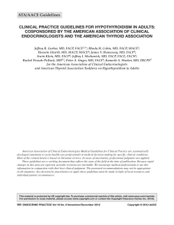
![Reproductive Ethics [PDF] - Center for Genetics and Society](http://s1.paperzz.com/store/data/000442537_1-119aece63f6ed7a909e482fc5b42ebba-250x500.png)