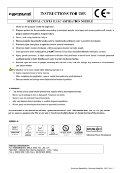
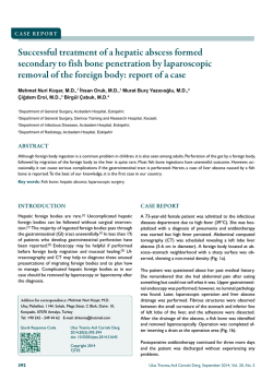
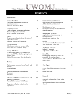
7|P age
Cold abscess of Sternum: Reports of two cases
Email: [email protected]
SANDESH MADI , SANDEEP
NAIK , SHARATH RAO
1
1
VIJAYAN ,
1
MONAPPA
1
Department of Orthopaedics, Kasturba Medical college, Manipal Univer-
1
ABSTRACT
sity, Manipal, Karnataka, India
Received
16 April 2015
Accepted 22 May 2015
Introduction
Sternal involvement is seen in less than 1% of skeletal
tuberculosis. Sternal TB was first reported in 1918 [1] and
until 2007, fewer than 35 cases are documented in English
literature [2]. Even in tubercular endemic belts, there are
only limited and isolated case reports [3]. We report two
Sternal bone affection by Mycobacterium tuberculosis is
a rare entity. Clinical manifestations can vary widely.
There are only a few isolated case reports of tubercular
sternal osteomyelitis in both endemic belts and otherwise. We report tubercular abscess of sternum in apparently two healthy young individuals with no predisposing factors, who presented to us with chief complains of
chronic anterior chest pain. Diagnosis was confirmed by
histo-pathological and microbiological work-up. Combined surgical drainage and anti-tubercular drugs gave
satisfactory outcomes. A high index of suspicion for
tuberculosis is necessary whenever unusual sites are encountered in order to facilitate early diagnosis and
prompt management.
cases of tubercular abscess of sternum in otherwise
healthy individuals. Both cases were diagnosed relatively
KEY WORDS:
early in the course of the disease and responded well to a
combined medical and surgical treatment with no recurrence or complications at one year follow up.
Tuberculosis
Cold abscess
Sternum
Chest pain
Drainage
Case 1
Case 2
A 22 year old student was referred to the Orthopedics de-
A 31 year old architect presented to OPD with chief com-
partment with history of dull aching and left sided anterior
plains of vague anterior wall chest pain of four months
chest wall pain of six months duration. She had apparently
duration. Patient also noticed a localized bulge over the
consulted a general practitioner for this complaint, but the
medial aspect of right clavicle for past one month. There
diagnosis was elusive, as the chest X-ray was apparently
was no history of trauma or constitutional symptoms. On
normal and clinical picture did not fit into any medical or
local examination, a solitary, ~5x6cms, firm swelling over
gastroenterological disorder. There was no history of
the manubrium sterni with moderate tenderness was not-
trauma or constitutional symptoms. There was a ~3x2cm,
ed. The chest x- ray was apparently normal and the rest of
firm, non-transilluminant swelling with mild tenderness
systemic examination was also normal. CT of thorax re-
arising from the body of the sternum. There was no re-
vealed
gional lymphadenopathy and systemic examination was
5.1x1.9x8.5cms suggestive of an abscess overlying the
normal. CT thorax revealed irregular lytic destruction of
manubrium sterni and an ill-defined osteolytic lesion with
rd
a
multi-loculated
collection
measuring
sternal body at level of 3 costochondral junction with an
sclerosis of superior half of manubrium sternum com-
ill-defined abscess measuring 3.3x2.2x2.2cm (Figure 1).
municating with the collection (Figure 2).
Clinical and radiological diagnosis favored the possibility
Correspondence to: Dr Sandesh Madi
ANNALS OF BRITISH MEDICAL SCIENCES. 2015; 1(1): 7-9
of cold abscess in both scenarios and a formal incision and
8|P age
drainage was performed. The purulent material and ne-
Figure 2. CT sagittal view of case 2 showing involvement
crotic tissue samples were sent for histopathological and
of manubrium sterni and cold abscess tracking to subcuta-
microbiological analysis which confirmed the tubercular
neous plane
pathology. The investigational work up of the two cases is
compared in Table 1. Both patients were started on antitubercular regimen [four drugs for two months followed
by two drugs for 10 months daily]. Patients recovered
completely at the end of 12 months of a close follow-up
with no recurrence or complications.
Table 1: A Comparison of investigational work up of the
two cases.
INVESTIGATIONS
CASE 1
CASE 2
Hb
TLC
DLC
12.0 g/dl
6.4x10³ /µL
Lymphocytes15.7%
Neutrophils71.6%
11.2 g/dl
8.7x10³ /µL
Lymphocytes19.7%
Neutrophils-68.2%
ESR
44 mm/hour
63 mm/hour
the primary pulmonary foci or direct contiguous spread
CRP
3.0 mg/L
70.3 mg/L
from the hilar lymph nodes. Typically, in the event of
HIV 1&2
RBS
AFB (pus)
PCR (nested)
Biopsy
Negative
88 mg/dl
Not seen
Positive
Granuloma
Negative
96 mg/dl
Scanty growth
Positive
Granuloma
immunosuppressive states such as HIV/AIDS, diabetes,
Discussion
Tubercular affection of the sternum is known to occur in
less than 1% of all skeletal tuberculosis [2]. Mechanism of
involvement is either lympho-hematogenous spread from
Hb- Hemoglobin; TLC- Total Leucocyte Count; DLC- Differential Leucocyte Count; ESR- Erythrocyte Sedimentation Rate; CRP- C-Reactive
Protein; AFB- Acid Fast Bacilli; PCR- Polymerase Chain Reaction.
chronic kidney disease, corticosteroid/chemotherapy use,
alcohol abuse, severe malnutrition and low socioeconomic levels, a dormant infection gets reactivated and
disseminates resulting in osteoarticular involvement. Sternal TB has also been reported in two exceptional conditions: after sternotomy for cardiac surgeries [4] and fol-
Figure 1. CT scan sagittal view of case 1 showing involvement of body of sternum and presence of sequestrum.
lowing BCG vaccination in infants [5]. Clinically, patient
can
present
with
anterior
chest
wall
swelling,
pre/retrosternal dull aching pain or in long standing cases,
indolent ulcers/discharging sinuses over anterior chest
wall and sternal fracture. Presence of constitutional symptoms can be variable. Differential diagnosis includes numerous diverse disorders such as granulomatous diseases
(sarcoidosis), chronic infections (fungal/parasitic) and
various malignancies (lymphomas and metastasis).
Serological markers of inflammation like ESR, CRP and
TLC are neither specific nor entirely reliable [7]. In the
chest x-ray (PA and lateral views), it is difficult to appreciate the sternal affection. Moreover, radiological signs
occur much later than the presenting clinical features, and
abscesses or sinuses are present much before the focus is
detected [3]. CT scans define the extent of bone destruction, while MRI is useful to determine soft tissue exten-
ANNALS OF BRITISH MEDICAL SCIENCES. 2015; 1(1): 7-9
9|P age
sion. However, neither can confirm the diagnosis and only
Conclusion
biopsy is the gold standard. It is possible to ascertain the
Tubercular abscess of sternum is a rare clinical entity but
diagnosis by fine needle aspiration cytology or marginal
can be detected early owing to its superficial location.
biopsy from an established sinus tract. Under the micro-
Anterior chest pain and swelling are early manifestations,
scope, extensive areas of caseous necrosis surrounded by
which unless appropriately evaluated can lead to serious
lymphocytes and epitheloid cells are typically observed.
complications which are difficult to manage. Exclusion of
PCR for M. tuberculosis complex is a rapid test that
tuberculosis should not be prejudiced based upon age, sex
shows good correlation with histological findings, with an
or immunity status. Long term medical management is the
85% sensitivity and 80% specificity [6]. The surgical role
mainstay of treatment supplemented with surgical drain-
in sternal TB encompasses the drainage of abscess, se-
age of the abscess. A periodic follow-up is warranted to
questrectomy, sinus tract excision and flap reconstruction
monitor the response to treatment and look out for com-
for sternal defects besides an open biopsy. A structured
plications. Informed consent from both patients has been
regimen comprising of four drugs [Isoniazid (300mg),
obtained for publication.
Rifampicin (450mg), Pyrazinamide (750mg) and Ethambutol (800mg)] is the mainstay of treatment. It is usually
Conflict of Interest
initiated with all four drugs daily for two months in the
We declare that we have no conflict of interest.
intensive phase followed by a maintenance phase with
Isoniazid and Rifampicin daily for 10-18 months. However, there is no consensus in terms of drug combinations as
References
1
well as duration of the regime especially in osteo-articular
TB even in endemic belts. Generally, combined clinical
2
and radiological signs of improvements are taken into
consideration to decide about termination of the regimen.
3
Clinical signs of improvement include weight gain, decrease in pain and swelling, and healing of sinuses. Radio-
4
logical signs of healing, however, usually lag behind clinical improvement by several weeks and are all the more
difficult to appreciate in the sternum. Possible complica-
5
tions of sternal tuberculosis include secondary infection,
fistula formation, spontaneous fractures of the sternum,
compression or erosion of the large blood vessels, com-
6
pression of the trachea and migration of tubercular abscess
into the mediastinum, pleural cavity or subcutaneous tis-
7
sues [8]. A close follow up is also essential to monitor
response to treatment, patients’ compliance, drug resistance and adverse side effects.
ANNALS OF BRITISH MEDICAL SCIENCES. 2015; 1(1): 7-9
8
Vaughn RT. Acute osteomyelitis of the sternum:
woody phlegmon: osteotomy and drainage. Surg
Clin North Am 1918; 5: 253-262.
Jain VK, Singh Y, Shukla A, Mittal D. Tuberculous osteomyelitis of sternum: a case report. J
Clin Diagn Res 2007; 1: 163-7.
Tuli SM, Sinha GP. Skeletal tuberculosis. "Unusual" lesions. Indian J Orthop 1969; 3: 5-18.
Kim HJ, Kim JB, Chung CH. Chronic Sternum
Wound Infection Caused by Mycobacterium tuberculosis After Cardiac Surgery. Ann Thorac
Surg, 2012; 94(4): 1332-1335.
Kato Y, Horikawa Y, Nishimura Y, Shimoda H,
Shigeto E, Ueda K.. Sternal tuberculosis in a
9‐month‐old infant after BCG vaccination. Acta
Paediatrica 2000; 89(12): 1495-1497.
Vasa M, Ohikhuare C, Brickner L. Primary sternal tuberculosis osteomyelitis: A case report and
discussion. Can J Infect Dis Med Microbiol.
2009; 20(4): 181.
Watts HG, Angrles L, Lifesto RM. Current concepts review: tuberculosis of bones and joints. J
Bone Joint Surg Am. 1996; 78:288–298.
Sharma S, Juneja M, Garg A. Primary tubercular
osteomyelitis of the sternum. Indian J Pediatr.
2005; 72(8):709-10.
SANDESH MADI, SANDEEP VIJAYAN, MONAPPA NAIK, SHARATH RAO, ABMS Sternal bone affection by Mycobacterium tuberculosis is a rare entity. Clinical manifestations can vary widely. There are only a few isolated case reports of tubercular sternal osteomyelitis in both endemic belts and otherwise. We report tubercular abscess of sternum in apparently two healthy young individuals with no predisposing factors, who presented to us with chief complains of chronic anterior chest pain. Diagnosis was confirmed by histo-pathological and microbiological work-up. Combined surgical drainage and anti-tubercular drugs gave satisfactory outcomes. A high index of suspicion for tuberculosis is necessary whenever unusual sites are encountered in order to facilitate early diagnosis and prompt management. KEY WORDS: Tuberculosis Cold abscess Sternum Chest pain Drainag
© Copyright 2026 Paperzz