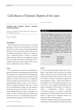
GÖĞÜS DUVARINDA SAPTANAN İNTRAMÜSKÜLER
HEMANJİOM: OLGU SUNUMU
Intramuscular Hemangıoma of the Chest Wall: Case Report
Murat SARIÇAM1, Serkan KAYA1,Bayram METİN2, Emre ÖZKAN1, Mustafa Fatih ERKOÇ3
ÖZET
Ağrı Devlet Hastanesi, Göğüs
1
Cerrahisi Kliniği, Ağrı
Bozok Üniversitesi, Tıp Fakültesi,
2
Göğüs Cerrahisi Anabilim Dalı,
Yozgat
Bozok Üniversitesi, Tıp Fakültesi,
3
Radyoloji Anabilim Dalı, Yozgat
İntramüsküler hemanjiomlar nadir görülen ve esas olarak ekstremite kaslarında saptanan selim tümörlerdir. Ağrı şikayeti veya aşırı büyümeye neden olmadıkları durumlarda
tanı konulması için geçen süre oldukça uzun olmaktadır. Kliniğimize sol yan göğüs duvarında yavaş büyüme gösteren lezyonu nedeniyle başvuran 23 yaşında bir erkek hasta
olgusu sunmaktayız. Manyetik rezonans görüntülemesi sol latissimus dorsi kası içinde
10x5x4 cm. boyutlarında vasküler bir kitle olduğunu raporladı. Çevre dokulardan güvenli
sınırlar dahilinde eksiksiz tümör rezeksiyonu uyguladık ve histopatolojik inceleme kavernöz tip intramüsküler hemanjiom olduğunu gösterdi. Hastada ameliyat sonrasındaki 1
yılda nüks saptanmadı.
Anahtar kelimeler: Hemanjiom, Göğüs kafesi, Tanı
ABSTRACT
Murat SARIÇAM, Uzm. Dr.
Serkan KAYA, Uzm. Dr.
Bayram METİN, Yrd. Doç. Dr.
Emre ÖZKAN, Uzm. Dr.
Mustafa Fatih ERKOÇ, Yrd. Doç. Dr.
Intramuscular hemangiomas are rare benign tumors and are mainly affecting the
extremity muscles. If they do not cause pain or excessive, the diagnosis may require
quite a long time. We present a 23 year old male patient who was admitted to our clinic
with a slowly enlarging lession in the left lateral chest wall. Magnetic resonance imaging
reported a vascular mass measuring 10x5x4 cm. in the left latissimus dorsi muscle. We
performed a complete resection of the tumor with safe margins of the surrounding tissue
and the histopathologic examination demonstrated an intramuscular hemangioma of
the cavernous type. The patient has been free of recurrence for 1 year after surgery.
Keywords: Hemangioma; Chest wall; Diagnosis
İletişim:
Op. Dr. Murat SARIÇAM
Fırat Mah. 956.Sok. Güzelbahçe
Sitesi E Blok No:10 D:10 Ağrı
Tel: 0533- 6961299
e-mail:
[email protected]
Geliş tarihi/Received: 29.04.2013
Kabul tarihiAccepted: 09.12.2013
Bozok Tıp Derg 2014;4(3):69-71
Bozok Med J 2014;4(3):69-71
69
SARIÇAM ve ark.
İntramüsküler Hemanjiom
Bozok Tıp Derg 2014;4(3):69-71
Bozok Med J 2014;4(3):69-71
INTRODUCTION
Intramuscular hemangiomas (IMH) are rare benign
neoplasms mostly originating from extremity muscles;
even rarer are those affecting the chest wall. The
etiology of their growths have been explained by
traumatic, congenital and hormonal theories (1). IMH
are histologically divided into cavernous and capillary
variants where mixtures of both types have also been
noted. The cavernous type is characterized by larger
size and a long clinical history while a small lesion and
shorter history is evident in capillary type (2). If they do
not reach a considerable size or cause pain, they may
not be noticed for a long time and the correct diagnosis
may be difficult. Histological examination of the biopsy
specimen is mandatory to evaluate the exact nature
of the lesion so a large surgical biopsy is needed for a
definitive diagnosis. Increasing growth of the tumor
size, functional impairment, cosmetic deformity and
intractable pain are indications for surgery.
necrosis (Figure 3). Histologic examination determined
intramuscular
cavernous
hemangioma.
The
postoperative period was uneventful and the patient
was discharged 2 days after the operation. He has
been followed up for 1 year without any evidence of
recurrence or any complications.
We report a case of an intramuscular hemangioma of
the chest wall that had remained undiagnosed for 3
years and was completely resected.
CASE REPORT
A 23-year-old male was admitted to our hospital with
a palpable mass in the left lateral portion of the chest.
This lession which appeared 3 years ago was slowly
enlarging and causing only cosmetic deformity but not
pain. He had no story of smoking, chest trauma, longterm medication or a previous operation in his clinical
history. Physical examination revealed a painless soft
tissue mass lacking discrete margins in the left lateral
side of the chest. Magnetic resonance imaging (MRI) in
T1 sequence reported a mass measuring 10x5x4 cm. and
containing vascular components in the left latissimus
dorsi muscle (Figure 1). Through a left lateral incision,
the mass was completely excised with safe margins of
the surrounding muscle (Figure 2). Microscopically the
tumor was made of proliferating and dilated vessel
components which was accompanied by mature fatty
tissue and focal calcifications with no sign of malignancy
such as endothelial bulging, pleomorphism, mitosis or
70
Figure 1. MRI revealing a vascular mass in the chest
wall
Figure 2. Excision and macroscopic view of the tumor
Bozok Tıp Derg 2014;4(3):69-71
Bozok Med J 2014;4(3):69-71
Figure 3. Hematoxylin and eosin–stained section
(original magnification x 200)
DISCUSSION
Intramuscular hemangiomas are rare, representing
0,8% of all hemangiomas and occuring commonly in
young adults with 94% presenting in patients younger
than 30 years (1). Lower extremity is the most common
site of involvement while the localization in the chest
wall is very rare (2). 90% of IMH are misdiagnosed because they lack constitutional symptoms where pain is
the main symptom in 60% of the cases (3). Although
these tumors are accepted to be completely benign
and never metastatizing, a local recurrence rate up to
18% after surgical excision has been reported (1-3).
Computerized tomography scan may be helpful in
showing the involvement of adjacent structures and
revealing focal calcifications (phleboliths) that are present in approximately 25% of cases. MRI is important
to differentiate between types of hemangiomas and
visualize the extent of the tumors. Angiography may be
helpful about the relationship between the tumor and
a neurovascular bundle if MRI has not provided sufficient information (4).
Differential diagnosis includes infection, bone tumor,
lipoma, liposarcoma, elastofibroma dorsi and desmoid
SARIÇAM ve ark.
İntramüsküler Hemanjiom
tumor. MRI is reliable in detecting intramuscular hemangiomas which show intermediate signal on T1WI and
hyperintense signal on T2WI with strong postcontrast
enhancement while other soft-tissue tumors show hyperintense signal on only T2WI. Histologic study of the
biopsy or surgical specimen is the only way to reach
a definitive diagnosis (5). Complete surgical excision
with clear margins is the safest therapeutic approach
while cryotherapy, radiotherapy, electrocoagulation
and embolization may cause benefits in cases whom
excision is impractical or partial. Adjuvant therapy with
interferon-α may be considered in cases of recurrence
(6-7).
In conclusion IMH of the chest wall are very rare entities that require a high index of suspicion for an accurate diagnosis. Complete surgical excision is a vital
necessity for the treatment.
REFERENCES
1. Allen PW, Enzinger FM. Hemangioma of skeletal muscle:
an analysis of 89 cases. Cancer. 1972;29(1):8-22.
2. Cohen AJ, Youkey JR, Claggett GP, Huggins M,
Nadalo L, d’Avis JC. Intramuscular hemangioma. JAMA.
1983;249(19):2680-2.
3. Wild TA, Raab P, Krauspe R. Hemangioma of skeletal
muscle. Arch Orthop Trauma Surg. 2000;120(3-4):139-43.
4. Ly JQ, Sanders TG. Case 65: Hemangioma of the chest
wall. Radiology. 2003;229(3):726-9.
5. Beham AJ, Fletcher CDM. Intramuscular angioma: A
clinicopathologic analysis of 74 cases. Histopathology.
1991;18(1):53–9.
6. Lee JK, Lim SC. Intramuscular hemangiomas of the
mylohyoid and sternocleidomastoid muscle. Auris Nasus
Larynx. 2005;32(3):323–7.
7. Ulku R, Onat S, Avci A, Ozmen CA. Resection of intercostal
hemangioma with involved chest wall and ribs in an 11-yearold girl. Tex Heart Inst J. 2010;37(4):486–9.
71
© Copyright 2026 Paperzz