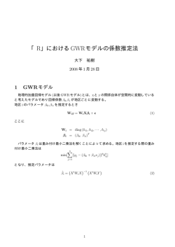
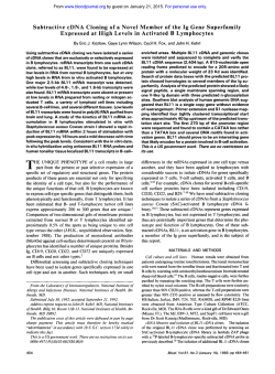
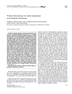
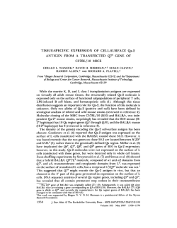
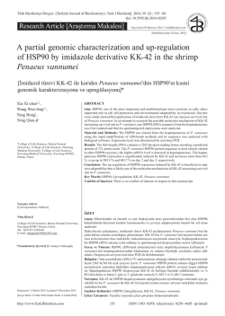
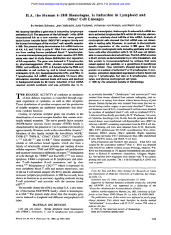
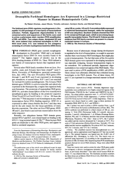
Turkish Journal of Biology
Turk J Biol
(2014) 38: 664-670
© TÜBİTAK
doi:10.3906/biy-1401-91
http://journals.tubitak.gov.tr/biology/
Research Article
Cloning and expression analysis of 1-deoxy-D-xylulose-5-phosphate synthase
gene from the medicinal plant Conyza blinii H.Lév.
1
1
1,2
1
1
1,
1
1
Rong SUN , Shan LIU , Jing-Lei GAO , Zi-Zhong TANG , Hui CHEN *, Cheng-Lei LI , Qi WU
Department of Biology and Science, College of Life and Basic Science, Sichuan Agricultural University, Ya’an, P.R. China
2
Department of Biological and Chemical Engineering, Panzhihua University, Panzhihua, P.R. China
Received: 31.01.2014
Accepted: 10.06.2014
Published Online: 05.09.2014
Printed: 30.09.2014
Abstract: Conyza blinii H.Lév. is a traditional Chinese medicinal plant that is distributed mainly in southwestern Sichuan and northern
Yunnan. Its characteristic product is blinin, which has, among other properties, antigastric ulcer activity, and can serve as a qualitycontrol standard for such medicine. The problem is that C. blinii only produces low yields of blinin. As a diterpene, blinin is likely
formed by the methylerythritol phosphate pathway. While 1-deoxy-D-xylulose-5-phosphate synthase (DXS) is the first rate-limiting
enzyme in diterpenoid biosynthesis, it is a switch in the pathway. The DXS gene was successfully cloned and characterized from C.
blinii by homologous cloning and rapid-amplification of cDNA ends (RACE). It was designated cbDXS and contains a 2190-bp open
reading frame encoding 730 amino acids (aa), including a 17-aa signal peptide and a 713-aa mature protein. Semiquantitative RTPCR was used to determine the expression levels of cbDXS in different C. blinii tissues at the seedling stage. The corresponding blinin
concentrations were also analyzed by high-performance liquid chromatography (HPLC). The cbDXS gene showed tissue specificity.
Moreover, its expression levels were highly correlated to blinin concentrations. In summary, it is suggested that overexpression of this
gene may increase flux toward blinin synthesis.
Key words: Conyza blinii H.Lév., blinin, DXS gene, RACE, semiquantitative RT-PCR, HPLC
1. Introduction
Conyza blinii H.Lév. is a member of the plant family
Compositae. It is a traditional Chinese medicinal plant that
is mainly distributed in southwestern Sichuan and northern
Yunnan. It is used as a powerful drug for acute icteric
hepatitis and chronic tracheitis. Its secondary metabolites
include flavonoids, saponins, and blinin, among others. Of
these, blinin is the characteristic product present in C. blinii
and can be used as a quality-control standard in medicine
(Wang et al., 2010). However, the plant produces only low
yields of blinin, which are insufficient to meet the demand
for pharmaceutical preparations. Improving blinin content
is the key to improving the quality of C. blinii. Although
knowledge of blinin biosynthesis and its pathway will
provide a foundation for improving its content, there have
been no studies on the blinin biosynthesis pathway.
Blinin is a new clerodane diterpene lactone. Terpenes
can be biosynthesized via 2 pathways in plants: the
mevalonate pathway or the methylerythritol phosphate
(MEP) pathway (Ma et al., 2006). As a diterpene, blinin is
likely formed by the latter (Figure 1). There are a number of
key enzymes that regulate flux through the MEP pathway:
*Correspondence: [email protected]
664
1-deoxy-D-xylulose-5-phosphate
synthase
(DXS)
catalyzes the reaction of pyruvate with glyceraldehyde3-phosphate, resulting in 1-deoxyxylulose-5-phosphate.
This is the first step in the MEP pathway, and it has been
documented in Coleus forskohlii (Engprasert et al., 2005)
and Elaeis guineensis (Khemvong and Suvachittanont,
2005). DXS has been suggested as a regulatory enzyme
in isoprenoid biosynthesis. In Catharanthus roseus hairy
roots, the expression of DXS and geraniol-10-hydroxylase
or anthranilate synthase increases accumulation of
the terpenoid indole alkaloid (Peebles et al., 2011). In
addition, overexpression of DXS increases anthraquinone
production in transgenic cell-suspension cultures of
Morinda citrifolia (Quevedo et al., 2010).
The study of changes in gene expression in different
cell types or the same cell types at different developmental
stages or under different developmental conditions has
become a research hotspot in biology (Liu et al., 2004).
Worapan et al. (2010) and Zhang et al. (2007) analyzed
the mRNA expression levels of genes from various organs
of Croton stellatopilosus and Triticum aestivum Linn.
Semiquantitative RT-PCR is an effective method for
SUN et al. / Turk J Biol
O
O
O
OH
COO
DXS
O
OH
P
O
OH
O
O
H
O
O
P
O
HO
O
O
OAc
HO
O
Figure 1. MEP pathway for blinin biosynthesis.
studying gene transcription levels (Cottrez et al., 1994)
that is widely used due to its simple operation. Ding et al.
(2010) used semiquantitative RT-PCR to investigate the
effects of Pseudoperonospora cubensis Rostow, salicylic
acid, and CaCl2 on the expression of a resistance gene
analog (RGA) in cucumber (Cucumis sativus L.) cultivar
Dongnong 129, a variety that is resistant to downy mildew.
Results showed that all of the treatments could increase
the expression of RGA1 and RGA5. Using this method,
Gao et al. (2006) determined the expression of the human
bone morphogenetic protein-2 gene in different tissues of
tobacco plants. The expression levels of a fusion protein in
root and stem tissues were significantly higher than those
in leaf tissues.
The aim of this work is to clone the DXS gene and
analyze its expression. Rapid amplification of cDNA
ends (RACE) technology was used to obtain the target
gene. Semiquantitative RT-PCR was used to detect gene
expression levels. Furthermore, blinin concentrations were
analyzed by high-performance liquid chromatography
(HPLC).
After RNase H treatment, the resulting single-stranded
cDNA mixtures were used as templates for the following
PCR steps. According to the highly conserved amino
acids, sets of degenerate primers were designed manually
by Primer 5.0 software and used for core fragment
amplification (Table) with the following PCR program:
94 °C for 4 min, then 30 cycles of 94 °C for 50 s, 50 °C
for 40 s, and 72 °C for 50 s, with a final extension at 72
°C for 10 min. The PCR product was purified and cloned
2. Materials and methods
2.1. Plant material
The leaves, stems, and roots of Conyza blinii H.Lév. were
obtained from plants growing in Panzhihua, a city located
in southwestern Sichuan Province, in July 2012. All samples
were identified by Prof Chun-Bang Ding of Sichuan
Agricultural University. A voucher specimen (41945) was
deposited in the Herbarium of the Department of Biology,
Sichuan University, China.
2.2. Cloning of DXS cDNA
Total RNA was isolated from young leaves using the RNA
Isolating Reagent Kit (TIANDZ). Total RNA (10 μg)
was reverse-transcribed according to the manufacturer’s
protocol (RevertAid First Strand cDNA Synthesis Kit,
Thermo Scientific) with oligo(dT)18 primer (Thermo).
Table. Primers used in this study.
Primer
Sequence (5’→3’)
Core fragment primers
DXSuF
ACACRCAGTTACKTTSGC
DXSuR
AATGCCAWTGCCTNTTGG
5’ and 3’ RACE primers
3’-128
AGGGCTGGTTTGGTAGGAGCAGATGG
3’-209
GTGGTGATGGCTCCTTCTTGTGAGTCCG
5’-151
TCTGCTCCTACCAAACCAGCCCTGTCTAT
5’-339
TTATCTGGTGGGAGGAGTGAACCTATGC
5’-564
GCCTTTGCCTTTCTCGGTAACAATGTGGAC
Recloning primers
DXSR
AAAATATATGGCTTCTTATAGTGC
DXSF
AGACTTTGGCATCATCTTTTCCCTG
Semiquantitative RT-PCR primers
GAPDHsu
GTGGTGCCAAGAAAGTG
GAPDHsd
GCTAGAGGAGCAGGACA
DXSsu
AGGTTTAGCAACCGAAGG
DXSsd
TATCTGGTGGGAGGAGTGA
665
SUN et al. / Turk J Biol
into the pMD19-T simple vector (TaKaRa), transformed
into Escherichia coli strain DH5α, and sequenced. The core
fragment was subsequently used to design the specific
primers for cloning the full-length cDNA of DXS by RACE.
The 5’ and 3’ end cDNA fragments were amplified
using the SMARTer RACE cDNA Amplification Kit
(Clontech). The first-strand RACE-ready cDNA was
prepared according to the kit and then used as a template
for the second-strand RACE. The nested gene-specific
primers designed for the 5’ and 3’ RACE are shown in
the Table. The first cycle of amplification for RACE (both
5’ RACE and 3’ RACE) used the outside primers and
UMP (universal primer A mix, provided in the kit) with
the first-strand RACE-ready cDNA as templates. For the
nested amplification, nested primers and NUP (nested
universal primer A, provided in the kit) were used with
the products of the previous amplification as templates.
The PCR programs for 5’ and 3’ RACE were carried out
under the conditions described in the kit’s introduction.
The first round consisted of 25 cycles of amplification
(94 °C for 30 s, 68 °C for 30 s, and 72 °C for 2 min). The
melting temperature of the next amplification round was
increased by 2 °C and the number of cycles changed to 20.
The nested PCR products were also purified and cloned
into the pMD19-T simple vector, transformed into strain
DH5α, and sequenced. The full-length cDNA sequence
was obtained by assembling the core fragment, 3’ RACE,
and 5’ RACE with DNAMAN software. The full-length
cDNA sequence was then recloned using the gene-specific
primers, which were designed according to the splicing
sequence (Table). The sequence was then sequenced and
analyzed by bioinformatic methods.
2.3. Bioinformatic analysis
The nucleotide sequence and the deduced amino acid
(aa) sequence were analyzed and subjected to BLAST
comparisons on the National Center for Biotechnology
Information (NCBI) website. The SignalP 4.1 server was
used to predict the signal peptide, and PSORT was used
to predict its subcellular localization. The secondary
structure was determined by the SOPMA server. MEGA
5.20 was used to construct the phylogenetic tree.
2.4. Extraction of blinin and determination of its content
Different tissues of C. blinii at the seedling stage were
separately crushed in a grinder. A 1.5-g sample was then
extracted with 45 mL of methanol by Soxhlet extraction.
When the sample became colorless with a 50-mL volume,
the solution was analyzed by HPLC. In addition, a blinin
standard and methanol were used to produce standard
solutions of different concentrations.
Blinin was analyzed on a Symmetry Shield C18 column
with acetonitrile, methanol, and water (15:40:45, v/v) as
666
the mobile phase for elution. The flow rate was 1.0 mL/
min and the injection volume was 20 μL. Detection was
performed under UV light (210 nm) at 25 °C. Three
extraction samples were prepared for HPLC analysis, and
each sample was injected 3 times.
2.5. Semiquantitative RT-PCR
Semiquantitative RT-PCR was used to determine the
expression levels of DXS in the different types of tissues
at the seedling stage. Total RNA was isolated from leaves,
stems, and roots. The single-stranded cDNA was then
reverse-transcribed with the PrimeScript RT Reagent
Kit with gDNA Eraser (Thermo). The primer sets used
to amplify DXS and the housekeeping gene GAPDH are
shown in the Table. The PCRs were performed under
optimal conditions. The PCR products were separated
on a 2.0% agarose gel and stained with GoldView. Band
intensities were measured using Quantity One software.
3. Results
The gene that was successfully cloned and characterized
from C. blinii was named cbDXS. Its GenBank accession
number is KJ155788. PCR amplification and sequence
assembly showed that cbDXS consists of 2479 bp. It contains
a 2190-bp open reading frame encoding 730 aa, including
a 17-aa signal peptide and a 713-aa mature protein. The
entire coding region was compared with other plant DXS
genes and revealed high levels of similarity (77.65%;
Figure 2). According to the BLAST results, cbDXS had
high similarity to DXS from Tagetes erecta (83%), Solanum
lycopersicum (76%), and Picea glauca (74%). BLASTP
comparisons suggested that the protein belongs to the
transketolase and dehydrogenase E1 component family
(Figure 3).
The protein had a calculated molecular mass of 78
kDa and an isoelectric point of 6.96. Its 2-dimensional
structure was predicted by PBIL based on the SOPMA
method, giving a composition of 38.68% alpha helices,
13.44% extended strands, 6.31% beta turns, and 41.56%
random coils. PSORT suggested localization of cbDXS
protein in the chloroplast. This coincides with an earlier
report predicting that most of the DXS proteins belong to
the chloroplast (Krushkal et al., 2003).
All of the tested seedling-stage tissues expressed cbDXS,
with the gene showing strong tissue specificity (Figure 4).
The highest levels of transcripts were detected in the leaves
(72.67%) and the roots (28.00%); the lowest expression was
found in the stems (17.67%). Blinin concentrations were
analyzed by HPLC. The equation of the calibration curve
was y = 48.822x + 88.279, with R2 = 0.9991. The highest
level of blinin was found in the leaves (0.961%). The blinin
SUN et al. / Turk J Biol
CONYZA_BLINII
STEVIA_REBAUDIANA
TAGETES_ERECTA
ARTEMISIA_APIACEA
TAXUS_CUSPIDATA
MASYSALKGAFVPTTLAQDGYSPSSLPNLSTNAIMPLNKRKFMKF..........FAAAKDNATNEHGD......LVRAIGSVA...TNLKYSGEKPKTP
MAVAGSTMN.................LHLTSSPYKTVPSLCKFTRKQFR......LKASATNPDAEDGK......MMFKNDKPN...LKVEFAGEKPVTP
MALCGALKGGFVP........IAQNGYTSSSLLNPSANAIMPSNKRKFLGI....VAVSKEHATNEHED......LTTMDKTTS...TTLKYSGDKPKTP
MALSAFAFP............TQINHRSVTSNQPLIQHCLFGTDLN.........SSQKPFNQIVKRSN.....GIRSTLSERG......EYHSQRPPTP
MAATIGMGSMVASSSSLFK..NGGNSRTESGTSLCSKKGFMVSQKKKYTALQRAGASQRKHGIVSANSNNADGESIIKDIVRREDAPLKIEYTGEKPPTP
81
68
79
68
98
CONYZA_BLINII
STEVIA_REBAUDIANA
TAGETES_ERECTA
ARTEMISIA_APIACEA
TAXUS_CUSPIDATA
ILDTINYPKHMKNLSVEELERLADELREEIVYTVSKTGGHLSSSLGVVELTVSLHHVFNTPDDKIIWDVGHQAYPHKILTGRRSRMETMRQTCGLAGFPK
LLDTINYPVHMKNLTTQDLEQLAAELRQDIVYSVANTGGHLSSSLGVVELSVALHHVFNTPDDKIIWDVGHQAYPHKILTGRRSKMHTIRKTSGLAGFPK
ILDTINYPIHMKNLCVEELVKLADELREEIVYTVSKTGGHLSSSLGVVELTVSLHHVFNTPEDKIIWDVGHQAYPHKILTGRRSRMRTIRQTFGLAGFPK
LLDTINYPIHMKNLSVKELKQLADELRSDVIFNVSKTGGHLGSSLGVVELTVALHYVFNTPQDKILWDVGHQSYPHKILTGRRDQMHTIRQTNGLAGFTK
LLDTINYPKHMKNLNVQDLKQLAKELREEIIFGVSKTGGHLSASLGVVDLTVALHYVFNTPQDKIVWDVGHQSYPHKILTGRRSNMSSIRQTSGIAGFPK
181
168
179
168
198
CONYZA_BLINII
STEVIA_REBAUDIANA
TAGETES_ERECTA
ARTEMISIA_APIACEA
TAXUS_CUSPIDATA
RDESQHDAFGAGHSSTSISAGLGMAIGRDLLGKDNHVIAVIGDGAMTAGQAYEAMNNAGYLDSNIIIILNDNRQVSLPTATLDGPAPPVGALSRSLTRLQ
RDESAHDAFGAGHSSTSISAGLGMAVGRDLLGKTNNVISVIGDGAMTAGRAYEAINNAGFLDSNLIVVLNDNKQVSLPTATLDGPATPVGALSGALSKLQ
RDESNHDAFGAGHSSTSISAGLGMAVGRDLLGKNNHVIAVIGDGAMTAGQAYEAMNNAGYLDSNLIIVLNDNRQVSLPTATIDGPAPPVGALSRSLTRLQ
RSESEHDCFGTGHSSTTISAGLGMAVGRDLKGGTNDVVAIIGDGAMTAGQAYEAMNNAGYLDSDMIVILNDNKQVSLPTANLDGPIPPVGALSSALSRLQ
RAESEHDAFGAGHSSTSISAALGMAAGRDLLGLPNHCIAVIGDGAMTAGQAYEAMNNAGFLDSNLIIILNDNKQVSLPTATVDGPAPPVGALSKALSKIQ
281
268
279
268
298
CONYZA_BLINII
STEVIA_REBAUDIANA
TAGETES_ERECTA
ARTEMISIA_APIACEA
TAXUS_CUSPIDATA
TSRKVRQLREVAKGVTKQLGDKTHEVAAKMDSYMKGMVGGQAASLFEELGLYYVGPVDGHNVEDLVHVLTKIKSMPSAGPVLVHIVTEKGKGYHPAEIAA
ASTKFRKLREAAKSITKQIGPQAHEVAAKVDEYARGMISASGSTLFEELGLYYIGPVDGHNVEDLVNIFEKVKSMPAPGPVLIHIVTEKGKGYPPAEAAA
TSQKFRQLREAAKEVTKQLGDKTHEVAAKMDSLVKGMVGGQGASMFEELGLYYVGPVDGHNLEDLVYVFDKIKSMTAPGPVLVHIVTEKGKGYPPAEVAA
SNRPLRELREVAKEVTKQIGGPMHELAAKVDEYARGMISGSGSTLFEELGLYYIGPVDGHSIDDLVAILKEVKSTKTTGPVLIHVITEKGRGYPYAEKAA
ASKKFRLLREAAKDITQQIGGPTHEIAAKVDEYARGMISAPRSTLLEELGLYYIGPVDGHSIQDLVAILENVKAMPAPGPVLIHVVTEKGKGYPPAEKAA
381
368
379
368
398
CONYZA_BLINII
STEVIA_REBAUDIANA
TAGETES_ERECTA
ARTEMISIA_APIACEA
TAXUS_CUSPIDATA
DKMHGVVKFNTQTGKQVKVKAKTLSYTQYFADSLVAEAERDEKIIAIHAAMGGGTGLNTFQKQFPTRCFDVGIAEQHAVTFAAGLATEGLKPFCAIYSSF
DRMHGVVKFDVPTGKQFKTKSPTLSYTQYFAESLIKEAEADNKIVAIHAAMGGGTGLNYFQKKCPERCFDVGIAEQHAVTFAAGLATEGLKPFCAIYSSF
DKMHGVVKFDTQTGKQKKNKTKTLSYTQYFVDSLVAEAKEDDKIVAIHAAMGGGTGLNTFQKEFPARCFDVGIAEQHAITFAAGLATEGLKPFCAIYSSF
DKYHGVGKFDPATGKQFKSSAPTQSYTTYFAEALIAEAEADKKIVGIHAAMGGGTGMNLFLRRFPTRCFDVGIAEQHAVTFAAGLACEGLKPFCAIYSSF
DRLHGVVKFDPSTGKQFKAKSPTLSYTQYFAEGLIAEAEKDDKIVGIHAAMGGGTGLNIFQKRFPERCFDVGIAEQHAVTFAAGLATEGLKPFCAIYSSF
481
468
479
468
498
CONYZA_BLINII
STEVIA_REBAUDIANA
TAGETES_ERECTA
ARTEMISIA_APIACEA
TAXUS_CUSPIDATA
LQRGYDQVVHDVDLQKLPVRFAMDRAGLVGADGPTHCGAFDTAFMACLPNMVVMAPSCESELMHMVATAAAIDDRPSCFRYPRGNGIGSLLPPDNKGTPV
LQRGYDQVVHDVDLQKLPVRFAMDRAGLVGADGPTHCGAFDITYMACLPNMVVMAPADEAELMHMVATAAAIDDRPSCFRFPRGNGIGAPLPPNNKGIPI
LQRGYDQVVHDVDLQKLPVRFAMDRAGLVGADGPTHCGAFDTTFMACLPNMVVMAPSCEAELMNMVATAVAIDDRPSCFRYPRGNGIGSILPANNKGTLI
LQRGYDQVVHDVDLQKLPVRFAMDRAGLVGADGPTHSGSFDVTFMACLPNMVVMAPSDEAELFNMVATAAAIDDRPSCFRYPRGNGIGVPLPPGNKGVPL
LQRGYDQVVHDVDLQKLPVRFAMDRAGLVGADGPTHCGAFDITYMACLPNMIVMAPCDEAELIHMVATAAAIDDRPSCFRFPRGNGIGVPLPPNNKGTPV
581
568
579
568
598
CONYZA_BLINII
STEVIA_REBAUDIANA
TAGETES_ERECTA
ARTEMISIA_APIACEA
TAXUS_CUSPIDATA
EIGRGRVLREGSRVALLGYGTIVQSCLAASEILQALGISVTVADARFCKPLDGSLIKQLANEHEVLITIEEGSIGGFSSHVSHYLALNGLLDGNLKWRAI
EVGKGRILLEGTRVAILGYGSIVQECLGAASLLQAHNVSATVADARFCKPLDTGLIRRLANEHEVLLTVEEGSIGGFGSHVAHFLSINGLLDGKLKLRAM
EVGTGRVIKEGNRVALLGYGTIVQSCLAASEVLKKIGISVTVADARFCKPLDGNLIKQLANEHEVLITVEEGSIGGFSSHVSHFLALNGLLDGHLKWRAM
EVGKGRIMLEGQRVALLGYGTAVQSCMAAATIVQERGLNITVADARFCKPLDHSLIRALAKTHEVLITVEEGSIGGFGSHVAHFLALDGLLDGNLKWRPL
EIGKGRILAEGTRVAILGFGSIIQNCLGAREMLEKQGISVTVADARFCKPLDGDLLRRLVKEHEILITVEEGSVGGFGSHVSHFLALNGLLDGKLKWRPM
681
668
679
668
698
CONYZA_BLINII
STEVIA_REBAUDIANA
TAGETES_ERECTA
ARTEMISIA_APIACEA
TAXUS_CUSPIDATA
TLPDRYIDHGAQSNQIEEAGLSPKHISATVLSLIGESKDSLSLINVI
TLPDKYIDHGAPQDQLEETGLSSKHICSSLLSLLGKPKEALQYKSIM
MLPDRYIEHGAQSDQIEEAGLSSKHIAATVLSLIGGSKETLHALNV.
VLPDRYIDHGAPADQLAEAGLTPSHIAATVFNVLGQTREALEVMS..
VLPDRYIEHGAPKDQMDEAGLSSRHIAATVMSLMGKPQAAFDLQ...
728
715
725
713
742
Figure 2. Alignment of the deduced amino acid sequences of cbDXS and related DXS proteins from Stevia rebaudiana,
Tagetes erecta, Artemisia apiacea, and Taxus cuspidata. Residues shaded in black are identical residues that are conserved
in the 5 sequences; residues shaded in gray are identical in 4 of the sequences shown. The thick horizontal line indicates
the region corresponding to the putative thiamine–diphosphate binding site, and the pyrimidine-binding domain is
marked by a double horizontal line. The transketolase motif is indicated by a long arrow.
Figure 3. The result of protein comparisons by BLASTP.
667
SUN et al. / Turk J Biol
0.8
Leaf
Stem
Root
1.0
CbDXS
GAPDH
0.6
0.8
Blinin content (%)
Intensity relative to GAPDH
0.7
0.5
0.4
0.3
0.2
0.4
0.2
0.1
0.0
0.6
Leaf
Stem
Root
0.0
Leaf
Stem
Root
Figure 4. Expression levels of cbDXS in different tissues of C.
blinii at the seedling stage determined by semiquantitative RTPCR. Intensities are relative to GAPDH. Each tissue was analyzed
in 3 replicates, and each band intensity was determined 3 times.
Standard deviation was calculated by SPSS software.
Figure 5. Blinin concentrations in different tissues of C. blinii
at the seedling stage determined by HPLC. Each tissue was
analyzed in 3 replicates. Standard deviation was calculated by
SPSS software.
level was low in the roots and in the stems (0.262% and
0.228%, respectively) (Figure 5). The data showed that the
expression levels of cbDXS and blinin concentrations were
highly correlated.
al., 2002). The cbDXS and DXS from Tagetes erecta belong
to the DXS2 family, while the DXS from Artemisia annua
belongs to DXS1 family, explaining our phylogenetic
results.
The highest expression level of cbDXS was
determined in the leaves, and the lowest level in the
stems. The high expression in the leaves might be due
to the increased demand for pigments for chloroplast
formation and growth (Mayrhofer et al., 2005). Blinin
is a new clerodane diterpenoid lactone. Similar to
gibberellin, it may be produced mainly in tender leaves
and root tips. Photosynthesis occurs in the leaf, and its
metabolism is therefore more vigorous than the root’s.
Consequently, leaf blinin concentration was higher than
root concentration.
The expression studies indicate correlation between
levels of accumulated cbDXS mRNA in given tissues and
the blinin concentrations. The cbDXS expression is highly
correlated to blinin concentration. The results support the
view that the cbDXS gene is involved in blinin biosynthesis.
Overexpression of cbDXS is an attractive goal for metabolic
engineering aimed at increasing blinin biosynthesis.
Aside from being a characteristic compound of C. blinii,
blinin also has antiulcer activity (Su et al., 2007). Moreover,
it has been reported that clerodane diterpenoids may
prevent insects from feeding, and they shows antitumor
activity (Xu et al., 1998). Studies show the medical value
of blinin. In this manuscript, a basic study was performed
of its biosynthetic pathway. However, systematic research
into the blinin pathway is still lacking. Further work
4. Discussion
DXS plays an important role in the secondary metabolic
pathway. It is a switch that regulates flux through the MEP
pathway. It has been reported that the overexpression
of DXS in Arabidopsis led to an increase in terpenoid
concentrations (Estévez et al., 2001). Overexpression of
DXS in transgenic cell-suspension cultures of Morinda
citrifolia increases anthraquinone production (Quevedo
et al., 2010). These results suggest that the overexpression
of DXS can increase secondary metabolic concentrations;
however, cloning and analysis of the DXS gene are
important prerequisites. If we know nothing about the
gene we cannot overexpress it. In this study we successfully
cloned cbDXS from C. blinii and analyzed its expression to
provide a basis for further study.
DXS has been identified and characterized from many
plants (Bouvier et al., 1998; Lange et al., 1998; Lee et al.,
2007). However, nothing is known about DXS from C.
blinii. We successfully cloned cbDXS from this plant. It
contained a 2190-bp open reading frame encoding 730
aa. This result is in line with a previous study suggesting
that plant DXS protein is approximately 691 to 738 aa long
(Jin et al., 2007). Cluster analysis showed a close genetic
relationship between cbDXS and the DXS protein from
Tagetes erecta (Figure 6) and a distant genetic relationship
with the DXS protein from Artemisia annua. Plant DXS
can be classified into 2 clusters: DXS1 and DXS2 (Walter et
668
SUN et al. / Turk J Biol
Conyza blinii
Tagetes erecta |AAG10432.1|
Cucumis sativus |XP 004146353.1|
Antirrhinum majus |AAW28999.1|
Mentha x piperita |AAC33513.1|
Salvia miltiorrhiza |ACQ66107.1|
Ricinus communis |XP 002533688.1|
Vitis vinifera |XP 002271585.1|
Glycine max |XP 003550669.1|
Populus trichocarpa |ERP59641.1|
Theobroma cacao |EOY31729.1|
Catharanthus roseus |ABI35993.1|
Stevia rebaudiana |CAD22155.2|
Taxus x media |AAS89342.1|
Artemisia annua |AAD56390.2|
Medicago truncatula |CAD22530.1|
Andrographis paniculata |AAP14353.1|
Oryza sativa Indica Group |EEC79215.1|
Setaria italica |XP 004962111.1|
Zea mays |ABP88134.1|
0.1
Figure 6. Phylogenetic analysis of DXS proteins from different plant species. Sequence analysis
was performed using MEGA 5.2. The neighbor-joining method was used to create the tree.
Accession numbers are written after the plant names.
should be invested in cloning other genes of this pathway,
expressing those genes in plants, and understanding their
functions with the aim of enhancing blinin concentrations
and thereby enabling development of its potential.
Acknowledgment
This work was supported by funding from the Science and
Technology Department of Sichuan Province (contract
number: 2012ZZ046).
References
Bouvier F, d’Harlingue A, Suire C, Backhaus RA, Camara B (1998).
Dedicated roles of plastid transketolases during the early onset
of isoprenoid biogenesis in pepper fruits. Plant Physiol 117:
1423–1431.
Estévez JM, Cantero A, Reindl A, Reichler S, León P (2001).
1-Deoxy-D-xylulose-5- phosphate synthase, a limiting enzyme
for plastidic isoprenoid biosynthesis in plants. J Biol Chem 276:
22901–22909.
Cottrez F, Auriault C, Capron A, Groux H (1994). Quantitative PCR:
validation of the use of a multispecific internal control. Nucleic
Acids Res 22: 2712–2713.
Gao Y, Suo GL, Han J, He ZQ, Yao W, Dai JW (2006). Expression of
human BMP-2 gene in different tissues of tobacco plants. Acta
Genet Sin 33: 56–62.
Ding GH, Xu CM, Yu H, Zhou XY, Qin ZW (2010). Expression
analysis of the resistance gene analog in cucumber by semiquantitative RT-PCR. Acta Bot Boreal-Occident Sin 30: 659–
664.
Engprasert S, Taura F, Shoyama Y (2005). Molecular cloning,
expression and characterization of recombinant 1-deoxy-Dxylulose-5-phosphate reductoisomerase from Coleus forskohlii
Briq. Plant Sci 169: 287–294.
Jin R, Zhu CQ, Xu CJ (2007). I-V deoxy xylulose-phosphate synthase
(DXS) et gene encoding. Chinese J Cell Biol 29: 706–712
(article in Chinese).
Khemvong S, Suvachittanont W (2005). Molecular cloning and
expression of a cDNA encoding 1-deoxy-D-xylulose-5phosphate synthase from oil palm Elaeis guineensis Jacq. Plant
Sci 169: 571–578.
669
SUN et al. / Turk J Biol
Krushkal J, Pistilli M, Ferrell KM, Souret FF, Weathers PJ (2003).
Computational analysis of the evolution of the structure and
function of 1-deoxy-D-xylulose-5-phosphate synthase, a key
regulator of the mevalonate-independent pathway in plants.
Gene 313: 127–138.
Lange BM, Wildung MR, McCaskill D, Croteau R (1998). A family
of transketolases that directs isoprenoid biosynthesis via a
mevalonate-independent pathway. P Natl Acad Sci U S A 95:
2100–2104.
Lee JK, Oh DK, Kim SY (2007). Cloning and characterization of the
dxs gene, encoding 1-deoxy-D-xylulose-5-phosphate synthase
from Agrobacterium tumefaciens, and its overexpression in
Agrobacterium tumefaciens. J Biotechnol 128: 555–566.
Liu ZC, Zhao J, Liu MJ (2004). An introduction to mRNA differential
display technique. Mol Plant Breeding 2: 895–900.
Ma L, Ding P, Yang GX, He GY (2006). Advances on the plant
terpenoid isoprenoid biosynthetic pathway and its key
enzymes. Biotechnol Bull 1: 22–30.
Mayrhofer S, Teuber M, Zimmer I, Louis S, Fischbach RJ, Schnitzler
JP (2005). Diurnal and seasonal variation of isoprene
biosynthesis-related genes in grey poplar leaves. Plant Physiol
139: 474–484.
Peebles CA, Sander GW, Hughes EH, Peacock R, Shanks JV, San KY
(2011). The expression of 1-deoxy-D-xylulose synthase and
geraniol-10-hydroxylase or anthranilate synthase increases
terpenoid indole alkaloid accumulation in Catharanthus roseus
hairy roots. Metab Eng 13: 234–240.
Quevedo C, Perassolo M, Alechine E, Corach D, Giulietti AM,
Talou JR (2010). Increasing anthraquinone production by
overexpression of 1-deoxy-D-xylulose-5- phosphate synthase
in transgenic cell suspension cultures of Morinda citrifolia.
Biotechnol Lett 32: 997–1003.
670
Su YF, Chen L, Lu Y, Chai X, Li M, Guo DA (2007). Chemical
constituents and their antiulcerogenic studies on whole herb
of Conyza blinii. Chinese Traditional and Herbal Drugs 38:
332–334.
Walter MH, Hans J, Strack D (2002). Two distantly related genes
encoding
1-deoxy-d-xylulose-5-phosphate
synthases:
differential regulation in shoots and apocarotenoidaccumulating mycorrhizal roots. Plant J 31: 243–254.
Wang R, Li WY, Xu H, Zhao SM, Hu YC, Li YM, Wang ZT (2010).
M. qualitas medicinae Gentian. Journal of Chinese Medicinal
Materials 33: 884–886 (article in Chinese).
Worapan S, Juraithip W, Tanawan S, Tossaton C, Yutaka E, Wanchai
DE (2010). Cloning and expression of 1-deoxy-D-xylulose5phosphate synthase cDNA from Croton stellatopilosus and
expression of 2C-methyl-D-erythritol4-phosphate synthase
and geranylgeranyl diphosphate synthase, key enzymes of
plaunotol biosynthesis. J Plant Physiol 167: 292–300.
Xu JH, Shang ZZ, Yang SH, Chen HM, Min ZD (1998). Et actio
est ratio agendi, ratione studiorum Cornelius diterpenoid
insect aspernabilis. Acta Entomol Sin 41: 366–370 (article in
Chinese).
Zhang XJ, Zhang LS, Yang Y (2007). Analysis of wheat dehydrin like
gene during water stress condition by semi-quantitative RTPCR. Acta Bot Boreal-Occident Sin 27: 2158–2162.
© Copyright 2026 Paperzz