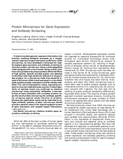
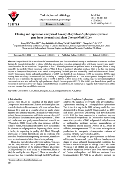
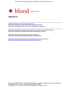
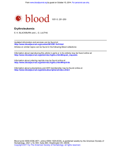
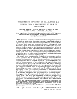
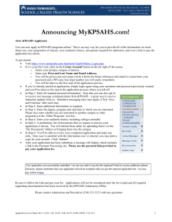
From www.bloodjournal.org by guest on January 21, 2015. For personal use only.
Subtractive c D N A Cloning of a Novel Member of the Ig Gene Superfamily
Expressed at High Levels in Activated B Lymphocytes
By Eric J. Kozlow, Gaye Lynn Wilson, Cecil H. Fox, and John H. Kehrl
Using subtractive cDNA cloning w e have isolated a series
of cDNA clones that are exclusively or selectively expressed
in B lymphocytes. mRNA transcripts from one such cDNA
clone, referred to as BL11, were found to be expressed at
low levels in RNA from normal B lymphocytes, but at very
high levels in RNA from in vitro activated B lymphocytes.
One major 2.5-kb B L l l mRNA transcript was detected.
while low levels of 4.8-, 1-8-,and 1.6-kb transcripts were
also found. B L l l mRNA transcripts were absent or present
at low levels in RNA prepared from resting or mitogen activated T cells, a variety of lymphoid cell lines including
several B-cell lines, and several different tissues. Low levels
of BL11 transcripts were found in poly(A) RNA purified from
brain and lung. A study of the kinetics of B L l l mRNA accumulation in B lymphocytes stimulated in vitro with
Staphylococcus aureus Cowan strain I showed a rapid induction of B L l l mRNA within 2 hours of stimulation with
peak expressionby 1 6 hours and a mild decrease with time
following the peak levels. Consistent with the in vitro data,
in situ hybridization using antisense B L l l RNA probes and
human tonsillar tissue localized B L l l transcripts in B-cell-
enriched areas. Multiple B L l l cDNA and genomic clones
were isolated and sequenced to complete and verify the
BL11 cDNA sequence (2,404 bp). A 615-nucleotide open
reading frame predicted t o encode for a 205-amino acid
protein with a molecular weight of 23 Kd was identified.
Search of protein data bases with the predicted B L l l protein showed homologies to several members of the l g superfamily. Analysis of the predicted protein showed a likely
signal peptide, a single membrane spanning region, and
one V-like l g domain with three predicted n-glycosylation
sites. Southern blot analysis of human genomic DNA suggested that B L l l is a single copy gene without evidence
of rearrangement. Primer extension and S1 nuclease mapping identified four tightly clustered transcriptional start
sites approximately 40 bp upstream of the predicted translation start site. The first 270 bp of the promoter region
were sequenced and found to contain a CATAA box rather
than a TATAA box and several DNA motifs found in activation genes. B L l l should prove t o be an interesting gene
that likely encodes for a protein involved in B-cell activation.
This is a US government work. There are no restrictions on
its use.
T
differences in the mRNAs expressed in one cell type versus
another, and they have been applied to lymphocytes with
considerable success to isolate cDNAs for genes specifically
expressed in T cells, T-cell subsets, activated T cells, and B
cell^.^-'^ For example, cDNA clones for several B-cell-specific
cell surface proteins have been isolated including CDl9,
CD20, CD22, and B29.7-'0We have used subtractive cloning
techniques to isolate a series of cDNAs from a Staphylococcus
aureus Cowan (SAC)-activated B lymphocyte cDNA library."." These subtracted cDNAs represent genes expressed
in B lymphocytes, but not expressed in T lymphocytes, and
thus are potentially important genes that determine the phenotype and function of B lymphocytes. One of these subtracted cDNAs, BLl 1, is an activation gene in B lymphocytes,
a member of the Ig gene superfamily, and is the subject of
this report.
HE UNIQUE PHENOTYPE of a cell results in large
part from the present or past selective expression of a
specific set of regulatory and structural genes. The protein
products of these genes are essential not only for specifying
the identity of a cell type, but also for the performance of
the unique functions of that cell. B lymphocytes are known
to express cell-type specific genes that distinguish them, both
phenotypically and functionally, from T lymphocytes. It has
been estimated that B- and T-lymphocyte tumor cell lines
express approximately 200 to 300 genes that are unique.'
Comparison of two-dimensional gels of membrane proteins
extracted from normal B or T lymphocytes identified approximately 0.5% of the spots as being unique to one cell
type versus the other (J.H.K., unpublished observation, September 1988). The production of monoclonal antibodies
(MoAbs) against cell-surface determinants present on B lymphocytes has identified a number of unique proteins. Besides
Ig, CD19, CD20, CD22, and CD72 are uniquely expressed
on B cells and not other types.2
Differential screening and subtractive cloning techniques
have been used to isolate genes specifically expressed in one
cell type and not in another. Such techniques rely on small
From the Laboratory of Immunoregulation, National Institute of
Allergy and Infectious Diseases, National Institutes of Health, Bethesda. MD.
Submitted July 30, 1992; accepted September 21, 1992.
Address reprint requests to John H. Kehrl, MD, National Institutes
of Health, Bldg 10, Room llB-13, National Institutes ofHealth, Bethesda, MD 20892.
The publication COSIS of this article were defrayed in part by page
charge payment. This article must therefore be hereby marked
"advertisement" in accordance with 18 U.S.C. section 1734 solely to
indicate this fact.
This is a US government work. There are no restrictions on its use.
0006-49 71/93/8102-0023$0.00/0
454
MATERIALS AND METHODS
Cell culture and cell lines. Human tonsils were obtained from
patients undergoing routine tonsillectomies. The tonsil mononuclear
cells were teased from the tonsillar tissue and fractionated into T and
B cells by rosetting with aminoethylisothiouronium bromide-treated
sheep red blood cells." The B cells, rosette-negative cells, were further
purified by repeating the rosetting step. The T cells were further purified by nylon wool columns. The B-cell preparations were routinely
greater than 96% CD20 positive, whereas the T-cell preparations were
greater than 98% CD3 positive as assessed by flow cytometry. The
HS-Sultan, Jurkat, IM9, T24, 702, RAMOS, and RPMI 8226 lines
were obtained from American Type Culture Collection (ATCC,
Rockville, MD). The BJA-B cells were a kind gift of Dr Edward Oates
(Miami, FL). The MJ, EBV-3, MT2, and SupTl cell lines were a gift
from Dr Scott Koenig (MedImmune, Rockville, MD).
cDNA libraries and isolation of BLI 1 cDNA clones. The isolation
of the original BLI I cDNA clone was performed by screening an
SAC-activated B-lymphocyte cDNA library in lambda ZAP phage
with a "P-labeled B-lymphocyte-specific subtracted cDNA probe as
previously described." The isolation of additional BLll cDNA clones
Blood, Vol81, No 2 (January 15). 1993:pp 454-461
From www.bloodjournal.org by guest on January 21, 2015. For personal use only.
455
NOVEL cDNA EXPRESSED AT HIGH LEVELS IN B CELLS
was performed by screening the B-lymphocyte cDNA library with
the original 32P-labeledcDNA probe or with various 32P-labeled
polymerase chain reaction(PCR)-amplified cDNA probes constructed
from different regions of the original BLI I cDNA. Further 5’ cDNA
sequence information was obtaining by PCR using purified DNA
from approximately I X IO7 phage of the SAC-activated B-cell cDNA
library with two primers whose DNA sequences were derived from
the lambda phage vector adjacent to the cloning site (sense) and the
5’ portion of the known BLI 1 cDNA (antisense). The resulting PCR
product was directly subcloned into the pCRII plasmid (Invitrogen,
San Diego, CA) and DNA sequenced.
Genomic libraries and isolation of BLll genomic clones. BLI 1
genomic clones were obtained by screening I X IO6 phage from a
human lymphocyte genomic library in lambda DASH (Stratagene,
La Jolla, CA) phage with a full-length ”P-labeled BLll cDNA probe
and subsequently with a PCR-generated probe correspondingto positions +44 to +209 in Fig l. The PCR probe was prepared using
standard methodologies with a Perkin Elmer Cetus DNA thermal
cycler (Perkin Elmer Cetus, Nomalk, CT)and 5‘ and 3’primers whose
sequences were based on the cDNA sequence. Nitrocellulose filteters
were hybridized with the probes as previously described.” Positive
plaques were identified and three positive clones were isolated from
each screening. The initial BLI 1 genomic clones were partially sequenced via thermal cycle DNA sequencing, but were found to lack
the coding region for the amino terminus of the predicted BLl I
protein on the basis of a failure to sequence with or hybridize to
specific oligonucleotides whose sequence was based on the 5‘ end of
the BL1 I cDNA. The subsequent BLI 1 genomic clones isolated with
the above PCR fragment were partially mapped by restriction enzyme
analysis and a 6-kb EcoRI restriction fragment that hybridized to a
5’ BLI 1 oligonucleotidewas subcloned into pBluescript (Stratagene)
for DNA sequencing.
DNA sequencing of BLll and analysis of predicted protein. DNA
sequencing was performed on double-stranded plasmid DNA templates or purified phage DNA using the dideoxy chain termination
technique with Sequenase followingthe manufacturer’s protocols (US
Biochemicals, Cleveland, OH) or with Taq polymerase following the
manufacturer’s protocols (Life Technologies, GIBCO/BRL, Gaithersburg, MD) using a Perkin Elmer Cetus DNA thermal cycler. Oligonucleotides were synthesized using an Applied Biosystems 392
DNA synthesizer (Foster City, CA). The sequencingproject was performed with the aid of the Assemgel program in PCGENE (Intelligenetics, Mountain View, CA). The DNA and protein data base
searches were performed with the FASTA program (Advanced Scientific Computing Center, Frederick, MD). The protein alignments
were performed with the PCOMPARE program (Intelligenetics).
Primer extension and SI nuclease mapping. Primer extension
was performed accordingto previously published methods.14Briefly,
15 pg of total RNA from tonsillar B cells activated with SAC for 72
hours or from phorbol-12-myristate-13-acetate (PMA)-activated
Jurkat cells was mixed with a 32P-end-labeledoligonucleotide complementary to a 5’ portion of BLI 1 mRNA (bases +56 thru +85 in
Fig 1) in an 80% formamide based hybridization buffer, heated to
85°C for 10 minutes, and incubated overnight at 35°C. After purification of the RNA-DNA hybrid the extension reaction was performed with 40 U AMV reverse transcriptase (RT) in RT buffer, at
42°C for 90 minutes. The extension products were treated with
RNase, size fractionated by electrophoresis on an 8% denaturing
polyacrylamide gel, and visualized by autoradiography. The S 1 nuclease assay was performed accordingto previously published metho d ~ Briefly,
. ~ ~ a 32P-end-labeledsynthetic 96-base oligonucleotide
(antisenseto bases -48 through +58), which spanned the promoterexon 1 junction based on the primer extension assay, was mixed with
IO pg of total RNA from SAC-activated tonsillar B cells or PMA-
1
~X::GCTCTGCAGCTCGTGGc~GcGGcGcAGcGcTcc~GccATGTcGcGcGGccTcc~Gc
61
TTCTGCTCCTGAGCTGCGCCTACAGCCTGGCTCCCGCGACG
6
121
L
L
L
L
S
C
A
Y
S
L
A
P
C
S
E
D
V
D
L
P
C
T
A
P
P
A
T P E V K V A
GGATCCGCAGGTTCCCTACACGG
W D P Q V P Y T
26
46
181
TCTCCTGGGTCMGTTATTGGAGGGTGGTCMGAGAGGATGGAGACACCCCAGCMGACC
241
V S W V K L L E G G E E R M E T P Q E D
66
ACCTCAGGGGACAGCACTATCATCA~GGGC~TGGTTCTTTCGACGCCCCCMTG
H L R G Q H Y H Q K G Q N G S F D A P N
86
301
106
361
126
421
146
481
L
541
601
661
721
781
841
L
L
L
V
I
K F A
166
TTTCTCCCAG
R L Q S I F P D F S K A G H E R A F L P
186
TTACCTCCCCAAATMGCATTTAGGGCTAGTGACTCCTCACMGACAGMCTGGTATGAG
V T S P N K H L G L V T P H I( T E L V
205
CAGGATTTCTGCAGGTTCTTCTTCCTGMGCTGAGGCTCAGGGGTGTGCCTGTCTGTTAC
ACTGGAGGAGAGMGMTGAGCCTACGCTGMGATGGCATCCTGTCMGTCCTTCACCTC
ACTGAAAACATCTGGMGGGGATCCCACCCCATTTTCTGTGGGCAGGCCTCGAAAACCAT
A
F
Y
L
T
L
I
I
F
T
C
CACATGACCACATAGCATGAGGCCACTGCTGCTTCTCCATGGCCACCTTTTCAGCGATGT
ATGCAGCTATCTGGTCMCCTCCTGGACATTTTTTCAGTCATAT~GCTATGGTGAGA
TGCAGCTGGAAAAGGGTCTTGG~TATCMTGCCCCCAGCTGGCCCGTGACAGACTCC
1021 TGAGGACACCTGTCCTCTTCTGCATCTTGGGGACATCTCTTTGMTTTTCTGTGTTTTGC
1081 TGTACCAGCCCAGATGTTTTACGTCTGGGAGAAATTGACAGATCMGCTGTGAGACAGTG
1141
1201
1261
1321
1381 TGTGMCTGACAGGCAGCCTGGACATAGAGAGGGAGMGMGTCAGAGAGGGTGACMGA
1441 TAGAGAGCTATTTMTGGCCGGCTGGAAATGCTGGGGCTGACGGTGCAGTCTGGGTGCTCG
1501 CCCACTTGTCCCACTATCTGGGTGCATGATCTTGAGCMGTTCCTTCTGGTGTCTGCTTT
1561 C T C C A T T G T A A h C C A C M G G C T G T T G C A T G G G C T M T ~ G A T C A T A T A C G T ~ T T A
1621 TTTGAAAACATATAAAGCACTATACAGATTCGAAACTCCATTGAGTCATTATCCTTGCTA
1681 TGATGATGGTGTTTTGGGGATGAGAGGGTGCTATCCATTTCTCATGTTTTCCATTGTTTG
1741 AAACAAAGMGGTTACCMGMGCCTT~CCTGTAGCCTTCTGTAGGMTTCTTTTGGGGA
1801 AGTGAGGMGCCAGGTCCACGGTCTGTTCTTGMGCAGTAGCCTMCACACTCCMGATA
1861 TGGACACACGGGAGCCGCTGGCA~GGGACTTCACGMGTGTTGCATGGATGTTTTAGC
1921 CATTGTTGGCTTTCCCTTATCAAACTTGGGCCCTTCCCTTCTTGGTTTCCAAAGGCATTT
2081 ATTGCTGAGTTATATGTTCACTGTCCCCCTMTATTAGGGAGT~CGGATACCMGTT
2141 GATTTAGTGTTTTTACCTCTGTCTTGGCTTTCATGTTATTAAACGTATGCATGTCMCM
2201 G G G T G T T T T T C T G T T T T A T A T T C M C T C A T M G A C T T T G G G A T A G G W T G A G T M T G
2261 GTTACTAGGCTTMTACCTGGGTGATTACATMTCTGTACMCGMCCCCCATGATGTM
2321 G T T T A C C T A T G T M C A A A C C T G C A C T T A T A C C C A T C M C T T ~ T ~ G T T ~ T A
2381 W C A T A T A C A A A T M A A N A A
901
961
Fig 1. Nucleotide and predicted protein sequence of BL11. The
complete DNA sequence of BL11 attained from sequence information derived from SAC-activated B-lymphocyte cDNA library
clones (nucleotides1 8 through 2404). nested primer PCR of the Blymphocyte library (nucleotides4 4 through 209). and genomic sequence (nucleotides 1 through 17). The transcriptional start sites,
as determined by primer extension and S1 nuclease mapping, are
indicated with dots. The amino acid sequence is numbered on the
left and the nucleic acid sequence is numberedon the right. The Nterminal signal peptide and the hydrophobictransmembrane region
are underlined.
activated Jurkat cells in an 80%formamide-based hybridization buffer,
heated to 85°C for 10 minutes, and incubated at 35°C overnight.
Digestion with various amounts of SI nuclease was performed at
37°C for 60 minutes. Products from the SI nuclease treatment were
visualized and sized as above for primer extension.
Northern and Southern blot analysis. Total RNA was prepared
by a guanidine thiocyanate method.16 The RNA was size fractionated
on a 1% agarose/formaldehydegel, transferred to nitrocellulose,UV
cross-linked, and hybridized to a 32P-labeledBLI 1 random primed
cDNA probe using a nonformamide-based hybridization method. l 3
The blots were washed at high stringency and hybridization signals
were detected by autoradiography. The multiple tissue RNA blot was
purchased from Clontech (Palo Alto, CA). The blot was hybridized
and washed according to the manufacturer’s suggestions.
In situ hybridization. A protocol similar to that described by
Pardue” was used for RNA hybridization with previously published
modifications.” Paraffin-fixed human tonsil sections were mounted
on silanized slides, deparaffinized, and digested with proteinase K.
The slides were then acetylated and prehybridized at 45°C for 2 hours
in a 50% formamide-based solution. Hybridization was performed
in the same solution with the addition of an equal volume of 20%
dextran sulfate in 50% formamide and probe at 8 X IO4 cpm per 25
mm2 of specimen. Ordinary coverslips were sealed at the edges and
the slides hybridized at 42°C. 35S dCTP-labeled sense and antisense
From www.bloodjournal.org by guest on January 21, 2015. For personal use only.
KOZLOW ET AL
456
RNA probes were made from the pBluscript B L I I plasmid using the
T3 and T7 promoters. The probes were hydrolyzed in a carbonate
buffer to a length o f approximately 300 bp. They were purified by
repeated precipitation with ethanol and had specific activities of 2 X
10’ cpmlpg. After overnight hybridization, the coverslips were removed and the slides were washed once in 50% formamide 1 X SSC
with 1 mmol L EDTA and 5 mmol L dithiothreitol (DTT), and five
times at 60°C with 2 X SSC with EDTA and DTT followed by digestion of single-stranded RNA with RNase A and RNase TI. Slides
were then washed in 0.3 mol L ammonium acetate in alcohol and
dried overnight. Slides were dipped in NTB2 emulsion, exposed for
3 days, and developed in Kodak D-19 (Eastman Kodak, Rochester,
NY) diluted 1:I. Fixed slides were stained with hematoxylin and
eosin and examined at l00X with darkfield illumination on an
adapted microscope.
1
2
3
4
5
6
7
8
-28s
-18s
RESULTS
Isolation of B L l l cDNAs and DNA sequence analysis. The original 2.0-kb BLI 1 cDNA clone was isolated via
screening a human B a l l cDNA library with a B-T subtracted
cDNA probe. Initial Northern blotswith the BLl1 clone verified that the BLI 1 transcripts were differentially expressed
as a major 2.5-kb mRNA transcript and three lesser transcripts of I .6, I .8, and 4.8 kb present in RNA derived from
B lymphocytes, but not from T lymphocytes (see below).
Partial DNA sequence analysis and search of Genbank (Advanced Scientific Computing Center) with the derived sequence information did not show any significant homologies
to known DNA sequences. The initial BLI 1 clone was fully
sequenced but was found to lack an open reading frame that
was predicted to encode a protein. The original BLI 1 cDNA
was used to re-screen the SAC-activated B-cell cDNA library.
Ten cDNA clones were isolated and the longest cDNA (2.2
kb) was sequenced. This cDNA diverged from the original
BLll cDNA clone, and on closer observation the original
BL11 cDNA was found to have an unspliced intron at the 5’
end that accounted for the failure to find an open reading
frame. This was based on a subsequent comparison with a
BLl 1 genomic clone. The 2.2-kb cDNA was sequenced and
Table 1. Alignment Scores From Comparison of BL11
With Other Members of the l g Superfamily
From the V-set. C1-set, and C2-set
~~
c1-set
V-set
1.MyelinPo
2. Rabbit IgHV
3. Chicken B-G
~~
5.1
4.9
5.4
1.lgrC
2. lg XC
3. CMRF
CZ-set
1.1
1.3
2.5
l.MAG(III)
2. CEA(IV)
3. CD3
1.7
3.0
3.2
_ _ _ _ _ _ _ ~
The table lists the align scores standard deviation units after comparison
of the B L l l V domain with various members of lg superfamily, including
three members of the V set, C1 set, and C2 set. The region compared
in all cases begins 10 amino acids before the cysteine in the beta strand
B of the lg domain and extends to 10 amino acids after the cysteine in
the beta strand F of the lg domain. The amino acid sequences were
obtained from the NBRF protein data base or the translated Genbank
data base using the following accession numbers or references: myelin
Po (MPRTO), Rabbit IgH V (RABIHABI), Chicken B-G (CHKBGB-1). lg x
C (KBHU), lg X C (CZHU), CMRF?* MAG(III) (RATMAG-l), CEA(1V)
(HUMCEA-1). and CD3.2sScores were determined using PCOMPARE
(Intelligenetics)using 1 0 0 random permutations. Scores above 3 standard
deviation units are considered significant.
0
2
8
16
2 4 4 8 9 6 1 2 0
HOURS
Fig 2. Notthem blot analysis of BLl 1 transcripts in activated B
lymphocytes. Total cellular RNA was preparedfrom B lymphocytes
activated in vitro with SAC for the indicated time points, and electrophoresed on a 1% agarose/formaldehyde gel followed by transfer
to nitrocellulose membrane. Each lane was loaded with 1 5 r g of
RNA. The blot was probed with the full-length B L l l cDNA insert.
The positions of 1 8 s and 28s ribosomal RNA are shown at the right
of the figure.
found to contain an open reading frame, but no initiating
methionine was identified. The 5’ 165 nucleotides of the 2.2
kb clone were amplified by PCR and used as a probe to isolate
an additional 50 clones from the SAC-activated B-lymphocyte
cDNA library. Eight of these clones appeared to be full length
(approximately 2.3 kb) and the 5’ ends were sequenced. Although the derived sequence information verified the presence
of the open reading frame, a translational start codon could
not be identified. In an attempt to screen the entire SACactivated B-lymphocyte cDNA library for additional BLl I
5’ sequence information, nested primer PCR was performed
on purified DNA from I X IO’ phage particles using primers
corresponding to the 5’sequence of BLl 1 cDNA and lambda
phage vector sequence. This amplification resulted in the
generation of DNA fragments with an additional 25 nucleotides of 5’ information that contained a start codon. To confirm the sequence information derived from PCR amplification, genomic sequence information corresponding to this
5’ region was attained. Three genomic clones were isolated
from the screening of I X IO6 genomic phage particles with
the 5’ 165 nucleotide BL cDNA probe. Sequencing of these
clones showed that the PCR-generated sequence information
was accurate. The PCR-generated 5’ fragment was again used
as a probe to rescreen the SAC-activated cDNA library and
a 2.3-kb cDNA clone was isolated that contained the initiation
ATG. Analysis of the 2.3-kb BLI 1 cDNA showed an ATGinitiated open reading frame of 6 15 bases, which encoded for
a predicted protein of 205 amino acids with a molecular mass
of 23.5 Kd (Fig I). The ATG was in fair context for initiation
of translation.’* Hydrophobicity plots showed the presence
of the signal peptide and a single membrane spanning domain
(amino acids 144 through 164) with a predicted intracyto-
From www.bloodjournal.org by guest on January 21, 2015. For personal use only.
457
NOVEL cDNA EXPRESSED AT HIGH LEVELS IN B CELLS
plasmic region of 42 amino acids. The predicted extracellular
portion of the molecule has three potential n-linked glycosylation sites. Near the completion of this study, Zhou et all9
reported the isolation and characterization of an essentially
identical cDNA sequence.
Homologies to other proteins and domain structure. The
BLll predicted protein was used to search the National
Biomedical Research Foundation (NBRF; Advanced Scientific Computing Center) protein data base and the translated Genbank database. Significant homologies with members of the Ig gene superfamily were found. The best scores
on search of the protein data bases were with proteins that
possessed Ig V-like domains. The best match was with the
chicken B G antigens:' which are part of the chicken major
histocompatibility complex and contain a single IgV-like domain. A quantitative measurement of BLI l amino acid sequence homology with several members of the Ig family was
performed using the PCOMPARE program (Table I)?'
PCOMPARE measures the similarities between two sequences before and after 100 random shufflings of each of
the sequences. The degree of similarity between the two sequences is indicated by the number of standard deviations
that this sequence deviates from what is expected when two
random sequences are compared. This analysis confirmed
that BLI I has the highest degree of sequence homology with
other members of the Ig superfamily that contain an Ig V
domain. This contrasts with the lower scores found with Ig
C domains (Table I).
Expression of BLll mRNA and in situ hybridization. Northern blot hybridization of total cellular RNA
samples with the BLI 1 cDNA full-length probe showed the
presence of a major transcript of approximately 2.5 kb in
SAC-activated B lymphocytes (Fig 2). In addition, there are
three minor transcripts of approximately 1.6, 1.8, and 4.8
kb Seen in SAC-activated B lymphocytes. There is only a
small amount of BLI 1 RNA expression in unstimulated tonsil
Blymphocytes. BLI I mRNA transcripts were increased in
RNA derived from tonsil B cells stimulated with SAC and
PMA for 2 hours, peaked at 24 hours, and gradually decreased
over a 120-hour culture period (Fig 3). Only very low or
negligible expression was seen in phytohemagglutinin (PHA)/
PMA-activated T cells and various cell lines of T-lymphocytic
origin including MJ and MT-4, which are transformed with
the human T-cell leukemia virus (Fig 3A). Two-day exposures
ofthe autoradiographs did identify a BLI I signal in mitogen
activated T cells (data not shown). Northern blot analysis of
total RNA derived from various b e l l lines including RAMOS, RMPI 8226, IM-9, or BJAB showed only low levels
of BLl I mRNA transcripts relative to stimulated tonsil B
cells (Fig 3B). The 2 5 , 1.8-, and 1.6-kb BLl I mRNA transcripts were also detected in poly(A) RNA prepared from
human brain and lung. In contrast, there was minimal to
nonexistent expression in other human tissues including
heart, placenta, liver, skeletal muscle, kidney, and pancreas
(Fig4). Thus, based on the Northern blot data, BLI 1 mRNA
is expressed at high levels in activated B cells, but expressed
at lower levels in several other cell types as well. The nature
of the cells in the brain and lung that express BLI 1 mRNA
remains to be determined.
1 2 3 4 5
6
7
8
91011
-28s
- 18s
.%
I
n
A
1
2
3
4
5
6
7
0
-28s
-18s
8
B
Fig 3. Northern blot analysis of BL11 transcripts in different
cell lines. Northern blot analysis of BL11 mRNA transcripts in
different cell lines. (A) Comparison of the levels of BL11 transcripts in RNA derived from various T-cell lines. Tonsil T cells
were stimulated with PHA and PMA for 12 hours; MJ and MT4 are HTLV-I transformed T-cell lines; and SUPTl , CEM, and
Jurkat are T-cell leukemia cell lines; EBV 3 is an EBV transformed
B-cell line; T24 is a bladder carcinoma cell line; and tonsil B cells
were stimulated with SAC for 12 hours. The Jurkat cells were
treated with okadaic acid (On) 70 ng/mL for 1 2 hours or with
PHA (2pg/mL) and PMA (20 ng/mL) for 2 4 hours. (B) Comparison
of the levels of B L l l mRNA transcripts in RNA derived from various B-cell lines. Tonsil B cells were stimulated with SAC and
PMA (20 ng/mL) for 24 hours; IM9 is an EBV transformed B-cell
line; RAMOS and BJAB are EBV-negative Burkitt's lymphoma
cell lines; 702 is a murine pre-B cell line; HS-Sultan is an EBNApositive plasmacytoma cell line; and RPMl 8226 is a myeloma
cell line. Total cellular RNA was prepared from each of the indicated cells, electrophoresed on a 1% agarose/formaldehyde
(15 pg of RNA/lane), followed by transfer t o nitrocellulose membranes. Equivalent RNA loading was verified by ethidium bromide
staining. The RNA blot was probed with a full-length B L l l cDNA
insert. The positions of 18s and 2 8 s ribosomal RNA are shown.
From www.bloodjournal.org by guest on January 21, 2015. For personal use only.
KOZLOW ET AL
458
1 2 3 4 5 6 7 8
9.57.54.4-
2.4-
transcriptional start sites of the BLl I gene. Primer extension
was performed with I5 pg of total RNA from 72-hour SACactivated B lymphocytes and a BLl I reverse primer. The
results show a collection of extension products ranging from
38 to 41 bp 5’ of the presumed initiator ATG (Fig 6A). RNA
from Jurkat cells, a human T-lymphocyte-derived leukemic
cell line that expressed very low amounts of BLI I mRNA
had no detectable levels of BLI 1 extension products in the
region corresponding to the B-cell transcripts. However, a
single extension product 28 nucleotides 5’ of the B-cell transcripts was detected with the Jurkat RNA. An SI nuclease
assay was performed to confirm the results obtained with
primer extension. An oligonucleotide that extended 78 bp 5‘
1.35-
Fig 4. Northem blot analysis of BL11 transcripts in diRerent human tissues. A human muttiple tissue Northem blot containing 2
r g of poly A RNA from the indicated tissues was probed with the
full-length BL11 cDNA insert. The positions of RNA size markers
are shown at the right of the figure.
To characterize the expression of BLI 1 mRNA in vivo,
we used in situ hybridization to localize BLI I mRNA in
human tonsillar tissue. 3SS-labeledsense and antisense RNA
probes were made and sheared to 200 to 300 bases. The antisense probe is complementary to BLI I mRNA and should
detect BLI 1 transcripts. In situ hybridization with paraffinfixed tonsillar tissue identified a clear hybridization signal
with the antisense probe, but not with the sense probe (Fig
5). The strongest signal was in the mantle zone of the lymphoid follicle, and a less intense although clearly present signal
was also seen in the germinal center; both sites markedly
enriched with B lymphocytes.
Southern blot analysis of human genomic DNA with the
BLIl cDNA. Human genomic DNA was prepared from
various lymphoid cell lines, tonsil B cells, and placenta. The
DNA was digested with EcoRI or Hind111 and the Southern
blot was hybridized with a full-length BLI I cDNA. An identical restriction pattern was found with each of the DNAs
(data not shown). Thus, BLI I appears to be a unique gene
without evidence of rearrangement. A 6-kb EcoRI band
identified on the Southern blot likely corresponds to the 6kb band that was subcloned from the human genomic library
and found to contain the promoter region and the 5’ portion
of the BLl1 cDNA (see below).
Mapping the BL1 I transcriptional start sites. Primer extension and S1 nuclease assays were used to determine the
Fig 5. In situ hybridization with BL11 RNA probe. ”S dCTPlabeledsense (A) and antisense (Band C) BL11 RNA w m hybridized
to human tonsil tissue and then stained with hematoxylin and eosin.
Panels A and B were photographed using dark field illumination,
whereas panel C was photographedwith visible light. When viewed
with dark field illumination, a positive signal is light while a negative
signal is dark.
From www.bloodjournal.org by guest on January 21, 2015. For personal use only.
NOVEL cDNA EXPRESSED AT HIGH LEVELS IN B CELLS
459
1 2
NT
100-
1 2 3
-Probe
90.
90-
80
80Fig 6. Primer extension and S1 nuclease mapping
of the BL11 transcription start sites. (A) The primer
extension reactionswere performed by annealing a -Pend-labeled 30-bp oligonucleotide (antisense to bp 56
through
85) to 15 pg of 72-hour SAC-activated Blymphocyte total RNA (lane 1)or Jurkat total RNA (lane
2)and extended with AMV reverse transcriptase. The
size in nucleotidesof the major products 5 to the primer
is indicated on the left. (6) S1 nuclease analysis was
performed by annealing a 32P-end-labeled96-bp oligonucleotide probe (antisense to bp - 48 to
58) t o
15 pg of 72-hour SAC-activated 6-lymphocyte total
RNA (lanes 1 and 2) or Jurkat total RNA (lane 3).followed by digestion with S1 nuclease. The amount of
S1 used is indicatedbelow each lane. An unrelated DNA
sequence was used to determine the length of the
primer extension and size of the protected fragments
after S1 digestion.
NT
100-
70-
70
60-
60
50-
50
+
+
+
40
40-
A
of the presumed initiator ATG was synthesized using DNA
sequence information from one of the BLI 1 genomic clones.
A collection of products were protected from SI nuclease
digestion by h e l l RNA whose sizes correlated precisely with
the sizes identified from the primer extension products (Fig
6B). Also consistent with the primer extension data, RNA
from Jurkat cells did not protect the probe from SI nuclease
digestion, except at a location that corresponded to the primer
extension product found with Jurkat RNA.
BLIl promoter sequence. A 6.0-kb fragment from the
BLl1 genomic clone that contained the 5’ portion of the
cDNA sequence information was subcloned into pBluescript.
The DNA sequence information 270 bp 5’of the transcription
start site as previously determined by SI nuclease mapping
and primer extension was determined; The putative promoter
sequence lacked a clear TATA box, although a sequence element “CATAAAA” was located 30 bp upstream of the start
sites. Search for known cis-elements present in the BLl 1 promoter showed a putative NF-KBsite?2 GGGAATCCCC, located between -73 and -62 relative to the transcriptional
start sites. An addition four potential SP-I sites:3 G/T
GGGGGGPuPuPy, were located within the first 220 bp of
the transcription start site (Fig 7). No AP-Iz4 binding sites
were noted in the first 270 bp, nor were any consensus sequences for transcription factors known to be important in
B-cell-specific expression including EBFl and Oct-2.25*26
DISCUSSION
Using a subtractive hybridization technique we have isolated a new member, BL11, of the Ig gene superfamily from
an SAC-activated B-lymphocyte cDNA library. Among the
subtracted cDNA clones we have isolated, BLl1 was the most
common following exclusion of the class I1 gene cDNAs.
Furthermore, based on multiple screenings of an activated
B-cell cDNA library, BLl I composes approximately 0.1% of
the library and, thus, represents a relatively common gene
in activated B cells. Despite the frequency of this gene within
the cDNA library, numerous difficulties were encountered
in obtaining a full-length cDNA clone that contained the
initiating ATG. Some of the problems were undoubtedly related to the high GC content and short length of the 5’ untranslated region of BLl 1. In addition, attempts to confirm
the cDNA sequence via sequencing genomic clones required
two screenings of a human genomic library because of the
presence of a relatively large intron at the 5’ end of the gene.
Eventually, a 2.3-kb cDNA, which includes the likely initiating ATG, was isolated.
The RNA expression pattern seen with various Northern
blots probed with BLl I cDNA probe suggests that there is a
restricted expression of this gene. No or very low levels of
expression were found in total RNA derived from various
T-lymphocytic cells lines or nonlymphocytic cell lines. Also,
-270
-260
-250
-240
-230
c c K % c c u D c c ~ G . C O C U O C G ~ C C C C C C C C C C
-220 sp-l -210
-200
-190
-180
sp-1
cGCCCGCCCCGGaCQCCC.CGCGOCC.000c-CCCCCCCCCC
-170
sp-l
-150
sp-1
-130
GGGAcGGGGGCCGC.CDCCCGC~CGOOQeOOOCGCCCCCCCC
-120
-110
-100
-90
-80
GGCCWACGG GATTGCGcGccIcc CGCGCCCGCCE
-70 m-m -60
-50
-40
-30
GMTCCCCCG GGXGGCGCCC AGGGUGFTC C C c u c G G o c GGGCATMAA
-20
-10
10
20
GGGCAGCCCC GCCCQCCCCC eiebxmc CBocrcCmx UGCCCCGCA
30
40
GcGCTccA6c CATG
-
Fig 7. BL11 promoter region. Nucleotidesupstream of the transcriptional start site and a pottion of the first exon until the ATG
are numbered relative t o the transcription start sites. The major
transcription start sites are indicated by dots. Potentially important
cis-elements are underlined and labeled.
From www.bloodjournal.org by guest on January 21, 2015. For personal use only.
460
there was either negligible or only low level expression in
total RNA isolated from cell lines of B-lymphocytic origin
including Epstein-Barr virus (EBV) immortalized B cells.
BLl1 transcripts were most striking in RNA derived from
tonsillar B lymphocytes stimulated with SAC. RNA isolated
from B lymphocytes activated with SAC exhibits expression
of four different BL 1 1 RNA transcripts. The major transcript
identified on Northern blot analysis with the BLl 1 cDNA
was 2.5 kb, which was representative of the BL11 cDNAs
isolated from the cDNA library. No cDNA clones were isolated from the cDNA library that conformed to the larger
RNA transcript of 4.8 kb. One BLl1 cDNA clone of 4.0 kb
was isolated that contained unspliced introns, suggestingthat
the 4.8-kb BLl1 mRNAs observed on Northern blot analysis
represents partially spliced RNAs. The 1.8- and 1.6-kb transcripts likely represent the usage of alternative more 3’ polyadenylation sites. In situ hybridization analysis of BL11 RNA
expression in human tonsillar lymphoid tissue showed
expression in both the germinal center and the surrounding
mantle zone, both B-cell-rich regions. There appears to be
more prominent expression in the mantle zone, although it
is difficult to accurately quantify given the differing density
of cells within the germinal center versus the mantle zone.
Hybridization of the BLl 1 cDNA probe to the multiple
tissue blot identified BLl 1 mRNA transcripts in poly(A) RNA
derived from both brain and lung. This tissue expression was
somewhat surprising given the restricted pattern of expression
seen in various lymphoid and nonlymphoid cell lines. It is
unlikely that the levels of expression seen in brain and lung
results from B-lymphocyte infiltration of these tissues. There
are now a number of cell surface determinants which, while
originally thought to be lymphocyte specific, have been identified in brain. Thy-1 is a good example of such a protein
that is expressed on T lymphocytes as well as neuronal cells.”
It will be of interest to identify which cells in the brain express
BL11.
The BLl1 mRNA is predicted to encode for 23-Kd protein
containing 205 amino acids. The initial assessment of the
signal peptide cleavage site based on the method of von
Heinje” predicted the cleavage site between amino acids 19
and 20. However, this site does not conform to the (-3, -1)
rule by virtue of a proline at position -2 relative to the cleavage site. The (-3, -1) rule is a feature of the majority of
signal peptide cleavage sites previously identified, although
not a universal requirement, as there are documented exceptions to this rule. Assuming the cleavage site between amino
acids 19 and 20, the core BLl 1 protein would have a molecular mass of 2 1 Kd. In the extracellular domain of the BL 1 1
are three potential N-linked glycosylation sites (Asn-X-Ser/
Thr). On the basis of hydrophobicity analysis a transmembrane domain is predicted to exist between amino acids 143
and 163, resulting in a relatively small intracytoplasmic tail
of 42 amino acids. Search of the protein data bases with the
intracytoplasmic tail amino acid sequence failed to identify
any significant homologies. The intracytoplasmic region is
relatively charged with six positively and three negatively
charged amino acids.
Comparison of the BLl 1 protein sequence with the NBRF
data base and the translated Genbank data base showed sub-
KOZLOW ET AL
stantial homology with human, murine, and rabbit Ig variable
regions and with other members of the V-set of the Ig super
family. The assignment of BLl1 to the V-set is supported by
the presence of the additional C and C” subsegments possessed
by BLll as well as the higher align scores with the V-set
members versus the C 1 and C2 set members.29Based on the
homology with Ig V-regions we would predict that a disulfide
bond links amino acids 35 and 107.
The overall structure of the BLll protein with a single
V-set Ig domain, a transmembrane domain, and a short intracytoplasmic domain is similar to several other proteins
including CD7,30the myelin Po protein,31the CMRF35 prot e i ~ ~and
, ~ *both chains of the rat CD8 h e t e r ~ d i m e rCD7
.~~
and CD8, in contrast to BLl I , are expressed predominantly
on T cells rather than B cells. The CMRF35 protein is broadly
expressed on myeloid cells and subpopulations of peripheral
blood B and T cells.32Besides BLl 1 another member of the
Ig superfamily has been shown to be induced after B-cell
activation; however, instead of a single Ig domain B7 has
two Ig domains, one V-like and one C-like. In addition, B7
has been shown to be a ligand for the T-cell surface protein
CD28.34It will be of interest to determine if, similar to B7,
BL11 interacts with another cell surface molecule present on
T cells.
In addition to isolating and characterizing the BLl 1
cDNAs, several BLl I genomic clones were isolated and used
to verify the BLl1 cDNA as well as to localize the BLI 1
promoter region. Transcriptional initiation was clustered
around a 4-bp span located approximately 40 bp 5’ of the
translational start ATG as determined by primer extension
and S 1 nuclease mapping. A CATAA box rather than a TATAA box is likely important in transcription initiation. An
NF-KBsite was found at -73 bp relative to the transcriptional
start site and four potential SP-1 sites were identified. Otherwise there were not any other DNA motifs commonly found
in B-cell-specific promoters. The promoter region of the BL 1 1
gene is GC rich, a characteristic of “housekeeping” genes
rather than tissue specific or activation genes. This was somewhat surprising because BLl1 fits into these latter categories.
Further studies of the BLl1 promoter are in progress to identify genetic elements that are responsible for the restricted
expression of this gene, which may lead to the discovery of
transactivating factors that participate in the regulation of
BLl 1 expression.
In the course of our study of the BLl 1 gene, Zhou et all9
identified a cDNA for the same gene. The predicted amino
acid sequence was identical, although the BLI 1 cDNA extends both 5’ and 3’ from their reported DNA sequence. These
investigators have also prepared an MoAb which shows that
the protein is expressed in lymphoid tissues with a distribution
similar to the distribution of the BLl 1 mRNA. The MoAb
also reacted with concanavalin A- or PMA-stimulated peripheral blood lymphocytes, suggesting expression on activated T cells. While we did detect some BLl1 mRNA in
stimulated T cells, the levels were much less than those found
in activated B cells. In conclusion, BLl 1 is a member of the
Ig superfamily that is rapidly induced on at least a portion
of B lymphocytes after B-cell activation. Future studies will
From www.bloodjournal.org by guest on January 21, 2015. For personal use only.
NOVEL cDNA EXPRESSED AT HIGH LEVELS IN B CELLS
concentrate on identifying a ligand for BLl 1 and characterizing the promoter region.
ACKNOWLEDGMENT
The authors thank Dr Anthony S. Fauci for his advice and support
and Mary Rust for her editorial assistance.
REFERENCES
1. Davis M, Cohen DI, Neilsen EA, De Franco AL, Paul WE: B
and T cell tumors, in Vitetta E, Fox CF (eds):University of California
at Los Angeles Symposium. New York, NY,Academic, 1982, p 2 15
2. Clark EA, Ledbetter JA: Structure, function, and genetics of
human B cell-associated surface molecules. Adv Cancer Res 52:81,
1989
3. Hedrick S, Cohen D, Nielson E, Davis MM: Isolation of cDNA
clones encoding T cell-specific membrane associated proteins. Nature
308:149, 1984
4. Burd PR, Freeman GJ, Wilson SD, Berman M, Dekruyff R,
Billings PB, Dorf M E Cloning and characterization of a novel T cell
activation gene. J Immunol 139:3126, 1987
5. Lobe DG,Havele C, Bleackley RC: Cloning of two genes that
are specifically expressed in activated cytotoxic T lymphocytes. Proc
Natl Acad Sci USA 83:1448, 1986
6. Zipfel PF, Irving SG, Kelly K, Siebenlist U: Complexity of the
primary genetic response to mitogenic activation of human T cells.
Mol Cell Biol9:1041, 1989
7. Hermanson GG, Eisenberg D, Kincade PW, Wall R B29: A
member of the immunoglobulin gene superfamily exclusively expressed on B-lineage cells. Proc Natl Acad Sci USA 85:6890, 1988
8. Tedder TF, Streuli M, Schlossman SF, Saito H: Isolation and
structure of a cDNA encoding the B1 (CD20) cell-surface antigen of
human B lymphocytes. Proc Natl Acad Sci USA 85:208, 1988
9. Tedder TF, Isaacs CM: Isolation of cDNAs encoding the CD 19
antigen of human and mouse B lymphocytes. J Immunol 143:712,
1989
10. Wilson GL, Fox CH, Fauci AS, Kehrl JH: cDNA cloning of
the B cell membrane protein CD22: A mediator of B-B cell interactions. J Exp Med 173:137, 1991
1 1. Hong JX, Wilson GL, Fox CH, Kehrl JH: Use of subtractive
cloning to isolate B lymphocyte specific genes: Characterization of a
novel B cell activation gene. J Immunol (in press)
12. Falkoff RJM, Zhu LP, Fauci A S Separate signals form human
B cell proliferation and differentiation in response to Staphylococcus
aureus: Evidence for a two-signal model of B cell activation. J Immunol 129:97, 1982
13. Church G, Gilbert W Genomic sequencing. Proc Natl Acad
Sci USA 81:1991, 1984
14. Brent R, Kingston RE, Moore DD, Seidman JG, Smith JA,
Strultl K (eds): Current Protocals in Molecular Biology. 1992, p 4.7 1
15. Brent R, Kingston RE, Moore DD, Seidman JG, Smith JA,
Strultl K (eds): Current Protocols in Molecular Biology. 1992, p 4.61
16. Sambrook J, Fritsch EF, Maniatis T (eds): Molecular Cloning.
A Laboratory Manual (ed 2). Cold Spring Harbor, NY, Cold Spring
Harbor Laboratory, 1989, p 7.6
46 1
17. Pardue ML: In situ hybridization, in Hames BD, Higgins SJ
(eds): Nucleic Acid Hybridization. Washington, DC,IRL, 1985, p
I79
18. Kozak M: The scanning model for translation: An update. J
Cell Biol 108:229, 1989
19. Zhou L-J, Schwarting R, Smith HM, Tedder T F A novel cellsurface molecule expressed by human interdigitating reticulum cells,
langerhans cells, and activated lymphocytes is a new member of the
Ig superfamily. J Immunol 149:735, 1992
20. Miller MM, Goto R, Young S, Chirivella J, Hawke D, Miyada
CG: Immunoglobulin variable-region-like domains of diverse sequence within the major histocompatibility complex of the chicken.
Proc Natl Acad Sci USA 88:4377, 1991
2 1. Needleman SB, Wunsch CD: Description of the method used
in PCOMPARE and SCANSIM. J Mol Biol48:443, 1970
22. Lenardo MJ, Baltimore D NF-KB:A pleiotropic mediator of
inducible and tissue-specific gene control. Cell 58:227, 1989
23. Dynan WS, Tjian R: Isolation of transcription factors that
discriminate between different promoters recognized by RNA polymeraes ll. Cell 32:669, 1983
24. Angel P, Imagawa M, Chiu R, Stein B, Imbra RJ, Rahmsdorf
HJ, Jonat C, Herrlich P, Karin M: Phorbol ester-inducible genes
contain a common cis element recognized by a TPA-modulated
transactivating factor. Cell 49:729, 1987
25. Hagman J, Travis A, Grosschedl R: A novel lineage-specific
nuclear factor regulates mb-1 gene transcription at the early stages
of B cell differentiation. EMBO J 10:3409, 1991
26. Staudt LM, Singh H, Sen R, Wirth T, Sharp PA, Baltimore
D: A lymphoid-specific protein binding to the octamer motiff of immunoglobulin genes. Nature 323:640, 1986
27. Williams AF, Gagnon J: Neuronal cell thy-I glycoprotein homology with Ig. Science 216:697, 1982
28. Von Heijne G: Description ofthe method used in PSIGNAL.
Nucleic Acids Res 14:4683, 1986
29. Williams AF, Barclay AN: The immunoglobulin superfamily:
domains for cell surface recognition. Annu Rev Immunol6:38 I , 1988
30. Aruffo A, Seed B: Molecular cloning of two CD7 (T-cell leukemia antigen) cDNAs by a COS cell expression system. EMBO J
6:3313, 1987
3 1. Lemke G, Axel R: Isolation and sequence of a cDNA encoding
the major structural protein of peripheral myelin. Cell 40501, 1985
32. Jackson DG, Hart DNJ, Starling G, Bell JI: Molecular cloning
of a novel member of the immunoglobulin gene superfamily homologous to the polymeric immunoglobulin receptor. Eur J Immunol
22:1157, 1992
33. Littman DR: The structure ofthe CD4 and CD8 genes. Annu
Rev Immunol 5561, 1987
34. Freeman GJ, Gray GS, Gimmi CD, Lombard DB, Zhou LJ, White M, Fingeroth JD, Gribben JG, Nadler LM: Structure,
expression, and T cell costimulatory activity of the murine homologue
of the human B lymphocyte activation antigen B7. J Exp Med 174:
625, 1991
From www.bloodjournal.org by guest on January 21, 2015. For personal use only.
1993 81: 454-461
Subtractive cDNA cloning of a novel member of the Ig gene
superfamily expressed at high levels in activated B lymphocytes
EJ Kozlow, GL Wilson, CH Fox and JH Kehrl
Updated information and services can be found at:
http://www.bloodjournal.org/content/81/2/454.full.html
Articles on similar topics can be found in the following Blood collections
Information about reproducing this article in parts or in its entirety may be found online at:
http://www.bloodjournal.org/site/misc/rights.xhtml#repub_requests
Information about ordering reprints may be found online at:
http://www.bloodjournal.org/site/misc/rights.xhtml#reprints
Information about subscriptions and ASH membership may be found online at:
http://www.bloodjournal.org/site/subscriptions/index.xhtml
Blood (print ISSN 0006-4971, online ISSN 1528-0020), is published weekly by the American
Society of Hematology, 2021 L St, NW, Suite 900, Washington DC 20036.
Copyright 2011 by The American Society of Hematology; all rights reserved.
© Copyright 2026 Paperzz