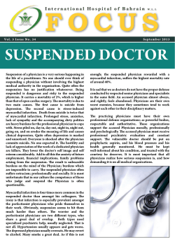
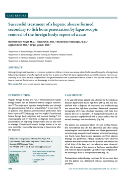
Turkish Journal of Medical Sciences
Turk J Med Sci
(2014) 44: 404-410
© TÜBİTAK
doi:10.3906/sag-1302-88
http://journals.tubitak.gov.tr/medical/
Research Article
Effects of whole-body vibration on plasma sclerostin level in healthy women*
1
2,
3
4
Muharrem ÇİDEM , Yunus KARAKOÇ **, Hakan EKMEKÇİ , Suat Hayri KÜÇÜK ,
5
5
5
1
Murat ULUDAĞ , Kerem GÜN , Şafak Sahir KARAMEHMETOĞLU , İlhan KARACAN
1
Department of Physical Medicine and Rehabilitation, Bağcılar Training and Research Hospital, İstanbul, Turkey
2
Department of Biophysics, Faculty of Medicine, İnönü University, Malatya, Turkey
3
Department of Biochemistry, Cerrahpaşa Faculty of Medicine, İstanbul University, İstanbul, Turkey
4
Department of Biochemistry, Bağcılar Training and Research Hospital, İstanbul, Turkey
5
Department of Physical Medicine and Rehabilitation, Cerrahpaşa Medical Faculty, İstanbul University, İstanbul, Turkey
Received: 19.02.2013
Accepted: 01.08.2013
Published Online: 31.03.2014
Printed: 30.04.2014
Background/aim: To determine whether plasma sclerostin levels are affected by applying whole-body vibration treatments.
Materials and methods: Following a pilot study, the present prospective, randomized, controlled single-blind study was performed on
16 healthy volunteer women (ages 20 to 40 years). Subjects were randomly divided into 2 groups, and whole-body vibration was applied
to the treatment group but not to the controls. The plasma sclerostin levels were measured before the treatment and at the 10th minute
after whole-body vibration on the 1st, 2nd, and 5th days of application.
Results: The plasma sclerostin level measured at 10 min after the whole-body vibration treatment increased 91% (P = 0.024) on the 1st
day and decreased 31.5% (P = 0.03) on the 5th day in the whole-body vibration group. In the control group, there was no change in the
plasma sclerostin level at any time. A progressive increase in baseline plasma sclerostin levels during the 5 days of vibration sessions
was also found.
Conclusion: Our study demonstrated that whole-body vibration can change plasma sclerostin levels, and that this change is detectable
10 min after whole-body vibration treatments.
Key words: Sclerostin, cyclic mechanical loading, vibration, osteocyte, bone
1. Introduction
Sclerostin, a SOST gene protein, potently inhibits
Wnt canonical signaling by binding to lowdensity lipoprotein receptor-5 and is a potent inhibitor of
bone formation (1–3). It is produced almost exclusively
by osteocytes. Sclerostin expression is also regulated by
mechanical strain in adult bone. Osteocytes, the most
abundant cells in bone, have mechanosensory properties,
and mechanical loading triggers the cells to modulate
bone homeostasis (1,2,4,5).
It has been recently shown that mechanical stimulation
of long bones in mice suppresses sclerostin expression in
osteocytes and simultaneously induces bone formation in
rodents (2,6,7). Conversely, mechanical unloading causes
upregulation of sclerostin activity in mice and disuse
osteoporosis in humans (1,4).
Sclerostin acts in a local paracrine fashion in the bone
microenvironment. Mirza et al. showed that sclerostin
also enters the circulatory system, where it may regulate
bone mass by acting as an endocrine hormone. They
also demonstrated that sclerostin can be measured in
peripheral serum (8).
Plasma sclerostin level may be used as an indicator to
evaluate the bone response to mechanical loading. Despite
a growing understanding of the basic biology of sclerostin,
no data are available on circulating sclerostin levels in
humans exposed to cyclic mechanical loading.
In this study, we aimed to determine whether plasma
sclerostin level is affected by whole-body vibration (WBV)
in healthy women.
2. Materials and methods
Ethical approval was obtained from the Institutional
Review Board (Ethics Committee of İstanbul University’s
İstanbul Medical Faculty, 2011/228-473). All testing
* This study was presented as a poster at the IOF 2nd Regional Middle East and Africa Osteoporosis Meeting/6th Pan Arab
Osteoporosis Congress in Jordan, 27 September through 1 October 2012. Unique protocol IDs are BEAH FTR-1 and NCT01310335
at ClinicalTrials.gov.
** Correspondence: [email protected]
404
ÇİDEM et al. / Turk J Med Sci
procedures and the experimental protocol were explained,
and written informed consent was received.
To determine the earliest changes in plasma sclerostin
levels, we designed a pilot study on 33 voluntary adult
healthy women working in our hospital. In our pilot
study, plasma sclerostin levels tended to increase during
the vibration and at 10 and 30 min after vibration, even
though the elevation was not statistically significant.
However, the increase in sclerostin level was more obvious
10 min after vibration in each of the 3 groups. Based
on these findings, the present study was planned as a
prospective, randomized, controlled, single-blind, singlecenter experimental trial.
2.1. Participants
Among young-adult healthy women working in our
hospital, 34 subjects who voluntarily agreed to participate
were assessed for eligibility. None of subjects who
participated in the pilot study were enrolled in this study.
Exclusion criteria are given in Table 1.
The participants meeting the criteria were randomized
into the WBV group and the control group. Only one
investigator was involved in the randomization process,
which employed a computerized random number generator.
This study followed the guidelines of the CONSORT
statement for individual randomized, controlled trials with
no pharmacological treatment (Figure 1).
The mean age of the participants was 28.8 ± 6.5 years in
the WBV group (n = 8) and 29.3 ± 4.2 years in the control
group (n = 8) (P = 0.859). The body mass index was 22.2
± 1.9 kg/m2 in the WBV group and 21.0 ± 2.2 kg/m2 in the
control group (P = 0.281)
2.2. WBV application
During WBV, the subjects were asked to stand upright on a
vibration platform. Their ankles and knees were in a neutral
position. The hip joint was held in abduction so that there
was 30 cm between the heels. Subjects were barefooted, and
no sponge or foam was placed between the vibration platform
and their feet (Figure 2). A synchronous WBV device was
used in the present study. The whole plate oscillated with a
linear movement upward and downward. The frequency of
vibrations was set to 40 Hz, with a peak-to-peak amplitude
of 2 mm and acceleration of 2.7 × g. WBV was applied at a
rate of one session per day from Monday to Friday. WBV was
applied on the 1st and 2nd days for 60 s (2 × 30 s treatments),
on the 3rd day for 120 s (4 × 30 s treatments), on the 4th day
for 150 s (5 × 30 s treatments), and on the 5th day for 210 s
(3 × 30 s treatments + 2 × 60 s treatments). After each 30-s
application of vibration, the subjects were told to relax for 10
s without changing their position (rest period).
2.3. Blood samples
In the WBV group, blood samples were obtained before
WBV (pre-WBV) and at the 10th minute after WBV (postWBV10) in all participants on the 1st, 2nd, and 5th days by
inserting an intravenous cannula into the antecubital vein.
In the control group, 2 blood samples were obtained on
the same day. The second samples were obtained 10 min after
the first sample.
Table 1. Exclusion criteria.
1. Lower extremity problems
a. Orthopedic problems: shortness of legs, congenital anomalies, etc.
b. Joint disease (arthritis, joint prosthesis, etc.), other painful pathologies in the lower extremities (fractures, tendinitis, bursitis, etc.)
c. Circulation problems in the lower extremities
2. Dorsolumbar diseases: Vertebral fractures, disk hernias, spondylodiscitis, etc.
3. Systemic disease cases
a. Systemic bone disease: osteoporosis, osteomalacia, Paget’s disease
b. Hypertension (>135 mmHg systolic, >85 mmHg diastolic), heart diseases
c. Infectious diseases
d. Endocrine diseases (diabetes mellitus, etc.)
4. Neurological diseases
5. Menstrual cycle disorders, amenorrhea, lactation, oral contraceptive use
6. During the ovulatory period (11th–16th day of menses)
7. Obesity (BMI > 30 kg/m2) or low BMI (BMI < 20 kg/m2)
8. Vertigo
9. Cognitive function disorders
10. Nonpalpable antecubital vein
BMI: Body mass index.
405
Enrollment
ÇİDEM et al. / Turk J Med Sci
Assessed for eligibility
(n = 34 Volunteer healthy women aged 20 - 40 years)
Excluded (n = 14)
2 had vertigo
4 for menstrual irregularity
1 for lactation
1 had amenorrhea
2 had polycystic ovary syndrome
3 during the ovulatory period
1 nonpalpable antecubital vein
Analysis
Follow-up
Allocation
Consent Obtained & Randomized
(n = 20 )
Allocated to WBV (n = 10)
Allocated to Control (n = 10)
Lost to follow-up (n = 2)
Lost to follow-up (n = 2)
Discontinued intervention (n = 2)
2 for personal reason
Discontinued intervention (n = 2)
1 for personal reason
1 for hemolytic blood sample
Analyzed (n = 8)
Analyzed (n = 8)
Figure 1. Flow chart of participants considered for inclusion.
2.4. Plasma sclerostin, estradiol, and parathyroid
hormone levels
Plasma was collected using EDTA and was centrifuged for
15 min at 400 × g within 30 min of collection. Aliquots of
plasma were added to Eppendorf tubes and stored at –20° C.
Plasma sclerostin levels were measured blindly by
only one investigator. No information was given to the
lab workers regarding blood sample identification or time
when the blood was taken. Plasma sclerostin levels were
measured using a Human Sclerostin ELISA kit (Cusabio,
Catalog No. CSB-E13146h, Newark, DE, USA). All
assays were performed according to the manufacturer’s
instructions. The minimum detectable concentration of
human sclerostin is typically <0.012 ng/mL. None of the
measured sclerostin values in our subjects were below the
limit of detection for this assay. Intraassay precision is less
than 8%.
406
Figure 2. Posture of the participants during WBV.
ÇİDEM et al. / Turk J Med Sci
Plasma parathyroid hormone (PTH) and estradiol (E2)
levels were measured to test whether hemoconcentration
or hemodilution occur during or after WBV. PTH and
E2 were analyzed in EDTA-plasma samples using an
electrochemiluminescence immunoassay (Elecsys PTH,
Elecsys-Estradiol II; Roche Diagnostics, Mannheim,
Germany) and the Cobas 601 analyzing system (Roche
Diagnostics).
2.5. Statistical analysis
Continuous variables were summarized as the arithmetic
mean and standard deviation (SD). The Kolmogorov–
Smirnov test was used to confirm that data were normally
distributed. Within-group comparisons were done using
paired t-tests and general linear model repeated measures.
Pearson’s correlation coefficient was used to test for
associations between variables. A P-value of <0.05 was
considered statistically significant. The software package
used for data management was PASW Statistic 18.
was 0.785 ± 0.302 ng/mL before WBV and 1.212 ± 0.287
ng/mL at 10 min after WBV on the 1st day. For the given
effect size (plasma sclerostin level means of 0.785 vs.
1.212), SD (0.302 vs. 0.287), sample sizes (8 and 8), and
alpha (0.050, 2-tailed), the power was 0.695. The mean
plasma sclerostin was 1.408 ± 0.478 ng/mL before WBV
and 0.832 ± 0.302 ng/mL at 10 min after WBV on the 5th
day. For the given effect size (plasma sclerostin level means
of 1.408 vs. 0.832), SD (0.478 vs. 0.287), sample sizes (8
and 8), and alpha (0.050, 2-tailed), the power was 0.649.
4. Discussion
In this study, the impact of cyclical mechanical loading
on plasma sclerostin level was investigated for the first
time. Our results revealed a significant change in plasma
sclerostin levels 10 min after the cyclic mechanical
stimulus. Baseline plasma sclerostin levels were also found
to have increased progressively over the course of 5 days of
mechanical loading sessions.
Bone tissue sclerostin levels are tightly regulated by
mechanical strain (2). Sclerostin is expressed almost
exclusively in osteocytes, and these osteocytes are able to
detect mechanical strain (9). Osteocytes are stimulated
by mechanical deformation of bone (10). The main
mechanical factors determining the impact of mechanical
loading on bones are strain (strain magnitude, strain
rate), stress, and the frequency or the number of loading
cycles (11–15). The extent of bone deformation, or strain
magnitude, is directly proportional to the force magnitude
to which the bone is exposed (11). According to Newton’s
second law, the force magnitude is directly proportional
to acceleration (16). Subjects were asked to stand upright
to create maximum mechanical loading on the lower
extremity bones. The frequency of vibrations was set to 40
Hz, with acceleration of 2.7 × g in the present study.
We do not know the mechanism of the changes that
occurred in the plasma sclerostin levels at post-WBV10.
However, some mechanisms can be suggested. The
increase in plasma sclerostin level 10 min after WBV may
be explained by increased sclerostin secretion from the
osteocytes and passage to the systemic blood circulation
via lacuna–canalicular fluid. Sclerostin is a protein
hormone (8). Protein hormones differ from steroid
3. Results
In the control group, plasma sclerostin levels measured in
the 1st and 2nd blood samples were 0.835 ± 0.218 ng/mL
and 0.878 ± 0.279 ng/mL, respectively (P = 0.602).
In the WBV group, the plasma sclerostin levels
increased significantly 10 min after WBV on the 1st
day (P = 0.024). The plasma sclerostin levels decreased
significantly 10 min after WBV on the 5th day (P = 0.030).
No significant change in sclerostin level with WBV was
identified on the 2nd day (P = 0.159) (Table 2).
The levels of plasma sclerostin measured 10 min after
WBV declined progressively during the 5 days of vibration
sessions (P = 0.026). Post hoc analysis revealed that the
levels of plasma sclerostin measured 10 min after WBV on
the 5th day were significantly lower than those on the 1st
day (P = 0.037) (Figure 3).
Only the 5-day cyclical mechanical stimulus was found
to have caused a significant increase in the baseline plasma
sclerostin levels (P = 0.008). Post hoc analysis revealed that
the baseline levels of plasma sclerostin on the 5th day were
significantly higher than those on the 1st day (P = 0.013)
No significant change in plasma E2 or PTH levels was
observed after WBV (Table 3).
The effects of WBV on plasma sclerostin levels at the
10th minute were evaluated. The mean plasma sclerostin
Table 2. The plasma sclerostin levels before and after WBV (ng/mL).
Time point
Day 1
Day 2
Day 5
Pre-WBV
0.785 ± 0.302
0.998 ± 0.264
1.408 ± 0.478
0.013b
Post-WBV10
1.212 ± 0.287a
0.903 ± 0.358a
0.832 ± 0.302a,b
0.037b
P-value
0.024
0.159
0.030
a
a
P-value
b
a
: Comparison with pre-WBV, b: comparison with 1st day.
a
407
ÇİDEM et al. / Turk J Med Sci
1.21
1.20
1.10
1.00
0.90
0.80
0.70
–31.5%p=0.03
1.30
1.40
10 min after WBV
–11.1%
1.40
Pre WBV
91.0%p=0.024
Plasma sclerostin level (ng/mL)
1.50
0.99
0.9
0.83
0.78
Day 1 1
Day 2 2
Day 5 3
4
Figure 3. Changes in baseline plasma sclerostin levels and plasma
sclerostin levels at 10 min after WBV.
hormones in that they can be deposited inside the cell (17).
A cyclic mechanical stimulus may trigger the secretion of
sclerostin, which is mechanosensitive, from osteocytes.
Cyclic mechanical loading can cause compression and
decompression in bone tissue. As a result, lacuna–
canalicular fluid moves into the blood vessels (18). WBV
also significantly increases blood flow to tissue exposed to
vibration (19,20). Relocation of lacuna–canalicular fluid
and its mixture with increased blood circulation may
cause more leakage of sclerostin into the blood circulation.
Sclerostin is reported to be higher in weight-bearing
than in nonweight-bearing disciplines in athletes (3). A
progressive mechanical loading was applied during the
5-day vibration sessions in the present study. Our findings
showed a progressive increase in baseline plasma sclerostin
levels during the 5-day vibration sessions. Relocation of
lacuna–canalicular fluid and its mixture with increased
blood circulation may also help explain the progressive
increase in baseline plasma sclerostin levels.
We also determined that the plasma sclerostin level
at the 10th minute after WBV decreased progressively
during the 5-day vibration sessions (Figure 3). Despite the
increased baseline level of plasma sclerostin during the
5-day vibration sessions, the plasma sclerostin level was
found to decrease at 10 min after WBV on the 5th day.
This finding suggests that after the 5-day, high-frequency,
high-acceleration WBV treatment, cyclic mechanical
stimulus may quickly inhibit sclerostin activity. A
multisession, cyclic mechanical stimulus plays an anabolic
and antiresorptive role in bone (21,22). Bone formation
is known to increase on one side and bone resorption to
decrease on the other side when the sclerostin activity
decreases (23–25). Therefore, the decrease in the plasma
sclerostin level at 10 min after WBV and the lower plasma
sclerostin level at 10 min after WBV on the 5th day of
WBV compared to the 1st day of WBV would seem to be
compatible with the literature.
Some limitations should be noted. Whether WBV
causes a volume change in plasma was not tested in this
study. Plasma-volume alterations can change hormone
concentrations. Bouts of acute exercise produce transient
plasma-volume changes (26). However, Cardinale et al.
reported no changes in plasma volume due to a single
session of WBV (30-Hz, 4-mm amplitude for 5 sets of 1
min separated by a 1-min resting period) (27). Fricke et al.
showed that WBV does not influence serum E2 levels in
women (28). In this study, the absence of change in these
hormone levels after WBV suggests that there was no
hemoconcentration or hemodilution during or after WBV.
Bone strength and quality is important for osteoporosis
(29–31). Evaluation of the response given by the bone to
cyclic mechanical loading may be important in terms of
in vivo determination of the strength and quality of bone.
Postmenopausal osteoporosis is the most common form
of osteoporosis (30). Therefore, female subjects were
preferred in the study. Findings of this study may be useful
for future studies investigating the bone strength and
quality in patients with postmenopausal osteoporosis.
In conclusion, this study is the first to show the changes
in plasma sclerostin level with WBV. Our study showed
that WBV can cause changes in plasma sclerostin levels
in adult women. This change is noticeable 10 min after
whole-body vibration. WBV can also affect the baseline
Table 3. The plasma E2 and PTH levels before and after whole-body vibration (pg/mL).
Hormone
Time point
Day 1
Day 2
Day 5
E2
Pre-WBV
140.1 ± 77.2
120.8 ± 58.0
118.3 ± 43.0
Post-WBV10
137.5 ± 74.5
119.3 ± 59.3
115.3 ± 34.9
P-value
0.134
0.568
0.410
Pre-WBV
57.1 ± 27.0
60.8 ± 36.4
59.1 ± 30.4
Post-WBV10
54.0 ± 22.7
51.8 ± 24.7
58.6 ± 31.2
P-value
0.290
0.091
0.889
PTH
408
ÇİDEM et al. / Turk J Med Sci
plasma sclerostin levels when it is applied on consecutive
days. However, future studies are needed to delineate the
exact mechanism of changes in plasma sclerostin level
with WBV.
Acknowledgment
This study was supported by a grant from the Bağcılar
Training and Research Hospital Scientific Fund.
References
1.
2.
Gaudio A, Pennisi P, Bratengeier C, Torrisi V, Lindner B,
Mangiafico RA, Pulvirenti I, Hawa G, Tringali G, Fiore
CE. Increased sclerostin serum levels associated with
bone formation and resorption markers in patients with
immobilization-induced bone loss. J Clin Endocrinol Metab
2010; 95: 2248–2253.
Robling AG, Niziolek PJ, Baldridge LA, Condon KW, Allen
MR, Alam I, Mantila SM, Gluhak-Heinrich J, Bellido TM,
Harris SE et al. Mechanical stimulation of bone in vivo reduces
osteocyte expression of Sost/sclerostin. J Biol Chem 2008; 283:
5866–5875.
13. Ott SM. Sclerostin and Wnt signaling--the pathway to bone
strength. J Clin Endocrinol Metab 2005; 90: 6741–6743.
14. Nicolella DP, Moravits DE, Gale AM, Bonewald LF, Lankford
J. Osteocyte lacunae tissue strain in cortical bone. J Biomech
2006; 39: 1735–1743.
15. Hacyan S. What does it mean to modify or test Newton’s
second law? Am J Phys 2009; 77: 607–609.
16. Rubin CT, McLeod KJ. Promotion of bony ingrowth by
frequency-specific, low-amplitude mechanical strain. Clin
Orthop Relat Res 1994; 298: 165–174.
3.
Lombardi G, Lanteri P, Colombini A, Mariotti M, Banfi G.
Sclerostin concentrations in athletes: role of load and gender.
J Biol Regul Homeost Agents 2012; 26: 157–163.
17. Montgomery R, Conway TW, Spector AA. Biochemistry: A
Case-Oriented Approach. 6th ed. St. Louis, Mo, USA: C.V.
Mosby Company; 1996.
4.
Lin C, Jiang X, Dai Z, Guo X, Weng T, Wang J, Li Y, Feng G,
Gao X, He L. Sclerostin mediates bone response to mechanical
unloading through antagonizing Wnt/beta-catenin signaling. J
Bone Miner Res 2009; 24: 1651–1661.
18. Price C, Zhou X, Li W, Wang L. Real-time measurement of
solute transport within the lacunar-canalicular system of
mechanically loaded bone: direct evidence for load-induced
fluid flow. J Bone Miner Res 2011; 26: 277–285.
5.
Mödder UI, Hoey KA, Amin S, McCready LK, Achenbach SJ,
Riggs BL, Melton LJ, Khosla S. Relation of age, gender, and
bone mass to circulating sclerostin levels in women and men. J
Bone Miner Res 2011; 26: 373–379.
19. Lohman EB 3rd, Petrofsky JS, Maloney-Hinds C, BettsSchwab H, Thorpe D. The effect of whole body vibration on
lower extremity skin blood flow in normal subjects. Med Sci
Monit 2007; 13: CR71-6.
6.
Moustafa A, Sugiyama T, Saxon LK, Zaman G, Sunters A,
Armstrong VJ, Javaheri B, Lanyon LE, Price JS. The mouse
fibula as a suitable bone for the study of functional adaptation
to mechanical loading. Bone 2009; 44: 930–935.
20. Kerschan-Schindl K, Grampp S, Henk C, Resch H, Preisinger
E, Fialka-Moser V, Imhof H. Whole-body vibration exercise
leads to alterations in muscle blood volume. Clin Physiol 2001;
21: 377–382.
7.
Suva LJ. Sclerostin and the unloading of bone. J Bone Miner
Res 2009; 24: 1649–1650.
8.
Mirza FS, Padhi ID, Raisz LG, Lorenzo JA. Serum sclerostin
levels negatively correlate with parathyroid hormone levels
and free estrogen index in postmenopausal women. J Clin
Endocrinol Metab 2010; 95: 1991–1997.
21. Sun YQ, McLeod KJ, Rubin CT. Mechanically induced
periosteal bone formation is paralleled by the upregulation of
collagen type one mRNA in osteocytes as measured by in situ
reverse transcript-polymerase chain reaction. Calcif Tissue Int
1995; 57: 456–462.
9.
Cullen DM, Smith RT, Akhter MP. Bone-loading response
varies with strain magnitude and cycle number. J Appl Physiol
2001; 91: 1971–1976.
10. Torcasio A, van Lenthe GH, Van Oosterwyck H. The
importance of loading frequency, rate and vibration for
enhancing bone adaptation and implant osseointegration. Eur
Cell Mater 2008; 16: 56–68.
11. Nikander R, Kannus P, Rantalainen T, Uusi-Rasi K, Heinonen
A, Sievänen H. Cross-sectional geometry of weight-bearing
tibia in female athletes subjected to different exercise loadings.
Osteoporos Int 2010; 21: 1687–1694.
12. Yang PF, Brüggemann GP, Rittweger. What do we currently
know from in vivo bone strain measurements in humans? J
Musculoskelet Neuronal Interact 2011; 11: 8–20.
22. Judex S, Rubin CT. Is bone formation induced by highfrequency mechanical signals modulated by muscle activity?
J Musculoskelet Neuronal Interact 2010; 10: 3–11.
23. Mödder UI, Clowes JA, Hoey K, Peterson JM, McCready L,
Oursler MJ, Riggs BL, Khosla S. Regulation of circulating
sclerostin levels by sex steroids in women and in men. J Bone
Miner Res 2011; 26: 27–34.
24. Tian X, Jee WS, Li X, Paszty C, Ke HZ. Sclerostin antibody
increases bone mass by stimulating bone formation and
inhibiting bone resorption in a hindlimb-immobilization rat
model. Bone 2011; 48: 197–201.
25. Padhi D, Jang G, Stouch B, Fang L, Posvar E. Single-dose,
placebo-controlled, randomized study of AMG 785, a
sclerostin monoclonal antibody. J Bone Miner Res 2011; 26:
19–26.
409
ÇİDEM et al. / Turk J Med Sci
26. Kargotich S, Goodman C, Keast D, Morton AR. The
influence of exercise-induced plasma volume changes on the
interpretation of biochemical parameters used for monitoring
exercise, training and sport. Sports Med 1998; 26: 101–117.
27. Cardinale M, Soiza RL, Leiper JB, Gibson A, Primrose WR.
Hormonal responses to a single session of whole-body
vibration exercise in older individuals. Br J Sports Med 2010;
44: 284–288.
28. Fricke O, Semler O, Land C, Beccard R, Thoma P, Schoenau E.
Hormonal and metabolic responses to whole body vibration in
healthy adults. Endocrinologist 2009; 19: 24–30.
410
29. Martin RM, Correa PH. Bone quality and osteoporosis therapy.
Arq Bras Endocrinol Metabol 2010; 54: 186–199.
30. Briot K, Cortet B, Thomas T, Audran M, Blain H, Breuil V,
Chapuis L, Chapurlat R, Fardellone P, Feron JM et al. 2012
update of French guidelines for the pharmacological treatment
of postmenopausal osteoporosis. Joint Bone Spine 2012; 79:
304–313.
31. Uğurlu M, Yılmaz S, Deveci A, Ünlü S, Tunç B, Üstü Y, Sayıt E,
Sanisoğlu SY. The epidemiologic characteristics of patients that
underwent surgery for hip fracture. Turk J Med Sci 2012; 42:
299–305.
© Copyright 2026 Paperzz