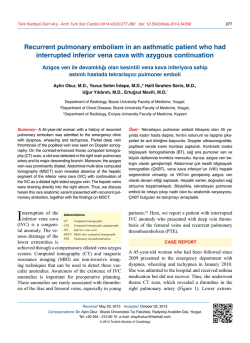
Türk Kardiyol Dern Arş - Arch Turk Soc Cardiol 2014;42(6):571-573 doi: 10.5543/tkda.2014.43788
571
Successful stenting of systemic venous pathway
stenosis after double switch repair for congenitally
corrected transposition of great arteries in a child
Doğuştan düzeltilmiş büyük damar transpozisyonu nedeniyle
double switch ameliyatı yapılan bir çocukta gelişen
sistemik venöz darlıkta başarılı stent işlemi
Arda Saygılı, M.D., Yusuf Yalçınbaş, M.D.,# Ahmet Arnaz, M.D.,# Tayyar Sarıoğlu, M.D.,*
Department of Pediatric Cardiology, Acıbadem University Faculty of Medicine, Istanbul;
#
Department of Cardiac Surgery, Acıbadem Bakirköy Hospital, Istanbul;
*Department of Cardiac Surgery, Acıbadem University Faculty of Medicine, Istanbul
Summary– An 8-year-old boy with previous shunt operation for corrected transposition of great arteries, ventricular
septal defect, pulmonary stenosis and multiple aortopulmonary collateral arteries underwent corrective surgery. In the
early post-operative period, there were clinical findings of
superior vena cava obstruction. Cardiac catheterization at
72 h following surgery showed a systemic venous baffle stenosis between the vena cava and right atrium. A stent was
successfully implanted in the vena cava percutaneously,
and the stenosis was relieved. Her symptoms resolved in a
short time period, and she was extubated rapidly. During the
follow-up, excellent maintenance and patency of systemic
venous baffle were observed.
S
ystemic venous pathway (SVP) stenosis is one of
the post-operative complications of the mustard
repair procedure for complete transposition or double
switch operations. An obstructed SVP following atrial switch operations is the most common indication
for stent delivery in the venous system.[1,2] Covered
and bare metal stents are the most-suitable treatment
options for vessel stenosis in congenital heart diseases.[2-5]
CASE REPORT
An 8-year-old boy was admitted with a small restrictive ventricular septal defect, congenitally corrected
Özet– Doğuştan düzeltilmiş büyük damar transpozisyonu,
ventriküler septal defekt, pulmoner darlık ve çoklu aortopulmoner kollateral arter nedeni ile önce şant ameliyatı
olan sekiz yaşındaki erkek çocuk, tam düzeltme ameliyatı
oldu. Erken ameliyat sonrası dönemde superior vena kava
tıkanıklığı bulguları gelişti. Ameliyat sonrası 72. saatte
yapılan kalp kateterizasyonu vena kava ve sağ atriyum
arasında, sistemik venöz baffle darlığı olduğunu gösterdi. Stent perkütan olarak vena kavaya başarıyla yerleştirilerek darlık giderildi. Hastanın belirtileri kısa bir sürede
kayboldu ve hasta hızlıca ekstübe edildi. Takip sırasında
sistemik baffle açıklığının mükemmel düzeyde sağlandığı
gözlendi.
transposition of the great
arteries, pulmonary stenosis, and aortopulmonary
collaterals along with large
atrial septal defects.
Abbreviations:
CP
PAP
RA
SVC
SVP
Cheatham platinum
Pulmonary artery pressure
Right atrium
Superior vena cava
Systemic venous pathway
After an arterial double
switch operation which included pulmonary xenograft
conduit repair, the post-operative intensive care unit
stay was uneventful. After extubation, the patient’s
superior vena cava (SVC) dilatation presented with
obstruction symptoms as well as facial swelling, head
fullness, and dyspnea. The patient had also hepatomegaly, tibial edema, and abdominal distension. A
Received: January 11, 2014 Accepted: May 30, 2014
Correspondence: Dr. Arda Saygılı. Acıbadem Hastanesi Çocuk Kardiyolojisi,
Tekin Sokak, No: 8, Acıbadem, 34718 İstanbul, Turkey.
Tel: +90 212 - 414 44 08 e-mail: [email protected]
© 2014 Turkish Society of Cardiology
Türk Kardiyol Dern Arş
572
chest X-ray revealed cardiomegaly with pleural effusion. Echocardiography demonstrated a patent inferior
part of systemic venous return with SVC dilatation
and dynamic obstruction during the diastolic phase of
the ventricular contraction. During the catheterization,
the mean pressure gradients between the SVC and the
right atrium (RA) and for the inferior caval vein were
11 mmHg (SVC: 25 mmHg; RA: 14 mmHg) and 14
mmHg, respectively. The mean pulmonary artery pressure (PAP) was 29 mmHg (systolic PAP: 40 mmHg;
diastolic PAP: 20 mmHg). Angiography demonstrated
a patent pulmonary outflow tract with SVP stenosis.
Therefore, a stent to connect the SVC to the RA was
planned to implant (Figure 1a and Video 1*).
Repeated hemodynamic measurement demonstrated a complete lack of obstruction without a pressure
gradient in the upper part of the SVC-to-RA stent.
Angiography also showed that the stent was properly
placed (Figure 1b and Video 2*). Ascites was reduced,
and abdominal distension resolved soon after the stent
implantation.
Under general anesthesia and after right femoral vein cannulation, SVC was crossed by a 6F rightguided catheter. The catheter was then exchanged for
a standard extra-stiff wire guide, and the 6F sheath
was replaced with a long 14 French sheath. Based on
SVC angiographic findings (SVC diameter: 12 mm),
a cheatham platinum (CP) (Numed NY, USA) balloon
expendable, 34 mm (CP8Z34) stent and balloon in a
balloon (14 mm) were selected. The balloon was then
inflated at 3 atm at a diameter of 14 mm with a procedural time of 15 min and a fluoroscopy time of eight
min.
DISCUSSION
A
The patient without any clinical signs of jugular
vein restenosis was discharged with aspirin therapy.
Repeated echocardiography showed no residual stenosis and the room air saturation remained at 93%
during follow-up. At 6-10 months of surgery, the patient is still asymptomatic.
Several conditions may lead to stenosis of the systemic veins, including SVC and IVC obstruction after
an atrial switch operation.[1-3] The reported incidence
of systemic venous baffle obstruction after an atrial
switch (Mustard or Senning) operation ranges from
0% to 20%.[2] Obstruction of the SVP is a rare, yet
known complication in the early post-operative course
of the Senning procedure.[2-5]
It is established that acute obstruction of the SVC
pathway after atrial repair is not well tolerated. It may
B
Figure 1. Angiocardiogram of the patient with superior vena cava and systemic venous baffle obstruction after the senning procedure (A) and immediately after the stent procedure (B).
Stenting of systemic venous pathway stenosis after surgical repair for congenital heart disease
be responsible for the venous insufficiency and superior vena cave syndrome.[1-4] In the early post-operative course, clinical systemic venous stenosis was
dramatically symptomatic in our patient. As a result,
we may conclude that it is unlikely certain to what
degree stenosis intervention should be performed in
symptomatic patients. In addition, one editorial paper
reported that the importance of mild gradients across
the polyvinylpyrrolidones (arbitrarily <3 mmHg) is
unknown, whereas gradients of 10-15 mmHg was
found in patients with protein-losing enteropathy.[3,4]
Stents are used as the primary approach in such
lesions, as the procedure is simple and effective.[1-3]
There are no evidence-based protocols which serve as
indications for the intervention; however, stenting is
known to be critical in many asymptomatic patients
with minimal gradients and modest stenosis (3340%). Therefore, clinical symptomatic obstructions
or obstructions larger than 50% of the large systemic
veins as confirmed by angiography are considered appropriate for stent implantation.[2] Patients with atrial
baffles are also good candidates for stenting.[2] A self
or balloon-expandable and a covered or bare metal CP
stent is the best choice for the vessels as well as for
SVP stenosis.[3-5]
Utilizing a balloon expandable stent in combination with a CP stent is a simple and effective procedure which can be used successfully for the treatment of SVP obstruction after the Senning procedure
573
thanks to its ability for rapid and dramatic relief in
selected patients.
Conflict-of-interest issues regarding the authorship or
article: None declared.
*Supplementary video file associated with this article
can be found in the online version of the journal.
REFERENCES
1.Michel-Behnke I, Hagel KJ, Bauer J, Schranz D. Superior
caval venous syndrome after atrial switch procedure: relief
of complete venous obstruction by gradual angioplasty and
placement of stents. Cardiol Young 1998;8:443-8. CrossRef
2. Tzifa A, Marshall AC, McElhinney DB, Lock JE, Geggel RL.
Endovascular treatment for superior vena cava occlusion or
obstruction in a pediatric and young adult population: a 22year experience. J Am Coll Cardiol 2007;49:1003-9. CrossRef
3. Rosenthal E, Qureshi SA. Stenting of systemic venous pathways after atrial repair for complete transposition. Heart
1998;79:211-2.
4. Ward CJ, Mullins CE, Nihill MR, Grifka RG, Vick GW 3rd.
Use of intravascular stents in systemic venous and systemic
venous baffle obstructions. Short-term follow-up results. Circulation 1995;91:2948-54. CrossRef
5. Daehnert I, Hennig B, Wiener M, Rotzsch C. Interventions in
leaks and obstructions of the interatrial baffle late after Mustard and Senning correction for transposition of the great arteries. Catheter Cardiovasc Interv 2005;66:400-7. CrossRef
Key words: Child; double switch; stenosis; transposition of great arteries; stents.
Anahtar sözcükler: Çocuk; double switch; darlık; düzeltilmiş büyük
damar transpozisyonu; stent.
© Copyright 2026 Paperzz