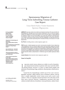
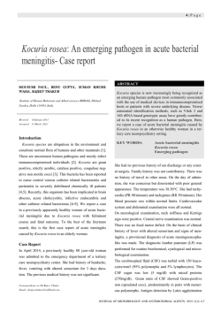
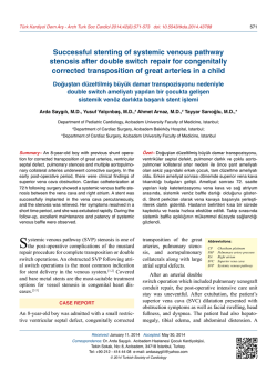
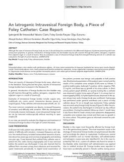
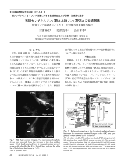
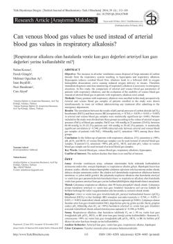
ORIGINAL ARTICLE
53
An Evaluation of Complications in Ultrasound-Guided
Central Venous Catheter Insertion in the
Emergency Department
Acil Serviste Ultrasonografi Eşliğinde Takılan Santral
Venöz Kataterlerin Komplikasyon Açısından Değerlendirilmesi
Engin OZAKIN, Rumeysa CAN, Nurdan ACAR, Filiz BALOGLU KAYA, Arif Alper CEVİK
Department of Emergency Medicine, Eskisehir Osmangazi University Faculty of Medicine, Eskisehir
SUMMARY
ÖZET
Objectives
In emergency departments, emergency physicians frequently have
to perform central venous access. In cases where peripheral venous
access is not possible, central venous access is required for dialysis,
fulfillment of urgent fluid need, or central venous pressure measurement. This study was carried out to evaluate the emergence
of complications in the process of and in the 15 days following the
insertion of central venous catheter under ultrasound guidance in
the emergency department.
Amaç
Acil servislerde acil tıp hekimlerince santral damar yolu işlemi sık
uygulanır. Periferik damar yolu açılamadığı hallerde, diyaliz, acil
sıvı ihtiyacı veya santral venöz basınç ölçümü gereken durumlarda hastalar için santral damar yolu gerekmektedir. Acil serviste,
ultrasonografi (USG) kılavuzluğunda uygulanan acil santral venöz
katater girişimi sürecinde ve uygulamayı takip eden 15 gün içerisinde komplikasyon varlığını değerlendirmek amacı ile bu çalışma
yapıldı.
Methods
For this study, patients who presented to the emergency department over a period of eight months with an urgent need for central
catheter were examined prospectively. Age, gender, and accompanying diseases of patients as well as the type, time, duration, and
indication of the venous access were recorded. Furthermore, the
amount of experience of the physician was taken into consideration.
Gereç ve Yöntem
Sekiz aylık sürede acil servise başvuran ve acil santral katater gereksinimi olan hastalar ileriye dönük olarak incelendi. Hastaların yaşı,
cinsiyeti, eşlik eden hastalıkları ile tercih edilen girişimin yolu, saati,
süresi ve endikasyonu kaydedildi. Ayrıca girişimi yapan hekimin çalışma yılı da değerlendirmeye dahil edildi.
Results
In the emergency department, physicians performed ultrasoundguided central venous catheter insertion for 74 patients (40 men
and 34 women). For access, internal jugular vein was used in 65
(87.8%) patients, and femoral vein was used in 9 (12.2%) patients.
The reason for access was urgent dialysis need in 55 (74.3%), CVP
measurement in 3 (4.1%), fluid support due to severe hypovolemia
in 6 (8.1%), and difficulty of peripheral venous access in 10 (13.5%)
patients. None of the patients developed complications in the process of or after the insertion. Patients did not have infections related to the catheter in 15 days following the insertion.
Bulgular
Ultrasonografi eşliğinde santral venöz katater takılan 74 (40 erkek,
34 kadın) hastanın 65’inde (%87.8) internal juguler ven, dokuzunda (%12.2) femoral ven girişim için kullanıldı. Uygulama olguların
55’inde (%74.3) acil diyaliz ihtiyacı, üçünde (%4.1) CVP ölçümü, altısında (%8.1) ciddi hipovolemi için sıvı desteği, 10’unda (%13.5) periferik damar yolu güçlüğü nedeniyle yapıldı. Hastaların hiçbirinde
işlem esnasında ve sonrasında komplikasyon izlenmedi. Yatırıldıkları
bölümde takiplerinde 15 günlük süre içerisinde katater ile ilişkili enfeksiyon da saptanmadı.
Conclusions
Central venous access is frequently required in emergency departments. The risk of complication is little if any in ultrasonographyguided access carried out under appropriate conditions.
Sonuç
Acil servislerde santral damar yolu gereksinimi sıktır. Kılavuzların önerisi doğrultusunda USG eşliğinde uygun şartlar altında yapılan girişimlerde komplikasyon riski yok denecek kadar azdır.
Key words: Central venous catheter; emergency department; ultrasoundguided.
Anahtar sözcükler: Santral venöz katater; acil servis; ultrason kılavuzluğu.
Submitted: February 07, 2014 Accepted: March 20, 2014 Published online: June 03, 2014
Correspondence: Dr. Engin Ozakin. Eskisehir Osmangazi Universitesi Tıp Fakultesi,
Acil Tıp Anabilim Dalı, 26240 Eskisehir, Turkey.
e-mail: [email protected]
Turk J Emerg Med 2014;14(2):53-58
doi: 10.5505/1304.7361.2014.93275
54
Turk J Emerg Med 2014;14(2):53-58
Introduction
The insertion of central venous catheters (CVC) has increased
in emergency departments particularly with the spread in
usage of ultrasonography (US). While internal jugular vein is
commonly preferred for placement under ultrasound guidance, subclavian and femoral vein access has decreased due
to higher complication risks. Emergency physicians apply
CVC primarily in cases of hemodialysis, difficulty of peripheral venous access, measurement of central venous pressure
(CVP), and need for rapid fluid resuscitation.[1]
Following the insertion of CVC in the emergency department, complications such as infection, pneumothorax,
hemothorax, subcutaneous hemorrhage, or puncture of
vertebral and cervical arteries, catheter breakage, catheter malposition, thrombus formation, and infection may
emerge.[1-3]
In order to reduce CVC complications, the healthcare personnel placing the CVC is required to work under sterile conditions, be experienced, and use the appropriate technique
for each unique patient. The quality of material used is also
important.[4] This study focuses on the complications that
may develop in the process of and in the 15 days following
the insertion of CVC under ultrasound guidance in our clinic.
Materials and Methods
This study was carried out prospectively in the emergency
department of a university between January 2011 and August 2011 after the approval of the local board of ethics
was obtained. The study involved patients aged over 18 in
urgent need of CVC, who agreed to take part in the study
or whose relatives gave consent. Patients with trauma, who
were pregnant at the time of admittance, and patients who
has two or more septic inflammatory response syndrome
criteria[5] (fever of more than 38°C (100.4°F) or less than 36°C
(96.8°F), heart rate of more than 90 beats per minute, respiratory rate of more than 20 breaths per minute or arterial
carbon dioxide tension (PaCO2) of less than 32 mmHg, white
blood cell count >12,000/μL or <4,000/μL or >10% immature forms) were excluded. All interventions were performed
by emergency physicians under US guidance, who previously received training on US. For the purpose of the study, age,
gender, and accompanying diseases of patients as well as
the type, time, duration, and indication of the venous access
were recorded. Furthermore, the physician’s level of experience was taken into consideration. All patients were taken to
a unit where vital and cardiac findings were monitored. The
patients or their relatives were informed and their consent
was received. In supine position, the patients were evaluated for an appropriate vein for US-guided intervention. For
this purpose, the anatomic characteristics of the patients as
well as the proximity of vein to the skin, lumen diameters,
and the proximity of vein to vital organs were checked. After
the location of access was determined, local skin cleaning
was performed with 10% povidone-iodine. The probe was
covered with sterile glove (Figure 1) and area of access was
covered with sterile drape. Once sterility was assured, sedoanalgesia and/or local anesthesia were administered with
the agents appropriate for the clinical situation of each patient. 7.5 MHz linear probe, used in US scan (Sonosite, Titan)
was covered appropriately. The vascular structures in the relevant area were displayed on the transverse axis (Figure 2).
The intervention was performed on the location where the
vein is most proximate to the skin, the lumen is largest, and
the adjacent artery is most protected. During the intervention, the needle movements were followed on the US screen
dynamically. When the blood flow into the injector in the
vein became clear, the catheter (double lumen hemodialysis
catheter, 12F, 15 cm, Sentia) was placed using the Seldinger
method. Blood and fluid flow were checked using heparincontaining fluid (50 U/ml), administered through the catheter. Following the intervention, all patients were checked for
subcutaneous emphysema, local hematoma, and bleeding
by physical examination, for pneumothorax and hemothorax by US, and for the position of catheters and again pneumothorax and hemothorax by chest radiography. Then, in
the intensive care unit or other departments where patients
were transferred, they were observed for 15 days to detect
any CVC-induced infections or other complications due to
catheter placement by emergency physicians. Rash, temperTable 1. Patient characteristics
Properties n
%
Sex
Male 40
54.1
Female 34
45.9
Past medical history
Diabetes mellitus
15
20.3
Renal insufficiency
15
20.3
Hypertension 13
17.6
Malignancy
6
8.1
None
23
31.1
Catheter location
Internal jugular vein
65
87.8
Femoral vein
9
12.2
Dialysis
55
74.4
CVP
3
4.1
Hypovolemia
6
8.1
10
13.5
İndications
Difficult peripheral venous access
Özakın E et al.
Complications in Ultrasound-Guided Central Venous Catheter Insertion in ED
Figure 1. Ultrasound-guided central venous catheter insertion.
ature rise, and swelling on the location where the catheter
was inserted were considered local symptoms of infection
and systemic inflammatory response syndrome criteria were
considered the systemic symptoms. SPSS 20 was used for
data analysis.
Results
Of 74 patients that had central venous catheter insertion, 40
(54.1%) were male and 34 (45.9%) were female. The mean
age was 63.7±12.42 (range: 32-85). The medical histories of
the patients showed that 15 (20.3%) patients had diabetes
mellitus, 15 (20.3%) had chronic renal failure, 13 (17.6%)
had hypertension, 6 (8.1%) had malignity, and 2 (2.7%) had
chronic liver disease. In 23 (31.1%) patients, there were no
comorbid diseases in past medical history. Internal jugular
vein catheterization was preferred in 65 (87.8%) patients,
and femoral vein catheterization was preferred in 9 (12.2%)
patients. 29 (39.2%) interventions were performed between
8 am and 4 pm, 31 (41.9%) between 4 pm and 12 am, and
14 (18.9%) between 12 am and 8 am. CVC indicated urgent
dialysis need in 55 (74.3%), need for CVP measurement in
3 (4.1%), urgent fluid need due to severe hypovolemia in
6 (8.1%), and difficulty of peripheral venous access in 10
(13.5%) patients. The average duration of the intervention
was 12.34±6.54 (range: 6-37) minutes in internal jugular vein
access, 14.56±6.3 (range: 6-29) minutes in femoral vein access, and 12.61± 6.51 (range: 6-37) minutes in total. The intervention was successful in the first attempt in 52 (70.3%)
patients, in the second attempt in 18 (24.3%) patients, and in
55
56
Turk J Emerg Med 2014;14(2):53-58
CVC. Furthermore, external jugular, subclavian, femoral,
basilic-cephalic, and rarely portal, inferior vena cava and
hepatic veins may be used for this purpose. The Seldinger
technique is commonly preferred because it is easy, fast, and
reliable. For the patients in our study, the Seldinger method
was used, and internal jugular and femoral veins were preferred for insertion.
Figure 2.The vascular structures in the relevant area were displayed on the transverse axis.
three or more attempts in 4 (5.5%) patients. 17 (23%) catheters were placed by emergency medical physicians with one
year of experience, 25 (33.8%) catheters by physicians with
two years of experience, 23 (%31.1) catheters by physicians
with three years of experience, 6 (8.1%) catheters by physicians with four years of experience, and 3 (4.1%) catheters by
physicians with five years of experience.
In the case of one patient, because blood and fluid flow
could not be assured through the catheter inserted in the
right internal jugular vein, the intervention was completed
successfully from the left. No complication was found in
examinations, US, or additional tests performed after the
interventions. There was no anomaly in catheter positions,
but five patients suffered from temporary dysrhythmia because the catheter was inserted overmuch. In consideration
of their clinical indications, 61 (82.4%) patients were transferred to the intensive care unit and 13 (17.6%) patients to
other units. Six (8.1%) of these patients died in one week
due to causes independent of catheter complications. The
6 patients that died and other patients observed in relevant
units for 15 days did not develop local or systemic infections
or mechanic complication due to catheter insertion.
Discussion
CVC is a common practice in emergency and intensive care
units. The major cases that require CVC are CVP measurement, long-term parenteral treatment, high-concentration
fluid and drug administration, recurring blood and blood
products administration, hyperalimentation, hemodialysis,
and plasmapheresis.
Today, internal jugular veins are frequently preferred for
CVC, which is a vital and life-saving intervention for critical
patients, may cause high-cost complications, and the mortality rate of this intervention was reported as 20%.[6] The
literature lists 35 types of complications.[7] Among the complications that may arise are infection, sepsis, hemorrhage,
pneumothorax, air embolism, arterial or nerve laceration,
cardiac perforation, arrhythmia, loss of guidewire, catheter
malposition, extravasation, infiltration, edema, refractory
bleeding, catheter breakage, catheter blockage, and thrombophlebitis.[7] That is why it is important to control complications by clinical and radiological means after the intervention.
The success of intervention depends on the characteristics
of the anatomic location and the experience of the practitioner.[8,9] The majority of mechanic complications emerge during or right after the intervention. Thrombosis is seen more
frequently in cases where the intervention is difficult and the
practitioner is inexperienced. The incidence of thrombotic
complications ranges between 5 and 50%.[6] The rate of mortality is high when thrombus breaks up in the catheter and
mixes with blood. Additionally, thrombus formation in the
catheter is associated with increased infection. Embolism is
also a fatal complication of catheter insertion.[10]
Ventricular dysrhythmia and bundle branch blocks may
emerge if the catheter reaches the right atrium during the
intervention. Inserting a catheter shorter than 16 cm may
prevent these complications.[11,12] Because of the patients’
movements, catheter migration of up to 3 cm is frequent,
which shows up in the form of delayed arrhythmia. In our
study, 5 patients suffered from temporary dysrhythmia because the catheter was inserted overmuch. The dysrhythmia
disappeared when the catheter was brought back to the appropriate location.
Pneumothorax and hemothorax may develop when the
practitioner is inexperienced or the patient is in a wrong
position. These complications develop more commonly in
access through the subclavian vein. That is why, after the
invention, the patient should be observed using physical examination as well as chest radiography and US.
Infection is one of the most frequent complications associated with CVC insertion. Hospital-acquired infection is the
most common third infection following ventilator-related
Özakın E et al.
Complications in Ultrasound-Guided Central Venous Catheter Insertion in ED
pneumonia and urinary tract infection associated with urinary catheterization.[13] The Center for Disease Control (CDC)
reported that 250,000 catheter-related infections occur annually and that the relevant annual mortality rate is 20%.[14] Approximately 90% of the bacteremia develops due to CVC.[15]
With the spread of the use of US in emergency departments,
the rate of success in ultrasound-guided interventions has
increased, and the risk of complication reduced in the last
5-10 years.[16,17] The Agency for Healthcare Research and
Quality, the Institute of Medicine, and the National Institute
for Health and Clinical Excellence recommend that such interventions should be performed under ultrasound guidance.
In internal jugular venous catheterizations, the use of realtime two-dimensional US was reported to yield fewer complications compared to landmark guide techniques.[17-19]
A meta-analysis study, involving 18 researchers and 1646
patients, compared the groups for which US was used and
not used, and reported that the use of US reduced complications considerably among both children and adults.[6] There
is no adequate research on subclavian and femoral vein access. In internal jugular vein catheterization, performed in
non-emergency cases and without the use of US, carotid artery puncture of 5.9% in average was reported.[20] In another
research, the rate was reported as 3-5%.[9] The rate increases
when catheterization is performed under emergency and by
inexperienced physicians.[21] In cases where there is a delay
in diagnosis, hemorrhagic and neurological complications
may develop.[22,23] It is reported in the literature that the rate
of carotid artery puncture in US-guided internal jugular vein
catheterization ranges between 2 and 9%.[24] In our study,
there was no artery puncture in US-guided catheterization.
It is not recommended to perform catheterization without
US on patients with coagulopathy; however, it is also reported that experienced physicians may perform it safely.[25]
In our study, although two patients had coagulopathy (liver
failure) (INR of the first patient: 2.1, and INR of the second
patient: 1.94), the interventions were completed without
any complications. Furthermore, it is reported that the risk
of bleeding is highest in patients with thrombocytopenia;
however, no bleeding was detected in two patients that
were diagnosed with thrombocytopenia in the present
study.
In our study, as mentioned above, six patients died in one
week after the intervention. The cause of death was respiratory insufficiency in three patients, electrolyte abnormality in two patients, and liver failure in one patient. In the
remaining 68 patients, no findings of local infection or SIRS
criteria were detected.
In a comparison of our study and literature, we determined
fewer complications in our study. Reasons for these results
may include careful applications using sterile techniques,
accordance with procedural rules, and experienced emergency physicians for US-guided catheterization (minimum
50 procedures per physician).
Conclusion
CVC interventions may cause severe complications when
not performed under appropriate conditions. The present
study shows that emergency physicians perform CVC interventions under emergency conditions without any complications, provided that the environment is sterile, the appropriate method is selected, and US is used. We believe that
the use of US should be more widespread, and emergency
physicians should enhance their experience in order to perform these interventions successfully.
Limitations
There were some limitations of our study. There may not be
enough patients included in the study to observe complications. Physicians who performed procedures were well experienced. Observing time duration was 15 days.
Conflict of Interest
The authors declare that there is no potential conflicts of interest.
References
1. Ruesch S, Walder B, Tramèr MR. Complications of central venous catheters: internal jugular versus subclavian access--a
systematic review. Crit Care Med 2002;30:454-60. CrossRef
2. Sznajder JI, Zveibil FR, Bitterman H, Weiner P, Bursztein
S. Central vein catheterization. Failure and complication
rates by three percutaneous approaches. Arch Intern Med
1986;146:259-61. CrossRef
3. Bona RD. Thrombotic complications of central venous catheters in cancer patients. Semin Thromb Hemost 1999;25:14755. CrossRef
4. Batra RK, Guleria S, Mandal S. Unusual complication of internal jugular vein cannulation. Indian J Chest Dis Allied Sci
2002;44:137-9.
5. Bone RC, Balk RA, Cerra FB. Definitions for sepsis and organ
failure and guidelines for the use of innovative therapies in
sepsis. The ACCP/SCCM Consensus Conference Committee.
American College of Chest Physicians/Society of Critical Care
Medicine. Chest 1992;101:1644-55. CrossRef
6. Polderman KH, Girbes AJ. Central venous catheter use. Part 1:
mechanical complications. Intensive Care Med 2002;28:1-17.
7. Silberzweig JE, Sacks D, Khorsandi AS, Bakal CW; Society of Interventional Radiology Technology Assessment Committee.
Reporting standards for central venous access. J Vasc Interv
57
58
Turk J Emerg Med 2014;14(2):53-58
Radiol 2003;14:443-52. CrossRef
8. Eisen LA, Narasimhan M, Berger JS, Mayo PH, Rosen MJ,
Schneider RF. Mechanical complications of central venous
catheters. J Intensive Care Med 2006;21:40-6. CrossRef
9. McGee DC, Gould MK. Preventing complications of central
venous catheterization. N Engl J Med 2003;348:1123-33. CrossRef
10.Brown S. Complications with the use of venous access devices. U.S. Pharmacist, 2002.
11.Boyd R, Saxe A, Phillips E. Effect of patient position upon
success in placing central venous catheters. Am J Surg
1996;172:380-2. CrossRef
12.Lefrant JY, Muller L, De La Coussaye JE, Prudhomme M, Ripart
J, Gouzes C, et al. Risk factors of failure and immediate complication of subclavian vein catheterization in critically ill patients. Intensive Care Med 2002;28:1036-41. CrossRef
13.Richards MJ, Edwards JR, Culver DH, Gaynes RP. Nosocomial infections in combined medical-surgical intensive care
units in the United States. Infect Control Hosp Epidemiol
2000;21:510-5. CrossRef
14.O’Grady NP, Alexander M, Dellinger EP, Gerberding JL, Heard
SO, Maki DG, et al. Guidelines for the prevention of intravascular catheter-related infections. Centers for Disease Control
and Prevention. MMWR Recomm Rep 2002;51:1-29.
15.Sherertz RJ, Ely EW, Westbrook DM, Gledhill KS, Streed SA,
Kiger B, et al. Education of physicians-in-training can decrease the risk for vascular catheter infection. Ann Intern Med
2000;132:641-8. CrossRef
16.Leung J, Duffy M, Finckh A. Real-time ultrasonographicallyguided internal jugular vein catheterization in the emergency department increases success rates and reduces complications: a randomized, prospective study. Ann Emerg Med
2006;48:540-7. CrossRef
17. Hilty WM, Hudson PA, Levitt MA, Hall JB. Real-time ultrasoundguided femoral vein catheterization during cardiopulmonary
resuscitation. Ann Emerg Med 1997;29:331-7. CrossRef
18.Calvert N, Hind D, McWilliams R, Davidson A, Beverley CA,
Thomas SM. Ultrasound for central venous cannulation:
economic evaluation of cost-effectiveness. Anaesthesia
2004;59:1116-20. CrossRef
19.Randolph AG, Cook DJ, Gonzales CA, Pribble CG. Ultrasound
guidance for placement of central venous catheters: a metaanalysis of the literature. Crit Care Med 1996;24:2053-8. CrossRef
20. el-Shahawy MA, Khilnani H. Carotid-jugular arteriovenous fistula: a complication of temporary hemodialysis catheter. Am
J Nephrol 1995;15:332-6. CrossRef
21.Garutti I, Olmedilla L, Pérez-Peña JM, Jiménez C, Sanz FJ, Bermejo L, et al. Internal jugular vein catheterization performed
by resident and staff physicians. Rev Esp Anestesiol Reanim
1993;40:360-2.
22.Jobes DR, Schwartz AJ, Greenhow DE, Stephenson LW, Ellison N. Safer jugular vein cannulation: recognition of arterial
puncture and preferential use of the external jugular route.
Anesthesiology 1983;59:353-5. CrossRef
23. Oliver WC Jr, Nuttall GA, Beynen FM, Raimundo HS, Abenstein
JP, Arnold JJ. The incidence of artery puncture with central
venous cannulation using a modified technique for detection
and prevention of arterial cannulation. J Cardiothorac Vasc
Anesth 1997;11:851-5. CrossRef
24.Droll KP, Lossing AG. Carotid-jugular arteriovenous fistula:
case report of an iatrogenic complication following internal
jugular vein catheterization. J Clin Anesth 2004;16:127-9. CrossRef
25.Doerfler ME, Kaufman B, Goldenberg AS. Central venous
catheter placement in patients with disorders of hemostasis.
Chest 1996;110:185-8. CrossRef
© Copyright 2026 Paperzz