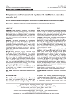
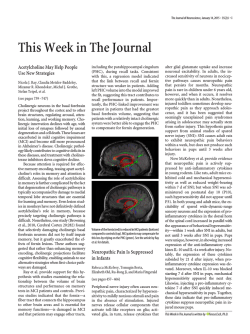
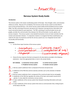
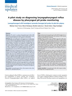
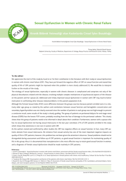
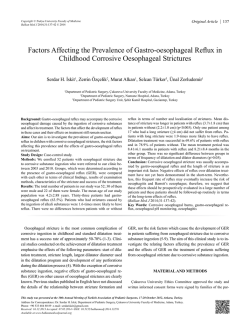
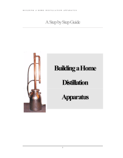
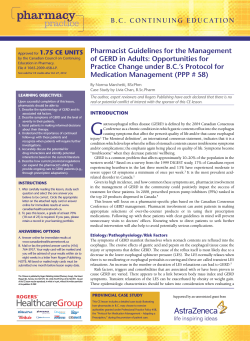
Turkish Journal of Medical Sciences Turk J Med Sci (2014) 44: 243-248 © TÜBİTAK doi:10.3906/sag-1209-30 http://journals.tubitak.gov.tr/medical/ Research Article Expression of NADPH-d in the vagal nuclei of the chronic esophagitis model in rats 1, 2 1 1 Ümit Süleyman ŞEHİRLİ *, Halil TUĞTEPE , Ural VERİMLİ , Özlem KİRAZLI , 1 2 Mazhar ÖZKAN , Emrullah Tolga DAĞLI 1 Department of Anatomy, Faculty of Medicine, Marmara University, İstanbul, Turkey 2 Department of Pediatric Surgery, Faculty of Medicine, Marmara University, İstanbul, Turkey Received: 11.09.2012 Accepted: 18.03.2013 Published Online: 15.01.2014 Printed: 14.02.2014 Aim: To evaluate the changes in the number of NADPH diaphorase (NADPH-d) stained neurons in the vagal nuclei in a chronic esophagitis model. Materials and methods: There were 3 groups of rats examined: 1) a chronic gastroesophageal reflux rat group, which was created by a partial gastric outlet obstruction; 2) a sham group; and 3) a ranitidine treated group. Serial sections of brainstems of all groups were cut and NADPH-d staining, which selectively stains the nitric acid synthase-containing neurons, was done. Results: Histopathological changes due to chronic reflux esophagitis was observed in the reflux group. The ranitidine treatment and sham control groups showed no changes related to esophagitis. The staining in the dorsal motor nucleus of the vagus and nucleus tractus solitarius showed statistically significant differences compared to the control group (P < 0.0001). Conclusion: The increased nitric oxide expressions in the dorsal vagal nucleus and nucleus tractus solitarius are most probably due to adaptive changes to disturbed esophageal motility and mucosal damage. Key words: Esophagitis, lower esophageal sphincter, nitric oxide, dorsal motor nucleus, nucleus tractus solitarius 1. Introduction Disturbances of the lower esophageal sphincter function and of esophageal motility are important factors in the development of reflux esophagitis. Dysfunction of the lower esophageal sphincter (LES) due to inappropriate smooth muscle relaxation and loss of muscle tone are the major factors which lead to gastric reflux (1). The mechanism behind LES dysfunction has been a major subject of research. Some agents, such as the oral β-2 agonists and anticholinergics, and certain foods or beverages, such as coffee, can cause smooth muscle relaxation (1,2). A decrease occurs in the contractile smooth muscle response and the relaxant response of smooth muscle to serotonin in chronic reflux esophagitis (3). Although esophagitis is a common health problem, the changes that occur during esophagitis in the central nervous system are not well known. The control of the nervous system over the factors that control the LES is important. There are anatomical and physiological studies showing that the dorsal motor nucleus of the vagus (DMV) plays a role in the parasympathetic control of the gastrointestinal tract (4–6). *Correspondence: [email protected] Nitric oxide (NO) is a freely diffusing gas. NO takes part in different functions such as cognition, synaptic plasticity, sleep, neurosecretion, appetite, temperature, and homeostasis (7). NO can be measured by means of nitric oxide synthase (NOS) because NO is a highly diffusible and short-lived gas (8). NADPH-diaphorase (NADPH-d) selectively stains NOS-containing neurons (9). NO acts as a neurotransmitter of nonadrenergic, noncholinergic inhibitory nerves innervating gastrointestinal (GI) smooth muscles. It is claimed that NOS might be transported toward the axon terminal and is probably recycled by retrograde transport (10). Berthoud showed that some NADPH-d–positive vagal efferent terminals contact NADPH-d–positive enteric neurons, which project to the circular muscle (11). NO has a role in the control of smooth muscle tone and motility as well as fluid secretion in the GI tract. NO released from the nitrergic nerves and the vascular endothelium results in vasodilatation and leads to an increase in mucosal blood flow, which might help to protect the mucosa (12). The cyclic guanosine monophosphate (GMP), cyclic GMPindependent mechanisms, and decreased intracellular Ca2+ 243 ŞEHİRLİ et al. / Turk J Med Sci levels play an important role in NO-induced relaxation in the GI smooth muscles (3,12). Likewise, neurotransmitters such as acetylcholine, ATP, peptides, and norepinephrine have a role in the control of the NO release from the nitrergic nerves. The inhibition of the NO synthesis due to pathological conditions can cause an increase in GI muscle contractility and an impairment of GI function (12). A recent study indicated that acid and pepsin perfusion of the esophagus results in c-Fos expression in the amygdala, paraventricular nucleus, parabrachial nucleus, nucleus tractus solitarius (NTS), DMV, nucleus ambiguus (NA), medullary reticular nucleus, and the area postrema. NOS reactivity was observed in the paraventricular nucleus, the parabrachial nucleus, the nucleus of the tractus solitarius, and the dorsal motor nucleus of the vagus, as well as the medullary reticular nucleus and the area postrema. Previous authors suggested that during esophageal acid exposure, NOS-containing neuronal cells are activated to modulate esophageal reflux (5). Zhang et al. (6) examined the effect of electrical stimulation of the NA and DMV. Electrical stimulation of the NA had no effect on the volume of gastric juice, titratable acidity, or acid concentration, but an increase in bicarbonate secretion was shown. Electrical stimulation of the DMV showed a significant increase in titratable acidity, in the volume of gastric juice, and in the acid concentration. It was suggested that these findings indicate that DMV innervates both gastric glands and gastric smooth muscles (6). The response of the central nervous system to diseases of the GI system has not been a major area of research. The present study aims to show whether there are any changes of NOS expression in the brainstem in a chronic gastroesophageal reflux model. We aimed to understand the response of the nuclei of the vagus nerve to chronic esophagitis. 2. Materials and methods Male Sprague-Dawley rats weighing between 200 and 250 g were housed in standard conditions. Standard laboratory chow was withdrawn 16 h before surgery with free access to water. All experimental protocols were approved by the Animal Care and Use Committee of the Marmara University School of Medicine. The study was planned to include 3 groups. After intraperitoneal anesthesia of ketamine hydrochloride (20 mg/kg) and xylazine hydrochloride (100 mg/kg), the abdomen was opened with an upper midline incision. In the chronic gastroesophageal reflux group (n = 4), chronic acid reflux esophagitis was induced by pyloric stenosis plus limiting ridge ligation method (13). The duodenum near the pyloric ring was wrapped with a small piece of an 18F Nelaton catheter and the transitional region between the forestomach and the glandular portion (limiting ridge) was ligated by a nonabsorbable suture. 244 In the sham group (n = 4), rats underwent a sham operation. In the ranitidine group (n = 4), after the surgical procedure to prevent development of esophagitis, the rats were treated with ranitidine, a histamine H2 receptor antagonist group antacid (14), at 0.5 mg kg–1 day–1. The plan was to observe the pure effect of surgical procedures without inflammation of the mucosa of the stomach and esophagus. All animals were perfused and fixed with 4% paraformaldehyde transcardiacally 1 month after surgery. The horizontal sections taken from the lower part of the esophagus and stomach were processed routinely, embedded in paraffin, sectioned at 3–5 µm, and stained with hematoxylin-eosin. Each specimen was examined by light microscopy to detect signs of inflammatory changes. The brains were removed, put overnight in a 4% paraformaldehyde containing fixative, and then transferred to a 30% sucrose solution. Next, 40-µm serial coronal frozen sections of brainstem were taken on a sliding microtome. All the sections were put into Tris buffer solution (TBS) at pH 7.6, washed in TBS of pH 7.6 for 30 min, and then washed for 30 min in 0.05 M TrisHCl buffer of pH 8.0 containing 0.2% Triton X-100. The sections were then incubated in a solution containing 0.05 M Tris-HCl buffer of pH 8.0 containing 0.2% Triton X-100, nitroblue tetrazolium (Sigma N6876), and reduced β-NADPH-d tetrasodium salt (Sigma N1630) at 37 °C. After incubation, the sections were rinsed in TBS of pH 7.6 for 30 min. Sections were then transferred to slides in serial order and were mounted using DPX (Sigma DPX Mountant for Histology 44581). In each section we counted all the cells that had a nucleus stained for NADPH-d. The average number of NADPH-d–stained neurons per section for each rat was calculated by dividing the total number of stained neurons counted in all sections by the number of sections taken from each brain nucleus. The numbers of cells containing NADPH-d were counted unilaterally in specific nuclei in several sections: 5 sections for DMV, NTS, and NA. GraphPad Prism 5.1 was used for analysis of data. Data were expressed as mean ± SE of the respective brain areas. The groups were compared by one-way ANOVA post-hoc Tukey test. P < 0.05 was considered as an indication of statistical significance. 3. Results 3.1. Histopathological examination The lower esophagus and stomach of all animals was evaluated histopathologically. Evidence of chronic reflux esophagitis was decided on the basis of the following features: thickness of the esophageal mucosa, elongation of the lamina propria papillae, infiltration by inflammatory cells, interruption of the lamina muscularis mucosa, and amount of collagen fibers in the lamina propria and submucosa. The treatment and control groups did not indicate any major pathological changes (Figures 1a and 1b). ŞEHİRLİ et al. / Turk J Med Sci The sections of the brainstem were examined under a light microscope. NADPH-d activity was observed as a blue color within the perikarya, dendrites, and axons. There was no significant difference between the 2 sides of the nucleus for either DMV or NTS. The total number a of labeled neurons in each nucleus was evaluated. The mean number of labeled neurons in the DMV was 14.95 ± 1.37 in the chronic gastroesophageal reflux group, whereas this number in the control group was 1.3 ± 0.30 and in the ranitidine group it was 2.8 ± 0.46 (Figures 2a–2c). b Figure 1. a) Basal cell hyperplasia, elongation of lamina propria, and chronic inflammatory cell infiltration as histopathological findings of chronic esophagitis in chronic gastroesophageal reflux group; b) stomach of ranitidine group. a b c Figure 2. NADPH-d–stained neurons in the DMV: a) sham group, b) ranitidine group, c) chronic gastroesophageal reflux group. There was a statistically significant difference between the NADPH-d positive neurons in the DMV of the chronic gastroesophageal reflux group compared to the sham and ranitidine groups. 245 ŞEHİRLİ et al. / Turk J Med Sci There was a statistically significant difference between the chronic gastroesophageal reflux group and the other 2 groups, the sham and ranitidine groups (P < 0.0001). A statistically significant difference was also observed in NTS between the chronic gastroesophageal reflux group and the other 2 groups (Figures 3a–3c and 4a–4d). The number of labeled neurons was 21.45 ± 2.8 in the chronic gastroesophageal reflux group, whereas this number in the control group was 3.06 ± 0.75 and in the ranitidine group was 3.75 ± 0.99. There was no statistically significant difference between sham and ranitidine groups. The results are given in the Table. 4. Discussion The vagus nerve is the one of the major structures that have influence over the neural control of the GI system. The DMV, NTS, and NA are the vagal nuclei taking part in a the central control mechanism of the GI tract. The control and integration of visceral functions are closely related to vagal reflexes. Dysfunctional vagal reflexes are associated with GI pathologies and digestive disorders (15–21). The rostral and caudal portions of the DMV contain the esophageal preganglionic neurons. The esophagus and the LES receive inhibitory input from the caudal neurons and excitatory input from the rostral neurons (4,20,22–24). The excitation is provided by the preganglionic cholinergic projections onto cholinergic neurons, while the inhibition is provided by nitrergic enteric neurons (4,25–27). The activation of the intrinsic nitrergic inhibitory neurons by the preganglionic vagal efferents in the caudal DMV leads to LES relaxation during deglutition (4,20,22,23). The stimulation of gastric vagal afferents and vagovagal reflexes causes transient LES relaxation, which may lead to gastroesophageal reflux (4,28,29) The central, intermediate, b 20 *** The number of NOS labeling neurons The number of NOS labeling neurons Figure 3. The NADPH-d–stained neurons in the NTS: a) sham group, b) ranitidine group, c) chronic gastroesophageal reflux group. NADPH-d–positive neurons in the NTS and DMV were more abundant in the chronic gastroesophageal reflux group compared to the sham and ranitidine groups. a 15 10 5 0 CHRONIC GER* DMV RANITIDINE DMV SHAM DMV 30 *** b 20 10 0 CHRONIC GER* NTS RANITIDINE NTS SHAM NTS Figure 4. There was a statistically significant difference between the chronic gastroesophageal reflux group and sham and ranitidine groups in a) DMV and b) NTS. ***: P < 0.001. 246 ŞEHİRLİ et al. / Turk J Med Sci Table. The average number of neurons counted per section of DMV and NTS. Nucleus DMV NTS Sham group 1.3 ± 0.30 3.06 ± 0.75 Ranitidine group 2.8 ± 0.46 3.75 ± 0.99 Chronic GER group 14.95 ± 1.37* 21.45 ± 2.8 * GER: gastroesophageal reflux, DMV: dorsal motor nucleus of vagus, NTS: nucleus tractus solitarius, *: P < 0.0001. and interstitial NTS subnuclei and the compact formation of NA are active during deglutitive activity, whereas commissural and dorsomedial NTS subnuclei and the caudal DMV are active during LES relaxation (4). The commissural and dorsomedial NTS subnuclei and caudal DMV are active in controlling the transient LES relaxation (5). Shuai and Xie (5) emphasized the effect of DMV, NTS, and NA on the esophageal motility. The proposed mechanism was that the acid-pepsin exposure to the lower part of the esophagus stimulates the mucosal receptors, which in turn activate the neurons of the NTS through the vagal afferents and finally the neurons in DMV and NA to modulate the esophageal peristalsis. The neural control of esophageal motility has been a major subject of many studies (4). However, the changes that occur in the central nervous system due to long-term acid exposure in the esophagus are not well explained. In this study, the difference in NADPH-d staining in the sham, ranitidine, and chronic gastroesophageal reflux groups was studied. To our knowledge the effects of chronic esophagitis in the brainstem have not been examined until now. For this reason, the present study was designed to investigate the alterations occurring after a longer period of chronic gastroesophageal reflux. In a previous study, the results of acute acid exposure were evaluated (5). However, these methods might have led to severe esophageal damage. Thus, it was not analogous to the human situation. The chronic esophagitis model used in this study is a well-validated method. In the hematoxylineosin staining of the esophagus the pathologic changes related to chronic esophagitis were verified. In this model, eliminating the mechanic effect of surgical procedures is critical to isolate the effect of increased acidity and mucosal damage on vagal nuclei. Therefore, ranitidine was given to surgically operated rats and thus the increase in acidity and the development of mucosal damage were suppressed. In the ranitidine-treated group, the gastric and esophageal mucosa was intact. The number of NADPH-d stained neurons did not show any significant difference between the sham and ranitidine groups. Several studies demonstrated the presence and importance of NO in vagal pathways. NO is involved in the control of esophageal peristalsis (30–32). Murray et al. (33) suggested that NO has an inhibitory effect on the esophageal muscle contraction. It was reported that NOS expression increases in the nuclei of DMV and NTS due to acid exposure in the esophagus (5). In the present study, we also found an increase of NOS expression in the nuclei of the DMV and NTS. The esophageal peristalsis has been controlled by the caudal and rostral DMV, which have inhibitory and excitatory effects on the esophagus, respectively (4). We could not observe any prominent difference in NADPH-d staining of the 2 parts of the nucleus. There was no staining difference between right and left sides of the nuclei. This is in accordance with the histological changes seen in the esophagus. The findings due to esophagitis were observed all around the horizontal sections of the esophagus. Therefore, the nuclei at each site seem to be affected equally. We generally observed no stained neurons in NA. However, 1, or at most 2, neurons could be stained in a very few sections of NA. This finding can be considered in agreement with the study of Shuai and Xie (5), in which the number of NOS-positive neurons in NA was 2 and there was no significant difference between the control and experimental groups. The model in this study causes disturbed esophageal peristalsis, and also mucosal damage of stomach and esophagus, due to increased acidity. As a result, chronic changes that occur in esophagitis may lead to adaptive changes in the brainstem nuclei. The increased NADPH-d staining is probably a sign of ability to modulate the disturbed esophageal peristalsis. The increase of NOS expression may be a protection mechanism against mucosal damage. Acknowledgments We would like to thank Prof RW Guillery and Prof Safiye Çavdar for their comments concerning an earlier draft of the manuscript. We would also like to thank Dr Hasan Yananlı for his help with statistical evaluation and Dr Kıran Demir for some of the histological preparations. This project was supported by the Marmara University Scientific Research Fund, SAG-BGS-060904-0085. 247 ŞEHİRLİ et al. / Turk J Med Sci References 1. Wolfe M, Michael M. An overview of gastroesophageal reflux disease. Am J Manag Care 2000; 6: 461–6. 2. Goyal RK, Chaudhury A. Physiology of normal esophageal motility. J Clin Gastroenterol 2008; 42: 610–9. 3. Tugtepe H, Tugay M, Bozkurt S, Yildiz F, Utkan T, Yegen B, Dagli TE. Esophageal smooth muscle reactivity is impaired in chronic reflux esophagitis by both receptor- and nonreceptormediated mechanisms. J Pediatr Surg 2007; 42: 641–6. 4. Neuhuber WL, Raab M, Berthoud HR, Wörl J. Innervation of the mammalian esophagus. Adv Anat Embryol Cell Biol 2006; 185: 1–73. 5. Shuai XW, Xie PY. Expression and localization of c-Fos and NOS in the central nerve system following esophageal acid stimulation in rats. World J Gastroenterol 2004; 10: 2287–91. 6. Zhang XY, Ai HB, Cui XY. Effects of nucleus ambiguus and dorsal motor nuclei of vagus on gastric H(+) and HCO(3)(-) secretion in rats. World J Gastroenterol 2006; 12: 3271–4. 7. Guix FX, Uribesalgo I, Coma M, Muñoz FJ. The physiology and pathophysiology of nitric oxide in the brain. Prog Neurobiol 2005; 76: 126–52. 8. Bredt DS, Hwang PM, Snyder SH. Localization of nitric oxide synthase indicating a neural role for nitric oxide. Nature 1995; 347: 768–70. 9. Dawson TM, Bredt DS, Fotuhi M, Hwang PM, Snyder SH. Nitric oxide synthase and neuronal NADPH diaphorase are identical in brain and peripheral tissues. Proc Natl Acad Sci USA 1991; 88: 7797–801. 10. Forster ER, Southam E. The intrinsic and vagal extrinsic innervation of the rat stomach contains nitric oxide synthase. Neuroreport 1993 Mar; 4: 275–8. 11. Berthoud HR. Anatomical demonstration of vagal input to nicotinamide acetamide dinucleotide phosphate diaphorasepositive (nitrergic) neurons in rat fundic stomach. J Comp Neurol 1995; 31; 358: 428–39. 12. Toda N, Herman AG. Gastrointestinal function regulation by nitrergic efferent nerves. Pharmacol Rev 2005; 57: 315–38. 13. Omura N, Kashiwagi H, Chen G, Suzuki Y, Yano F, Aoki T. Establishment of surgically induced chronic acid reflux esophagitis in rats. Scand J Gastroenterol 1999; 34: 948–53. 14. Özer M, Duman M, Taş Ş, Demirci Y, Aydın MF, Reyhan E, Atıcı AE, Bostancı EB, Akoğlu M, Genç E. In vitro effects of famotidine and ranitidine on lower esophageal sphincter tone in rats. Turk J Gastroenterol 2012; 23: 438–43. 15. Yamato S, Saha JK, Goyal RK. Role of nitric oxide in lower esophageal sphincter relaxation to swallowing. Life Sci 1992; 50: 1263–72. 16. Saito Y, Kawashima Y, Kondo A, Chikumaru Y, Matsui A, Nagata I, Ohno K. Dysphagia-gastroesophageal reflux complex: complications due to dysfunction of solitary tract nucleus-mediated vago-vagal reflex. Neuropediatrics 2006; 37: 115–20. 17. Thumshirn M. Gastrointestinal motility disorders relevant to general practice. Praxis (Bern 1994) 2002; 91: 1741–7. 248 18. Ghia JE, Blennerhassett P, El-Sharkawy RT, Collins SM. The protective effect of the vagus nerve in a murine model of chronic relapsing colitis. Am J Physiol Gastrointest Liver Physiol 2007; 293: G711–8. 19. Andrews PL, Sanger GJ. Abdominal vagal afferent neurons: an important target for the treatment of gastrointestinal dysfunction. Curr Opin Pharmacol 2002; 2: 650–6. 20. Hornby PJ, Abrahams TP. Central control of lower esophageal sphincter relaxation. Am J Med 2000; 108 Suppl: 90–8 21. Faris PL, Hofbauer RD, Daughters R, Vandenlangenberg E, Iversen L, Goodale RL, Maxwell R, Eckert ED, Hartman BK. De-stabilization of the positive vago-vagal reflex in bulimia nervosa. Physiol Behav 2008; 94: 136–53. 22. Chang HY, Mashimo H, Goyal RK. Musings on the wanderer: what’s new in our understanding of vago-vagal reflex? IV. Current concepts of vagal efferent projections to the gut. Am J Physiol Gastrointest Liver Physiol 2003; 284: G357–66. 23. Hyland NP, Abrahams TP, Fuchs K, Burmeister MA, Hornby PJ. Organization and neurochemistry of vagal preganglionic neurons innervating the lower esophageal sphincter in ferrets. J Comp Neurol 2001; 430: 222–34. 24. Rossiter CD, Norman WP, Jain M, Hornby PJ, Benjamin S, Gillis RA. Control of lower esophageal sphincter pressure by two sites in dorsal motor nucleus of the vagus. Am J Physiol 1990; 259: G899–906. 25. Bieger D, Triggle C. Pharmacological properties of mechanical responses of the rat oesophageal muscularis mucosae to vagal and field stimulation. Br J Pharmacol 1985; 84: 93–106. 26. Storr M, Geisler F, Neuhuber WL, Schusdziarra V, Allescher HD. Characterization of vagal input to the rat esophageal muscle. Auton Neurosci 2001; 91: 1–9. 27. Watson N, Reddy H, Eglen RM. Characterization of muscarinic receptor and beta-adrenoceptor interactions in guinea-pig oesophageal muscularis mucosae. Eur J Pharmacol 1995; 294: 779–85. 28. Mittal RK, Holloway R, Dent J. Effect of atropine on the frequency of reflux and transient lower esophageal sphincter relaxation in normal subjects. Gastroenterology 1995; 109: 1547–54. 29. Sifrim D, Holloway R. Transient lower esophageal sphincter relaxations: how many or how harmful? Am J Gastroenterol 2001; 96: 2529–32. 30. Tøttrup A, Knudsen MA, Gregersen H. The role of the L-arginine-nitric oxide pathway in relaxation of the opossum lower oesophageal sphincter. Br J Pharmacol 1991; 104: 113–6. 31. Yamato S, Spechler SJ, Goyal RK. Role of nitric oxide in esophageal peristalsis in the opossum. Gastroenterology 1992; 103: 197–204. 32. Zheng ZL, Rogers RC, Travagli RA. Selective gastric projections of nitric oxide synthase-containing vagal brainstem neurons. Neuroscience 1999; 90: 685–94. 33. Murray J, Du C, Ledlow A, Bates JN, Conklin JL. Nitric oxide: mediator of nonadrenergic noncholinergic responses opossum esophageal muscle. Am J Physiol 1991; 261: G401–6.
© Copyright 2026 Paperzz