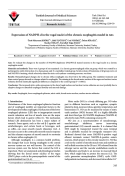
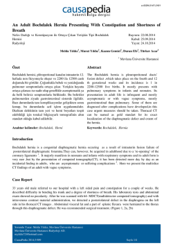
2014; 5 (1): 1-6
1
doi: 10.5799/ahinjs.01.2014.01.0349
JCEI / Journal of Clinical and Experimental Investigations ORIGINAL ARTICLE / ÖZGÜN ARAŞTIRMA
Intragastric manometric measurements of patients with hiatal hernia: A prospective
controlled study
Hiatal Hernili hastalarda intragastrik manometrik ölçümler: Prospektif kontrollü bir çalışma
Kemal Peker1, Abdullah İnal1, Mustafa Emiroğlu2, İsmayil Yılmaz1, İsmail Demiryılmaz3, Arda Işık1
ABSTRACT
ÖZET
Objective: Hiatal hernia is a disorder in which abdominal contents, especially gastroesophageal junction and
proximal stomach are displaced through an incompetent
esophageal hiatus into mediastinum. Aim of this study is
to investigate the relationship betweeen intragastric pressure and incompetent cardioesophageal sphincter and
hiatal hernia.
Amaç: Hiatus hernisi, diafragmanın özofageal hiatusdaki
anatomik zayıflık ya da defekt nedeniyle intraabdominal
organların toraksa çıkışı olarak tanımlanabilir. Bu çalışmanın amacı kardio özofageal yetmezlikli ve hiatal hernili
hastaların intragastrik basınç düzeylerini incelemekti.
Methods: The sample is composed of 81 individuals;
51.9% of which are male (n: 42) and 48.1% of which are
female (n: 39) with median age of 42.00. Two groups
have been constituted: A control group of healthy volunteers (G1) and a patient group with hiatal hernia (G2).
After gastroscopy, all patients and controls underwent
gastric and esophageal manometry using a water perfused catheter
Results: Mean intragastric pressure was 1,308 mmHg
for G1 and, -0,105 mmHg for G2. Mean lower esophageal pressure were 13,143 mmHg and 11,65 mmHg,
and upper esophageal pressure were 35,273 mmHg and
27,211 mmHg for G1 and G2, respectively. 24 hour pH
metry test revealed 26.8% physiologic gastroesophageal
reflux and 73.2% pathologic gastroesophageal reflux in
G2. There were statistically significant difference between
two groups for mean intragastric pressure (p <0.001) and
mean lower esophageal pressure (p <0.05).
Conclusion: This study shows that hiatal hernia results
in significantly lower intragastric pressures. J Clin Exp Invest 2014; 5 (1): 1-6
Yöntemler: Çalışmaya 81 hasta dâhil edildi. Hastaların
42 (%51,9)’si erkek iken 39 (%48,1)’u bayan idi. Yaş ortalamaları 42 yıl olarak tespit edildi. Hastalardan iki grup
oluşturuldu. Bir grup (G1) sağlıklı gönüllü hastalardan oluşur iken, ikinci grup (G2) hiatal herni hali olan hastalardan
oluşturuldu. Gastroskopiden sonra, tüm hastalara su perfüzyon sistemli kateter kullanılarak gastrik ve özofageal
manometrik ölçümler yapıldı.
Bulgular: Ortalama intragastrik basınç düzeyi kontrol
grubu için 1,308 mmHg iken, hasta grubu için bu değer
-0,105 mmHg olarak ölçüldü. Kontrol grubunda distal
özofageal gastrik basınç düzeyi 13,143 mmHg iken hasta grubunda bu oran 11,65 mmHg olarak, üst özofageal
sfinkter basınç düzeyi sırayla 35,273 mmHg ve 27,211
mmHg olarak ölçüldü. Hiatal herni hali mevcut olan G2
için yapılan 24 saatlik pH monitörizasyonunda %26,8
hastada, reflü fizyolojik sınırlarda iken %73,2 hastada reflü hali patolojik sınırlarda tespit edildi. Gruplar arası intragastrik ve distal özofageal basınç düzeyleri açısından
yapılan ikili karşılaştırmalarda sonuçlar istatistiksel olarak
anlamlı idi (p<0.001, p<0.05).
Sonuç: Çalışma neticesinde Hiatal Herni halinin intragastrik basınç düzeyini düşürdüğü kanatine varılmıştır.
Key words: Hiatal Hernia, Manometry, Intragastric Pressures
Anahtar kelimeler: Hiatal herni, manometre, intragastrik
basınç
INTRODUCTION
of ingested food into the esophagus through gastroesophageal junction (GEJ) with the coordinated
action of lower esophageal sphincter (LES), and
its associated structures gastric sling and crural
The junction between esophagus and stomach is
a complex structure which functions to keep gastric content in stomach while preventing the reflux
Erzincan University, Department of General Surgery, Erzincan, Turkey
Tepecik Training and Research Hospital, Department of General Surgery, İzmir, Turkey
3
İbni Sina Hospital, Department of General Surgery, Kayseri, Turkey
1
2
Correspondence: Kemal Peker,
Mengücek Gazi Training and Research Hospital, Erzincan, Turkey Email: [email protected]
Received: 28.08.2013, Accepted: 21.10.2013
Copyright © JCEI / Journal of Clinical and Experimental Investigations 2014, All rights reserved
2
Peker et al. Intragastric manometry in patients with hiatal hernia
diaphragm. The same structure also permits retrograde passage of air and gastric contents into the
esophagus during ructus and vomiting. A delicately
regulated high pressure zone achieves this precise
coordination [1,2]. Gastroesophageal reflux disease
(GERD) seems to be originated from the imbalance
between defensive factors and aggressive factors. GEJ constitutes the first and essential line of
esophageal defense against acid damage of refluxate, and impairment in this barrier concludes with
pathological reflux [3].
Hiatal hernia (HH) is a disturbance in which
parts of abdominal contents, especially GEJ and
stomach are displaced proximally through an incompetent esophageal hiatus into mediastinum [4].
Four types of esophageal hiatal hernias have been
defined: sliding (type I), paraesophageal (type II),
combined (type III), which include elements of types
I and II, and giant paraesophageal (type IV). Each
type may be presented with different symptoms and
complications. Sypmtoms and consequences of the
disease necessitates proper and prompt diagnosis
[5]. Type I sliding hiatal hernia accounts for more
than 95% of all hiatal hernias while the remaining
5% represents paraesophageal hiatal hernias [6].
In this study, our purpose is to evaluate relationship between intragastric pressure levels and
HH. We also aimed to assess effect of this pathological condition on gastro-esophageal reflux disease.
METHODS
A prospective controlled clinical study was conducted at Mengücek Gazi Training and Research Hospital in Erzincan, Turkey. This study was approved
by the Ethics Committee of the Erzincan University
and performed in accordance with Declaration of
Helsinki. All patients were asked to provide written
informed consent prior to enrollment, after explanation of the associated risks and benefits and description of the study protocol.
The sample is composed of 81 individuals.
51.9% of participants were male (n: 42), and 48.1%
were female (n: 39). Median age of the sample was
42.00 and mean age was 42.29. All of patients were
evaluated by gastroscopy and manometric motility
tests. Two groups have been constituted from the
sample. First group (G1) was composed of 40 patients whose cardio-esophageal sphincter function
was normal. Second group (G2) was composed of
41 patients who had cardio-esophageal sphincter
insufficiency and HH. The body mass index (BMI)
was calculated as 22,66 for G1 and 22,91 for G2. All
J Clin Exp Invest patients and controls underwent gastric and esophageal manometry using a water perfused catheter
with nine radially aligned channels attached to a
hydraulic capillary infusion system. Measured intragastric (IG), distal esophageal sphincter (LES) and
upper esophageal sphincter (UES) pressure levels
were recorded. Additionally, patients in Group 2
(G2) had a 24-hour acidity analysis by pH meter.
Exclusion criteria for the subjects with normal
gastroesophageal function included any abdominal
symptoms or any medication that could affect the
gastroesophageal segment high-pressure zone, including antacids, H2 blockers, proton pump inhibitors, prokinetic agents, antibiotics and anticholinergics, GERD, hiatal hernia, conditions and disorders
including a history of abdominal pain, heartburn,
difficulty in swallowing, pain on swallowing, dysphagia, abdominal surgery involving the stomach or
esophagus, nausea or vomiting, diabetes, scleroderma, esophageal motility disorders, noncardiac
chest pain, achalasia, and existing pregnancy. Exclusion criteria for hiatal hernia patients were any
history of surgery for esophagus or stomach.
Endoscopic evaluation of the study subjects
All subjects were undergone upper endoscopy after an overnight fast with an Olympus Evis Exera
II CRV-180 (Olympus Corp. Tokyo, Japan) endoscope. Subjects were kept in the left lateral decubitus position during the procedure. Benzocaine
spray was used to anesthetize the posterior pharyngeal wall since the endoscopy was performed
unsedated. Subjects were evaluated for the presence of esophagitis and for any abnormalities in
stomach and duodenum, including hiatal hernia.
Manometric evaluation
All the patients were undergone gastro-esophageal
manometry testing. Manometric testing was performed with a 9E-12-100A Menfis Biomedica 9-way
catheter (Menfis biomedica s.r.l. Bologna, Italy).
The PVC catheter for measurement has external
diameter of 12 Fr (4 mm), operative length of 100
cm, and total length of 180 cm. It has one central
lumen of 1.4 mm diameter, which opens at the tip
and four lumina which open with 4 side-ports of 0.8
mm diameter with radial arrangement at 3 cm far
from the tip. The remaining four lumina open with 4
side-ports, 5 cm apart along the catheter in helicoidal arrangement. Centimetric markings are starting
from the side-port no 8. (Figure 1)
Before the test, device was calibrated on the
same level with the patient. Examination was performed with rapid pull-through technique when the
www.jceionline.org Vol 5, No 1, March 2014
Peker et al. Intragastric manometry in patients with hiatal hernia
patient was placed in supine right lateral position.
The catheter was pulled by mechanical device with
3
a speed of 1 Sec/2,5mm under resting condition.
(Figure 2).
Figure 1. Diagram of gastric manometry catheter which was used in the study
Figure 2. Graphic
view of gastric
manometric measurements of one
patient
pH-Monitoring
All the G2 patients underwent 24 h distal esophageal pH monitoring. Proton-pump inhibitor medications were discontinued at least 2 weeks before
testing and other reflux medications were discontinued at least 72 h before testing. The pH monitoring
was performed with an antimony crystal pH electrode (Menfis Biomedica) 5 cm above the manometrically measured LES, when patient was placed
in supine right lateral position. The electrode was
connected to a digital recording device. The stored
data were transferred to a computer and analyzed
using a standard software package.
Statistical analysis
Data was analyzed by using SPSS 15.0 software.
According to normality tests, the distribution is not
J Clin Exp Invest normal therefore, non-parameters tests were used.
Mann-Whitney U test was used, in order to compare
intergroup differences. P value less than 0.05 was
accepted as significant.
RESULTS
Mean IG pressure level was measured 1.308 ±
1.392 mmHg for Group 1 (G1) and -0.105 ± 1.651
mmHg for Group 2 (G2). Mean LES pressure and
mean UES pressure of Group 1 (G1) were measured 13.143 ± 5.962 mmHg and 35.273 ± 17.202
mmHg, respectively. Mean LES pressure and mean
UES pressure of Group 2 (G2) were measured
11.65 ± 7.145 mmHg and 27.211 ± 10.062 mmHg,
respectively. As a result of 24-hour pH meter measurements for Group 2 (G2), there were no gastro-
www.jceionline.org Vol 5, No 1, March 2014
Peker et al. Intragastric manometry in patients with hiatal hernia
4
esophageal reflux in 11of patients (26.8%), and
gastroesophageal reflux condition was defined in
30 of patients (73.2%) at pathological levels. Results of mean IG, LES and UES pressure levels are
shown on Table 1.
According to test results, the difference between the mean intragastric pressure levels of
experiments and control groups was significant (p
<0.001). Although LES pressure levels were lower
in experimental group, there was not a statistically
Table 1. Comparison of pressure
level in IG, LES
and UES between
control and patient groups for
intragastric pressures
IG
LES
UES
significant difference between LES pressure levels
of control and experimental groups (p=0. 0,153).
However; there was an unexpected significant difference between UES pressure levels of control
and experimental groups (p< 0.05). The results of
the comparison to reflux are shown in Table 2.
The differences are found to be statistically
significant between means and mean ranks of IG,
LES and UES in patients with and without gastroesophageal reflux disease.
Percentiles
Group
n
X ± SD
25.
50 (Median)
75.
G1
40
1,308 ± 1,392
0,39
1,27
1,80
G2
41
-0,105 ± 1,651
-0,74
0,40
0,94
G1
40
13,143 ± 5,962
9,20
11,15
15,71
G2
41
11,65 ± 7,145
6,99
10,43
14,31
G1
40
35,273± 17,202
23,23
30,53
41,88
G2
41
27,211± 10,062
20,35
26,20
32,95
Ua
pb
423,0
<0,1**
669,5
0,153
594,5
0,027*
a. Mann Whitney U Test
b. Monte Carlo Sig.(2-tailed) Based on 10000 sampled table. ***p<0,001 , *p<0,05
Comparison of pressure level in IG, LES and UES between groups was shown in this table.
Table 2. Comparison of pressure
level in IG, LES
and UES between
groups that were
detected
reflux
and the patients
that were not detected reflux was
shown in this table.
IG
LES
UES
Percentiles
n
X ± SD
25.
50 (Median)
75.
No reflux
11
-0,538 ± 1,878
-1,80
-0,23
1,16
Reflux
30
0,053 ± 1,564
-0,34
0,46
0,93
No reflux
11
11,29 ± 6,975
4,95
11,35
14,63
Reflux
30
11,782 ± 7,32
7,02
10,10
14,19
No reflux
11
30,852 ± 10,841
23,64
27,60
36,73
Reflux
30
25,876 ± 9,604
20,21
25,04
32,23
Ua
pb
139,5
0,472
161,0
0,913
121,5
0,208
a. Mann Whitney U Test
b. Monte Carlo Sig.(2-tailed) Based on 10000 sampled table. ***p<0,001, *p<0,05
DISCUSSION
Since direct measurement of abdominal pressure
(IAP) is unfeasible under most circumstances, the
homogenous transmission of pressure within the
abdomen allows IAP to be estimated via the bladder, rectum or stomach [7]. A number of factors, including patient position, ventilatory state and bowel
function influences IAP. Intragastric pressure (IGP)
is commonly employed as a substitute measurement for IAP in a number of researchs [8,9].
J Clin Exp Invest Intragastric pressure was significantly higher
in obese and overweight patients when compared
with people with a normal BMI during both expiration and inspiration [10]. The concept of increased
gastroesophageal pressure gradient (GEPG) in
obese patients was already introduced more than
two decades ago. Hypothesis is originated from
the assumption that increase in intraperitoneal and
abdominal wall fat mass can increase the intraabdominal, and therefore perigastric pressure [11].
Studies of other hollow organs have confirmed the
www.jceionline.org Vol 5, No 1, March 2014
Peker et al. Intragastric manometry in patients with hiatal hernia
presence of increased intra-abdominal pressure
in patients with central obesity [12]. Mercer et al.
evaluated eight normal weight and eight morbidly
obese subjects without clinical evidence of GERD.
The authors found no significant difference in mean
LES pressure between the two groups during either
expiration or inspiration [11]. In contrast, the GEPG
was significantly higher in the obese patients in
comparison with normal controls both in expiration
and inspiration. Consequently, GEPG/LES pressure ratio was also significantly higher in obese patients when compared to normal controls, both in
expiration and inspiration [13]. A high BMI increases
GEPG mainly by raising IGP [14].
In literature, there are many studies defining
the relationship between intragastric pressure level
and obesity. Yet, there are not sufficient data defining the relationship between hiatal hernia and intragastric pressure levels. Vegesna et al. [15] determined that IGP levels in HH are distinctively lower
than IGP levels of ordinary patients in manometric
studies. Similarly, we found out that IGP levels in
hiatal hernia are lower than IGP levels of ordinary
patients. The values we obtained in our study were
lower than numbers Vegesna et al. determined in
their studies. The reason for such difference was
that Vegesna et al. performed manometric measurements in the course of endoscopic operation.
Conversely, we performed manometric measurements in resting period following endoscopic operation in our study. The results we obtained seem to
be more objective.
Hiatus hernia causes a spatial seperation in
pressure components of lower esophagus which is
derived from the intrinsic lower esophageal sphincter and the extrinsic compression of the esophagus
within the hiatal canal, and reduces lower esophageal sphincter pressure and alters its dynamic responsiveness [16]. GERD is very common in the
western society; 10–20% of the population was reported to be suffer from one or more of the cardinal
symptoms at least weekly [17]. The main mechanism by which gastroesophageal reflux takes place
is the transient relaxation of the lower esophageal
sphincter (TLESR) [18]. In patients with HH however, the reflux occurs through other mechanisms
[19].
Ott et al. [20] in a study with 319 patients found
that rate of acid exposure was 3.7% in patients without HH and this rate was found out 5.7% in patients
with HH. They also determined that this rate was 6.6
% in patients with large HH, and rate was found 4.8
% in patients with small HH. They found that the reJ Clin Exp Invest 5
sults of 24-hour pH meter were normal in 237 (74%)
patients, while the result of 24-hour pH meter was
abnormal in 82 (26%) patients. Similarly, Ping et al.
[21] showed that LES pressure is low in the patients
with HH and patients with HH were also associated
with gastro-esophageal reflux disease more commonly. Lord et al. [22] found that erosive esophagitis
rate increased in the patients with HH. Accordingly, we defined 41 patients with hiatal hernia in our
study. The results of 24-hour pH meter were abnormal in 30 (73.2 %) patients and reflux was encountered in these patients, as well. The results were
normal at 11 (26.8 %) patients. Likewise, we found
that mean LES pressure was 11,65 mmHg for the
patient group and mean LES pressure was lower
in comparison with control group. This value was
found out to be 13,14 mmHg for the control group.
Pressure differences between two groups can be
attributed to increased reflux in patients with HH.
In conclusion, IGP and LES pressures reduce
in patients with HH. Hiatal hernia may cause disruption in barrier function of GEJ, and therefore
change the pressure gradients between body compartments. Disorders of anatomic structures such
as LES, crural diaphragm and gastric sling lead to
alterations in physiological function. Treatment approaches directed to correct disturbances in normal
structure and function may provide improvements in
symptoms and normal physiological function.
The study was presented on the 48th Congress
of the European Society for Surgical Research,
İstanbul- Turkey, 2013.
REFERENCES
1. Boeckxstaens GE. The lower oesophageal sphincter.
Neurogastroenterol Motil 2005; 17: 13-21.
2. Sidhu SA, Triadafilopoulos G. Neuro-regulation of lower esophageal sphincter function as treatment for gastroesophageal reflux disease. World J Gastroenterol
2008;14:985-990.
3. Hyun JJ, Bak YT. Clinical Significance of Hiatal Hernia.
Gut and Liver 2011;5:267-277.
4. Marchand P. The anatomy of esophageal hiatus of the
diaphragm and the pathogenesis of hiatus herniation.
J Thorac Surg 1959;37:81-92.
5. Dean C, Etienne D, Carpentier B, et al. Hiatal hernias.
Surg Radiol Anat 2012;34:291-299.
6. Kahrilas PJ. Hiatus hernia causes reflux: fact or fiction?
Gullet 1993;3:21-30.
7. Turnbull D, Webber S, Hamnegard CH, et al. İntraabdominal pressure measurement: validation of intragastric pressure as a measure of intra-abdominal
pressure. Brit J Anaesth 2007;98:628-623.
www.jceionline.org Vol 5, No 1, March 2014
6
Peker et al. Intragastric manometry in patients with hiatal hernia
8. Balogh Z, Jones F, D’Amours S, et al. Continuous intraabdominal pressure measurement technique. Am J
Surg 2004;188:679-684.
9. Ravishankar N, Hunter J. Measurement of intra-abdominal pressure in intensive care units in the United
Kingdom: a national postal questionnaire study. Br J
Anaesth 2005;94:763-766.
10. Pandolfino JE, El-Serag HB, Zhang Q, et al. Obesity: a challenge to esophagogastric junction integrity.
Gastroenterology 2006;130:639-649.
11. Mercer CD, Wren SF, DaCosta LR, et al. Lower
esophageal sphincter pressure and gastroesophageal pressure gradients in excessively obese patients. J
Med 1987;18:135–146.
12. Sugerman HJ, DeMaria EJ, Felton WL 3rd, et al. Increased intra abdominal pressure and cardiac filling
pressures in obesity-associated pseudotumor cerebri.
Neurology 1997;49:507–511.
13. Fass R. The Pathophysiological Mechanisms of GERD
in the Obese Patient. Dig Dis Sci 2008;53:2300–2306.
14. El Serag HB, Tran T, Richardson P, et al. Anthropometric correlates of intragastric pressure. Scand J
Gastroenterol 2006;41:887–91.
15. Vegesna A, Besetty R, Kalra A, et al. Induced opening of the gastroesophageal junction occurs at a lower
gastric pressure in gerd patients and in hiatal hernia
subjects than in normal control subjects. Gastroenterol Res Pract 2010;2010:857654.
J Clin Exp Invest 16. Kahrilas PJ, Lin S, Chen J, et al. The effect of hiatus
hernia on gastro-oesophageal junction pressure. Gut
1999;44:476-82.
17. Dent J, El Serag HB, Wallander MA, et al. Epidemiology of gastrooesophageal reflux disease: A systematic review. Gut 2005;54: 710–717.
18. Holloway RH. The anti-reflux barrier and mechanisms
of gastro-oesophageal reflux. Best Pract Res Clin
Gastroenterol 2000;14:681–699.
19. van Herwaarden MA, Samsom M, Smout AJ. Excess
gastroesophageal reflux in patients with hiatus hernia
is caused by mechanisms other than transient LES
relaxations. Gastroenterology 2000;119:1439–1446.
20. Ott DJ, Glauser SJ, Ledbetter MS, et al. Association of Hiatal Hernia and Gastroesophageal
Reflux:Correlation Between Presence and Size of Hiatal Hernia and 24-hour pH Monitoring of the Esophagus.AJR 1995;165:557-559.
21. Ye P, Li ZS, Xu GM, et al. Esophageal motility in patients with sliding hiatal hernia with reflux esophagitis.
Chinese Medical Journal 2008;121:898-903.
22. Lord RV, DeMeester SR, Peters JH, et al. Hiatal hernia, lower esophageal sphincter incompetence, and
effectiveness of Nissen fundoplication in the spectrum
of gastroesophageal reflux disease. J Gastrointest
Surg 2009;13:602-610.
www.jceionline.org Vol 5, No 1, March 2014
© Copyright 2026 Paperzz