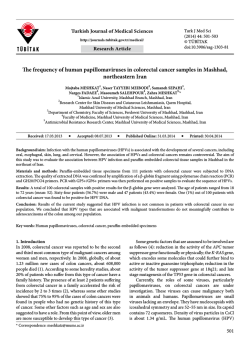
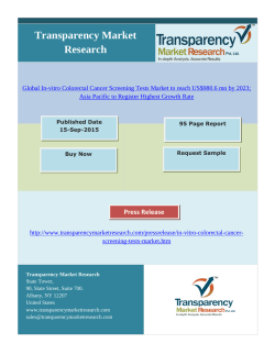
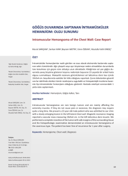
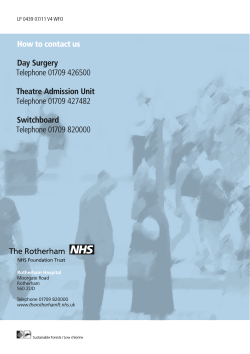
CASE REPORT
&
Hastal›klar› Dergisi
Journal of Diseases of the Colon and Rectum
Diffuculties of Laparoscopic Rectum Cancer Surgery
in a Patient with Cirrhosis
Sirozlu Bir Hastada Laparoskopik Rektum Kanseri Cerrahisindeki
Zorluklar
BORA KAR‹P1, MET‹N KESK‹N2, EMRE BALIK2
1Fatih Sultan Mehmet E¤itim Ve Araflt›rma Hastanesi, Genel Cerrahi Klini¤i, ‹stanbul - Türkiye 2‹stanbul Üniversitesi, ‹stanbul T›p Fakültesi Genel
Cerrahi Ana Bilim Dal›, ‹stanbul - Türkiye
ÖZET
Sirozda, portal hipertansiyon mezenterik konjesyon ve
ödeme neden olur. Bu mezenterik de¤ifliklikler ameliyat
esnas›nda baz› problemlere yol açabilmekte olup ameliyat
öncesi bilgisayarl› tomografi ile tespit edilebilmektedir.
58 yafl›nda Child A siroz tan›l› bir hastaya laparoskopi
yard›ml› extralevator abdominoperineal rezeksiyon
ameliyat› uyguland›. Mezenterik venöz göllenme ve
perivasküler fibrozise ba¤l› inferior mezenterik ven
zorlukla diseke edildi. Dev ödematöz spesmen
extralevator eksizyon defektinden zorlukla kar›n d›fl›na
al›nabildi.
Sirozlu kolorektal kanser hastalar›n›n tedavisi zorluklar
içermektedir. Mezenterik fibrozis ve mezorektal ödem
rektal kanser cerrahisi esnas›nda karfl›lafl›lan ana sorunlar
olup, her ikisi de ameliyat öncesi bilgisayarl› tomografi
ile tespit edilebilmektedir. Gastrointestinal cerrahlar bu
ABSTRACT
Portal hypertension in cirrhosis causes mesenteric
congestion and edema. These mesenteric changes can
be predicted by computerized tomography and may lead
some problems during colorectal surgery.
A 58 year old male patient with Child A cirrhosis
underwent a laparoscopy assisted extralevator
abdominoperineal excision. Secondary to mesenteric
venous stasis and perivasculary fibrosis, the dissection
of the inferior mesenteric vein was problematic. The
removal of the giant, edematous specimen out of the
abdomen could hardly be completed through the
extralevator excision defect.
The management of colorectal cancer patients with
cirrhosis is challenging. Mesenteric fibrosis and
mesorectal edema seems to be the main per-operative
problem in the surgical treatment of rectal cancer and
Baflvuru Tarihi: 16.05.2014 Kabul Tarihi: 20.08.2014
Dr. Bora Karip
Fatih Sultan Mehmet E¤itim Ve Araflt›rma Hastanesi
E5 Karayolu Üzeri ‹çerenköy / Ataflehir 34752 ‹stanbul - Türkiye
Tel: 0532.6478667
e-mail: [email protected]
Kolon Rektum Hast Derg 2014;24:95-99
© TKRCD 2014
96
KAR‹P et al.
mezenterik de¤iflikler konusunda dikkatli olmal›d›r.
Extralevator eksizyonun temin etti¤i genifl perineal
boflluk dev spesmenin kar›n d›fl›na al›nmas›nda hastay›
ek bir laparotomiden koruyabilir.
Anahtar Kelimeler: Kolorektal kanser, Siroz, Sisli
mezenter, Laparoskopi
Kolon Rektum Hast Derg, Eylül 2014
both can be predicted preoperatively by computerized
tomography. Gastrointestinal surgeons must be aware
of these mesenteric changes. Extralevator excision of
the perineum may provide a larger perineal space and
saves the patient from an additional laparotomy for the
removal of giant specimen.
Key words: Colorectal cancer, Cirrhosis, Misty
mesentery, Laparoscopic
Introduction
Nowadays, total mesorectal excision (TME), is the gold
standard for the treatment of rectal cancer due to lower
local recurrence rates, better survival and functional
outcomes. TME can be performed with lower morbidity
and mortality rates by well-trained and experienced
surgeons, either open or laparoscopic.
Portal hypertension in cirrhosis causes mesenteric
congestion and edema. This condition may cause a
difficult dissection of the mesenteric vessels during the
operation. Another problem is the dissection of the
enlarged mesorectum in a relatively narrow pelvis.
Taking the specimen out of the abdomen, through
perineum may be challenging, if the operation is an
abdominoperineal resection (APR).
In this case report we discussed the problems occurred
during a laparoscopy assisted abdominoperineal excision
in a patient with cirrhosis.
Case Report
A 58 year old male patient was referred to our outpatient
clinic with a complaint of rectal bleeding. He had a
medical history of chronic viral hepatitis C and Child A
cirrhosis. A digital rectal examination revealed a rectal
mass, 4 cm above the anal verge. Endoscopic studies
showed an irregular mass, just above the dentate line
and grade 2 esophageal varicose veins. Biopsy of the
rectal mass resulted in adenocarcinoma.
After imaging studies, clinical stage of the patient was
found to be T3, N1 and M0. Mesenteric changes due to
cirrhosis could also be detected by the same imaging
studies. He was referred to oncology for neoadjuvant
chemoradiotherapy (nCRT).
After completion of long course nCRT, a laparoscopy
assisted abdominoperineal resection was planned. He
had no previous abdominal surgery and his body mass
© TKRCD 2014
index (BMI) was calculated as 24 kg/m2. During the
operation inferior mesenteric vein (IMV) could not be
isolated due to peri-vascular fibrosis. Fibrotic tissue
including IMV was divided by vascular stapler. Inferior
mesenteric artery was divided easily. An unusual bleeding
occurred during lateral peritoneal dissection of the
descending colon from the enlarged veins of peritoneum.
After reaching the level of sacral promontory, the enlarged
mesorectum secondary to mesenteric edema limited our
surgical plane, through the minor pelvis both anteriorly
and posteriorly (Fig. 1). With gentle dissection and great
effort, we could reach pelvic floor and TME was
completed. The rest of the operation was continued from
perineum and an extended (extralevator) perineal
resection (eAPR) was performed (Fig. 2). Specimen was
removed out of the abdomen successfully through the
perineal defect with excessive effort. The defect could
be closed in primary fashion after dissection of
subcutaneous fat tissue and no flap reconstruction was
needed.
Figure 1. A relatively narrow pelvis. Pelvic peritoneum
is marked with white line. Black line is showing the
borders of enlarged mesorectum.
Vol. 24, No.3
DIFFUCULTIES OF LAPAROSCOPIC RECTUM CANCER SURGERY IN A PATIENT WITH CIRRHOSIS
Figure 2. Large perineal defect of extended perineal
excision.
The maximum diameter and circumference of the
specimen was 15 and 45 centimeters respectively (Fig.
3). After pathological investigation, specimen was
reported as ypT2, N (0) with a complete mesorectal
fascia. Patient's postoperative course was uneventful.
He was referred to oncology to complete his
chemotherapy.
Discussion
TME, defined by Heald has a proven survival benefit
on rectal cancer surgery.1 Especially in the last decade,
laparoscopic TME for rectal cancer gained popularity
and is being implemented successfully. Technical
difficulties of the procedure are narrow pelvis, higher T
values of TNM classification, tumor close to anal canal,
preoperative radiotherapy and obesity.2,3
Figure 3. Giant abdominoperineal excision specimen.
97
Our patient was in Child A group and eligible for curative
surgery. In cirrhotic patients, the prognosis of colorectal
cancer varies. Prognosis of cancer depends on to the
liver reserve and functions instead of the cancer stage.4
Gervaz et al, studied cirrhotic patients with colorectal
cancer and insisted Child classification is to be more
useful than TNM staging, to predict survival after
colorectal cancer surgery.
Portal hypertension is an inevitable result of liver fibrosis
caused by cirrhosis. This high pressure of portal system
reflects to the mesentery as mesenteric and bowel wall
edema, inflammation and finally mesenteric fibrosis.
These changes can be detected by computerized
tomography (CT).5 In one study, CT images of cirrhotic
patients were evaluated and radiologic mesenteric edema
was found in 86% of the patient.6 Mindelzun et al called
this kind of mesenteric CT findings as ''misty mesentery''.
They evaluated CT images showing high density
mesenteric infiltrations and found cirrhosis to be one of
the causes of this entity. Other causes are inflammatory
cells invading mesentery, tumor deposits and increased
fluid component (edema, lymphatic stasis, blood).7 CT
examination of our patient showed increased patchy
density around the inferior mesenteric vein and also an
enlarged edematous mesorectum (Figs. 4,5). These
changes handicapped the isolation of IMV of the patient.
We overcame this problem by dividing the fibrotic tissue
including the vein by a vascular stapler rather than
Figure 4. CT showing atrophy, parenchymal nodularity
of the liver and patchy infiltration of the mesentery
surrounding inferior mesenteric vein (arrow).
© TKRCD 2014
98
KAR‹P et al.
Kolon Rektum Hast Derg, Eylül 2014
Figure 5. CT showing rectum and edematous, enlarged
mesorectum.
Figure 6. Extralevator (marked with black line) and
traditional (marked with red line) excision of
abdominoperineal resection for rectal cancer.
forcing further dissection which might lead a serious
hemorrhage. Among cirrhotic patients conversion to
open surgery rates were found to be high secondary to
difficult dissection. Recent publications are offering
laparoscopic colorectal surgery in cirrhotic patients for
better results.8
Firstly, Miles described the abdominoperineal resection
in 1908 and his first description of the technique has not
changed too much. In the last few years extended or
cylindrical resection of rectum (eAPR) was defined.
eAPR has lower rates of intraoperative bowel perforation
and circumferential margin positivity compared to
conventional APR. It aims to create a cylindrical specimen
including levator muscles (Fig. 6). Technique provides
a better survival.9,10 For our case, beyond the oncologic
benefits, this extended resection provided us more space
to take the enlarged specimen out of the abdomen.
Due to the venous stasis and fibrosis of the mesentery,
in cirrhotic patients colorectal surgery may be challenging.
Preoperative CT changes of mesentery should be a clue
for the surgeon about possible difficulties of mesenteric
dissection. Extended abdominoperineal excision may
provide a larger perineal space for the removal of giant
edematous specimen.
References
1. Wibe A, Rendedal PR, Svensson E, Norstein J, Eide
TJ, Myrvold HE, et al. Prognostic significance of
the circumferential-resection margin following total
meso-rectal excision for rectal cancer. Br J Surg
2002;89:327-34.
2. Akiyoshi T, Kuroyanagi H, Oya M, Konishi T,
Fukuda M, Fujimoto Y, et al. Factors affecting the
difficulty of laparoscopic total mesorectal excision
with double stapling technique anastomosis for low
rectal cancer. Surgery. 2009 Sep;146(3):483-9.
3. Hartley J E, Mehigan B J, Qureshi A E, Duthie G S,
Lee P W R, Monson J R T. Total mesorectal excision:
Assessment of the laparoscopic approach. Diseases
of the Colon & Rectum March 2001;44(3):315-321.
4. Gervaz P, Pak-art R, Nivatvongs S, Wolff B G,
Larson D, Ringel S. Colorectal Adenocarcinoma in
Cirrhotic Patients. J Am Coll Surg 2003;196:874-79.
5. Silverman PM, Baker ME, Cooper C, Kelvin FM.
CT appearance of diffuse mesenteric edema. J
Comput Assist Tomogr 1986;10:67-70.
© TKRCD 2014
Vol. 24, No.3
DIFFUCULTIES OF LAPAROSCOPIC RECTUM CANCER SURGERY IN A PATIENT WITH CIRRHOSIS
99
6. Chopra S, Dodd GD 3rd, Chintapalli KN, Esola CC,
Ghiatas AA. Mesenteric, omental, and retroperitoneal
edema in cirrhosis: frequency and spectrum of CT
findings. Radiolog 1999;211(3):737-42.
7. Mindelzun R E, Jeffrey R B, Lane M J, Silverman
P M. The misty mesentery on CT: Differential
Diagnosis. AJR 1996;167: 61-5.
8. Adani GL. Colorectal Surgery in Cirrhotic Patients:
The Role of Laparoscopy. Ann Surg. 2013 Oct 28.
[Epub ahead of print]
9. West NP, Finan PJ, Anderin C, Lindholm J, Holm
T, Quirke P. Evidence of the oncologic superiority
of cylindrical abdominoperineal excision for low
rectal cancer. J Clin Oncol 2008;26(21):3517-22.
10. Yu HC, Peng H, He XS, Zhao RS. Comparison of
short- and long-term outcomes after extralevator
abdominoperineal excision and standard
abdominoperineal excision for rectal cancer: a
systematic review and meta-analysis. Int J Colorectal
Dis. 2013 Nov 23. [Epub ahead of print]
© TKRCD 2014
© Copyright 2026 Paperzz