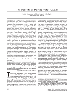
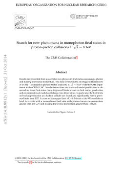
388
CASE REPORT
Croat Med J. 2014;55:388-93
doi: 10.3325/cmj.2014.55.388
Volumetric analysis of
cerebrospinal fluid and brain
parenchyma in a patient
with hydranencephaly and
macrocephaly – case report
Milan Radoš1*, Marijan
Klarica2* Branka MučićPucić3, Ines Nikić1,
Marina Raguž1, Valentina
Galkowski1, Dora Mandić1,
Darko Orešković4*
Croatian Institute for Brain
Research, School of Medicine,
University of Zagreb, Zagreb,
Croatia
1
Department of Pharmacology,
School of Medicine, University of
Zagreb, Zagreb, Croatia
2
The aim of this study was to perform for the first time the
intracranial volumetric analysis of cerebrospinal fluid (CSF)
and brain parenchyma in the supratentorial and infratentorial space in a 30-year-old female patient with hydranencephaly and macrocephaly. A head scan performed
using a 3T magnetic resonance was followed by manual
segmentation of the brain parenchyma and CSF on T2
coronal brain sections. The volume of CSF and brain parenchyma was measured separately for the supratentorial
and infratentorial space. The total volume of the intracranial space was 3645.5 cm3. In the supratentorial space, the
volume of CSF was 3375.2 cm3 and the volume of brain parenchyma was 80.3 cm3. In the infratentorial space, the volume of CSF was 101.3 cm3 and the volume of the brain parenchyma was 88.7 cm3. In the supratentorial space, there
was severe malacia of almost all brain parenchyma with
no visible remnants of the choroid plexuses. Infratentorial structures of the brainstem and cerebellum were hypoplastic but completely developed. Since our patient had
no choroid plexuses in the supratentorial space and no
obstruction between dural sinuses and CSF, development
of hydrocephalus and macrocephaly cannot be explained
by the classic hypothesis of CSF physiology with secretion,
unidirectional circulation, and absorption as its basic postulates. However, the origin and turnover of the enormous
amount of intracranial CSF volume, at least 10-fold larger
than normal, and the mechanisms of macroencephaly development could be elucidated by the new hypothesis of
CSF physiology recently published by our research team.
Special Hospital for Chronic
Children Diseases, Gornja Bistra,
Bistra, Croatia
3
Department of Molecular Biology,
Ruđer Bošković Institute, Zagreb,
Croatia
4
*These authors equally contributed
to the study.
Received: June 26, 2014
Accepted: July 11, 2014
Correspondence to:
Marijan Klarica
University of Zagreb, School of
Medicine
Department of Pharmacology and
Croatian Institute for Brain Research
Šalata 11
10 000 Zagreb, Croatia
[email protected]
www.cmj.hr
389
Radoš et al: Hydranencephaly and macrocephaly in a thirty years old patient
Hydranencephaly is a difficult and rare developmental disorder that occurs in fewer than 1 of 10 000 newborn children, most likely due to bilateral occlusion of the internal
carotid artery between 8 and 12 weeks of pregnancy (1-3).
It is often characterized by heavy malacia of the supratentorial structures perfused by the anterior and middle cerebral arteries. Smaller remaining parts of the supratentorial functional parenchyma are located in the area of
diencephalic structures and in the areas of the temporal
and occipital lobes that are perfused by the posterior cerebral artery (1,3,4). Infratentorial structures are usually
morphologically fully developed and functional, and patients are able to perform functions such as suction, swallowing, crying, and moving of the extremities (5-7). This
ability and frequently typical neurocranium morphology
in newborns hinder the diagnosis of the disease immediately after birth. However, over the next few months an
extreme delay in neurological development is observed,
often accompanied by hypertensive hydrocephalus and
a progressive increase in the head circumference that
leads to macrocephaly (head circumference is two standard deviations larger than the average for a certain age).
Although most of these patients die within the first few
years of life, a small number of them lives for 20 years or
longer (6,8-10).
ing the following months, so that at 2.5 months of age it
was 45 cm (standard deviation [SD] = 3.43), at 5.5 months
49 cm (SD = 4.52), at 6 months 53 cm (SD = 6.45), at 9
months 57 cm (SD = 8.40), and at the time of the study 75
cm (SD = 14.1).
Signs of spastic tetraparesis started to appear from the 5th
month of age and progressed to such a degree that the
patient needed continuous medical care. Menstrual cycles
are regular but the secondary sex characteristics are incompletely developed. A written informed consent of the
legal guardian was obtained before MR exam.
Magnetic resonance imaging (MRI) was performed using
the 3T MR scanner (Magnetom TrioTim, Siemens, Erlagen,
Germany), with a 12-channel Tx/Rc head coil, the only one
that has a sufficient diameter to perform head scan in a
patient with severe macrocephaly. After the head scan, a
coronal T2 sequence (TR = 6000 ms, TE = 106 ms, matrix
320 × 320, FOV = 29.9 × 29.9 cm, voxel size 1 × 1 × 4 mm)
was used for a manual segmentation of CSF and brain parenchyma in the supratentorial and infratentorial spaces
using the Analyze 8.1 program (Mayo Clinic, Rochester, MN,
USA), which measures the volume of the segmented structures in radiological sections (Figure 1).
Our patient, aged 30 years, is one of the oldest living patients
with hydranencephaly. The clinical picture is dominated by
permanent vegetative state with spastic tetraparesis and
the morphological picture by extreme macrocephaly. Although in many patients with hydranencephaly detailed
qualitative descriptions of intracranial changes have been
made, to our knowledge no volumetric analysis of the cerebrospinal fluid and residual parenchyma has been performed separately for the supratentorial and infratentorial
spaces. The aim of this article was to quantify the volume
reduction of the parenchyma and volume increase of cerebrospinal fluid (CSF) in the supratentorial and infratentorial spaces and to discuss the developmental mechanism
of hypertensive hydrocephalus and macrocephaly with respect to dynamic changes of head circumference.
Case report
We report on the case of a 30-year-old woman with hydranencephaly, in permanent vegetative state, with severe spastic tetraparesis and emphasized macrocephaly.
Immediately after birth she had a somewhat larger head
circumference but without manifest neurological symptoms. The head circumference was rapidly increasing dur-
Figure 1. Example of manual segmentation of intracranial parenchyma and cerebrospinal fluid (CSF) in Analyze 8.1 software
on T2 coronal slices. 1) Supratentorial CSF space – red line; 2)
supratentorial parenchyma – green line; 3) infratentorial CSF
space – yellow line; and 4) infratentorial parenchyma – blue line.
www.cmj.hr
390
CASE REPORT
The analysis of the standard structural MR sequences
showed macrocephaly with atypically shaped bones of the
neurocranium and a particularly marked anterioposterior
diameter. Almost all brain parenchyma of the supratentorial space was affected by malacic changes. A small part of
the remaining parenchyma was located in the diencephalon area, the mediobasal part of the left temporal lobe, and
the mediobasal part of the occipital lobes, with one small
island of parenchyma on the right side, just below the frontal bone. There were no visible remains of the ventricular
system or choroid plexuses.
The volumetric analysis showed that the total intracranial
volume was greatly increased (3645.5 cm3), while the total
amount of parenchyma was significantly reduced (169.0
cm3), which represents a significant deviation from the average (according to our results in a group of healthy individuals 22.8 ± 0.7 years old, the average intracranial volume
was 1512.3 ± 146.5 cm3, with a parenchyma volume 1355.7
cm3) (11).
The greatest part of the increased intracranial volume was
located in the supratentorial space with 3375.2 cm3 of CSF,
while the brain parenchyma was reduced to only 80.3 cm3.
In the infratentorial area, the volume of the brainstem and
cerebellum parenchyma was 88.7 cm3, with the CSF volume being 101.3 cm3.
Discussion
We report on a patient with an extremely large intracranial space, which was primarily the result of an increase in
Croat Med J. 2014;55:388-93
supratentorial space. The supratentorial space was mainly
filled with CSF, with only small areas of sustained parenchyma. These results comply with the assumed pathophysiological mechanism of hydranencephaly development due
to impaired perfusion in the area supplied by the internal
carotid arteries. A small island of sustained brain parenchyma below the frontal bone was primarily perfused by
a branch of the right middle meningeal artery and was in
no way connected with the rest of the brain parenchyma.
Reports of marked macrocephaly are very common in patients with hydranencephaly, but there is no satisfactory
pathophysiological explanation for this impressive clinical
presentation.
The generally accepted classic hypothesis of CSF pathophisiology has three basic postulates: active secretion of
CSF predominantly in the choroid plexuses, unidirectional
flow of CSF from the ventricular system to the subarachnoidal area, and resorption of CSF in the dural venous sinuses via arachnoid granulations (11-15). Macrocephaly
can only develop if hypertensive hydrocephalus exists before closing of the neurocranial sutures (16). According to
this hypothesis, hypertensive hydrocephalus theoretically
develops as a consequence of increased secretion of CSF
in the choroid plexuses or reduced absorption of CSF in
the arachnoid granulations in the dural sinuses, or possibly as a consequence of obstruction of CSF circulation.
Since the choroid plexuses are defined as the main place
of CSF secretion (17), it seemed logical that their removal
(if the CSF pathways are blocked) should prevent hydrocephalus development, and result in the patient’s recovery.
For many years, plexectomy was the most popular form of
Figure 2. (A) Transversal T2 slice across the supratentorial space with almost complete malacia of the cerebral hemispheres and
with spared falx and meninges. (B) Coronal T2 slice with preserved parts of brain parenchyma in the right frontal lobe, diencephalon,
mediobasal left temporal lobe, and brainstem. (C) Sagittal T2 slice with visible remnants of the diencephalon, occipital lobe, brainstem, and cerebellum. Marked thickening of the neurocranial bones is visible in all figures.
www.cmj.hr
391
Radoš et al: Hydranencephaly and macrocephaly in a thirty years old patient
hydrocephalus treatment, but because of universally poor
results, it has no place in the current treatment of hydrocephalus (18,19). The introduction of endoscopic method
renewed the interest in surgical procedures on the choroid plexuses, especially in the mid 1990s (20-22). Although
the results were somewhat better than those obtained using the classic surgical approach, the same problems persisted. The ventricular size was not significantly reduced by
choroid plexus coagulation and only 35% of the patients
achieved long-term control without cerebrospinal fluid
shunts (21). Another study (23) found shunting to be required in 48% of the cases, which was done from one week
to thirteen months after the choroid plexus coagulation.
All this shows that hydrocephalus can develop even when
the plexus has been removed, indicating that its role in the
pathophysiology of hydrocephalus is still unclear, but that
it clearly does not participate significantly in CSF formation. Despite the resurgence of interest in this approach
brought about by each new generation of endoscopes, it
has never achieved more than transient acceptance because of its fundamental ineffectiveness.
by water influx into CSF in aquaporins knockout mice using MRI technique (27), showing that essential for CSF homeostasis was water movement within the pericapillary
spaces, rather than choroid plexuses and arachnoid villi.
It is very hard to explain the developmental mechanism of
hydrocephalus and macrocephaly in our patient using the
basic postulates of the classic hypothesis. Since she had no
choroid plexuses in the supratentorial space, the cause of
intracranial hypertension and macrocephaly remains unclear. This is especially the case if we take into account that
is strongly believed, even taken as a fact (24), that CSF is
continuously actively produced by the choroid plexus at
a rate of about 500 mL per day. Even more, while in the
above mentioned cases it was attempted to stop the hydrocephalus development by removing the choroid plexuses, in our patient the opposite happened: an extensive
hydrocephalus, macrocephaly, and severe hydranencephaly (CSF occupies approximately 95% of the cranial cavity) developed without the existence of choroid plexuses
in either the lateral or the third ventricle (Figure 1 and 2).
Furthermore, it is difficult to explain the constant everyday
presence and turnover of such a large amount of CSF, and
the maintenance of 30 years of CSF homeostasis.
In conclusion, our patient is one of the oldest living patients with hydranencephaly and to our knowledge in such
patients no volumetric analysis of the CSF and residual parenchyma has ever been made separately for the supratentorial and infratentorial space. Furthermore, since our patient has no choroid plexuses in the supratentorial space
and no obstruction between the dural sinuses and CSF, the
development of hydrocephalus and macrocephaly cannot
be explained by the classic hypothesis of CSF physiology.
However, it can be explained by the new hypothesis of CSF
physiology and development of hydrocephalus (25,28-30).
This article supports the new hypothesis of CSF physiology (25,26,28) but could also serve as an encouragement
to other clinicians to present similar observations to the
scientific public.
However, all this can be explained with the new hypothesis of CSF physiology, since it is based primarily on the
hydrostatic and osmotic forces at the level of cerebral and
spinal capillary network, which has more than 5000 times
larger surface than the choroid plexuses (25,26). CNS capillary network is the main area where the volumes of the intracranial fluids and waste products of CNS metabolism are
regulated (25,26). This hypothesis has recently been tested
This new hypothesis (25,26,28) enables us to explain the
existence of a permanently large amount of CSF in a case
when the brain mass is reduced and supratentorial choroid plexuses do not exist. In this situation, vascularity is
still sufficient to permanently maintain the achieved fluid
relations inside the cranium. Furthermore, the presented
case of hydranencephaly and macrocephaly and other
mentioned facts indicate that the key role in the development of hydrocephalus is not played by the choroid plexuses, and that the development of hydrocephalus cannot
be explained by a dysfunction in CSF secretion, circulation,
and absorption. It seems that the etiology of hydrocephalus, hydranencephaly, and macrocephaly is more complex
(28,29) but that it definitely depends on CNS pathophysiological conditions that affect the regulation of fluid volume
in the CNS capillary network.
Funding This study was supported by the Ministry of Science, Education
and Sport of the Republic of Croatia (Projects: 1. Hydrodynamics of cerebrospinal fluid. No. 098-1080231-2328; and 2. Pathophysiology of cerebrospinal
fluid and intracranial pressure. No. 108-1080231-0023).
Ethical approval A written informed consent of the legal guardian was obtained before MR exam.
Declaration of authorship MR performed MRI exam, participated in writing of the manuscript and preparation of the figures. BMP gathered clinical
data about the patient and participated in writing of the manuscript. DO
and MK participated in writing of the manuscript and offered explanation
of the presented case according to their new hypothesis of CSF physiology.
IN, VG, MR, and DM performed volumetric analysis and participated in writing of the manuscript.
Competing interests MK is a Vice Dean of the University of Zagreb School
of Medicine, one of the owners of the Croatian Medical Journal. To ensure
that any possible conflict of interest relevant to the journal has been ad-
www.cmj.hr
392
CASE REPORT
dressed, this article was reviewed according to best practice guidelines of
international editorial organizations. All authors have completed the Unified Competing Interest form at www.icmje.org/coi_disclosure.pdf (available on request from the corresponding author) and declare: no support
from any organization for the submitted work; no financial relationships
with any organizations that might have an interest in the submitted work in
the previous 3 years; no other relationships or activities that could appear to
have influenced the submitted work.
Croat Med J. 2014;55:388-93
the cerebrospinal fluid. Edinburgh: Churchill-Livingstone; 1987.
14 McComb JG. Recent research into the nature of cerebrospinalfluid formation and absorption. J Neurosurg. 1983;59:369-83.
Medline:6886750 doi:10.3171/jns.1983.59.3.0369
15 Plum F, Siesjo BK. Recent Advances in CSF physiology.
Anesthesiology. 1975;42:708-30. Medline:236708
doi:10.1097/00000542-197506000-00015
References
1
Cecchetto G, Milanese L, Giordano R, Viero A, Suma V, Manara R.
pressure, and hydrocephalus in the infant and young child.
Looking at the missing brain: Hydranencephaly case series and
Top Magn Reson Imaging. 2007;18:31-51. Medline:17607142
literature review. Pediatr Neurol. 2013;48:152-8. Medline:23337012
doi:10.1016/j.pediatrneurol.2012.10.009
2
Lam YH, Tang MHY. Serial sonographic features of a fetus with
hydranencephaly from 11 weeks to term. Ultrasound Obstet
3
Choroid plexectomy for the treatment of chronic infected
hydrocephalus. Childs Nerv Syst. 1988;4:139-43. Medline:3396020
Quek YW, Su PH, Tsao TF, Chen JY, Ng YY, Hu JM, et al.
cerebrospinal-fluid formation 5 years after bilateral choroid
doi:10.1016/S1875-9572(08)60011-X
plexectomy - Case-report. J Neurosurg. 1976;44:735-9.
techniques in the treatment of hydrocephalus. Neurosurg Rev.
Counter SA. Preservation of brainstem neurophysiological
2008;31:249-62. Medline:18463909 doi:10.1007/s10143-008-
Medline:17719607 doi:10.1016/j.jns.2007.06.043
0131-y
21 Pople IK, Ettles D. The role of endoscopic choroid-plexus
Pant S, Kaur G, De JK. Hydranencephaly. Kathmandu Univ Med J
coagulation in the management of hydrocephalus. Neurosurgery.
(KUMJ). 2010;8:83-6. Medline:21209513
1995;36:698-701. Medline:7596499 doi:10.1227/00006123-
Petridis AK, Krause-Titz UR, Doukas A, Mehdorn HM. Delayed
diagnosis of hydranencephaly in a nine-month-old child. Clinics
199504000-00009
22 Wellons JC, Tubbs RS, Leveque JCA, Blount JP, Oakes WJ. Choroid
and Practice. 2011;1:e65. Medline:24765326 doi:10.4081/cp.2011.
plexectomy reduces neurosurgical intervention in patients
e65
with hydranencephaly. Pediatr Neurosurg. 2002;36:148-52.
Bae JS, Jang MU, Park SS. Prolonged survival to adulthood
of an individual with hydranencephaly. Clin Neurol
9
Medline:1083897 doi:10.3171/jns.1976.44.6.0735
20Enchev Y, Oi S. Historical trends of neuroendoscopic surgical
Medline:11754063 doi:10.1002/ajmg.10051
function in hydranencephaly. J Neurol Sci. 2007;263:198-207.
8
doi:10.1007/BF00270903
19 Milhorat TH, Hammock MK, Chien T, Davis DA. Normal rate of
carotid arteries. Pediatr Neonatol. 2008;49:43-7. Medline:18947016
with vascular malformations. Am J Med Genet. 2001;104:295-8.
7
42. Medline:17864139 doi:10.1097/00000658-191908000-00001
18 Lapras C, Mertens P, Guilburd JN, Lapras C, Pialat J, Patet JD.
0705.2000.00165.x
4Stevenson DA, Hart BL, Clericuzio CL. Hydranencephaly in an infant
6
doi:10.1097/RMR.0b013e3180d0a753
17 Dandy WE. Experimental hydrocephalus. Ann Surg. 1919;70:129-
Gynecol. 2000;16:77-9. Medline:11084971 doi:10.1046/j.1469-
Hydranencephaly associated with interruption of bilateral internal
5
16 Vertinsky AT, Barnes PD. Macrocephaly, increased intracranial
Medline:11919449 doi:10.1159/000048370
23 Griffith HB, Jamjoom AB. The treatment of childhood
Neurosurg. 2008;110:307-9. Medline:18222607 doi:10.1016/j.
hydrocephalus by choroid plexus coagulation and artificial
clineuro.2007.12.003
cerebrospinal fluid perfusion. Br J Neurosurg. 1990;4:95-100.
McAbee GN, Chan A, Erde EL. Prolonged survival with
hydranencephaly: Report of two patients and literature review.
Medline:2357290 doi:10.3109/02688699008992706
24 MacAulay N, Zeuthen T. Water transport between CNS
Pediatr Neurol. 2000;23:80-4. Medline:10963978 doi:10.1016/
compartments: contributions of aquaporins and cotransporters.
S0887-8994(00)00154-5
Neuroscience. 2010;168:941-56. Medline:19761815 doi:10.1016/j.
10 Merker B. Life expectancy in hydranencephaly. Clin Neurol
Neurosurg. 2008;110:213-4. Medline:18201820 doi:10.1016/j.
clineuro.2007.11.011
11 Brodbelt A, Stoodley M. CSF pathways: a review. Br
J Neurosurg. 2007;21:510-20. Medline:17922324
doi:10.1080/02688690701447420
12 Brown PD, Davies SL, Seake T, Millar ID. Molecular mechanisms of
cerebrospinal fluid production. Neuroscience. 2004;129:957-70.
Medline:15561411 doi:10.1016/j.neuroscience.2004.07.003
13 Davson H, Welch K, Segal MB. Physiology and pathophysiology of
www.cmj.hr
neuroscience.2009.09.016
25 Bulat M, Klarica M. Recent insights into a new hydrodynamics
of the cerebrospinal fluid. Brain Res Rev. 2011;65:99-112.
Medline:20817024 doi:10.1016/j.brainresrev.2010.08.002
26 Oreskovic D, Klarica M. The formation of cerebrospinal fluid:
Nearly a hundred years of interpretations and misinterpretations.
Brain Res Rev. 2010;64:241-62. Medline:20435061 doi:10.1016/j.
brainresrev.2010.04.006
27 Igarashi H, Tsujita M, Kwee IL, Nakada T. Water influx into
cerebrospinal fluid is primarily controlled by aquaporin-4, not
393
Radoš et al: Hydranencephaly and macrocephaly in a thirty years old patient
by aquaporin-1: O-17 JJVCPE MRI study in knockout mice.
Neuroreport. 2014;25:39-43. Medline:24231830
28 Oreskovic D, Klarica M. Development of hydrocephalus and
30 Klarica M, Rados M, Erceg G, Petosic A, Jurjevic I, Oreskovic D. The
influence of body position on cerebrospinal fluid pressure gradient
and movement in cats with normal and impaired craniospinal
classical hypothesis of cerebrospinal fluid hydrodynamics: Facts
communication. PLoS ONE. 2014;9:e95229. Medline:24748150
and illusions. Prog Neurobiol. 2011;94:238-58. Medline:21641963
doi:10.1371/journal.pone.0095229
doi:10.1016/j.pneurobio.2011.05.005
29 Klarica M, Mise B, Vladic A, Rados M, Oreskovic D. “Compensated
hyperosmolarity” of cerebrospinal fluid and the development
of hydrocephalus. Neuroscience. 2013;248C:278-89.
Medline:23806710 doi:10.1016/j.neuroscience.2013.06.022
www.cmj.hr
© Copyright 2026 Paperzz