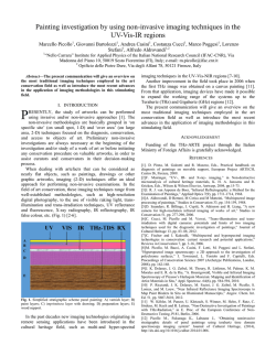
Title
Imaging of interstitial fluid in skin and subcutaneous tissue using dualfrequency ultrasonography before and immediately after lymph drainage in
breast cancer-related lymphedema patients
Author(s)
稲垣, 美佐子
Citation
以下に掲載:Journal of Tsuruma Health Science Society, Kanazawa
University, 37(2) pp.13-21.金沢大学つるま保健学会. 共著者:Misako
Dai, Mihoe Katayama, Junko Sugama, Mayumi Okuwa, Takeshi Ueyama,
Tomoko Minamiyama, Keiko Ota, Terumi Iuchi, Masaru atsumoto, Toshio
Nakatani, Hiromi Sanada
Issue Date
2014-03-22
Type
Thesis or Dissertation
Text version
ETD
URL
http://hdl.handle.net/2297/38956
Right
学位授与機関
金沢大学
学位の種類
博士(保健学)
学位授与年月日
2014年3月22日
学位授与番号
甲第4010号
*KURAに登録されているコンテンツの著作権は,執筆者,出版社(学協会)などが有します。
*KURAに登録されているコンテンツの利用については,著作権法に規定されている私的使用や引用などの範囲内で行ってください。
*著作権法に規定されている私的使用や引用などの範囲を超える利用を行う場合には,著作権者の許諾を得てください。ただし,著作権者
から著作権等管理事業者(学術著作権協会,日本著作出版権管理システムなど)に権利委託されているコンテンツの利用手続については
,各著作権等管理事業者に確認してください。
http://dspace.lib.kanazawa-u.ac.jp/dspace/
Journal of Tsuruma Health Science Society Kanazawa University Vol. 37(2)
Original Article
13 ~ 21 2013
Imaging of interstitial fluid in skin and subcutaneous tissue using
dual-frequency ultrasonography before and immediately after
lymph drainage in breast cancer-related lymphedema patients
Misako Dai,Mihoe Katayama*,Junko Sugama**,Mayumi Okuwa**,
Takeshi Ueyama*,Tomoko Minamiyama*,Keiko Ota*,Terumi Iuchi,
Masaru Matsumoto,Toshio Nakatani*,Hiromi Sanada***
Abstract
Background: Breast cancer-related lymphedema (BRCL) occurs in 12-28% of cases after
radical lymph node surgery and/or radiotherapy of the lymph nodes. Quantitative assessments
before and immediately after treatment for lymphedema are important to continue adequate
treatment to prevent cellulitis and reduce edema. The present study aimed to assess skin
hardness and the utility of dual-frequency ultrasonography of the skin and subcutaneous tissue
for assessing BRCL before and immediately after manual lymph drainage (MLD).
Methods: This observational study examined 15 patients with unilateral secondary
lymphedema attending two lymphedema clinics in Japan between June 2012 and June 2013.
Skin hardness was assessed using a 10-cm visual analog scale (VAS). Ultrasonography was
performed at 20 MHz for skin and 10 MHz for subcutaneous tissue. Edema was evaluated
using circumference, skin elasticity and skin moisture.
Results: Patients were assigned to Group A (n= 10 ) with decreased VAS and Group B
(n= 5 ) with unchanged VAS. Results of ultrasonography and other characteristics were
then compared between groups. Group A showed decreased numbers of low-echogenic
pixels (LEPs) and decreased pixel uniformity after MLD, whereas Group B did not. Edema
evaluations showed small differences or were unchanged.
Conclusion: These findings suggest that interstitial fluid in the dermis and subcutaneous
tissue is decreased after lymph drainage in patients who report softening of the skin after
MLD, and can therefore be used as a quantitative indicator for the effectiveness of MLD.
Key words
lymphedema, ultrasound imaging, quantitative assessment, lymph drainage, prevention
Introduction
Breast cancer-related lymphedema (BRCL)
remains an important complication, occurring in 1228% of cases after radical lymph node surgery and/or
radiotherapy of the lymph nodes1-3). Lymphedema is
a particular type of edema caused by dysfunction of
the lymphatic system, resulting in the accumulation
of protein-rich fluid in the dermis and hypodermis4-6).
Initially presenting as unilateral painless swelling
that usually starts on the dorsal aspect of the arm,
Doctoral Course, Deviation of Health Science, Kanazawa University
Kanazawa Card: ovascular Hospital
*
** Institute of Medical, Pharmaceutical and Health Sciences, Kanazawa University
*** Department of erotological nursing/ wound care management, division of health science and nursing, graduate school of medicine,
the University of Tokyo
— 13 —
Misako Dai,et al.
including non-pitting edema, later stages include
increased volume of the upper limb, hardening of the
skin, and a risk of recurrent infection7). Lymphedema
management programs are often associated with
combined decongestive physical therapy (CDP),
which aims to reduce limb volume, restore limb
shape, and improve skin and tissue condition 8 ) .
Patients have to continue lymphedema management
for the long term8). In particular, lymph drainage is
typically performed by hand, so the effects will not
be the same for each condition every time. Adequate
assessment of fluid accumulation is important for
patients and clinicians to assess the effectiveness of
treatment. In most cases, effects on clinical findings
are assessed qualitatively before and immediately
after lymph drainage based on pitting of the skin on
the affected limb9). This method has a long history,
but provides no direct information about edema
within the dermis and subcutaneous tissue10). This is
problematic for assessing improvements in morbidity,
preventing cellulitis and maintaining quality of life
(QOL). Several researchers have described reductions
in limb volume and improved QOL following CDP
including lymph drainage for at least 4 weeks11・12).
However, these effects were assessed over the long
term, not before and immediately after treatments.
Quantitative analyses have been reported using
lymphoscintigraphy13) and near-infrared fluorescence
imaging 14 ) . Immediately after lymph drainage,
lymph flow rate and lymph volume were increased.
However, these imaging methods do not show
changes in the condition of the skin and subcutaneous
tissue. In addition, these methods are time-consuming
and expensive, and require intradermal injection
of a radionuclide. Quantitative, non-invasive, realtime methods of assessment have therefore yet to be
reported.
Ultrasonography is an imaging modality that
has been routinely used for more than 20 years in
dermatology, and can demonstrate dermal edema.
The ultrasonography device enables simple, noninvasive, quantitative imaging in real time. Some
studies have identified particular aspects of
lymphedema on ultrasonography 15-17). The affected
limbs have shown increases in both skin thickness16)
and the number of low echogenicity pixels (LEPs)15・
using a 20-MHz probe. Subcutaneous tissue has
shown a cobblestone appearance and thickening
on imaging at 7.5-10 MHz16・17). Such studies have
revealed characteristics of skin and subcutaneous
tissue showing lymphedema, but have not made
comparisons to clinical assessments or assessed the
effects of treatment. Balzarini reported skin hardness
and ultrasound imaging in subcutaneous tissue at
7 . 5 MHz and provided qualitative assessments of
“soft fluid”,“medium mix”,and“hard fibrosis”17).
However, the method used to classify skin hardness
is unknown and only subcutaneous tissue was tested
without control. Changes in imaging results for
accumulation of fluid in the skin and subcutaneous
tissue before and immediately after have thus yet to
be researched in detail.
The purpose of the present study was to assess
skin hardness and clarify the utility of dual-frequency
ultrasonography of the skin and subcutaneous tissue
for assessing BRCL before and immediately after
lymph drainage.
16)
Materials and Methods
1. Study design and participants
This observational study was performed from June
2012 to June 2013, involving a series of patients with
unilateral secondary lymphedema attending two
lymphedema clinics in Japan.
Subjects who fulfilled the following criteria
were eligible for the study: unilateral upper limb
lymphedema after treatment for breast cancer; > 12
months after surgery or adjuvant treatment, in order
to provide a reliable follow-up period to detect any
possible metastases; lymphedema stage II or late II
according to the criteria of the International Society of
Lymphedema18); and continued CDP including lymph
drainage. Exclusion criteria included subjects with
active cancer and those on diuretic therapy or other
edema-influencing drugs. Patients were identified and
recruited by physicians specializing in lymphedema. A
researcher and patient then measured each parameter
before and immediately after manual lymph drainage
(MLD). MLD was performed by a therapist licensed
in lymph drainage by the Medical Lymph Drainage
Association of Japan.
All protocols were approved by the ethics
— 14 —
Imaging of interstitial fluid in skin and subcutaneous tissue using dual-frequency ultrasonography before
and immediately after lymph drainage in breast cancer-related lymphedema patients
committee at Kanazawa University and all
participants provided written informed consent prior
to enrolment in the study.
2. Procedures
Before the start of measurements, the investigator
marked the site of examinations at 10 cm proximal
to the ulnar styloid process for both affected and
unaffected arms. The patients assessed skin hardness
by themselves using pinch and lift of a skinfold. A
researcher then performed edema evaluations and
ultrasound imaging. Measurements were completed
each day between 13:00 and 16:00 and the patient
sat in a chair with the arm supported initially in
abduction. The researcher provided supervision on
how to perform assessment and analyze ultrasound
imaging for 2 physicians specializing in lymphedema
and 3 sonographers.
3. Ultrasonography
Images of the skin were recorded before and
immediately after MLD using a Dermascan
C u l t r a s o u n d s y s t e m ( C o r t e x Te c h n o l o g y,
Smedevaenget, Denmark) at 20 MHz. Ultrasound gel
was applied liberally to the skin and the probe placed
transversely on the arm. Field size was set to 13.42
mm wide and 22.40 mm deep. A two-dimensional
image of the skin was produced and recorded by
computer and viewed in gray scale. Gain was adjusted
as necessary to optimize image quality and boundary
definition.
Images of subcutaneous tissue were recorded
before and immediately after MLD using Mylab5
(Hitachi Medical, Chiba, Japan) with a frequency of
10 MHz. Imaging of the skin, subcutaneous tissue,
muscle, and sometimes bone was possible. Ultrasound
gel was again applied to the area being examined.
In addition, a gel“stand-off”(a small polyethylene
bag containing ultrasound gel) was placed on the arm
to aid delineation and identification of the gel/skin
boundary. Measurement dimensions were set to 4.0
cm wide and 5.0 cm deep to encompass the full depth
of subcutaneous tissue. Gain was adjusted to increase
resolution of the deeper boundaries and compensate
for the natural attenuation in signal as the sound wave
passes through tissue.
The patient sat in a chair with the arm supported
initially in abduction. Gain was adjusted to increase
resolution of the deeper boundaries and compensate
for the natural attenuation in signal as the sound wave
passes through tissue.
We decided quantitative parameters through
qualitative assessment. In each image of the skin,
the number of LEPs16-19) and subcutaneous pixel
uniformity20) was measured using Image J analysis
software (v1.42q; National Institutes of Health). In
this system, amplitudes of echoes from single image
elements (pixels) are assigned to a numeric scale (0 to
255). The low echogenic range extends from 0 to 30.
LEP (%) = number of low echogenic pixels (0-30) /
number of total pixels (0-255) × 100
The uniformity of subcutaneous tissue considered
as control was computed.
ROImax − ROImin
Pixel uniformity = × 100
ROImax + ROImin
where ROI max and ROI min were the maximum and
minimum pixel values in the same region of interest
(ROI), respectively, in the medial forearm. Care was
taken to avoid including edge artifacts in the ROI. To
help minimize the effect of noise on measurement, the
image was convolved with a 9-point low-pass filter21).
4. Skin hardness
Skin hardness was assessed before and immediately
after MLD by each patient using a 10 -cm visual
analog scale (VAS), with 0 cm as soft and 10 cm as
hard. After MLD, we considered skin hardness to be
softened when VAS decreased more than 0.5 cm. The
researcher and physical therapists were blinded to
group allocations.
5. Edema evaluations
The principal researcher undertook measurements
of arm circumference using a tape measure at 10 cm
below the elbow18). Skin elasticity was determined
using a non-invasive, in vivo suction skin elastin
meter (Cutometer MPA 580 ; Curage & Khazaka,
Cologne, Germany) with 450 mbar and 2 -mm
aperture size probe22・23). We determined the following
parameters: R 0 , which looks at the maximum
amplitude and represents the passive behavior of
the skin to force; R 2 , gross elasticity (resistance
versus ability to return) and R7, portion of elasticity
compared to the complete curve. Skin moisture
was measured using a Moisture Meter D (Delfin
Technologies, Kuopio, Finland) with a 2-mm probe
— 15 —
Misako Dai,et al.
head24).
6. Characteristics
These data included age, body mass index
(BMI), circumference at baseline, and duration of
lymphedema.
7. Analysis
We divided patients into Group A if VAS
immediately after MLD was decreased compared to
before MLD, and to Group B if VAS was unchanged.
We compared these groups in terms of the main
outcomes of findings on ultrasonography imaging
and edema evaluations. Data are presented as mean
± standard. We used the Wilcoxon test and paired
t test for comparisons before and just after lymph
drainage. A value of p < 0 . 05 was chosen as the
level of significance. JMP® statistical software (SAS
Institute, State of North Carolina , USA) was used for
all calculations.
Results
A total of 21 patients were initially recruited, but 6
patients did not meet the inclusion criteria. As a result,
analysis was performed for 15 patients. Ten patients
felt their skin had softened and were categorized as
Group A. Five patients considered their skin was
unchanged and thus comprised Group B (Table 1).
Groups A and B showed no significant differences
in arm circumferences, age, BMI, or duration of
lymphedema (Table 2).
1. LEPs in the skin
The dermis was easily identified on ultrasound
images and defined as the space between the
epidermal entrance echo and the interface with the
hypo-echogenic subcutaneous space (Figure 1).
Images from Group A showed decreased hypoechogenicity of images after MLD, whereas no
changes were seen in Group B. The results of LEP
measurements are shown in Figures 2 and 3. Group
A showed decreased LEPs in skin immediately after
MLD (p=0.01), while Group B showed no changes.
Subcutaneous tissue showed a cobblestone
appearance on ultrasonography in Group A (Figure 4).
The unevenness of the internal echo appeared more
even in Group A, but again no change was seen in
Group B. Group A showed decreased pixel uniformity
in subcutaneous tissue immediately after MLD
(p=0.03), while Group B showed no change (Figures 5,
6).
2. Edema evaluations
Circumference of the upper limb was significantly
decreased immediately after MLD compared to
before MLD (p=0.00), by 0.2 cm in Group A and by
0.3 cm in Group B. R2 was significantly decreased
immediately after MLD in Group A (p= 0 . 01 ),
and R 0 was significantly decreased after MLD in
Group B (p=0.00). Differences were small in both
circumference and skin viscoelasticity. Skin moisture
was unchanged after MLD in both groups (Table 3).
Discussion
This study is the first to report changes in skin and
subcutaneous tissue immediately after MLD using
LEP and pixel uniformity from ultrasound imaging.
Table 1. Skin hardness (VAS) before and immediately after MLD
Before MLD
After MLD
Median Min Max Median Min Max P
Group A (n=10) 7.4 6.5 8.4
5.6
4.0 7.0 0.00*
Group B (n=5)
5.2
5.0 5.5
5.2
5.0 5.5 0.35
n, number of women in test; Min, minimum value;
Max, maximum value
* p < 0.05, Wilcoxon test
The skin became softer in Group A
Skin hardness did not change in Group B
VAS (cm)
Table 2. Characteristics of subjects in Groups A and B
Parameters (n=15)
Age (years)
BMI (kg/m2 )
Duration of lymphedema (years)
Difference in circumference between
affected and unaffected limbs (cm)
Group A (n=10)
Median Min
Max
58
24.7
5 .0
52
20.5
2.0
66
27.4
21.0
2 .0
0.0
11.5
Group B (n=5)
Median Min
Max
n, number of women in test ; Min, minimum value; Max, maximum value
* p < 0.05, Wilcoxon test
— 16 —
P
59
23.3
4.5
30
20.8
3 .0
64
24.9
7. 0
0.19
0.29
0.12
0.2
0 .0
5. 6
0.07
Imaging of interstitial fluid in skin and subcutaneous tissue using dual-frequency ultrasonography before
and immediately after lymph drainage in breast cancer-related lymphedema patients
Figure 1. Ultrasonographic images of the dermal layer before and immediately after MLD of a woman (52 years old).
(a) before MLD in unaffected limb; (b) immediately after MLD in unaffected limb; (c) before MLD in group A; (d) immediately
after MLD in group A; (e) before MLD in group B; (f) immediately after MLD in group B.
Group A shows decreased low-intensity findings in the superficial layer just after MLD (white arrowheads). No changes are
apparent in Group B and unaffected limb.
70
*
70
60
60
50
50
40
40
30
30
20
20
10
10
0
0
Figure 2. Dermal echogenicity of before MLD (open bar)
and immediatley after MLD (shaded bar) in
Group A.
LEPs in dermal images, shown as a percentage of the
total number of pixels. Data are shown as mean values
with standard deviation.
*p < 0.05
LEP decreased significantly in Group A (p=0.01), while
Group B showed no change.
Figure 3. Dermal echogenicity of before MLD (open bar)
and immediately after MLD (shaded bar) in
Group B.
LEPs in dermal images, shown as a percentage of the
total number of pixels. Data are shown as mean values
with standard deviation.
No significant differences are seen between before and
immediately after MLD.
— 17 —
Misako Dai,et al.
The study focused on changes in skin hardness from
before to immediately after lymph drainage and
assessed changes of fluid accumulation in skin and
subcutaneous tissue using dual-frequency ultrasound
imaging. Following lymph drainage, LEP of the
dermis and pixel uniformity of the subcutaneous tissue
were significantly decreased only in Group A. These
findings suggest that interstitial fluid in the dermis
and subcutaneous tissue are decreased after lymph
drainage in patients who report the skin becoming
softer. In addition, even if edema evaluations appear
unchanged, LEP can show changes in skin. Clinical
assessment using ultrasonography therefore appears
feasible.
The reduction in LEP following lymph drainage
in the present study was due to decreased levels
of water in the dermal layer. In a previous study,
LEP was reportedly increased in limbs affected by
lymphedema15). Histological findings from a previous
study indicated that collagen bundles in the papillary
dermis are thin25). In addition, the histological findings
of Tassenov26) show decreased collagen density in
the dermal layer and low pigmentation on the medial
side of the affected forearm. In other words, collagen
in the dermal layer is below the resolution of the
ultrasonography device (60 µm), and does not reflect
ultrasound. In a study that used magnetic resonance
imaging (MRI), LEP and the T2 value on MRI showed
a positive correlation27). The T2 value indicates the
amount of water. Pixel uniformity in subcutaneous
tissue was significantly decreased after MLD in
those patients in whom the skin became softer.
Subcutaneous tissue in lymphedema has already been
reported to include irregular fat cells26). In addition,
MRI spectroscopy has shown that limbs affected
by lymphedema contain more water than healthy
limbs26). In other words, subcutaneous tissue contains
not only irregular fat, but also an accumulation of
excess water. Pixel uniformity enables quantification
of internal heterogeneity on ultrasonography20). The
decrease in pixel uniformity in the present study was
therefore thought to indicate a reduction in the amount
of water in subcutaneous tissue. LEP and pixel
uniformity will therefore offer original quantitative
parameters to assess the effect of lymph drainage.
Based on the results for patient characteristics,
severity was low among those patients who had no
changes in skin hardness. These patients also required
continuation of self-management. Decreases in LEP
can be shown even without changes in skin hardness
or circumference. Our hope is that these findings
will serve as a great motivation for continuing selfmanagement over the long-term. Clinically significant
changes between before and after lymph drainage
were not observed for circumference, which has
been the gold standard for assessment methods, or
for skin viscoelasticity and the amount of water in
the dermis, which have been used in clinical studies.
However, the difference in data was very small.
Changes in the dermal layer and subcutaneous tissue
in terms of LEP and pixel uniformity were identified,
and ultrasonography was found to be useful for
assessments immediately before and after lymph
drainage.
The present results suggest that ultrasonography
is useful for clinicians and researchers to assess
lymphedema, allowing comparison between
individuals and assessment of the pathological state.
The key limitation to the present study was
considered to be the small number of subjects.
In conclusion, edema in the skin and subcutaneous
tissue were reduced immediately after MLD in
patients who reported softening of the skin. LEP
and pixel uniformity appear useful for clinicians and
researchers in assessing the effects of MLD.
Acknowledgment
This work was supported by Grant-in-Aid for
Young Scientists (B) Grant Number 25862142.
— 18 —
Disclosure
None.
Imaging of interstitial fluid in skin and subcutaneous tissue using dual-frequency ultrasonography before
and immediately after lymph drainage in breast cancer-related lymphedema patients
Figure 4. Ultrasonographic images of the subcutaneous tissue before and immediately after MLD of a woman (60 years old)
(a) before MLD in unaffected limb; (b) immediately after MLD in unaffected limb; (c) before MLD in group A; (d) immediately
after MLD in group A; (e) before MLD in group B; (f)immediately after MLD in group B.
Unevenness in the internal echo within subcutaneous tissue (white arrowheads) appears to become more even in Group A,
but no change is seen in Group B and unaffected limb.
*
Figure 5. Pixel uniformity in subcutaneous tissue before
MLD (open bar) and immediately after MLD
(shaded bar) in Group A.
Data are shown as mean values with standard deviation.
*P < 0.05
Pixel uniformity decreased significantly in Group A, while
Group B showed no change. (p=0.03)
Figure 6. Pixel uniformity in subcutaneous tissue before
MLD (open bar) immediately after MLD (shaded
bar) in Group B.
Data shown are mean values with standard deviation.
No significantly changes before and immediately after
MLD.
— 19 —
Misako Dai,et al.
Table 3. Edema evaluations before and immediately after MLD
Parameters
Group A (n=10)
Circumference (cm)
Skin elasticity
R0 (mm)
R2
R7
Skin moisture (g/m2 h)
Group B (n=5)
Circumference (cm)
Skin elasticity
R0 (mm)
R2
R7
Skin moisture (g/m2h)
Before MLD
Mean ± SD
25.8 ±
0.17
0.77
0.41
49.49
4.5
25.5 ±
± 0.07
± 0.12
± 0.12
± 18.61
22.0 ±
0.17
0.84
0.51
31.56
After MLD
Mean ± SD
±
±
±
±
0.21
0.72
0.38
50.94
2.0
0.02
0.06
0.14
2.06
n, number of women in test; SD, standard deviation
* p < 0.05 , Paired t test
1)
2)
3)
4)
5)
6)
7)
8)
9)
References
Meric F, Buchholz TA, Mirza NQ, et al: Long-term
complications associated with breast-conservation surgery
and radiotherapy. Ann Surg Oncol 9: 543-549, 2002
Ozaslan C, Kuru B: Lymphedema after treatment of breast
cancer. Am J Surg 187: 69-72, 2004
Clark B, Sitzia J, Harlow W: Incidence and risk of arm
oedema following treatment for breast cancer; a three-year
follow-up study. Q J Med 98: 343-348, 2005
Vignes S: Lipoedema. Ann Dermatol Venereol 133: 91-3,
2006
Kerchner K, Fleischer A, Yosipovitch G: Lower extremity
lymphedema update: pathophysiology, diagnosis, and
treatment guidelines. J Am Acad Dermatol 59: 324-31,
13)
14)
15)
16)
2008
Rudkin GH, Miller TA: Lipedema: a clinical entity distinct
from lymphedema. Plast Reconstr Surg 94: 841-7, 1994
Mortimer PS: Managing lymphedema. Clin Dermatol
13(5): 499-505, 1995
Ko DSC, Lerner R, Klose G: Effective treatment of
lymphedema of the extremities. Arch Surg 133: 452-8,
1998
Mani R, Ross JN, Keefe M: Measurements of limb
oedema in chronic venous disease. Skin Res Technol 1:
51-4, 1995
10) Barnes MD, Mani R, Barrett DF, et al: How to measure
changes in oedema in patients with chronic venous ulcers?
Phlebology 7: 31-5, 1992
11) Williams AF, Vadgama A, Franks PJ, et al: A randomized
controlled crossover study of manual lymphatic drainage
therapy in women with breast cancer-related lymphedema.
Eur J Cancer Care (Engl) 11(4): 254-61, 2002
12) McNeely ML, Magee DJ, Lees AW, et al: The addition of
manual lymph drainage to compression therapy for breast
17)
18)
19)
20)
21)
22)
— 20 —
4 .3
0.00*
± 0.14
± 0.11
± 0.72
± 17.96
0.07
0.01*
0.07
0.16
21.8 ±
0.19
0.87
0.53
32.37
P
±
±
±
±
1 .9
0.00*
0.03
0.06
0.13
3.19
0.00*
0.27
0.19
0.11
cancer related lymphedema: a randomized controlled trial.
Breast Cancer Res Treat 86(2): 95-106, 2004
Kafejian-Haddad AP, Janeiro Perez MC, Castiglioni MLV,
et al: Lymphscintigraphic evaluation of manual lymphatic
drainage for lower extremity lymphedema. Lymphology
39: 41-8, 2006
Tan IC, Maus EA, Maus JC, et al: Assessment of
lymphatic contractile function after manual lymphatic
drainage using near-infrared fluorescence imaging. Arch
Phys Med Rehabil 92(5): 756-64, 2011
Gniadecka M: Localization of dermal edema in
lipodermatosclerosis, lymphedema, and cardiac
insufficiency. J Am Acad Dermatol 35(1): 37-41, 1996
Naouri M, Samimi M, Atlan M, et al: High-resolution
cutaneous ultrasonography to differentiate lipoedema from
lymphedema. Br J Dermatol 163: 296-301, 2010
Balzarini A, Milella M, Civelli E, et al: Ultrasonography
of arm edema after axillary dissection for breast cancer: a
preliminary study. Lymphology 34: 152-5, 2001
Piller N, Carati C: The diagnosis and treatment of
peripheral lymphedema. Lymphology 42: 146-147, 2009
Gniadecka M, Gniadecki R, Serup J, et al: Ultrasound
structure and digital image analysis of the subepidermal
low echogenic band in aged human skin: diurnal changes
and interindividual variability. J Invest Dermatol 102(3):
362-365, 1994
Ueta M, Sugama J, Konya C, et al: Use of ultrasound
in assessment of necrotic tissue in pressure ulcers with
adjacent undermining. J Wound Care 20(11): 1-5, 2011
Price RR, Axel L, Morgan T, et al: Quality assurance
methods and phantoms for magnetic resonance imaging.
Med Phys 17(2): 287-295, 1990
Ohshima H, Kinoshita S, Oyobikawa M, et al: Use of
Cutometer area parameters in evaluating age-related
changes in the skin elasticity of the cheek. Skin Res
Imaging of interstitial fluid in skin and subcutaneous tissue using dual-frequency ultrasonography before
and immediately after lymph drainage in breast cancer-related lymphedema patients
Technol 19: 238-242, 2013
23) Dobrev H: In vivo study of skin mechanical properties in
psoriasis vulgaris. Acta Derm Venereol 80: 263-266, 2000
24) Mayrovitz HN, Davey S, Shapiro E: Suitability of single
tissue dielectric constant measurements to assess local
tissue water in normal and lymphedematous skin. Clin
Physical Funct Imaging 29: 123-7, 2009
25) de Rigal J, Escoffier C, Querleux B, et al: Assessment of
aging of the human skin by in vivo ultrasonic imaging. J
Invest Dermatol 93(5): 621-5, 1989
26) Tassenoy A, De Mey J, Stadnik T, et al: Histological
findings compared with magnetic resonance and
ultrasonographic imaging in irreversible postmastectomy
lymphedema: a case study. Lymphat Res Biol 7(3): 145151, 2009
27) Gniadecka M, Jemec G.B.E: Quantitative evaluation of
chronological ageing and photoageing in vivo: studies on
skin echogenicity and thickness. Br J Dermatol 139: 815821, 1998
乳癌術後の上肢リンパ浮腫患者に対するリンパドレナージ前後の
皮膚と皮下組織の超音波診断画像による組織間液量変化の定量的評価
臺 美佐子,片山美豊恵 *,須釜 淳子 **,大桑麻由美 **,上山 武史 *,南山 朋子 *,
太田 恵子 *,井内 映美,松本 勝,中谷 壽男 **,真田 弘美 ***
要 旨
【背景】
乳癌術後の上肢リンパ浮腫 (BRCL) に対する、標準治療のうち特に徒手的リンパドレ
ナージ(MLD)の直前・直後の皮膚と皮下組織の病態変化を定量的に評価することは、
浮腫悪化防止、
蜂窩織炎予防を目標とした適切なケア継続に重要である。本研究の目的は、
MLD 前後の皮膚硬さおよび皮膚・皮下組織を超音波診断装置で明らかにすることである。
【方法】
研究デザインは観察研究、解析対象者は BRCL15 名、測定部位は前腕内側部とした。
MLD 前後に皮膚硬さを Visual analog scale(VAS) で評価した。超音波診断装置は、皮膚
に対して 20MHz で低輝度所見割合(LEP)を算出し、皮下組織に対して 10MHz で Pixel
uniformity を算出した。臨床指標は周囲径、皮膚粘弾性、真皮水分量を測定した。いずれ
も MLD 前後を比較した。
【結果】
VAS 変化により皮膚が柔らかくなった群 10 名、変わらない群 5 名であった。両者の基
礎情報に統計学的有意差はなかった。超音波診断画像から LEP と Piexel uniformity は柔ら
かくなった群で MLD 後が前と比較して統計学的有意に減少した。臨床指標は両群ともに
臨床的意義のある差はなかった。
【結論】
BRCL の MLD 前後で皮膚が柔らかくなった者の皮膚、皮下組織の組織間液減少が LEP
とPixel uniformityより推測された。皮膚硬さが変わらない者は組織間液は変化しなかった。
視覚的に観察可能な非侵襲的指標として今後臨床応用が期待できる。一方、皮膚硬さが変
わらない者に対するケア継続のための看護援助検討が課題である。
— 21 —
© Copyright 2026 Paperzz