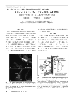
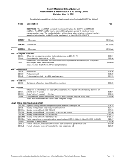
MULTIPL MYELOM TANISI VE TEDAVİYE YANIT KRİTERLERİ
Ali Zahit BOLAMAN
OLGU SUNUMU
Lower Extremity Post-Traumatic
Lymphedema Secondary to Accidental
Injection of a Veterinary Drug: Case Report
Murat KADAN,a
Gökhan ARSLAN,a
Gökhan EROL,a
Erkan KAYA,a
Suat DOĞANCI,a
Cengiz BOLCAL,a
Ufuk DEMİRKILIÇa
Department of Cardiovascular Surgery,
Gülhane Military Medical Academy,
Ankara
a
Geliş Tarihi/Received: 10.05.2014
Kabul Tarihi/Accepted: 11.06.2014
Yazışma Adresi/Correspondence:
Murat KADAN
Gülhane Military Medical Academy,
Department of Cardiovascular Surgery,
Ankara,
TÜRKİYE/TURKEY
[email protected]
ABSTRACT Despite improvements in medical technologies, there is still not a holistic approach to
lymphatic disorders. However, secondary lymphedema is a preventable form of lymphatic disorders
by its association with the underlying etiology. Thus, it is easier to prevent than cure. In this report,
we present a patient admitted to our clinic with an interesting traumatic lymphedema secondary
to accidental injection of tilmicosin phosphate, which is often used as macrolide antibiotic in veterinary practice.
Key Words: Lymphedema; macrolides; necrosis; fibrosis
ÖZET Tıp teknolojilerinde yaşanan gelişmelere rağmen, halen lenfatik hastalıklar için etkin bir
yaklaşım bulunmamaktadır. Bununla birlikte, sekonder lenfatik hastalıklar, altta yatan etyolojik
faktörlerin önlenmesi ile önlenebilir patolojilerdir. Bu yazımızda, sıklıkla veteriner hekimlikte makrolid grubu antibiyotik olarak kullanılan tilmicosin fosfat molekülünün kazara enjeksiyonu sonucu
gelişmiş travmatik lenfödem tablosu ile kliniğimize başvurmuş ilginç bir vaka sunulmaktadır.
Anahtar Kelimeler: Lenfödem; makrolidler; nekroz; fibrozis
L
Damar Cer Derg
ymphedema is significant disorder that can be defined as abnormal
accumulation of the interstitial fluid in the intracellular space. It is
closely related to lymphatic and venous pathologies. Although it is
more common in females, it can affect both genders.1 This disorder can be
classified into two forms, according to its etiologies. If there is not another
underlying disorder, it is called as primary lymphedema, and if there is an
underlying cause, it is called as secondary form.1,2
Main etiologic factor of secondary lymphedema is the structural and
functional change on lymphatic ducts and vessels, which are almost irreversible. For this reason, most of the secondary lymphedema cases cannot
be cured even if the etiology is well known.2
doi: 10.9739/uvcd.2014-40474
Copyright © 2014 by
Ulusal Vasküler Cerrahi Derneği
Turkiye Klinikleri J Int Med Sci 2008, 4
Main diagnostic tools are lymphoscintigraphy and Doppler ultrasonography.3 Lymphoscintigraphy has 73-97% sensitivity with 100% specificity for lymphatic disorders. It also has a major role for the differentiation
of venous disorders to lymphatic disorders. In posttrombotic venous disor1
Ali Zahit BOLAMAN
ders, the subfacial lymphatic flow is affected while
the epifacial flow is unaffected. However, in lymphatic disorders, both of these lymphatic flows are
affected, thus lymphoscintigraphy can define these
two entities.1,4
Treatment of lymphedema includes medical treatment, mechanical treatment and surgical treatment. Preventing of the etiologic factors or its
sequences are the cornerstones of prophylaxis. Several molecules such as diuretics or benzopyrones
can be used for medical treatment. Surgical treatment can only be done in 5-10% of the patients.
Mechanical compression therapy is the most effective approach for symptomatic relief. Compression
therapy reduces edema by increasing the interstitial pressure, therefore reduces the tension of the
skin. Reducing skin tension not only provides
symptomatic relief, but also prevents the possible
complications such as infection or concomitant
thrombosis. This is why compression devices are
widely used in clinical practice both for therapeutic and prophylactic purposes.3
In this report, we aimed to present an unusual
case of secondary traumatic lymphedema, which
developed after misinjection of a veterinary drug.
The informed consent of the patient was obtained
before the preparation of this paper.
CASE REPORT
A 20-year-old male patient presented to our department with swelling of his right limb. His history revealed an accidental injection to his right
calf one month previously. The injected drug was
tilmicosin phosphate, a macrolide antibiotic, used
in the veterinary practice. The injection volume,
which was not known exactly, was probably 10 ml
(60 mg tilmicosin phosphate). After injection, the
affected limb swelled day by day, and the patient
admitted to another hospital on 20th day after injection. Compartment syndrome was diagnosed in
this center, and topical corticoids, systemic flurbiprofen (100 mg twice a day) and pregabalin treatment (75 mg twice a day) were started. He was
discharged from hospital nine days later without
any obvious symptomatic relief. The patient was
2
MULTIPL MYELOM TANISI VE TEDAVİYE YANIT KRİTERLERİ
then admitted our department with his ongoing
complaints.
On physical examination, right lower limb
swelling and stiffness with mild erythema, and
edema near the injection area were detected (Figure 1). His right limb was 7 cm larger at the ankle,
and 4 cm larger at calf level, when compared with
the contralateral extremity. He had a limitation of
right ankle motion. The patient had positive of
Homan’s sign at his right calf.
On venous Doppler ultrasonography, the deep
veins were normal while chronic thrombophlebitis
was detected in both great and small saphenous
veins with significant subcutaneous edema. On
three-phase bone scintigraphy, there was a local
necrosis and heterotopic ossification area, which
was probably secondary to injection or hemorrhage
in gastrocnemius muscle. There were no signs of
reflex sympathetic dystrophy (RSD) on bone
scintigraphy. On lymphoscintigraphy, there was a
delay at the distribution of the Tc-99m at the affected limb, which was concordant with lymphedema (Figure 2).
The patient was referred to immunology department for the investigation of hypersensitivity disorders. Examinations did not reveal any
evidence for allergic, immunologic or rheumatologic disorders. The patient was diagnosed with
acute traumatic lymphedema after all investigations.
FIGURE 1: The appearance of typical lymphedema at ankle and foot with
erythema at the injection site.
Turkiye Klinikleri J Int Med Sci 2008, 4
MULTIPL MYELOM TANISI VE TEDAVİYE YANIT KRİTERLERİ
FIGURE 2: Lymphoscintigraphy of the patient. a. Anterior view, on 20th
minute. b. Anterior view, at 1st hour.
Intermittent pneumatic compression therapy
was started with 50 mmHg pressure, twice a day.
Flurbiprofen (100 mg twice a day) and, diosmin/hesperidin combination (450/50 mg twice a
day) were added to mechanical compression therapy. After 5 days, his right limb was 3 cm larger at
the ankle, and 1.5 cm larger at calf level when compared with the contralateral extremity. After 30
days, there was no significant difference between
the circumferences of lower extremities.
DISCUSSION
Limb swelling has deterministic effects on quality
of life. Bilateral involvement is rare, and particularly seen in the primary form. However, secondary forms are much more common, and often
courses unilaterally. Primary form can be separated
into three subgroups; congenital (0-1 year), precox
(1-35 year) and tarda (after 35 year).1 Secondary
form is associated with lymphatic obstruction secondary to several causes such as; post-inflammation,
surgical procedures, infections (cellulites, erysipelas
etc.), secondary fibrotic events and trauma.1,5
Turkiye Klinikleri J Int Med Sci 2008, 4
Ali Zahit BOLAMAN
Traumatic lymphedema is not a rare disorder
in the population suffering from lymphedema.
However, we cannot find any secondary forms that
develop after misinjection of a drug used in veterinary practice. In our case, the molecule included
tilmicosin phosphate, which is a systemic antimicrobial agent.
Tilmicosin phosphate is a subgroup of
macrolides, and especially effective on gram-positive bacteria. It is a parenteral solution for cattle
and sheep, and primarily shows its effect with inhibiting the protein synthesis.5,6 Prospectus of this
agent recommends subcutaneous injection, and it
is stated that an intramuscular injection must be
avoided. The most important side effect of this
agent is cardiotoxicity due to its negative inotropic,
myocardial depression and tachycardia effects. The
other side effects include local edema, sometimes
necrosis and scars, particularly seen after intramuscular injections.
In our patient, there was topical edema in skin
without any superficial necrosis or scar formation.
However, there was a local necrosis in gastrocnemius muscle, which can be secondary to the intramuscular misinjection of this molecule. We
consider that misinjection probably caused local
necrosis, and a scar formation began near the
necrotic area. These structural changes probably irreversibly interrupted and/or disrupted local lymphatic ducts and vessels, resulting in an impaired
lymphatic flow.
Reconstructive lymphatic surgery was not
suitable due to the localization of the lesion, which
was so distal for such a kind of surgery. Lymphovenous shunt was also not suitable due to concomitant venous thrombophlebitis.7,8
In the follow up of our patient, symptomatic
relief was seen with medical therapy and compression treatment, and a surgical intervention were
not preferred.
In conclusion, we consider that main cause of
the lymphedema in our case was the misinjection
which caused local necrosis and scar formation,
rather than other factors that may cause superficial inflammation (such as erysipelas or cellulitis).
3
Ali Zahit BOLAMAN
From this point of view, we conclude that misapplication of parenteral drugs by inappropriate
routes, as well as to inappropriate locations, can
cause severe damage to local important structures.
On the other hand, secondary lymphedema
should be kept in mind in patients with lym-
1.
2.
3.
4
Oz BS, Sargın M, Iyem H, Bolcal C,
Mataracı I, Akay HT, Dogancı S, Tatar H.
The incidence and factors influencing
lymphedema in lower extremities. Turkish
J Thorac Cardiovasc Surg 2006;14(4):3047.
Rijke AM, Croft BY, Johnson RA, de Jongste
AB, Camps JA. Lymphoscintigraphy and lymphedema of the lower extremities. J Nucl Med
1990;31(6):990-8.
Tiwari A, Cheng KS, Button M, Myint F, Hamilton G. Differential diagnosis, investigation, and
MULTIPL MYELOM TANISI VE TEDAVİYE YANIT KRİTERLERİ
phedema-like leg swelling after unusual interventions.
Conflict of Interest
Authors declared no conflict of interest or financial support.
REFERENCES
4.
5.
6.
current treatment of lower limb lymphedema.
Arch Surg 2003;138(2):152-61.
Khan O, Maharaj P, Rampaul R, Archibald A,
Naipaul R, Loutan N. Lymphoscintigraphic
evaluation of chronic lower limb oedema.
West Indian Med J 2003;52(2):136-9.
Özışık K, Aydın H. Çocukluk çağında
lenfatiko-venöz şant uygulaması. Damar Cer
Derg 2009;18(1):20-2.
Reuter RZ, Carroll JA, Dailey JW, Cook BJ,
Galyean ML. Effects of dietary energy source
and level and injection of tilmicosin phosphate
7.
8.
on immune function in lipopolysaccharidechallenged beef steers. J Anim Sci 2008;
86(8):1963-76.
Szczesny G, Olszewski WL, Deszczynski J.
[Post-traumatic lymphatic and venous
drainage changes in persistent edema of
lower extremities.] Chir Narzadow Ruchu
Ortop Pol 2000;65(3):315-25.
Olszewski WL. The lymphovenous microsurgical shunts for treatment of lymphedema of
lower limbs: indications in 2011. Int Angiol
2011;30(6):499-503.
Turkiye Klinikleri J Int Med Sci 2008, 4
© Copyright 2026 Paperzz