Universite de Sherbrooke
Radiosensibilisation de l'ADN par le 5-bromodeoxyuridine : l'importance de la
structure et de la sequence de l'ADN
Par
Marie-Eve Dextraze
Departement de medecine nucleaire et radiobiologie
These presentee a la Faculte de medecine et des sciences de la sante
en vue de l'obtention du grade de
philosophiae doctor (Ph.D.) en science des radiations et imagerie nucleaire
Sherbrooke, Quebec, Canada
17juin2010
Jean-Paul Jay-Gerin, PhD
Darel Hunting, PhD
Richard Wagner, PhD
Antonio Conconi, PhD
Dindial Ramotar, PhD
© Marie-Eve Dextraze, 2010
Membres du jury d'evaluation
President du jury, Departement de medecine nucleaire et
radiobiologie, FMSS
Directeur de recherche, Departement de medecine nucleaire
et radiobiologie, FMSS
Directeur de recherche, Departement de medecine nucleaire
et radiobiologie, FMSS
Departement de microbiologic et infectiologie, FMSS
Departement de Medecine et Specialites Medicales, Faculte
de medecine, Universite de Montreal
1*1
Library and Archives
Canada
Bibliotheque et
Archives Canada
Published Heritage
Branch
Direction du
Patrimoine de I'edition
395 Wellington Street
OttawaONK1A0N4
Canada
395, rue Wellington
Ottawa ON K1A 0N4
Canada
Your We Votre reference
ISBN: 978-0-494-75078-0
Our file Notre reference
ISBN: 978-0-494-75078-0
NOTICE:
AVIS:
The author has granted a nonexclusive license allowing Library and
Archives Canada to reproduce,
publish, archive, preserve, conserve,
communicate to the public by
telecommunication or on the Internet,
loan, distribute and sell theses
worldwide, for commercial or noncommercial purposes, in microform,
paper, electronic and/or any other
formats.
L'auteur a accorde une licence non exclusive
permettant a la Bibliotheque et Archives
Canada de reproduire, publier, archiver,
sauvegarder, conserver, transmettre au public
par telecommunication ou par I'lnternet, preter,
distribuer et vendre des theses partout dans le
monde, a des fins commerciales ou autres, sur
support microforme, papier, electronique et/ou
autres formats.
The author retains copyright
ownership and moral rights in this
thesis. Neither the thesis nor
substantial extracts from it may be
printed or otherwise reproduced
without the author's permission.
L'auteur conserve la propriete du droit d'auteur
et des droits moraux qui protege cette these. Ni
la these ni des extraits substantiels de celle-ci
ne doivent etre imprimes ou autrement
reproduits sans son autorisation.
In compliance with the Canadian
Privacy Act some supporting forms
may have been removed from this
thesis.
Conformement a la loi canadienne sur la
protection de la vie privee, quelques
formulaires secondaires ont ete enleves de
cette these.
While these forms may be included
in the document page count, their
removal does not represent any loss
of content from the thesis.
Bien que ces formulaires aient inclus dans
la pagination, il n'y aura aucun contenu
manquant.
1*1
Canada
Insanity: Doing the same thing over and over again and expecting different results
- Albert Einstein
The trouble with having an open mind, of course, is that people will insist on coming
along and trying to put things in it
- Terry Pratchett
Resume
Radiosensibilisation de l'ADN par Ie 5-bromodeoxyuridine : l'importance de la
structure et de la sequence de l'ADN
Par
Marie-Eve Dextraze
Departement de medecine nucleaire et radiobiologie
These presentee a la Faculte de medecine et des sciences de la sante en vue de l'obtention
du diplome de philosophiae doctor (Ph.D.) en sciences des radiations et imagerie
biomedicale, Faculte de medecine et des sciences de la sante, Universite de Sherbrooke,
Sherbrooke, Quebec, Canada, J1H 5N4
Les dimeres interbrins sont des lesions de type complexe, ou les deux brins d'ADN sont
pontes de facon covalente. Par consequent, ce type de lesion est tres toxique pour la
cellule, car il nuit a la separation des brins d'ADN necessaire a des processus cruciaux
pour la cellule, comme la replication et la transcription. De plus, des experiences recentes
montrent que la reparation des dimeres interbrins passe par la formation d'un bris double
brin, une autre lesion avec un potentiel toxique eleve. Ce n'est que tout recemment qu'on
a montre que la radiation ionisante menait a la formation de dimeres interbrins dans
l'ADN cellulaire. On en sait done encore tres peu sur les conditions dans lesquelles se
produisent les dimeres et comment ils sont repares.
Recemment, a la suite d'une exposition aux radiations ionisantes, notre groupe a mis en
evidence la formation de dimeres interbrins dans un ADN ou une thymidine avait ete
remplacee par le 5-bromo-2'-desoxyuridine (BrdU). Ces dimeres n'etaient formes que
lorsque le BrdU se trouvait au centre d'une zone mesappariee. Puisque e'etait la premiere
fois que ce type de dommage etait observe lors d'une exposition de l'ADN brome a la
radiation ionisante, ma these a porte sur 1'exploration de la formation du dimere interbrin,
particulierement sur les conditions qui favorisaient sa formation. Les trois articles
presentes dans cette these montrent que la forme de l'ADN (forme A vs forme B), la
sequence, ainsi que le type de radiation employe ont une influence importante sur le type
et la frequence du dommage produit.
Ces resultats montrent qu'on en sait encore tres peu sur le mecanisme reel de
radiosensibilisation de l'ADN brome dans les cellules. Cependant, ils mettent aussi en
evidence la reactivite distincte des regions mesappariees de l'ADN, ainsi que leur fort
potentiel pour la formation de dimeres. Or, ces regions mesappariees ne represented
qu'une fraction des structures secondaires et tertiaires de l'ADN presentes dans la cellule.
MOTS-CLES : 5-bromodeoxyuridine, dommages a l'ADN, dimeres interbrins, structure
de l'ADN, radiation ionisante.
Table des matieres
Resume
i
Table des matieres
Hi
Listedes tableaux
v
Listedes figures
vi
Listedes abreviations
Chapitre I - Introduction
1.1 -Structure del'ADN
1.1.1 - Composantes de l'ADN
1.1.2 -Forme A, Bet Z
1.1.3 - ADN cellulaire : un environnement dynamique
vii
1
/
1
2
5
1.2 — Effet des radiations ionisantes sur l'ADN
1.2.1 - Rayonnement ionisant et interaction avec la matiere
1.2.2 - Radiolyse de l'eau
1.2.3 - Effet direct et indirect
1.2.4 - Interaction des radicaux avec l'ADN
8
10
11
13
8
1.3 -Le 5-Bromodeoxyuridine
1.3.1 -Structures etproprietes
1.3.2 - Radiosensibilisateur : essais precliniques
1.3.3 - Radiosensibilisateur : essais cliniques
1.3.4 - Reevaluation du mecanisme de radiosensibilisation du BrdU
17
17
19
21
22
Chapitre II - l er article
24
Chapitre III - 2 e article
55
Chapitre IV - 3 e article
82
Chapitre V - 4C article
106
Chapitre VI - Resultats supplementaires
131
VIA -EDTA : un capteur efficace de radicaux hydroxyles
131
VI.2 - BrdU en tant que capteur d'electrons aqueux
134
VI.3 - Le BrdU et I'effet direct des radiations
137
Chapitre VII - Discussion
VII. 1 - Effet de la structure de l'ADN
VILLI - ADN-A vs ADN-B
VII. 1.2-Flexibilitede l'ADN
141
141
142
144
VII.2 - Effet de la sequence deVADN
VII.3 — Structure des dimeres interbrins : effet de I'environnement
Chapitre VIII - Conclusion et perspectives
146
149
154
VIII. 1 - Formation de dimeres interbrins : est-ce que laflexibilite de I 'ADN est la cle?
154
VIII.2 — Impact de la structure tertiaire sur la formation de dommages a I'ADN cellulaire
156
Remerciements
159
References
160
Liste des tableaux
Tableau I-1: Caracteristiques des formes de I'ADN.
4
Tableau III-l: Localization of Strand Breaks on the Brominated and Semicomplementary Strands.... 68
Liste des figures
Figure 1-1 : Composantes de I'ADN.
2
Figure 1-2 : Formes A, B etZde I'ADN
3
Figure 1-3 : Condensation de I'ADN en chromatine
6
Figure 1-4 : Replication de I'ADN.
7
Figure 1-5 : Depot d 'energie le long de la trajectoire de I 'electron primaire
9
Figure 1-6: Radiolyse del'eau a I'echelle de lapicoseconde
10
Figure 1-7 : Effet direct et indirect des radiations sur I'ADN.
12
Figure 1-8 : Types de dommages formes par I'exposition de I'ADN aux radiations ionisantes
14
Figure 1-9: Frequence des dommages subispar I'ADN cellulaire
15
Figure 1-10 : Mecanisme deformation d'un dimere interbrinpar les radiations ionisantes
16
Figure 1-11 : Structure de la desoxythymidine et du BrdU
18
Figure 1-12 : Mecanisme deformation des cassures dans I 'ADN brome expose aux rayons UV.
19
Figure II-1 : Sequences of the brominated (B) and non-brominated (T) oligonucleotides
31
Figure 11-2 : Strand breaks and base lesions as a function of hydration
34
Figure II-3 : Damage yield as a function of DNA hydration:
36
Figure II-4 : Damage as a function of DNA sequence
37
Figure II-5 : Damage yield in the mismatch zone as a function of DNA structure
39
Figure 11-6 : Interstrand cross-link yield as a function of DNA structure
40
Figure II- 7 : Major degradation pathway for BrdU-substituted DNA for single stranded, double
stranded, and mismatched DNA for A- and B-DNA
45
Figure III-l : Oligonucleotide design
61
Figure 111-2 : Contribution of double strandedness to strand breaks as a function of sequence
63
Figure III-3 : Damage yield as a function of sequence in mismatched DNA
65
Figure III-4 : Interstrand cross-link structures and strand breaks location as a function of sequence. 66
Figure IV-1: Oligonucleotide design
88
Figure IV-2: Method validation
92
Figure IV-3: Cross-linked sites as a function of sequence (ionizing radiation)
94
Figure IV-4: Overall treatment profile for AABrUAA/ZAATAA and GGBrUGGI/AATAA
95
Figure IV-5: Cross-linked sites as a function of sequence (UV radiation)
96
Figure IV-S1: Formation of Y cross-links following mild hydrolysis treatment.
102
Figure IV-S2: Identification of Y cross-link
103
Figure V-l : Comparison of interstrand cross-links (ICLs) formed in BrdU-substituted and
unsubstituted DNA
117
Figure V-2 : Proposed mechanism for ICL formation in mismatched DNA
121
Figure VI-1 : Captation des radicaux hydroxylespar I'EDTA
132
Figure Vl-2 : Gel de la formation des cassures a I'ADN brome et non brome enfonction de la quantite
d'EDTA
133
Figure VI-3 : Conversion du BrU en uracile enfonction de la dose.
135
Figure VI-4 : Degradation duBrUselon les conditions d'irradiation
136
Figure VI-5 : 5-BrU en tant que capteur d'electrons solvates
136
Figure VI-6 : Effet du tampon sur {'irradiation d'ADN brome hydrate.
139
Figure VII-1 : Generation des dommages a I'ADN brome enfonction de la conformation
143
Figure VII-2 : Effet de I'ouverture des brins d'ADN par I'ARN polymerase sur les dommages causes
par I'irradiation de I'ADN brome.
145
Figure V1I-3 : Production de bandes multiples dpartir d'un radical unique
148
Figure VIII-J : Structure de I 'ADN en presence de mesappariements multiples
155
Liste des abreviations
Acide desoxyribonucleique
Acide ethylene-diamine-tetraacetique
Acide ribonucleique
Adenine
Atome d'hydrogene
5-bromo-2'-desoxyuridine
5-bromouracile
Chromatographie liquide haute performance
Cobalt-60
Cytosine
Electron aqueux
Electron-volt
Epidermal Growth Factor
Gray
Guanine
Hydrogene
Methyl
Oxyde nitreux
Paire de bases
Peroxyde d'hydrogene
Proton
Radicaux hydroxyles
Reparation par excision des nucleotides
Taux d'hydratation
Thymine
Transfert d'energie lineaire
Ultraviolet
ADN
EDTA
ARN
A
H
BrdU
BrU
HPLC
60
Co
C
eaq
eV
EGF
Gy
G
H2
-CH 3
N20
pb
H202
H+
'OH
NER
r
T
TEL
UV
Chapitre I - Introduction
1.1 - Structure de I'ADN
"To say that a man is made up of certain chemical elements is a satisfactory description only
for those who intend to use him as afertilizer " — Hermann Joseph Mueller
1.1.1 — Composantes de I 'ADN
De la meme facon qu'un roman ne se resume pas qu'a un melange des 26 lettres de
Falphabet, et une symphonie a 8 notes, la vie ne se resume pas a la combinaison
aleatoire de A, C, G, et T. Et pourtant... Ce sont des proprietes physiques et
chimiques de l'ADN que decoulent les caracteristiques qui sont a Porigine de la
fonction centrale de cette molecule pour la cellule. La double helice d'ADN est
composee de deux brins antiparalleles, eux-memes formes a partir d'une repetition de
nucleotides, qui comprend une base, un sucre (desoxyribose) et un groupement
phosphate (figure 1-1). C'est la succession de desoxyriboses et de groupements
phosphates qui forme le squelette de la molecule d'ADN, tandis que les bases
correspondent a la partie variable de la molecule, celle ou est contenue 1'information.
II y a quatre bases dites « natives » dans l'ADN; 1'adenine, la guanine, la cytosine et
la thymine, symbolisees par les lettres A, G, C et T. Les autres bases, comme la
methylcytosine, correspondent souvent a des modifications epigenetiques ou a des
produits de deamination d'une des quatre bases natives.
2
<X I
HO
I
5'
N
O"
"O
€3
A
NHj>
O-P-OCH,
\ A,
1
Phosphate
bond
N
I
^
*M^
.
H T3i
O
DNA
single strand
H
i
o
" K
H
o-i-o£-
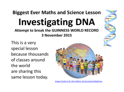
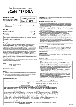
Figure 1-1 : Composantes de l'ADN. Modifie a partir de Lehnert, 2007, p 31.
On separe les quatre bases en deux categories : les purines (A et G) et les pyrimidines
(C et T). L'adenine (A) s'apparie avec la thymine (T), et la guanine (G) avec la
cytosine (C). Chaque base d'un brin s'apparie a sa base complementaire situee sur le
brin oppose grace a la formation de ponts hydrogene. Ces ponts hydrogene permettent
a l'ADN d'adopter une structure a la fois robuste et flexible, ou la proximite des deux
brins est maintenue, tout en minimisant l'energie necessaire a leur separation.
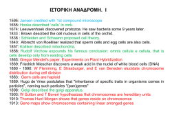
1.1.2 - Forme A, B et Z
La double helice d'ADN est une molecule extremement flexible, et peut adopter un
nombre impressionnant de configurations en reponse aux pressions de son
environnement. La figure 1-2 montre trois des formes les mieux connues jusqu'a
3
present, sort les formes A, B, et Z. Les caracteristiques de ces formes sont presentees
dans le tableau 1-1.
Figure 1-2 : Formes A, B et Z de l'ADN (de gauche a droite).
La forme B est la mieux connue, car elle est adoptee dans des conditions
physiologiques, notamment dans les tetes intactes des spermatozoifdes (Voet et Voet,
1995). La forme A, quant a elle, est retrouvee en presence d'un duplexe ADN/ARN,
ou lorsque l'ADN est deshydrate, par exemple en presence de 80 % ethanol. Bien que
l'homologie de la forme A dans ces deux conditions ait ete remise en question,
notamment a cause des differences dans la conformation du sucre (Horton et Finzel,
1996), la forme generate de l'ADN-A demeure la meme. La formation d'ADN-Z a
surtout ete observee au niveau de l'ADN synthetique contenant une grande proportion
de guanines en presence de concentrations elevees en sel, ainsi qu'en presence de
cytosines methylees en position C5 (Behe et Felsenfeld, 1981, Thamann et ai, 1981).
4
Tableau 1-1 : Caracteristiques des formes de l'ADN.
A
B
Z
Sens du pas de l'helice
Droite
Droite
Gauche
Diametre
-26 A
-20 A
-18 A
11
10
12
Rotation de l'helice / paire de bases
33°
36°
60°
Longueur d'helice / tour
28 A
34 A
45 A
C3'-endo
C2'-endo
C2' -endo (C et T)
Paires de bases/ tour d'helice
Phage des sucres
C3' -endo (A et G)
Dans la cellule, la demonstration de 1'existence de l'ADN-Z a ete plus ardue. La
transition entre la forme B et la forme Z necessite de I'energie, qui devient disponible
dans certaines conditions dans la cellule, comme le superenroulement negatif de
l'ADN observe en aval de l'ARN polymerase durant la transcription (Liu et Wang,
1987). Depuis les premieres evidences de l'existence de l'ADN-Z, sa formation lors
de la transcription a ete correlee par plusieurs experiences (Herbert et Rich, 1999,
Jiang et al, 1991, Lafer et al, 1983). De plus, le groupe de Rich a observe que la
proteine ADAR1, une ARN adenosine deaminase, liait specifiquement la forme Z de
l'ADN (Herbert et al, 1995). L'ADN-Z semble impliquee dans plusieurs processus
fonctionnels de la cellule, notamment la regulation de l'expression, le positionnement
des nucleosomes, le remodelage de la chromatine et la recombinaison (Li et al,
2009).
Ces trois conformations ne sont qu'un exemple des multiples configurations que
l'ADN peut adopter en fonction des pressions de son environnement. Plusieurs
5
proteines presentes dans le noyau de la cellule sont aussi en mesure de modifier la
structure de l'ADN, que ce soit pour l'empaqueter (chromatine), ou pour controler les
differents processus cellulaires qui impliquent l'ADN (transcription, replication,
reparation).
1.1.3 - ADNcellulaire : un environnement dynamique
Lorsqu'on pense a l'ADN, la premiere image qui nous vient en tete est generalement
celle de la double helice. On oublie souvent que l'ADN est avant tout une structure
dynamique, en constant changement. Meme la chromatine, l'agencement de proteines
et d'ARN qui permet la compression de 4 metres d'ADN dans un noyau de 5 jam de
diametre, represente un niveau d'organisation
superieur qui affecte
autant
1'accessibility de l'ADN aux proteines du noyau que l'organisation generate de l'ADN
en regions fonctionnelles.
La figure 1-3 montre les niveaux successifs d'organisation de l'ADN cellulaire. La
double helice d'ADN s'enroule autour de proteines appelees histones pour former les
nucleosomes. Ces nucleosomes forment a leur tour des solenoides de 30 ran de
diametre qui se compactent en filaments. Durant la mitose, ces filaments se
condensent encore davantage pour former des chromatides, qui permettent la
repartition d'une copie du genome a chaque cellule-fille.
6
Figure 1-3 : Condensation de l'ADN en chromatine. Source: (Lehnert, 2007, p
206)
Cependant, il existe aussi plusieurs processus dynamiques qui impliquent l'ADN dans
la cellule. Dans les regions activement transcrites du genome, la structure meme de la
chromatine est constamment modifiee afin de moduler 1'accessibilite de l'ADN aux
proteines responsables de la regulation de la transcription. Les modifications posttraductionnelles des histones comptent pour une fraction non neghgeable des
methodes utilisees par la cellule pour reguler cette accessibilite, et constituent en
quelque sorte un code qui identifie les regions de l'ADN qui doivent etre liberees des
contraintes steriques de la chromatine pour faciliter la transcription, en fonction des
besoins de la cellule. Dans d'autres cas, les histones sont aussi modifiees pour faire
entrer en dormance certaines parties du genome (Voir Mellor, 2006, pour une revue
de l'impact des modifications nucleosomales sur la transcription).
D'autres phenomenes, qu'ils soient constants (transcription, reparation) ou ponctuels
(replication) font de l'ADN cellulaire une molecule en constants changements.
7
5'
Lagging strand
with fragments
Figure 1-4 : Replication de l'ADN. Source : (Lehnert, 2007, p 35)
Lors de la replication, les deux brins de la double helice d'ADN sont separes afin de
permettre la reproduction de rinformation contenue dans chaque chromosome. La
figure 1-4 montre une representation schematisee de la replication de l'ADN. Les deux
copies ainsi creees permettront la formation de deux cellules-filles avec la meme
information genetique. Cependant, la figure 1-4 ne montre qu'une vision simplifiee de
ce qui se produit dans la cellule. En realite, la replication est un processus complexe
qui necessite la coordination de dizaines de proteines (Pollard et Earshaw, 2002),
meme dans les systemes bacteriens, qui sont beaucoup moins complexes. Une
helicase doit separer les deux brins d'ADN en amont de l'ADN polymerase, l'enzyme
responsable de la polymerisation du nouveau brin, et qui comprend elle-meme
souvent plusieurs sous-unites. D'autres proteines sont responsables de proteger les
brins d'ADN des nucleases alors qu'ils sont sous forme simple brin. Finalement, un
agencement complexe de proteines est charge de s'assurer que le brin d'ADN
nouvellement synthetise ne comprend pas d'erreurs, et de reparer celles qui pourraient
s'etreproduites.
8
La replication n'est qu'un exemple parmi tant d'autres ou la structure de l'ADN est
modifiee par un phenomene de regulation cellulaire. Pour cette raison, il est crucial de
prendre en compte cet aspect du metabolisme de la cellule lorsqu'on etudie les
consequences d'un agent exterieur sur l'ADN, notamment la radiation.
1.2 - Effet des radiations ionisantes sur l'ADN
1.2.1 — Rayonnement ionisant et interaction avec la matiere
L'interaction du rayonnement avec la matiere a pour consequence le transfert d'une
partie ou de la totalite de I'energie de la particule initiale le long de sa trajectoire. La
notion de TEL (transfert d'energie lineaire, keV/um) est utilisee pour caracteriser ce
depot d'energie en fonction de la distance parcourue dans la matiere. Par exemple, le
Cobalt-60 (60Co), utilise en radiotherapie, produit 2 photons avec une energie
moyenne de 1.25 MeV dont le TEL est d'environ 0.2 keV/um. On distingue ainsi les
sources de radiation a haut TEL (particules lourdes, rayons alpha), ou la densite des
ionisations est elevee le long de la trajectoire, de celles a bas TEL (60Co), ou la densite
des ionisations est plus faible.
9
y
.
(o)
Excited molecules from primary excitation
(•)
Positive ions from primary ionization
v/
0
*
Positive ions and excited molecules from 8-electrons
Secondary positive ions and excited electrons
Electrons
X
Primary electron (100 keV)
e
80-100 A
@-
v"*)
/5%>>
x^w-T'
•' S-electron (10 keV)
-yy^
©..
-@
Figure 1-5 : Depot d'energie le long de la trajectoire de I'electron primaire. L'electron
primaire (ici avec une energie de 100 keV) depose son energie en excitant (points
blanc) ou en ionisant (points noirs) la matiere qu'il rencontre le long de sa trajectoire.
Le depot de I'energie peut ensuite mener a une serie d'ionisations et d'excitations
secondares dans un volume de 80 a 100 A autour du site de l'interaction (appelee
grappe). La production d'electrons 5, peu probable pour le rayonnement a faible TEL,
peut se produire lorsque les grappes se recoupent avec un rayonnement a haut TEL.
Source : (Lehnert, 2007, p 13)
Dans le cas de la radiation electromagnetique (p. ex. les photons), le depot de la dose
passe par le transfert de I'energie a des electrons primaires tres energetiques. II y aura
par la suite formation d'especes reactives le long de la trajectoire de I'electron
primaire par des processus d'excitation et d'ionisation (figure 1-5). Ces processus
creent aussi des electrons de basse energie (<20 eV) qui, pour la plupart, ont une
energie qui se trouve sous le seuil d'ionisation. Malgre qu'ils soient produits en tres
grande quantite, on a longtemps cru qu'ils avaient peu d'importance au niveau de la
radiobiologie, puisqu'ils ne possedaient pas suffisamment d'energie pour ioniser la
matiere. Cependant, les travaux de Leon Sanche ont montre que les electrons de basse
energie etaient en mesure, par un phenomene d'attachement dissociatif, de causer des
dommages dans la matiere, notamment l'ADN (Boudaiffa et ah, 2000; pour une revue
de la litterature, voir Sanche, 2002). Cependant, la chimie des radiations classique
10
tient surtout compte des produits issus de l'ionisation de la matiere le long de la
trajectoire incidente.
1.2.2 - Radiolyse de I 'eau
Dans les systemes biologiques, l'eau est une composante majeure; elle represente
entre 70 et 80 % du contenu d'une cellule. Ainsi, la probabilite que la radiation
interagisse avec l'eau est importante si on la compare aux autres molecules. Pour cette
raison, beaucoup d'efforts ont ete consacres a la comprehension des reactions qui se
produisent a la suite de cette interaction. Un schema simplifie de la decomposition de
l'eau est presente a la figure 1-6.
Excitation
H20 - • H20*
H 2 0* - • H" + "OH
lonisation
H 2 0 - • H 2 CT + eH 2 0" + + H 2 0 - • H 3 0 + + "OH
e- + n H 2 0 - • e-aq
Figure 1-6 : Radiolyse de l'eau a l'echelle de la picoseconde.
C'est durant l'etape physique (~10~16 s) que se produisent les premieres etapes de la
radiolyse, soit l'excitation et l'ionisation des molecules d'eau le long de la trajectoire
incidente. Lors de l'etape physico-chimique ( ^ 0 1 2 s), il y a dissociation des
molecules d'eau en 'OH, H' et
H.T,0+
(voir figure 1-6). De plus, les electrons, qui ont
ete attaches lors des ionisations initiales, perdent graduellement leur energie suite a
11
d'autres ionisations et excitations, et vont ainsi se thermaliser. II y aura ensuite
reorganisation des molecules d'eau autour de I'electron pour former un electron
hydrate. A cette etape, la diffusion n'entre pas en jeu dans les differentes reactions.
Lors de 1'etape subsequente de chimie non homogene qui se deroule au sein des
grappes ainsi formees autour des interactions primaires, ce sont ces diverses especes
reactives qui vont diffuser et se combiner pour former les especes moleculaires des
produits de la radiolyse de l'eau telles que H2 et H2O2. A 10"6 s, c'est-a-dire a la fin de
l'expansion des grappes et au debut de l'etape de chimie homogene, les especes
presentes de la radiolyse de l'eau sont, principalement 'OH, e"aq, H", H2O2, H2, H30+
et OH". Ce sont precisement ces especes qui reagiront ensuite avec le materiel
biologique pour produire l'effet indirect des radiations.
1.2.3 - Effet direct et indirect
Bien qu'on puisse decrire l'effet direct/indirect des radiations sur la matiere
biologique a differents niveaux (moleculaire, cellulaire, vasculaire), dans le cas de
l'ADN on decrit l'effet direct des radiations comme l'interaction directe de la
particule incidente avec l'ADN (figure 1-7).
12
Figure 1-7 : Effet direct et indirect des radiations sur l'ADN. Source : (Lehnert,
2007, p 11)
L'effet indirect, lui, implique un depot d'energie aux molecules adjacentes, suivi de la
formation d'especes reactives par excitation ou ionisation. Ce sont les especes
reactives, le plus souvent les produits issus de la radiolyse de l'eau dans le cas du
materiel biologique irradie en solution diluee, qui vont ensuite interagir avec l'ADN.
Lorsqu'on tient compte des couches d'hydratation de l'ADN en solution, on distingue
alors trois types d'effets : l'effet direct, 'md'Vect et q^as"' d;reet. On qiudifie d'effei
quasi-direct la production de radicaux dans la couche primaire interne (T < 9
FbO/nucleotide) car ils sont immediatement transferes a l'ADN. La couche primaire
d'hydratation se distingue de la couche secondaire par la faible mobilite des
molecules d'eau (temps de residence superieur a la picoseconde) et la densite plus
importante de ces molecules. La portion interne de la couche primaire comprend les
molecules d'eau qui sont etroitement lies a la molecule d'ADN et qui demeurent
associees a l'ADN meme a 0 % d'humidite (Feig et Pettitt, 1998). Ce n'est done pas
surprenant que la production de radicaux hydroxyles ne soit observee que dans la
couche primaire externe a partir de T ~ 9 F^O/nucleotide (Debije et al, 2000, La Vere
et al,
1996).
La couche secondaire d'hydratation commence a T ~ 20
13
H20/nucleotide, et il est impossible de la distinguer du reste des molecules d'eau en
solution par la mobilite ou la densite des molecules d'eau (Mroczka et Bernhard,
1993, Saenger, 1984, Wang et al, 1993).
1.2.4 - Interaction des radicaux avec I 'ADN
Suite a I'interaction de la radiation avec la matiere biologique et la generation de
radicaux qui attaquent l'ADN, plusieurs types de dommages sont produits. Suite a des
experiences menees avec des capteurs de radicaux, on estime que 60-70% des
dommages subis par la cellule proviennent de I'interaction des radicaux *OH avec
l'ADN (Chapman et al, 1973, Roots et Okada, 1972), alors que les electrons solvates,
qui creent surtout des lesions aux bases, ont peu ou pas d'effet sur la mortalite
cellulaire. Les radicaux *OH interagissent avec les doubles liaisons des bases et sont
d'excellents abstracteurs d'atomes d'hydrogene sur le desoxyribose (koH = 2.3 x 10⁹ L
mol"1 s"1, Motohashi et Saito, 1993), ce qui mene a la formation de bases modifiees et
a la formation de cassures.
14
m
~^'
C^
Abasic
Slte
Single-strand
break
Double-strand
break
DNA-protein
cross-link
Damaged
base
Figure 1-8 : Types de dommages formes par 1'exposition de l'ADN aux
radiations ionisantes. Source : (Lehnert, 2007, p 124)
Parmi les dommages subis par l'ADN cellulaire a la suite d'une exposition aux
radiations ionisantes, on compte deux grandes categories, qui sont schematisees a la
figure 1-8 :
- les dommages simples :
- site abasique
- base modifiee
- dommage au sucre
- bris simple brin
- les dommages complexes
- lesions multiples (incluant les bris double brin)
- pontage ADN-proteine
- pontage ADN-ADN
La figure 1-9 montre la frequence de ces differents dommages subis par la cellule lors
d'une exposition a 1 Gy d'une source de radiation a faible TEL (Lehnert, 2007). Bien
15
que les lesions simples soient les plus nombreuses, ce sont les lesions complexes,
incluant les bris double brin, qui sont principalement responsables de la mort
cellulaire.
TABLE 6.1
Estimation of the Number of Early Physical and
Biochemical Changes That Occur When Mammalian
Cells Are Irradiated with 1 Gy of Low LET Radiation
Initial Physical Damage
Ionization in the cell nucleus
Ionization directly in DNA
Excitation directly in DNA
~1000,000
~2000
~2000
Selected Biochemical Damage
Damaged bases
Damaged sugars
DNA single-strand breaks (SSBs)
Alkali-labile sites
Double-strand breaks (DSBs)
DNA-protein cross-links (DPC)
1000-2000
1200
1000
250
40
150
Selected Cellular Effects
Lethal events
Chromosome aberrations
Hypoxanthine phosphoribosyltransferase
(Hprt) aberrations
~0.2-0.S
~1
10" s
Figure 1-9: Frequence des dommages subis par l'ADN cellulaire. Source
(Lehnert, 2007, p 125)
Les lesions multiples comprennent les bris double brin, et sont majoritairement
formes par un mecanisme bi-radicalaire (Milligan et al, 1995), soit deux radicaux qui
reagissent a proximite l'un de l'autre pour former deux lesions. Ces lesions sont
particulierement toxiques pour la cellule, car elles compliquent le processus de
reparation.
16
o
O
>v
a
\
Figure I-10 : Mecanisme de formation d'un dimere interbrin par les radiations
ionisantes. Modifie a partir de Regulus et at, 2007. Suite a l'exposition de
l'ADN aux radiations ionisantes, les radicaux hydroxyles issus de la radiolyse
de l'eau font l'abstraction d'atomes d'hydrogene, ce qui entraine la formation
d'un radical sur le desoxyribose en position C4'. Ce radical mene ensuite a la
formation du produit 2 par un processus de (3-elimination, en plus de la
liberation de la base et de l'extremite 3', ce qui produit une cassure tranche.
Une cytosine situee sur le brin en face peut alors attaquer 2 pour former le
dimere interbrin.
Dans le cas des pontages ADN-ADN, ce n'est que recemment qu'on a etabli que ces
dommages etaient produits par les radiations ionisantes, qui jusque-la n'avaient ete
observes qu'a la suite d'une exposition aux rayons UVA ou a des agents
therapeutiques comme la mitomycin C. En effet, les groupes de Cadet et Greenberg
ont etabli en 2007 qu'il y avait formation d'un dimere interbrin par les radicaux OH
dans l'ADN synthetique et dans les cellules (Regulus et at, 2007, Sczepanski et at,
2009a). Ce dimere interbrin est d'autant plus interessant qu'il inclut aussi une cassure
simple brin, et s'inscrit done dans la categorie des lesions multiples, tres
17
dommageables pour la cellule (figure I-10). De plus, le groupe de Greenberg a
recemment montre que la reparation de ce dimere transitait par la formation d'une
cassure double brin par le complexe UvrABC dans un systeme bacterien (Sczepanski
etal, 2009b).
Cependant, de facon generale, on en sait encore tres peu sur la formation et la
reparation des dimeres interbrins causes par les radiations ionisantes, entre autres
parce que leur identification dans la cellule est tres recente. La majeure partie de
1'information connue a ce jour a ete recueillie a la suite d'une exposition a des agents
therapeutiques (souvent des agents alkylants bifonctionnels) ou aux rayons
ultraviolets (UVA). Neanmoins, ces travaux ont deja montre que la reparation des
dimeres interbrins neoessitait la coordination de plusieurs systemes de reparation
(Noll et al, 2006) comme la recombinaison homologue et la reparation par excision
des nucleotides (NER). De plus, 1'analyse de la reparation des dimeres interbrins est
compliquee par le fait qu'ils n'alterent pas la structure de l'ADN de facon identique;
certains, comme la mitomycin C, ont peu ou pas d'effet sur la structure de la double
helice (Rajski et Williams, 1998), tandis que les dimeres produits par le cisdismminedichloroplatine
creeent
un
renflement
important
dans
l'ADN
(desenroulement de 79°, flechissement de 45°, Malinge et al, 1994), ce qui rend le
dimere plus susceptible d'etre reconnu par la NER (Noll et al, 2006). Ainsi, il reste
encore beaucoup a faire afin de mieux comprendre comment les dimeres interbrins
sont formes par la radiation ionisante, comment ils sont repares, et quelle est leur
importance dans l'impact global de l'exposition de la cellule aux radiations.
1.3 - Le 5-Bromodeoxyuridine
1.3.1 - Structures et proprietes
18
Le 5-bromo-2'-desoxyuridine (BrdU) est un analogue halogene de la thymidine qui
peut s'incorporer dans l'ADN lors de la replication et ainsi se substituer aux
thymidines (figure I-ll). Bien qu'il soit aujourd'hui surtout utilise pour la detection
de la replication des cellules (Eisch et Mandyam, 2007, Leif et al., 2004, QuinonesHinojosa et al, 2005), le BrdU a d'abord ete developpe en tant que photo- et
radiosensibilisateur (Dewey et Humphrey, 1965, Sano et al, 1968). L'interet pour le
BrdU en tant que radiosensibilisateur decoule de trois principales caracteristiques.
o
V^
o
o
d
V^
°
d
Figure I - l l : Structure de la desoxythymidine et du BrdU.
La premiere est que la taille du brome est semblable a celle du groupement -CH3 de la
thymidine (r = 0.195 et 0.20 nm, respectivement), ce qui lui permet d'etre incorpore
dans l'ADN cellulaire durant la replication. La seconde est que le BrdU, une fois qu'il
est incorpore dans l'ADN cellulaire, ne presente qu'un faible effet toxique; meme
lorsque 50% des thymidines sont remplacees par le BrdU, on ne constate qu'une
faible diminution de la survie cellulaire (Iliakis et al, 1989, Szybalski, 1974). Enfin,
la troisieme caracteristique du BrdU est que l'electronegativite du brome en fait un
bon capteur d'electrons (keaq = 1.6 x 10¹⁰ dm3 mol"1 s"1 a pH 7.0, Patterson et Bansal,
1972) et un bon groupement partant, ce qui permet la creation d'un radical uridin-5-yl
tres reactif, notamment pour ce qui est de l'abstraction d'atomes d'hydrogene fe.prOH
= 4.1 x 10⁷ dm3 mol"1 s"1, Mertens et Sonntag, 1994). La figure 1-12 decrit le
mecanisme de formation d'une cassure a l'ADN causee par l'exposition aux rayons
UV d'un ADN brome. Le mecanisme est generalement suppose etre semblable pour
une irradiation aux rayons ionisants, notamment en ce qui a trait a l'abstraction d'un
atome d'hydrogene sur le sucre du nucleotide en 5'.
19
S
9
>
S
o
^
A
A
A-
T
1
t
9
h
O*
U
"-—' >i
i
a
O"
-
^
o"
H
?
i
°" UsCo
o
HA
„
o^o.
»•
44
Figure 1-12 : Mecanisme de formation des cassures dans l'ADN brome expose aux
rayons UV. A la suite de reposition aux rayons UV (313 nm), un electron est
transfere de la base en 5' du BrdU. II y a ensuite depart de 1'anion brome et formation
d'un radical uridine-5-yl. Ce radical fait I'abstraction d'un atome d'hydrogene sur la
position CI' du sucre en 5' (C2' dans le cas de la radiation ionisante). II y a ensuite
transfert d'un hydrogene et formation d'une cassure par la liberation de l'extremite 3'
du desoxyribose.
D'autres analogues halogenes de la thymine sont aussi de bons capteurs d'electrons,
dans l'ordre suivant: fluorouracile < chlorouracile < bromouracile. Cependant, le 5fluorouracile n'est pas incorpore dans l'ADN, et le chlorouracile forme moins de
radical uracil-yl que le bromouracile (Bansal et ah, 1972, Patterson et Bansal, 1972).
Pour cette raison, c'est
surtout le BrdU qui a fait l'objet
radiosensibilisation.
1.3.2 - Radiosensibilisateur : essais precliniques
d'etudes de
20
II y a deja plus de 40 ans que le BrdU a ete identifie en tant que radiosensibilisateur
(Dewey et Humphrey, 1965). En effet, l'ADN cellulaire ou une partie des thymidines
a ete remplacee par le BrdU subit une augmentation des cassures simples et doubles
(Limoli et Ward, 1993, Saffhill et Ockey, 1985) et des aberrations chromosomiques
(Dewey et al., 1966) apres une exposition aux radiations ionisantes. Le mecanisme de
radiosensibilisation de l'ADN brome passe principalement par l'attachement
dissociatif d'un electron sur le BrdU, puis par le depart d'un anion brome et la
formation du radical uridin-5-yl (Wang et al., 2006). Comme il a ete mentionne plus
haut, ce radical est tres reactif, et va done faire l'abstraction d'un atome d'hydrogene
sur le CI' ou le C2' du deoxyribose situe en 5' du BrdU pour dormer l'uridine
(Schyman et al., 2007). Le ribose radicalaire ainsi cree mene ensuite a la formation de
cassures. A l'origine, on croyait que seuls les electrons aqueux issus de la radiolyse de
l'eau etaient responsables de la formation de cassures, mais des donnees recentes du
groupe de Leon Sanche ont montre qu'il etait possible que les electrons de basse
energie interviennent aussi dans le mecanisme de radiosensibilisation (Abdoul-Carime
et al., 2000b, Dugal et al., 2000, Li et al., 2003). Toutefois, il est important de noter
que la contribution des electrons aqueux a la radiosensibilisation de l'ADN brome
dans les cellules est probablement la plus importante, puisqu'on assiste a la disparition
de la majorite du nombre de cassures double en presence d'acetone, un capteur
d'electrons aqueux (Webb et al, 1993).
D'autres facteurs affectent la radiosensibilisation des cellules par le BrdU, notamment
le pourcentage de remplacement de la thymidine (Ling et Ward, 1990), la structure de
la chromatine (Lawrence et al., 1995), le temps de doublement de la lignee cellulaire,
ainsi que la phase de croissance (exponentielle vs plateau) ou se produit 1'irradiation
(Iliakis et al, 1989, Wang et al, 1994). La presence ou l'absence d'oxygene influence
aussi grandement la production de dommages, d'abord parce que I'oxygene agit a titre
de capteur d'electrons (keaq = 1.9 x 10¹⁰ dm3 mol"1 s"1, Buxton et al, 1988), mais aussi
parce que I'oxygene a demontre une influence importante sur le type de dommage
produit, notamment en fixant les dommages causes a l'ADN par I'addition d'un
radical superoxide. Tous ces facteurs et bien d'autres ont joue un role important dans
21
Petablissement
des
protocoles
cliniques
utilisant
le
BrdU
en
tant
que
radiosensibilisateur (Coleman et Mitchell, 1999, McGinn et Kinsella, 1992).
1.3.3 - Radiosensibilisateur : essais cliniques
Le but premier de la radiotherapie est de cibler specifiquement les cellules
cancereuses, tout en epargnant les cellules saines environnantes. Puisque la cible
principale des radiations est l'ADN, beaucoup de radiosensibilisateurs ont ete
developpes au cours des dernieres annees qui ciblent principalement l'ADN des
cellules cancereuses. A l'origine, I'utilisation du BrdU en tant que radiosensibilisateur
utilise dans le traitement du cancer a souleve beaucoup d'interet, car c'est l'un des
rares a s'incorporer dans l'ADN cellulaire. Puisque le BrdU est incorpore durant la
replication, la theorie voulait que les cellules tumorales, qui se divisent plus
rapidement, incorporent plus de BrdU dans leur ADN et soient done plus sensibilisees
que les cellules saines environnantes. Bien que les essais cliniques aient montre des
resultats encourageants quant a l'efficacite des pyrimidines halogenees en tant que
radiosensibilisateur (Kinsella et al, 1984, Levin et al, 1995, Phillips et al, 1991,
Urtasun et al,
1996), certains problemes ont ete reveles. Entres autres, la
photosensibilite accrue des patients a ete traitee en minimisant l'exposition au soleil et
en prescrivant une creme protectrice topique. La dehalogenation du BrdU par le foie a
ete minimisee en injectant directement le BrdU dans la region de la tumeur.
Cependant, deux facteurs sont demeures problematiques au cours des differents essais
cliniques : le peu de connaissances concernant la vitesse de replication des cellules
saines environnantes, ainsi que l'incapacite de mesurer facilement 1'incorporation
reelle du BrdU dans les cellules. Ce premier facteur pourrait expliquer pourquoi l'un
des premiers essais cliniques a montre un effet positif pour la combinaison du BrdU et
de la radiation (Sano et al, 1965), alors qu'un autre n'a montre aucun effet (Bagshaw
et al, 1967), en plus de noter une augmentation du dommage aux tissus normaux.
Dans ce dernier cas, le champ d'irradiation couvrait aussi la muqueuse de la cavite
22
buccale et du pharynx, qui se divise plus rapidement que les cellules tumorales. Ainsi,
les cellules saines ont incorpore plus de BrdU que les cellules tumorales. Une autre
etude de phase I a utilise une approche inusitee pour une tumeur entouree de cellules
saines qui se divisaient plus rapidement (Eisbruch et al, 1999). En effet, ces cellules
incorporent plus de BrdU et seraient done normalement plus sensibilisees aux
radiations que les cellules tumorales. Cependant, pour la meme raison, le BrdU est
dime plus rapidement lorsque l'infusion est stoppee, tandis que les cellules tumorales
maintiennent encore un taux de remplacement el eve plusieurs jours apres I'arret, au
moment ou est prevue 1'irradiation.
Plusieurs etudes ont montre un effet positif de l'utilisation du BrdU en tant que
radiosensibilisateur sur la survie des patients atteints de differents types de cancer.
Cependant, une etude de phase III parue en 2004, ou le BrdU etait utilise en
combinaison avec un agent chimiotherapeutique, n'a pas reussi a montrer une
amelioration de la survie des patients (Prados et al, 2004). Apres la parution de cette
etude, les travaux sur l'utilisation du BrdU en clinique ont a toute fin cesses.
1.3.4 - Reevaluation du mecanisme de radiosensibilisation du BrdU
Malgre I'arret des travaux cliniques sur le BrdU, l'interet pour le radiosensibilisateur
est demeure present, entres autres parce que e'est l'un des rares agents a sensibiliser
directement l'ADN, et aussi a cause de son faible niveau de toxicite. C'est pour cette
raison que notre groupe a entrepris de reevaluer le mecanisme moleculaire de
radiosensibilisation de l'ADN par le BrdU, en utilisant un modele simple constitue de
courts oligonucleotides synthetiques. C'est ainsi qu'en 2004, notre groupe a montre
que l'effet radiosensibilisateur du BrdU dependait de l'etat d'hybridation de l'ADN.
En effet, apres irradiation dans des conditions hypoxiques, nous avons observe un
plus grand rendement au niveau de la formation de cassures dans un ADN simple brin
ou une thymidine avait ete substitue par un BrdU que dans un ADN double brin
23
(Cecchini et al, 2004). Plus etonnant encore, la presence d'un mesappariement de 5
bases autour du site de modification favorisait la production de cassures par rapport a
un ADN double brin, et entrainait la formation d'un nouveau type de dommage, qui
n'avait jamais ete observe jusque la pour un ADN brome : le dimere interbrin
(Cecchini etal, 2005).
Ainsi, mon projet avait pour but d'approfondir nos connaissances sur la formation du
dimere interbrin dans l'ADN brome soumis aux radiations ionisantes, avec une
attention particuliere sur 1'importance de la structure et de la sequence de l'ADN au
site de la substitution. Cette information permettrait non seulement de mieux
comprendre le mecanisme de radiosensibilisation du BrdU pour un eventuel
recommencement des essais cliniques, mais fournirait aussi de I'information sur la
reactivite des regions mesappariees de l'ADN. Les resultats obtenus sont presenter
dans les sections suivantes.
Chapitre II - 1 e r article
5-Bromodeoxyuridine
Radiosensitization:
Conformation-dependent
DNA
damage.
Auteurs de Particle : Marie-Eve Dextraze, J. Richard Wagner and Darel J. Hunting
Statut de I'article : Publie.
Biochemistry (2007), 46: 9089-97.
Avant-propos :
Contribution a la redaction et a I 'experimentation :
J'ai effectue la totalite des experiences presentees dans cet article. J'ai aussi
redige cet article, sous la supervision de mes co-directeurs, Richard Wagner et Darel
Hunting.
Resume :
Dans cet article, nous presentons l'effet de la conformation de l'ADN sur la
production de dommage a l'ADN brome. A I'aide d'un modele experimental ou
l'ADN est deshydrate puis graduellement rehydrate, nous montrons que la forme B de
l'ADN est necessaire pour la production de bris et de cassures, la forme A ne
produisant que des lesions qui peuvent etre revelees par un traitement alcalin. Cet
article vient appuyer notre hypothese, qui soutient que Penvironnement ou le radical
initial est cree - et plus particulierement la structure de l'ADN dans cet article - lors
25
de I'irradiation de l'ADN a un effet important sur la letalite du dommage qui est
produit.
26
5-BromodeoxyuridineRadiosensitization:
Conformation-Dependent DNA Damage
f This work was supported by the National Cancer Institute of Canada.
Marie-Eve Dextraze, J. Richard Wagner and DareU. Hunting*
Center for Research in Radiooncology (CR2), Department of Nuclear Medicine and
Radiobiology, Faculty of Medicine, Universite de Sherbrooke, Quebec, Canada JIH
5N4
27
28
ABBREVIATIONS
A,
Adenine;
AB,
bromodeoxyuridine-substituted
oligonucleotide; AT, unsubstituted oligonucleotide; BrdU, 5-bromodeoxyuridine;
CldC, 5-chlorodeoxycytidine; CldU, 5-chlorodeoxyuridine; dCMP, deoxycytidine
monophosphate; DSBs, double strand breaks; DSc, complementary double stranded;
DSsc, semi-complementary double stranded; dU, Deoxyuridinyl radical; e", electron;
eaq, solvated electron; eidn, kinetic electron; EDTA, ethylenediaminetetraacetic acid; T,
Gamma (HaO/nucleotide); ICL, Interstrand cross-link; als, alkali-labile DNA lesion;
sb,
strand
break;
SS,
single
stranded;
oligonucleotide; UHV, ultrahigh vacuum.
T,
thymine;
TA,
complementary
29
ABSTRACT
DNA structure has recently emerged as one of the key factors
governing the ability of 5-bromodeoxyuridine (BrdU) to radiosensitize DNA. Here,
we report the dependence of the specific damage induced by BrdU for different DNA
conformations. Strand breaks are specific for B-form DNA, whereas A-DNA only
undergoes formation of piperidine-sensitive DNA lesions. Interstrand cross-links are
only found in semi-complementary B-DNA. DNA conformation was altered by
gradually rehydrating lyophilized DNA samples, which induces an A- to B-form
transition. These results were also validated by irradiating DNA in solution, in the
presence or absence of 80% ethanol to induce an A- or B-form, respectively. Alkalilabile DNA lesions were revealed using hot piperidine to transform both base and
sugar lesions into strand breaks. We also analyzed the location of damage as a
function of DNA structure: piperidine-sensitive lesions were observed exclusively at
the site of BrdU substitution, whereas strand breaks were able to migrate along the
DNA strand, with a clear preference for the adenine 5' of the BrdU. Thus, not only the
hybridization state but also the DNA conformation affect the degree of sensitization
by BrdU by influencing the amount and type of damage produced. Although clinical
trials using BrdU as a radiosensitizer have been disappointing up to this point, these
new findings point to several key features of BrdU radiosensitization that may alter
future radiotherapeutic studies.
30
5-bromodeoxyuridine (BrdU), an analogue of thymidine, radiosensitizes cells (1-2)
leading to single- and double strand breaks (3-4), chromosomal aberrations (5) and
cell death. The mechanism for single strand break formation involves electron
attachment to BrdU, followed by the departure of a bromide anion and generation of a
uracil-5-yl radical that further reacts to create strand breaks. Although the
radiosensitizing activity of BrdU was discovered more than forty years ago, clinical
studies have given disappointing results (6-10), failing to show a survival advantage
for patients with a range of tumor types. Nevertheless, the relatively low toxicity of
BrdU as well as its rare ability to directly radiosensitize DNA suggest that further
studies are warranted. We therefore decided to re-explore the molecular basis of BrdU
radiosensitization in order to reach a better understanding of the conditions that favor
BrdU-related damage, and especially the structural requirements to maximize DNA
sensitization by BrdU.
Changes in DNA structure with increasing levels of hydration have been thoroughly
documented. Variations in structure (11-13), compaction, (14-15) and reactivity to
ionizing radiation (16-21) as a function of hydration have been extensively studied in
the last decades. At low levels of hydration (0 < T < 6, where T represents the number
of water molecules per nucleotide), Na-DNA adopts a B-like conformation, which
changes to an A-form at higher levels (6 < T < 20). At these lower levels of hydration,
radiation can ionize the water of hydration, but the initial ionization holes appear to be
transferred to DNA in what is described as the quasi-direct effect. Thus, hydroxyl
radicals are not observed below T ~ 9 (22-23), in what corresponds to the inner
primary hydration shell. Hydroxyl radical formation has been identified in the outer
primary hydration shell, which contains an additional 11-12 moles of water per mole
of nucleotide. The secondary hydration shell is formed at higher levels of hydration ( r
> 20), and is indistinguishable from bulk water (12, 17-18, 22).
The importance of DNA structure for sensitization by BrdU has already been
demonstrated by our group (24-26). It was shown that hybridization decreased
31
sensitization by up to 20-fold compared to that of single stranded DNA. Moreover, a
hybridized BrdU-substituted oligonucleotide with a 5-base mismatch produced an
interstrand cross-link (ICL) that was dependent on the presence of a mismatch. Here,
we examine other aspects of DNA conformation that affect BrdU sensitization. First,
we increased the hydration level, thereby changing DNA conformation, and found
that strand breaks for BrdU-substituted DNA are specific for B-form DNA, and are
not found in A-form DNA. We also investigated the effect of DNA conformation in
solution by using ethanol to dehydrate DNA and produce an A-conformation. For
both experimental approaches, irradiation of A-DNA produced BrdU-specific alkalilabile lesions, as revealed by treatment with hot piperidine, whereas BrdU-specific
strand breaks were only found in B-DNA. ICL production also displayed the same
specificity for the B-conformation.
Description
Sequence
Abbreviation
Non brominated
single stranded
ojgonuclecdde
5' C-G-A-G-T-A-C-T-G-C-A-A-T-A-A-C-G-T-G-T-A-C-A-G-C 3' SSAT*
Non brominated
complementary
o.igon.icleo:ide
5' C-G-A-G-T-A-C-T-G-C-A-A-T-A-A-C-G-T-G-T-A-C-A-G-C 3' DSc AT*//TA
3' G-C-T-C-A-T-G-A-C-G-T-T-A-T-T-G-C-A-C-A-T-G-T-C-G 5'
Hon brominated
semi-complementary
oligonucleotide
5' C-G-A-G-T-A-C-T-G-C-A-A-T-A-A-C-G-T-G-T-A-C-A-G-C 3' DSscAT*//AT
3' G-C-T-C-A-T-G-A-C-G. .
. ,G-C-A-C-A-T-G-T-C-G 5'
Brominated
single stranded
ojgonacleo:ide
5' C-G-A-GkT-A-C-T-G-C-A-A-D-A-A-C-G-T-G-T-A-C-A-G-C 3' SSAB*
Brominated
complementary
ojgonjcleo;ide
5' C-n-A-G-T-A-C-T-G-C-A-A-R-A-A-r-G-T-G-T-A-C-A-G-C 3'
3' G-C-T-C-A-T-G-A-C-G-T-T-A-T-T-G-C-A-C-A-T-G-T-C-G 5' DSc ABV/TA
Brominated semicvmplenientary
oligonucleotide
5' C-G-A-G-T-A C^T-G-C-A A B-A-A-C-G-T-G-T A C A G-C 3' DSscAB*//AT
3' G-C-T-C-A-T-G-A-C-G^A
A/ G-C-A-C-A-T-G-T-C-G 5'
^-A.^A-^
Figure II-1 : Sequences of the brominated (B) and non-brominated (T)
oligonucleotides. The asterisk (*) indicates the labeled strand. The hybridization state
is indicated as follows: single-stranded (SS), double stranded, complementary (DSc),
and semi-complementary (DSsc).
32
MATERIALS AND METHOD
Oligonucleotides
5-bromodeoxyuridine modified and nonmodified oligonucleotides were purchased
from the University Core DNA Services (University of Calgary, AB, Canada).
Sequences are shown in Figure II-1. Oligonucleotides were end-labeled with
P [y-
ATP] using T4 polynucleotide kinase (Amersham Pharmacia biotech). Labeling was
carried at an initial oligonucleotide concentration of 1 uM for 45 min at 37 °C with 10
U of kinase. The enzyme was inactivated by heating for 10 min at 75 °C.
Oligonucleotides were diluted to 400 nM and purified on a G50 Sephadex
microcolumn. Hybridization was carried out at a final concentration of 100 nM of the
labeled strand and a 2-fold excess of the unlabeled strand. Samples were heated to 82
°C for 5 min then cooled slowly for 3 h. Hybridization controls were carried out as
described in Cecchini et al. (25). Deionized, sterile water was used in all experimental
protocols.
Hydration of DNA samples
Aliquots of the initial labeling reaction were diluted to a final concentration of 20
nM in phosphate buffer (10 mM, pH 7.5). DNA samples were pipetted into 0.5 mL
Eppendorf tubes and dried using a Speedvad evaporator for 45 min, and then hydrated
by placing the tubes in scintillation vials containing various saturated solutions, in
order to control the relative humidity. Scintillation vials contained roughly 0.5 g of
crystals and 4 mL of the corresponding saturated solution. Samples were hydrated for
24 h at 4 °C before irradiation. Saturated solutions yielded levels of relative humidity
similar to those reported by Stokes (27) and were measured using a VWR
33
Hygrometer: K 2 C0 3 (~ 45%), NaCl (~ 76%), KC1 (~ 84%), H 2 0 (~ 99%). The
relationship between relative humidity and T, as determined by Huttermann (14, 16),
was used in the present study. In addition, this relationship between steady-state
hydration levels and relative humidity was confirmed by our group using plasmid
DNA (28). Under these conditions, control samples of dried, unirradiated samples
showed less than 1% breaks. Control samples of deoxygenated DNA solutions were
irradiated in the presence of 50 mM EDTA (pH 8.0) in order to prevent hydroxyl
radical degradation, leaving only BrdU-specific strand breaks, as described in
Cecchini et al. (25).
Ethanol experiments
Single stranded (SS AB* and SS AT*), double stranded (DSc AB*//TA and DSc
AT*//TA), and hybridized semi-complementary (DSsc AB*//AT and DSsc AT*//AT)
oligonucleotides (20 nM final concentration, in 10 mM phosphate buffer at pH 7.5)
were deoxygenated by bubbling for 1 min with N2 and irradiated in water or 80%
ethanol. Certain experiments were conducted in the presence of 0 2 or N 2 0 to
scavenge solvated electrons. All samples contained EDTA (25 mM, pH 8.0) to
scavenge hydroxyl radicals, whether ethanol was present or absent.
Irradiation and treatment of DNA samples
DNA was irradiated in a Gammacell 220 (60Co) with either 300 or 2400 Gy at a
dose/rate of 3.06 Gy/min. After irradiation, hydrated samples were redissolved at their
original concentration by adding 20 \iL of water and gently pipetting the solution to
resuspend the DNA. Samples from ethanol experiments were dried with a Speedvac
and resuspended in 20 uL of water. For both experiments (hydration and ethanol), 10
ul was taken from each sample and treated with 10% hot piperidine (30 min at 90 °C)
to reveal alkali-labile DNA lesions.
34
Gel electrophoresis and analysis
Samples were loaded on a 20% denaturing (7 M urea) polyacrylamide gel (35 x 43
cm). A molecular weight ladder was generated by a G+A Maxam & Gilbert
sequencing treatment (29). Electrophoresis was carried out at 40 W for 2 h 45, with a
30 min pre-run at 45 W. The gel was exposed overnight in a Phosphor Screen cassette
(Molecular Dynamics, Inc.), and scanned with a fluorescence scanner (Storm,
Molecular Dynamics Inc.). The bands were quantified using ImageQuant software
(Molecular Dynamics) as described in Cecchini et al. (24).
Pipendme treatment
+
DSscAB*//AT
DSsc ATT/AT
DSscAB7/AT
DSsc AT7/AT
"iiT
tl
m
C O R - ^ ~ ^ l
1
2 3 4 5
3 »
J31
i c tt l |
5
O
8 910111213
141516171819
202122232425
26
Parental
strand
A — »
A
*
BrU/TA—
*
A
»
Bromine
region
»
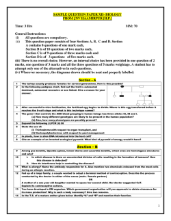
Figure II-2 : Strand breaks and base lesions as a function of hydration. Hybridized,
semi-complementary DNA, unsubstituted (DSsc AT*//AT) or substituted (DSsc
AB*//AT) with BrdU was irradiated with increasing levels of hydration (from T ~ 6 to
r ~ 31). Lanes 2, 8, 14 and 20 are unirradiated controls, whereas lanes 3, 9, 15, and 21
35
are positive controls, irradiated in solution under a nitrogen atmosphere with EDTA in
phosphate buffer. Lanes 14 to 25 were treated with hot piperidine to reveal alkalilabile DNA lesions. (Note: the portion of the gel between the wells and the parental
oligonucleotide is not shown)
RESULTS
Hydration and BrdU sensitization
We first examined the role of DNA structure in BrdU sensitization by gradually
increasing the DNA hydration level from r = 6 t o r = 31. Although BrdU is a wellknown radiosensitizer in solution (30-32) and in cells (33-35), we found no BrdUspecific strand breaks when a hybridized, semi-complementary oligonucleotide (DSsc
AB*//AT, Figure II-1) was hydrated between T ~ 6 and T = 21 (Figure II-2, lanes 4—6
and 10-12 and Figure II-3). Specific BrdU sensitization (strand breaks and ICLs) was
only found when the hydration level reached r = 31 (Figure H-2, lane 7), where there
was a 5-fold increase in damage in DNA substituted with BrdU, compared to that in
unsubstituted DNA (Figure II-3). BrdU-specific ICLs were only found at V ~ 31 (not
shown). Because it has been reported that dehalogenation (36-37), uracil-5-yl radical
production (38-41), and base fragmentation (42-43) occur in dehydrated BrdUsubstituted DNA, we decided to test whether other types of BrdU specific damage
occurred at lower hydration levels. Thus, we treated irradiated DNA with hot
piperidine to reveal alkali-labile DNA lesions.
36
5
10
15
20
Hydration (H:0/nt)
25
30
35
Figure II-3 : Damage yield as a function of DNA hydration. Strand breaks (sb; circles:
o, •) and strand breaks + alkali-labile DNA lesions (sb + als); squares: • , •) were
measured for unsubstituted DNA (DSsc AT*//AT, open symbols: o, n) and BrdUsubstituted DNA (DSsc AB*//AT, filled symbols: • , •) in phosphate buffer.
Treatment with hot piperidine revealed DNA lesions that were created between T ~
6 and T = 31 only in substituted DNA (lanes 14-19 of Figure II-2 and Figure II-3).
Although treatment with piperidine revealed low levels of DNA lesions present in
unirradiated
substituted
oligonucleotides
(Figure
II-2, lane
14),
irradiation
substantially increased the amount of DNA damage. Both strand breaks and alkalilabile lesions are quite specific for BrdU because irradiation of unsubstituted
oligonucleotides induced strand breaks in fewer than 4% of the molecules under these
conditions, compared to 16% with substituted DNA (Figure II-3). The only notable
damage not specific to BrdU was the creation of alkali-labile DNA lesions at the
central thymidine between T = 14 and T = 31 (Figure II-2, lanes 23-25). Although
BrdU-specific degradation at T ~ 31 is concentrated in the mismatch region (Figure
II-2, lane 7), leaving the double stranded portion of the oligonucleotide relatively
unharmed, it extends farther on either side of the mismatch than in oligonucleotides
irradiated in solution (lane 3). To further investigate this, we examined damage
localization as a function of DNA sequence. Panel A of Figure II-4 shows a clear bias
for strand scission on the adenine 5' of the BrdU, both in solution and in hydrated
DNA, in accordance with previous studies (24). The second adenine 5' of the BrdU
(i.e., AABrU) is also affected, albeit to a lesser extent. Damage migration 3' of the
37
BrdU was also observed, but occurred predominantly in hydrated DNA (Figure II-4,
panel A). Treatment with hot piperidine revealed that alkali-labile DNA lesions arise
virtually exclusively at the BrdU site (Figure II-4, panel B).
A
Strand breaks
O Solution
0 Hydrated
1
ii
*M .r-ga
M
Br
Mismatch zone
DNA sequence (5' to 3')
B
Strand breaks
+
Alkali-labile
lesions
• Solution
I
• Hydrated
<
z
Q
kfi
_^iL
Br
Mismatch zone
DNA sequence (5' to 3')
Figure II-4 : Damage as a function of DNA sequence. The relative yield of damage
was measured for each base of the mismatch (AABrAA, see Figure II-1 for the
complete sequence) and for two bases 5' of the mismatched, substituted
oligonucleotide (DSsc AB*//AT). Strand breaks (panel A) and strand breaks + alkalilabile DNA lesions (panel B) were measured for both solution and hydrated DNA ( r
= 31, in phosphate buffer). Base lesions and sugar damage were revealed using hot
piperidine. The background signal was removed by subtracting damage at each
nucleotide of the unsubstituted oligonucleotide (DSsc AT*//AT).
38
The role ofDNA structure in BrdU sensitization in solution
Single stranded (SS AB* and SS AT*, Figure II-l), double stranded (DSc AB*//TA
and DSc AT*//TA, Figure II-l), and semi-complementary (DSsc AB*//AT and DSsc
AT*//AT, Figure II-l) oligonucleotides were irradiated in the presence or absence of
80% ethanol to induce an A- or B-form DNA, respectively. Single stranded
oligonucleotides substituted with BrdU showed no evidence of conformation
dependent strand break formation (Figure II-5, panel A). However, for hybridized,
semi-complementary DNA, an 8-fold increase in strand breaks in the BrdU region
was observed when BrdU-DNA was in a B-form rather than in A-form (Figure II-5,
panel C). Similar results were obtained for double stranded DNA (not shown).
Treatment with hot piperidine exposed a similar tendency for alkali-labile DNA
lesions. ICLs were also specific to B-form DNA, with a 4-fold increase (Figure II-6)
compared to A-DNA. Substitution with BrdU always produced substantially more
damage than in unsubstituted oligonucleotides, under all conditions (Figure II-5,
panels B and D). Irradiation in the presence of an electron scavenger (N2O ; ke = 9.1 x
109 L mol"1 s"1 (44)) reduced the percentage of damaged molecules from 9.7 ± 0.3 to
3.7 ± 0.1% (after correction for unirradiated DNA) following a dose of 2400 Gy. In
the presence of ethanol, a hydroxyl radical scavenger (ke ~ 1.9 x 10⁹ (44)), N2O
induced a further reduction in strand break formation from 1.2 ± 0.3% to 0.4 ± 0.2%.
When DNA was irradiated with 300 Gy in the presence of another electron scavenger,
0 2 (ke = 1.9 x 10¹⁰ L mol"1 s'1 (44)), a similar reduction was observed from 3.5 ± 1.2%
with N2 to 0.7 ± 0.8% with O2. Under these latter conditions, strand break levels were
indistinguishable from unirradiated samples. At 300 Gy, addition of ethanol had no
effect on radioinduced damage (0.4 ± 0.5% and 0.4 ± 0.7% without or with O2,
respectively). Evidently, the presence of 25 mM EDTA in all samples efficiently
reduced hydroxyl radical attack because total degradation of non-substituted DNA
was less than 6% following a dose of 2400 Gy, compared to more than 60% in the
absence of any hydroxyl radical scavenger (not shown).
39
DISCUSSION
Hydration and BrdU sensitization
B
18
18
16
14
16
I
-
2 12
14
£ . 12 TJ
.2 10
T
-
.2 10
|
>.
S. 8
«
p*
V&7?.
Unirradiated
i en
r
4 -
Water
2
80% Ethanol
F?^
ryjr^yjj
Unirradiated
Water
vmtA
80% Ethanol
Single stranded unsubstituted DNA
Single stranded BrdU substituted DNA
D
30
30 T
I
25
25
b 20
•a
o
f- 15
o>
1
C 20
•o
0
CB
I 10
n
&
a
5
ITT7m
Unirradiated
T
§1
Water
V7777,
80% Ethanol
Semi-complementary BrdU substituted DNA
!
i 10
a
5
V%7/.
0
Unirradiated
Water
80% Ethanol
Semi<omplementary unsubstituted DNA
Figure II-5 : Damage yield in the mismatch zone as a function of DNA structure.
Strand breaks (hatched) and strand breaks + alkali-labile DNA lesions (plain) were
measured in the mismatch zone in the absence or presence of 80 % ethanol for single
stranded DNA with (SS AB*, panel A) or without (SS AT*, panel B) BrdU and
hybridized, semi-complementary DNA with (DSsc AB*//AT, panel C) or without
(DSsc AT*//AT, panel D) BrdU. DNA lesions (base lesions and sugar damage) were
revealed using hot piperidine. Double stranded DNA adopts an A conformation in
80% ethanol and a B conformation in aqueous solution.
When a hybridized, semi-complementary oligonucleotide is irradiated at increasing
levels of hydration, the frequency of strand breakage in BrdU-substituted DNA is
indistinguishable from non-substituted DNA between V ~ 6 and r ~ 21. Specific
BrdU sensitization (strand breaks + ICL) is only found at F ~ 31. In contrast, when
40
irradiated DNA is treated with hot piperidine, alkali-labile DNA lesions that are
specific for BrdU-substituted DNA are revealed over the entire hydration range
(Figure II-2, lanes 16-19, and Figure II-3). Such evidence of DNA lesions under low
hydration conditions is in agreement with previous results obtained under Ultra High
Vacuum (UHV) (42-43). However, in these previous studies, no evidence of strand
breaks was detected, most probably because irradiation under UHV precluded any
hydration of DNA. Our experimental system indicates that a striking change in
reactivity occurs between T = 21 and T ~ 31, leading to strand breaks as well as
alkali-labile DNA lesions when DNA is in a higher hydration state.
2,5
O Unirradiated
E B-DNA
D A-DNA
2,0 -
2 1,5
-i 1,0
o
0,5 -
0,0
DSsc AB7/AT
DSsc AT7/AT
Figure II-6 : Interstrand cross-link yield as a function of DNA structure. Interstrand
cross-links (ICLs) were measured in the absence (B-DNA) or presence (A-DNA) of
80% ethanol for mismatched DNA, brominated semi-complementary DNA (DSsc
AB*//AT), or non-brominated semi-complementary DNA (DSsc AT*//AT),
respectively.
DNA adopts an A-form between 45 ( r = 6) and 90% ( r = 20) of relative humidity,
which shifts to a B-form when hydration is increased (11-14). Because BrdU
sensitization has been shown to be extremely dependent on the hybridization state and
on the presence of a mismatched region, we propose that the change in reactivity
observed between r = 21 and F = 31 occurs because of a conversion from A-form to
B-form DNA. However, it should be noted that at T = 21, the conversion to B-form is
already relatively complete and thus one would expect to observe strand breaks at this
level of hydration, according to the hypothesis stated above. However, the hydration
41
levels reported here were not measured by us, but were assumed on the basis of the
measurement of relative humidity and the hydration levels published for these values.
The use of synthetic oligonucleotides rather than plasmid DNA may produce slightly
different levels of hydration. Plasmid or cellular DNA, even when every care is taken
to purify it, often contains contaminants (e.g., proteins and Tris buffer) originating
from the extraction protocol or the storage conditions. The presence of other
molecules could affect the measurement of the hydration levels of plasmid DNA.
Therefore, it is possible that the reason no strand breaks are observed at T ~ 21 is
because the hydration level is slightly lower than T ~ 20, where DNA would mostly
still be in A-form. For this reason, we proceeded to validate our hypothesis by
inducing an A-form DNA in solution using ethanol, to verify the effect of
conformation on DNA radiosensitization by BrdU.
Damage localization and DNA structure
It is well known that strand breaks occur predominantly at the nucleotide 5' to the
BrdU. This is because the pathway leading to strand breaks involves hydrogen
abstraction from the 2'-deoxyribose moiety of the base 5' to BrdU (45). When we
examined strand break location as a function of DNA structure, we observed a similar
tendency for both dissolved and hydrated DNA ( r = 31, Figure II-4, panel A),
although damage spread farther in the hydrated sample (Figure II-2, lane 7) than in
solution (lane 2). In the former case, the limited availability of water molecules
probably allows radicals to migrate farther along the DNA strand before being trapped
by H2O. Remarkably, piperidine-sensitive DNA lesions occurred exclusively at the
site of BrdU substitution (Figure II-4, panel B). Several factors other than
conformation may explain this observation; a higher hydration level may favor
protonation of the uracil-5-yl radical and creation of a radical cation, thus allowing
migration of the damage. However, no preference for the G's near the initial damage
site was observed, as would be expected if this were the case. In addition, the greater
mobility and wobbling afforded by a higher hydration level of DNA may facilitate
42
charge transfer of radicals to distant bases (46). This could also explain why a
Gaussian distribution of DNA damage is observed (Figure II-4, panel B) when all
types of damage (strand breaks + alkali-labile DNA lesions) are taken into account.
The increase in mobility generated by the formation of a mismatch zone (24) has
already been proposed by our group to be responsible for the increase in strand breaks
and the production of interstrand cross-links observed in mismatched DNA. Indeed,
the mobility of the DNA bases is likely to affect the ability of any radical created in
DNA to react and migrate, and higher levels of hydration greatly increase the mobility
of DNA compared to that of solid-state DNA. However, hydration also induces a
change in conformation that affects the number of potential donors and acceptors near
the radical created at the BrdU site. Therefore, it becomes extremely difficult to
distinguish between the two factors. Because several factors can indeed affect the
chemistry of solid-state DNA and for the reasons cited in the previous section, we
proceeded to examine the effect of conformation on the radiosensitization of BrdUsubstituted DNA in solution.
The role of DNA structure for BrdU sensitization in solution
It is well established that ethanol will cause a conformational change in DNA from
the canonical B-DNA to A-DNA (47). In light of our results with hydrated DNA, we
examined the effect of conformation for BrdU-substituted DNA in solution. In this
system, 25 mM EDTA (£OH = 4.0 x 10⁸ L mol"1 s"1 (48)) was added to each sample to
scavenge hydroxyl radicals and thus to reduce both random breakage of our DNA as
well as site-specific strand breakage resulting from hydroxyl radical attack on BrdU.
Therefore, the majority of strand breaks produced by irradiation of these samples were
assumed to be due to the interaction of solvated electrons with BrdU. With this
experimental system, we saw no effect of ethanol on the sensitization of single
stranded DNA by BrdU (Figure II-5, panels A and B), whereas mismatched DNA
displayed a conformation dependency similar to what was observed with the
hydration experiment, regardless of whether strand breaks (Figure II-5, panels C and
43
D) or ICLs (Figure II-6) were considered. It is possible that the interaction of ethanol
with the uracil-5-yl radical could prevent the attack of the latter on the sugar and the
subsequent creation of a strand break. However, our observation that ethanol does not
influence break formation in single stranded DNA argues against this hypothesis
(Figure II-5A). A slight reduction of alkali-labile DNA lesions was observed upon
addition of ethanol and could be attributed to a low level of remaining hydroxyl
radicals that are scavenged by ethanol. Although solvated electrons are thought to be
the primary reactive species to interact with BrdU, hydroxyl radicals can also attack
BrdU, leading to debromination and creation of a strand break (37). Because further
scavenging of these hydroxyl radicals by ethanol could explain part of the reduction in
strand breaks, we performed another experiment where DNA was again irradiated in
the presence or absence of ethanol, but also in the presence of dinitrous oxide (N2O),
an efficient electron scavenger. Although we observed a diminution in strand breaks
in both A-form and B-form DNA in the presence of N2O, we were unable to eliminate
all strand breaks produced at 2400 Gy. Because it is possible that a substantial amount
of N2O is consumed at this high dose, we lowered the dose to 300 Gy and used
oxygen, another well-known electron scavenger. Strand breaks produced in B-form
DNA were completely suppressed by irradiation in the presence of oxygen. However,
the number of strand breaks produced in A-form DNA at 300 Gy was too low to
observe an effect of the presence of oxygen. These results clearly indicate that,
although hydroxyl radicals may be responsible for a small portion of strand breaks
generated in B-form DNA at 2400 Gy, the majority of damage to BrdU-substituted
DNA indeed occurs through a reductive pathway, and that the reduction in strand
break formation observed upon addition of ethanol originates from a change in DNA
conformation. Thus, the results obtained with ethanol support those from our
hydration experiments: the type of DNA damage resulting from BrdU sensitization is
dependent on DNA conformation. Irradiation of A-DNA will induce alkali-labile
DNA lesions, whereas B-DNA will lead primarily to frank strand breaks and lower
levels of alkali-labile DNA lesions.
44
DNA structure has an important influence on the production of strand breaks. Both
simulations (49) and experimental results (50) suggest that hydroxyl radical attack is
dependent on DNA structure, both on the DNA form (A, B or Z) and on the
hybridization state (single or double stranded). Several papers also report that
chromatin structure per se affects the extent of DNA damage by ionizing radiation
(51-55). As for brominated DNA, recent evidence by Kimura et al. pointed to
enhanced reactivity in Z-DNA compared to that in B-DNA in 8-bromo-2'deoxyguanosine substituted oligonucleotides (56). Cecchini et al. (25) also presented
evidence that hybridization lowered the total yield of single strand breaks induced by
y-rays in BrdU-substituted DNA by up to 20-fold, and that substituted double stranded
oligonucleotides containing a mismatch produced ICLs. Previous reports have already
shown that DNA structure strongly influences the preference for hydrogen abstraction
on CI' or C2' by a uracil-5-yl radical generated by photoirradiation of BrdUsubstituted DNA (57-58; for a review, see 59). Thus, it is likely that DNA
conformation also affects the type of damage resulting from uracil-5-yl radical
production. In our case, the A-form that exists between T ~ 6-21 probably prevents
both the H-atom abstraction from the 2'-deoxyribose moiety and the migration of
damage along the DNA strand, precluding the formation of frank strand breaks. Thus,
the uracil-5-yl radical remains localized at the BrdU site, possibly allowing slower,
competing pathways to create other types of lesions that can be revealed by piperidine
treatment and are located exclusively at the site of BrdU substitution. However, the
structural conditions necessary for the generation of strand breaks exist in B-form
DNA at r ~ 31 and in solution. Although the nature of the alkali-labile lesions was
not determined in these experiments, it is entirely possible that they coincide with the
damage leading to the production of the base fragments observed under UHV (42-43).
Electron attachment to BrdU leads to the formation of the same uracil-5-yl radical,
whether the electron is solvated or not (38-41), although it is also possible that the
dissociative electron attachment mechanism that was proposed by Sanche's group
(60) could also yield products different from those observed in solution. Because of
the high dependence of BrdU sensitization on DNA structure, the experimental
system presented here may be a sensitive probe for DNA conformation.
45
Interstrand
/ C " ^ B-DNA <
~ .
."-> <
: breaks
—
^
Strand
Double stranded - - ^ B r d U + e " - - •>• Br" + d U ' < "
>
••-'"
Single stranded
%*_ * A-DNA
^
\_->
- - £
Alkali-labile
|esions
Figure II-7 : Major degradation pathway for BrdU-substituted DNA for single
stranded, double stranded, and mismatched DNA for A- and B-DNA. (Note: Only the
most important lesions are indicated. Minor levels of alkali-labile lesions are
generated in B-DNA.)
Figure II-7 summarizes our conclusions. We propose that, during irradiation,
electron attachment to DNA occurs in all oligonucleotides, independent of the
hybridization state and structure. However, dehalogenation and creation of the uracil5-yl, which are dependent on the probability of electron localization on BrdU, are
strongly influenced by DNA structure (25). Furthermore, once the uracil-5-yl radical
is formed, the type of damage will also depend on DNA conformation. We propose
that in A-DNA, the uracil-5-yl radical is unable to abstract a hydrogen atom from the
adjacent 2'-deoxyribose, thus localizing the radical at the site of substitution. Further
reactions with adjoining donors will lead to the creation of alkali-labile lesions that
possibly include the fragmented base that was observed by Sanche's group (42-43). In
B-DNA, the structural requirements for hydrogen abstraction exist, and strand breaks
will occur, although a certain amount of transient, unfavorable structure must also
exist because a fraction of the uracil-5-yl radical population will generate alkali-labile
DNA lesions and ICLs. B-DNA also allows the migration of the original radical to
distant bases, thereby permitting delocalized induction of strand breaks. Therefore, ADNA will produce piperidine-sensitive DNA lesions localized on BrdU, whereas BDNA will also produce strand breaks that can spread on either side of the original
radical. ICL production will be limited to mismatched DNA in B-form. Single
stranded DNA will not be affected because of its less ordered configuration in
46
solution. It is interesting to note that the reactivity of the mismatched nucleotides in
semi-complementary DNA is affected by DNA conformation. This observation
appears to favor the zipper-like structure for mismatches (61-63) rather than the open
bubble model (24), which would possibly not be affected by DNA conformation.
BrdU and DNA structure: biological implications
In light of the results presented in this article, we propose that the fate of BrdU upon
irradiation is highly dependent on the regional conformation of DNA. BrdUsubstituted DNA in A-form will only produce base lesions, as was observed in
experiments studying dehydrated DNA. Strand breaks will occur at BrdU sites when
DNA adopts a B-form, because the DNA conformation is now favorable to the steps
leading to sugar-phosphate backbone cleavage. How this hypothesis would translate
into a cellular environment is still unknown: in cells, DNA is mostly in B-form, but is
wrapped tightly around nucleosomes to form chromatin. DNA packing and chromatin
structure was shown to have an effect on radiation sensitivity (64), and some evidence
points to a similar influence on BrdU sensitization (34, 65).
Although clinical studies using BrdU as a radiosensitizer have been disappointing
up to this point, a new class of halogenated pyrimi dines has been emerging as a potent
radiosensitizer (66-67). Sensitization by chlorodeoxycytidine (CldC) relies on the
elevated levels of deoxycytidine kinase and dCMP deaminase in human tumors
compared to normal tissues. CldC is incorporated in DNA as chlorodeoxyuridine
(CldU). Because electron attachment to CldU will yield the same uracil-5-yl radical
that is responsible for BrdU sensitization, it is extremely likely that similar
sensitization mechanisms will also apply to CldC, including the production of ICLs.
Therefore, it is crucial to pursue the study of the chemical processes leading to BrdUand CldC-induced damage in order to develop and exploit tumor radiosensitizers.
47
ACKNOWLEDGMENTS We thank Drs. Jean Cadet, Tsvetan Gantchev and
Francois Bergeron for their helpful comments.
48
References
1. Dewey, W. C , and Humphrey, R. M. (1965) Increase in Radiosensitivity to
Ionizing Radiation Related to Replacement of Thymidine in Mammalian Cells with 5Bromodeoxyuridine. Radiat. Res. 26, 538-553.
2. Sano, K., Hoshino, T., and Nagai, M. (1968) Radiosensitization of Brain Tumor
Cells with a Thymidine Analogue (Bromouridine). J. Neurosurg. 28, 530-538.
3. Limoli, C. L., and Ward, J. F. (1993) A New Method for Introducing DoubleStrand Breaks into Cellular DNA. Radiat. Res. 134, 160-169.
4. Saffhill, R., and Ockey, C. H. (1985) Strand Breaks Arising from the Repair of
the 5-Bromodeoxyuridine-Substituted Template and Methyl MethanesulphonateInduced Lesions can Explain the Formation of Sister Chromatid Exchanges.
Chromosoma. 92, 218-224.
5. Dewey, W. C , Sedita, B. A., and Humphrey, R. M. (1966) Radiosensitization of
X Chromosome of Chinese Hamster Cells Related to Incorporation of 5Bromodeoxyuridine. Science. 152, 519-521.
6. Coleman, C. N., and Mitchell, J. B. (1999) Clinical Radiosensitization: Why it
does and does Not Work. J. Clin. Oncol. 17, 1-3.
7. Dabaja, B. S., McLaughlin, P., Ha, C. S., Pro, B., Meyers, C. A., Seabrooke, L.
F., Wilder, R. B., Kyritsis, A. P., Preti, H. A., Yung, W. K., Levin, V., Cabanillas, F.,
and Cox, J. D. (2003) Primary Central Nervous System Lymphoma: Phase I
Evaluation of Infusional Bromodeoxyuridine with Whole Brain Accelerated
Fractionation Radiation Therapy After Chemotherapy. Cancer. 98, 1021-1028.
8. Groves, M. D., Maor, M. H., Meyers, C , Kyritsis, A. P., Jaeckle, K. A., Yung,
W. K., Sawaya, R. E., Hess, K., Bruner, J. M., Peterson, P., and Levin, V. A. (1999) A
Phase II Trial of High-Dose Bromodeoxyuridine with Accelerated Fractionation
Radiotherapy Followed by Procarbazine, Lomustine, and Vincristine for Glioblastoma
Multiforme. Int. J. Radiat. Oncol. Biol. Phys. 45, 127-135.
9. Prados, M. D., Scott, C , Sandler, H., Buckner, J. C , Phillips, T., Schultz, C ,
Urtasun, R., Davis, R., Gutin, P., Cascino, T. L., Greenberg, H. S., and Curran, W.
J.,Jr. (1999) A Phase 3 Randomized Study of Radiotherapy Plus Procarbazine,
CCNU, and Vincristine (PCV) with Or without BUdR for the Treatment of Anaplastic
Astrocytoma: A Preliminary Report of RTOG 9404. Int. J. Radiat. Oncol. Biol. Phys.
¥5,1109-1115.
10. Prados, M. D., Seiferheld, W., Sandler, H. M., Buckner, J. C , Phillips, T.,
Schultz, C , Urtasun, R., Davis, R., Gutin, P., Cascino, T. L., Greenberg, H. S., and
Curran, W. J.,Jr. (2004) Phase III Randomized Study of Radiotherapy Plus
49
Procarbazine, Lomustine, and Vincristine with Or without BUdR for Treatment of
Anaplastic Astrocytoma: Final Report of RTOG 9404. Int. J. Radiat. Oncol. Biol.
Phys. 58, 1147-1152.
11. Goodfellow, J. M., Cruzeiro-Hansson, L., Norberto de Souza, O., Parker, K.,
Sayle, T., and Umrania, Y. (1994) DNA Structure, Hydration and Dynamics. Int. J.
Radiat. Biol. 66, 471-478.
12. Saenger, W. (1984) Principles of Nucleic Acid Structure. Springer-Verlag, New
York.
13. Wolf, B., and Hanlon, S. (1975) Structural Transitions of Deoxyribonucleic
Acid in Aqueous Electrolyte Solutions. II. the Role of Hydration. Biochemistry. 14,
1661-1670.
14. Milano, M. T., and Bernhard, W. A. (1999) The Effect of Packing and
Conformation on Free Radical Yields in Films of Variably Hydrated DNA. Radiat.
Res. 151, 39-49.
15. Milano, M. T., and Bernhard, W. A. (1999) The Influence of Packing on Free
Radical Yields in Solid-State DNA: Film Compared to Lyophilized Frozen Solution.
Radiat. Res. 152, 196-201.
16. Huttermann, J., Rohrig, M., and Kohnlein, W. (1992) Free Radicals from
Irradiated Lyophilized DNA: Influence of Water of Hydration. Int. J. Radiat. Biol. 61,
299-313.
17. Wang, W., Becker, D., and Sevilla, M. D. (1993) The Influence of Hydration on
the Absolute Yields of Primary Ionic Free Radicals in Gamma-Irradiated DNA at 77
K. I. Total Radical Yields. Radiat. Res. 135, 146-154.
18. Mroczka, N., and Bernhard, W. A. (1993) Hydration Effects on Free Radical
Yields in DNA X-Irradiated at 4 K. Radiat. Res. 135, 155-159.
19. Wang, W., Yan, M., Becker, D., and Sevilla, M. D. (1994) The Influence of
Hydration on the Absolute Yields of Primary Free Radicals in Gamma-Irradiated
DNA at 77 K. II. Individual Radical Yields. Radiat. Res. 137, 2-10.
20. Weiland, B., and Huttermann, J. (1998) Free Radicals from X-Irradiated 'Dry'
and Hydrated Lyophilized DNA as Studied by Electron Spin Resonance
Spectroscopy: Analysis of Spectral Components between 77K and Room
Temperature. Int. J. Radiat. Biol. 74, 341-358.
21. Yokoya, A., Cunniffe, S. M., and O'Neill, P. (2002) Effect of Hydration on the
Induction of Strand Breaks and Base Lesions in Plasmid DNA Films by GammaRadiation. J. Am. Chem. Soc. 124, 8859-8866.
22. La Vere, T., Becker, D., and Sevilla, M. D. (1996) Yields of OH in GammaIrradiated DNA as a Function of DNA Hydration: Hole Transfer in Competition with
OH Formation. Radiat. Res. 145, 673-680.
50
23. Debije, M. G., Strickler, M. D., and Bernhard, W. A. (2000) On the Efficiency
of Hole and Electron Transfer from the Hydration Layer to DNA: An EPR Study of
Crystalline DNA X-Irradiated at 4 K. Radial Res. 154, 163-170.
24. Cecchini, S., Girouard, S., Huels, M. A., Sanche, L., and Hunting, D. J. (2005)
Interstrand Cross-Links: A New Type of Gamma-Ray Damage in
Bromodeoxyuridine-Substituted DNA. Biochemistry. 44, 1932-1940.
25. Cecchini, S., Girouard, S., Huels, M. A., Sanche, L., and Hunting, D. J. (2004)
Single-Strand-Specific Radiosensitization of DNA by Bromodeoxyuridine. Radiat.
Res. 162, 604-615.
26. Cecchini, S., Masson, C , La Madeleine, C , Huels, M. A., Sanche, L., Wagner,
J. R., and Hunting, D. J. (2005) Interstrand Cross-Link Induction by UV Radiation in
Bromodeoxyuridine-Substituted DNA: Dependence on DNA Conformation.
Biochemistry. 44, 16957-16966.
27. Stokes, R. H., and Robinson, R. A. (1949) Standard Solutions for Humidity
Control at 25°C. Ind. Eng. Chem. 41, 2013-2013.
28. Cai, Z., Cloutier, P., Hunting, D., and Sanche, L. (2006) Enhanced DNA
Damage Induced by Secondary Electron Emission from a Tantalum Surface Exposed
to Soft X Rays. Radiation Research. 165, 365-371.
29. Maxam, A. M., and Gilbert, W. (1980) Sequencing End-Labeled DNA with
Base-Specific Chemical Cleavages. Methods Enzymol. 65, 499-560.
30. Zimbrick, J. D., Ward, J. F., and Myers, L. S.,Jr. (1969) Studies on the Chemical
Basis of Cellular Radiosensitization by 5-Bromouracil Substitution in DNA. I. Pulseand Steady-State Radiolysis of 5-Bromouracil and Thymine. Int. J. Radiat. Biol.
Relat. Stud. Phys. Chem. Med. 16, 505-523.
31. Patterson, L. K., and Bansal, K. M. (1972) Pulse Radiolysis Studies of 5Halouracils in Aqueous Solutions. J. Phys. Chem. 76, 2392-2399.
32. Adams, G. E., and Willson, R. L. (1972) On the Mechanism of BUdR
Sensitization: A Pulse Radiolysis Study of One Electron Transfer in Nucleic-Acid
Derivatives. Int. J. Radiat. Biol. Relat. Stud. Phys. Chem. Med. 22, 589-597.
33. Ling, L. L., and Ward, J. F. (1990) Radiosensitization of Chinese Hamster V79
Cells by Bromodeoxyuridine Substitution of Thymidine: Enhancement of RadiationInduced Toxicity and DNA Strand Break Production by Monofilar and Bifilar
Substitution. Radiat. Res. 121, 76-83.
34. Lawrence, T. S., Davis, M. A., and Normolle, D. P. (1995) Effect of
Bromodeoxyuridine on Radiation-Induced DNA Damage and Repair Based on DNA
Fragment Size using Pulsed-Field Gel Electrophoresis. Radiat. Res. 144, 282-287.
51
35. Webb, C. F., Jones, G. D., Ward, J. F., Moyer, D. J., Aguilera, J. A., and Ling,
L. L. (1993) Mechanisms of Radiosensitization in Bromodeoxyuridine-Substituted
Cells. Int. J. Radial Biol. 64, 695-705.
36. Abdoul-Carime, H., Huels, M. A., Briining, F., Illenberger, E., and Sanche, L.
(2000) Dissociative Electron Attachment to Gas-Phase 5-Bromouracil. J. Chem. Phys.
775,2517-2521.
37. Bansal, K. M , Patterson, L. K., and Schuler, R. H. (1972) The Production of
Halide Ion in the Radiolysis of Aqueous Solutions of the 5-Halouracils. J. Phys.
Chem. 76, 2386-2392.
38. Klyachko, D. V., Huels, M. A., and Sanche, L. (1999) Halogen Anion
Formation in 5-Halouracil Films: X Rays Compared to Subionization Electrons.
Radiat. Res. 151, 177-187.
39. Abdoul-Carime, H., Huels, M. A., Illenberger, E., and Sanche, L. (2001)
Sensitizing DNA to Secondary Electron Damage: Resonant Formation of Oxidative
Radicals from 5-Halouracils. J. Am. Chem. Soc. 123, 5354-5355.
40. Graslund, A., Rupprecht, A., Kohnlein, W., and Huttermann, J. (1981)
Radiation-Induced Free Radicals in Oriented Bromouracil-Substituted DNA. Radiat.
Res. 88, 1-10.
41. Sevilla, M. D., Failor, R., and Zorman, G. (1974) Radicals Formed After
Electron Attachment to 5-Halouracils in Aqueous Glasses. J. Phys. Chem. 78, 696699.
42. Abdoul-Carime, H., Dugal, P. C , and Sanche, L. (2000) Damage Induced by 130 eV Electrons on Thymine- and Bromouracil-Substituted Oligonucleotides. Radiat.
Res. 153, 23-28.
43. Dugal, P. -., Abdoul-Carime, H., and Sanche, L. (2000) Mechanisms for LowEnergy (0.5-30 eV) Electron-Induced Pyrimidine Ring Fragmentation within
Thymine- and Halogen-Substituted Single Strands of DNA. J. Phys. Chem. B. 104,
5610-5617.
44. Buxton, G. V., Greenstock, C. L., Helman, W. P., and Ross Alberta, B. (1988)
Critical Review of Rate Constants for Reactions of Hydrated Electrons, Hydrogen
Atoms and Hydroxyl Radicals in Aqueous Solution. J. Phys. Chem. Ref. Data. 17,
513-886.
45. Zimbrick, J. D., Ward, J. F., and Myers, L. S.,Jr. (1969) Studies on the Chemical
Basis of Cellular Radiosensitization by 5-Bromouracil Substitution in DNA. II. Pulseand Steadystate Radiolysis of Bromouracil-Substituted and Unsubstituted DNA. Int.
J. Radiat. Biol. Relat. Stud. Phys. Chem. Med. 16, 525-534.
46. Valis, L., Wang, Q., Raytchev, M., Buchvarov, I., Wagenknecht, H. A., and
Fiebig, T. (2006) Base Pair Motions Control the Rates and Distance Dependencies of
52
Reductive and Oxidative DNA Charge Transfer. Proc. Natl. Acad. Sci. U. S. A. 103,
10192-10195.
47. Ivanov, V. I., Minchenkova, L. E., Minyat, E. E., Frank-Kamenetskii, M. D.,
and Schyolkina, A. K. (1974) The B to A Transition of DNA in Solution. Journal of
Molecular Biology. 87, 817-833.
48. Lati, J., and Meyerstein, D. (1978) Oxidation of First-Row Bivalent TransitionMetal Complexes Containing Ethylenediaminetetra-Acetate and Nitrilotriacetate
Ligands by Free Radicals: A Pulse-Radiolysis Study. J. Chem. Soc, Dalton Trans. ,
1105-1118.
49. Michalik, V., Spotheim Maurizot, M., and Charlier, M. (1995) Calculation of
Hydroxyl Radical Attack on Different Forms of DNA. J. Biomol. Struct. Dyn. 13,
565-575.
50. Tartier, L., Michalik, V., Spotheim-Maurizot, M., Rahmouni, A. R., Sabattier,
R., and Charlier, M. (1994) Radiolytic Signature of Z-DNA. Nucleic Acids Res. 22,
5565-5570.
51. Warters, R. L., and Lyons, B. W. (1992) Variation in Radiation-Induced
Formation of DNA Double-Strand Breaks as a Function of Chromatin Structure.
Radiat. Res. 130, 309-318.
52. Valota, A., Ballarini, F., Friedland, W., Jacob, P., Ottolenghi, A., and Paretzke,
H. G. (2003) Modelling Study on the Protective Role of OH Radical Scavengers and
DNA Higher-Order Structures in Induction of Single- and Double-Strand Break by
Gamma-Radiation. Int. J. Radiat. Biol. 79, 643-653.
53. Olive, P. (1992) DNA Organization Affects Cellular Radiosensitivity and
Detection of Initial Strand Breaks. Int. J. Radiat. Biol. 62, 389-396.
54. Elmroth, K., Nygren, J., Stenerlow, B., and Hultborn, R. (2003) Chromatin- and
Temperature-Dependent Modulation of Radiation-Induced Double-Strand Breaks. Int.
J. Radiat. Biol. 79, 809-816.
55. Nygren, J., Ljungman, M., and Ahnstrom, G. (1995) Chromatin Structure and
Radiation-Induced DNA Strand Breaks in Human Cells: Soluble Scavengers and
DNA-Bound Proteins Offer a Better Protection Against Single- than Double-Strand
Breaks. Int. J. Radiat. Biol. 68, 11-18.
56. Kimura, T., Kawai, K., Tojo, S., and Majima, T. (2004) One-Electron
Attachment Reaction of B- and Z-DNA Modified by 8-Bromo-2'-Deoxyguanosine. J.
Org. Chem. 69, 1169-1173.
57. Sugiyama, H., Fujimoto, K., and Saito, I. (1997) Preferential CI' Hydrogen
Abstraction by a Uracilyl Radical in a DNA-RNA Hybrid. Tetrahedron Lett. 38,
8057-8060.
53
58. Kawai, K., Saito, I., and Sugiyama, H. (1999) Conformation-Dependent
Photochemistry of 5-Halouracil-Containing DNA: Stereospecific 2'a-Hydroxylation
of Deoxyribose in Z-Form DNA. J. Am. Chem. Soc. 121, 1391-1392.
59. Xu, Y., and Sugiyama, H. (2006) Photochemical Approach to Probing Different
DNA Structures. Angew. Chem., Int. Ed 45, 1354-1362.
60. Boudaiffa, B., Cloutier, P., Hunting, D., Huels, M. A., and Sanche, L. (2000)
Resonant Formation of DNA Strand Breaks by Low-Energy (3 to 20 eV) Electrons.
Science. 287, 1658-1660.
61. Chou, S. H., Chin, K. H., and Wang, A. H. (2003) Unusual DNA Duplex and
Hairpin Motifs. Nucleic Acids Res. 31, 2461-2474.
62. Kondo, J., Umeda, S., Fujita, K., Sunami, T., and Takenaka, A. (2004) X-Ray
Analyses of d(GCGAXAGC) Containing G and T at X: The Base-Intercalated Duplex
is Still Stable Even in Point Mutants at the Fifth Residue. J. Synchrotron Radiat. 11,
117-120.
63. Shepard, W., Cruse, W. B., Fourme, R., de la Fortelle, E., and Prange, T. (1998)
A Zipper-Like Duplex in DNA: The Crystal Structure of d(GCGAAAGCT) at 2.1 A
Resolution. Structure. 6, 849-861.
64. Oleinick, N. L., Balasubramaniam, U., Xue, L., and Chiu, S. (1994) Nuclear
Structure and the Microdistribution of Radiation Damage in DNA. Int. J. Radiat. Biol.
66, 523-529.
65. Iliakis, G., Kurtzman, S., Pantelias, G., and Okayasu, R. (1989) Mechanism of
Radiosensitization by Halogenated Pyrimidines: Effect of BrdU on Radiation
Induction of DNA and Chromosome Damage and its Correlation with Cell Killing.
Radiat. Res. 119, 286-304.
66. Greer, S., Alvarez, M., Mas, M., Wozniak, C , Arnold, D., Knapinska, A.,
Norris, C , Burk, R., Aller, A., and Dauphinee, M. (2001) Five-Chlorodeoxycytidine,
a Tumor-Selective Enzyme-Driven Radiosensitizer, Effectively Controls Five
Advanced Human Tumors in Nude Mice. Int. J. Radiat. Oncol. Biol. Phys. 51, 791806.
67. Perez, L. M., Mekras, J. A., Briggle, T. V., and Greer, S. (1984) Marked
Radiosensitization of Cells in Culture to X Ray by 5-Chlorodeoxycytidine
Coadministered with Tetrahydrouridine, and Inhibitors of Pyrimidine Biosynthesis.
Int. J. Radiat. Oncol. Biol. Phys. 10, 1453-1458.
For Table of Contents Use Only
Marie-Eve Dextraze, Richard J. Wagner and Darel J. Hunting. 5-Bromodeoxyuridine
radiosensitization: conformation-dependent DNA damage.
B'lnHiSS'effegl
Y-rays
*- Alkalt-labile lesions
4-
.
r'
<>
•SS-tS
/-SEES "
Strand breaks
x
—> B-DNA
Chapitre III - 2e article
Reaching for the other side: generating sequence-dependent interstrand crosslinks with 5-bromodeoxyuridine and y-rays.
Auteurs de l'article : Marie-Eve Dextraze, Sylvain Cecchini, Francois Bergeron,
Sonia Girouard, Kathleen Turcotte, J. Richard Wagner and Darel J. Hunting.
Statut de l'article : publie.
Biochemistry (2009), 48: 2005-11.
Avant-propos :
Contribution a la redaction et a I 'experimentation :
Sonia Girouard a fait les experiences d'irradiation des differentes sequences
d'ADN, sous la supervision de Sylvain Cecchini, alors etudiant au Ph.D. L'irradiation
d'ADN simple brin et l'analyse par HPLC a ete faite par Kathleen Turcotte (stagiaire)
sous ma supervision. J'ai analyse les donnees brutes et j'ai redige la totalite de cet
article, sous la supervision de mes co-directeurs, Richard Wagner et Darel Hunting.
Resume :
Dans cet article, nous presentons l'effet de la sequence sur la production de dommage
a l'ADN brome. Douze sequences ont ete etudiees, comprenant trois variations du
brin brome et quatre variations du brin semi-complementaire. A I'aide de ce modele
experimental, nous montrons que la sequence d'ADN entourant le BrdU influence
56
enormement la production de dommage a I'ADN brome. En effet, autant que le
rendement des dommages (production de cassures et de dimeres interbrins) que la
localisation du dommage est affectee par la sequence. Plus precisement, cet article
montre qu'a partir d'un radical initial, plusieurs structures de dimere interbrins
peuvent etre formees, ce qui signale une chimie complexe ou la migration de radical
pourrait etre impliquee. Cet article vient appuyer notre hypothese, qui soutient que
l'environnement ou le radical initial est cree - et plus particulierement la sequence de
I'ADN dans cet article - lors de 1'irradiation de I'ADN a un effet important sur la
letalite du dommage qui est produit.
57
Reaching for the Other Side: Generating
Sequence-Dependent Interstrand Cross-Links
with 5-Bromodeoxyuridine and y-rays
f This work was supported by the Cancer Research Society.
Marie-Eve Dextraze, Sylvain Cecchini, Francois Bergeron, Sonia Girouard, Kathleen
Turcotte, J. Richard Wagner and Darel J. Hunting*
ABBREVIATIONS
A, Adenine; BrdU, 5-bromo-2'-deoxyuridine;
Br
U, 5-
bromo-2'-deoxyuridine-substituted oligonucleotide; C, cytosine; eaq, solvated electron;
EDTA,
ethylenediammetetraacetic
acid;
FapyGua,
2,6-diamino-4-hydroxy-5-
formamidopyrimidine; HPLC, High Performance Liquid Chromatography; G,
guanine; ICL, interstrand cross-link; 2-PrOH, 2-propanol; T, thymine; TEAA,
triethyl ammonium acetate.
59
ABSTRACT
Interstrand cross-links impede critical cellular processes such as
transcription and replication and are thus considered to be one of the most toxic types
of DNA damage. Although several studies now point to the existence of y-radiationinduced cross-links in cellular DNA, little is known about the characteristics required
for their creation. Recently, we reported the formation of interstrand cross-links that
were specific for mismatched nucleotides within 5-bromo-2'-deoxyuridine-substituted
DNA. Given the structural specificity for interstrand cross-link formation, it is likely
that open or mismatched regions of DNA in cells may be particularly favorable for
cross-link production. Herein, we investigated the effect of the local DNA sequence
on the formation of interstrand cross-links, using 5-bromo-2'-deoxyuridine to generate
radicals in a mismatched region of DNA. We investigated a total of 12 variations of
bases in the mismatched region. The oligonucleotides were irradiated with y-rays, and
interstrand cross-link formation was analyzed by denaturing gel electrophoresis. We
found that the efficiency of cross-link formation was highly dependent on the nature
of mismatched bases and, on the basis of electrophoretic mobility, observed several
distinctive cross-link structures with specific DNA sequences. This study provides
new insights into the reactivity of mismatched DNA and the mechanisms leading to
interstrand
cross-link
formation.
The
potential
application
of
5-bromo-2'-
deoxyuridine-induced interstrand cross-links to the field of DNA repair is discussed.
60
Among the plethora of DNA lesions produced by ionizing radiation, double-strand
breaks are generally considered to be the most toxic (1-3). However, evidence of the
formation of another potentially more toxic type of lesion is now emerging.
Interstrand cross-links (ICLs) prevent the separation of the DNA strands, thus
blocking several key cellular processes (4, 5). Traditionally, ionizing radiation has not
been regarded as a cross-linking agent; however, several instances of intrastrand
cross-link formation have been reported (6-12), and recent data from Greenberg and
co-workers point to the existence of interstrand cross-links induced by hydroxyl
radicals in synthetic DNA (13, 14).
Although 5-bromo-2'-deoxyuridine (BrdU), a thymidine analogue that can be
incorporated into DNA, is widely used as a marker of replicating cells (15-17), it was
first reported as a photo- and radiosensitizing agent (18, 19). When exposed to
ionizing radiation, BrdU produces single- and double-strand breaks (20, 21), as well
as chromosomal aberrations (22) that are generally believed to be responsible for its
sensitizing properties. Recently, we reported the formation of a new type of BrdUsensitized damage in synthetic DNA: ICLs that were specific for mismatched
nucleotides within BrdU-substituted DNA (23). These ICLs were later found to be
highly dependent on the presence of B-form DNA, supporting the evidence that the
regional structure of DNA is a prerequisite for their formation (24) and that these
ICLs may only be produced in open or mismatched regions of cellular DNA.
Although open regions of DNA transiently exist during transcription and replication
and mismatched regions are found in centromeres (25-27), there are surprisingly few
studies asking how these regions are affected by ionizing radiation (28, 29). Given the
structural prerequisites for ICL formation observed in our studies, it is possible that
open regions of DNA may be particularly favorable for ICL production.
Here, we used BrdU, together with ionizing radiation, as a tool to generate radicals
in open regions of DNA and to examine how these regions react. To study the
reactivity of radicals with different DNA bases in an open region of DNA, we
61
modified the nucleotide sequence surrounding the site of BrdU substitution in both the
brominated and the opposite semi-complementary strands. We investigated a total of
12 mismatched oligonucleotides, using three variations of the brominated strand and
four variations of the semi-complementary strand. The yield of both ICLs and strand
breaks was found to be highly dependent on the identity of the neighboring bases.
Furthermore, we observed several distinctive ICL structures that were specific for
given sequences. This study provides new insights into the reactivity of mismatched
DNA and the mechanisms leading to interstrand cross-link formation. Finally, we
propose that, given the sequence specificity of BrdU-induced ICLs, this model system
may provide a method of producing single ICL structures for use in DNA repair
studies.
Oligonucleotide design
5' C-G-A-G-T-A-C-T-G-C-N-N-B-N-N-C-G-T-G-T-A-C-A-G-C 3'
3' G-C-T-C-A-T-G-A-C-G-N-N-N-N-N-G-C-A-C-A-T-G-T-C-G 5'
.
|
Constant region
..
Mismatch
„
||
region
||
l
Constant region
|
Mismatch sequences
Single stranded Complementary
ABrU
Br
— AABrUAA — — AA UAA —
— TT A TT —
GBrll
Br
—GGBrUGG — —GG UGG—
— CCACC —
TBrU
Br
— ATBrUTA — — AT UTA —
— TA A AT —
S jiTH-complementary
AATAA CCCCC GGGGG ATTTA
- ^ V
"X>^5^=
^ ^
-^7^"Vi/"
Figure III-1 : Oligonucleotide design. Each 25-mer sequence consisted of a variable
central 5 bp region, surrounded by two constant regions. Three variations of the
central region of the BrdU-substituted (B) strand were synthesized (AABrUAA,
GGBrUGG and ATBrUTA) and were either irradiated in single-stranded form or
62
hybridized with their complementary or one of the four semicomplementary strands to
form double-stranded of mismatched oligonucleotides, respectively. The mismatch
size is indicated for each combination.
MATERIALS AND METHODS
Oligonucleotide Design
5-Bromo-2'-deoxyuridine-modified
and
nonmodified
oligonucleotides
were
purchased from the University Core DNA Services (University of Calgary, Calgary,
AB). Oligonucleotides were designed on the basis of previous sequences used by our
group (23, 24). Each sequence included two constant regions, with a variable central 5
bp region described in Figure III-l. Three variations of the brominated sequence were
used (AABrUAA, GGBrUGG, and ATBrUTA) and were hybridized either with their
complementary sequence or with one of the semicomplementary sequences (AATAA,
CCCCC, GGGGG, ATTTA). The choice of hybridized sequences determined the
regional conformation of the DNA (double-stranded or mismatched).
Sequence-dependent interstrand cross-link and strand break formation
Control experiments were performed using single- and double-stranded DNA to
verify the hybridization specificity previously observed with AABrUAA (30). For
mismatched DNA, each combination of brominated and semicomplementary strands
was irradiated and analyzed on the same gel. Also, for each permutation, the
production of damage on each DNA strand was investigated by alternatively labeling
either the brominated or the semicomplementary strand. A comparison of interstrand
cross-link
structures
was
performed
using
the
following
guidelines.
(1)
Oligonucleotides were labeled and irradiated concomitantly and were analyzed
together on the same gel by denaturing polyacrylamide gel electrophoresis. (2) For
each sequence, an equal amount of radioactivity was loaded on the gel. (3) The
63
structures presented in Figure III-4 were taken from a single gel in which the original
alignment was conserved.
4b -i
40 •
—fc 3S SK
13
a>
30 •
J£
CO
2b •
£
?0 -
a
•a
c
15 10 -
co
5
0 AABrtJAA
GGBrUGG
ATBrUTA
Brominated strand sequence
Figure III-2 : Contribution of double strandedness to strand breaks as a function of
sequence. The three variations of the brominated strand were irradiated as singlestranded (white) or double stranded DNA (black). The yield of strand breaks in the
brominated region was measured by denaturing gel electrophoresis.
Experimental Procedures
In each experiment, the 5' end of one strand was end-labeled with
P using T4
polynucleotide kinase (Amersham Pharmacia Biotech). Hybridization was carried out
by adding a 2-fold concentration of complementary oligonucleotide, heating to 90 °C,
and slowly cooling to room temperature for 2.5 h. The hybridization state was
confirmed by nondenaturing gel electrophoresis. Prior to irradiation, DNA was
bubbled for 1 min with N2 to remove excess oxygen and then irradiated with 750 Gy
in a Gammacell-220 irradiator (60Co, Nordion Canada, dose rate of 5.08 Gy/min) at a
final concentration of 0.3 uM in phosphate buffer (10 mM, pH 7.0), using 25 mM
EDTA as a hydroxyl radical scavenger, as described by Cecchini et al. (23, 30).
Strand breaks and interstrand cross-links were assessed by denaturing gel
electrophoresis, followed by quantification of damage by the ImageQuant software, as
described by Cecchini et al. (23).
64
Conversion ofBrdU to Uracil
Conversion of BrdU to uracil in single-stranded DNA was assessed by high
performance liquid chromatography (HPLC). The three BrdU-substituted sequences
(AABrUAA, GGBrUGG and ATBrUTA) were irradiated with 750 Gy as singlestranded oligonucleotides in 10 mM phosphate buffer (pH 7.0) and 25 mM EDTA
(pH 8.0). These conditions were similar to those used in the gel electrophoresis
experiments, except that the DNA concentration was 25 uM rather than 0.3 uM. After
irradiation, DNA was digested using 3 units each of snake venom phosphodiesterase
(phosphodiesterase I, USB) and nuclease PI (Sigma) and incubated at 37 °C for 120
min. Alkaline phosphatase (3 units, Roche) was then added, and digestion was
continued for 120 min at 37 °C to generate nucleosides. Chloroform extraction was
performed to remove proteins prior to injection onto the HPLC system. HPLC
analysis was performed using an Alliance system (Waters 2795 or 2690) connected to
dual-wavelength UV detectors (Waters 2487) and a Millenium workstation (Waters
version 4). The analysis of 2'-deoxyuridine formation was carried out as follows: 150
pmol of digested DNA was separated using a reversed phase column (5um ODS-A.
250 mm x 6.0 mm; YMC). The gradient was as follows: 95% solvent Al and 5%
solvent B for 5 min, changing to 85% solvent Al and 15% solvent B over 45 min
(0.25%/min). Solvent Al was composed of 25 mM triethylammonium acetate
(TEAA; pH 7.0). Solvent B was composed of 95% acetonitrile and 5% H2O. The flow
rate was 1 mL/min, and the column was maintained at a temperature of 20 °C.
65
A
^rn
AABrUAA
GGBrUGG
ATBrUTA
Brominated strand sequence
AABrUAA
GGBrUGG
ATBrUTA
Brominated strand sequence
Figure III-3 : Damage yield as a function of sequence in mismatched DNA.
Interstrand cross-links and total cross-link plus frank strand break damage (panels A
and B, respectively) were measured following irradiation of the 12 combinations.
Each variation of the brominated strand was hybridized with each of the four
variations of the semicomplementary strand (AATAA, white; CCCCC, light gray;
GGGGG, dark gray; and ATTTA, black). Total frank damage corresponds to the sum
of interstrand cross-links and strand breaks located on both the brominated and
seTisoniipIsrnentary strand.
66
Brominated strand
AABrUAA
•o
AATAA
*
^nj^7
ccccc
~ ^4a^
GGBrUGG
s
^£5=^
ATBrUTA
""
^ia^7
(0
Sc
o
o
E
Q.
|
•
CO
GGGGG
Al
^T^ 77 ^
^a^
^°T^
^i^ 7
^i^
, ^ P 7^
^ ^a^
*
*
^ P ^7
^n^
^OH^ £ / ^ F ^
Figure III-4 : Interstrand cross-link structures and strand breaks location as a function
of sequence. For each of the mismatched sequences, the cross-link patterns and the
location of strand breaks were reported (only cross-link structures are shown on the
left). The size of the asterisk indicates the relative yield of strand break for a particular
nucleotide, while dashed lines specify the existence of possible canonical WatsonCrick bonds.
RESULTS
Contribution of Secondary Structure to Strand Break Formation as a Function of
Sequence
We first investigated the effect of adjacent bases on BrdU-related damage in singleand double-stranded complementary DNA. In agreement with previous results, Figure
III-2 shows an increase in the number of strand breaks in single-stranded compared to
double-stranded DNA containing BrdU (30). The yield of strand breaks on the
brominated strand, for both single- and double-stranded DNA, was similar for both
67
AABrUAA and GGBrUGG, whereas there was more damage in ATBrUTA in both
hybridization states. However, conversion of BrdU to 2'-deoxyuridine by irradiation
of single-stranded DNA was not significantly affected by the surrounding nucleotides
(AABrUAA, 25.3 ± 7.0%; GGBrUGG, 24.9 ± 5.9%; ATBrUTA 28.0 ± 7.8%).
Interstrand cross-links were not found in either hybridization state, regardless of the
sequence (not shown).
Sequence-Dependent Formation of Interstrand Cross-Links and Strand Breaks in
Mismatched DNA
Interstrand cross-links specific for BrdU-substituted mismatched DNA were first
reported by our group in 2005 (25), using the AABrUAA//CCCCC mismatched
oligonucleotide. Using AABrUAA//AATAA, we also showed that B-form DNA was
necessary for the production of ICLs (24). Here, we investigated the effect of several
sequences on the production of ICLs and strand breaks, to understand the role of DNA
sequence in ICL formation. Figure III-3 shows the variation in the yields of ICL and
the formation of total frank damage as a function of sequence (panels A and B,
respectively).
In
general,
pyrimidines,
either
the
brominated
or
the
semicomplementary strand, tend to produce more ICLs and strand breaks, while
guanine typically does not promote cross-linking. For the three combinations that
produced a mismatched region smaller than 5 bp or where internal Watson-Crick
bonds
could
occur;
namely,
AABlUAA//ATTTA,
GGBrUGG//CCCCC,
and
ATBrUTA//AATAA, production of ICLs and strands breaks was the lowest, with the
exception of AABrUAA//GGGGG. Table 1 shows the strand location of breaks as a
function of DNA sequence. Typically, there was no significant difference between
strand breaks produced on each strand (i.e., brominated vs. nonbrominated) of a given
combination.
Br
However,
GG UGG//GGGGG
five
permutations
diverged
from
this
pattern:
Br
and AT UTA//ATTTA produced more breaks on the
brominated strand, whereas the generation of breaks was more important in the non
brominated semicomplementary strands of GGBrUGG//AATAA, GGBrUGG//ATTTA
and AABlUAA//GGGGG. Labeling either the brominated or the semicomplementary
68
strand did not affect the yield of ICLs (not shown). Figure III-4 shows the detailed
location and frequency of these strand breaks for each strand, as well as the
corresponding ICL structures. Several distinct, sequence-dependent structures are
observed,
with
only
two
permutations
producing
no
cross-links
at
all:
Brominated
and
AABrUAA//GGGGG and GGBrUGG//CCCCC.
Tableau III-l : Localization
Semicomplementary Strands
sequence
Strand
Breaks on
strand breaks (%)
brominated
strand
AABrUAA
of
the
statistical
significance
semicomplementary
strand
15.90 ±3.66
AABrUAA//AATAA
5.60 ±0.78
5.77 ±3.39
0.9137
AABrUAA//CCCCC
6.28 ± 2.85
9.42 ± 3.64
0.2186
AABrUAA//GGGGG
1.30 ±0.30
4.53 ± 0.98
0.0004
AABrUAA//ATTTA
8.05 ± 5.87
4.45 ± 0.97
0.3395
GGBrUGG
preferred strand
(in bold)
AABrUAA//GGGGG
17.85 ±3.89
GGBrUGG//AATAA
2.90 ±1.13
6.55 ± 0.08
0.0450
GGBrUGG//CCCCC
2.25 ± 0.49
2.92 ± 0.80
0.3770
GGBrUGG//GGGGG
8.90 ±0.71
4.97 ±1.02
0.0189
GGBrUGG//GGGGG
GGBrUGG//ATTTA
3.47 ±1.86
12.91 ±2.52
0.0064
GGBrUGG//ATTTA
ATBrUTA
33.30 ±6.66
ATBrUTA//AATAA
4.85 ±1.48
4.14 ±1.89
0.6891
ATBrUTA//CCCCC
13.18 ±4.56
11.48 ±1.64
0.5725
ATBrUTA//GGGGG
6.35 ±2.87
5.51 ±1.02
0.6550
ATBrUTA//ATTTA
21.14 ±5.46
8.01 ± 4.20
0.0063
a
GGBrUGG//AATAA
/> determined by a paired Student's t test
ATBrUTA//ATTTA
69
DISCUSSION
Contribution of secondary structure to strand break formation as a function of
sequence
The interaction of ionizing radiation with DNA produces radicals through direct and
indirect processes that eventually lead to base lesions and strand breaks (3). However,
with the exception of charge transfer along the double helix, there is little evidence
that the nature of the bases surrounding the initial radical influences the type of lesion
produced (31-34). Even in tandem lesions such as intra- and interstrand cross-links,
only the bases immediately adjacent to the initial radical are involved (3, 8, 35-39).
This study provides the first evidence that the identity of the bases adjacent to the
initial radical affects both the type (i.e., strand break vs cross-links) and location of
lesions produced. Furthermore, we show that, even in single-stranded DNA, the
nature of the bases surrounding the BrdU substitution determines the extent of strand
breakage following irradiation. We previously reported the formation of strand breaks
that were specific for single-stranded oligonucleotides in BrdU-substituted DNA (30).
As expected, we observed that the same single-strand specificity first described for
strand break generation in AABrUAA also extended to GGBrUGG and ATBrUTA
(Figure III-2). More intriguing was the observation that the identity of neighboring
bases affects the production of strand breaks. Replacement of the DNA bases next to
BrdU (replacement of A with T) in AABrUAA increases the degree of formation of
strand breaks in single-stranded ATBrUTA 2-fold. Conversely, replacement of A with
G, which have oxidation potentials of 1.56 and 1.29 V at pH 7.0, respectively (3), did
not affect the production of strand breaks in single- or double-stranded DNA. Thus,
the degree of formation of single strand breaks is increased by the presence of a base
with a relatively high electron affinity (T) but is not affected by a base with a low
oxidation potential. Given the fact that T is a preferential site of electron addition in
70
normal DNA (40, 41), it is possible that T acts as an additional "electron antenna" to
capture electrons and subsequently transfer them onto BrdU. However, the level of
conversion of BrdU to 2'-deoxyuridine is only slightly (but not significantly) higher
for ATBrUTA than for AABrUAA and GGBrUGG, indicating that this step is not
limiting the production of strand breaks, and that generation of other products in
AABrUAA and GGBrUGG may explain the reduction observed in strand break yields.
Incidentally, formation of intrastrand cross-links between BrdU and adjacent purines
has been observed after UV irradiation (38). Although the mechanism for UV
sensitization of BrdU-substituted DNA involves the production of two adjacent
radicals, only one radical is produced in the case of ionizing radiation. However, it is
possible that the same products may be produced by alternative pathways, depending
on the radiation source. These results eloquently illustrate that not only the secondary
structure but also the surrounding sequence affects the ability of BrdU to damage
DNA during irradiation, influencing the type and yield of lesions produced.
Sequence-Dependent Formation of Interstrand Cross-Links and Strand Breaks in
Mismatched DNA
ICL and Strand Break Yields
Figure III-3 A shows that the efficiency of cross-link formation is highly dependent
on the nature of the mismatched bases in the brominated as well as the opposite
semicomplementary strand. These cross-links are produced with the same yield,
whether the brominated or semi-complementary strand is labeled, which indicates that
they represent interstrand cross-links, rather than interhelix cross-links. Cross-link
preferences increased in the following order: T > A ~ G and C > T > A > G i n the
brominated and semicomplementary strands, respectively. Our observation that
pyrimidines, regardless of the strand where they were located, produced more total
frank damage (Figure III-3B) supports the hypothesis that solvated electrons may be
attracted to sites containing a large number of electron affinic pyrimidines.
71
Interestingly, cytosine is present on the opposite strand of the two combinations that
produced high yields of ICLs, which points to the 5,6-double bond of cytosine as a
preferred site of attack for cross-linking (6, 42). The size of the mismatch also
influences the production of ICLs and strand breaks; the three combinations that
either produced a mismatched region smaller than 5 bp or where internal WatsonCrick
bonds
are
possible
(AABrUAA//ATTTA,
GGBrUGG//CCCCC
and
Br
AT UTA//AATAA) resulted in less damage than the other sequences. Interestingly,
AABrUAA//GGGGG also produced the least damage of any sequence, possibly
because of a particular secondary structure, as discussed below. Incidentally, the yield
of strand breaks produced in GGBrUGG//CCCCC is similar to that produced in the
complementary double-stranded counterpart, with no detectable ICL produced. This
indicates that a single mismatch in a run of GC base pairs closely resembles
complementary DNA. In contrast, the A-T rich sequences, AABrUAA//ATTTA and
ATBrUTA//AATAA, in which the Watson-Crick bonds are surrounded by mismatched
base pairs, probably produce a structure close to a typical 5 bp mismatched DNA.
More intriguing, however, is the fact that AABrUAA//GGGGG, the only other
sequence to produce no detectable cross-links and only trace levels of strand breaks,
cannot form canonical DNA base pairs. Over the years, several reports have been
published indicating that stretches of mismatched GA can form sheared base pairs,
provided that certain requirements are satisfied regarding the surrounding nucleobases
(26, 43, 44). However, these sheared base pairs were only reported for alternating
GA//AG sequences, with no indication that an AA//GG mismatch can produce such a
structure. Another possibility is that wobble G-A bonds are responsible for this
behavior. Certainly, the data presented here provide further evidence that BrdU may
serve as an internal sensor for probing DNA structure (24, 45). Moreover, our results
indicate that the reactivity of open or mismatched regions of cellular DNA to radicals,
as well as the type of damage that is produced, is probably highly dependent on the
nature of bases surrounding the initial radical.
72
Strand Break Location and ICL Structures
For BrdU-substituted DNA, the mechanism of single-strand break formation
involves attachment of an electron to BrdU, followed by departure of bromide anion,
leading to the generation of uracil-5-yl radicals. In turn, these radicals react with DNA
bases and sugar moieties to produce ICLs and strand breaks. Our results show that
strand breaks are produced on the 5' side of BrdU and at several positions on the
opposite semicomplementary strand, indicating that in mismatched DNA, uracil-5-yl
radicals are able to attack multiple sites (Figure III-4). It is unclear whether these
strand breaks arise from the direct reaction of uracil-5-yl radicals with sugar moieties
within the open duplex structure (as far as several bases away) and/or from a
multistep reaction involving the formation of base radicals in the proximity of the
initial uracil-5-yl radical, followed by transfer to the site of damage. The presence of a
relatively long mismatched region in our experimental system is likely to alter the
distance of reactive sites normally found in double-stranded DNA, which may permit
a diffusion of damage in the proximity of nascent uracil-5-yl radicals.
Although strong evidence exists that mismatched nucleotides disrupt the normal
behavior of DNA, such as charge transfer along the double helix (40, 46), little is
known about how consecutive mismatches react following treatment with ionizing
radiation. Thus, it is possible that the inherent flexibility of these DNA regions allows
reactions that are not typically seen in complementary DNA. Also, given the fact that
uracil-5-yl radicals are good hydrogen atom abstractors (k2-piOH = 4.1 x 10 dm mol"
s'1, (47)), mismatched regions of DNA may be particularly favorable for radical
transfer to distant bases, followed by strand breakage. Our observation that fewer
strand breaks occur on the brominated strand when BrdU is surrounded by guanines
(Table 1, GGBrUGG//AATAA and GGBrUGG//ATTTA) suggests that adjacent bases,
particularly guanine, interfere with the pathway leading to frank strand breaks. This
hypothesis is supported by the decreased yield of strand breaks that is observed when
purines are flanking the site of BrdU substitution, and which may be attributable to
competitive reactions such as the formation of intrastrand cross-links. Whether this
73
interference involves the intermediate formation of guanine radical cations has not
been established. Most often, these radicals do not directly lead to frank strand breaks
but give 8-oxo-7,8-dihydroguanine, which is a relatively stable lesion, and other
compounds, including 2,6-diamino-4-hydroxy-5-formamidopyrimidine
(FapyGua)
and 2,2,4-triamino-(5//)-oxazolone, which are alkali labile lesions (48) Previous
observations by Graslund et al. (49) indicated an increase in the level of guanine
cation radicals in BrdU-substituted oriented fibers, compared to unsubstituted fibers.
However, this experiment was conducted in solid state hydrated DNA in the absence
of radical scavengers. Thus, these radicals could also arise from either the direct
interaction of radiation or hydroxyl radical attack on BrdU.
If we set aside the hypothesis of radical transfer, then the distributions of strand
breaks shown in Figure III-4 must arise from the physical contact of the uracil-5-yl
radical with the damaged sugar moiety. Strikingly, in the case of AABrUAA//CCCCC
and ATBrUTA//CCCCC, bases at the 5' extremities of the mismatch are those with the
higher yield of strand breaks, with strand breaks even being produced outside the
mismatch in the case of GGBrUGG//AATAA. If these strand breaks truly reflect the
proximity of BrdU, this suggests that the structure of mismatched DNA regions is
highly dependent on the sequence. In double-stranded complementary DNA, no
strand breaks are observed on the strand opposite the BrdU. Undoubtedly, the
presence of a mismatch in our experimental system affects the number and location of
nucleotides susceptible to react, especially if mismatched regions of DNA adopt a
zipper-like structure rather than a "bubble" configuration (23, 24, 27, 50-52). In
zipper-like DNA structures, the bases interlace with their counterpart on the opposite
strand, promoting close base-base contacts that may favor the transfer of a radical to
the other strand. One of the best indications in favor of this model is the creation of
ICLs, where a radical on one strand attacks a nucleotide on the opposite strand to
generate a covalent bond.
74
Without a doubt, the most striking feature of the results presented in this study is the
diversity of ICL structures that are obtained when the bases surrounding the BrdU are
modified (Figure III-4). Overall, 10-12 distinct bands are observed, with several of
them present in multiple DNA sequences. Of these, the three-band pattern observed in
AABrUAA//CCCCC, ATBrUTA//CCCCC, and ATBrUTA//ATTTA is perhaps the most
intriguing. First, it is present in pyrimidines rich sequences. Second, at least one of
these bands is observed with a combination of other bands in four other sequences
(AABrUAA//ATTTA,
ATBrUTA//AATAA,
ATBrUTA//GGGGG
and
Br
GG UGG//GGGGG), which indicates that they must represent structures with a
higher likelihood of formation. This is supported by the higher ICL yield found in
sequences that produce this three-band pattern. Unfortunately, the chemical structure
of these cross-links cannot be inferred from their electrophoretic mobility.
Nevertheless, valuable information can be extracted from these data, especially
concerning the large number of sites that are involved in ICL formation. If, as
discussed above, we dismiss radical transfer, then ICLs can only occur when the
radical located at the site of BrdU substitution reacts with one of the five bases on the
opposite strand. Because denaturing gel electrophoresis cannot resolve symmetrical
structures with the same molecular weight, this predicts that only three different bands
should be observed on denaturing gels. Since a greater number of bands are observed,
this supports our hypothesis that transfer of a radical to adjacent nucleotides does
occur, on both the brominated and the opposite strand, which would greatly increase
the number of sites susceptible to being part of an ICL and consequently the number
of bands found on the gel. Although radical transfer has not traditionally been
considered to be part of the sensitization mechanism, the data presented here certainly
raise the question of whether transfer to distant bases is an important part of BrdU
sensitization in mismatched DNA. Undoubtedly, the identity of the surrounding bases
influences the location of strand breaks, as well as the ICL structures that are
produced in open or mismatched regions of DNA. Given the specificity we reported
for ICL formation, it is entirely possible that ICL "hot spots" exist in regions of
cellular DNA that possess the appropriate sequence and structural characteristics.
75
5-Bromodeoxyuridine: a Tool for the Study of ICL Repair?
In this study, we found that BrdU-substituted DNA, when exposed to ionizing
radiation, produces highly sequence-dependent ICLs. These ICLs are so dependent on
the nucleotide sequence that 10-12 structures result from a single initial radical. More
importantly, we were able to produce a single major ICL structure with
GGBrUGG//AATAA. This is of particular interest to the field of ICL repair, which
requires the production of single structures to study how these lesions are processed
by DNA repair and replication machinery. Furthermore, because most ICL-inducing
agents studied to date produce bulky lesions as well as mono-adducts, which can
influence repair mechanisms, it would be advantageous to investigate how cells deal
with non-bulky DNA cross-link lesions (53, 54). However, several of the agents
developed over the years to form these types of cross-links cannot be incorporated
into cellular DNA (55-57), and thus they may not adequately represent repair
mechanisms in cells. Finally, as indirect evidence of the existence of BrdU-specific
ICLs was recently found in cells (58, 59), information regarding its repair in cancer
cells would be of high interest in view of future preclinical and clinical studies (60).
76
References
1. Jeggo, P. A., and Lobrich, M. (2007) DNA Double-Strand Breaks: Their Cellular
and Clinical Impact? Oncogene. 26,11\1-11\9.
2. Jeggo, P., and Lobrich, M. (2006) Radiation-Induced DNA Damage Responses.
Radiat. Prot. Dosimetry. 122, 124-127.
3. von Sonntag, C. (2006) Free-Radical-Induced DNA Damage and its Repair.
Springer-Verlag, Berlin.
4. Noll, D. M., Mason, T. M., and Miller, P. S. (2006) Formation and Repair of
Interstrand Cross-Links in DNA. Chem. Rev. 106, 277-301.
5. Grillari, J., Katinger, H., and Voglauer, R. (2007) Contributions of DNA
Interstrand Cross-Links to Aging of Cells and Organisms. Nucleic Acids Res. 35,
7566-7576.
6. Hong, H., Cao, H., and Wang, Y. (2007) Formation and Genotoxicity of a
Guanine-Cytosine Intrastrand Cross-Link Lesion in Vivo. Nucleic Acids Res. 35,
7118-7127.
7. Regulus, P., Duroux, B., Bayle, P. A., Favier, A., Cadet, J., and Ravanat, J. L.
(2007) Oxidation of the Sugar Moiety of DNA by Ionizing Radiation Or Bleomycin
could Induce the Formation of a Cluster DNA Lesion. Proc. Natl. Acad. Sci. U. S. A.
104, 14032-14037.
8. Bellon, S., Ravanat, J. L., Gasparutto, D., and Cadet, J. (2002) Cross-Linked
Thymine-Purine Base Tandem Lesions: Synthesis, Characterization, and
Measurement in Gamma-Irradiated Isolated DNA. Chem. Res. Toxicol. 15, 598-606.
9. Jiang, Y., Hong, H., Cao, H., and Wang, Y. (2007) In Vivo Formation and in
Vitro Replication of a Guanine-Thymine Intrastrand Cross-Link Lesion|.
Biochemistry (N. Y. ) . 46, \2151-12162.
10. Zhang, Q., and Wang, Y. (2003) Independent Generation of 5-(2'Deoxycytidinyl)Methyl Radical and the Formation of a Novel Cross-Link Lesion
between 5-Methylcytosine and Guanine. J. Am. Chem. Soc. 125, 12795-12802.
11. Zhang, Q., and Wang, Y. (2005) Generation of 5-(2'-Deoxycytidyl)Methyl
Radical and the Formation of Intrastrand Cross-Link
Lesions in
Oligodeoxyribonucleotides. Nucl. Acids Res. 33, 1593-1603.
12. Gu, C , and Wang, Y. (2004) LC-MS/MS Identification and Yeast Polymerase n
Bypass of a Novel y-Irradiation-Induced Intrastrand Cross-Link Lesion G[8-5]Cf.
Biochemistry (N. Y. ) . 43, 6745-6750.
13. Ding, H., and Greenberg, M. M. (2007) Gamma-Radiolysis and Hydroxyl
Radical Produce Interstrand Cross-Links in DNA Involving Thymidine. Chem. Res.
Toxicol 20, 1623-1628.
77
14. Hong, I. S., and Greenberg, M. M. (2005) Efficient DNA Interstrand Cross-Link
Formation from a Nucleotide Radical. J. Am. Chem. Soc. 127, 3692-3693.
15. Leif, R. C , Stein, J. H., and Zucker, R. M. (2004) A Short History of the Initial
Application of Anti-5-BrdU to the Detection and Measurement of S Phase. Cytometry
A. 58, 45-52.
16. Quinones-Hinojosa, A., Sanai, N., Smith, J. S., and McDermott, M. W. (2005)
Techniques to Assess the Proliferative Potential of Brain Tumors. J. Neurooncol. 74,
19-30.
17. Eisch, A. J., and Mandyam, C. D. (2007) Adult Neurogenesis: Can Analysis of
Cell Cycle Proteins Move Us "Beyond BrdU"? Curr. Pharm. Biotechnol. 8, 147-165.
18. Dewey, W. C , and Humphrey, R. M. (1965) Increase in Radiosensitivity to
Ionizing Radiation Related to Replacement of Thymidine in Mammalian Cells with 5Bromodeoxyuridine. Radiat. Res. 26, 538-553.
19. Sano, K., Hoshino, T., and Nagai, M. (1968) Radiosensitization of Brain Tumor
Cells with a Thymidine Analogue (Bromouridine). J. Neurosurg. 28, 530-538.
20. Limoli, C. L., and Ward, J. F. (1993) A New Method for Introducing DoubleStrand Breaks into Cellular DNA. Radiat. Res. 134, 160-169.
21. Saffhill, R., and Ockey, C. H. (1985) Strand Breaks Arising from the Repair of
the 5-Bromodeoxyuridine-Substituted Template and Methyl MethanesulphonateInduced Lesions can Explain the Formation of Sister Chromatid Exchanges.
Chromosoma. 92, 218-224.
22. Dewey, W. C , Sedita, B. A., and Humphrey, R. M. (1966) Radiosensitization of
X Chromosome of Chinese Hamster Cells Related to Incorporation of 5Bromodeoxyuridine. Science. 152, 519-521.
23. Cecchini, S., Girouard, S., Huels, M. A., Sanche, L., and Hunting, D. J. (2005)
Interstrand Cross-Links: A New Type of Gamma-Ray Damage in
Bromodeoxyuridine-Substituted DNA. Biochemistry. 44, 1932-1940.
24. Dextraze, M. -., Wagner, J. R., and Hunting, D. J. (2007) 5-Bromodeoxyuridine
Radiosensitization: Conformation-Dependent DNA Damage. Biochemistry. 46, 90899097.
25. Ferrer, N., Azorin, F., Villasante, A., Gutierrez, C , and Abad, J. P. (1995)
Centromeric Dodeca-Satellite DNA Sequences Form Fold-Back Structures. J. Mol.
Biol. 245, 8-21.
26. Huertas, D., and Azorin, F. (1996) Structural Polymorphism of Homopurine
DNA Sequences. d(GGA)n and d(GGGA)n Repeats Form Intramolecular Hairpins
Stabilized by Different Base-Pairing Interactions. Biochemistry. 35, 13125-13135.
78
27. Shepard, W., Cruse, W. B., Fourme, R., de la Fortelle, E., and Prange, T. (1998)
A Zipper-Like Duplex in DNA: The Crystal Structure of d(GCGAAAGCT) at 2.1 a
Resolution. Structure. 6, 849-861.
28. Elmroth, K., Nygren, J., Stenerlow, B., and Hultborn, R. (2003) Chromatin- and
Temperature-Dependent Modulation of Radiation-Induced Double-Strand Breaks. Int.
J. Radiat. Biol. 79, 809-816.
29. Waiters, R. L., and Lyons, B. W. (1992) Variation in Radiation-Induced
Formation of DNA Double-Strand Breaks as a Function of Chromatin Structure.
Radiat. Res. 130, 309-318.
30. Cecchini, S., Girouard, S., Huels, M. A., Sanche, L., and Hunting, D. J. (2004)
Single-Strand-Specific Radiosensitization of DNA by Bromodeoxyuridine. Radiat.
Res. 162, 604-615.
31. Edfeldt, N. B., Harwood, E. A., Sigurdsson, S. T., Hopkins, P. B., and Reid, B.
R. (2004) Sequence Context Effect on the Structure of Nitrous Acid Induced DNA
Interstrand Cross-Links. Nucleic Acids Res. 32, 2795-2801.
32. Giese, B. (2002) Electron Transfer in DNA. Curr. Opin. Chem. Biol. 6, 612-618.
33. Giese, B., Amaudrut, J., Kohler, A. K., Spormann, M., and Wessely, S. (2001)
Direct Observation of Hole Transfer through DNA by Hopping between Adenine
Bases and by Tunnelling. Nature. 412, 318-320.
34. Wagenknecht, H. A. (2006) Electron Transfer Processes in DNA: Mechanisms,
Biological Relevance and Applications in DNA Analytics. Nat. Prod. Rep. 23, 9731006.
35. Box, H. C , Dawidzik, J. B., and Budzinski, E. E. (2001) Free Radical-Induced
Double Lesions in DNA. Free Radic. Biol. Med. 31, 856-868.
36. Kovanda, J., Kejnovsky, E., Arnold, L., and Kypr, J. (1996) UV Light-Induced
Crosslinking of Short DNA Duplex Strands: Nucleotide Sequence Preferences and a
Prominent Role of the Duplex Ends. J. Biomol. Struct. Dyn. 14, 57-65.
37. Nejedly, K., Kittner, R., Pospisilova, S., and Kypr, J. (2001) Crosslinking of the
Complementary Strands of DNA by UV Light: Dependence on the Oligonucleotide
Composition of the UV Irradiated DNA. Biochim. Biophys. Acta. 1517, 365-375.
38. Zeng, Y., and Wang, Y. (2006) Sequence-Dependent Formation of Intrastrand
Crosslink Products from the UVB Irradiation of Duplex DNA Containing a 5-Bromo2'-Deoxyuridine Or 5-Bromo-2'-Deoxycytidine. Nucleic Acids Res. 34, 6521-6529.
39. Zeng, Y., and Wang, Y. (2007) UVB-Induced Formation of Intrastrand CrossLink Products of DNA in MCF-7 Cells Treated with 5-Bromo-2'-Deoxyuridine.
Biochemistry. 46, 8189-8195.
79
40. Wagenknecht, H. A. (2003) Reductive Electron Transfer and Transport of
Excess Electrons in DNA. Angew. Chem. Int. Ed Engl. 42, 2454-2460.
41. Fuciarelli, A. F., Sisk, E. C , and Zimbrick, J. D. (1994) Electron Migration in
Oligonucleotides upon Gamma-Irradiation in Solution. Int. J. Radiat. Biol. 65, 409418.
42. Zhang, Q., and Wang, Y. (2004) Independent Generation of the 5-Hydroxy-5,6Dihydrothymidin-6-Yl Radical and its Reactivity in Dinucleoside Monophosphates. J.
Am. Chem. Soc. 126, 13287-13297.
43. Heus, H. A., Wijmenga, S. S., Hoppe, H., and Hilbers, C. W. (1997) The
Detailed Structure of Tandem G.A Mismatched Base-Pair Motifs in RNA Duplexes is
Context Dependent. J. Mol. Biol. 271, 147-158.
44. Li, Y., Zon, G., and Wilson, W. D. (1991) Thermodynamics of DNA Duplexes
with Adjacent G.A Mismatches. Biochemistry. 30, 7566-7572.
45. Xu, Y., Tashiro, R., and Sugiyama, H. (2007) Photochemical Determination of
Different DNA Structures. Nat. Protoc. 2, 78-87.
46. Boon, E. M., and Barton, J. K. (2003) DNA Electrochemistry as a Probe of Base
Pair Stacking in A-, B-, and Z-Form DNA. Bioconjugate Chem. 14, 1140-1147.,
47. Mertens, R., and Sonntag, C. v. (1994) Determination of the Kinetics of Vinyl
Radical Reactions by the Characteristic Visible Absorption Spectra of Vinylperoxyl
Radicals. Angewandte Chemie International Edition in English. 33, 1262-1264.
48. Cadet, J., Douki, T., Gasparutto, D., and Ravanat, J. L. (2003) Oxidative
Damage to DNA: Formation, Measurement and Biochemical Features. Mutat. Res.
531, 5-23.
49. Graslund, A., Rupprecht, A., Kohnlein, W., and Huttermann, J. (1981)
Radiation-Induced Free Radicals in Oriented Bromouracil-Substituted DNA. Radiat.
Res. 88, 1-10.
50. Chou, S. H., Chin, K. H., and Wang, A. H. (2003) Unusual DNA Duplex and
Hairpin Motifs. Nucleic Acids Res. 31, 2461-2474.
51. Chou, S. H., and Chin, K. H. (2001) Zipper-Like Watson-Crick Base-Pairs. J.
Mol. Biol. 312, 753-768.
52. Chou, S. H., and Chin, K. H. (2001) Solution Structure of a DNA Double Helix
Incorporating Four Consecutive Non-Watson-Crick Base-Pairs. J. Mol. Biol. 312,
769-781.
53. Berardini, M., Mackay, W., and Loechler, E. L. (1997) Evidence for a
Recombination-Independent Pathway for the Repair of DNA Interstrand Cross-Links
Based on a Site-Specific Study with Nitrogen Mustard. Biochemistry. 36, 3506-3513.
80
54. McHugh, P. J., Spanswick, V. J., and Hartley, J. A. (2001) Repair of DNA
Interstrand Crosslinks: Molecular Mechanisms and Clinical Relevance. Lancet Oncol.
2, 483-490.
55. Alzeer, J., and Scharer, O. D. (2006) A Modified Thymine for the Synthesis of
Site-Specific Thymine-Guanine DNA Interstrand Crosslinks. Nucleic Acids Res. 34,
4458-4466.
56. Noll, D. M., Noronha, A. M., Wilds, C. J., and Miller, P. S. (2004) Preparation
of Interstrand Cross-Linked DNA Oligonucleotide Duplexes. Front. Biosci. 9, 421437.
57. Wilds, C. J., Noronha, A. M., Robidoux, S., and Miller, P. S. (2005) Synthesis
and Characterization of DNA Duplexes Containing an N3T-Ethyl-N3T Interstrand
Crosslink in Opposite Orientations. Nucleosides Nucleotides Nucleic Acids. 24, 965969.
58. Wojcik, A., Bochenek, A., Lankoff, A., Lisowska, H., Padjas, A., Szumiel, I.,
von Sonntag, C , and Obe, G. (2006) DNA Interstrand Crosslinks are Induced in Cells
Prelabelled with 5-Bromo-2'-Deoxyuridine and Exposed to UVC Radiation. J.
Photochem. Photobiol. B. 84, 15-20.
59. Bruckmann, E., Wojcik, A., and Obe, G. (1999) X-Irradiation of Gl CHO Cells
Induces SCE which are both True and False in BrdU-Substituted Cells but Only False
in Biotin-dUTP-Substituted Cells. Chromosome Res. 7, 277-288.
60. Wardman, P. (2007) Chemical Radiosensitizers for use in Radiotherapy. Clin.
Oncol. (R. Coll. Radiol). 19, 397-417.
81
For Table of Contents Use Only
Marie-Eve Dextraze, Sylvain Cecchini, Francois Bergeron, Sonia Girouard, Kathleen
Turcotte, J. Richard Wagner and Darel J. Hunting. Reaching for the other side:
generating sequence-dependent interstrand cross-links with 5-bromodeoxyuridine and
y-rays.
y-rays
_»*»••«<*-_
— 4 m * -
Interstrand
cross-links
£r" %
_
__
<*#S*
^
AA B 'UAA GGB'IK3G AT<"UTA
Chapitre IV - 3e article
Mapping interstrand cross-links induced by gamma and UVB radiation in
bromodeoxyuridine-substituted DNA.
Auteurs de l'article : Marie-Eve Dextraze, J. Richard Wagner and Darel J. Hunting
Statut de l'article : En preparation.
Avant-propos
Contribution a la redaction et a I 'experimentation :
J'ai effectue la totalite des experiences presentees dans cet article. J'ai aussi
redige cet article, sous la supervision de mes co-directeurs, Richard Wagner et Darel
Hunting.
Resume :
Cet article presente une methode visant a identifier l'emplacement des dimeres
interbrins formes lorsque l'ADN brome est soumis aux radiations. En effet, nos
experiences precedentes ont montre que pres d'une douzaine de bandes differentes
etaient observees lorsque l'ADN brome irradie etait analyse par electrophorese, ce qui
represente au moins une douzaine de structures distinctes potentielles. Grace a cette
methode, les dimeres sont transformes en cassures, ce qui permet de determiner leur
emplacement dans l'ADN. Ainsi, nous avons etabli que les dimeres formes etaient
hautement dependants de la sequence entourant le BrdU et la source de radiation
83
utilisee pour produire les dimeres. Les resultats montrent clairement que le transfert
de radicaux est possible dans un ADN mesapparie. Les resultats de cette etude
suggerent aussi, pour la premiere fois, que le radical cation forme lors de I'irradiation
de l'ADN brome avec des rayons UV est implique dans la formation du dimere.
84
Mapping interstrand cross-links induced by
gamma and UVB radiation in
bromodeoxyuridine-substituted DNA
f This work was supported by the Natural Sciences and Engineering Research
Council
Marie-Eve Dextraze, J. Richard Wagner and DarelJ. Hunting*
Center for Research in Radiooncology (CR2), Department of Nuclear Medicine and
Radiobiology, Faculty of Medicine, Universite de Sherbrooke, Quebec, Canada JIH
5N4
85
ABSTRACT
Recently, we reported the formation of interstrand cross-links
that were specific for mismatched nucleotides within bromodeoxyuridine-substituted
DNA that had been exposed to ionizing and UV radiation. Given that several bands
were observed when the cross-links were analyzed by denaturing gel electrophoresis,
we proposed that these bands represented distinct structures formed by a single initial
radical. To further study the formation of interstrand cross-links in mismatched DNA,
we submitted the purified cross-links to formic acid and piperidine treatment, thus
transforming the cross-linked bases into strand breaks, so that their location could
then be mapped using denaturing gel electrophoresis. Using this method, we observed
that bases adjacent to the site of the initial radical were involved in the cross-links,
which supports the hypothesis that radical transfer occurs in mismatched DNA, and is
highly dependent on sequence.
86
Double-strand breaks are generally considered to be one of the most toxic types of
DNA lesions produced by ionizing radiation (1-3). However, evidence of the
formation of another potentially more toxic type of lesion, interstrand cross-links, is
now emerging. Interstrand cross-links (ICLs), where the two DNA strands are
covalently linked, prevent the separation of the DNA strands, thus blocking several
key cellular processes (4-6). Traditionally, ionizing radiation has not been regarded as
a cross-linking agent; however, several instances of intrastrand cross-link formation
have been reported and recent data points to the existence of interstrand cross-links
induced by hydroxyl radicals in synthetic DNA and in cells (7, 8). Nevertheless, there
is yet very little information regarding ICL formation and repair in cells that have
been subjected to ionizing radiation.
5-bromo-2'-deoxyuridine (BrdU) is a thymidine analogue that can be incorporated
into cellular DNA. Although it is now widely used as a marker of replicating cells (9,
10), it was first reported as a photo- and radiosensitizing agent (11, 12). When
exposed to ionizing radiation, BrdU produces single- and double-strand breaks, as
well as chromosomal aberrations that are generally believed to be responsible for its
sensitizing properties (13-15). The mechanism for single strand break formation
involves electron attachment to BrdU, followed by the departure of a bromide anion
and generation of an uridin-yl radical that further reacts to create strand breaks (16).
Recently, we reported the formation of a new type of BrdU-sensitized damage in
synthetic DNA: ICLs that were specific for mismatched nucleotides within BrdUsubstituted DNA (17). These ICLs were later found to be highly dependent on the
presence regional DNA structure and sequence (18, 19), supporting the evidence that
a precise regional environment was a prerequisite for their formation and that these
ICLs may only be produced in open or mismatched regions of cellular DNA.
Furthermore, when the effect of the surrounding sequence was studied, several bands
with distinct electrophoretic mobility were observed in the region corresponding to
the ICL, outlining the possibility that several ICL structures were formed by a single
initial radical.
87
In the present study, we modified a method developed by Millard et al. (20) to
determine the location of ICLs in BrdU-substituted DNA exposed to ionizing
radiation. The ICLs were purified by preparative denaturing gel electrophoresis, and
then were treated with cold formic acid and hot piperidine, thus transforming the
cross-linked sites into strand breaks. This protocol allows us to map the precise
location of the ICLs. To validate the protocol, we applied it to Mitomycin-induced
cross-links already described in the study by Millard, and confirmed that the observed
location of the cross-link with our method was the same as originally reported.
Moreover, the sequence preference was in good agreement with the literature. Finally,
we applied this method to 3 of the 12 BrdU-substituted sequences originally studied.
Furthermore, we compared the ICLs produced under gamma and UV radiation as a
function of sequence. We found that the sites involved in the cross-linking process are
highly dependent on the surrounding sequence, as well as the type of radiation used.
Furthermore, our results substantiate the hypothesis that radical transfer does occur in
consecutive mismatches, further supporting our proposition that mismatched or open
regions of cellular DNA are hotspots for ICL formation.
88
MATERIALS AND METHODS
Oligonucleotide design
5' C G A G T A C T G C N u N 12 Bj3 N14 N^ C G T G T A C A G C 3'
3' G-C T-C-A-T-G-A-C-G-N^-N^-N^-N^-Nn-G-C-A-C-A T-G-T-C-G 5'
|
Constant region
|
Mismatch region
Mismatch sequences
AABAA // AATAA
|
Constant region
|
Cross-link structures
AA B UAA
AATAA
AA S UAA GGB'UGG
CCCCC
AATAA
AABAA//CCCCC
GGBGG//AATAA
^^yL
Figure IV-1: Oligonucleotide design. Each 25-mer sequence consisted of a variable
central 5 bp region, surrounded by two constant regions. The ICLs formed following
irradiation of three mismatch sequences were investigated, with the ICL bands
observed by denaturing gel electrophoresis shown right.
Oligonucleotides
5-Bromodeoxyuridine
modified
and
nonmodified
oligonucleotides were purchased from the University Core DNA Services (University
of Calgary, AB, Canada). Oligonucleotides were designed on the basis of previous
sequences used by our group. Each sequence included two constant regions, with a
variable central 5 bp region described in Figure IV-1. DNA was end-labeled either
with
P [y-ATP] using T4 polynucleotide kinase (Amersham Pharmacia Biotech) for
5' labeling, or
P [a-ATP] using terminal transferase (Roche) for 3' labeling.
Labeling was carried at an initial oligonucleotide concentration of 2 \xM. for 45min at
37 °C with 10 U of enzyme. The enzyme was inactivated by heating for 10 min at 75
°C. Oligonucleotides were then purified on a G50 Sephadex microcolumn and the
89
labeling efficiency was assessed by counting lul of the labeling reaction with a
RackBeta 1219 liquid scintillation counter (LKB Wallac). Hybridization was carried
out at a final concentration of 1.4 uM of the labeled strand and a 2-fold excess of the
unlabeled strand in a volume of 56ul. Samples were heated to 82 °C for 5 min, then
cooled slowly for 2.5h. Deionized, sterile water was used in all experimental
protocols.
Preparation of mitomycin C-cross-linked DNA
Cross-links
were
prepared
as
described in details by Millard et al (20). Briefly, 100 pmol of DNA was radiolabeled,
as described above. Afterwards, 16 pmol of labeled DNA was hybridized with 76
pmol of complementary DNA and hybridized by heating to 82 °C for 5 min, then
cooled slowly for 2.5h and lyophilized. The double stranded DNA was then
resuspended in 100 uL of 15 mM Tris (pH 7.5) and 10 JJ,L of mitomycin C (20 mM in
33% aqueous methanol) and incubated for 1 h at 37 °C. The samples were then placed
in a septa covered tube and deoxygenated by bubbling through with nitrogen for 15
min, before being put on ice. Activation of mitomycin C was carried out by 3
additions of 6 uL of fresh sodium hydrosulfite at 15 minutes intervals. The sodium
hydrosulfite solution was prepared by flushing 0.0087 g of sodium hydrosulfite
(Sigma) in a septa-covered tube with nitrogen, then by adding 1.5 mL of
deoxygenated water (bubbled for 25 min with argon) with a syringe through the
septum, for a final concentration of 33 mM. The sodium hydrosulfite solution was
rapidly vortexed and used within 1 min. Fifteen minutes after the last addition, 3ul of
glycerol was added and the cross-linked DNA was loaded onto the preparative gel.
Irradiation
For gamma-irradiation, 50ul of 5' labeled, hybridized DNA, 3ul of
EDTA 0.5M (used as a hydroxyl radical scavenger) and 3ul of 100 mM phosphate
buffer, pH 7.5, was bubbled for 2 min with N2 to remove excess oxygen.
Oligonucleotides were given 2400 Gy, either with a Gammacell 220 (60Co, Nordion
Canada, 2.6 Gy/min) or with a Gammacell 3000 Elan ( lj7 Cs, Best Theratronics, 12.1
Gy/min). For UV irradiation, 50ul of hybridized DNA and 3ul of 100 mM phosphate
90
buffer (pH 7.5) was irradiated with 10 J/cm for 90 min using a 1000 W Hg-Xe arc
lamp (Oriel Corp.) fitted with an infrared filter and monochromator set at 313 ±2 nm
(Spectral Energy). The results for each experiment were confirmed by performing the
experiment with the 3' ends labeled.
Cross-link purification by preparative gel electrophoresis Interstrand cross-links and
undamaged DNA were separated by preparative denaturing gel electrophoresis.
Briefly, irradiated or mitomycin-treated samples were loaded onto a 19.5 cm x 20.5
cm denaturing gel (IX TBE buffer, 20% acrylamide and 7M urea), where a reversed
comb had been used to create a single well. Migration was carried out at 20 W for
~lhl5, or until bromophenol blue migrated to 10cm of the bottom of the gel. The gel
was then exposed for at least 2h in a Phosphor Screen cassette (Molecular Dynamics,
Inc.), and was scanned with a fluorescence scanner (Storm, Molecular Dynamics,
Inc.) The bands corresponding to the ICLs and parental strand were excised and
recuperated in 1.5 mL tubes. The gel plugs were then flash-frozen in liquid nitrogen
and crushed with a pestle. Sterile water (1 mL) was added to each tube, and samples
were vortexed twice (4 hours and overnight, respectively) in order to extract the DNA.
Each time, the mixture of eluted DNA and acrylamide was centrifuged for 5 min
before the DNA supernatant was recuperated. Samples were desalted using either two
consecutive homemade Sephadex G-50 columns or a single Illustra NAP-25 column
(GE Healthcare), and then dried using a Speedvac evaporator.
Mild hydrolysis treatment and gel analysis
The
purified
DNA
was
resuspended in lOOul sterile water and the recuperation efficiency was assessed by
counting lul with a LKB scintillation counter. Unirradiated single stranded DNA,
purified ICLs, as well as the irradiated parental strand was submitted to the treatment.
The mild hydrolysis treatment was as follow: for each reaction, 20 000 cpm was
diluted in a final volume of 20ul. Afterwards, 50ul of formic acid (J.T. Baker, 88%)
was added to the chilled tubes, with a 5 min incubation period on ice. Sample were
then lyophilized with a Speedvac evaporator and washed once with 30ul distilled
91
water to remove the remaining formic acid. Subsequently, 20ul of 10% piperidine was
added and the DNA was incubated for 30min at 90°C. Samples were then lyophilized
and three water chases were performed by adding sequentially 30ul, 30ul and 20 ul
distilled water and evaporating to dryness. Control experiments using only the cold
formic acid and hot piperidine treatments were also loaded on the same gel. Following
the hydrolysis treatment, the analysis was performed by classical denaturing gel
electrophoresis (IX TBE buffer, 20% acrylamide, 7M urea, 35 x 43 cm) using a DNA
ladder generated by chemical sequencing (G + A reaction).
RESULTS
Method validation: Mitomycin C
The increased reactivity found in an open region of DNA following irradiation was
first reported by our group in 2009, where we showed that a single radical created in a
5-base mismatched region created up to twelve distinct bands when ICLs production
was examined by denaturing gel electrophoresis (19). We studied twelve sequence
combinations, and found that in a given sequence, the initial radical produced several
bands, some of which were also observed in other sequences. At that time, we
proposed that these bands represented different structures, and that this was evidence
that radical migration occurred between the site of the initial radical and surrounding
nucleotides, which then lead to ICL production. However, it was impossible to
confirm this by gel electrophoresis. Given the large number of possible structures, we
decided to characterize the cross-links by gel electrophoresis using their sensitivity to
mild acidic and basic conditions as criteria for locating their position in
oligonucleotides. To this end, the analysis of cross-links induced by BrdU was
accomplished using a method first developed by Hopkins in 1991.
92
5'AACCTATAA [TTCG^ATATTGjyCGniCT] ATAATTA3'
3'GAT ATT [ AAGC^TATAAC^GC^GAl TATTAATTAA5'
B
Upper sequence
Lower sequence
Figure IV-2: Method validation. The use of the mild hydrolysis protocol to locate the
cross-linked sites was validated with cross-link with a well-known structure. Three
cross-linking sites were present at G13, G19, and G21 (panel A, on the upper strand).
When the mild hydrolysis protocol was applied, these sites were further processes as
strand breaks, and appeared as shorter fragments on a denaturing gel electrophoresis.
The relative yield is shown for both on the upper strand and lower strand (panel B,
upper and lower panel, respectively).
To validate the method proposed in this paper, we applied it to an ICL with a
chemistry and sequence preference that was well-known and confirmed by both gel
electrophoresis and mass spectrometry. Mitomycin C is a chemotherapeutic agent that
reacts with two adjacent guanines upon reductive activation (21-23), and is one of the
cross-linking agents initially investigated by Millard et al. Thus, we applied our mild
hydrolysis protocol to cross-links that had been induced by mitomycin C within one
of the sequences that had been used originally.
93
Figure IV-2 (panel A) shows the sequence used to validate our method using
mitomycin C-induced cross-links. Three 5'-CpG-3' sites are included (at positions 13,
19 and 21 on the upper strand, respectively), which represent three separate possible
interstrand cross-linking sites. Panel B shows the bases that were revealed by the mild
hydrolysis treatment, which reflect the bases that are involved in the ICLs. As
expected, the three potential cross-linking sites show increased reactivity, independent
of the strand that is labeled, although the reactivity of the G21 site was closer to the
background level when the upper strand was investigated. Furthermore, in good
agreement with the results presented by Hopkins, we obtained ratios of 1.3:1 and
2.6:1 for the reactivity between the 5'-GCGC and the 5'-TCGA sites (for the upper
and lower strands, respectively), compared to 2:1 in the literature (24). Thus, the
mitomycin-induced ICL responds to the mild hydrolysis treatment, revealing the sites
of the cross-linked guanines, as well as the existence of a preferred cross-linking
sequence.
Ionizing irradiation
To study the reactions involved in the cross-linking process, three of the twelve
sequences we had previously studied were chosen, each with a distinctive band
pattern. AABrUAA//AATAA was chosen because of the distinctive pattern of bands
on gel, as well as the fact that these bands were for the most part unique to this
sequence. AABrUAA//CCCCC was chosen because of the three-band ICL pattern,
whose bands were most commonly found among the other sequences and which
possibly
represented
structures
with
a
high
probability
of
formation.
GGBrUGG//AATAA was chosen because of its single major band, since it would be a
likely target for future structure determination. Furthermore, these sequences allowed
us to examine the effect of variations in the brominated and opposite strand on the
chemistry of ICL formation. •
94
AABAA//AATAA
1
AABAA // CCCCC
.••III
i
I
I
I
1
1
1
I
r v • *- v
I
ICsmatch Sequence
o "1
GGBGG // AATAA
UMM^T
'
I
I
I
I
I
I
I
I
Mnmatch Sequence
o •
iLjJlll* uillUL
Mil I I I ill li Sequence
C . O O O O O O O O
•Bsmatch Sequence
Figure IV-3: Cross-linked sites as a function of sequence (ionizing radiation). The
mild hydrolysis protocol was applied to purified ICLs formed after exposition of the
three mismatched sequences (left, AABrUAA//AATAA; center, AABrUAA//CCCCC;
right, GGBrUGG//AATAA) to ionizing irradiation. The degradation profile is shown
for the brominated strand (upper line) and the opposite strand (lower line) for 5'
labeled oligos.
Figure IV-3 shows the location of cross-linking sites in each of the three chosen
sequences when ionizing radiation was used. While several sites on the brominated
strand are involved in the cross-linking process in the case of AABrUAA//AATAA,
only one major site is observed for AABrUAA//CCCCC. As for GGBrUGG//AATAA,
while two notable sites are observed on the brominated strand, in both cases the base
react with the 3' (AH) adenine on the opposite strand. This allows the identification of
the major band observed on gel as a cross-link between B13-A14 and the minor band as
G12-A14. Furthermore, the G12-A14 ICL was found to lose one of its four arms, thus
converting it from a X to a Y type structure (see supporting information).
When the reactivity of the different ICLs to the hydrolysis treatment was investigated,
two different profiles were observed. For all but one of the sequence, no damage
modification was observed on gels when DNA was treated with formic acid only, i.e.
I
95
no additional cross-link unhooking or phosphodiester bond cleavage was found (in the
form of parental strand or strand break increase, respectively). Only in the case of
AABrUAA//AATAA treated with y-rays did formic acid lead to an increase in the
proportion of the parental strand (figure IV-4). Formic acid led to the unhooking of
the ICLs, which then created further strand breaks when the piperidine treatment was
applied. However, when this sequence was irradiated with UV rays, the behavior was
similar to the other sequences, with no discernable reaction to formic acid.
AABAA//AATAA
so*^
• i ICL
^M
^ 60Q
I40- i
2? 20A_
•
•
Parental strand
Strand breaks
•
•
•
ICL
Parental strand
Strand breaks
_
1nl a In
1
's s s
•
Treatment
,-
GGBGG//AATAA
_
II,,Ion lon
<"°
•
60
I40 '
I "
* 20-
In_JrL
y
Tireafment
Figure IV-4: Overall treatment profile for AABrUAAZ/AATAA and
GGBrUGG//AATAA. The relative yield of each form of DNA (ICL, parental strand,
DNA fragments) is shown after each step of the mild hydrolysis treatment was applied
Br,
to purified ICLs. Only in the case of AA UAA//AATAA does the ICLs react to the
96
formic acid treatment, which appears as additional parental strand. Neither
AABrUAA//CCCCC nor GGBrUGG//AATAA showed any increase in damage upon
exposure to formic acid (only GGBrUGG//AATAA is shown).
UV irradiation
AABAA//AATAA
AABAA // CCCCC
O
© o v- v- • v- *-
O
Hsmatch Sequence
(
I
O
V- V- •
V- VHsmatcti Sequence
C
I
O
O
G
O
O
GGBGG // AATAA
Hsmatch Sequence
C
I
Hsmatch Sequence
Figure IV-5: Cross-linked sites as a function of sequence (UV radiation). The mild
hydrolysis protocol was applied to purified ICLs formed after exposition of the three
mismatched sequences (left, AABrUAA//AATAA; center, AABrUAA//CCCCC; right,
GGBrUGG//AATAA) to UV rays. The degradation profile is shown for the
brominated strand (upper row) and the opposite strand (lower row) for 5' labeled
oligos.
Although UV radiation also induces strand breaks and ICLs in BrdU-substituted
DNA, the reaction mechanism is different from that of ionizing radiation. For
example, in the case of ionizing radiation, fewer strand breaks are found in perfectly
complementary double stranded DNA compared to single stranded DNA, whereas the
opposite is found with UV rays. Thus, we proceeded to examine how the type of
radiation changed the ICL chemistry. Figure IV-5 shows the location of cross-linking
97
sites in each of the three chosen sequence when UV radiation was used. Both in the
case of AABrUAA//AATAA and AABrUAA//CCCCC, the site of the BrdU
substitution was the major site of cross-linking on the brominated strand. Although
the cross-linking sites on the opposite strand varied, in both cases the guanine at the 5'
extremity of the bubble (Gio) was extremely reactive. On the other hand, when 4
guanines were present on the brominated strand in GGBrUGG//AATAA, the Gio site
disappeared and reactivity was increased in the brominated strand, with a primary site
at A14 on the opposite strand, similar to what was observed for y-rays.
DISCUSSION
Method validation: Mitomycin C
In order to analyze products formed by radiation or other types of damaging agents,
the typical procedure is to identify the damage by mass spectrometry, and in the case
of unknown damage, subsequently confirm the structure with larger amounts of
damage by nuclear magnetic resonance (NMR) (25). However, this approach may be
difficult because of the number and low yield of potential products in model systems
such as oligonucleotides containing BrdU. In addition, it is necessary to digest
modified oligonucleotides to single components in order to efficiently separate and
detect them by mass spectrometry, and thus, the position of the damage in the
oligonucleotide is sacrificed during analysis. In 1991, Millard et al. first reported a
method to identify the sites of cross-linking in mitomycin C-induced ICLs. The
heterogeneous mixture of cross-links was studied by first purifying ICLs on a
preparative gel and then submitting each one to random cleavage by hydroxyl
radicals. Thus, the sites where a decrease in strand breakage was observed
corresponded to the cross-linked sites. However, this method only allowed for indirect
identification of the cross-linking sites, and could not be applied if several sites were
98
in close proximity. For example, the cross-links in the two nearby guanines of the 5'GCGC site could not be easily resolved. Thus, we modified this method to reveal only
the bases on each strand that had been damaged in the cross-linking process.
As expected, figure IV-2 shows that the three cross-link sites respond to the mild
hydrolysis treatment, revealing cross-links at each guanine on the upper and lower
strands. In this case, the two cross-links of the 5'-GCGC region become easily
distinguishable, indicating that this method is suitable even when the potential crosslinking sites are in close proximity. However, it is important to note that the mild
hydrolysis protocol may not be applicable to every type of ICL. For example,
treatment of an ICL formed when DNA is exposed to high doses of ionizing radiation
may reveal damage that was not initially exposed, such as base lesions. Furthermore,
given that the mild hydrolysis response of the G21 site on the upper strand was
relatively close to the background level, it was crucial to validate these results by
labeling either the opposite strand or by labeling the 3' end as well as the 5' end.
Nevertheless, we believe that the method proposed in this paper has the potential to
serve as a powerful probe to determine the site of cross-links and give initial
information about their acid and base lability.
Ionizing radiation
When our group first observed that an impressive number of bands were produced
from a single initial radical in a mismatched DNA, we proposed that these bands
corresponded to distinct structures, and that they were evidence that radical migration
occurred between the site of the BrdU substitution and the surrounding nucleotides.
Strikingly, AABrUAA//CCCCC represents what was expected if no radical transfer
occurred between the bases; a major cross-linking site at the site of the initial radical
creation on the brominated strand, which reacts with each of the bases on the opposite
strand (figure IV-3). The fact that bases on the 5' side of the mismatch are favored
99
probably reflects close structural proximity to the BrdU site, as the strand break
pattern is similar (19).
In the case of AABrUAA//AATAA and GGBrUGG//AATAA, however, BrdU is not
the only cross-linking site on the brominated strand. The 5' guanine of
GGBrUGG//AATAA is strongly involved in the cross-linking process, as are several
nucleotides in AABrUAA//AATAA. As the mild hydrolysis protocol reveals only the
bases that were modified by the cross-linking process, this provides clear evidence
that the initial radical is able to migrate to surrounding nucleotides before it reacts
with the other strand to create an ICL. It is important at this point to clarify that the
cross-linking sites discussed here do not represent intrastrand cross-links, as the
purification step separated them from the ICLs using denaturing gel electrophoresis.
However, when the hydrolysis protocol was applied to the parental strand, (i.e. DNA
that had the same molecular weight as the undamaged DNA) a strong response was
always found for the BrdU site and the 5' base (not shown). Whether these represent
intrastrand cross-links or damage that had not completely gone through the strand
breakage process remains unclear. Although intrastrand cross-links were already
reported by Wang in BrdU-DNA irradiated with UV (26), it has yet to be established
clearly if it the case for ionizing radiation. However, our observation that radical
transfer occurs between the bases on the brominated strand certainly substantiates this
possibility.
In addition to revealing the nucleotides involved in the cross-links, the mild
hydrolysis protocol presented here reveals additional information concerning the ICLs
that were formed
for each sequence. For example, the G12-A14 ICL in
GGBrUGG//AATAA transforms from a four-armed "X" structure to a three armed
"Y" structure, where a strand break is created but the two strands remain covalently
linked. In contrast, the major ICL of GGBrUGG//AATAA, the B I3 -Ai 4 ICL, results in
the degradation of the cross-link and the disconnection of the two strands. This
implies that two drastically different ICL structures are formed, and that the ICL
100
formed between Gn and A H in GG rUGG//AATAA is different from the ones formed
in AABrUAA//AATAA. This may result from either a reactivity specific to guanine
rather than purines, or a difference in the structural environment. Incidentally, the
mild hydrolysis protocol also revealed that the ICLs formed in AABrUAA//AATAA
also possessed distinct characteristics, as they were the only ones observed to react
upon treatment with formic acid (figure IV-4). Whether this indicates that the ICLs
are reverting under acid conditions or that base release occurs will have to be
investigated. Certainly, the information obtained by this experiment sheds light not
only on the location, but also on the chemical nature of the cross-links, which will aid
in their identification.
UV irradiation
The major difference between irradiation of BrdU-substituted DNA by ionizing or UV
radiation is the source of the electron that reacts with BrdU to create the uracil-5-yl
radical. Whereas with gamma rays the electron is a product of water radiolysis, in the
case of UV radiation, it is the base 5' to the BrdU that provides the electron (16, 27).
This means that, in addition to the uracil-5-yl radical, a radical cation is created. This
difference is responsible for the drastic change in reactivity between the two types of
radiation for BrdU-substituted DNA that we already reported (17, 28, 29). This
evidence is also highlighted here by the contrasts in the results presented in figures
IV-3 and IV-5.
Strikingly, both in AABrUAA//AATAA and AABrUAA//CCCCC, the guanine just
outside the bubble at the 5' extremity (Gio) was extremely reactive. Given the fact that
guanine has a low oxidation potential, it is entirely possible that the radical cation
created on A12 transfers to the Gio guanine on the other strand. Whether the preference
for the 5' guanine reflects a preferred transfer route or close structural proximity
101
remains unclear. However, the hypothesis that radical cation transfer to guanine
occurs is reinforced by the results obtained from irradiation of GGBrUGG//AATAA,
where guanine sinks are present on the brominated strand. In this case, the reactivity
on the Gio base disappears; instead, the guanines surrounding the BrdU become
highly reactive. This implies not only that the radical cation produced by irradiation of
BrdU-substituted DNA is able to transfer to distant bases within a mismatch, but also
that this transfer is involved in the cross-linking process. Given that, in a similar
experimental setup, only intrastrand cross-links were found when complementary
BrdU-substituted DNA was studied (26), this represents further evidence that
mismatched regions of DNA display increased reactivity (30).
Conclusion
In this study, we mapped the bases involved in ICL formation of BrdU-substituted
DNA exposed to ionizing radiation. We observed that the identity of the nucleotides
surrounding the initial radical had a dramatic effect on the end products. The mild
hydrolysis ICL mapping treatment allowed us to probe further into the cross-linking
mechanism observed in BrdU-substituted DNA, and provides clear evidence of
radical transfer in a mismatched DNA for the first time. In addition, it also helped
identify prospective targets among a number of structures for a subsequent structure
study. This protocol, which requires only minute amounts of purified material ( <10
ng), can be used to probe the structure of a mixture of ICLs that are located in close
proximity, or to examine sequence preference. We believe that this method will be a
useful tool to gather further knowledge regarding the formation of interstrand crosslinks.
102
Supporting information:
Identification of the ICL degrading into a Y-cross-link upon hydrolysis treatment
5' labeling
3' labeling
«••{ i l | |
P„e„,a, «.d -[ ^ ^
Unlreated
m^^
Ireated
li
#
^
ICLs
#
Untreated
|
# i N
|y ^
Mcn6
I reated
Figure IV-S 1: Formation of Y cross-links following mild hydrolysis treatment. The
damaged species that appeared following treatment of purified ICLs migrated
between the ICLs (upper region on gel) and the parental strand (lower region). These
bands where present when the ICL was labeled at the 5' end, as well as when the
opposite strand was labeled on the 3' end, but not when the brominated strand was
labeled at the 3' end, and was identified as a Y cross-link.
When the purified ICLs produced from irradiation of GG UGG//AATAA were
treated and analyzed by gel electrophoresis, formation of an additional band was
observed between the ICLs and the parental strand. This band was present when the
ICL was labeled at the 5' end, as well as when the opposite strand was labeled on the
3' end, but not when the brominated strand was labeled at the 3' end (figure IV-S1).
Because of this, the band was identified as a Y cross-link, with the 5' arm of the
103
brominated strand still linked to the opposite strand, while the 3' arm had been
severed by the treatment.
This implied that the site of the break could be identified by the length of the 3'
fragment. The strand break profiles following treatment of the purified ICL labeled at
the 5' (left) and at the 3' (right) end is shown in figure IV-S2, panel A.
h
f«-
jjjllHfT-r iiWilli***
c
a
» •
o
*
&
*
ft
a
o
A
S 4-
A
B°
—»
—
Figure IV-S2: Identification of Y cross-link. The strand break profile is shown in
panel A for the 5' (left) and 3' (right) labeled brominated (upper row) or opposite
strand (lower row). The normalized profile is shown in panel B for the brominated
strand labeled at the 5' (left) and 3' end (right).
Given the shape of the Y cross-link observed following treatment, we deduced that
the additional fragment is only observed when the 3' arm of the brominated strand is
analyzed (upper right quadrant of figure IV-S2, panel A), with the results from the 5'
labeled DNA showing only the degradation profile. In order to facilitate the
comparison between the 5' and the 3' labeled DNA, we normalized the profiles using
the yield observed at the BrdU substitution (shown in figure IV-S2B). From this, we
see that the guanine 5' of the BrdU is the most likely candidate as the site of breakage
leading to the formation of the Y cross-link.
104
References
1. Jeggo,P.A. and Lobrich,M. (2007) DNA double-strand breaks: Their cellular and
clinical impact? Oncogene, 26, 7717-7719.
2. Jeggo,P. and Lobrich,M. (2006) Radiation-induced DNA damage responses.
Radiat. Prot. Dosimetry, 122,124-127.
3. von Sonntag,C. (2006) Free-radical-induced DNA damage and its repair.
4. Grillari,J., Katinger,H. and Voglauer,R. (2007) Contributions of DNA interstrand
cross-links to aging of cells and organisms. Nucleic Acids Res., 35, 7566-7576.
5. Rothfuss,A. and Grompe,M. (2004) Repair kinetics of genomic interstrand DNA
cross-links: Evidence for DNA double-strand break-dependent activation of the
fanconi anemia/BRCA pathway. Mol. Cell. Biol, 24, 123-134.
6. SczepanskiJ.T., Jacobs,A.C, Van Houten,B- and Greenberg,M.M. {2009) Doublestrand break formation during nucleotide excision repair of a DNA interstrand crosslink. Biochemistry, 48, 7565-7567.
7. Ding,H. and Greenberg,M.M. (2007) Gamma-radiolysis and hydroxyl radical
produce interstrand cross-links in DNA involving thymidine. Chem. Res. Toxicol, 20,
1623-1628.
8. HongT.S. and Greenberg,M.M. (2005) Efficient DNA interstrand cross-link
formation from a nucleotide radical. J. Am. Chem. Soc, 127, 3692-3693.
9. Leif,R.C, Stein,J.H. and Zucker,R.M. (2004) A short history of the initial
application of anti-5-BrdU to the detection and measurement of S phase. Cytometry
A., 58, 45-52.
10. Quinones-Hinojosa,A., Sanai,N., Smith,J.S. and McDermott,M.W. (2005)
Techniques to assess the proliferative potential of brain tumors. J. Neurooncol, 74,
19-30.
11. Dewey,W.C. and Humphrey,R.M. (1965) Increase in radiosensitivity to ionizing
radiation related to replacement of thymidine in mammalian cells with 5bromodeoxyuridine. Radiat. Res., 26, 538-553.
12. Sano,K., Hoshino,T. and Nagai,M. (1968) Radiosensitization of brain tumor cells
with a thymidine analogue (bromouridine). J. Neurosurg., 28, 530-538.
13. Limoli,C.L. and Ward,J.F. (1993) A new method for introducing double-strand
breaks into cellular DNA. Radiat. Res., 134,160-169.
14. Saffhill,R. and Ockey,C.H. (1985) Strand breaks arising from the repair of the 5bromodeoxyuridine-substituted template and methyl methanesulphonate-induced
lesions can explain the formation of sister chromatid exchanges. Chromosoma, 92,
218-224.
15. Dewey,W.C, Sedita,B-A. and Humphrey,R.M. (1966) Radiosensitization of X
chromosome of Chinese hamster cells related to incorporation of 5bromodeoxyuridine. Science, 152,519-521.
16. Schyman,P., Zhang,R.B., Eriksson,L.A. and Laaksonen,A. (2007) Hydrogen
abstraction from deoxyribose by a neighbouring uracil-5-yl radical. Phys. Chem.
Chem. Phys., 9, 5975-5979.
105
17. Cecchini,S., Girouard,S., Huels,M.A., Sanche,L. and Hunting,D.J. (2005)
Interstrand cross-links: A new type of gamma-ray damage in bromodeoxyuridinesubstituted DNA. Biochemistry, 44, 1932-1940.
18. Dextraze,M.-., Wagner,J.R. and Hunting,D.J. (2007) 5-bromodeoxyuridine
radiosensitization: Conformation-dependent DNA damage. Biochemistry, 46, 90899097.
19. Dextraze,M.E., Cecchini,S., Bergeron,F., Girouard,S., Turcotte,K., Wagner,J.R.
and Hunting,D.J. (2009) Reaching for the other side: Generating sequence-dependent
interstrand cross-links with 5-bromodeoxyuridine and gamma-rays. Biochemistry, 48,
2005-2011.
20. Millard,J.T., Weidner,M.F., KirchnerJ.J., Ribeiro,S. and Hopkins,P.B. (1991)
Sequence preferences of DNA interstrand crosslinking agents: Quantitation of
interstrand crosslink locations in DNA duplex fragments containing multiple
crosslinkable sites. Nucleic Acids Res., 19,1885-1891.
21. Scharer,O.D. (2005) DNA interstrand crosslinks: Natural and drug-induced DNA
adducts that induce unique cellular responses. ChemBioChem, 6, 27-32.
22. Noll,D.M., Mason,T.M. and Miller,P.S. (2006) Formation and repair of interstrand
cross-links in DNA. Chem. Rev., 106,277-301.
23. Noll,D.M., Noronha,A-M., Wilds,C.J. and Miller,P.S. (2004) Preparation of
interstrand cross-linked DNA oligonucleotide duplexes. Front. Biosci., 9,421-437.
24. Borowy-Borowski,H., Lipman,R. and Tomasz,M. (1990) Recognition between
mitomycin C and specific DNA sequences for cross-link formation. Biochemistry, 29,
2999-3006.
25. Wang,Y. and Wang,Y. (2003) Structure elucidation of DNA interstrand cross-link
by a combination of nuclease PI digestion with mass spectrometry. Anal. Chem., 75,
6306-6313.
26. Zeng,Y. and Wang,Y. (2006) Sequence-dependent formation of intrastrand
crosslink products from the UVB irradiation of duplex DNA containing a 5-bromo-2'deoxyuridine or 5-bromo-2'-deoxycytidine. Nucleic Acids Res., 34,6521-6529.
27. Cook,G.P. and Greenberg,M.M. (1996) A novel mechanism for the formation of
direct strand breaks upon anaerobic photolysis of duplex DNA containing 5bromodeoxyuridine. J. Am. Chem. Soc, 118, 10025-10030.
28. Cecchini,S., Girouard,S., Huels,M.A., Sanche,L. and Hunting,D.J. (2004) Singlestrand-specific radiosensitization of DNA by bromodeoxyuridine. Radiat. Res., 162,
604-615.
29. Cecchini,S., Masson,C, La Madeleine,C, Huels,M.A., Sanche,L., Wagner,J.R.
and Hunting,D.J. (2005) Interstrand cross-link induction by UV radiation in
bromodeoxyuridine-substituted DNA: Dependence on DNA conformation.
Biochemistry, 44, 16957-16966.
30. Dextraze,M., Gantchev,T., Girouard,S. and Hunting,D. (2010) DNA interstrand
cross-links induced by ionizing radiation: An unsung lesion. Mutation
Research/Reviews in Mutation Research, 704, 101-107.
Chapitre V - 4e article
DNA interstrand cross-links induced by ionizing radiation: an unsung lesion.
Auteurs de l'article : Marie-Eve Dextraze, Tsvetan Gantchev, Sonia Girouard and
Darel J. Hunting.
Statut de l'article: publie
Mutation Research (2010), 704 : 101-7.
Avant-propos :
Contribution a la redaction et a I 'experimentation :
J'ai redige 95 % de cet article sous la supervision de mon directeur de
recherche. Tsvetan Gantchev a redige une partie de la section 1.2 qui porte sur la
reparation des dimeres interbrins, en plus de la section 4.2, qui decrit ses travaux de
recherche. J'ai aussi ete responsable de la preparation des figures et de la mise en
page, en plus d'avoir revise l'article en fonction des commentaires des arbitres.
Resume :
Cet article est une revue de litterature presentant les resultats des derniers travaux de
recherche dans notre laboratoire, incluant les resultats obtenus durant ma these. De
plus, nous presentons une revue de litterature concernant ce qui est connu sur la
formation de dimeres interbrins par les radiations ionisantes dans l'ADN cellulaire.
Nous presentons aussi notre hypothese selon laquelle la structure secondaire et
107
tertiaire de l'ADN cellulaire est susceptible d'induire des dommages inattendus, dont
les dimeres interbrins. Cette hypothese est reprise plus en detail dans les sections
discussion et conclusion de ma these.
DNA mterstrand cross-links induced by
ionizing radiation: An unsung lesion
Marie-Eve Dextraze, Tsvetan Gantchev, Sonia Girouard and Darel Hunting*
Center for Research in Radio-Oncology (CR2) Department of Nuclear Medicine and
Radiobiology, Faculty of Medicine and Life Sciences, Universite de Sherbrooke
3001, 12e Avenue Nord, Sherbrooke J1H 5N4, Canada
109
Abstract
The induction of DNA interstrand cross-links by ionizing radiation has been largely
ignored in favour of studies on double-strand break formation and repair. At least part
of the problem is technical; it is difficult to detect and quantify interstrand cross-links
when the same agent forms both cross-links and single strand breaks because the
detection of interstrand cross-links generally involves a denaturation step. Our group
has studied the induction of interstrand cross-links following irradiation of DNA
containing bromouracil at specific sites. We found that the formation of interstrand
cross-links requires the presence of a few (3-5) mismatched bases, comprising the
bromouracil. In the absence of mismatched bases, no radiation-induced cross-linking
was observed; however, even in the absence of bromouracil, cross-linking still
occurred, albeit at a lower efficiency. Our molecular modelling studies demonstrate
that the mobility of the bases in the mismatched region is essential for the crosslinking process. Thus, our hypothesis is that ionizing radiation induces DNA
interstrand cross-links in non-hybridized regions of DNA. Some obvious examples of
such DNA regions are replication forks, transcription bubbles and the D-loop of
telomeres. However, an abundance of studies have made it clear that there must be
many single stranded regions in the genome, such as hairpins and cruciforms. For
example, alpha satellite DNA, in centromere regions of human chromosomes, forms
hairpins. Thus, a variety of non-B DNA structures (hairpins, slipped DNA and
tetrahelical structures) exist in the genome and should be susceptible to the formation
of radiation induced interstrand cross-links. Although interstrand cross-links have thus
far been virtually ignored in radiation biology, it will be worthwhile to develop
methods to detect their presence following exposure of cells to biologically relevant
levels of ionizing radiation, since, on a per lesions basis, they are probably more toxic
than double strand breaks.
110
Contents
1. Interstrand cross-links vs. double strand breaks
1.1. Double strand breaks induced by ionizing radiation
1.2. Interstrand cross-links induced by ionizing radiation
1.3. Technical difficulties
2. BrdU: a good idea which failed...
3. Generating interstrand cross-links with BrdU and y-rays
4. Reaching for the other side: a case for flexibility?
4.1. ICL formation in BrdU-substituted and unsubstituted DNA
4.2. Mismatches facilitate electron addition to BrdU and charge transfer along the
DNA
4.3. Single mismatches and y-radiation
4.4. Consecutive mismatches and y-radiation
5. Higher-order chromatin structure and DNA damage
Ill
1.
Interstrand cross-links vs. double strand breaks
1.1
Double strand breaks induced by ionizing radiation
Among the numerous types of DNA lesions presently attributed to ionizing radiation,
complex lesions, including double strand breaks (DSBs1), are generally considered to
be the most toxic [1-3]. Double strand breaks arise either from a single radical leading
to the formation of lesions on both strands or, more probably, from two separate
strand breaks in close proximity [4]. Although the yield of double strand breaks in
cells that are submitted to low LET radiation is much lower than single strand breaks
and base lesions (40/Gy vs. 2000/Gy, respectively, [5], most of the toxicity of
ionizing radiation is currently attributed to DSBs. The repair of double strand breaks
require a complex repair mechanism that is not yet fully understood, despite
continuous effort and decades of experiments [5,6]. Moreover, defective or
incomplete repair of DSBs leads to deletions, insertions, as well as chromosome
rearrangements, which can trigger cell death, or lead to oncogenesis [7]. Thus, it is not
so surprising that DSBs are usually the focal point of DNA damage studies. However,
strong evidence for the formation of another potentially more toxic type of lesion by
ionizing radiation is now emerging.
1.2
Interstrand cross-links induced by ionizing radiation
Agents that induce interstrand cross-links (ICLs), where the two DNA strand are
covalently linked, have been used to treat cancer for many years. Indeed, 60 % of the
most potent genotoxic anticancer agents are bifunctional alkylating agents that are
capable of forming ICLs [8]. One of the first ICLs to be identified was formed by
mustard gas (l,5-dichloro-3-thiapentane), which was developed prior to the First
Abbreviations:
BrdU, 5-Bromo-2'-deoxyuridine; CldC, 5-Chlorodeoxycytidine;
CldU, 5-Chlorodeoxyuridine; DSBs, Double strands breaks; ET, Electron transfer;
Gy, Gray; HMG, High mobility group proteins; ICLs, Interstrand cross-links; PCV,
Procarbazine, lomustine and vincristine; UVB, Ultraviolet B.
112
World War. Another example of an ICL-inducing agent used in clinic is mitomycin C,
which is extracted from Streptomyces caespitosis, and is used to treat solid tumors
containing hypoxic regions [8,9]. Because interstrand cross-links prevent DNA strand
separation, their presence impedes DNA replication and transcription [10].
Furthermore, there is growing evidence that ICL repair involves the generation of a
DSB during the unhooking process [11,12]. These lesions are therefore extremely
cytotoxic and mutagenic. In addition, over the past decade there is a growing
appreciation that ICLs induced by chemical or oxidative agents present a challenge to
the cellular DNA repair systems. Most of the information regarding cross-link repair
was gathered using psoralens and UVA radiation. Although the majority of ICLs
appear to be substrates for the nucleotide excision repair (NER) pathway [13], which
specializes in the removal of bulky lesions in locally distorted DNA, often the repair
is blocked or of low efficiency [14-16]. The different ICL repair efficiencies have
been found to depend on various factors [8,17,18], including the individual ICL
chemical structure (e.g. the spacer length in (bis)cis-Pt and nitrogen mustard) [19],
DNA sequence [20] and DNA local dynamic structure and flexibility [21]..There is
very little information about repair of gamma-radiation induced ICL per se, since the
only relevant studies concern a tandem lesion comprised of a single strand break and a
DNA cross-link, but it is not yet clear if the cross-link is mostly intra- or interstrand
[22]. A recent study from the group of Greenberg showed that this same ICL can be
transformed into a DSB by UvrABC, but no detailed information exists regarding
repair in eukaryotes [12].
Many of the studies on repair of interstrand cross-links used cells from Fanconi
anemia patients. Fanconi anemia is a rare, autosomal, recessive human disease that is
characterized by a hypersensitivity to ICL-inducing agents such as mitomycin C
[11,23-25].
Several genes have been identified that are involved in the initial
unhooking step, including XPF-ERCCl [26], however a global picture of the repair of
ICLs remains elusive, perhaps due to the number of ICLs structures that are studied.
Several approaches have been devised to study ICLs, one of which is the use of
chemically synthesized ICLs for use in repair studies [17,27]. Traditionally, ionizing
113
radiation has not been regarded as a cross-linking agent; however, several instances of
intrastrand cross-link formation have now been reported [22,28-33], and recent data
from various groups points to the existence of interstrand cross-links induced by
hydroxyl radicals in synthetic DNA [34,35]. This is most intriguing, considering that
Fanconi anemia patients sometimes suffer from ionizing radiation hypersensitivity
[36,37].
1.3
Technical difficulties
There are several technical hurdles associated with the study of ICL formation by an
agent that also produce high yields of strand breaks, as is the case of ionizing
radiation. Typical methods used in ICL detection, such as the alkaline comet assay
and alkaline elution [38,39], are not sensitive when the same agent produces both
cross-links and strand breaks. These methods most often rely on the inability of the
DNA strands to separate under alkaline conditions due to the presence of the crosslinks, resulting in a decrease of electrophoretic mobility, for example. However, the
strand breaks that are concomitantly produced by the radiation also decrease the
length of the DNA fragments, thus increasing their electrophoretic mobility and
masking the effect of the ICLs. Moreover, the conditions used to separate the strands
also expose alkali-labile lesions, which are transformed into strand breaks, further
increasing this effect.
Other techniques, such as high performance
liquid
chromatography and mass spectrometry, have proven most useful in the identification
of several types of lesions [40-42], including ICLs formed by ionizing radiation
[22,35]. These methods are highly sensitive, and can be used to study extracts of
cellular DNA that has been exposed to ionizing radiation. However, two technical
difficulties are associated with these techniques. First, the extraction protocol may
modify the irradiation products, leading to the uncoupling of the cross-linked
nucleotides, which will then appear as two modified bases. This issue, which is now
well known in the case of other radiation products such as 8-oxoguanine, can certainly
be minimized by careful control of the extraction and digestion conditions [43]. The
other difficulty is that is it somewhat complex to confirm, after a potential lesion has
114
been found, whether the lesion observed is in fact an interstrand cross-link rather than
simply an intrastrand cross-link [22].
For these reasons, our current knowledge of the biological significance of radiationinduced cross-links as well as the underlying mechanisms is very limited in
comparison to the panoply of mechanisms actually occurring in cells that are
submitted to radiation injury. Thus, the need is pressing to better understand how this
type of lesion is produced and repaired in normal and tumor cells, and how this
knowledge can be used to gain an advantage in radiotherapy. For this, new methods
will have to be developed in order to study ICLs that are formed concomitantly with
other types of lesions, such as strand breaks and alkali-labile lesions.
2.
BrdU: a good idea which failed
5-bromo-2'-deoxyuridine (BrdU) is a thymidine analog that can be incorporated into
DNA by cells. It is now widely used as a marker of replicating cells [44-46], although
it was first reported as a photo- and radiosensitizing agent [47,48]. When exposed to
ionizing radiation, BrdU produces DNA single and double strand breaks [49,50], as
well as chromosomal aberrations [51] that are generally believed to be responsible for
its sensitizing properties. The mechanism for single strand break formation involves
electron attachment to BrdU, followed by the departure of a bromide anion and
generation of an uridin-yl radical that further reacts to create strand breaks [52].
Originally, the appealing aspect of BrdU sensitization was that the thymidine analog
was incorporated during DNA replication, and incorporation into DNA was a
prerequisite for sensitization [53]. Thus, theoretically, rapidly cycling tumor cells
would incorporate more BrdU and be more sensitized than the surrounding, mostly
quiescent, normal cells. However, clinical studies have given disappointing results,
failing to show a survival advantage for patients with a range of tumor types [54-57].
More precisely, a phase III study conducted in 2004 on patients diagnosed with
115
anaplastic astrocytoma using BrdU in conjunction with procarbazine, lomustine and
vincristine (PCV) showed that addition of BrdU showed no survival advantage [58].
Thus, clinical studies using BrdU as a radiosensitizer are not currently a high priority.
BrdU has become, as Szybalski presciently described it in 1974 [59]: "a good idea
which failed". Nevertheless, the relatively low toxicity of BrdU as well as its rare
ability to directly radiosensitize DNA makes it worthy of further studies.
Furthermore, a new class of halogenated pyrimidines, 5-chlorodeoxycytidine (CldC)
has been found to be a potent radiosensitizer but an understanding of the sensitization
mechanisms remains critical to the success of any radiosensitizer in the clinic.
Radiosensitization by CldC relies on the elevated levels of deoxycytidine kinase and
dCMP deaminase found in tumor cells rather than simply on increased incorporation
in rapidly cycling cells [60,61], thus circumventing problems associated with the
possible incorporation of BrdU in rapidly cycling normal cells surrounding the tumor,
which results in loss of specificity. However, CldC is incorporated into DNA as 5chlorodeoxyuridine (CldU), and will almost certainly yield the same uridin-yl radical
responsible for DNA damage in BrdU.
3.
Generating interstrand cross-links with BrdU and y-rays
Thus, it is not so surprising that a number of groups have maintained interest in BrdU
as a radiosensitizer. Recent studies have shown that our understanding of BrdU
sensitization at the time of the clinical trials was incomplete. For example, in the case
of BrdU-substituted DNA that had been irradiated with UV-rays, the formation of
intrastrand cross-links was observed in synthetic DNA and in cells [62,63], and
generation of interstrand cross-links was inferred from the increase in sister chromatid
exchanges in cells where BrdU partially replaced thymidine [64,65]. In the case of
ionizing radiation, when the effect of DNA strandedness on BrdU radiosensitization
was examined, it was observed that in synthetic DNA, the presence of a double
116
stranded DNA greatly decreased strand breaks compared to a DNA where a 5-base
mismatch region had been introduced [66].
More importantly, the formation of a new type of BrdU-sensitized damage was
observed: ICLs that were specific for mismatched nucleotides within BrdUsubstituted DNA [67]. These ICLs were later found to be highly dependent on the
regional DNA structure and sequence [68,69], supporting the evidence that a precise
regional environment of DNA was necessary for their formation, such as open or
mismatched regions of cellular DNA.
These results, while they are not yet all confirmed in cells, certainly underline the fact
that our knowledge of the radiosensitizing properties of BrdU is yet imperfect. More
importantly, they offer a potential explanation as to the failure of the phase III clinical
trial, given that the PCV treatment already includes a cross-linking agent, which may
explain why no survival advantage was observed with the addition of BrdU. This
illustrates the importance of comprehensive fundamental testing of the sensitizing
mechanisms prior to the introduction of new therapeutic agents.
Another intriguing observation was that, when BrdU was present in a mismatched
region of DNA, there was a marked increase in both reactivity and ICL formation.
Furthermore, an impressive dependence on sequence was found when the mismatch
sequence surrounding the BrdU site was modified, which was not observed in
perfectly double-stranded DNA [68]. Finally, evidence of radical transfer from the
initial site of radical formation to distant bases on both strands was observed, a feature
that is generally believed to be hindered by the presence of a mismatch [70-72].
Taken together, these results indicate that mismatched (and presumably open) regions
of synthetic DNA exhibit a distinct chemical reactivity, which may be maintained in
the cellular environment. Although open regions of DNA transiently exist during
transcription and replication and mismatched regions are found in centromeres [7375], there are surprisingly few studies asking how these regions are affected by
117
ionizing radiation. Given the structural prerequisites for ICL formation observed in
our studies, it is possible that these regions of DNA may be particularly favorable for
ICL production.
4.
Reaching for the other side: a case for flexibility?
Substituted
Unsubstituted
QOBOQ
QQTOQ
4rTt
**T**
^ i
9QQOO
H
OQQO**
Figure V-l : Comparison of interstrand cross-links (ICLs) formed in BrdU-substituted
and unsubstituted DNA. Three DNA sequences containing a 5-base mismatch where
the central thymidine was substituted with BrdU (left) or unsubstituted (right) were
irradiated with 6uCo. The irradiated DNA was analyzed by denaturing gel
electrophoresis. Only the ICL region is shown here. The experimental conditions were
as described in [68]. All the band patterns shown here were observed on the same gel.
4.1
ICL formation in BrdU-substituted and unsubstituted DNA
It would be easy to dismiss the reactivity found in mismatched DNA that is
substituted with BrdU as the exception rather than the rule; the uracil-5-yl radical that
is generated abstracts hydrogen atoms from surrounding radicals at an efficient rate
(k2.pK)H = 4.1 x 10⁷ dm3 mof1 s'\
[76]). However, of the 12 BrdU-substituted
118
mismatch combinations that were examined in the sequence study, ICLs were
observed upon irradiation of three of these same sequences in the absence of BrdU
substitution (Figure V-l). In contrast, no ICL was observed when these sequences
were irradiated within perfectly complementary double stranded DNA. Thus, ICL
formation by ionizing radiation in mismatched DNA is not confined uniquely to
BrdU-substituted DNA, but is also possible in unsubstituted DNA that possesses the
appropriate sequence and structure. Interestingly, the same is true for mismatched
DNA that is irradiated with UVB rays [77], although both the chemical pathways
leading to DNA damage as well as the end products are substantially different.
At the time of the first observations of ICL formation in mismatched DNA, it was
proposed that the added flexibility introduced by the presence of the 5-base mismatch
was responsible for the increased reactivity, conferring single stranded properties to
the bases within the mismatch (66].
4.2
Mismatches facilitate electron addition to BrdU and charge transfer along the
DNA
In the context of the efficiency of BrdU mediated DNA damage, two primary
electron-transfer (ET) processes are of outmost importance: i) primary capture of the
hydrated electron by DNA, which depends on the base electron affinity and
susceptibility for hydrated electrons and; ii) the intrahelical DNA excess electron
density transfer. The accommodation of mismatches within DNA locally perturbs the
double helix dynamic structure. The wobble conformations (non-Watson-Crick base
pairing) are dynamically unstable and are characterized by transient flipping out of the
unpaired bases. It has been long speculated that ET efficiency along DNA could be
sensitive to such structural perturbations. Indeed, recently a more effective ET has
been reported for mismatched duplexes than for complementary DNA [78].
Mechanistic substantiation of the favorable effect of mismatches on ET efficiency was
obtained by our molecular dynamics simulations with DNA duplexes containing
single pyrimidine-pyrimidine (dT*dT or BrdU'dT) mismatches [79]. This study
119
revealed that apart from the mismatched bases flipping, the penultimate base pairs are
also affected and, depending on the neighboring sequence, can form a network of
relatively stable cross-strand contacts, i.e. an alternative to the Watson-Crick Hbonding pattern (Figure V-2A). The later is deemed to present a specific path for a
more effective electron injection or hopping beyond the mismatched site and also to
facilitate secondary oxido-reductive processes. In addition, the close proximity of the
nucleotides on opposite strands provides favorable geometric and dynamic conditions
for interstrand interactions of damaged bases, eventually resulting in the formation of
cross-links or strand breaks on the opposite strand (Figure V-2B) [68]. In another
series of molecular dynamics simulations, we probed the interactions of the hydrated
electrons with complementary and mismatched BrdU-substitited DNA duplexes
[80,81]. Rather unexpectedly, it was found that the hydrated electron tends to localize
longer or to visit more frequently the BrdU site of the mismatched duplex, than that of
the complementary one, i.e. BrdU when incorporated in mismatched DNA is more
accessible for a direct reduction by solvated electrons.
Altogether, the above
structure-mechanistic studies emphasize the favorable effect of mismatches on the
BrdU-dependent DNA damage and subsequent ICL formation.
4.3
Single mismatches and y-radiation
The reaction of mismatched DNA to radiation injury has mainly been studied as
single mismatches, with two non-pairing bases on the opposite sides of the helix, as
well as single bulges, where a base is usually flipped out of the helix. These
mismatches may occur in a number of circumstances, such as deamination or slipping
of the DNA polymerase during replication [82,83]. These disruptions of the helix
structure are known to hinder charge transfer along the DNA strand [70-72,84], and
are recognized by the nucleotide excision repair, homologous recombination and
translesion replication pathways [8]. However, very little is known about how
consecutive mismatches react to ionizing radiation. There are several examples of
consecutive mismatches in cells [74,85]. For example, DNA hairpins are believed to
be involved in the regulation of transcription. Proteins such as the high mobility group
120
(HMG) chromosomal proteins are known to play a role in chromosomal structure,
function and transcription. The HMG proteins bind to regions containing inverted
repeats in plasmid DNA, and are able to remove a transcription block in a region of
DNA containing a cruciform structure [86,87]. Transcription of other genes also
appear to be regulated by the binding of secondary structures in DNA; f3-casein,
epidermal growth factor and c-myc [88-90].
4.4
Consecutive mismatches and y-radiation
121
A
B
Figure V-2 : Proposed mechanism for ICL formation in mismatched DNA. The
presence of consecutive mismatches induces the formation of a zipper-like DNA
structure, which increases interstrand contacts between bases (panel A). Upon
exposure to ionizing radiation, the radicals that are generated are able to react with
bases on the other strand to form ICLs. The dashed arrow shows that additional ICL
structures can be formed if radical transfer occur along the DNA strand, as proposed
in [68].
Consecutive mismatches are observed in human centromeres [73,91] and in vitro
studies show that these sequences adopt a zipper-like structure (Figure V-2A) with
surprisingly few structural adjustments - even when internal canonical base pairs are
present - while keeping the general structure of the double helix relatively intact [92].
Thus, various types of secondary structures exist in cellular DNA, and play a crucial
role in the regulation of cellular processes. Given that these structures often display a
slightly modified helix structure in order to accommodate the mismatches, with
regions of highly reactive single-stranded DNA, it would be of great interest to know
how these regions are affected by radiation. Additionally, given the zipper-like
structures that are often adopted by consecutives mismatches, as well as the
122
predisposition of this type of sequences to form ICLs in synthetic DNA upon
irradiation, it would also be interesting to know if this is mirrored in cellular DNA.
5.
Higher order chromatin structure and DNA damage
Of course, mismatched DNA does not represent the perfect model system to study all
types of DNA damage induced by radiation injury. However, it is certainly more
likely than double stranded DNA to approach the inherent dynamics of cellular DNA,
such as breathing, as well as the various processes involving DNA, such as
transcription and replication. These processes are already known to greatly influence
the response to radiation [93,94], even though wide gaps remain in our understanding
of this effect. For example, replication and transcription disrupts chromatin structure,
and DNA that is organized into nucleosomes is less sensitive to radiation than naked
DNA [95], while transcriptionnally active DNA is sensitized. On a higher level of
complexity, the toxicity of radiation through the cell cycle is also an indication of this
phenomenon. In a classic experiment from the laboratory of Charlier, it was observed
that upon binding of the lac repressor, strand breaks induced by radiation decreased or
increased, depending on the specific regional changes in the DNA conformation [96].
In the case of alternative structures of DNA such as hairpins and cruciforms, no direct
evidence exists for an increase or decrease in the damage yield upon exposition to
ionizing radiation. However, inverted repeats, as well as micro- and minisatellites, are
capable of adopting these configurations, and display an increased susceptibility to
strand breaks and chromosomal rearrangements [97-99]. This fragility was proposed
to occur at replication forks that were blocked by the presence of the hairpin or by
attack of the single stranded region of the hairpin loop by nucleases [100]. In the case
of ionizing radiation, the latter scenario is most interesting, because single stranded
DNA is also more sensitive to radical attack than double stranded DNA. Thus, the
123
susceptibility of these fragile sites to strand breakage could very well be found also
upon exposure to ionizing radiation.
These results, combined with those reviewed previously, underline the possibility that
other types of lesions may be produced by ionizing radiation in active cellular DNA
that were not previously observed in classical experiments using naked double
stranded DNA. They also suggest that distinct DNA structures are likely to react
differently to radicals produced by ionizing radiation. Dynamic DNA structures such
as hairpins, holiday junctions and highly transcribed regions of DNA may produce
unexpected lesions. These lesions may also be more toxic than similar lesions
produced in other regions, given the importance of these DNA structures for the
regulation of critical cellular processes. In the case where a particular DNA structure
is required for a particular function, such as initiation of transcription or DNA repair,
if the structure is damaged, the function may very well be lost also. Thus, although
much of the work regarding the consequences of injury to cellular DNA has
concentrated on lesion characterization and quantification, the existing data certainly
raises the question as to whether location is an equally important factor. For this, the
study of radiation injury to alternative DNA structures, such as hairpins and
mismatches, may prove invaluable, acting as a model for dynamic cellular DNA.
This work was funded by NSERC (Natural Sciences and Engineering Research
Council)
Conflict of Interest statement:
The authors declare that there are no conflicts of interest.
References
124
[I] P.A. Jeggo, M. Lobrich. DNA double-strand breaks: Their cellular and clinical
impact? Oncogene. 26 (2007) 7717-7719.
[2] P. Jeggo, M. Lobrich. Radiation-induced DNA damage responses, Radiat. Prot.
Dosimetry. 122 (2006) 124-127.
[3] C. von Sonntag. Free-radical-induced DNA damage and its repair, (2006).
[4] J.R. Milligan, J.Y. Ng, C.C. Wu, J.A. Aguilera, R.C. Fahey, J.F. Ward. DNA
repair by thiols in air shows two radicals make a double-strand break, Radiat. Res.
143(1995)273-280.
[5] S. Lehnert. Biomolecular Action of Ionizing Radiation, Taylor & Francis, New
York, 2007.
{6] B. Pardo, B. Gomez-Gonzalez, A. Aguilera. DNA repair in mammalian cells:
DNA double-strand break repair: How to fix a broken relationship, Cell Mol. Life Sci.
66 (2009) 1039-1056.
[7] K.K. Khanna, S.P. Jackson. DNA double-strand breaks: Signaling, repair and the
cancer connection, Nat. Genet. 27 (2001) 247-254.
[8] D.M. Noll, T.M. Mason, P.S. Miller. Formation and repair of interstrand crosslinks in DNA, Chem. Rev. 106 (2006) 277-301.
[9] O.D. Scharer. DNA interstrand crosslinks: Natural and drug-induced DNA adducts
that induce unique cellular responses, Chembiochem. 6 (2005) 27-32.
[10] J. Grillari, H. Katinger, R. Voglauer. Contributions of DNA interstrand crosslinks to aging of cells and organisms, Nucleic Acids Res. 35 (2007) 7566-7576.
[II] A. Rothfuss, M. Grompe. Repair kinetics of genomic interstrand DNA crosslinks: Evidence for DNA double-strand break-dependent activation of the fanconi
anemia/BRCA pathway, Mol. Cell. Biol. 24 (2004) 123-134.
[12] J.T. Sczepanski, A.C. Jacobs, B. Van Houten, M.M. Greenberg. Double-strand
break formation during nucleotide excision repair of a DNA interstrand cross-link,
Biochemistry. 48 (2009) 7565-7567.
[13] T. Lindahl, P. Karran, R.D. Wood. DNA excision repair pathways, Curr. Opin.
Genet. Dev. 7 (1997) 158-169.
[14] J.J. Truglio, D.L. Croteau, B. Van Houten, C. Kisker. Prokaryotic nucleotide
excision repair: The UvrABC system, Chem. Rev. 106 (2006) 233-252.
[15] G. Duval-Valentin, M. Takasugi, C. Helene, E. Sage. Triple helix-directed
psoralen crosslinks are recognized by uvr(A)BC excinuclease, J. Mol. Biol. 278
(1998)815-825.
[16] F.X. Barre, U. Asseline, A. Harel-Bellan. Asymmetric recognition of psoralen
interstrand crosslinks by the nucleotide excision repair and the error-prone repair
pathways, J. Mol. Biol. 286 (1999) 1379-1387.
[17] M.B. Smeaton, E.M. Hlavin, T. McGregor Mason, A.M. Noronha, C.J. Wilds,
P.S. Miller. Distortion-dependent unhooking of interstrand cross-links in mammalian
cell extracts, Biochemistry. 47 (2008) 9920-9930.
[18] M.B. Smeaton, E.M. Hlavin, A.M. Noronha, S.P. Murphy, C.J. Wilds, P.S.
Miller. Effect of cross-link structure on DNA interstrand cross-link repair synthesis,
Chem. Res. Toxicol. 22 (2009) 1285-1297.
[19] J.A. Brandsma, M. de Ruijter, R. Visse, D. van Meerten, M. van der Kaaden, J.G.
Moggs, P. van de Putte. The in vitro more efficiently repaired cisplatin adduct cis-
125
pt.GG is in vivo a more mutagenic lesion than the relative slowly repaired cis-pt.GCG
adduct, Mutat. Res. 362 (1996) 29-40.
[20] M. Ramaswamy, A.T. Yeung. Sequence-specific interactions of UvrABC
endonuclease with psoralen interstrand cross-links, J. Biol. Chem. 269 (1994) 485492.
[21] Y. Cai, D.J. Patel, N.E. Geacintov, S. Broyde. Dynamics of a benzo[a]pyrenederived guanine DNA lesion in TGT and CGC sequence contexts: Enhanced mobility
in TGT explains conformational heterogeneity, flexible bending, and greater
susceptibility to nucleotide excision repair, J. Mol. Biol. 374 (2007) 292-305.
[22] P. Regulus, B. Duroux, P.A. Bayle, A. Favier, J. Cadet, J.L. Ravanat. Oxidation
of the sugar moiety of DNA by ionizing radiation or bleomycin could induce the
formation of a cluster DNA lesion, Proc. Natl. Acad. Sci. U. S. A. 104 (2007) 1403214037.
[23] K.J. Patel, H. Joenje. Fanconi anemia and DNA replication repair, DNA Repair
(Amst). 6 (2007) 885-890.
[24] F. Rosselli, D. Briot, P. Pichierri. The fanconi anemia pathway and the DNA
interstrand cross-links repair, Biochimie. 85 (2003) 1175-1184.
[25] M.L. Dronkert, R. Kanaar. Repair of DNA interstrand cross-links, Mutat. Res.
486(2001)217-247.
[26] L.J. Niedernhofer, H. Odijk, M. Budzowska, E. van Drunen, A. Maas, A.F. Theil,
J. de Wit, N.G. Jaspers, H.B. Beverloo, J.H. Hoeijmakers, R. Kanaar. The structurespecific endonuclease Erccl-xpf is required to resolve DNA interstrand cross-linkinduced double-strand breaks, Mol. Cell. Biol. 24 (2004) 5776-5787.
[27] D.M. Noll, A.M. Noronha, C.J. Wilds, P.S. Miller. Preparation of interstrand
cross-linked DNA oligonucleotide duplexes, Front. Biosci. 9 (2004) 421-437.
[28] H. Hong, H. Cao, Y. Wang. Formation and genotoxicity of a guanine-cytosine
intrastrand cross-link lesion in vivo, Nucleic Acids Res. 35 (2007) 7118-7127.
[29] S. Bellon, J.L. Ravanat, D. Gasparutto, J. Cadet. Cross-linked thymine-purine
base tandem lesions: Synthesis, characterization, and measurement in gammairradiated isolated DNA, Chem. Res. Toxicol. 15 (2002) 598-606.
[30] Y. Jiang, H. Hong, H. Cao, Y. Wang. In vivo formation and in vitro replication
of a Guanine-Thymine intrastrand cross-link lesiont, Biochemistry (N. Y. ). 46
(2007)12757-12763.
[31] Q. Zhang, Y. Wang. Independent generation of 5-(2'-deoxycytidinyl)methyl
radical and the formation of a novel cross-link lesion between 5-methylcytosine and
guanine, J. Am. Chem. Soc. 125 (2003) 12795-12802.
[32] Q. Zhang, Y. Wang. Generation of 5-(2'-deoxycytidyl)methyl radical and the
formation of intrastrand cross-link lesions in oligodeoxyribonucleotides, Nucl. Acids
Res. 33 (2005) 1593-1603.
[33] C. Gu, Y. Wang. LC-MS/MS identification and yeast polymerase n bypass of a
novel y-irradiation-induced intrastrand cross-link lesion G[8-5]Cf, Biochemistry (N.
Y.). 43 (2004) 6745-6750.
[34] H. Ding, M.M. Greenberg. Gamma-radiolysis and hydroxyl radical produce
interstrand cross-links in DNA involving thymidine, Chem. Res. Toxicol. 20 (2007)
1623-1628.
126
[35] I.S. Hong, M.M. Greenberg. Efficient DNA interstrand cross-link formation from
a nucleotide radical, J. Am. Chem. Soc. 127 (2005) 3692-3693.
[36] V.M. Kuhnert, L.A. Kachnic, L. Li, M. Purschke, L. Gheorghiu, R. Lee, K.D.
Held, H. Willers. FANCD2-deficient human fibroblasts are hypersensitive to ionising
radiation at oxygen concentrations of 0% and 3% but not under normoxic conditions,
Int. J. Radiat. Biol. 85 (2009) 523-531.
{37] B.P. Alter. Radiosensitivity in fanconi's anemia patients, Radiother. Oncol. 62
(2002) 345-347.
[38] J. A. Hartley, R.L. Souhami, M.D. Berardini. Electrophoretic and
chromatographic separation methods used to reveal interstrand crosslinking of nucleic
acids, J. Chromatogr. 618 (1993) 277-288.
[39] J.H. Wu, J.B. Wilson, A.M. Wolfreys, A. Scott, N.J. Jones. Optimization of the
comet assay for the sensitive detection of PUVA-induced DNA interstrand crosslinks, Mutagenesis. 24(2009) 173-181.
[40] Y. Wang, Q. Zhang, Y. Wang. Tandem mass spectrometry for the determination
of the sites of DNA interstrand cross-link, J. Am. Soc. Mass Spectrom. 15 (2004)
1565-1571.
[41] Y. Wang, Y. Wang. Structure elucidation of DNA interstrand cross-link by a
combination of nuclease PI digestion with mass spectrometry, Anal. Chem. 75
(2003)6306-6313.
{42] F. Bergeron, K. Klarskov, D.J. Hunting, J.R. Wagner. Near-UV photolysis of 2methyl-1,4-naphthoquinone-DNA duplexes: Characterization of reversible and stable
interstrand cross-links between quinone and adenine moieties, Chem. Res. Toxicol.
20 (2007) 745-756.
[43] A.R. Collins, J. Cadet, L. Moller, H.E. Poulsen, J. Vina. Are we sure we know
how to measure 8-oxo-7,8-dihydroguanine in DNA from human cells? Arch.
Biochem. Biophys. 423 (2004) 57-65.
[44] R.C. Leif, J.H. Stein, R.M. Zucker. A short history of the initial application of
anti-5-BrdU to the detection and measurement of S phase, Cytometry A. 58 (2004)
45-52.
[45] A. Quinones-Hinojosa, N. Sanai, J.S. Smith, M.W. McDermott. Techniques to
assess the proliferative potential of brain tumors, J. Neurooncol. 74 (2005) 19-30.
[46] A.J. Eisch, CD. Mandyam. Adult neurogenesis: Can analysis of cell cycle
proteins move us "Beyond BrdU"? Curr. Pharm. Biotechnol. 8 (2007) 147-165.
[47] W.C. Dewey, R.M. Humphrey. Increase in radiosensitivity to ionizing radiation
related to replacement of thymidine in mammalian cells with 5-bromodeoxyuridine,
Radiat. Res. 26 (1965) 538-553.
[48] K. Sano, T. Hoshino, M. Nagai. Radiosensitization of brain tumor cells with a
thymidine analogue (bromouridine), J. Neurosurg. 28 (1968) 530-538.
[49] C.L. Limoli, J.F. Ward. A new method for introducing double-strand breaks into
cellular DNA, Radiat. Res. 134 (1993) 160-169.
[50] R. Saffhill, C.H. Ockey. Strand breaks arising from the repair of the 5bromodeoxyuridine-substituted template and methyl methanesulphonate-induced
lesions can explain the formation of sister chromatid exchanges, Chromosoma. 92
(1985)218-224.
127
[51] W.C. Dewey, B.A. Sedita, R.M. Humphrey. Radiosensitization of X
chromosome of Chinese hamster cells related to incorporation of 5bromodeoxyuridine, Science. 152 (1966) 519-521.
[52] P. Schyman, R.B. Zhang, L.A. Eriksson, A. Laaksonen. Hydrogen abstraction
from deoxyribose by a neighbouring uracil-5-yl radical, Phys. Chem. Chem. Phys. 9
(2007) 5975-5979.
[53] T.J. Kinsella, J.B. Mitchell, A. Russo, G. Morstyn, E. Glatstein. The use of
halogenated thymidine analogs as clinical radiosensitizers: Rationale, current status,
and future prospects: Non-hypoxic cell sensitizers, Int. J. Radiat. Oncol. Biol. Phys.
10(1984)1399-1406.
[54] C.N. Coleman, J.B. Mitchell. Clinical radiosensitization: Why it does and does
not work, J. Clin. Oncol. 17 (1999) 1-3.
[55] B.S. Dabaja, P. McLaughlin, C.S. Ha, B. Pro, C.A. Meyers, L.F. Seabrooke, R.B.
Wilder, A.P. Kyritsis, H.A. Preti, W.K. Yung, V. Levin, F. Cabanillas, J.D. Cox.
Primary central nervous system lymphoma: Phase I evaluation of infusional
bromodeoxyuridine with whole brain accelerated fractionation radiation therapy after
chemotherapy, Cancer. 98 (2003) 1021-1028.
[56] M.D. Groves, M.H. Maor, C. Meyers, A.P. Kyritsis, K.A. Jaeckle, W.K. Yung,
R.E. Sawaya, K. Hess, J.M. Bruner, P. Peterson, V.A. Levin. A phase II trial of highdose bromodeoxyuridine with accelerated fractionation radiotherapy followed by
procarbazine, lomustine, and vincristine for glioblastoma multiforme, Int. J. Radiat.
Oncol. Biol. Phys. 45 (1999) 127-135.
[57] M.D. Prados, C. Scott, H. Sandler, J.C. Buckner, T. Phillips, C. Schultz, R.
Urtasun, R. Davis, P. Gutin, T.L. Cascino, H.S. Greenberg, W.J. Curran Jr. A phase 3
randomized study of radiotherapy plus procarbazine, CCNU, and vincristine (PCV)
with or without BUdR for the treatment of anaplastic astrocytoma: A preliminary
report of RTOG 9404, Int. J. Radiat. Oncol. Biol. Phys. 45 (1999) 1109-1115.
[58] M.D. Prados, W. Seiferheld, H.M. Sandler, J.C. Buckner, T. Phillips, C. Schultz,
R. Urtasun, R. Davis, P. Gutin, T.L. Cascino, H.S. Greenberg, W.J. Curran Jr. Phase
III randomized study of radiotherapy plus procarbazine, lomustine, and vincristine
with or without BUdR for treatment of anaplastic astrocytoma: Final report of RTOG
9404, Int. J. Radiat. Oncol. Biol. Phys. 58 (2004) 1147-1152.
[59] W. Szybalski. X-ray sensitization by halopyrimidines, Cancer Chemother. Rep.
58 (1974) 539-557.
[60] S. Greer, M. Alvarez, M. Mas, C. Wozniak, D. Arnold, A. Knapinska, C. Norris,
R. Burk, A. Aller, M. Dauphinee. Five-chlorodeoxycytidine, a tumor-selective
enzyme-driven radiosensitizer, effectively controls five advanced human tumors in
nude mice, Int. J. Radiat. Oncol. Biol. Phys. 51 (2001) 791-806.
[61] L.M. Perez, J.A. Mekras, T.V. Briggle, S. Greer. Marked radiosensitization of
cells in culture to X ray by 5-chlorodeoxycytidine coadministered with
tetrahydrouridine, and inhibitors of pyrimidine biosynthesis, Int. J. Radiat. Oncol.
Biol. Phys. 10 (1984) 1453-1458.
[62] Y. Zeng, Y. Wang. UVB-induced formation of intrastrand cross-link products of
DNA in MCF-7 cells treated with 5-bromo-2'-deoxyuridine, Biochemistry. 46 (2007)
8189-8195.
128
[63] Y. Zeng, Y. Wang. Sequence-dependent formation of intrastrand crosslink
products from the UVB irradiation of duplex DNA containing a 5-bromo-2'deoxyuridine or 5-bromo-2'-deoxycytidine, Nucleic Acids Res. 34 (2006) 6521-6529.
[64] A. Wojcik, A. Bochenek, A. Lankoff, H. Lisowska, A. Padjas, I. Szumiel, C. von
Sonntag, G. Obe. DNA interstrand crosslinks are induced in cells prelabelled with 5bromo-2'-deoxyuridine and exposed to UVC radiation, J. Photochem. Photobiol. B.
84(2006)15-20.
[65] E. Bruckmann, A. Wojcik, G. Obe. X-irradiation of Gl CHO cells induces SCE
which are both true and false in BrdU-substituted cells but only false in biotin-dUTPsubstituted cells, Chromosome Res. 7 (1999) 277-288.
[66] S. Cecchini, S. Girouard, M.A. Huels, L. Sanche, DJ. Hunting. Single-strandspecific radiosensitization of DNA by bromodeoxyuridine, Radiat. Res. 162 (2004)
604-615.
[67] S. Cecchini, S. Girouard, M.A. Huels, L. Sanche, D.J. Hunting. Interstrand crosslinks: A new type of gamma-ray damage in bromodeoxyuridine-substituted DNA,
Biochemistry. 44 (2005) 1932-1940.
[68] M.E. Dextraze, S. Cecchini, F. Bergeron, S. Girouard, K. Turcotte, J.R. Wagner,
D.J. Hunting. Reaching for the other side: Generating sequence-dependent interstrand
cross-links with 5-bromodeoxyuridine and gamma-rays, Biochemistry. 48 (2009)
2005-2011.
[69] M.-. Dextraze, J.R. Wagner, D.J. Hunting. 5-bromodeoxyuridine
radiosensitization: Conformation-dependent DNA damage, Biochemistry. 46 {2007)
9089-9097.
[70] N.B. Edfeldt, E.A. Harwood, S.T. Sigurdsson, P.B. Hopkins, B.R. Reid.
Sequence context effect on the structure of nitrous acid induced DNA interstrand
cross-links, Nucleic Acids Res. 32 (2004) 2795-2801.
[71] B. Giese. Electron transfer in DNA, Curr. Opin. Chem. Biol. 6 (2002) 612-618.
[72] B. Giese, J. Amaudrut, A.K. Kohler, M. Spormann, S. Wessely. Direct
observation of hole transfer through DNA by hopping between adenine bases and by
tunnelling, Nature. 412 (2001) 318-320.
[73] N. Ferrer, F. Azorin, A. Villasante, C. Gutierrez, J.P. Abad. Centromeric dodecasatellite DNA sequences form fold-back structures, J. Mol. Biol. 245 (1995) 8-21.
[74] D. Huertas, F. Azorin. Structural polymorphism of homopurine DNA sequences.
d(GGA)n and d(GGGA)n repeats form intramolecular hairpins stabilized by different
base-pairing interactions, Biochemistry. 35 (1996) 13125-13135.
[75] W. Shepard, W.B. Cruse, R. Fourme, E. de la Fortelle, T. Prange. A zipper-like
duplex in DNA: The crystal structure of d(GCGAAAGCT) at 2.1 a resolution,
Structure. 6(1998)849-861.
[76] R. Mertens, C.v. Sonntag. Determination of the kinetics of vinyl radical reactions
by the characteristic visible absorption spectra of vinylperoxyl radicals, Angewandte
Chemie International Edition in English. 33 (1994) 1262-1264.
[77] S. Cecchini, C. Masson, C. La Madeleine, M.A. Huels, L. Sanche, J.R. Wagner,
D.J. Hunting. Interstrand cross-link induction by UV radiation in bromodeoxyuridinesubstituted DNA: Dependence on DNA conformation, Biochemistry. 44 (2005)
16957-16966.
129
[78] T. Ito, A. Kondo, T. Kamashita, K. Tanabe, H. Yamada, S. Nishimoto. Pathways
of excess electron transfer in phenothiazine-tethered DNA containing single-base
mismatches, Org. Biomol. Chem. 7 (2009) 2077-2081.
[79] T.G. Gantchev, S. Cecchini, D.J. Hunting. Dynamic conformational states of
DNA containing T.T or BrdU.T mispaired bases: Wobble H-bond pairing versus
cross-strand inter-atomic contacts, J. Mol. Model. 11 (2005) 141-159.
[80] T.G. Gantchev, D.J. Hunting. Probing the interactions of the solvated electron
with DNA by molecular dynamics simulations: Bromodeoxyuridine substituted DNA,
J. Mol. Model. 14 (2008) 451-464.
[81] T.G. Gantchev, D.J. Hunting. Probing the interactions of the solvated electron
with DNA by molecular dynamics simulations: II. bromodeoxyuridine-thymidine
mismatched DNA, J. Mol. Model. 15 (2009) 9-23.
[82] A. Bhattacharyya, D.M. Lilley. The contrasting structures of mismatched DNA
sequences containing looped-out bases (bulges) and multiple mismatches (bubbles),
Nucleic Acids Res. 17 (1989) 6821-6840.
[83] Y.H. Wang, J. Griffith. Effects of bulge composition and flanking sequence on
the kinking of DNA by bulged bases, Biochemistry. 30 (1991) 1358-1363.
[84] H.A. Wagenknecht. Electron transfer processes in DNA: Mechanisms, biological
relevance and applications in DNA analytics, Nat. Prod. Rep. 23 (2006) 973-1006.
[85] Y. Li, G. Zon, W.D. Wilson. Thermodynamics of DNA duplexes with adjacent
G.A mismatches, Biochemistry. 30 (1991) 7566-7572.
[86] R.M. Wadkins. Targeting DNA secondary structures, Curr. Med. Chem. 7
(2000) 1-15.
[87] S. Waga, H. Shirakawa, S. Mizuno, M. Yoshida. The selective binding of HMG1
to the cruciform DNA structure and the subsequent resumption of transcription,
Nucleic Acids Symp. Ser. (22) (1990) 81-82.
[88] S. Altiok, B. Groner. Beta-casein mRNA sequesters a single-stranded nucleic
acid-binding protein which negatively regulates the beta-casein gene promoter, Mol.
Cell. Biol. 14 (1994) 6004-6012.
[89] L.L. Chen, MX. Clawson, S. Bilgrami, G. Carmichael. A sequence-specific
single-stranded DNA-binding protein that is responsive to epidermal growth factor
recognizes an SI nuclease-sensitive region in the epidermal growth factor receptor
promoter, Cell Growth Differ. 4 (1993) 975-983.
[90] R. Duncan, L. Bazar, G. Michelotti, T. Tomonaga, H. Krutzsch, M. Avigan, D.
Levens. A sequence-specific, single-strand binding protein activates the far upstream
element of c-myc and defines a new DNA-binding motif, Genes Dev. 8 (1994) 465480.
[91] S.H. Chou, K.H. Chin. Solution structure of a DNA double helix incorporating
four consecutive non-watson-crick base-pairs, J. Mol. Biol. 312 (2001) 769-781.
[92] S.H. Chou, K.H. Chin. Zipper-like watson-crick base-pairs, J. Mol. Biol. 312
(2001) 753-768.
[93] L. Tartier, V. Michalik, M. Spotheim-Maurizot, A.R. Rahmouni, R. Sabattier, M.
Charlier. Radiolytic signature of Z-DNA, Nucleic Acids Res. 22 (1994) 5565-5570.
[94] V. Michalik, M. Spotheim Maurizot, M. Charlier. Calculation of hydroxyl radical
attack on different forms of DNA, J. Biomol. Struct. Dyn. 13 (1995) 565-575.
130
[95] R.L. Warters, B.W. Lyons. Variation in radiation-induced formation of DNA
double-strand breaks as a function of chromatin structure, Radiat. Res. 130 (1992)
309-318.
[96] J. Franchet-Beuzit, M. Spotheim-Maurizot, R. Sabattier, B. Blazy-Baudras, M.
Charlier. Radiolytic footprinting. beta rays, gamma photons, and fast neutrons probe
DNA-protein interactions, Biochemistry. 32 (1993) 2104-2110.
[97] C.H. Freudenreich. Chromosome fragility: Molecular mechanisms and cellular
consequences, Front. Biosci. 12 (2007) 4911-4924.
[98] A.T. Jonstrup, T. Thomsen, Y. Wang, B.R. Knudsen, J. Koch, A.H. Andersen.
Hairpin structures formed by alpha satellite DNA of human centromeres are cleaved
by human topoisomerase Ilalpha, Nucleic Acids Res. 36 (2008) 6165-6174.
[99] T. Lukusa, J.P. Fryns. Human chromosome fragility, Biochim. Biophys. Acta.
1779 (2008) 3-16.
[100] K.S. Lobachev, A. Rattray, V. Narayanan. Hairpin- and cruciform-mediated
chromosome breakage: Causes and consequences in eukaryotic cells, Front. Biosci.
12(2007)4208-4220.
Chapitre VI - Res u I tats supplementaires
VI.1 - EDTA : un capteur efficace de radicaux hydroxyles
La radiolyse de l'eau genere plusieurs types de radicaux qui, ensemble, sont a
l'origine d'une chimie complexe qui n'a pas encore ete totalement elucidee. Pour cette
raison, il est souvent utile de pouvoir selectionner une ou plusieurs especes a etudier
en inactivant les especes radicalaires non desirees. Par exemple, dans le cas du BrdU,
les electrons aqueux sont responsables de la majorite des dommages specifiques a
l'ADN brome. Cependant, aux doses utilisees pour etudier ces dommages, les bris
causes a l'ADN par les radicaux hydroxyles sont si importants qu'ils masquent ceux
qui sont causes par le BrdU. Pour cette raison, nous utilisons des capteurs de radicaux
hydroxyles pour voir les dommages qui sont specifiques au BrdU. Typiquement, ce
sont des alcools comme l'isobutanol et le tert-butanol (kon = 3.3 x 10 et 6.0 x 10 L
mol"1 s"1, Buxton et al, 1988) qui sont utilises en tant que capteurs de radicaux
hydroxyles dans ce type d'experience. Cependant, il est necessaire de barboter les
echantillons avec de l'azote afin d'enlever l'oxygene present en solution, qui est un
excellent capteur d'electrons. La presence d'alcool en solution affecte la densite de la
solution et rend le barbotage inefficace et peu reproductible. Pour cette raison, nous
avons choisi un autre capteur de radicaux hydroxyles, moins connu mais tout aussi
O
1
1
efficace; l'EDTA (acide ethylenediaminetetraacetique, &OH = 4.0 x 10 L mol" s" ,
Mertens et Sonntag, 1994).
132
10|
A (de 0 a 25 m M E D T A )
h
Brome
Non brome
\
80-
100T.
i
«J
•o
z
a
4020-
- • - Brome
-®- Non brome
80
<
^
ir--i
F
—a-
s£
\
\
60 \
k
\
4020-
0
Concentration EOTA (mM)
iv-
^~—?
T
^^———4
10
20
Concentration EDTA (mM)
30
B (de 0 a 2 5 m M E D T A )
1.S-.
Brome
Non brome
- • - Brome
HS- Non brome
to
•4)
E
1.0£
(A
E
0.51
01
•o
s?
K*
^
Concentration EDTA (mM)
-F
oof0
10
20
Concentration EDTA (mM)
i
30
Figure VI-1 : Captation des radicaux hydroxyles par l'EDTA. Un ADN brome semicomplementaire a ete irradie avec 2400 Gy en presence de concentrations croissantes
d'EDTA. La formation de cassures (A) et de dimeres interbrins (B) a ete mesuree par
gel d'electrophorese. Un agrandissement de chaque figure est montre a droite afin de
mettre en evidence la production de dommages a faible concentration d'EDTA.
Afin de valider l'utilisation de l'EDTA en tant que capteur des radicaux hydroxyles
dans notre systeme experimental, nous avons determine sa capacite a capter les
radicaux hydroxyles formes dans nos conditions, de meme que sa reactivite en
presence d'ADN brome. La figure VI-IA montre que pour l'ADN non substitue, un
maximum de radicaux *OH sont captes a partir de 25 mM, avec une pente initiate tres
marquee jusqu'a 5 mM. On note la formation d'une faible quantite de dimeres
interbrins formes par les radicaux *OH, qui disparaissent en presence de 2 mM
EDTA. Ces dimeres interbrins, formes par les *OH, pourraient s'apparenter a ceux
observes par le groupe de Cadet et de Greenberg (Regulus et al., 2007, Sczepanski et
al, 2009a), bien que ceux qui aient ete observes soient formes dans une region
parfaitement complementaire de l'ADN, plutot que dans une zone desappariee.
133
La reactivite de I'ADN brome est peu ou pas affectee par la presence d'EDTA lors de
l'irradiation, comme le montrent les figures VI-1A et B, ainsi que dans la figure VI-2.
?
?+
1,
2
AA50JAA/AATAA
•fl)
^3
TJ
3
.fc
c
°0
B
2
to
5
AAIAA/AATAA
•o
[EDTA](mM) 25( .
————*
1
=0
(0
fc
3
[EDTA] (mM)
C
z£ .cJ——
i -— •
•
a)
25Q
i
2ts
Dimere interbrin • [
Brin parental -T
A
n
f"
B-,Bi
#•>
• *» •
Zone desappariee
* s *<
lit *
f-
Figure VI-2 : Gel de la formation des cassures a I'ADN brome et non brome en
fonction de la quantite d'EDTA. II s'agit d'un des gels a partir duquel les donnees de
la figure VII ont ete tirees.
Dans la figure A, on assiste aussi a une diminution marquee des cassures alors que la
concentration d'EDTA augmente, comme c'etait le cas avec I'ADN non brome.
Cependant, le plateau se situe a un niveau plus eleve, qui correspond aux cassures
specifiques au BrdU qui demeurent constantes meme en presence de grandes quantites
d'EDTA. Le phenomene est encore plus evident lorsque la formation de dimeres
interbrins est mesuree (figure VI-1B); il y a augmentation de la formation de dimeres
134
en fonction de la concentration d'EDTA, au fur et a mesure que les "OH sont captes.
Ceci indique que l'EDTA, contrairement a des capteurs comme le dithiothreitol, n'est
pas un agent de reparation chimique.
Ces resultats nous permettent de confirmer que l'EDTA est un capteur de "OH
efficace et facile d'utilisation, et que son utilisation n'affecte pas la reactivite de
1'ADN brome.
VI.2 - BrdU en tant que capteur d'electrons aqueux
Bien que les "OH reagissent aussi avec le BrdU, les electrons aqueux sont considered
comme I'espece reactive la plus importante pour la radiosensibilisation de 1'ADN par
le BrdU. En effet, les "OH peuvent entrainer la formation de bris sur toutes les bases
de 1'ADN, tandis que les electrons solvates creent peu ou pas de cassures. Dans le cas
de l'ADN brome, cependant, les electrons solvates sont en mesure de produire un
radical susceptible de produire des cassures. Ainsi, en modifiant une base de l'ADN,
on augmente la production de dommages en amplifiant le pouvoir d'agression d'une
espece reactive qui, autrement, semble tres peu contribuer a la toxicite des radiations.
La figure VI-2 montre la conversion du BrU en uracile par les radiations ionisantes.
En realite, le BrU s'avere etre un excellent dosimetre, special ement pour la mesure de
la production d'electrons aqueux. Lorsque les radicaux "OH sont captes par
l'isobutanol et que seuls les electrons aqueux sont disponibles pour reagir avec le
BrU, la production d'uracile est lineaire en fonction de la dose, et cela, au-dela de la
dose testee, soit 2400 Gy (figure VI-2, courbe noire). La conversion en uracile est
aussi lineaire en absence d'isobutanol (figure VI-2, courbe grise), bien que l'efficacite
soit plus faible. Ce resultat s'explique facilement avec les chromatogrammes HPLC
de la figure VI-3. Lorsque le BrU est irradie en presence d'isobutanol (courbe gris
clair), 100 % du dommage est converti en uracile, alors qu'en absence d'isobutanol
135
(courbe gris fonce), la presence de *OH entraine la production d'autres types de
dommages, ce qui diminue la quantite d'uracile formee.
- • - Avec isobutanol
-»- Sans isobutanol
Dose (Gy)
Figure VI-3 : Conversion du BrU en uracile en fonction de la dose. Le BrU
(concentration finale de 5 mM BrU, 10 mM tampon phosphate pH 7.0) a ete irradie
dans des conditions hypoxiques en presence ou en absence de 2% isobutanol. La
conversion du BrU en uracile a ete mesuree par HPLC sur une colonne en phase
inversee couplee a un detecteur UV (260 nm). Conditions d'elution : 0-25 min; 10
mM tampon phosphate pH 5.5, 25-45 min; +2% acetonitrile / min, 45-70 min; 0 %
acetonitrile.
BrU non irradie
Irradie avec isobutanol
Irradie sans isobutanol
Temps (min)
136
Figure VI-4 : Degradation du BrU selon les conditions d'irradiation. Le BrU a ete
irradie avec 2400 Gy en presence ou en absence de 2% isobutanol. Les electrons
solvates issus de la radiolyse de l'eau convertissent le BrU (25 min) en uracile (9
min).
Cette capacite du BrU de capter les electrons et de les pieger rapidement en
produisant de I'uracile, notre laboratoire l'a utilisee pour etendre I'utilisation du BrU
et du BrdU a la captation des electrons solvates. La figure VI-4 montre que la
production de cassures a l'ADN brome, qui depend des electrons aqueux, est
radicalement diminuee en presence de concentrations croissantes de BrU. D'autres
capteurs d'electrons aqueux sont couramment utilises dans le domaine de la chimie
des radiations, notamment l'oxygene et l'oxyde nitreux (N2O), ou encore l'acetone.
Cependant, les premiers sont des gaz, dont il peut etre difficile de controler
precisement
la
concentration
dans
des
conditions
experimentales,
plus
particulierement dans les cellules. De plus, dans le cas de composes comme l'acetone,
ces produits sont susceptibles de deshydrater l'ADN et de modifier sa structure, ou
encore de denaturer certaines proteines. Dans le cas ou ces effets seraient
problematiques, I'utilisation du BrU ou du BrdU comme capteur d'electrons pourrait
s'averer tres interessante.
0
E
0
i_
JQ
15i
3
re
CO
3
(A
V)
(0
O
0)
10-
5-
•O
^•8
Concentration Bromouracile (mM)
Figure VI-5 : 5-BrU en tant que capteur d'electrons solvates.
137
Cependant, il est important de mentionner que d'autres tests pourraient etre faits pour
valider le 5-BrU en tant que capteur d'electrons solvates. En effet, la captation de
l'electron entraine la formation d'un radical uracil-5-yl tres reactif, qui est susceptible
de causer la production d'autres dommages a l'ADN - c'est entre autres ainsi que se
forment les cassures lorsque le BrdU est expose aux radiations. Cependant, nous
n'avons observe aucune augmentation dans la formation de cassures franches en
fonction de la concentration de BrU presente en solution au moment de 1'irradiation,
mais la production d'autres types de dommage (p. ex. la formation de lesions aux
bases) n'a pas ete etudiee. Ainsi, le pouvoir d'agression du radical uracil-5-yl a
proximite de l'ADN devra etre davantage etudie. Dans le cas d'une application
potentielle dans les cellules, il est aussi important de noter qu'une concentration de
lOuM BrU dans le milieu de culture est generalement utilisee. Puisque la
concentration de BrU qui parvient au noyau risque d'etre encore plus faible, cela
signifie que pour des doses elevees, le BrU pourrait ne pas capter tous les electrons
produits lors de Firradiation (environ 0.3 uM d'electrons produits pour 1 Gy). Malgre
cette limitation, nous croyons que le BrU montre un potentiel interessant en tant que
capteur d'electrons solvates.
VI.3 - Le BrdU et I'effet direct des radiations
La conclusion du premier article, ou la formation de dommage par les radiations
ionisantes a l'ADN brome a differents niveaux d'hydratation est etudiee, est que la
presence d'une forme B de l'ADN est necessaire a la production de cassures et de
dimeres. Cependant, ces resultats peuvent aussi etre etudies du point de vue de I'effet
direct/indirect des radiations. En effet, a bas niveau d'hydratation (0 < T < 6, ou
r represente le nombre de molecules d'eau par nucleotides), la radiation peut ioniser
les molecules d'eau, mais cette ionisation se transfere immediatement a l'ADN. Ce
transfert est decrit comme I'effet quasi-direct des radiations (Debije et al, 2000, La
Vere et al, 1996). De fait, les radicaux *OH ne sont pas observes avant T = 9, dans ce
qui est decrit comme la couche primaire interne d'hydratation, mais sont plutot
138
detectes dans la couche primaire externe (10 < T < 20). La couche secondaire
d'hydratation (T > 20) ne peut etre differenciee de l'eau en fonction de la reactivite
avec l'ADN. Done, T ~ 6 represente I'effet quasi direct, tandis que T~ 14 et 21
montrent I'effet indirect des radiations sur un ADN de forme A. Enfin, un niveau
d'hydratation de T ~ 31, quant a lui, montre I'effet indirect des radiations dans un
ADN de forme B. Ainsi, les resultats presentes dans le premier article permettent de
confirmer 1'existence d'un effet direct et specifique des radiations pour le BrdU, effet
qui avait deja ete montre en phase gazeuse et solide sur des nucleotides et de courts
oligonucleotides, principalement sous la forme de dommages aux bases suite a une
exposition a des electrons de basse energie (Abdoul-Carime et al, 2000a, AbdoulCarime et al, 2000b, Dugal et al, 2000). Cependant, l'ADN est dans une forme A a
r ~ 6, ce qui fait que seules des lesions aux bases sont observees. Cependant, une
question demeure : est-ce que I'effet direct des radiations sur l'ADN brome produit
seulement des lesions aux bases, ou d'autres types de dommages sont aussi produits
quand une conformation appropriee de l'ADN est introduite? Des experiences
recentes montrent que les electrons de basse energie peuvent mener a la production de
cassures et a la formation d'uracile dans un ADN simple brin brome (Li et al, en
preparation). Cependant, le mecanisme exact de sensibilisation n'est pas encore
elucide et il est possible que la presence d'un ADN double brin affecte les produits de
reaction finaux.
139
- • - Sans tampon
- « - Tampon Phosphate
- * - Tampon Tris
E
o
h
2{
H
3
W
15-
0)
l_
w 10H
(0
n
o
a)
5-
N
<v
^
NT
v
V
Hydratation (H 2 0/nucleotide)
Figure VI-6 : Effet du tampon sur l'irradiation d'ADN brome hydrate.
Pour repondre a cette question, d'autres experiences ont ete menees a l'aide de ce
systeme experimental. En effet, a T > 9, il y a formation de radicaux *OH et
d'electrons solvates issus de I'effet indirect des radiations. Cependant, il est possible
de soustraire ces especes reactives de 1'equation en utilisant des capteurs de radicaux.
Par exemple, le fait que les experiences d'hydratation presentees dans le premier
article se soient deroulees en presence d'oxygene (ke = 1.9 x 10¹⁰ L mol"1 s"1, Buxton
et al, 1988) et de tampon phosphate (un capteur d'electrons solvates a haute
concentration, Jortner et al, 1962) diminue radicalement la quantite d'electrons
solvates susceptibles de reagir avec le BrdU. Ainsi, en changeant le tampon present
lors de l'irradiation, il est possible de minimiser l'importance de I'effet indirect afin
de pouvoir etudier la composante directe de I'effet des radiations. La figure VI-5
montre I'effet de differents tampons sur la formation de cassures a l'ADN brome par
la radiation ionisante. En absence de tampon (ligne noire), l'ADN est beaucoup plus
instable, ce qui entraine non seulement une plus grande quantite de cassures dans la
region du brome, mais une augmentation generale de la formation de dommages, que
ce soit pour l'ADN brome ou non brome. Un phenomene semblable est observe
lorsque des plasmides sont irradies par des electrons de basse energie sur du tantale.
Bien que les conditions d'irradiation soient tres differentes, il est possible que cette
140
instability soit une caracteristique propre a l'ADN solubilise, peut-etre causee par une
augmentation de l'oxydation. En presence de tampon phosphate, l'ADN est stabilise,
et on ne retrouve que des dommages qui sont specifiques au brome (ligne gris fonce).
Cependant, il est peu probable que ces dommages soient dus aux electrons solvates,
puisque 1'irradiation se fait en presence d'oxygene et de tampon phosphate. Toutefois,
les radicaux *OH sont aussi susceptibles de reagir specifiquement avec le BrdU pour
produire des dommages. Aussi, 1'irradiation a ete reprise, cette fois en presence de
tampon Tris, un excellent capteur de *OH (ligne gris pale). En presence de Tris, la
formation de cassures diminue par un facteur de 5. De plus, lorsque les cassures sont
observees sur gel, on constate que les cassures, qui s'etendaient sur une distance de
plusieurs bases en presence de tampon phosphate, sont limitees a la base en 5' du
BrdU dans le cas du tampon Tris. Ainsi, puisque les electrons aqueux et les *OH sont
captes par l'oxygene et le Tris, respectivement, il est tres probable que les cassures
qui demeurent soient attribuables a I'effet direct. Cependant, comme c'etait le cas
pour les dommages produits par I'effet indirect, une forme d'ADN B est necessaire a
la production de cassures. Ainsi, ces resultats montrent qu'il est tout a fait possible
que I'effet direct des radiations sur l'ADN brome produise des cassures dans l'ADN
cellulaire.
Chapitre VII - Discussion
VII.1 - Effet de la structure de I'ADN
Notre laboratoire a ete le premier a mettre en evidence l'importance de la structure de
l'ADN pour la radiosensibilisation de l'ADN par le BrdU. En effet, Cecchini et al. a
demontre en 2005 que la presence d'une region ouverte de l'ADN (par exemple, une
bulle formee par un ADN semi-complementaire) augmentait la formation de cassures
suite a une irradiation aux rayons gamma (Cecchini et al, 2004). De plus, notre
laboratoire a aussi etabli que cette structure de l'ADN entrainait la formation de
dimeres interbrins (Cecchini et al, 2005). Ces dimeres interbrins sont d'une
importance cruciale pour la radiosensibilisation de l'ADN par le BrdU, car si leur
existence etait demontree dans les cellules, cela aurait des implications considerables
quant au mecanisme de radiosensibilisation de l'ADN brome. En effet, jusqu'a
present, ce sont les bris doubles qui sont juges comme responsables des proprietes
radiosensibilisatrices du BrdU. Les dimeres interbrins, parce qu'ils creent un lien
covalent entre les deux brins d'ADN, empechent la separation des brins d'ADN. Cette
separation est l'un des mecanismes impliques dans au moins deux des processus
cruciaux pour la cellule; la transcription et la replication. Pour cette raison, les
dimeres interbrins sont consideres comme l'un des dommages les plus toxiques pour
la cellule.
Notre laboratoire a done decide de reevaluer les bases moleculaires de la
radiosensibilisation de l'ADN par le BrdU, et plus precisement de determiner quelles
etaient les conditions qui maximisaient la formation de dimeres interbrins. Puisque
l'importance de la structure avait deja ete etablie par notre laboratoire, mon projet a
porte sur un autre type de structure; la forme de la double helice d'ADN.
142
VII 1.1- ADN-A vs ADN-B
Ces dernieres annees, plusieurs formes d'ADN ont ete observees dans les cellules.
Parmi elles, la forme B et la forme A sont les plus connues et les mieux caracterisees.
Les resultats presentes dans le premier article montrent encore une fois 1'importance
de la structure dans la radiosensibilisation de l'ADN par le BrdU. L'ADN-A ne
produit que des lesions alcalines (i.e., qui sont revelees par un traitement alcalin)
localisees au site du BrdU, tandis que l'ADN-B produit des cassures qui s'etalent
jusqu'a 3 pb du site du radical initial, en plus d'entrainer la formation de dimeres. La
figure VII-1 montre le modele que nous avons formule en fonction des observations
presentees dans cet article :
143
A
A
A
U
A
ADN-B'
A
A
B
A
A
IT
A
A
ADN-A
V
>
A
^A
A
A
Figure VII-1 : Generation des dommages a I'ADN brome en fonction de la
conformation.
Dans les deux cas, le modele prevoit qu'il y ait attachement de I'electron au site du
BrdU et formation du radical uridine-5-yl. Cependant, lorsque I'ADN brome adopte
une forme B, la structure de I'ADN fait en sorte que le radical est en mesure de faire
1'abstraction d'un hydrogene sur le sucre du nucleotide en 5', plus precisement sur le
CI' ou le C2'. Par la suite, les reactions subsequentes menent a la formation d'une
cassure ou d'un dimere. De cette facon, il y a production de dommages qui ne sont
pas localises uniquement au site de la substitution du BrdU. Alors que pour un ADNA, le radical, possiblement parce que la proximite des bases est differente, est
144
incapable de completer 1'abstraction au niveau du sucre en 5'. II est alors possible
qu'une autre reaction avec un nucleotide adjacent ait lieu, ou que le radical soit tout
simplement fixe par reaction avec l'eau; le dommage demeure localise au site de
substitution, et n'apparait qu'apres un traitement alcalin. Le dommage qui resulte de
l'irradiation d'un ADN brome de forme A, bien qu'il n'ait pas ete identifie
precisement, correspond probablement a une lesion au niveau de l'uracile. Le groupe
de Leon Sanche a en effet deja observe le depart de fragments CN et OCN suite a
l'irradiation de BrdU par des electrons de basse energie (Abdoul-Carime et al, 2000b,
Dugal et al, 2000). Ces fragments correspondent a la fragmentation de la base plutot
qu'a celle du sucre, ce qui indique qu'il est peu probable que le radical uracil-5-yl soit
transfere au sucre.
Ces resultats ont montre une fois de plus l'importance de la structure de l'helice pour
la radiosensibilisation de l'ADN par le BrdU. En effet, la forme de l'ADN et son etat
d'hybridation influencent:
•
la quantite de dommage;
•
le type de dommage (dimere interbrin, cassure, lesion aux bases);
•
la localisation du dommage (au site de substitution vs. migration du
dommage).
VII. 1.2- Flexibility de I 'ADN
Un autre aspect qui semble determiner la formation de dommages a l'ADN brome est
la flexibilite de la double helice. En effet, notre laboratoire a deja propose que c'est la
flexibilite resultant de 1'introduction d'une region ouverte (bulle) qui est responsable
de la formation du dimere interbrin, qui n'est pas observe lorsqu'un ADN
parfaitement complementaire est irradie. Cette flexibilite permettrait a d'autres bases,
qui ne sont normalement pas a proximite, de reagir avec le radical uridine-5-yl pour
produire d'autres types de dommages.
145
Un des premiers volets de mon projet de recherche a ete de reproduire l'ouverture des
brins qui se produit dans la cellule lors de la transcription, afin de determiner si des
processus cellulaires pouvaient augmenter l'effet du BrdU suite a une irradiation, ou
encore si la radiosensibilisation de l'ADN brome se produisait dans des zones
specifiques de la cellule (p.ex. bulles de transcription, fourches de replication,
centromeres). Pour cela, j'ai utilise un systeme modele avec l'ARN polymerase
& Escherichia coli. Bien que ce projet n'ait pas ete mene a terme, une conclusion a
tout de meme pu etre tiree de cette serie d'experiences; la flexibilite de l'ADN est
importante pour la radiosensibilite de l'ADN brome. La figure VII-2 montre que les
dimeres qui sont formes par les rayons gamma en presence d'une bulle disparaissent
lorsque l'ARN polymerase est presente.
ADN seul
ADN + ARN polymerase
Source de radiations
Figure VII-2 : Effet de l'ouverture des brins d'ADN par l'ARN polymerase sur les
dommages causes par l'irradiation de l'ADN brome.
II est possible que cette diminution soit due a 1'interference de la proteine avec la
reaction des radicaux, ce qui diminuerait d'une part le nombre d'electrons solvates
susceptibles de reagir avec le BrdU, et qui d'autre part pourrait favoriser la formation
146
de ponts entre I'ADN et la proteine. En effet, la formation de ponts entre l'ARN
polymerase et I'ADN a ete observee dans ce systeme, mais elle n'etait cependant pas
dependante de la presence du BrdU. Cependant, notre hypothese est que la separation
des brins d'ADN par l'ARN polymerase, ainsi que les ponts hydrogenes qui sont
formes entre I'ADN et la proteine, nuisent a la formation du dimere interbrin en
eloignant physiquement les brins l'un de l'autre. La ou la region semi-complementaire
permettait une plus grande flexibilite, le mecanisme de formation d'une bulle de
transcription necessite le placement des brins d'ADN a un endroit precis dans
l'espace, ce qui diminue la flexibilite de la region ouverte et nuit a l'attaque du radical
uridine-5-yl sur les autres bases. Cela n'exclut pas que les regions ouvertes de I'ADN
cellulaire soient plus sensibilisees par le BrdU que les autres, mais ces resultats
indiquent que la flexibilite de I'ADN dans ces regions sera un facteur determinant
pour la production de dommages.
Enfin, une autre observation interessante est que la diminution observee en presence
de l'ARN polymerase est moins marquee lorsque I'ADN est soumis aux rayons UV
qu'aux rayons gamma. Cette observation Concorde avec d'autres resultats a l'UV, qui
montrent que la photosensibilisation de I'ADN brome est independante de la
structure. La difference entre ces differents mecanismes de sensibilisation est tres
marquee, et devra etre elucidee par d'autres experiences.
VII.2 - Effet de la sequence de I'ADN
Bien que la reactivite des differentes bases de I'ADN soit l'un des aspects les plus
etudies dans le domaine de la chimie des radiations, l'impact de la sequence sur la
production de dommage au niveau de I'ADN cellulaire est encore largement
incompris. De plus, comme la sequence de I'ADN peut influencer la structure
secondaire et tertiaire de I'ADN, il est possible que la reactivite des radicaux soit
147
d'autant plus influencee. Pour cette raison, un des volets de mon projet de recherche a
porte sur l'effet de la sequence sur la production de dommages a l'ADN brome.
Les resultats presentes dans le second article montrent encore une fois 1'importance
cruciale de l'environnement ou le radical est cree : la figure III-3 etablit clairement a
quel point la sequence influence la production des dommages a l'ADN brome, que ce
soit au niveau de la formation des dimeres ou des cassures.
Toutefois, ce sont les resultats presentes a la figure III-4 qui sont les plus
spectaculaires. En effet, lorsqu'il est soumis aux radiations, le BrdU produit un radical
unique - le radical uridine-5-yl - qui est a I'origine de tous les produits qui sont
observes par gel, que ce soit les cassures ou les dimeres interbrins. La figure III-4
montre qu'a partir de ce radical unique, jusqu'a 10-12 structures de dimeres distinctes
peuvent etre separees par electrophorese sur gel denaturant, en plus de produire des
cassures qui s'etendent sur l'ensemble de la region desappariee. Ces nombreux
produits distincts representent une indication supplementaire que la reactivite est
grandement augmentee au site d'une region ouverte de l'ADN. De plus, cette
observation est la premiere indication qu'un radical forme dans une region ouverte de
l'ADN est en mesure de migrer a un site distant du site de formation initial. En effet,
si la migration etait impossible, seule une attaque d'une des 5 bases sur le brin oppose
au brin brome serait en mesure de former des dimeres interbrins. Comme
1'electrophorese sur gel est incapable de resoudre des structures symetriques, cela
signifie qu'un maximum de 3 bandes distinctes pourrait etre observe sur gel (figure
VII-3, reaction de gauche). Entre 10 et 12 bandes distinctes peuvent etre visualisees
sur gel, ce qui indique qu'il est possible que le radical initial puisse transferer sur
d'autres bases du brin brome avant d'attaquer le brin oppose et ainsi generer un
dimere interbrin (figure VII-3, reaction de droite). Cette constatation, si elle est
appliquee a l'ADN cellulaire, implique que la structure de l'ADN est susceptible
d'influencer le type et la complexite des dommages produits, et que les modeles
experimentaux utilisant uniquement de l'ADN complementaire pourraient en realite
148
ne pas representer adequatement ce qui se produit lors d'une irradiation de I'ADN
cellulaire.
Figure VII-3 : Production de bandes multiples a partir d'un radical unique.
Cette notion, que la presence d'une structure particuliere dans I'ADN affecte la
reactivite des bases et le type de dommage produit, est d'autant plus intrigante qu'elle
ne s'applique pas qu'a I'ADN brome. En effet, des douze sequences bromees qui ont
ete etudiees dans le second article, on a observe la formation de dimeres interbrins
dans 1'equivalent non brome de trois sequences. Ces dimeres interbrins, formes par les
rayons ionisants dans des conditions hypoxiques et en absence de radicaux
hydroxyles, sont specifiques a la presence d'une zone desappariee de I'ADN. La
149
figure V-l montre le patron de bandes obtenu avec les trois sequences qui ont produit
des dimeres dans I'ADN non brome, ainsi que leurs equivalents obtenus avec I'ADN
brome. Bien que plusieurs bandes formees dans I'ADN brome n'aient pas
d'equivalent pour I'ADN non brome, d'autres bandes, dont la bande centrale de
GGTGG//ATTTA, est presente dans les deux ADN, et est amplifiee par la presence
du BrdU. Ceci indique que, bien que la chimie de la formation des dimeres interbrins
est probablement tres differente entre I'ADN brome et I'ADN non brome - entre
autres, il n'y a pas de formation de cassures au niveau de I'ADN non brome - une
certaine similarity existe probablement. De facon plus importante, ces resultats
revelent que les regions ouvertes de I'ADN sont particulierement favorables a la
production de dimeres interbrins, surtout si on considere que des mecanismes distincts
semblent etre impliques dans la formation de dimeres dans I'ADN brome et non
brome.
VII.3 - Structure des dimeres interbrins : effet de I'environnement
Si les deux premiers articles ont montre l'importance de la structure et de la sequence
de I'ADN - l'importance de I'environnement ou le radical initial est genere, done pour la formation de dimeres, le troisieme article montre plus precisement l'effet qu'a
cet environnement sur la structure du dimere qui est forme. Dans le troisieme article,
nous avons etudie les sites impliques dans la formation des dimeres interbrins dans
trois des douze sequences qui avaient ete etudiees dans le second article. Nous avons
modifie une methode developpee en 1991 par Millard et al. (Millard et al, 1991), ou
l'emplacement des dimeres etait determine par clivage aleatoire de I'ADN ponte.
Cette methode produisait des cassures a toutes les bases de I'ADN, sauf aux sites de
pontage. Une diminution des cassures sur gel d'electrophorese signalait done la
presence d'un dimere interbrin. Cependant, cette methode s'appliquait difficilement si
deux dimeres etaient localises sur des bases adjacentes, et representait au mieux une
demonstration indirecte de la presence de dimeres interbrins. Pour cette raison, nous
150
avons modifie la methode pour nous permettre de visualiser seulement les bases qui
avaient ete endommagees par la presence du dimere.
La figure IV-3 montre que pour les trois sequences qui ont ete etudiees, et dont les
dimeres interbrins ont un patron de bandes tres different sur gel d'electrophorese, la
localisation des dimeres interbrins est aussi tres differente. Ces resultats confirment
aussi notre hypothese initiale, presentee dans le second article, que le transfert de
radical est possible dans un ADN desapparie, autant sur le brin brome que sur le brin
oppose. En effet, dans le cas de AABrUAA//AATAA et GGBrUGG//AATAA, d'autres
bases que le BrdU sont impliquees dans la formation du dimere, ce qui implique que
le radical initial forme au site du BrdU a transfere aux autres nucleotides avant
d'attaquer le brin oppose pour former le dimere.
Cette hypothese est aussi confirmee par les resultats obtenus pour les memes
sequences irradiees a l'UV. Dans le cas d'ADN brome irradie par les rayons UV, un
radical cation est aussi cree, preferentiellement sur la base en 5' du BrdU (cette
preference depend du potentiel d'oxydation des bases adjacentes). Dans l'ADN
double brin, le radical cation a tendance a migrer le long de I'helice pour se localiser
sur les guanines, a cause de leur faible potentiel d'oxydation. Etonnamment, dans le
cas de AABrUAA//AATAA et AABrUAA//CCCCC, la guanine en 5' de l'extremite de
la bulle sur le brin oppose au BrdU est extremement reactive. Cela suggere que non
seulement le radical cation peut etre transfere sur le brin oppose dans un ADN
mesapparie, mais aussi que ce transfert est implique dans la formation de dimeres.
Cette
hypothese
a
ete
confirmee
lorsque
les
resultats
obtenus
avec
GGBrUGG//AATAA ont ete analyses; lorsque des guanines entourent le BrdU, la
guanine du brin oppose n'est plus impliquee dans la formation du dimere.
Ainsi, le troisieme article montre clairement qu'a partir d'un radical initial unique,
une variete impressionnante de structures peut etre formee. De plus, nos resultats
(figure V-l) montrent aussi que cette reactivite ne s'applique pas qu'a l'ADN
151
substitue avec le BrdU, mais aussi a l'ADN non substitue, et qu'elle ne depend pas du
type de radiation (ionisante ou UV) qui est employe pour creer le radical. Cette
facilite a former des dimeres semble etre specifique a l'ADN mesapparie, puisqu'elle
n'est pas observee dans l'ADN parfaitement complementaire. Or, bien que des
mesappariements multiples existent dans la cellule, par exemple durant la
transcription ou la replication, ou encore dans les centromeres, il y a tres peu d'etudes
qui se sont interessees a connaitre comment ces regions sont affectees par les
radiations. Etant donnee la specificite de structure que nous avons observee pour la
formation de dimeres, il est possible que ces regions de l'ADN cellulaire soient
particulierement favorables a la formation de dimeres interbrins.
Puisque nous connaissons maintenant les sites privilegies de formation des dimeres
interbrins dans l'ADN brome, il sera plus facile de trouver les structures des dimeres
interbrins en utilisant la spectrometrie de masse. Des essais preliminaries avaient
donne un nombre trop eleve de candidats, mais puisqu'on connait maintenant les
bases les plus susceptibles de reagir ensemble, il sera plus facile de selectionner les
candidats les plus interessants.
Bien sur, le but ultime de ce projet est d'identifier la structure de quelques dimeres
interbrins, et de confirmer s'ils sont produits par les radiations dans l'ADN cellulaire
brome. Si c'est le cas, il faudra examiner l'impact de ces dimeres sur la cellule, ainsi
que les mecanismes qui sont impliques dans la reparation de ce type de lesion. Enfin,
l'utilisation du BrdU en tant que radiosensibilisateur en clinique devra eventuellement
etre reevaluee.
II est important de mentionner ici quelques mots sur la reparation des dimeres
interbrins. En effet, comme il en a ete question dans la section 1.2 du chapitre V,
meme si beaucoup d'efforts ont ete consacres a 1'identification des mecanismes qui
regissent la reparation des dimeres interbrins, on en sait toujours tres peu a ce sujet.
D'autant plus qu'une quantite croissante d'etudes montrent que la voie de reparation
152
depend des caracteristiques du dimere (Cai et al, 2007, Smeaton et al, 2008,
Smeaton et al, 2009). Par exemple, Noll et al. (Noll et al, 2006) a propose que le
decrochement (unkooking) du dimere par le systeme NER n'etait peut-etre pas une
caracteristique generate de la reparation des dimeres, comme on le croyait auparavant,
mais que cette etape etait plutot specifique aux psoralenes, qui represente l'un des
systemes les plus utilises pour etudier les dimeres. A ce stade, il est done difficile de
prevoir comment le dimere interbrin cause par le BrdU risque d'etre repare, d'autant
plus qu'il semble se former preferentiellement dans des regions ou la structure de
l'ADN n'est pas sous forme double brin (par exemple, les zones de transcription ou
les epingles a cheveux), et dont l'existence necessite possiblement une voie de
regulation pour inhiber le travail de la NER dans ces regions. Afin d'eclaircir le
mecanisme de reparation, I'utilisation de lignees cellulaires deficientes en reparation
serait possible, notamment I'utilisation de lignees Fanconi, qui sont particulierement
sensibles aux agents produisant des dimeres. Cependant, une des difficultes
importante qui est associee a ce systeme est que le BrdU produit a la fois des dimeres
et des cassures, qui sont susceptibles de sollicker les memes voies de reparation. Cette
prochaine etape represente done un grand defi, qui devrait nous permettre de mieux
comprendre le mecanisme de radiosensibilisation de l'ADN par le BrdU. Les
connaissances recueillies jusqu'a present devraient faciliter la transition vers 1'etude
in vivo.
En effet, mes travaux de these ont montre que les caracteristiques de l'environnement
ou se trouve le BrdU sont critiques pour la production de dommages, et que :
la structure de l'ADN (etat d'hybridation, forme A/B);
la sequence de l'ADN entourant le BrdU;
-
le type de radiation utilise (radiations ionisantes, UVB).
Influencaient:
la quantite de dommage produit;
le type de dommage produit (lesions aux bases, cassures, dimeres
interbrins);
l'emplacement du dommage.
153
Tous ces facteurs ont probablement une incidence importante sur la letalite des
dommages qui sont produits lorsque des cellules dont I'ADN a ete substitue avec le
BrdU sont soumises aux radiations. Bien que d'autres travaux soient necessaires afin
de comprendre comment le BrdU endommage I'ADN cellulaire, les resultats
presentes ici montre la facilite avec laquelle I'ADN brome est en mesure de produire
des dimeres interbrins lorsque soumis a la radiation. Ce resultat est particulierement
interessant, puisqu'il permet de fournir une piste de reflexion quant a l'echec des
essais cliniques utilisant le BrdU. En effet, la derniere etude de phase III utilisait aussi
un agent formant des dimeres interbrins. Done, il est possible qu'aucun avantage
supplementaire n'ait ete observe a l'ajout du BrdU, puisque les systemes de reparation
de la cellule etaient deja sollicites par le meme type de dommage. Ainsi, dans
l'eventualite ou de nouvelles etudes cliniques seraient ouvertes, il serait preferable
d'utiliser le BrdU en conjonction avec un agent qui cible un autre mecanisme.
Toutefois, ce qui ressort principalement des resultats presentes ici, e'est l'importance
de la structure pour la formation des dimeres interbrins. Cette observation souleve
deux autres questions :
Pourquoi les mesappariements favorisent-ils I'apparition de dimeres interbrins?
Quel est I 'impact de la structure secondaire et tertiaire sur la formation de dommages
a I 'ADN cellulaire?
Bien que beaucoup de questions demeurent, tant au sujet de l'origine que des
consequences de la formation de dimeres interbrins dans I'ADN, il existe tout de
meme quelques pistes de reflexion interessantes, qui pourraient mener a une meilleure
comprehension de l'impact des dimeres interbrins dans le dommage cause a I'ADN
par les radiations ionisantes. Ce sont ces pistes de reflexion, en lien avec les deux
questions precedentes, qui seront abordees en guise de conclusion.
Chapitre VIII - Conclusion et perspectives
VIII.1 - Formation de dimeres interbrins : est-ce que la flexibility de
I'ADN est la cle?
L'impact des radiations sur les mesappariements des bases de l'ADN a surtout ete
etudie pour les mesappariements simples, en utilisant soit 2 bases non canoniques (par
exemple, A/C), soit une base supplemental (bulge). Ces mesappariements se
produisent naturellement dans la cellule (Bhattacharyya et Lilley, 1989, Wang et
Griffith, 1991), par exemple par deamination d'une base, ou encore lorsque l'ADN
polymerase « glisse » lors de la replication et incorpore des bases supplementaires, et
sont reconnus par les systemes de reparation par excision des nucleotides, de
recombinaison homologue et de replication de translesions. Ces mesappariements
entrainent des interruptions dans la structure de la double helice de l'ADN, ce qui nuit
au transfer! des radicaux dans l'ADN (Giese, 2002, Wagenknecht, 2006). Cependant,
l'impact des radiations sur les mesappariements multiples est encore largement
inconnu, bien qu'il existe plusieurs exemples dans la cellule (Huertas et Azorin, 1996,
Li et al, 1991). Par exemple, plusieurs structures en epingle sont impliquees dans la
regulation de la transcription. Les proteines HMG lient les structures cruciformes afin
d'enlever certains blocages retrouves durant la transcription (Wadkins, 2000, Waga et
al, 1990). D'autres proteines importantes, comme la (3-caseine, EGF et c-myc, sont
regulees par la liaison de structures secondaires a l'ADN (Altiok et Groner, 1994,
Chen et al, 1993, Duncan et al, 1994). Enfin, les centromeres, qui contiennent eux
aussi des mesappariements multiples, adoptent parfois des structures en fermetureeclair, ou les bases sont empilees les unes sur les autres (Chou et Chin, 2001, Ferrer et
al, 1995).
155
Figure VIII-1 : Structure de l'ADN en presence de mesappariements multiples. En
presence de mesappariements multiples, la structure normale de la double helice
(gauche) est modifiee. Deux modeles existent: le modele de la bulle (centre), ou les
bases s'ecartent les unes des autres, et le modele en fermeture-eclair (droite), ou les
bases s'imbriquent les unes dans les autres.
Lorsque la formation de dimere interbrin a ete observee pour la premiere fois par
notre groupe, nous avons propose que c'est cette structure en fermeture-eclair qui
favorisait l'attaque du radical uridin-yl sur le brin oppose. A la figure VIII-1, on peut
voir l'agencement des bases de l'ADN. Alors que l'image de gauche represente un
ADN sans mesappariement, l'image centrale represente le modele de la bulle, ou les
bases tendent a s'ecarter les unes des autres, tandis que l'image de droite represente le
modele en fermeture-eclair. Nous avons propose que la proximite des bases dans la
structure en fermeture-eclair favorise la reaction du radical initial avec les bases sur le
b^'n cropose, ce oui entrainerait la formation de dimeres interbrins, ainsi que la
formation de cassures sur le brin oppose au brin brome.
Lorsque cette structure a ete modelisee par simulation informatique, notre groupe a
trouve que la presence de mesappariements perturbait la structure canonique de
l'ADN, ce qui se traduisait par une augmentation de la mobilite des bases impliquees
dans le mesappariement, ainsi que des bases adjacentes (Gantchev et al, 2005). Ainsi,
de nouveaux sites devenaient accessibles pour reagir avec le radical initial. De plus,
dans le cas de l'ADN brome, 1'electron tendait a se localiser plus longtemps au site de
substitution du BrdU dans un ADN comportant un desappariement que dans un ADN
156
parfaitement complementaire, ce qui se traduisait par une augmentation de la
formation de dommages (Gantchev et Hunting, 2008, Gantchev et Hunting, 2009).
Ainsi, ce modele veut que la presence de mesappariements rapproche les bases l'une
de l'autre, ce qui favorise l'attaque du radical sur le brin oppose plutot que sur le
ribose du nucleotide en 5', qui mene normalement a la formation de cassures. La
flexibilite accrue revele de nouveaux sites, ce qui fait que de nouveaux produits de
reaction peuvent etre observes, incluant les dimeres interbrins. Mon projet a aussi mis
en evidence que c'est probablement la flexibilite, plus que la simple presence d'une
region ouverte de l'ADN, qui facilite la formation des dimeres interbrins. En effet,
Fouverture mecanique des brins d'ADN par l'ARN polymerase ne semble pas
faciliter la production de dimeres ou de cassures. Dans ce cas, bien que l'ARN
polymerase ouvre l'helice d'ADN pour permettre la transcription, il est probable que
les deux brins soient tenus a l'ecart l'un de l'autre, ce qui nuit a l'interaction du
radical initial avec le brin oppose.
La presence de mesappariements consecutifs, ainsi que l'ouverture des brins d'ADN
par l'ARN polymerase durant la transcription, ne sont que deux exemples parmi tant
d'autres des modifications de structure que l'ADN subit constamment dans la cellule.
Comme notre groupe l'a demontre, ces modifications peuvent avoir des consequences
importantes sur la formation de dommages par les radiations ionisantes. Bien que de
grands progres aient ete accomplis ces dernieres annees dans le domaine de la chimie
radicalaire de l'ADN, nous croyons que pour etre en mesure de mieux comprendre
l'impact des radiations sur l'ADN cellulaire, la nature dynamique de l'ADN devra
etre pris en compte.
VIII.2 - Impact de la structure tertiaire sur la formation de
dommages a l'ADN cellulaire
157
Lorsqu'on pense a l'ADN, la premiere image qui nous vient en tete est generalement
celle de la double helice. On oublie souvent que l'ADN est avant tout une structure
dynamique, en constant changement. Lorsqu'on a commence a s'interesser aux
consequences de l'irradiation sur l'ADN cellulaire, c'est surtout l'ADN double brin
qui a ete etudie, d'abord a cause des difficultes associees a la reproduction des
processus qui impliquent l'ADN cellulaire, mais aussi parce que, meme aujourd'hui,
on en sait encore tres peu sur ce qui se passe en temps reel dans la cellule.
Malgre tout, nous avons obtenu, au fil des annees, une image un peu plus precise de
ce qui se produit lorsque la cellule est exposee aux radiations. Par exemple, le groupe
de Charlier a deja etabli que les formes A, B, et Z de l'ADN repondaient
differemment aux radiations (Michalik et al, 1995, Tartier et al, 1994), et que la
liaison du represseur lac augmentait ou diminuait la formation de cassures simple
brin, en fonction du changement de conformation occasionne par la liaison de la
proteine (Franchet-Beuzit et al, 1993). L'ADN, qui dans la cellule est organise en
nucleosome, est moins sensible aux radiations que l'ADN seul (Waiters et Lyons,
1992), tandis que les regions qui sont activement transcrites sont plus sensibles que
les regions inactives. La variation de la radiosensibilite pendant les differentes phases
du cycle cellulaire represente un autre exemple que differents processus cellulaires
affectent la reponse de la cellule aux radiations, bien que dans ce cas precis il reste
encore beaucoup de phenomenes incompris.
Dans bien des cas, il n'existe pas d'evidence directe de l'effet de structures
secondaires et tertiaires, comme les structures en epingles et les cruciformes, sur
1'augmentation ou la diminution des dommages causes a l'ADN par les radiations
ionisantes. Cependant, les repetitions inversees, ainsi que les micro- et les
minisatellites, sont capables d'adopter ces conformations, et on a observe une
augmentation de la susceptibilite a la formation de cassures et aux rearrangements
chromosomaux (Freudenreich, 2007, Jonstrup et al, 2008, Lukusa et Fryns, 2008).
On a propose que cette fragilite pouvait provenir des fourches de replication bloquees
158
par la presence de ces structures, ou de I'attaque de la region simple brin de la boucle
par les nucleases (Lobachev et al, 2007). Dans le cas de l'exposition de I'ADN
cellulaire aux radiations, cette derniere hypothese est particulierement interessante,
puisque I'ADN simple brin est aussi plus sensible que I'ADN double brin a I'attaque
radicalaire. Ainsi, la fragilite intrinseque de ces sites pourrait se refleter dans la
reponse aux radiations.
Cette observation, lorsqu'elle est combinee aux resultats presentes ici, souligne la
possibilite que des types de dommages, qui n'ont jamais ete observes jusqu'a present
avec les experiences utilisant I'ADN double brin nu, soient produits dans I'ADN
cellulaire. Cela suggere aussi que certaines structures d'ADN reagissent differemment
lorsqu'exposees aux radiations, et que des structures dynamiques, comme les
structures en epingle, les jonctions de Holiday et les regions activement transcrites de
I'ADN, pourraient generer des lesions inattendues, comme des dimeres interbrins. Ces
lesions pourraient s'averer d'autant plus toxiques qu'elles seraient produites dans des
regions impliquees dans des processus de regulation cruciaux pour la cellule. En effet,
dans le cas ou une structure particuliere est requise pour une fonction donnee, une
atteinte a la structure signifierait aussi une atteinte a la fonction.
Ainsi, bien que la plupart des travaux sur l'exposition de I'ADN cellulaire aux
radiations se soient concentres sur la caracterisation et la quantification des lesions, il
est possible que l'emplacement des lesions soit un facteur aussi important. Pour ce
faire, l'etude de structures alternatives d'ADN, comme les structures en epingle et les
mesappariements multiples, pourrait s'averer tres utile.
159
Remerciements
"While I'm still confused and uncertain, it's on a much higher plane, d'you see, and at least I
know I'm bewildered about the really fundamental and important facts of the universe."
Treatle nodded. "I hadn't looked at it like that," he said, "But you're absolutely right. He's
really pushed back the boundaries of ignorance." - Terry Pratchett, Equal Rites
Je tiens a remercier, bien sur, mes directeurs de recherche, pour m' avoir donne la
chance de pouvoir entreprendre un doctorat sous leur direction. Darel; pour les delis
qu'il m'a lances et qui m'ont permis de progresser. Richard; pour ses questions
toujours pointues, et ses reponses qui m'ont permis de naviguer un peu plus
facilement dans le domaine de la chimie.
Merci a Sylvain, sans qui ce projet n'existerait pas, et qui est une preuve concrete de
1'existence des artistes dans le domaine de la recherche. Merci a Sonia, pour m'avoir
accompagne dans mes premiers pas au labo. Merci a Elise, Audrey et Kathleen;
j'espere ne pas les avoir decouragees de la recherche en les supervisant. Merci a
Ariane, pour des discussions toujours passionnantes sur le magasinage. Merci a tous
ceux que j'ai cotoyes (Francois, Daniel, Pierre, Rosalie, Lamia... et tous ceux que
j'oublie) pour ces belles annees passees avec vous.
Merci a Etienne, qui est la preuve qu'en recherche on trouve souvent beaucoup plus
que ce qu'on cherchait a l'origine.
160
References
Abdoul-Carime, H, Huels, M A, Briining, F, Illenberger, E et Sanche, L. (2000a)
Dissociative electron attachment to gas-phase 5-bromouracil, J. Chem. Phys.,
113,2517-2521.
Abdoul-Carime, H, Dugal, P C et Sanche, L. (2000b) Damage induced by 1-30 eV
electrons on thymine- and bromouracil-substituted oligonucleotides, Radiat. Res.,
153, 23-28.
Altiok, S et Groner, B. (1994) Beta-casein mRNA sequesters a single-stranded nucleic
acid-binding protein which negatively regulates the beta-casein gene promoter,
Mol. Cell. Biol, 14, 6004-6012.
Bagshaw, M A, Doggett, R L, Smith, K C, Kaplan, H S et Nelsen, T S. (1967) Intraarterial 5-bromodeoxyuridine and x-ray therapy, Am. J. Roentgenol. Radium
Ther. Nucl. Med, 99, 886-894.
Bansal, K M, Patterson, L K et Schuler, R H. (1972) The production of halide ion in
the radiolysis of aqueous solutions of the 5-halouracils, J. Phys. Chem., 76, 23862392.
Behe, M et Felsenfeld, G. (1981) Effects of methylation on a synthetic
polynucleotide: The B—Z transition in poly(dG-m5dC).poly(dG-m5dC), Proc.
Natl. Acad. Sci. U. S. A., 78, 1619-1623.
Bhattacharyya, A et Lilley, D M. (1989) The contrasting structures of mismatched
DNA sequences containing looped-out bases (bulges) and multiple mismatches
(bubbles), Nucleic Acids Res., 17, 6821-6840.
Boudaiffa, B, Cloutier, P, Hunting, D, Huels, M A et Sanche, L. (2000) Resonant
formation of DNA strand breaks by low-energy (3 to 20 eV) electrons, Science,
287, 1658-1660.
Buxton, G V, Greenstock, C L, Helman, W P et Ross Alberta, B. (1988) Critical
review of rate constants for reactions of hydrated electrons, hydrogen atoms and
hydroxyl radicals in aqueous solution, J. Phys. Chem. Ref. Data, 17, 513-886.
Cai, Y, Patel, D J, Geacintov, N E et Broyde, S. (2007) Dynamics of a
benzo[a]pyrene-derived guanine DNA lesion in TGT and CGC sequence
contexts: Enhanced mobility in TGT explains conformational heterogeneity,
flexible bending, and greater susceptibility to nucleotide excision repair, J. Mol.
Biol, 374, 292-305.
Cecchini, S, Girouard, S, Huels, M A, Sanche, L et Hunting, D J. (2005) Interstrand
cross-links: A new type of gamma-ray damage in bromodeoxyuridine-substituted
DNA, Biochemistry, 44, 1932-1940.
Cecchini, S, Girouard, S, Huels, M A, Sanche, L et Hunting, D J. (2004) Singlestrand-specific radiosensitization of DNA by bromodeoxyuridine, Radiat. Res.,
162,604-615.
Chapman, J D, Reuvers, A P, Borsa, J et Greenstock, C L. (1973) Chemical
radioprotection and radiosensitization of mammalian cells growing in vitro,
Radiat. Res., 56, 291-306.
161
Chen, L L, Clawson, M L, Bilgrami, S et Carmichael, G. (1993) A sequence-specific
single-stranded DNA-binding protein that is responsive to epidermal growth
factor recognizes an S1 nuclease-sensitive region in the epidermal growth factor
receptor promoter, Cell Growth Differ., 4, 975-983.
Chou, S H et Chin, K H. (2001) Zipper-like watson-crick base-pairs, J. Mol. Biol,
312, 753-768.
Coleman, C N et Mitchell, J B. (1999) Clinical radiosensitization: Why it does and
does not work, J. Clin. Oncol, 17,1-3.
Debije, M G, Strickler, M D et Bernhard, W A. {2000) On the efficiency of hole and
electron transfer from the hydration layer to DNA: An EPR study of crystalline
DNA X-irradiated at 4 K, Radial Res., 154,163-170.
Dewey, W C, Sedita, B A et Humphrey, R M. (1966) Radiosensitization of X
chromosome of Chinese hamster cells related to incorporation of 5bromodeoxyuridine, Science, 152, 519-521.
Dewey, W C et Humphrey, R M. (1965) Increase in radiosensitivity to ionizing
radiation related to replacement of thymidine in mammalian cells with 5bromodeoxyuridine, Radiat. Res., 26, 538-553.
Dugal, P -, Abdoul-Carime, H et Sanche, L. (2000) Mechanisms for low-energy (0.530 eV) electron-induced pyrimidine ring fragmentation within thymine- and
halogen-substituted single strands of DNA, J. Phys. Chem. B, 104, 5610-5617.
Duncan, R, Bazar, L, Michelotti, G, Tomonaga, T, Krutzsch, H, Avigan, M et Levens,
D. (1994) A sequence-specific, single-strand binding protein activates the far
upstream element of c-myc and defines a new DNA-binding motif, Genes Dev.,
8, 465-480.
Eisbruch, A, Robertson, J M, Johnston, C M, Tworek, J, Reynolds, K R, Roberts, J A
et Lawrence, T S. (1999) Bromodeoxyuridine alternating with radiation for
advanced uterine cervix cancer: A phase I and drug incorporation study, J. Clin.
Oncol, 17,31-40.
Eisch, A J et Mandyam, C D. (2007) Adult neurogenesis: Can analysis of cell cycle
proteins move us "Beyond BrdU"? Curr. Pharm. Biotechnol, 8, 147-165.
Feig, M et Pettitt, B M. (1998) A molecular simulation picture of DNA hydration
around A- and B-DNA, Biopolymers, 48, 199-209.
Ferrer, N, Azorin, F, Villasante, A, Gutierrez, C et Abad, J P. (1995) Centromeric
dodeca-satellite DNA sequences form fold-back structures, J. Mol. Biol, 245, 821.
Franchet-Beuzit, J, Spotheim-Maurizot, M, Sabattier, R, Blazy-Baudras, B et Charlier,
M. (1993) Radiolytic footprinting. beta rays, gamma photons, and fast neutrons
probe DNA-protein interactions, Biochemistry, 32, 2104-2110.
Freudenreich, C H. (2007) Chromosome fragility: Molecular mechanisms and cellular
consequences, Front. Biosci., 12, 4911-4924.
Gantchev, T G et Hunting, D J. (2009) Probing the interactions of the solvated
electron with DNA by molecular dynamics simulations: II. bromodeoxyuridinethymidine mismatched DNA, J. Mol. Model, 15, 9-23.
Gantchev, T G et Hunting, D J. (2008) Probing the interactions of the solvated
electron with DNA by molecular dynamics simulations: Bromodeoxyuridine
substituted DNA, J. Mol. Model, 14, 451-464.
162
Gantchev, T G, Cecchini, S et Hunting, D J. (2005) Dynamic conformational states of
DNA containing T.T or BrdU.T mispaired bases: Wobble H-bond pairing versus
cross-strand inter-atomic contacts, J. Mol. Model, 11, 141-159.
Giese, B. (2002) Electron transfer in DNA, Curr. Opin. Chem. Biol, 6, 612-618.
Herbert, A et Rich, A. (1999) Left-handed Z-DNA: Structure and function, Genetica,
106, 37-47.
Herbert, A, Lowenhaupt, K, Spitzner, J et Rich, A. (1995) Chicken double-stranded
RNA adenosine deaminase has apparent specificity for Z-DNA, Proc. Natl Acad.
Sci. U. S. A., 92, 7550-7554.
Horton, N C et Finzel, B C. (1996) The structure of an RNA/DNA hybrid: A substrate
of the ribonuclease activity of HIV-1 reverse transcriptase, J. Mol Biol, 264,
521-533.
Huertas, D et Azorin, F. (1996) Structural polymorphism of homopurine DNA
sequences. d(GGA)n and d(GGGA)n repeats form intramolecular hairpins
stabilized by different base-pairing interactions, Biochemistry, 35,13125-13135.
Iliakis, G, Kurtzman, S, Pantelias, G et Okayasu, R. (1989) Mechanism of
radiosensitization by halogenated pyrimidines: Effect of BrdU on radiation
induction of DNA and chromosome damage and its correlation with cell killing,
Radiat. Res., 119, 286-304.
Jiang, H, Zacharias, W et Amirhaeri, S. (1991) Potassium permanganate as an in situ
probe for B-Z and Z-Z junctions, Nucleic Acids Res., 19, 6943-6948.
Jonstrup, A T, Thomsen, T, Wang, Y, Knudsen, B R, Koch, J et Andersen, A H.
(2008) Hairpin structures formed by alpha satellite DNA of human centromeres
are cleaved by human topoisomerase Ilalpha, Nucleic Acids Res., 36, 6165-6174.
Jortner, J, Ottolenghi, M, Rabani, J et Stein, G. (1962) Conversion of solvated
electrons into hydrogen atoms in the photo- and radiation chemistry of aqueous
solutions, J. Chem. Phys., 37,2488-2495.
Kinsella, T J, Russo, A, Mitchell, J B, Rowland, J, Jenkins, J, Schwade, J, Myers, C
E, Collins, J M, Speyer, J et Kornblith, P. (1984) A phase I study of intermittent
intravenous bromodeoxyuridine (BUdR) with conventional fractionated
irradiation, Int. J. Radiat. Oncol. Biol Phys., 10, 69-76.
La Vere, T, Becker, D et Sevilla, M D. (1996) Yields of OH in gamma-irradiated
DNA as a function of DNA hydration: Hole transfer in competition with OH
formation, Radiat. Res., 145, 673-680.
Lafer, E M, Valle, R P, Moller, A, Nordheim, A, Schur, P H, Rich, A et Stollar, B D.
(1983) Z-DNA-specific antibodies in human systemic lupus erythematosus, J.
Clin. Invest., 71,314-321.
Lawrence, T S, Davis, M A et Normolle, D P. (1995) Effect of bromodeoxyuridine on
radiation-induced DNA damage and repair based on DNA fragment size using
pulsed-field gel electrophoresis, Radiat. Res., 144,282-287.
Lehnert, S. (2007) Biomolecular action of ionizing radiation, Editions Taylor &
Francis, New York.
Leif, R C, Stein, J H et Zucker, R M. (2004) A short history of the initial application
of anti-5-BrdU to the detection and measurement of S phase, Cytometry A., 58,
45-52.
163
Levin, V A, Prados, M R, Wara, W M, Davis, R L, Gutin, P H, Phillips, T L,
Lamborn, K et Wilson, C B. (1995) Radiation therapy and bromodeoxyuridine
chemotherapy followed by procarbazine, lomustine, and vincristine for the
treatment of anaplastic gliomas, Int. J. Radiat. Oncol. Biol. Phys., 32, 75-83.
Li, H, Xiao, J, Li, J, Lu, L, Feng, S et Droge, P. (2009) Human genomic Z-DNA
segments probed by the Z alpha domain of ADAR1, Nucleic Acids Res., 37,
2737-2746.
Li, X, Sevilla, M D et Sanche, L. (2003) DFT investigation of dehalogenation of
adenine-halouracil base pairs upon low-energy electron attachment, J. Am. Chem.
Soc, 125, 8916-8920.
Li, Y, Zon, G et Wilson, W D. (1991) Thermodynamics of DNA duplexes with
adjacent G.A mismatches, Biochemistry, 30, 7566-7572.
Limoli, C L et Ward, J F. (1993) A new method for introducing double-strand breaks
into cellular DNA, Radiat. Res., 134, 160-169.
Ling, L L et Ward, J F. (1990) Radiosensitization of Chinese hamster V79 cells by
bromodeoxyuridine substitution of thymidine: Enhancement of radiation-induced
toxicity and DNA strand break production by monofilar and bifilar substitution,
Radiat. Res., 121, 76-83.
Liu, L F et Wang, J C. (1987) Supercoiling of the DNA template during transcription,
Proc. Natl. Acad. Sci. U. S. A., 84, 7024-7027.
Lobachev, K S, Rattray, A et Narayanan, V. (2007) Hairpin- and cruciform-mediated
chromosome breakage: Causes and consequences in eukaryotic cells, Front.
Bioscl, 12,4208-4220.
Lukusa, T et Fryns, J P. (2008) Human chromosome fragility, Biochim. Biophys. Acta,
1779, 3-16.
Malinge, J M, Perez, C et Leng, M. (1994) Base sequence-independent distorsions
induced by interstrand cross-links in cis-diamminedichloroplatinum (Il)-modified
DNA, Nucleic Acids Res., 22, 3834-3839.
McGinn, C J et Kinsella, T J. (1992) The experimental and clinical rationale for the
use of S-phase-specific radiosensitizers to overcome tumor cell repopulation,
Semin. Oncol, 19, 21-28.
Mellor, J. (2006) Dynamic nucleosomes and gene transcription, Trends Genet., 22,
320-329.
Mertens, R et Sonntag, C v. (1994) Determination of the kinetics of vinyl radical
reactions by the characteristic visible absorption spectra of vinylperoxyl radicals,
Angewandte Chemie International Edition in English, 33, 1262-1264.
Michalik, V, Spotheim Maurizot, M et Charlier, M. (1995) Calculation of hydroxyl
radical attack on different forms of DNA, J. Biomol. Struct. Dyn., 13, 565-575.
Millard, J T, Weidner, M F, Kirchner, J J, Ribeiro, S et Hopkins, P B. (1991)
Sequence preferences of DNA interstrand crosslinking agents: Quantitation of
interstrand crosslink locations in DNA duplex fragments containing multiple
crosslinkable sites, Nucleic Acids Res., 19,1885-1891.
Milligan, J R, Ng, J Y, Wu, C C, Aguilera, J A, Fahey, R C et Ward, J F. (1995) DNA
repair by thiols in air shows two radicals make a double-strand break, Radiat.
Res., 143, 273-280.
164
Motohashi, N et Saito, Y. (1993) Competitive measurement of rate constants for
hydroxyl radical reactions using radiolytic hydroxylation of benzoate, Chem.
Pharm. Bull, 41, 1842-1845.
Mroczka, N et Bernhard, W A. (1993) Hydration effects on free radical yields in DNA
X-irradiated at 4 K, Radial Res., 135, 155-159.
Noll, D M, Mason, T M et Miller, P S. (2006) Formation and repair of interstrand
cross-links in DNA, Chem. Rev., 106,277-301.
Patterson, L K et Bansal, K M. (1972) Pulse radiolysis studies of 5-halouracils in
aqueous solutions, J. Phys. Chem., 76, 2392-2399.
Phillips, T L, Levin, V A, Ahn, D K, Gutin, P H, Davis, R L, Wilson, C B, Prados, M
D, Wara, W M et Flam, M S. (1991) Evaluation of bromodeoxyuridine in
glioblastoma multiforme: A northern California cancer center phase II study, Int.
J. Radiat. Oncol. Biol. Phys., 21, 709-714.
Pollard, T D et Earshaw, W C. (2002). The S phase and DNA replication, Cell
Biology, 1st, Ed Elservier Science, Philadelphia. 697-711.
Prados, M D, Seiferheld, W, Sandler, H M, Buckner, J C, Phillips, T, Schultz, C,
Urtasun, R, Davis, R, Gutin, P, Cascino, T L, Greenberg, H S et Curran, W J,Jr.
(2004) Phase III randomized study of radiotherapy plus procarbazine, lomustine,
and vincristine with or without BUdR for treatment of anaplastic astrocytoma:
Final report of RTOG 9404, Int. J. Radiat. Oncol. Biol. Phys., 58,1147-1152.
Quinones-Hinojosa, A, Sanai, N, Smith, J S et McDermott, M W. (2005) Techniques
to assess the proliferative potential of brain tumors, J. Neurooncol, 74, 19-30.
Rajski, S R et Williams, R M. (1998) DNA cross-linking agents as antitumor drugs,
Chem. Rev., 98, 2723-2796.
Regulus, P, Duroux, B, Bayle, P A, Favier, A, Cadet, J et Ravanat, J L. (2007)
Oxidation of the sugar moiety of DNA by ionizing radiation or bleomycin could
induce the formation of a cluster DNA lesion, Proc. Natl. Acad. Sci. U. S. A.,
104, 14032-14037.
Roots, R et Okada, S. (1972) Protection of DNA molecules of cultured mammalian
cells from radiation-induced single-strand scissions by various alcohols and SH
compounds, Int. J. Radiat. Biol. Relat. Stud. Phys. Chem. Med., 21, 329-342.
Saenger, W. (1984) Principles of nucleic acid structure, Editions Springer-Verlag,
New York.
Saffhill, R et Ockey, C H. (1985) Strand breaks arising from the repair of the 5bromodeoxyuridine-substituted template and methyl methanesulphonate-induced
lesions can explain the formation of sister chromatid exchanges, Chromosoma,
92,218-224.
Sanche, L. (2002) Nanoscopic aspects of radiobiological damage: Fragmentation
induced by secondary low-energy electrons, Mass Spectrom. Rev., 21, 349-369.
Sano, K, Hoshino, T et Nagai, M. (1968) Radiosensitization of brain tumor cells with
a thymidine analogue (bromouridine), J. Neurosurg., 28, 530-538.
Sano, K, Sato, F, Hoshino, T et Nagai, M. (1965) Experimental and clinical studies of
radiosensitizers in brain tumors, with special reference to BUdR-antimetabolite
continuous regional infusion-radiation therapy (BAR therapy), Neurol. Med.
Chir. (Tokyo), 7,51-72.
165
Schyman, P, Zhang, R B, Eriksson, L A et Laaksonen, A. (2007) Hydrogen
abstraction from deoxyribose by a neighbouring uracil-5-yl radical, Phys. Chem.
Chem. Phys., 9, 5975-5979.
Sczepanski, J T, Jacobs, A C, Majumdar, A et Greenberg, M M. (2009a) Scope and
mechanism of interstrand cross-link formation by the C4'-oxidized abasic site, J.
Am. Chem. Soc, 131, 11132-11139.
Sczepanski, J T, Jacobs, A C, Van Houten, B et Greenberg, M M. -(2009b) Doublestrand break formation during nucleotide excision repair of a DNA interstrand
cross-link, Biochemistry, 48, 7565-7567.
Smeaton, M B, Hlavin, E M, Noronha, A M, Murphy, S P, Wilds, C J et Miller, P S.
(2009) Effect of cross-link structure on DNA interstrand cross-link repair
synthesis, Chem. Res. Toxicol., 22,1285-1297.
Smeaton, M B, Hlavin, E M, McGregor Mason, T, Noronha, A M, Wilds, C J et
Miller, P S. (2008) Distortion-dependent unhooking of interstrand cross-links in
mammalian cell extracts, Biochemistry, 47, 9920-9930.
Szybalski, W. (1974) X-ray sensitization by halopyrimidines, Cancer Chemother.
Rep., 58, 539-557.
Tartier, L, Michalik, V, Spotheim-Maurizot, M, Rahmouni, A R, Sabattier, R et
Charlier, M. (1994) Radiolytic signature of Z-DNA, Nucleic Acids Res., 22,
5565-5570.
Thamann, T J, Lord, R C, Wang, A H et Rich, A. (1981) The high salt form of
poly(dG-dC).poly(dG-dC) is left-handed Z-DNA: Raman spectra of crystals and
solutions, Nucleic Acids Res., 9, 5443-5457.
Urtasun, R C, Kinsella, T J, Farnan, N, DelRowe, J D, Lester, S G et Fulton, D S.
(1996) Survival improvement in anaplastic astrocytoma, combining external
radiation with halogenated pyrimidines: Final report of RTOG 86-12, phase I-II
study, Int. J. Radiat. Oncol. Biol. Phys., 36,1163-1167.
Voet, D et Voet, J G. (1995). Structure des acides nucleiques et leur manipulation,
Biochimie, 2nd, Ed DeBoeck Universite, Paris, Bruxelles. 848-860.
Wadkins, R M. (2000) Targeting DNA secondary structures, Curr. Med. Chem., 7, 115.
Waga, S, Shirakawa, H, Mizuno, S et Yoshida, M. (1990) The selective binding of
HMG1 to the cruciform DNA structure and the subsequent resumption of
transcription, Nucleic Acids Symp. Ser., (22), 81-82.
Wagenknecht, H A. (2006) Electron transfer processes in DNA: Mechanisms,
biological relevance and applications in DNA analytics, Nat. Prod. Rep., 23, 9731006.
Wang, C R, Hu, A et Lu, Q B. (2006) Direct observation of the transition state of
ultrafast electron transfer reaction of a radiosensitizing drug bromodeoxyuridine,
J. Chem. Phys., 124,241102.
Wang, W, Becker, D et Sevilla, M D. (1993) The influence of hydration on the
absolute yields of primary ionic free radicals in gamma-irradiated DNA at 77 K.
I. total radical yields, Radiat. Res., 135, 146-154.
Wang, Y, Pantelias, G E et Iliakis, G. (1994) Mechanism of radiosensitization by
halogenated pyrimidines: The contribution of excess DNA and chromosome
166
damage in BrdU radiosensitization may be minimal in plateau-phase cells, Int. J.
Radiat. Biol, 66, 133-142.
Wang, Y H et Griffith, J. (1991) Effects of bulge composition and flanking sequence
on the kinking of DNA by bulged bases, Biochemistry, 30, 1358-1363.
Waiters, R L et Lyons, B W. (1992) Variation in radiation-induced formation of DNA
double-strand breaks as a function of chromatin structure, Radiat. Res., 130, 309318.
Webb, C F, Jones, G D, Ward, J F, Moyer, D J, Aguilera, J A et Ling, L L. (1993)
Mechanisms of radiosensitization in bromodeoxyuridine-substituted cells, Int. J.
Radiat. Biol, 64, 695-705.
© Copyright 2026 Paperzz