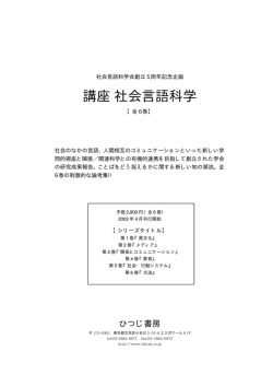
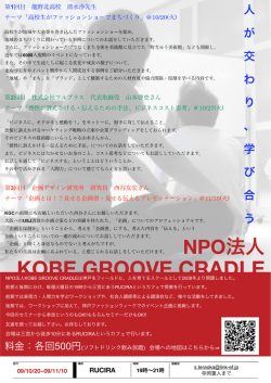
第回臨床解剖研究会記録 ..~ 骨造成術に必要な口蓋に分布する血管と神経の肉眼的解析 佐藤 巌1 浅海利恵子2 河合泰輔2 1日本歯科大学生命歯学部解剖学第一講座 目 的 歯科インプラント術では自家骨移植を伴う骨造成術 に下顎骨や上顎骨の隣接部位から骨片を取ることが多 い.特に歯周外科手術(移植)では骨造成にわずかな 自家骨で十分な量を確保できるために,安心性や利便 性から周辺の部位である口蓋から採取が行われてい る.口蓋は角化層(肥厚した重層扁平上皮)で被わ れ,血管や神経の走行は肉眼的に把握するのは難し い.外科的侵襲が小さく,合併症回避安心安全の部位 の確認のために解剖学的特色の把握が不可欠であるこ とから,口蓋の骨の形態の把握と神経・血管分布を評 価することで骨片採取の有用性を検討した. 三輪容子1 代居 敬2 2同大学生命歯学部歯科放射線学講座 方法と材料 日本歯科大学生命歯学部において人体解剖学実習に 供された,頭頸部に腫瘍疾患のないご献体 8 体( 72 ~100歳)(男性4 例,女性4 例)と大学所蔵の骨 格標本74体(42~83歳)(男性36例,女性38例) を観察資料とした.方法はコーンビーム CT(CBCT) にて撮影し(管電圧 85 kv ,管電流 3 mA , 17 sec ) 得られた CBCT 画像を口蓋溝の位置,形状について 観察し,溝の深さと溝の長さについて計測を行った. 肉眼的には実体顕微鏡下で口蓋に分布する血管や神経 の走行を検討した. Fig. 1 Frontal section of CBCT image. 4 pattern of the groove on the palate. A: bimodal, B: modal, C: protrusion type, D: irregular. In each pattern to form a groove (arrows) 12 臨床解剖研究会記録 No. 14 2014. 2 春原正隆1 Fig. 2 The inferior view of soft palate (73-year-old female). Distribution of the blood vessels and nerves (arrows) in the left and right side of the alveolar median palatine process of the maxilla from the palate bone. In the alveolar region of the upper jaw, complex vessels are located on the median side. A: greater palatine artery, N: greater palatine nerve, V: greater palatine vein, (bar 1 cm) 結果と考察 骨格標本からの口蓋溝形態では双峰型(有歯顎 1 例 5,無歯顎11例21.2) ,山型(有歯顎 7 例35, 無歯顎17例32.6),突起型(有歯顎 9 例45,無歯 顎 4 例 7.7 ),不定形(有歯顎 3 例 15 ,無歯顎 20 例38.6),最大隆起部位は頬骨下稜の前方(有歯顎 1 例 5,無歯顎 1 例 2),直下(有歯顎 1 例 5, 無歯顎 8 例16),後方(有歯顎18例90,無歯顎41 例 82 )であった( Fig. 1 ).また,左右の溝の位置 は正中線から 8.6~16.6 mm で,溝の深さは 1.5~2.8 mm ,長さは 4.5 ~ 14.2 mm であった.解剖標本では 大口蓋神経は大口蓋孔を出た後,唇側寄りに走行する 枝と口蓋中央側に走行する枝がみられ,口蓋中央側枝 の例が多かった( 12 / 16, 75 ).また,大口蓋動脈 の通路に臼歯部口蓋側に溝や管を認め,この溝や管を 通過し,正中側に多くの枝を出し,一部は前歯部の切 歯溝に向かう枝を認めた( Fig. 2 ).口蓋の溝を越え ない臼歯部では血管が少なく,出血の可能性が低いこ とが示唆された.骨に隆起がはっきり認められる例は 血管の局在性を認めたが,平坦な例はかならずしも当 てはまらない傾向がみられた.このことから口蓋から の骨採取にはあらかじめ口蓋突起の構造を十分把握す ることが重要であると示唆された. 文 献 1) Benninger B, Andrews K, Carter W. 2012. Clinical measurements of hard palate and implications for subepithelial connective tissue grafts with suggestions for palatal nomenclature. J Oral Maxillofac Surg 70: 149153 2) Gauthier A, L áezy JP, Vacher C. 2002. Vascularization of the palate in maxillary osteotomies: anatomical study. Surg Radiol Anat 24: 1317 3) Harnet JC, Feki A, Maillot C. 1994.The greater palatine canal: Radio-anatomical study and clinical value. J Radiol 75: 287293 4) Li KK, Meara JG, Alexander A Jr. 1996. Location of the descending palatine artery in relation to the Le Fort I osteotomy. J Oral Maxillofac Surg 54: 822825, discussion 826827 5) Mellema JW, Tami TA. 2004. An endoscopic study of the greater palatine nerve. Am J Rhinol 18: 99103 Macroscopic and CBCT analysis of vessels and nerves distributed in the palate for bone reclamation for dental implant Iwao SATO1, Rieko ASAUMI2, Taisuke KAWAI2, Yoko MIWA1, Takashi YOSUE2, Masataka SUNOHARA1 Departments of 1Anatomy and 2Oral and Maxillofacial Radiology, School of Life Dentistry at Tokyo, Nippon Dental University The removal of pieces of bone from the maxilla and mandible for autogenous bone grafts in dental implant treatment is often performed to ensure a su‹cient amount of bone material. However, the palate is covered with a keratinized layer (stratiˆed squamous thickened epithelium), and the existence in this layer of some blood vessels and nerves renders risk for surgical treatment. We examined the usefulness of bone fragments collected from this site, in which nerves and blood vessels are speciˆcally located. We anatomically and macroscopically observed six human cadavers and dry skull specimens using CBCT images. We also examined the courses of the blood vessels and nerves. The greater palatine nerves ran to the palate center side (CL) and close to the lips. In the posterior region of the palate-side passage, the greater palatine artery passed through the tube or groove with many branches to the median side, and a branch toward the incisor groove of the anterior part was also recognized. These routes of supply indicate a slight possibility of bleeding at the site of the bony tube and groove. These localized nerves and blood vessels and the structure of the palatine bone are important to consider when taking bone from the palate, as suggested by this study. Key words: CBCT, dental implant, greater palatine artery, nerve, bony tube and groove 骨造成術に必要な口蓋に分布する血管と神経の肉眼的解析 13
© Copyright 2026 Paperzz