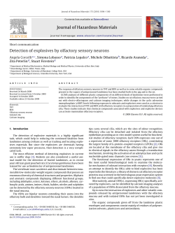
ACTA otorhinolaryngologica italica 2015;35:307-313; doi: 10.14639/0392-100X-669
Review
Outcome predictors of treatment effectiveness for
fungal malignant external otitis: a systematic review
Predittori di efficacia nel trattamento dell’otite esterna maligna fungina: una review
sistematica
M. Mion, R. BOVo, R. Marchese-Ragona, A. Martini
Institute of Otolaryngology, Department of Neurosciences, Padova University, Italy
Summary
The aim of this review is to summarise literature data on clinical aspects and traditional management of fungal malignant external otitis
(FMEO), and to identify potential predictive factors of positive treatment outcome. Articles were initially selected based on their titles or
abstracts. Full articles were then retrieved and further scrutinised according to predetermined criteria. Reference lists of selected articles
were searched for any missed publications. The selected articles were methodologically evaluated. Of an initial 143 references, 14 were
selected that focalised on the management of FMEO. The majority of studies demonstrated a correlation between treatment effectiveness,
assessed as symptom resolution, and clinical and management variables: abstention from surgical debridement, absence of facial palsy,
Aspergillus spp. as causative pathogen and absence of imaging findings at diagnosis and follow-up. The effectiveness of FMEO treatment
depends on the assessment of cranial nerve state, the causative pathogen and imaging findings. Above all, absence of facial nerve palsy,
Aspergillus spp. and absence of radiological signs at diagnosis and during follow-up correlate with symptom resolution. The fact that
conservative treatment may be associated with a better outcome than surgical debridement could purely reflect that patients with more aggressive and advanced illness required debridement, whereas milder disease was treated conservatively. Thus, caution should be advised in
the interpretation of data due to the need for further trials on the topic.
Key words: Fungal • Malignant external otitis • Predictive factors • Review • Treatment
Riassunto
Obiettivo dello studio è stato riassumere i dati della letteratura sugli aspetti clinici e la gestione abituale dell’otite esterna maligna fungina
(OEMF), ed identificare possibili fattori predittivi di esito positivo del trattamento. Gli articoli sono stati inizialmente selezionati sulla
base del titolo e degli abstract. Sono poi stati recuperati ed analizzati per intero seguendo criteri predeterminati. È stata stilata una lista
di riferimento degli articoli selezionati per cercare eventuali pubblicazioni mancati. Gli studi raccolti sono stati infine valutati metodologicamente. Dei 143 articoli iniziali, ne sono stati selezionati 14 focalizzati sulla gestione dell’OEMF. La maggior parte di questi ha dimostrato una correlazione tra l’efficacia del trattamento, inteso come risoluzione dei sintomi, ed alcune variabili cliniche e di gestione della
patologia quali l’astensione da procedure chirurgiche invasive, l’assenza di paralisi facciale, l’Aspergillus spp come patogeno causante e
l’assenza di segni radiologici alla diagnosi e nel corso del follow-up. L’efficacia del trattamento dipende dalla valutazione dello stato dei
nervi cranici, dal patogeno alla base e dai segni radiologici, più precisamente: l’assenza di paralisi facciale, l’Aspergillus spp e l’imaging
negativo alla diagnosi e durante il follow-up correlano con la risoluzione dei sintomi. L’evidenza che il trattamento farmacologico possa
associarsi ad un miglior outcome rispetto a procedure chirurgiche invasive potrebbe semplicemente riflettere il fatto che pazienti affetti da
una patologia più avanzata richiedono un approccio più aggressivo mentre le forme più lievi possono essere trattate in modo conservativo.
È necessario quindi prestare attenzione nell’interpretazione dei dati a causa della necessità di ulteriori studi sull’argomento.
Parole chiave: Fungina • Otite esterna maligna • Fattori predittivi • Review • Trattamento
Acta Otorhinolaryngol Ital 2015;35:307-313
Introduction
Malignant external otitis (MEO) is an aggressive and
potentially fatal infection that originates in the external
ear canal and spreads progressively along the soft tissue
and bone of the skull base 1. Since Chandler’s publication
of the first comprehensive case series of MEO in 1968 1,
the most commonly reported causative pathogen is Pseudomonas aeruginosa.
Fungi are rarely involved in MEO, but have been identified in immunocompromised patients, such as those with
AIDS or acute leukaemia. Aspergillus fumigates is the
most common cause of fungal malignant external otitis
(FMEO) 2. Aspergillus FMEO occurs in immunocompromised patients, usually with profound and long-lasting
neutropenia or under long-term steroid therapy, as well
as in individuals with uncontrolled diabetes mellitus 2.
307
M. Mion et al.
The disease manifests as a painful inflammation of the
external ear canal, associated with purulent otorrhoea
and granulation polyps. Otalgia is presenting symptom in 75% of cases 3; it is intense, particularly during
the night, and associated with severe temporal or occipital headache. The purulent otorrhoea appears with
a frequency ranging from 50 to 80% and varies from
a moist and modest secretion to greenish malodorous
and abundant exudates. Histologically, granulation tissue is characterised by non-specific inflammation with
inflammatory cell infiltration and hyperplasia of squamous epithelium 3.
The progression of the disease has been divided into three
clinical stages: in the first, there is infection of the external auditory canal and adjacent soft tissues with severe
pain, with or without facial nerve palsy. The second stage
is characterised by extension of infection with osteitis of
skull base and temporal bone, or multiple cranial nerve
neuropathies. In the third stage, the infection reaches the
intracranial structures, neck spaces and large blood vessels. This stage is always associated with poor prognosis.
The most frequent causes of death are meningitis, large
vessel septic thrombophlebitis or rupture, septicaemia,
pneumonia caused by inhalation for vagal paralysis and
cerebrovascular accident 3.
The treatment of FMEO classically includes extensive
surgical debridement and intensive long-term antifungal
therapy with amphotericin B and/or itraconazole and/or
voriconazole. Despite this management, FMEO is associated with substantial morbidity and mortality, mostly due
to late diagnosis and patient comorbidities 2. Treatment
failure can also be a result of suboptimal therapeutic management as a consequence of antifungal agent toxicity.
In particular, the side effects of amphotericin B, particularly renal failure, may require interruption of antifungal
agents or decrease in dosage 2, even if newer liposomal
amphotericin formulations have largely eliminated renal
failure as a side effect of this therapy.
Based on these premises, the aim of this review was to
summarise literature data on clinical aspects and traditional management of FMEO, and to identify potential
predictive factors of positive treatment outcome.
sible therapies and pitfalls of FMEO. The choice to
limit the included studies to those publishing after
2000 was due to the need for an updated approach to
the disease.
In order to identify relevant studies, as the first step, a
search was carried out in the Medline databases using
a combination of MeSH and keyword terms related to
FMEO (i.e., fungal or mycotic malignant external otitis).
No language limitations were set.
This first search step allowed the identification of a list
of potential citations for inclusion in this review. Titles
and abstracts on this list were screened by one independent reviewer (MM), who then determined whether to retrieve the full-text or not on the basis of the citations’
potential relevance to the review’s clinical research
question.
As a further expansion of the search, searches were performed “by hand” within the Scopus database, the authors’ personal libraries, and the references lists of the
full-text studies were performed to identify potential additional relevant citations.
All the retrieved full-texts were included in the review by
consensus of all authors.
Data extraction from the included publications was
performed by the same author who performed the initial search. Data regarding the demographic features
of the sample, comorbidities, pathogens, complications, imaging findings, treatments and symptoms
resolution were put into descriptive tables. All data
have been represented as they appeared in the original
publication.
Table I. Grading of evidence statements.
Level of evidence Type of evidence
1++
1+
12++
Materials and methods
Search strategy
The search strategy was designed to include papers on the
basis of their relevance for answering the clinical research
question: “Can FMEO treatment effectiveness, assessed
as symptoms’ resolution, be predicted by any clinical and/
or management variable?”
The inclusion criteria were based on the type of the
study: articles that were published after 2000, focusing on clinical manifestations, diagnostic tools, pos308
2+
2-
3
4
High quality meta-analyses, systematic reviews of
RCTs, or RCTs with a very low risk of bias.
Well-conduced meta-analyses, systematic reviews
of RCTs, or RCTs with a low risk of bias.
Meta-analyses, systematic reviews of RCTs, or
RCTs with a high risk of bias*.
High-quality systematic reviews of case-control or
cohort studies.
High quality case-control or cohort studies with a
very low risk of confounding, bias or chance and a
high probability that the relationship is casual.
Well-conducted case-control or cohort studies with
a low risk of confounding, bias or chance and a
moderate probability that the relationship is casual.
Case-control or cohort studies with a high risk of
confounding, bias or chance and a significant risk
that the relationship is casual*.
Non-analytic studies (for example case reports,
case series).
Expert opinion, formal consensus.
Studies with a level of evidence “-” are not used as a basis for making
recommendation; RCT: randomised controlled trial
* Outcome predictors of treatment effectiveness for fungal malignant external otitis
Quality assessment
As a quality assessment strategy, the included studies
were methodologically appraised according to the National Institute for Health and Clinical Excellence’s levels
of evidence (Table I).
Statistical analyses
Descriptive baseline and follow-up data were presented for
each patient. Statistical analyses were conducted with the aim
to identify potential predictors of treatment effectiveness.
Numerical continuous variables were expressed as
means ± standard deviation and nominal variables were
described by the absolute and relative (%) frequency.
For all statistical procedures, symptom resolution was the
main outcome variable which is adopted in a regression
analysis to be predicted by the dichotomic study variables
(i.e. sex [M/F], diabetes [yes/no], pathogen [Aspergillus/
no Aspergillus], facial palsy [yes/no], imaging findings
[yes/no], treatment [monotherapy/no monotherapy] [surgical debridement/no surgical debridement]).
For dichotomic variables, single variable regression analysis was performed, with the aim to screen among the potential predictors of the outcome variable. For all statistical analyses, the level of significance was set at p < 0.05.
All statistical procedures were performed with the Statistical Package for the Social Sciences (SPSS 19.0; SPSS
Inc., Chicago, Ill).
References retrieved by
electronic search strategy.
Medline (PubMed)
Total: n = 143
Excluded studies based on abstract
n = 129
Duplicates: n = 21
References not focused on fungal
malignant external otitis: n = 107
No abstract available: n = 1
Included studies relevant to
review on the basis of title and
abstract
n = 14
Excluded studies not meeting
inclusion criteria
n = 3
Published before 2000
Included full text articles
(effective studies) for detail
evaluation of eligibility
n = 11
Additional studies identified by
referenced linkage
n = 3
Results
Literature search
One hundred forty-three citations were retrieved from the
first phase of the search. A total of 132 references were
excluded after screening of the titles and abstracts because they were clearly not relevant for this review, published before the year 2000, were not relevant to the clinical research question, or were duplicate studies. Full texts
of the remaining 11 references were retrieved, along with
three additional full text articles that were identified as
potentially relevant by the second-step search expansion.
Based on the inclusion criteria, 14 articles were selected
for inclusion in this review (Fig. 1).
Overview of analysed studies
Of the 14 studies included in the review, all except three 2 4 5
are case reports. The average age of the 25 subjects is
62.6 ± 18, with 19 males and six females (Tables IIa, IIb).
80% of patients had diabetes mellitus.
Regarding the pathogen implicated, in the majority of cases Aspergillus spp. was found (61%), followed by Candida spp. (27%) and Scedosporium apiospermum (7%).
Only one study 6 described Malassezia sympodialis as the
cause of the disease.
52% of the subjects developed facial nerve palsy.
Studies included in the review
n = 14
Fig. 1. Study flow diagram.stract
Table IIa. Baseline and follow-up data on the study population.
All
No. of patients
Sex
Age (range)
Diabetes
Aspergillus spp.
Candida spp.
S. apiospermum
M. sympodialis
Facial nerve palsy
Imaging findings
Monotherapy
Duration
Combination therapy
Duration
Surgical debridement
Symptom resolution
25
19 (M) / 6 (F)
62.6 ± 18 (7-85)
80%
16 (61%)
7 (27%)
2 (7%)
1 (4%)
13 (52%)
9 (36%)
16 (64%)
178.4 days
9 (36%)
151.62 days
14 (56%)
15 (60%)
309
M. Mion et al.
Table IIb. Summary of reviewed studies.
Main author
Year of Age M/F Comorbidity Pathogen
paper (y)
Nerve
palsy
CT or MRI Therapy
findings
SD
Symptom
resolution
Walton et al. 7
2014
83
M
DM2
A. flavus
VII
Y
Voric iv (2 wk) then oral (11 mo)
N
Y
Tarazi et al. 4
2012
77
M
DM2
Aspergillus
No
Unspec
AmB then voric
N
Y
Tarazi et al. 4
2012
85
F
DM2
Aspergillus
VII
Unspec
AmB then voric
N
Y
Tarazi et al. 4
2012
70
F
DM2
Aspergillus
No
Unspec
Voric
N
Y
Bovo et al. 3
2012
69
M
DM2
A. fumigatus
VII
Y
Voric iv (400 mg x2 I day, then 200 mg Y
x2 for 3 mo)
No - facial palsy
Lilic et al. 8
2012
70
M
DM2
S. apiospermum
VII
Y
Ciprofloxacin (oral + topical)
N
No - died
Halsey et al. 9
2011
62
M
AML
A. wentii
VII
Unspec
AmB (2 wk) then Itz (8 wk)
Y
No - facial palsy
Hamzany et al. 5
2011
68
M
DM2
Candida
VII
Y
Voric + cloxacillin (42 days)
Y
No - died
Hamzany et al. 5
2011
68
M
DM2
Candida
VII
Y
Voric (42 days)
Y
No
Hamzany et al. 5
2011
68
M
DM2
Candida
VII
N
Voric(42 days)
Y
No
Hamzany et al. 5
2011
68
M
DM2
Candida
VII
N
Ciproflox + ceftaz (110 days) then
ceftaz + ofloxacin + vanco (66 days)
then flucon + meropenem (60 days)
then AmB (30 days)
Y
Y
Hamzany et al. 5
2011
68
M
DM2
Candida
VII
N
Flucon (75 days) then flucon +
rifampicin + fusidic ac (30 days)
Y
Y
Hamzany et al. 5
2011
68
M
DM2
Candida
No
N
Ceftaz (78 days) then meropenem (55
days) + Itz (225 days)
Y
Y
Hamzany et al. 5
2011
68
M
DM2
Aspergillus
No
Unspec
Meropenem (40 days) then flucon
(42 days) then flucon + rifampicin +
linezolid (60 days)
Y
Y
Hamzany et al. 5
2011
68
F
DM2
Aspergillus
No
Unspec
Voric + cotrimox (130 days)
N
Y
Hamzany et al. 5
2011
68
F
Nil
Aspergillus
No
Unspec
Ceftaz (42 days) then flucon +
ampicillin (30 days)
N
Y
Parize et al. 2
2009
48
M
Polychondritis
A. niger
No
Y
Voric (19 wk at 400 mg/day)
N
Y
Parize et al. 2
2009
40
F
IDDM, renal tx, A. niger
CRF
No
Y
Voric (52 wk at 600 mg/day)
Y
No - hearing loss
Ling et al. 10
2008
77
M
NIDDM, HTN,
gout
Candida,
A. flavus
VII
Y
Voric (14 wk at 400 mg/day) +
caspofungin
Y
Y
Narozny et al. 11
2006
65
M
NIDDM
Aspergillus
VII, IX, X Unspec
AmB + Itz
Y
No - died
Tzuku et al.
12
2006
17
F
IDDM
S. apiospermum
No
Y
AmB
Y
No – died
Bellini et al. 13
2003
73
M
NIDDM
A. niger
Unspec
Unspec
AmB (2 g in 3 wk) then Itz (13 wk at
600 mg/day)
N
Y
Shelton et al. 14
2002
58
M
Nil
A. niger
Unspec
Unspec
Itz (6 wk)
N
Y
Finer et al.
2002
7
M
Neuroblastoma A. flavus
Unspec
Unspec
AmB (6 days) then Itz (77 wk at
200 mg/day)
N
Y
2000
53
M
NIDDM
15
Chai et al. 6
M. sympodialis
VII, IX,
Unspec
AmB than flucon
N
No - facial palsy
X, XII
M: male; F: female; DM2: diabetes mellitus type II; AML: acute myeloid leukaemia; NIDDM: non insulin-dependent diabetes mellitus; tx: transplant; CRF: chronic renal
failure; HTN: hypertension; Y: yes; N: no; Unspec: unspecified; Voric: voriconazole; AmB: amphotericin B; Itz: itraconazole; Cirpoflox: ciprofloxacin; Ceftaz: caftazidime;
Vanco: vamcomycin; Flucon: fluconazole; Cotrimox: cotrimoxazol
In 36% of cases, there was evidence of the disease by
imaging, seen as soft-tissue filling of mastoid air cells
and middle ear and thickening of the roof of the external ear canal at the magnetic resonance 2 or skull base
osteomyelitis with involvement of the temporal bone
by CT 7.
64% of patients were treated with monotherapy, i.e.
voriconazole or amphotericin B, and the average duration of the treatment was 178.4 days. 36% of cases were
treated with associations of antifungal drugs for a mean
period of 151.62 days. 56% of subjects underwent surgi310
cal debridement. 60% of the 25 cases showed complete
resolution of symptoms.
In accordance with the clinical research question underlying this review, correlation between symptom resolution and clinical/management variables was assessed in
all studies.
Correlation analysis
In accordance to the main criterion for inclusion in this
review, all the studies assessed the correlation between
symptom resolution and dichotomic study variables (Ta-
Outcome predictors of treatment effectiveness for fungal malignant external otitis
Table III. Single variable correlation analysis. Predictors of symptom
resolution.
Predictors
Correlation with
p value
symptom resolution
No surgical debridement
Absence of facial palsy
Aspergillus as causative
pathogen
Absence of imaging
findings
Monotherapy
Diabetes
Male sex
5.416
5.416
4.250
0.022
0.022
0.041
4.250
0.041
2.097
1.427
0.412
0.173
0.307
0.702
ble III). Single variable regression analyses showed that
four of the dichotomic predictors were related with treatment outcomes: abstention from surgical debridement
and absence of facial palsy (χ2 = 5.416, p = 0.022), Aspergillus spp. as causative pathogen and absence of imaging findings were correlated with resolution of symptoms
(χ2 = 4.250, p = 0.041).
Male sex, presence of diabetes, preference for monotherapy over a combination of anti-fungal agents was not related with treatment outcomes.
Assessment of the quality of studies
All 14 studies could be categorised as evidence level 3
according to the National Institute for Health and Clinical Excellence’s levels of evidence. Thus, when considering the quality of those selected, the assessment showed a
good level of the reviewed articles as well as their qualitative homogeneity. However, their methodological heterogeneity did not allow for a meta-analysis of data.
Discussion
MEO is an insidious disease with frequently delayed diagnosis. Almost 95% of the cases reported in the literature are attributed to P. aeruginosa 3. Nevertheless, other
associated bacteria or fungi can have an important role
in the aetiology of MEO, particularly in immunocompromised patients, where Pseudomonas is not the predominant pathologic organism 3. Thus, even the isolation of
Pseudomonas from aural discharge may be not sufficient
or specific enough to distinguish between the two forms.
In fact, P. aeruginosa is frequently a saprophyte in the
external auditory meatus, as Staphylococcus coagulase
negative, diphtheroids, Micrococcus spp., Alternaria spp.,
Penicillium spp. and miceti.
As emphasised by Leonetti and Marzo 16, patients are either referred with previous cultures and biopsy findings
or are extremely tender on external auditory meatus manipulation; general anaesthesia is required to obtain an adequate biopsy. For these reasons, it is tempting to rely ei-
ther on previous findings or on bacteriologic data to direct
therapy. As a consequence, Pseudomonas isolation may
delay correct diagnosis of fungal infection: the failure to
respond to anti-pseudomonal therapy can lead to surgical
exploration and detection of miceti 9.
On the other hand, Aspergillus spp. is also frequently isolated from external auditory canal smears and diagnosis
of FMEO should be based on histopathologic confirmation on deep tissue biopsy or isolation from blood cultures
or fistula exudates 17. Deep tissue specimens obtained at
surgery are necessary to diagnose fungi: this should reduce the risk of contamination or isolation of pathogenic
saprophytes 12. Sometimes, fungal cultures at the time of
admission are negative and convert to positive only in
later samples. In these cases, the primary infection can
be caused by a bacterial pathogen, and owing to the prolonged course of the disease and antibiotic treatment, patients acquire an opportunistic fungal infection 5.
Aspergillus fumigatus was once thought to be the most
common fungal pathogen in FMEO 10, with Aspergillus
flavus assumed to be a less frequent cause of the condition. However, recent research reveals that A. flavus is
about 100 times more virulent than A. fumigatus and has
an optimum temperature for growth of 37°C, which may
explain its particular pathogenicity in humans 7.
MRI and CT can be used in association with other imaging modes for diagnosis and follow-up of FMEO (i.e.,
Tc99MDP bone scanning, Ga76 citrate scanning and Ga67
SPECT). It is important to underline that the repeated
studies performed in the patients with persistent disease
showed that the number of subjects with radiological evidence of bone involvement increased in FMEO and decreased in MEO 5.
The presence of fungal pathogen makes the disease more
invasive 5. This was further confirmed by the higher number of facial nerve palsies in the patients with FMEO: VII
nerve palsy is reported in 75% of patients with Aspergillus spp. infection, compared with only 34% in MEO due
to P. aeruginosa 5 9.
The decision between conservative antimicrobial therapy
and surgical treatment can present a therapeutic challenge
in the management of these life-threatening infections,
especially in patients with existing immunodeficiency
and illness 3. Although bone sequestra and abscess are
treated surgically, the need for more aggressive treatment
is debatable. Some authors suggest that prompt surgical debridement consisting of radical mastoidectomy is
indicated in the majority of cases, particularly in fungal
diseases, which are more invasive with respect to bacterial pathogens 2 3. In the series reported by Hamzany 5, extensive surgery was carried out in 78% of FMEO vs 18%
in bacterial ones. However, other authors stress the fact
that extensive surgery may be counterproductive because
of the risk of exposing healthy bone to infection 18. Unfortunately, there are neither guidelines nor definite rec311
M. Mion et al.
ommendations with regards to surgical treatment of the
different forms of MEO.
The important principles of FMEO treatment include aggressive control of diabetes, improvement of immunocompetency when possible, and prolonged administration
of adequate doses of antifungal agents, although the optimum duration of therapy remains unknown 9. Amphotericin B and itraconazole were favoured for treatment of
FMEO in the earlier case reports, whereas voriconazole
has played a role in the therapy of the majority of reported
cases since 2008. Voriconazole is currently recommended
as first-line treatment in cases of invasive aspergillosis 7.
The intravenous form is recommended for use in systematically unwell patients, with the oral form being reserved
for those who are stable or have improved following initial
intravenous treatment. Voriconazole is widely distributed
throughout tissues and, in its oral form, is not usually associated with worsening of renal function. This is particularly important as patients with comorbidities affecting
renal function such as diabetes, who develop aspergillosis
FMEO, therefore require treatment with voriconazole.
The most commonly reported side effect of this antifungal agent include visual disturbances, particularly to colour vision, abnormal liver function tests, deranged renal
function, skin photosensitivity, skin cancer and electrolyte
abnormalities 7. As noted in the literature, amphotericin B
was most frequently used, although it has a significantly
poorer safety profile, including adverse effects on renal
function. Itraconazole and caspofungin have also been
used in cases reported in the literature 7.
Regarding hyperbaric oxygen, there is as yet no uniformity in treatment design. Several reports suggested a beneficial effect of hyperbaric oxygen, along with traditional
treatment 10 11, whereas the Cochrane Collaboration Database 19 found no clear data supporting its efficacy relative
to antibiotics and/or surgery.
According to our research, abstention from surgical debridement and the absence of facial palsy are positive predictors
of symptoms resolution, followed by Aspergillus spp. as
causative pathogen and absence of imaging findings.
The counterproductivity of extensive surgery found from
our research confirms the conclusions of Carfrae et al. 18,
but the fact that conservative treatment is statistically associated with a better outcome than surgical debridement
could purely reflect the disease stage rather than actively
contributing to the results: patients with more aggressive and advanced illness required debridement, whereas
milder disease was treated conservatively.
Sparing the facial nerve and absence of radiological signs
intuitively indicate a less invasive form of FMEO, which
correlates with a better prognosis. Even if the presence of
fungal pathogen makes the disease more invasive than the
bacterial form of MEO, Aspergillus spp. is more susceptible to anti-fungal agents than other fungi, i.e. Candida spp.
The reviewed studies were selected on the basis of their
312
description of clinical aspects and consequent management of FMEO. The choice to limit the included studies
to those published after 2000 was due to the need for an
updated approach to this disease. In spite of this strategy, unfortunately, the statistical analysis of predictors of
outcome is flawed with a small series of case reports and
study designs were so heterogeneous that meta-analysis of
data could not be performed, and the extraction of sound,
clinically-useful, conclusions was hard to perform. As a
strength of the review, the studies had the common feature
of relying on an accurate characterisation of management,
using updated diagnostic techniques and treatments. Also,
importantly, despite their methodological heterogeneity,
all studies provided an acceptable 3 level of evidence according to the National Institute for Health and Clinical
Excellence’s grading. Within these limitations, some correlations between the outcomes of FMEO therapy and
study variables were reported.
Notwithstanding this interesting information, it must be
recognised that the available literature is too limited to
permit a recommendation of any particular strategy in
the clinical setting: decisions based solely on the studied factors cannot be recommended, especially because
an integrated analysis of other risk factors (e.g., age, comorbidities, among others), must be taken into account.
More information should be drawn on the amount of the
advancement needed to achieve the best balance between
benefit and discomfort, which was shown to be a critical aspect in the FMEO literature. Moreover, as a further
limitation of this review, it should be pointed out that the
external validity is not optimal due to the poor consistency between the reviewed studies as concerns the methodological features.
Conclusions
This systematic review identified 14 studies trying to
relate specific clinical and management variables to the
effectiveness of FMEO treatment. The outcome depends
particularly on absence of facial nerve palsy, Aspergillus
spp. as causative pathogen and absence of radiological
signs at diagnosis and during follow-up. The fact that conservative treatment is statistically associated with a better
outcome than surgical debridement could purely reflect
the disease stage rather than actively contributing to the
results: patients with more aggressive and advanced illness required debridement, whereas milder disease was
treated conservatively.
Notwithstanding, the overall conclusions that can be
drawn from this review are still limited by the somewhat inconsistent findings and the heterogeneity of study
designs. Thus, it is suggested that further trials are performed before recommending any suggestions in the clinical setting.
Outcome predictors of treatment effectiveness for fungal malignant external otitis
References
1
2
3
treated with hyperbaric oxygen. Internat J Infect Dis
2008;12:550-2.
Chandler JR. Malignant external otitis. Laryngoscope
1968;78:1257-94.
11
Parize P, Chandesris MO, Lanternier F, et al. Antifungal
therapy of Aspergillus invasive otitis externa: efficacy of
voriconazole and review. Antimicrob Agents Chemother
2009;53:1048-53.
Narozny W, Kuczkowski J, Stankiewicz C, et al. Value of hyperbaric oxygen in bacterial and fungal malignant external
otitis treatment. Eur Arch Otorhinolaryngol 2006;263:680-4.
12
Tuzcu A, Bahceci M, Celen MK, et al. Necrotizing (malignant) otitis externa: an unusual localization of mucormycosis. Indian J Med Microbiol 2006;24:289-91.
13
Bellini C, Antonini P, Ermanni S, et al. Malignant otitis externa due to Aspergillus niger. Scand J Infect Dis 2003;35:284-8.
Bovo R, Benatti A, Ciorba A, et al. Pseudomonas and Aspergillus interaction in malignant external otitis: risk of treatment failure. Acta Otorhinolaringol Ital 2012;32:416-9.
4
Tarazi AE, Al-Tawfiq A, Abdi RF. Fungal malignant otitis
externa: pitfalls, diagnosis, and treatment. Otol Neurotol
2012;33:769-73.
14
Shelton JC, Antonelli PJ, Hackett R. Skull base fungal
osteomyelitis in an immunocompetent host. Otolaryng
2002;126:76-8.
5
Hamzany Y, Soudry E, Preis M, et al. Fungal malignant external otitis. J of Infect 2011;62:226-31.
15
6
Chai FC, Auret K, Christiansen, Yuen PW, et al. Malignant
otitis externa caused by Malassezia sympodialis. Head Neck
2000;22:87-9.
Finer G, Greenberg D, Leibovitz E, et al. Conservative treatment of malignant (invasive) external otitis caused by Aspergillus flavus with oral itraconazole solution in a neutropenic
patient. Scand J Infect Dis 2002;34:227-9.
16
Walton J, Coulson C. Fungal malignant otitis externa with
facial nerve palsy: tissue biopsy aids diagnosis. Case Rep
Otolayngol 2014;2014:192318.
Marzo SJ, Leonetti JP. Invasive fungal and bacterial infections of the temporal bone. Laryngoscope 2003;113:1503-7.
17
Lilic N, Mowjood M, Wong M. A rare and sinister variant of
a common ailment: Fungal malignant otitis externa. J Surg
Case Rep 2012;2012(9):4.
Martínez-Berriotxoa A, Montejo M, Aguirrebengoa K, et al.
Otomastoiditis caused by Aspergillus in AIDS. Enferm Infecc Microbiol Clin 1997;15:200-2.
18
Carfrae MJ, Kesser BW. Malignant otitis externa. Otolaryngol Clin North Am 2008;41:537-49.
19
Phillips JS, Jones SEM. Hyperbaric oxygen as an adjuvant treatment for malignant otitis externa (Review). The
Cochrane collaboration. Cochrane Database Syst Rev
2005;18(2):CD004617.
7
8
9
Halsey C, Lumley H, Luckit J. Necrotising external otitis caused by Aspergillus wentii: a case report. Mycoses
2011;54:e211-3.
10
Ling SS, Sader C. Fungal malignant otitis externa
Received: April 2, 2015 - Received: June 18, 2015
Address for correspondence: Marta Mion, Institute of Otolaryngology, Department of Neurosciences, Padova University,
via Giustiniani 2, 35121 Padova, Italy. Tel +39 049 8218626.
Fax: +39 049 8213113. E-mail: [email protected]
313
© Copyright 2026 Paperzz